Myxosarcoma
Left atrial myxosarcoma with systemic metastasis: a case report. (1/19)
The term myxosarcoma is currently not used in standard classification for soft tissue tumors, but restricted to cardiac tumors. Primary cardiac myxosarcoma is a very rare disease and is difficult to differentiate from myxoma clinically and pathologically. We report a case of left atrial myxosarcoma with widespread systemic metastasis in a 21-yr-old male. The patient presented with sudden onset of intermittent dyspnea and orthopnea. Echocardiography showed a mobile, pedunculated tumor, 7.5x5x2 cm in size, at left atrium. Histologically, the excised tumor showed an amorphous finely fibrillar and mucinous stroma, in which irregular cords and clusters of lepidic cells and large stellate cells with plump vesicular nuclei resembled the usual type of cardiac myxoma were noted. And it showed focally cellular area with great nuclear pleomorphism and frequent mitoses. The patient received combination chemotherapy, peripheral blood stem cell collection transplantation and operations for systemic metastases in the brain, skeletal muscle and lung. He is alive at present 37 months after initial diagnosis and has no more new metastatic lesion. (+info)Primary intracranial myxoid chondrosarcoma: report of a case and review of the literature. (2/19)
The authors present a case of primary intracranial extraosseous myxoid chondrosarcoma without any attachment to the cranium or the meninges. The clinical and radiological findings of the primary intraparenchymal tumor are described with a review of the literature concerning cranial and intracranial myxoid chondrosarcoma. (+info)Giant cell malignant fibrous histiocytoma of the breast: a case report. (3/19)
A case of primary malignant fibrous histiocytoma of the breast is reported. The patient was a 48-yr-old woman with a huge tumor involving almost the entire left breast. The central portion of her left breast was already rotted by extensive necrosis and inflammation. She was treated by radical mastectomy and axillary lymphadenectomy to level I. Pathologic examination supported by an immunohistochemical staining confirmed the tumor as malignant fibrous histiocytoma of giant cell type. Axillary lymph nodes were free from tumor metastasis. She had not taken any postoperative adjuvant therapy. The metastasis to lungs was found 2 months after the operation, and she died within 6 months. (+info)Tumors of the heart. An analysis of 79 cases. (4/19)
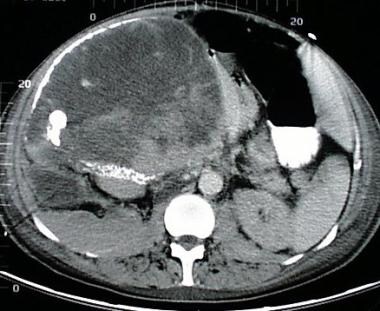
There were 79 cases of cardiac tumors seen from 1957 to July 1988. 49 (62.0%) of them were benign and 30 (38.0%) malignant. All the 49 benign tumors except 2 were surgically excised and found to be myxoma. Of them, 18 patients were male and 31 female. 85.7% of the tumors were located in the left atrium, 12.2% in the right atrium and 2.0% in the left ventricle. Palpitation, dyspnea, chest oppression, fever, episodes of syncope and hemiplegia, cough, diastolic and systolic murmurs at the apical or tricuspid area were the common symptoms and signs. Atrial fibrillation was found only in 2 cases. Echocardiographic findings were diagnostic while ECG and X-ray findings were nonspecific. Four patients died after operation. Of the 30 cases of malignant tumors, 15 were secondary tumors metastasized mainly from the lung or mediastinal malignancies. Of 11 primary tumor cases (7 males and 4 females), 3 were malignant lymphoma, 2 mesothelioma of pericardium, 2 malignant myxoma, 1 angiosarcoma, 1 leiomyosarcoma, 1 fibrosarcoma and 1 rhabdomyosarcoma. Another 4 cases were not studied histopathologically. The clinical manifestations, ECG and X-ray findings of the 11 primary tumors were nonspecific but echocardiography was helpful to the diagnosis. Six patients were operated on and 1 died during hospitalization. (+info)Skp2 overexpression is highly representative of intrinsic biological aggressiveness and independently associated with poor prognosis in primary localized myxofibrosarcomas. (5/19)
PURPOSE: Two SCF(Skp2) ubiquitin ligase-related proteins, Skp2 and cyclin-dependent kinase subunit 1 (Cks1), are involved in posttranscriptional degradation of p27(Kip1) tumor suppressor. We analyzed the prognostic utility of p27(Kip1) and its interacting cell cycle regulators in myxofibrosarcomas. EXPERIMENTAL DESIGN: Clinicopathologic features and tissue microarray-based immunohistochemical expression of p27(Kip1), Skp2, Cks1, cyclin E, cyclin A, Ki-67, and minichromosome maintenance protein 2 (Mcm2) were assessed in 70 primary myxofibrosarcomas and correlated with clinical outcomes. Skp2 mRNA expression and the relationship between Skp2 and p27(Kip1) proteins were examined in six cases by semiquantitative reverse transcription-PCR and Western blotting, respectively. RESULTS: High indices of Skp2 (> or =10%), cyclin A (> or =10%), and Mcm2 (> or =50%) were adverse prognosticators at the univariate level. Furthermore, co-overexpression of Skp2 and cyclin A identified highly lethal cases in the entire cohort [P < 0.0001 for disease-specific survival (DSS), P = 0.0004 for overall survival (OS)] and the lower-grade subset (Federation Nationale des Centres de Lutte Contre le Cancer grade 1 and 2; P = 0.0006 for DSS, P = 0.0093 for OS). In multivariate analyses, Skp2 overexpression overshadowed most intrinsic clinicopathologic factors and independently correlated with worse metastasis-free survival (P = 0.0012), DSS (P = 0.0234), and OS (P = 0.0056). Notably, positive margins independently predicted inferior local recurrence-free survival (P = 0.0012) and also negatively affected metastasis-free survival (P = 0.0471), DSS (P = 0.0152), and OS (P = 0.0173). Reverse transcription-PCR showed up-regulation of Skp2 mRNA in four cases and Western blotting displayed a matched expression pattern of Skp2. CONCLUSIONS: Margin status and intrinsic property of myxofibrosarcomas both affect patient survival. Skp2 overexpression is highly representative of the biological aggressiveness of myxofibrosarcomas and plays an important prognostic role. (+info)Spontaneous subcutaneous myxosarcoma in a captive European hedgehog (Erinsceus europaeus). (6/19)
A surgically excised biopsy representing a subcutaneous mass on the left side of the neck from a 3-year-old female European hedgehog (Erinsceus europaeus) was presented. Spontaneous myxosarcoma was diagnosed based on histological, immunohistochemical, and ultrastructural characteristics. The neoplasm grossly consisted of a firm, pale, multilobulated mass with a characteristic clear gelatinous fluid. Histologically, the neoplasm was nonencapsulated and composed of pleomorphic stellate or spindle-shaped vimentin and periodic acid-Schiff-positive cells arranged in loose sheets and occasionally whorls. The neoplastic cells were suspended in Alcian blue-positive stroma and contained infrequent mitotic figures. Evidence of a viral etiology was not detected using electron microscopy and polymerase chain reaction. This is the first case report of a myxosarcoma in a captive European hedgehog. (+info)Left atrial myxosarcoma with previously detected intestinal metastasis. (7/19)
Primary cardiac myxosarcoma is a rare disease; it is exceedingly rare for symptoms of systemic metastasis to precede diagnosis of the primary cardiac tumor. We describe the case of a previously healthy 60-year-old man with left atrial myxosarcoma, who had first presented with jejunal intussusception due to intestinal polyposis. Three months after resection of the jejunum, the patient experienced cerebral infarction and pulmonary edema. Further physical evaluation, which included echocardiography for the 1st time, revealed a mass in the left atrium that protruded through the mitral valve into the left ventricle. At emergency cardiac surgery, we found that the tumor involved multiple sites of the left atrium, the pulmonary veins, and the mitral anterior leaflet. Two months after surgery, the patient died of massive cerebral hemorrhage. Necropsy disclosed multiple recurrences of the cardiac myxosarcoma and widespread metastatic lesions. The intestinal polyps that had been resected originally were diagnosed, on retrospective histopathologic examination, as metastases of the myxosarcoma. In this unusual case, the metastatic lesions were the 1st clinical manifestations of a malignant cardiac tumor. (+info)Myxoid chondrosarcoma of the sinonasal cavity in a child: a case report. (8/19)
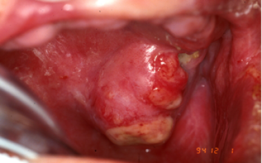
Chondrosarcomas are malignant tumors of cartilage that rarely involve the sinonasal region, and myxoid chondrosarcoma is a rare histologic variant of chondrosarcoma that usually occurs in the soft tissue of extremities. Although several case reports and results of small series of chondrosarcomas in the sinonasal region in children are available, myxoid type chondrosarcoma is extremely rare. We recently experienced a case of low grade myxoid chondrosarcoma involving the sinonasal cavity in a 10-year-old boy, and here we report its radiologic-pathologic findings. In this case, chondroid calcification on CT and septal and marginal enhancement on MRI suggested a chondrosarcoma. Whole body PET-CT demonstrated no definite metastatic lesion and a low peak standardized uptake value primary tumor. However, no definite distinguishing imaging features were observed that distinguished low grade myxoid chondrosarcoma from conventional chondrosarcoma. (+info)Myxosarcoma is a very rare type of soft tissue sarcoma, a cancer that develops in the soft tissues of the body, such as fat, muscle, nerves, blood vessels, and fibrous tissues. Myxosarcomas are characterized by the presence of mucoid or gelatinous material in the tumor, which is composed of an abnormal accumulation of acid mucopolysaccharides. These tumors typically affect adults, with a peak incidence in the sixth to seventh decade of life. They usually occur in the extremities, particularly the lower limbs, and can also arise in the retroperitoneum or other deep soft tissues. Myxosarcomas are classified into several subtypes based on their histological features, with the most common being the myxofibrosarcoma. Treatment typically involves surgical resection with wide margins, often followed by radiation therapy and/or chemotherapy. The prognosis for patients with myxosarcoma depends on several factors, including the size and location of the tumor, the histological grade, and the patient's age and overall health.
Heart neoplasms are abnormal growths or tumors that develop within the heart tissue. They can be benign (noncancerous) or malignant (cancerous). Benign tumors, such as myxomas and rhabdomyomas, are typically slower growing and less likely to spread, but they can still cause serious complications if they obstruct blood flow or damage heart valves. Malignant tumors, such as angiosarcomas and rhabdomyosarcomas, are fast-growing and have a higher risk of spreading to other parts of the body. Symptoms of heart neoplasms can include shortness of breath, chest pain, fatigue, and irregular heart rhythms. Treatment options depend on the type, size, and location of the tumor, and may include surgery, radiation therapy, or chemotherapy.
 Myxosarcoma - Wikipedia
 "Diagnostic and therapeutic approach to cardiac myxosarcoma in a dog" by MERİÇ KOCATÜRK, HAKAN SALCI et al.
 Adrenal Carcinoma: Practice Essentials, Background, Pathophysiology
 Canine Sarcoma | The National Canine Cancer Foundation
 Loving an Old Dog - Smart Dog University
 Atlantic Coast Veterinary Conference 2001 - VIN
 The Anesthesia Era 1845-1875 - The ASCO Post
 Comments on the US National Toxicology Program technical reports on toxicology and carcinogenesis study in rats exposed to...
 5 Minute Rounds
 Sarcoma risk and dioxin emissions from incinerators and industrial plants: a population-based case-control study (Italy) |...
 Research Update
 US Patent Application for VIRION-DERIVED NANOSPHERES FOR SELECTIVE DELIVERY OF THERAPEUTIC AND DIAGNOSTIC AGENTS TO CANCER...
 20 Overview of Tumors - KIPDF.COM
 Sarcoma
- Soft Tissue Sarcoma
Summary Report | CureHunter
Sarcoma
- Soft Tissue Sarcoma
Summary Report | CureHunter Alopecia and fur loss in degus - World of degus