Connective Tissue
Soft Tissue Neoplasms
Connective Tissue Diseases
Soft Tissue Injuries
Soft Tissue Infections
Connective Tissue Growth Factor
Sarcoma
Connective Tissue Cells
Mixed Connective Tissue Disease
Pancreatic Neoplasms
Neoplasms
Palate, Soft
Neoplasms, Cystic, Mucinous, and Serous
Leiomyosarcoma
Immunohistochemistry
Neoplasms, Multiple Primary
Neoplasms, Second Primary
Liposarcoma
Tomography, X-Ray Computed
Histiocytoma, Benign Fibrous
Muscle Neoplasms
Collagen
Neoplasms, Connective and Soft Tissue
Staphylococcal Skin Infections
Neoplasms, Connective Tissue
Treatment Outcome
Retrospective Studies
Histiocytoma, Malignant Fibrous
Tendons
Lipoma
Immediate-Early Proteins
Intercellular Signaling Peptides and Proteins
Adenocarcinoma, Mucinous
Sarcoma, Synovial
Ligaments
Neoplasms, Muscle Tissue
Myeloproliferative Disorders
Surgical Flaps
Dog Diseases
Neoplasm Staging
Cellulitis
Rhabdomyosarcoma
Neoplasm Recurrence, Local
Magnetic Resonance Imaging
Fibroblasts
Skin
Gastrointestinal Neoplasms
Therapy, Soft Tissue
Neoplasms, Experimental
Cystadenoma
Tumor Markers, Biological
Neoplasms, Plasma Cell
Face
Ovarian Neoplasms
Subcutaneous Tissue
Vascular Neoplasms
Chin
Neoplasm Proteins
Hemangiosarcoma
Scleroderma, Systemic
Cystadenoma, Mucinous
Neoplasms, Radiation-Induced
Prognosis
Neoplasm Metastasis
Carcinoma, Pancreatic Ductal
Reconstructive Surgical Procedures
Testicular Neoplasms
Neoplasms, Vascular Tissue
Bone and Bones
Fatal Outcome
Neoplasms, Adipose Tissue
Chondrosarcoma
Palatal Neoplasms
Collagen Diseases
Carcinoma, Papillary
Nerve Sheath Neoplasms
Adenocarcinoma, Papillary
Antigens, Neoplasm
Abdominal Neoplasms
Mandible
Pseudoxanthoma Elasticum
Maxilla
Follow-Up Studies
Neoplasms, Glandular and Epithelial
Histocytochemistry
Cystadenocarcinoma, Mucinous
Biopsy
Biomechanical Phenomena
Thoracic Neoplasms
Ifosfamide
Hematologic Neoplasms
Gingival Diseases
Facial Neoplasms
Neoplasms, Adnexal and Skin Appendage
Marfan Syndrome
Sarcoma, Clear Cell
Biopsy, Needle
Heart Neoplasms
Sweat Gland Neoplasms
Spinal Neoplasms
Chondroma
Facial Bones
Neoplasms, Fibrous Tissue
Extracellular Matrix
Mesenchymoma
Ehlers-Danlos Syndrome
Gingival Neoplasms
Fibrosis
Immunoenzyme Techniques
Microscopy, Electron
Combined Modality Therapy
RNA, Messenger
Cystadenocarcinoma
Granulation Tissue
Carcinoma
Gingivoplasty
Peripheral Nervous System Neoplasms
Vertical Dimension
Cells, Cultured
Chemotherapy, Cancer, Regional Perfusion
Fibrosarcoma
Meningeal Neoplasms
Cystadenoma, Serous
Microscopy, Electron, Scanning
Stress, Mechanical
Giant Cell Tumors
Dogs
Neoplasms, Germ Cell and Embryonal
Bone Marrow Neoplasms
Retroperitoneal Neoplasms
Biocompatible Materials
Debridement
Neoplasm Transplantation
Hemangiopericytoma
Neurilemmoma
Sensitivity and Specificity
Tongue
Malocclusion, Angle Class II
Colorectal Neoplasms
Esthetics
Chondrosarcoma, Mesenchymal
Joint Diseases
Cosmetic Techniques
Hemangioendothelioma
Extracellular Matrix Proteins
Nephroblastoma Overexpressed Protein
Imaging, Three-Dimensional
Pharmacokinetics of irinotecan and its metabolites SN-38 and APC in children with recurrent solid tumors after protracted low-dose irinotecan. (1/17)
Irinotecan (IRN), a topoisomerase I interactive agent, has significant antitumor activity in early Phase I studies in children with recurrent solid tumors. However, the disposition of IRN and its metabolites, SN-38 and APC, in children has not been reported. Children with solid tumors refractory to conventional therapy received IRN by a 1-h i.v. infusion at either 20, 24, or 29 mg/m2 daily for 5 consecutive days for 2 weeks. Serial blood samples were collected after doses 1 and 10 of the first course. IRN, SN-38, and APC lactone concentrations were determined by an isocratic high-performance liquid chromatography assay. A linear four-compartment model was fit simultaneously to the IRN, SN-38, and APC plasma concentration versus time data. Systemic clearance rate for IRN was 58.7 +/- 18.8 liters/h/m2 (mean +/- SD). The mean +/- SD ng/ml x h single-day lactone SN-38 area under the concentration-time curve (AUC(0-->6) was 90.9 +/- 96.4, 103.7 +/- 62.4, and 95.3 +/- 63.9 at IRN doses of 20, 24, and 29 mg/m2, respectively. The relative extent of IRN conversion to SN-38 and metabolism to APC measured after dose 1 were 0.49 +/- 0.33 and 0.29 +/- 0.17 (mean +/- SD). No statistically significant intrapatient difference was noted for SN-38 area under the concentration-time curve. Large interpatient variability in IRN and metabolite disposition was observed. The relative extent of conversion and the SN-38 systemic exposure achieved with this protracted schedule of administration were much greater than reported in adults or children receiving larger intermittent doses. (+info)Neoplasia in zebrafish (Danio rerio) treated with 7,12-dimethylbenz[a]anthracene by two exposure routes at different developmental stages. (2/17)
Using zebrafish, Danio rerio, initial pioneering work in the 1960s revealed carcinogen responsiveness of fish, yet very few subsequent tumorigenesis investigations have utilized this species. We exposed embryos (60 hours postfertilization) and fry (3 week posthatch) to 7,12-dimethylbenz[a]anthracene (DMBA) by immersion in aqueous solutions for 24 hours, at concentrations of 0-1 or 0-5 ppm (mg/L), respectively. Juvenile zebrafish 2 months posthatch were fed a diet containing 0-1,000 ppm DMBA for 4 months. Fish were sampled for histologic evaluation at 7-12 months after the onset of carcinogen treatment. Fry were most responsive to DMBA and showed the widest diversity of target tissues and histologic types of neoplasia, having several types of epithelial, mesenchymal, and neural neoplasia. The principal target tissues for carcinogenic response were liver following embryo or fry exposure, with gill and blood vessel the second and third most responsive tissues in fry. Intestine was the primary target and gill a secondary target in fish that received dietary DMBA as juveniles. These studies indicate that young zebrafish are most responsive to DMBA, showing a greater diversity of neoplasm types than rainbow trout. Thus, zebrafish are a valuable model system in which to study mechanistic aspects of the carcinogenesis process. (+info)Neoplasia in zebrafish (Danio rerio) treated with N-methyl-N'-nitro-N-nitrosoguanidine by three exposure routes at different developmental stages. (3/17)
We exposed embryos (83 hours postfertilizaton) and fry (3 weeks posthatch) to N-methyl-N'-nitro-N-nitrosoguanidine (MNNG) by immersion in aqueous solutions of 0-10 ppm for 1 hour (embryo) or 0-2 ppm for 24 hours (fry). Zebrafish embryos were microinjected with MNNG at levels of 0 or 96 ng/egg. Diets containing 0-2,000 ppm MNNG were fed to juvenile zebrafish for 3 months beginning at 2 months posthatch. Fish were sampled for histopathologic study at 6-12 months after initiation of carcinogen exposure. Embryos and fry were both quite responsive to MNNG; however, juvenile zebrafish were remarkably refractory to MNNG-induced neoplasia. Principal target organs in zebrafish treated as embryos with MNNG were liver and testis, with hepatocellular adenoma the most prevalent hepatic neoplasm. A variety of mesenchymal neoplasms occurred in zebrafish following embryo exposure to MNNG, including chondroma, hemangioma, hemangiosarcoma, leiomyosarcoma, and rhabdomyosarcoma. Testis and blood vessels were primary target organs for MNNG following fry exposure, with seminoma, hemangioma, hemangiosarcoma, and various other epithelial and mesenchymal neoplasms occurring. The zebrafish is a responsive, cost-effective lower vertebrate model system in which to study mechanisms of carcinogenesis. (+info)Alpha-inhibin immunoreactivity in soft-tissue neoplasia. (4/17)
Antibodies directed against alpha-inhibin have been previously reported as staining both sex cord-stromal neoplasms as well as adrenal cortical tumors. This relatively restricted immunoreactivity pattern is useful in the assessment of retroperitoneal masses, especially in a setting of limited tissue (e.g., needle biopsy). However, no study to date has evaluated alpha-inhibin immunoreactivity in soft-tissue neoplasms, which frequently enter the differential diagnosis of retroperitoneal masses. We investigate the incidence of alpha-inhibin staining in a variety of soft-tissue neoplasms by using formalin-fixed, paraffin-embedded tissue sections from 282 previously classified soft-tissue neoplasms with anti-alpha-inhibin (Serotec, 1:75). A modified avidin-biotin complex method was used after heat-induced epitope retrieval. Cytoplasmic granular staining was considered positive. Of the 282 tumors studied, a total of 8 (2.8%) demonstrated positive staining with anti-alpha-inhibin antibody. These included 4 of 25 liposarcomas (16%), 2 of 18 angiosarcomas (11%), 1 of 48 lipomas (2.1%), and 1 of 1 rhabdomyoma (100%). Negative staining was noted among hemangiomas (0/28), schwannomas (0/32), leiomyomas (0/16), fibrosarcomas (0/2), fibromas (0/11), dermatofibromas (0/9), neurofibromas (0/6), synovial sarcomas (0/15), rhabdomyosarcomas (0/10), Triton tumors (0/2), and malignant fibrous histiocytomas (0/59). We conclude that rare soft-tissue tumors, especially those exhibiting either lipomatous or vascular differentiation, demonstrate alpha-inhibin immunoreactivity. These findings re-emphasize the need for a well-construed antibody panel when immunohistochemical methods are employed in the evaluation of retroperitoneal neoplasms. However, the rarity of alpha-inhibin expression by soft-tissue neoplasms provides further support for its overall specificity as a marker of adrenal cortical differentiation in the biopsy evaluation of a retroperitoneal mass. (+info)Nuclear beta-catenin in mesenchymal tumors. (5/17)
Beta-catenin is a crucial part of the Wnt and E-cadherin signalling pathways, which are involved in tumorigenesis. Dysregulation of these pathways allow beta-catenin to accumulate and translocate to the nucleus, where it may activate oncogenes. Such nuclear accumulation can be detected by immunohistochemistry, which may be useful in diagnosis. Although the role of beta-catenin has been established in various types of carcinomas, relatively little is known about its status in mesenchymal tumors. A number of studies suggest that beta-catenin dysregulation is important in desmoid-type fibromatosis, as well as in synovial sarcoma. We wished to determine whether nuclear beta-catenin expression is specific to and sensitive for particular bone and soft-tissue tumors, including sporadic desmoid-type fibromatosis. We studied the nuclear expression of beta-catenin using tissue microarrays in a comprehensive range of bone and soft-tissue tumor types. A total of 549 cases were included in our panel. Nuclear immunohistochemical staining was determined to be either high level (>25% of cells), low level (0-25%) or none. High-level nuclear beta-catenin staining was seen in a very limited subset of tumor types, including desmoid-type fibromatosis (71% of cases), solitary fibrous tumor (40%), endometrial stromal sarcoma (40%) and synovial sarcoma (28%). Although occasional cases of fibrosarcoma, clear cell sarcoma and carcinosarcoma had high-level staining, no high-level nuclear beta-catenin expression was seen in any of 381 fibrohistocytic, muscular, adipocytic, chondroid or osseous tumor cases representing 42 diagnostic categories. All primary immunostain tissue microarray images are made publicly accessible in a searchable database. High-level nuclear beta-catenin staining serves as a useful diagnostic tool, as it is specific to a small subset of mesenchymal tumors. (+info)A molecular map of mesenchymal tumors. (6/17)
BACKGROUND: Bone and soft tissue tumors represent a diverse group of neoplasms thought to derive from cells of the mesenchyme or neural crest. Histological diagnosis is challenging due to the poor or heterogenous differentiation of many tumors, resulting in uncertainty over prognosis and appropriate therapy. RESULTS: We have undertaken a broad and comprehensive study of the gene expression profile of 96 tumors with representatives of all mesenchymal tissues, including several problem diagnostic groups. Using machine learning methods adapted to this problem we identify molecular fingerprints for most tumors, which are pathognomonic (decisive) and biologically revealing. CONCLUSION: We demonstrate the utility of gene expression profiles and machine learning for a complex clinical problem, and identify putative origins for certain mesenchymal tumors. (+info)Activation of the mTOR pathway in sporadic angiomyolipomas and other perivascular epithelioid cell neoplasms. (7/17)
Angiomyolipoma (AML) belong to a family of tumors known as perivascular epithelioid cell tumors (PEComas) that share a common immunophenotypic profile of muscle and melanocytic differentiation. These tumors are clonal in nature and have a strong association with tuberous sclerosis. Genetic analyses have reported allelic imbalance at the TSC2 locus on 16p13. In the context of non-tuberous sclerosis complex (TSC), non-lymphangioleiomyomatosis-associated AMLs, and non-renal PEComas, the functional status of the TSC2 signaling pathway has not been reported. Studies over the last several years have uncovered a critical role of the TSC1/2 genes in negatively regulating the Rheb/mTOR/p70S6K cascade. Here, we examined the activity of this pathway in sporadic AMLs and PEComas using immunohistochemical and biochemical analyses. We found increased levels of phospho-p70S6K, a marker of mTOR activity, in 15 of 15 non-TSC AMLs. This was accompanied by reduced phospho-AKT expression, a pattern that is consistent with the disruption of TSC1/2 function. Western blot analysis confirmed mTOR activation concurrent with the loss of TSC2 and not TSC1 in sporadic AMLs. Similarly, elevated phospho-p70S6K and reduced phospho-AKT expression was detected in 14 of 15 cases of extrarenal PEComas. These observations provide the first functional evidence that mTOR activation is common to sporadic, non-TSC-related AMLs and PEComas. This suggests the possibility that mTOR inhibitors such as rapamycin may be therapeutic for this class of disease. (+info)Side population cells isolated from mesenchymal neoplasms have tumor initiating potential. (8/17)
Although many cancers are maintained by tumor-initiating cells, this has not been shown for mesenchymal tumors, in part due to the lack of unique surface markers that identify mesenchymal progenitors. An alternative technique to isolate stem-like cells is to isolate side population (SP) cells based on efflux of Hoechst 33342 dye. We examined 29 mesenchymal tumors ranging from benign to high-grade sarcomas and identified SP cells in all but six samples. There was a positive correlation between the percentage of SP cells and the grade of the tumor. SP cells preferentially formed tumors when grafted into immunodeficient mice, and only cells from tumors that developed from the SP cells had the ability to initiate tumor formation upon serial transplantation. Although SP cells are able to efflux rhodamine dye in addition to Hoechst 33342, we found that the ability to efflux rhodamine dye did not identify a population of cells enriched for tumor-initiating capacity. Here, we identify a subpopulation of cells within a broad range of benign and malignant mesenchymal tumors with tumor-initiating capacity. In addition, our data suggest that the proportion of SP cells could be used as a prognostic factor and that therapeutically targeting this subpopulation of cells could be used to improve patient outcome. (+info)Connective tissue is a type of biological tissue that provides support, strength, and protection to various structures in the body. It is composed of cells called fibroblasts, which produce extracellular matrix components such as collagen, elastin, and proteoglycans. These components give connective tissue its unique properties, including tensile strength, elasticity, and resistance to compression.
There are several types of connective tissue in the body, each with its own specific functions and characteristics. Some examples include:
1. Loose or Areolar Connective Tissue: This type of connective tissue is found throughout the body and provides cushioning and support to organs and other structures. It contains a large amount of ground substance, which allows for the movement and gliding of adjacent tissues.
2. Dense Connective Tissue: This type of connective tissue has a higher concentration of collagen fibers than loose connective tissue, making it stronger and less flexible. Dense connective tissue can be further divided into two categories: regular (or parallel) and irregular. Regular dense connective tissue, such as tendons and ligaments, has collagen fibers that run parallel to each other, providing great tensile strength. Irregular dense connective tissue, such as the dermis of the skin, has collagen fibers arranged in a more haphazard pattern, providing support and flexibility.
3. Adipose Tissue: This type of connective tissue is primarily composed of fat cells called adipocytes. Adipose tissue serves as an energy storage reservoir and provides insulation and cushioning to the body.
4. Cartilage: A firm, flexible type of connective tissue that contains chondrocytes within a matrix of collagen and proteoglycans. Cartilage is found in various parts of the body, including the joints, nose, ears, and trachea.
5. Bone: A specialized form of connective tissue that consists of an organic matrix (mainly collagen) and an inorganic mineral component (hydroxyapatite). Bone provides structural support to the body and serves as a reservoir for calcium and phosphate ions.
6. Blood: Although not traditionally considered connective tissue, blood does contain elements of connective tissue, such as plasma proteins and leukocytes (white blood cells). Blood transports nutrients, oxygen, hormones, and waste products throughout the body.
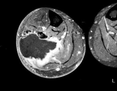
Soft tissue neoplasms refer to abnormal growths or tumors that develop in the soft tissues of the body. Soft tissues include muscles, tendons, ligaments, fascia, nerves, blood vessels, fat, and synovial membranes (the thin layer of cells that line joints and tendons). Neoplasms can be benign (non-cancerous) or malignant (cancerous), and their behavior and potential for spread depend on the specific type of neoplasm.
Benign soft tissue neoplasms are typically slow-growing, well-circumscribed, and rarely spread to other parts of the body. They can often be removed surgically with a low risk of recurrence. Examples of benign soft tissue neoplasms include lipomas (fat tumors), schwannomas (nerve sheath tumors), and hemangiomas (blood vessel tumors).
Malignant soft tissue neoplasms, on the other hand, can grow rapidly, invade surrounding tissues, and may metastasize (spread) to distant parts of the body. They are often more difficult to treat than benign neoplasms and require a multidisciplinary approach, including surgery, radiation therapy, and chemotherapy. Examples of malignant soft tissue neoplasms include sarcomas, such as rhabdomyosarcoma (arising from skeletal muscle), leiomyosarcoma (arising from smooth muscle), and angiosarcoma (arising from blood vessels).
It is important to note that soft tissue neoplasms can occur in any part of the body, and their diagnosis and treatment require a thorough evaluation by a healthcare professional with expertise in this area.
Connective tissue diseases (CTDs) are a group of disorders that involve the abnormal production and accumulation of abnormal connective tissues in various parts of the body. Connective tissues are the structural materials that support and bind other tissues and organs together. They include tendons, ligaments, cartilage, fat, and the material that fills the spaces between cells, called the extracellular matrix.
Connective tissue diseases can affect many different systems in the body, including the skin, joints, muscles, lungs, kidneys, gastrointestinal tract, and blood vessels. Some CTDs are autoimmune disorders, meaning that the immune system mistakenly attacks healthy connective tissues. Others may be caused by genetic mutations or environmental factors.
Some examples of connective tissue diseases include:
* Systemic lupus erythematosus (SLE)
* Rheumatoid arthritis (RA)
* Scleroderma
* Dermatomyositis/Polymyositis
* Mixed Connective Tissue Disease (MCTD)
* Sjogren's syndrome
* Ehlers-Danlos syndrome
* Marfan syndrome
* Osteogenesis imperfecta
The specific symptoms and treatment of connective tissue diseases vary depending on the type and severity of the condition. Treatment may include medications to reduce inflammation, suppress the immune system, or manage pain. In some cases, surgery may be necessary to repair or replace damaged tissues or organs.
Soft tissue injuries refer to damages that occur in the body's connective tissues, such as ligaments, tendons, and muscles. These injuries can be caused by various events, including accidents, falls, or sports-related impacts. Common soft tissue injuries include sprains, strains, and contusions (bruises).
Sprains occur when the ligaments, which connect bones to each other, are stretched or torn. This usually happens in the joints like ankles, knees, or wrists. Strains, on the other hand, involve injuries to the muscles or tendons, often resulting from overuse or sudden excessive force. Contusions occur when blood vessels within the soft tissues get damaged due to a direct blow or impact, causing bleeding and subsequent bruising in the affected area.
Soft tissue injuries can cause pain, swelling, stiffness, and limited mobility. In some cases, these injuries may require medical treatment, including physical therapy, medication, or even surgery, depending on their severity and location. It is essential to seek proper medical attention for soft tissue injuries to ensure appropriate healing and prevent long-term complications or chronic pain.
Soft tissue infections are medical conditions that involve infection of the soft tissues of the body, which include the skin, muscles, fascia (the connective tissue that surrounds muscles), and tendons. These infections can be caused by various types of bacteria, viruses, fungi, or parasites.
Soft tissue infections can range from mild to severe, depending on the type of organism causing the infection, the extent of tissue involvement, and the patient's overall health status. Some common types of soft tissue infections include:
1. Cellulitis: This is a bacterial infection that affects the skin and underlying tissues. It typically presents as a red, swollen, warm, and painful area on the skin, often accompanied by fever and chills.
2. Abscess: An abscess is a localized collection of pus in the soft tissues, caused by an infection. It can appear as a swollen, tender, and warm lump under the skin, which may be filled with pus.
3. Necrotizing fasciitis: This is a rare but severe soft tissue infection that involves the rapid destruction of fascia and surrounding tissues. It is often caused by a mixture of bacteria and can progress rapidly, leading to shock, organ failure, and even death if not treated promptly.
4. Myositis: This is an inflammation of the muscle tissue, which can be caused by a bacterial or viral infection. Symptoms may include muscle pain, swelling, weakness, and fever.
5. Erysipelas: This is a superficial skin infection that affects the upper layers of the skin and the lymphatic vessels. It typically presents as a raised, red, and painful rash with clear borders.
Treatment for soft tissue infections depends on the type and severity of the infection but may include antibiotics, drainage of pus or abscesses, and surgery in severe cases. Preventive measures such as good hygiene, wound care, and prompt treatment of injuries can help reduce the risk of developing soft tissue infections.
Connective Tissue Growth Factor (CTGF) is a cysteine-rich peptide growth factor that belongs to the CCN family of proteins. It plays an important role in various biological processes, including cell adhesion, migration, proliferation, and extracellular matrix production. CTGF is involved in wound healing, tissue repair, and fibrosis, as well as in the pathogenesis of several diseases such as cancer, diabetic nephropathy, and systemic sclerosis. It is expressed in response to various stimuli, including growth factors, cytokines, and mechanical stress. CTGF interacts with a variety of signaling molecules and integrins to regulate cellular responses and tissue homeostasis.
Sarcoma is a type of cancer that develops from certain types of connective tissue (such as muscle, fat, fibrous tissue, blood vessels, or nerves) found throughout the body. It can occur in any part of the body, but it most commonly occurs in the arms, legs, chest, and abdomen.
Sarcomas are classified into two main groups: bone sarcomas and soft tissue sarcomas. Bone sarcomas develop in the bones, while soft tissue sarcomas develop in the soft tissues of the body, such as muscles, tendons, ligaments, fat, blood vessels, and nerves.
Sarcomas can be further classified into many subtypes based on their specific characteristics, such as the type of tissue they originate from, their genetic makeup, and their appearance under a microscope. The different subtypes of sarcoma have varying symptoms, prognoses, and treatment options.
Overall, sarcomas are relatively rare cancers, accounting for less than 1% of all cancer diagnoses in the United States each year. However, they can be aggressive and may require intensive treatment, such as surgery, radiation therapy, and chemotherapy.
Connective tissue cells are a type of cell that are responsible for the production and maintenance of the extracellular matrix (ECM), which provides structural support and separates different tissues in the body. There are several types of connective tissue cells, including:
1. Fibroblasts: These are the most common type of connective tissue cell. They produce and maintain the ECM by synthesizing and secreting collagen, elastin, and other proteins that give the matrix its strength and elasticity.
2. Chondrocytes: These cells are found in cartilage and are responsible for producing and maintaining the cartilaginous matrix, which is composed of collagen and proteoglycans.
3. Osteoblasts: These cells are responsible for the formation and mineralization of bone tissue. They produce and secrete type I collagen and other proteins that form the organic matrix of bone, and they also regulate the deposition of calcium salts that mineralize the matrix.
4. Adipocytes: These are fat cells that store energy in the form of lipids. They are found in adipose tissue, which is a type of connective tissue that provides insulation and cushioning to the body.
5. Macrophages: These are large, mobile phagocytic cells that play an important role in the immune system. They are derived from monocytes and are found in many types of connective tissue, where they help to remove foreign particles, debris, and microorganisms.
6. Mast cells: These are connective tissue cells that contain granules filled with histamine, heparin, and other substances that are involved in inflammation and allergic reactions. They play a role in the immune response by releasing these granules when activated by antigens or other stimuli.
Connective tissue cells are essential for maintaining the structure and function of the body's tissues and organs, and they play an important role in wound healing, tissue repair, and the immune response.
Mixed Connective Tissue Disease (MCTD) is a rare overlapping condition of the connective tissues, characterized by the presence of specific autoantibodies against a protein called "U1-snRNP" or "U1-small nuclear ribonucleoprotein." This disorder has features of various connective tissue diseases such as systemic lupus erythematosus (SLE), scleroderma, polymyositis, and rheumatoid arthritis. Symptoms may include swollen hands, joint pain and swelling, muscle weakness, skin thickening, lung involvement, and Raynaud's phenomenon. The exact cause of MCTD is unknown, but it is believed to involve both genetic and environmental factors leading to an autoimmune response. Early diagnosis and treatment are essential for better disease management and preventing severe complications.
Pancreatic neoplasms refer to abnormal growths in the pancreas that can be benign or malignant. The pancreas is a gland located behind the stomach that produces hormones and digestive enzymes. Pancreatic neoplasms can interfere with the normal functioning of the pancreas, leading to various health complications.
Benign pancreatic neoplasms are non-cancerous growths that do not spread to other parts of the body. They are usually removed through surgery to prevent any potential complications, such as blocking the bile duct or causing pain.
Malignant pancreatic neoplasms, also known as pancreatic cancer, are cancerous growths that can invade and destroy surrounding tissues and organs. They can also spread (metastasize) to other parts of the body, such as the liver, lungs, or bones. Pancreatic cancer is often aggressive and difficult to treat, with a poor prognosis.
There are several types of pancreatic neoplasms, including adenocarcinomas, neuroendocrine tumors, solid pseudopapillary neoplasms, and cystic neoplasms. The specific type of neoplasm is determined through various diagnostic tests, such as imaging studies, biopsies, and blood tests. Treatment options depend on the type, stage, and location of the neoplasm, as well as the patient's overall health and preferences.
Neoplasms are abnormal growths of cells or tissues in the body that serve no physiological function. They can be benign (non-cancerous) or malignant (cancerous). Benign neoplasms are typically slow growing and do not spread to other parts of the body, while malignant neoplasms are aggressive, invasive, and can metastasize to distant sites.
Neoplasms occur when there is a dysregulation in the normal process of cell division and differentiation, leading to uncontrolled growth and accumulation of cells. This can result from genetic mutations or other factors such as viral infections, environmental exposures, or hormonal imbalances.
Neoplasms can develop in any organ or tissue of the body and can cause various symptoms depending on their size, location, and type. Treatment options for neoplasms include surgery, radiation therapy, chemotherapy, immunotherapy, and targeted therapy, among others.
The soft palate, also known as the velum, is the rear portion of the roof of the mouth that is made up of muscle and mucous membrane. It extends from the hard palate (the bony front part of the roof of the mouth) to the uvula, which is the small piece of tissue that hangs down at the back of the throat.
The soft palate plays a crucial role in speech, swallowing, and breathing. During swallowing, it moves upward and backward to block off the nasal cavity, preventing food and liquids from entering the nose. In speech, it helps to direct the flow of air from the mouth into the nose, which is necessary for producing certain sounds.
Anatomically, the soft palate consists of several muscles that allow it to change shape and move. These muscles include the tensor veli palatini, levator veli palatini, musculus uvulae, palatopharyngeus, and palatoglossus. The soft palate also contains a rich supply of blood vessels and nerves that provide sensation and help regulate its function.
Neoplasms: Neoplasms refer to abnormal growths of tissue that can be benign (non-cancerous) or malignant (cancerous). They occur when the normal control mechanisms that regulate cell growth and division are disrupted, leading to uncontrolled cell proliferation.
Cystic Neoplasms: Cystic neoplasms are tumors that contain fluid-filled sacs or cysts. These tumors can be benign or malignant and can occur in various organs of the body, including the pancreas, ovary, and liver.
Mucinous Neoplasms: Mucinous neoplasms are a type of cystic neoplasm that is characterized by the production of mucin, a gel-like substance produced by certain types of cells. These tumors can occur in various organs, including the ovary, pancreas, and colon. Mucinous neoplasms can be benign or malignant, and malignant forms are often aggressive and have a poor prognosis.
Serous Neoplasms: Serous neoplasms are another type of cystic neoplasm that is characterized by the production of serous fluid, which is a thin, watery fluid. These tumors commonly occur in the ovary and can be benign or malignant. Malignant serous neoplasms are often aggressive and have a poor prognosis.
In summary, neoplasms refer to abnormal tissue growths that can be benign or malignant. Cystic neoplasms contain fluid-filled sacs and can occur in various organs of the body. Mucinous neoplasms produce a gel-like substance called mucin and can also occur in various organs, while serous neoplasms produce thin, watery fluid and commonly occur in the ovary. Both mucinous and serous neoplasms can be benign or malignant, with malignant forms often being aggressive and having a poor prognosis.
Bone neoplasms are abnormal growths or tumors that develop in the bone. They can be benign (non-cancerous) or malignant (cancerous). Benign bone neoplasms do not spread to other parts of the body and are rarely a threat to life, although they may cause problems if they grow large enough to press on surrounding tissues or cause fractures. Malignant bone neoplasms, on the other hand, can invade and destroy nearby tissue and may spread (metastasize) to other parts of the body.
There are many different types of bone neoplasms, including:
1. Osteochondroma - a benign tumor that develops from cartilage and bone
2. Enchondroma - a benign tumor that forms in the cartilage that lines the inside of the bones
3. Chondrosarcoma - a malignant tumor that develops from cartilage
4. Osteosarcoma - a malignant tumor that develops from bone cells
5. Ewing sarcoma - a malignant tumor that develops in the bones or soft tissues around the bones
6. Giant cell tumor of bone - a benign or occasionally malignant tumor that develops from bone tissue
7. Fibrosarcoma - a malignant tumor that develops from fibrous tissue in the bone
The symptoms of bone neoplasms vary depending on the type, size, and location of the tumor. They may include pain, swelling, stiffness, fractures, or limited mobility. Treatment options depend on the type and stage of the tumor but may include surgery, radiation therapy, chemotherapy, or a combination of these treatments.
Skin neoplasms refer to abnormal growths or tumors in the skin that can be benign (non-cancerous) or malignant (cancerous). They result from uncontrolled multiplication of skin cells, which can form various types of lesions. These growths may appear as lumps, bumps, sores, patches, or discolored areas on the skin.
Benign skin neoplasms include conditions such as moles, warts, and seborrheic keratoses, while malignant skin neoplasms are primarily classified into melanoma, squamous cell carcinoma, and basal cell carcinoma. These three types of cancerous skin growths are collectively known as non-melanoma skin cancers (NMSCs). Melanoma is the most aggressive and dangerous form of skin cancer, while NMSCs tend to be less invasive but more common.
It's essential to monitor any changes in existing skin lesions or the appearance of new growths and consult a healthcare professional for proper evaluation and treatment if needed.
Leiomyosarcoma is a type of cancer that arises from the smooth muscle cells, which are responsible for the involuntary contractions of various organs and blood vessels. It most commonly occurs in the uterus, soft tissues (such as muscles and fat), and the gastrointestinal tract.
Leiomyosarcomas can vary in their aggressiveness and may spread to other parts of the body (metastasize) through the bloodstream or lymphatic system. The prognosis for leiomyosarcoma depends on several factors, including the location and size of the tumor, the patient's age and overall health, and the extent of metastasis. Treatment typically involves surgical removal of the tumor, along with radiation therapy and/or chemotherapy to help prevent recurrence or spread of the cancer.
Immunohistochemistry (IHC) is a technique used in pathology and laboratory medicine to identify specific proteins or antigens in tissue sections. It combines the principles of immunology and histology to detect the presence and location of these target molecules within cells and tissues. This technique utilizes antibodies that are specific to the protein or antigen of interest, which are then tagged with a detection system such as a chromogen or fluorophore. The stained tissue sections can be examined under a microscope, allowing for the visualization and analysis of the distribution and expression patterns of the target molecule in the context of the tissue architecture. Immunohistochemistry is widely used in diagnostic pathology to help identify various diseases, including cancer, infectious diseases, and immune-mediated disorders.
Multiple primary neoplasms refer to the occurrence of more than one primary malignant tumor in an individual, where each tumor is unrelated to the other and originates from separate cells or organs. This differs from metastatic cancer, where a single malignancy spreads to multiple sites in the body. Multiple primary neoplasms can be synchronous (occurring at the same time) or metachronous (occurring at different times). The risk of developing multiple primary neoplasms increases with age and is associated with certain genetic predispositions, environmental factors, and lifestyle choices such as smoking and alcohol consumption.
A "second primary neoplasm" is a distinct, new cancer or malignancy that develops in a person who has already had a previous cancer. It is not a recurrence or metastasis of the original tumor, but rather an independent cancer that arises in a different location or organ system. The development of second primary neoplasms can be influenced by various factors such as genetic predisposition, environmental exposures, and previous treatments like chemotherapy or radiation therapy.
It is important to note that the definition of "second primary neoplasm" may vary slightly depending on the specific source or context. In general medical usage, it refers to a new, separate cancer; however, in some research or clinical settings, there might be more precise criteria for defining and diagnosing second primary neoplasms.
Liposarcoma is a type of soft tissue sarcoma, which is a cancer that develops in the soft tissues of the body, such as fat, muscle, nerves, blood vessels, and fibrous tissues. Specifically, liposarcoma arises from fat cells (adipocytes) or their precursors.
There are several subtypes of liposarcoma, which differ in their appearance under the microscope, genetic features, and clinical behavior. These include well-differentiated, dedifferentiated, myxoid, round cell, and pleomorphic liposarcomas. The most common sites for liposarcoma are the thigh, retroperitoneum (the area behind the abdominal cavity), and the buttock.
Liposarcomas can grow slowly or rapidly, and they may spread to other parts of the body (metastasize) through the bloodstream or lymphatic system. Treatment typically involves surgical removal of the tumor, often followed by radiation therapy and/or chemotherapy. The prognosis for liposarcoma depends on several factors, including the type and grade of the tumor, its size and location, and whether it has spread to other parts of the body.
X-ray computed tomography (CT or CAT scan) is a medical imaging method that uses computer-processed combinations of many X-ray images taken from different angles to produce cross-sectional (tomographic) images (virtual "slices") of the body. These cross-sectional images can then be used to display detailed internal views of organs, bones, and soft tissues in the body.
The term "computed tomography" is used instead of "CT scan" or "CAT scan" because the machines take a series of X-ray measurements from different angles around the body and then use a computer to process these data to create detailed images of internal structures within the body.
CT scanning is a noninvasive, painless medical test that helps physicians diagnose and treat medical conditions. CT imaging provides detailed information about many types of tissue including lung, bone, soft tissue and blood vessels. CT examinations can be performed on every part of the body for a variety of reasons including diagnosis, surgical planning, and monitoring of therapeutic responses.
In computed tomography (CT), an X-ray source and detector rotate around the patient, measuring the X-ray attenuation at many different angles. A computer uses this data to construct a cross-sectional image by the process of reconstruction. This technique is called "tomography". The term "computed" refers to the use of a computer to reconstruct the images.
CT has become an important tool in medical imaging and diagnosis, allowing radiologists and other physicians to view detailed internal images of the body. It can help identify many different medical conditions including cancer, heart disease, lung nodules, liver tumors, and internal injuries from trauma. CT is also commonly used for guiding biopsies and other minimally invasive procedures.
In summary, X-ray computed tomography (CT or CAT scan) is a medical imaging technique that uses computer-processed combinations of many X-ray images taken from different angles to produce cross-sectional images of the body. It provides detailed internal views of organs, bones, and soft tissues in the body, allowing physicians to diagnose and treat medical conditions.
Kidney neoplasms refer to abnormal growths or tumors in the kidney tissues that can be benign (non-cancerous) or malignant (cancerous). These growths can originate from various types of kidney cells, including the renal tubules, glomeruli, and the renal pelvis.
Malignant kidney neoplasms are also known as kidney cancers, with renal cell carcinoma being the most common type. Benign kidney neoplasms include renal adenomas, oncocytomas, and angiomyolipomas. While benign neoplasms are generally not life-threatening, they can still cause problems if they grow large enough to compromise kidney function or if they undergo malignant transformation.
Early detection and appropriate management of kidney neoplasms are crucial for improving patient outcomes and overall prognosis. Regular medical check-ups, imaging studies, and urinalysis can help in the early identification of these growths, allowing for timely intervention and treatment.
Benign fibrous histiocytoma (BFH) is a common benign tumor of the skin and superficial soft tissues. It primarily affects middle-aged adults and is more prevalent in men than women. The exact cause of BFH is unknown, but it's thought to arise from dermal fibroblasts or histiocytes.
Medical Definition: Benign Fibrous Histiocytoma (BFH) is a benign, slowly growing, solitary cutaneous or subcutaneous nodular tumor predominantly composed of a mixture of fibroblastic and histiocytic-like cells. The tumor typically presents as a well-circumscribed, firm, dome-shaped papule or nodule, ranging in size from a few millimeters to several centimeters. Histologically, BFH is characterized by the proliferation of spindle-shaped fibroblasts and histiocytes arranged in a storiform pattern, along with variable amounts of collagen deposition, multinucleated giant cells, and hemosiderin deposits. The lesion usually has a pushing border with no invasion into the surrounding tissues. BFH generally follows a benign clinical course, with local recurrence being uncommon following complete surgical excision.
Muscle neoplasms are abnormal growths or tumors that develop in the muscle tissue. They can be benign (non-cancerous) or malignant (cancerous). Benign muscle neoplasms are typically slow-growing and do not spread to other parts of the body, while malignant muscle neoplasms, also known as soft tissue sarcomas, can grow quickly, invade nearby tissues, and metastasize (spread) to distant parts of the body.
Soft tissue sarcomas can arise from any of the muscles in the body, including the skeletal muscles (voluntary muscles that attach to bones and help with movement), smooth muscles (involuntary muscles found in the walls of blood vessels, digestive tract, and other organs), or cardiac muscle (the specialized muscle found in the heart).
There are many different types of soft tissue sarcomas, each with its own set of characteristics and prognosis. Treatment for muscle neoplasms typically involves a combination of surgery, radiation therapy, and chemotherapy, depending on the type, size, location, and stage of the tumor.
Collagen is the most abundant protein in the human body, and it is a major component of connective tissues such as tendons, ligaments, skin, and bones. Collagen provides structure and strength to these tissues and helps them to withstand stretching and tension. It is made up of long chains of amino acids, primarily glycine, proline, and hydroxyproline, which are arranged in a triple helix structure. There are at least 16 different types of collagen found in the body, each with slightly different structures and functions. Collagen is important for maintaining the integrity and health of tissues throughout the body, and it has been studied for its potential therapeutic uses in various medical conditions.
Neoplasms of connective and soft tissue are abnormal growths or tumors that develop in the body's supportive tissues, such as cartilage, tendons, ligaments, fascia, and fat. These neoplasms can be benign (non-cancerous) or malignant (cancerous).
Benign connective and soft tissue neoplasms include:
- Lipomas: slow-growing, fatty tumors that develop under the skin.
- Fibromas: firm, benign tumors that develop in connective tissue such as tendons or ligaments.
- Nevi (plural of nevus): benign growths made up of cells called melanocytes, which produce pigment.
Malignant connective and soft tissue neoplasms include:
- Sarcomas: a type of cancer that develops in the body's supportive tissues such as muscle, bone, fat, cartilage, or blood vessels. There are many different types of sarcomas, including liposarcoma (fatty tissue), rhabdomyosarcoma (muscle), and osteosarcoma (bone).
- Desmoid tumors: a rare type of benign tumor that can become aggressive and invade surrounding tissues. While not considered cancerous, desmoid tumors can cause significant morbidity due to their tendency to grow and infiltrate nearby structures.
Connective and soft tissue neoplasms can present with various symptoms depending on their location and size. Treatment options include surgery, radiation therapy, chemotherapy, or a combination of these modalities. Regular follow-up care is essential to monitor for recurrence or metastasis (spread) of the tumor.
Staphylococcal skin infections are a type of skin infection caused by Staphylococcus aureus (S. aureus) bacteria, which commonly live on the skin and inside the nose without causing harm. However, if they enter the body through a cut or scratch, they can cause an infection.
There are several types of staphylococcal skin infections, including:
1. Impetigo: A highly contagious superficial skin infection that typically affects children and causes red, fluid-filled blisters that burst and leave a yellowish crust.
2. Folliculitis: An inflammation of the hair follicles that causes red, pus-filled bumps or pimples on the skin.
3. Furunculosis: A deeper infection of the hair follicle that forms a large, painful lump or boil under the skin.
4. Cellulitis: A potentially serious bacterial infection that affects the deeper layers of the skin and can cause redness, swelling, warmth, and pain in the affected area.
5. Abscess: A collection of pus that forms in the skin, often caused by a staphylococcal infection.
Treatment for staphylococcal skin infections typically involves antibiotics, either topical or oral, depending on the severity and location of the infection. In some cases, drainage of pus or other fluids may be necessary to promote healing. Preventing the spread of staphylococcal skin infections involves good hygiene practices, such as washing hands frequently, covering wounds and cuts, and avoiding sharing personal items like towels or razors.
Neoplasms of connective tissue are abnormal growths or tumors that develop from the cells that form the body's supportive framework, including bones, cartilage, tendons, ligaments, and other connective tissues. These neoplasms can be benign (non-cancerous) or malignant (cancerous), and they can cause various symptoms depending on their location and size.
There are several types of connective tissue neoplasms, including:
1. Fibroma: A benign tumor that arises from fibrous connective tissue.
2. Fibrosarcoma: A malignant tumor that develops from fibrous connective tissue.
3. Lipoma: A benign tumor that arises from fat cells.
4. Liposarcoma: A malignant tumor that develops from fat cells.
5. Chondroma: A benign tumor that arises from cartilage.
6. Chondrosarcoma: A malignant tumor that develops from cartilage.
7. Osteoma: A benign tumor that arises from bone.
8. Osteosarcoma: A malignant tumor that develops from bone.
9. Giant cell tumors: Benign or malignant tumors that contain many giant cells, which are large, multinucleated cells.
10. Synovial sarcoma: A malignant tumor that arises from the synovial tissue that lines joints and tendons.
Connective tissue neoplasms can cause various symptoms depending on their location and size. For example, a benign lipoma may cause a painless lump under the skin, while a malignant osteosarcoma may cause bone pain, swelling, and fractures. Treatment options for connective tissue neoplasms include surgery, radiation therapy, chemotherapy, or a combination of these approaches.
Treatment outcome is a term used to describe the result or effect of medical treatment on a patient's health status. It can be measured in various ways, such as through symptoms improvement, disease remission, reduced disability, improved quality of life, or survival rates. The treatment outcome helps healthcare providers evaluate the effectiveness of a particular treatment plan and make informed decisions about future care. It is also used in clinical research to compare the efficacy of different treatments and improve patient care.
Lung neoplasms refer to abnormal growths or tumors in the lung tissue. These tumors can be benign (non-cancerous) or malignant (cancerous). Malignant lung neoplasms are further classified into two main types: small cell lung carcinoma and non-small cell lung carcinoma. Lung neoplasms can cause symptoms such as cough, chest pain, shortness of breath, and weight loss. They are often caused by smoking or exposure to secondhand smoke, but can also occur due to genetic factors, radiation exposure, and other environmental carcinogens. Early detection and treatment of lung neoplasms is crucial for improving outcomes and survival rates.
Retrospective studies, also known as retrospective research or looking back studies, are a type of observational study that examines data from the past to draw conclusions about possible causal relationships between risk factors and outcomes. In these studies, researchers analyze existing records, medical charts, or previously collected data to test a hypothesis or answer a specific research question.
Retrospective studies can be useful for generating hypotheses and identifying trends, but they have limitations compared to prospective studies, which follow participants forward in time from exposure to outcome. Retrospective studies are subject to biases such as recall bias, selection bias, and information bias, which can affect the validity of the results. Therefore, retrospective studies should be interpreted with caution and used primarily to generate hypotheses for further testing in prospective studies.
Thyroid neoplasms refer to abnormal growths or tumors in the thyroid gland, which can be benign (non-cancerous) or malignant (cancerous). These growths can vary in size and may cause a noticeable lump or nodule in the neck. Thyroid neoplasms can also affect the function of the thyroid gland, leading to hormonal imbalances and related symptoms. The exact causes of thyroid neoplasms are not fully understood, but risk factors include radiation exposure, family history, and certain genetic conditions. It is important to note that most thyroid nodules are benign, but a proper medical evaluation is necessary to determine the nature of the growth and develop an appropriate treatment plan.
The term "extremities" in a medical context refers to the most distant parts of the body, including the hands and feet (both fingers and toes), as well as the arms and legs. These are the farthest parts from the torso and head. Medical professionals may examine a patient's extremities for various reasons, such as checking circulation, assessing nerve function, or looking for injuries or abnormalities.
Malignant fibrous histiocytoma (MFH) is not a specific type of histiocytoma; rather, it is a type of soft tissue sarcoma. Histiocytomas are benign tumors that arise from cells called histiocytes, which are part of the immune system. MFH, on the other hand, is a malignant (cancerous) tumor that can arise in various types of soft tissues, such as muscle, fat, tendons, and ligaments.
MFH was once thought to originate from histiocytes, but more recent research suggests that it may actually arise from undifferentiated mesenchymal cells, which are capable of developing into a variety of different cell types. MFH is the most common type of soft tissue sarcoma in adults over the age of 50 and typically presents as a painless mass in the extremities or retroperitoneum (the area in the back of the abdomen).
The tumor is characterized by the presence of fibroblastic and histiocytic-like cells, which can be quite pleomorphic (varied in shape and size) and may contain numerous mitotic figures (indicating rapid cell division). Treatment typically involves surgical excision, often followed by radiation therapy and/or chemotherapy. The prognosis for MFH depends on several factors, including the tumor's location, size, grade (degree of differentiation), and the patient's age and overall health.
A tendon is the strong, flexible band of tissue that connects muscle to bone. It helps transfer the force produced by the muscle to allow various movements of our body parts. Tendons are made up of collagen fibers arranged in parallel bundles and have a poor blood supply, making them prone to injuries and slow to heal. Examples include the Achilles tendon, which connects the calf muscle to the heel bone, and the patellar tendon, which connects the kneecap to the shinbone.
A lipoma is a common, benign (non-cancerous) soft tissue growth. It is composed of adipose or fatty tissue and typically found just beneath the skin, but they can also occur deeper within the body. Lipomas are usually round, moveable, and painless, although they may cause discomfort if they grow large enough to put pressure on nearby nerves or if they're located in a sensitive area. They generally grow slowly over time. Surgical removal is an option if the lipoma becomes bothersome or grows significantly in size. It's important to note that while lipomas are typically harmless, any new lumps or bumps should be evaluated by a healthcare professional to confirm the diagnosis and rule out other more serious conditions.
Immediate-early proteins (IEPs) are a class of regulatory proteins that play a crucial role in the early stages of gene expression in viral infection and cellular stress responses. These proteins are synthesized rapidly, without the need for new protein synthesis, after the induction of immediate-early genes (IEGs).
In the context of viral infection, IEPs are often the first proteins produced by the virus upon entry into the host cell. They function as transcription factors that bind to specific DNA sequences and regulate the expression of early and late viral genes required for replication and packaging of the viral genome.
IEPs can also be involved in modulating host cell signaling pathways, altering cell cycle progression, and inducing apoptosis (programmed cell death). Dysregulation of IEPs has been implicated in various diseases, including cancer and neurological disorders.
It is important to note that the term "immediate-early proteins" is primarily used in the context of viral infection, while in other contexts such as cellular stress responses or oncogene activation, these proteins may be referred to by different names, such as "early response genes" or "transcription factors."
Intercellular signaling peptides and proteins are molecules that mediate communication and interaction between different cells in living organisms. They play crucial roles in various biological processes, including cell growth, differentiation, migration, and apoptosis (programmed cell death). These signals can be released into the extracellular space, where they bind to specific receptors on the target cell's surface, triggering intracellular signaling cascades that ultimately lead to a response.
Peptides are short chains of amino acids, while proteins are larger molecules made up of one or more polypeptide chains. Both can function as intercellular signaling molecules by acting as ligands for cell surface receptors or by being cleaved from larger precursor proteins and released into the extracellular space. Examples of intercellular signaling peptides and proteins include growth factors, cytokines, chemokines, hormones, neurotransmitters, and their respective receptors.
These molecules contribute to maintaining homeostasis within an organism by coordinating cellular activities across tissues and organs. Dysregulation of intercellular signaling pathways has been implicated in various diseases, such as cancer, autoimmune disorders, and neurodegenerative conditions. Therefore, understanding the mechanisms underlying intercellular signaling is essential for developing targeted therapies to treat these disorders.
Adenocarcinoma, mucinous is a type of cancer that begins in the glandular cells that line certain organs and produce mucin, a substance that lubricates and protects tissues. This type of cancer is characterized by the presence of abundant pools of mucin within the tumor. It typically develops in organs such as the colon, rectum, lungs, pancreas, and ovaries.
Mucinous adenocarcinomas tend to have a distinct appearance under the microscope, with large pools of mucin pushing aside the cancer cells. They may also have a different clinical behavior compared to other types of adenocarcinomas, such as being more aggressive or having a worse prognosis in some cases.
It is important to note that while a diagnosis of adenocarcinoma, mucinous can be serious, the prognosis and treatment options may vary depending on several factors, including the location of the cancer, the stage at which it was diagnosed, and the individual's overall health.
The term "DNA, neoplasm" is not a standard medical term or concept. DNA refers to deoxyribonucleic acid, which is the genetic material present in the cells of living organisms. A neoplasm, on the other hand, is a tumor or growth of abnormal tissue that can be benign (non-cancerous) or malignant (cancerous).
In some contexts, "DNA, neoplasm" may refer to genetic alterations found in cancer cells. These genetic changes can include mutations, amplifications, deletions, or rearrangements of DNA sequences that contribute to the development and progression of cancer. Identifying these genetic abnormalities can help doctors diagnose and treat certain types of cancer more effectively.
However, it's important to note that "DNA, neoplasm" is not a term that would typically be used in medical reports or research papers without further clarification. If you have any specific questions about DNA changes in cancer cells or neoplasms, I would recommend consulting with a healthcare professional or conducting further research on the topic.
A fibroma is a benign (non-cancerous) tumor that consists primarily of fibrous or connective tissue. It can occur in various parts of the body, including the skin, mouth, and internal organs. The term "fibroma" is often used to describe any benign fibrous growth, but there are specific types of fibromas such as dermatofibroma (found in the skin), oral fibroma (found in the mouth), and benign fibrous histiocytoma (found in soft tissues).
It's important to note that while fibromas are generally harmless, they can cause discomfort or problems depending on their size and location. If a fibroma is causing issues or there's concern about its growth or malignancy, it should be evaluated by a healthcare professional for potential removal or further assessment.
Synovial sarcoma is a rare type of cancer that typically develops in the soft tissues surrounding the joints, such as the synovial membrane, which lines the joint capsules. Despite its name, synovial sarcoma does not necessarily arise from the synovium. It is called so due to its resemblance to this tissue under a microscope.
This form of sarcoma primarily affects young adults and can be found in various parts of the body, but it most commonly occurs in the extremities, particularly near the knees. Synovial sarcoma is characterized by specific genetic changes that result in the formation of fusion proteins, which contribute to uncontrolled cell growth and tumor development.
There are two main subtypes of synovial sarcoma: monophasic and biphasic. Monophasic synovial sarcoma is composed of either spindle-shaped (spaghetti-like) cells or epithelioid (roundish) cells, while biphasic synovial sarcoma contains both types of cells. A third subtype, called poorly differentiated synovial sarcoma, has a more aggressive behavior and is composed of small round cells that do not resemble the typical spindle or epithelioid cells.
Treatment for synovial sarcoma usually involves surgical removal of the tumor, often followed by radiation therapy and/or chemotherapy to reduce the risk of recurrence and metastasis. The prognosis varies depending on factors such as the size and location of the tumor, the patient's age, and the presence of metastases at diagnosis.
Ligaments are bands of dense, fibrous connective tissue that surround joints and provide support, stability, and limits the range of motion. They are made up primarily of collagen fibers arranged in a parallel pattern to withstand tension and stress. Ligaments attach bone to bone, and their function is to prevent excessive movement that could cause injury or dislocation.
There are two main types of ligaments: extracapsular and intracapsular. Extracapsular ligaments are located outside the joint capsule and provide stability to the joint by limiting its range of motion. Intracapsular ligaments, on the other hand, are found inside the joint capsule and help maintain the alignment of the joint surfaces.
Examples of common ligaments in the body include the anterior cruciate ligament (ACL) and posterior cruciate ligament (PCL) in the knee, the medial collateral ligament (MCL) and lateral collateral ligament (LCL) in the elbow, and the coracoacromial ligament in the shoulder.
Injuries to ligaments can occur due to sudden trauma or overuse, leading to sprains, strains, or tears. These injuries can cause pain, swelling, bruising, and limited mobility, and may require medical treatment such as immobilization, physical therapy, or surgery.
Neoplasms in muscle tissue refer to abnormal and excessive growths of muscle cells that can be benign or malignant. These growths can arise from any of the three types of muscle tissue: skeletal, cardiac, or smooth muscle. Neoplasms in muscle tissue are classified based on their origin, behavior, and histological features.
Benign neoplasms in muscle tissue include leiomyomas (smooth muscle), rhabdomyomas (skeletal muscle), and myxomas (cardiac muscle). These tumors are usually slow-growing and do not invade surrounding tissues or spread to other parts of the body.
Malignant neoplasms in muscle tissue, also known as sarcomas, include leiomyosarcoma (smooth muscle), rhabdomyosarcoma (skeletal muscle), and angiosarcoma (cardiac muscle). These tumors are aggressive, invasive, and have the potential to metastasize to other parts of the body.
Symptoms of neoplasms in muscle tissue depend on their location, size, and type. They may include a painless or painful mass, weakness, fatigue, weight loss, and difficulty swallowing or breathing. Treatment options for neoplasms in muscle tissue include surgery, radiation therapy, chemotherapy, and targeted therapy. The choice of treatment depends on the type, stage, location, and patient's overall health condition.
Myeloproliferative disorders (MPDs) are a group of rare, chronic blood cancers that originate from the abnormal proliferation or growth of one or more types of blood-forming cells in the bone marrow. These disorders result in an overproduction of mature but dysfunctional blood cells, which can lead to serious complications such as blood clots, bleeding, and organ damage.
There are several subtypes of MPDs, including:
1. Chronic Myeloid Leukemia (CML): A disorder characterized by the overproduction of mature granulocytes (a type of white blood cell) in the bone marrow, leading to an increased number of these cells in the blood. CML is caused by a genetic mutation that results in the formation of the BCR-ABL fusion protein, which drives uncontrolled cell growth and division.
2. Polycythemia Vera (PV): A disorder characterized by the overproduction of all three types of blood cells - red blood cells, white blood cells, and platelets - in the bone marrow. This can lead to an increased risk of blood clots, bleeding, and enlargement of the spleen.
3. Essential Thrombocythemia (ET): A disorder characterized by the overproduction of platelets in the bone marrow, leading to an increased risk of blood clots and bleeding.
4. Primary Myelofibrosis (PMF): A disorder characterized by the replacement of normal bone marrow tissue with scar tissue, leading to impaired blood cell production and anemia, enlargement of the spleen, and increased risk of infections and bleeding.
5. Chronic Neutrophilic Leukemia (CNL): A rare disorder characterized by the overproduction of neutrophils (a type of white blood cell) in the bone marrow, leading to an increased number of these cells in the blood. CNL can lead to an increased risk of infections and organ damage.
MPDs are typically treated with a combination of therapies, including chemotherapy, targeted therapy, immunotherapy, and stem cell transplantation. The choice of treatment depends on several factors, including the subtype of MPD, the patient's age and overall health, and the presence of any comorbidities.
Liver neoplasms refer to abnormal growths in the liver that can be benign or malignant. Benign liver neoplasms are non-cancerous tumors that do not spread to other parts of the body, while malignant liver neoplasms are cancerous tumors that can invade and destroy surrounding tissue and spread to other organs.
Liver neoplasms can be primary, meaning they originate in the liver, or secondary, meaning they have metastasized (spread) to the liver from another part of the body. Primary liver neoplasms can be further classified into different types based on their cell of origin and behavior, including hepatocellular carcinoma, cholangiocarcinoma, and hepatic hemangioma.
The diagnosis of liver neoplasms typically involves a combination of imaging studies, such as ultrasound, CT scan, or MRI, and biopsy to confirm the type and stage of the tumor. Treatment options depend on the type and extent of the neoplasm and may include surgery, radiation therapy, chemotherapy, or liver transplantation.
Parotid neoplasms refer to abnormal growths or tumors in the parotid gland, which is the largest of the salivary glands and is located in front of the ear and extends down the neck. These neoplasms can be benign (non-cancerous) or malignant (cancerous).
Benign parotid neoplasms are typically slow-growing, painless masses that may cause facial asymmetry or difficulty in chewing or swallowing if they become large enough to compress surrounding structures. The most common type of benign parotid tumor is a pleomorphic adenoma.
Malignant parotid neoplasms, on the other hand, are more aggressive and can invade nearby tissues and spread to other parts of the body. They may present as rapidly growing masses that are firm or fixed to surrounding structures. Common types of malignant parotid tumors include mucoepidermoid carcinoma, adenoid cystic carcinoma, and squamous cell carcinoma.
The diagnosis of parotid neoplasms typically involves a thorough clinical evaluation, imaging studies such as CT or MRI scans, and fine-needle aspiration biopsy (FNAB) to determine the nature of the tumor. Treatment options depend on the type, size, and location of the neoplasm but may include surgical excision, radiation therapy, and chemotherapy.
A surgical flap is a specialized type of surgical procedure where a section of living tissue (including skin, fat, muscle, and/or blood vessels) is lifted from its original site and moved to another location, while still maintaining a blood supply through its attached pedicle. This technique allows the surgeon to cover and reconstruct defects or wounds that cannot be closed easily with simple suturing or stapling.
Surgical flaps can be classified based on their vascularity, type of tissue involved, or method of transfer. The choice of using a specific type of surgical flap depends on the location and size of the defect, the patient's overall health, and the surgeon's expertise. Some common types of surgical flaps include:
1. Random-pattern flaps: These flaps are based on random blood vessels within the tissue and are typically used for smaller defects in areas with good vascularity, such as the face or scalp.
2. Axial pattern flaps: These flaps are designed based on a known major blood vessel and its branches, allowing them to cover larger defects or reach distant sites. Examples include the radial forearm flap and the anterolateral thigh flap.
3. Local flaps: These flaps involve tissue adjacent to the wound and can be further classified into advancement, rotation, transposition, and interpolation flaps based on their movement and orientation.
4. Distant flaps: These flaps are harvested from a distant site and then transferred to the defect after being tunneled beneath the skin or through a separate incision. Examples include the groin flap and the latissimus dorsi flap.
5. Free flaps: In these flaps, the tissue is completely detached from its original blood supply and then reattached at the new site using microvascular surgical techniques. This allows for greater flexibility in terms of reach and placement but requires specialized expertise and equipment.
Surgical flaps play a crucial role in reconstructive surgery, helping to restore form and function after trauma, tumor removal, or other conditions that result in tissue loss.
There is no medical definition for "dog diseases" as it is too broad a term. However, dogs can suffer from various health conditions and illnesses that are specific to their species or similar to those found in humans. Some common categories of dog diseases include:
1. Infectious Diseases: These are caused by viruses, bacteria, fungi, or parasites. Examples include distemper, parvovirus, kennel cough, Lyme disease, and heartworms.
2. Hereditary/Genetic Disorders: Some dogs may inherit certain genetic disorders from their parents. Examples include hip dysplasia, elbow dysplasia, progressive retinal atrophy (PRA), and degenerative myelopathy.
3. Age-Related Diseases: As dogs age, they become more susceptible to various health issues. Common age-related diseases in dogs include arthritis, dental disease, cancer, and cognitive dysfunction syndrome (CDS).
4. Nutritional Disorders: Malnutrition or improper feeding can lead to various health problems in dogs. Examples include obesity, malnutrition, and vitamin deficiencies.
5. Environmental Diseases: These are caused by exposure to environmental factors such as toxins, allergens, or extreme temperatures. Examples include heatstroke, frostbite, and toxicities from ingesting harmful substances.
6. Neurological Disorders: Dogs can suffer from various neurological conditions that affect their nervous system. Examples include epilepsy, intervertebral disc disease (IVDD), and vestibular disease.
7. Behavioral Disorders: Some dogs may develop behavioral issues due to various factors such as anxiety, fear, or aggression. Examples include separation anxiety, noise phobias, and resource guarding.
It's important to note that regular veterinary care, proper nutrition, exercise, and preventative measures can help reduce the risk of many dog diseases.
Neoplasm staging is a systematic process used in medicine to describe the extent of spread of a cancer, including the size and location of the original (primary) tumor and whether it has metastasized (spread) to other parts of the body. The most widely accepted system for this purpose is the TNM classification system developed by the American Joint Committee on Cancer (AJCC) and the Union for International Cancer Control (UICC).
In this system, T stands for tumor, and it describes the size and extent of the primary tumor. N stands for nodes, and it indicates whether the cancer has spread to nearby lymph nodes. M stands for metastasis, and it shows whether the cancer has spread to distant parts of the body.
Each letter is followed by a number that provides more details about the extent of the disease. For example, a T1N0M0 cancer means that the primary tumor is small and has not spread to nearby lymph nodes or distant sites. The higher the numbers, the more advanced the cancer.
Staging helps doctors determine the most appropriate treatment for each patient and estimate the patient's prognosis. It is an essential tool for communication among members of the healthcare team and for comparing outcomes of treatments in clinical trials.
Cellulitis is a medical condition characterized by an infection and inflammation of the deeper layers of the skin (dermis and subcutaneous tissue) and surrounding soft tissues. It's typically caused by bacteria, most commonly group A Streptococcus and Staphylococcus aureus.
The affected area often becomes red, swollen, warm, and painful, and may be accompanied by systemic symptoms such as fever, chills, and fatigue. Cellulitis can spread rapidly and potentially become life-threatening if left untreated, so it's important to seek medical attention promptly if you suspect you have this condition. Treatment typically involves antibiotics, rest, elevation of the affected limb (if applicable), and pain management.
Infectious skin diseases are conditions characterized by an infection or infestation of the skin caused by various microorganisms such as bacteria, viruses, fungi, or parasites. These organisms invade the skin, causing inflammation, redness, itching, pain, and other symptoms. Examples of infectious skin diseases include:
1. Bacterial infections: Cellulitis, impetigo, folliculitis, and MRSA (methicillin-resistant Staphylococcus aureus) infections are examples of bacterial skin infections.
2. Viral infections: Herpes simplex virus (HSV), varicella-zoster virus (VZV), human papillomavirus (HPV), and molluscum contagiosum are common viruses that can cause skin infections.
3. Fungal infections: Tinea pedis (athlete's foot), tinea corporis (ringworm), candidiasis (yeast infection), and pityriasis versicolor are examples of fungal skin infections.
4. Parasitic infestations: Scabies, lice, and bed bugs are examples of parasites that can cause infectious skin diseases.
Treatment for infectious skin diseases depends on the underlying cause and may include topical or oral antibiotics, antiviral medications, antifungal treatments, or insecticides to eliminate parasitic infestations. Proper hygiene, wound care, and avoiding contact with infected individuals can help prevent the spread of infectious skin diseases.
Rhabdomyosarcoma is a type of cancer that develops in the body's soft tissues, specifically in the muscle cells. It is a rare and aggressive form of sarcoma, which is a broader category of cancers that affect the connective tissues such as muscles, tendons, cartilages, bones, blood vessels, and fatty tissues.
Rhabdomyosarcomas can occur in various parts of the body, including the head, neck, arms, legs, trunk, and genitourinary system. They are more common in children than adults, with most cases diagnosed before the age of 18. The exact cause of rhabdomyosarcoma is not known, but genetic factors and exposure to radiation or certain chemicals may increase the risk.
There are several subtypes of rhabdomyosarcoma, including embryonal, alveolar, pleomorphic, and spindle cell/sclerosing. The type and stage of the cancer determine the treatment options, which may include surgery, radiation therapy, chemotherapy, or a combination of these approaches. Early diagnosis and prompt treatment are crucial for improving the prognosis and long-term survival rates.
Mandibular neoplasms refer to abnormal growths or tumors that develop in the mandible, which is the lower jawbone. These growths can be benign (non-cancerous) or malignant (cancerous). Benign neoplasms are typically slow-growing and rarely spread to other parts of the body, while malignant neoplasms can invade surrounding tissues and may metastasize (spread) to distant sites.
Mandibular neoplasms can have various causes, including genetic mutations, exposure to certain chemicals or radiation, and infection with certain viruses. The symptoms of mandibular neoplasms may include swelling or pain in the jaw, difficulty chewing or speaking, numbness in the lower lip or chin, loose teeth, and/or a lump or mass in the mouth or neck.
The diagnosis of mandibular neoplasms typically involves a thorough clinical examination, imaging studies such as X-rays, CT scans, or MRI scans, and sometimes a biopsy to confirm the type and extent of the tumor. Treatment options depend on the type, stage, and location of the neoplasm, and may include surgery, radiation therapy, chemotherapy, or a combination of these approaches. Regular follow-up care is essential to monitor for recurrence or metastasis.
Local neoplasm recurrence is the return or regrowth of a tumor in the same location where it was originally removed or treated. This means that cancer cells have survived the initial treatment and started to grow again in the same area. It's essential to monitor and detect any local recurrence as early as possible, as it can affect the prognosis and may require additional treatment.
Medical Definition:
Magnetic Resonance Imaging (MRI) is a non-invasive diagnostic imaging technique that uses a strong magnetic field and radio waves to create detailed cross-sectional or three-dimensional images of the internal structures of the body. The patient lies within a large, cylindrical magnet, and the scanner detects changes in the direction of the magnetic field caused by protons in the body. These changes are then converted into detailed images that help medical professionals to diagnose and monitor various medical conditions, such as tumors, injuries, or diseases affecting the brain, spinal cord, heart, blood vessels, joints, and other internal organs. MRI does not use radiation like computed tomography (CT) scans.
Fibroblasts are specialized cells that play a critical role in the body's immune response and wound healing process. They are responsible for producing and maintaining the extracellular matrix (ECM), which is the non-cellular component present within all tissues and organs, providing structural support and biochemical signals for surrounding cells.
Fibroblasts produce various ECM proteins such as collagens, elastin, fibronectin, and laminins, forming a complex network of fibers that give tissues their strength and flexibility. They also help in the regulation of tissue homeostasis by controlling the turnover of ECM components through the process of remodeling.
In response to injury or infection, fibroblasts become activated and start to proliferate rapidly, migrating towards the site of damage. Here, they participate in the inflammatory response, releasing cytokines and chemokines that attract immune cells to the area. Additionally, they deposit new ECM components to help repair the damaged tissue and restore its functionality.
Dysregulation of fibroblast activity has been implicated in several pathological conditions, including fibrosis (excessive scarring), cancer (where they can contribute to tumor growth and progression), and autoimmune diseases (such as rheumatoid arthritis).
In medical terms, the skin is the largest organ of the human body. It consists of two main layers: the epidermis (outer layer) and dermis (inner layer), as well as accessory structures like hair follicles, sweat glands, and oil glands. The skin plays a crucial role in protecting us from external factors such as bacteria, viruses, and environmental hazards, while also regulating body temperature and enabling the sense of touch.
Nose neoplasms refer to abnormal growths or tumors in the nasal cavity or paranasal sinuses. These growths can be benign (non-cancerous) or malignant (cancerous). Benign neoplasms are typically slow-growing and do not spread to other parts of the body, while malignant neoplasms can invade surrounding tissues and have the potential to metastasize.
Nose neoplasms can cause various symptoms such as nasal congestion, nosebleeds, difficulty breathing through the nose, loss of smell, facial pain or numbness, and visual changes if they affect the eye. The diagnosis of nose neoplasms usually involves a combination of physical examination, imaging studies (such as CT or MRI scans), and biopsy to determine the type and extent of the growth. Treatment options depend on the type, size, location, and stage of the neoplasm and may include surgery, radiation therapy, chemotherapy, or a combination of these approaches.
Gastrointestinal (GI) neoplasms refer to abnormal growths in the gastrointestinal tract, which can be benign or malignant. The gastrointestinal tract includes the mouth, esophagus, stomach, small intestine, large intestine, rectum, and anus.
Benign neoplasms are non-cancerous growths that do not invade nearby tissues or spread to other parts of the body. They can sometimes be removed completely and may not cause any further health problems.
Malignant neoplasms, on the other hand, are cancerous growths that can invade nearby tissues and organs and spread to other parts of the body through the bloodstream or lymphatic system. These types of neoplasms can be life-threatening if not diagnosed and treated promptly.
GI neoplasms can cause various symptoms, including abdominal pain, bloating, changes in bowel habits, nausea, vomiting, weight loss, and anemia. The specific symptoms may depend on the location and size of the neoplasm.
There are many types of GI neoplasms, including adenocarcinomas, gastrointestinal stromal tumors (GISTs), lymphomas, and neuroendocrine tumors. The diagnosis of GI neoplasms typically involves a combination of medical history, physical examination, imaging studies, and biopsy. Treatment options may include surgery, radiation therapy, chemotherapy, targeted therapy, or immunotherapy.
Soft tissue therapy is a form of manual therapy that involves the assessment, treatment, and rehabilitation of soft tissues such as muscles, tendons, ligaments, and fascia. The goal of soft tissue therapy is to improve the overall health and function of these tissues, reduce pain and inflammation, and promote relaxation and well-being.
Soft tissue therapy may include a variety of techniques, such as:
* Massage: This involves the manipulation of muscles and other soft tissues using pressure and movement. There are many different types of massage, including Swedish, deep tissue, sports, and trigger point massage.
* Myofascial release: This is a technique that involves applying sustained pressure to the connective tissue (fascia) that surrounds and supports muscles and other structures in the body. The goal is to release restrictions and improve mobility.
* Soft tissue mobilization: This is a hands-on technique that involves applying rhythmic pressure and movement to soft tissues to help break up adhesions, reduce scar tissue, and improve circulation.
* Strain-counterstrain: This is a gentle manual therapy technique that involves positioning the body in a way that reduces tension on affected muscles and soft tissues. The goal is to relieve pain and restore normal movement.
Soft tissue therapy can be helpful for a wide range of conditions, including muscle strains and sprains, repetitive strain injuries, chronic pain, headaches, and post-surgical rehabilitation. It is often used in conjunction with other treatments, such as exercise and physical therapy, to provide a comprehensive approach to care.
In medical terms, a "lip" refers to the thin edge or border of an organ or other biological structure. However, when people commonly refer to "the lip," they are usually talking about the lips on the face, which are part of the oral cavity. The lips are a pair of soft, fleshy tissues that surround the mouth and play a crucial role in various functions such as speaking, eating, drinking, and expressing emotions.
The lips are made up of several layers, including skin, muscle, blood vessels, nerves, and mucous membrane. The outer surface of the lips is covered by skin, while the inner surface is lined with a moist mucous membrane. The muscles that make up the lips allow for movements such as pursing, puckering, and smiling.
The lips also contain numerous sensory receptors that help detect touch, temperature, pain, and other stimuli. Additionally, they play a vital role in protecting the oral cavity from external irritants and pathogens, helping to keep the mouth clean and healthy.
In medical terms, the orbit refers to the bony cavity or socket in the skull that contains and protects the eye (eyeball) and its associated structures, including muscles, nerves, blood vessels, fat, and the lacrimal gland. The orbit is made up of several bones: the frontal bone, sphenoid bone, zygomatic bone, maxilla bone, and palatine bone. These bones form a pyramid-like shape that provides protection for the eye while also allowing for a range of movements.
Experimental neoplasms refer to abnormal growths or tumors that are induced and studied in a controlled laboratory setting, typically in animals or cell cultures. These studies are conducted to understand the fundamental mechanisms of cancer development, progression, and potential treatment strategies. By manipulating various factors such as genetic mutations, environmental exposures, and pharmacological interventions, researchers can gain valuable insights into the complex processes underlying neoplasm formation and identify novel targets for cancer therapy. It is important to note that experimental neoplasms may not always accurately represent human cancers, and further research is needed to translate these findings into clinically relevant applications.
Cystadenoma is a type of benign tumor (not cancerous), which arises from glandular epithelial cells and is covered by a thin layer of connective tissue. These tumors can develop in various locations within the body, including the ovaries, pancreas, and other organs that contain glands.
There are two main types of cystadenomas: serous and mucinous. Serous cystadenomas are filled with a clear or watery fluid, while mucinous cystadenomas contain a thick, gelatinous material. Although they are generally not harmful, these tumors can grow quite large and cause discomfort or other symptoms due to their size or location. In some cases, cystadenomas may undergo malignant transformation and develop into cancerous tumors, known as cystadenocarcinomas. Regular medical follow-up and monitoring are essential for individuals diagnosed with cystadenomas to ensure early detection and treatment of any potential complications.
Tumor markers are substances that can be found in the body and their presence can indicate the presence of certain types of cancer or other conditions. Biological tumor markers refer to those substances that are produced by cancer cells or by other cells in response to cancer or certain benign (non-cancerous) conditions. These markers can be found in various bodily fluids such as blood, urine, or tissue samples.
Examples of biological tumor markers include:
1. Proteins: Some tumor markers are proteins that are produced by cancer cells or by other cells in response to the presence of cancer. For example, prostate-specific antigen (PSA) is a protein produced by normal prostate cells and in higher amounts by prostate cancer cells.
2. Genetic material: Tumor markers can also include genetic material such as DNA, RNA, or microRNA that are shed by cancer cells into bodily fluids. For example, circulating tumor DNA (ctDNA) is genetic material from cancer cells that can be found in the bloodstream.
3. Metabolites: Tumor markers can also include metabolic products produced by cancer cells or by other cells in response to cancer. For example, lactate dehydrogenase (LDH) is an enzyme that is released into the bloodstream when cancer cells break down glucose for energy.
It's important to note that tumor markers are not specific to cancer and can be elevated in non-cancerous conditions as well. Therefore, they should not be used alone to diagnose cancer but rather as a tool in conjunction with other diagnostic tests and clinical evaluations.
Plasma cell neoplasms are a type of cancer that originates from plasma cells, which are a type of white blood cell found in the bone marrow. These cells are responsible for producing antibodies to help fight off infections. When plasma cells become cancerous and multiply out of control, they can form a tumor called a plasmacytoma.
There are two main types of plasma cell neoplasms: solitary plasmacytoma and multiple myeloma. Solitary plasmacytoma is a localized tumor that typically forms in the bone, while multiple myeloma is a systemic disease that affects multiple bones and can cause a variety of symptoms such as bone pain, fatigue, and anemia.
Plasma cell neoplasms are diagnosed through a combination of tests, including blood tests, imaging studies, and bone marrow biopsy. Treatment options depend on the stage and extent of the disease, but may include radiation therapy, chemotherapy, and stem cell transplantation.
In medical terms, the face refers to the front part of the head that is distinguished by the presence of the eyes, nose, and mouth. It includes the bones of the skull (frontal bone, maxilla, zygoma, nasal bones, lacrimal bones, palatine bones, inferior nasal conchae, and mandible), muscles, nerves, blood vessels, skin, and other soft tissues. The face plays a crucial role in various functions such as breathing, eating, drinking, speaking, seeing, smelling, and expressing emotions. It also serves as an important identifier for individuals, allowing them to be recognized by others.
Ovarian neoplasms refer to abnormal growths or tumors in the ovary, which can be benign (non-cancerous) or malignant (cancerous). These growths can originate from various cell types within the ovary, including epithelial cells, germ cells, and stromal cells. Ovarian neoplasms are often classified based on their cell type of origin, histological features, and potential for invasive or metastatic behavior.
Epithelial ovarian neoplasms are the most common type and can be further categorized into several subtypes, such as serous, mucinous, endometrioid, clear cell, and Brenner tumors. Some of these epithelial tumors have a higher risk of becoming malignant and spreading to other parts of the body.
Germ cell ovarian neoplasms arise from the cells that give rise to eggs (oocytes) and can include teratomas, dysgerminomas, yolk sac tumors, and embryonal carcinomas. Stromal ovarian neoplasms develop from the connective tissue cells supporting the ovary and can include granulosa cell tumors, thecomas, and fibromas.
It is essential to diagnose and treat ovarian neoplasms promptly, as some malignant forms can be aggressive and potentially life-threatening if not managed appropriately. Regular gynecological exams, imaging studies, and tumor marker tests are often used for early detection and monitoring of ovarian neoplasms. Treatment options may include surgery, chemotherapy, or radiation therapy, depending on the type, stage, and patient's overall health condition.
Subcutaneous tissue, also known as the subcutis or hypodermis, is the layer of fatty connective tissue found beneath the dermis (the inner layer of the skin) and above the muscle fascia. It is composed mainly of adipose tissue, which serves as a energy storage reservoir and provides insulation and cushioning to the body. The subcutaneous tissue also contains blood vessels, nerves, and immune cells that support the skin's functions. This layer varies in thickness depending on the location in the body and can differ significantly between individuals based on factors such as age, genetics, and weight.
Vascular neoplasms are a type of tumor that develops from cells that line the blood vessels or lymphatic vessels. These tumors can be benign (non-cancerous) or malignant (cancerous). Benign vascular neoplasms, such as hemangiomas and lymphangiomas, are usually harmless and may not require treatment unless they cause symptoms or complications. Malignant vascular neoplasms, on the other hand, are known as angiosarcomas and can be aggressive, spreading to other parts of the body and potentially causing serious health problems.
Angiosarcomas can develop in any part of the body but are most commonly found in the skin, particularly in areas exposed to radiation or chronic lymph edema. They can also occur in the breast, liver, spleen, and heart. Treatment for vascular neoplasms depends on the type, location, size, and stage of the tumor, as well as the patient's overall health. Treatment options may include surgery, radiation therapy, chemotherapy, or a combination of these approaches.
Salivary gland neoplasms refer to abnormal growths or tumors that develop in the salivary glands. These glands are responsible for producing saliva, which helps in digestion, lubrication of food and maintaining oral health. Salivary gland neoplasms can be benign (non-cancerous) or malignant (cancerous).
Benign neoplasms are slow-growing and typically do not spread to other parts of the body. They may cause symptoms such as swelling, painless lumps, or difficulty swallowing if they grow large enough to put pressure on surrounding tissues.
Malignant neoplasms, on the other hand, can be aggressive and have the potential to invade nearby structures and metastasize (spread) to distant organs. Symptoms of malignant salivary gland neoplasms may include rapid growth, pain, numbness, or paralysis of facial nerves.
Salivary gland neoplasms can occur in any of the major salivary glands (parotid, submandibular, and sublingual glands) or in the minor salivary glands located throughout the mouth and throat. The exact cause of these neoplasms is not fully understood, but risk factors may include exposure to radiation, certain viral infections, and genetic predisposition.
Appendiceal neoplasms refer to various types of tumors that can develop in the appendix, a small tube-like structure attached to the large intestine. These neoplasms can be benign or malignant and can include:
1. Adenomas: These are benign tumors that arise from the glandular cells lining the appendix. They are usually slow-growing and may not cause any symptoms.
2. Carcinoids: These are neuroendocrine tumors that arise from the hormone-producing cells in the appendix. They are typically small and slow-growing, but some can be aggressive and spread to other parts of the body.
3. Mucinous neoplasms: These are tumors that produce mucin, a slippery substance that can cause the appendix to become distended and filled with mucus. They can be low-grade (less aggressive) or high-grade (more aggressive) and may spread to other parts of the abdomen.
4. Adenocarcinomas: These are malignant tumors that arise from the glandular cells lining the appendix. They are relatively rare but can be aggressive and spread to other parts of the body.
5. Pseudomyxoma peritonei: This is a condition in which mucin produced by an appendiceal neoplasm leaks into the abdominal cavity, causing a jelly-like accumulation of fluid and tissue. It can be caused by both benign and malignant tumors.
Treatment for appendiceal neoplasms depends on the type and stage of the tumor, as well as the patient's overall health. Treatment options may include surgery, chemotherapy, or radiation therapy.
The "chin" is the lower, prominent part of the front portion of the jaw in humans and other animals. In medical terms, it is often referred to as the mentum or the symphysis of the mandible. The chin helps in protecting the soft tissues of the mouth and throat during activities such as eating, speaking, and swallowing. It also plays a role in shaping the overall appearance of the face. Anatomically, the chin is formed by the fusion of the two halves of the mandible (lower jawbone) at the symphysis menti.
Eye neoplasms, also known as ocular tumors or eye cancer, refer to abnormal growths of tissue in the eye. These growths can be benign (non-cancerous) or malignant (cancerous). Eye neoplasms can develop in various parts of the eye, including the eyelid, conjunctiva, cornea, iris, ciliary body, choroid, retina, and optic nerve.
Benign eye neoplasms are typically slow-growing and do not spread to other parts of the body. They may cause symptoms such as vision changes, eye pain, or a noticeable mass in the eye. Treatment options for benign eye neoplasms include monitoring, surgical removal, or radiation therapy.
Malignant eye neoplasms, on the other hand, can grow and spread rapidly to other parts of the body. They may cause symptoms such as vision changes, eye pain, floaters, or flashes of light. Treatment options for malignant eye neoplasms depend on the type and stage of cancer but may include surgery, radiation therapy, chemotherapy, or a combination of these treatments.
It is important to note that early detection and treatment of eye neoplasms can improve outcomes and prevent complications. Regular eye exams with an ophthalmologist are recommended for early detection and prevention of eye diseases, including eye neoplasms.
Cephalometry is a medical term that refers to the measurement and analysis of the skull, particularly the head face relations. It is commonly used in orthodontics and maxillofacial surgery to assess and plan treatment for abnormalities related to the teeth, jaws, and facial structures. The process typically involves taking X-ray images called cephalograms, which provide a lateral view of the head, and then using various landmarks and reference lines to make measurements and evaluate skeletal and dental relationships. This information can help clinicians diagnose problems, plan treatment, and assess treatment outcomes.
In the field of medicine, "time factors" refer to the duration of symptoms or time elapsed since the onset of a medical condition, which can have significant implications for diagnosis and treatment. Understanding time factors is crucial in determining the progression of a disease, evaluating the effectiveness of treatments, and making critical decisions regarding patient care.
For example, in stroke management, "time is brain," meaning that rapid intervention within a specific time frame (usually within 4.5 hours) is essential to administering tissue plasminogen activator (tPA), a clot-busting drug that can minimize brain damage and improve patient outcomes. Similarly, in trauma care, the "golden hour" concept emphasizes the importance of providing definitive care within the first 60 minutes after injury to increase survival rates and reduce morbidity.
Time factors also play a role in monitoring the progression of chronic conditions like diabetes or heart disease, where regular follow-ups and assessments help determine appropriate treatment adjustments and prevent complications. In infectious diseases, time factors are crucial for initiating antibiotic therapy and identifying potential outbreaks to control their spread.
Overall, "time factors" encompass the significance of recognizing and acting promptly in various medical scenarios to optimize patient outcomes and provide effective care.
A neoplasm is a tumor or growth that is formed by an abnormal and excessive proliferation of cells, which can be benign or malignant. Neoplasm proteins are therefore any proteins that are expressed or produced in these neoplastic cells. These proteins can play various roles in the development, progression, and maintenance of neoplasms.
Some neoplasm proteins may contribute to the uncontrolled cell growth and division seen in cancer, such as oncogenic proteins that promote cell cycle progression or inhibit apoptosis (programmed cell death). Others may help the neoplastic cells evade the immune system, allowing them to proliferate undetected. Still others may be involved in angiogenesis, the formation of new blood vessels that supply the tumor with nutrients and oxygen.
Neoplasm proteins can also serve as biomarkers for cancer diagnosis, prognosis, or treatment response. For example, the presence or level of certain neoplasm proteins in biological samples such as blood or tissue may indicate the presence of a specific type of cancer, help predict the likelihood of cancer recurrence, or suggest whether a particular therapy will be effective.
Overall, understanding the roles and behaviors of neoplasm proteins can provide valuable insights into the biology of cancer and inform the development of new diagnostic and therapeutic strategies.
Bacterial skin diseases are a type of infectious skin condition caused by various species of bacteria. These bacteria can multiply rapidly on the skin's surface when given the right conditions, leading to infection and inflammation. Some common bacterial skin diseases include:
1. Impetigo: A highly contagious superficial skin infection that typically affects exposed areas such as the face, hands, and feet. It is commonly caused by Staphylococcus aureus or Streptococcus pyogenes bacteria.
2. Cellulitis: A deep-skin infection that can spread rapidly and involves the inner layers of the skin and underlying tissue. It is often caused by Group A Streptococcus or Staphylococcus aureus bacteria.
3. Folliculitis: An inflammation of hair follicles, usually caused by an infection with Staphylococcus aureus or other bacteria.
4. Furuncles (boils) and carbuncles: Deep infections that develop from folliculitis when the infection spreads to surrounding tissue. A furuncle is a single boil, while a carbuncle is a cluster of boils.
5. Erysipelas: A superficial skin infection characterized by redness, swelling, and warmth in the affected area. It is typically caused by Group A Streptococcus bacteria.
6. MRSA (Methicillin-resistant Staphylococcus aureus) infections: Skin infections caused by a strain of Staphylococcus aureus that has developed resistance to many antibiotics, making it more difficult to treat.
7. Leptospirosis: A bacterial infection transmitted through contact with contaminated water or soil and characterized by flu-like symptoms and skin rashes.
Treatment for bacterial skin diseases usually involves the use of topical or oral antibiotics, depending on the severity and location of the infection. In some cases, drainage of pus-filled abscesses may be necessary to promote healing. Proper hygiene and wound care can help prevent the spread of these infections.
Hemangiosarcoma is a type of cancer that arises from the cells that line the blood vessels (endothelial cells). It most commonly affects middle-aged to older dogs, but it can also occur in cats and other animals, as well as rarely in humans.
This cancer can develop in various parts of the body, including the skin, heart, spleen, liver, and lungs. Hemangiosarcomas of the skin tend to be more benign and have a better prognosis than those that arise internally.
Hemangiosarcomas are highly invasive and often metastasize (spread) to other organs, making them difficult to treat. The exact cause of hemangiosarcoma is not known, but exposure to certain chemicals, radiation, and viruses may increase the risk of developing this cancer. Treatment options typically include surgery, chemotherapy, and/or radiation therapy, depending on the location and stage of the tumor.
Systemic Scleroderma, also known as Systemic Sclerosis (SSc), is a rare, chronic autoimmune disease that involves the abnormal growth and accumulation of collagen in various connective tissues, blood vessels, and organs throughout the body. This excessive collagen production leads to fibrosis or scarring, which can cause thickening, hardening, and tightening of the skin and damage to internal organs such as the heart, lungs, kidneys, and gastrointestinal tract.
Systemic Scleroderma is characterized by two main features: small blood vessel abnormalities (Raynaud's phenomenon) and fibrosis. The disease can be further classified into two subsets based on the extent of skin involvement: limited cutaneous systemic sclerosis (lcSSc) and diffuse cutaneous systemic sclerosis (dcSSc).
Limited cutaneous systemic sclerosis affects the skin distally, typically involving fingers, hands, forearms, feet, lower legs, and face. It is often associated with Raynaud's phenomenon, calcinosis, telangiectasias, and pulmonary arterial hypertension.
Diffuse cutaneous systemic sclerosis involves more extensive skin thickening and fibrosis that spreads proximally to affect the trunk, upper arms, thighs, and face. It is commonly associated with internal organ involvement, such as interstitial lung disease, heart disease, and kidney problems.
The exact cause of Systemic Scleroderma remains unknown; however, it is believed that genetic, environmental, and immunological factors contribute to its development. There is currently no cure for Systemic Scleroderma, but various treatments can help manage symptoms, slow disease progression, and improve quality of life.
Mucinous cystadenoma is a type of benign tumor that arises from the epithelial cells lining the mucous membranes of the body. It is most commonly found in the ovary, but can also occur in other locations such as the pancreas or appendix.
Mucinous cystadenomas are characterized by the production of large amounts of mucin, a slippery, gel-like substance that accumulates inside the tumor and causes it to grow into a cystic mass. These tumors can vary in size, ranging from a few centimeters to over 20 centimeters in diameter.
While mucinous cystadenomas are generally benign, they have the potential to become cancerous (mucinous cystadenocarcinoma) if left untreated. Symptoms of mucinous cystadenoma may include abdominal pain or swelling, bloating, and changes in bowel movements or urinary habits. Treatment typically involves surgical removal of the tumor.
Uterine neoplasms refer to abnormal growths in the uterus, which can be benign (non-cancerous) or malignant (cancerous). These growths can originate from different types of cells within the uterus, leading to various types of uterine neoplasms. The two main categories of uterine neoplasms are endometrial neoplasms and uterine sarcomas.
Endometrial neoplasms develop from the endometrium, which is the inner lining of the uterus. Most endometrial neoplasms are classified as endometrioid adenocarcinomas, arising from glandular cells in the endometrium. Other types include serous carcinoma, clear cell carcinoma, and mucinous carcinoma.
Uterine sarcomas, on the other hand, are less common and originate from the connective tissue (stroma) or muscle (myometrium) of the uterus. Uterine sarcomas can be further divided into several subtypes, such as leiomyosarcoma, endometrial stromal sarcoma, and undifferentiated uterine sarcoma.
Uterine neoplasms can cause various symptoms, including abnormal vaginal bleeding or discharge, pelvic pain, and difficulty urinating or having bowel movements. The diagnosis typically involves a combination of imaging tests (such as ultrasound, CT, or MRI scans) and tissue biopsies to determine the type and extent of the neoplasm. Treatment options depend on the type, stage, and patient's overall health but may include surgery, radiation therapy, chemotherapy, or hormone therapy.
Neoplasms are abnormal growths of cells or tissues in the body that can be benign (non-cancerous) or malignant (cancerous). When referring to "Complex and Mixed Neoplasms," it is typically used in the context of histopathology, where it describes tumors with a mixture of different types of cells or growth patterns.
A complex neoplasm usually contains areas with various architectural patterns, cell types, or both, making its classification challenging. It may require extensive sampling and careful examination to determine its nature and behavior. These neoplasms can be either benign or malignant, depending on the specific characteristics of the tumor cells and their growth pattern.
A mixed neoplasm, on the other hand, is a tumor that contains more than one type of cell or tissue component, often arising from different germ layers (the three primary layers of embryonic development: ectoderm, mesoderm, and endoderm). A common example of a mixed neoplasm is a teratoma, which can contain tissues derived from all three germ layers, such as skin, hair, teeth, bone, and muscle. Mixed neoplasms can also be benign or malignant, depending on the specific components of the tumor.
It's important to note that the classification and behavior of complex and mixed neoplasms can vary significantly based on their location in the body, cellular composition, and other factors. Accurate diagnosis typically requires a thorough examination by an experienced pathologist and may involve additional tests, such as immunohistochemistry or molecular analysis, to determine the appropriate treatment and management strategies.
Neoplasm invasiveness is a term used in pathology and oncology to describe the aggressive behavior of cancer cells as they invade surrounding tissues and organs. This process involves the loss of cell-to-cell adhesion, increased motility and migration, and the ability of cancer cells to degrade the extracellular matrix (ECM) through the production of enzymes such as matrix metalloproteinases (MMPs).
Invasive neoplasms are cancers that have spread beyond the original site where they first developed and have infiltrated adjacent tissues or structures. This is in contrast to non-invasive or in situ neoplasms, which are confined to the epithelial layer where they originated and have not yet invaded the underlying basement membrane.
The invasiveness of a neoplasm is an important prognostic factor in cancer diagnosis and treatment, as it can indicate the likelihood of metastasis and the potential effectiveness of various therapies. In general, more invasive cancers are associated with worse outcomes and require more aggressive treatment approaches.
Radiation-induced neoplasms are a type of cancer or tumor that develops as a result of exposure to ionizing radiation. Ionizing radiation is radiation with enough energy to remove tightly bound electrons from atoms or molecules, leading to the formation of ions. This type of radiation can damage DNA and other cellular structures, which can lead to mutations and uncontrolled cell growth, resulting in the development of a neoplasm.
Radiation-induced neoplasms can occur after exposure to high levels of ionizing radiation, such as that received during radiation therapy for cancer treatment or from nuclear accidents. The risk of developing a radiation-induced neoplasm depends on several factors, including the dose and duration of radiation exposure, the type of radiation, and the individual's genetic susceptibility to radiation-induced damage.
Radiation-induced neoplasms can take many years to develop after initial exposure to ionizing radiation, and they often occur at the site of previous radiation therapy. Common types of radiation-induced neoplasms include sarcomas, carcinomas, and thyroid cancer. It is important to note that while ionizing radiation can increase the risk of developing cancer, the overall risk is still relatively low, especially when compared to other well-established cancer risk factors such as smoking and exposure to certain chemicals.
Endocrine gland neoplasms refer to abnormal growths (tumors) that develop in the endocrine glands. These glands are responsible for producing hormones, which are chemical messengers that regulate various functions and processes in the body. Neoplasms can be benign or malignant (cancerous). Benign neoplasms tend to grow slowly and do not spread to other parts of the body. Malignant neoplasms, on the other hand, can invade nearby tissues and organs and may also metastasize (spread) to distant sites.
Endocrine gland neoplasms can occur in any of the endocrine glands, including:
1. Pituitary gland: located at the base of the brain, it produces several hormones that regulate growth and development, as well as other bodily functions.
2. Thyroid gland: located in the neck, it produces thyroid hormones that regulate metabolism and calcium balance.
3. Parathyroid glands: located near the thyroid gland, they produce parathyroid hormone that regulates calcium levels in the blood.
4. Adrenal glands: located on top of each kidney, they produce hormones such as adrenaline, cortisol, and aldosterone that regulate stress response, metabolism, and blood pressure.
5. Pancreas: located behind the stomach, it produces insulin and glucagon, which regulate blood sugar levels, and digestive enzymes that help break down food.
6. Pineal gland: located in the brain, it produces melatonin, a hormone that regulates sleep-wake cycles.
7. Gonads (ovaries and testicles): located in the pelvis (ovaries) and scrotum (testicles), they produce sex hormones such as estrogen, progesterone, and testosterone that regulate reproductive function and secondary sexual characteristics.
Endocrine gland neoplasms can cause various symptoms depending on the type and location of the tumor. For example, a pituitary gland neoplasm may cause headaches, vision problems, or hormonal imbalances, while an adrenal gland neoplasm may cause high blood pressure, weight gain, or mood changes.
Diagnosis of endocrine gland neoplasms typically involves a combination of medical history, physical examination, imaging studies such as CT or MRI scans, and laboratory tests to measure hormone levels. Treatment options may include surgery, radiation therapy, chemotherapy, or hormonal therapy, depending on the type and stage of the tumor.
Prognosis is a medical term that refers to the prediction of the likely outcome or course of a disease, including the chances of recovery or recurrence, based on the patient's symptoms, medical history, physical examination, and diagnostic tests. It is an important aspect of clinical decision-making and patient communication, as it helps doctors and patients make informed decisions about treatment options, set realistic expectations, and plan for future care.
Prognosis can be expressed in various ways, such as percentages, categories (e.g., good, fair, poor), or survival rates, depending on the nature of the disease and the available evidence. However, it is important to note that prognosis is not an exact science and may vary depending on individual factors, such as age, overall health status, and response to treatment. Therefore, it should be used as a guide rather than a definitive forecast.
Neoplasm metastasis is the spread of cancer cells from the primary site (where the original or primary tumor formed) to other places in the body. This happens when cancer cells break away from the original (primary) tumor and enter the bloodstream or lymphatic system. The cancer cells can then travel to other parts of the body and form new tumors, called secondary tumors or metastases.
Metastasis is a key feature of malignant neoplasms (cancers), and it is one of the main ways that cancer can cause harm in the body. The metastatic tumors may continue to grow and may cause damage to the organs and tissues where they are located. They can also release additional cancer cells into the bloodstream or lymphatic system, leading to further spread of the cancer.
The metastatic tumors are named based on the location where they are found, as well as the type of primary cancer. For example, if a patient has a primary lung cancer that has metastasized to the liver, the metastatic tumor would be called a liver metastasis from lung cancer.
It is important to note that the presence of metastases can significantly affect a person's prognosis and treatment options. In general, metastatic cancer is more difficult to treat than cancer that has not spread beyond its original site. However, there are many factors that can influence a person's prognosis and response to treatment, so it is important for each individual to discuss their specific situation with their healthcare team.
Maxillary neoplasms refer to abnormal growths or tumors in the maxilla, which is the upper jaw bone. These growths can be benign (non-cancerous) or malignant (cancerous). Benign neoplasms are slow-growing and do not spread to other parts of the body, while malignant neoplasms can invade surrounding tissues and spread to distant sites.
Maxillary neoplasms can cause various symptoms such as swelling, pain, numbness, loose teeth, or difficulty in chewing or swallowing. They may also cause nasal congestion, nosebleeds, or visual changes if they affect the eye or orbit. The diagnosis of maxillary neoplasms usually involves a combination of clinical examination, imaging studies such as CT or MRI scans, and biopsy to determine the type and extent of the tumor.
Treatment options for maxillary neoplasms depend on several factors, including the type, size, location, and stage of the tumor, as well as the patient's overall health and preferences. Treatment may include surgery, radiation therapy, chemotherapy, or a combination of these modalities. Regular follow-up care is essential to monitor for recurrence or metastasis and ensure optimal outcomes.
Pancreatic ductal carcinoma (PDC) is a specific type of cancer that forms in the ducts that carry digestive enzymes out of the pancreas. It's the most common form of exocrine pancreatic cancer, making up about 90% of all cases.
The symptoms of PDC are often vague and can include abdominal pain, jaundice (yellowing of the skin and eyes), unexplained weight loss, and changes in bowel movements. These symptoms can be similar to those caused by other less serious conditions, which can make diagnosis difficult.
Pancreatic ductal carcinoma is often aggressive and difficult to treat. The prognosis for PDC is generally poor, with a five-year survival rate of only about 9%. Treatment options may include surgery, chemotherapy, radiation therapy, or a combination of these approaches. However, because PDC is often not detected until it has advanced, treatment is frequently focused on palliative care to relieve symptoms and improve quality of life.
Reconstructive surgical procedures are a type of surgery aimed at restoring the form and function of body parts that are defective or damaged due to various reasons such as congenital abnormalities, trauma, infection, tumors, or disease. These procedures can involve the transfer of tissue from one part of the body to another, manipulation of bones, muscles, and tendons, or use of prosthetic materials to reconstruct the affected area. The goal is to improve both the physical appearance and functionality of the body part, thereby enhancing the patient's quality of life. Examples include breast reconstruction after mastectomy, cleft lip and palate repair, and treatment of severe burns.
Myoepithelioma is a very rare, benign (non-cancerous) tumor that arises from the myoepithelial cells, which are found in various glands throughout the body, including salivary glands, sweat glands, and mammary glands. These tumors typically appear as slow-growing, painless masses. While they are usually benign, some myoepitheliomas can become malignant (cancerous) and invasive, leading to more serious health concerns. Treatment for myoepithelioma typically involves surgical removal of the tumor.
Testicular neoplasms are abnormal growths or tumors in the testicle that can be benign (non-cancerous) or malignant (cancerous). They are a type of genitourinary cancer, which affects the reproductive and urinary systems. Testicular neoplasms can occur in men of any age but are most commonly found in young adults between the ages of 15 and 40.
Testicular neoplasms can be classified into two main categories: germ cell tumors and non-germ cell tumors. Germ cell tumors, which arise from the cells that give rise to sperm, are further divided into seminomas and non-seminomas. Seminomas are typically slow-growing and have a good prognosis, while non-seminomas tend to grow more quickly and can spread to other parts of the body.
Non-germ cell tumors are less common than germ cell tumors and include Leydig cell tumors, Sertoli cell tumors, and lymphomas. These tumors can have a variety of clinical behaviors, ranging from benign to malignant.
Testicular neoplasms often present as a painless mass or swelling in the testicle. Other symptoms may include a feeling of heaviness or discomfort in the scrotum, a dull ache in the lower abdomen or groin, and breast enlargement (gynecomastia).
Diagnosis typically involves a physical examination, imaging studies such as ultrasound or CT scan, and blood tests to detect tumor markers. Treatment options depend on the type and stage of the neoplasm but may include surgery, radiation therapy, chemotherapy, or a combination of these modalities. Regular self-examinations of the testicles are recommended for early detection and improved outcomes.
A neoplasm of vascular tissue is an abnormal growth or mass of cells in the blood vessels or lymphatic vessels. These growths can be benign (non-cancerous) or malignant (cancerous). Benign neoplasms, such as hemangiomas and lymphangiomas, are typically not harmful and may not require treatment. However, they can cause symptoms if they grow large enough to press on nearby organs or tissues. Malignant neoplasms, such as angiosarcomas, are cancerous and can invade and destroy surrounding tissue, as well as spread (metastasize) to other parts of the body. Treatment for vascular tissue neoplasms depends on the type, size, location, and stage of the growth, and may include surgery, radiation therapy, chemotherapy, or a combination of these.
Lymphoma is a type of cancer that originates from the white blood cells called lymphocytes, which are part of the immune system. These cells are found in various parts of the body such as the lymph nodes, spleen, bone marrow, and other organs. Lymphoma can be classified into two main types: Hodgkin lymphoma (HL) and non-Hodgkin lymphoma (NHL).
HL is characterized by the presence of a specific type of abnormal lymphocyte called Reed-Sternberg cells, while NHL includes a diverse group of lymphomas that lack these cells. The symptoms of lymphoma may include swollen lymph nodes, fever, night sweats, weight loss, and fatigue.
The exact cause of lymphoma is not known, but it is believed to result from genetic mutations in the lymphocytes that lead to uncontrolled cell growth and division. Exposure to certain viruses, chemicals, and radiation may increase the risk of developing lymphoma. Treatment options for lymphoma depend on various factors such as the type and stage of the disease, age, and overall health of the patient. Common treatments include chemotherapy, radiation therapy, immunotherapy, and stem cell transplantation.
"Bone" is the hard, dense connective tissue that makes up the skeleton of vertebrate animals. It provides support and protection for the body's internal organs, and serves as a attachment site for muscles, tendons, and ligaments. Bone is composed of cells called osteoblasts and osteoclasts, which are responsible for bone formation and resorption, respectively, and an extracellular matrix made up of collagen fibers and mineral crystals.
Bones can be classified into two main types: compact bone and spongy bone. Compact bone is dense and hard, and makes up the outer layer of all bones and the shafts of long bones. Spongy bone is less dense and contains large spaces, and makes up the ends of long bones and the interior of flat and irregular bones.
The human body has 206 bones in total. They can be further classified into five categories based on their shape: long bones, short bones, flat bones, irregular bones, and sesamoid bones.
A fatal outcome is a term used in medical context to describe a situation where a disease, injury, or illness results in the death of an individual. It is the most severe and unfortunate possible outcome of any medical condition, and is often used as a measure of the severity and prognosis of various diseases and injuries. In clinical trials and research, fatal outcome may be used as an endpoint to evaluate the effectiveness and safety of different treatments or interventions.
Neoplasms in adipose tissue refer to abnormal and excessive growths of cells that form tumors within the fatty connective tissue. These neoplasms can be benign or malignant (cancerous). Benign neoplasms, such as lipomas, are slow-growing and typically do not spread to other parts of the body. Malignant neoplasms, on the other hand, are cancerous and can invade surrounding tissues and spread to distant sites in the body (metastasis). An example of a malignant neoplasm in adipose tissue is liposarcoma. It's important to note that while some neoplasms may not cause any symptoms, others can cause pain, swelling or other uncomfortable sensations, and therefore should be evaluated by a medical professional for proper diagnosis and treatment.
Chondrosarcoma is a type of cancer that develops in the cartilaginous tissue, which is the flexible and smooth connective tissue found in various parts of the body such as the bones, ribs, and nose. It is characterized by the production of malignant cartilage cells that can invade surrounding tissues and spread to other parts of the body (metastasis).
Chondrosarcomas are typically slow-growing tumors but can be aggressive in some cases. They usually occur in adults over the age of 40, and men are more commonly affected than women. The most common sites for chondrosarcoma development include the bones of the pelvis, legs, and arms.
Treatment for chondrosarcoma typically involves surgical removal of the tumor, along with radiation therapy or chemotherapy in some cases. The prognosis for chondrosarcoma depends on several factors, including the size and location of the tumor, the grade of malignancy, and whether it has spread to other parts of the body.
Palatal neoplasms refer to abnormal growths or tumors that occur on the palate, which is the roof of the mouth. These growths can be benign (non-cancerous) or malignant (cancerous). Benign neoplasms are typically slower growing and less likely to spread, while malignant neoplasms are more aggressive and can invade nearby tissues and organs.
Palatal neoplasms can have various causes, including genetic factors, environmental exposures, and viral infections. They may present with symptoms such as mouth pain, difficulty swallowing, swelling or lumps in the mouth, bleeding, or numbness in the mouth or face.
The diagnosis of palatal neoplasms typically involves a thorough clinical examination, imaging studies, and sometimes biopsy to determine the type and extent of the growth. Treatment options depend on the type, size, location, and stage of the neoplasm but may include surgery, radiation therapy, chemotherapy, or a combination of these approaches. Regular follow-up care is essential to monitor for recurrence or spread of the neoplasm.
Collagen diseases, also known as collagen disorders or connective tissue diseases, refer to a group of medical conditions that affect the body's connective tissues. These tissues provide support and structure for various organs and systems in the body, including the skin, joints, muscles, and blood vessels.
Collagen is a major component of connective tissues, and it plays a crucial role in maintaining their strength and elasticity. In collagen diseases, the body's immune system mistakenly attacks healthy collagen, leading to inflammation, pain, and damage to the affected tissues.
There are several types of collagen diseases, including:
1. Systemic Lupus Erythematosus (SLE): This is a chronic autoimmune disease that can affect various organs and systems in the body, including the skin, joints, kidneys, heart, and lungs.
2. Rheumatoid Arthritis (RA): This is a chronic inflammatory disease that primarily affects the joints, causing pain, swelling, and stiffness.
3. Scleroderma: This is a rare autoimmune disorder that causes thickening and hardening of the skin and connective tissues, leading to restricted movement and organ damage.
4. Dermatomyositis: This is an inflammatory muscle disease that can also affect the skin, causing rashes and weakness.
5. Mixed Connective Tissue Disease (MCTD): This is a rare autoimmune disorder that combines symptoms of several collagen diseases, including SLE, RA, scleroderma, and dermatomyositis.
The exact cause of collagen diseases is not fully understood, but they are believed to be related to genetic, environmental, and hormonal factors. Treatment typically involves a combination of medications, lifestyle changes, and physical therapy to manage symptoms and prevent complications.
Carcinoma, papillary is a type of cancer that begins in the cells that line the glandular structures or the lining of organs. In a papillary carcinoma, the cancerous cells grow and form small finger-like projections, called papillae, within the tumor. This type of cancer most commonly occurs in the thyroid gland, but can also be found in other organs such as the lung, breast, and kidney. Papillary carcinoma of the thyroid gland is usually slow-growing and has a good prognosis, especially when it is diagnosed at an early stage.
Nerve sheath neoplasms are a group of tumors that arise from the cells surrounding and supporting the nerves. These tumors can be benign or malignant and include schwannomas, neurofibromas, and malignant peripheral nerve sheath tumors (MPNSTs). Schwannomas develop from the Schwann cells that produce the myelin sheath of the nerve, while neurofibromas arise from the nerve's supporting cells called fibroblasts. MPNSTs are cancerous tumors that can grow rapidly and invade surrounding tissues. Nerve sheath neoplasms can cause various symptoms depending on their location and size, including pain, numbness, weakness, or paralysis in the affected area.
Adenocarcinoma, papillary is a type of cancer that begins in the glandular cells and grows in a finger-like projection (called a papilla). This type of cancer can occur in various organs, including the lungs, pancreas, thyroid, and female reproductive system. The prognosis and treatment options for papillary adenocarcinoma depend on several factors, such as the location and stage of the tumor, as well as the patient's overall health. It is important to consult with a healthcare professional for an accurate diagnosis and personalized treatment plan.
Neoplasm antigens, also known as tumor antigens, are substances that are produced by cancer cells (neoplasms) and can stimulate an immune response. These antigens can be proteins, carbohydrates, or other molecules that are either unique to the cancer cells or are overexpressed or mutated versions of normal cellular proteins.
Neoplasm antigens can be classified into two main categories: tumor-specific antigens (TSAs) and tumor-associated antigens (TAAs). TSAs are unique to cancer cells and are not expressed by normal cells, while TAAs are present at low levels in normal cells but are overexpressed or altered in cancer cells.
TSAs can be further divided into viral antigens and mutated antigens. Viral antigens are produced when cancer is caused by a virus, such as human papillomavirus (HPV) in cervical cancer. Mutated antigens are the result of genetic mutations that occur during cancer development and are unique to each patient's tumor.
Neoplasm antigens play an important role in the immune response against cancer. They can be recognized by the immune system, leading to the activation of immune cells such as T cells and natural killer (NK) cells, which can then attack and destroy cancer cells. However, cancer cells often develop mechanisms to evade the immune response, allowing them to continue growing and spreading.
Understanding neoplasm antigens is important for the development of cancer immunotherapies, which aim to enhance the body's natural immune response against cancer. These therapies include checkpoint inhibitors, which block proteins that inhibit T cell activation, and therapeutic vaccines, which stimulate an immune response against specific tumor antigens.
Abdominal neoplasms refer to abnormal growths or tumors in the abdomen that can be benign (non-cancerous) or malignant (cancerous). These growths can occur in any of the organs within the abdominal cavity, including the stomach, small intestine, large intestine, liver, pancreas, spleen, and kidneys.
Abdominal neoplasms can cause various symptoms depending on their size, location, and type. Some common symptoms include abdominal pain or discomfort, bloating, changes in bowel habits, unexplained weight loss, fatigue, and fever. In some cases, abdominal neoplasms may not cause any symptoms until they have grown quite large or spread to other parts of the body.
The diagnosis of abdominal neoplasms typically involves a combination of physical exam, medical history, imaging studies such as CT scans or MRIs, and sometimes biopsy to confirm the type of tumor. Treatment options depend on the type, stage, and location of the neoplasm but may include surgery, radiation therapy, chemotherapy, targeted therapy, or a combination of these approaches.
The mandible, also known as the lower jaw, is the largest and strongest bone in the human face. It forms the lower portion of the oral cavity and plays a crucial role in various functions such as mastication (chewing), speaking, and swallowing. The mandible is a U-shaped bone that consists of a horizontal part called the body and two vertical parts called rami.
The mandible articulates with the skull at the temporomandibular joints (TMJs) located in front of each ear, allowing for movements like opening and closing the mouth, protrusion, retraction, and side-to-side movement. The mandible contains the lower teeth sockets called alveolar processes, which hold the lower teeth in place.
In medical terminology, the term "mandible" refers specifically to this bone and its associated structures.
Pseudoxanthoma Elasticum (PXE) is a rare genetic disorder characterized by the calcification and fragmentation of elastic fibers in the skin, eyes, and cardiovascular system. This causes changes in these tissues, leading to the clinical features of the disease. In the skin, this manifests as yellowish papules and plaques, often located on the neck, axillae, and flexural areas. In the eyes, it can cause angioid streaks, peau d'orange, and choroidal neovascularization, potentially leading to visual loss. In the cardiovascular system, calcification of the elastic fibers in the arterial walls can lead to premature atherosclerosis and increased risk of cardiovascular events. The disease is caused by mutations in the ABCC6 gene.
The maxilla is a paired bone that forms the upper jaw in vertebrates. In humans, it is a major bone in the face and plays several important roles in the craniofacial complex. Each maxilla consists of a body and four processes: frontal process, zygomatic process, alveolar process, and palatine process.
The maxillae contribute to the formation of the eye sockets (orbits), nasal cavity, and the hard palate of the mouth. They also contain the upper teeth sockets (alveoli) and help form the lower part of the orbit and the cheekbones (zygomatic arches).
Here's a quick rundown of its key functions:
1. Supports the upper teeth and forms the upper jaw.
2. Contributes to the formation of the eye sockets, nasal cavity, and hard palate.
3. Helps shape the lower part of the orbit and cheekbones.
4. Partakes in the creation of important sinuses, such as the maxillary sinus, which is located within the body of the maxilla.
Gingiva is the medical term for the soft tissue that surrounds the teeth and forms the margin of the dental groove, also known as the gum. It extends from the mucogingival junction to the base of the cervical third of the tooth root. The gingiva plays a crucial role in protecting and supporting the teeth and maintaining oral health by providing a barrier against microbial invasion and mechanical injury.
Follow-up studies are a type of longitudinal research that involve repeated observations or measurements of the same variables over a period of time, in order to understand their long-term effects or outcomes. In medical context, follow-up studies are often used to evaluate the safety and efficacy of medical treatments, interventions, or procedures.
In a typical follow-up study, a group of individuals (called a cohort) who have received a particular treatment or intervention are identified and then followed over time through periodic assessments or data collection. The data collected may include information on clinical outcomes, adverse events, changes in symptoms or functional status, and other relevant measures.
The results of follow-up studies can provide important insights into the long-term benefits and risks of medical interventions, as well as help to identify factors that may influence treatment effectiveness or patient outcomes. However, it is important to note that follow-up studies can be subject to various biases and limitations, such as loss to follow-up, recall bias, and changes in clinical practice over time, which must be carefully considered when interpreting the results.
Neoplasms are abnormal growths of cells or tissues that serve no purpose and can be benign (non-cancerous) or malignant (cancerous). Glandular and epithelial neoplasms refer to specific types of tumors that originate from the glandular and epithelial tissues, respectively.
Glandular neoplasms arise from the glandular tissue, which is responsible for producing and secreting substances such as hormones, enzymes, or other fluids. These neoplasms can be further classified into adenomas (benign) and adenocarcinomas (malignant).
Epithelial neoplasms, on the other hand, develop from the epithelial tissue that lines the outer surfaces of organs and the inner surfaces of cavities. These neoplasms can also be benign or malignant and are classified as papillomas (benign) and carcinomas (malignant).
It is important to note that while both glandular and epithelial neoplasms can become cancerous, not all of them do. However, if they do, the malignant versions can invade surrounding tissues and spread to other parts of the body, making them potentially life-threatening.
Histochemistry is the branch of pathology that deals with the microscopic localization of cellular or tissue components using specific chemical reactions. It involves the application of chemical techniques to identify and locate specific biomolecules within tissues, cells, and subcellular structures. This is achieved through the use of various staining methods that react with specific antigens or enzymes in the sample, allowing for their visualization under a microscope. Histochemistry is widely used in diagnostic pathology to identify different types of tissues, cells, and structures, as well as in research to study cellular and molecular processes in health and disease.
An adenoma is a benign (noncancerous) tumor that develops from glandular epithelial cells. These types of cells are responsible for producing and releasing fluids, such as hormones or digestive enzymes, into the surrounding tissues. Adenomas can occur in various organs and glands throughout the body, including the thyroid, pituitary, adrenal, and digestive systems.
Depending on their location, adenomas may cause different symptoms or remain asymptomatic. Some common examples of adenomas include:
1. Colorectal adenoma (also known as a polyp): These growths occur in the lining of the colon or rectum and can develop into colorectal cancer if left untreated. Regular screenings, such as colonoscopies, are essential for early detection and removal of these polyps.
2. Thyroid adenoma: This type of adenoma affects the thyroid gland and may result in an overproduction or underproduction of hormones, leading to conditions like hyperthyroidism (overactive thyroid) or hypothyroidism (underactive thyroid).
3. Pituitary adenoma: These growths occur in the pituitary gland, which is located at the base of the brain and controls various hormonal functions. Depending on their size and location, pituitary adenomas can cause vision problems, headaches, or hormonal imbalances that affect growth, reproduction, and metabolism.
4. Liver adenoma: These rare benign tumors develop in the liver and may not cause any symptoms unless they become large enough to press on surrounding organs or structures. In some cases, liver adenomas can rupture and cause internal bleeding.
5. Adrenal adenoma: These growths occur in the adrenal glands, which are located above the kidneys and produce hormones that regulate stress responses, metabolism, and blood pressure. Most adrenal adenomas are nonfunctioning, meaning they do not secrete excess hormones. However, functioning adrenal adenomas can lead to conditions like Cushing's syndrome or Conn's syndrome, depending on the type of hormone being overproduced.
It is essential to monitor and manage benign tumors like adenomas to prevent potential complications, such as rupture, bleeding, or hormonal imbalances. Treatment options may include surveillance with imaging studies, medication to manage hormonal issues, or surgical removal of the tumor in certain cases.
In the context of human anatomy, the thigh is the part of the lower limb that extends from the hip to the knee. It is the upper and largest portion of the leg and is primarily composed of the femur bone, which is the longest and strongest bone in the human body, as well as several muscles including the quadriceps femoris (front thigh), hamstrings (back thigh), and adductors (inner thigh). The major blood vessels and nerves that supply the lower limb also pass through the thigh.
Mucinous cystadenocarcinoma is a type of cancer that arises from the mucin-producing cells in the lining of a cyst. It is a subtype of cystadenocarcinoma, which is a malignant tumor that develops within a cyst. Mucinous cystadenocarcinomas are typically found in the ovary or pancreas but can also occur in other organs such as the appendix and the respiratory tract.
These tumors are characterized by the production of large amounts of mucin, a gel-like substance that can accumulate within the cyst and cause it to grow. Mucinous cystadenocarcinomas tend to grow slowly but can become quite large and may eventually spread (metastasize) to other parts of the body if left untreated.
Symptoms of mucinous cystadenocarcinoma depend on the location and size of the tumor, but they may include abdominal pain or discomfort, bloating, changes in bowel movements, or vaginal bleeding. Treatment typically involves surgical removal of the tumor, followed by chemotherapy or radiation therapy to kill any remaining cancer cells. The prognosis for mucinous cystadenocarcinoma depends on several factors, including the stage of the disease at diagnosis and the patient's overall health.
A biopsy is a medical procedure in which a small sample of tissue is taken from the body to be examined under a microscope for the presence of disease. This can help doctors diagnose and monitor various medical conditions, such as cancer, infections, or autoimmune disorders. The type of biopsy performed will depend on the location and nature of the suspected condition. Some common types of biopsies include:
1. Incisional biopsy: In this procedure, a surgeon removes a piece of tissue from an abnormal area using a scalpel or other surgical instrument. This type of biopsy is often used when the lesion is too large to be removed entirely during the initial biopsy.
2. Excisional biopsy: An excisional biopsy involves removing the entire abnormal area, along with a margin of healthy tissue surrounding it. This technique is typically employed for smaller lesions or when cancer is suspected.
3. Needle biopsy: A needle biopsy uses a thin, hollow needle to extract cells or fluid from the body. There are two main types of needle biopsies: fine-needle aspiration (FNA) and core needle biopsy. FNA extracts loose cells, while a core needle biopsy removes a small piece of tissue.
4. Punch biopsy: In a punch biopsy, a round, sharp tool is used to remove a small cylindrical sample of skin tissue. This type of biopsy is often used for evaluating rashes or other skin abnormalities.
5. Shave biopsy: During a shave biopsy, a thin slice of tissue is removed from the surface of the skin using a sharp razor-like instrument. This technique is typically used for superficial lesions or growths on the skin.
After the biopsy sample has been collected, it is sent to a laboratory where a pathologist will examine the tissue under a microscope and provide a diagnosis based on their findings. The results of the biopsy can help guide further treatment decisions and determine the best course of action for managing the patient's condition.
Biomechanics is the application of mechanical laws to living structures and systems, particularly in the field of medicine and healthcare. A biomechanical phenomenon refers to a observable event or occurrence that involves the interaction of biological tissues or systems with mechanical forces. These phenomena can be studied at various levels, from the molecular and cellular level to the tissue, organ, and whole-body level.
Examples of biomechanical phenomena include:
1. The way that bones and muscles work together to produce movement (known as joint kinematics).
2. The mechanical behavior of biological tissues such as bone, cartilage, tendons, and ligaments under various loads and stresses.
3. The response of cells and tissues to mechanical stimuli, such as the way that bone tissue adapts to changes in loading conditions (known as Wolff's law).
4. The biomechanics of injury and disease processes, such as the mechanisms of joint injury or the development of osteoarthritis.
5. The use of mechanical devices and interventions to treat medical conditions, such as orthopedic implants or assistive devices for mobility impairments.
Understanding biomechanical phenomena is essential for developing effective treatments and prevention strategies for a wide range of medical conditions, from musculoskeletal injuries to neurological disorders.
Thoracic neoplasms refer to abnormal growths or tumors that develop in the thorax, which is the area of the body that includes the chest and lungs. These neoplasms can be benign (non-cancerous) or malignant (cancerous). Malignant thoracic neoplasms are often referred to as lung cancer, but they can also include other types of cancer such as mesothelioma, thymoma, and esophageal cancer.
Thoracic neoplasms can cause various symptoms depending on their location and size. Common symptoms include coughing, chest pain, shortness of breath, hoarseness, and difficulty swallowing. Treatment options for thoracic neoplasms depend on the type, stage, and location of the tumor, as well as the patient's overall health. Treatment may include surgery, radiation therapy, chemotherapy, targeted therapy, or a combination of these approaches.
Ifosfamide is an alkylating agent, which is a type of chemotherapy medication. It works by interfering with the DNA of cancer cells, preventing them from dividing and growing. Ifosfamide is used to treat various types of cancers, such as testicular cancer, small cell lung cancer, ovarian cancer, cervical cancer, and certain types of sarcomas.
The medical definition of Ifosfamide is:
Ifosfamide is a synthetic antineoplastic agent, an oxazaphosphorine derivative, with the chemical formula C6H15Cl2N2O2P. It is used in the treatment of various malignancies, including germ cell tumors, sarcomas, lymphomas, and testicular cancer. The drug is administered intravenously and exerts its cytotoxic effects through the alkylation and cross-linking of DNA, leading to the inhibition of DNA replication and transcription. Ifosfamide can cause significant myelosuppression and has been associated with urotoxicity, neurotoxicity, and secondary malignancies. Therefore, it is essential to monitor patients closely during treatment and manage any adverse effects promptly.
A mouth neoplasm refers to an abnormal growth or tumor in the oral cavity, which can be benign (non-cancerous) or malignant (cancerous). Malignant mouth neoplasms are also known as oral cancer. They can develop on the lips, gums, tongue, roof and floor of the mouth, inside the cheeks, and in the oropharynx (the middle part of the throat at the back of the mouth).
Mouth neoplasms can have various causes, including genetic factors, tobacco use, alcohol consumption, and infection with human papillomavirus (HPV). Symptoms may include a lump or thickening in the oral soft tissues, white or red patches, persistent mouth sores, difficulty swallowing or speaking, and numbness in the mouth. Early detection and treatment of mouth neoplasms are crucial for improving outcomes and preventing complications.
Hematologic neoplasms, also known as hematological malignancies, are a group of diseases characterized by the uncontrolled growth and accumulation of abnormal blood cells or bone marrow cells. These disorders can originate from the myeloid or lymphoid cell lines, which give rise to various types of blood cells, including red blood cells, white blood cells, and platelets.
Hematologic neoplasms can be broadly classified into three categories:
1. Leukemias: These are cancers that primarily affect the bone marrow and blood-forming tissues. They result in an overproduction of abnormal white blood cells, which interfere with the normal functioning of the blood and immune system. There are several types of leukemia, including acute lymphoblastic leukemia (ALL), chronic lymphocytic leukemia (CLL), acute myeloid leukemia (AML), and chronic myeloid leukemia (CML).
2. Lymphomas: These are cancers that develop from the lymphatic system, which is a part of the immune system responsible for fighting infections. Lymphomas can affect lymph nodes, spleen, bone marrow, and other organs. The two main types of lymphoma are Hodgkin lymphoma (HL) and non-Hodgkin lymphoma (NHL).
3. Myelomas: These are cancers that arise from the plasma cells, a type of white blood cell responsible for producing antibodies. Multiple myeloma is the most common type of myeloma, characterized by an excessive proliferation of malignant plasma cells in the bone marrow, leading to the production of abnormal amounts of monoclonal immunoglobulins (M proteins) and bone destruction.
Hematologic neoplasms can have various symptoms, such as fatigue, weakness, frequent infections, easy bruising or bleeding, weight loss, swollen lymph nodes, and bone pain. The diagnosis typically involves a combination of medical history, physical examination, laboratory tests, imaging studies, and sometimes bone marrow biopsy. Treatment options depend on the type and stage of the disease and may include chemotherapy, radiation therapy, targeted therapy, immunotherapy, stem cell transplantation, or a combination of these approaches.
Orbital neoplasms refer to abnormal growths or tumors that develop in the orbit, which is the bony cavity that contains the eyeball, muscles, nerves, fat, and blood vessels. These neoplasms can be benign (non-cancerous) or malignant (cancerous), and they can arise from various types of cells within the orbit.
Orbital neoplasms can cause a variety of symptoms depending on their size, location, and rate of growth. Common symptoms include protrusion or displacement of the eyeball, double vision, limited eye movement, pain, swelling, and numbness in the face. In some cases, orbital neoplasms may not cause any noticeable symptoms, especially if they are small and slow-growing.
There are many different types of orbital neoplasms, including:
1. Optic nerve glioma: a rare tumor that arises from the optic nerve's supportive tissue.
2. Orbital meningioma: a tumor that originates from the membranes covering the brain and extends into the orbit.
3. Lacrimal gland tumors: benign or malignant growths that develop in the lacrimal gland, which produces tears.
4. Orbital lymphangioma: a non-cancerous tumor that arises from the lymphatic vessels in the orbit.
5. Rhabdomyosarcoma: a malignant tumor that develops from the skeletal muscle cells in the orbit.
6. Metastatic tumors: cancerous growths that spread to the orbit from other parts of the body, such as the breast, lung, or prostate.
The diagnosis and treatment of orbital neoplasms depend on several factors, including the type, size, location, and extent of the tumor. Imaging tests, such as CT scans and MRI, are often used to visualize the tumor and determine its extent. A biopsy may also be performed to confirm the diagnosis and determine the tumor's type and grade. Treatment options include surgery, radiation therapy, chemotherapy, or a combination of these approaches.
Gingival diseases are infections or inflammations that affect the gingiva, which is the part of the gum around the base of the teeth. These diseases can be caused by bacteria found in dental plaque and can lead to symptoms such as redness, swelling, bleeding, and receding gums. If left untreated, gingival diseases can progress to periodontal disease, a more serious condition that can result in tooth loss. Common types of gingival diseases include gingivitis and periodontitis.
Facial neoplasms refer to abnormal growths or tumors that develop in the tissues of the face. These growths can be benign (non-cancerous) or malignant (cancerous). Facial neoplasms can occur in any of the facial structures, including the skin, muscles, bones, nerves, and glands.
Benign facial neoplasms are typically slow-growing and do not spread to other parts of the body. Examples include papillomas, hemangiomas, and neurofibromas. While these tumors are usually harmless, they can cause cosmetic concerns or interfere with normal facial function.
Malignant facial neoplasms, on the other hand, can be aggressive and invasive. They can spread to other parts of the face, as well as to distant sites in the body. Common types of malignant facial neoplasms include basal cell carcinoma, squamous cell carcinoma, and melanoma.
Treatment for facial neoplasms depends on several factors, including the type, size, location, and stage of the tumor. Treatment options may include surgery, radiation therapy, chemotherapy, or a combination of these approaches. It is important to seek medical attention promptly if you notice any unusual growths or changes in the skin or tissues of your face.
Heterotopic ossification (HO) is a medical condition where bone tissue forms outside the skeleton, in locations where it does not typically exist. This process can occur in various soft tissues, such as muscles, tendons, ligaments, or even inside joint capsules. The abnormal bone growth can lead to pain, stiffness, limited range of motion, and, in some cases, loss of function in the affected area.
There are several types of heterotopic ossification, including:
1. Myositis ossificans - This form is often associated with trauma or injury, such as muscle damage from a fracture, surgery, or direct blow. It typically affects young, active individuals and usually resolves on its own within months to a few years.
2. Neurogenic heterotopic ossification (NHO) - Also known as "traumatic heterotopic ossification," this form is often linked to spinal cord injuries, brain injuries, or central nervous system damage. NHO can cause significant impairment and may require surgical intervention in some cases.
3. Fibrodysplasia ossificans progressiva (FOP) - This rare, genetic disorder causes progressive heterotopic ossification throughout the body, starting in early childhood. The condition significantly impacts mobility and quality of life, with no known cure.
The exact mechanisms behind heterotopic ossification are not fully understood, but it is believed that a combination of factors, including inflammation, tissue injury, and genetic predisposition, contribute to its development. Treatment options may include nonsteroidal anti-inflammatory drugs (NSAIDs), radiation therapy, physical therapy, or surgical removal of the abnormal bone growth, depending on the severity and location of the HO.
Intestinal neoplasms refer to abnormal growths in the tissues of the intestines, which can be benign or malignant. These growths are called neoplasms and they result from uncontrolled cell division. In the case of intestinal neoplasms, these growths occur in the small intestine, large intestine (colon), rectum, or appendix.
Benign intestinal neoplasms are not cancerous and often do not invade surrounding tissues or spread to other parts of the body. However, they can still cause problems if they grow large enough to obstruct the intestines or cause bleeding. Common types of benign intestinal neoplasms include polyps, leiomyomas, and lipomas.
Malignant intestinal neoplasms, on the other hand, are cancerous and can invade surrounding tissues and spread to other parts of the body. The most common type of malignant intestinal neoplasm is adenocarcinoma, which arises from the glandular cells lining the inside of the intestines. Other types of malignant intestinal neoplasms include lymphomas, sarcomas, and carcinoid tumors.
Symptoms of intestinal neoplasms can vary depending on their size, location, and type. Common symptoms include abdominal pain, bloating, changes in bowel habits, rectal bleeding, weight loss, and fatigue. If you experience any of these symptoms, it is important to seek medical attention promptly.
Neoplasms, adnexal and skin appendage refer to abnormal growths or tumors that develop in the sweat glands, hair follicles, or other structures associated with the skin. These growths can be benign (non-cancerous) or malignant (cancerous), and they can occur anywhere on the body.
Adnexal neoplasms are tumors that arise from the sweat glands or hair follicles, including the sebaceous glands, eccrine glands, and apocrine glands. These tumors can range in size and severity, and they may cause symptoms such as pain, itching, or changes in the appearance of the skin.
Skin appendage neoplasms are similar to adnexal neoplasms, but they specifically refer to tumors that arise from structures such as hair follicles, nails, and sweat glands. Examples of skin appendage neoplasms include pilomatricomas (tumors of the hair follicle), trichilemmomas (tumors of the outer root sheath of the hair follicle), and sebaceous adenomas (tumors of the sebaceous glands).
It is important to note that while many adnexal and skin appendage neoplasms are benign, some can be malignant and may require aggressive treatment. If you notice any unusual growths or changes in your skin, it is important to consult with a healthcare professional for further evaluation and care.
Marfan syndrome is a genetic disorder that affects the body's connective tissue. Connective tissue helps to strengthen and support various structures in the body, including the skin, ligaments, blood vessels, and heart. In Marfan syndrome, the body produces an abnormal amount of a protein called fibrillin-1, which is a key component of connective tissue. This leads to problems with the formation and function of connective tissue throughout the body.
The most serious complications of Marfan syndrome typically involve the heart and blood vessels. The aorta, which is the large artery that carries blood away from the heart, can become weakened and stretched, leading to an increased risk of aortic dissection or rupture. Other common features of Marfan syndrome include long, thin fingers and toes; tall stature; a curved spine; and eye problems such as nearsightedness and lens dislocation.
Marfan syndrome is usually inherited in an autosomal dominant pattern, which means that a child has a 50% chance of inheriting the gene mutation from a parent who has the condition. However, about 25% of cases are the result of a new mutation and occur in people with no family history of the disorder. There is no cure for Marfan syndrome, but treatment can help to manage the symptoms and reduce the risk of complications.
Sarcoma, clear cell, is a rare type of cancer that arises from certain types of connective tissue in the body. It is called "clear cell" because the cancer cells have a clear appearance when viewed under a microscope due to the presence of lipids or glycogen within the cytoplasm.
Clear cell sarcoma can occur in various parts of the body, but it most commonly affects the soft tissues of the extremities, such as the legs and arms. It is an aggressive cancer that tends to spread to other parts of the body, including the lungs, lymph nodes, and bones.
Clear cell sarcoma typically occurs in young adults, with a median age at diagnosis of around 30 years old. The exact cause of this type of sarcoma is not known, but it has been linked to genetic mutations involving the EWSR1 gene. Treatment for clear cell sarcoma usually involves surgery to remove the tumor, followed by radiation therapy and/or chemotherapy to kill any remaining cancer cells. Despite treatment, the prognosis for patients with clear cell sarcoma is generally poor, with a five-year survival rate of around 50%.
A needle biopsy is a medical procedure in which a thin, hollow needle is used to remove a small sample of tissue from a suspicious or abnormal area of the body. The tissue sample is then examined under a microscope to check for cancer cells or other abnormalities. Needle biopsies are often used to diagnose lumps or masses that can be felt through the skin, but they can also be guided by imaging techniques such as ultrasound, CT scan, or MRI to reach areas that cannot be felt. There are several types of needle biopsy procedures, including fine-needle aspiration (FNA) and core needle biopsy. FNA uses a thin needle and gentle suction to remove fluid and cells from the area, while core needle biopsy uses a larger needle to remove a small piece of tissue. The type of needle biopsy used depends on the location and size of the abnormal area, as well as the reason for the procedure.
Heart neoplasms are abnormal growths or tumors that develop within the heart tissue. They can be benign (noncancerous) or malignant (cancerous). Benign tumors, such as myxomas and rhabdomyomas, are typically slower growing and less likely to spread, but they can still cause serious complications if they obstruct blood flow or damage heart valves. Malignant tumors, such as angiosarcomas and rhabdomyosarcomas, are fast-growing and have a higher risk of spreading to other parts of the body. Symptoms of heart neoplasms can include shortness of breath, chest pain, fatigue, and irregular heart rhythms. Treatment options depend on the type, size, and location of the tumor, and may include surgery, radiation therapy, or chemotherapy.
Sweat gland neoplasms are abnormal growths that develop in the sweat glands. These growths can be benign (noncancerous) or malignant (cancerous). Benign sweat gland neoplasms include hidradenomas and syringomas, which are usually slow-growing and cause little to no symptoms. Malignant sweat gland neoplasms, also known as sweat gland carcinomas, are rare but aggressive cancers that can spread to other parts of the body. They may cause symptoms such as a lump or mass under the skin, pain, swelling, and redness. Treatment typically involves surgical removal of the growth.
Wound healing is a complex and dynamic process that occurs after tissue injury, aiming to restore the integrity and functionality of the damaged tissue. It involves a series of overlapping phases: hemostasis, inflammation, proliferation, and remodeling.
1. Hemostasis: This initial phase begins immediately after injury and involves the activation of the coagulation cascade to form a clot, which stabilizes the wound and prevents excessive blood loss.
2. Inflammation: Activated inflammatory cells, such as neutrophils and monocytes/macrophages, infiltrate the wound site to eliminate pathogens, remove debris, and release growth factors that promote healing. This phase typically lasts for 2-5 days post-injury.
3. Proliferation: In this phase, various cell types, including fibroblasts, endothelial cells, and keratinocytes, proliferate and migrate to the wound site to synthesize extracellular matrix (ECM) components, form new blood vessels (angiogenesis), and re-epithelialize the wounded area. This phase can last up to several weeks depending on the size and severity of the wound.
4. Remodeling: The final phase of wound healing involves the maturation and realignment of collagen fibers, leading to the restoration of tensile strength in the healed tissue. This process can continue for months to years after injury, although the tissue may never fully regain its original structure and function.
It is important to note that wound healing can be compromised by several factors, including age, nutrition, comorbidities (e.g., diabetes, vascular disease), and infection, which can result in delayed healing or non-healing chronic wounds.
Spinal neoplasms refer to abnormal growths or tumors found within the spinal column, which can be benign (non-cancerous) or malignant (cancerous). These tumors can originate in the spine itself, called primary spinal neoplasms, or they can spread to the spine from other parts of the body, known as secondary or metastatic spinal neoplasms. Spinal neoplasms can cause various symptoms, such as back pain, neurological deficits, and even paralysis, depending on their location and size. Early diagnosis and treatment are crucial to prevent or minimize long-term complications and improve the patient's prognosis.
A chondroma is a benign, slow-growing tumor that develops in the cartilage. Cartilage is a type of connective tissue found in various parts of the body, including the joints, ribcage, and nose. Chondromas are most commonly found in the hands and feet.
Chondromas are typically small, measuring less than 2 centimeters in diameter, and they usually do not cause any symptoms. However, if a chondroma grows large enough to press on nearby nerves or blood vessels, it may cause pain, numbness, or weakness in the affected area.
Chondromas are usually diagnosed through imaging tests such as X-rays, CT scans, or MRI scans. If a chondroma is suspected based on these tests, a biopsy may be performed to confirm the diagnosis and rule out other types of tumors.
Treatment for chondromas typically involves surgical removal of the tumor. In most cases, this can be done using minimally invasive techniques that allow for quicker recovery times. After surgery, patients will need to follow up with their healthcare provider to ensure that the tumor has been completely removed and to monitor for any signs of recurrence.
The facial bones, also known as the facial skeleton, are a series of bones that make up the framework of the face. They include:
1. Frontal bone: This bone forms the forehead and the upper part of the eye sockets.
2. Nasal bones: These two thin bones form the bridge of the nose.
3. Maxilla bones: These are the largest bones in the facial skeleton, forming the upper jaw, the bottom of the eye sockets, and the sides of the nose. They also contain the upper teeth.
4. Zygomatic bones (cheekbones): These bones form the cheekbones and the outer part of the eye sockets.
5. Palatine bones: These bones form the back part of the roof of the mouth, the side walls of the nasal cavity, and contribute to the formation of the eye socket.
6. Inferior nasal conchae: These are thin, curved bones that form the lateral walls of the nasal cavity and help to filter and humidify air as it passes through the nose.
7. Lacrimal bones: These are the smallest bones in the skull, located at the inner corner of the eye socket, and help to form the tear duct.
8. Mandible (lower jaw): This is the only bone in the facial skeleton that can move. It holds the lower teeth and forms the chin.
These bones work together to protect vital structures such as the eyes, brain, and nasal passages, while also providing attachment points for muscles that control chewing, expression, and other facial movements.
Neoplasms of fibrous tissue are abnormal growths or tumors that originate from fibroblasts, the cells responsible for producing connective tissue in the body. These neoplasms can be benign or malignant (cancerous). Benign fibrous neoplasms include fibromas and fibrohistiocytic tumors, while malignant fibrous neoplasms are called fibrosarcomas. Fibrosarcomas are aggressive tumors that invade surrounding tissues and can metastasize (spread) to other parts of the body.
Fibrous tissue neoplasms can occur in any part of the body, but they are most commonly found in the soft tissues such as muscles, tendons, and ligaments. They can also develop in bones, where they are called osteosarcomas. Symptoms of fibrous tissue neoplasms depend on their size and location, but may include a painless mass or swelling, limited mobility, or pain if the tumor is pressing on nerves or blood vessels.
Diagnosis of fibrous tissue neoplasms typically involves imaging tests such as X-rays, CT scans, or MRI scans, followed by a biopsy to confirm the type and grade of the tumor. Treatment options may include surgery, radiation therapy, chemotherapy, or a combination of these approaches. Regular follow-up care is important to monitor for recurrence or metastasis.
The extracellular matrix (ECM) is a complex network of biomolecules that provides structural and biochemical support to cells in tissues and organs. It is composed of various proteins, glycoproteins, and polysaccharides, such as collagens, elastin, fibronectin, laminin, and proteoglycans. The ECM plays crucial roles in maintaining tissue architecture, regulating cell behavior, and facilitating communication between cells. It provides a scaffold for cell attachment, migration, and differentiation, and helps to maintain the structural integrity of tissues by resisting mechanical stresses. Additionally, the ECM contains various growth factors, cytokines, and chemokines that can influence cellular processes such as proliferation, survival, and differentiation. Overall, the extracellular matrix is essential for the normal functioning of tissues and organs, and its dysregulation can contribute to various pathological conditions, including fibrosis, cancer, and degenerative diseases.
Mesenchymoma is a very rare type of tumor that contains a mixture of different types of mesenchymal tissues, such as muscle, fat, bone, cartilage, or fibrous tissue. It typically occurs in children and young adults, and can be found in various parts of the body, including the head, neck, retroperitoneum (the area behind the abdominal cavity), and the limbs.
Mesenchymomas are usually slow-growing and may not cause any symptoms until they reach a large size. Treatment typically involves surgical removal of the tumor, but radiation therapy or chemotherapy may also be used in some cases. The prognosis for mesenchymoma depends on several factors, including the location and size of the tumor, the patient's age and overall health, and the specific types of tissue that are present in the tumor.
Colonic neoplasms refer to abnormal growths in the large intestine, also known as the colon. These growths can be benign (non-cancerous) or malignant (cancerous). The two most common types of colonic neoplasms are adenomas and carcinomas.
Adenomas are benign tumors that can develop into cancer over time if left untreated. They are often found during routine colonoscopies and can be removed during the procedure.
Carcinomas, on the other hand, are malignant tumors that invade surrounding tissues and can spread to other parts of the body. Colorectal cancer is the third leading cause of cancer-related deaths in the United States, and colonic neoplasms are a significant risk factor for developing this type of cancer.
Regular screenings for colonic neoplasms are recommended for individuals over the age of 50 or those with a family history of colorectal cancer or other risk factors. Early detection and removal of colonic neoplasms can significantly reduce the risk of developing colorectal cancer.
Ehlers-Danlos syndrome (EDS) is a group of inherited disorders that affect connective tissues, which are the proteins and chemicals in the body that provide structure and support for skin, bones, blood vessels, and other organs. People with EDS have stretching (elastic) skin and joints that are too loose (hypermobile). There are several types of EDS, each with its own set of symptoms and level of severity. Some of the more common types include:
* Classical EDS: This type is characterized by skin that can be stretched far beyond normal and bruises easily. Affected individuals may also have joints that dislocate easily.
* Hypermobile EDS: This type is marked by joint hypermobility, which can lead to frequent dislocations and subluxations (partial dislocations). Some people with this type of EDS also have Marfan syndrome-like features, such as long fingers and a curved spine.
* Vascular EDS: This type is caused by changes in the COL3A1 gene and is characterized by thin, fragile skin that tears or bruises easily. People with vascular EDS are at risk of serious complications, such as arterial rupture and organ perforation.
* Kyphoscoliosis EDS: This type is marked by severe kyphoscoliosis (a forward curvature of the spine) and joint laxity. Affected individuals may also have fragile skin that tears or bruises easily.
EDS is typically inherited in an autosomal dominant manner, meaning that a person only needs to inherit one copy of the altered gene from either parent to develop the condition. However, some types of EDS are inherited in an autosomal recessive manner, which means that a person must inherit two copies of the altered gene (one from each parent) to develop the condition.
There is no cure for EDS, and treatment is focused on managing symptoms and preventing complications. This may include physical therapy to strengthen muscles and improve joint stability, bracing to support joints, and surgery to repair damaged tissues or organs.
Gingival neoplasms refer to abnormal growths or tumors that occur in the gingiva, which are the part of the gums that surround the teeth. These growths can be benign (non-cancerous) or malignant (cancerous). Benign neoplasms include conditions such as fibromas, papillomas, and hemangiomas, while malignant neoplasms are typically squamous cell carcinomas.
Gingival neoplasms can present with a variety of symptoms, including swelling, bleeding, pain, and loose teeth. They may also cause difficulty with chewing, speaking, or swallowing. The exact cause of these neoplasms is not always known, but risk factors include tobacco use, alcohol consumption, poor oral hygiene, and certain viral infections.
Diagnosis of gingival neoplasms typically involves a thorough clinical examination, including a dental exam and biopsy. Treatment options depend on the type and stage of the neoplasm, but may include surgery, radiation therapy, chemotherapy, or a combination of these approaches. Regular dental check-ups and good oral hygiene practices can help to detect gingival neoplasms at an early stage and improve treatment outcomes.
Fibrosis is a pathological process characterized by the excessive accumulation and/or altered deposition of extracellular matrix components, particularly collagen, in various tissues and organs. This results in the formation of fibrous scar tissue that can impair organ function and structure. Fibrosis can occur as a result of chronic inflammation, tissue injury, or abnormal repair mechanisms, and it is a common feature of many diseases, including liver cirrhosis, lung fibrosis, heart failure, and kidney disease.
In medical terms, fibrosis is defined as:
"The process of producing scar tissue (consisting of collagen) in response to injury or chronic inflammation in normal connective tissue. This can lead to the thickening and stiffening of affected tissues and organs, impairing their function."
Immunoenzyme techniques are a group of laboratory methods used in immunology and clinical chemistry that combine the specificity of antibody-antigen reactions with the sensitivity and amplification capabilities of enzyme reactions. These techniques are primarily used for the detection, quantitation, or identification of various analytes (such as proteins, hormones, drugs, viruses, or bacteria) in biological samples.
In immunoenzyme techniques, an enzyme is linked to an antibody or antigen, creating a conjugate. This conjugate then interacts with the target analyte in the sample, forming an immune complex. The presence and amount of this immune complex can be visualized or measured by detecting the enzymatic activity associated with it.
There are several types of immunoenzyme techniques, including:
1. Enzyme-linked Immunosorbent Assay (ELISA): A widely used method for detecting and quantifying various analytes in a sample. In ELISA, an enzyme is attached to either the capture antibody or the detection antibody. After the immune complex formation, a substrate is added that reacts with the enzyme, producing a colored product that can be measured spectrophotometrically.
2. Immunoblotting (Western blot): A method used for detecting specific proteins in a complex mixture, such as a protein extract from cells or tissues. In this technique, proteins are separated by gel electrophoresis and transferred to a membrane, where they are probed with an enzyme-conjugated antibody directed against the target protein.
3. Immunohistochemistry (IHC): A method used for detecting specific antigens in tissue sections or cells. In IHC, an enzyme-conjugated primary or secondary antibody is applied to the sample, and the presence of the antigen is visualized using a chromogenic substrate that produces a colored product at the site of the antigen-antibody interaction.
4. Immunofluorescence (IF): A method used for detecting specific antigens in cells or tissues by employing fluorophore-conjugated antibodies. The presence of the antigen is visualized using a fluorescence microscope.
5. Enzyme-linked immunosorbent assay (ELISA): A method used for detecting and quantifying specific antigens or antibodies in liquid samples, such as serum or culture supernatants. In ELISA, an enzyme-conjugated detection antibody is added after the immune complex formation, and a substrate is added that reacts with the enzyme to produce a colored product that can be measured spectrophotometrically.
These techniques are widely used in research and diagnostic laboratories for various applications, including protein characterization, disease diagnosis, and monitoring treatment responses.
Electron microscopy (EM) is a type of microscopy that uses a beam of electrons to create an image of the sample being examined, resulting in much higher magnification and resolution than light microscopy. There are several types of electron microscopy, including transmission electron microscopy (TEM), scanning electron microscopy (SEM), and reflection electron microscopy (REM).
In TEM, a beam of electrons is transmitted through a thin slice of the sample, and the electrons that pass through the sample are focused to form an image. This technique can provide detailed information about the internal structure of cells, viruses, and other biological specimens, as well as the composition and structure of materials at the atomic level.
In SEM, a beam of electrons is scanned across the surface of the sample, and the electrons that are scattered back from the surface are detected to create an image. This technique can provide information about the topography and composition of surfaces, as well as the structure of materials at the microscopic level.
REM is a variation of SEM in which the beam of electrons is reflected off the surface of the sample, rather than scattered back from it. This technique can provide information about the surface chemistry and composition of materials.
Electron microscopy has a wide range of applications in biology, medicine, and materials science, including the study of cellular structure and function, disease diagnosis, and the development of new materials and technologies.
Combined modality therapy (CMT) is a medical treatment approach that utilizes more than one method or type of therapy simultaneously or in close succession, with the goal of enhancing the overall effectiveness of the treatment. In the context of cancer care, CMT often refers to the combination of two or more primary treatment modalities, such as surgery, radiation therapy, and systemic therapies (chemotherapy, immunotherapy, targeted therapy, etc.).
The rationale behind using combined modality therapy is that each treatment method can target cancer cells in different ways, potentially increasing the likelihood of eliminating all cancer cells and reducing the risk of recurrence. The specific combination and sequence of treatments will depend on various factors, including the type and stage of cancer, patient's overall health, and individual preferences.
For example, a common CMT approach for locally advanced rectal cancer may involve preoperative (neoadjuvant) chemoradiation therapy, followed by surgery to remove the tumor, and then postoperative (adjuvant) chemotherapy. This combined approach allows for the reduction of the tumor size before surgery, increases the likelihood of complete tumor removal, and targets any remaining microscopic cancer cells with systemic chemotherapy.
It is essential to consult with a multidisciplinary team of healthcare professionals to determine the most appropriate CMT plan for each individual patient, considering both the potential benefits and risks associated with each treatment method.
Messenger RNA (mRNA) is a type of RNA (ribonucleic acid) that carries genetic information copied from DNA in the form of a series of three-base code "words," each of which specifies a particular amino acid. This information is used by the cell's machinery to construct proteins, a process known as translation. After being transcribed from DNA, mRNA travels out of the nucleus to the ribosomes in the cytoplasm where protein synthesis occurs. Once the protein has been synthesized, the mRNA may be degraded and recycled. Post-transcriptional modifications can also occur to mRNA, such as alternative splicing and addition of a 5' cap and a poly(A) tail, which can affect its stability, localization, and translation efficiency.
Mediastinal neoplasms refer to abnormal growths or tumors located in the mediastinum, which is the central compartment of the thoracic cavity that lies between the lungs and contains various vital structures such as the heart, esophagus, trachea, blood vessels, lymph nodes, and nerves. Mediastinal neoplasms can be benign (non-cancerous) or malignant (cancerous), and they can arise from any of the tissues or organs within the mediastinum.
Benign mediastinal neoplasms may include thymomas, lipomas, neurofibromas, or teratomas, among others. These tumors are typically slow-growing and rarely spread to other parts of the body. However, they can still cause symptoms or complications by compressing adjacent structures within the mediastinum, such as the airways, blood vessels, or nerves.
Malignant mediastinal neoplasms are cancerous tumors that can invade and destroy surrounding tissues and may spread (metastasize) to other parts of the body. Common types of malignant mediastinal neoplasms include thymic carcinomas, lymphomas, germ cell tumors, and neuroendocrine tumors. These tumors often require aggressive treatment, such as surgery, radiation therapy, and chemotherapy, to control their growth and spread.
It is important to note that mediastinal neoplasms can present with various symptoms depending on their location, size, and type. Some patients may be asymptomatic, while others may experience cough, chest pain, difficulty breathing, hoarseness, or swallowing difficulties. A thorough diagnostic workup, including imaging studies and biopsies, is necessary to confirm the diagnosis and determine the best course of treatment for mediastinal neoplasms.
Cystadenocarcinoma is a type of tumor that arises from the epithelial lining of a cyst, and it has the potential to invade surrounding tissues and spread (metastasize) to other parts of the body. It typically affects glandular organs such as the ovaries, pancreas, and salivary glands.
Cystadenocarcinomas can be classified into two types: serous and mucinous. Serous cystadenocarcinomas produce a watery fluid, while mucinous cystadenocarcinomas produce a thick, mucus-like fluid. Both types of tumors can be benign or malignant, but malignant cystadenocarcinomas are more aggressive and have a higher risk of metastasis.
Symptoms of cystadenocarcinoma depend on the location and size of the tumor. In some cases, there may be no symptoms until the tumor has grown large enough to cause pain or other problems. Treatment typically involves surgical removal of the tumor, along with any affected surrounding tissue. Chemotherapy and radiation therapy may also be used in some cases to help prevent recurrence or spread of the cancer.
Granulation tissue is the pinkish, bumpy material that forms on the surface of a healing wound. It's composed of tiny blood vessels (capillaries), white blood cells, and fibroblasts - cells that produce collagen, which is a protein that helps to strengthen and support the tissue.
Granulation tissue plays a crucial role in the wound healing process by filling in the wound space, contracting the wound, and providing a foundation for the growth of new skin cells (epithelialization). It's typically formed within 3-5 days after an injury and continues to develop until the wound is fully healed.
It's important to note that while granulation tissue is a normal part of the healing process, excessive or overgrowth of granulation tissue can lead to complications such as delayed healing, infection, or the formation of hypertrophic scars or keloids. In these cases, medical intervention may be necessary to manage the excess tissue and promote proper healing.
Carcinoma is a type of cancer that develops from epithelial cells, which are the cells that line the inner and outer surfaces of the body. These cells cover organs, glands, and other structures within the body. Carcinomas can occur in various parts of the body, including the skin, lungs, breasts, prostate, colon, and pancreas. They are often characterized by the uncontrolled growth and division of abnormal cells that can invade surrounding tissues and spread to other parts of the body through a process called metastasis. Carcinomas can be further classified based on their appearance under a microscope, such as adenocarcinoma, squamous cell carcinoma, and basal cell carcinoma.
Bile duct neoplasms, also known as cholangiocarcinomas, refer to a group of malignancies that arise from the bile ducts. These are the tubes that carry bile from the liver to the gallbladder and small intestine. Bile duct neoplasms can be further classified based on their location as intrahepatic (within the liver), perihilar (at the junction of the left and right hepatic ducts), or distal (in the common bile duct).
These tumors are relatively rare, but their incidence has been increasing in recent years. They can cause a variety of symptoms, including jaundice, abdominal pain, weight loss, and fever. The diagnosis of bile duct neoplasms typically involves imaging studies such as CT or MRI scans, as well as blood tests to assess liver function. In some cases, a biopsy may be necessary to confirm the diagnosis.
Treatment options for bile duct neoplasms depend on several factors, including the location and stage of the tumor, as well as the patient's overall health. Surgical resection is the preferred treatment for early-stage tumors, while chemotherapy and radiation therapy may be used in more advanced cases. For patients who are not candidates for surgery, palliative treatments such as stenting or bypass procedures may be recommended to relieve symptoms and improve quality of life.
Gingivoplasty is a surgical procedure in dentistry that involves the reshaping or contouring of the gingiva (gums). This procedure is typically performed for aesthetic purposes, to improve the appearance of gums that are uneven or have an irregular shape. It can also be done to remove excess gum tissue that may be covering too much of a tooth, making it appear shorter than the other teeth.
Gingivoplasty is often recommended as a part of periodontal treatment to ensure the proper fit and function of dental restorations or to manage and prevent gum disease. The procedure involves removing and reshaping the gingival tissue to create a more aesthetically pleasing and healthy gum line.
It's important to note that while gingivoplasty can improve the appearance of the gums, it does not address any underlying issues related to gum disease or bone loss. Additional periodontal treatments may be necessary to address these concerns.
Maxillofacial development refers to the growth and formation of the bones, muscles, and soft tissues that make up the face and jaw (maxillofacial region). This process begins in utero and continues throughout childhood and adolescence. It involves the coordinated growth and development of multiple structures, including the upper and lower jaws (maxilla and mandible), facial bones, teeth, muscles, and nerves.
Abnormalities in maxillofacial development can result in a range of conditions, such as cleft lip and palate, jaw deformities, and craniofacial syndromes. These conditions may affect a person's appearance, speech, chewing, and breathing, and may require medical or surgical intervention to correct.
Healthcare professionals involved in the diagnosis and treatment of maxillofacial developmental disorders include oral and maxillofacial surgeons, orthodontists, pediatricians, geneticists, and other specialists.
Peripheral nervous system (PNS) neoplasms refer to tumors that originate in the peripheral nerves, which are the nerves outside the brain and spinal cord. These tumors can be benign or malignant (cancerous). Benign tumors, such as schwannomas and neurofibromas, grow slowly and do not spread to other parts of the body. Malignant tumors, such as malignant peripheral nerve sheath tumors (MPNSTs), can invade nearby tissues and may metastasize (spread) to other organs.
PNS neoplasms can cause various symptoms depending on their location and size. Common symptoms include pain, weakness, numbness, or tingling in the affected area. In some cases, PNS neoplasms may not cause any symptoms until they become quite large. Treatment options for PNS neoplasms depend on several factors, including the type, size, and location of the tumor, as well as the patient's overall health. Treatment options may include surgery, radiation therapy, chemotherapy, or a combination of these approaches.
Thymus neoplasms are abnormal growths in the thymus gland that result from uncontrolled cell division. The term "neoplasm" refers to any new and abnormal growth of tissue, also known as a tumor. Thymus neoplasms can be benign or malignant (cancerous).
Malignant thymus neoplasms are called thymomas or thymic carcinomas. Thymomas are the most common type and tend to grow slowly, invading nearby tissues and organs. They can also spread (metastasize) to other parts of the body. Thymic carcinomas are rarer and more aggressive, growing and spreading more quickly than thymomas.
Symptoms of thymus neoplasms may include coughing, chest pain, difficulty breathing, or swelling in the neck or upper chest. Treatment options for thymus neoplasms depend on the type, size, location, and stage of the tumor, as well as the patient's overall health. Treatment may include surgery, radiation therapy, chemotherapy, or a combination of these approaches.
Splenic neoplasms refer to abnormal growths or tumors in the spleen, which can be benign (non-cancerous) or malignant (cancerous). These growths can arise from various cell types present within the spleen, including hematopoietic cells (red and white blood cells, platelets), stromal cells (supporting tissue), or lymphoid cells (part of the immune system).
There are several types of splenic neoplasms:
1. Hematologic malignancies: These are cancers that affect the blood and bone marrow, such as leukemias, lymphomas, and multiple myeloma. They often involve the spleen, causing enlargement (splenomegaly) and neoplastic infiltration of splenic tissue.
2. Primary splenic tumors: These are rare and include benign lesions like hemangiomas, lymphangiomas, and hamartomas, as well as malignant tumors such as angiosarcoma, littoral cell angiosarcoma, and primary splenic lymphoma.
3. Metastatic splenic tumors: These occur when cancer cells from other primary sites spread (metastasize) to the spleen. Common sources of metastasis include lung, breast, colon, and ovarian cancers, as well as melanomas and sarcomas.
Symptoms of splenic neoplasms may vary depending on the type and extent of the disease but often include abdominal pain or discomfort, fatigue, weight loss, and anemia. Diagnosis typically involves imaging studies (such as ultrasound, CT, or MRI scans) and sometimes requires a biopsy for confirmation. Treatment options depend on the type of neoplasm and may include surgery, chemotherapy, radiation therapy, targeted therapy, or immunotherapy.
The term "vertical dimension" is used in dentistry, specifically in the field of prosthodontics, to refer to the measurement of the distance between two specific points in the vertical direction when the jaw is closed. The most common measurement is the "vertical dimension of occlusion," which is the distance between the upper and lower teeth when the jaw is in a balanced and comfortable position during resting closure.
The vertical dimension is an important consideration in the design and fabrication of dental restorations, such as dentures or dental crowns, to ensure proper function, comfort, and aesthetics. Changes in the vertical dimension can occur due to various factors, including tooth loss, jaw joint disorders, or muscle imbalances, which may require correction through dental treatment.
"Cells, cultured" is a medical term that refers to cells that have been removed from an organism and grown in controlled laboratory conditions outside of the body. This process is called cell culture and it allows scientists to study cells in a more controlled and accessible environment than they would have inside the body. Cultured cells can be derived from a variety of sources, including tissues, organs, or fluids from humans, animals, or cell lines that have been previously established in the laboratory.
Cell culture involves several steps, including isolation of the cells from the tissue, purification and characterization of the cells, and maintenance of the cells in appropriate growth conditions. The cells are typically grown in specialized media that contain nutrients, growth factors, and other components necessary for their survival and proliferation. Cultured cells can be used for a variety of purposes, including basic research, drug development and testing, and production of biological products such as vaccines and gene therapies.
It is important to note that cultured cells may behave differently than they do in the body, and results obtained from cell culture studies may not always translate directly to human physiology or disease. Therefore, it is essential to validate findings from cell culture experiments using additional models and ultimately in clinical trials involving human subjects.
Regional perfusion chemotherapy for cancer is a medical treatment in which a specific area or region of the body is infused with high concentrations of cancer-killing (cytotoxic) drugs via a temporary isolation and perfusion of that region. This technique is typically used to treat isolated areas of cancer that are locally advanced, recurrent, or cannot be removed surgically.
The procedure involves isolating the regional blood circulation by cannulating the artery and vein that supply blood to the target area, often the limbs (such as in melanoma or sarcoma) or the liver (for liver tumors). The chemotherapeutic drugs are then introduced into the isolated arterial circulation, allowing for a high concentration of the drug to be delivered directly to the cancerous tissue while minimizing systemic exposure and toxicity.
After the infusion, the region is rinsed with a blood-substitute solution to remove any residual chemotherapeutic agents before reconnecting the circulation. This procedure can be repeated multiple times if necessary.
Regional perfusion chemotherapy has been shown to improve local control and potentially increase survival rates in certain types of cancer, while reducing systemic side effects compared to traditional intravenous chemotherapy. However, it is a complex and invasive procedure that requires specialized medical expertise and facilities.
Fibrosarcoma is a type of soft tissue cancer that develops in the fibrous (or connective) tissue found throughout the body, including tendons, ligaments, and muscles. It is characterized by the malignant proliferation of fibroblasts, which are the cells responsible for producing collagen, a structural protein found in connective tissue.
The tumor typically presents as a painless, firm mass that grows slowly over time. Fibrosarcomas can occur at any age but are more common in adults between 30 and 60 years old. The exact cause of fibrosarcoma is not well understood, but it has been linked to radiation exposure, certain chemicals, and genetic factors.
There are several subtypes of fibrosarcoma, including adult-type fibrosarcoma, infantile fibrosarcoma, and dedifferentiated fibrosarcoma. Treatment usually involves surgical removal of the tumor, often followed by radiation therapy and/or chemotherapy to reduce the risk of recurrence. The prognosis for patients with fibrosarcoma depends on several factors, including the size and location of the tumor, the patient's age and overall health, and the presence or absence of metastasis (spread of cancer to other parts of the body).
Breast neoplasms refer to abnormal growths in the breast tissue that can be benign or malignant. Benign breast neoplasms are non-cancerous tumors or growths, while malignant breast neoplasms are cancerous tumors that can invade surrounding tissues and spread to other parts of the body.
Breast neoplasms can arise from different types of cells in the breast, including milk ducts, milk sacs (lobules), or connective tissue. The most common type of breast cancer is ductal carcinoma, which starts in the milk ducts and can spread to other parts of the breast and nearby structures.
Breast neoplasms are usually detected through screening methods such as mammography, ultrasound, or MRI, or through self-examination or clinical examination. Treatment options for breast neoplasms depend on several factors, including the type and stage of the tumor, the patient's age and overall health, and personal preferences. Treatment may include surgery, radiation therapy, chemotherapy, hormone therapy, or targeted therapy.
Vaginal neoplasms refer to abnormal growths or tumors in the vagina. These growths can be benign (non-cancerous) or malignant (cancerous). The two main types of vaginal neoplasms are:
1. Vaginal intraepithelial neoplasia (VAIN): This is a condition where the cells on the inner lining of the vagina become abnormal but have not invaded deeper tissues. VAIN can be low-grade or high-grade, depending on the severity of the cell changes.
2. Vaginal cancer: This is a malignant tumor that arises from the cells in the vagina. The two main types of vaginal cancer are squamous cell carcinoma and adenocarcinoma. Squamous cell carcinoma is the most common type, accounting for about 85% of all cases.
Risk factors for vaginal neoplasms include human papillomavirus (HPV) infection, smoking, older age, history of cervical cancer or precancerous changes, and exposure to diethylstilbestrol (DES) in utero. Treatment options depend on the type, stage, and location of the neoplasm but may include surgery, radiation therapy, chemotherapy, or a combination of these approaches.
Meningeal neoplasms, also known as malignant meningitis or leptomeningeal carcinomatosis, refer to cancerous tumors that originate in the meninges, which are the membranes covering the brain and spinal cord. These tumors can arise primarily from the meningeal cells themselves, although they more commonly result from the spread (metastasis) of cancer cells from other parts of the body, such as breast, lung, or melanoma.
Meningeal neoplasms can cause a variety of symptoms, including headaches, nausea and vomiting, mental status changes, seizures, and focal neurological deficits. Diagnosis typically involves imaging studies (such as MRI) and analysis of cerebrospinal fluid obtained through a spinal tap. Treatment options may include radiation therapy, chemotherapy, or surgery, depending on the type and extent of the tumor. The prognosis for patients with meningeal neoplasms is generally poor, with a median survival time of several months to a year.
Adenocarcinoma is a type of cancer that arises from glandular epithelial cells. These cells line the inside of many internal organs, including the breasts, prostate, colon, and lungs. Adenocarcinomas can occur in any of these organs, as well as in other locations where glands are present.
The term "adenocarcinoma" is used to describe a cancer that has features of glandular tissue, such as mucus-secreting cells or cells that produce hormones. These cancers often form glandular structures within the tumor mass and may produce mucus or other substances.
Adenocarcinomas are typically slow-growing and tend to spread (metastasize) to other parts of the body through the lymphatic system or bloodstream. They can be treated with surgery, radiation therapy, chemotherapy, targeted therapy, or a combination of these treatments. The prognosis for adenocarcinoma depends on several factors, including the location and stage of the cancer, as well as the patient's overall health and age.
A serous cystadenoma is a type of benign tumor that arises from the epithelial cells lining the serous glands, which are glands that produce a watery, lubricating fluid. This type of tumor typically develops in the ovary or the pancreas.
Serous cystadenomas of the ovary are usually filled with a clear, watery fluid and have multiple loculations (compartments). They can vary in size from a few millimeters to several centimeters in diameter. Although these tumors are benign, they can cause symptoms if they become large enough to press on surrounding organs or if they rupture and release their contents into the abdominal cavity.
Serous cystadenomas of the pancreas are less common than ovarian serous cystadenomas. They typically occur in the tail of the pancreas and can range in size from a few millimeters to several centimeters in diameter. These tumors are usually asymptomatic, but they can cause symptoms such as abdominal pain or discomfort if they become large enough to press on surrounding organs.
It is important to note that while serous cystadenomas are generally benign, there is a small risk that they may undergo malignant transformation and develop into a type of cancer known as a serous cystadenocarcinoma. For this reason, it is important for patients with these tumors to be followed closely by a healthcare provider and to have regular imaging studies and/or surgical excision to monitor for any changes in the tumor.
Scanning electron microscopy (SEM) is a type of electron microscopy that uses a focused beam of electrons to scan the surface of a sample and produce a high-resolution image. In SEM, a beam of electrons is scanned across the surface of a specimen, and secondary electrons are emitted from the sample due to interactions between the electrons and the atoms in the sample. These secondary electrons are then detected by a detector and used to create an image of the sample's surface topography. SEM can provide detailed images of the surface of a wide range of materials, including metals, polymers, ceramics, and biological samples. It is commonly used in materials science, biology, and electronics for the examination and analysis of surfaces at the micro- and nanoscale.
Pelvic neoplasms refer to abnormal growths or tumors located in the pelvic region. These growths can be benign (non-cancerous) or malignant (cancerous). They can originate from various tissues within the pelvis, including the reproductive organs (such as ovaries, uterus, cervix, vagina, and vulva in women; and prostate, testicles, and penis in men), the urinary system (kidneys, ureters, bladder, and urethra), the gastrointestinal tract (colon, rectum, and anus), as well as the muscles, nerves, blood vessels, and other connective tissues.
Malignant pelvic neoplasms can invade surrounding tissues and spread to distant parts of the body (metastasize). The symptoms of pelvic neoplasms may vary depending on their location, size, and type but often include abdominal or pelvic pain, bloating, changes in bowel or bladder habits, unusual vaginal bleeding or discharge, and unintentional weight loss. Early detection and prompt treatment are crucial for improving the prognosis of malignant pelvic neoplasms.
Tongue neoplasms refer to abnormal growths or tumors that develop in the tongue tissue. These growths can be benign (non-cancerous) or malignant (cancerous).
Benign tongue neoplasms may include entities such as papillomas, fibromas, or granular cell tumors. They are typically slow growing and less likely to spread to other parts of the body.
Malignant tongue neoplasms, on the other hand, are cancers that can invade surrounding tissues and spread to other parts of the body. The most common type of malignant tongue neoplasm is squamous cell carcinoma, which arises from the thin, flat cells (squamous cells) that line the surface of the tongue.
Tongue neoplasms can cause various symptoms such as a lump or thickening on the tongue, pain or burning sensation in the mouth, difficulty swallowing or speaking, and unexplained bleeding from the mouth. Early detection and treatment are crucial for improving outcomes and preventing complications.
Mechanical stress, in the context of physiology and medicine, refers to any type of force that is applied to body tissues or organs, which can cause deformation or displacement of those structures. Mechanical stress can be either external, such as forces exerted on the body during physical activity or trauma, or internal, such as the pressure changes that occur within blood vessels or other hollow organs.
Mechanical stress can have a variety of effects on the body, depending on the type, duration, and magnitude of the force applied. For example, prolonged exposure to mechanical stress can lead to tissue damage, inflammation, and chronic pain. Additionally, abnormal or excessive mechanical stress can contribute to the development of various musculoskeletal disorders, such as tendinitis, osteoarthritis, and herniated discs.
In order to mitigate the negative effects of mechanical stress, the body has a number of adaptive responses that help to distribute forces more evenly across tissues and maintain structural integrity. These responses include changes in muscle tone, joint positioning, and connective tissue stiffness, as well as the remodeling of bone and other tissues over time. However, when these adaptive mechanisms are overwhelmed or impaired, mechanical stress can become a significant factor in the development of various pathological conditions.
Giant cell tumors (GCTs) are a type of benign or rarely malignant bone tumor that is characterized by the presence of multinucleated giant cells. These tumors typically affect adults between the ages of 20 and 40, and they can occur in any bone, but they most commonly involve the long bones near the knee joint.
GCTs are composed of three types of cells: mononuclear stromal cells, which produce the matrix of the tumor; multinucleated osteoclast-like giant cells, which resemble the bone-resorbing cells found in normal bone; and macrophages, which are part of the body's immune system.
The mononuclear stromal cells produce a variety of growth factors that stimulate the formation and activity of the osteoclast-like giant cells, leading to localized bone destruction. The tumor may cause pain, swelling, and limited mobility in the affected area.
While GCTs are typically benign, they can be aggressive and locally destructive, with a tendency to recur after surgical removal. In some cases, GCTs may undergo malignant transformation, leading to the development of sarcomas. Treatment options for GCTs include curettage (scraping out) of the tumor, followed by bone grafting or the use of a cement spacer to fill the defect, and/or adjuvant therapy with radiation or chemotherapy.
In medicine, elasticity refers to the ability of a tissue or organ to return to its original shape after being stretched or deformed. This property is due to the presence of elastic fibers in the extracellular matrix of the tissue, which can stretch and recoil like rubber bands.
Elasticity is an important characteristic of many tissues, particularly those that are subjected to repeated stretching or compression, such as blood vessels, lungs, and skin. For example, the elasticity of the lungs allows them to expand and contract during breathing, while the elasticity of blood vessels helps maintain normal blood pressure by allowing them to expand and constrict in response to changes in blood flow.
In addition to its role in normal physiology, elasticity is also an important factor in the diagnosis and treatment of various medical conditions. For example, decreased elasticity in the lungs can be a sign of lung disease, while increased elasticity in the skin can be a sign of aging or certain genetic disorders. Medical professionals may use techniques such as pulmonary function tests or skin biopsies to assess elasticity and help diagnose these conditions.
I believe there might be a misunderstanding in your question. "Dogs" is not a medical term or condition. It is the common name for a domesticated carnivore of the family Canidae, specifically the genus Canis, which includes wolves, foxes, and other extant and extinct species of mammals. Dogs are often kept as pets and companions, and they have been bred in a wide variety of forms and sizes for different purposes, such as hunting, herding, guarding, assisting police and military forces, and providing companionship and emotional support.
If you meant to ask about a specific medical condition or term related to dogs, please provide more context so I can give you an accurate answer.
Anal gland neoplasms, also known as anal sac tumors, are abnormal growths that develop from the cells lining the anal glands. These glands are located on either side of the anus in dogs and some other animals, and they produce a scent used for marking territory.
Anal gland neoplasms can be benign or malignant (cancerous). Malignant tumors are more common and tend to grow quickly, invading surrounding tissues and spreading to other parts of the body (metastasis). Common symptoms of anal gland neoplasms include straining to defecate, bleeding from the rectum, and a firm mass that can be felt near the anus.
Treatment for anal gland neoplasms typically involves surgical removal of the tumor. In some cases, radiation therapy or chemotherapy may also be recommended. The prognosis for animals with anal gland neoplasms depends on several factors, including the size and location of the tumor, whether it has spread to other parts of the body, and the overall health of the animal.
Neoplasms, germ cell and embryonal are types of tumors that originate from the abnormal growth of cells. Here's a brief medical definition for each:
1. Neoplasms: Neoplasms refer to abnormal tissue growths or masses, which can be benign (non-cancerous) or malignant (cancerous). They result from uncontrolled cell division and may invade surrounding tissues or spread to other parts of the body through a process called metastasis.
2. Germ Cell Tumors: These are rare tumors that develop from the germ cells, which give rise to sperm and eggs in the reproductive organs (ovaries and testes). They can be benign or malignant and may occur in both children and adults. Germ cell tumors can also arise outside of the reproductive organs, a condition known as extragonadal germ cell tumors.
3. Embryonal Tumors: These are a type of malignant neoplasm that primarily affects infants and young children. They develop from embryonic cells, which are immature cells present during fetal development. Embryonal tumors can occur in various organs, including the brain (medulloblastomas), nervous system (primitive neuroectodermal tumors or PNETs), and other areas like the kidneys and liver.
It is essential to note that these conditions require professional medical evaluation and treatment by healthcare professionals with expertise in oncology and related fields.
Bone marrow neoplasms are a type of cancer that originates in the bone marrow, which is the spongy tissue inside bones where blood cells are produced. These neoplasms can be divided into two main categories: hematologic (or liquid) malignancies and solid tumors.
Hematologic malignancies include leukemias, lymphomas, and multiple myeloma. Leukemias are cancers of the white blood cells, which normally fight infections. In leukemia, the bone marrow produces abnormal white blood cells that do not function properly, leading to an increased risk of infection, anemia, and bleeding.
Lymphomas are cancers of the lymphatic system, which helps to fight infections and remove waste from the body. Lymphoma can affect the lymph nodes, spleen, thymus gland, and bone marrow. There are two main types of lymphoma: Hodgkin's lymphoma and non-Hodgkin's lymphoma.
Multiple myeloma is a cancer of the plasma cells, which are a type of white blood cell that produces antibodies to help fight infections. In multiple myeloma, abnormal plasma cells accumulate in the bone marrow and produce large amounts of abnormal antibodies, leading to bone damage, anemia, and an increased risk of infection.
Solid tumors of the bone marrow are rare and include conditions such as chordomas, Ewing sarcomas, and osteosarcomas. These cancers originate in the bones themselves or in other tissues that support the bones, but they can also spread to the bone marrow.
Treatment for bone marrow neoplasms depends on the type and stage of cancer, as well as the patient's overall health. Treatment options may include chemotherapy, radiation therapy, stem cell transplantation, targeted therapy, or a combination of these approaches.
Retroperitoneal neoplasms refer to abnormal growths or tumors that develop in the retroperitoneal space. This is the area located behind the peritoneum, which is the membrane that lines the abdominal cavity and covers the abdominal organs. The retroperitoneal space contains several vital structures such as the kidneys, adrenal glands, pancreas, aorta, and lymphatic vessels.
Retroperitoneal neoplasms can be benign or malignant (cancerous). Malignant retroperitoneal neoplasms are often aggressive and can invade surrounding tissues and organs, leading to various complications. Common types of retroperitoneal neoplasms include lymphomas, sarcomas, and metastatic tumors from other primary sites. Symptoms may vary depending on the size and location of the tumor but can include abdominal or back pain, weight loss, and swelling in the legs. Diagnosis typically involves imaging studies such as CT scans or MRI, followed by a biopsy to determine the type and grade of the tumor. Treatment options may include surgery, radiation therapy, chemotherapy, or a combination of these approaches.
Biocompatible materials are non-toxic and non-reacting substances that can be used in medical devices, tissue engineering, and drug delivery systems without causing harm or adverse reactions to living tissues or organs. These materials are designed to mimic the properties of natural tissues and are able to integrate with biological systems without being rejected by the body's immune system.
Biocompatible materials can be made from a variety of substances, including metals, ceramics, polymers, and composites. The specific properties of these materials, such as their mechanical strength, flexibility, and biodegradability, are carefully selected to meet the requirements of their intended medical application.
Examples of biocompatible materials include titanium used in dental implants and joint replacements, polyethylene used in artificial hips, and hydrogels used in contact lenses and drug delivery systems. The use of biocompatible materials has revolutionized modern medicine by enabling the development of advanced medical technologies that can improve patient outcomes and quality of life.
Debridement is a medical procedure that involves the removal of dead, damaged, or infected tissue to improve the healing process or prevent further infection. This can be done through various methods such as surgical debridement (removal of tissue using scalpel or scissors), mechanical debridement (use of wound irrigation or high-pressure water jet), autolytic debridement (using the body's own enzymes to break down and reabsorb dead tissue), and enzymatic debridement (application of topical enzymes to dissolve necrotic tissue). The goal of debridement is to promote healthy tissue growth, reduce the risk of infection, and improve overall wound healing.
Stomach neoplasms refer to abnormal growths in the stomach that can be benign or malignant. They include a wide range of conditions such as:
1. Gastric adenomas: These are benign tumors that develop from glandular cells in the stomach lining.
2. Gastrointestinal stromal tumors (GISTs): These are rare tumors that can be found in the stomach and other parts of the digestive tract. They originate from the stem cells in the wall of the digestive tract.
3. Leiomyomas: These are benign tumors that develop from smooth muscle cells in the stomach wall.
4. Lipomas: These are benign tumors that develop from fat cells in the stomach wall.
5. Neuroendocrine tumors (NETs): These are tumors that develop from the neuroendocrine cells in the stomach lining. They can be benign or malignant.
6. Gastric carcinomas: These are malignant tumors that develop from the glandular cells in the stomach lining. They are the most common type of stomach neoplasm and include adenocarcinomas, signet ring cell carcinomas, and others.
7. Lymphomas: These are malignant tumors that develop from the immune cells in the stomach wall.
Stomach neoplasms can cause various symptoms such as abdominal pain, nausea, vomiting, weight loss, and difficulty swallowing. The diagnosis of stomach neoplasms usually involves a combination of imaging tests, endoscopy, and biopsy. Treatment options depend on the type and stage of the neoplasm and may include surgery, chemotherapy, radiation therapy, or targeted therapy.
Neoplasm transplantation is not a recognized or established medical procedure in the field of oncology. The term "neoplasm" refers to an abnormal growth of cells, which can be benign or malignant (cancerous). "Transplantation" typically refers to the surgical transfer of living cells, tissues, or organs from one part of the body to another or between individuals.
The concept of neoplasm transplantation may imply the transfer of cancerous cells or tissues from a donor to a recipient, which is not a standard practice due to ethical considerations and the potential harm it could cause to the recipient. In some rare instances, researchers might use laboratory animals to study the transmission and growth of human cancer cells, but this is done for scientific research purposes only and under strict regulatory guidelines.
In summary, there is no medical definition for 'Neoplasm Transplantation' as it does not represent a standard or ethical medical practice.
The skull is the bony structure that encloses and protects the brain, the eyes, and the ears. It is composed of two main parts: the cranium, which contains the brain, and the facial bones. The cranium is made up of several fused flat bones, while the facial bones include the upper jaw (maxilla), lower jaw (mandible), cheekbones, nose bones, and eye sockets (orbits).
The skull also provides attachment points for various muscles that control chewing, moving the head, and facial expressions. Additionally, it contains openings for blood vessels, nerves, and the spinal cord to pass through. The skull's primary function is to protect the delicate and vital structures within it from injury and trauma.
Hemangiopericytoma is a rare type of soft tissue sarcoma, which is a cancer that develops from the cells that surround blood vessels. It specifically arises from the pericytes, which are cells that help regulate blood flow in capillaries. Hemangiopericytomas typically form in the membranes surrounding the brain and spinal cord (meninges), but they can also occur in other parts of the body such as the lungs, abdomen, or extremities.
These tumors usually grow slowly, but they can become aggressive and spread to other parts of the body (metastasize). Symptoms depend on the location of the tumor, but may include headaches, seizures, weakness, or numbness in the arms or legs. Diagnosis typically involves imaging tests like MRI or CT scans, followed by a biopsy to confirm the presence of cancer cells. Treatment usually consists of surgical removal of the tumor, often accompanied by radiation therapy and/or chemotherapy to help prevent recurrence or spread of the disease.
A neurilemmoma, also known as schwannoma or peripheral nerve sheath tumor, is a benign, slow-growing tumor that arises from the Schwann cells, which produce the myelin sheath that surrounds and insulates peripheral nerves. These tumors can occur anywhere along the course of a peripheral nerve, but they most commonly affect the acoustic nerve (vestibulocochlear nerve), leading to a type of tumor called vestibular schwannoma or acoustic neuroma. Neurilemmomas are typically encapsulated and do not invade the surrounding tissue, although larger ones may cause pressure-related symptoms due to compression of nearby structures. Rarely, these tumors can undergo malignant transformation, leading to a condition called malignant peripheral nerve sheath tumor or neurofibrosarcoma.
Liposarcoma, myxoid type, is a specific subtype of liposarcoma, which is a malignant (cancerous) tumor that develops from fat cells. Myxoid liposarcoma is characterized by the presence of a gel-like substance in the tumor tissue. It usually occurs in deep soft tissues, such as muscles, tendons, and ligaments, and can be found in various parts of the body, but it most commonly affects the thigh.
Myxoid liposarcoma tends to grow slowly and has a better prognosis compared to other subtypes of liposarcoma. However, it can still metastasize (spread) to other parts of the body, such as the lungs, bones, and lymph nodes. Treatment typically involves surgical removal of the tumor, with radiation therapy and/or chemotherapy used in some cases to help reduce the risk of recurrence or spread.
It's important to note that while I strive to provide accurate information, my responses should not be used as a substitute for professional medical advice, diagnosis, or treatment.
Sensitivity and specificity are statistical measures used to describe the performance of a diagnostic test or screening tool in identifying true positive and true negative results.
* Sensitivity refers to the proportion of people who have a particular condition (true positives) who are correctly identified by the test. It is also known as the "true positive rate" or "recall." A highly sensitive test will identify most or all of the people with the condition, but may also produce more false positives.
* Specificity refers to the proportion of people who do not have a particular condition (true negatives) who are correctly identified by the test. It is also known as the "true negative rate." A highly specific test will identify most or all of the people without the condition, but may also produce more false negatives.
In medical testing, both sensitivity and specificity are important considerations when evaluating a diagnostic test. High sensitivity is desirable for screening tests that aim to identify as many cases of a condition as possible, while high specificity is desirable for confirmatory tests that aim to rule out the condition in people who do not have it.
It's worth noting that sensitivity and specificity are often influenced by factors such as the prevalence of the condition in the population being tested, the threshold used to define a positive result, and the reliability and validity of the test itself. Therefore, it's important to consider these factors when interpreting the results of a diagnostic test.
In medical terms, the tongue is a muscular organ in the oral cavity that plays a crucial role in various functions such as taste, swallowing, and speech. It's covered with a mucous membrane and contains papillae, which are tiny projections that contain taste buds to help us perceive different tastes - sweet, salty, sour, and bitter. The tongue also assists in the initial process of digestion by moving food around in the mouth for chewing and mixing with saliva. Additionally, it helps in forming words and speaking clearly by shaping the sounds produced in the mouth.
Malocclusion, Angle Class II is a type of dental malocclusion where the relationship between the maxilla (upper jaw) and mandible (lower jaw) is such that the lower molar teeth are positioned posteriorly relative to the upper molar teeth. This results in an overbite, which means that the upper front teeth overlap the lower front teeth excessively. The classification was proposed by Edward Angle, an American orthodontist who is considered the father of modern orthodontics. In this classification system, Class II malocclusion is further divided into three subclasses (I, II, and III) based on the position of the lower incisors relative to the upper incisors.
Colorectal neoplasms refer to abnormal growths in the colon or rectum, which can be benign or malignant. These growths can arise from the inner lining (mucosa) of the colon or rectum and can take various forms such as polyps, adenomas, or carcinomas.
Benign neoplasms, such as hyperplastic polyps and inflammatory polyps, are not cancerous but may need to be removed to prevent the development of malignant tumors. Adenomas, on the other hand, are precancerous lesions that can develop into colorectal cancer if left untreated.
Colorectal cancer is a malignant neoplasm that arises from the uncontrolled growth and division of cells in the colon or rectum. It is one of the most common types of cancer worldwide and can spread to other parts of the body through the bloodstream or lymphatic system.
Regular screening for colorectal neoplasms is recommended for individuals over the age of 50, as early detection and removal of precancerous lesions can significantly reduce the risk of developing colorectal cancer.
"Esthetics" is a term that refers to the branch of knowledge dealing with the principles of beauty and artistic taste, particularly as they relate to the appreciation of beauty in the visual arts. However, it is important to note that "esthetics" is not typically used as a medical term.
In the context of healthcare and medicine, the term that is more commonly used is "aesthetics," which refers to the study and theory of beauty and taste, but in relation to medical treatments or procedures that aim to improve or restore physical appearance. Aesthetic medicine includes procedures such as cosmetic surgery, dermatology, and other treatments aimed at enhancing or restoring physical appearance for reasons that are not related to medical necessity.
Therefore, the term "esthetics" is more appropriately used in the context of art, beauty, and culture rather than medicine.
Duodenal neoplasms refer to abnormal growths in the duodenum, which is the first part of the small intestine that receives digestive secretions from the pancreas and bile duct. These growths can be benign or malignant (cancerous).
Benign neoplasms include adenomas, leiomyomas, lipomas, and hamartomas. They are usually slow-growing and do not spread to other parts of the body. However, they may cause symptoms such as abdominal pain, bleeding, or obstruction of the intestine.
Malignant neoplasms include adenocarcinomas, neuroendocrine tumors (carcinoids), lymphomas, and sarcomas. They are more aggressive and can invade surrounding tissues and spread to other parts of the body. Symptoms may include abdominal pain, weight loss, jaundice, anemia, or bowel obstruction.
The diagnosis of duodenal neoplasms is usually made through imaging tests such as CT scans, MRI, or endoscopy with biopsy. Treatment depends on the type and stage of the tumor and may include surgery, chemotherapy, radiation therapy, or a combination of these modalities.
Chondrosarcoma, mesenchymal is a type of chondrosarcoma, which is a malignant (cancerous) tumor that arises from cartilaginous tissue. It is a rare and aggressive subtype of chondrosarcoma, accounting for less than 10% of all cases.
Mesenchymal chondrosarcomas are characterized by their undifferentiated small round blue cells intermixed with well-differentiated cartilaginous areas. They can occur in any age group but are more common in children and young adults. These tumors can arise in any bone, but they most commonly involve the long bones of the extremities, pelvis, and spine.
Mesenchymal chondrosarcomas tend to be aggressive with a high risk of local recurrence and metastasis (spread) to other parts of the body, such as the lungs, lymph nodes, or other bones. Treatment typically involves surgical resection of the tumor, often followed by radiation therapy and/or chemotherapy. The prognosis for mesenchymal chondrosarcoma is generally poorer than for other subtypes of chondrosarcoma due to its aggressive behavior and higher likelihood of metastasis.
Joint diseases is a broad term that refers to various conditions affecting the joints, including but not limited to:
1. Osteoarthritis (OA): A degenerative joint disease characterized by the breakdown of cartilage and underlying bone, leading to pain, stiffness, and potential loss of function.
2. Rheumatoid Arthritis (RA): An autoimmune disorder causing inflammation in the synovial membrane lining the joints, resulting in swelling, pain, and joint damage if left untreated.
3. Infectious Arthritis: Joint inflammation caused by bacterial, viral, or fungal infections that spread through the bloodstream or directly enter the joint space.
4. Gout: A type of arthritis resulting from the buildup of uric acid crystals in the joints, typically affecting the big toe and characterized by sudden attacks of severe pain, redness, and swelling.
5. Psoriatic Arthritis (PsA): An inflammatory joint disease associated with psoriasis, causing symptoms such as pain, stiffness, and swelling in the joints and surrounding tissues.
6. Juvenile Idiopathic Arthritis (JIA): A group of chronic arthritis conditions affecting children, characterized by joint inflammation, pain, and stiffness.
7. Ankylosing Spondylitis: A form of arthritis primarily affecting the spine, causing inflammation, pain, and potential fusion of spinal vertebrae.
8. Bursitis: Inflammation of the fluid-filled sacs (bursae) that cushion joints, leading to pain and swelling.
9. Tendinitis: Inflammation or degeneration of tendons, which connect muscles to bones, often resulting in pain and stiffness near joints.
These conditions can impact the function and mobility of affected joints, causing discomfort and limiting daily activities. Proper diagnosis and treatment are essential for managing joint diseases and preserving joint health.
Cosmetic techniques refer to medical or surgical procedures that are performed with the primary goal of improving the appearance or aesthetics of an individual. These techniques can be non-invasive, minimally invasive, or surgical in nature and may involve various treatments such as:
1. Botulinum toxin (Botox) injections: used to reduce wrinkles and fine lines by temporarily paralyzing the underlying muscles.
2. Dermal fillers: injected beneath the skin to add volume, smooth out wrinkles, and enhance facial features.
3. Chemical peels: a chemical solution is applied to the skin to remove damaged outer layers, revealing smoother, more even-toned skin.
4. Microdermabrasion: a minimally abrasive procedure that uses fine crystals or diamond tips to exfoliate and remove dead skin cells, resulting in a refreshed appearance.
5. Laser resurfacing: using laser technology to improve the texture, tone, and overall appearance of the skin by removing damaged layers and stimulating collagen production.
6. Micro-needling: a minimally invasive treatment that involves puncturing the skin with fine needles to promote collagen production and skin rejuvenation.
7. Facelift surgery (rhytidectomy): a surgical procedure that tightens loose or sagging skin on the face and neck, restoring a more youthful appearance.
8. Blepharoplasty: cosmetic eyelid surgery that removes excess fat, muscle, and skin from the upper and/or lower eyelids to improve the appearance of tired or aging eyes.
9. Rhinoplasty: nose reshaping surgery that can correct various aesthetic concerns such as a bulbous tip, crooked bridge, or wide nostrils.
10. Breast augmentation: surgical enhancement of the breasts using implants or fat transfer to increase size, improve symmetry, or restore volume lost due to aging, pregnancy, or weight loss.
11. Liposuction: a surgical procedure that removes excess fat from various areas of the body, such as the abdomen, hips, thighs, and arms, to contour and shape the body.
These cosmetic techniques aim to enhance an individual's appearance, boost self-confidence, and help them feel more comfortable in their own skin.
Hemangioendothelioma is a rare type of vascular tumor, which means it arises from the endothelial cells that line the blood vessels. It can occur in various parts of the body, but it most commonly involves the soft tissues and bones. Hemangioendotheliomas are often classified as borderline malignant tumors because they can behave either indolently (like a benign tumor) or aggressively (like a malignant tumor), depending on their specific type and location.
There are several subtypes of hemangioendothelioma, including:
1. Epithelioid hemangioendothelioma: This subtype typically affects young adults and can involve various organs, such as the liver, lungs, or soft tissues. It tends to have a more indolent course but can metastasize in some cases.
2. Kaposiform hemangioendothelioma: This is an aggressive subtype that usually occurs in infants and children. It often involves the skin and soft tissues, causing local invasion and consumptive coagulopathy (Kasabach-Merritt phenomenon).
3. Retiform hemangioendothelioma: A rare and low-grade malignant tumor that typically affects the skin and subcutaneous tissue of adults. It has a favorable prognosis with a low risk of metastasis.
4. Papillary intralymphatic angioendothelioma (PILA): This is a rare, slow-growing tumor that usually occurs in the head and neck region of children and young adults. It has an excellent prognosis with no reported cases of metastasis or recurrence after complete surgical resection.
Treatment for hemangioendotheliomas typically involves surgical excision when possible. Other treatment options, such as radiation therapy, chemotherapy, or targeted therapies, may be considered depending on the tumor's location, size, and behavior. Regular follow-up is essential to monitor for potential recurrence or metastasis.
Extracellular matrix (ECM) proteins are a group of structural and functional molecules that provide support, organization, and regulation to the cells in tissues and organs. The ECM is composed of a complex network of proteins, glycoproteins, and carbohydrates that are secreted by the cells and deposited outside of them.
ECM proteins can be classified into several categories based on their structure and function, including:
1. Collagens: These are the most abundant ECM proteins and provide strength and stability to tissues. They form fibrils that can withstand high tensile forces.
2. Proteoglycans: These are complex molecules made up of a core protein and one or more glycosaminoglycan (GAG) chains. The GAG chains attract water, making proteoglycans important for maintaining tissue hydration and resilience.
3. Elastin: This is an elastic protein that allows tissues to stretch and recoil, such as in the lungs and blood vessels.
4. Fibronectins: These are large glycoproteins that bind to cells and ECM components, providing adhesion, migration, and signaling functions.
5. Laminins: These are large proteins found in basement membranes, which provide structural support for epithelial and endothelial cells.
6. Tenascins: These are large glycoproteins that modulate cell adhesion and migration, and regulate ECM assembly and remodeling.
Together, these ECM proteins create a microenvironment that influences cell behavior, differentiation, and function. Dysregulation of ECM proteins has been implicated in various diseases, including fibrosis, cancer, and degenerative disorders.
Nephroblastoma overexpressed protein, also known as NOV or CCN3, is a member of the CCN family of proteins that are involved in cell growth, differentiation, and migration. It was originally identified as being highly expressed in nephroblastoma (also known as Wilms' tumor), a type of kidney cancer that typically affects children. NOV has been found to play a role in various biological processes, including angiogenesis, cell adhesion, and apoptosis. It can act as both a positive and negative regulator of cell growth and differentiation, depending on the context. Abnormal expression of NOV has been implicated in several types of cancer, including nephroblastoma, breast cancer, and prostate cancer.
Skull neoplasms refer to abnormal growths or tumors that develop within the skull. These growths can be benign (non-cancerous) or malignant (cancerous). They can originate from various types of cells, such as bone cells, nerve cells, or soft tissues. Skull neoplasms can cause various symptoms depending on their size and location, including headaches, seizures, vision problems, hearing loss, and neurological deficits. Treatment options include surgery, radiation therapy, and chemotherapy. It is important to note that a neoplasm in the skull can also refer to metastatic cancer, which has spread from another part of the body to the skull.
Three-dimensional (3D) imaging in medicine refers to the use of technologies and techniques that generate a 3D representation of internal body structures, organs, or tissues. This is achieved by acquiring and processing data from various imaging modalities such as X-ray computed tomography (CT), magnetic resonance imaging (MRI), ultrasound, or confocal microscopy. The resulting 3D images offer a more detailed visualization of the anatomy and pathology compared to traditional 2D imaging techniques, allowing for improved diagnostic accuracy, surgical planning, and minimally invasive interventions.
In 3D imaging, specialized software is used to reconstruct the acquired data into a volumetric model, which can be manipulated and viewed from different angles and perspectives. This enables healthcare professionals to better understand complex anatomical relationships, detect abnormalities, assess disease progression, and monitor treatment response. Common applications of 3D imaging include neuroimaging, orthopedic surgery planning, cancer staging, dental and maxillofacial reconstruction, and interventional radiology procedures.
An abscess is a localized collection of pus caused by an infection. It is typically characterized by inflammation, redness, warmth, pain, and swelling in the affected area. Abscesses can form in various parts of the body, including the skin, teeth, lungs, brain, and abdominal organs. They are usually treated with antibiotics to eliminate the infection and may require drainage if they are large or located in a critical area. If left untreated, an abscess can lead to serious complications such as sepsis or organ failure.
The Elastic Modulus, also known as Young's modulus, is a measure of the stiffness of a material. It is defined as the ratio of stress (force per unit area) to strain (partial deformation or change in length per unit length) in the elastic range of deformation of a material. In other words, it measures how much a material will deform (change in length or size) when subjected to a given amount of force. A higher elastic modulus indicates that a material is stiffer and less likely to deform, while a lower elastic modulus indicates that a material is more flexible and will deform more easily. The elastic modulus is typically expressed in units of Pascals (Pa) or Gigapascals (GPa).
Urinary Bladder Neoplasms are abnormal growths or tumors in the urinary bladder, which can be benign (non-cancerous) or malignant (cancerous). Malignant neoplasms can be further classified into various types of bladder cancer, such as urothelial carcinoma, squamous cell carcinoma, and adenocarcinoma. These malignant tumors often invade surrounding tissues and organs, potentially spreading to other parts of the body (metastasis), which can lead to serious health consequences if not detected and treated promptly and effectively.
Ewing sarcoma is a type of cancer that originates in bones or the soft tissues surrounding them, such as muscles and tendons. It primarily affects children and adolescents, although it can occur in adults as well. The disease is characterized by small, round tumor cells that typically grow quickly and are prone to metastasize (spread) to other parts of the body, most commonly the lungs, bones, and bone marrow.
Ewing sarcoma is caused by a genetic abnormality, specifically a chromosomal translocation that results in the fusion of two genes, EWSR1 and FLI1. This gene fusion leads to the formation of an abnormal protein that disrupts normal cell growth and division processes, ultimately resulting in cancer.
Symptoms of Ewing sarcoma can vary depending on the location and size of the tumor but may include pain or swelling in the affected area, fever, fatigue, and weight loss. Diagnosis typically involves imaging studies such as X-rays, CT scans, or MRI scans to locate the tumor, followed by a biopsy to confirm the presence of cancer cells. Treatment may involve surgery, radiation therapy, chemotherapy, or a combination of these approaches, depending on the stage and location of the disease.
Animal disease models are specialized animals, typically rodents such as mice or rats, that have been genetically engineered or exposed to certain conditions to develop symptoms and physiological changes similar to those seen in human diseases. These models are used in medical research to study the pathophysiology of diseases, identify potential therapeutic targets, test drug efficacy and safety, and understand disease mechanisms.
The genetic modifications can include knockout or knock-in mutations, transgenic expression of specific genes, or RNA interference techniques. The animals may also be exposed to environmental factors such as chemicals, radiation, or infectious agents to induce the disease state.
Examples of animal disease models include:
1. Mouse models of cancer: Genetically engineered mice that develop various types of tumors, allowing researchers to study cancer initiation, progression, and metastasis.
2. Alzheimer's disease models: Transgenic mice expressing mutant human genes associated with Alzheimer's disease, which exhibit amyloid plaque formation and cognitive decline.
3. Diabetes models: Obese and diabetic mouse strains like the NOD (non-obese diabetic) or db/db mice, used to study the development of type 1 and type 2 diabetes, respectively.
4. Cardiovascular disease models: Atherosclerosis-prone mice, such as ApoE-deficient or LDLR-deficient mice, that develop plaque buildup in their arteries when fed a high-fat diet.
5. Inflammatory bowel disease models: Mice with genetic mutations affecting intestinal barrier function and immune response, such as IL-10 knockout or SAMP1/YitFc mice, which develop colitis.
Animal disease models are essential tools in preclinical research, but it is important to recognize their limitations. Differences between species can affect the translatability of results from animal studies to human patients. Therefore, researchers must carefully consider the choice of model and interpret findings cautiously when applying them to human diseases.
Fasciitis is a medical condition characterized by inflammation or irritation of the fascia, which are the bands of connective tissue that surround muscles, tendons, and bones in the body. The most common type of fasciitis is plantar fasciitis, which affects the fascia on the bottom of the foot and can cause heel pain. Other types of fasciitis include:
* Achilles tendonitis or Achilles tendinopathy, which affects the fascia that connects the calf muscle to the heel bone
* Shin splints, which affect the fascia that covers the front of the lower leg
* Necrotizing fasciitis, a rare and serious bacterial infection that can cause extensive tissue damage and is potentially life-threatening.
The symptoms of fasciitis may include pain, stiffness, or tenderness in the affected area, especially after prolonged periods of rest or physical activity. Treatment for fasciitis typically involves rest, ice, compression, and elevation (RICE) of the affected area, as well as physical therapy exercises to stretch and strengthen the fascia and surrounding muscles. In some cases, medication or surgery may be necessary to relieve symptoms and promote healing.
Adrenal cortex neoplasms refer to abnormal growths (tumors) in the adrenal gland's outer layer, known as the adrenal cortex. These neoplasms can be benign or malignant (cancerous). Benign tumors are called adrenal adenomas, while cancerous tumors are called adrenocortical carcinomas.
Adrenal cortex neoplasms can produce various hormones, leading to different clinical presentations. For instance, they may cause Cushing's syndrome (characterized by excessive cortisol production), Conn's syndrome (caused by aldosterone excess), or virilization (due to androgen excess). Some tumors may not produce any hormones and are discovered incidentally during imaging studies for unrelated conditions.
The diagnosis of adrenal cortex neoplasms typically involves a combination of imaging techniques, such as CT or MRI scans, and hormonal assessments to determine if the tumor is functional or non-functional. In some cases, a biopsy may be necessary to confirm the diagnosis and differentiate between benign and malignant tumors. Treatment options depend on the type, size, location, and hormonal activity of the neoplasm and may include surgical excision, radiation therapy, chemotherapy, or a combination of these approaches.
Urogenital neoplasms refer to abnormal growths or tumors that occur in the urinary and genital organs. These can include various types of cancer, such as bladder cancer, kidney cancer, prostate cancer, testicular cancer, cervical cancer, ovarian cancer, and others. Some urogenital neoplasms may be benign (non-cancerous), while others are malignant (cancerous) and can spread to other parts of the body.
The term "urogenital" refers to the combined urinary and genital systems in the human body. The urinary system includes the kidneys, ureters, bladder, and urethra, which are responsible for filtering waste from the blood and eliminating it as urine. The genital system includes the reproductive organs such as the ovaries, fallopian tubes, uterus, vagina, prostate gland, testicles, and penis.
Urogenital neoplasms can cause various symptoms depending on their location and size. Common symptoms include blood in urine, pain during urination, difficulty urinating, abnormal discharge, lumps or swelling in the genital area, and unexplained weight loss. If you experience any of these symptoms, it is important to consult a healthcare professional for further evaluation and treatment.
A fine-needle biopsy (FNB) is a medical procedure in which a thin, hollow needle is used to obtain a sample of cells or tissue from a suspicious or abnormal area in the body, such as a lump or mass. The needle is typically smaller than that used in a core needle biopsy, and it is guided into place using imaging techniques such as ultrasound, CT scan, or MRI.
The sample obtained during an FNB can be used to diagnose various medical conditions, including cancer, infection, or inflammation. The procedure is generally considered safe and well-tolerated, with minimal risks of complications such as bleeding, infection, or discomfort. However, the accuracy of the diagnosis depends on the skill and experience of the healthcare provider performing the biopsy, as well as the adequacy of the sample obtained.
Overall, FNB is a valuable diagnostic tool that can help healthcare providers make informed decisions about treatment options and improve patient outcomes.
Elastin is a protein that provides elasticity to tissues and organs, allowing them to resume their shape after stretching or contracting. It is a major component of the extracellular matrix in many tissues, including the skin, lungs, blood vessels, and ligaments. Elastin fibers can stretch up to 1.5 times their original length and then return to their original shape due to the unique properties of this protein. The elastin molecule is made up of cross-linked chains of the protein tropoelastin, which are produced by cells called fibroblasts and then assembled into larger elastin fibers by enzymes called lysyl oxidases. Elastin has a very long half-life, with some estimates suggesting that it can remain in the body for up to 70 years or more.
Peritoneal neoplasms refer to tumors or cancerous growths that develop in the peritoneum, which is the thin, transparent membrane that lines the inner wall of the abdomen and covers the organs within it. These neoplasms can be benign (non-cancerous) or malignant (cancerous). Malignant peritoneal neoplasms are often associated with advanced stages of gastrointestinal, ovarian, or uterine cancers and can spread (metastasize) to other parts of the abdomen.
Peritoneal neoplasms can cause various symptoms such as abdominal pain, bloating, nausea, vomiting, loss of appetite, and weight loss. Diagnosis typically involves imaging tests like CT scans or MRIs, followed by a biopsy to confirm the presence of cancerous cells. Treatment options may include surgery, chemotherapy, radiation therapy, or a combination of these approaches, depending on the type, stage, and location of the neoplasm.
Transforming Growth Factor-beta (TGF-β) is a type of cytokine, which is a cell signaling protein involved in the regulation of various cellular processes, including cell growth, differentiation, and apoptosis (programmed cell death). TGF-β plays a critical role in embryonic development, tissue homeostasis, and wound healing. It also has been implicated in several pathological conditions such as fibrosis, cancer, and autoimmune diseases.
TGF-β exists in multiple isoforms (TGF-β1, TGF-β2, and TGF-β3) that are produced by many different cell types, including immune cells, epithelial cells, and fibroblasts. The protein is synthesized as a precursor molecule, which is cleaved to release the active TGF-β peptide. Once activated, TGF-β binds to its receptors on the cell surface, leading to the activation of intracellular signaling pathways that regulate gene expression and cell behavior.
In summary, Transforming Growth Factor-beta (TGF-β) is a multifunctional cytokine involved in various cellular processes, including cell growth, differentiation, apoptosis, embryonic development, tissue homeostasis, and wound healing. It has been implicated in several pathological conditions such as fibrosis, cancer, and autoimmune diseases.
Biological models, also known as physiological models or organismal models, are simplified representations of biological systems, processes, or mechanisms that are used to understand and explain the underlying principles and relationships. These models can be theoretical (conceptual or mathematical) or physical (such as anatomical models, cell cultures, or animal models). They are widely used in biomedical research to study various phenomena, including disease pathophysiology, drug action, and therapeutic interventions.
Examples of biological models include:
1. Mathematical models: These use mathematical equations and formulas to describe complex biological systems or processes, such as population dynamics, metabolic pathways, or gene regulation networks. They can help predict the behavior of these systems under different conditions and test hypotheses about their underlying mechanisms.
2. Cell cultures: These are collections of cells grown in a controlled environment, typically in a laboratory dish or flask. They can be used to study cellular processes, such as signal transduction, gene expression, or metabolism, and to test the effects of drugs or other treatments on these processes.
3. Animal models: These are living organisms, usually vertebrates like mice, rats, or non-human primates, that are used to study various aspects of human biology and disease. They can provide valuable insights into the pathophysiology of diseases, the mechanisms of drug action, and the safety and efficacy of new therapies.
4. Anatomical models: These are physical representations of biological structures or systems, such as plastic models of organs or tissues, that can be used for educational purposes or to plan surgical procedures. They can also serve as a basis for developing more sophisticated models, such as computer simulations or 3D-printed replicas.
Overall, biological models play a crucial role in advancing our understanding of biology and medicine, helping to identify new targets for therapeutic intervention, develop novel drugs and treatments, and improve human health.
In medical terms, the "neck" is defined as the portion of the body that extends from the skull/head to the thorax or chest region. It contains 7 cervical vertebrae, muscles, nerves, blood vessels, lymphatic vessels, and glands (such as the thyroid gland). The neck is responsible for supporting the head, allowing its movement in various directions, and housing vital structures that enable functions like respiration and circulation.
Collagen Type I is the most abundant form of collagen in the human body, found in various connective tissues such as tendons, ligaments, skin, and bones. It is a structural protein that provides strength and integrity to these tissues. Collagen Type I is composed of three alpha chains, two alpha-1(I) chains, and one alpha-2(I) chain, arranged in a triple helix structure. This type of collagen is often used in medical research and clinical applications, such as tissue engineering and regenerative medicine, due to its excellent mechanical properties and biocompatibility.
Gingival recession is the term used to describe the exposure of the root surface of a tooth as a result of the loss of gum tissue (gingiva) due to periodontal disease or improper oral hygiene practices. It can also occur due to other factors such as aggressive brushing, grinding or clenching of teeth, and misaligned teeth. Gingival recession is often characterized by red, swollen, or sensitive gums, and can lead to tooth sensitivity, decay, and even tooth loss if left untreated.
A mutation is a permanent change in the DNA sequence of an organism's genome. Mutations can occur spontaneously or be caused by environmental factors such as exposure to radiation, chemicals, or viruses. They may have various effects on the organism, ranging from benign to harmful, depending on where they occur and whether they alter the function of essential proteins. In some cases, mutations can increase an individual's susceptibility to certain diseases or disorders, while in others, they may confer a survival advantage. Mutations are the driving force behind evolution, as they introduce new genetic variability into populations, which can then be acted upon by natural selection.
Pleural neoplasms refer to abnormal growths or tumors that develop in the pleura, which is the thin, double layered membrane that surrounds the lungs and lines the inside of the chest wall. These neoplasms can be benign (non-cancerous) or malignant (cancerous).
Malignant pleural neoplasms are often associated with lung cancer, mesothelioma, or metastasis from other types of cancer. They can cause symptoms such as chest pain, cough, shortness of breath, and weight loss. Diagnosis typically involves imaging tests like X-rays or CT scans, followed by biopsy to confirm the type of tumor. Treatment options may include surgery, radiation therapy, chemotherapy, or a combination of these approaches.
Aggressive fibromatosis, also known as Desmoid tumor or Desmoid-type fibromatosis, is a rare, non-cancerous (benign) connective tissue neoplasm. It is characterized by the proliferation of fibroblasts and excessive deposition of collagen in the affected area.
Aggressive fibromatosis typically involves the deep soft tissues such as muscle, fascia, or aponeurosis. The tumor can grow aggressively, invading surrounding tissues but rarely metastasizing to distant organs. It can cause significant morbidity due to local invasion and destruction of adjacent structures.
The exact cause of aggressive fibromatosis is unknown, although it has been associated with genetic mutations in the beta-catenin gene (CTNNB1) or familial adenomatous polyposis (FAP). Treatment options for aggressive fibromatosis include surgical resection, radiation therapy, medical management with nonsteroidal anti-inflammatory drugs (NSAIDs), and targeted therapies such as tyrosine kinase inhibitors. The choice of treatment depends on the location, size, growth rate, and symptoms associated with the tumor.
Growth substances, in the context of medical terminology, typically refer to natural hormones or chemically synthesized agents that play crucial roles in controlling and regulating cell growth, differentiation, and division. They are also known as "growth factors" or "mitogens." These substances include:
1. Proteins: Examples include insulin-like growth factors (IGFs), transforming growth factor-beta (TGF-β), platelet-derived growth factor (PDGF), and fibroblast growth factors (FGFs). They bind to specific receptors on the cell surface, activating intracellular signaling pathways that promote cell proliferation, differentiation, and survival.
2. Steroids: Certain steroid hormones, such as androgens and estrogens, can also act as growth substances by binding to nuclear receptors and influencing gene expression related to cell growth and division.
3. Cytokines: Some cytokines, like interleukins (ILs) and hematopoietic growth factors (HGFs), contribute to the regulation of hematopoiesis, immune responses, and inflammation, thus indirectly affecting cell growth and differentiation.
These growth substances have essential roles in various physiological processes, such as embryonic development, tissue repair, and wound healing. However, abnormal or excessive production or response to these growth substances can lead to pathological conditions, including cancer, benign tumors, and other proliferative disorders.
Prospective studies, also known as longitudinal studies, are a type of cohort study in which data is collected forward in time, following a group of individuals who share a common characteristic or exposure over a period of time. The researchers clearly define the study population and exposure of interest at the beginning of the study and follow up with the participants to determine the outcomes that develop over time. This type of study design allows for the investigation of causal relationships between exposures and outcomes, as well as the identification of risk factors and the estimation of disease incidence rates. Prospective studies are particularly useful in epidemiology and medical research when studying diseases with long latency periods or rare outcomes.
Elastic tissue is a type of connective tissue found in the body that is capable of returning to its original shape after being stretched or deformed. It is composed mainly of elastin fibers, which are protein molecules with a unique structure that allows them to stretch and recoil. Elastic tissue is found in many areas of the body, including the lungs, blood vessels, and skin, where it provides flexibility and resilience.
The elastin fibers in elastic tissue are intertwined with other types of connective tissue fibers, such as collagen, which provide strength and support. The combination of these fibers allows elastic tissue to stretch and recoil efficiently, enabling organs and tissues to function properly. For example, the elasticity of lung tissue allows the lungs to expand and contract during breathing, while the elasticity of blood vessels helps maintain blood flow and pressure.
Elastic tissue can become less flexible and resilient with age or due to certain medical conditions, such as emphysema or Marfan syndrome. This can lead to a variety of health problems, including respiratory difficulties, cardiovascular disease, and skin sagging.
A hemangioma is a benign (noncancerous) vascular tumor or growth that originates from blood vessels. It is characterized by an overgrowth of endothelial cells, which line the interior surface of blood vessels. Hemangiomas can occur in various parts of the body, but they are most commonly found on the skin and mucous membranes.
Hemangiomas can be classified into two main types:
1. Capillary hemangioma (also known as strawberry hemangioma): This type is more common and typically appears during the first few weeks of life. It grows rapidly for several months before gradually involuting (or shrinking) on its own, usually within the first 5 years of life. Capillary hemangiomas can be superficial, appearing as a bright red, raised lesion on the skin, or deep, forming a bluish, compressible mass beneath the skin.
2. Cavernous hemangioma: This type is less common and typically appears during infancy or early childhood. It consists of large, dilated blood vessels and can occur in various organs, including the skin, liver, brain, and gastrointestinal tract. Cavernous hemangiomas on the skin appear as a rubbery, bluish mass that does not typically involute like capillary hemangiomas.
Most hemangiomas do not require treatment, especially if they are small and not causing any significant problems. However, in cases where hemangiomas interfere with vital functions, impair vision or hearing, or become infected, various treatments may be considered, such as medication (e.g., corticosteroids, propranolol), laser therapy, surgical excision, or embolization.
The mouth mucosa refers to the mucous membrane that lines the inside of the mouth, also known as the oral mucosa. It covers the tongue, gums, inner cheeks, palate, and floor of the mouth. This moist tissue is made up of epithelial cells, connective tissue, blood vessels, and nerve endings. Its functions include protecting the underlying tissues from physical trauma, chemical irritation, and microbial infections; aiding in food digestion by producing enzymes; and providing sensory information about taste, temperature, and texture.
Neoplastic gene expression regulation refers to the processes that control the production of proteins and other molecules from genes in neoplastic cells, or cells that are part of a tumor or cancer. In a normal cell, gene expression is tightly regulated to ensure that the right genes are turned on or off at the right time. However, in cancer cells, this regulation can be disrupted, leading to the overexpression or underexpression of certain genes.
Neoplastic gene expression regulation can be affected by a variety of factors, including genetic mutations, epigenetic changes, and signals from the tumor microenvironment. These changes can lead to the activation of oncogenes (genes that promote cancer growth and development) or the inactivation of tumor suppressor genes (genes that prevent cancer).
Understanding neoplastic gene expression regulation is important for developing new therapies for cancer, as targeting specific genes or pathways involved in this process can help to inhibit cancer growth and progression.
Ileal neoplasms refer to abnormal growths in the ileum, which is the final portion of the small intestine. These growths can be benign or malignant (cancerous). Common types of ileal neoplasms include:
1. Adenomas: These are benign tumors that can develop in the ileum and have the potential to become cancerous over time if not removed.
2. Carcinoids: These are slow-growing neuroendocrine tumors that typically start in the ileum. They can produce hormones that cause symptoms such as diarrhea, flushing, and heart problems.
3. Adenocarcinomas: These are malignant tumors that develop from the glandular cells lining the ileum. They are relatively rare but can be aggressive and require prompt treatment.
4. Lymphomas: These are cancers that start in the immune system cells found in the ileum's lining. They can cause symptoms such as abdominal pain, diarrhea, and weight loss.
5. Gastrointestinal stromal tumors (GISTs): These are rare tumors that develop from the connective tissue of the ileum's wall. While most GISTs are benign, some can be malignant and require treatment.
It is important to note that early detection and treatment of ileal neoplasms can significantly improve outcomes and prognosis. Regular screenings and check-ups with a healthcare provider are recommended for individuals at higher risk for developing these growths.
Bronchial neoplasms refer to abnormal growths or tumors in the bronchi, which are the large airways that lead into the lungs. These neoplasms can be benign (non-cancerous) or malignant (cancerous). Malignant bronchial neoplasms are often referred to as lung cancer and can be further classified into small cell lung cancer and non-small cell lung cancer, depending on the type of cells involved.
Benign bronchial neoplasms are less common than malignant ones and may include growths such as papillomas, hamartomas, or chondromas. While benign neoplasms are not cancerous, they can still cause symptoms and complications if they grow large enough to obstruct the airways or if they become infected.
Treatment for bronchial neoplasms depends on several factors, including the type, size, location, and stage of the tumor, as well as the patient's overall health and medical history. Treatment options may include surgery, radiation therapy, chemotherapy, or a combination of these approaches.
Spinal cord neoplasms refer to abnormal growths or tumors within the spinal cord. These can be benign (non-cancerous) or malignant (cancerous). They originate from the cells within the spinal cord itself (primary tumors), or they may spread to the spinal cord from other parts of the body (metastatic tumors). Spinal cord neoplasms can cause various symptoms depending on their location and size, including back pain, neurological deficits, and even paralysis. Treatment options include surgery, radiation therapy, and chemotherapy.
Necrotizing fasciitis is a serious bacterial infection that affects the fascia, which is the tissue that surrounds muscles, nerves, and blood vessels. The infection can also spread to the muscle and skin. It is often caused by a combination of different types of bacteria, including group A Streptococcus and Staphylococcus aureus.
The infection causes extensive tissue damage and necrosis (death) of the fascia and surrounding tissues. It can progress rapidly and can be fatal if not treated promptly with aggressive surgical debridement (removal of dead tissue) and antibiotics.
Symptoms of necrotizing fasciitis include severe pain, swelling, redness, and warmth in the affected area; fever; chills; and general weakness. It is important to seek medical attention immediately if these symptoms occur, as early diagnosis and treatment can significantly improve outcomes.
Skin diseases, also known as dermatological conditions, refer to any medical condition that affects the skin, which is the largest organ of the human body. These diseases can affect the skin's function, appearance, or overall health. They can be caused by various factors, including genetics, infections, allergies, environmental factors, and aging.
Skin diseases can present in many different forms, such as rashes, blisters, sores, discolorations, growths, or changes in texture. Some common examples of skin diseases include acne, eczema, psoriasis, dermatitis, fungal infections, viral infections, bacterial infections, and skin cancer.
The symptoms and severity of skin diseases can vary widely depending on the specific condition and individual factors. Some skin diseases are mild and can be treated with over-the-counter medications or topical creams, while others may require more intensive treatments such as prescription medications, light therapy, or even surgery.
It is important to seek medical attention if you experience any unusual or persistent changes in your skin, as some skin diseases can be serious or indicative of other underlying health conditions. A dermatologist is a medical doctor who specializes in the diagnosis and treatment of skin diseases.
A pleomorphic adenoma is a type of benign (non-cancerous) tumor that typically develops in the salivary glands, although they can also occur in other areas such as the nasopharynx and skin. "Pleomorphic" refers to the diverse appearance of the cells within the tumor, which can vary in size, shape, and arrangement.
Pleomorphic adenomas are composed of a mixture of epithelial and mesenchymal cells, which can form glandular structures, squamous (scale-like) cells, and areas that resemble cartilage or bone. These tumors tend to grow slowly and usually do not spread to other parts of the body.
While pleomorphic adenomas are generally not dangerous, they can cause problems if they become large enough to press on surrounding tissues or structures. In some cases, these tumors may also undergo malignant transformation, leading to a cancerous growth known as carcinoma ex pleomorphic adenoma. Surgical removal is the standard treatment for pleomorphic adenomas, and the prognosis is generally good with proper management.
Mesna is a medication used in the prevention and treatment of hemorrhagic cystitis (inflammation and bleeding of the bladder) caused by certain chemotherapy drugs, specifically ifosfamide and cyclophosphamide. Mesna works by neutralizing the toxic metabolites of these chemotherapy agents, which can cause bladder irritation and damage.
Mesna is administered intravenously (into a vein) along with ifosfamide or cyclophosphamide, and it may also be given as a separate infusion after the chemotherapy treatment. The dosage and timing of Mesna administration are determined by the healthcare provider based on the patient's weight, kidney function, and the dose of chemotherapy received.
It is important to note that Mesna does not have any direct anticancer effects and is used solely to manage the side effects of chemotherapy.
A tibial fracture is a medical term that refers to a break in the shin bone, which is called the tibia. The tibia is the larger of the two bones in the lower leg and is responsible for supporting much of your body weight. Tibial fractures can occur in various ways, such as from high-energy trauma like car accidents or falls, or from low-energy trauma in individuals with weakened bones due to osteoporosis or other medical conditions.
Tibial fractures can be classified into different types based on the location, pattern, and severity of the break. Some common types of tibial fractures include:
1. Transverse fracture: A straight break that goes across the bone.
2. Oblique fracture: A diagonal break that slopes across the bone.
3. Spiral fracture: A break that spirals around the bone, often caused by twisting or rotational forces.
4. Comminuted fracture: A break where the bone is shattered into multiple pieces.
5. Open fracture: A break in which the bone pierces through the skin, increasing the risk of infection.
6. Closed fracture: A break in which the bone does not pierce through the skin.
Tibial fractures can cause symptoms such as pain, swelling, bruising, deformity, and difficulty walking or bearing weight on the affected leg. Treatment for tibial fractures may include immobilization with a cast or brace, surgery to realign and stabilize the bone with plates, screws, or rods, and rehabilitation to restore strength, mobility, and function to the injured limb.
Reverse Transcriptase Polymerase Chain Reaction (RT-PCR) is a laboratory technique used in molecular biology to amplify and detect specific DNA sequences. This technique is particularly useful for the detection and quantification of RNA viruses, as well as for the analysis of gene expression.
The process involves two main steps: reverse transcription and polymerase chain reaction (PCR). In the first step, reverse transcriptase enzyme is used to convert RNA into complementary DNA (cDNA) by reading the template provided by the RNA molecule. This cDNA then serves as a template for the PCR amplification step.
In the second step, the PCR reaction uses two primers that flank the target DNA sequence and a thermostable polymerase enzyme to repeatedly copy the targeted cDNA sequence. The reaction mixture is heated and cooled in cycles, allowing the primers to anneal to the template, and the polymerase to extend the new strand. This results in exponential amplification of the target DNA sequence, making it possible to detect even small amounts of RNA or cDNA.
RT-PCR is a sensitive and specific technique that has many applications in medical research and diagnostics, including the detection of viruses such as HIV, hepatitis C virus, and SARS-CoV-2 (the virus that causes COVID-19). It can also be used to study gene expression, identify genetic mutations, and diagnose genetic disorders.
Disease progression is the worsening or advancement of a medical condition over time. It refers to the natural course of a disease, including its development, the severity of symptoms and complications, and the impact on the patient's overall health and quality of life. Understanding disease progression is important for developing appropriate treatment plans, monitoring response to therapy, and predicting outcomes.
The rate of disease progression can vary widely depending on the type of medical condition, individual patient factors, and the effectiveness of treatment. Some diseases may progress rapidly over a short period of time, while others may progress more slowly over many years. In some cases, disease progression may be slowed or even halted with appropriate medical interventions, while in other cases, the progression may be inevitable and irreversible.
In clinical practice, healthcare providers closely monitor disease progression through regular assessments, imaging studies, and laboratory tests. This information is used to guide treatment decisions and adjust care plans as needed to optimize patient outcomes and improve quality of life.
Carcinoma, acinar cell is a type of pancreatic cancer that originates in the acinar cells of the pancreas. The acinar cells are responsible for producing digestive enzymes. This type of cancer is relatively rare and accounts for less than 5% of all pancreatic cancers. It typically presents with symptoms such as abdominal pain, weight loss, and jaundice. Treatment options may include surgery, chemotherapy, and radiation therapy.
In epidemiology, the incidence of a disease is defined as the number of new cases of that disease within a specific population over a certain period of time. It is typically expressed as a rate, with the number of new cases in the numerator and the size of the population at risk in the denominator. Incidence provides information about the risk of developing a disease during a given time period and can be used to compare disease rates between different populations or to monitor trends in disease occurrence over time.
Head and neck neoplasms refer to abnormal growths or tumors in the head and neck region, which can be benign (non-cancerous) or malignant (cancerous). These tumors can develop in various sites, including the oral cavity, nasopharynx, oropharynx, larynx, hypopharynx, paranasal sinuses, salivary glands, and thyroid gland.
Benign neoplasms are slow-growing and generally do not spread to other parts of the body. However, they can still cause problems if they grow large enough to press on surrounding tissues or structures. Malignant neoplasms, on the other hand, can invade nearby tissues and organs and may also metastasize (spread) to other parts of the body.
Head and neck neoplasms can have various symptoms depending on their location and size. Common symptoms include difficulty swallowing, speaking, or breathing; pain in the mouth, throat, or ears; persistent coughing or hoarseness; and swelling or lumps in the neck or face. Early detection and treatment of head and neck neoplasms are crucial for improving outcomes and reducing the risk of complications.
A fascia is a band or sheet of connective tissue, primarily collagen, that covers, connects, and separates muscles, organs, and other structures in the body. It provides support and stability, allows for smooth movement between structures, and has the ability to transmit forces throughout the body. Fascia is found throughout the body, and there are several layers of it, including superficial fascia, deep fascia, and visceral fascia. Injury, inflammation, or strain to the fascia can cause pain and restriction of movement.
A foreign-body reaction is an immune response that occurs when a non-native substance, or "foreign body," is introduced into the human body. This can include things like splinters, surgical implants, or even injected medications. The immune system recognizes these substances as foreign and mounts a response to try to eliminate them.
The initial response to a foreign body is often an acute inflammatory reaction, characterized by the release of chemical mediators that cause vasodilation, increased blood flow, and the migration of white blood cells to the site. This can result in symptoms such as redness, swelling, warmth, and pain.
If the foreign body is not eliminated, a chronic inflammatory response may develop, which can lead to the formation of granulation tissue, fibrosis, and encapsulation of the foreign body. In some cases, this reaction can cause significant tissue damage or impede proper healing.
It's worth noting that not all foreign bodies necessarily elicit a strong immune response. The nature and size of the foreign body, as well as its location in the body, can all influence the severity of the reaction.
Foot diseases refer to various medical conditions that affect the foot, including its structures such as the bones, joints, muscles, tendons, ligaments, blood vessels, and nerves. These conditions can cause symptoms like pain, swelling, numbness, difficulty walking, and skin changes. Examples of foot diseases include:
1. Plantar fasciitis: inflammation of the band of tissue that connects the heel bone to the toes.
2. Bunions: a bony bump that forms on the joint at the base of the big toe.
3. Hammertoe: a deformity in which the toe is bent at the middle joint, resembling a hammer.
4. Diabetic foot: a group of conditions that can occur in people with diabetes, including nerve damage, poor circulation, and increased risk of infection.
5. Athlete's foot: a fungal infection that affects the skin between the toes and on the soles of the feet.
6. Ingrown toenails: a condition where the corner or side of a toenail grows into the flesh of the toe.
7. Gout: a type of arthritis that causes sudden, severe attacks of pain, swelling, redness, and tenderness in the joints, often starting with the big toe.
8. Foot ulcers: open sores or wounds that can occur on the feet, especially in people with diabetes or poor circulation.
9. Morton's neuroma: a thickening of the tissue around a nerve between the toes, causing pain and numbness.
10. Osteoarthritis: wear and tear of the joints, leading to pain, stiffness, and reduced mobility.
Foot diseases can affect people of all ages and backgrounds, and some may be prevented or managed with proper foot care, hygiene, and appropriate medical treatment.
An oxyphilic adenoma is a type of benign tumor that develops in the endocrine glands, specifically in the parathyroid gland. This type of adenoma is characterized by the presence of cells called oxyphils, which have an abundance of mitochondria and appear pink on histological examination due to their high oxidative enzyme activity. Oxyphilic adenomas are a common cause of primary hyperparathyroidism, a condition in which the parathyroid glands produce too much parathyroid hormone (PTH), leading to an imbalance of calcium and phosphorus metabolism. Symptoms of primary hyperparathyroidism may include fatigue, weakness, bone pain, kidney stones, and psychological disturbances. Treatment typically involves surgical removal of the affected parathyroid gland.
Reproducibility of results in a medical context refers to the ability to obtain consistent and comparable findings when a particular experiment or study is repeated, either by the same researcher or by different researchers, following the same experimental protocol. It is an essential principle in scientific research that helps to ensure the validity and reliability of research findings.
In medical research, reproducibility of results is crucial for establishing the effectiveness and safety of new treatments, interventions, or diagnostic tools. It involves conducting well-designed studies with adequate sample sizes, appropriate statistical analyses, and transparent reporting of methods and findings to allow other researchers to replicate the study and confirm or refute the results.
The lack of reproducibility in medical research has become a significant concern in recent years, as several high-profile studies have failed to produce consistent findings when replicated by other researchers. This has led to increased scrutiny of research practices and a call for greater transparency, rigor, and standardization in the conduct and reporting of medical research.
Nervous system neoplasms are abnormal growths or tumors that occur within the nervous system, which includes the brain, spinal cord, and peripheral nerves. These tumors can be benign (non-cancerous) or malignant (cancerous), and their growth can compress or infiltrate surrounding tissues, leading to various neurological symptoms. The causes of nervous system neoplasms are not fully understood but may involve genetic factors, exposure to certain chemicals or radiation, and certain viral infections. Treatment options depend on the type, location, and size of the tumor and can include surgery, radiation therapy, chemotherapy, or a combination of these approaches.
Articular ligaments, also known as fibrous ligaments, are bands of dense, fibrous connective tissue that connect and stabilize bones to each other at joints. They help to limit the range of motion of a joint and provide support, preventing excessive movement that could cause injury. Articular ligaments are composed mainly of collagen fibers arranged in a parallel pattern, making them strong and flexible. They have limited blood supply and few nerve endings, which makes them less prone to injury but also slower to heal if damaged. Examples of articular ligaments include the anterior cruciate ligament (ACL) and posterior cruciate ligament (PCL) in the knee joint, and the medial collateral ligament (MCL) and lateral collateral ligament (LCL) in the elbow joint.
Osteomyelitis is a medical condition characterized by an infection that involves the bone or the bone marrow. It can occur as a result of a variety of factors, including bacterial or fungal infections that spread to the bone from another part of the body, or direct infection of the bone through trauma or surgery.
The symptoms of osteomyelitis may include pain and tenderness in the affected area, fever, chills, fatigue, and difficulty moving the affected limb. In some cases, there may also be redness, swelling, and drainage from the infected area. The diagnosis of osteomyelitis typically involves imaging tests such as X-rays, CT scans, or MRI scans, as well as blood tests and cultures to identify the underlying cause of the infection.
Treatment for osteomyelitis usually involves a combination of antibiotics or antifungal medications to eliminate the infection, as well as pain management and possibly surgical debridement to remove infected tissue. In severe cases, hospitalization may be necessary to monitor and manage the condition.
Experimental liver neoplasms refer to abnormal growths or tumors in the liver that are intentionally created or manipulated in a laboratory setting for the purpose of studying their development, progression, and potential treatment options. These experimental models can be established using various methods such as chemical induction, genetic modification, or transplantation of cancerous cells or tissues. The goal of this research is to advance our understanding of liver cancer biology and develop novel therapies for liver neoplasms in humans. It's important to note that these experiments are conducted under strict ethical guidelines and regulations to minimize harm and ensure the humane treatment of animals involved in such studies.
The Musculoskeletal System is a complex system composed of the bones, joints, muscles, tendons, ligaments, and associated tissues that work together to provide form, support, stability, and movement to the body. It serves various functions including:
1. Protection: The musculoskeletal system protects vital organs by encasing them in bones, such as the ribcage protecting the lungs and heart, and the skull protecting the brain.
2. Support and Movement: Muscles and bones work together to enable movement and maintain posture. Muscles contract to pull on bones, causing joint motion and producing movements like walking, running, or jumping.
3. Storage: Bones act as a reservoir for essential minerals like calcium and phosphorus, which can be released into the bloodstream when needed.
4. Hematopoiesis: Within the bone marrow, hematopoietic cells produce blood cells, including red blood cells, white blood cells, and platelets.
5. Endocrine Function: Bone tissue is also an endocrine organ, producing hormones like osteocalcin and FGF23 that regulate various physiological processes, such as energy metabolism and mineral homeostasis.
Dysfunctions or injuries in the musculoskeletal system can result in conditions like arthritis, fractures, muscle strains, tendonitis, and other painful or debilitating ailments that impact an individual's quality of life and mobility.
Vulvar neoplasms refer to abnormal growths or tumors in the vulvar region, which is the exterior female genital area including the mons pubis, labia majora, labia minora, clitoris, and the vaginal vestibule. These neoplasms can be benign (non-cancerous) or malignant (cancerous).
Benign vulvar neoplasms may include conditions such as vulvar cysts, fibromas, lipomas, or condylomas (genital warts). They are typically slow-growing and less likely to spread or invade surrounding tissues.
Malignant vulvar neoplasms, on the other hand, are cancers that can invade nearby tissues and potentially metastasize (spread) to distant parts of the body. The most common types of malignant vulvar neoplasms are squamous cell carcinoma, vulvar melanoma, and adenocarcinoma.
Early detection and treatment of vulvar neoplasms are essential for improving prognosis and reducing the risk of complications or recurrence. Regular gynecological examinations, self-examinations, and prompt attention to any unusual symptoms or changes in the vulvar area can help ensure timely diagnosis and management.
Ear neoplasms refer to abnormal growths or tumors that occur in the ear. These growths can be benign (non-cancerous) or malignant (cancerous) and can affect any part of the ear, including the outer ear, middle ear, inner ear, and the ear canal.
Benign ear neoplasms are typically slow-growing and do not spread to other parts of the body. Examples include exostoses, osteomas, and ceruminous adenomas. These types of growths are usually removed surgically for cosmetic reasons or if they cause discomfort or hearing problems.
Malignant ear neoplasms, on the other hand, can be aggressive and may spread to other parts of the body. Examples include squamous cell carcinoma, basal cell carcinoma, and adenoid cystic carcinoma. These types of tumors often require more extensive treatment, such as surgery, radiation therapy, and chemotherapy.
It is important to note that any new growth or change in the ear should be evaluated by a healthcare professional to determine the nature of the growth and develop an appropriate treatment plan.
Janus Kinase 2 (JAK2) is a tyrosine kinase enzyme that plays a crucial role in intracellular signal transduction. It is named after the Roman god Janus, who is depicted with two faces, as JAK2 has two similar phosphate-transferring domains. JAK2 is involved in various cytokine receptor-mediated signaling pathways and contributes to hematopoiesis, immune function, and cell growth.
Mutations in the JAK2 gene have been associated with several myeloproliferative neoplasms (MPNs), including polycythemia vera, essential thrombocythemia, and primary myelofibrosis. The most common mutation is JAK2 V617F, which results in a constitutively active enzyme that promotes uncontrolled cell proliferation and survival, contributing to the development of these MPNs.
Cartilage is a type of connective tissue that is found throughout the body in various forms. It is made up of specialized cells called chondrocytes, which are embedded in a firm, flexible matrix composed of collagen fibers and proteoglycans. This unique structure gives cartilage its characteristic properties of being both strong and flexible.
There are three main types of cartilage in the human body: hyaline cartilage, elastic cartilage, and fibrocartilage.
1. Hyaline cartilage is the most common type and is found in areas such as the articular surfaces of bones (where they meet to form joints), the nose, trachea, and larynx. It has a smooth, glassy appearance and provides a smooth, lubricated surface for joint movement.
2. Elastic cartilage contains more elastin fibers than hyaline cartilage, which gives it greater flexibility and resilience. It is found in structures such as the external ear and parts of the larynx and epiglottis.
3. Fibrocartilage has a higher proportion of collagen fibers and fewer chondrocytes than hyaline or elastic cartilage. It is found in areas that require high tensile strength, such as the intervertebral discs, menisci (found in joints like the knee), and the pubic symphysis.
Cartilage plays a crucial role in supporting and protecting various structures within the body, allowing for smooth movement and providing a cushion between bones to absorb shock and prevent wear and tear. However, cartilage has limited capacity for self-repair and regeneration, making damage or degeneration of cartilage tissue a significant concern in conditions such as osteoarthritis.
Vimentin is a type III intermediate filament protein that is expressed in various cell types, including mesenchymal cells, endothelial cells, and hematopoietic cells. It plays a crucial role in maintaining cell structure and integrity by forming part of the cytoskeleton. Vimentin is also involved in various cellular processes such as cell division, motility, and intracellular transport.
In addition to its structural functions, vimentin has been identified as a marker for epithelial-mesenchymal transition (EMT), a process that occurs during embryonic development and cancer metastasis. During EMT, epithelial cells lose their polarity and cell-cell adhesion properties and acquire mesenchymal characteristics, including increased migratory capacity and invasiveness. Vimentin expression is upregulated during EMT, making it a potential target for therapeutic intervention in cancer.
In diagnostic pathology, vimentin immunostaining is used to identify mesenchymal cells and to distinguish them from epithelial cells. It can also be used to diagnose certain types of sarcomas and carcinomas that express vimentin.
Functional Orthodontic Appliances are removable or fixed devices used in orthodontics to correct the alignment and/or positioning of jaw bones and/or teeth. They work by harnessing the power of muscle function and growth to achieve desired changes in the dental arches and jaws. These appliances are typically used in growing children and adolescents, but can also be used in adults in certain cases. Examples of functional orthodontic appliances include activators, bionators, twin blocks, and Herbst appliances. The specific type of appliance used will depend on the individual patient's needs and treatment goals.
An incisor is a type of tooth that is primarily designed for biting off food pieces rather than chewing or grinding. They are typically chisel-shaped, flat, and have a sharp cutting edge. In humans, there are eight incisors - four on the upper jaw and four on the lower jaw, located at the front of the mouth. Other animals such as dogs, cats, and rodents also have incisors that they use for different purposes like tearing or gnawing.
Osteosarcoma is defined as a type of cancerous tumor that arises from the cells that form bones (osteoblasts). It's the most common primary bone cancer, and it typically develops in the long bones of the body, such as the arms or legs, near the growth plates. Osteosarcoma can metastasize (spread) to other parts of the body, including the lungs, making it a highly malignant form of cancer. Symptoms may include bone pain, swelling, and fractures. Treatment usually involves a combination of surgery, chemotherapy, and/or radiation therapy.
Survival analysis is a branch of statistics that deals with the analysis of time to event data. It is used to estimate the time it takes for a certain event of interest to occur, such as death, disease recurrence, or treatment failure. The event of interest is called the "failure" event, and survival analysis estimates the probability of not experiencing the failure event until a certain point in time, also known as the "survival" probability.
Survival analysis can provide important information about the effectiveness of treatments, the prognosis of patients, and the identification of risk factors associated with the event of interest. It can handle censored data, which is common in medical research where some participants may drop out or be lost to follow-up before the event of interest occurs.
Survival analysis typically involves estimating the survival function, which describes the probability of surviving beyond a certain time point, as well as hazard functions, which describe the instantaneous rate of failure at a given time point. Other important concepts in survival analysis include median survival times, restricted mean survival times, and various statistical tests to compare survival curves between groups.
A cyst is a closed sac, having a distinct membrane and division between the sac and its surrounding tissue, that contains fluid, air, or semisolid material. Cysts can occur in various parts of the body, including the skin, internal organs, and bones. They can be caused by various factors, such as infection, genetic predisposition, or blockage of a duct or gland. Some cysts may cause symptoms, such as pain or discomfort, while others may not cause any symptoms at all. Treatment for cysts depends on the type and location of the cyst, as well as whether it is causing any problems. Some cysts may go away on their own, while others may need to be drained or removed through a surgical procedure.
"Foreign bodies" refer to any object or substance that is not normally present in a particular location within the body. These can range from relatively harmless items such as splinters or pieces of food in the skin or gastrointestinal tract, to more serious objects like bullets or sharp instruments that can cause significant damage and infection.
Foreign bodies can enter the body through various routes, including ingestion, inhalation, injection, or penetrating trauma. The location of the foreign body will determine the potential for harm and the necessary treatment. Some foreign bodies may pass through the body without causing harm, while others may require medical intervention such as removal or surgical extraction.
It is important to seek medical attention if a foreign body is suspected, as untreated foreign bodies can lead to complications such as infection, inflammation, and tissue damage.
A "cheek" is the fleshy, muscular area of the face that forms the side of the face below the eye and above the jaw. It contains the buccinator muscle, which helps with chewing by moving food to the back teeth for grinding and also assists in speaking and forming facial expressions. The cheek also contains several sensory receptors that allow us to perceive touch, temperature, and pain in this area of the face. Additionally, there is a mucous membrane lining inside the mouth cavity called the buccal mucosa which covers the inner surface of the cheek.
Doxorubicin is a type of chemotherapy medication known as an anthracycline. It works by interfering with the DNA in cancer cells, which prevents them from growing and multiplying. Doxorubicin is used to treat a wide variety of cancers, including leukemia, lymphoma, breast cancer, lung cancer, ovarian cancer, and many others. It may be given alone or in combination with other chemotherapy drugs.
Doxorubicin is usually administered through a vein (intravenously) and can cause side effects such as nausea, vomiting, hair loss, mouth sores, and increased risk of infection. It can also cause damage to the heart muscle, which can lead to heart failure in some cases. For this reason, doctors may monitor patients' heart function closely while they are receiving doxorubicin treatment.
It is important for patients to discuss the potential risks and benefits of doxorubicin therapy with their healthcare provider before starting treatment.
Fibrillar collagens are a type of collagen that form rope-like fibrils in the extracellular matrix of connective tissues. They are composed of three polypeptide chains, called alpha chains, which are coiled together in a triple helix structure. The most common types of fibrillar collagens are Type I, II, III, V, and XI. These collagens provide strength and support to tissues such as tendons, ligaments, skin, and bones. They also play important roles in the regulation of cell behavior and tissue development. Mutations in genes encoding fibrillar collagens can lead to a variety of connective tissue disorders, including osteogenesis imperfecta, Ehlers-Danlos syndrome, and Marfan syndrome.
A pancreatectomy is a surgical procedure in which all or part of the pancreas is removed. There are several types of pancreatectomies, including:
* **Total pancreatectomy:** Removal of the entire pancreas, as well as the spleen and nearby lymph nodes. This type of pancreatectomy is usually done for patients with cancer that has spread throughout the pancreas or for those who have had multiple surgeries to remove pancreatic tumors.
* **Distal pancreatectomy:** Removal of the body and tail of the pancreas, as well as nearby lymph nodes. This type of pancreatectomy is often done for patients with tumors in the body or tail of the pancreas.
* **Partial (or segmental) pancreatectomy:** Removal of a portion of the head or body of the pancreas, as well as nearby lymph nodes. This type of pancreatectomy is often done for patients with tumors in the head or body of the pancreas that can be removed without removing the entire organ.
* **Pylorus-preserving pancreaticoduodenectomy (PPPD):** A type of surgery used to treat tumors in the head of the pancreas, as well as other conditions such as chronic pancreatitis. In this procedure, the head of the pancreas, duodenum, gallbladder, and bile duct are removed, but the stomach and lower portion of the esophagus (pylorus) are left in place.
After a pancreatectomy, patients may experience problems with digestion and blood sugar regulation, as the pancreas plays an important role in these functions. Patients may need to take enzyme supplements to help with digestion and may require insulin therapy to manage their blood sugar levels.
Neoplastic cell transformation is a process in which a normal cell undergoes genetic alterations that cause it to become cancerous or malignant. This process involves changes in the cell's DNA that result in uncontrolled cell growth and division, loss of contact inhibition, and the ability to invade surrounding tissues and metastasize (spread) to other parts of the body.
Neoplastic transformation can occur as a result of various factors, including genetic mutations, exposure to carcinogens, viral infections, chronic inflammation, and aging. These changes can lead to the activation of oncogenes or the inactivation of tumor suppressor genes, which regulate cell growth and division.
The transformation of normal cells into cancerous cells is a complex and multi-step process that involves multiple genetic and epigenetic alterations. It is characterized by several hallmarks, including sustained proliferative signaling, evasion of growth suppressors, resistance to cell death, enabling replicative immortality, induction of angiogenesis, activation of invasion and metastasis, reprogramming of energy metabolism, and evading immune destruction.
Neoplastic cell transformation is a fundamental concept in cancer biology and is critical for understanding the molecular mechanisms underlying cancer development and progression. It also has important implications for cancer diagnosis, prognosis, and treatment, as identifying the specific genetic alterations that underlie neoplastic transformation can help guide targeted therapies and personalized medicine approaches.
The abdominal wall refers to the group of muscles, fascia (sheaths of connective tissue), and skin that make up the front and sides of the abdomen, extending from the thorax (chest) to the pelvis. It provides protection to the abdominal organs, supports the trunk, and allows for movement of the torso.
The main muscles of the anterior abdominal wall include:
1. Rectus sheaths (Rectus Abdominis): paired vertical muscles running from the pubic symphysis to the xiphoid process and costal cartilages of ribs 5-7.
2. External obliques: thin, irregular muscles that lie over the lower part of the abdomen and run diagonally downward and forward from the lower ribs to the iliac crest (pelvic bone) and pubic tubercle.
3. Internal obliques: thicker muscles that lie under the external obliques, running diagonally upward and forward from the iliac crest to the lower ribs.
4. Transverse abdominis: deepest of the abdominal muscles, lying horizontally across the abdomen, attaching from the lower ribs to the pelvis.
These muscles are interconnected by various layers of fascia and aponeuroses (flat, broad tendons), forming a complex structure that allows for both stability and mobility. The linea alba, a fibrous band, runs down the midline of the anterior abdominal wall, connecting the rectus sheaths.
Damage to the abdominal wall can occur due to trauma, surgery, or various medical conditions, which may require surgical intervention for repair.
Keratins are a type of fibrous structural proteins that constitute the main component of the integumentary system, which includes the hair, nails, and skin of vertebrates. They are also found in other tissues such as horns, hooves, feathers, and reptilian scales. Keratins are insoluble proteins that provide strength, rigidity, and protection to these structures.
Keratins are classified into two types: soft keratins (Type I) and hard keratins (Type II). Soft keratins are found in the skin and simple epithelial tissues, while hard keratins are present in structures like hair, nails, horns, and hooves.
Keratin proteins have a complex structure consisting of several domains, including an alpha-helical domain, beta-pleated sheet domain, and a non-repetitive domain. These domains provide keratin with its unique properties, such as resistance to heat, chemicals, and mechanical stress.
In summary, keratins are fibrous structural proteins that play a crucial role in providing strength, rigidity, and protection to various tissues in the body.
Antineoplastic agents, alkylating, are a class of chemotherapeutic drugs that work by alkylating (adding alkyl groups) to DNA, which can lead to the death or dysfunction of cancer cells. These agents can form cross-links between strands of DNA, preventing DNA replication and transcription, ultimately leading to cell cycle arrest and apoptosis (programmed cell death). Examples of alkylating agents include cyclophosphamide, melphalan, and cisplatin. While these drugs are designed to target rapidly dividing cancer cells, they can also affect normal cells that divide quickly, such as those in the bone marrow and digestive tract, leading to side effects like anemia, neutropenia, thrombocytopenia, and nausea/vomiting.
The oculomotor muscles are a group of extraocular muscles that control the movements of the eye. They include:
1. Superior rectus: This muscle is responsible for elevating the eye and helping with inward rotation (intorsion) when looking downwards.
2. Inferior rectus: It depresses the eye and helps with outward rotation (extorsion) when looking upwards.
3. Medial rectus: This muscle adducts, or moves, the eye towards the midline of the face.
4. Inferior oblique: The inferior oblique muscle intorts and elevates the eye.
5. Superior oblique: It extorts and depresses the eye.
These muscles work together to allow for smooth and precise movements of the eyes, enabling tasks such as tracking moving objects, reading, and maintaining visual fixation on a single point in space.
Leg injuries refer to damages or harm caused to any part of the lower extremity, including the bones, muscles, tendons, ligaments, blood vessels, and other soft tissues. These injuries can result from various causes such as trauma, overuse, or degenerative conditions. Common leg injuries include fractures, dislocations, sprains, strains, contusions, and cuts. Symptoms may include pain, swelling, bruising, stiffness, weakness, or difficulty walking. The specific treatment for a leg injury depends on the type and severity of the injury.
Skeletal muscle, also known as striated or voluntary muscle, is a type of muscle that is attached to bones by tendons or aponeuroses and functions to produce movements and support the posture of the body. It is composed of long, multinucleated fibers that are arranged in parallel bundles and are characterized by alternating light and dark bands, giving them a striped appearance under a microscope. Skeletal muscle is under voluntary control, meaning that it is consciously activated through signals from the nervous system. It is responsible for activities such as walking, running, jumping, and lifting objects.
Myelodysplastic-myeloproliferative diseases (MDS/MPD) are a group of rare and complex bone marrow disorders that exhibit features of both myelodysplastic syndromes (MDS) and myeloproliferative neoplasms (MPN). MDS is characterized by ineffective hematopoiesis, leading to cytopenias, and dysplastic changes in the bone marrow. MPNs are clonal disorders of the hematopoietic stem cells resulting in increased proliferation of one or more cell lines, often leading to elevated blood counts.
MDS/MPD share features of both these entities, with patients showing signs of both ineffective hematopoiesis and increased cell production. These disorders have overlapping clinical, laboratory, and morphological characteristics, making their classification challenging. The World Health Organization (WHO) has recognized several MDS/MPD subtypes, including chronic myelomonocytic leukemia (CMML), juvenile myelomonocytic leukemia (JMML), atypical chronic myeloid leukemia (aCML), and myelodysplastic/myeloproliferative neoplasm, unclassifiable (MDS/MPN, U).
The pathogenesis of MDS/MPD involves genetic mutations that affect various cellular processes, such as signal transduction, epigenetic regulation, and splicing machinery. The prognosis for patients with MDS/MPD varies depending on the specific subtype, age, performance status, and the presence of certain genetic abnormalities. Treatment options may include supportive care, chemotherapy, targeted therapy, or stem cell transplantation.
Cell proliferation is the process by which cells increase in number, typically through the process of cell division. In the context of biology and medicine, it refers to the reproduction of cells that makes up living tissue, allowing growth, maintenance, and repair. It involves several stages including the transition from a phase of quiescence (G0 phase) to an active phase (G1 phase), DNA replication in the S phase, and mitosis or M phase, where the cell divides into two daughter cells.
Abnormal or uncontrolled cell proliferation is a characteristic feature of many diseases, including cancer, where deregulated cell cycle control leads to excessive and unregulated growth of cells, forming tumors that can invade surrounding tissues and metastasize to distant sites in the body.
Paranasal sinus neoplasms refer to abnormal growths or tumors that develop within the paranasal sinuses, which are air-filled cavities located inside the skull near the nasal cavity. These tumors can be benign (noncancerous) or malignant (cancerous), and they can arise from various types of tissue within the sinuses, such as the lining of the sinuses (mucosa), bone, or other soft tissues.
Paranasal sinus neoplasms can cause a variety of symptoms, including nasal congestion, nosebleeds, facial pain or numbness, and visual disturbances. The diagnosis of these tumors typically involves a combination of imaging studies (such as CT or MRI scans) and biopsy to determine the type and extent of the tumor. Treatment options may include surgery, radiation therapy, chemotherapy, or a combination of these approaches, depending on the specific type and stage of the neoplasm.
Medical Definition:
"Risk factors" are any attribute, characteristic or exposure of an individual that increases the likelihood of developing a disease or injury. They can be divided into modifiable and non-modifiable risk factors. Modifiable risk factors are those that can be changed through lifestyle choices or medical treatment, while non-modifiable risk factors are inherent traits such as age, gender, or genetic predisposition. Examples of modifiable risk factors include smoking, alcohol consumption, physical inactivity, and unhealthy diet, while non-modifiable risk factors include age, sex, and family history. It is important to note that having a risk factor does not guarantee that a person will develop the disease, but rather indicates an increased susceptibility.
Orthopedic procedures are surgical or nonsurgical methods used to treat musculoskeletal conditions, including injuries, deformities, or diseases of the bones, joints, muscles, ligaments, and tendons. These procedures can range from simple splinting or casting to complex surgeries such as joint replacements, spinal fusions, or osteotomies (cutting and repositioning bones). The primary goal of orthopedic procedures is to restore function, reduce pain, and improve the quality of life for patients.
Tensile strength is a material property that measures the maximum amount of tensile (pulling) stress that a material can withstand before failure, such as breaking or fracturing. It is usually measured in units of force per unit area, such as pounds per square inch (psi) or pascals (Pa). In the context of medical devices or biomaterials, tensile strength may be used to describe the mechanical properties of materials used in implants, surgical tools, or other medical equipment. High tensile strength is often desirable in these applications to ensure that the material can withstand the stresses and forces it will encounter during use.
Neoplasm seeding, also known as tumor seeding or iatrogenic implantation, is a rare complication that can occur during surgical procedures. It refers to the accidental spread of cancer cells from the primary tumor site to other locations in the body, usually along the path of a surgical incision or via bodily fluids. This can result in new tumor growths (metastases) at these sites, which may complicate treatment and worsen the patient's prognosis.
Neoplasm seeding is more commonly associated with certain types of surgeries, such as those involving the liver, pancreas, or other organs with highly vascular tumors. It can also occur during biopsy procedures, where a needle is used to remove tissue samples for diagnostic purposes. While neoplasm seeding is a known risk of these procedures, it is relatively uncommon and often outweighed by the benefits of timely and effective treatment.
Elastomers are a type of polymeric material that exhibit elastic behavior when subjected to deforming forces. They have the ability to return to their original shape and size after being stretched or compressed, making them ideal for use in applications where flexibility, resilience, and durability are required.
Elastomers are composed of long chains of repeating molecular units called monomers, which are cross-linked together to form a three-dimensional network. This cross-linking gives elastomers their unique properties, such as high elasticity, low compression set, and resistance to heat, chemicals, and weathering.
Some common examples of elastomers include natural rubber, silicone rubber, neoprene, nitrile rubber, and polyurethane. These materials are used in a wide range of applications, from automotive parts and medical devices to footwear and clothing.
Molecular sequence data refers to the specific arrangement of molecules, most commonly nucleotides in DNA or RNA, or amino acids in proteins, that make up a biological macromolecule. This data is generated through laboratory techniques such as sequencing, and provides information about the exact order of the constituent molecules. This data is crucial in various fields of biology, including genetics, evolution, and molecular biology, allowing for comparisons between different organisms, identification of genetic variations, and studies of gene function and regulation.
Medical survival rate is a statistical measure used to determine the percentage of patients who are still alive for a specific period of time after their diagnosis or treatment for a certain condition or disease. It is often expressed as a five-year survival rate, which refers to the proportion of people who are alive five years after their diagnosis. Survival rates can be affected by many factors, including the stage of the disease at diagnosis, the patient's age and overall health, the effectiveness of treatment, and other health conditions that the patient may have. It is important to note that survival rates are statistical estimates and do not necessarily predict an individual patient's prognosis.
Facial injuries refer to any damage or trauma caused to the face, which may include the bones of the skull that form the face, teeth, salivary glands, muscles, nerves, and skin. Facial injuries can range from minor cuts and bruises to severe fractures and disfigurement. They can be caused by a variety of factors such as accidents, falls, sports-related injuries, physical assaults, or animal attacks.
Facial injuries can affect one or more areas of the face, including the forehead, eyes, nose, cheeks, ears, mouth, and jaw. Common types of facial injuries include lacerations (cuts), contusions (bruises), abrasions (scrapes), fractures (broken bones), and burns.
Facial injuries can have significant psychological and emotional impacts on individuals, in addition to physical effects. Treatment for facial injuries may involve simple first aid, suturing of wounds, splinting or wiring of broken bones, reconstructive surgery, or other medical interventions. It is essential to seek prompt medical attention for any facial injury to ensure proper healing and minimize the risk of complications.
The hard palate is the anterior, bony part of the roof of the mouth, forming a vertical partition between the oral and nasal cavities. It is composed of the maxilla and palatine bones, and provides attachment for the muscles of the soft palate, which functions in swallowing, speaking, and breathing. The hard palate also contains taste buds that contribute to our ability to taste food.
Cell division is the process by which a single eukaryotic cell (a cell with a true nucleus) divides into two identical daughter cells. This complex process involves several stages, including replication of DNA, separation of chromosomes, and division of the cytoplasm. There are two main types of cell division: mitosis and meiosis.
Mitosis is the type of cell division that results in two genetically identical daughter cells. It is a fundamental process for growth, development, and tissue repair in multicellular organisms. The stages of mitosis include prophase, prometaphase, metaphase, anaphase, and telophase, followed by cytokinesis, which divides the cytoplasm.
Meiosis, on the other hand, is a type of cell division that occurs in the gonads (ovaries and testes) during the production of gametes (sex cells). Meiosis results in four genetically unique daughter cells, each with half the number of chromosomes as the parent cell. This process is essential for sexual reproduction and genetic diversity. The stages of meiosis include meiosis I and meiosis II, which are further divided into prophase, prometaphase, metaphase, anaphase, and telophase.
In summary, cell division is the process by which a single cell divides into two daughter cells, either through mitosis or meiosis. This process is critical for growth, development, tissue repair, and sexual reproduction in multicellular organisms.
Cerebral ventricle neoplasms refer to tumors that develop within the cerebral ventricles, which are fluid-filled spaces in the brain. These tumors can arise from various types of cells within the ventricular system, including the ependymal cells that line the ventricles, choroid plexus cells that produce cerebrospinal fluid, or other surrounding tissues.
Cerebral ventricle neoplasms can cause a variety of symptoms depending on their size and location, such as headaches, nausea, vomiting, vision changes, imbalance, weakness, or difficulty with mental tasks. The treatment options for these tumors may include surgical resection, radiation therapy, and chemotherapy, depending on the type and extent of the tumor. Regular follow-up care is essential to monitor for recurrence and manage any long-term effects of treatment.
Epithelium is the tissue that covers the outer surface of the body, lines the internal cavities and organs, and forms various glands. It is composed of one or more layers of tightly packed cells that have a uniform shape and size, and rest on a basement membrane. Epithelial tissues are avascular, meaning they do not contain blood vessels, and are supplied with nutrients by diffusion from the underlying connective tissue.
Epithelial cells perform a variety of functions, including protection, secretion, absorption, excretion, and sensation. They can be classified based on their shape and the number of cell layers they contain. The main types of epithelium are:
1. Squamous epithelium: composed of flat, scalelike cells that fit together like tiles on a roof. It forms the lining of blood vessels, air sacs in the lungs, and the outermost layer of the skin.
2. Cuboidal epithelium: composed of cube-shaped cells with equal height and width. It is found in glands, tubules, and ducts.
3. Columnar epithelium: composed of tall, rectangular cells that are taller than they are wide. It lines the respiratory, digestive, and reproductive tracts.
4. Pseudostratified epithelium: appears stratified or layered but is actually made up of a single layer of cells that vary in height. The nuclei of these cells appear at different levels, giving the tissue a stratified appearance. It lines the respiratory and reproductive tracts.
5. Transitional epithelium: composed of several layers of cells that can stretch and change shape to accommodate changes in volume. It is found in the urinary bladder and ureters.
Epithelial tissue provides a barrier between the internal and external environments, protecting the body from physical, chemical, and biological damage. It also plays a crucial role in maintaining homeostasis by regulating the exchange of substances between the body and its environment.
Cell differentiation is the process by which a less specialized cell, or stem cell, becomes a more specialized cell type with specific functions and structures. This process involves changes in gene expression, which are regulated by various intracellular signaling pathways and transcription factors. Differentiation results in the development of distinct cell types that make up tissues and organs in multicellular organisms. It is a crucial aspect of embryonic development, tissue repair, and maintenance of homeostasis in the body.
RNA (Ribonucleic acid) is a single-stranded molecule similar in structure to DNA, involved in the process of protein synthesis in the cell. It acts as a messenger carrying genetic information from DNA to the ribosomes, where proteins are produced.
A neoplasm, on the other hand, is an abnormal growth of cells, which can be benign or malignant. Benign neoplasms are not cancerous and do not invade nearby tissues or spread to other parts of the body. Malignant neoplasms, however, are cancerous and have the potential to invade surrounding tissues and spread to distant sites in the body through a process called metastasis.
Therefore, an 'RNA neoplasm' is not a recognized medical term as RNA is not a type of growth or tumor. However, there are certain types of cancer-causing viruses known as oncoviruses that contain RNA as their genetic material and can cause neoplasms. For example, human T-cell leukemia virus (HTLV-1) and hepatitis C virus (HCV) are RNA viruses that can cause certain types of cancer in humans.
Antinuclear antibodies (ANA) are a type of autoantibody that target structures found in the nucleus of a cell. These antibodies are produced by the immune system and attack the body's own cells and tissues, leading to inflammation and damage. The presence of ANA is often used as a marker for certain autoimmune diseases, such as systemic lupus erythematosus (SLE), Sjogren's syndrome, rheumatoid arthritis, scleroderma, and polymyositis.
ANA can be detected through a blood test called the antinuclear antibody test. A positive result indicates the presence of ANA in the blood, but it does not necessarily mean that a person has an autoimmune disease. Further testing is usually needed to confirm a diagnosis and determine the specific type of autoantibodies present.
It's important to note that ANA can also be found in healthy individuals, particularly as they age. Therefore, the test results should be interpreted in conjunction with other clinical findings and symptoms.
Raynaud's disease, also known as Raynaud's phenomenon or syndrome, is a condition that affects the blood vessels, particularly in the fingers and toes. It is characterized by episodes of vasospasm (constriction) of the small digital arteries and arterioles, which can be triggered by cold temperatures or emotional stress. This results in reduced blood flow to the affected areas, causing them to become pale or white and then cyanotic (blue) due to the accumulation of deoxygenated blood. As the episode resolves, the affected areas may turn red as blood flow returns, sometimes accompanied by pain, numbness, or tingling sensations.
Raynaud's disease can be primary, meaning it occurs without an underlying medical condition, or secondary, which is associated with connective tissue disorders, autoimmune diseases, or other health issues such as carpal tunnel syndrome, vibration tool usage, or smoking. Primary Raynaud's is more common and tends to be less severe than secondary Raynaud's.
Treatment for Raynaud's disease typically involves avoiding triggers, keeping the body warm, and using medications to help dilate blood vessels and improve circulation. In some cases, lifestyle modifications and smoking cessation may also be recommended to manage symptoms and prevent progression of the condition.
In medical terms, the jaw is referred to as the mandible (in humans and some other animals), which is the lower part of the face that holds the lower teeth in place. It's a large, horseshoe-shaped bone that forms the lower jaw and serves as a attachment point for several muscles that are involved in chewing and moving the lower jaw.
In addition to the mandible, the upper jaw is composed of two bones known as the maxillae, which fuse together at the midline of the face to form the upper jaw. The upper jaw holds the upper teeth in place and forms the roof of the mouth, as well as a portion of the eye sockets and nasal cavity.
Together, the mandible and maxillae allow for various functions such as speaking, eating, and breathing.
Foot injuries refer to any damage or trauma caused to the various structures of the foot, including the bones, muscles, tendons, ligaments, blood vessels, and nerves. These injuries can result from various causes such as accidents, sports activities, falls, or repetitive stress. Common types of foot injuries include fractures, sprains, strains, contusions, dislocations, and overuse injuries like plantar fasciitis or Achilles tendonitis. Symptoms may vary depending on the type and severity of the injury but often include pain, swelling, bruising, difficulty walking, and reduced range of motion. Proper diagnosis and treatment are crucial to ensure optimal healing and prevent long-term complications.
Cerebellar neoplasms refer to abnormal growths or tumors that develop in the cerebellum, which is the part of the brain responsible for coordinating muscle movements and maintaining balance. These tumors can be benign (non-cancerous) or malignant (cancerous), and they can arise from various types of cells within the cerebellum.
The most common type of cerebellar neoplasm is a medulloblastoma, which arises from primitive nerve cells in the cerebellum. Other types of cerebellar neoplasms include astrocytomas, ependymomas, and brain stem gliomas. Symptoms of cerebellar neoplasms may include headaches, vomiting, unsteady gait, coordination problems, and visual disturbances. Treatment options depend on the type, size, and location of the tumor, as well as the patient's overall health and age. Treatment may involve surgery, radiation therapy, chemotherapy, or a combination of these approaches.
Common bile duct neoplasms refer to abnormal growths that can occur in the common bile duct, which is a tube that carries bile from the liver and gallbladder into the small intestine. These growths can be benign or malignant (cancerous).
Benign neoplasms of the common bile duct include papillomas, adenomas, and leiomyomas. Malignant neoplasms are typically adenocarcinomas, which arise from the glandular cells lining the duct. Other types of malignancies that can affect the common bile duct include cholangiocarcinoma, gallbladder carcinoma, and metastatic cancer from other sites.
Symptoms of common bile duct neoplasms may include jaundice (yellowing of the skin and eyes), abdominal pain, dark urine, and light-colored stools. Diagnosis may involve imaging tests such as CT scans or MRCP (magnetic resonance cholangiopancreatography) and biopsy to confirm the type of neoplasm. Treatment options depend on the type and stage of the neoplasm and may include surgery, radiation therapy, chemotherapy, or a combination of these approaches.
Prognathism is a dental and maxillofacial term that refers to a condition where the jaw, particularly the lower jaw (mandible), protrudes or sticks out beyond the normal range, resulting in the forward positioning of the chin and teeth. It can be classified as horizontal or vertical, depending on whether the protrusion is side-to-side or up-and-down.
This condition can be mild or severe and may affect one's appearance and dental health. In some cases, it can also cause issues with speaking, chewing, and breathing. Prognathism can be a result of genetic factors or certain medical conditions, such as acromegaly or gigantism. Treatment options for prognathism include orthodontic treatment, surgery, or a combination of both.
Gallbladder neoplasms refer to abnormal growths in the tissue of the gallbladder, which can be benign or malignant. Benign neoplasms are non-cancerous and typically do not spread to other parts of the body. Malignant neoplasms, also known as gallbladder cancer, can invade nearby tissues and organs and may metastasize (spread) to distant parts of the body. Gallbladder neoplasms can cause symptoms such as abdominal pain, jaundice, and nausea, but they are often asymptomatic until they have advanced to an advanced stage. The exact causes of gallbladder neoplasms are not fully understood, but risk factors include gallstones, chronic inflammation of the gallbladder, and certain inherited genetic conditions.
Neoplasm grading is a system used by pathologists to classify the degree of abnormality in cells that make up a tumor (neoplasm). It provides an assessment of how quickly the tumor is likely to grow and spread. The grade helps doctors predict the prognosis and determine the best treatment options.
Neoplasm grading typically involves evaluating certain cellular features under a microscope, such as:
1. Differentiation or degree of maturity: This refers to how closely the tumor cells resemble their normal counterparts in terms of size, shape, and organization. Well-differentiated tumors have cells that look more like normal cells and are usually slower growing. Poorly differentiated tumors have cells that appear very abnormal and tend to grow and spread more aggressively.
2. Mitotic count: This is the number of times the tumor cells divide (mitosis) within a given area. A higher mitotic count indicates a faster-growing tumor.
3. Necrosis: This refers to areas of dead tissue within the tumor. A significant amount of necrosis may suggest a more aggressive tumor.
Based on these and other factors, pathologists assign a grade to the tumor using a standardized system, such as the Bloom-Richardson or Scarff-Bloom-Richardson grading systems for breast cancer or the Fuhrman grading system for kidney cancer. The grade usually consists of a number or a range (e.g., G1, G2, G3, or G4) or a combination of grades (e.g., low grade, intermediate grade, and high grade).
In general, higher-grade tumors have a worse prognosis than lower-grade tumors because they are more likely to grow quickly, invade surrounding tissues, and metastasize (spread) to other parts of the body. However, neoplasm grading is just one aspect of cancer diagnosis and treatment planning. Other factors, such as the stage of the disease, location of the tumor, patient's overall health, and specific molecular markers, are also considered when making treatment decisions.
Rodent-borne diseases are infectious diseases transmitted to humans (and other animals) by rodents, their parasites or by contact with rodent urine, feces, or saliva. These diseases can be caused by viruses, bacteria, fungi, or parasites. Some examples of rodent-borne diseases include Hantavirus Pulmonary Syndrome, Leptospirosis, Salmonellosis, Rat-bite fever, and Plague. It's important to note that rodents can also cause allergic reactions in some people through their dander, urine, or saliva. Proper sanitation, rodent control measures, and protective equipment when handling rodents can help prevent the spread of these diseases.
Transforming Growth Factor-beta 1 (TGF-β1) is a cytokine that belongs to the TGF-β superfamily. It is a multifunctional protein involved in various cellular processes, including cell growth, differentiation, apoptosis, and extracellular matrix production. TGF-β1 plays crucial roles in embryonic development, tissue homeostasis, and repair, as well as in pathological conditions such as fibrosis and cancer. It signals through a heteromeric complex of type I and type II serine/threonine kinase receptors, leading to the activation of intracellular signaling pathways, primarily the Smad-dependent pathway. TGF-β1 has context-dependent functions, acting as a tumor suppressor in normal and early-stage cancer cells but promoting tumor progression and metastasis in advanced cancers.
Photogrammetry is not typically considered a medical term, but rather it is a technique used in various fields including engineering, architecture, and geology. However, it has found some applications in the medical field, particularly in orthopedics and wound care. Here's a definition that covers its general use as well as its medical applications:
Photogrammetry is the science of making measurements from photographs, especially for recovering the exact positions of surface points on an object. It involves the use of photography to accurately measure and map three-dimensional objects or environments. In the medical field, photogrammetry can be used to create 3D models of body parts (such as bones or wounds) by capturing multiple images from different angles and then processing them using specialized software. These 3D models can help healthcare professionals plan treatments, monitor progress, and assess outcomes in a more precise manner.
There are many diseases that can affect cats, and the specific medical definitions for these conditions can be quite detailed and complex. However, here are some common categories of feline diseases and examples of each:
1. Infectious diseases: These are caused by viruses, bacteria, fungi, or parasites. Examples include:
* Feline panleukopenia virus (FPV), also known as feline parvovirus, which can cause severe gastrointestinal symptoms and death in kittens.
* Feline calicivirus (FCV), which can cause upper respiratory symptoms such as sneezing and nasal discharge.
* Feline leukemia virus (FeLV), which can suppress the immune system and lead to a variety of secondary infections and diseases.
* Bacterial infections, such as those caused by Pasteurella multocida or Bartonella henselae, which can cause abscesses or other symptoms.
2. Neoplastic diseases: These are cancerous conditions that can affect various organs and tissues in cats. Examples include:
* Lymphoma, which is a common type of cancer in cats that can affect the lymph nodes, spleen, liver, and other organs.
* Fibrosarcoma, which is a type of soft tissue cancer that can arise from fibrous connective tissue.
* Squamous cell carcinoma, which is a type of skin cancer that can be caused by exposure to sunlight or tobacco smoke.
3. Degenerative diseases: These are conditions that result from the normal wear and tear of aging or other factors. Examples include:
* Osteoarthritis, which is a degenerative joint disease that can cause pain and stiffness in older cats.
* Dental disease, which is a common condition in cats that can lead to tooth loss, gum inflammation, and other problems.
* Heart disease, such as hypertrophic cardiomyopathy (HCM), which is a thickening of the heart muscle that can lead to congestive heart failure.
4. Hereditary diseases: These are conditions that are inherited from a cat's parents and are present at birth or develop early in life. Examples include:
* Polycystic kidney disease (PKD), which is a genetic disorder that causes cysts to form in the kidneys and can lead to kidney failure.
* Hypertrophic cardiomyopathy (HCM), which can be inherited as an autosomal dominant trait in some cats.
* Progressive retinal atrophy (PRA), which is a group of genetic disorders that cause degeneration of the retina and can lead to blindness.
Systemic Lupus Erythematosus (SLE) is a complex autoimmune disease that can affect almost any organ or system in the body. In SLE, the immune system produces an exaggerated response, leading to the production of autoantibodies that attack the body's own cells and tissues, causing inflammation and damage. The symptoms and severity of SLE can vary widely from person to person, but common features include fatigue, joint pain, skin rashes (particularly a "butterfly" rash across the nose and cheeks), fever, hair loss, and sensitivity to sunlight.
Systemic lupus erythematosus can also affect the kidneys, heart, lungs, brain, blood vessels, and other organs, leading to a wide range of symptoms such as kidney dysfunction, chest pain, shortness of breath, seizures, and anemia. The exact cause of SLE is not fully understood, but it is believed to involve a combination of genetic, environmental, and hormonal factors. Treatment typically involves medications to suppress the immune system and manage symptoms, and may require long-term management by a team of healthcare professionals.
Gene expression is the process by which the information encoded in a gene is used to synthesize a functional gene product, such as a protein or RNA molecule. This process involves several steps: transcription, RNA processing, and translation. During transcription, the genetic information in DNA is copied into a complementary RNA molecule, known as messenger RNA (mRNA). The mRNA then undergoes RNA processing, which includes adding a cap and tail to the mRNA and splicing out non-coding regions called introns. The resulting mature mRNA is then translated into a protein on ribosomes in the cytoplasm through the process of translation.
The regulation of gene expression is a complex and highly controlled process that allows cells to respond to changes in their environment, such as growth factors, hormones, and stress signals. This regulation can occur at various stages of gene expression, including transcriptional activation or repression, RNA processing, mRNA stability, and translation. Dysregulation of gene expression has been implicated in many diseases, including cancer, genetic disorders, and neurological conditions.
Computer-assisted image processing is a medical term that refers to the use of computer systems and specialized software to improve, analyze, and interpret medical images obtained through various imaging techniques such as X-ray, CT (computed tomography), MRI (magnetic resonance imaging), ultrasound, and others.
The process typically involves several steps, including image acquisition, enhancement, segmentation, restoration, and analysis. Image processing algorithms can be used to enhance the quality of medical images by adjusting contrast, brightness, and sharpness, as well as removing noise and artifacts that may interfere with accurate diagnosis. Segmentation techniques can be used to isolate specific regions or structures of interest within an image, allowing for more detailed analysis.
Computer-assisted image processing has numerous applications in medical imaging, including detection and characterization of lesions, tumors, and other abnormalities; assessment of organ function and morphology; and guidance of interventional procedures such as biopsies and surgeries. By automating and standardizing image analysis tasks, computer-assisted image processing can help to improve diagnostic accuracy, efficiency, and consistency, while reducing the potential for human error.
Collagen Type III, also known as Collagen III Alpha 1 (COL3A1), is a type of collagen that is found in various connective tissues throughout the body. It is a fibrillar collagen that is produced by fibroblasts and is a major component of reticular fibers, which provide structural support to organs such as the liver, spleen, and lymph nodes. Collagen Type III is also found in the walls of blood vessels, the skin, and the intestinal tract.
Mutations in the COL3A1 gene can lead to a rare genetic disorder called Ehlers-Danlos syndrome type IV, which is characterized by fragile and elastic skin, easy bruising, and spontaneous rupture of blood vessels. Collagen Type III has been studied for its potential role in various other medical conditions, including fibrosis, cancer, and cardiovascular disease.
Signal transduction is the process by which a cell converts an extracellular signal, such as a hormone or neurotransmitter, into an intracellular response. This involves a series of molecular events that transmit the signal from the cell surface to the interior of the cell, ultimately resulting in changes in gene expression, protein activity, or metabolism.
The process typically begins with the binding of the extracellular signal to a receptor located on the cell membrane. This binding event activates the receptor, which then triggers a cascade of intracellular signaling molecules, such as second messengers, protein kinases, and ion channels. These molecules amplify and propagate the signal, ultimately leading to the activation or inhibition of specific cellular responses.
Signal transduction pathways are highly regulated and can be modulated by various factors, including other signaling molecules, post-translational modifications, and feedback mechanisms. Dysregulation of these pathways has been implicated in a variety of diseases, including cancer, diabetes, and neurological disorders.
Dermatomyositis is a medical condition characterized by inflammation and weakness in the muscles and skin. It is a type of inflammatory myopathy, which means that it causes muscle inflammation and damage. Dermatomyositis is often associated with a distinctive rash that affects the skin around the eyes, nose, mouth, fingers, and toes.
The symptoms of dermatomyositis can include:
* Progressive muscle weakness, particularly in the hips, thighs, shoulders, and neck
* Fatigue
* Difficulty swallowing or speaking
* Skin rash, which may be pink or purple and is often accompanied by itching
* Muscle pain and tenderness
* Joint pain and swelling
* Raynaud's phenomenon, a condition that affects blood flow to the fingers and toes
The exact cause of dermatomyositis is not known, but it is believed to be related to an autoimmune response in which the body's immune system mistakenly attacks healthy tissue. Treatment for dermatomyositis typically involves medications to reduce inflammation and suppress the immune system, as well as physical therapy to help maintain muscle strength and function.
I apologize for any confusion, but "dioxoles" is not a medical term or concept. It is a chemical term that refers to a specific type of organic compound containing a dioxole ring, which is a six-membered ring consisting of two oxygen atoms and two carbon atoms. Dioxoles can be found in various natural and synthetic substances, but they are not typically relevant to medical definitions or concepts.
If you have any questions related to medical terminology or concepts, I would be happy to help answer them for you.
In the context of traditional Chinese medicine, meridians are believed to be energy pathways or channels within the body through which Qi (vital energy) flows. There are said to be 12 main meridians and eight extra meridians that connect various organs and systems in the body. According to this belief, blockages or imbalances in the flow of Qi through these meridians can lead to illness or disease.
It's important to note that this concept of meridians is not recognized by modern Western medicine. The anatomical structures and physiological functions of meridians have not been scientifically validated, and the theories surrounding them are considered alternative or complementary medicine approaches.
A phenotype is the physical or biochemical expression of an organism's genes, or the observable traits and characteristics resulting from the interaction of its genetic constitution (genotype) with environmental factors. These characteristics can include appearance, development, behavior, and resistance to disease, among others. Phenotypes can vary widely, even among individuals with identical genotypes, due to differences in environmental influences, gene expression, and genetic interactions.
In medical terms, the foot is the part of the lower limb that is distal to the leg and below the ankle, extending from the tarsus to the toes. It is primarily responsible for supporting body weight and facilitating movement through push-off during walking or running. The foot is a complex structure made up of 26 bones, 33 joints, and numerous muscles, tendons, ligaments, and nerves that work together to provide stability, balance, and flexibility. It can be divided into three main parts: the hindfoot, which contains the talus and calcaneus (heel) bones; the midfoot, which includes the navicular, cuboid, and cuneiform bones; and the forefoot, which consists of the metatarsals and phalanges that form the toes.
Maxillary sinus neoplasms refer to abnormal growths or tumors that develop in the maxillary sinuses, which are located in the upper part of your cheekbones, below your eyes. These growths can be benign (non-cancerous) or malignant (cancerous).
Benign neoplasms may include conditions such as an osteoma (a benign bone tumor), a papilloma (a benign growth of the lining of the sinus), or a fibrous dysplasia (a condition where bone is replaced by fibrous tissue).
Malignant neoplasms, on the other hand, can be primary (originating in the maxillary sinuses) or secondary (spreading to the maxillary sinuses from another site in the body). Common types of malignant tumors that arise in the maxillary sinus include squamous cell carcinoma, adenocarcinoma, and mucoepidermoid carcinoma.
Symptoms of maxillary sinus neoplasms may include nasal congestion, nosebleeds, facial pain or numbness, vision changes, and difficulty swallowing or speaking. Treatment options depend on the type, size, and location of the tumor but may include surgery, radiation therapy, chemotherapy, or a combination of these approaches.
Epithelioid Hemangioendothelioma is a rare type of vascular tumor that can develop in various parts of the body, such as the liver, lungs, bones, and soft tissues. It is characterized by the abnormal growth of endothelial cells, which line the interior surface of blood vessels.
Epithelioid Hemangioendothelioma is classified as a borderline malignant tumor, meaning it has the potential to behave in a benign or malignant manner. The tumor typically grows slowly and may remain localized for an extended period, but it can also metastasize (spread) to other parts of the body.
The epithelioid variant of Hemangioendothelioma is named for its distinctive appearance under a microscope. The tumor cells are large and have an epithelial-like morphology, which means they resemble the cells that make up the outer layer of the skin and other organs.
Clinical presentation and management of Epithelioid Hemangioendothelioma depend on the location and extent of the tumor. Treatment options may include surgery, radiation therapy, chemotherapy, or a combination of these approaches. Regular follow-up is essential to monitor for any signs of recurrence or progression.
'Staining and labeling' are techniques commonly used in pathology, histology, cytology, and molecular biology to highlight or identify specific components or structures within tissues, cells, or molecules. These methods enable researchers and medical professionals to visualize and analyze the distribution, localization, and interaction of biological entities, contributing to a better understanding of diseases, cellular processes, and potential therapeutic targets.
Medical definitions for 'staining' and 'labeling' are as follows:
1. Staining: A process that involves applying dyes or stains to tissues, cells, or molecules to enhance their contrast and reveal specific structures or components. Stains can be categorized into basic stains (which highlight acidic structures) and acidic stains (which highlight basic structures). Common staining techniques include Hematoxylin and Eosin (H&E), which differentiates cell nuclei from the surrounding cytoplasm and extracellular matrix; special stains, such as PAS (Periodic Acid-Schiff) for carbohydrates or Masson's trichrome for collagen fibers; and immunostains, which use antibodies to target specific proteins.
2. Labeling: A process that involves attaching a detectable marker or tag to a molecule of interest, allowing its identification, quantification, or tracking within a biological system. Labels can be direct, where the marker is directly conjugated to the targeting molecule, or indirect, where an intermediate linker molecule is used to attach the label to the target. Common labeling techniques include fluorescent labels (such as FITC, TRITC, or Alexa Fluor), enzymatic labels (such as horseradish peroxidase or alkaline phosphatase), and radioactive labels (such as ³²P or ¹⁴C). Labeling is often used in conjunction with staining techniques to enhance the specificity and sensitivity of detection.
Together, staining and labeling provide valuable tools for medical research, diagnostics, and therapeutic development, offering insights into cellular and molecular processes that underlie health and disease.
Polymerase Chain Reaction (PCR) is a laboratory technique used to amplify specific regions of DNA. It enables the production of thousands to millions of copies of a particular DNA sequence in a rapid and efficient manner, making it an essential tool in various fields such as molecular biology, medical diagnostics, forensic science, and research.
The PCR process involves repeated cycles of heating and cooling to separate the DNA strands, allow primers (short sequences of single-stranded DNA) to attach to the target regions, and extend these primers using an enzyme called Taq polymerase, resulting in the exponential amplification of the desired DNA segment.
In a medical context, PCR is often used for detecting and quantifying specific pathogens (viruses, bacteria, fungi, or parasites) in clinical samples, identifying genetic mutations or polymorphisms associated with diseases, monitoring disease progression, and evaluating treatment effectiveness.
Mast cells are a type of white blood cell that are found in connective tissues throughout the body, including the skin, respiratory tract, and gastrointestinal tract. They play an important role in the immune system and help to defend the body against pathogens by releasing chemicals such as histamine, heparin, and leukotrienes, which help to attract other immune cells to the site of infection or injury. Mast cells also play a role in allergic reactions, as they release histamine and other chemicals in response to exposure to an allergen, leading to symptoms such as itching, swelling, and redness. They are derived from hematopoietic stem cells in the bone marrow and mature in the tissues where they reside.
"Neoplasms by site" refers to the classification and description of abnormal growths or tumors based on their location within the body. This term is often used in pathology reports, medical literature, and research to provide a more specific identification and understanding of the type of neoplasm, its behavior, and potential impact on the patient's health.
Neoplasms can develop in any organ or tissue in the body, and their growth patterns and characteristics may vary depending on the site. For example, a neoplasm arising in the lung will have different clinical symptoms, diagnostic approaches, treatment options, and prognosis compared to a neoplasm found in the breast or colon.
By specifying the site of the neoplasm, healthcare providers can make more informed decisions about diagnosis, staging, and treatment, as well as monitor potential recurrence or metastasis. Additionally, researchers can use this information to better understand the underlying causes and risk factors associated with specific types of neoplasms, which may lead to the development of new prevention strategies and treatments.
Neoplasms of bone tissue refer to abnormal and excessive growths or tumors that develop in the bone. These growths can be benign (non-cancerous) or malignant (cancerous). Benign neoplasms, such as osteochondromas and enchondromas, are slow-growing and rarely spread to other parts of the body. However, they may cause problems if they grow too large and compress surrounding tissues. Malignant neoplasms, on the other hand, can invade and destroy nearby bone tissue and may metastasize (spread) to other organs in the body. Examples of malignant bone tumors include osteosarcoma, chondrosarcoma, and Ewing sarcoma. Treatment for bone neoplasms depends on several factors, including the type, size, location, and stage of the tumor, as well as the patient's age and overall health.
I'm happy to help! However, I must clarify that "materials testing" is not a medical term per se. It is a term used in various fields, including engineering and materials science, to describe the process of subjecting materials to different tests to evaluate their properties and performance under various conditions.
In the context of medical devices or implants, materials testing may refer to the evaluation of the physical and mechanical properties of materials used in their construction. These tests can include assessments of strength, durability, biocompatibility, and other factors that are critical to ensuring the safety and efficacy of medical devices.
Medical device manufacturers must comply with regulatory standards for materials testing to ensure that their products meet specific requirements for performance, safety, and quality. These standards may vary depending on the type of device, its intended use, and the country or region in which it will be marketed and sold.
An open fracture, also known as a compound fracture, is a type of bone injury in which the bone breaks and penetrates through the skin, creating an open wound. This condition exposes the fractured bone to the external environment, increasing the risk of infection and complicating the healing process. Open fractures can result from high-energy trauma such as car accidents, falls from significant heights, or industrial incidents. Immediate medical attention is crucial for proper treatment and prevention of infection.
Tissue engineering is a branch of biomedical engineering that combines the principles of engineering, materials science, and biological sciences to develop functional substitutes for damaged or diseased tissues and organs. It involves the creation of living, three-dimensional structures that can restore, maintain, or improve tissue function. This is typically accomplished through the use of cells, scaffolds (biodegradable matrices), and biologically active molecules. The goal of tissue engineering is to develop biological substitutes that can ultimately restore normal function and structure in damaged tissues or organs.
The Fluorescent Antibody Technique (FAT) is a type of immunofluorescence assay used in laboratory medicine and pathology for the detection and localization of specific antigens or antibodies in tissues, cells, or microorganisms. In this technique, a fluorescein-labeled antibody is used to selectively bind to the target antigen or antibody, forming an immune complex. When excited by light of a specific wavelength, the fluorescein label emits light at a longer wavelength, typically visualized as green fluorescence under a fluorescence microscope.
The FAT is widely used in diagnostic microbiology for the identification and characterization of various bacteria, viruses, fungi, and parasites. It has also been applied in the diagnosis of autoimmune diseases and certain cancers by detecting specific antibodies or antigens in patient samples. The main advantage of FAT is its high sensitivity and specificity, allowing for accurate detection and differentiation of various pathogens and disease markers. However, it requires specialized equipment and trained personnel to perform and interpret the results.
Histological techniques are a set of laboratory methods and procedures used to study the microscopic structure of tissues, also known as histology. These techniques include:
1. Tissue fixation: The process of preserving tissue specimens to maintain their structural integrity and prevent decomposition. This is typically done using formaldehyde or other chemical fixatives.
2. Tissue processing: The preparation of fixed tissues for embedding by removing water, fat, and other substances that can interfere with sectioning and staining. This is usually accomplished through a series of dehydration, clearing, and infiltration steps.
3. Embedding: The placement of processed tissue specimens into a solid support medium, such as paraffin or plastic, to facilitate sectioning.
4. Sectioning: The cutting of thin slices (usually 4-6 microns thick) from embedded tissue blocks using a microtome.
5. Staining: The application of dyes or stains to tissue sections to highlight specific structures or components. This can be done through a variety of methods, including hematoxylin and eosin (H&E) staining, immunohistochemistry, and special stains for specific cell types or molecules.
6. Mounting: The placement of stained tissue sections onto glass slides and covering them with a mounting medium to protect the tissue from damage and improve microscopic visualization.
7. Microscopy: The examination of stained tissue sections using a light or electron microscope to observe and analyze their structure and composition.
These techniques are essential for the diagnosis and study of various diseases, including cancer, neurological disorders, and infections. They allow pathologists and researchers to visualize and understand the cellular and molecular changes that occur in tissues during disease processes.
The thoracic wall refers to the anatomical structure that surrounds and protects the chest cavity or thorax, which contains the lungs, heart, and other vital organs. It is composed of several components:
1. Skeletal framework: This includes the 12 pairs of ribs, the sternum (breastbone) in the front, and the thoracic vertebrae in the back. The upper seven pairs of ribs are directly attached to the sternum in the front through costal cartilages. The lower five pairs of ribs are not directly connected to the sternum but are joined to the ribs above them.
2. Muscles: The thoracic wall contains several muscles, including the intercostal muscles (located between the ribs), the scalene muscles (at the side and back of the neck), and the serratus anterior muscle (on the sides of the chest). These muscles help in breathing by expanding and contracting the ribcage.
3. Soft tissues: The thoracic wall also contains various soft tissues, such as fascia, nerves, blood vessels, and fat. These structures support the functioning of the thoracic organs and contribute to the overall stability and protection of the chest cavity.
The primary function of the thoracic wall is to protect the vital organs within the chest cavity while allowing for adequate movement during respiration. Additionally, it provides a stable base for the attachment of various muscles involved in upper limb movement and posture.
Melanoma is defined as a type of cancer that develops from the pigment-containing cells known as melanocytes. It typically occurs in the skin but can rarely occur in other parts of the body, including the eyes and internal organs. Melanoma is characterized by the uncontrolled growth and multiplication of melanocytes, which can form malignant tumors that invade and destroy surrounding tissue.
Melanoma is often caused by exposure to ultraviolet (UV) radiation from the sun or tanning beds, but it can also occur in areas of the body not exposed to the sun. It is more likely to develop in people with fair skin, light hair, and blue or green eyes, but it can affect anyone, regardless of their skin type.
Melanoma can be treated effectively if detected early, but if left untreated, it can spread to other parts of the body and become life-threatening. Treatment options for melanoma include surgery, radiation therapy, chemotherapy, immunotherapy, and targeted therapy, depending on the stage and location of the cancer. Regular skin examinations and self-checks are recommended to detect any changes or abnormalities in moles or other pigmented lesions that may indicate melanoma.
Antineoplastic combined chemotherapy protocols refer to a treatment plan for cancer that involves the use of more than one antineoplastic (chemotherapy) drug given in a specific sequence and schedule. The combination of drugs is used because they may work better together to destroy cancer cells compared to using a single agent alone. This approach can also help to reduce the likelihood of cancer cells becoming resistant to the treatment.
The choice of drugs, dose, duration, and frequency are determined by various factors such as the type and stage of cancer, patient's overall health, and potential side effects. Combination chemotherapy protocols can be used in various settings, including as a primary treatment, adjuvant therapy (given after surgery or radiation to kill any remaining cancer cells), neoadjuvant therapy (given before surgery or radiation to shrink the tumor), or palliative care (to alleviate symptoms and prolong survival).
It is important to note that while combined chemotherapy protocols can be effective in treating certain types of cancer, they can also cause significant side effects, including nausea, vomiting, hair loss, fatigue, and an increased risk of infection. Therefore, patients undergoing such treatment should be closely monitored and managed by a healthcare team experienced in administering chemotherapy.
Prostatic neoplasms refer to abnormal growths in the prostate gland, which can be benign or malignant. The term "neoplasm" simply means new or abnormal tissue growth. When it comes to the prostate, neoplasms are often referred to as tumors.
Benign prostatic neoplasms, such as prostate adenomas, are non-cancerous overgrowths of prostate tissue. They usually grow slowly and do not spread to other parts of the body. While they can cause uncomfortable symptoms like difficulty urinating, they are generally not life-threatening.
Malignant prostatic neoplasms, on the other hand, are cancerous growths. The most common type of prostate cancer is adenocarcinoma, which arises from the glandular cells in the prostate. Prostate cancer often grows slowly and may not cause any symptoms for many years. However, some types of prostate cancer can be aggressive and spread quickly to other parts of the body, such as the bones or lymph nodes.
It's important to note that while prostate neoplasms can be concerning, early detection and treatment can significantly improve outcomes for many men. Regular check-ups with a healthcare provider are key to monitoring prostate health and catching any potential issues early on.
A cell line that is derived from tumor cells and has been adapted to grow in culture. These cell lines are often used in research to study the characteristics of cancer cells, including their growth patterns, genetic changes, and responses to various treatments. They can be established from many different types of tumors, such as carcinomas, sarcomas, and leukemias. Once established, these cell lines can be grown and maintained indefinitely in the laboratory, allowing researchers to conduct experiments and studies that would not be feasible using primary tumor cells. It is important to note that tumor cell lines may not always accurately represent the behavior of the original tumor, as they can undergo genetic changes during their time in culture.
Malignant histiocytic disorders are a group of rare and aggressive cancers that affect the mononuclear phagocyte system, which includes histiocytes or cells that originate from bone marrow precursors called monoblasts. These disorders are characterized by the uncontrolled proliferation of malignant histiocytes, leading to tissue invasion and damage.
There are several types of malignant histiocytic disorders, including:
1. Acute Monocytic Leukemia (AML-M5): This is a subtype of acute myeloid leukemia that affects the monocyte cell lineage and can involve the skin, lymph nodes, and other organs.
2. Langerhans Cell Histiocytosis (LCH): Although primarily considered a benign histiocytic disorder, some cases of LCH can progress to a malignant form with aggressive behavior and poor prognosis.
3. Malignant Histiocytosis (MH): This is a rare and aggressive disorder characterized by the infiltration of malignant histiocytes into various organs, including the liver, spleen, and lymph nodes.
4. Histiocytic Sarcoma (HS): This is a highly aggressive cancer that arises from malignant histiocytes and can affect various organs, such as the skin, lymph nodes, and soft tissues.
Symptoms of malignant histiocytic disorders depend on the type and extent of organ involvement but may include fever, fatigue, weight loss, anemia, and enlarged lymph nodes or organs. Treatment typically involves a combination of chemotherapy, radiation therapy, and/or stem cell transplantation. The prognosis for malignant histiocytic disorders is generally poor, with a high risk of relapse and a low overall survival rate.
In situ hybridization (ISH) is a molecular biology technique used to detect and localize specific nucleic acid sequences, such as DNA or RNA, within cells or tissues. This technique involves the use of a labeled probe that is complementary to the target nucleic acid sequence. The probe can be labeled with various types of markers, including radioisotopes, fluorescent dyes, or enzymes.
During the ISH procedure, the labeled probe is hybridized to the target nucleic acid sequence in situ, meaning that the hybridization occurs within the intact cells or tissues. After washing away unbound probe, the location of the labeled probe can be visualized using various methods depending on the type of label used.
In situ hybridization has a wide range of applications in both research and diagnostic settings, including the detection of gene expression patterns, identification of viral infections, and diagnosis of genetic disorders.
Adenocarcinoma, follicular is a type of cancer that develops in the follicular cells of the thyroid gland. The thyroid gland is a butterfly-shaped endocrine gland located in the neck that produces hormones responsible for regulating various bodily functions such as metabolism and growth.
Follicular adenocarcinoma arises from the follicular cells, which are responsible for producing thyroid hormones. This type of cancer is typically slow-growing and may not cause any symptoms in its early stages. However, as it progresses, it can lead to a variety of symptoms such as a lump or nodule in the neck, difficulty swallowing, hoarseness, or pain in the neck or throat.
Follicular adenocarcinoma is usually treated with surgical removal of the thyroid gland (thyroidectomy), followed by radioactive iodine therapy to destroy any remaining cancer cells. In some cases, additional treatments such as radiation therapy or chemotherapy may be necessary. The prognosis for follicular adenocarcinoma is generally good, with a five-year survival rate of around 90%. However, this can vary depending on the stage and aggressiveness of the cancer at the time of diagnosis.
Macroglossia is a medical term that refers to an abnormally large tongue in relation to the size of the oral cavity. It can result from various conditions, including certain genetic disorders (such as Down syndrome and Beckwith-Wiedemann syndrome), hormonal disorders (such as acromegaly), inflammatory diseases (such as amyloidosis), tumors or growths on the tongue, or neurological conditions. Macroglossia can cause difficulties with speaking, swallowing, and breathing, particularly during sleep. Treatment depends on the underlying cause but may include corticosteroids, radiation therapy, surgery, or a combination of these approaches.
Disease-free survival (DFS) is a term used in medical research and clinical practice, particularly in the field of oncology. It refers to the length of time after primary treatment for a cancer during which no evidence of the disease can be found. This means that the patient shows no signs or symptoms of the cancer, and any imaging studies or other tests do not reveal any tumors or other indications of the disease.
DFS is often used as an important endpoint in clinical trials to evaluate the effectiveness of different treatments for cancer. By measuring the length of time until the cancer recurs or a new cancer develops, researchers can get a better sense of how well a particular treatment is working and whether it is improving patient outcomes.
It's important to note that DFS is not the same as overall survival (OS), which refers to the length of time from primary treatment until death from any cause. While DFS can provide valuable information about the effectiveness of cancer treatments, it does not necessarily reflect the impact of those treatments on patients' overall survival.
Rhabdomyosarcoma, embryonal is a type of soft tissue sarcoma, which is a cancer that develops in the body's connective tissues, such as muscles, tendons, ligaments, and cartilage. Specifically, embryonal rhabdomyosarcoma is a subtype of rhabdomyosarcoma that arises from cells that are in the process of becoming muscle cells. This type of cancer typically affects children, with most cases diagnosed before the age of 10.
Embryonal rhabdomyosarcoma can develop in various parts of the body, including the head and neck, genitourinary tract (reproductive and urinary organs), and extremities. The tumors are often aggressive and fast-growing, but they can be treated with a combination of surgery, radiation therapy, and chemotherapy.
The medical definition of embryonal rhabdomyosarcoma is: "A malignant neoplasm composed of small, round to avoid cells with hyperchromatic nuclei and scant cytoplasm, often arranged in a loose, fascicular pattern. It arises from primitive muscle cells and typically affects children and adolescents. The tumor can develop in various parts of the body, including the head and neck, genitourinary tract, and extremities."
The tibia, also known as the shin bone, is the larger of the two bones in the lower leg and part of the knee joint. It supports most of the body's weight and is a major insertion point for muscles that flex the foot and bend the leg. The tibia articulates with the femur at the knee joint and with the fibula and talus bone at the ankle joint. Injuries to the tibia, such as fractures, are common in sports and other activities that put stress on the lower leg.
Lip neoplasms refer to abnormal growths or tumors that occur in the lip tissue. These growths can be benign (non-cancerous) or malignant (cancerous). Benign lip neoplasms include conditions such as papillomas, fibromas, and mucocele, while malignant lip neoplasms are typically squamous cell carcinomas.
Squamous cell carcinoma of the lip is the most common type of lip cancer, accounting for about 90% of all lip cancers. It usually develops on the lower lip, and is often associated with prolonged sun exposure, smoking, and alcohol consumption. Symptoms may include a sore or lump on the lip that does not heal, bleeding, pain, numbness, or difficulty moving the lips.
It's important to note that any abnormal growth or change in the lips should be evaluated by a healthcare professional for proper diagnosis and treatment.
Edema is the medical term for swelling caused by excess fluid accumulation in the body tissues. It can affect any part of the body, but it's most commonly noticed in the hands, feet, ankles, and legs. Edema can be a symptom of various underlying medical conditions, such as heart failure, kidney disease, liver disease, or venous insufficiency.
The swelling occurs when the capillaries leak fluid into the surrounding tissues, causing them to become swollen and puffy. The excess fluid can also collect in the cavities of the body, leading to conditions such as pleural effusion (fluid around the lungs) or ascites (fluid in the abdominal cavity).
The severity of edema can vary from mild to severe, and it may be accompanied by other symptoms such as skin discoloration, stiffness, and pain. Treatment for edema depends on the underlying cause and may include medications, lifestyle changes, or medical procedures.
I'm sorry for any confusion, but "Finite Element Analysis" (FEA) is not a medical term. It is a computational technique used in engineering and physical sciences. FEA is a computerized method for predicting how a product reacts to real-world forces, vibration, heat, fluid flow, and other physical effects. It's a way that engineers can simulate the performance of a product or system before it is built, which can help reduce costs, improve quality, and shorten the development time.
However, in a medical context, FEA might be used in the field of biomechanical engineering to analyze the mechanical behavior of biological systems, such as bones, joints, or soft tissues, under various loads and conditions. This can help researchers and clinicians better understand the mechanisms of injury, disease, or the effects of treatment, and develop more effective prevention, diagnostic, or therapeutic strategies.
Retrognathia is a dental and maxillofacial term that refers to a condition where the mandible (lower jaw) is positioned further back than normal, relative to the maxilla (upper jaw). This results in the chin appearing recessed or set back, and can lead to various functional and aesthetic problems. In severe cases, retrognathia can interfere with speaking, chewing, and breathing, and may require orthodontic or surgical intervention for correction.
Anti-bacterial agents, also known as antibiotics, are a type of medication used to treat infections caused by bacteria. These agents work by either killing the bacteria or inhibiting their growth and reproduction. There are several different classes of anti-bacterial agents, including penicillins, cephalosporins, fluoroquinolones, macrolides, and tetracyclines, among others. Each class of antibiotic has a specific mechanism of action and is used to treat certain types of bacterial infections. It's important to note that anti-bacterial agents are not effective against viral infections, such as the common cold or flu. Misuse and overuse of antibiotics can lead to antibiotic resistance, which is a significant global health concern.
'Hyalin' is not a medical condition or disease, but rather a histological term used to describe a particular type of tissue structure. Hyalin refers to the homogeneous, translucent, and eosinophilic (pink) appearance of a tissue under a microscope due to the accumulation of an amorphous, acellular, and protein-rich matrix.
Hyalinization can occur in various tissues, including blood vessels, cardiac valves, cartilage, and other connective tissues. It is often associated with aging, injury, inflammation, or degenerative changes, such as those seen in hyaline membrane disease (a respiratory disorder in premature infants) or hypertrophic cardiomyopathy (thickening of the heart muscle).
In summary, Hyalin is a histological term used to describe the appearance of tissue under a microscope due to the accumulation of an amorphous, acellular, and protein-rich matrix.
Cysteine-rich protein 61 (CYR61), also known as CCN1, is a matricellular protein that belongs to the CCN family. This protein is composed of four distinct domains: an insulin-like growth factor binding domain, a von Willebrand type C repeat domain, a thrombospondin type 1 repeat domain, and a C-terminal cysteine knot domain.
CYR61 plays important roles in various biological processes, including cell adhesion, migration, proliferation, differentiation, and survival. It is involved in the regulation of angiogenesis, wound healing, tissue repair, and tumorigenesis. Dysregulation of CYR61 has been implicated in several pathological conditions, such as fibrosis, atherosclerosis, and cancer.
In summary, Cysteine-rich protein 61 (CYR61) is a matricellular protein that regulates various cellular processes and is involved in the development of several diseases.
Microscopy is a technical field in medicine that involves the use of microscopes to observe structures and phenomena that are too small to be seen by the naked eye. It allows for the examination of samples such as tissues, cells, and microorganisms at high magnifications, enabling the detection and analysis of various medical conditions, including infections, diseases, and cellular abnormalities.
There are several types of microscopy used in medicine, including:
1. Light Microscopy: This is the most common type of microscopy, which uses visible light to illuminate and magnify samples. It can be used to examine a wide range of biological specimens, such as tissue sections, blood smears, and bacteria.
2. Electron Microscopy: This type of microscopy uses a beam of electrons instead of light to produce highly detailed images of samples. It is often used in research settings to study the ultrastructure of cells and tissues.
3. Fluorescence Microscopy: This technique involves labeling specific molecules within a sample with fluorescent dyes, allowing for their visualization under a microscope. It can be used to study protein interactions, gene expression, and cell signaling pathways.
4. Confocal Microscopy: This type of microscopy uses a laser beam to scan a sample point by point, producing high-resolution images with reduced background noise. It is often used in medical research to study the structure and function of cells and tissues.
5. Scanning Probe Microscopy: This technique involves scanning a sample with a physical probe, allowing for the measurement of topography, mechanical properties, and other characteristics at the nanoscale. It can be used in medical research to study the structure and function of individual molecules and cells.
Ultrasonography, also known as sonography, is a diagnostic medical procedure that uses high-frequency sound waves (ultrasound) to produce dynamic images of organs, tissues, or blood flow inside the body. These images are captured in real-time and can be used to assess the size, shape, and structure of various internal structures, as well as detect any abnormalities such as tumors, cysts, or inflammation.
During an ultrasonography procedure, a small handheld device called a transducer is placed on the patient's skin, which emits and receives sound waves. The transducer sends high-frequency sound waves into the body, and these waves bounce back off internal structures and are recorded by the transducer. The recorded data is then processed and transformed into visual images that can be interpreted by a medical professional.
Ultrasonography is a non-invasive, painless, and safe procedure that does not use radiation like other imaging techniques such as CT scans or X-rays. It is commonly used to diagnose and monitor conditions in various parts of the body, including the abdomen, pelvis, heart, blood vessels, and musculoskeletal system.
Autoantibodies are defined as antibodies that are produced by the immune system and target the body's own cells, tissues, or organs. These antibodies mistakenly identify certain proteins or molecules in the body as foreign invaders and attack them, leading to an autoimmune response. Autoantibodies can be found in various autoimmune diseases such as rheumatoid arthritis, lupus, and thyroiditis. The presence of autoantibodies can also be used as a diagnostic marker for certain conditions.
A base sequence in the context of molecular biology refers to the specific order of nucleotides in a DNA or RNA molecule. In DNA, these nucleotides are adenine (A), guanine (G), cytosine (C), and thymine (T). In RNA, uracil (U) takes the place of thymine. The base sequence contains genetic information that is transcribed into RNA and ultimately translated into proteins. It is the exact order of these bases that determines the genetic code and thus the function of the DNA or RNA molecule.
The Ki-67 antigen is a cellular protein that is expressed in all active phases of the cell cycle (G1, S, G2, and M), but not in the resting phase (G0). It is often used as a marker for cell proliferation and can be found in high concentrations in rapidly dividing cells. Immunohistochemical staining for Ki-67 can help to determine the growth fraction of a group of cells, which can be useful in the diagnosis and prognosis of various malignancies, including cancer. The level of Ki-67 expression is often associated with the aggressiveness of the tumor and its response to treatment.
I'm sorry for any confusion, but "mechanical processes" is not a specific medical term. However, in a broader scientific context, mechanical processes refer to actions or events that involve forces and the movement or conversion of energy. This could include physical movements or manipulations of the body or bodily systems, such as surgical procedures, or the functioning of mechanical medical devices like pacemakers. If you have a more specific context in mind, I'd be happy to help further!
Neuroepithelial neoplasms are a type of tumor that arises from the neuroepithelium, which is the tissue in the developing embryo that gives rise to the nervous system. These tumors can occur anywhere along the nervous system, including the brain and spinal cord (central nervous system) or the peripheral nerves.
Neuroepithelial neoplasms can be benign or malignant, and they can vary widely in their behavior and prognosis. Some common types of neuroepithelial neoplasms include:
1. Astrocytomas: These are tumors that arise from astrocytes, a type of star-shaped glial cell in the brain. Astrocytomas can be low-grade (slow-growing) or high-grade (fast-growing), and they can occur in different parts of the brain.
2. Oligodendrogliomas: These are tumors that arise from oligodendrocytes, a type of glial cell that provides support and insulation to nerve cells in the brain. Oligodendrogliomas are typically low-grade and slow-growing.
3. Ependymomas: These are tumors that arise from the ependyma, which is the tissue that lines the ventricles (fluid-filled spaces) in the brain and the spinal cord canal. Ependymomas can be benign or malignant, and they can occur in the brain or the spinal cord.
4. Medulloblastomas: These are fast-growing tumors that arise from primitive neuroectodermal cells in the cerebellum (the part of the brain that controls balance and coordination). Medulloblastomas are highly malignant and can spread to other parts of the brain and spinal cord.
5. Glioblastomas: These are the most common and aggressive type of primary brain tumor. They arise from astrocytes and can grow rapidly, invading surrounding brain tissue.
Neuroepithelial neoplasms are typically treated with surgery, radiation therapy, and chemotherapy, depending on the type and location of the tumor. The prognosis varies widely depending on the specific type and stage of the tumor.
Epithelial attachment is a general term that refers to the point where epithelial cells, which are the cells that line the outer surfaces of organs and blood vessels, adhere or attach to an underlying structure. In the context of the mouth and teeth, epithelial attachment is often used to describe the connection between the gum tissue (gingiva) and the tooth surface.
In a healthy mouth, the gingival tissue fits tightly around each tooth, forming a protective seal that helps prevent bacteria and other harmful substances from entering the spaces between the teeth and gums. This tight seal is maintained by specialized epithelial cells called junctional epithelial cells, which form a barrier between the oral environment and the underlying connective tissue.
When the gingival tissue becomes inflamed due to factors such as poor oral hygiene or certain medical conditions, the epithelial attachment can become compromised, leading to a condition known as gingivitis. If left untreated, gingivitis can progress to periodontal disease, which is characterized by the destruction of the tissues that support the teeth, including the bone and connective tissue.
In summary, epithelial attachment refers to the point where epithelial cells adhere to an underlying structure, and in the context of oral health, it describes the connection between the gum tissue and the tooth surface.
Joint instability is a condition characterized by the loss of normal joint function and increased risk of joint injury due to impaired integrity of the supporting structures, such as ligaments, muscles, or cartilage. This can result in excessive movement or laxity within the joint, leading to decreased stability and increased susceptibility to dislocations or subluxations. Joint instability may cause pain, swelling, and limited range of motion, and it can significantly impact a person's mobility and quality of life. It is often caused by trauma, degenerative conditions, or congenital abnormalities and may require medical intervention, such as physical therapy, bracing, or surgery, to restore joint stability.
The Predictive Value of Tests, specifically the Positive Predictive Value (PPV) and Negative Predictive Value (NPV), are measures used in diagnostic tests to determine the probability that a positive or negative test result is correct.
Positive Predictive Value (PPV) is the proportion of patients with a positive test result who actually have the disease. It is calculated as the number of true positives divided by the total number of positive results (true positives + false positives). A higher PPV indicates that a positive test result is more likely to be a true positive, and therefore the disease is more likely to be present.
Negative Predictive Value (NPV) is the proportion of patients with a negative test result who do not have the disease. It is calculated as the number of true negatives divided by the total number of negative results (true negatives + false negatives). A higher NPV indicates that a negative test result is more likely to be a true negative, and therefore the disease is less likely to be present.
The predictive value of tests depends on the prevalence of the disease in the population being tested, as well as the sensitivity and specificity of the test. A test with high sensitivity and specificity will generally have higher predictive values than a test with low sensitivity and specificity. However, even a highly sensitive and specific test can have low predictive values if the prevalence of the disease is low in the population being tested.
In medical terms, toes are the digits located at the end of the foot. Humans typically have five toes on each foot, consisting of the big toe (hallux), second toe, third toe, fourth toe, and little toe (fifth toe). The bones of the toes are called phalanges, with the exception of the big toe, which has a different bone structure and is composed of a proximal phalanx, distal phalanx, and sometimes a sesamoid bone.
Toes play an essential role in maintaining balance and assisting in locomotion by helping to push off the ground during walking or running. They also contribute to the overall stability and posture of the body. Various medical conditions can affect toes, such as ingrown toenails, bunions, hammertoes, and neuromas, which may require specific treatments or interventions to alleviate pain, restore function, or improve appearance.
A cadaver is a deceased body that is used for medical research or education. In the field of medicine, cadavers are often used in anatomy lessons, surgical training, and other forms of medical research. The use of cadavers allows medical professionals to gain a deeper understanding of the human body and its various systems without causing harm to living subjects. Cadavers may be donated to medical schools or obtained through other means, such as through consent of the deceased or their next of kin. It is important to handle and treat cadavers with respect and dignity, as they were once living individuals who deserve to be treated with care even in death.
The dermis is the layer of skin located beneath the epidermis, which is the outermost layer of the skin. It is composed of connective tissue and provides structure and support to the skin. The dermis contains blood vessels, nerves, hair follicles, sweat glands, and oil glands. It is also responsible for the production of collagen and elastin, which give the skin its strength and flexibility. The dermis can be further divided into two layers: the papillary dermis, which is the upper layer and contains finger-like projections called papillae that extend upwards into the epidermis, and the reticular dermis, which is the lower layer and contains thicker collagen bundles. Together, the epidermis and dermis make up the true skin.
Necrosis is the premature death of cells or tissues due to damage or injury, such as from infection, trauma, infarction (lack of blood supply), or toxic substances. It's a pathological process that results in the uncontrolled and passive degradation of cellular components, ultimately leading to the release of intracellular contents into the extracellular space. This can cause local inflammation and may lead to further tissue damage if not treated promptly.
There are different types of necrosis, including coagulative, liquefactive, caseous, fat, fibrinoid, and gangrenous necrosis, each with distinct histological features depending on the underlying cause and the affected tissues or organs.
Adrenal gland neoplasms refer to abnormal growths or tumors in the adrenal glands. These glands are located on top of each kidney and are responsible for producing hormones that regulate various bodily functions such as metabolism, blood pressure, and stress response. Adrenal gland neoplasms can be benign (non-cancerous) or malignant (cancerous).
Benign adrenal tumors are called adenomas and are usually small and asymptomatic. However, some adenomas may produce excessive amounts of hormones, leading to symptoms such as high blood pressure, weight gain, and mood changes.
Malignant adrenal tumors are called adrenocortical carcinomas and are rare but aggressive cancers that can spread to other parts of the body. Symptoms of adrenocortical carcinoma may include abdominal pain, weight loss, and hormonal imbalances.
It is important to diagnose and treat adrenal gland neoplasms early to prevent complications and improve outcomes. Diagnostic tests may include imaging studies such as CT scans or MRIs, as well as hormone level testing and biopsy. Treatment options may include surgery, radiation therapy, chemotherapy, or a combination of these approaches.
Epithelioid cells are a type of cell that can be found in certain types of tissue in the body, including connective tissue and some organs. These cells have a characteristic appearance under a microscope, with an enlarged, oval or round shape and a pale, abundant cytoplasm. They may also have a nucleus that is centrally located and has a uniform, rounded shape.
Epithelioid cells are often seen in the context of inflammation or disease, particularly in relation to granulomatous disorders such as sarcoidosis and tuberculosis. In these conditions, epithelioid cells can form clusters known as granulomas, which are a hallmark of the diseases. The exact function of epithelioid cells is not fully understood, but they are thought to play a role in the immune response and may help to contain and eliminate foreign substances or pathogens from the body.
"Age factors" refer to the effects, changes, or differences that age can have on various aspects of health, disease, and medical care. These factors can encompass a wide range of issues, including:
1. Physiological changes: As people age, their bodies undergo numerous physical changes that can affect how they respond to medications, illnesses, and medical procedures. For example, older adults may be more sensitive to certain drugs or have weaker immune systems, making them more susceptible to infections.
2. Chronic conditions: Age is a significant risk factor for many chronic diseases, such as heart disease, diabetes, cancer, and arthritis. As a result, age-related medical issues are common and can impact treatment decisions and outcomes.
3. Cognitive decline: Aging can also lead to cognitive changes, including memory loss and decreased decision-making abilities. These changes can affect a person's ability to understand and comply with medical instructions, leading to potential complications in their care.
4. Functional limitations: Older adults may experience physical limitations that impact their mobility, strength, and balance, increasing the risk of falls and other injuries. These limitations can also make it more challenging for them to perform daily activities, such as bathing, dressing, or cooking.
5. Social determinants: Age-related factors, such as social isolation, poverty, and lack of access to transportation, can impact a person's ability to obtain necessary medical care and affect their overall health outcomes.
Understanding age factors is critical for healthcare providers to deliver high-quality, patient-centered care that addresses the unique needs and challenges of older adults. By taking these factors into account, healthcare providers can develop personalized treatment plans that consider a person's age, physical condition, cognitive abilities, and social circumstances.
Antineoplastic agents are a class of drugs used to treat malignant neoplasms or cancer. These agents work by inhibiting the growth and proliferation of cancer cells, either by killing them or preventing their division and replication. Antineoplastic agents can be classified based on their mechanism of action, such as alkylating agents, antimetabolites, topoisomerase inhibitors, mitotic inhibitors, and targeted therapy agents.
Alkylating agents work by adding alkyl groups to DNA, which can cause cross-linking of DNA strands and ultimately lead to cell death. Antimetabolites interfere with the metabolic processes necessary for DNA synthesis and replication, while topoisomerase inhibitors prevent the relaxation of supercoiled DNA during replication. Mitotic inhibitors disrupt the normal functioning of the mitotic spindle, which is essential for cell division. Targeted therapy agents are designed to target specific molecular abnormalities in cancer cells, such as mutated oncogenes or dysregulated signaling pathways.
It's important to note that antineoplastic agents can also affect normal cells and tissues, leading to various side effects such as nausea, vomiting, hair loss, and myelosuppression (suppression of bone marrow function). Therefore, the use of these drugs requires careful monitoring and management of their potential adverse effects.
S100 proteins are a family of calcium-binding proteins that are involved in the regulation of various cellular processes, including cell growth and differentiation, intracellular signaling, and inflammation. They are found in high concentrations in certain types of cells, such as nerve cells (neurons), glial cells (supporting cells in the nervous system), and skin cells (keratinocytes).
The S100 protein family consists of more than 20 members, which are divided into several subfamilies based on their structural similarities. Some of the well-known members of this family include S100A1, S100B, S100 calcium-binding protein A8 (S100A8), and S100 calcium-binding protein A9 (S100A9).
Abnormal expression or regulation of S100 proteins has been implicated in various pathological conditions, such as neurodegenerative diseases, cancer, and inflammatory disorders. For example, increased levels of S100B have been found in the brains of patients with Alzheimer's disease, while overexpression of S100A8 and S100A9 has been associated with the development and progression of certain types of cancer.
Therefore, understanding the functions and regulation of S100 proteins is important for developing new diagnostic and therapeutic strategies for various diseases.
In situ hybridization, fluorescence (FISH) is a type of molecular cytogenetic technique used to detect and localize the presence or absence of specific DNA sequences on chromosomes through the use of fluorescent probes. This technique allows for the direct visualization of genetic material at a cellular level, making it possible to identify chromosomal abnormalities such as deletions, duplications, translocations, and other rearrangements.
The process involves denaturing the DNA in the sample to separate the double-stranded molecules into single strands, then adding fluorescently labeled probes that are complementary to the target DNA sequence. The probe hybridizes to the complementary sequence in the sample, and the location of the probe is detected by fluorescence microscopy.
FISH has a wide range of applications in both clinical and research settings, including prenatal diagnosis, cancer diagnosis and monitoring, and the study of gene expression and regulation. It is a powerful tool for identifying genetic abnormalities and understanding their role in human disease.
A myxoma is a type of benign (non-cancerous) tumor that develops in the heart, specifically in the heart's chambers or valves. It is the most common primary cardiac tumor in adults and typically affects the left atrium. Myxomas are composed of gelatinous, mucoid material and may have a stalk-like attachment to the endocardium (the inner lining of the heart).
Myxomas can vary in size and may cause symptoms such as shortness of breath, fatigue, chest pain, coughing, and fever. These symptoms are due to obstruction of blood flow within the heart or embolization (detachment and travel) of tumor fragments to other parts of the body. Surgical removal is usually required to treat myxomas, as they can lead to serious complications if left untreated.
Hydrophilic contact lenses are a type of contact lens that is designed to absorb and retain water. These lenses are made from materials that have an affinity for water, which helps them to remain moist and comfortable on the eye. The water content of hydrophilic contact lenses can vary, but typically ranges from 30-80% by weight.
Hydrophilic contact lenses are often used to correct refractive errors such as myopia (nearsightedness), hyperopia (farsightedness), and astigmatism. They can be made in a variety of materials, including soft hydrogel and silicone hydrogel.
One advantage of hydrophilic contact lenses is that they tend to be more comfortable to wear than other types of contacts, as they retain moisture and conform closely to the shape of the eye. However, they may also be more prone to deposits and buildup, which can lead to protein accumulation and discomfort over time. Proper care and cleaning are essential to maintain the health of the eyes when wearing hydrophilic contact lenses.
Adjuvant radiotherapy is a type of cancer treatment that uses radiation therapy as an adjunct to a primary surgical procedure. The goal of adjuvant radiotherapy is to eliminate any remaining microscopic cancer cells that may be present in the surrounding tissues after surgery, thereby reducing the risk of local recurrence and improving the chances of cure.
Radiotherapy involves the use of high-energy radiation to destroy cancer cells and shrink tumors. In adjuvant radiotherapy, the radiation is usually delivered to the tumor bed and regional lymph nodes in order to target any potential sites of residual disease. The timing and dosing of adjuvant radiotherapy may vary depending on the type and stage of cancer being treated, as well as other factors such as patient age and overall health status.
Adjuvant radiotherapy is commonly used in the treatment of various types of cancer, including breast, colorectal, lung, head and neck, and gynecologic cancers. Its use has been shown to improve survival rates and reduce the risk of recurrence in many cases, making it an important component of comprehensive cancer care.
Bone diseases is a broad term that refers to various medical conditions that affect the bones. These conditions can be categorized into several groups, including:
1. Developmental and congenital bone diseases: These are conditions that affect bone growth and development before or at birth. Examples include osteogenesis imperfecta (brittle bone disease), achondroplasia (dwarfism), and cleidocranial dysostosis.
2. Metabolic bone diseases: These are conditions that affect the body's ability to maintain healthy bones. They are often caused by hormonal imbalances, vitamin deficiencies, or problems with mineral metabolism. Examples include osteoporosis, osteomalacia, and Paget's disease of bone.
3. Inflammatory bone diseases: These are conditions that cause inflammation in the bones. They can be caused by infections, autoimmune disorders, or other medical conditions. Examples include osteomyelitis, rheumatoid arthritis, and ankylosing spondylitis.
4. Degenerative bone diseases: These are conditions that cause the bones to break down over time. They can be caused by aging, injury, or disease. Examples include osteoarthritis, avascular necrosis, and diffuse idiopathic skeletal hyperostosis (DISH).
5. Tumors and cancers of the bone: These are conditions that involve abnormal growths in the bones. They can be benign or malignant. Examples include osteosarcoma, chondrosarcoma, and Ewing sarcoma.
6. Fractures and injuries: While not strictly a "disease," fractures and injuries are common conditions that affect the bones. They can result from trauma, overuse, or weakened bones. Examples include stress fractures, compound fractures, and dislocations.
Overall, bone diseases can cause a wide range of symptoms, including pain, stiffness, deformity, and decreased mobility. Treatment for these conditions varies depending on the specific diagnosis but may include medication, surgery, physical therapy, or lifestyle changes.
Tetrahydroisoquinolines (TIQs) are not a medical condition, but rather a class of organic compounds that have been studied in the field of medicine and neuroscience. TIQs are naturally occurring substances found in various foods, beverages, and plants, as well as produced endogenously in the human body. They have been shown to have various pharmacological activities, including acting as weak psychoactive agents, antioxidants, and inhibitors of certain enzymes. Some TIQs have also been implicated in the pathophysiology of certain neurological disorders such as Parkinson's disease. However, more research is needed to fully understand their roles and potential therapeutic applications.
The femur is the medical term for the thigh bone, which is the longest and strongest bone in the human body. It connects the hip bone to the knee joint and plays a crucial role in supporting the weight of the body and allowing movement during activities such as walking, running, and jumping. The femur is composed of a rounded head, a long shaft, and two condyles at the lower end that articulate with the tibia and patella to form the knee joint.
A neurofibroma is a benign (non-cancerous) tumor that develops from the nerve sheath, which is the protective covering around nerves. These tumors can grow anywhere on the body and can be found under the skin or deep inside the body. Neurofibromas can vary in size, and they may cause symptoms such as pain, numbness, or tingling if they press on nearby nerves.
Neurofibromas are a common feature of neurofibromatosis type 1 (NF1), a genetic disorder that affects approximately 1 in every 3,000 people worldwide. NF1 is characterized by the development of multiple neurofibromas and other tumors, as well as skin changes such as café-au-lait spots and freckling.
It's important to note that while most neurofibromas are benign, they can rarely undergo malignant transformation and become cancerous. If you have a neurofibroma or are concerned about your risk of developing one, it's important to seek medical advice from a healthcare professional who is familiar with this condition.
Tissue scaffolds, also known as bioactive scaffolds or synthetic extracellular matrices, refer to three-dimensional structures that serve as templates for the growth and organization of cells in tissue engineering and regenerative medicine. These scaffolds are designed to mimic the natural extracellular matrix (ECM) found in biological tissues, providing a supportive environment for cell attachment, proliferation, differentiation, and migration.
Tissue scaffolds can be made from various materials, including naturally derived biopolymers (e.g., collagen, alginate, chitosan, hyaluronic acid), synthetic polymers (e.g., polycaprolactone, polylactic acid, poly(lactic-co-glycolic acid)), or a combination of both. The choice of material depends on the specific application and desired properties, such as biocompatibility, biodegradability, mechanical strength, and porosity.
The primary functions of tissue scaffolds include:
1. Cell attachment: Providing surfaces for cells to adhere, spread, and form stable focal adhesions.
2. Mechanical support: Offering a structural framework that maintains the desired shape and mechanical properties of the engineered tissue.
3. Nutrient diffusion: Ensuring adequate transport of nutrients, oxygen, and waste products throughout the scaffold to support cell survival and function.
4. Guided tissue growth: Directing the organization and differentiation of cells through spatial cues and biochemical signals.
5. Biodegradation: Gradually degrading at a rate that matches tissue regeneration, allowing for the replacement of the scaffold with native ECM produced by the cells.
Tissue scaffolds have been used in various applications, such as wound healing, bone and cartilage repair, cardiovascular tissue engineering, and neural tissue regeneration. The design and fabrication of tissue scaffolds are critical aspects of tissue engineering, aiming to create functional substitutes for damaged or diseased tissues and organs.
Rejuvenation, in the context of medicine and aesthetics, refers to the process or procedures aimed at restoring a youthful appearance or vitality. This can be achieved through various treatments such as hormone replacement therapy, cosmetic surgery, skin treatments, and lifestyle changes. However, it is important to note that while these procedures can help improve one's appearance or vitality, they do not halt the aging process entirely.
Fibronectin is a high molecular weight glycoprotein that is found in many tissues and body fluids, including plasma, connective tissue, and the extracellular matrix. It is composed of two similar subunits that are held together by disulfide bonds. Fibronectin plays an important role in cell adhesion, migration, and differentiation by binding to various cell surface receptors, such as integrins, and other extracellular matrix components, such as collagen and heparan sulfate proteoglycans.
Fibronectin has several isoforms that are produced by alternative splicing of a single gene transcript. These isoforms differ in their biological activities and can be found in different tissues and developmental stages. Fibronectin is involved in various physiological processes, such as wound healing, tissue repair, and embryonic development, and has been implicated in several pathological conditions, including fibrosis, tumor metastasis, and thrombosis.
F344 is a strain code used to designate an outbred stock of rats that has been inbreeded for over 100 generations. The F344 rats, also known as Fischer 344 rats, were originally developed at the National Institutes of Health (NIH) and are now widely used in biomedical research due to their consistent and reliable genetic background.
Inbred strains, like the F344, are created by mating genetically identical individuals (siblings or parents and offspring) for many generations until a state of complete homozygosity is reached, meaning that all members of the strain have identical genomes. This genetic uniformity makes inbred strains ideal for use in studies where consistent and reproducible results are important.
F344 rats are known for their longevity, with a median lifespan of around 27-31 months, making them useful for aging research. They also have a relatively low incidence of spontaneous tumors compared to other rat strains. However, they may be more susceptible to certain types of cancer and other diseases due to their inbred status.
It's important to note that while F344 rats are often used as a standard laboratory rat strain, there can still be some genetic variation between individual animals within the same strain, particularly if they come from different suppliers or breeding colonies. Therefore, it's always important to consider the source and history of any animal model when designing experiments and interpreting results.
Hallux varus is a condition where the big toe (hallux) angles away from the second toe. This condition can lead to pain, difficulty walking, and deformity of the forefoot. It can be congenital or acquired as a result of trauma, surgery, or other conditions such as neuromuscular disorders or arthritis. In some cases, treatment may involve surgical correction to realign the big toe and improve function and appearance.
A lung is a pair of spongy, elastic organs in the chest that work together to enable breathing. They are responsible for taking in oxygen and expelling carbon dioxide through the process of respiration. The left lung has two lobes, while the right lung has three lobes. The lungs are protected by the ribcage and are covered by a double-layered membrane called the pleura. The trachea divides into two bronchi, which further divide into smaller bronchioles, leading to millions of tiny air sacs called alveoli, where the exchange of gases occurs.
A contracture, in a medical context, refers to the abnormal shortening and hardening of muscles, tendons, or other tissue, which can result in limited mobility and deformity of joints. This condition can occur due to various reasons such as injury, prolonged immobilization, scarring, neurological disorders, or genetic conditions.
Contractures can cause significant impairment in daily activities and quality of life, making it difficult for individuals to perform routine tasks like dressing, bathing, or walking. Treatment options may include physical therapy, splinting, casting, medications, surgery, or a combination of these approaches, depending on the severity and underlying cause of the contracture.
The buttocks are the rounded part of the lower back, above the hips. They are formed by the masses of muscle tissue (gluteal muscles) and fat that cover the coccyx and sacrum, which are the terminal parts of the vertebral column. The primary function of the gluteal muscles is to provide stability and strength for walking, running, and jumping movements.
In anatomical terms, the buttocks are also known as the natis or nates. Medical professionals may use these terms when discussing conditions or treatments related to this area of the body.
Rheumatic diseases are a group of disorders that cause pain, stiffness, and swelling in the joints, muscles, tendons, ligaments, or bones. They include conditions such as rheumatoid arthritis, osteoarthritis, systemic lupus erythematosus (SLE), gout, ankylosing spondylitis, psoriatic arthritis, and many others. These diseases can also affect other body systems including the skin, eyes, lungs, heart, kidneys, and nervous system. Rheumatic diseases are often chronic and may be progressive, meaning they can worsen over time. They can cause significant pain, disability, and reduced quality of life if not properly diagnosed and managed. The exact causes of rheumatic diseases are not fully understood, but genetics, environmental factors, and immune system dysfunction are believed to play a role in their development.
Amputation is defined as the surgical removal of all or part of a limb or extremity such as an arm, leg, foot, hand, toe, or finger. This procedure is typically performed to remove damaged or dead tissue due to various reasons like severe injury, infection, tumors, or chronic conditions that impair circulation, such as diabetes or peripheral arterial disease. The goal of amputation is to alleviate pain, prevent further complications, and improve the patient's quality of life. Following the surgery, patients may require rehabilitation and prosthetic devices to help them adapt to their new physical condition.
Neoplasms of nerve tissue are abnormal growths or tumors that originate in the nervous system, including the brain, spinal cord, and peripheral nerves. These neoplasms can be benign or malignant (cancerous) and can cause a variety of symptoms depending on their location and size.
Benign nerve tissue neoplasms are typically slow-growing and do not spread to other parts of the body. Examples include schwannomas, neurofibromas, and meningiomas. These tumors arise from the supporting cells of the nervous system, such as Schwann cells, which produce the myelin sheath that insulates nerve fibers.
Malignant nerve tissue neoplasms, on the other hand, are cancerous and can invade nearby tissues and spread to other parts of the body. These tumors are less common than benign neoplasms and can be difficult to treat. Examples include glioblastoma multiforme, a highly aggressive brain cancer, and malignant peripheral nerve sheath tumors, which arise from the cells that surround peripheral nerves.
Symptoms of nerve tissue neoplasms can vary widely depending on their location and size. Some common symptoms include headaches, seizures, weakness or numbness in the limbs, difficulty with coordination or balance, and changes in vision, hearing, or speech. Treatment options for nerve tissue neoplasms may include surgery, radiation therapy, chemotherapy, or a combination of these approaches.
In medical terms, dissection refers to the separation of the layers of a biological tissue or structure by cutting or splitting. It is often used to describe the process of surgically cutting through tissues, such as during an operation to separate organs or examine their internal structures.
However, "dissection" can also refer to a pathological condition in which there is a separation of the layers of a blood vessel wall by blood, creating a false lumen or aneurysm. This type of dissection is most commonly seen in the aorta and can be life-threatening if not promptly diagnosed and treated.
In summary, "dissection" has both surgical and pathological meanings related to the separation of tissue layers, and it's essential to consider the context in which the term is used.
Diagnostic imaging is a medical specialty that uses various technologies to produce visual representations of the internal structures and functioning of the body. These images are used to diagnose injury, disease, or other abnormalities and to monitor the effectiveness of treatment. Common modalities of diagnostic imaging include:
1. Radiography (X-ray): Uses ionizing radiation to produce detailed images of bones, teeth, and some organs.
2. Computed Tomography (CT) Scan: Combines X-ray technology with computer processing to create cross-sectional images of the body.
3. Magnetic Resonance Imaging (MRI): Uses a strong magnetic field and radio waves to generate detailed images of soft tissues, organs, and bones.
4. Ultrasound: Employs high-frequency sound waves to produce real-time images of internal structures, often used for obstetrics and gynecology.
5. Nuclear Medicine: Involves the administration of radioactive tracers to assess organ function or detect abnormalities within the body.
6. Positron Emission Tomography (PET) Scan: Uses a small amount of radioactive material to produce detailed images of metabolic activity in the body, often used for cancer detection and monitoring treatment response.
7. Fluoroscopy: Utilizes continuous X-ray imaging to observe moving structures or processes within the body, such as swallowing studies or angiography.
Diagnostic imaging plays a crucial role in modern medicine, allowing healthcare providers to make informed decisions about patient care and treatment plans.
The periodontal ligament, also known as the "PDL," is the soft tissue that connects the tooth root to the alveolar bone within the dental alveolus (socket). It consists of collagen fibers organized into groups called principal fibers and accessory fibers. These fibers are embedded into both the cementum of the tooth root and the alveolar bone, providing shock absorption during biting and chewing forces, allowing for slight tooth movement, and maintaining the tooth in its position within the socket.
The periodontal ligament plays a crucial role in the health and maintenance of the periodontium, which includes the gingiva (gums), cementum, alveolar bone, and the periodontal ligament itself. Inflammation or infection of the periodontal ligament can lead to periodontal disease, potentially causing tooth loss if not treated promptly and appropriately.
Radiotherapy, also known as radiation therapy, is a medical treatment that uses ionizing radiation to kill cancer cells, shrink tumors, and prevent the growth and spread of cancer. The radiation can be delivered externally using machines or internally via radioactive substances placed in or near the tumor. Radiotherapy works by damaging the DNA of cancer cells, which prevents them from dividing and growing. Normal cells are also affected by radiation, but they have a greater ability to repair themselves compared to cancer cells. The goal of radiotherapy is to destroy as many cancer cells as possible while minimizing damage to healthy tissue.
Retinal neoplasms are abnormal growths or tumors that develop in the retina, which is the light-sensitive tissue located at the back of the eye. These neoplasms can be benign or malignant and can have varying effects on vision depending on their size, location, and type.
Retinal neoplasms can be classified into two main categories: primary and secondary. Primary retinal neoplasms originate from the retina or its surrounding tissues, while secondary retinal neoplasms spread to the retina from other parts of the body.
The most common type of primary retinal neoplasm is a retinoblastoma, which is a malignant tumor that typically affects children under the age of five. Other types of primary retinal neoplasms include capillary hemangioma, cavernous hemangioma, and combined hamartoma of the retina and RPE (retinal pigment epithelium).
Secondary retinal neoplasms are usually metastatic tumors that spread to the eye from other parts of the body, such as the lung, breast, or skin. These tumors can cause vision loss, eye pain, or floaters, and may require treatment with radiation therapy, chemotherapy, or surgery.
It is important to note that retinal neoplasms are relatively rare, and any symptoms or changes in vision should be evaluated by an ophthalmologist as soon as possible to rule out other potential causes and develop an appropriate treatment plan.
Gene expression profiling is a laboratory technique used to measure the activity (expression) of thousands of genes at once. This technique allows researchers and clinicians to identify which genes are turned on or off in a particular cell, tissue, or organism under specific conditions, such as during health, disease, development, or in response to various treatments.
The process typically involves isolating RNA from the cells or tissues of interest, converting it into complementary DNA (cDNA), and then using microarray or high-throughput sequencing technologies to determine which genes are expressed and at what levels. The resulting data can be used to identify patterns of gene expression that are associated with specific biological states or processes, providing valuable insights into the underlying molecular mechanisms of diseases and potential targets for therapeutic intervention.
In recent years, gene expression profiling has become an essential tool in various fields, including cancer research, drug discovery, and personalized medicine, where it is used to identify biomarkers of disease, predict patient outcomes, and guide treatment decisions.
Absorbable implants are medical devices that are designed to be placed inside the body during a surgical procedure, where they provide support, stabilization, or other functions, and then gradually break down and are absorbed by the body over time. These implants are typically made from materials such as polymers, proteins, or ceramics that have been engineered to degrade at a controlled rate, allowing them to be resorbed and eliminated from the body without the need for a second surgical procedure to remove them.
Absorbable implants are often used in orthopedic, dental, and plastic surgery applications, where they can help promote healing and support tissue regeneration. For example, absorbable screws or pins may be used to stabilize fractured bones during the healing process, after which they will gradually dissolve and be absorbed by the body. Similarly, absorbable membranes may be used in dental surgery to help guide the growth of new bone and gum tissue around an implant, and then be resorbed over time.
It's important to note that while absorbable implants offer several advantages over non-absorbable materials, such as reduced risk of infection and improved patient comfort, they may also have some limitations. For example, the mechanical properties of absorbable materials may not be as strong as those of non-absorbable materials, which could affect their performance in certain applications. Additionally, the degradation products of absorbable implants may cause local inflammation or other adverse reactions in some patients. As with any medical device, the use of absorbable implants should be carefully considered and discussed with a qualified healthcare professional.
Interstitial lung diseases (ILDs) are a group of disorders characterized by inflammation and scarring (fibrosis) in the interstitium, the tissue and space around the air sacs (alveoli) of the lungs. The interstitium is where the blood vessels that deliver oxygen to the lungs are located. ILDs can be caused by a variety of factors, including environmental exposures, medications, connective tissue diseases, and autoimmune disorders.
The scarring and inflammation in ILDs can make it difficult for the lungs to expand and contract normally, leading to symptoms such as shortness of breath, cough, and fatigue. The scarring can also make it harder for oxygen to move from the air sacs into the bloodstream.
There are many different types of ILDs, including:
* Idiopathic pulmonary fibrosis (IPF): a type of ILD that is caused by unknown factors and tends to progress rapidly
* Hypersensitivity pneumonitis: an ILD that is caused by an allergic reaction to inhaled substances, such as mold or bird droppings
* Connective tissue diseases: ILDs can be a complication of conditions such as rheumatoid arthritis and scleroderma
* Sarcoidosis: an inflammatory disorder that can affect multiple organs, including the lungs
* Asbestosis: an ILD caused by exposure to asbestos fibers
Treatment for ILDs depends on the specific type of disease and its underlying cause. Some treatments may include corticosteroids, immunosuppressive medications, and oxygen therapy. In some cases, a lung transplant may be necessary.
Articular Range of Motion (AROM) is a term used in physiotherapy and orthopedics to describe the amount of movement available in a joint, measured in degrees of a circle. It refers to the range through which synovial joints can actively move without causing pain or injury. AROM is assessed by measuring the degree of motion achieved by active muscle contraction, as opposed to passive range of motion (PROM), where the movement is generated by an external force.
Assessment of AROM is important in evaluating a patient's functional ability and progress, planning treatment interventions, and determining return to normal activities or sports participation. It is also used to identify any restrictions in joint mobility that may be due to injury, disease, or surgery, and to monitor the effectiveness of rehabilitation programs.
A cohort study is a type of observational study in which a group of individuals who share a common characteristic or exposure are followed up over time to determine the incidence of a specific outcome or outcomes. The cohort, or group, is defined based on the exposure status (e.g., exposed vs. unexposed) and then monitored prospectively to assess for the development of new health events or conditions.
Cohort studies can be either prospective or retrospective in design. In a prospective cohort study, participants are enrolled and followed forward in time from the beginning of the study. In contrast, in a retrospective cohort study, researchers identify a cohort that has already been assembled through medical records, insurance claims, or other sources and then look back in time to assess exposure status and health outcomes.
Cohort studies are useful for establishing causality between an exposure and an outcome because they allow researchers to observe the temporal relationship between the two. They can also provide information on the incidence of a disease or condition in different populations, which can be used to inform public health policy and interventions. However, cohort studies can be expensive and time-consuming to conduct, and they may be subject to bias if participants are not representative of the population or if there is loss to follow-up.
Carcinogens are agents (substances or mixtures of substances) that can cause cancer. They may be naturally occurring or man-made. Carcinogens can increase the risk of cancer by altering cellular DNA, disrupting cellular function, or promoting cell growth. Examples of carcinogens include certain chemicals found in tobacco smoke, asbestos, UV radiation from the sun, and some viruses.
It's important to note that not all exposures to carcinogens will result in cancer, and the risk typically depends on factors such as the level and duration of exposure, individual genetic susceptibility, and lifestyle choices. The International Agency for Research on Cancer (IARC) classifies carcinogens into different groups based on the strength of evidence linking them to cancer:
Group 1: Carcinogenic to humans
Group 2A: Probably carcinogenic to humans
Group 2B: Possibly carcinogenic to humans
Group 3: Not classifiable as to its carcinogenicity to humans
Group 4: Probably not carcinogenic to humans
This information is based on medical research and may be subject to change as new studies become available. Always consult a healthcare professional for medical advice.
Methicillin-Resistant Staphylococcus aureus (MRSA) is a type of bacteria that is resistant to many antibiotics, including methicillin and other related antibiotics such as oxacillin, penicillin, and amoxicillin. This bacterium can cause a range of infections, from skin infections to more severe and potentially life-threatening conditions such as pneumonia, bloodstream infections, and surgical site infections.
MRSA is often associated with healthcare settings, where it can spread through contaminated surfaces, equipment, and direct contact with an infected person or carrier. However, community-associated MRSA (CA-MRSA) has also emerged as a significant public health concern, causing infections outside of healthcare facilities, such as in schools, gyms, and other community settings.
It's important to note that while MRSA is resistant to certain antibiotics, there are still some treatment options available for MRSA infections, including vancomycin, linezolid, daptomycin, and others. However, the emergence of MRSA strains with reduced susceptibility to these antibiotics has become a growing concern, highlighting the importance of infection control measures and the development of new antimicrobial agents.
A joint is the location at which two or more bones make contact. They are constructed to allow movement and provide support and stability to the body during motion. Joints can be classified in several ways, including structure, function, and the type of tissue that forms them. The three main types of joints based on structure are fibrous (or fixed), cartilaginous, and synovial (or diarthrosis). Fibrous joints do not have a cavity and have limited movement, while cartilaginous joints allow for some movement and are connected by cartilage. Synovial joints, the most common and most movable type, have a space between the articular surfaces containing synovial fluid, which reduces friction and wear. Examples of synovial joints include hinge, pivot, ball-and-socket, saddle, and condyloid joints.
"Holothuria" is a genus of marine invertebrate animals, also known as sea cucumbers. They belong to the class Holothuroidea and the phylum Echinodermata. Sea cucumbers are characterized by their elongated, cylindrical body shape and leathery skin. They have a simple, tube-like gut and a set of complex internal organs used for feeding and respiration.
Holothuria species are found in oceans worldwide, inhabiting various depths from shallow waters to the deep sea. They play an important role in marine ecosystems by helping to recycle nutrients and maintain sediment stability. Some Holothuria species have commercial value as food in certain cultures, while others are harvested for their medicinal properties.
A dose-response relationship in the context of drugs refers to the changes in the effects or symptoms that occur as the dose of a drug is increased or decreased. Generally, as the dose of a drug is increased, the severity or intensity of its effects also increases. Conversely, as the dose is decreased, the effects of the drug become less severe or may disappear altogether.
The dose-response relationship is an important concept in pharmacology and toxicology because it helps to establish the safe and effective dosage range for a drug. By understanding how changes in the dose of a drug affect its therapeutic and adverse effects, healthcare providers can optimize treatment plans for their patients while minimizing the risk of harm.
The dose-response relationship is typically depicted as a curve that shows the relationship between the dose of a drug and its effect. The shape of the curve may vary depending on the drug and the specific effect being measured. Some drugs may have a steep dose-response curve, meaning that small changes in the dose can result in large differences in the effect. Other drugs may have a more gradual dose-response curve, where larger changes in the dose are needed to produce significant effects.
In addition to helping establish safe and effective dosages, the dose-response relationship is also used to evaluate the potential therapeutic benefits and risks of new drugs during clinical trials. By systematically testing different doses of a drug in controlled studies, researchers can identify the optimal dosage range for the drug and assess its safety and efficacy.
Monoclonal antibodies are a type of antibody that are identical because they are produced by a single clone of cells. They are laboratory-produced molecules that act like human antibodies in the immune system. They can be designed to attach to specific proteins found on the surface of cancer cells, making them useful for targeting and treating cancer. Monoclonal antibodies can also be used as a therapy for other diseases, such as autoimmune disorders and inflammatory conditions.
Monoclonal antibodies are produced by fusing a single type of immune cell, called a B cell, with a tumor cell to create a hybrid cell, or hybridoma. This hybrid cell is then able to replicate indefinitely, producing a large number of identical copies of the original antibody. These antibodies can be further modified and engineered to enhance their ability to bind to specific targets, increase their stability, and improve their effectiveness as therapeutic agents.
Monoclonal antibodies have several mechanisms of action in cancer therapy. They can directly kill cancer cells by binding to them and triggering an immune response. They can also block the signals that promote cancer growth and survival. Additionally, monoclonal antibodies can be used to deliver drugs or radiation directly to cancer cells, increasing the effectiveness of these treatments while minimizing their side effects on healthy tissues.
Monoclonal antibodies have become an important tool in modern medicine, with several approved for use in cancer therapy and other diseases. They are continuing to be studied and developed as a promising approach to treating a wide range of medical conditions.
Fibroepithelial neoplasms are benign (non-cancerous) growths that consist of both fibrous and epithelial tissue. These types of neoplasms can occur in various parts of the body, but they are most commonly found in the skin and mucous membranes. A well-known example of a fibroepithelial neoplasm is a skin tag (acrochordon). Other examples include fibroma, papilloma, and neurofibroma.
Fibroepithelial neoplasms are typically slow-growing and cause little to no discomfort or symptoms. However, they may be removed for cosmetic reasons or if they become irritated, inflamed, or start to bleed. In rare cases, a fibroepithelial neoplasm can undergo malignant transformation and develop into cancer. It is essential to have any new or changing growths evaluated by a healthcare professional to determine the appropriate course of action.
A fibroma, ossifying is a benign (non-cancerous) tumor that typically develops in the periodontal ligament, which is the tissue that connects the tooth to the jawbone. This type of fibroma is characterized by the formation of bone-like tissue within the tumor. It usually appears as a firm, slow-growing nodule or mass that can cause pain or discomfort, particularly when biting down on the affected tooth.
The exact cause of ossifying fibromas is not well understood, but they are thought to arise from an overgrowth of cells in the periodontal ligament. They are more common in women than men and typically occur in people between the ages of 20 and 40. Treatment usually involves surgical removal of the tumor, along with any affected tissue or teeth. In some cases, recurrence may occur, so regular follow-up appointments with a dental professional are recommended.
Activator appliances are a type of removable orthodontic device used to expand the arch of the teeth and make other adjustments to the bite. They are typically made of acrylic material and may include metal components such as screws or wires that can be adjusted to apply pressure to specific teeth or areas of the jaw.
The activator appliance works by using gentle forces to gradually move the teeth into their desired positions over time. It is often used in conjunction with other orthodontic treatments, such as braces or aligners, to help achieve optimal results. The appliance may be worn for several hours each day or overnight, depending on the specific treatment plan.
Activator appliances are typically custom-made for each patient based on a detailed evaluation of their oral structure and bite pattern. They can be used to treat a variety of orthodontic issues, including overbites, underbites, crossbites, and crowded teeth. Regular adjustments and follow-up appointments with an orthodontist are necessary to ensure that the appliance is working effectively and to make any necessary modifications to the treatment plan.
The periosteum is a highly vascularized and innervated tissue that surrounds the outer surface of bones, except at the articular surfaces. It consists of two layers: an outer fibrous layer containing blood vessels, nerves, and fibroblasts; and an inner cellular layer called the cambium or osteogenic layer, which contains progenitor cells capable of bone formation and repair.
The periosteum plays a crucial role in bone growth, remodeling, and healing by providing a source of osteoprogenitor cells and blood supply. It also contributes to the sensation of pain in response to injury or inflammation of the bone. Additionally, the periosteum can respond to mechanical stress by activating bone formation, making it an essential component in orthopedic treatments such as distraction osteogenesis.
Neuroectodermal tumors (NETs) are a diverse group of neoplasms that arise from the embryonic cells of the neural crest, which is a part of the ectoderm that gives rise to various tissues such as peripheral nerves, nerve sheath, adrenal medulla, and melanocytes. These tumors can occur in both children and adults, and they can be benign or malignant.
The term "neuroectodermal tumor" encompasses a wide range of tumors, including:
1. Neuroblastoma: This is the most common extracranial solid tumor in children, which arises from the sympathetic nervous system. It typically affects children under the age of 5 and can occur anywhere along the sympathetic chain, but it most commonly occurs in the abdomen.
2. Ganglioneuroblastoma: This is a rare tumor that arises from the same cells as neuroblastoma, but it tends to have a more favorable prognosis. It can occur at any age, but it is most common in children under 10 years old.
3. Pheochromocytoma and Paraganglioma: These are rare tumors that arise from the chromaffin cells of the adrenal gland or other sympathetic ganglia. They can produce excessive amounts of catecholamines, leading to hypertension and other symptoms.
4. Medulloblastoma: This is a malignant brain tumor that arises from the cerebellum. It is the most common malignant brain tumor in children.
5. Malignant peripheral nerve sheath tumors (MPNSTs): These are rare tumors that arise from the cells that surround and protect nerves. They can occur sporadically or in association with neurofibromatosis type 1.
6. Merkel cell carcinoma: This is a rare and aggressive skin cancer that arises from the Merkel cells, which are located in the epidermis and function as touch receptors.
The diagnosis of NETs typically involves imaging studies such as CT or MRI scans, as well as biopsy and histopathological examination. Treatment may include surgery, radiation therapy, chemotherapy, or targeted therapy depending on the type and stage of the tumor.
A nose, in a medical context, refers to the external part of the human body that is located on the face and serves as the primary organ for the sense of smell. It is composed of bone and cartilage, with a thin layer of skin covering it. The nose also contains nasal passages that are lined with mucous membranes and tiny hairs known as cilia. These structures help to filter, warm, and moisturize the air we breathe in before it reaches our lungs. Additionally, the nose plays an essential role in the process of verbal communication by shaping the sounds we make when we speak.
A newborn infant is a baby who is within the first 28 days of life. This period is also referred to as the neonatal period. Newborns require specialized care and attention due to their immature bodily systems and increased vulnerability to various health issues. They are closely monitored for signs of well-being, growth, and development during this critical time.
Microfilament proteins are a type of structural protein that form part of the cytoskeleton in eukaryotic cells. They are made up of actin monomers, which polymerize to form long, thin filaments. These filaments are involved in various cellular processes such as muscle contraction, cell division, and cell motility. Microfilament proteins also interact with other cytoskeletal components like intermediate filaments and microtubules to maintain the overall shape and integrity of the cell. Additionally, they play a crucial role in the formation of cell-cell junctions and cell-matrix adhesions, which are essential for tissue structure and function.
Staphylococcal infections are a type of infection caused by Staphylococcus bacteria, which are commonly found on the skin and nose of healthy people. However, if they enter the body through a cut, scratch, or other wound, they can cause an infection.
There are several types of Staphylococcus bacteria, but the most common one that causes infections is Staphylococcus aureus. These infections can range from minor skin infections such as pimples, boils, and impetigo to serious conditions such as pneumonia, bloodstream infections, and toxic shock syndrome.
Symptoms of staphylococcal infections depend on the type and severity of the infection. Treatment typically involves antibiotics, either topical or oral, depending on the severity and location of the infection. In some cases, hospitalization may be necessary for more severe infections. It is important to note that some strains of Staphylococcus aureus have developed resistance to certain antibiotics, making them more difficult to treat.
A wound infection is defined as the invasion and multiplication of microorganisms in a part of the body tissue, which has been damaged by a cut, blow, or other trauma, leading to inflammation, purulent discharge, and sometimes systemic toxicity. The symptoms may include redness, swelling, pain, warmth, and fever. Treatment typically involves the use of antibiotics and proper wound care. It's important to note that not all wounds will become infected, but those that are contaminated with bacteria, dirt, or other foreign substances, or those in which the skin's natural barrier has been significantly compromised, are at a higher risk for infection.
A "Plasma Cell Granuloma" is a specific type of granulomatous inflammation that is characterized by the presence of numerous plasma cells. Plasma cells are white blood cells that produce antibodies, which are proteins that help the body fight off infections and diseases. In a Plasma Cell Granuloma, there is an excessive accumulation of these cells, leading to the formation of a nodular lesion or mass.
Plasma Cell Granulomas can occur in various organs, including the skin, lungs, gastrointestinal tract, and oral cavity. They are often associated with chronic inflammation, autoimmune disorders, or malignancies. The exact cause of Plasma Cell Granulomas is not always known, but they may be triggered by infections, foreign bodies, or other stimuli that induce an immune response.
Histologically, a Plasma Cell Granuloma is composed of a central area of plasma cells surrounded by a rim of lymphocytes and macrophages. The lesion may also contain multinucleated giant cells, eosinophils, and other inflammatory cells. Treatment options for Plasma Cell Granulomas depend on the location and extent of the lesion, as well as the underlying cause. Surgical excision is often curative, but medical therapy may be necessary in some cases.
Respiratory tract neoplasms refer to abnormal growths or tumors that occur in the respiratory system, which includes the nose, throat (pharynx), voice box (larynx), windpipe (trachea), bronchi, and lungs. These growths can be benign or malignant (cancerous). Malignant neoplasms are cancerous tumors that can invade nearby tissues, spread to other parts of the body, and interfere with normal respiratory function, leading to serious health consequences.
Respiratory tract neoplasms can have various causes, including genetic factors, exposure to environmental carcinogens such as tobacco smoke, asbestos, and radon, and certain viral infections. Symptoms of respiratory tract neoplasms may include coughing, wheezing, shortness of breath, chest pain, hoarseness, or blood in the sputum. Diagnosis typically involves imaging tests such as X-rays, CT scans, or PET scans, as well as biopsies to determine the type and extent of the tumor. Treatment options may include surgery, radiation therapy, chemotherapy, targeted therapy, or a combination of these approaches.
In the field of medical imaging, "phantoms" refer to physical objects that are specially designed and used for calibration, quality control, and evaluation of imaging systems. These phantoms contain materials with known properties, such as attenuation coefficients or spatial resolution, which allow for standardized measurement and comparison of imaging parameters across different machines and settings.
Imaging phantoms can take various forms depending on the modality of imaging. For example, in computed tomography (CT), a common type of phantom is the "water-equivalent phantom," which contains materials with similar X-ray attenuation properties as water. This allows for consistent measurement of CT dose and image quality. In magnetic resonance imaging (MRI), phantoms may contain materials with specific relaxation times or magnetic susceptibilities, enabling assessment of signal-to-noise ratio, spatial resolution, and other imaging parameters.
By using these standardized objects, healthcare professionals can ensure the accuracy, consistency, and reliability of medical images, ultimately contributing to improved patient care and safety.
"Nude mice" is a term used in the field of laboratory research to describe a strain of mice that have been genetically engineered to lack a functional immune system. Specifically, nude mice lack a thymus gland and have a mutation in the FOXN1 gene, which results in a failure to develop a mature T-cell population. This means that they are unable to mount an effective immune response against foreign substances or organisms.
The name "nude" refers to the fact that these mice also have a lack of functional hair follicles, resulting in a hairless or partially hairless phenotype. This feature is actually a secondary consequence of the same genetic mutation that causes their immune deficiency.
Nude mice are commonly used in research because their weakened immune system makes them an ideal host for transplanted tumors, tissues, and cells from other species, including humans. This allows researchers to study the behavior of these foreign substances in a living organism without the complication of an immune response. However, it's important to note that because nude mice lack a functional immune system, they must be kept in sterile conditions and are more susceptible to infection than normal mice.
A cicatrix is a medical term that refers to a scar or the process of scar formation. It is the result of the healing process following damage to body tissues, such as from an injury, wound, or surgery. During the healing process, specialized cells called fibroblasts produce collagen, which helps to reconnect and strengthen the damaged tissue. The resulting scar tissue may have a different texture, color, or appearance compared to the surrounding healthy tissue.
Cicatrix formation is a natural part of the body's healing response, but excessive scarring can sometimes cause functional impairment, pain, or cosmetic concerns. In such cases, various treatments may be used to minimize or improve the appearance of scars, including topical creams, steroid injections, laser therapy, and surgical revision.
Proteoglycans are complex, highly negatively charged macromolecules that are composed of a core protein covalently linked to one or more glycosaminoglycan (GAG) chains. They are a major component of the extracellular matrix (ECM) and play crucial roles in various biological processes, including cell signaling, regulation of growth factor activity, and maintenance of tissue structure and function.
The GAG chains, which can vary in length and composition, are long, unbranched polysaccharides that are composed of repeating disaccharide units containing a hexuronic acid (either glucuronic or iduronic acid) and a hexosamine (either N-acetylglucosamine or N-acetylgalactosamine). These GAG chains can be sulfated to varying degrees, which contributes to the negative charge of proteoglycans.
Proteoglycans are classified into four major groups based on their core protein structure and GAG composition: heparan sulfate/heparin proteoglycans, chondroitin/dermatan sulfate proteoglycans, keratan sulfate proteoglycans, and hyaluronan-binding proteoglycans. Each group has distinct functions and is found in specific tissues and cell types.
In summary, proteoglycans are complex macromolecules composed of a core protein and one or more GAG chains that play important roles in the ECM and various biological processes, including cell signaling, growth factor regulation, and tissue structure maintenance.
Nonparametric statistics is a branch of statistics that does not rely on assumptions about the distribution of variables in the population from which the sample is drawn. In contrast to parametric methods, nonparametric techniques make fewer assumptions about the data and are therefore more flexible in their application. Nonparametric tests are often used when the data do not meet the assumptions required for parametric tests, such as normality or equal variances.
Nonparametric statistical methods include tests such as the Wilcoxon rank-sum test (also known as the Mann-Whitney U test) for comparing two independent groups, the Wilcoxon signed-rank test for comparing two related groups, and the Kruskal-Wallis test for comparing more than two independent groups. These tests use the ranks of the data rather than the actual values to make comparisons, which allows them to be used with ordinal or continuous data that do not meet the assumptions of parametric tests.
Overall, nonparametric statistics provide a useful set of tools for analyzing data in situations where the assumptions of parametric methods are not met, and can help researchers draw valid conclusions from their data even when the data are not normally distributed or have other characteristics that violate the assumptions of parametric tests.
Rheumatoid arthritis (RA) is a systemic autoimmune disease that primarily affects the joints. It is characterized by persistent inflammation, synovial hyperplasia, and subsequent damage to the articular cartilage and bone. The immune system mistakenly attacks the body's own tissues, specifically targeting the synovial membrane lining the joint capsule. This results in swelling, pain, warmth, and stiffness in affected joints, often most severely in the hands and feet.
RA can also have extra-articular manifestations, affecting other organs such as the lungs, heart, skin, eyes, and blood vessels. The exact cause of RA remains unknown, but it is believed to involve a complex interplay between genetic susceptibility and environmental triggers. Early diagnosis and treatment are crucial in managing rheumatoid arthritis to prevent joint damage, disability, and systemic complications.
Leiomyoma is a benign (non-cancerous) tumor that originates from the smooth muscle cells. It most commonly occurs in the uterus, where it is also known as a fibroid, but can also develop in other parts of the body such as the skin, gastrointestinal tract, and genitourinary system. Leiomyomas are typically slow-growing and often cause no symptoms, although they can lead to various complications depending on their size and location. Treatment options for leiomyomas include surveillance, medication, or surgical removal.
Western blotting is a laboratory technique used in molecular biology to detect and quantify specific proteins in a mixture of many different proteins. This technique is commonly used to confirm the expression of a protein of interest, determine its size, and investigate its post-translational modifications. The name "Western" blotting distinguishes this technique from Southern blotting (for DNA) and Northern blotting (for RNA).
The Western blotting procedure involves several steps:
1. Protein extraction: The sample containing the proteins of interest is first extracted, often by breaking open cells or tissues and using a buffer to extract the proteins.
2. Separation of proteins by electrophoresis: The extracted proteins are then separated based on their size by loading them onto a polyacrylamide gel and running an electric current through the gel (a process called sodium dodecyl sulfate-polyacrylamide gel electrophoresis or SDS-PAGE). This separates the proteins according to their molecular weight, with smaller proteins migrating faster than larger ones.
3. Transfer of proteins to a membrane: After separation, the proteins are transferred from the gel onto a nitrocellulose or polyvinylidene fluoride (PVDF) membrane using an electric current in a process called blotting. This creates a replica of the protein pattern on the gel but now immobilized on the membrane for further analysis.
4. Blocking: The membrane is then blocked with a blocking agent, such as non-fat dry milk or bovine serum albumin (BSA), to prevent non-specific binding of antibodies in subsequent steps.
5. Primary antibody incubation: A primary antibody that specifically recognizes the protein of interest is added and allowed to bind to its target protein on the membrane. This step may be performed at room temperature or 4°C overnight, depending on the antibody's properties.
6. Washing: The membrane is washed with a buffer to remove unbound primary antibodies.
7. Secondary antibody incubation: A secondary antibody that recognizes the primary antibody (often coupled to an enzyme or fluorophore) is added and allowed to bind to the primary antibody. This step may involve using a horseradish peroxidase (HRP)-conjugated or alkaline phosphatase (AP)-conjugated secondary antibody, depending on the detection method used later.
8. Washing: The membrane is washed again to remove unbound secondary antibodies.
9. Detection: A detection reagent is added to visualize the protein of interest by detecting the signal generated from the enzyme-conjugated or fluorophore-conjugated secondary antibody. This can be done using chemiluminescent, colorimetric, or fluorescent methods.
10. Analysis: The resulting image is analyzed to determine the presence and quantity of the protein of interest in the sample.
Western blotting is a powerful technique for identifying and quantifying specific proteins within complex mixtures. It can be used to study protein expression, post-translational modifications, protein-protein interactions, and more. However, it requires careful optimization and validation to ensure accurate and reproducible results.
Neuroendocrine tumors (NETs) are a diverse group of neoplasms that arise from cells of the neuroendocrine system, which is composed of dispersed neuroendocrine cells throughout the body, often in close association with nerves and blood vessels. These cells have the ability to produce and secrete hormones or hormone-like substances in response to various stimuli. NETs can occur in a variety of organs, including the lungs, pancreas, small intestine, colon, rectum, stomach, and thyroid gland, as well as in some less common sites such as the thymus, adrenal glands, and nervous system.
NETs can be functional or nonfunctional, depending on whether they produce and secrete hormones or hormone-like substances that cause specific symptoms related to hormonal excess. Functional NETs may give rise to a variety of clinical syndromes, such as carcinoid syndrome, Zollinger-Ellison syndrome, pancreatic neuroendocrine tumor syndrome (also known as Verner-Morrison or WDHA syndrome), and others. Nonfunctional NETs are more likely to present with symptoms related to the size and location of the tumor, such as abdominal pain, intestinal obstruction, or bleeding.
The diagnosis of NETs typically involves a combination of imaging studies, biochemical tests (e.g., measurement of serum hormone levels), and histopathological examination of tissue samples obtained through biopsy or surgical resection. Treatment options depend on the type, location, stage, and grade of the tumor, as well as the presence or absence of functional symptoms. They may include surgery, radiation therapy, chemotherapy, targeted therapy, and/or peptide receptor radionuclide therapy (PRRT).
Neuroectodermal tumors, primitive, peripheral (PNET) are a group of rare and aggressive malignancies that primarily affect children and young adults. These tumors arise from the primitive neuroectodermal cells, which are the precursors to the nervous system. PNETs can occur in various locations throughout the body, but when they occur outside the central nervous system (CNS), they are referred to as peripheral PNETs (pPNETs).
Peripheral PNETs are similar to Ewing sarcoma, another type of small, round blue cell tumor that arises from primitive neuroectodermal cells. In fact, some researchers consider pPNETs and Ewing sarcomas to be part of the same disease spectrum, known as the Ewing family of tumors (EFT).
Peripheral PNETs can occur in any part of the body, but they most commonly affect the bones and soft tissues of the trunk, extremities, and head and neck region. The symptoms of pPNET depend on the location and size of the tumor, but they may include pain, swelling, decreased mobility, and systemic symptoms such as fever and weight loss.
The diagnosis of pPNET typically involves a combination of imaging studies (such as MRI or CT scans), biopsy, and molecular testing. The treatment usually involves a multimodal approach that includes surgery, chemotherapy, and radiation therapy. Despite aggressive treatment, the prognosis for patients with pPNET remains poor, with a five-year survival rate of approximately 30%.
Myositis ossificans is a medical condition characterized by the formation of bone tissue within the soft tissues, particularly in the muscles. It is also known as heterotopic ossification, which means "bone formation in an abnormal location." This condition usually occurs after trauma or injury to the muscle, such as a severe contusion (bruise) or a muscle strain or tear.
In myositis ossificans, the body's repair process goes awry, and instead of healing the muscle with normal scar tissue, bone tissue forms within the muscle. This can cause pain, stiffness, and limited mobility in the affected area. In some cases, the bone tissue may continue to grow and harden over time, leading to further complications.
Myositis ossificans is typically diagnosed through imaging tests such as X-rays, CT scans, or MRI scans, which can show the presence of bone tissue within the muscle. Treatment may include physical therapy, pain management, and in some cases, surgery to remove the excess bone tissue. Preventive measures such as early mobilization and protection of the affected area may help reduce the risk of developing myositis ossificans after an injury.
Prostheses: Artificial substitutes or replacements for missing body parts, such as limbs, eyes, or teeth. They are designed to restore the function, appearance, or mobility of the lost part. Prosthetic devices can be categorized into several types, including:
1. External prostheses: Devices that are attached to the outside of the body, like artificial arms, legs, hands, and feet. These may be further classified into:
a. Cosmetic or aesthetic prostheses: Primarily designed to improve the appearance of the affected area.
b. Functional prostheses: Designed to help restore the functionality and mobility of the lost limb.
2. Internal prostheses: Implanted artificial parts that replace missing internal organs, bones, or tissues, such as heart valves, hip joints, or intraocular lenses.
Implants: Medical devices or substances that are intentionally placed inside the body to replace or support a missing or damaged biological structure, deliver medication, monitor physiological functions, or enhance bodily functions. Examples of implants include:
1. Orthopedic implants: Devices used to replace or reinforce damaged bones, joints, or cartilage, such as knee or hip replacements.
2. Cardiovascular implants: Devices that help support or regulate heart function, like pacemakers, defibrillators, and artificial heart valves.
3. Dental implants: Artificial tooth roots that are placed into the jawbone to support dental prostheses, such as crowns, bridges, or dentures.
4. Neurological implants: Devices used to stimulate nerves, brain structures, or spinal cord tissues to treat various neurological conditions, like deep brain stimulators for Parkinson's disease or cochlear implants for hearing loss.
5. Ophthalmic implants: Artificial lenses that are placed inside the eye to replace a damaged or removed natural lens, such as intraocular lenses used in cataract surgery.
Primary myelofibrosis (PMF) is a rare, chronic bone marrow disorder characterized by the replacement of normal bone marrow tissue with fibrous scar tissue, leading to impaired production of blood cells. This results in cytopenias (anemia, leukopenia, thrombocytopenia), which can cause fatigue, infection susceptibility, and bleeding tendencies. Additionally, PMF is often accompanied by the proliferation of abnormal megakaryocytes (large, atypical bone marrow cells that produce platelets) and extramedullary hematopoiesis (blood cell formation outside the bone marrow, typically in the spleen and liver).
PMF is a type of myeloproliferative neoplasm (MPN), which is a group of clonal stem cell disorders characterized by excessive proliferation of one or more types of blood cells. PMF can present with various symptoms such as fatigue, weight loss, night sweats, abdominal discomfort due to splenomegaly (enlarged spleen), and bone pain. In some cases, PMF may progress to acute myeloid leukemia (AML).
The exact cause of PMF remains unclear; however, genetic mutations are known to play a significant role in its development. The Janus kinase 2 (JAK2), calreticulin (CALR), and MPL genes have been identified as commonly mutated in PMF patients. These genetic alterations contribute to the dysregulated production of blood cells and the activation of signaling pathways that promote fibrosis.
Diagnosis of PMF typically involves a combination of clinical evaluation, complete blood count (CBC), bone marrow aspiration and biopsy, cytogenetic analysis, and molecular testing to identify genetic mutations. Treatment options depend on the individual patient's symptoms, risk stratification, and disease progression. They may include observation, supportive care, medications to manage symptoms and control the disease (such as JAK inhibitors), and stem cell transplantation for eligible patients.
Elasticity imaging techniques are non-invasive medical diagnostic methods used to evaluate the stiffness or elasticity of various tissues in the body, such as organs, muscles, and breast tissue. These techniques can help detect and diagnose abnormalities, including tumors, lesions, and other conditions that may affect tissue stiffness.
There are several types of elasticity imaging techniques, including:
1. Ultrasound Elastography: This technique uses ultrasound waves to apply pressure to tissues and measure their deformation or strain. The degree of deformation is then used to calculate the stiffness of the tissue.
2. Magnetic Resonance Elastography (MRE): MRE uses magnetic resonance imaging (MRI) to create images of tissue elasticity. A mechanical device is used to apply vibrations to the body, and the resulting motion is measured using MRI to determine tissue stiffness.
3. Shear Wave Elastography: This technique uses acoustic radiation force impulses to generate shear waves in tissues. The speed of these waves is then measured to calculate tissue stiffness.
4. Strain Imaging: This technique measures the amount of deformation or strain that occurs in tissues when they are compressed or stretched. It can be used to detect areas of increased stiffness, such as tumors or scar tissue.
Elasticity imaging techniques have several advantages over traditional diagnostic methods, including their non-invasive nature and ability to provide real-time images of tissue elasticity. They are also useful for monitoring changes in tissue stiffness over time, making them valuable tools for evaluating the effectiveness of treatments and monitoring disease progression.
The periodontium is a complex structure in the oral cavity that surrounds and supports the teeth. It consists of four main components:
1. Gingiva (gums): The pink, soft tissue that covers the crown of the tooth and extends down to the neck of the tooth, where it meets the cementum.
2. Cementum: A specialized, calcified tissue that covers the root of the tooth and provides a surface for the periodontal ligament fibers to attach.
3. Periodontal ligament (PDL): A highly vascular and cell-rich connective tissue that attaches the cementum of the tooth root to the alveolar bone, allowing for tooth mobility and absorption of forces during chewing.
4. Alveolar bone: The portion of the jawbone that contains the sockets (alveoli) for the teeth. It is a spongy bone with a rich blood supply that responds to mechanical stresses from biting and chewing, undergoing remodeling throughout life.
Periodontal diseases, such as gingivitis and periodontitis, affect the health and integrity of the periodontium, leading to inflammation, bleeding, pocket formation, bone loss, and ultimately tooth loss if left untreated.
Malocclusion, Angle Class III is a type of orthodontic problem characterized by a misalignment of the teeth and jaws. This classification was first described by Edward Angle, an American dentist who is considered the father of modern orthodontics. In Class III malocclusion, the lower jaw (mandible) protrudes forward beyond the upper jaw (maxilla), resulting in a misaligned bite.
In this condition, the lower front teeth are positioned in front of the upper front teeth when the jaws are closed. This can lead to various dental and skeletal problems, such as abnormal tooth wear, difficulty in chewing and speaking, and aesthetic concerns. Class III malocclusion can be mild, moderate, or severe and may require orthodontic treatment, including braces, appliances, or even surgery, to correct the problem.
Polycythemia Vera is a type of myeloproliferative neoplasm, a group of rare blood cancers. In Polycythemia Vera, the body produces too many red blood cells, leading to an increased risk of blood clots and thickening of the blood, which can cause various symptoms such as fatigue, headache, dizziness, and itching. It can also lead to enlargement of the spleen. The exact cause of Polycythemia Vera is not known, but it is associated with genetic mutations in the JAK2 gene in most cases. It is a progressive disease that can lead to complications such as bleeding, thrombosis, and transformation into acute leukemia if left untreated.
"Cattle" is a term used in the agricultural and veterinary fields to refer to domesticated animals of the genus *Bos*, primarily *Bos taurus* (European cattle) and *Bos indicus* (Zebu). These animals are often raised for meat, milk, leather, and labor. They are also known as bovines or cows (for females), bulls (intact males), and steers/bullocks (castrated males). However, in a strict medical definition, "cattle" does not apply to humans or other animals.
I believe there may be some confusion in your question. "Rabbits" is a common name used to refer to the Lagomorpha species, particularly members of the family Leporidae. They are small mammals known for their long ears, strong legs, and quick reproduction.
However, if you're referring to "rabbits" in a medical context, there is a term called "rabbit syndrome," which is a rare movement disorder characterized by repetitive, involuntary movements of the fingers, resembling those of a rabbit chewing. It is also known as "finger-chewing chorea." This condition is usually associated with certain medications, particularly antipsychotics, and typically resolves when the medication is stopped or adjusted.
Osteotomy is a surgical procedure in which a bone is cut to shorten, lengthen, or change its alignment. It is often performed to correct deformities or to realign bones that have been damaged by trauma or disease. The bone may be cut straight across (transverse osteotomy) or at an angle (oblique osteotomy). After the bone is cut, it can be realigned and held in place with pins, plates, or screws until it heals. This procedure is commonly performed on bones in the leg, such as the femur or tibia, but can also be done on other bones in the body.
Myositis is a medical term that refers to inflammation of the muscle tissue. This condition can cause various symptoms, including muscle weakness, pain, swelling, and stiffness. There are several types of myositis, such as polymyositis, dermatomyositis, and inclusion body myositis, which have different causes and characteristics.
Polymyositis is a type of myositis that affects multiple muscle groups, particularly those close to the trunk of the body. Dermatomyositis is characterized by muscle inflammation as well as a skin rash. Inclusion body myositis is a less common form of myositis that typically affects older adults and can cause both muscle weakness and wasting.
The causes of myositis vary depending on the type, but they can include autoimmune disorders, infections, medications, and other medical conditions. Treatment for myositis may involve medication to reduce inflammation, physical therapy to maintain muscle strength and flexibility, and lifestyle changes to manage symptoms and prevent complications.
Essential thrombocythemia (ET) is a myeloproliferative neoplasm (MPN), a type of blood cancer characterized by the overproduction of platelets (thrombocytosis) in the bone marrow. In ET, there is an excessive proliferation of megakaryocytes, the precursor cells that produce platelets. This leads to increased platelet counts in the peripheral blood, which can increase the risk of blood clots (thrombosis) and bleeding episodes (hemorrhage).
The term "essential" is used to indicate that the cause of this condition is not known or idiopathic. ET is primarily a disease of older adults, but it can also occur in younger individuals. The diagnosis of essential thrombocythemia requires careful evaluation and exclusion of secondary causes of thrombocytosis, such as reactive conditions, inflammation, or other myeloproliferative neoplasms.
The clinical presentation of ET can vary widely among patients. Some individuals may be asymptomatic and discovered only during routine blood tests, while others may experience symptoms related to thrombosis or bleeding. Common symptoms include headaches, visual disturbances, dizziness, weakness, numbness, or tingling in the extremities, if there are complications due to blood clots in the brain or other parts of the body. Excessive bruising, nosebleeds, or blood in the stool can indicate bleeding complications.
Treatment for essential thrombocythemia is aimed at reducing the risk of thrombosis and managing symptoms. Hydroxyurea is a commonly used medication to lower platelet counts, while aspirin may be prescribed to decrease the risk of blood clots. In some cases, interferon-alpha or ruxolitinib might be considered as treatment options. Regular follow-up with a hematologist and monitoring of blood counts are essential for managing this condition and detecting potential complications early.
Glycosaminoglycans (GAGs) are long, unbranched polysaccharides composed of repeating disaccharide units. They are a major component of the extracellular matrix and connective tissues in the body. GAGs are negatively charged due to the presence of sulfate and carboxyl groups, which allows them to attract positively charged ions and water molecules, contributing to their ability to retain moisture and maintain tissue hydration and elasticity.
GAGs can be categorized into four main groups: heparin/heparan sulfate, chondroitin sulfate/dermatan sulfate, keratan sulfate, and hyaluronic acid. These different types of GAGs have varying structures and functions in the body, including roles in cell signaling, inflammation, and protection against enzymatic degradation.
Heparin is a highly sulfated form of heparan sulfate that is found in mast cells and has anticoagulant properties. Chondroitin sulfate and dermatan sulfate are commonly found in cartilage and contribute to its resiliency and ability to withstand compressive forces. Keratan sulfate is found in corneas, cartilage, and bone, where it plays a role in maintaining the structure and function of these tissues. Hyaluronic acid is a large, nonsulfated GAG that is widely distributed throughout the body, including in synovial fluid, where it provides lubrication and shock absorption for joints.
Post-traumatic neoplasms refer to abnormal growths or tumors that develop as a direct result of previous trauma or injury to the affected area. The trauma can cause an alteration in the cellular growth and division, leading to the development of a neoplasm. These neoplasms can be benign or malignant, and their formation is often associated with chronic inflammation and tissue repair processes initiated by the trauma. It's important to note that not all traumas will result in neoplasms, but an increased risk has been observed in certain cases. A healthcare professional should evaluate any new growths or abnormalities to determine their nature and appropriate course of treatment.
Blood vessels are the part of the circulatory system that transport blood throughout the body. They form a network of tubes that carry blood to and from the heart, lungs, and other organs. The main types of blood vessels are arteries, veins, and capillaries. Arteries carry oxygenated blood away from the heart to the rest of the body, while veins return deoxygenated blood back to the heart. Capillaries connect arteries and veins and facilitate the exchange of oxygen, nutrients, and waste materials between the blood and the body's tissues.
Anatomic models are three-dimensional representations of body structures used for educational, training, or demonstration purposes. They can be made from various materials such as plastic, wax, or rubber and may depict the entire body or specific regions, organs, or systems. These models can be used to provide a visual aid for understanding anatomy, physiology, and pathology, and can be particularly useful in situations where actual human specimens are not available or practical to use. They may also be used for surgical planning and rehearsal, as well as in medical research and product development.
Equipment design, in the medical context, refers to the process of creating and developing medical equipment and devices, such as surgical instruments, diagnostic machines, or assistive technologies. This process involves several stages, including:
1. Identifying user needs and requirements
2. Concept development and brainstorming
3. Prototyping and testing
4. Design for manufacturing and assembly
5. Safety and regulatory compliance
6. Verification and validation
7. Training and support
The goal of equipment design is to create safe, effective, and efficient medical devices that meet the needs of healthcare providers and patients while complying with relevant regulations and standards. The design process typically involves a multidisciplinary team of engineers, clinicians, designers, and researchers who work together to develop innovative solutions that improve patient care and outcomes.
Proto-oncogene proteins c-kit, also known as CD117 or stem cell factor receptor, are transmembrane receptor tyrosine kinases that play crucial roles in various biological processes, including cell survival, proliferation, differentiation, and migration. They are encoded by the c-KIT gene located on human chromosome 4q12.
These proteins consist of an extracellular ligand-binding domain, a transmembrane domain, and an intracellular tyrosine kinase domain. The binding of their ligand, stem cell factor (SCF), leads to receptor dimerization, autophosphorylation, and activation of several downstream signaling pathways such as PI3K/AKT, MAPK/ERK, and JAK/STAT.
Abnormal activation or mutation of c-kit proto-oncogene proteins has been implicated in the development and progression of various malignancies, including gastrointestinal stromal tumors (GISTs), acute myeloid leukemia (AML), mast cell diseases, and melanoma. Targeted therapies against c-kit, such as imatinib mesylate (Gleevec), have shown promising results in the treatment of these malignancies.
An oncogene protein fusion is a result of a genetic alteration in which parts of two different genes combine to create a hybrid gene that can contribute to the development of cancer. This fusion can lead to the production of an abnormal protein that promotes uncontrolled cell growth and division, ultimately resulting in a malignant tumor. Oncogene protein fusions are often caused by chromosomal rearrangements such as translocations, inversions, or deletions and are commonly found in various types of cancer, including leukemia and sarcoma. These genetic alterations can serve as potential targets for cancer diagnosis and therapy.
Plastic surgery is a medical specialty that involves the restoration, reconstruction, or alteration of the human body. It can be divided into two main categories: reconstructive surgery and cosmetic surgery.
Reconstructive surgery is performed to correct functional impairments caused by burns, trauma, birth defects, or disease. The goal is to improve function, but may also involve improving appearance.
Cosmetic (or aesthetic) surgery is performed to reshape normal structures of the body in order to improve the patient's appearance and self-esteem. This includes procedures such as breast augmentation, rhinoplasty, facelifts, and tummy tucks.
Plastic surgeons use a variety of techniques, including skin grafts, tissue expansion, flap surgery, and fat grafting, to achieve their goals. They must have a thorough understanding of anatomy, as well as excellent surgical skills and aesthetic judgment.
Trophoblastic neoplasms are a group of rare tumors that originate from the trophoblast, which is the outer layer of cells that surrounds a developing embryo and helps to form the placenta during pregnancy. These tumors can be benign or malignant and are characterized by their ability to produce human chorionic gonadotropin (hCG), a hormone that is normally produced during pregnancy.
There are several types of trophoblastic neoplasms, including:
1. Hydatidiform mole: A benign growth that forms in the uterus when a fertilized egg implants but does not develop into a normal embryo. There are two types of hydatidiform moles: complete and partial. Complete moles have no fetal tissue, while partial moles have some fetal tissue.
2. Invasive mole: A malignant form of hydatidiform mole that invades the uterine wall and may spread to other parts of the body.
3. Choriocarcinoma: A rapidly growing and highly invasive malignant tumor that can arise from a hydatidiform mole, a normal pregnancy, or an ectopic pregnancy. It can spread quickly to other parts of the body, such as the lungs, liver, and brain.
4. Placental site trophoblastic tumor (PSTT): A rare type of trophoblastic neoplasm that arises from the cells that attach the placenta to the uterine wall. It is usually slow-growing but can be aggressive in some cases.
5. Epithelioid trophoblastic tumor (ETT): Another rare type of trophoblastic neoplasm that arises from the cells that form the placental villi. It is typically low-grade and has a good prognosis, but it can recur in some cases.
The treatment for trophoblastic neoplasms depends on the type and stage of the tumor. Treatment options may include surgery, chemotherapy, radiation therapy, or a combination of these approaches. Regular monitoring of hCG levels is also important to ensure that the tumor has been completely removed and to detect any recurrence early.
Hyaluronic acid is a glycosaminoglycan, a type of complex carbohydrate, that is naturally found in the human body. It is most abundant in the extracellular matrix of soft connective tissues, including the skin, eyes, and joints. Hyaluronic acid is known for its remarkable capacity to retain water, which helps maintain tissue hydration, lubrication, and elasticity. Its functions include providing structural support, promoting wound healing, and regulating cell growth and differentiation. In the medical field, hyaluronic acid is often used in various forms as a therapeutic agent for conditions like osteoarthritis, dry eye syndrome, and skin rejuvenation.
A muscle is a soft tissue in our body that contracts to produce force and motion. It is composed mainly of specialized cells called muscle fibers, which are bound together by connective tissue. There are three types of muscles: skeletal (voluntary), smooth (involuntary), and cardiac. Skeletal muscles attach to bones and help in movement, while smooth muscles are found within the walls of organs and blood vessels, helping with functions like digestion and circulation. Cardiac muscle is the specific type that makes up the heart, allowing it to pump blood throughout the body.
Squamous cell carcinoma is a type of skin cancer that begins in the squamous cells, which are flat, thin cells that form the outer layer of the skin (epidermis). It commonly occurs on sun-exposed areas such as the face, ears, lips, and backs of the hands. Squamous cell carcinoma can also develop in other areas of the body including the mouth, lungs, and cervix.
This type of cancer usually develops slowly and may appear as a rough or scaly patch of skin, a red, firm nodule, or a sore or ulcer that doesn't heal. While squamous cell carcinoma is not as aggressive as some other types of cancer, it can metastasize (spread) to other parts of the body if left untreated, making early detection and treatment important.
Risk factors for developing squamous cell carcinoma include prolonged exposure to ultraviolet (UV) radiation from the sun or tanning beds, fair skin, a history of sunburns, a weakened immune system, and older age. Prevention measures include protecting your skin from the sun by wearing protective clothing, using a broad-spectrum sunscreen with an SPF of at least 30, avoiding tanning beds, and getting regular skin examinations.
Tissue Microarray (TMA) analysis is a surgical pathology technique that allows for the simultaneous analysis of multiple tissue samples (known as "cores") from different patients or even different regions of the same tumor, on a single microscope slide. This technique involves the extraction of small cylindrical samples of tissue, which are then arrayed in a grid-like pattern on a recipient paraffin block. Once the TMA is created, sections can be cut and stained with various histochemical or immunohistochemical stains to evaluate the expression of specific proteins or other molecules of interest.
Tissue Array Analysis has become an important tool in biomedical research, enabling high-throughput analysis of tissue samples for molecular markers, gene expression patterns, and other features that can help inform clinical decision making, drug development, and our understanding of disease processes. It's widely used in cancer research to study the heterogeneity of tumors, identify new therapeutic targets, and evaluate patient prognosis.
Osteoma is a benign (noncancerous) tumor that is made up of mature bone tissue. It usually grows slowly over a period of years and is most commonly found in the skull or jaw, although it can occur in other bones of the body as well. Osteomas are typically small, but they can grow to be several centimeters in size. They may cause symptoms if they press on nearby tissues or structures, such as nerves or blood vessels. In some cases, osteomas may not cause any symptoms and may only be discovered during routine imaging studies. Treatment for osteoma is typically not necessary unless it is causing problems or growing rapidly. If treatment is needed, it may involve surgical removal of the tumor.
A case-control study is an observational research design used to identify risk factors or causes of a disease or health outcome. In this type of study, individuals with the disease or condition (cases) are compared with similar individuals who do not have the disease or condition (controls). The exposure history or other characteristics of interest are then compared between the two groups to determine if there is an association between the exposure and the disease.
Case-control studies are often used when it is not feasible or ethical to conduct a randomized controlled trial, as they can provide valuable insights into potential causes of diseases or health outcomes in a relatively short period of time and at a lower cost than other study designs. However, because case-control studies rely on retrospective data collection, they are subject to biases such as recall bias and selection bias, which can affect the validity of the results. Therefore, it is important to carefully design and conduct case-control studies to minimize these potential sources of bias.
A rhabdoid tumor is a rare and aggressive type of cancer that typically develops in the kidneys of children, but can also occur in other areas of the body such as the brain, soft tissues, and lungs. These tumors are characterized by the presence of cells with a unique appearance, known as rhabdoid cells, which have large nuclei, prominent nucleoli, and eosinophilic inclusions.
Rhabdoid tumors can occur in both children and adults, but they are most commonly found in children under the age of 3. They are often resistant to conventional cancer treatments such as chemotherapy and radiation therapy, making them difficult to treat. The prognosis for patients with rhabdoid tumors is generally poor, with a high rate of recurrence and metastasis.
The exact cause of rhabdoid tumors is not known, but they are associated with mutations in the SMARCB1 or SMARCA4 genes, which are involved in regulating gene expression and maintaining genomic stability. These genetic changes can occur spontaneously or may be inherited from a parent.
Treatment for rhabdoid tumors typically involves a combination of surgery, chemotherapy, and radiation therapy. In some cases, stem cell transplantation or targeted therapies may also be used. Despite aggressive treatment, the prognosis for patients with rhabdoid tumors remains poor, with a five-year survival rate of less than 20%.
Histiocytes are a type of immune cell that are part of the mononuclear phagocyte system. They originate from monocytes, which are derived from hematopoietic stem cells in the bone marrow. Histiocytes play an important role in the immune system by engulfing and destroying foreign substances, such as bacteria and viruses, as well as removing dead cells and other debris from the body. They can be found in various tissues throughout the body, including the skin, lymph nodes, spleen, and liver.
Histiocytes include several different types of cells, such as macrophages, dendritic cells, and Langerhans cells. These cells have different functions but all play a role in the immune response. For example, macrophages are involved in inflammation and tissue repair, while dendritic cells are important for presenting antigens to T cells and initiating an immune response.
Abnormal accumulations or dysfunction of histiocytes can lead to various diseases, such as histiocytosis, which is a group of disorders characterized by the abnormal proliferation and accumulation of histiocytes in various tissues.
Hydroxyproline is not a medical term per se, but it is a significant component in the medical field, particularly in the study of connective tissues and collagen. Here's a scientific definition:
Hydroxyproline is a modified amino acid that is formed by the post-translational modification of the amino acid proline in collagen and some other proteins. This process involves the addition of a hydroxyl group (-OH) to the proline residue, which alters its chemical properties and contributes to the stability and structure of collagen fibers. Collagen is the most abundant protein in the human body and is a crucial component of connective tissues such as tendons, ligaments, skin, and bones. The presence and quantity of hydroxyproline can serve as a marker for collagen turnover and degradation, making it relevant to various medical and research contexts, including the study of diseases affecting connective tissues like osteoarthritis, rheumatoid arthritis, and Ehlers-Danlos syndrome.
Cecal neoplasms refer to abnormal growths in the cecum, which is the first part of the large intestine or colon. These growths can be benign (non-cancerous) or malignant (cancerous). Common types of cecal neoplasms include adenomas (benign tumors that can become cancerous over time), carcinoids (slow-growing tumors that usually don't spread), and adenocarcinomas (cancers that start in the glands that line the inside of the cecum).
Symptoms of cecal neoplasms may include changes in bowel habits, such as diarrhea or constipation; abdominal pain or cramping; blood in the stool; and unexplained weight loss. Treatment options depend on the type and stage of the neoplasm but may include surgery, chemotherapy, radiation therapy, or a combination of these approaches. Regular screening is recommended for people at high risk for developing colorectal cancer, including those with a family history of the disease or certain genetic mutations.
Leukemia, B-cell is a type of cancer that affects the blood and bone marrow, characterized by an overproduction of abnormal B-lymphocytes, a type of white blood cell. These abnormal cells accumulate in the bone marrow and interfere with the production of normal blood cells, leading to anemia, infection, and bleeding.
B-cells are a type of lymphocyte that plays a crucial role in the immune system by producing antibodies to help fight off infections. In B-cell leukemia, the cancerous B-cells do not mature properly and accumulate in the bone marrow, leading to a decrease in the number of healthy white blood cells, red blood cells, and platelets.
There are several types of B-cell leukemia, including acute lymphoblastic leukemia (ALL) and chronic lymphocytic leukemia (CLL). ALL is more common in children and young adults, while CLL is more common in older adults. Treatment options for B-cell leukemia depend on the type and stage of the disease and may include chemotherapy, radiation therapy, stem cell transplantation, or targeted therapies.
C57BL/6 (C57 Black 6) is an inbred strain of laboratory mouse that is widely used in biomedical research. The term "inbred" refers to a strain of animals where matings have been carried out between siblings or other closely related individuals for many generations, resulting in a population that is highly homozygous at most genetic loci.
The C57BL/6 strain was established in 1920 by crossing a female mouse from the dilute brown (DBA) strain with a male mouse from the black strain. The resulting offspring were then interbred for many generations to create the inbred C57BL/6 strain.
C57BL/6 mice are known for their robust health, longevity, and ease of handling, making them a popular choice for researchers. They have been used in a wide range of biomedical research areas, including studies of cancer, immunology, neuroscience, cardiovascular disease, and metabolism.
One of the most notable features of the C57BL/6 strain is its sensitivity to certain genetic modifications, such as the introduction of mutations that lead to obesity or impaired glucose tolerance. This has made it a valuable tool for studying the genetic basis of complex diseases and traits.
Overall, the C57BL/6 inbred mouse strain is an important model organism in biomedical research, providing a valuable resource for understanding the genetic and molecular mechanisms underlying human health and disease.
The scapula, also known as the shoulder blade, is a flat, triangular bone located in the upper back region of the human body. It serves as the site of attachment for various muscles that are involved in movements of the shoulder joint and arm. The scapula has several important features:
1. Three borders (anterior, lateral, and medial)
2. Three angles (superior, inferior, and lateral)
3. Spine of the scapula - a long, horizontal ridge that divides the scapula into two parts: supraspinous fossa (above the spine) and infraspinous fossa (below the spine)
4. Glenoid cavity - a shallow, concave surface on the lateral border that articulates with the humerus to form the shoulder joint
5. Acromion process - a bony projection at the top of the scapula that forms part of the shoulder joint and serves as an attachment point for muscles and ligaments
6. Coracoid process - a hook-like bony projection extending from the anterior border, which provides attachment for muscles and ligaments
Understanding the anatomy and function of the scapula is essential in diagnosing and treating various shoulder and upper back conditions.
Positron-Emission Tomography (PET) is a type of nuclear medicine imaging that uses small amounts of radioactive material, called a radiotracer, to produce detailed, three-dimensional images. This technique measures metabolic activity within the body, such as sugar metabolism, to help distinguish between healthy and diseased tissue, identify cancerous cells, or examine the function of organs.
During a PET scan, the patient is injected with a radiotracer, typically a sugar-based compound labeled with a positron-emitting radioisotope, such as fluorine-18 (^18^F). The radiotracer accumulates in cells that are metabolically active, like cancer cells. As the radiotracer decays, it emits positrons, which then collide with electrons in nearby tissue, producing gamma rays. A special camera, called a PET scanner, detects these gamma rays and uses this information to create detailed images of the body's internal structures and processes.
PET is often used in conjunction with computed tomography (CT) or magnetic resonance imaging (MRI) to provide both functional and anatomical information, allowing for more accurate diagnosis and treatment planning. Common applications include detecting cancer recurrence, staging and monitoring cancer, evaluating heart function, and assessing brain function in conditions like dementia and epilepsy.
A cell line is a culture of cells that are grown in a laboratory for use in research. These cells are usually taken from a single cell or group of cells, and they are able to divide and grow continuously in the lab. Cell lines can come from many different sources, including animals, plants, and humans. They are often used in scientific research to study cellular processes, disease mechanisms, and to test new drugs or treatments. Some common types of human cell lines include HeLa cells (which come from a cancer patient named Henrietta Lacks), HEK293 cells (which come from embryonic kidney cells), and HUVEC cells (which come from umbilical vein endothelial cells). It is important to note that cell lines are not the same as primary cells, which are cells that are taken directly from a living organism and have not been grown in the lab.
The scalp is the anatomical region located at the upper part of the human head, covering the skull except for the face and the ears. It is made up of several layers: the skin, the connective tissue, the galea aponeurotica (a strong, flat, tendinous sheet), loose areolar tissue, and the periosteum (the highly vascularized innermost layer that attaches directly to the skull bones). The scalp has a rich blood supply and is home to numerous sensory receptors, including those for touch, pain, and temperature. It also contains hair follicles, sebaceous glands, and sweat glands.
'Gene expression regulation' refers to the processes that control whether, when, and where a particular gene is expressed, meaning the production of a specific protein or functional RNA encoded by that gene. This complex mechanism can be influenced by various factors such as transcription factors, chromatin remodeling, DNA methylation, non-coding RNAs, and post-transcriptional modifications, among others. Proper regulation of gene expression is crucial for normal cellular function, development, and maintaining homeostasis in living organisms. Dysregulation of gene expression can lead to various diseases, including cancer and genetic disorders.
DNA Mutational Analysis is a laboratory test used to identify genetic variations or changes (mutations) in the DNA sequence of a gene. This type of analysis can be used to diagnose genetic disorders, predict the risk of developing certain diseases, determine the most effective treatment for cancer, or assess the likelihood of passing on an inherited condition to offspring.
The test involves extracting DNA from a patient's sample (such as blood, saliva, or tissue), amplifying specific regions of interest using polymerase chain reaction (PCR), and then sequencing those regions to determine the precise order of nucleotide bases in the DNA molecule. The resulting sequence is then compared to reference sequences to identify any variations or mutations that may be present.
DNA Mutational Analysis can detect a wide range of genetic changes, including single-nucleotide polymorphisms (SNPs), insertions, deletions, duplications, and rearrangements. The test is often used in conjunction with other diagnostic tests and clinical evaluations to provide a comprehensive assessment of a patient's genetic profile.
It is important to note that not all mutations are pathogenic or associated with disease, and the interpretation of DNA Mutational Analysis results requires careful consideration of the patient's medical history, family history, and other relevant factors.
In medical terms, the leg refers to the lower portion of the human body that extends from the knee down to the foot. It includes the thigh (femur), lower leg (tibia and fibula), foot, and ankle. The leg is primarily responsible for supporting the body's weight and enabling movements such as standing, walking, running, and jumping.
The leg contains several important structures, including bones, muscles, tendons, ligaments, blood vessels, nerves, and joints. These structures work together to provide stability, support, and mobility to the lower extremity. Common medical conditions that can affect the leg include fractures, sprains, strains, infections, peripheral artery disease, and neurological disorders.
Retinoblastoma is a rare type of eye cancer that primarily affects young children, typically developing in the retina (the light-sensitive tissue at the back of the eye) before the age of 5. This malignancy originates from immature retinal cells called retinoblasts and can occur in one or both eyes (bilateral or unilateral).
There are two main types of Retinoblastoma: heritable and non-heritable. The heritable form is caused by a genetic mutation that can be inherited from a parent or may occur spontaneously during embryonic development. This type often affects both eyes and has an increased risk of developing other cancers. Non-heritable Retinoblastoma, on the other hand, occurs due to somatic mutations (acquired during life) that affect only the retinal cells in one eye.
Symptoms of Retinoblastoma may include a white pupil or glow in photographs, crossed eyes, strabismus (misalignment of the eyes), poor vision, redness, or swelling in the eye. Treatment options depend on various factors such as the stage and location of the tumor(s), patient's age, and overall health. These treatments may include chemotherapy, radiation therapy, laser therapy, cryotherapy (freezing), thermotherapy (heating), or enucleation (removal of the affected eye) in advanced cases.
Early detection and prompt treatment are crucial for improving the prognosis and preserving vision in children with Retinoblastoma. Regular eye examinations by a pediatric ophthalmologist or oncologist are recommended to monitor any changes and ensure timely intervention if necessary.
Skin aging, also known as cutaneous aging, is a complex and multifactorial process characterized by various visible changes in the skin's appearance and function. It can be divided into two main types: intrinsic (chronological or natural) aging and extrinsic (environmental) aging.
Intrinsic aging is a genetically determined and time-dependent process that results from internal factors such as cellular metabolism, hormonal changes, and genetic predisposition. The primary features of intrinsic aging include gradual thinning of the epidermis and dermis, decreased collagen and elastin production, reduced skin cell turnover, and impaired wound healing. Clinically, these changes present as fine wrinkles, dryness, loss of elasticity, and increased fragility of the skin.
Extrinsic aging, on the other hand, is caused by external factors such as ultraviolet (UV) radiation, pollution, smoking, alcohol consumption, and poor nutrition. Exposure to these environmental elements leads to oxidative stress, inflammation, and DNA damage, which accelerate the aging process. The main features of extrinsic aging are coarse wrinkles, pigmentary changes (e.g., age spots, melasma), irregular texture, skin laxity, and increased risk of developing skin cancers.
It is important to note that intrinsic and extrinsic aging processes often interact and contribute to the overall appearance of aged skin. A comprehensive approach to skincare should address both types of aging to maintain healthy and youthful-looking skin.
In the context of nutrition and health, minerals are inorganic elements that are essential for various bodily functions, such as nerve impulse transmission, muscle contraction, maintaining fluid and electrolyte balance, and bone structure. They are required in small amounts compared to macronutrients (carbohydrates, proteins, and fats) and are obtained from food and water.
Some of the major minerals include calcium, phosphorus, magnesium, sodium, potassium, and chloride, while trace minerals or microminerals are required in even smaller amounts and include iron, zinc, copper, manganese, iodine, selenium, and fluoride.
It's worth noting that the term "minerals" can also refer to geological substances found in the earth, but in medical terminology, it specifically refers to the essential inorganic elements required for human health.
Viscosity is a physical property of a fluid that describes its resistance to flow. In medical terms, viscosity is often discussed in relation to bodily fluids such as blood or synovial fluid (found in joints). The unit of measurement for viscosity is the poise, although it is more commonly expressed in millipascals-second (mPa.s) in SI units. Highly viscous fluids flow more slowly than less viscous fluids. Changes in the viscosity of bodily fluids can have significant implications for health and disease; for example, increased blood viscosity has been associated with cardiovascular diseases, while decreased synovial fluid viscosity can contribute to joint pain and inflammation in conditions like osteoarthritis.
Central nervous system (CNS) neoplasms refer to a group of abnormal growths or tumors that develop within the brain or spinal cord. These tumors can be benign or malignant, and their growth can compress or disrupt the normal functioning of surrounding brain or spinal cord tissue.
Benign CNS neoplasms are slow-growing and rarely spread to other parts of the body. However, they can still cause significant problems if they grow large enough to put pressure on vital structures within the brain or spinal cord. Malignant CNS neoplasms, on the other hand, are aggressive tumors that can invade and destroy surrounding tissue. They may also spread to other parts of the CNS or, rarely, to other organs in the body.
CNS neoplasms can arise from various types of cells within the brain or spinal cord, including nerve cells, glial cells (which provide support and insulation for nerve cells), and supportive tissues such as blood vessels. The specific type of CNS neoplasm is often used to help guide treatment decisions and determine prognosis.
Symptoms of CNS neoplasms can vary widely depending on the location and size of the tumor, but may include headaches, seizures, weakness or paralysis, vision or hearing changes, balance problems, memory loss, and changes in behavior or personality. Treatment options for CNS neoplasms may include surgery, radiation therapy, chemotherapy, or a combination of these approaches.
Tissue distribution, in the context of pharmacology and toxicology, refers to the way that a drug or xenobiotic (a chemical substance found within an organism that is not naturally produced by or expected to be present within that organism) is distributed throughout the body's tissues after administration. It describes how much of the drug or xenobiotic can be found in various tissues and organs, and is influenced by factors such as blood flow, lipid solubility, protein binding, and the permeability of cell membranes. Understanding tissue distribution is important for predicting the potential effects of a drug or toxin on different parts of the body, and for designing drugs with improved safety and efficacy profiles.
Solitary fibrous tumors (SFTs) are rare type of slow-growing neoplasms that typically arise from the pleura, the thin layer of tissue that covers the lungs. However, they can also occur in other locations throughout the body such as the peritoneum, meninges, and deep soft tissues.
SFTs are composed of spindle-shaped cells arranged in a patternless architecture, with a variably collagenous stroma. They are usually well-circumscribed and encapsulated, although they can become invasive in some cases. The cellularity of SFTs varies from low to high, and the tumors can contain staghorn vessels, which are dilated blood vessels with a branching pattern.
The majority of SFTs are benign, but approximately 10-20% of them can be malignant or have aggressive behavior, with potential for local recurrence and distant metastasis. The diagnosis of SFT is usually made by histopathological examination of the tumor tissue, which shows characteristic features such as CD34 and Bcl-2 positivity on immunohistochemistry.
Treatment options for SFTs include surgical resection with wide margins, radiation therapy, and systemic therapy with chemotherapy or targeted agents. The choice of treatment depends on the location, size, and behavior of the tumor, as well as the patient's overall health status. Regular follow-up is necessary to monitor for recurrence or metastasis.
A kidney, in medical terms, is one of two bean-shaped organs located in the lower back region of the body. They are essential for maintaining homeostasis within the body by performing several crucial functions such as:
1. Regulation of water and electrolyte balance: Kidneys help regulate the amount of water and various electrolytes like sodium, potassium, and calcium in the bloodstream to maintain a stable internal environment.
2. Excretion of waste products: They filter waste products from the blood, including urea (a byproduct of protein metabolism), creatinine (a breakdown product of muscle tissue), and other harmful substances that result from normal cellular functions or external sources like medications and toxins.
3. Endocrine function: Kidneys produce several hormones with important roles in the body, such as erythropoietin (stimulates red blood cell production), renin (regulates blood pressure), and calcitriol (activated form of vitamin D that helps regulate calcium homeostasis).
4. pH balance regulation: Kidneys maintain the proper acid-base balance in the body by excreting either hydrogen ions or bicarbonate ions, depending on whether the blood is too acidic or too alkaline.
5. Blood pressure control: The kidneys play a significant role in regulating blood pressure through the renin-angiotensin-aldosterone system (RAAS), which constricts blood vessels and promotes sodium and water retention to increase blood volume and, consequently, blood pressure.
Anatomically, each kidney is approximately 10-12 cm long, 5-7 cm wide, and 3 cm thick, with a weight of about 120-170 grams. They are surrounded by a protective layer of fat and connected to the urinary system through the renal pelvis, ureters, bladder, and urethra.
Osteogenesis is the process of bone formation or development. It involves the differentiation and maturation of osteoblasts, which are bone-forming cells that synthesize and deposit the organic matrix of bone tissue, composed mainly of type I collagen. This organic matrix later mineralizes to form the inorganic crystalline component of bone, primarily hydroxyapatite.
There are two primary types of osteogenesis: intramembranous and endochondral. Intramembranous osteogenesis occurs directly within connective tissue, where mesenchymal stem cells differentiate into osteoblasts and form bone tissue without an intervening cartilage template. This process is responsible for the formation of flat bones like the skull and clavicles.
Endochondral osteogenesis, on the other hand, involves the initial development of a cartilaginous model or template, which is later replaced by bone tissue. This process forms long bones, such as those in the limbs, and occurs through several stages involving chondrocyte proliferation, hypertrophy, and calcification, followed by invasion of blood vessels and osteoblasts to replace the cartilage with bone tissue.
Abnormalities in osteogenesis can lead to various skeletal disorders and diseases, such as osteogenesis imperfecta (brittle bone disease), achondroplasia (a form of dwarfism), and cleidocranial dysplasia (a disorder affecting skull and collarbone development).
'Digestive System Neoplasms' refer to new and abnormal growths of tissue in the digestive system that can be benign or malignant. These growths are also known as tumors, and they can occur in any part of the digestive system, including the esophagus, stomach, small intestine, large intestine (colon and rectum), liver, bile ducts, pancreas, and gallbladder. Neoplasms in the digestive system can interfere with normal digestion and absorption of nutrients, cause bleeding, obstruct the digestive tract, and spread to other parts of the body (metastasis) if they are malignant.
Benign neoplasms are not cancerous and do not usually spread to other parts of the body. They can often be removed surgically and may not require further treatment. Malignant neoplasms, on the other hand, are cancerous and can invade nearby tissues and organs and spread to other parts of the body. Treatment for malignant neoplasms in the digestive system typically involves a combination of surgery, radiation therapy, and chemotherapy.
The causes of digestive system neoplasms are varied and include genetic factors, environmental exposures, lifestyle factors (such as diet and smoking), and infectious agents. Prevention strategies may include maintaining a healthy diet, avoiding tobacco and excessive alcohol consumption, practicing safe sex, getting vaccinated against certain viral infections, and undergoing regular screenings for certain types of neoplasms (such as colonoscopies for colorectal cancer).
The liver is a large, solid organ located in the upper right portion of the abdomen, beneath the diaphragm and above the stomach. It plays a vital role in several bodily functions, including:
1. Metabolism: The liver helps to metabolize carbohydrates, fats, and proteins from the food we eat into energy and nutrients that our bodies can use.
2. Detoxification: The liver detoxifies harmful substances in the body by breaking them down into less toxic forms or excreting them through bile.
3. Synthesis: The liver synthesizes important proteins, such as albumin and clotting factors, that are necessary for proper bodily function.
4. Storage: The liver stores glucose, vitamins, and minerals that can be released when the body needs them.
5. Bile production: The liver produces bile, a digestive juice that helps to break down fats in the small intestine.
6. Immune function: The liver plays a role in the immune system by filtering out bacteria and other harmful substances from the blood.
Overall, the liver is an essential organ that plays a critical role in maintaining overall health and well-being.
The knee joint, also known as the tibiofemoral joint, is the largest and one of the most complex joints in the human body. It is a synovial joint that connects the thighbone (femur) to the shinbone (tibia). The patella (kneecap), which is a sesamoid bone, is located in front of the knee joint and helps in the extension of the leg.
The knee joint is made up of three articulations: the femorotibial joint between the femur and tibia, the femoropatellar joint between the femur and patella, and the tibiofibular joint between the tibia and fibula. These articulations are surrounded by a fibrous capsule that encloses the synovial membrane, which secretes synovial fluid to lubricate the joint.
The knee joint is stabilized by several ligaments, including the medial and lateral collateral ligaments, which provide stability to the sides of the joint, and the anterior and posterior cruciate ligaments, which prevent excessive forward and backward movement of the tibia relative to the femur. The menisci, which are C-shaped fibrocartilaginous structures located between the femoral condyles and tibial plateaus, also help to stabilize the joint by absorbing shock and distributing weight evenly across the articular surfaces.
The knee joint allows for flexion, extension, and a small amount of rotation, making it essential for activities such as walking, running, jumping, and sitting.
Compressive strength is a measure of the maximum compressive load that a material or structure can withstand before failure or deformation. It is typically expressed in units of pressure, such as pounds per square inch (psi) or megapascals (MPa). Compressive strength is an important property in the design and analysis of structures and materials, as it helps to ensure their safety and durability under compressive loads.
In medical terminology, compressive strength may refer to the ability of biological tissues, such as bone or cartilage, to withstand compressive forces without deforming or failing. For example, osteoporosis is a condition characterized by reduced bone density and compressive strength, which can increase the risk of fractures in affected individuals. Similarly, degenerative changes in articular cartilage can lead to decreased compressive strength and joint pain or stiffness.
Mandibular diseases refer to conditions that affect the mandible, or lower jawbone. These diseases can be classified as congenital (present at birth) or acquired (developing after birth). They can also be categorized based on the tissues involved, such as bone, muscle, or cartilage. Some examples of mandibular diseases include:
1. Mandibular fractures: These are breaks in the lower jawbone that can result from trauma or injury.
2. Osteomyelitis: This is an infection of the bone and surrounding tissues, which can affect the mandible.
3. Temporomandibular joint (TMJ) disorders: These are conditions that affect the joint that connects the jawbone to the skull, causing pain and limited movement.
4. Mandibular tumors: These are abnormal growths that can be benign or malignant, and can develop in any of the tissues of the mandible.
5. Osteonecrosis: This is a condition where the bone tissue dies due to lack of blood supply, which can affect the mandible.
6. Cleft lip and palate: This is a congenital deformity that affects the development of the face and mouth, including the lower jawbone.
7. Mandibular hypoplasia: This is a condition where the lower jawbone does not develop properly, leading to a small or recessed chin.
8. Developmental disorders: These are conditions that affect the growth and development of the mandible, such as condylar hyperplasia or hemifacial microsomia.
Bursitis is the inflammation or irritation of the bursa, a small fluid-filled sac that provides a cushion between bones and muscles, tendons, or skin around a joint. The bursae help to reduce friction and provide smooth movement of the joints. Bursitis can occur in any joint but is most common in the shoulder, elbow, hip, knee, and heel.
The inflammation of the bursa can result from various factors, including repetitive motions, injury or trauma to the joint, bacterial infection, or underlying health conditions such as rheumatoid arthritis or gout. The symptoms of bursitis include pain and tenderness in the affected area, swelling, warmth, and redness. Treatment for bursitis typically involves resting and immobilizing the affected joint, applying ice to reduce swelling, taking anti-inflammatory medications, and undergoing physical therapy exercises to improve strength and flexibility. In severe cases, corticosteroid injections or surgery may be necessary to alleviate symptoms and promote healing.
Alveolar Soft Part Sarcoma (ASPS) is a rare type of sarcoma, which is a cancer that develops in the body's connective or supportive tissues such as muscles, tendons, ligaments, cartilage, nerves, and blood vessels. ASPS typically arises in deep soft tissues, often in the legs or arms, but can also occur in other parts of the body like the head and neck region.
ASPS is called "alveolar" because the cancer cells sometimes form structures that look like the air sacs (alveoli) found in the lungs. The term "soft part" indicates that this type of sarcoma usually arises in the soft tissues of the body.
Histologically, ASPS is characterized by the presence of distinctive organoid nests or alveolar structures composed of large polygonal cells with eosinophilic cytoplasm and distinct cell borders. The nuclei are round to oval, with finely dispersed chromatin and prominent nucleoli. Immunohistochemically, ASPS cells typically express TFE3, a transcription factor that can be used in the diagnosis of this tumor type.
ASPS tends to grow slowly but can metastasize (spread) to other parts of the body, such as the lungs, brain, and bones. It primarily affects adolescents and young adults, with a slight female predominance. Treatment usually involves surgical resection, radiation therapy, and/or systemic treatment like targeted therapy or chemotherapy. The prognosis for ASPS is variable, depending on factors such as the tumor's size, location, and extent of metastasis at diagnosis.
Tracheal neoplasms refer to abnormal growths or tumors in the trachea, which is the windpipe that carries air from the nose and throat to the lungs. These growths can be benign (non-cancerous) or malignant (cancerous). Malignant tracheal neoplasms are relatively rare and can be primary (originating in the trachea) or secondary (spreading from another part of the body, such as lung cancer). Primary tracheal cancers can be squamous cell carcinoma, adenoid cystic carcinoma, mucoepidermoid carcinoma, or sarcomas. Symptoms may include cough, difficulty breathing, wheezing, or chest pain. Treatment options depend on the type, size, and location of the neoplasm and can include surgery, radiation therapy, chemotherapy, or a combination of these approaches.
Rectal neoplasms refer to abnormal growths in the tissues of the rectum, which can be benign or malignant. They are characterized by uncontrolled cell division and can invade nearby tissues or spread to other parts of the body (metastasis). The most common type of rectal neoplasm is rectal cancer, which often begins as a small polyp or growth in the lining of the rectum. Other types of rectal neoplasms include adenomas, carcinoids, and gastrointestinal stromal tumors (GISTs). Regular screenings are recommended for early detection and treatment of rectal neoplasms.
In anatomical terms, the shoulder refers to the complex joint of the human body that connects the upper limb to the trunk. It is formed by the union of three bones: the clavicle (collarbone), scapula (shoulder blade), and humerus (upper arm bone). The shoulder joint is a ball-and-socket type of synovial joint, allowing for a wide range of movements such as flexion, extension, abduction, adduction, internal rotation, and external rotation.
The shoulder complex includes not only the glenohumeral joint but also other structures that contribute to its movement and stability, including:
1. The acromioclavicular (AC) joint: where the clavicle meets the acromion process of the scapula.
2. The coracoclavicular (CC) ligament: connects the coracoid process of the scapula to the clavicle, providing additional stability to the AC joint.
3. The rotator cuff: a group of four muscles (supraspinatus, infraspinatus, teres minor, and subscapularis) that surround and reinforce the shoulder joint, contributing to its stability and range of motion.
4. The biceps tendon: originates from the supraglenoid tubercle of the scapula and passes through the shoulder joint, helping with flexion, supination, and stability.
5. Various ligaments and capsular structures that provide additional support and limit excessive movement in the shoulder joint.
The shoulder is a remarkable joint due to its wide range of motion, but this also makes it susceptible to injuries and disorders such as dislocations, subluxations, sprains, strains, tendinitis, bursitis, and degenerative conditions like osteoarthritis. Proper care, exercise, and maintenance are essential for maintaining shoulder health and function throughout one's life.
Jejunal neoplasms refer to abnormal growths or tumors in the jejunum, which is the middle section of the small intestine. These neoplasms can be benign (non-cancerous) or malignant (cancerous). Malignant jejunal neoplasms are often aggressive and can spread to other parts of the body, making them potentially life-threatening.
There are several types of jejunal neoplasms, including:
1. Adenocarcinomas: These are cancerous tumors that develop from the glandular cells lining the jejunum. They are the most common type of jejunal neoplasm.
2. Carcinoid tumors: These are slow-growing neuroendocrine tumors that arise from the hormone-producing cells in the jejunum. While they are usually benign, some can become malignant and spread to other parts of the body.
3. Gastrointestinal stromal tumors (GISTs): These are rare tumors that develop from the connective tissue cells in the jejunum. They can be benign or malignant.
4. Lymphomas: These are cancerous tumors that develop from the immune system cells in the jejunum. They are less common than adenocarcinomas but can be aggressive and spread to other parts of the body.
5. Sarcomas: These are rare cancerous tumors that develop from the connective tissue cells in the jejunum. They can be aggressive and spread to other parts of the body.
Symptoms of jejunal neoplasms may include abdominal pain, bloating, diarrhea, weight loss, and bleeding in the stool. Treatment options depend on the type and stage of the neoplasm but may include surgery, chemotherapy, radiation therapy, or a combination of these approaches.
Carcinoma, neuroendocrine is a type of cancer that arises from the neuroendocrine cells, which are specialized cells that have both nerve and hormone-producing functions. These cells are found throughout the body, but neuroendocrine tumors (NETs) most commonly occur in the lungs, gastrointestinal tract, pancreas, and thyroid gland.
Neuroendocrine carcinomas can be classified as well-differentiated or poorly differentiated based on how closely they resemble normal neuroendocrine cells under a microscope. Well-differentiated tumors tend to grow more slowly and are less aggressive than poorly differentiated tumors.
Neuroendocrine carcinomas can produce and release hormones and other substances that can cause a variety of symptoms, such as flushing, diarrhea, wheezing, and heart palpitations. Treatment for neuroendocrine carcinoma depends on the location and extent of the tumor, as well as the patient's overall health. Treatment options may include surgery, radiation therapy, chemotherapy, targeted therapy, or a combination of these approaches.
"Hallux" is a medical term that refers to the big toe or great toe, which is the first digit of the human foot. It is derived from Latin, where "hallus" means "big toe." In some contexts, specific pathologies or conditions related to the big toe may also be referred to as hallux issues, such as hallux valgus (a common foot deformity where the big toe drifts toward the second toe) or hallux rigidus (a form of degenerative arthritis that affects the big toe joint).
Sjögren's syndrome is a chronic autoimmune disorder in which the body's immune system mistakenly attacks its own moisture-producing glands, particularly the tear and salivary glands. This can lead to symptoms such as dry eyes, dry mouth, and dryness in other areas of the body. In some cases, it may also affect other organs, leading to a variety of complications.
There are two types of Sjögren's syndrome: primary and secondary. Primary Sjögren's syndrome occurs when the condition develops on its own, while secondary Sjögren's syndrome occurs when it develops in conjunction with another autoimmune disease, such as rheumatoid arthritis or lupus.
The exact cause of Sjögren's syndrome is not fully understood, but it is believed to involve a combination of genetic and environmental factors. Treatment typically focuses on relieving symptoms and may include artificial tears, saliva substitutes, medications to stimulate saliva production, and immunosuppressive drugs in more severe cases.
In medical terms, ribs are the long, curved bones that make up the ribcage in the human body. They articulate with the thoracic vertebrae posteriorly and connect to the sternum anteriorly via costal cartilages. There are 12 pairs of ribs in total, and they play a crucial role in protecting the lungs and heart, allowing room for expansion and contraction during breathing. Ribs also provide attachment points for various muscles involved in respiration and posture.
An angiomyoma is a benign tumor that is composed of both blood vessels and smooth muscle cells. It is also known as a vascular leiomyoma. These types of tumors can occur in various parts of the body, but when they occur in the uterus, they are often referred to as fibroids. Angiomyomas are typically slow-growing and asymptomatic, but in some cases, they may cause symptoms such as heavy menstrual bleeding, pelvic pain, or pressure on surrounding organs. Treatment options for angiomyomas may include observation, medication, or surgical removal.
X-ray tomography, also known as computed tomography (CT) or computerized axial tomography (CAT), is a medical imaging technique that uses X-rays to create detailed cross-sectional images of the body. In this technique, an X-ray source and detectors rotate around the patient, acquiring multiple X-ray projections at different angles. A computer then processes these projections to reconstruct tomographic images (slices) of the internal structures of the body, such as bones, organs, and soft tissues.
The term "tomography" comes from the Greek words "tome," meaning slice or section, and "graphein," meaning to write or record. X-ray tomography allows radiologists and other medical professionals to visualize and diagnose various conditions, such as fractures, tumors, infections, and internal injuries, more accurately and efficiently than with traditional X-ray imaging techniques.
It is important to note that while X-ray tomography provides valuable diagnostic information, it does involve exposure to ionizing radiation. Therefore, the benefits of the examination should outweigh the potential risks, and the use of this technique should be justified based on clinical necessity and patient safety considerations.
Hyperplasia is a medical term that refers to an abnormal increase in the number of cells in an organ or tissue, leading to an enlargement of the affected area. It's a response to various stimuli such as hormones, chronic irritation, or inflammation. Hyperplasia can be physiological, like the growth of breast tissue during pregnancy, or pathological, like in the case of benign or malignant tumors. The process is generally reversible if the stimulus is removed. It's important to note that hyperplasia itself is not cancerous, but some forms of hyperplasia can increase the risk of developing cancer over time.
The palate is the roof of the mouth in humans and other mammals, separating the oral cavity from the nasal cavity. It consists of two portions: the anterior hard palate, which is composed of bone, and the posterior soft palate, which is composed of muscle and connective tissue. The palate plays a crucial role in speech, swallowing, and breathing, as it helps to direct food and air to their appropriate locations during these activities.
Carcinogenicity tests are a type of toxicity test used to determine the potential of a chemical or physical agent to cause cancer. These tests are typically conducted on animals, such as rats or mice, and involve exposing the animals to the agent over a long period of time, often for the majority of their lifespan. The animals are then closely monitored for any signs of tumor development or other indicators of cancer.
The results of carcinogenicity tests can be used by regulatory agencies, such as the U.S. Environmental Protection Agency (EPA) and the Food and Drug Administration (FDA), to help determine safe exposure levels for chemicals and other agents. The tests are also used by industry to assess the potential health risks associated with their products and to develop safer alternatives.
It is important to note that carcinogenicity tests have limitations, including the use of animals, which may not always accurately predict the effects of a chemical on humans. Additionally, these tests can be time-consuming and expensive, which has led to the development of alternative test methods, such as in vitro (test tube) assays and computational models, that aim to provide more efficient and ethical alternatives for carcinogenicity testing.
Pituitary neoplasms refer to abnormal growths or tumors in the pituitary gland, a small endocrine gland located at the base of the brain. These neoplasms can be benign (non-cancerous) or malignant (cancerous), with most being benign. They can vary in size and may cause various symptoms depending on their location, size, and hormonal activity.
Pituitary neoplasms can produce and secrete excess hormones, leading to a variety of endocrine disorders such as Cushing's disease (caused by excessive ACTH production), acromegaly (caused by excessive GH production), or prolactinoma (caused by excessive PRL production). They can also cause local compression symptoms due to their size, leading to headaches, vision problems, and cranial nerve palsies.
The exact causes of pituitary neoplasms are not fully understood, but genetic factors, radiation exposure, and certain inherited conditions may increase the risk of developing these tumors. Treatment options for pituitary neoplasms include surgical removal, radiation therapy, and medical management with drugs that can help control hormonal imbalances.
Anatomic landmarks are specific, identifiable structures or features on the body that are used as references in medicine and surgery. These landmarks can include bones, muscles, joints, or other visible or palpable features that help healthcare professionals identify specific locations, orient themselves during procedures, or measure changes in the body.
Examples of anatomic landmarks include:
* The anterior iliac spine, a bony prominence on the front of the pelvis that can be used to locate the hip joint.
* The cubital fossa, a depression at the elbow where the median nerve and brachial artery can be palpated.
* The navel (umbilicus), which serves as a reference point for measuring distances in the abdomen.
* The xiphoid process, a small piece of cartilage at the bottom of the breastbone that can be used to locate the heart and other structures in the chest.
Anatomic landmarks are important for accurate diagnosis, treatment planning, and surgical procedures, as they provide reliable and consistent reference points that can help ensure safe and effective care.
Alveolar bone loss refers to the breakdown and resorption of the alveolar process of the jawbone, which is the part of the jaw that contains the sockets of the teeth. This type of bone loss is often caused by periodontal disease, a chronic inflammation of the gums and surrounding tissues that can lead to the destruction of the structures that support the teeth.
In advanced stages of periodontal disease, the alveolar bone can become severely damaged or destroyed, leading to tooth loss. Alveolar bone loss can also occur as a result of other conditions, such as osteoporosis, trauma, or tumors. Dental X-rays and other imaging techniques are often used to diagnose and monitor alveolar bone loss. Treatment may include deep cleaning of the teeth and gums, medications, surgery, or tooth extraction in severe cases.
A meningioma is a type of slow-growing tumor that forms on the membranes (meninges) surrounding the brain and spinal cord. It's usually benign, meaning it doesn't spread to other parts of the body, but it can still cause serious problems if it grows and presses on nearby tissues.
Meningiomas most commonly occur in adults, and are more common in women than men. They can cause various symptoms depending on their location and size, including headaches, seizures, vision or hearing problems, memory loss, and changes in personality or behavior. In some cases, they may not cause any symptoms at all and are discovered only during imaging tests for other conditions.
Treatment options for meningiomas include monitoring with regular imaging scans, surgery to remove the tumor, and radiation therapy to shrink or kill the tumor cells. The best treatment approach depends on factors such as the size and location of the tumor, the patient's age and overall health, and their personal preferences.
Fracture healing is the natural process by which a broken bone repairs itself. When a fracture occurs, the body responds by initiating a series of biological and cellular events aimed at restoring the structural integrity of the bone. This process involves the formation of a hematoma (a collection of blood) around the fracture site, followed by the activation of inflammatory cells that help to clean up debris and prepare the area for repair.
Over time, specialized cells called osteoblasts begin to lay down new bone matrix, or osteoid, along the edges of the broken bone ends. This osteoid eventually hardens into new bone tissue, forming a bridge between the fracture fragments. As this process continues, the callus (a mass of newly formed bone and connective tissue) gradually becomes stronger and more compact, eventually remodeling itself into a solid, unbroken bone.
The entire process of fracture healing can take several weeks to several months, depending on factors such as the severity of the injury, the patient's age and overall health, and the location of the fracture. In some cases, medical intervention may be necessary to help promote healing or ensure proper alignment of the bone fragments. This may include the use of casts, braces, or surgical implants such as plates, screws, or rods.
Silicones are not a medical term, but they are commonly used in the medical field, particularly in medical devices and healthcare products. Silicones are synthetic polymers made up of repeating units of siloxane, which is a chain of alternating silicon and oxygen atoms. They can exist in various forms such as oils, gels, rubbers, and resins.
In the medical context, silicones are often used for their unique properties, including:
1. Biocompatibility - Silicones have a low risk of causing an adverse reaction when they come into contact with living tissue.
2. Inertness - They do not react chemically with other substances, making them suitable for use in medical devices that need to remain stable over time.
3. Temperature resistance - Silicones can maintain their flexibility and elasticity even under extreme temperature conditions.
4. Gas permeability - Some silicone materials allow gases like oxygen and water vapor to pass through, which is useful in applications where maintaining a moist environment is essential.
5. Durability - Silicones have excellent resistance to aging, weathering, and environmental factors, ensuring long-lasting performance.
Examples of medical applications for silicones include:
1. Breast implants
2. Contact lenses
3. Catheters
4. Artificial joints and tendons
5. Bandages and wound dressings
6. Drug delivery systems
7. Medical adhesives
8. Infant care products (nipples, pacifiers)
The forefoot is the front part of the human foot that contains the toes and the associated bones, muscles, ligaments, and tendons. It is made up of five long bones called metatarsals and fourteen phalanges, which are the bones in the toes. The forefoot plays a crucial role in weight-bearing, balance, and propulsion during walking and running. The joints in the forefoot allow for flexion, extension, abduction, and adduction of the toes, enabling us to maintain our footing on various surfaces and adapt to different terrain.
Incidental findings are diagnoses or conditions that are discovered unintentionally while evaluating a patient for a different condition or symptom. These findings are not related to the primary reason for the medical examination, investigation, or procedure. They can occur in various contexts such as radiology studies, laboratory tests, or physical examinations.
Incidental findings can sometimes lead to further evaluation and management, depending on their nature and potential clinical significance. However, they also pose challenges related to communication, informed consent, and potential patient anxiety or harm. Therefore, it is essential to have clear guidelines for managing incidental findings in clinical practice.
"Swine" is a common term used to refer to even-toed ungulates of the family Suidae, including domestic pigs and wild boars. However, in a medical context, "swine" often appears in the phrase "swine flu," which is a strain of influenza virus that typically infects pigs but can also cause illness in humans. The 2009 H1N1 pandemic was caused by a new strain of swine-origin influenza A virus, which was commonly referred to as "swine flu." It's important to note that this virus is not transmitted through eating cooked pork products; it spreads from person to person, mainly through respiratory droplets produced when an infected person coughs or sneezes.
Carcinosarcoma is a rare and aggressive type of cancer that occurs when malignant epithelial cells (carcinoma) coexist with malignant mesenchymal cells (sarcoma) in the same tumor. This mixed malignancy can arise in various organs, but it is most commonly found in the female reproductive tract, particularly in the uterus and ovaries.
In a carcinosarcoma, the epithelial component typically forms glands or nests, while the mesenchymal component can differentiate into various tissue types such as bone, cartilage, muscle, or fat. The presence of both malignant components in the same tumor makes carcinosarcomas particularly aggressive and challenging to treat.
Carcinosarcomas are also known by other names, including sarcomatoid carcinoma, spindle cell carcinoma, or pseudosarcoma. The prognosis for patients with carcinosarcoma is generally poor due to its high propensity for local recurrence and distant metastasis. Treatment usually involves a combination of surgery, radiation therapy, and chemotherapy.
Microfibrils are tiny, thread-like structures that are found in the extracellular matrix (the material that surrounds and supports cells) of many types of biological tissues. They are made up of bundles of long, thin proteins called fibrillins, which are joined together by other proteins such as microfibril-associated glycoproteins (MAGPs).
Microfibrils play an important role in providing structural support and elasticity to tissues. They are particularly abundant in the connective tissue that surrounds blood vessels, where they help to regulate the diameter of the vessels and maintain blood pressure. Microfibrils are also found in the elastic fibers of the lungs, skin, and other tissues, where they contribute to the ability of these tissues to stretch and recoil.
In addition to their structural roles, microfibrils have been shown to play a role in regulating cell behavior and signaling. For example, they can bind to growth factors and other signaling molecules, helping to control the activity of these molecules and influence cellular processes such as proliferation, differentiation, and migration.
Abnormalities in microfibril structure or function have been linked to a number of diseases, including Marfan syndrome, Loeys-Dietz syndrome, and cutis laxa. These conditions are characterized by problems with connective tissue strength and elasticity, which can lead to a range of symptoms such as skeletal abnormalities, cardiovascular disease, and skin fragility.
Congenital cortical hyperostosis is a rare, inherited bone disorder that is characterized by abnormal thickening of the outer layer of bones (cortical hyperostosis). This condition primarily affects the skull and long bones of the arms and legs. The exact cause of congenital cortical hyperostosis is not fully understood, but it is believed to be related to mutations in certain genes that regulate bone growth and development.
The symptoms of congenital cortical hyperostosis can vary widely from person to person, depending on the severity and location of the bone abnormalities. Some common features of this condition include:
* A thickened skull, which may cause a prominent forehead or a misshapen head
* Abnormally thick and dense long bones in the arms and legs, which can make them heavy and difficult to move
* Delayed growth and development
* Increased risk of fractures
* Pain and stiffness in the affected bones
Congenital cortical hyperostosis is typically diagnosed based on a combination of clinical symptoms, imaging studies (such as X-rays or CT scans), and genetic testing. There is no cure for this condition, but treatment may involve pain management, physical therapy, and surgery to correct any bone deformities. In some cases, the symptoms of congenital cortical hyperostosis may improve over time, but in others, they may persist throughout life.
Cyst fluid refers to the fluid accumulated within a cyst, which is a closed sac-like or capsular structure, typically filled with liquid or semi-solid material. Cysts can develop in various parts of the body for different reasons, and the composition of cyst fluid may vary depending on the type of cyst and its location.
In some cases, cyst fluid might contain proteins, sugars, hormones, or even cells from the surrounding tissue. Infected cysts may have pus-like fluid, while cancerous or precancerous cysts might contain abnormal cells or tumor markers. The analysis of cyst fluid can help medical professionals diagnose and manage various medical conditions, including infections, inflammatory diseases, genetic disorders, and cancers.
It is important to note that the term 'cyst fluid' generally refers to the liquid content within a cyst, but the specific composition and appearance of this fluid may vary significantly depending on the underlying cause and type of cyst.
Mouth diseases refer to a variety of conditions that affect the oral cavity, including the lips, gums, teeth, tongue, palate, and lining of the mouth. These diseases can be caused by bacteria, viruses, fungi, or other organisms. They can also result from injuries, chronic illnesses, or genetic factors.
Some common examples of mouth diseases include dental caries (cavities), periodontal disease (gum disease), oral herpes, candidiasis (thrush), lichen planus, and oral cancer. Symptoms may include pain, swelling, redness, bleeding, bad breath, difficulty swallowing or speaking, and changes in the appearance of the mouth or teeth. Treatment depends on the specific diagnosis and may involve medications, dental procedures, or lifestyle changes.
Leukemia is a type of cancer that originates from the bone marrow - the soft, inner part of certain bones where new blood cells are made. It is characterized by an abnormal production of white blood cells, known as leukocytes or blasts. These abnormal cells accumulate in the bone marrow and interfere with the production of normal blood cells, leading to a decrease in red blood cells (anemia), platelets (thrombocytopenia), and healthy white blood cells (leukopenia).
There are several types of leukemia, classified based on the specific type of white blood cell affected and the speed at which the disease progresses:
1. Acute Leukemias - These types of leukemia progress rapidly, with symptoms developing over a few weeks or months. They involve the rapid growth and accumulation of immature, nonfunctional white blood cells (blasts) in the bone marrow and peripheral blood. The two main categories are:
- Acute Lymphoblastic Leukemia (ALL) - Originates from lymphoid progenitor cells, primarily affecting children but can also occur in adults.
- Acute Myeloid Leukemia (AML) - Develops from myeloid progenitor cells and is more common in older adults.
2. Chronic Leukemias - These types of leukemia progress slowly, with symptoms developing over a period of months to years. They involve the production of relatively mature, but still abnormal, white blood cells that can accumulate in large numbers in the bone marrow and peripheral blood. The two main categories are:
- Chronic Lymphocytic Leukemia (CLL) - Affects B-lymphocytes and is more common in older adults.
- Chronic Myeloid Leukemia (CML) - Originates from myeloid progenitor cells, characterized by the presence of a specific genetic abnormality called the Philadelphia chromosome. It can occur at any age but is more common in middle-aged and older adults.
Treatment options for leukemia depend on the type, stage, and individual patient factors. Treatments may include chemotherapy, targeted therapy, immunotherapy, stem cell transplantation, or a combination of these approaches.
Klippel-Trenaunay-Weber Syndrome (KTWS) is a rare and complex congenital vascular disorder that affects the development of blood vessels, soft tissues, and bones. It is also known as Klippel-Trenaunay syndrome or KTS.
The medical definition of KTWS includes the following features:
1. Port-wine stain (capillary malformation): A red or purple birthmark caused by an abnormal collection of blood vessels in the skin, often present at birth and usually affecting one limb or part of the body.
2. Venous and lymphatic abnormalities: Varicose veins, dilated veins, or abnormal vein patterns may be present, along with lymphatic malformations that can cause swelling in the affected area.
3. Soft tissue and bone hypertrophy: Overgrowth of soft tissues and bones in the affected limb or region, leading to asymmetry and sometimes functional impairment.
4. Other possible features: May include skin abnormalities, such as increased hair growth or changes in texture; joint deformities; and orthopedic problems, like scoliosis or hip dysplasia.
It is important to note that the severity of KTWS can vary significantly from person to person, ranging from mild symptoms to severe cases with significant functional impairment. The condition is not typically life-threatening but may require ongoing medical management and surveillance to address potential complications, such as infections, bleeding, or deep vein thrombosis.
Desmin is a type of intermediate filament protein that is primarily found in the cardiac and skeletal muscle cells, as well as in some types of smooth muscle cells. It is an important component of the cytoskeleton, which provides structural support to the cell and helps maintain its shape. Desmin plays a crucial role in maintaining the integrity of the sarcomere, which is the basic contractile unit of the muscle fiber. Mutations in the desmin gene can lead to various forms of muscular dystrophy and other inherited muscle disorders.
A joint capsule is the fibrous sac that encloses a synovial joint, which is a type of joint characterized by the presence of a cavity filled with synovial fluid. The joint capsule provides stability and strength to the joint, while also allowing for a range of motion. It consists of two layers: an outer fibrous layer and an inner synovial membrane. The fibrous layer is made up of dense connective tissue that helps to stabilize the joint, while the synovial membrane produces synovial fluid, which lubricates the joint and reduces friction during movement.
Pulmonary fibrosis is a specific type of lung disease that results from the thickening and scarring of the lung tissues, particularly those in the alveoli (air sacs) and interstitium (the space around the air sacs). This scarring makes it harder for the lungs to properly expand and transfer oxygen into the bloodstream, leading to symptoms such as shortness of breath, coughing, fatigue, and eventually respiratory failure. The exact cause of pulmonary fibrosis can vary, with some cases being idiopathic (without a known cause) or related to environmental factors, medications, medical conditions, or genetic predisposition.
A liver cell adenoma is a benign tumor that develops in the liver and is composed of cells similar to those normally found in the liver (hepatocytes). These tumors are usually solitary, but multiple adenomas can occur, especially in women who have taken oral contraceptives for many years. Liver cell adenomas are typically asymptomatic and are often discovered incidentally during imaging studies performed for other reasons. In rare cases, they may cause symptoms such as abdominal pain or discomfort, or complications such as bleeding or rupture. Treatment options include monitoring with periodic imaging studies or surgical removal of the tumor.
Limb salvage is a medical term used to describe the surgical procedures and treatments aimed at preserving and restoring the functionality of a severely injured or diseased limb, rather than amputating it. The goal of limb salvage is to improve the patient's quality of life by maintaining their mobility, independence, and overall well-being.
Limb salvage may involve various surgical techniques such as vascular reconstruction, bone realignment, muscle flap coverage, and external fixation. These procedures aim to restore blood flow, stabilize bones, cover exposed tissues, and prevent infection. Additionally, adjuvant therapies like hyperbaric oxygen treatment, physical therapy, and pain management may be employed to support the healing process and improve functional outcomes.
Limb salvage is typically considered when a limb is threatened by conditions such as severe trauma, tumors, infections, or peripheral arterial disease. The decision to pursue limb salvage over amputation depends on factors like the patient's overall health, age, and personal preferences, as well as the extent of the injury or disease, potential for recovery, and likelihood of successful rehabilitation.
The alveolar process is the curved part of the jawbone (mandible or maxilla) that contains sockets or hollow spaces (alveoli) for the teeth to be embedded. These processes are covered with a specialized mucous membrane called the gingiva, which forms a tight seal around the teeth to help protect the periodontal tissues and maintain oral health.
The alveolar process is composed of both compact and spongy bone tissue. The compact bone forms the outer layer, while the spongy bone is found inside the alveoli and provides support for the teeth. When a tooth is lost or extracted, the alveolar process begins to resorb over time due to the lack of mechanical stimulation from the tooth's chewing forces. This can lead to changes in the shape and size of the jawbone, which may require bone grafting procedures before dental implant placement.
Tenosynovitis is a medical condition characterized by inflammation of the lining (synovium) surrounding a tendon, which is a cord-like structure that attaches muscle to bone. This inflammation can cause pain, swelling, and difficulty moving the affected joint. Tenosynovitis often affects the hands, wrists, feet, and ankles, and it can result from various causes, including infection, injury, overuse, or autoimmune disorders like rheumatoid arthritis. Prompt diagnosis and treatment of tenosynovitis are essential to prevent complications such as tendon rupture or chronic pain.
The synovial membrane, also known as the synovium, is the soft tissue that lines the inner surface of the capsule of a synovial joint, which is a type of joint that allows for smooth movement between bones. This membrane secretes synovial fluid, a viscous substance that lubricates and nourishes the cartilage and helps to reduce friction within the joint during movement.
The synovial membrane has a highly specialized structure, consisting of two layers: the intima and the subintima. The intima is a thin layer of cells that are in direct contact with the synovial fluid, while the subintima is a more fibrous layer that contains blood vessels and nerves.
The main function of the synovial membrane is to produce and regulate the production of synovial fluid, as well as to provide nutrients to the articular cartilage. It also plays a role in the immune response within the joint, helping to protect against infection and inflammation. However, abnormalities in the synovial membrane can lead to conditions such as rheumatoid arthritis, where the membrane becomes inflamed and produces excess synovial fluid, leading to pain, swelling, and joint damage.
Orthognathic surgery, also known as corrective jaw surgery, is a surgical procedure performed to correct and realign the bones of the jaws and face to improve their function and appearance. The surgery is typically recommended when there are significant skeletal discrepancies or dental malocclusions that cannot be corrected with orthodontic treatment alone.
Orthognathic surgery involves making precise cuts in the jawbones, repositioning them, and securing them in their new position using plates, screws, or wires. The procedure can be performed on the upper jaw (maxilla), lower jaw (mandible), or both, depending on the nature of the problem.
The goals of orthognathic surgery include improving bite function, chewing and swallowing ability, speech, breathing, and facial aesthetics. Patients who undergo this surgery often experience significant improvements in their quality of life and self-confidence. However, it is important to note that orthognathic surgery requires careful planning, coordination between the oral surgeon and orthodontist, and a commitment to post-surgical recovery and rehabilitation.
Silicone elastomers are a type of synthetic rubber made from silicone, which is a polymer composed primarily of silicon-oxygen bonds. They are known for their durability, flexibility, and resistance to heat, cold, and moisture. Silicone elastomers can be manufactured in various forms, including liquids, gels, and solids, and they are used in a wide range of medical applications such as:
1. Breast implants: Silicone elastomer shells filled with silicone gel are commonly used for breast augmentation and reconstruction.
2. Contact lenses: Some contact lenses are made from silicone elastomers due to their high oxygen permeability, which allows for better eye health.
3. Catheters: Silicone elastomer catheters are flexible and resistant to kinking, making them suitable for long-term use in various medical procedures.
4. Implantable drug delivery systems: Silicone elastomers can be used as a matrix for controlled release of drugs, allowing for sustained and targeted medication administration.
5. Medical adhesives: Silicone elastomer adhesives are biocompatible and can be used to attach medical devices to the skin or other tissues.
6. Sealants and coatings: Silicone elastomers can be used as sealants and coatings in medical devices to prevent leakage, improve durability, and reduce infection risk.
It is important to note that while silicone elastomers are generally considered safe for medical use, there have been concerns about the potential health risks associated with breast implants, such as capsular contracture, breast pain, and immune system reactions. However, these risks vary depending on the individual's health status and the specific type of silicone elastomer used.
Histiocytic sarcoma is a rare type of cancer that originates from histiocytes, which are cells that are part of the immune system and found in various tissues throughout the body. These cells normally function to help fight infection and remove foreign substances. In histiocytic sarcoma, there is an abnormal accumulation and proliferation of these cells, leading to the formation of tumors.
Histiocytic sarcoma can affect people of any age but is more commonly found in adults, with a slight male predominance. It can occur in various parts of the body, such as the lymph nodes, skin, soft tissues, and internal organs like the spleen, liver, and lungs. The exact cause of histiocytic sarcoma remains unknown, but it is not considered to be hereditary.
The symptoms of histiocytic sarcoma depend on the location and extent of the tumor(s). Common signs include swollen lymph nodes, fatigue, fever, weight loss, night sweats, and pain or discomfort in the affected area. Diagnosis typically involves a combination of imaging studies (like CT scans, PET scans, or MRI), biopsies, and laboratory tests to confirm the presence of histiocytic sarcoma and assess its extent.
Treatment for histiocytic sarcoma usually involves a multidisciplinary approach, including surgery, radiation therapy, and chemotherapy. The choice of treatment depends on several factors, such as the location and stage of the disease, the patient's overall health, and their personal preferences. Clinical trials may also be an option for some patients, allowing them to access new and experimental therapies.
Prognosis for histiocytic sarcoma is generally poor, with a five-year survival rate of approximately 15-30%. However, outcomes can vary significantly depending on individual factors, such as the patient's age, the extent of the disease at diagnosis, and the effectiveness of treatment. Continued research is necessary to improve our understanding of this rare cancer and develop more effective therapies for those affected.
CCN (CYR61, CTGF, NOV) intercellular signaling proteins are a group of matricellular proteins that regulate various cellular processes, including proliferation, adhesion, migration, and survival. They are named after the three original members of this protein family: CYR61 (cysteine-rich, angiogenic inducer, 61 kDa), CTGF (connective tissue growth factor), and NOV (nephroblastoma overexpressed).
These proteins contain several functional domains that allow them to interact with various extracellular matrix components, growth factors, and cell surface receptors. They play important roles in development, tissue repair, and disease processes such as fibrosis, cancer, and cardiovascular diseases.
Intercellular signaling through CCN proteins involves complex interactions between different cells and their microenvironment, and can have both positive and negative effects on cell behavior depending on the context. For example, CCN proteins can promote or inhibit angiogenesis, inflammation, and tumor growth, depending on the specific protein, cell type, and disease state involved.
Overall, CCN intercellular signaling proteins are important regulators of cell-matrix interactions and cell communication, and have potential as therapeutic targets for various diseases.
'Foot bones,' also known as the tarsal and metatarsal bones, are the 26 bones that make up the foot in humans. The foot is divided into three parts: the hindfoot, midfoot, and forefoot.
The hindfoot contains two bones: the talus, which connects to the leg bone (tibia), and the calcaneus (heel bone). These bones form the ankle joint and heel.
The midfoot is made up of five irregularly shaped bones called the navicular, cuboid, and three cuneiform bones. These bones help form the arch of the foot and connect the hindfoot to the forefoot.
The forefoot contains the metatarsals (five long bones) and the phalanges (14 small bones). The metatarsals connect the midfoot to the toes, while the phalanges make up the toes themselves.
These bones work together to provide stability, support, and movement for the foot, allowing us to walk, run, and jump.
Adipose tissue, also known as fatty tissue, is a type of connective tissue that is composed mainly of adipocytes (fat cells). It is found throughout the body, but is particularly abundant in the abdominal cavity, beneath the skin, and around organs such as the heart and kidneys.
Adipose tissue serves several important functions in the body. One of its primary roles is to store energy in the form of fat, which can be mobilized and used as an energy source during periods of fasting or exercise. Adipose tissue also provides insulation and cushioning for the body, and produces hormones that help regulate metabolism, appetite, and reproductive function.
There are two main types of adipose tissue: white adipose tissue (WAT) and brown adipose tissue (BAT). WAT is the more common form and is responsible for storing energy as fat. BAT, on the other hand, contains a higher number of mitochondria and is involved in heat production and energy expenditure.
Excessive accumulation of adipose tissue can lead to obesity, which is associated with an increased risk of various health problems such as diabetes, heart disease, and certain types of cancer.
The zygoma is the scientific name for the cheekbone. It is a part of the facial skeleton that forms the prominence of the cheek and houses the maxillary sinus, one of the pairs of paranasal sinuses. The zygomatic bone, also known as the malar bone, contributes to the formation of the zygoma.
Laryngeal neoplasms refer to abnormal growths or tumors in the larynx, also known as the voice box. These growths can be benign (non-cancerous) or malignant (cancerous). Laryngeal neoplasms can affect any part of the larynx, including the vocal cords, epiglottis, and the area around the vocal cords called the ventricle.
Benign laryngeal neoplasms may include papillomas, hemangiomas, or polyps. Malignant laryngeal neoplasms are typically squamous cell carcinomas, which account for more than 95% of all malignant laryngeal tumors. Other types of malignant laryngeal neoplasms include adenocarcinoma, sarcoma, and lymphoma.
Risk factors for developing laryngeal neoplasms include smoking, alcohol consumption, exposure to industrial chemicals, and a history of acid reflux. Symptoms may include hoarseness, difficulty swallowing, sore throat, ear pain, or a lump in the neck. Treatment options depend on the type, size, location, and stage of the neoplasm but may include surgery, radiation therapy, chemotherapy, or a combination of these treatments.
Keratoacanthoma is a rapidly growing, dome-shaped, skin tumor that typically arises on sun-exposed areas such as the face, arms, and legs. It is considered a low-grade squamous cell carcinoma (a type of skin cancer) because it shares some characteristics with both benign and malignant tumors.
Keratoacanthomas usually develop over a period of several weeks to months, growing rapidly in size before eventually stabilizing and then gradually regressing on their own within a few months to a year. However, the regression process can take years, and some lesions may not regress completely, leading to cosmetic concerns or even local invasion.
Histologically, keratoacanthomas are characterized by a central keratin-filled crater surrounded by a well-differentiated layer of squamous epithelial cells. The tumor's growth pattern and histological features can make it difficult to distinguish from other types of skin cancer, such as squamous cell carcinoma.
Treatment options for keratoacanthomas include surgical excision, cryosurgery, curettage and electrodesiccation, and topical therapies like imiquimod or 5-fluorouracil. The choice of treatment depends on various factors such as the size, location, and number of lesions, as well as patient preferences and overall health status.
Pseudomyxoma Peritonei (PMP) is a rare, slow-growing, and invasive cancer that typically starts in the appendix as a low-grade mucinous neoplasm, although it can also arise from other organs of the abdominal cavity. The primary characteristic of PMP is the accumulation of copious amounts of gelatinous ascites (peritoneal fluid containing mucin) within the peritoneal cavity, causing progressive abdominal distension and discomfort.
The condition is classified into three main histological subtypes: disseminated peritoneal adenomucinosis (DPAM), peritoneal mucinous carcinomatosis (PMCA), and hybrid tumors. DPAM is the least aggressive form, while PMCA is more invasive and has a worse prognosis.
The primary treatment for Pseudomyxoma Peritonei involves cytoreductive surgery (CRS) combined with hyperthermic intraperitoneal chemotherapy (HIPEC). This approach aims to remove all visible tumors and destroy any remaining cancer cells within the abdominal cavity. Early diagnosis and aggressive treatment can significantly improve the prognosis for patients with PMP, although long-term survival rates remain variable due to the disease's rarity and heterogeneity.
Gastrointestinal Stromal Tumors (GISTs) are rare, but potentially aggressive neoplasms that arise from the interstitial cells of Cajal or their precursors in the gastrointestinal tract. These tumors can be found anywhere along the digestive tract, including the stomach, small intestine, colon, and rectum. They are usually characterized by the presence of specific genetic mutations, most commonly involving the KIT (CD117) or PDGFRA genes. GISTs can vary in size and may present with a range of symptoms, such as abdominal pain, bleeding, or obstruction, depending on their location and size. Treatment typically involves surgical resection, and in some cases, targeted therapy with kinase inhibitors.
Risk assessment in the medical context refers to the process of identifying, evaluating, and prioritizing risks to patients, healthcare workers, or the community related to healthcare delivery. It involves determining the likelihood and potential impact of adverse events or hazards, such as infectious diseases, medication errors, or medical devices failures, and implementing measures to mitigate or manage those risks. The goal of risk assessment is to promote safe and high-quality care by identifying areas for improvement and taking action to minimize harm.
Genital neoplasms in males refer to abnormal growths or tumors that develop in the male reproductive organs. These can be benign (non-cancerous) or malignant (cancerous).
Malignant genital neoplasms are often referred to as genital cancers. The most common types of male genital cancers include:
1. Penile Cancer: This occurs when cancer cells form in the tissues of the penis.
2. Testicular Cancer: This forms in the testicles (testes), which are located inside the scrotum.
3. Prostate Cancer: This is a common cancer in men, forming in the prostate gland, which is part of the male reproductive system that helps make semen.
4. Scrotal Cancer: This is a rare form of cancer that forms in the skin or tissue of the scrotum.
5. Penile Intraepithelial Neoplasia (PeIN): This is not cancer, but it is considered a pre-cancerous condition of the penis.
Early detection and treatment of genital neoplasms can significantly improve the prognosis. Regular self-examinations and medical check-ups are recommended, especially for individuals with risk factors such as smoking, HIV infection, or a family history of these cancers.
Osteogenesis, distraction refers to a surgical procedure and controlled rehabilitation process used in orthopedic surgery, oral and maxillofacial surgery, and neurosurgery to lengthen bones or correct bone deformities. The term "osteogenesis" means bone formation, while "distraction" refers to the gradual separation of bone segments.
In this procedure, a surgeon first cuts the bone (osteotomy) and then applies an external or internal distraction device that slowly moves apart the cut ends of the bone. Over time, new bone forms in the gap between the separated bone segments through a process called distraction osteogenesis. This results in increased bone length or correction of deformities.
Distraction osteogenesis is often used to treat various conditions such as limb length discrepancies, craniofacial deformities, and spinal deformities. The procedure requires careful planning, precise surgical technique, and close postoperative management to ensure optimal outcomes.
The skull base is the lower part of the skull that forms the floor of the cranial cavity and the roof of the facial skeleton. It is a complex anatomical region composed of several bones, including the frontal, sphenoid, temporal, occipital, and ethmoid bones. The skull base supports the brain and contains openings for blood vessels and nerves that travel between the brain and the face or neck. The skull base can be divided into three regions: the anterior cranial fossa, middle cranial fossa, and posterior cranial fossa, which house different parts of the brain.
Transgenic mice are genetically modified rodents that have incorporated foreign DNA (exogenous DNA) into their own genome. This is typically done through the use of recombinant DNA technology, where a specific gene or genetic sequence of interest is isolated and then introduced into the mouse embryo. The resulting transgenic mice can then express the protein encoded by the foreign gene, allowing researchers to study its function in a living organism.
The process of creating transgenic mice usually involves microinjecting the exogenous DNA into the pronucleus of a fertilized egg, which is then implanted into a surrogate mother. The offspring that result from this procedure are screened for the presence of the foreign DNA, and those that carry the desired genetic modification are used to establish a transgenic mouse line.
Transgenic mice have been widely used in biomedical research to model human diseases, study gene function, and test new therapies. They provide a valuable tool for understanding complex biological processes and developing new treatments for a variety of medical conditions.
Mammary neoplasms in animals refer to abnormal growths or tumors that occur in the mammary glands. These tumors can be benign (non-cancerous) or malignant (cancerous). Benign tumors are slow growing and rarely spread to other parts of the body, while malignant tumors are aggressive, can invade surrounding tissues, and may metastasize to distant organs.
Mammary neoplasms are more common in female animals, particularly those that have not been spayed. The risk factors for developing mammary neoplasms include age, reproductive status, hormonal influences, and genetic predisposition. Certain breeds of dogs, such as poodles, cocker spaniels, and dachshunds, are more prone to developing mammary tumors.
Clinical signs of mammary neoplasms may include the presence of a firm, discrete mass in the mammary gland, changes in the overlying skin such as ulceration or discoloration, and evidence of pain or discomfort in the affected area. Diagnosis is typically made through a combination of physical examination, imaging studies (such as mammography or ultrasound), and biopsy with histopathological evaluation.
Treatment options for mammary neoplasms depend on the type, size, location, and stage of the tumor, as well as the animal's overall health status. Surgical removal is often the primary treatment modality, and may be curative for benign tumors or early-stage malignant tumors. Radiation therapy and chemotherapy may also be used in cases where the tumor has spread to other parts of the body. Regular veterinary check-ups and monitoring are essential to ensure early detection and treatment of any recurrence or new mammary neoplasms.
Skin and connective tissue diseases are a group of disorders that primarily affect the skin, mucous membranes, and/or connective tissues (such as cartilage, fat, muscle, and blood vessels). These diseases can be caused by various factors including genetics, autoimmune processes, infections, allergic reactions, or environmental exposures. They can manifest as rashes, blisters, scars, inflammation, pigmentation changes, and structural abnormalities of the skin and underlying tissues.
Examples of skin and connective tissue diseases include:
1. Systemic lupus erythematosus (SLE) - an autoimmune disease that can affect various organs, including the skin, joints, kidneys, and brain.
2. Dermatomyositis - an inflammatory disease characterized by muscle weakness and a distinctive rash on the face, neck, and chest.
3. Scleroderma - a rare autoimmune disorder that causes thickening and hardening of the skin and connective tissues.
4. Psoriasis - a chronic skin condition marked by red, scaly patches and silvery scales.
5. Eczema (atopic dermatitis) - an inflammatory skin condition characterized by itchy, red, and cracked skin.
6. Morphea - a localized form of scleroderma that affects the skin and underlying tissues.
7. Lichen planus - an inflammatory condition that causes flat, purple, itchy bumps on the skin and mucous membranes.
8. Vasculitis - inflammation of blood vessels that can cause various symptoms, including skin rashes, joint pain, and organ damage.
9. Ehlers-Danlos syndrome (EDS) - a group of inherited disorders that affect connective tissues, leading to joint hypermobility, skin fragility, and other symptoms.
10. Marfan syndrome - an inherited disorder that affects the body's connective tissue and can cause problems in the heart, blood vessels, eyes, and skeleton.
Treatment for skin and connective tissue diseases varies depending on the specific diagnosis and severity of the condition. It may include topical creams or ointments, oral medications, phototherapy, lifestyle changes, and, in some cases, surgery.
The pelvis is the lower part of the trunk, located between the abdomen and the lower limbs. It is formed by the fusion of several bones: the ilium, ischium, and pubis (which together form the hip bone on each side), and the sacrum and coccyx in the back. The pelvis has several functions including supporting the weight of the upper body when sitting, protecting the lower abdominal organs, and providing attachment for muscles that enable movement of the lower limbs. In addition, it serves as a bony canal through which the reproductive and digestive tracts pass. The pelvic cavity contains several vital organs such as the bladder, parts of the large intestine, and in females, the uterus, ovaries, and fallopian tubes.
Infratentorial neoplasms refer to tumors that originate in the region of the brain called the posterior fossa, which is located below the tentorium cerebelli (a membranous structure that separates the cerebrum from the cerebellum). This area contains several important structures such as the cerebellum, pons, medulla oblongata, and fourth ventricle. Infratentorial neoplasms can be benign or malignant and can arise from various cell types including nerve cells, glial cells, or supportive tissues. They can cause a variety of symptoms depending on their location and size, such as headache, vomiting, unsteady gait, weakness, numbness, vision changes, hearing loss, and difficulty swallowing or speaking. Treatment options may include surgery, radiation therapy, and chemotherapy.
Biliary tract neoplasms refer to abnormal growths or tumors that develop in the biliary system, which includes the gallbladder, bile ducts inside and outside the liver, and the ducts that connect the liver to the small intestine. These neoplasms can be benign (non-cancerous) or malignant (cancerous).
Malignant biliary tract neoplasms are often referred to as cholangiocarcinoma if they originate in the bile ducts, or gallbladder cancer if they arise in the gallbladder. These cancers are relatively rare but can be aggressive and difficult to treat. They can cause symptoms such as jaundice (yellowing of the skin and eyes), abdominal pain, weight loss, and dark urine.
Risk factors for biliary tract neoplasms include chronic inflammation of the biliary system, primary sclerosing cholangitis, liver cirrhosis, hepatitis B or C infection, parasitic infections, and certain genetic conditions. Early detection and treatment can improve outcomes for patients with these neoplasms.
The Kaplan-Meier estimate is a statistical method used to calculate the survival probability over time in a population. It is commonly used in medical research to analyze time-to-event data, such as the time until a patient experiences a specific event like disease progression or death. The Kaplan-Meier estimate takes into account censored data, which occurs when some individuals are lost to follow-up before experiencing the event of interest.
The method involves constructing a survival curve that shows the proportion of subjects still surviving at different time points. At each time point, the survival probability is calculated as the product of the conditional probabilities of surviving from one time point to the next. The Kaplan-Meier estimate provides an unbiased and consistent estimator of the survival function, even when censoring is present.
In summary, the Kaplan-Meier estimate is a crucial tool in medical research for analyzing time-to-event data and estimating survival probabilities over time while accounting for censored observations.
In medical terms, "gels" are semi-solid colloidal systems in which a solid phase is dispersed in a liquid medium. They have a viscous consistency and can be described as a cross between a solid and a liquid. The solid particles, called the gel network, absorb and swell with the liquid component, creating a system that has properties of both solids and liquids.
Gels are widely used in medical applications such as wound dressings, drug delivery systems, and tissue engineering due to their unique properties. They can provide a moist environment for wounds to heal, control the release of drugs over time, and mimic the mechanical properties of natural tissues.
A bone fracture is a medical condition in which there is a partial or complete break in the continuity of a bone due to external or internal forces. Fractures can occur in any bone in the body and can vary in severity from a small crack to a shattered bone. The symptoms of a bone fracture typically include pain, swelling, bruising, deformity, and difficulty moving the affected limb. Treatment for a bone fracture may involve immobilization with a cast or splint, surgery to realign and stabilize the bone, or medication to manage pain and prevent infection. The specific treatment approach will depend on the location, type, and severity of the fracture.
Gelatin is not strictly a medical term, but it is often used in medical contexts. Medically, gelatin is recognized as a protein-rich substance that is derived from collagen, which is found in the skin, bones, and connective tissue of animals. It is commonly used in the production of various medical and pharmaceutical products such as capsules, wound dressings, and drug delivery systems due to its biocompatibility and ability to form gels.
In a broader sense, gelatin is a translucent, colorless, flavorless food ingredient that is derived from collagen through a process called hydrolysis. It is widely used in the food industry as a gelling agent, thickener, stabilizer, and texturizer in various foods such as candies, desserts, marshmallows, and yogurts.
It's worth noting that while gelatin has many uses, it may not be suitable for vegetarians or those with dietary restrictions since it is derived from animal products.
Iris neoplasms refer to abnormal growths or tumors that develop in the iris, which is the colored part of the eye. These neoplasms can be benign (non-cancerous) or malignant (cancerous). Benign iris neoplasms are typically slow-growing and do not spread to other parts of the body. Malignant iris neoplasms, on the other hand, can grow quickly and may spread to other parts of the eye or nearby structures, such as the ciliary body or choroid.
Iris neoplasms can cause various symptoms, including changes in the appearance of the eye, such as a visible mass or discoloration, pain, redness, light sensitivity, blurred vision, or changes in the size or shape of the pupil. The diagnosis of iris neoplasms typically involves a comprehensive eye examination, including a visual acuity test, refraction, slit-lamp examination, and sometimes imaging tests such as ultrasound or optical coherence tomography (OCT).
Treatment options for iris neoplasms depend on the type, size, location, and severity of the tumor. Small, benign iris neoplasms may not require treatment and can be monitored over time. Larger or malignant iris neoplasms may require surgical removal, radiation therapy, or other treatments to prevent complications or spread to other parts of the eye or body. It is essential to seek medical attention promptly if you experience any symptoms of iris neoplasms or notice any changes in your vision or the appearance of your eyes.
Abdominal radiography, also known as a KUB (kidneys, ureters, bladder) X-ray, is a medical imaging technique used to examine the abdominal cavity. It involves using ionizing radiation to produce images of the internal structures of the abdomen, including the bones, organs, and soft tissues.
The procedure typically involves the patient lying down on a table while a specialized X-ray machine captures images of the abdomen from different angles. The images produced can help doctors diagnose and monitor a variety of conditions, such as kidney stones, intestinal obstructions, and abnormalities in the spine or other bones.
Abdominal radiography is a quick, painless, and non-invasive procedure that requires little preparation on the part of the patient. However, it does involve exposure to radiation, so it is typically only used when necessary and when other imaging techniques are not appropriate.
Anthozoa is a major class of marine animals, which are exclusively aquatic and almost entirely restricted to shallow waters. They are classified within the phylum Cnidaria, which also includes corals, jellyfish, sea anemones, and hydroids. Anthozoans are characterized by their lack of medusa stage in their life cycle, as they exist solely as polyps.
This class is divided into two main subclasses: Hexacorallia (also known as Zoantharia) and Octocorallia (also known as Alcyonaria). The primary differences between these subclasses lie in the structure of their polyps and the composition of their skeletons.
1. Hexacorallia: These are commonly referred to as 'stony' or 'hard' corals, due to their calcium carbonate-based skeletons. They have a simple polyp structure with six-fold symmetry (hence the name Hexacorallia), featuring 6 tentacles around the mouth opening. Examples of Hexacorallia include reef-building corals, sea fans, and black corals.
2. Octocorallia: These are also called 'soft' corals or 'leather' corals because they lack a calcium carbonate skeleton. Instead, their supporting structures consist of proteins and other organic compounds. Octocorallia polyps exhibit eight-fold symmetry (hence the name Octocorallia), with eight tentacles around the mouth opening. Examples of Octocorallia include sea fans, sea whips, and blue corals.
Anthozoa species are primarily found in tropical and subtropical oceans, but some can be found in colder, deeper waters as well. They play a crucial role in marine ecosystems by providing habitats and shelter for various other marine organisms, particularly on coral reefs. Additionally, they contribute to the formation of limestone deposits through their calcium carbonate-based skeletons.
Dinosaurs are a group of reptiles that were the dominant terrestrial vertebrates for over 160 million years, from the late Triassic period until the end of the Cretaceous period. They first appeared approximately 230 million years ago and went extinct around 65 million years ago.
Dinosaurs are characterized by their upright stance, with legs positioned directly under their bodies, and a wide range of body sizes and shapes. Some dinosaurs were enormous, such as the long-necked sauropods that could reach lengths of over 100 feet, while others were small and agile.
Dinosaurs are classified into two main groups: the saurischians (lizard-hipped) and the ornithischians (bird-hipped). The saurischians include both the large carnivorous theropods, such as Tyrannosaurus rex, and the long-necked sauropods. The ornithischians were primarily herbivores and included a diverse array of species, such as the armored ankylosaurs and the horned ceratopsians.
Despite their extinction, dinosaurs have left a lasting impact on our planet and continue to be a source of fascination for people of all ages. The study of dinosaurs, known as paleontology, has shed light on many aspects of Earth's history and the evolution of life on our planet.
An external fixator is a type of orthopedic device used in the treatment of severe fractures or deformities of bones. It consists of an external frame that is attached to the bone with pins or wires that pass through the skin and into the bone. This provides stability to the injured area while allowing for alignment and adjustment of the bone during the healing process.
External fixators are typically used in cases where traditional casting or internal fixation methods are not feasible, such as when there is extensive soft tissue damage, infection, or when a limb needs to be gradually stretched or shortened. They can also be used in reconstructive surgery for bone defects or deformities.
The external frame of the fixator is made up of bars and clamps that are adjustable, allowing for precise positioning and alignment of the bones. The pins or wires that attach to the bone are carefully inserted through small incisions in the skin, and are held in place by the clamps on the frame.
External fixators can be used for a period of several weeks to several months, depending on the severity of the injury and the individual's healing process. During this time, the patient may require regular adjustments and monitoring by an orthopedic surgeon or other medical professional. Once the bone has healed sufficiently, the external fixator can be removed in a follow-up procedure.
Analysis of Variance (ANOVA) is a statistical technique used to compare the means of two or more groups and determine whether there are any significant differences between them. It is a way to analyze the variance in a dataset to determine whether the variability between groups is greater than the variability within groups, which can indicate that the groups are significantly different from one another.
ANOVA is based on the concept of partitioning the total variance in a dataset into two components: variance due to differences between group means (also known as "between-group variance") and variance due to differences within each group (also known as "within-group variance"). By comparing these two sources of variance, ANOVA can help researchers determine whether any observed differences between groups are statistically significant, or whether they could have occurred by chance.
ANOVA is a widely used technique in many areas of research, including biology, psychology, engineering, and business. It is often used to compare the means of two or more experimental groups, such as a treatment group and a control group, to determine whether the treatment had a significant effect. ANOVA can also be used to compare the means of different populations or subgroups within a population, to identify any differences that may exist between them.
Extraoral traction appliances are orthodontic devices used to correct significant dental and skeletal discrepancies, typically in cases of severe malocclusion. These appliances are worn externally on the face or head, and they work by applying gentle force to the teeth and jaws to guide them into proper alignment.
Extraoral traction appliances can be used to treat a variety of orthodontic problems, including:
* Protruding front teeth (overjet)
* Severe crowding or spacing
* Class II or Class III malocclusions (where the upper and lower jaws do not align properly)
* Jaw growth abnormalities
There are several types of extraoral traction appliances, including:
1. **Headgear:** This is the most common type of extraoral appliance. It consists of a metal frame that attaches to braces on the back teeth and a strap that fits around the head or neck. The strap applies pressure to the teeth and jaws, helping to correct alignment issues.
2. **Facemask:** A facemask is used to treat Class III malocclusions, where the lower jaw protrudes forward. It consists of a metal frame that attaches to braces on the upper teeth and a strap that fits around the head. The strap pulls the upper jaw forward, helping to align it with the lower jaw.
3. **Reverse pull headgear:** This type of appliance is used to treat patients with a receding chin or small lower jaw. It works by applying pressure to the back of the head, which encourages the growth and development of the lower jaw.
4. **Jaw separators:** These are used in cases where the jaws need to be separated to allow for proper alignment. They consist of two metal bars that fit over the upper and lower teeth, with a screw mechanism that gradually increases the space between them.
Extraoral traction appliances can be uncomfortable to wear at first, but most patients adjust to them over time. It is important to follow the orthodontist's instructions carefully when wearing these appliances to ensure proper alignment and prevent damage to the teeth and jaws.
Non-Hodgkin lymphoma (NHL) is a type of cancer that originates in the lymphatic system, which is part of the immune system. It involves the abnormal growth and proliferation of malignant lymphocytes (a type of white blood cell), leading to the formation of tumors in lymph nodes, spleen, bone marrow, or other organs. NHL can be further classified into various subtypes based on the specific type of lymphocyte involved and its characteristics.
The symptoms of Non-Hodgkin lymphoma may include:
* Painless swelling of lymph nodes in the neck, armpits, or groin
* Persistent fatigue
* Unexplained weight loss
* Fever
* Night sweats
* Itchy skin
The exact cause of Non-Hodgkin lymphoma is not well understood, but it has been associated with certain risk factors such as age (most common in people over 60), exposure to certain chemicals, immune system deficiencies, and infection with viruses like Epstein-Barr virus or HIV.
Treatment for Non-Hodgkin lymphoma depends on the stage and subtype of the disease, as well as the patient's overall health. Treatment options may include chemotherapy, radiation therapy, immunotherapy, targeted therapy, stem cell transplantation, or a combination of these approaches. Regular follow-up care is essential to monitor the progression of the disease and manage any potential long-term side effects of treatment.
Karyotyping is a medical laboratory test used to study the chromosomes in a cell. It involves obtaining a sample of cells from a patient, usually from blood or bone marrow, and then staining the chromosomes so they can be easily seen under a microscope. The chromosomes are then arranged in pairs based on their size, shape, and other features to create a karyotype. This visual representation allows for the identification and analysis of any chromosomal abnormalities, such as extra or missing chromosomes, or structural changes like translocations or inversions. These abnormalities can provide important information about genetic disorders, diseases, and developmental problems.
A precancerous condition, also known as a premalignant condition, is a state of abnormal cellular growth and development that has a higher-than-normal potential to progress into cancer. These conditions are characterized by the presence of certain anomalies in the cells, such as dysplasia (abnormal changes in cell shape or size), which can indicate an increased risk for malignant transformation.
It is important to note that not all precancerous conditions will eventually develop into cancer, and some may even regress on their own. However, individuals with precancerous conditions are often at a higher risk of developing cancer compared to the general population. Regular monitoring and appropriate medical interventions, if necessary, can help manage this risk and potentially prevent or detect cancer at an early stage when it is more treatable.
Examples of precancerous conditions include:
1. Dysplasia in the cervix (cervical intraepithelial neoplasia or CIN)
2. Atypical ductal hyperplasia or lobular hyperplasia in the breast
3. Actinic keratosis on the skin
4. Leukoplakia in the mouth
5. Barrett's esophagus in the digestive tract
Regular medical check-ups, screenings, and lifestyle modifications are crucial for individuals with precancerous conditions to monitor their health and reduce the risk of cancer development.
Urethral neoplasms refer to abnormal growths or tumors in the urethra, which is the tube that carries urine from the bladder out of the body. These growths can be benign (non-cancerous) or malignant (cancerous).
Benign urethral neoplasms may include conditions such as urethral polyps or papillomas, which are usually not life-threatening and can often be removed with surgery.
Malignant urethral neoplasms, on the other hand, are cancerous tumors that can invade surrounding tissues and spread to other parts of the body. These include urethral carcinomas, which can be further classified into different types such as squamous cell carcinoma, transitional cell carcinoma, and adenocarcinoma, depending on the type of cells involved.
Urethral neoplasms are relatively rare, but when they do occur, they can cause a variety of symptoms such as difficulty urinating, blood in the urine, pain during urination or sexual intercourse, and discharge from the urethra. Treatment options depend on the type, location, and stage of the neoplasm, and may include surgery, radiation therapy, chemotherapy, or a combination of these approaches.
Penetrating wounds are a type of traumatic injury that occurs when an object pierces through the skin and underlying tissues, creating a hole or cavity in the body. These wounds can vary in severity, depending on the size and shape of the object, as well as the location and depth of the wound.
Penetrating wounds are typically caused by sharp objects such as knives, bullets, or glass. They can damage internal organs, blood vessels, nerves, and bones, leading to serious complications such as bleeding, infection, organ failure, and even death if not treated promptly and properly.
The management of penetrating wounds involves a thorough assessment of the wound and surrounding tissues, as well as the identification and treatment of any associated injuries or complications. This may include wound cleaning and closure, antibiotics to prevent infection, pain management, and surgery to repair damaged structures. In some cases, hospitalization and close monitoring may be necessary to ensure proper healing and recovery.
Jaw neoplasms refer to abnormal growths or tumors in the jawbone (mandible) or maxilla (upper jaw). These growths can be benign (non-cancerous) or malignant (cancerous). Benign neoplasms are not considered life-threatening, but they can still cause problems by invading nearby tissues and causing damage. Malignant neoplasms, on the other hand, can spread to other parts of the body and can be life-threatening if not treated promptly and effectively.
Jaw neoplasms can present with various symptoms such as swelling, pain, loose teeth, numbness or tingling in the lips or tongue, difficulty chewing or swallowing, and jaw stiffness or limited movement. The diagnosis of jaw neoplasms typically involves a thorough clinical examination, imaging studies such as X-rays, CT scans, or MRI, and sometimes a biopsy to determine the type and extent of the tumor.
Treatment options for jaw neoplasms depend on several factors, including the type, size, location, and stage of the tumor, as well as the patient's overall health and medical history. Treatment may involve surgery, radiation therapy, chemotherapy, or a combination of these modalities. Regular follow-up care is essential to monitor for recurrence or metastasis (spread) of the neoplasm.
A Granular Cell Tumor (GCT) is a rare, usually benign neoplasm that can occur in various parts of the body. These tumors are typically composed of large polygonal cells with abundant eosinophilic granular cytoplasm, which contain numerous mitochondria. They often involve the skin and subcutaneous tissues, but they can also arise in the oral cavity, gastrointestinal tract, respiratory system, and other visceral organs.
Granular Cell Tumors are thought to originate from Schwann cells, which are nerve sheath cells, although their exact origin is still a matter of debate. They usually present as solitary, slow-growing nodules or masses that are often painless, but they can become symptomatic if they involve sensitive areas or if they undergo malignant transformation, which occurs in about 1-2% of cases.
The diagnosis of Granular Cell Tumors is usually made based on histopathological examination of a biopsy specimen. Immunohistochemical staining can be used to confirm the Schwann cell origin of these tumors, as they typically express S-100 protein and other markers of neural differentiation.
Treatment options for Granular Cell Tumors depend on their location, size, and behavior. Solitary, benign tumors can often be excised surgically with a wide margin to reduce the risk of recurrence. However, malignant tumors or those that cannot be completely removed may require more aggressive treatment, such as radiation therapy or chemotherapy. Regular follow-up is recommended to monitor for recurrence or metastasis.
Localized scleroderma, also known as morphea, is a rare autoimmune disorder that affects the skin and connective tissues. It is characterized by thickening and hardening (sclerosis) of the skin in patches or bands, usually on the trunk, limbs, or face. Unlike systemic scleroderma, localized scleroderma does not affect internal organs, although it can cause significant disfigurement and disability in some cases.
There are two main types of localized scleroderma: plaque morphea and generalized morphea. Plaque morphea typically presents as oval or circular patches of thickened, hard skin that are often white or pale in the center and surrounded by a purple or darker border. Generalized morphea, on the other hand, is characterized by larger areas of sclerosis that can cover much of the body surface.
The exact cause of localized scleroderma is not fully understood, but it is thought to involve an overactive immune system response that leads to inflammation and scarring of the skin and underlying tissues. Treatment typically involves a combination of topical therapies (such as corticosteroids or calcineurin inhibitors), phototherapy, and systemic medications (such as methotrexate or mycophenolate mofetil) in more severe cases.
The dental sac, also known as the dental follicle, is a soft tissue structure that surrounds the developing tooth crown during odontogenesis, which is the process of tooth development. It is derived from the ectoderm and mesenchyme of the embryonic oral cavity. The dental sac gives rise to several important structures associated with the tooth, including the periodontal ligament, cementum, and the alveolar bone that surrounds and supports the tooth in the jaw.
The dental sac plays a critical role in tooth development by regulating the mineralization of the tooth crown and providing a protective environment for the developing tooth. It also contains cells called odontoblasts, which are responsible for producing dentin, one of the hard tissues that make up the tooth. Abnormalities in the development or growth of the dental sac can lead to various dental anomalies, such as impacted teeth, dilacerated roots, and other developmental disorders.
In medical terms, pressure is defined as the force applied per unit area on an object or body surface. It is often measured in millimeters of mercury (mmHg) in clinical settings. For example, blood pressure is the force exerted by circulating blood on the walls of the arteries and is recorded as two numbers: systolic pressure (when the heart beats and pushes blood out) and diastolic pressure (when the heart rests between beats).
Pressure can also refer to the pressure exerted on a wound or incision to help control bleeding, or the pressure inside the skull or spinal canal. High or low pressure in different body systems can indicate various medical conditions and require appropriate treatment.
Immunophenotyping is a medical laboratory technique used to identify and classify cells, usually in the context of hematologic (blood) disorders and malignancies (cancers), based on their surface or intracellular expression of various proteins and antigens. This technique utilizes specific antibodies tagged with fluorochromes, which bind to the target antigens on the cell surface or within the cells. The labeled cells are then analyzed using flow cytometry, allowing for the detection and quantification of multiple antigenic markers simultaneously.
Immunophenotyping helps in understanding the distribution of different cell types, their subsets, and activation status, which can be crucial in diagnosing various hematological disorders, immunodeficiencies, and distinguishing between different types of leukemias, lymphomas, and other malignancies. Additionally, it can also be used to monitor the progression of diseases, evaluate the effectiveness of treatments, and detect minimal residual disease (MRD) during follow-up care.
The humerus is the long bone in the upper arm that extends from the shoulder joint (glenohumeral joint) to the elbow joint. It articulates with the glenoid cavity of the scapula to form the shoulder joint and with the radius and ulna bones at the elbow joint. The proximal end of the humerus has a rounded head that provides for movement in multiple planes, making it one of the most mobile joints in the body. The greater and lesser tubercles are bony prominences on the humeral head that serve as attachment sites for muscles that move the shoulder and arm. The narrow shaft of the humerus provides stability and strength for weight-bearing activities, while the distal end forms two articulations: one with the ulna (trochlea) and one with the radius (capitulum). Together, these structures allow for a wide range of motion in the shoulder and elbow joints.
The fibula is a slender bone located in the lower leg of humans and other vertebrates. It runs parallel to the larger and more robust tibia, and together they are known as the bones of the leg or the anterior tibial segment. The fibula is the lateral bone in the leg, positioned on the outside of the tibia.
In humans, the fibula extends from the knee joint proximally to the ankle joint distally. Its proximal end, called the head of the fibula, articulates with the lateral condyle of the tibia and forms part of the inferior aspect of the knee joint. The narrowed portion below the head is known as the neck of the fibula.
The shaft of the fibula, also called the body of the fibula, is a long, thin structure that descends from the neck and serves primarily for muscle attachment rather than weight-bearing functions. The distal end of the fibula widens to form the lateral malleolus, which is an important bony landmark in the ankle region. The lateral malleolus articulates with the talus bone of the foot and forms part of the ankle joint.
The primary functions of the fibula include providing attachment sites for muscles that act on the lower leg, ankle, and foot, as well as contributing to the stability of the ankle joint through its articulation with the talus bone. Fractures of the fibula can occur due to various injuries, such as twisting or rotational forces applied to the ankle or direct trauma to the lateral aspect of the lower leg.
Dermatofibrosarcoma protuberans (DFSP) is a rare type of skin cancer that begins in the middle layer of the skin known as the dermis. It often appears as a scar or bruise that does not go away and may grow slowly over time, sometimes spreading to deeper tissues and other parts of the body. DFSP can be difficult to treat if it has spread, but when caught early, it is usually curable with surgery.
DFSP is characterized by the growth of abnormal fibroblasts, which are cells that produce collagen, a protein that helps make up connective tissues in the body. The exact cause of DFSP is not known, but it has been linked to genetic mutations and previous injuries or surgeries to the skin.
Treatment for DFSP typically involves surgical removal of the tumor, along with a margin of healthy tissue around it. In some cases, radiation therapy or targeted therapy may also be used to help ensure that all cancer cells have been removed. Regular follow-up care is important to monitor for any signs of recurrence or spread of the disease.
"Wistar rats" are a strain of albino rats that are widely used in laboratory research. They were developed at the Wistar Institute in Philadelphia, USA, and were first introduced in 1906. Wistar rats are outbred, which means that they are genetically diverse and do not have a fixed set of genetic characteristics like inbred strains.
Wistar rats are commonly used as animal models in biomedical research because of their size, ease of handling, and relatively low cost. They are used in a wide range of research areas, including toxicology, pharmacology, nutrition, cancer, cardiovascular disease, and behavioral studies. Wistar rats are also used in safety testing of drugs, medical devices, and other products.
Wistar rats are typically larger than many other rat strains, with males weighing between 500-700 grams and females weighing between 250-350 grams. They have a lifespan of approximately 2-3 years. Wistar rats are also known for their docile and friendly nature, making them easy to handle and work with in the laboratory setting.
Tissue expansion is a surgical procedure that involves the gradual stretching and expansion of surrounding skin to repair or reconstruct defects, typically caused by trauma, burns, birth defects, or cancer removal. In this process, a silicone balloon expander is inserted under the skin near the area to be repaired and then gradually filled with saline solution over time, causing the skin to stretch and grow. This allows new, healthy tissue to grow, which can then be used to reconstruct the defective area. The expanded skin has a similar texture, color, and sensation to the surrounding skin, resulting in a more natural-looking repair.
Astrocytoma is a type of brain tumor that arises from astrocytes, which are star-shaped glial cells in the brain. These tumors can occur in various parts of the brain and can have different grades of malignancy, ranging from low-grade (I or II) to high-grade (III or IV). Low-grade astrocytomas tend to grow slowly and may not cause any symptoms for a long time, while high-grade astrocytomas are more aggressive and can grow quickly, causing neurological problems.
Symptoms of astrocytoma depend on the location and size of the tumor but may include headaches, seizures, weakness or numbness in the limbs, difficulty speaking or swallowing, changes in vision or behavior, and memory loss. Treatment options for astrocytomas include surgery, radiation therapy, chemotherapy, or a combination of these approaches. The prognosis for astrocytoma varies widely depending on the grade and location of the tumor, as well as the age and overall health of the patient.
The term "back" is a common word used to describe the large posterior part of the body of a human or an animal, which extends from the neck to the pelvis and contains the spine, spinal cord, ribs, muscles, and other various tissues. In medical terms, the back is also known as the dorsal region. It provides support, protection, and mobility for the body, allowing us to stand upright, bend, twist, and perform various physical activities. The back is susceptible to various injuries, disorders, and conditions, such as back pain, strains, sprains, herniated discs, scoliosis, and arthritis, among others.
Hodgkin disease, also known as Hodgkin lymphoma, is a type of cancer that originates in the white blood cells called lymphocytes. It typically affects the lymphatic system, which is a network of vessels and glands spread throughout the body. The disease is characterized by the presence of a specific type of abnormal cell, known as a Reed-Sternberg cell, within the affected lymph nodes.
The symptoms of Hodgkin disease may include painless swelling of the lymph nodes in the neck, armpits, or groin; fever; night sweats; weight loss; and fatigue. The exact cause of Hodgkin disease is unknown, but it is thought to involve a combination of genetic, environmental, and infectious factors.
Hodgkin disease is typically treated with a combination of chemotherapy, radiation therapy, and/or immunotherapy, depending on the stage and extent of the disease. With appropriate treatment, the prognosis for Hodgkin disease is generally very good, with a high cure rate. However, long-term side effects of treatment may include an increased risk of secondary cancers and other health problems.
A Granulosa Cell Tumor is a type of sex cord-stromal tumor, which are uncommon neoplasms that arise from the supporting cells of the ovary or testis. These tumors account for approximately 5% of all ovarian tumors and can occur at any age, but they are most commonly found in perimenopausal and postmenopausal women.
Granulosa cell tumors originate from the granulosa cells, which are normally responsible for producing estrogen and supporting the development of the egg within the ovarian follicle. These tumors can be functional, meaning they produce hormones, or nonfunctional. Functional granulosa cell tumors often secrete estrogen, leading to symptoms such as irregular menstrual periods, postmenopausal bleeding, and, in rare cases, the development of male characteristics (virilization) due to androgen production.
Granulosa cell tumors are typically slow-growing and can vary in size. They are often diagnosed at an early stage because they cause symptoms related to hormonal imbalances or, less commonly, due to abdominal pain or distention caused by the growing mass. The diagnosis is usually confirmed through imaging studies (such as ultrasound, CT, or MRI) and a biopsy or surgical removal of the tumor, followed by histopathological examination.
Treatment for granulosa cell tumors typically involves surgery to remove the tumor and, in some cases, adjacent organs if there is evidence of spread. The role of chemotherapy and radiation therapy is less clear, but they may be used in certain situations, such as advanced-stage disease or high-risk features. Regular follow-up with imaging studies and tumor marker measurements (such as inhibin) is essential due to the risk of recurrence, even many years after initial treatment.
T-cell lymphoma is a type of cancer that affects the T-cells, which are a specific type of white blood cell responsible for immune function. These lymphomas develop from mature T-cells and can be classified into various subtypes based on their clinical and pathological features.
T-cell lymphomas can arise in many different organs, including the lymph nodes, skin, and other soft tissues. They often present with symptoms such as enlarged lymph nodes, fever, night sweats, and weight loss. The diagnosis of T-cell lymphoma typically involves a biopsy of the affected tissue, followed by immunophenotyping and genetic analysis to determine the specific subtype.
Treatment for T-cell lymphomas may include chemotherapy, radiation therapy, immunotherapy, or stem cell transplantation, depending on the stage and aggressiveness of the disease. The prognosis for T-cell lymphoma varies widely depending on the subtype and individual patient factors.
Oligonucleotide Array Sequence Analysis is a type of microarray analysis that allows for the simultaneous measurement of the expression levels of thousands of genes in a single sample. In this technique, oligonucleotides (short DNA sequences) are attached to a solid support, such as a glass slide, in a specific pattern. These oligonucleotides are designed to be complementary to specific target mRNA sequences from the sample being analyzed.
During the analysis, labeled RNA or cDNA from the sample is hybridized to the oligonucleotide array. The level of hybridization is then measured and used to determine the relative abundance of each target sequence in the sample. This information can be used to identify differences in gene expression between samples, which can help researchers understand the underlying biological processes involved in various diseases or developmental stages.
It's important to note that this technique requires specialized equipment and bioinformatics tools for data analysis, as well as careful experimental design and validation to ensure accurate and reproducible results.
Genital neoplasms in females refer to abnormal growths or tumors that occur in the female reproductive organs. These can be benign (non-cancerous) or malignant (cancerous). The most common types of female genital neoplasms are:
1. Cervical cancer: This is a malignancy that arises from the cells lining the cervix, usually caused by human papillomavirus (HPV) infection.
2. Uterine cancer: Also known as endometrial cancer, this type of female genital neoplasm originates in the lining of the uterus (endometrium).
3. Ovarian cancer: This is a malignancy that develops from the cells in the ovaries, which can be difficult to detect at an early stage due to its location and lack of symptoms.
4. Vulvar cancer: A rare type of female genital neoplasm that affects the external female genital area (vulva).
5. Vaginal cancer: This is a malignancy that occurs in the vagina, often caused by HPV infection.
6. Gestational trophoblastic neoplasia: A rare group of tumors that develop from placental tissue and can occur during or after pregnancy.
Regular screening and early detection are crucial for successful treatment and management of female genital neoplasms.
The torso refers to the central part of the human body, which is composed of the spine, ribcage, and the abdomen. It does not include the head, neck, arms, or legs. In anatomical terms, it is often used to describe the area between the neck and the pelvis.
Esophageal neoplasms refer to abnormal growths in the tissue of the esophagus, which is the muscular tube that connects the throat to the stomach. These growths can be benign (non-cancerous) or malignant (cancerous). Malignant esophageal neoplasms are typically classified as either squamous cell carcinomas or adenocarcinomas, depending on the type of cell from which they originate.
Esophageal cancer is a serious and often life-threatening condition that can cause symptoms such as difficulty swallowing, chest pain, weight loss, and coughing. Risk factors for esophageal neoplasms include smoking, heavy alcohol consumption, gastroesophageal reflux disease (GERD), and Barrett's esophagus. Treatment options may include surgery, radiation therapy, chemotherapy, or a combination of these approaches.
Perivascular Epithelioid Cell Neoplasms (PEComas) are a rare group of mesenchymal tumors that demonstrate unique clinical and pathological features. These neoplasms are characterized by the proliferation of perivascular epithelioid cells (PECs), which are distinctive cells with an epithelioid appearance and a close association with blood vessel walls.
PEComas can occur in various organs, such as the kidney, liver, lung, pancreas, and gastrointestinal tract, but they most commonly involve the uterus. The World Health Organization (WHO) recognizes three main types of PEComas: epithelioid angiomyolipoma, clear cell "sugar" tumor, and lymphangioleiomyomatosis (LAM).
PEComas exhibit a wide range of clinical behaviors, from benign to malignant. Malignant PEComas typically display features such as infiltrative growth, high cellularity, nuclear atypia, increased mitotic activity, and necrosis. The pathogenesis of PEComas is not well understood, but recent studies suggest that they may be related to the TSC1 or TSC2 gene mutations, which are also associated with tuberous sclerosis complex (TSC), a genetic disorder characterized by benign tumors in multiple organs.
Diagnosis of PEComas is based on histopathological examination and immunohistochemical staining. The typical immunophenotype of PECs includes positivity for both melanocytic markers (such as HMB-45 and Melan-A) and smooth muscle markers (such as actin and desmin).
Treatment options for PEComas depend on the tumor's location, size, and clinical behavior. Surgical resection is the primary treatment modality for localized, symptomatic, or malignant PEComas. In some cases, systemic therapy with mammalian target of rapamycin (mTOR) inhibitors may be considered, particularly in metastatic or recurrent tumors.
The sclera is the tough, white, fibrous outer coating of the eye in humans and other vertebrates, covering about five sixths of the eyeball's surface. It provides protection for the delicate inner structures of the eye and maintains its shape. The sclera is composed mainly of collagen and elastic fiber, making it strong and resilient. Its name comes from the Greek word "skleros," which means hard.
Pancreaticoduodenectomy, also known as the Whipple procedure, is a complex surgical operation that involves the removal of the head of the pancreas, the duodenum (the first part of the small intestine), the gallbladder, and the distal common bile duct. In some cases, a portion of the stomach may also be removed. The remaining parts of the pancreas, bile duct, and intestines are then reconnected to allow for the digestion of food and drainage of bile.
This procedure is typically performed as a treatment for various conditions affecting the pancreas, such as tumors (including pancreatic cancer), chronic pancreatitis, or traumatic injuries. It is a major surgical operation that requires significant expertise and experience to perform safely and effectively.
X-ray microtomography, often referred to as micro-CT, is a non-destructive imaging technique used to visualize and analyze the internal structure of objects with high spatial resolution. It is based on the principles of computed tomography (CT), where multiple X-ray images are acquired at different angles and then reconstructed into cross-sectional slices using specialized software. These slices can be further processed to create 3D visualizations, allowing researchers and clinicians to examine the internal structure and composition of samples in great detail. Micro-CT is widely used in materials science, biology, medicine, and engineering for various applications such as material characterization, bone analysis, and defect inspection.
Adenoid cystic carcinoma (AdCC) is a rare type of cancer that can occur in various glands and tissues of the body, most commonly in the salivary glands. AdCC is characterized by its slow growth and tendency to spread along nerves. It typically forms solid, cystic, or mixed tumors with distinct histological features, including epithelial cells arranged in tubular, cribriform, or solid patterns.
The term "carcinoma" refers to a malignant tumor originating from the epithelial cells lining various organs and glands. In this case, adenoid cystic carcinoma is a specific type of carcinoma that arises in the salivary glands or other glandular tissues.
The primary treatment options for AdCC include surgical resection, radiation therapy, and sometimes chemotherapy. Despite its slow growth, adenoid cystic carcinoma has a propensity to recur locally and metastasize to distant sites such as the lungs, bones, and liver. Long-term follow-up is essential due to the risk of late recurrences.
"Gene rearrangement" is a process that involves the alteration of the order, orientation, or copy number of genes or gene segments within an organism's genome. This natural mechanism plays a crucial role in generating diversity and specificity in the immune system, particularly in vertebrates.
In the context of the immune system, gene rearrangement occurs during the development of B-cells and T-cells, which are responsible for adaptive immunity. The process involves breaking and rejoining DNA segments that encode antigen recognition sites, resulting in a unique combination of gene segments and creating a vast array of possible antigen receptors.
There are two main types of gene rearrangement:
1. V(D)J recombination: This process occurs in both B-cells and T-cells. It involves the recombination of variable (V), diversity (D), and joining (J) gene segments to form a functional antigen receptor gene. In humans, there are multiple copies of V, D, and J segments for each antigen receptor gene, allowing for a vast number of possible combinations.
2. Class switch recombination: This process occurs only in mature B-cells after antigen exposure. It involves the replacement of the constant (C) region of the immunoglobulin heavy chain gene with another C region, resulting in the production of different isotypes of antibodies (IgG, IgA, or IgE) that have distinct effector functions while maintaining the same antigen specificity.
These processes contribute to the generation of a diverse repertoire of antigen receptors, allowing the immune system to recognize and respond effectively to a wide range of pathogens.
A papilloma is a benign (noncancerous) tumor that grows on a stalk, often appearing as a small cauliflower-like growth. It can develop in various parts of the body, but when it occurs in the mucous membranes lining the respiratory, digestive, or genitourinary tracts, they are called squamous papillomas. The most common type is the skin papilloma, which includes warts. They are usually caused by human papillomavirus (HPV) infection and can be removed through various medical procedures if they become problematic or unsightly.
Ultrasonic therapy, also known as therapeutic ultrasound, is a treatment method used in physical therapy and rehabilitation that utilizes sound waves with frequencies higher than the upper limit of human hearing. In most cases, the frequency ranges from 800,000 to 2,000,000 Hz (cycles per second).
During ultrasonic therapy, a small device called a transducer is placed in direct contact with the patient's skin. The transducer emits ultrasonic waves that are primarily absorbed by soft tissues directly beneath the skin's surface, including muscles, tendons, and ligaments. These sound waves cause microscopic vibrations in the tissue molecules, which can produce various therapeutic effects:
1. Deep heating: The vibration of tissue molecules generates heat within the treated area, increasing local blood flow, reducing muscle tension, and promoting healing. This effect is particularly beneficial for treating chronic pain, muscle spasms, joint stiffness, and soft tissue injuries.
2. Cavitation: High-intensity ultrasonic waves can create tiny gas bubbles in the fluid surrounding the tissue cells. When these bubbles collapse (a process called cavitation), they generate intense localized pressure that may help break down scar tissue, reduce adhesions, and improve tissue mobility.
3. Non-thermal effects: Low-intensity ultrasonic waves can stimulate cellular processes without causing significant heating. These non-thermal effects include enhanced metabolism, increased collagen production, and improved nutrient exchange in the treated tissues, which may contribute to faster healing and tissue regeneration.
Ultrasonic therapy is generally considered safe when performed by a trained healthcare professional. However, it should be avoided in certain situations, such as over areas with malignant tumors, infected tissues, or near metal implants (due to the risk of heating). Pregnant women should also avoid therapeutic ultrasound, especially during the first trimester, due to potential risks to fetal development.
"Stichopus" is not a medical term, but a genus name in the class Holothuroidea, which includes sea cucumbers. Sea cucumbers are marine animals with a leathery skin and elongated body, found on the ocean floor. They have a long history of use in traditional medicine in many cultures, particularly in Asia. Some species of Stichopus, such as S. variegatus (known as "trepang" or "beche-de-mer"), are harvested for their supposed medicinal properties and consumed as food. However, there is limited scientific evidence to support the majority of these health claims.
Decorin is a small proteoglycan, a type of protein with a attached sugar chain, that is found in the extracellular matrix of connective tissues in the body. It is composed of a core protein and one or more glycosaminoglycan (GAG) chains, specifically dermatan sulfate. Decorin plays important roles in the organization and biomechanical properties of collagen fibrils, regulation of cell proliferation and migration, and modulation of growth factor activity. It has been studied for its potential role in various physiological and pathological processes, including wound healing, fibrosis, and cancer.
Community-acquired infections are those that are acquired outside of a healthcare setting, such as in one's own home or community. These infections are typically contracted through close contact with an infected person, contaminated food or water, or animals. Examples of community-acquired infections include the common cold, flu, strep throat, and many types of viral and bacterial gastrointestinal infections.
These infections are different from healthcare-associated infections (HAIs), which are infections that patients acquire while they are receiving treatment for another condition in a healthcare setting, such as a hospital or long-term care facility. HAIs can be caused by a variety of factors, including contact with contaminated surfaces or equipment, invasive medical procedures, and the use of certain medications.
It is important to note that community-acquired infections can also occur in healthcare settings if proper infection control measures are not in place. Healthcare providers must take steps to prevent the spread of these infections, such as washing their hands regularly, using personal protective equipment (PPE), and implementing isolation precautions for patients with known or suspected infectious diseases.
Pregnancy is a physiological state or condition where a fertilized egg (zygote) successfully implants and grows in the uterus of a woman, leading to the development of an embryo and finally a fetus. This process typically spans approximately 40 weeks, divided into three trimesters, and culminates in childbirth. Throughout this period, numerous hormonal and physical changes occur to support the growing offspring, including uterine enlargement, breast development, and various maternal adaptations to ensure the fetus's optimal growth and well-being.
Translocation, genetic, refers to a type of chromosomal abnormality in which a segment of a chromosome is transferred from one chromosome to another, resulting in an altered genome. This can occur between two non-homologous chromosomes (non-reciprocal translocation) or between two homologous chromosomes (reciprocal translocation). Genetic translocations can lead to various clinical consequences, depending on the genes involved and the location of the translocation. Some translocations may result in no apparent effects, while others can cause developmental abnormalities, cancer, or other genetic disorders. In some cases, translocations can also increase the risk of having offspring with genetic conditions.
Inbred strains of mice are defined as lines of mice that have been brother-sister mated for at least 20 consecutive generations. This results in a high degree of homozygosity, where the mice of an inbred strain are genetically identical to one another, with the exception of spontaneous mutations.
Inbred strains of mice are widely used in biomedical research due to their genetic uniformity and stability, which makes them useful for studying the genetic basis of various traits, diseases, and biological processes. They also provide a consistent and reproducible experimental system, as compared to outbred or genetically heterogeneous populations.
Some commonly used inbred strains of mice include C57BL/6J, BALB/cByJ, DBA/2J, and 129SvEv. Each strain has its own unique genetic background and phenotypic characteristics, which can influence the results of experiments. Therefore, it is important to choose the appropriate inbred strain for a given research question.
Radiopharmaceuticals are defined as pharmaceutical preparations that contain radioactive isotopes and are used for diagnosis or therapy in nuclear medicine. These compounds are designed to interact specifically with certain biological targets, such as cells, tissues, or organs, and emit radiation that can be detected and measured to provide diagnostic information or used to destroy abnormal cells or tissue in therapeutic applications.
The radioactive isotopes used in radiopharmaceuticals have carefully controlled half-lives, which determine how long they remain radioactive and how long the pharmaceutical preparation remains effective. The choice of radioisotope depends on the intended use of the radiopharmaceutical, as well as factors such as its energy, range of emission, and chemical properties.
Radiopharmaceuticals are used in a wide range of medical applications, including imaging, cancer therapy, and treatment of other diseases and conditions. Examples of radiopharmaceuticals include technetium-99m for imaging the heart, lungs, and bones; iodine-131 for treating thyroid cancer; and samarium-153 for palliative treatment of bone metastases.
The use of radiopharmaceuticals requires specialized training and expertise in nuclear medicine, as well as strict adherence to safety protocols to minimize radiation exposure to patients and healthcare workers.
Carpal Tunnel Syndrome (CTS) is a common peripheral nerve disorder that affects the median nerve, which runs from the forearm into the hand through a narrow tunnel-like structure in the wrist called the carpal tunnel. The condition is caused by compression or pinching of the median nerve as it passes through this tunnel, leading to various symptoms such as numbness, tingling, and weakness in the hand and fingers.
The median nerve provides sensation to the thumb, index finger, middle finger, and half of the ring finger. It also controls some small muscles in the hand that allow for fine motor movements. When the median nerve is compressed or damaged due to CTS, it can result in a range of symptoms including:
1. Numbness, tingling, or burning sensations in the fingers (especially the thumb, index finger, middle finger, and half of the ring finger)
2. Pain or discomfort in the hand, wrist, or forearm
3. Weakness in the hand, leading to difficulty gripping objects or making a fist
4. A sensation of swelling or inflammation in the fingers, even if there is no visible swelling present
5. Nighttime symptoms that may disrupt sleep patterns
The exact cause of Carpal Tunnel Syndrome can vary from person to person, but some common risk factors include:
1. Repetitive hand and wrist motions (such as typing, writing, or using tools)
2. Prolonged exposure to vibrations (from machinery or power tools)
3. Wrist trauma or fractures
4. Pregnancy and hormonal changes
5. Certain medical conditions like diabetes, rheumatoid arthritis, and thyroid disorders
6. Obesity
7. Smoking
Diagnosis of Carpal Tunnel Syndrome typically involves a physical examination, medical history review, and sometimes specialized tests like nerve conduction studies or electromyography to confirm the diagnosis and assess the severity of the condition. Treatment options may include splinting, medication, corticosteroid injections, and in severe cases, surgery to relieve pressure on the median nerve.
High Mobility Group AT-Hook 2 (HMGA2) protein is a non-histone chromatin protein that belongs to the HMGA family. This protein contains structural DNA-binding domains called AT-hooks, which allow it to bind to the minor groove of AT-rich sequences in the promoter or enhancer regions of genes.
HMGA2 protein plays a crucial role in regulating gene transcription, chromatin architecture, and nuclear organization during development and differentiation. It is involved in various cellular processes such as proliferation, apoptosis, and senescence. Moreover, HMGA2 has been implicated in several human diseases, including cancer, where its overexpression is often associated with poor prognosis and aggressive tumor behavior.
In summary, HMGA2 protein is a DNA-binding protein that regulates gene expression and is involved in development, differentiation, and disease, particularly cancer.
In medical and embryological terms, the mesoderm is one of the three primary germ layers in the very early stages of embryonic development. It forms between the ectoderm and endoderm during gastrulation, and it gives rise to a wide variety of cell types, tissues, and organs in the developing embryo.
The mesoderm contributes to the formation of structures such as:
1. The connective tissues (including tendons, ligaments, and most of the bones)
2. Muscular system (skeletal, smooth, and cardiac muscles)
3. Circulatory system (heart, blood vessels, and blood cells)
4. Excretory system (kidneys and associated structures)
5. Reproductive system (gonads, including ovaries and testes)
6. Dermis of the skin
7. Parts of the eye and inner ear
8. Several organs in the urogenital system
Dysfunctions or abnormalities in mesoderm development can lead to various congenital disorders and birth defects, highlighting its importance during embryogenesis.
Tumor suppressor protein p53, also known as p53 or tumor protein p53, is a nuclear phosphoprotein that plays a crucial role in preventing cancer development and maintaining genomic stability. It does so by regulating the cell cycle and acting as a transcription factor for various genes involved in apoptosis (programmed cell death), DNA repair, and cell senescence (permanent cell growth arrest).
In response to cellular stress, such as DNA damage or oncogene activation, p53 becomes activated and accumulates in the nucleus. Activated p53 can then bind to specific DNA sequences and promote the transcription of target genes that help prevent the proliferation of potentially cancerous cells. These targets include genes involved in cell cycle arrest (e.g., CDKN1A/p21), apoptosis (e.g., BAX, PUMA), and DNA repair (e.g., GADD45).
Mutations in the TP53 gene, which encodes p53, are among the most common genetic alterations found in human cancers. These mutations often lead to a loss or reduction of p53's tumor suppressive functions, allowing cancer cells to proliferate uncontrollably and evade apoptosis. As a result, p53 has been referred to as "the guardian of the genome" due to its essential role in preventing tumorigenesis.
Postoperative complications refer to any unfavorable condition or event that occurs during the recovery period after a surgical procedure. These complications can vary in severity and may include, but are not limited to:
1. Infection: This can occur at the site of the incision or inside the body, such as pneumonia or urinary tract infection.
2. Bleeding: Excessive bleeding (hemorrhage) can lead to a drop in blood pressure and may require further surgical intervention.
3. Blood clots: These can form in the deep veins of the legs (deep vein thrombosis) and can potentially travel to the lungs (pulmonary embolism).
4. Wound dehiscence: This is when the surgical wound opens up, which can lead to infection and further complications.
5. Pulmonary issues: These include atelectasis (collapsed lung), pneumonia, or respiratory failure.
6. Cardiovascular problems: These include abnormal heart rhythms (arrhythmias), heart attack, or stroke.
7. Renal failure: This can occur due to various reasons such as dehydration, blood loss, or the use of certain medications.
8. Pain management issues: Inadequate pain control can lead to increased stress, anxiety, and decreased mobility.
9. Nausea and vomiting: These can be caused by anesthesia, opioid pain medication, or other factors.
10. Delirium: This is a state of confusion and disorientation that can occur in the elderly or those with certain medical conditions.
Prompt identification and management of these complications are crucial to ensure the best possible outcome for the patient.
Hyperthermia, induced, is a medically controlled increase in core body temperature beyond the normal range (36.5-37.5°C or 97.7-99.5°F) to a target temperature typically between 38-42°C (100.4-107.6°F). This therapeutic intervention is used in various medical fields, including oncology and critical care medicine. Induced hyperthermia can be achieved through different methods such as whole-body heating or localized heat application, often combined with chemotherapy or radiation therapy to enhance treatment efficacy.
In the context of oncology, hyperthermia is used as a sensitizer for cancer treatments by increasing blood flow to tumors, enhancing drug delivery, and directly damaging cancer cells through protein denaturation and apoptosis at higher temperatures. In critical care settings, induced hyperthermia may be applied in therapeutic hypothermia protocols to protect the brain after cardiac arrest or other neurological injuries by decreasing metabolic demand and reducing oxidative stress.
It is essential to closely monitor patients undergoing induced hyperthermia for potential adverse effects, including cardiovascular instability, electrolyte imbalances, and infections, and manage these complications promptly to ensure patient safety during the procedure.
A biological marker, often referred to as a biomarker, is a measurable indicator that reflects the presence or severity of a disease state, or a response to a therapeutic intervention. Biomarkers can be found in various materials such as blood, tissues, or bodily fluids, and they can take many forms, including molecular, histologic, radiographic, or physiological measurements.
In the context of medical research and clinical practice, biomarkers are used for a variety of purposes, such as:
1. Diagnosis: Biomarkers can help diagnose a disease by indicating the presence or absence of a particular condition. For example, prostate-specific antigen (PSA) is a biomarker used to detect prostate cancer.
2. Monitoring: Biomarkers can be used to monitor the progression or regression of a disease over time. For instance, hemoglobin A1c (HbA1c) levels are monitored in diabetes patients to assess long-term blood glucose control.
3. Predicting: Biomarkers can help predict the likelihood of developing a particular disease or the risk of a negative outcome. For example, the presence of certain genetic mutations can indicate an increased risk for breast cancer.
4. Response to treatment: Biomarkers can be used to evaluate the effectiveness of a specific treatment by measuring changes in the biomarker levels before and after the intervention. This is particularly useful in personalized medicine, where treatments are tailored to individual patients based on their unique biomarker profiles.
It's important to note that for a biomarker to be considered clinically valid and useful, it must undergo rigorous validation through well-designed studies, including demonstrating sensitivity, specificity, reproducibility, and clinical relevance.
Cell movement, also known as cell motility, refers to the ability of cells to move independently and change their location within tissue or inside the body. This process is essential for various biological functions, including embryonic development, wound healing, immune responses, and cancer metastasis.
There are several types of cell movement, including:
1. **Crawling or mesenchymal migration:** Cells move by extending and retracting protrusions called pseudopodia or filopodia, which contain actin filaments. This type of movement is common in fibroblasts, immune cells, and cancer cells during tissue invasion and metastasis.
2. **Amoeboid migration:** Cells move by changing their shape and squeezing through tight spaces without forming protrusions. This type of movement is often observed in white blood cells (leukocytes) as they migrate through the body to fight infections.
3. **Pseudopodial extension:** Cells extend pseudopodia, which are temporary cytoplasmic projections containing actin filaments. These protrusions help the cell explore its environment and move forward.
4. **Bacterial flagellar motion:** Bacteria use a whip-like structure called a flagellum to propel themselves through their environment. The rotation of the flagellum is driven by a molecular motor in the bacterial cell membrane.
5. **Ciliary and ependymal movement:** Ciliated cells, such as those lining the respiratory tract and fallopian tubes, have hair-like structures called cilia that beat in coordinated waves to move fluids or mucus across the cell surface.
Cell movement is regulated by a complex interplay of signaling pathways, cytoskeletal rearrangements, and adhesion molecules, which enable cells to respond to environmental cues and navigate through tissues.
Actin is a type of protein that forms part of the contractile apparatus in muscle cells, and is also found in various other cell types. It is a globular protein that polymerizes to form long filaments, which are important for many cellular processes such as cell division, cell motility, and the maintenance of cell shape. In muscle cells, actin filaments interact with another type of protein called myosin to enable muscle contraction. Actins can be further divided into different subtypes, including alpha-actin, beta-actin, and gamma-actin, which have distinct functions and expression patterns in the body.
Arthritis is a medical condition characterized by inflammation in one or more joints, leading to symptoms such as pain, stiffness, swelling, and reduced range of motion. There are many different types of arthritis, including osteoarthritis, rheumatoid arthritis, psoriatic arthritis, gout, and lupus, among others.
Osteoarthritis is the most common form of arthritis and is caused by wear and tear on the joints over time. Rheumatoid arthritis, on the other hand, is an autoimmune disorder in which the body's immune system mistakenly attacks the joint lining, causing inflammation and damage.
Arthritis can affect people of all ages, including children, although it is more common in older adults. Treatment for arthritis may include medications to manage pain and reduce inflammation, physical therapy, exercise, and in some cases, surgery.
A chronic disease is a long-term medical condition that often progresses slowly over a period of years and requires ongoing management and care. These diseases are typically not fully curable, but symptoms can be managed to improve quality of life. Common chronic diseases include heart disease, stroke, cancer, diabetes, arthritis, and COPD (chronic obstructive pulmonary disease). They are often associated with advanced age, although they can also affect children and younger adults. Chronic diseases can have significant impacts on individuals' physical, emotional, and social well-being, as well as on healthcare systems and society at large.
'Tumor cells, cultured' refers to the process of removing cancerous cells from a tumor and growing them in controlled laboratory conditions. This is typically done by isolating the tumor cells from a patient's tissue sample, then placing them in a nutrient-rich environment that promotes their growth and multiplication.
The resulting cultured tumor cells can be used for various research purposes, including the study of cancer biology, drug development, and toxicity testing. They provide a valuable tool for researchers to better understand the behavior and characteristics of cancer cells outside of the human body, which can lead to the development of more effective cancer treatments.
It is important to note that cultured tumor cells may not always behave exactly the same way as they do in the human body, so findings from cell culture studies must be validated through further research, such as animal models or clinical trials.
The pancreas is a glandular organ located in the abdomen, posterior to the stomach. It has both exocrine and endocrine functions. The exocrine portion of the pancreas consists of acinar cells that produce and secrete digestive enzymes into the duodenum via the pancreatic duct. These enzymes help in the breakdown of proteins, carbohydrates, and fats in food.
The endocrine portion of the pancreas consists of clusters of cells called islets of Langerhans, which include alpha, beta, delta, and F cells. These cells produce and secrete hormones directly into the bloodstream, including insulin, glucagon, somatostatin, and pancreatic polypeptide. Insulin and glucagon are critical regulators of blood sugar levels, with insulin promoting glucose uptake and storage in tissues and glucagon stimulating glycogenolysis and gluconeogenesis to raise blood glucose when it is low.
Microbial collagenase is not a medical term per se, but it does refer to an enzyme that is used in various medical and research contexts. Collagenases are a group of enzymes that break down collagen, a structural protein found in connective tissues such as skin, tendons, and ligaments. Microbial collagenase is a type of collagenase that is produced by certain bacteria, such as Clostridium histolyticum.
In medical terms, microbial collagenase is used in various therapeutic and research applications, including:
1. Wound healing: Microbial collagenase can be used to break down and remove necrotic tissue from wounds, which can help promote healing and prevent infection.
2. Dental applications: Collagenases have been used in periodontal therapy to remove calculus and improve the effectiveness of root planing and scaling procedures.
3. Research: Microbial collagenase is a valuable tool for researchers studying the structure and function of collagen and other extracellular matrix proteins. It can be used to digest tissue samples, allowing scientists to study the individual components of the extracellular matrix.
It's important to note that while microbial collagenase has many useful applications, it must be used with care, as excessive or improper use can damage healthy tissues and cause adverse effects.
Chromosome aberrations refer to structural and numerical changes in the chromosomes that can occur spontaneously or as a result of exposure to mutagenic agents. These changes can affect the genetic material encoded in the chromosomes, leading to various consequences such as developmental abnormalities, cancer, or infertility.
Structural aberrations include deletions, duplications, inversions, translocations, and rings, which result from breaks and rearrangements of chromosome segments. Numerical aberrations involve changes in the number of chromosomes, such as aneuploidy (extra or missing chromosomes) or polyploidy (multiples of a complete set of chromosomes).
Chromosome aberrations can be detected and analyzed using various cytogenetic techniques, including karyotyping, fluorescence in situ hybridization (FISH), and comparative genomic hybridization (CGH). These methods allow for the identification and characterization of chromosomal changes at the molecular level, providing valuable information for genetic counseling, diagnosis, and research.
Cell adhesion refers to the binding of cells to extracellular matrices or to other cells, a process that is fundamental to the development, function, and maintenance of multicellular organisms. Cell adhesion is mediated by various cell surface receptors, such as integrins, cadherins, and immunoglobulin-like cell adhesion molecules (Ig-CAMs), which interact with specific ligands in the extracellular environment. These interactions lead to the formation of specialized junctions, such as tight junctions, adherens junctions, and desmosomes, that help to maintain tissue architecture and regulate various cellular processes, including proliferation, differentiation, migration, and survival. Disruptions in cell adhesion can contribute to a variety of diseases, including cancer, inflammation, and degenerative disorders.
Multivariate analysis is a statistical method used to examine the relationship between multiple independent variables and a dependent variable. It allows for the simultaneous examination of the effects of two or more independent variables on an outcome, while controlling for the effects of other variables in the model. This technique can be used to identify patterns, associations, and interactions among multiple variables, and is commonly used in medical research to understand complex health outcomes and disease processes. Examples of multivariate analysis methods include multiple regression, factor analysis, cluster analysis, and discriminant analysis.
Dental implants are artificial tooth roots that are surgically placed into the jawbone to replace missing or extracted teeth. They are typically made of titanium, a biocompatible material that can fuse with the bone over time in a process called osseointegration. Once the implant has integrated with the bone, a dental crown, bridge, or denture can be attached to it to restore function and aesthetics to the mouth.
Dental implants are a popular choice for tooth replacement because they offer several advantages over traditional options like dentures or bridges. They are more stable and comfortable, as they do not rely on adjacent teeth for support and do not slip or move around in the mouth. Additionally, dental implants can help to preserve jawbone density and prevent facial sagging that can occur when teeth are missing.
The process of getting dental implants typically involves several appointments with a dental specialist called a prosthodontist or an oral surgeon. During the first appointment, the implant is placed into the jawbone, and the gum tissue is stitched closed. Over the next few months, the implant will fuse with the bone. Once this process is complete, a second surgery may be necessary to expose the implant and attach an abutment, which connects the implant to the dental restoration. Finally, the crown, bridge, or denture is attached to the implant, providing a natural-looking and functional replacement for the missing tooth.
"Sex factors" is a term used in medicine and epidemiology to refer to the differences in disease incidence, prevalence, or response to treatment that are observed between males and females. These differences can be attributed to biological differences such as genetics, hormones, and anatomy, as well as social and cultural factors related to gender.
For example, some conditions such as autoimmune diseases, depression, and osteoporosis are more common in women, while others such as cardiovascular disease and certain types of cancer are more prevalent in men. Additionally, sex differences have been observed in the effectiveness and side effects of various medications and treatments.
It is important to consider sex factors in medical research and clinical practice to ensure that patients receive appropriate and effective care.
Staphylococcus aureus is a type of gram-positive, round (coccal) bacterium that is commonly found on the skin and mucous membranes of warm-blooded animals and humans. It is a facultative anaerobe, which means it can grow in the presence or absence of oxygen.
Staphylococcus aureus is known to cause a wide range of infections, from mild skin infections such as pimples, impetigo, and furuncles (boils) to more severe and potentially life-threatening infections such as pneumonia, endocarditis, osteomyelitis, and sepsis. It can also cause food poisoning and toxic shock syndrome.
The bacterium is often resistant to multiple antibiotics, including methicillin, which has led to the emergence of methicillin-resistant Staphylococcus aureus (MRSA) strains that are difficult to treat. Proper hand hygiene and infection control practices are critical in preventing the spread of Staphylococcus aureus and MRSA.
Muscular diseases, also known as myopathies, refer to a group of conditions that affect the functionality and health of muscle tissue. These diseases can be inherited or acquired and may result from inflammation, infection, injury, or degenerative processes. They can cause symptoms such as weakness, stiffness, cramping, spasms, wasting, and loss of muscle function.
Examples of muscular diseases include:
1. Duchenne Muscular Dystrophy (DMD): A genetic disorder that results in progressive muscle weakness and degeneration due to a lack of dystrophin protein.
2. Myasthenia Gravis: An autoimmune disease that causes muscle weakness and fatigue, typically affecting the eyes and face, throat, and limbs.
3. Inclusion Body Myositis (IBM): A progressive muscle disorder characterized by muscle inflammation and wasting, typically affecting older adults.
4. Polymyositis: An inflammatory myopathy that causes muscle weakness and inflammation throughout the body.
5. Metabolic Myopathies: A group of inherited disorders that affect muscle metabolism, leading to exercise intolerance, muscle weakness, and other symptoms.
6. Muscular Dystonias: Involuntary muscle contractions and spasms that can cause abnormal postures or movements.
It is important to note that muscular diseases can have a significant impact on an individual's quality of life, mobility, and overall health. Proper diagnosis and treatment are crucial for managing symptoms and improving outcomes.
A Glomus tumor is a rare, benign (non-cancerous) neoplasm that arises from the glomus body, a specialized form of blood vessel found in the skin, particularly in the fingers and toes. These tumors are highly vascular and usually appear as small, blue or red nodules just beneath the nail bed or on the fingertips. They can also occur in other parts of the body such as the stomach, lung, and kidney, but these locations are much less common.
Glomus tumors typically present with symptoms like severe pain, especially when exposed to cold temperatures or pressure. The pain is often described as sharp, stabbing, or throbbing, and it can be debilitating for some individuals. Diagnosis of glomus tumors usually involves a physical examination, imaging studies such as MRI or CT scans, and sometimes biopsy. Treatment options include surgical excision, which is often curative, and in some cases, embolization or sclerotherapy may be used to reduce the blood flow to the tumor before surgery.
Submandibular gland neoplasms refer to abnormal growths or tumors that develop in the submandibular glands. These are one of the three pairs of major salivary glands located beneath the jaw and produce saliva that helps in digestion. Submandibular gland neoplasms can be benign (non-cancerous) or malignant (cancerous).
Benign neoplasms are typically slow-growing, do not invade surrounding tissues, and rarely spread to other parts of the body. Common types of benign submandibular gland neoplasms include pleomorphic adenomas and monomorphic adenomas.
Malignant neoplasms, on the other hand, are aggressive and can invade nearby structures or metastasize (spread) to distant organs. Common types of malignant submandibular gland neoplasms include mucoepidermoid carcinoma, adenoid cystic carcinoma, and acinic cell carcinoma.
Symptoms of submandibular gland neoplasms may include a painless swelling or mass in the neck, difficulty swallowing, speaking, or breathing, numbness or tingling in the tongue or lips, and unexplained weight loss. Treatment options depend on the type, size, location, and stage of the tumor but often involve surgical excision, radiation therapy, and/or chemotherapy. Regular follow-up care is essential to monitor for recurrence or metastasis.
Up-regulation is a term used in molecular biology and medicine to describe an increase in the expression or activity of a gene, protein, or receptor in response to a stimulus. This can occur through various mechanisms such as increased transcription, translation, or reduced degradation of the molecule. Up-regulation can have important functional consequences, for example, enhancing the sensitivity or response of a cell to a hormone, neurotransmitter, or drug. It is a normal physiological process that can also be induced by disease or pharmacological interventions.
A maxillary osteotomy is a surgical procedure that involves making cuts in the bone of the upper jaw (maxilla). This type of surgery may be performed for various reasons, such as to correct jaw deformities, realign the jaws, or treat sleep apnea. In some cases, it may also be done in conjunction with other procedures, such as a genioplasty (chin surgery) or rhinoplasty (nose surgery).
During a maxillary osteotomy, an incision is made inside the mouth, and the surgeon carefully cuts through the bone of the upper jaw. The maxilla is then repositioned as needed and held in place with small plates and screws. In some cases, bone grafts may also be used to help support the new position of the jaw. After the surgery, the incision is closed with stitches.
It's important to note that a maxillary osteotomy is a complex surgical procedure that requires careful planning and execution. It should only be performed by an experienced oral and maxillofacial surgeon or craniofacial surgeon. As with any surgery, there are risks involved, including infection, bleeding, and reactions to anesthesia. It's important to discuss these risks with your surgeon and to follow all post-operative instructions carefully to help ensure a successful recovery.
An amino acid sequence is the specific order of amino acids in a protein or peptide molecule, formed by the linking of the amino group (-NH2) of one amino acid to the carboxyl group (-COOH) of another amino acid through a peptide bond. The sequence is determined by the genetic code and is unique to each type of protein or peptide. It plays a crucial role in determining the three-dimensional structure and function of proteins.
The Ilizarov technique is a surgical method used for limb lengthening and reconstruction. It involves the use of an external fixation device, which consists of rings connected by adjustable rods and wires that are attached to the bone. This apparatus allows for gradual distraction (slow, steady stretching) of the bone, allowing new bone tissue to grow in the gap created by the distraction. The Ilizarov technique can be used to treat various conditions such as limb length discrepancies, bone deformities, and nonunions (failed healing of a fracture). It is named after its developer, Gavriil Abramovich Ilizarov, a Soviet orthopedic surgeon.
Eyelid neoplasms refer to abnormal growths or tumors that develop in the tissues of the eyelids. These growths can be benign (non-cancerous) or malignant (cancerous). Common types of benign eyelid neoplasms include papillomas, hemangiomas, and nevi. Malignant eyelid neoplasms are typically classified as basal cell carcinomas, squamous cell carcinomas, or melanomas. These malignant tumors can be aggressive and may spread to other parts of the body if left untreated. Treatment options for eyelid neoplasms depend on the type, size, and location of the growth, as well as the patient's overall health. Surgical excision is often the preferred treatment approach, although radiation therapy and chemotherapy may also be used in some cases. Regular follow-up care is important to monitor for recurrence or new growths.
Polymyositis is defined as a rare inflammatory disorder that causes muscle weakness and inflammation (swelling) of the muscles. It primarily affects the skeletal muscles, which are the muscles responsible for voluntary movements such as walking, talking, and swallowing. The onset of polymyositis can occur at any age but is most commonly seen in adults between 31 to 60 years old, with women being slightly more affected than men.
The exact cause of polymyositis remains unknown; however, it is believed to be an autoimmune disorder, where the body's immune system mistakenly attacks its own muscle tissue. Certain factors such as genetics, viral infections, and exposure to certain drugs may contribute to the development of this condition.
Polymyositis can cause various symptoms, including:
- Progressive muscle weakness and wasting, particularly affecting the proximal muscles (those closest to the trunk of the body) such as the hips, thighs, shoulders, and upper arms.
- Difficulty climbing stairs, lifting objects, or rising from a seated position.
- Fatigue and stiffness, especially after periods of inactivity.
- Joint pain and swelling.
- Difficulty swallowing or speaking.
- Shortness of breath due to weakened respiratory muscles.
Diagnosis of polymyositis typically involves a combination of medical history, physical examination, laboratory tests, electromyography (EMG), and muscle biopsy. Treatment usually includes medications such as corticosteroids and immunosuppressants to reduce inflammation and control the immune response. Physical therapy may also be recommended to help maintain muscle strength and flexibility.
If left untreated, polymyositis can lead to significant disability and complications, including respiratory failure, malnutrition, and cardiovascular disease. Early diagnosis and treatment are crucial for improving outcomes and preventing long-term complications.
Musculoskeletal diseases are a group of medical conditions that affect the bones, joints, muscles, tendons, ligaments, and nerves. These diseases can cause pain, stiffness, limited mobility, and decreased function in the affected areas of the body. They include a wide range of conditions such as:
1. Osteoarthritis: A degenerative joint disease characterized by the breakdown of cartilage in joints, leading to pain, stiffness, and loss of mobility.
2. Rheumatoid arthritis: An autoimmune disorder that causes inflammation in the lining of the joints, resulting in swelling, pain, and bone erosion.
3. Gout: A form of arthritis caused by the buildup of uric acid crystals in the joints, leading to severe pain, redness, and swelling.
4. Osteoporosis: A condition characterized by weakened bones that are more susceptible to fractures due to decreased bone density.
5. Fibromyalgia: A disorder that causes widespread muscle pain, fatigue, and tenderness in specific areas of the body.
6. Spinal disorders: Conditions affecting the spine, such as herniated discs, spinal stenosis, or degenerative disc disease, which can cause back pain, numbness, tingling, or weakness.
7. Soft tissue injuries: Damage to muscles, tendons, and ligaments, often caused by overuse, strain, or trauma.
8. Infections: Bone and joint infections (septic arthritis or osteomyelitis) can cause pain, swelling, and fever.
9. Tumors: Benign or malignant growths in bones, muscles, or soft tissues can lead to pain, swelling, and limited mobility.
10. Genetic disorders: Certain genetic conditions, such as Marfan syndrome or Ehlers-Danlos syndrome, can affect the musculoskeletal system and cause various symptoms.
Treatment for musculoskeletal diseases varies depending on the specific condition but may include medications, physical therapy, exercise, surgery, or a combination of these approaches.
Proto-oncogene proteins are normal cellular proteins that play crucial roles in various cellular processes, such as signal transduction, cell cycle regulation, and apoptosis (programmed cell death). They are involved in the regulation of cell growth, differentiation, and survival under physiological conditions.
When proto-oncogene proteins undergo mutations or aberrations in their expression levels, they can transform into oncogenic forms, leading to uncontrolled cell growth and division. These altered proteins are then referred to as oncogene products or oncoproteins. Oncogenic mutations can occur due to various factors, including genetic predisposition, environmental exposures, and aging.
Examples of proto-oncogene proteins include:
1. Ras proteins: Involved in signal transduction pathways that regulate cell growth and differentiation. Activating mutations in Ras genes are found in various human cancers.
2. Myc proteins: Regulate gene expression related to cell cycle progression, apoptosis, and metabolism. Overexpression of Myc proteins is associated with several types of cancer.
3. EGFR (Epidermal Growth Factor Receptor): A transmembrane receptor tyrosine kinase that regulates cell proliferation, survival, and differentiation. Mutations or overexpression of EGFR are linked to various malignancies, such as lung cancer and glioblastoma.
4. Src family kinases: Intracellular tyrosine kinases that regulate signal transduction pathways involved in cell proliferation, survival, and migration. Dysregulation of Src family kinases is implicated in several types of cancer.
5. Abl kinases: Cytoplasmic tyrosine kinases that regulate various cellular processes, including cell growth, differentiation, and stress responses. Aberrant activation of Abl kinases, as seen in chronic myelogenous leukemia (CML), leads to uncontrolled cell proliferation.
Understanding the roles of proto-oncogene proteins and their dysregulation in cancer development is essential for developing targeted cancer therapies that aim to inhibit or modulate these aberrant signaling pathways.
A chordoma is a rare, slow-growing tumor that typically develops in the bones of the spine or skull. These tumors originate from remnants of the notochord, a structure that forms during embryonic development and eventually becomes part of the spinal cord. Chordomas are usually low-grade malignancies but can be aggressive and locally invasive, potentially causing pain, neurological symptoms, or structural damage to the spine or skull. Treatment typically involves surgical resection, often combined with radiation therapy.
The basement membrane is a thin, specialized layer of extracellular matrix that provides structural support and separates epithelial cells (which line the outer surfaces of organs and blood vessels) from connective tissue. It is composed of two main layers: the basal lamina, which is produced by the epithelial cells, and the reticular lamina, which is produced by the connective tissue. The basement membrane plays important roles in cell adhesion, migration, differentiation, and survival.
The basal lamina is composed mainly of type IV collagen, laminins, nidogens, and proteoglycans, while the reticular lamina contains type III collagen, fibronectin, and other matrix proteins. The basement membrane also contains a variety of growth factors and cytokines that can influence cell behavior.
Defects in the composition or organization of the basement membrane can lead to various diseases, including kidney disease, eye disease, and skin blistering disorders.
The temporal bone is a paired bone that is located on each side of the skull, forming part of the lateral and inferior walls of the cranial cavity. It is one of the most complex bones in the human body and has several important structures associated with it. The main functions of the temporal bone include protecting the middle and inner ear, providing attachment for various muscles of the head and neck, and forming part of the base of the skull.
The temporal bone is divided into several parts, including the squamous part, the petrous part, the tympanic part, and the styloid process. The squamous part forms the lateral portion of the temporal bone and articulates with the parietal bone. The petrous part is the most medial and superior portion of the temporal bone and contains the inner ear and the semicircular canals. The tympanic part forms the lower and anterior portions of the temporal bone and includes the external auditory meatus or ear canal. The styloid process is a long, slender projection that extends downward from the inferior aspect of the temporal bone and serves as an attachment site for various muscles and ligaments.
The temporal bone plays a crucial role in hearing and balance, as it contains the structures of the middle and inner ear, including the oval window, round window, cochlea, vestibule, and semicircular canals. The stapes bone, one of the three bones in the middle ear, is entirely encased within the petrous portion of the temporal bone. Additionally, the temporal bone contains important structures for facial expression and sensation, including the facial nerve, which exits the skull through the stylomastoid foramen, a small opening in the temporal bone.
Ras genes are a group of genes that encode for proteins involved in cell signaling pathways that regulate cell growth, differentiation, and survival. Mutations in Ras genes have been associated with various types of cancer, as well as other diseases such as developmental disorders and autoimmune diseases. The Ras protein family includes H-Ras, K-Ras, and N-Ras, which are activated by growth factor receptors and other signals to activate downstream effectors involved in cell proliferation and survival. Abnormal activation of Ras signaling due to mutations or dysregulation can contribute to tumor development and progression.
The Achilles tendon, also known as the calcaneal tendon, is a strong band of tissue that connects the calf muscles to the heel bone (calcaneus). It plays a crucial role in enabling activities such as walking, running, and jumping by facilitating the movement of the foot downward, which is called plantar flexion. Injuries to the Achilles tendon, such as tendinitis or ruptures, can be quite painful and impact mobility.
The frontal bone is the bone that forms the forehead and the upper part of the eye sockets (orbits) in the skull. It is a single, flat bone that has a prominent ridge in the middle called the superior sagittal sinus, which contains venous blood. The frontal bone articulates with several other bones, including the parietal bones at the sides and back, the nasal bones in the center of the face, and the zygomatic (cheek) bones at the lower sides of the orbits.
Sprague-Dawley rats are a strain of albino laboratory rats that are widely used in scientific research. They were first developed by researchers H.H. Sprague and R.C. Dawley in the early 20th century, and have since become one of the most commonly used rat strains in biomedical research due to their relatively large size, ease of handling, and consistent genetic background.
Sprague-Dawley rats are outbred, which means that they are genetically diverse and do not suffer from the same limitations as inbred strains, which can have reduced fertility and increased susceptibility to certain diseases. They are also characterized by their docile nature and low levels of aggression, making them easier to handle and study than some other rat strains.
These rats are used in a wide variety of research areas, including toxicology, pharmacology, nutrition, cancer, and behavioral studies. Because they are genetically diverse, Sprague-Dawley rats can be used to model a range of human diseases and conditions, making them an important tool in the development of new drugs and therapies.
Reticulin is a type of protein fiber that forms part of the extracellular matrix in various connective tissues in the body. It is composed of collagenous and non-collagenous proteins, and it has a reticular or network-like structure when viewed under a microscope. In histology (the study of the microscopic structure of tissues), reticulin fibers are often stained to help identify certain types of cells or structures.
In particular, reticulin fibers are often found in close association with certain types of cells, such as hematopoietic stem cells and neurons. They provide structural support and help regulate the function of these cells. In addition, reticulin fibers play a role in the immune response, wound healing, and tissue repair.
Abnormal accumulations of reticulin fibers can be seen in various disease states, such as fibrosis (excessive scarring) and certain types of cancer. For example, increased reticulin fibers are often found in the liver in patients with cirrhosis, a condition characterized by extensive scarring and damage to the liver. Similarly, abnormal reticulin fiber deposition is seen in some forms of lymphoma, a type of cancer that affects the lymphatic system.
Heterologous transplantation is a type of transplantation where an organ or tissue is transferred from one species to another. This is in contrast to allogeneic transplantation, where the donor and recipient are of the same species, or autologous transplantation, where the donor and recipient are the same individual.
In heterologous transplantation, the immune systems of the donor and recipient are significantly different, which can lead to a strong immune response against the transplanted organ or tissue. This is known as a graft-versus-host disease (GVHD), where the immune cells in the transplanted tissue attack the recipient's body.
Heterologous transplantation is not commonly performed in clinical medicine due to the high risk of rejection and GVHD. However, it may be used in research settings to study the biology of transplantation and to develop new therapies for transplant rejection.
Keratin-7 is not a medical term itself, but it is a specific type of keratin protein that is often used in pathology as a marker for certain types of carcinomas. Keratins are a family of fibrous proteins that make up the structural framework of epithelial cells, which line the surfaces and glands of the body.
Keratin-7 is typically expressed in simple epithelia, such as those found in the gastrointestinal tract, pancreas, bile ducts, and respiratory and genitourinary tracts. It can be used as a marker to help identify carcinomas that arise from these tissues, such as adenocarcinomas of the pancreas or biliary system.
In medical terminology, keratin-7 positivity is often reported in the pathology report of a biopsy or surgical specimen to indicate the presence of this protein in cancer cells. This information can be helpful in determining the origin and behavior of the tumor, as well as guiding treatment decisions.
Endosseous dental implantation is a medical procedure that involves the placement of an artificial tooth root (dental implant) directly into the jawbone. The term "endosseous" refers to the surgical placement of the implant within the bone (endo- meaning "within" and -osseous meaning "bony"). This type of dental implant is the most common and widely used method for replacing missing teeth.
During the procedure, a small incision is made in the gum tissue to expose the jawbone, and a hole is drilled into the bone to receive the implant. The implant is then carefully positioned and secured within the bone. Once the implant has integrated with the bone (a process that can take several months), a dental crown or bridge is attached to the implant to restore function and aesthetics to the mouth.
Endosseous dental implantation is a safe and effective procedure that has a high success rate, making it an excellent option for patients who are missing one or more teeth due to injury, decay, or other causes.
A granuloma is a type of organized immune response that occurs when the body encounters a foreign substance that it cannot eliminate. A "foreign-body" granuloma specifically refers to this reaction in response to an exogenous material, such as a splinter, suture, or other types of medical implants.
Foreign-body granulomas are characterized by the formation of a collection of immune cells, including macrophages and lymphocytes, which surround and attempt to isolate the foreign material. Over time, this collection of immune cells can become walled off and form a well-circumscribed mass or nodule.
Foreign-body granulomas may cause localized symptoms such as pain, swelling, or inflammation, depending on their location and size. In some cases, they may also lead to complications such as infection or tissue damage. Treatment typically involves removing the foreign body, if possible, followed by anti-inflammatory therapy to manage any residual symptoms or complications.
Adjuvant chemotherapy is a medical treatment that is given in addition to the primary therapy, such as surgery or radiation, to increase the chances of a cure or to reduce the risk of recurrence in patients with cancer. It involves the use of chemicals (chemotherapeutic agents) to destroy any remaining cancer cells that may not have been removed by the primary treatment. This type of chemotherapy is typically given after the main treatment has been completed, and its goal is to kill any residual cancer cells that may be present in the body and reduce the risk of the cancer coming back. The specific drugs used and the duration of treatment will depend on the type and stage of cancer being treated.
B-cell lymphoma is a type of cancer that originates from the B-lymphocytes, which are a part of the immune system and play a crucial role in fighting infections. These cells can develop mutations in their DNA, leading to uncontrolled growth and division, resulting in the formation of a tumor.
B-cell lymphomas can be classified into two main categories: Hodgkin's lymphoma and non-Hodgkin's lymphoma. B-cell lymphomas are further divided into subtypes based on their specific characteristics, such as the appearance of the cells under a microscope, the genetic changes present in the cancer cells, and the aggressiveness of the disease.
Some common types of B-cell lymphomas include diffuse large B-cell lymphoma, follicular lymphoma, mantle cell lymphoma, and Burkitt lymphoma. Treatment options for B-cell lymphomas depend on the specific subtype, stage of the disease, and other individual factors. Treatment may include chemotherapy, radiation therapy, immunotherapy, targeted therapy, or stem cell transplantation.
A syndrome, in medical terms, is a set of symptoms that collectively indicate or characterize a disease, disorder, or underlying pathological process. It's essentially a collection of signs and/or symptoms that frequently occur together and can suggest a particular cause or condition, even though the exact physiological mechanisms might not be fully understood.
For example, Down syndrome is characterized by specific physical features, cognitive delays, and other developmental issues resulting from an extra copy of chromosome 21. Similarly, metabolic syndromes like diabetes mellitus type 2 involve a group of risk factors such as obesity, high blood pressure, high blood sugar, and abnormal cholesterol or triglyceride levels that collectively increase the risk of heart disease, stroke, and diabetes.
It's important to note that a syndrome is not a specific diagnosis; rather, it's a pattern of symptoms that can help guide further diagnostic evaluation and management.
Hypothalamic neoplasms refer to tumors that originate in the hypothalamus, a small region of the brain that is located at the base of the brain and forms part of the limbic system. The hypothalamus plays a critical role in regulating many bodily functions, including hormone release, temperature regulation, hunger, thirst, sleep, and emotional behavior.
Hypothalamic neoplasms can be benign or malignant and can arise from various cell types within the hypothalamus, such as neurons, glial cells, or supportive tissue. These tumors can cause a variety of symptoms depending on their size, location, and rate of growth. Common symptoms include endocrine disorders (such as diabetes insipidus or precocious puberty), visual disturbances, headaches, behavioral changes, and cognitive impairment.
The diagnosis of hypothalamic neoplasms typically involves a combination of clinical evaluation, imaging studies (such as MRI or CT scans), and sometimes biopsy or surgical removal of the tumor. Treatment options depend on the type, size, and location of the tumor but may include surgery, radiation therapy, chemotherapy, or a combination of these approaches. Regular follow-up care is essential to monitor for recurrence or progression of the tumor.
A "Drug Administration Schedule" refers to the plan for when and how a medication should be given to a patient. It includes details such as the dose, frequency (how often it should be taken), route (how it should be administered, such as orally, intravenously, etc.), and duration (how long it should be taken) of the medication. This schedule is often created and prescribed by healthcare professionals, such as doctors or pharmacists, to ensure that the medication is taken safely and effectively. It may also include instructions for missed doses or changes in the dosage.
Recombinant proteins are artificially created proteins produced through the use of recombinant DNA technology. This process involves combining DNA molecules from different sources to create a new set of genes that encode for a specific protein. The resulting recombinant protein can then be expressed, purified, and used for various applications in research, medicine, and industry.
Recombinant proteins are widely used in biomedical research to study protein function, structure, and interactions. They are also used in the development of diagnostic tests, vaccines, and therapeutic drugs. For example, recombinant insulin is a common treatment for diabetes, while recombinant human growth hormone is used to treat growth disorders.
The production of recombinant proteins typically involves the use of host cells, such as bacteria, yeast, or mammalian cells, which are engineered to express the desired protein. The host cells are transformed with a plasmid vector containing the gene of interest, along with regulatory elements that control its expression. Once the host cells are cultured and the protein is expressed, it can be purified using various chromatography techniques.
Overall, recombinant proteins have revolutionized many areas of biology and medicine, enabling researchers to study and manipulate proteins in ways that were previously impossible.
Epithelial cells are types of cells that cover the outer surfaces of the body, line the inner surfaces of organs and glands, and form the lining of blood vessels and body cavities. They provide a protective barrier against the external environment, regulate the movement of materials between the internal and external environments, and are involved in the sense of touch, temperature, and pain. Epithelial cells can be squamous (flat and thin), cuboidal (square-shaped and of equal height), or columnar (tall and narrow) in shape and are classified based on their location and function.
Apoptosis is a programmed and controlled cell death process that occurs in multicellular organisms. It is a natural process that helps maintain tissue homeostasis by eliminating damaged, infected, or unwanted cells. During apoptosis, the cell undergoes a series of morphological changes, including cell shrinkage, chromatin condensation, and fragmentation into membrane-bound vesicles called apoptotic bodies. These bodies are then recognized and engulfed by neighboring cells or phagocytic cells, preventing an inflammatory response. Apoptosis is regulated by a complex network of intracellular signaling pathways that involve proteins such as caspases, Bcl-2 family members, and inhibitors of apoptosis (IAPs).
I'm sorry for any confusion, but "Sports Equipment" is not a term that typically has a medical definition. It generally refers to the physical items or gear used in sports and athletic activities, such as balls, bats, helmets, protective pads, etc. If you're looking for a term related to sports that has a medical definition, I'd be happy to help with that!
Bacterial infections are caused by the invasion and multiplication of bacteria in or on tissues of the body. These infections can range from mild, like a common cold, to severe, such as pneumonia, meningitis, or sepsis. The symptoms of a bacterial infection depend on the type of bacteria invading the body and the area of the body that is affected.
Bacteria are single-celled microorganisms that can live in many different environments, including in the human body. While some bacteria are beneficial to humans and help with digestion or protect against harmful pathogens, others can cause illness and disease. When bacteria invade the body, they can release toxins and other harmful substances that damage tissues and trigger an immune response.
Bacterial infections can be treated with antibiotics, which work by killing or inhibiting the growth of bacteria. However, it is important to note that misuse or overuse of antibiotics can lead to antibiotic resistance, making treatment more difficult. It is also essential to complete the full course of antibiotics as prescribed, even if symptoms improve, to ensure that all bacteria are eliminated and reduce the risk of recurrence or development of antibiotic resistance.
A free tissue flap in medical terms refers to a surgical procedure where living tissue, along with its own blood supply, is lifted from its original location and moved to another part of the body for reconstruction or repair. The term "free" indicates that the tissue is completely detached from its original blood vessels and then reattached to new blood vessels at the recipient site using microvascular surgical techniques.
Free tissue flaps can be composed of various tissues, such as skin, muscle, fascia (the connective tissue beneath the skin), or bone. They are often used in reconstructive surgery following trauma, tumor removal, or for treating complex wounds that cannot heal on their own. The advantages of free tissue flaps include increased flexibility in choosing the type and size of tissue to be transferred, as well as improved blood flow to the transplanted tissue, which can enhance healing and overall surgical success.
An "osteotomy" refers to a surgical procedure in which a bone is cut. A "Le Fort osteotomy" is a specific type of osteotomy that involves cutting and repositioning the middle (midface) portion of the facial bones. There are three types of Le Fort osteotomies, named after the French surgeon René Le Fort who first described them:
1. Le Fort I osteotomy: This procedure involves making a horizontal cut through the lower part of the maxilla (upper jaw) and separating it from the rest of the facial bones. It is often used to treat conditions such as severe jaw deformities or obstructive sleep apnea.
2. Le Fort II osteotomy: In this procedure, an upward curved cut is made through the lower part of the maxilla and the middle portion of the nasal bones. This allows for the repositioning of the midface and nose. It may be used to treat conditions such as severe facial fractures or congenital deformities.
3. Le Fort III osteotomy: A Le Fort III osteotomy involves making a cut through the upper part of the maxilla, the orbital bones (bones surrounding the eyes), and the zygomatic bones (cheekbones). This procedure allows for significant repositioning of the midface and is often used to treat severe facial fractures or congenital deformities.
It's important to note that Le Fort osteotomies are complex surgical procedures that should only be performed by experienced oral and maxillofacial surgeons or craniofacial surgeons.
A skin ulcer is a defined as a loss of continuity or disruption of the skin surface, often accompanied by inflammation and/or infection. These lesions can result from various causes including pressure, venous or arterial insufficiency, diabetes, and chronic dermatological conditions. Skin ulcers are typically characterized by their appearance, depth, location, and underlying cause. Common types of skin ulcers include pressure ulcers (also known as bedsores), venous leg ulcers, arterial ulcers, and diabetic foot ulcers. Proper evaluation, wound care, management of underlying conditions, and prevention strategies are crucial in the treatment of skin ulcers to promote healing and prevent complications.
Oligodendroglioma is a type of brain tumor that originates from the glial cells, specifically the oligodendrocytes, which normally provide support and protection for the nerve cells (neurons) within the brain. This type of tumor is typically slow-growing and located in the cerebrum, particularly in the frontal or temporal lobes.
Oligodendrogliomas are characterized by their distinct appearance under a microscope, where the tumor cells have a round nucleus with a clear halo around it, resembling a "fried egg." They often contain calcifications and have a tendency to infiltrate the brain tissue, making them difficult to completely remove through surgery.
Oligodendrogliomas are classified based on their genetic profile, which includes the presence or absence of certain chromosomal abnormalities like 1p/19q co-deletion. This genetic information can help predict the tumor's behavior and response to specific treatments. Overall, oligodendrogliomas tend to have a better prognosis compared to other types of brain tumors, but their treatment and management depend on various factors, including the patient's age, overall health, and the extent of the tumor.
Reference values, also known as reference ranges or reference intervals, are the set of values that are considered normal or typical for a particular population or group of people. These values are often used in laboratory tests to help interpret test results and determine whether a patient's value falls within the expected range.
The process of establishing reference values typically involves measuring a particular biomarker or parameter in a large, healthy population and then calculating the mean and standard deviation of the measurements. Based on these statistics, a range is established that includes a certain percentage of the population (often 95%) and excludes extreme outliers.
It's important to note that reference values can vary depending on factors such as age, sex, race, and other demographic characteristics. Therefore, it's essential to use reference values that are specific to the relevant population when interpreting laboratory test results. Additionally, reference values may change over time due to advances in measurement technology or changes in the population being studied.
In medical terms, fingers are not specifically defined as they are common anatomical structures. However, I can provide you with a general anatomy definition:
Fingers are the terminal parts of the upper limb in primates, including humans, consisting of four digits (thumb, index, middle, and ring fingers) and one opposable thumb. They contain bones called phalanges, connected by joints that allow for movement and flexibility. Each finger has a nail, nerve endings for sensation, and blood vessels to supply nutrients and oxygen. Fingers are crucial for various activities such as grasping, manipulating objects, and tactile exploration of the environment.
Technetium Tc 99m Medronate is a radiopharmaceutical agent used in nuclear medicine for bone scintigraphy. It is a technetium-labeled bisphosphonate compound, which accumulates in areas of increased bone turnover and metabolism. This makes it useful for detecting and evaluating various bone diseases and conditions, such as fractures, tumors, infections, and arthritis.
The "Tc 99m" refers to the radioisotope technetium-99m, which has a half-life of approximately 6 hours and emits gamma rays that can be detected by a gamma camera. The medronate component is a bisphosphonate molecule that binds to hydroxyapatite crystals in bone tissue, allowing the radiolabeled compound to accumulate in areas of active bone remodeling.
Overall, Technetium Tc 99m Medronate is an important tool in nuclear medicine for diagnosing and managing various musculoskeletal disorders.
Angiomatosis is a medical term that refers to a benign condition characterized by the proliferation of blood vessels in various tissues and organs. It is typically composed of small, tangled blood vessels called capillaries, which can form clusters or networks. The condition can affect skin, internal organs, bones, and other tissues.
Angiomatosis is often asymptomatic and may be discovered incidentally during medical imaging or surgical procedures. In some cases, it may cause symptoms such as pain, swelling, or bleeding, depending on the location and extent of the lesions.
While angiomatosis is generally a benign condition, in rare cases, it can be associated with malignant tumors or other medical conditions. Treatment options for angiomatosis depend on the size, location, and symptoms of the lesions and may include observation, medication, or surgical removal.
The nasal cavity is the air-filled space located behind the nose, which is divided into two halves by the nasal septum. It is lined with mucous membrane and is responsible for several functions including respiration, filtration, humidification, and olfaction (smell). The nasal cavity serves as an important part of the upper respiratory tract, extending from the nares (nostrils) to the choanae (posterior openings of the nasal cavity that lead into the pharynx). It contains specialized structures such as turbinate bones, which help to warm, humidify and filter incoming air.
A carcinoid tumor is a type of slow-growing neuroendocrine tumor that usually originates in the digestive tract, particularly in the small intestine. These tumors can also arise in other areas such as the lungs, appendix, and rarely in other organs. Carcinoid tumors develop from cells of the diffuse endocrine system (also known as the neuroendocrine system) that are capable of producing hormones or biologically active amines.
Carcinoid tumors can produce and release various hormones and bioactive substances, such as serotonin, histamine, bradykinins, prostaglandins, and tachykinins, which can lead to a variety of symptoms. The most common syndrome associated with carcinoid tumors is the carcinoid syndrome, characterized by flushing, diarrhea, abdominal cramping, and wheezing or difficulty breathing.
Carcinoid tumors are typically classified as functional or nonfunctional based on whether they produce and secrete hormones that cause symptoms. Functional carcinoid tumors account for approximately 30% of cases and can lead to the development of carcinoid syndrome, while nonfunctional tumors do not produce significant amounts of hormones and are often asymptomatic until they grow large enough to cause local or distant complications.
Treatment options for carcinoid tumors depend on the location, size, and extent of the tumor, as well as whether it is functional or nonfunctional. Treatment may include surgery, medications (such as somatostatin analogs, chemotherapy, or targeted therapies), and radiation therapy. Regular follow-up with imaging studies and biochemical tests is essential to monitor for recurrence and assess treatment response.
I'm sorry for any confusion, but "Sarcoma, Experimental" is not a recognized medical term or definition. Sarcomas are a type of cancer that develop in the body's connective tissues, such as bones, muscles, tendons, cartilage, and fat. There are many different types of sarcomas, classified based on the specific type of tissue they originate from.
Experimental, on the other hand, refers to something that is being tested or tried out for the first time, typically as part of a scientific experiment or clinical trial. In the context of cancer treatment, an experimental therapy might refer to a new drug, procedure, or device that is still being studied in clinical trials to determine its safety and effectiveness.
Therefore, "Sarcoma, Experimental" could potentially refer to a clinical trial or research study involving a new treatment for sarcoma, but it would not be a medical definition in and of itself. If you have any specific questions about sarcomas or experimental treatments, I would recommend consulting with a healthcare professional or medical researcher for more accurate information.
Whole Body Imaging (WBI) is a diagnostic technique that involves obtaining images of the entire body or significant portions of it, typically for the purpose of detecting abnormalities such as tumors, fractures, infections, or other diseases. This can be achieved through various imaging modalities including:
1. Whole Body Computed Tomography (WBCT): This is a series of CT scans taken from head to toe to create detailed cross-sectional images of the body. It's often used in trauma situations to identify internal injuries.
2. Whole Body Magnetic Resonance Imaging (WBMRI): This uses magnetic fields and radio waves to produce detailed images of the body's internal structures. It's particularly useful for detecting soft tissue abnormalities.
3. Positron Emission Tomography - Computed Tomography (PET-CT): This combines PET and CT scans to create detailed, 3D images of the body's functional processes, such as metabolism or blood flow. It's often used in cancer diagnosis and staging.
4. Whole Body Bone Scan: This uses a small amount of radioactive material to highlight areas of increased bone turnover, which can indicate conditions like fractures, tumors, or infections.
5. Whole Body PET: Similar to WBMRI, this uses positron emission tomography to create detailed images of the body's metabolic processes, but it doesn't provide the same level of anatomical detail as PET-CT.
It's important to note that while WBI can be a powerful diagnostic tool, it also involves higher doses of radiation (in the case of WBCT and Whole Body Bone Scan) and greater costs compared to single or limited area imaging studies. Therefore, its use is typically reserved for specific clinical scenarios where the benefits outweigh the risks and costs.
Orbital diseases refer to a group of medical conditions that affect the orbit, which is the bony cavity in the skull that contains the eye, muscles, nerves, fat, and blood vessels. These diseases can cause various symptoms such as eyelid swelling, protrusion or displacement of the eyeball, double vision, pain, and limited extraocular muscle movement.
Orbital diseases can be broadly classified into inflammatory, infectious, neoplastic (benign or malignant), vascular, traumatic, and congenital categories. Some examples of orbital diseases include:
* Orbital cellulitis: a bacterial or fungal infection that causes swelling and inflammation in the orbit
* Graves' disease: an autoimmune disorder that affects the thyroid gland and can cause protrusion of the eyeballs (exophthalmos)
* Orbital tumors: benign or malignant growths that develop in the orbit, such as optic nerve gliomas, lacrimal gland tumors, and lymphomas
* Carotid-cavernous fistulas: abnormal connections between the carotid artery and cavernous sinus, leading to pulsatile proptosis and other symptoms
* Orbital fractures: breaks in the bones surrounding the orbit, often caused by trauma
* Congenital anomalies: structural abnormalities present at birth, such as craniofacial syndromes or dermoid cysts.
Proper diagnosis and management of orbital diseases require a multidisciplinary approach involving ophthalmologists, neurologists, radiologists, and other specialists.
Hemipelvectomy is a surgical procedure that involves the removal of the entire half of the pelvis, including the lower limb. This type of surgery is usually performed to remove cancerous tumors that have invaded the pelvic bones or surrounding soft tissues and cannot be controlled with radiation therapy or chemotherapy alone. Hemipelvectomy can be either radical (removal of the whole leg) or hindquarter amputation (removal of the lower leg). This is a major surgery with significant morbidity, but it may be necessary to prevent the spread of cancer and improve the patient's quality of life.
Dermatologic surgical procedures refer to various types of surgeries performed by dermatologists, which are aimed at treating and managing conditions related to the skin, hair, nails, and mucous membranes. These procedures can be divided into several categories, including:
1. Excisional surgery: This involves removing a lesion or growth by cutting it out with a scalpel. The resulting wound is then closed with stitches, sutures, or left to heal on its own.
2. Incisional biopsy: This is a type of excisional surgery where only a portion of the lesion is removed for diagnostic purposes.
3. Cryosurgery: This involves using extreme cold (usually liquid nitrogen) to destroy abnormal tissue, such as warts or precancerous growths.
4. Electrosurgical procedures: These use heat generated by an electric current to remove or destroy skin lesions. Examples include electrodessication and curettage (ED&C), which involves scraping away the affected tissue with a sharp instrument and then applying heat to seal the wound.
5. Laser surgery: Dermatologic surgeons use various types of lasers to treat a wide range of conditions, such as removing tattoos, reducing wrinkles, or treating vascular lesions.
6. Mohs micrographic surgery: This is a specialized surgical technique used to treat certain types of skin cancer, particularly basal cell carcinomas and squamous cell carcinomas. It involves removing the tumor in thin layers and examining each layer under a microscope until no cancer cells remain.
7. Scar revision surgery: Dermatologic surgeons can perform procedures to improve the appearance of scars, such as excising the scar and reclosing the wound or using laser therapy to minimize redness and thickness.
8. Hair transplantation: This involves removing hair follicles from one area of the body (usually the back of the head) and transplanting them to another area where hair is thinning or absent, such as the scalp or eyebrows.
9. Flap surgery: In this procedure, a piece of tissue with its own blood supply is moved from one part of the body to another and then reattached. This can be used for reconstructive purposes after skin cancer removal or trauma.
10. Liposuction: Dermatologic surgeons may perform liposuction to remove excess fat from various areas of the body, such as the abdomen, thighs, or chin.
An Enzyme-Linked Immunosorbent Assay (ELISA) is a type of analytical biochemistry assay used to detect and quantify the presence of a substance, typically a protein or peptide, in a liquid sample. It takes its name from the enzyme-linked antibodies used in the assay.
In an ELISA, the sample is added to a well containing a surface that has been treated to capture the target substance. If the target substance is present in the sample, it will bind to the surface. Next, an enzyme-linked antibody specific to the target substance is added. This antibody will bind to the captured target substance if it is present. After washing away any unbound material, a substrate for the enzyme is added. If the enzyme is present due to its linkage to the antibody, it will catalyze a reaction that produces a detectable signal, such as a color change or fluorescence. The intensity of this signal is proportional to the amount of target substance present in the sample, allowing for quantification.
ELISAs are widely used in research and clinical settings to detect and measure various substances, including hormones, viruses, and bacteria. They offer high sensitivity, specificity, and reproducibility, making them a reliable choice for many applications.
"Random allocation," also known as "random assignment" or "randomization," is a process used in clinical trials and other research studies to distribute participants into different intervention groups (such as experimental group vs. control group) in a way that minimizes selection bias and ensures the groups are comparable at the start of the study.
In random allocation, each participant has an equal chance of being assigned to any group, and the assignment is typically made using a computer-generated randomization schedule or other objective methods. This process helps to ensure that any differences between the groups are due to the intervention being tested rather than pre-existing differences in the participants' characteristics.
A teratoma is a type of germ cell tumor, which is a broad category of tumors that originate from the reproductive cells. A teratoma contains developed tissues from all three embryonic germ layers: ectoderm, mesoderm, and endoderm. This means that a teratoma can contain various types of tissue such as hair, teeth, bone, and even more complex organs like eyes, thyroid, or neural tissue.
Teratomas are usually benign (non-cancerous), but they can sometimes be malignant (cancerous) and can spread to other parts of the body. They can occur anywhere in the body, but they're most commonly found in the ovaries and testicles. When found in these areas, they are typically removed surgically.
Teratomas can also occur in other locations such as the sacrum, coccyx (tailbone), mediastinum (the area between the lungs), and pineal gland (a small gland in the brain). These types of teratomas can be more complex to treat due to their location and potential to cause damage to nearby structures.
Cone-beam computed tomography (CBCT) is a medical imaging technique that uses a cone-shaped X-ray beam to create detailed, cross-sectional images of the body. In dental and maxillofacial radiology, CBCT is used to produce three-dimensional images of the teeth, jaws, and surrounding bones.
CBCT differs from traditional computed tomography (CT) in that it uses a cone-shaped X-ray beam instead of a fan-shaped beam, which allows for a faster scan time and lower radiation dose. The X-ray beam is rotated around the patient's head, capturing data from multiple angles, which is then reconstructed into a three-dimensional image using specialized software.
CBCT is commonly used in dental implant planning, orthodontic treatment planning, airway analysis, and the diagnosis and management of jaw pathologies such as tumors and fractures. It provides detailed information about the anatomy of the teeth, jaws, and surrounding structures, which can help clinicians make more informed decisions about patient care.
However, it is important to note that CBCT should only be used when necessary, as it still involves exposure to ionizing radiation. The benefits of using CBCT must be weighed against the potential risks associated with radiation exposure.
Beta-thromboglobulin is a type of protein that is released from platelets (a component of blood) when they are activated. It is often used as a marker for platelet activation, which can occur in various physiological and pathological conditions such as hemostasis, thrombosis, inflammation, and atherosclerosis.
Beta-thromboglobulin is a member of the thromboglobulin family, which also includes platelet factor 4 (PF4) and other proteins that are involved in hemostasis and thrombosis. These proteins play important roles in the regulation of blood clotting and wound healing, but their excessive release or activation can contribute to the development of various cardiovascular diseases, such as myocardial infarction (heart attack) and stroke.
Elevated levels of beta-thromboglobulin have been found in patients with thromboembolic disorders, inflammatory bowel disease, cancer, and other conditions associated with platelet activation. Therefore, the measurement of beta-thromboglobulin can be useful in the diagnosis and monitoring of these diseases.
The pharynx is a part of the digestive and respiratory systems that serves as a conduit for food and air. It is a musculo-membranous tube extending from the base of the skull to the level of the sixth cervical vertebra where it becomes continuous with the esophagus.
The pharynx has three regions: the nasopharynx, oropharynx, and laryngopharynx. The nasopharynx is the uppermost region, which lies above the soft palate and is connected to the nasal cavity. The oropharynx is the middle region, which includes the area between the soft palate and the hyoid bone, including the tonsils and base of the tongue. The laryngopharynx is the lowest region, which lies below the hyoid bone and connects to the larynx.
The primary function of the pharynx is to convey food from the oral cavity to the esophagus during swallowing and to allow air to pass from the nasal cavity to the larynx during breathing. It also plays a role in speech, taste, and immune defense.
Carcinoma, islet cell, also known as pancreatic neuroendocrine tumor or pancreatic endocrine carcinoma, is a type of malignancy that arises from the islets of Langerhans within the pancreas. These tumors can produce and release hormones such as insulin, glucagon, gastrin, and somatostatin, leading to various clinical syndromes depending on the specific hormone produced.
Islet cell carcinomas are relatively rare, accounting for less than 5% of all pancreatic malignancies. They can occur at any age but are more common in adults between 40 and 60 years old. The prognosis for islet cell carcinoma varies widely depending on the stage and grade of the tumor, as well as the presence or absence of metastases. Treatment options may include surgery, chemotherapy, radiation therapy, and targeted therapies.
Endosonography, also known as endoscopic ultrasound (EUS), is a medical procedure that combines endoscopy and ultrasound to obtain detailed images and information about the digestive tract and surrounding organs. An endoscope, which is a flexible tube with a light and camera at its tip, is inserted through the mouth or rectum to reach the area of interest. A high-frequency ultrasound transducer at the tip of the endoscope generates sound waves that bounce off body tissues and create echoes, which are then translated into detailed images by a computer.
Endosonography allows doctors to visualize structures such as the esophageal, stomach, and intestinal walls, lymph nodes, blood vessels, and organs like the pancreas, liver, and gallbladder. It can help diagnose conditions such as tumors, inflammation, and infections, and it can also be used to guide biopsies or fine-needle aspirations of suspicious lesions.
Overall, endosonography is a valuable tool for the diagnosis and management of various gastrointestinal and related disorders.
An algorithm is not a medical term, but rather a concept from computer science and mathematics. In the context of medicine, algorithms are often used to describe step-by-step procedures for diagnosing or managing medical conditions. These procedures typically involve a series of rules or decision points that help healthcare professionals make informed decisions about patient care.
For example, an algorithm for diagnosing a particular type of heart disease might involve taking a patient's medical history, performing a physical exam, ordering certain diagnostic tests, and interpreting the results in a specific way. By following this algorithm, healthcare professionals can ensure that they are using a consistent and evidence-based approach to making a diagnosis.
Algorithms can also be used to guide treatment decisions. For instance, an algorithm for managing diabetes might involve setting target blood sugar levels, recommending certain medications or lifestyle changes based on the patient's individual needs, and monitoring the patient's response to treatment over time.
Overall, algorithms are valuable tools in medicine because they help standardize clinical decision-making and ensure that patients receive high-quality care based on the latest scientific evidence.
A Sertoli cell tumor is a rare type of sex-cord stromal tumor that develops in the testicles or, more rarely, in the ovaries. These tumors arise from the Sertoli cells, which are specialized cells within the testicle that help to nurture and protect the developing sperm cells. In the ovary, Sertoli cell tumors are thought to arise from similar cells that are part of the supporting tissue in the ovary.
Sertoli cell tumors can occur in people of any age but are most commonly found in middle-aged adults. They are usually slow-growing and may not cause any symptoms, especially if they are small. However, larger tumors or those that have spread (metastasized) may cause various symptoms depending on their location and size.
Symptoms of a Sertoli cell tumor can include:
* A painless lump or swelling in the testicle or ovary
* Abdominal pain or discomfort
* Bloating or a feeling of fullness in the abdomen
* Changes in bowel habits or urinary frequency
* Pain during sexual intercourse (in women)
* Hormonal imbalances, such as gynecomastia (breast development) in men or menstrual irregularities in women.
Diagnosis of a Sertoli cell tumor typically involves a combination of imaging tests, such as ultrasound, CT scan, or MRI, and blood tests to check for elevated levels of certain hormones that may be produced by the tumor. A biopsy may also be performed to confirm the diagnosis and determine the tumor's grade and stage.
Treatment for Sertoli cell tumors typically involves surgical removal of the tumor, along with any affected lymph nodes or other tissues. Additional treatments, such as radiation therapy or chemotherapy, may be recommended in cases where the tumor has spread or is at a higher risk of recurrence. Regular follow-up care is also important to monitor for any signs of recurrence or new tumors.
Procollagen is the precursor protein of collagen, which is a major structural protein in the extracellular matrix of various connective tissues, such as tendons, ligaments, skin, and bones. Procollagen is synthesized inside the cell (in the rough endoplasmic reticulum) and then processed by enzymes to remove specific segments, resulting in the formation of tropocollagen, which are the basic units of collagen fibrils.
Procollagen consists of three polypeptide chains (two alpha-1 and one alpha-2 chain), each containing a central triple-helical domain flanked by non-helical regions at both ends. These non-helical regions, called propeptides, are cleaved off during the processing of procollagen to tropocollagen, allowing the individual collagen molecules to align and form fibrils through covalent cross-linking.
Abnormalities in procollagen synthesis or processing can lead to various connective tissue disorders, such as osteogenesis imperfecta (brittle bone disease) and Ehlers-Danlos syndrome (a group of disorders characterized by joint hypermobility, skin hyperextensibility, and tissue fragility).
Mucoepidermoid carcinoma is a type of cancer that develops in the salivary glands or, less commonly, in other areas such as the lungs or skin. It is called "mucoepidermoid" because it contains two types of cells: mucus-secreting cells and squamous (or epidermoid) cells.
Mucoepidermoid carcinomas can vary in their behavior, ranging from low-grade tumors that grow slowly and rarely spread to other parts of the body, to high-grade tumors that are aggressive and can metastasize. The treatment and prognosis for mucoepidermoid carcinoma depend on several factors, including the grade and stage of the tumor, as well as the patient's overall health.
It is important to note that while I strive to provide accurate and up-to-date information, this definition may not capture all the nuances of this medical condition. Therefore, it is always best to consult with a healthcare professional for medical advice.
Orthodontics is a specialized branch of dentistry that focuses on the diagnosis, prevention, and treatment of dental and facial irregularities. The term "corrective" in this context refers to the use of appliances (such as braces, aligners, or other devices) to move teeth into their proper position and correct malocclusion (bad bite). This not only improves the appearance of the teeth but also helps to ensure better function, improved oral health, and overall dental well-being.
The goal of corrective orthodontics is to create a balanced and harmonious relationship between the teeth, jaws, and facial structures. Treatment may be recommended for children, adolescents, or adults and can help address various issues such as crowding, spacing, overbites, underbites, crossbites, open bites, and jaw growth discrepancies. A combination of techniques, including fixed or removable appliances, may be used to achieve the desired outcome. Regular follow-up appointments are necessary throughout treatment to monitor progress and make any necessary adjustments.
I'm sorry for any confusion, but "Hyraxes" is not a medical term. Hyraxes are small, mammalian herbivores that belong to the family Procaviidae and order Hyracoidea. They are native to Africa and the Middle East. If you have any questions about animals or a different topic, I'd be happy to try and help with those!
Alveolar ridge augmentation is a surgical procedure in dentistry that aims to reconstruct or enhance the volume and shape of the alveolar ridge, which is the bony ridge that supports the dental arch and holds the teeth in place. This procedure is often performed in preparation for dental implant placement when the jawbone lacks sufficient width, height, or density to support the implant securely.
The alveolar ridge augmentation process typically involves several steps:
1. Assessment: The dentist or oral surgeon evaluates the patient's oral condition and takes dental images (such as X-rays or CBCT scans) to determine the extent of bone loss and plan the surgical procedure accordingly.
2. Grafting material selection: Depending on the specific needs of the patient, various grafting materials can be used, including autografts (patient's own bone), allografts (bone from a human donor), xenografts (bone from an animal source), or synthetic materials.
3. Surgical procedure: The oral surgeon exposes the deficient area of the alveolar ridge and carefully places the grafting material, ensuring proper contour and stabilization. In some cases, a barrier membrane may be used to protect the graft and promote healing.
4. Healing period: After the surgery, a healing period is required for the grafted bone to integrate with the existing jawbone. This process can take several months, depending on factors such as the size of the graft and the patient's overall health.
5. Implant placement: Once the alveolar ridge augmentation has healed and sufficient bone volume has been achieved, dental implants can be placed to support replacement teeth, such as crowns, bridges, or dentures.
Alveolar ridge augmentation is a valuable technique for restoring jawbone structure and function, enabling patients with significant bone loss to receive dental implants and enjoy improved oral health and aesthetics.
Recurrence, in a medical context, refers to the return of symptoms or signs of a disease after a period of improvement or remission. It indicates that the condition has not been fully eradicated and may require further treatment. Recurrence is often used to describe situations where a disease such as cancer comes back after initial treatment, but it can also apply to other medical conditions. The likelihood of recurrence varies depending on the type of disease and individual patient factors.
A registry in the context of medicine is a collection or database of standardized information about individuals who share a certain condition or attribute, such as a disease, treatment, exposure, or demographic group. These registries are used for various purposes, including:
* Monitoring and tracking the natural history of diseases and conditions
* Evaluating the safety and effectiveness of medical treatments and interventions
* Conducting research and generating hypotheses for further study
* Providing information to patients, clinicians, and researchers
* Informing public health policy and decision-making
Registries can be established for a wide range of purposes, including disease-specific registries (such as cancer or diabetes registries), procedure-specific registries (such as joint replacement or cardiac surgery registries), and population-based registries (such as birth defects or cancer registries). Data collected in registries may include demographic information, clinical data, laboratory results, treatment details, and outcomes.
Registries can be maintained by a variety of organizations, including hospitals, clinics, academic medical centers, professional societies, government agencies, and industry. Participation in registries is often voluntary, although some registries may require informed consent from participants. Data collected in registries are typically de-identified to protect the privacy of individuals.
p53 is a tumor suppressor gene that encodes a protein responsible for controlling cell growth and division. The p53 protein plays a crucial role in preventing the development of cancer by regulating the cell cycle and activating DNA repair processes when genetic damage is detected. If the damage is too severe to be repaired, p53 can trigger apoptosis, or programmed cell death, to prevent the propagation of potentially cancerous cells. Mutations in the TP53 gene, which encodes the p53 protein, are among the most common genetic alterations found in human cancers and are often associated with a poor prognosis.
Clubfoot, also known as talipes equinovarus, is a congenital foot deformity where the foot is twisted inward and downward. The affected foot appears to be turned inward and downward, resembling a club or a bowling pin. This condition usually affects one foot but can occur in both feet as well.
The cause of clubfoot is not fully understood, but it is believed to be a combination of genetic and environmental factors. Clubfoot is often diagnosed at birth or during routine prenatal ultrasound exams. Treatment for clubfoot typically involves nonsurgical methods such as stretching, casting, and bracing to gradually correct the position of the foot over time. In some cases, surgery may be required to release tight tendons and realign the bones in the foot and ankle.
If left untreated, clubfoot can lead to significant mobility issues and difficulty walking or participating in activities. However, with early intervention and consistent treatment, most children with clubfoot are able to lead active and normal lives.
Sigmoid neoplasms refer to abnormal growths or tumors in the sigmoid colon, which is the lower portion of the large intestine that extends from the descending colon to the rectum. These neoplasms can be benign (non-cancerous) or malignant (cancerous).
Benign neoplasms, such as adenomas, are typically removed through a polypectomy during a colonoscopy to prevent their potential transformation into malignant tumors. Malignant neoplasms, on the other hand, are often referred to as sigmoid colon cancers and can be classified into different types based on their cellular origin, such as adenocarcinomas, lymphomas, carcinoids, or sarcomas.
Adenocarcinomas are the most common type of sigmoid neoplasm, accounting for more than 95% of all cases. These tumors originate from the glandular cells lining the colon's inner surface and can invade surrounding tissues, leading to local spread or distant metastasis if left untreated. Early detection and removal of sigmoid neoplasms significantly improve treatment outcomes and overall prognosis.
DNA-binding proteins are a type of protein that have the ability to bind to DNA (deoxyribonucleic acid), the genetic material of organisms. These proteins play crucial roles in various biological processes, such as regulation of gene expression, DNA replication, repair and recombination.
The binding of DNA-binding proteins to specific DNA sequences is mediated by non-covalent interactions, including electrostatic, hydrogen bonding, and van der Waals forces. The specificity of binding is determined by the recognition of particular nucleotide sequences or structural features of the DNA molecule.
DNA-binding proteins can be classified into several categories based on their structure and function, such as transcription factors, histones, and restriction enzymes. Transcription factors are a major class of DNA-binding proteins that regulate gene expression by binding to specific DNA sequences in the promoter region of genes and recruiting other proteins to modulate transcription. Histones are DNA-binding proteins that package DNA into nucleosomes, the basic unit of chromatin structure. Restriction enzymes are DNA-binding proteins that recognize and cleave specific DNA sequences, and are widely used in molecular biology research and biotechnology applications.
Dental esthetics refers to the branch of dentistry concerned with the aesthetic appearance of teeth and smile. It involves the use of various dental treatments and procedures to improve the color, shape, alignment, and position of teeth, thereby enhancing the overall facial appearance and self-confidence of a person. Some common dental esthetic treatments include tooth whitening, dental veneers, composite bonding, orthodontic treatment (braces), and dental implants. It is important to note that dental esthetics not only focuses on improving the appearance but also maintaining or improving oral health and function.
A laceration is a type of injury that results in a tear or ragged cut in the skin or mucous membrane, often caused by some form of trauma. This can include cuts from sharp objects, blunt force trauma, or accidents. Lacerations can vary greatly in severity, from minor injuries that only affect the top layer of skin to more serious wounds that penetrate deeper into underlying tissues and structures.
Lacerations are typically irregular in shape and may have jagged edges, unlike clean incisions caused by sharp objects. They can also be accompanied by bruising, swelling, and bleeding, depending on the severity of the injury. In some cases, lacerations may require medical attention to properly clean, close, and manage the wound to prevent infection and promote healing.
It is essential to assess the depth, location, and extent of a laceration to determine the appropriate course of action. Deeper lacerations that expose underlying tissues or structures, such as muscles, tendons, nerves, or blood vessels, may require sutures (stitches), staples, or adhesive strips to close the wound. In some instances, surgical intervention might be necessary to repair damaged tissues properly. Always consult a healthcare professional for proper evaluation and treatment of lacerations.
Graves' ophthalmopathy, also known as Graves' eye disease or thyroid eye disease, is an autoimmune condition that affects the eyes. It often occurs in individuals with Graves' disease, an autoimmune disorder that causes hyperthyroidism (overactive thyroid gland). However, it can also occur in people without Graves' disease.
In Graves' ophthalmopathy, the immune system attacks the tissue behind the eyes, causing inflammation and enlargement of the muscles, fatty tissue, and connective tissue within the orbit (eye socket). This leads to symptoms such as:
1. Protrusion or bulging of the eyes (exophthalmos)
2. Redness and swelling of the eyelids
3. Double vision (diplopia) due to restricted eye movement
4. Pain and discomfort, especially when looking up, down, or sideways
5. Light sensitivity (photophobia)
6. Tearing and dryness in the eyes
7. Vision loss in severe cases
The treatment for Graves' ophthalmopathy depends on the severity of the symptoms and may include medications to manage inflammation, eye drops or ointments for dryness, prisms to correct double vision, or surgery for severe cases.
Uterine cervical neoplasms, also known as cervical cancer or cervical dysplasia, refer to abnormal growths or lesions on the lining of the cervix that have the potential to become cancerous. These growths are usually caused by human papillomavirus (HPV) infection and can be detected through routine Pap smears.
Cervical neoplasms are classified into different grades based on their level of severity, ranging from mild dysplasia (CIN I) to severe dysplasia or carcinoma in situ (CIN III). In some cases, cervical neoplasms may progress to invasive cancer if left untreated.
Risk factors for developing cervical neoplasms include early sexual activity, multiple sexual partners, smoking, and a weakened immune system. Regular Pap smears and HPV testing are recommended for early detection and prevention of cervical cancer.
Regeneration in a medical context refers to the process of renewal, restoration, and growth that replaces damaged or missing cells, tissues, organs, or even whole limbs in some organisms. This complex biological process involves various cellular and molecular mechanisms, such as cell proliferation, differentiation, and migration, which work together to restore the structural and functional integrity of the affected area.
In human medicine, regeneration has attracted significant interest due to its potential therapeutic applications in treating various conditions, including degenerative diseases, trauma, and congenital disorders. Researchers are actively studying the underlying mechanisms of regeneration in various model organisms to develop novel strategies for promoting tissue repair and regeneration in humans.
Examples of regeneration in human medicine include liver regeneration after partial hepatectomy, where the remaining liver lobes can grow back to their original size within weeks, and skin wound healing, where keratinocytes migrate and proliferate to close the wound and restore the epidermal layer. However, the regenerative capacity of humans is limited compared to some other organisms, such as planarians and axolotls, which can regenerate entire body parts or even their central nervous system.
Pulmonary hypertension is a medical condition characterized by increased blood pressure in the pulmonary arteries, which are the blood vessels that carry blood from the right side of the heart to the lungs. This results in higher than normal pressures in the pulmonary circulation and can lead to various symptoms and complications.
Pulmonary hypertension is typically defined as a mean pulmonary artery pressure (mPAP) greater than or equal to 25 mmHg at rest, as measured by right heart catheterization. The World Health Organization (WHO) classifies pulmonary hypertension into five groups based on the underlying cause:
1. Pulmonary arterial hypertension (PAH): This group includes idiopathic PAH, heritable PAH, drug-induced PAH, and associated PAH due to conditions such as connective tissue diseases, HIV infection, portal hypertension, congenital heart disease, and schistosomiasis.
2. Pulmonary hypertension due to left heart disease: This group includes conditions that cause elevated left atrial pressure, such as left ventricular systolic or diastolic dysfunction, valvular heart disease, and congenital cardiovascular shunts.
3. Pulmonary hypertension due to lung diseases and/or hypoxia: This group includes chronic obstructive pulmonary disease (COPD), interstitial lung disease, sleep-disordered breathing, alveolar hypoventilation disorders, and high altitude exposure.
4. Chronic thromboembolic pulmonary hypertension (CTEPH): This group includes persistent obstruction of the pulmonary arteries due to organized thrombi or emboli.
5. Pulmonary hypertension with unclear and/or multifactorial mechanisms: This group includes hematologic disorders, systemic disorders, metabolic disorders, and other conditions that can cause pulmonary hypertension but do not fit into the previous groups.
Symptoms of pulmonary hypertension may include shortness of breath, fatigue, chest pain, lightheadedness, and syncope (fainting). Diagnosis typically involves a combination of medical history, physical examination, imaging studies, and invasive testing such as right heart catheterization. Treatment depends on the underlying cause but may include medications, oxygen therapy, pulmonary rehabilitation, and, in some cases, surgical intervention.
Loss of Heterozygosity (LOH) is a term used in genetics to describe the loss of one copy of a gene or a segment of a chromosome, where there was previously a pair of different genes or chromosomal segments (heterozygous). This can occur due to various genetic events such as mutation, deletion, or mitotic recombination.
LOH is often associated with the development of cancer, as it can lead to the loss of tumor suppressor genes, which normally help to regulate cell growth and division. When both copies of a tumor suppressor gene are lost or inactivated, it can result in uncontrolled cell growth and the formation of a tumor.
In medical terms, LOH is used as a biomarker for cancer susceptibility, progression, and prognosis. It can also be used to identify individuals who may be at increased risk for certain types of cancer, or to monitor patients for signs of cancer recurrence.
Fluorodeoxyglucose F18 (FDG-18) is not a medical condition, but a radiopharmaceutical used in medical imaging. It is a type of glucose (a simple sugar) that has been chemically combined with a small amount of a radioactive isotope called fluorine-18.
FDG-18 is used in positron emission tomography (PET) scans to help identify areas of the body where cells are using more energy than normal, such as cancerous tumors. The FDG-18 is injected into the patient's vein and travels throughout the body. Because cancer cells often use more glucose than normal cells, they tend to absorb more FDG-18.
Once inside the body, the FDG-18 emits positrons, which interact with electrons in nearby tissue, producing gamma rays that can be detected by a PET scanner. The resulting images can help doctors locate and assess the size and activity of cancerous tumors, as well as monitor the effectiveness of treatment.
Oral surgical procedures refer to various types of surgeries performed in the oral cavity and maxillofacial region, which includes the mouth, jaws, face, and skull. These procedures are typically performed by oral and maxillofacial surgeons, who are dental specialists with extensive training in surgical procedures involving the mouth, jaws, and face.
Some common examples of oral surgical procedures include:
1. Tooth extractions: This involves removing a tooth that is damaged beyond repair or causing problems for the surrounding teeth. Wisdom tooth removal is a common type of tooth extraction.
2. Dental implant placement: This procedure involves placing a small titanium post in the jawbone to serve as a replacement root for a missing tooth. A dental crown is then attached to the implant, creating a natural-looking and functional replacement tooth.
3. Jaw surgery: Also known as orthognathic surgery, this procedure involves repositioning the jaws to correct bite problems or facial asymmetry.
4. Biopsy: This procedure involves removing a small sample of tissue from the oral cavity for laboratory analysis, often to diagnose suspicious lesions or growths.
5. Lesion removal: This procedure involves removing benign or malignant growths from the oral cavity, such as tumors or cysts.
6. Temporomandibular joint (TMJ) surgery: This procedure involves treating disorders of the TMJ, which connects the jawbone to the skull and allows for movement when eating, speaking, and yawning.
7. Facial reconstruction: This procedure involves rebuilding or reshaping the facial bones after trauma, cancer surgery, or other conditions that affect the face.
Overall, oral surgical procedures are an important part of dental and medical care, helping to diagnose and treat a wide range of conditions affecting the mouth, jaws, and face.
The nasal bones are a pair of small, thin bones located in the upper part of the face, specifically in the middle of the nose. They articulate with each other at the nasal bridge and with the frontal bone above, the maxillae (upper jaw bones) on either side, and the septal cartilage inside the nose. The main function of the nasal bones is to form the bridge of the nose and protect the nasal cavity. Any damage to these bones can result in a fracture or broken nose.
Pathologic neovascularization is the abnormal growth of new blood vessels in previously avascular tissue or excessive growth within existing vasculature, which occurs as a result of hypoxia, inflammation, or angiogenic stimuli. These newly formed vessels are often disorganized, fragile, and lack proper vessel hierarchy, leading to impaired blood flow and increased vascular permeability. Pathologic neovascularization can be observed in various diseases such as cancer, diabetic retinopathy, age-related macular degeneration, and chronic inflammation. This process contributes to disease progression by promoting tumor growth, metastasis, and edema formation, ultimately leading to tissue damage and organ dysfunction.
In medical terms, "fossils" do not have a specific or direct relevance to the field. However, in a broader scientific context, fossils are the remains or impressions of prehistoric organisms preserved in petrified form or as a mold or cast in rock. They offer valuable evidence about the Earth's history and the life forms that existed on it millions of years ago.
Paleopathology is a subfield of paleontology that deals with the study of diseases in fossils, which can provide insights into the evolution of diseases and human health over time.
Radiographic image enhancement refers to the process of improving the quality and clarity of radiographic images, such as X-rays, CT scans, or MRI images, through various digital techniques. These techniques may include adjusting contrast, brightness, and sharpness, as well as removing noise and artifacts that can interfere with image interpretation.
The goal of radiographic image enhancement is to provide medical professionals with clearer and more detailed images, which can help in the diagnosis and treatment of medical conditions. This process may be performed using specialized software or hardware tools, and it requires a strong understanding of imaging techniques and the specific needs of medical professionals.
A neoplasm of gonadal tissue refers to an abnormal growth or tumor that develops in the reproductive organs, specifically the ovaries in women and the testes in men. These tumors can be benign (non-cancerous) or malignant (cancerous), and their growth can interfere with the normal function of the gonads.
Gonadal tissue neoplasms can have various causes, including genetic mutations, environmental factors, and hormonal imbalances. The symptoms of these tumors may vary depending on their size, location, and type, but they can include pelvic pain, bloating, abnormal menstruation, or a palpable mass in the affected area.
It is essential to diagnose and treat gonadal tissue neoplasms as early as possible to prevent complications such as infertility, metastasis, or death. Diagnostic procedures may include imaging tests, blood tests, and biopsies, while treatment options may include surgery, radiation therapy, chemotherapy, or hormone therapy.
SnRNP (small nuclear ribonucleoprotein) core proteins are a group of proteins that are associated with small nuclear RNAs (snRNAs) to form small nuclear ribonucleoprotein particles. These particles play crucial roles in various aspects of RNA processing, such as splicing, 3' end formation, and degradation.
The snRNP core proteins include seven Sm proteins (B, D1, D2, D3, E, F, and G) that form a heptameric ring-like structure called the Sm core, which binds to a conserved sequence motif in the snRNAs called the Sm site. In addition to the Sm proteins, there are also other core proteins such as Sm like (L) proteins and various other protein factors that associate with specific snRNP particles.
Together, these snRNP core proteins help to stabilize the snRNA, facilitate its assembly into functional ribonucleoprotein complexes, and participate in the recognition and processing of target RNAs during post-transcriptional regulation.
'Mammary neoplasms, experimental' is not a recognized medical term. However, I can provide definitions for the individual terms:
1. Mammary: Pertaining to the breast or mammary glands in females, which are responsible for milk production.
2. Neoplasms: Abnormal growths of tissue, also known as tumors or masses, that can be benign (non-cancerous) or malignant (cancerous).
3. Experimental: Relating to a scientific experiment or study, typically conducted in a controlled setting to test hypotheses and gather data.
In the context of medical research, 'experimental mammary neoplasms' may refer to artificially induced breast tumors in laboratory animals (such as rats or mice) for the purpose of studying the development, progression, treatment, and prevention of breast cancer. These studies can help researchers better understand the biology of breast cancer and develop new therapies and strategies for its diagnosis and management.
Femoral neoplasms refer to abnormal growths or tumors that develop in the femur, which is the long thigh bone in the human body. These neoplasms can be benign (non-cancerous) or malignant (cancerous). Benign femoral neoplasms are slow-growing and rarely spread to other parts of the body, while malignant neoplasms are aggressive and can invade nearby tissues and organs, as well as metastasize (spread) to distant sites.
There are various types of femoral neoplasms, including osteochondromas, enchondromas, chondrosarcomas, osteosarcomas, and Ewing sarcomas, among others. The specific type of neoplasm is determined by the cell type from which it arises and its behavior.
Symptoms of femoral neoplasms may include pain, swelling, stiffness, or weakness in the thigh, as well as a palpable mass or limited mobility. Diagnosis typically involves imaging studies such as X-rays, CT scans, or MRI, as well as biopsy to determine the type and grade of the tumor. Treatment options may include surgery, radiation therapy, chemotherapy, or a combination of these approaches, depending on the type, size, location, and stage of the neoplasm.
Collagenases are a group of enzymes that have the ability to break down collagen, which is a structural protein found in connective tissues such as tendons, ligaments, and skin. Collagen is an important component of the extracellular matrix, providing strength and support to tissues throughout the body.
Collagenases are produced by various organisms, including bacteria, animals, and humans. In humans, collagenases play a crucial role in normal tissue remodeling and repair processes, such as wound healing and bone resorption. However, excessive or uncontrolled activity of collagenases can contribute to the development of various diseases, including arthritis, periodontitis, and cancer metastasis.
Bacterial collagenases are often used in research and medical applications for their ability to digest collagen quickly and efficiently. For example, they may be used to study the structure and function of collagen or to isolate cells from tissues. However, the clinical use of bacterial collagenases is limited due to concerns about their potential to cause tissue damage and inflammation.
Overall, collagenases are important enzymes that play a critical role in maintaining the health and integrity of connective tissues throughout the body.
Fracture fixation, internal, is a surgical procedure where a fractured bone is fixed using metal devices such as plates, screws, or rods that are implanted inside the body. This technique helps to maintain the alignment and stability of the broken bone while it heals. The implants may be temporarily or permanently left inside the body, depending on the nature and severity of the fracture. Internal fixation allows for early mobilization and rehabilitation, which can result in a faster recovery and improved functional outcome.
Athletic injuries are damages or injuries to the body that occur while participating in sports, physical activities, or exercise. These injuries can be caused by a variety of factors, including:
1. Trauma: Direct blows, falls, collisions, or crushing injuries can cause fractures, dislocations, contusions, lacerations, or concussions.
2. Overuse: Repetitive motions or stress on a particular body part can lead to injuries such as tendonitis, stress fractures, or muscle strains.
3. Poor technique: Using incorrect form or technique during exercise or sports can put additional stress on muscles, joints, and ligaments, leading to injury.
4. Inadequate warm-up or cool-down: Failing to properly prepare the body for physical activity or neglecting to cool down afterwards can increase the risk of injury.
5. Lack of fitness or flexibility: Insufficient strength, endurance, or flexibility can make individuals more susceptible to injuries during sports and exercise.
6. Environmental factors: Extreme weather conditions, poor field or court surfaces, or inadequate equipment can contribute to the risk of athletic injuries.
Common athletic injuries include ankle sprains, knee injuries, shoulder dislocations, tennis elbow, shin splints, and concussions. Proper training, warm-up and cool-down routines, use of appropriate protective gear, and attention to technique can help prevent many athletic injuries.
Angiofibroma is a benign tumor that most commonly occurs in the nasopharynx (the upper part of the throat behind the nose) in adolescents and young adults, particularly males. It is composed of blood vessels and fibrous tissue. Angiofibromas are also known as juvenile nasopharyngeal angiofibromas because they often occur in young people and originate in the nasopharynx.
These tumors can cause symptoms such as nosebleeds, nasal congestion, and difficulty breathing through the nose. In some cases, they may also cause hearing problems or double vision. Angiofibromas are typically treated with surgery to remove the tumor. Radiation therapy may also be used in some cases.
It is important to note that angiofibroma is a specific type of tumor that has distinct characteristics and is treated differently from other types of tumors. If you have any concerns about this condition or if you are experiencing symptoms that you think may be related to an angiofibroma, it is important to consult with a healthcare professional for proper diagnosis and treatment.
Radiotherapy dosage refers to the total amount of radiation energy that is absorbed by tissues or organs, typically measured in units of Gray (Gy), during a course of radiotherapy treatment. It is the product of the dose rate (the amount of radiation delivered per unit time) and the duration of treatment. The prescribed dosage for cancer treatments can range from a few Gray to more than 70 Gy, depending on the type and location of the tumor, the patient's overall health, and other factors. The goal of radiotherapy is to deliver a sufficient dosage to destroy the cancer cells while minimizing damage to surrounding healthy tissues.
BALB/c is an inbred strain of laboratory mouse that is widely used in biomedical research. The strain was developed at the Institute of Cancer Research in London by Henry Baldwin and his colleagues in the 1920s, and it has since become one of the most commonly used inbred strains in the world.
BALB/c mice are characterized by their black coat color, which is determined by a recessive allele at the tyrosinase locus. They are also known for their docile and friendly temperament, making them easy to handle and work with in the laboratory.
One of the key features of BALB/c mice that makes them useful for research is their susceptibility to certain types of tumors and immune responses. For example, they are highly susceptible to developing mammary tumors, which can be induced by chemical carcinogens or viral infection. They also have a strong Th2-biased immune response, which makes them useful models for studying allergic diseases and asthma.
BALB/c mice are also commonly used in studies of genetics, neuroscience, behavior, and infectious diseases. Because they are an inbred strain, they have a uniform genetic background, which makes it easier to control for genetic factors in experiments. Additionally, because they have been bred in the laboratory for many generations, they are highly standardized and reproducible, making them ideal subjects for scientific research.
Flow cytometry is a medical and research technique used to measure physical and chemical characteristics of cells or particles, one cell at a time, as they flow in a fluid stream through a beam of light. The properties measured include:
* Cell size (light scatter)
* Cell internal complexity (granularity, also light scatter)
* Presence or absence of specific proteins or other molecules on the cell surface or inside the cell (using fluorescent antibodies or other fluorescent probes)
The technique is widely used in cell counting, cell sorting, protein engineering, biomarker discovery and monitoring disease progression, particularly in hematology, immunology, and cancer research.
Exotoxins are a type of toxin that are produced and released by certain bacteria into their external environment, including the surrounding tissues or host's bloodstream. These toxins can cause damage to cells and tissues, and contribute to the symptoms and complications associated with bacterial infections.
Exotoxins are typically proteins, and they can have a variety of effects on host cells, depending on their specific structure and function. Some exotoxins act by disrupting the cell membrane, leading to cell lysis or death. Others interfere with intracellular signaling pathways, alter gene expression, or modify host immune responses.
Examples of bacterial infections that are associated with the production of exotoxins include:
* Botulism, caused by Clostridium botulinum
* Diphtheria, caused by Corynebacterium diphtheriae
* Tetanus, caused by Clostridium tetani
* Pertussis (whooping cough), caused by Bordetella pertussis
* Food poisoning, caused by Staphylococcus aureus or Bacillus cereus
Exotoxins can be highly potent and dangerous, and some have been developed as biological weapons. However, many exotoxins are also used in medicine for therapeutic purposes, such as botulinum toxin (Botox) for the treatment of wrinkles or dystonia.
A clone is a group of cells that are genetically identical to each other because they are derived from a common ancestor cell through processes such as mitosis or asexual reproduction. Therefore, the term "clone cells" refers to a population of cells that are genetic copies of a single parent cell.
In the context of laboratory research, cells can be cloned by isolating a single cell and allowing it to divide in culture, creating a population of genetically identical cells. This is useful for studying the behavior and characteristics of individual cell types, as well as for generating large quantities of cells for use in experiments.
It's important to note that while clone cells are genetically identical, they may still exhibit differences in their phenotype (physical traits) due to epigenetic factors or environmental influences.
Radionuclide imaging, also known as nuclear medicine, is a medical imaging technique that uses small amounts of radioactive material, called radionuclides or radiopharmaceuticals, to diagnose and treat various diseases and conditions. The radionuclides are introduced into the body through injection, inhalation, or ingestion and accumulate in specific organs or tissues. A special camera then detects the gamma rays emitted by these radionuclides and converts them into images that provide information about the structure and function of the organ or tissue being studied.
Radionuclide imaging can be used to evaluate a wide range of medical conditions, including heart disease, cancer, neurological disorders, gastrointestinal disorders, and bone diseases. The technique is non-invasive and generally safe, with minimal exposure to radiation. However, it should only be performed by qualified healthcare professionals in accordance with established guidelines and regulations.
I'm not able to directly access real-time medical databases or search functions, but I can provide you with some information on the topic.
Bone demineralization is a process often associated with various medical and scientific techniques. It generally refers to the reduction of mineral content in bones, which can occur due to certain medical conditions, aging, or as part of laboratory procedures.
A bone demineralization technique in a laboratory setting typically involves the use of chemical solutions to remove minerals, mainly calcium and phosphate, from bone samples. This process is often used in research and scientific studies to isolate the organic matrix of bones, allowing for the study of its properties and components, such as collagen.
The demineralization process usually involves soaking bone specimens in a weak acid solution, like ethylenediaminetetraacetic acid (EDTA) or acetic acid, for several days to weeks, depending on the size and density of the bones. The procedure must be carefully controlled to avoid damaging the organic matrix while ensuring complete demineralization.
Keep in mind that this is a simplified explanation, and specific techniques and protocols may vary based on the research question and bone type being studied.
Brachytherapy is a type of cancer treatment that involves placing radioactive material directly into or near the tumor site. The term "brachy" comes from the Greek word for "short," which refers to the short distance that the radiation travels. This allows for a high dose of radiation to be delivered directly to the tumor while minimizing exposure to healthy surrounding tissue.
There are two main types of brachytherapy:
1. Intracavitary brachytherapy: The radioactive material is placed inside a body cavity, such as the uterus or windpipe.
2. Interstitial brachytherapy: The radioactive material is placed directly into the tumor or surrounding tissue using needles, seeds, or catheters.
Brachytherapy can be used alone or in combination with other cancer treatments such as surgery, external beam radiation therapy, and chemotherapy. It may be recommended for a variety of cancers, including prostate, cervical, vaginal, vulvar, head and neck, and skin cancers. The specific type of brachytherapy used will depend on the size, location, and stage of the tumor.
The advantages of brachytherapy include its ability to deliver a high dose of radiation directly to the tumor while minimizing exposure to healthy tissue, which can result in fewer side effects compared to other forms of radiation therapy. Additionally, brachytherapy is often a shorter treatment course than external beam radiation therapy, with some treatments lasting only a few minutes or hours.
However, there are also potential risks and side effects associated with brachytherapy, including damage to nearby organs and tissues, bleeding, infection, and pain. Patients should discuss the benefits and risks of brachytherapy with their healthcare provider to determine if it is an appropriate treatment option for them.
Denture liners are soft, flexible materials that are used to reline or temporarily repair the fitting surface of a denture. They are intended to improve the comfort and fit of the denture by filling in any spaces or irregularities between the denture and the gum tissue. Denture liners are typically made from materials such as silicone, polyphosphazine, or acrylic and can be added to the denture by a dental professional in the dental office. They may need to be replaced or re-adjusted periodically to maintain a proper fit.
Articular cartilage is the smooth, white tissue that covers the ends of bones where they come together to form joints. It provides a cushion between bones and allows for smooth movement by reducing friction. Articular cartilage also absorbs shock and distributes loads evenly across the joint, protecting the bones from damage. It is avascular, meaning it does not have its own blood supply, and relies on the surrounding synovial fluid for nutrients. Over time, articular cartilage can wear down or become damaged due to injury or disease, leading to conditions such as osteoarthritis.
Forensic ballistics is the scientific examination and analysis of firearms, ammunition, and crime scene bullet evidence to aid in criminal investigations. It involves the application of physics and engineering principles to study the characteristics of firearms, bullets, and the impacts they make. The primary goal is to link a specific firearm to a particular shooting incident or to determine the sequence of events in a shooting. This information can be crucial in helping to establish the facts of a case and can assist law enforcement in identifying suspects and bringing them to justice.
Exophthalmos is a medical condition that refers to the abnormal protrusion or bulging of one or both eyes beyond the normal orbit (eye socket). This condition is also known as proptosis. Exophthalmos can be caused by various factors, including thyroid eye disease (Graves' ophthalmopathy), tumors, inflammation, trauma, or congenital abnormalities. It can lead to various symptoms such as double vision, eye discomfort, redness, and difficulty closing the eyes. Treatment of exophthalmos depends on the underlying cause and may include medications, surgery, or radiation therapy.
Diagnostic errors refer to inaccurate or delayed diagnoses of a patient's medical condition, which can lead to improper or unnecessary treatment and potentially serious harm to the patient. These errors can occur due to various factors such as lack of clinical knowledge, failure to consider all possible diagnoses, inadequate communication between healthcare providers and patients, and problems with testing or interpretation of test results. Diagnostic errors are a significant cause of preventable harm in medical care and have been identified as a priority area for quality improvement efforts.
Penile neoplasms refer to abnormal growths or tumors in the penis. These can be benign (non-cancerous) or malignant (cancerous). The most common type of penile cancer is squamous cell carcinoma, which begins in the flat cells that line the surface of the penis. Other types of penile cancer include melanoma, basal cell carcinoma, and adenocarcinoma.
Benign penile neoplasms include conditions such as papillomas, condylomas, and peyronie's disease. These growths are usually not life-threatening, but they can cause discomfort, pain, or other symptoms that may require medical treatment.
It is important to note that any unusual changes in the penis, such as lumps, bumps, or sores, should be evaluated by a healthcare professional to determine the underlying cause and appropriate treatment.
Breast implants are medical devices that are inserted into the breast to enhance their size, shape, or fullness. They can also be used for breast reconstruction after a mastectomy or other medical treatments. Breast implants typically consist of a silicone shell filled with either saline (sterile saltwater) or silicone gel.
There are two main types of breast implants:
1. Saline-filled implants: These implants have a silicone outer shell that is filled with sterile saline solution after the implant has been inserted into the breast. This allows for some adjustment in the size and shape of the implant after surgery.
2. Silicone gel-filled implants: These implants have a silicone outer shell that is pre-filled with a cohesive silicone gel. The gel is designed to feel more like natural breast tissue than saline implants.
Breast implants come in various sizes, shapes, and textures, and the choice of implant will depend on several factors, including the patient's body type, desired outcome, and personal preference. It is important for patients considering breast implants to discuss their options with a qualified plastic surgeon who can help them make an informed decision based on their individual needs and goals.
The pleura is the medical term for the double-layered serous membrane that surrounds the lungs and lines the inside of the chest cavity. The two layers of the pleura are called the parietal pleura, which lines the chest cavity, and the visceral pleura, which covers the surface of the lungs.
The space between these two layers is called the pleural cavity, which contains a small amount of lubricating fluid that allows the lungs to move smoothly within the chest during breathing. The main function of the pleura is to protect the lungs and facilitate their movement during respiration.
Leukocidins are a type of protein toxin produced by some strains of bacteria. They are capable of lysing or destroying white blood cells (leukocytes), hence the name "leukocidins." These toxins contribute to the virulence of the bacteria, helping them evade the immune system and cause infection. A well-known example is Panton-Valentine leukocidin (PVL), which is produced by some strains of Staphylococcus aureus and has been associated with severe, invasive infections such as necrotizing pneumonia and skin abscesses.
Collagen Type V is a specific type of collagen, which is a protein that provides structure and strength to connective tissues in the body. Collagen Type V is found in various tissues, including the cornea, blood vessels, and hair. It plays a crucial role in the formation of collagen fibers and helps regulate the diameter of collagen fibrils. Mutations in the genes that encode for Collagen Type V can lead to various connective tissue disorders, such as Ehlers-Danlos syndrome and osteogenesis imperfecta.
Tendon injuries, also known as tendinopathies, refer to the damage or injury of tendons, which are strong bands of tissue that connect muscles to bones. Tendon injuries typically occur due to overuse or repetitive motion, causing micro-tears in the tendon fibers. The most common types of tendon injuries include tendinitis, which is inflammation of the tendon, and tendinosis, which is degeneration of the tendon's collagen.
Tendon injuries can cause pain, swelling, stiffness, and limited mobility in the affected area. The severity of the injury can vary from mild discomfort to severe pain that makes it difficult to move the affected joint. Treatment for tendon injuries may include rest, ice, compression, elevation (RICE) therapy, physical therapy, medication, or in some cases, surgery. Preventing tendon injuries involves warming up properly before exercise, using proper form and technique during physical activity, gradually increasing the intensity and duration of workouts, and taking regular breaks to rest and recover.
Odontogenic tumors are a group of neoplasms that originate from the dental tissues or their remnants, including the odontogenic epithelium, ectomesenchyme, and/or their derivatives. These tumors can be benign or malignant and may affect the jaw bones and surrounding structures. They can cause various symptoms, such as swelling, pain, loosening of teeth, and altered bite. The classification of odontogenic tumors includes a wide range of entities with different biological behaviors, clinical features, and treatment approaches. Accurate diagnosis is essential for proper management and prognosis.
A colonoscopy is a medical procedure used to examine the large intestine, also known as the colon and rectum. It is performed using a flexible tube with a tiny camera on the end, called a colonoscope, which is inserted into the rectum and gently guided through the entire length of the colon.
The procedure allows doctors to visually inspect the lining of the colon for any abnormalities such as polyps, ulcers, inflammation, or cancer. If any polyps are found during the procedure, they can be removed immediately using special tools passed through the colonoscope. Colonoscopy is an important tool in the prevention and early detection of colorectal cancer, which is one of the leading causes of cancer-related deaths worldwide.
Patients are usually given a sedative to help them relax during the procedure, which is typically performed on an outpatient basis in a hospital or clinic setting. The entire procedure usually takes about 30-60 minutes to complete, although patients should plan to spend several hours at the medical facility for preparation and recovery.
Neoplasms by histologic type refer to the classification and identification of tumors based on their microscopic appearance and characteristics. "Histology" is the study of the microscopic structure of tissues, and in the context of neoplasms, it involves examining tissue samples from a tumor to determine its cellular makeup and growth patterns.
Neoplasms can be broadly categorized into two main types: benign and malignant. Benign neoplasms are generally slow-growing, localized tumors that do not invade surrounding tissues or spread to other parts of the body. Malignant neoplasms, on the other hand, are cancerous tumors that can invade nearby tissues and spread (metastasize) to distant sites in the body.
Histologic typing of neoplasms involves examining tissue samples under a microscope to identify specific cell types, growth patterns, and other features that help determine the type of tumor. This information is crucial for making an accurate diagnosis, determining prognosis, and guiding treatment decisions. Some common histologic types of neoplasms include:
1. Carcinomas: These are malignant tumors that originate from epithelial cells, which line the surfaces of organs and glands. Examples include adenocarcinoma, squamous cell carcinoma, and transitional cell carcinoma.
2. Sarcomas: These are malignant tumors that develop from connective tissues such as bone, cartilage, muscle, fat, and blood vessels. Examples include osteosarcoma, chondrosarcoma, liposarcoma, and angiosarcoma.
3. Lymphomas: These are malignant tumors that arise from lymphocytes, a type of white blood cell involved in the immune system. Examples include Hodgkin lymphoma and non-Hodgkin lymphoma.
4. Leukemias: These are malignant disorders that affect the bone marrow and blood, leading to an overproduction of abnormal white blood cells.
5. Melanomas: These are malignant tumors that develop from melanocytes, the pigment-producing cells in the skin.
6. Germ cell tumors: These are malignant tumors that originate from reproductive cells (germ cells) and can occur in the ovaries or testicles. Examples include seminoma and nonseminomatous germ cell tumor.
7. Neuroendocrine tumors: These are malignant tumors that develop from cells of the neuroendocrine system, which regulates various bodily functions through hormone production. Examples include carcinoid tumors, pancreatic neuroendocrine tumors, and small cell lung cancer.
8. Mesotheliomas: These are rare malignant tumors that develop from the mesothelial cells lining the pleura (lungs), peritoneum (abdomen), or pericardium (heart). They are strongly associated with asbestos exposure.
Myofibromatosis is a rare, benign (non-cancerous) tumor condition that affects the soft tissues and bones. It primarily involves fibroblastic cells, which are responsible for producing connective tissue in the body. The tumors can develop as solitary or multiple lesions and may appear in various parts of the body, such as the skin, muscles, lungs, and gastrointestinal tract.
There are two main types of myofibromatosis:
1. Solitary myofibroma: This form is more common and typically affects infants and children under 2 years old. It presents as a single tumor that can grow in various sizes and may regress on its own over time.
2. Multicentric myofibromatosis: This less common but potentially more serious form involves multiple tumors appearing in different parts of the body. While these tumors are generally benign, they can cause complications depending on their location and size. In some cases, multicentric myofibromatosis may regress or spontaneously resolve without treatment; however, it can also progress and become life-threatening if left untreated.
Myofibromatosis is typically diagnosed through a combination of clinical examination, imaging studies (such as X-rays, CT scans, or MRIs), and biopsy. Treatment options depend on the severity and location of the tumors but may include observation, surgical removal, or medication to manage symptoms and prevent further growth.
Lymph nodes are small, bean-shaped organs that are part of the immune system. They are found throughout the body, especially in the neck, armpits, groin, and abdomen. Lymph nodes filter lymph fluid, which carries waste and unwanted substances such as bacteria, viruses, and cancer cells. They contain white blood cells called lymphocytes that help fight infections and diseases by attacking and destroying the harmful substances found in the lymph fluid. When an infection or disease is present, lymph nodes may swell due to the increased number of immune cells and fluid accumulation as they work to fight off the invaders.
Fracture fixation is a surgical procedure in orthopedic trauma surgery where a fractured bone is stabilized using various devices and techniques to promote proper healing and alignment. The goal of fracture fixation is to maintain the broken bone ends in correct anatomical position and length, allowing for adequate stability during the healing process.
There are two main types of fracture fixation:
1. Internal fixation: In this method, metal implants like plates, screws, or intramedullary rods are inserted directly into the bone to hold the fragments in place. These implants can be either removed or left in the body once healing is complete, depending on the type and location of the fracture.
2. External fixation: This technique involves placing pins or screws through the skin and into the bone above and below the fracture site. These pins are then connected to an external frame that maintains alignment and stability. External fixators are typically used when there is significant soft tissue damage, infection, or when internal fixation is not possible due to the complexity of the fracture.
The choice between internal and external fixation depends on various factors such as the type and location of the fracture, patient's age and overall health, surgeon's preference, and potential complications. Both methods aim to provide a stable environment for bone healing while minimizing the risk of malunion, nonunion, or deformity.
Contrast media are substances that are administered to a patient in order to improve the visibility of internal body structures or processes in medical imaging techniques such as X-rays, CT scans, MRI scans, and ultrasounds. These media can be introduced into the body through various routes, including oral, rectal, or intravenous administration.
Contrast media work by altering the appearance of bodily structures in imaging studies. For example, when a patient undergoes an X-ray examination, contrast media can be used to highlight specific organs, tissues, or blood vessels, making them more visible on the resulting images. In CT and MRI scans, contrast media can help to enhance the differences between normal and abnormal tissues, allowing for more accurate diagnosis and treatment planning.
There are several types of contrast media available, each with its own specific properties and uses. Some common examples include barium sulfate, which is used as a contrast medium in X-ray studies of the gastrointestinal tract, and iodinated contrast media, which are commonly used in CT scans to highlight blood vessels and other structures.
While contrast media are generally considered safe, they can sometimes cause adverse reactions, ranging from mild symptoms such as nausea or hives to more serious complications such as anaphylaxis or kidney damage. As a result, it is important for healthcare providers to carefully evaluate each patient's medical history and individual risk factors before administering contrast media.
Neoadjuvant therapy is a treatment regimen that is administered to patients before they undergo definitive or curative surgery for their cancer. The main goal of neoadjuvant therapy is to reduce the size and extent of the tumor, making it easier to remove surgically and increasing the likelihood of complete resection. This type of therapy often involves the use of chemotherapy, radiation therapy, or targeted therapy, and it can help improve treatment outcomes by reducing the risk of recurrence and improving overall survival rates. Neoadjuvant therapy is commonly used in the treatment of various types of cancer, including breast, lung, esophageal, rectal, and bladder cancer.
Transcription factors are proteins that play a crucial role in regulating gene expression by controlling the transcription of DNA to messenger RNA (mRNA). They function by binding to specific DNA sequences, known as response elements, located in the promoter region or enhancer regions of target genes. This binding can either activate or repress the initiation of transcription, depending on the properties and interactions of the particular transcription factor. Transcription factors often act as part of a complex network of regulatory proteins that determine the precise spatiotemporal patterns of gene expression during development, differentiation, and homeostasis in an organism.
Lymphatic metastasis is the spread of cancer cells from a primary tumor to distant lymph nodes through the lymphatic system. It occurs when malignant cells break away from the original tumor, enter the lymphatic vessels, and travel to nearby or remote lymph nodes. Once there, these cancer cells can multiply and form new tumors, leading to further progression of the disease. Lymphatic metastasis is a common way for many types of cancer to spread and can have significant implications for prognosis and treatment strategies.
Skull base neoplasms refer to abnormal growths or tumors located in the skull base, which is the region where the skull meets the spine and where the brain connects with the blood vessels and nerves that supply the head and neck. These neoplasms can be benign (non-cancerous) or malignant (cancerous), and they can arise from various types of cells in this area, including bone, nerve, glandular, and vascular tissue.
Skull base neoplasms can cause a range of symptoms depending on their size, location, and growth rate. Some common symptoms include headaches, vision changes, hearing loss, facial numbness or weakness, difficulty swallowing, and balance problems. Treatment options for skull base neoplasms may include surgery, radiation therapy, chemotherapy, or a combination of these approaches. The specific treatment plan will depend on the type, size, location, and stage of the tumor, as well as the patient's overall health and medical history.
Extracellular fluid (ECF) is the fluid that exists outside of the cells in the body. It makes up about 20-25% of the total body weight in a healthy adult. ECF can be further divided into two main components: interstitial fluid and intravascular fluid.
Interstitial fluid is the fluid that surrounds the cells and fills the spaces between them. It provides nutrients to the cells, removes waste products, and helps maintain a balanced environment around the cells.
Intravascular fluid, also known as plasma, is the fluid component of blood that circulates in the blood vessels. It carries nutrients, hormones, and waste products throughout the body, and helps regulate temperature, pH, and osmotic pressure.
Maintaining the proper balance of ECF is essential for normal bodily functions. Disruptions in this balance can lead to various medical conditions, such as dehydration, edema, and heart failure.
Cytodiagnosis is the rapid, initial evaluation and diagnosis of a disease based on the examination of individual cells obtained from a body fluid or tissue sample. This technique is often used in cytopathology to investigate abnormalities such as lumps, bumps, or growths that may be caused by cancerous or benign conditions.
The process involves collecting cells through various methods like fine-needle aspiration (FNA), body fluids such as urine, sputum, or washings from the respiratory, gastrointestinal, or genitourinary tracts. The collected sample is then spread onto a microscope slide, stained, and examined under a microscope for abnormalities in cell size, shape, structure, and organization.
Cytodiagnosis can provide crucial information to guide further diagnostic procedures and treatment plans. It is often used as an initial screening tool due to its speed, simplicity, and cost-effectiveness compared to traditional histopathological methods that require tissue biopsy and more extensive processing. However, cytodiagnosis may not always be able to distinguish between benign and malignant conditions definitively; therefore, additional tests or follow-up evaluations might be necessary for a conclusive diagnosis.
'Human bites' refer to wounds or injuries resulting from the human mouth coming into contact with another person's body tissue. These bites can occur during fights, accidents, or intentional acts and can cause damage ranging from minor abrasions to serious tissue injury or infection. Human bite wounds may also pose a risk of transmission for various pathogens, including bacteria like Streptococcus and Staphylococcus species, hepatitis B and C viruses, and herpes simplex virus. Proper evaluation, wound care, and potential antibiotic treatment are crucial to prevent complications associated with human bites.
Stromal cells, also known as stromal/stroma cells, are a type of cell found in various tissues and organs throughout the body. They are often referred to as the "connective tissue" or "supporting framework" of an organ because they play a crucial role in maintaining the structure and function of the tissue. Stromal cells include fibroblasts, adipocytes (fat cells), and various types of progenitor/stem cells. They produce and maintain the extracellular matrix, which is the non-cellular component of tissues that provides structural support and biochemical cues for other cells. Stromal cells also interact with immune cells and participate in the regulation of the immune response. In some contexts, "stromal cells" can also refer to cells found in the microenvironment of tumors, which can influence cancer growth and progression.
A computer simulation is a process that involves creating a model of a real-world system or phenomenon on a computer and then using that model to run experiments and make predictions about how the system will behave under different conditions. In the medical field, computer simulations are used for a variety of purposes, including:
1. Training and education: Computer simulations can be used to create realistic virtual environments where medical students and professionals can practice their skills and learn new procedures without risk to actual patients. For example, surgeons may use simulation software to practice complex surgical techniques before performing them on real patients.
2. Research and development: Computer simulations can help medical researchers study the behavior of biological systems at a level of detail that would be difficult or impossible to achieve through experimental methods alone. By creating detailed models of cells, tissues, organs, or even entire organisms, researchers can use simulation software to explore how these systems function and how they respond to different stimuli.
3. Drug discovery and development: Computer simulations are an essential tool in modern drug discovery and development. By modeling the behavior of drugs at a molecular level, researchers can predict how they will interact with their targets in the body and identify potential side effects or toxicities. This information can help guide the design of new drugs and reduce the need for expensive and time-consuming clinical trials.
4. Personalized medicine: Computer simulations can be used to create personalized models of individual patients based on their unique genetic, physiological, and environmental characteristics. These models can then be used to predict how a patient will respond to different treatments and identify the most effective therapy for their specific condition.
Overall, computer simulations are a powerful tool in modern medicine, enabling researchers and clinicians to study complex systems and make predictions about how they will behave under a wide range of conditions. By providing insights into the behavior of biological systems at a level of detail that would be difficult or impossible to achieve through experimental methods alone, computer simulations are helping to advance our understanding of human health and disease.
I'm sorry for any confusion, but the term "Sea Cucumbers" is not typically used in medical definitions. It is a common name given to marine animals belonging to the class Holothuroidea in the phylum Echinodermata. These are sausage-shaped, bottom-dwelling creatures found on the sea floor worldwide. They have a leathery skin and a set of tube feet used for locomotion. While they have some cultural and commercial importance in parts of the world, they do not have direct relevance to medical definitions.
Gas gangrene, also known as clostridial myonecrosis, is a severe and potentially life-threatening infection that can rapidly spread in the muscles and tissues. It is caused by certain types of bacteria, particularly Clostridium perfringens and other Clostridium species, which produce toxins and gases as they multiply within the body's tissues.
The infection often occurs in traumatized or compromised soft tissues, such as those that have been crushed, severely injured, or poorly perfused due to vascular insufficiency. Gas gangrene can also develop following surgical procedures, especially in cases where there is a lack of adequate blood supply or devitalized tissue.
The hallmark symptoms of gas gangrene include severe pain, swelling, discoloration, and a foul-smelling discharge at the infection site. Additionally, crepitus (a crackling or popping sensation) may be present due to the accumulation of gas within the tissues. If left untreated, gas gangrene can lead to sepsis, organ failure, and even death. Immediate medical attention, including surgical debridement, antibiotic therapy, and sometimes hyperbaric oxygen treatment, is crucial for managing this potentially fatal condition.
A reoperation is a surgical procedure that is performed again on a patient who has already undergone a previous operation for the same or related condition. Reoperations may be required due to various reasons, such as inadequate initial treatment, disease recurrence, infection, or complications from the first surgery. The nature and complexity of a reoperation can vary widely depending on the specific circumstances, but it often carries higher risks and potential complications compared to the original operation.
Electrochemotherapy is a medical treatment that combines the use of certain drugs with electrical pulses to increase the permeability of cell membranes, allowing for enhanced uptake of the drugs into cells. This approach is often used in the treatment of cancer, particularly in cases where the tumor is localized and not responsive to other forms of therapy.
The drugs most commonly used in electrochemotherapy are cytotoxic agents, such as bleomycin or cisplatin, which can effectively kill cancer cells when delivered in high concentrations. However, these drugs typically have poor membrane permeability, making it difficult to achieve therapeutic levels inside the cells.
To overcome this challenge, electrochemotherapy applies short, intense electrical pulses to the tumor site, creating temporary pores in the cell membranes. This allows for increased drug uptake and improved distribution of the cytotoxic agents within the cancer cells. The electrical pulses also have a direct effect on the cancer cells, further contributing to their destruction.
The benefits of electrochemotherapy include its ability to treat tumors with minimal invasiveness, reduced side effects compared to traditional chemotherapy, and potential synergy between the electrical pulses and cytotoxic drugs for improved treatment outcomes. Electrochemotherapy is often used in palliative care or as an adjunct to other cancer treatments, such as surgery, radiation therapy, or immunotherapy.
Smooth muscle, also known as involuntary muscle, is a type of muscle that is controlled by the autonomic nervous system and functions without conscious effort. These muscles are found in the walls of hollow organs such as the stomach, intestines, bladder, and blood vessels, as well as in the eyes, skin, and other areas of the body.
Smooth muscle fibers are shorter and narrower than skeletal muscle fibers and do not have striations or sarcomeres, which give skeletal muscle its striped appearance. Smooth muscle is controlled by the autonomic nervous system through the release of neurotransmitters such as acetylcholine and norepinephrine, which bind to receptors on the smooth muscle cells and cause them to contract or relax.
Smooth muscle plays an important role in many physiological processes, including digestion, circulation, respiration, and elimination. It can also contribute to various medical conditions, such as hypertension, gastrointestinal disorders, and genitourinary dysfunction, when it becomes overactive or underactive.
Nuclear proteins are a category of proteins that are primarily found in the nucleus of a eukaryotic cell. They play crucial roles in various nuclear functions, such as DNA replication, transcription, repair, and RNA processing. This group includes structural proteins like lamins, which form the nuclear lamina, and regulatory proteins, such as histones and transcription factors, that are involved in gene expression. Nuclear localization signals (NLS) often help target these proteins to the nucleus by interacting with importin proteins during active transport across the nuclear membrane.
Conjunctival neoplasms refer to abnormal growths or tumors that develop on the conjunctiva, which is the thin, clear mucous membrane that covers the inner surface of the eyelids and the outer surface of the eye. These neoplasms can be benign (non-cancerous) or malignant (cancerous).
Benign conjunctival neoplasms are typically slow-growing and do not spread to other parts of the body. They may include lesions such as conjunctival cysts, papillomas, or naevi (moles). These growths can usually be removed through simple surgical procedures with a good prognosis.
Malignant conjunctival neoplasms, on the other hand, are cancerous and have the potential to invade surrounding tissues and spread to other parts of the body. The most common type of malignant conjunctival neoplasm is squamous cell carcinoma, which arises from the epithelial cells that line the surface of the conjunctiva. Other less common types include melanoma, lymphoma, and adenocarcinoma.
Malignant conjunctival neoplasms typically require more extensive treatment, such as surgical excision, radiation therapy, or chemotherapy. The prognosis for malignant conjunctival neoplasms depends on the type and stage of the cancer at the time of diagnosis, as well as the patient's overall health and age. Early detection and prompt treatment are key to improving outcomes in patients with these conditions.
An autopsy, also known as a post-mortem examination or obduction, is a medical procedure in which a qualified professional (usually a pathologist) examines a deceased person's body to determine the cause and manner of death. This process may involve various investigative techniques, such as incisions to study internal organs, tissue sampling, microscopic examination, toxicology testing, and other laboratory analyses. The primary purpose of an autopsy is to gather objective evidence about the medical conditions and factors contributing to the individual's demise, which can be essential for legal, insurance, or public health purposes. Additionally, autopsies can provide valuable insights into disease processes and aid in advancing medical knowledge.
Eyelids are the thin folds of skin that cover and protect the front surface (cornea) of the eye when closed. They are composed of several layers, including the skin, muscle, connective tissue, and a mucous membrane called the conjunctiva. The upper and lower eyelids meet at the outer corner of the eye (lateral canthus) and the inner corner of the eye (medial canthus).
The main function of the eyelids is to protect the eye from foreign particles, light, and trauma. They also help to distribute tears evenly over the surface of the eye through blinking, which helps to keep the eye moist and healthy. Additionally, the eyelids play a role in facial expressions and non-verbal communication.
Cell survival refers to the ability of a cell to continue living and functioning normally, despite being exposed to potentially harmful conditions or treatments. This can include exposure to toxins, radiation, chemotherapeutic drugs, or other stressors that can damage cells or interfere with their normal processes.
In scientific research, measures of cell survival are often used to evaluate the effectiveness of various therapies or treatments. For example, researchers may expose cells to a particular drug or treatment and then measure the percentage of cells that survive to assess its potential therapeutic value. Similarly, in toxicology studies, measures of cell survival can help to determine the safety of various chemicals or substances.
It's important to note that cell survival is not the same as cell proliferation, which refers to the ability of cells to divide and multiply. While some treatments may promote cell survival, they may also inhibit cell proliferation, making them useful for treating diseases such as cancer. Conversely, other treatments may be designed to specifically target and kill cancer cells, even if it means sacrificing some healthy cells in the process.
Spinal diseases refer to a range of medical conditions that affect the spinal column, which is made up of vertebrae (bones), intervertebral discs, facet joints, nerves, ligaments, and muscles. These diseases can cause pain, discomfort, stiffness, numbness, weakness, or even paralysis, depending on the severity and location of the condition. Here are some examples of spinal diseases:
1. Degenerative disc disease: This is a condition where the intervertebral discs lose their elasticity and height, leading to stiffness, pain, and decreased mobility.
2. Herniated disc: This occurs when the inner material of the intervertebral disc bulges or herniates out through a tear in the outer layer, causing pressure on the spinal nerves and resulting in pain, numbness, tingling, or weakness in the affected area.
3. Spinal stenosis: This is a narrowing of the spinal canal or the neural foramen (the openings where the spinal nerves exit the spinal column), which can cause pressure on the spinal cord or nerves and result in pain, numbness, tingling, or weakness.
4. Scoliosis: This is a curvature of the spine that can occur in children or adults, leading to an abnormal posture, back pain, and decreased lung function.
5. Osteoarthritis: This is a degenerative joint disease that affects the facet joints in the spine, causing pain, stiffness, and decreased mobility.
6. Ankylosing spondylitis: This is a chronic inflammatory disease that affects the spine and sacroiliac joints, leading to pain, stiffness, and fusion of the vertebrae.
7. Spinal tumors: These are abnormal growths that can occur in the spinal column, which can be benign or malignant, causing pain, neurological symptoms, or even paralysis.
8. Infections: Bacterial or viral infections can affect the spine, leading to pain, fever, and other systemic symptoms.
9. Trauma: Fractures, dislocations, or sprains of the spine can occur due to accidents, falls, or sports injuries, causing pain, neurological deficits, or even paralysis.
A "knockout" mouse is a genetically engineered mouse in which one or more genes have been deleted or "knocked out" using molecular biology techniques. This allows researchers to study the function of specific genes and their role in various biological processes, as well as potential associations with human diseases. The mice are generated by introducing targeted DNA modifications into embryonic stem cells, which are then used to create a live animal. Knockout mice have been widely used in biomedical research to investigate gene function, disease mechanisms, and potential therapeutic targets.
The thyroid gland is a major endocrine gland located in the neck, anterior to the trachea and extends from the lower third of the Adams apple to the suprasternal notch. It has two lateral lobes, connected by an isthmus, and sometimes a pyramidal lobe. This gland plays a crucial role in the metabolism, growth, and development of the human body through the production of thyroid hormones (triiodothyronine/T3 and thyroxine/T4) and calcitonin. The thyroid hormones regulate body temperature, heart rate, and the production of protein, while calcitonin helps in controlling calcium levels in the blood. The function of the thyroid gland is controlled by the hypothalamus and pituitary gland through the thyroid-stimulating hormone (TSH).
Aging is a complex, progressive and inevitable process of bodily changes over time, characterized by the accumulation of cellular damage and degenerative changes that eventually lead to increased vulnerability to disease and death. It involves various biological, genetic, environmental, and lifestyle factors that contribute to the decline in physical and mental functions. The medical field studies aging through the discipline of gerontology, which aims to understand the underlying mechanisms of aging and develop interventions to promote healthy aging and extend the human healthspan.
Tissue fixation is a process in histology (the study of the microscopic structure of tissues) where fixed tissue samples are prepared for further examination, typically through microscopy. The goal of tissue fixation is to preserve the original three-dimensional structure and biochemical composition of tissues and cells as much as possible, making them stable and suitable for various analyses.
The most common method for tissue fixation involves immersing the sample in a chemical fixative, such as formaldehyde or glutaraldehyde. These fixatives cross-link proteins within the tissue, creating a stable matrix that maintains the original structure and prevents decay. Other methods of tissue fixation may include freezing or embedding samples in various media to preserve their integrity.
Properly fixed tissue samples can be sectioned, stained, and examined under a microscope, allowing pathologists and researchers to study cellular structures, diagnose diseases, and understand biological processes at the molecular level.
Alkaptonuria is a rare inherited metabolic disorder characterized by the accumulation of homogentisic acid in various tissues and body fluids due to a deficiency in the enzyme homogentisate 1,2-dioxygenase. This enzyme deficiency leads to an inability to break down tyrosine and phenylalanine amino acids properly, causing their byproduct, homogentisic acid, to build up in the body.
The accumulation of homogentisic acid can result in several clinical manifestations:
1. Dark urine: Homogentisic acid oxidizes and turns dark brown or black when exposed to air, giving the condition its name "alkaptonuria," derived from Greek words 'alos' (meaning 'strange') and 'kapto' (meaning 'I become black').
2. Arthritis: Over time, homogentisic acid deposits in connective tissues, particularly cartilage, causing damage and leading to a form of arthritis called ochronosis. This can result in stiffness, pain, and limited mobility in affected joints.
3. Heart problems: Homogentisic acid accumulation in heart valves may lead to thickening and calcification, potentially resulting in heart disease and valve dysfunction.
4. Kidney stones: The accumulation of homogentisic acid can form kidney stones, which can cause pain and potential kidney damage if they become lodged in the urinary tract.
There is no cure for alkaptonuria; however, treatment aims to manage symptoms and slow disease progression. A low-protein diet may help reduce tyrosine and phenylalanine intake, while increased hydration can help prevent kidney stone formation. Nitisinone, a medication that inhibits the production of homogentisic acid, has shown promise in managing alkaptonuria symptoms. Regular monitoring and early intervention are crucial to minimize complications associated with this rare condition.
A hamartoma is a benign tumor-like growth that is composed of an unusual mixture of cells and tissues that are normally found in the affected area. These growths can occur anywhere in the body, but they are most commonly found in the skin, lungs, and brain. Hamartomas are typically slow growing and do not spread to other parts of the body (metastasize). They are usually harmless, but in some cases, they may cause symptoms or complications depending on their size and location. In general, hamartomas do not require treatment unless they are causing problems.
I'm sorry for any confusion, but "Italy" is not a medical term or concept. It is a country located in Southern Europe. If you have any questions related to medical terminology or health-related topics, I would be happy to try and help answer those for you.
Osteochondroma is a benign (noncancerous) bone tumor that typically develops during childhood or adolescent growth years. It usually forms near the end of long bones, such as those in the arms and legs, but can also occur in other bones. An osteochondroma may have a cartilage cap covering its surface.
This type of tumor often grows slowly and typically stops growing once the person has stopped growing. In many cases, an osteochondroma doesn't cause any symptoms and doesn't require treatment. However, if it continues to grow or causes problems such as pain, restricted movement, or bone deformity, surgical removal may be necessary.
Most osteochondromas are solitary (occurring singly), but some people can develop multiple tumors, a condition known as multiple hereditary exostoses or diaphyseal aclasis. This genetic disorder is associated with a higher risk of developing sarcoma, a type of cancerous tumor that can arise from osteochondromas.
It's essential to have regular follow-ups with your healthcare provider if you have an osteochondroma to monitor its growth and any potential complications.
"Weight-bearing" is a term used in the medical field to describe the ability of a body part or limb to support the weight or pressure exerted upon it, typically while standing, walking, or performing other physical activities. In a clinical setting, healthcare professionals often use the term "weight-bearing exercise" to refer to physical activities that involve supporting one's own body weight, such as walking, jogging, or climbing stairs. These exercises can help improve bone density, muscle strength, and overall physical function, particularly in individuals with conditions affecting the bones, joints, or muscles.
In addition, "weight-bearing" is also used to describe the positioning of a body part during medical imaging studies, such as X-rays or MRIs. For example, a weight-bearing X-ray of the foot or ankle involves taking an image while the patient stands on the affected limb, allowing healthcare providers to assess any alignment or stability issues that may not be apparent in a non-weight-bearing position.
Tenascin is a large extracellular matrix protein that is involved in various biological processes, including cell adhesion, migration, and differentiation. It is found in high concentrations during embryonic development, tissue repair, and inflammation. Tenascin has a modular structure, consisting of multiple domains that can interact with various cell surface receptors and other extracellular matrix components. Its expression is regulated by a variety of growth factors, cytokines, and mechanical signals, making it an important player in the dynamic regulation of tissue architecture and function. In pathological conditions, abnormal tenascin expression has been implicated in various diseases, such as fibrosis, cancer, and autoimmune disorders.
A ganglion cyst is a type of fluid-filled sac that commonly develops on the back of the wrist, hands, or fingers. These cysts usually contain a clear, jelly-like material and are connected to a joint or tendon sheath. The exact cause of ganglion cysts is unknown, but they may form as a result of repetitive trauma or degeneration of the joint tissue.
Ganglion cysts can vary in size from small (pea-sized) to large (golf ball-sized). They are usually painless, but if they press on a nerve, they can cause tingling, numbness, or discomfort. In some cases, ganglion cysts may resolve on their own without treatment, while others may require medical intervention such as aspiration (draining the fluid) or surgical removal.
Inflammation is a complex biological response of tissues to harmful stimuli, such as pathogens, damaged cells, or irritants. It is characterized by the following signs: rubor (redness), tumor (swelling), calor (heat), dolor (pain), and functio laesa (loss of function). The process involves the activation of the immune system, recruitment of white blood cells, and release of inflammatory mediators, which contribute to the elimination of the injurious stimuli and initiation of the healing process. However, uncontrolled or chronic inflammation can also lead to tissue damage and diseases.
Proteins are complex, large molecules that play critical roles in the body's functions. They are made up of amino acids, which are organic compounds that are the building blocks of proteins. Proteins are required for the structure, function, and regulation of the body's tissues and organs. They are essential for the growth, repair, and maintenance of body tissues, and they play a crucial role in many biological processes, including metabolism, immune response, and cellular signaling. Proteins can be classified into different types based on their structure and function, such as enzymes, hormones, antibodies, and structural proteins. They are found in various foods, especially animal-derived products like meat, dairy, and eggs, as well as plant-based sources like beans, nuts, and grains.
A choristoma is a type of growth that occurs when normally functioning tissue is found in an abnormal location within the body. It is not cancerous or harmful, but it can cause problems if it presses on surrounding structures or causes symptoms. Choristomas are typically congenital, meaning they are present at birth, and are thought to occur due to developmental errors during embryonic growth. They can be found in various organs and tissues throughout the body, including the brain, eye, skin, and gastrointestinal tract.
Intravenous (IV) infusion is a medical procedure in which liquids, such as medications, nutrients, or fluids, are delivered directly into a patient's vein through a needle or a catheter. This route of administration allows for rapid absorption and distribution of the infused substance throughout the body. IV infusions can be used for various purposes, including resuscitation, hydration, nutrition support, medication delivery, and blood product transfusion. The rate and volume of the infusion are carefully controlled to ensure patient safety and efficacy of treatment.
The term "diabetic foot" refers to a condition that affects the feet of people with diabetes, particularly when the disease is not well-controlled. It is characterized by a combination of nerve damage (neuropathy) and poor circulation (peripheral artery disease) in the feet and lower legs.
Neuropathy can cause numbness, tingling, or pain in the feet, making it difficult for people with diabetes to feel injuries, cuts, blisters, or other foot problems. Poor circulation makes it harder for wounds to heal and increases the risk of infection.
Diabetic foot ulcers are a common complication of diabetic neuropathy and can lead to serious infections, hospitalization, and even amputation if not treated promptly and effectively. Preventive care, including regular foot exams, proper footwear, and good blood glucose control, is essential for people with diabetes to prevent or manage diabetic foot problems.
In medical terms, the "groin" refers to the area where the lower abdomen meets the thigh. It is located on both sides of the body, in front of the upper part of each leg. The groin contains several important structures such as the inguinal canal, which contains blood vessels and nerves, and the femoral artery and vein, which supply blood to and from the lower extremities. Issues in this region, such as pain or swelling, may indicate a variety of medical conditions, including muscle strains, hernias, or infections.
Ureteral neoplasms refer to abnormal growths or tumors in the ureters, which are the tubes that carry urine from the kidneys to the bladder. These neoplasms can be benign (non-cancerous) or malignant (cancerous). Benign ureteral neoplasms are rare and usually do not pose a significant health risk, although they may need to be removed if they cause obstructions or other complications.
Malignant ureteral neoplasms, on the other hand, are more serious and can spread to other parts of the body. The most common type of malignant ureteral neoplasm is transitional cell carcinoma (TCC), which arises from the cells that line the inside of the ureters. Other types of malignant ureteral neoplasms include squamous cell carcinoma, adenocarcinoma, and sarcoma.
Symptoms of ureteral neoplasms may include hematuria (blood in the urine), flank pain, weight loss, and fatigue. Diagnosis typically involves imaging tests such as CT scans or MRIs, as well as urine cytology and biopsy to confirm the presence of cancer cells. Treatment options may include surgery, radiation therapy, chemotherapy, or a combination of these approaches.
X-ray computed tomography (CT) scanner is a medical imaging device that uses computer-processed combinations of many X-ray images taken from different angles to produce cross-sectional (tomographic) images (virtual "slices") of the body. These cross-sections can then be manipulated, through either additional computer processing or interactive viewing, to show various bodily structures and functions in 2D or 3D.
In contrast to conventional X-ray imaging, CT scanning provides detailed images of many types of tissue including lung, bone, soft tissue and blood vessels. CT is often used when rapid, detailed images are needed such as in trauma situations or for the detection and diagnosis of stroke, cancer, appendicitis, pulmonary embolism, and musculoskeletal disorders.
CT scanning is associated with some risks, particularly from exposure to ionizing radiation, which can lead to cancer and other diseases. However, the benefits of CT scanning, in particular its ability to detect life-threatening conditions early and accurately, generally outweigh the risks. As a result, it has become an important tool in modern medicine.
Immobilization is a medical term that refers to the restriction of normal mobility or motion of a body part, usually to promote healing and prevent further injury. This is often achieved through the use of devices such as casts, splints, braces, slings, or traction. The goal of immobilization is to keep the injured area in a fixed position so that it can heal properly without additional damage. It may be used for various medical conditions, including fractures, dislocations, sprains, strains, and soft tissue injuries. Immobilization helps reduce pain, minimize swelling, and protect the injured site from movement that could worsen the injury or impair healing.
Ewing Sarcoma (EWS) RNA-Binding Protein, also known as EWSR1, is a protein that plays a role in gene expression by binding to RNA. It is a member of the FET family of proteins, which also includes FUS and TAF15. These proteins are involved in various cellular processes such as transcription, splicing, and translation.
Mutations in the EWSR1 gene have been associated with several types of cancer, most notably Ewing sarcoma, a rare tumor that typically affects children and adolescents. In Ewing sarcoma, a fusion protein is formed when EWSR1 combines with another protein, most commonly ETS translocation variant 1 (ETV1), FLI1, ERG or FEV. This fusion protein can lead to abnormal gene expression and tumor formation.
EWSR1 has also been found to be involved in other types of cancer such as acute myeloid leukemia, clear cell sarcoma, desmoplastic small round cell tumors and liposarcomas.
It's important to note that while EWSR1 is a RNA-binding protein, it can also bind to DNA in certain contexts, such as when it forms a fusion protein with an ETS transcription factor in Ewing sarcoma.
Minimally invasive surgical procedures are a type of surgery that is performed with the assistance of specialized equipment and techniques to minimize trauma to the patient's body. This approach aims to reduce blood loss, pain, and recovery time as compared to traditional open surgeries. The most common minimally invasive surgical procedure is laparoscopy, which involves making small incisions (usually 0.5-1 cm) in the abdomen or chest and inserting a thin tube with a camera (laparoscope) to visualize the internal organs.
The surgeon then uses long, slender instruments inserted through separate incisions to perform the necessary surgical procedures, such as cutting, coagulation, or suturing. Other types of minimally invasive surgical procedures include arthroscopy (for joint surgery), thoracoscopy (for chest surgery), and hysteroscopy (for uterine surgery). The benefits of minimally invasive surgical procedures include reduced postoperative pain, shorter hospital stays, quicker return to normal activities, and improved cosmetic results. However, not all surgeries can be performed using minimally invasive techniques, and the suitability of a particular procedure depends on various factors, including the patient's overall health, the nature and extent of the surgical problem, and the surgeon's expertise.
Interceptive orthodontics refers to a branch of orthodontics that focuses on the early interception and treatment of dental or oral issues in children, typically between the ages of 6 and 10. The goal of interceptive orthodontics is to correct developing problems before they become more serious and require extensive treatment in the future.
Interceptive orthodontic treatments may include the use of appliances such as space maintainers, palatal expanders, or partial braces to guide the growth and development of the teeth and jaws. These treatments can help to:
* Create more space for crowded teeth
* Correct bite problems
* Improve facial symmetry
* Guide jaw growth and development
* Reduce the risk of tooth damage due to thumb sucking or tongue thrusting habits
By addressing these issues early on, interceptive orthodontics can help to prevent more extensive and costly treatments later in life. It is important to note that not all children will require interceptive orthodontic treatment, and a thorough evaluation by an orthodontist is necessary to determine the most appropriate course of action for each individual case.
Autoimmune diseases are a group of disorders in which the immune system, which normally protects the body from foreign invaders like bacteria and viruses, mistakenly attacks the body's own cells and tissues. This results in inflammation and damage to various organs and tissues in the body.
In autoimmune diseases, the body produces autoantibodies that target its own proteins or cell receptors, leading to their destruction or malfunction. The exact cause of autoimmune diseases is not fully understood, but it is believed that a combination of genetic and environmental factors contribute to their development.
There are over 80 different types of autoimmune diseases, including rheumatoid arthritis, lupus, multiple sclerosis, type 1 diabetes, Hashimoto's thyroiditis, Graves' disease, psoriasis, and inflammatory bowel disease. Symptoms can vary widely depending on the specific autoimmune disease and the organs or tissues affected. Treatment typically involves managing symptoms and suppressing the immune system to prevent further damage.
A dislocation is a condition in which a bone slips out of its normal position in a joint. This can happen as a result of trauma or injury, such as a fall or direct blow to the body. Dislocations can cause pain, swelling, and limited mobility in the affected area. In some cases, a dislocation may also damage surrounding tissues, such as ligaments, tendons, and nerves.
Dislocations are typically treated by reducing the dislocation, which means putting the bone back into its normal position. This is usually done with the help of medication to relieve pain and relaxation techniques to help the person stay still during the reduction. In some cases, surgery may be necessary to repair damaged tissues or if the dislocation cannot be reduced through other methods. After the dislocation has been reduced, the joint may be immobilized with a splint or sling to allow it to heal properly.
It is important to seek medical attention promptly if you suspect that you have a dislocation. If left untreated, a dislocation can lead to further complications, such as joint instability and chronic pain.
Fascia lata is a medical term that refers to the thick, fibrous sheath of connective tissue that envelops and surrounds the thigh muscles (specifically, the quadriceps femoris and hamstrings). It is a type of fascia, which is the soft tissue component of the deep (internal) fascial system.
The fascia lata is continuous with the fascia of the hip and knee joints and plays an important role in providing stability, support, and protection to the muscles and other structures within the thigh. It also helps to facilitate the gliding and movement of muscles and tendons during physical activity.
Injuries or inflammation of the fascia lata can cause pain and discomfort, and may limit mobility and range of motion in the thigh and lower extremity. Conditions such as fascia lata strain, tears, or myofascial pain syndrome may require medical treatment, including physical therapy, medication, or in some cases, surgery.
An oncogene fusion, also known as oncogenic fusion or chimeric oncogene, is a result of a genetic rearrangement where parts of two different genes combine to form a hybrid gene. This fusion can lead to the production of an abnormal protein that contributes to cancer development and progression. In many cases, one of the fused genes is a proto-oncogene, a normal gene that regulates cell growth and division. When this gene is altered through fusion, it can acquire increased activity or new functions, promoting uncontrolled cell growth and eventually leading to tumor formation. Oncogene fusions are often associated with specific types of cancer and can be used as diagnostic markers or therapeutic targets for cancer treatment.
Neurofibromatosis 1 (NF1) is a genetic disorder that affects the development and growth of nerve tissue. It's also known as von Recklinghausen disease. NF1 is characterized by the growth of non-cancerous tumors on the nerves, as well as skin and bone abnormalities.
The symptoms of Neurofibromatosis 1 can vary widely, even among members of the same family. Some common features include:
* Multiple café au lait spots (flat, light brown patches on the skin)
* Freckles in the underarms and groin area
* Benign growths on or under the skin called neurofibromas
* Larger, more complex tumors called plexiform neurofibromas
* Optic gliomas (tumors that form on the optic nerve)
* Distinctive bone abnormalities, such as a curved spine (scoliosis) or an enlarged head (macrocephaly)
* Learning disabilities and behavioral problems
Neurofibromatosis 1 is caused by mutations in the NF1 gene, which provides instructions for making a protein called neurofibromin. This protein helps regulate cell growth and division. When the NF1 gene is mutated, the production of neurofibromin is reduced or absent, leading to uncontrolled cell growth and the development of tumors.
NF1 is an autosomal dominant disorder, which means that a person has a 50% chance of inheriting the mutated gene from an affected parent. However, about half of all cases are the result of new mutations in the NF1 gene, and occur in people with no family history of the disorder.
There is currently no cure for Neurofibromatosis 1, but treatments are available to manage the symptoms and complications of the disease. These may include medications to control pain or reduce the size of tumors, surgery to remove tumors or correct bone abnormalities, and physical therapy to improve mobility and strength. Regular monitoring by a healthcare team experienced in treating Neurofibromatosis 1 is also important to detect any changes in the condition and provide appropriate care.
Polymethyl methacrylate (PMMA) is a type of synthetic resin that is widely used in the medical field due to its biocompatibility and versatility. It is a transparent, rigid, and lightweight material that can be easily molded into different shapes and forms. Here are some of the medical definitions of PMMA:
1. A biocompatible acrylic resin used in various medical applications such as bone cement, intraocular lenses, dental restorations, and drug delivery systems.
2. A type of synthetic material that is used as a bone cement to fix prosthetic joint replacements and vertebroplasty for the treatment of spinal fractures.
3. A transparent and shatter-resistant material used in the manufacture of medical devices such as intravenous (IV) fluid bags, dialyzer housings, and oxygenators.
4. A drug delivery system that can be used to administer drugs locally or systemically, such as intraocular sustained-release drug implants for the treatment of chronic eye diseases.
5. A component of dental restorations such as fillings, crowns, and bridges due to its excellent mechanical properties and esthetic qualities.
Overall, PMMA is a versatile and valuable material in the medical field, with numerous applications that take advantage of its unique properties.
Endometrial neoplasms refer to abnormal growths or tumors in the endometrium, which is the innermost lining of the uterus. These neoplasms can be benign (non-cancerous) or malignant (cancerous). The two main types of endometrial cancer are type I, also known as endometrioid adenocarcinoma, and type II, which includes serous carcinoma, clear cell carcinoma, and carcinosarcoma.
Type I endometrial cancers are usually estrogen-dependent and associated with risk factors such as obesity, diabetes, and prolonged exposure to estrogen without progesterone. They tend to grow more slowly and have a better prognosis than type II cancers.
Type II endometrial cancers are less common but more aggressive, often presenting at an advanced stage and having a worse prognosis. They are not typically associated with hormonal factors and may occur in women who have gone through menopause.
Endometrial neoplasms can also include benign growths such as polyps, hyperplasia, and endometriosis. While these conditions are not cancerous, they can increase the risk of developing endometrial cancer and should be monitored closely by a healthcare provider.
In medical terms, the "head" is the uppermost part of the human body that contains the brain, skull, face, eyes, nose, mouth, and ears. It is connected to the rest of the body by the neck and is responsible for many vital functions such as sight, hearing, smell, taste, touch, and thought processing. The head also plays a crucial role in maintaining balance, speech, and eating.
Sinus histiocytosis is a rare condition characterized by an abnormal accumulation of histiocytes (a type of immune cell) in the sinuses. It is also known as Rosai-Dorfman disease when it occurs as a systemic disorder. In sinus histiocytosis, the histiocytes accumulate in the mucous membranes lining the sinuses, leading to their enlargement and possible obstruction. Symptoms may include nasal congestion, drainage, and pain. The exact cause of sinus histiocytosis is unknown, but it is not contagious or cancerous. Treatment typically involves monitoring and, in some cases, surgery to relieve symptoms caused by blockages.
In medical terms, the knee is referred to as the largest and one of the most complex joints in the human body. It is a hinge joint that connects the thigh bone (femur) to the shin bones (tibia and fibula), enabling movements like flexion, extension, and a small amount of rotation. The knee also contains several other components such as menisci, ligaments, tendons, and bursae, which provide stability, cushioning, and protection during movement.
Sublingual gland neoplasms refer to the abnormal growths or tumors that develop in the sublingual salivary glands, which are located beneath the tongue in the floor of the mouth. These neoplasms can be benign (non-cancerous) or malignant (cancerous).
Benign sublingual gland neoplasms are typically slow-growing and cause little to no discomfort, although they may become large enough to interfere with speaking, swallowing, or breathing. Malignant sublingual gland neoplasms, on the other hand, can grow rapidly, invade surrounding tissues, and potentially spread (metastasize) to other parts of the body.
The most common type of benign sublingual gland neoplasm is a pleomorphic adenoma, while malignant tumors may include mucoepidermoid carcinoma, adenoid cystic carcinoma, or squamous cell carcinoma. Treatment options for sublingual gland neoplasms depend on the type, size, location, and stage of the tumor but often involve surgical excision, with or without radiation therapy or chemotherapy. Regular follow-up care is essential to monitor for recurrence or metastasis.
Bone transplantation, also known as bone grafting, is a surgical procedure in which bone or bone-like material is transferred from one part of the body to another or from one person to another. The graft may be composed of cortical (hard outer portion) bone, cancellous (spongy inner portion) bone, or a combination of both. It can be taken from different sites in the same individual (autograft), from another individual of the same species (allograft), or from an animal source (xenograft). The purpose of bone transplantation is to replace missing bone, provide structural support, and stimulate new bone growth. This procedure is commonly used in orthopedic, dental, and maxillofacial surgeries to repair bone defects caused by trauma, tumors, or congenital conditions.
Streptococcal infections are a type of infection caused by group A Streptococcus bacteria (Streptococcus pyogenes). These bacteria can cause a variety of illnesses, ranging from mild skin infections to serious and potentially life-threatening conditions such as sepsis, pneumonia, and necrotizing fasciitis (flesh-eating disease).
Some common types of streptococcal infections include:
* Streptococcal pharyngitis (strep throat) - an infection of the throat and tonsils that can cause sore throat, fever, and swollen lymph nodes.
* Impetigo - a highly contagious skin infection that causes sores or blisters on the skin.
* Cellulitis - a bacterial infection of the deeper layers of the skin and underlying tissue that can cause redness, swelling, pain, and warmth in the affected area.
* Scarlet fever - a streptococcal infection that causes a bright red rash on the body, high fever, and sore throat.
* Necrotizing fasciitis - a rare but serious bacterial infection that can cause tissue death and destruction of the muscles and fascia (the tissue that covers the muscles).
Treatment for streptococcal infections typically involves antibiotics to kill the bacteria causing the infection. It is important to seek medical attention if you suspect a streptococcal infection, as prompt treatment can help prevent serious complications.
The pelvic bones, also known as the hip bones, are a set of three irregularly shaped bones that connect to form the pelvic girdle in the lower part of the human body. They play a crucial role in supporting the spine and protecting the abdominal and pelvic organs.
The pelvic bones consist of three bones:
1. The ilium: This is the largest and uppermost bone, forming the majority of the hip bone and the broad, flaring part of the pelvis known as the wing of the ilium or the iliac crest, which can be felt on the side of the body.
2. The ischium: This is the lower and back portion of the pelvic bone that forms part of the sitting surface or the "sit bones."
3. The pubis: This is the front part of the pelvic bone, which connects to the other side at the pubic symphysis in the midline of the body.
The pelvic bones are joined together at the acetabulum, a cup-shaped socket that forms the hip joint and articulates with the head of the femur (thigh bone). The pelvic bones also have several openings for the passage of blood vessels, nerves, and reproductive and excretory organs.
The shape and size of the pelvic bones differ between males and females due to their different roles in childbirth and locomotion. Females typically have a wider and shallower pelvis than males to accommodate childbirth, while males usually have a narrower and deeper pelvis that is better suited for weight-bearing and movement.
A Sertoli-Leydig cell tumor is a rare type of sex cord-stromal tumor that develops in the ovaries. These tumors arise from the cells that produce hormones and help to form and maintain the ovarian tissue. Sertoli-Leydig cell tumors can occur in people of any age but are most commonly found in women between the ages of 20 and 40.
These tumors can be functional, meaning they produce hormones, or nonfunctional. Functional Sertoli-Leydig cell tumors may cause symptoms related to the production of male hormones (androgens), such as excess facial hair, a deepened voice, and irregular menstrual periods. Nonfunctional tumors typically do not cause any specific symptoms and are often found during routine pelvic examinations or imaging studies performed for other reasons.
Sertoli-Leydig cell tumors are usually slow-growing and can vary in size. Most of these tumors are benign (not cancerous), but some can be malignant (cancerous) and may spread to other parts of the body. Treatment typically involves surgical removal of the tumor, and additional therapies such as chemotherapy or radiation therapy may be recommended depending on the stage and grade of the tumor. Regular follow-up care is essential to monitor for any recurrence of the tumor.
Chondroblastoma is a rare, benign (non-cancerous) bone tumor that typically develops in the epiphysis, which is the rounded end of a long bone near a joint. It primarily affects children and adolescents, with around 90% of cases occurring before the age of 20.
The tumor arises from chondroblasts, cells responsible for producing cartilage during bone growth. Chondroblastoma is usually slow-growing and typically causes localized pain, swelling, or tenderness in the affected area. In some cases, it may weaken the bone and lead to fractures.
Treatment generally involves surgical removal of the tumor, followed by curettage (scraping) of the surrounding bone tissue and replacement with bone grafts or substitutes. Recurrence is possible but rare, and long-term prognosis is usually favorable.
The pancreatic ducts are a set of tubular structures within the pancreas that play a crucial role in the digestive system. The main pancreatic duct, also known as the duct of Wirsung, is responsible for transporting pancreatic enzymes and bicarbonate-rich fluid from the pancreas to the duodenum, which is the first part of the small intestine.
The exocrine portion of the pancreas contains numerous smaller ducts called interlobular ducts and intralobular ducts that merge and ultimately join the main pancreatic duct. This system ensures that the digestive enzymes and fluids produced by the pancreas are effectively delivered to the small intestine, where they aid in the breakdown and absorption of nutrients from food.
In addition to the main pancreatic duct, there is an accessory pancreatic duct, also known as Santorini's duct, which can sometimes join the common bile duct before emptying into the duodenum through a shared opening called the ampulla of Vater. However, in most individuals, the accessory pancreatic duct usually drains into the main pancreatic duct before entering the duodenum.
Bone plates are medical devices used in orthopedic surgery to stabilize and hold together fractured or broken bones during the healing process. They are typically made of surgical-grade stainless steel, titanium, or other biocompatible materials. The plate is shaped to fit the contour of the bone and is held in place with screws that are inserted through the plate and into the bone on either side of the fracture. This provides stability and alignment to the broken bones, allowing them to heal properly. Bone plates can be used to treat a variety of fractures, including those that are complex or unstable. After healing is complete, the bone plate may be left in place or removed, depending on the individual's needs and the surgeon's recommendation.
Gingival overgrowth, also known as gingival hyperplasia or hypertrophy, refers to an abnormal enlargement or growth of the gum tissue (gingiva) surrounding the teeth. This condition can be caused by various factors, including poor oral hygiene, certain medications (such as phenytoin, cyclosporine, and calcium channel blockers), genetic predisposition, and systemic conditions like vitamin C deficiency or leukemia.
Gingival overgrowth can lead to several complications, such as difficulty in maintaining oral hygiene, which may result in periodontal disease, tooth decay, bad breath, and potential loss of teeth. In some cases, the enlarged gum tissue may also cause discomfort or pain during speaking, chewing, or brushing. Treatment for gingival overgrowth typically involves improving oral hygiene, adjusting medications if possible, and undergoing surgical procedures to remove the excess gum tissue. Regular dental check-ups and cleanings are essential in managing and preventing this condition.
Gas lasers are a type of laser that uses a gas as the gain medium, or the material through which the laser beam is amplified. In a gas laser, the gas is excited electrically or through the use of a radio frequency (RF) generator, causing the atoms or molecules within the gas to emit light at specific wavelengths.
The most common type of gas laser is the helium-neon (HeNe) laser, which produces a red beam at a wavelength of 632.8 nanometers. Other types of gas lasers include the carbon dioxide (CO2) laser, which produces an infrared beam and is commonly used for industrial cutting and welding applications, and the nitrogen laser, which produces a ultraviolet beam.
Gas lasers are known for their high efficiency, stability, and long lifespan. They are also relatively easy to maintain and operate, making them popular choices for a variety of industrial, scientific, and medical applications. In medicine, gas lasers are used for procedures such as laser surgery, where they can be used to cut or coagulate tissue with high precision.
"Cell count" is a medical term that refers to the process of determining the number of cells present in a given volume or sample of fluid or tissue. This can be done through various laboratory methods, such as counting individual cells under a microscope using a specialized grid called a hemocytometer, or using automated cell counters that use light scattering and electrical impedance techniques to count and classify different types of cells.
Cell counts are used in a variety of medical contexts, including hematology (the study of blood and blood-forming tissues), microbiology (the study of microscopic organisms), and pathology (the study of diseases and their causes). For example, a complete blood count (CBC) is a routine laboratory test that includes a white blood cell (WBC) count, red blood cell (RBC) count, hemoglobin level, hematocrit value, and platelet count. Abnormal cell counts can indicate the presence of various medical conditions, such as infections, anemia, or leukemia.
Mesothelioma is a rare and aggressive form of cancer that develops in the mesothelial cells, which are the thin layers of tissue that cover many of the internal organs. The most common site for mesothelioma to occur is in the pleura, the membrane that surrounds the lungs. This type is called pleural mesothelioma. Other types include peritoneal mesothelioma (which occurs in the lining of the abdominal cavity) and pericardial mesothelioma (which occurs in the lining around the heart).
Mesothelioma is almost always caused by exposure to asbestos, a group of naturally occurring minerals that were widely used in construction, insulation, and other industries because of their heat resistance and insulating properties. When asbestos fibers are inhaled or ingested, they can become lodged in the mesothelium, leading to inflammation, scarring, and eventually cancerous changes in the cells.
The symptoms of mesothelioma can take many years to develop after exposure to asbestos, and they may include chest pain, coughing, shortness of breath, fatigue, and weight loss. Treatment options for mesothelioma depend on the stage and location of the cancer, but may include surgery, radiation therapy, chemotherapy, or a combination of these approaches. Unfortunately, the prognosis for mesothelioma is often poor, with a median survival time of around 12-18 months after diagnosis.
A Brenner tumor is a rare type of benign (non-cancerous) ovarian tumor that originates from the tissue that lines the ovary (the epithelium). These tumors are typically small, slow-growing, and asymptomatic, although in some cases they may cause abdominal discomfort or bloating.
Brenner tumors are composed of transitional cells, which are similar to the cells found in the urinary bladder. They are usually solid and contain areas of calcification (calcium deposits). While most Brenner tumors are benign, a small percentage may become malignant (cancerous) and spread to other parts of the body.
The exact cause of Brenner tumors is not known, but they are more common in older women and are often found incidentally during routine pelvic exams or imaging studies. Treatment typically involves surgical removal of the tumor, and the prognosis is generally excellent, especially for benign tumors.
Capillaries are the smallest blood vessels in the body, with diameters that range from 5 to 10 micrometers. They form a network of tiny tubes that connect the arterioles (small branches of arteries) and venules (small branches of veins), allowing for the exchange of oxygen, carbon dioxide, nutrients, and waste products between the blood and the surrounding tissues.
Capillaries are composed of a single layer of endothelial cells that surround a hollow lumen through which blood flows. The walls of capillaries are extremely thin, allowing for easy diffusion of molecules between the blood and the surrounding tissue. This is essential for maintaining the health and function of all body tissues.
Capillaries can be classified into three types based on their structure and function: continuous, fenestrated, and sinusoidal. Continuous capillaries have a continuous layer of endothelial cells with tight junctions that restrict the passage of large molecules. Fenestrated capillaries have small pores or "fenestrae" in the endothelial cell walls that allow for the passage of larger molecules, such as proteins and lipids. Sinusoidal capillaries are found in organs with high metabolic activity, such as the liver and spleen, and have large, irregular spaces between the endothelial cells that allow for the exchange of even larger molecules.
Overall, capillaries play a critical role in maintaining the health and function of all body tissues by allowing for the exchange of nutrients, oxygen, and waste products between the blood and surrounding tissues.
Promoter regions in genetics refer to specific DNA sequences located near the transcription start site of a gene. They serve as binding sites for RNA polymerase and various transcription factors that regulate the initiation of gene transcription. These regulatory elements help control the rate of transcription and, therefore, the level of gene expression. Promoter regions can be composed of different types of sequences, such as the TATA box and CAAT box, and their organization and composition can vary between different genes and species.
"Comb" and "wattles" are terms used to describe fleshy appendages found in some animals, particularly birds. They are composed of skin and connective tissue, and are often brightly colored. While they don't have a direct medical definition, I can provide you with their general definitions:
1. Comb: In animals such as chickens, roosters, and other fowl, the comb is the fleshy, usually red, crown-like structure on top of the head. It varies in size and shape among different breeds and serves as a secondary sexual characteristic in males. The comb helps regulate body temperature and plays a role in courtship displays.
2. Wattles: These are the long, fleshy appendages that hang from either side of an animal's face or throat, often seen in conjunction with combs. Like combs, wattles are more prominent in males than females and serve as secondary sexual characteristics. They also play a role in thermoregulation and courtship displays.
While these structures are not typically associated with medical definitions, they can be subject to various health issues, such as frostbite, injuries, or infections. In those cases, veterinary medicine would address the specific health concerns related to combs and wattles.
A granuloma is a small, nodular inflammatory lesion that occurs in various tissues in response to chronic infection, foreign body reaction, or autoimmune conditions. Histologically, it is characterized by the presence of epithelioid macrophages, which are specialized immune cells with enlarged nuclei and abundant cytoplasm, often arranged in a palisading pattern around a central area containing necrotic debris, microorganisms, or foreign material.
Granulomas can be found in various medical conditions such as tuberculosis, sarcoidosis, fungal infections, and certain autoimmune disorders like Crohn's disease. The formation of granulomas is a complex process involving both innate and adaptive immune responses, which aim to contain and eliminate the offending agent while minimizing tissue damage.
Observer variation, also known as inter-observer variability or measurement agreement, refers to the difference in observations or measurements made by different observers or raters when evaluating the same subject or phenomenon. It is a common issue in various fields such as medicine, research, and quality control, where subjective assessments are involved.
In medical terms, observer variation can occur in various contexts, including:
1. Diagnostic tests: Different radiologists may interpret the same X-ray or MRI scan differently, leading to variations in diagnosis.
2. Clinical trials: Different researchers may have different interpretations of clinical outcomes or adverse events, affecting the consistency and reliability of trial results.
3. Medical records: Different healthcare providers may document medical histories, physical examinations, or treatment plans differently, leading to inconsistencies in patient care.
4. Pathology: Different pathologists may have varying interpretations of tissue samples or laboratory tests, affecting diagnostic accuracy.
Observer variation can be minimized through various methods, such as standardized assessment tools, training and calibration of observers, and statistical analysis of inter-rater reliability.
Bone marrow is the spongy tissue found inside certain bones in the body, such as the hips, thighs, and vertebrae. It is responsible for producing blood-forming cells, including red blood cells, white blood cells, and platelets. There are two types of bone marrow: red marrow, which is involved in blood cell production, and yellow marrow, which contains fatty tissue.
Red bone marrow contains hematopoietic stem cells, which can differentiate into various types of blood cells. These stem cells continuously divide and mature to produce new blood cells that are released into the circulation. Red blood cells carry oxygen throughout the body, white blood cells help fight infections, and platelets play a crucial role in blood clotting.
Bone marrow also serves as a site for immune cell development and maturation. It contains various types of immune cells, such as lymphocytes, macrophages, and dendritic cells, which help protect the body against infections and diseases.
Abnormalities in bone marrow function can lead to several medical conditions, including anemia, leukopenia, thrombocytopenia, and various types of cancer, such as leukemia and multiple myeloma. Bone marrow aspiration and biopsy are common diagnostic procedures used to evaluate bone marrow health and function.
Paraneoplastic syndromes refer to a group of rare disorders that are caused by an abnormal immune system response to a cancerous (malignant) tumor. These syndromes are characterized by symptoms or signs that do not result directly from the growth of the tumor itself, but rather from substances produced by the tumor or the body's immune system in response to the tumor.
Paraneoplastic syndromes can affect various organs and systems in the body, including the nervous system, endocrine system, skin, and joints. Examples of paraneoplastic syndromes include Lambert-Eaton myasthenic syndrome (LEMS), which affects nerve function and causes muscle weakness; cerebellar degeneration, which can cause difficulty with coordination and balance; and dermatomyositis, which is an inflammatory condition that affects the skin and muscles.
Paraneoplastic syndromes can occur in association with a variety of different types of cancer, including lung cancer, breast cancer, ovarian cancer, and lymphoma. Treatment typically involves addressing the underlying cancer, as well as managing the symptoms of the paraneoplastic syndrome.
Lymphatic diseases refer to a group of conditions that affect the lymphatic system, which is an important part of the immune and circulatory systems. The lymphatic system consists of a network of vessels, organs, and tissues that help to transport lymph fluid throughout the body, fight infection, and remove waste products.
Lymphatic diseases can be caused by various factors, including genetics, infections, cancer, and autoimmune disorders. Some common types of lymphatic diseases include:
1. Lymphedema: A condition that causes swelling in the arms or legs due to a blockage or damage in the lymphatic vessels.
2. Lymphoma: A type of cancer that affects the lymphatic system, including Hodgkin's and non-Hodgkin's lymphoma.
3. Infections: Certain bacterial and viral infections can affect the lymphatic system, such as tuberculosis, cat-scratch disease, and HIV/AIDS.
4. Autoimmune disorders: Conditions such as rheumatoid arthritis, lupus, and scleroderma can cause inflammation and damage to the lymphatic system.
5. Congenital abnormalities: Some people are born with abnormalities in their lymphatic system, such as malformations or missing lymph nodes.
Symptoms of lymphatic diseases may vary depending on the specific condition and its severity. Treatment options may include medication, physical therapy, surgery, or radiation therapy. It is important to seek medical attention if you experience symptoms of a lymphatic disease, as early diagnosis and treatment can improve outcomes.
I'm sorry for any confusion, but "Polyurethanes" are not a medical term. They are a type of polymer that is used in a variety of industrial and consumer products, including foam cushions, insulation, and packaging materials. Polyurethanes are created through a chemical reaction between diisocyanates and polyols. While they have many applications in the medical field, such as in the production of medical devices and equipment, they are not a medical term themselves.
Muir-Torre syndrome is a rare autosomal dominant genetic disorder that is a variant of Lynch syndrome, also known as hereditary non-polyposis colorectal cancer (HNPCC). It is characterized by the development of multiple benign and malignant skin tumors, including sebaceous adenomas, sebaceous epitheliomas, and basal cell carcinomas, in addition to an increased risk of various internal malignancies, particularly colorectal, endometrial, gastric, small intestine, pancreatic, and genitourinary tract cancers.
The syndrome is caused by mutations in the DNA mismatch repair genes, most commonly MLH1 and MSH2, but also including MSH6, PMS2, and EPCAM. These genetic defects lead to an accumulation of errors during DNA replication and a predisposition to cancer development.
Diagnosis of Muir-Torre syndrome is typically made based on the presence of both skin lesions and a personal or family history of internal malignancies. Genetic testing for mutations in the DNA mismatch repair genes can confirm the diagnosis and help guide cancer surveillance and management strategies. Treatment involves surgical excision of skin tumors, along with appropriate screening and treatment for internal malignancies based on individual risk assessments.
Aminopropionitrile is a chemical compound with the formula NPN(H2)CH2CH2CN. It is an irritant that can cause damage to the eyes, skin, and respiratory system. It is used in the manufacture of certain plastics and resins, and has also been studied for its potential effects on the human body. Some research suggests that aminopropionitrile may interfere with the normal functioning of collagen, a protein that helps to provide structure and support to tissues and organs in the body. This has led to interest in the use of aminopropionitrile as a potential treatment for certain conditions related to collagen, such as scleroderma. However, more research is needed to determine the safety and effectiveness of this use.
Prevalence, in medical terms, refers to the total number of people in a given population who have a particular disease or condition at a specific point in time, or over a specified period. It is typically expressed as a percentage or a ratio of the number of cases to the size of the population. Prevalence differs from incidence, which measures the number of new cases that develop during a certain period.
Aerobic bacteria are a type of bacteria that require oxygen to live and grow. These bacteria use oxygen as the final electron acceptor in their respiratory chain to generate energy in the form of ATP (adenosine triphosphate). Aerobic bacteria can be found in various environments, including soil, water, and the air, as well as on the surfaces of living things. Some examples of aerobic bacteria include species of Pseudomonas, Bacillus, and Staphylococcus.
It's worth noting that some bacteria can switch between aerobic and anaerobic metabolism depending on the availability of oxygen. These bacteria are called facultative anaerobes. In contrast, obligate anaerobes are bacteria that cannot tolerate oxygen and will die in its presence.
In the context of medicine, particularly in anatomy and physiology, "rotation" refers to the movement of a body part around its own axis or the long axis of another structure. This type of motion is three-dimensional and can occur in various planes. A common example of rotation is the movement of the forearm bones (radius and ulna) around each other during pronation and supination, which allows the hand to be turned palm up or down. Another example is the rotation of the head during mastication (chewing), where the mandible moves in a circular motion around the temporomandibular joint.
Calcinosis is a medical condition characterized by the abnormal deposit of calcium salts in various tissues of the body, commonly under the skin or in the muscles and tendons. These calcium deposits can form hard lumps or nodules that can cause pain, inflammation, and restricted mobility. Calcinosis can occur as a complication of other medical conditions, such as autoimmune disorders, kidney disease, and hypercalcemia (high levels of calcium in the blood). In some cases, the cause of calcinosis may be unknown. Treatment for calcinosis depends on the underlying cause and may include medications to manage calcium levels, physical therapy, and surgical removal of large deposits.
Ameloblastoma is a slow-growing, non-cancerous tumor that develops in the jawbone, typically in the lower jaw. It originates from the cells that form the enamel (the hard, outer surface of the teeth). This tumor can cause swelling, pain, and displacement or loosening of teeth. In some cases, it may also lead to fractures of the jawbone.
There are different types of ameloblastomas, including solid or multicystic, unicystic, and peripheral ameloblastoma. Treatment usually involves surgical removal of the tumor, with careful monitoring to ensure that it does not recur. In rare cases, more aggressive treatment may be necessary if the tumor is large or has invaded surrounding tissues.
It's important to note that while ameloblastomas are generally benign, they can still cause significant morbidity and should be treated promptly by an oral and maxillofacial surgeon or other qualified healthcare professional.
The shoulder joint, also known as the glenohumeral joint, is the most mobile joint in the human body. It is a ball and socket synovial joint that connects the head of the humerus (upper arm bone) to the glenoid cavity of the scapula (shoulder blade). The shoulder joint allows for a wide range of movements including flexion, extension, abduction, adduction, internal rotation, and external rotation. It is surrounded by a group of muscles and tendons known as the rotator cuff that provide stability and enable smooth movement of the joint.
In medical terms, the arm refers to the upper limb of the human body, extending from the shoulder to the wrist. It is composed of three major bones: the humerus in the upper arm, and the radius and ulna in the lower arm. The arm contains several joints, including the shoulder joint, elbow joint, and wrist joint, which allow for a wide range of motion. The arm also contains muscles, blood vessels, nerves, and other soft tissues that are essential for normal function.
Radiation dosage, in the context of medical physics, refers to the amount of radiation energy that is absorbed by a material or tissue, usually measured in units of Gray (Gy), where 1 Gy equals an absorption of 1 Joule of radiation energy per kilogram of matter. In the clinical setting, radiation dosage is used to plan and assess the amount of radiation delivered to a patient during treatments such as radiotherapy. It's important to note that the biological impact of radiation also depends on other factors, including the type and energy level of the radiation, as well as the sensitivity of the irradiated tissues or organs.
"Bites and stings" is a general term used to describe injuries resulting from the teeth or venomous secretions of animals. These can include:
1. Insect bites: The bite marks are usually small, punctate, and may be accompanied by symptoms such as redness, swelling, itching, and pain. Examples include mosquito, flea, bedbug, and tick bites.
2. Spider bites: Some spiders possess venomous fangs that can cause localized pain, redness, and swelling. In severe cases, systemic symptoms like muscle cramps, nausea, vomiting, and difficulty breathing may occur. The black widow and brown recluse spiders are notorious for their venomous bites.
3. Snake bites: Venomous snakes deliver toxic saliva through their fangs, which can lead to local tissue damage, swelling, pain, and potentially life-threatening systemic effects such as paralysis, bleeding disorders, and respiratory failure.
4. Mammal bites: Animal bites from mammals like dogs, cats, and wild animals can cause puncture wounds, lacerations, and crush injuries. They may also transmit infectious diseases, such as rabies.
5. Marine animal stings: Stings from jellyfish, sea urchins, stingrays, and other marine creatures can result in localized pain, redness, swelling, and systemic symptoms like difficulty breathing, muscle cramps, and altered heart rhythms. Some marine animals' venoms can cause severe allergic reactions or even death.
Treatment for bites and stings varies depending on the type and severity of the injury. It may include wound care, pain management, antibiotics to prevent infection, and in some cases, antivenom therapy to counteract the effects of venom. Seeking immediate medical attention is crucial in severe cases or when systemic symptoms are present.
In a medical context, "meat" generally refers to the flesh of animals that is consumed as food. This includes muscle tissue, as well as fat and other tissues that are often found in meat products. However, it's worth noting that some people may have dietary restrictions or medical conditions that prevent them from consuming meat, so it's always important to consider individual preferences and needs when discussing food options.
It's also worth noting that the consumption of meat can have both positive and negative health effects. On the one hand, meat is a good source of protein, iron, vitamin B12, and other essential nutrients. On the other hand, consuming large amounts of red and processed meats has been linked to an increased risk of heart disease, stroke, and certain types of cancer. Therefore, it's generally recommended to consume meat in moderation as part of a balanced diet.
Transfection is a term used in molecular biology that refers to the process of deliberately introducing foreign genetic material (DNA, RNA or artificial gene constructs) into cells. This is typically done using chemical or physical methods, such as lipofection or electroporation. Transfection is widely used in research and medical settings for various purposes, including studying gene function, producing proteins, developing gene therapies, and creating genetically modified organisms. It's important to note that transfection is different from transduction, which is the process of introducing genetic material into cells using viruses as vectors.
I'm sorry for any confusion, but "United States" is a geopolitical entity, specifically the name of the country consisting of 50 states, and it is not a medical term or concept. Therefore, I can't provide a medical definition for it. If you have any questions related to health, medicine, or biology, I would be happy to try to help answer those!
Carcinoma in situ is a medical term used to describe the earliest stage of cancer, specifically a type of cancer that begins in the epithelial tissue, which is the tissue that lines the outer surfaces of organs and body structures. In this stage, the cancer cells are confined to the layer of cells where they first developed and have not spread beyond that layer into the surrounding tissues or organs.
Carcinoma in situ can occur in various parts of the body, including the skin, cervix, breast, lung, prostate, bladder, and other areas. It is often detected through routine screening tests, such as Pap smears for cervical cancer or mammograms for breast cancer.
While carcinoma in situ is not invasive, it can still be a serious condition because it has the potential to develop into an invasive cancer if left untreated. Treatment options for carcinoma in situ may include surgery, radiation therapy, or other forms of treatment, depending on the location and type of cancer. It is important to consult with a healthcare provider to determine the best course of action for each individual case.
Antibiotics are a type of medication used to treat infections caused by bacteria. They work by either killing the bacteria or inhibiting their growth.
Antineoplastics, also known as chemotherapeutic agents, are a class of drugs used to treat cancer. These medications target and destroy rapidly dividing cells, such as cancer cells, although they can also affect other quickly dividing cells in the body, such as those in the hair follicles or digestive tract, which can lead to side effects.
Antibiotics and antineoplastics are two different classes of drugs with distinct mechanisms of action and uses. It is important to use them appropriately and under the guidance of a healthcare professional.
Tendinopathy is a general term referring to the degeneration or dysrepair of a tendon, which can result in pain and impaired function. It was previously referred to as tendinitis or tendinosis, but tendinopathy is now preferred because it describes various pathological conditions within the tendon, rather than a specific diagnosis.
Tendinopathy often develops due to overuse, repetitive strain, or age-related wear and tear. The condition typically involves collagen breakdown in the tendon, along with an increase in disorganized tenocytes (tendon cells) and vascular changes. This process can lead to thickening of the tendon, loss of elasticity, and the formation of calcium deposits or nodules.
Commonly affected tendons include the Achilles tendon, patellar tendon, rotator cuff tendons in the shoulder, and the extensor carpi radialis brevis tendon in the elbow (also known as tennis elbow). Treatment for tendinopathy often includes rest, physical therapy, exercise, pain management, and occasionally, surgical intervention.
Large B-cell lymphoma, diffuse is a type of cancer that starts in cells called B-lymphocytes, which are part of the body's immune system. "Large B-cell" refers to the size and appearance of the abnormal cells when viewed under a microscope. "Diffuse" means that the abnormal cells are spread throughout the lymph node or tissue where the cancer has started, rather than being clustered in one area.
This type of lymphoma is typically aggressive, which means it grows and spreads quickly. It can occur almost anywhere in the body, but most commonly affects the lymph nodes, spleen, and bone marrow. Symptoms may include swollen lymph nodes, fever, night sweats, weight loss, and fatigue.
Treatment for large B-cell lymphoma, diffuse typically involves chemotherapy, radiation therapy, or a combination of both. In some cases, stem cell transplantation or targeted therapy may also be recommended. The prognosis varies depending on several factors, including the stage and location of the cancer, as well as the patient's age and overall health.
Body water refers to the total amount of water present in the human body. It is an essential component of life and makes up about 60-70% of an adult's body weight. Body water is distributed throughout various fluid compartments within the body, including intracellular fluid (water inside cells), extracellular fluid (water outside cells), and transcellular fluid (water found in specific bodily spaces such as the digestive tract, eyes, and joints). Maintaining proper hydration and balance of body water is crucial for various physiological processes, including temperature regulation, nutrient transportation, waste elimination, and overall health.
An impacted tooth is a condition where a tooth fails to erupt into the oral cavity within its expected time frame, resulting in its partial or complete entrapment within the jawbone or soft tissues. This commonly occurs with wisdom teeth (third molars) but can affect any tooth. Impacted teeth may cause problems such as infection, decay of adjacent teeth, gum disease, or cyst formation, and they may require surgical removal.
Prednisolone is a synthetic glucocorticoid drug, which is a class of steroid hormones. It is commonly used in the treatment of various inflammatory and autoimmune conditions due to its potent anti-inflammatory and immunosuppressive effects. Prednisolone works by binding to specific receptors in cells, leading to changes in gene expression that reduce the production of substances involved in inflammation, such as cytokines and prostaglandins.
Prednisolone is available in various forms, including tablets, syrups, and injectable solutions. It can be used to treat a wide range of medical conditions, including asthma, rheumatoid arthritis, inflammatory bowel disease, allergies, skin conditions, and certain types of cancer.
Like other steroid medications, prednisolone can have significant side effects if used in high doses or for long periods of time. These may include weight gain, mood changes, increased risk of infections, osteoporosis, diabetes, and adrenal suppression. As a result, the use of prednisolone should be closely monitored by a healthcare professional to ensure that its benefits outweigh its risks.
A cavernous hemangioma is a type of benign vascular tumor that is made up of large, dilated blood vessels. It is characterized by the presence of large, "cavernous" spaces or sacs filled with blood. These lesions can occur in various parts of the body, but when they occur in the skin or mucous membranes, they appear as well-circumscribed rubbery masses that are compressible and blanchable (turn pale when pressed).
Cavernous hemangiomas are most commonly found on the face and neck, but they can also occur in other parts of the body such as the liver. They typically grow slowly during infancy or early childhood and then stabilize or even regress spontaneously over time. However, if they are located in critical areas such as the airway or near vital organs, they may require treatment to prevent complications.
Histologically, cavernous hemangiomas are composed of large, irregularly shaped vascular spaces lined by a single layer of endothelial cells and surrounded by fibrous tissue. Treatment options for cavernous hemangiomas include observation, compression therapy, laser therapy, surgical excision, or embolization.
The carpus is the region of the forelimb in animals that corresponds to the wrist in humans. It is located between the radius and ulna bones of the forearm and the metacarpal bones of the paw. The carpus is made up of several small bones called carpals, which provide flexibility and support for movement of the limb. The number and arrangement of these bones can vary among different animal species.
Hypertrophy, in the context of physiology and pathology, refers to an increase in the size of an organ or tissue due to an enlargement of its constituent cells. It is often used to describe the growth of muscle cells (myocytes) in response to increased workload or hormonal stimulation, resulting in an increase in muscle mass. However, hypertrophy can also occur in other organs such as the heart (cardiac hypertrophy) in response to high blood pressure or valvular heart disease.
It is important to note that while hypertrophy involves an increase in cell size, hyperplasia refers to an increase in cell number. In some cases, both hypertrophy and hyperplasia can occur together, leading to a significant increase in the overall size and function of the organ or tissue.
Body weight is the measure of the force exerted on a scale or balance by an object's mass, most commonly expressed in units such as pounds (lb) or kilograms (kg). In the context of medical definitions, body weight typically refers to an individual's total weight, which includes their skeletal muscle, fat, organs, and bodily fluids.
Healthcare professionals often use body weight as a basic indicator of overall health status, as it can provide insights into various aspects of a person's health, such as nutritional status, metabolic function, and risk factors for certain diseases. For example, being significantly underweight or overweight can increase the risk of developing conditions like malnutrition, diabetes, heart disease, and certain types of cancer.
It is important to note that body weight alone may not provide a complete picture of an individual's health, as it does not account for factors such as muscle mass, bone density, or body composition. Therefore, healthcare professionals often use additional measures, such as body mass index (BMI), waist circumference, and blood tests, to assess overall health status more comprehensively.
In the context of dentistry, a molar is a type of tooth found in the back of the mouth. They are larger and wider than other types of teeth, such as incisors or canines, and have a flat biting surface with multiple cusps. Molars are primarily used for grinding and chewing food into smaller pieces that are easier to swallow. Humans typically have twelve molars in total, including the four wisdom teeth.
In medical terminology outside of dentistry, "molar" can also refer to a unit of mass in the apothecaries' system of measurement, which is equivalent to 4.08 grams. However, this usage is less common and not related to dental or medical anatomy.
Northern blotting is a laboratory technique used in molecular biology to detect and analyze specific RNA molecules (such as mRNA) in a mixture of total RNA extracted from cells or tissues. This technique is called "Northern" blotting because it is analogous to the Southern blotting method, which is used for DNA detection.
The Northern blotting procedure involves several steps:
1. Electrophoresis: The total RNA mixture is first separated based on size by running it through an agarose gel using electrical current. This separates the RNA molecules according to their length, with smaller RNA fragments migrating faster than larger ones.
2. Transfer: After electrophoresis, the RNA bands are denatured (made single-stranded) and transferred from the gel onto a nitrocellulose or nylon membrane using a technique called capillary transfer or vacuum blotting. This step ensures that the order and relative positions of the RNA fragments are preserved on the membrane, similar to how they appear in the gel.
3. Cross-linking: The RNA is then chemically cross-linked to the membrane using UV light or heat treatment, which helps to immobilize the RNA onto the membrane and prevent it from washing off during subsequent steps.
4. Prehybridization: Before adding the labeled probe, the membrane is prehybridized in a solution containing blocking agents (such as salmon sperm DNA or yeast tRNA) to minimize non-specific binding of the probe to the membrane.
5. Hybridization: A labeled nucleic acid probe, specific to the RNA of interest, is added to the prehybridization solution and allowed to hybridize (form base pairs) with its complementary RNA sequence on the membrane. The probe can be either a DNA or an RNA molecule, and it is typically labeled with a radioactive isotope (such as ³²P) or a non-radioactive label (such as digoxigenin).
6. Washing: After hybridization, the membrane is washed to remove unbound probe and reduce background noise. The washing conditions (temperature, salt concentration, and detergent concentration) are optimized based on the stringency required for specific hybridization.
7. Detection: The presence of the labeled probe is then detected using an appropriate method, depending on the type of label used. For radioactive probes, this typically involves exposing the membrane to X-ray film or a phosphorimager screen and analyzing the resulting image. For non-radioactive probes, detection can be performed using colorimetric, chemiluminescent, or fluorescent methods.
8. Data analysis: The intensity of the signal is quantified and compared to controls (such as housekeeping genes) to determine the relative expression level of the RNA of interest. This information can be used for various purposes, such as identifying differentially expressed genes in response to a specific treatment or comparing gene expression levels across different samples or conditions.
A Giant Cell Tumor (GCT) of bone is a relatively uncommon, locally aggressive tumor that can sometimes become malignant. It is characterized by the presence of multinucleated giant cells which are distributed throughout the tumor tissue. These giant cells are thought to be derived from osteoclasts, which are specialized cells responsible for bone resorption.
GCTs typically affect adults in their 20s and 30s, with a slight female predominance. The most common sites of involvement include the long bones near the knee (distal femur and proximal tibia), as well as the distal radius, sacrum, and spine.
The tumor usually presents as pain and swelling in the affected area, sometimes accompanied by restricted mobility or pathological fractures due to bone weakening. The diagnosis is typically made based on imaging studies (such as X-rays, CT scans, or MRI) and confirmed through a biopsy.
Treatment options for GCTs of bone may include intralesional curettage with or without the use of adjuvant therapies (like phenol, liquid nitrogen, or cement), radiation therapy, or surgical resection. In some cases, systemic treatments like denosumab, a monoclonal antibody targeting RANKL, may be used to control the growth and spread of the tumor. Regular follow-ups are essential to monitor for potential recurrence, which can occur in up to 50% of cases within five years after treatment.
An injection is a medical procedure in which a medication, vaccine, or other substance is introduced into the body using a needle and syringe. The substance can be delivered into various parts of the body, including into a vein (intravenous), muscle (intramuscular), under the skin (subcutaneous), or into the spinal canal (intrathecal or spinal).
Injections are commonly used to administer medications that cannot be taken orally, have poor oral bioavailability, need to reach the site of action quickly, or require direct delivery to a specific organ or tissue. They can also be used for diagnostic purposes, such as drawing blood samples (venipuncture) or injecting contrast agents for imaging studies.
Proper technique and sterile conditions are essential when administering injections to prevent infection, pain, and other complications. The choice of injection site depends on the type and volume of the substance being administered, as well as the patient's age, health status, and personal preferences.
Experimental implants refer to medical devices that are not yet approved by regulatory authorities for general use in medical practice. These are typically being tested in clinical trials to evaluate their safety and efficacy. The purpose of experimental implants is to determine whether they can be used as a viable treatment option for various medical conditions. They may include, but are not limited to, devices such as artificial joints, heart valves, or spinal cord stimulators that are still in the developmental or testing stage. Participation in clinical trials involving experimental implants is voluntary and usually requires informed consent from the patient.
A finger joint, also known as an articulation, is the point where two bones in a finger connect and allow for movement. The majority of finger joints are classified as hinge joints, permitting flexion and extension movements. These joints consist of several components:
1. Articular cartilage: Smooth tissue that covers the ends of the bones, enabling smooth movement and protecting the bones from friction.
2. Joint capsule: A fibrous sac enclosing the joint, providing stability and producing synovial fluid for lubrication.
3. Synovial membrane: Lines the inner surface of the joint capsule and produces synovial fluid to lubricate the joint.
4. Volar plate (palmar ligament): A strong band of tissue located on the palm side of the joint, preventing excessive extension and maintaining alignment.
5. Collateral ligaments: Two bands of tissue located on each side of the joint, providing lateral stability and limiting radial and ulnar deviation.
6. Flexor tendons: Tendons that attach to the bones on the palmar side of the finger joints, facilitating flexion movements.
7. Extensor tendons: Tendons that attach to the bones on the dorsal side of the finger joints, enabling extension movements.
Finger joints are essential for hand function and enable activities such as grasping, holding, writing, and manipulating objects.
Wilms tumor, also known as nephroblastoma, is a type of kidney cancer that primarily affects children. It occurs in the cells of the developing kidneys and is named after Dr. Max Wilms, who first described this type of tumor in 1899. Wilms tumor typically develops before the age of 5, with most cases occurring in children under the age of 3.
The medical definition of Wilms tumor is:
A malignant, embryonal kidney tumor originating from the metanephric blastema, which is a mass of undifferentiated cells in the developing kidney. Wilms tumor is characterized by its rapid growth and potential for spread (metastasis) to other parts of the body, particularly the lungs and liver. The tumor usually presents as a large, firm, and irregular mass in the abdomen, and it may be associated with various symptoms such as abdominal pain, swelling, or blood in the urine.
Wilms tumor is typically treated with a combination of surgery, chemotherapy, and radiation therapy. The prognosis for children with Wilms tumor has improved significantly over the past few decades due to advances in treatment methods and early detection.
Adenolymphoma is a rare, benign tumor that arises from the lymphoid tissue found in glandular structures, such as the salivary glands. It is also known as Warthin's tumor or cystic papillary adenolymphoma.
The tumor is composed of multiple cyst-like spaces lined by columnar epithelial cells and surrounded by lymphoid tissue, which may contain lymphocytes, plasma cells, and occasionally, germinal centers. The etiology of adenolymphoma is unclear, but it has been associated with smoking and genetic factors.
Adenolymphomas are typically slow-growing and painless, although they can cause discomfort or facial asymmetry if they become large enough. They are usually diagnosed through imaging studies such as ultrasound, CT scan, or MRI, followed by a biopsy to confirm the diagnosis.
Treatment of adenolymphoma typically involves surgical excision, which is usually curative. Recurrence after surgery is rare, but long-term follow-up is recommended due to the potential for malignant transformation into squamous cell carcinoma or other malignancies.
Alkaline phosphatase (ALP) is an enzyme found in various body tissues, including the liver, bile ducts, digestive system, bones, and kidneys. It plays a role in breaking down proteins and minerals, such as phosphate, in the body.
The medical definition of alkaline phosphatase refers to its function as a hydrolase enzyme that removes phosphate groups from molecules at an alkaline pH level. In clinical settings, ALP is often measured through blood tests as a biomarker for various health conditions.
Elevated levels of ALP in the blood may indicate liver or bone diseases, such as hepatitis, cirrhosis, bone fractures, or cancer. Therefore, physicians may order an alkaline phosphatase test to help diagnose and monitor these conditions. However, it is essential to interpret ALP results in conjunction with other diagnostic tests and clinical findings for accurate diagnosis and treatment.
A tooth is a hard, calcified structure found in the jaws (upper and lower) of many vertebrates and used for biting and chewing food. In humans, a typical tooth has a crown, one or more roots, and three layers: the enamel (the outermost layer, hardest substance in the body), the dentin (the layer beneath the enamel), and the pulp (the innermost layer, containing nerves and blood vessels). Teeth are essential for proper nutrition, speech, and aesthetics. There are different types of teeth, including incisors, canines, premolars, and molars, each designed for specific functions in the mouth.
DNA primers are short single-stranded DNA molecules that serve as a starting point for DNA synthesis. They are typically used in laboratory techniques such as the polymerase chain reaction (PCR) and DNA sequencing. The primer binds to a complementary sequence on the DNA template through base pairing, providing a free 3'-hydroxyl group for the DNA polymerase enzyme to add nucleotides and synthesize a new strand of DNA. This allows for specific and targeted amplification or analysis of a particular region of interest within a larger DNA molecule.
CREST syndrome is a subtype of a autoimmune connective tissue disorder called scleroderma (systemic sclerosis). The name "CREST" is an acronym that stands for the following five features:
* Calcinosis: The formation of calcium deposits in the skin and underlying tissues, which can cause painful ulcers.
* Raynaud's phenomenon: A condition in which the blood vessels in the fingers and toes constrict in response to cold or stress, causing the digits to turn white or blue and become numb or painful.
* Esophageal dysmotility: Difficulty swallowing due to weakened muscles in the esophagus.
* Sclerodactyly: Thickening and tightening of the skin on the fingers.
* Telangiectasias: Dilated blood vessels near the surface of the skin, causing red spots or lines.
It's important to note that not everyone with CREST syndrome will have all five of these features, and some people may have additional symptoms not included in the acronym. Additionally, CREST syndrome is a chronic condition that can cause a range of complications, including lung fibrosis, kidney problems, and digital ulcers. Treatment typically focuses on managing specific symptoms and slowing the progression of the disease.
Chymases are a type of enzyme that belong to the family of serine proteases. They are found in various tissues and organs, including the heart, lungs, and immune cells called mast cells. Chymases play a role in several physiological and pathological processes, such as inflammation, tissue remodeling, and blood pressure regulation.
One of the most well-known chymases is found in the mast cells and is often referred to as "mast cell chymase." This enzyme can cleave and activate various proteins, including angiotensin I to angiotensin II, a potent vasoconstrictor that increases blood pressure. Chymases have also been implicated in the development of cardiovascular diseases, such as hypertension and heart failure, as well as respiratory diseases like asthma and chronic obstructive pulmonary disease (COPD).
In summary, chymases are a group of serine protease enzymes that play important roles in various physiological and pathological processes, particularly in inflammation, tissue remodeling, and blood pressure regulation.
Equinus deformity is a condition in which the ankle remains in a permanently plantarflexed position, meaning that the toes are pointing downward. This limitation in motion can occur in one or both feet and can be congenital (present at birth) or acquired. Acquired equinus deformity can result from conditions such as cerebral palsy, stroke, trauma, or prolonged immobilization. The limited range of motion in the ankle can cause difficulty walking, pain, and abnormalities in gait. Treatment options for equinus deformity may include physical therapy, bracing, orthotic devices, or surgery.
The Fluorescent Antibody Technique (FAT), Indirect is a type of immunofluorescence assay used to detect the presence of specific antigens in a sample. In this method, the sample is first incubated with a primary antibody that binds to the target antigen. After washing to remove unbound primary antibodies, a secondary fluorescently labeled antibody is added, which recognizes and binds to the primary antibody. This indirect labeling approach allows for amplification of the signal, making it more sensitive than direct methods. The sample is then examined under a fluorescence microscope to visualize the location and amount of antigen based on the emitted light from the fluorescent secondary antibody. It's commonly used in diagnostic laboratories for detection of various bacteria, viruses, and other antigens in clinical specimens.
Computer-assisted surgery (CAS) refers to the use of computer systems and technologies to assist and enhance surgical procedures. These systems can include a variety of tools such as imaging software, robotic systems, and navigation devices that help surgeons plan, guide, and perform surgeries with greater precision and accuracy.
In CAS, preoperative images such as CT scans or MRI images are used to create a three-dimensional model of the surgical site. This model can be used to plan the surgery, identify potential challenges, and determine the optimal approach. During the surgery, the surgeon can use the computer system to navigate and guide instruments with real-time feedback, allowing for more precise movements and reduced risk of complications.
Robotic systems can also be used in CAS to perform minimally invasive procedures with smaller incisions and faster recovery times. The surgeon controls the robotic arms from a console, allowing for greater range of motion and accuracy than traditional hand-held instruments.
Overall, computer-assisted surgery provides a number of benefits over traditional surgical techniques, including improved precision, reduced risk of complications, and faster recovery times for patients.
Multimodal imaging is a medical term that refers to the combination of two or more imaging techniques to obtain complementary information about the structure, function, and/or physiology of tissues, organs, or organ systems. This approach allows for a more comprehensive assessment of normal and abnormal processes in the body than can be achieved with any single imaging modality alone.
Commonly used imaging modalities in multimodal imaging include computed tomography (CT), magnetic resonance imaging (MRI), positron emission tomography (PET), single-photon emission computed tomography (SPECT), ultrasound, and optical imaging techniques. Each modality provides unique information that can be integrated to improve diagnostic accuracy, guide treatment planning, and monitor response to therapy.
For example, a patient with a suspected brain tumor may undergo both MRI and PET scans. The MRI provides detailed anatomical information about the size, shape, and location of the tumor, while the PET scan shows metabolic activity within the tumor, which can help distinguish between benign and malignant lesions.
Multimodal imaging is also used in research settings to study various physiological processes, such as blood flow, oxygenation, and neurotransmission, in both health and disease.
Coprophagia is a medical term that refers to the consumption or eating of feces. This behavior is generally considered abnormal in humans and most other animals, but it is a natural part of the life cycle for some species such as certain insects and rodents. In animals, coprophagia can sometimes be a sign of an underlying medical condition, nutritional deficiency, or behavioral issue. In humans, coprophagia is often associated with mental health disorders such as pica or obsessive-compulsive disorder (OCD). It's important to note that coprophagia can pose serious health risks, including the transmission of diseases and parasites, so it should be addressed with a healthcare professional if it occurs.
Thymoma is a type of tumor that originates from the thymus gland, which is a part of the immune system located in the chest behind the breastbone. Thymomas are typically slow-growing and often do not cause any symptoms until they have grown quite large or spread to other parts of the body.
Thymomas can be classified into different types based on their appearance under a microscope, such as type A, AB, B1, B2, and B3. These classifications are important because they can help predict how aggressive the tumor is likely to be and how it should be treated.
Symptoms of thymoma may include cough, chest pain, difficulty breathing, or swelling in the face or neck. Thymomas can also be associated with autoimmune disorders such as myasthenia gravis, which affects muscle strength and mobility. Treatment for thymoma typically involves surgical removal of the tumor, often followed by radiation therapy or chemotherapy to help prevent recurrence.
Vincristine is an antineoplastic agent, specifically a vinca alkaloid. It is derived from the Madagascar periwinkle plant (Catharanthus roseus). Vincristine binds to tubulin, a protein found in microtubules, and inhibits their polymerization, which results in disruption of mitotic spindles leading to cell cycle arrest and apoptosis (programmed cell death). It is used in the treatment of various types of cancer including leukemias, lymphomas, and solid tumors. Common side effects include peripheral neuropathy, constipation, and alopecia.
A rare disease, also known as an orphan disease, is a health condition that affects fewer than 200,000 people in the United States or fewer than 1 in 2,000 people in Europe. There are over 7,000 rare diseases identified, and many of them are severe, chronic, and often life-threatening. The causes of rare diseases can be genetic, infectious, environmental, or degenerative. Due to their rarity, research on rare diseases is often underfunded, and treatments may not be available or well-studied. Additionally, the diagnosis of rare diseases can be challenging due to a lack of awareness and understanding among healthcare professionals.
Echinodermata is a phylum in the animal kingdom that includes various marine organisms such as sea stars, sea urchins, sand dollars, brittle stars, and sea cucumbers. The name Echinodermata comes from the Greek words "echinos," meaning spiny, and "derma," meaning skin, which refers to the characteristic spiny skin of many echinoderms.
Echinoderms are bilaterally symmetrical as larvae but become radially symmetrical as adults, with their bodies organized around a central axis. They have a unique water vascular system that helps them move and respire, and most species have specialized structures called pedicellariae that help them clean and defend themselves.
Echinoderms are also known for their ability to regenerate lost body parts, and some species can even undergo asexual reproduction through fragmentation. They play important ecological roles in marine ecosystems, including grazing on algae and other organisms, breaking down organic matter, and serving as prey for larger animals.
Body composition refers to the relative proportions of different components that make up a person's body, including fat mass, lean muscle mass, bone mass, and total body water. It is an important measure of health and fitness, as changes in body composition can indicate shifts in overall health status. For example, an increase in fat mass and decrease in lean muscle mass can be indicative of poor nutrition, sedentary behavior, or certain medical conditions.
There are several methods for measuring body composition, including:
1. Bioelectrical impedance analysis (BIA): This method uses low-level electrical currents to estimate body fat percentage based on the conductivity of different tissues.
2. Dual-energy X-ray absorptiometry (DXA): This method uses low-dose X-rays to measure bone density and body composition, including lean muscle mass and fat distribution.
3. Hydrostatic weighing: This method involves submerging a person in water and measuring their weight underwater to estimate body density and fat mass.
4. Air displacement plethysmography (ADP): This method uses air displacement to measure body volume and density, which can be used to estimate body composition.
Understanding body composition can help individuals make informed decisions about their health and fitness goals, as well as provide valuable information for healthcare providers in the management of chronic diseases such as obesity, diabetes, and heart disease.
The spine, also known as the vertebral column, is a complex structure in the human body that is part of the axial skeleton. It is composed of 33 individual vertebrae (except in some people where there are fewer due to fusion of certain vertebrae), intervertebral discs, facet joints, ligaments, muscles, and nerves.
The spine has several important functions:
1. Protection: The spine protects the spinal cord, which is a major component of the nervous system, by enclosing it within a bony canal.
2. Support: The spine supports the head and upper body, allowing us to maintain an upright posture and facilitating movement of the trunk and head.
3. Movement: The spine enables various movements such as flexion (bending forward), extension (bending backward), lateral flexion (bending sideways), and rotation (twisting).
4. Weight-bearing: The spine helps distribute weight and pressure evenly across the body, reducing stress on individual vertebrae and other structures.
5. Blood vessel and nerve protection: The spine protects vital blood vessels and nerves that pass through it, including the aorta, vena cava, and spinal nerves.
The spine is divided into five regions: cervical (7 vertebrae), thoracic (12 vertebrae), lumbar (5 vertebrae), sacrum (5 fused vertebrae), and coccyx (4 fused vertebrae, also known as the tailbone). Each region has unique characteristics that allow for specific functions and adaptations to the body's needs.
I'm sorry for any confusion, but "Japan" is not a medical term. Japan is the name of a country, officially known as Nippon-koku or Nihon-koku in Japanese, and is located in East Asia. It is an island nation in the Pacific Ocean with a population of about 126 million people.
If you have any medical questions or terms that you would like me to define, please let me know!
Fournier gangrene is a type of necrotizing fasciitis, which is a severe soft tissue infection that involves the fascia (the layer of connective tissue covering the muscle). Fournier gangrene specifically affects the genital region and can spread to the abdominal wall or thighs. It's characterized by rapid progression, extensive tissue damage, and a high mortality rate if not treated promptly with surgical debridement (removal of dead tissue) and antibiotics. The infection typically involves multiple types of bacteria, both aerobic and anaerobic, and can arise from various sources such as urinary tract infections, anal abscesses, or trauma to the genital area.
The cervical vertebrae are the seven vertebrae that make up the upper part of the spine, also known as the neck region. They are labeled C1 to C7, with C1 being closest to the skull and C7 connecting to the thoracic vertebrae in the chest region. The cervical vertebrae have unique structures to allow for a wide range of motion in the neck while also protecting the spinal cord and providing attachment points for muscles and ligaments.
Tenotomy is a surgical procedure where a tight or contracted tendon is cut to help relieve tension, improve mobility, and treat various musculoskeletal conditions. Tendons are strong bands of tissue that connect muscles to bones. In some cases, tendons can become shortened, thickened, or stiff due to injury, disease, or overuse, leading to limited movement, pain, or deformity.
During a tenotomy, the surgeon locates the affected tendon and carefully incises it, allowing the attached muscle to lengthen gradually. This procedure can be performed on different tendons throughout the body, depending on the specific condition being addressed. Tenotomy is often used in conjunction with other treatments, such as physical therapy or casting, to ensure optimal recovery and functional improvement.
Common indications for tenotomy include:
1. Contractures in children with cerebral palsy or other neurological disorders
2. Shortening of the Achilles tendon (equinus deformity) in adults with foot drop or spasticity
3. Dupuytren's contracture, a thickening and shortening of the palmar fascia in the hand
4. Trigger finger, a condition where the flexor tendon in the finger becomes stuck due to nodule formation
5. Certain types of tendon injuries or tears that do not respond to non-surgical treatment.
Chlorophenols are a group of chemical compounds that consist of a phenol ring substituted with one or more chlorine atoms. They are widely used as pesticides, disinfectants, and preservatives. Some common examples of chlorophenols include pentachlorophenol, trichlorophenol, and dichlorophenol.
Chlorophenols can be harmful to human health and the environment. They have been linked to a variety of adverse health effects, including skin and eye irritation, respiratory problems, damage to the liver and kidneys, and an increased risk of cancer. Exposure to chlorophenols can occur through contact with contaminated soil, water, or air, as well as through ingestion or absorption through the skin.
It is important to handle chlorophenols with care and to follow proper safety precautions when using them. If you are concerned about exposure to chlorophenols, it is recommended that you speak with a healthcare professional for further guidance.
A joint capsule release, also known as a capsular release or joint mobilization, is a surgical procedure that aims to increase the range of motion and reduce pain in a stiff joint. The joint capsule is a fibrous sac that encloses the joint cavity, providing stability and lubrication. In some cases, this capsule can become thickened and contracted, restricting movement and causing discomfort.
During a joint capsule release, the surgeon makes an incision in the joint capsule to release the tight, restricted tissue. This procedure is often performed on joints such as the shoulder, hip, or knee, where stiffness can significantly impact function and quality of life. The goal of the procedure is to improve mobility, alleviate pain, and enhance the overall functioning of the joint. It's important to note that this is a specialized surgical technique, and the specific indications, benefits, and risks should be discussed with a qualified medical professional.
"Inbred strains of rats" are genetically identical rodents that have been produced through many generations of brother-sister mating. This results in a high degree of homozygosity, where the genes at any particular locus in the genome are identical in all members of the strain.
Inbred strains of rats are widely used in biomedical research because they provide a consistent and reproducible genetic background for studying various biological phenomena, including the effects of drugs, environmental factors, and genetic mutations on health and disease. Additionally, inbred strains can be used to create genetically modified models of human diseases by introducing specific mutations into their genomes.
Some commonly used inbred strains of rats include the Wistar Kyoto (WKY), Sprague-Dawley (SD), and Fischer 344 (F344) rat strains. Each strain has its own unique genetic characteristics, making them suitable for different types of research.
A gingivectomy is a dental procedure that involves the surgical removal or reshaping of the gum tissue (gingiva) to improve the health and appearance of the teeth and gums. This procedure is typically performed when the gums have become swollen, inflamed, or infected due to periodontal disease, which can cause the gums to recede and expose the tooth roots. By removing the affected gum tissue, a gingivectomy can help to eliminate pockets of bacteria and promote healthy gum growth.
During the procedure, a dental surgeon will use local anesthesia to numb the area and then carefully cut away the excess gum tissue using specialized instruments. The surgeon may also smooth and reshape the remaining gum tissue to create a more even and aesthetically pleasing appearance. After the procedure, patients may experience some discomfort, swelling, or bleeding, but these symptoms can typically be managed with over-the-counter pain medications and careful oral hygiene practices.
It's important to note that while a gingivectomy can help to improve the health of the gums and teeth, it is not a substitute for good oral hygiene habits. Regular brushing, flossing, and dental checkups are essential for maintaining healthy teeth and gums over the long term.
The maxillary sinuses, also known as the antrums of Highmore, are the largest of the four pairs of paranasal sinuses located in the maxilla bones. They are air-filled cavities that surround the nasolacrimal duct and are situated superior to the upper teeth and lateral to the nasal cavity. Each maxillary sinus is lined with a mucous membrane, which helps to warm, humidify, and filter the air we breathe. Inflammation or infection of the maxillary sinuses can result in conditions such as sinusitis, leading to symptoms like facial pain, headaches, and nasal congestion.
Dacarbazine is a medical term that refers to a chemotherapeutic agent used in the treatment of various types of cancer. It is an alkylating agent, which means it works by modifying the DNA of cancer cells, preventing them from dividing and growing. Dacarbazine is often used to treat malignant melanoma, Hodgkin's lymphoma, and soft tissue sarcomas.
The drug is typically administered intravenously in a hospital or clinic setting, and the dosage and schedule may vary depending on the type and stage of cancer being treated, as well as the patient's overall health and response to treatment. Common side effects of dacarbazine include nausea, vomiting, loss of appetite, and weakness or fatigue. More serious side effects, such as low white blood cell counts, anemia, and liver damage, may also occur.
It is important for patients receiving dacarbazine to follow their doctor's instructions carefully and report any unusual symptoms or side effects promptly. Regular monitoring of blood counts and other laboratory tests may be necessary to ensure safe and effective treatment.
"Cat" is a common name that refers to various species of small carnivorous mammals that belong to the family Felidae. The domestic cat, also known as Felis catus or Felis silvestris catus, is a popular pet and companion animal. It is a subspecies of the wildcat, which is found in Europe, Africa, and Asia.
Domestic cats are often kept as pets because of their companionship, playful behavior, and ability to hunt vermin. They are also valued for their ability to provide emotional support and therapy to people. Cats are obligate carnivores, which means that they require a diet that consists mainly of meat to meet their nutritional needs.
Cats are known for their agility, sharp senses, and predatory instincts. They have retractable claws, which they use for hunting and self-defense. Cats also have a keen sense of smell, hearing, and vision, which allow them to detect prey and navigate their environment.
In medical terms, cats can be hosts to various parasites and diseases that can affect humans and other animals. Some common feline diseases include rabies, feline leukemia virus (FeLV), feline immunodeficiency virus (FIV), and toxoplasmosis. It is important for cat owners to keep their pets healthy and up-to-date on vaccinations and preventative treatments to protect both the cats and their human companions.
Peptides are short chains of amino acid residues linked by covalent bonds, known as peptide bonds. They are formed when two or more amino acids are joined together through a condensation reaction, which results in the elimination of a water molecule and the formation of an amide bond between the carboxyl group of one amino acid and the amino group of another.
Peptides can vary in length from two to about fifty amino acids, and they are often classified based on their size. For example, dipeptides contain two amino acids, tripeptides contain three, and so on. Oligopeptides typically contain up to ten amino acids, while polypeptides can contain dozens or even hundreds of amino acids.
Peptides play many important roles in the body, including serving as hormones, neurotransmitters, enzymes, and antibiotics. They are also used in medical research and therapeutic applications, such as drug delivery and tissue engineering.
Ankle injuries refer to damages or traumas that occur in the ankle joint and its surrounding structures, including bones, ligaments, tendons, and muscles. The ankle joint is a complex structure composed of three bones: the tibia (shinbone), fibula (lower leg bone), and talus (a bone in the foot). These bones are held together by various strong ligaments that provide stability and enable proper movement.
There are several types of ankle injuries, with the most common being sprains, strains, and fractures:
1. Ankle Sprain: A sprain occurs when the ligaments surrounding the ankle joint get stretched or torn due to sudden twisting, rolling, or forced movements. The severity of a sprain can range from mild (grade 1) to severe (grade 3), with partial or complete tearing of the ligament(s).
2. Ankle Strain: A strain is an injury to the muscles or tendons surrounding the ankle joint, often caused by overuse, excessive force, or awkward positioning. This results in pain, swelling, and difficulty moving the ankle.
3. Ankle Fracture: A fracture occurs when one or more bones in the ankle joint break due to high-impact trauma, such as a fall, sports injury, or vehicle accident. Fractures can vary in severity, from small cracks to complete breaks that may require surgery and immobilization for proper healing.
Symptoms of ankle injuries typically include pain, swelling, bruising, tenderness, and difficulty walking or bearing weight on the affected ankle. Immediate medical attention is necessary for severe injuries, such as fractures, dislocations, or significant ligament tears, to ensure appropriate diagnosis and treatment. Treatment options may include rest, ice, compression, elevation (RICE), immobilization with a brace or cast, physical therapy, medication, or surgery, depending on the type and severity of the injury.
Glycoproteins are complex proteins that contain oligosaccharide chains (glycans) covalently attached to their polypeptide backbone. These glycans are linked to the protein through asparagine residues (N-linked) or serine/threonine residues (O-linked). Glycoproteins play crucial roles in various biological processes, including cell recognition, cell-cell interactions, cell adhesion, and signal transduction. They are widely distributed in nature and can be found on the outer surface of cell membranes, in extracellular fluids, and as components of the extracellular matrix. The structure and composition of glycoproteins can vary significantly depending on their function and location within an organism.
Mucinoses are a group of cutaneous disorders characterized by the abnormal deposit of mucin in the dermis. Mucin is a complex sugar-protein substance that provides cushioning and lubrication to various tissues in the body. In mucinoses, an excess of mucin accumulates in the skin, leading to various clinical manifestations such as papules, nodules, plaques, or generalized swelling.
Mucinoses can be classified into two main categories: primary and secondary. Primary mucinoses are caused by genetic mutations that affect the production or degradation of mucin, while secondary mucinoses occur as a result of other underlying medical conditions, such as autoimmune disorders, infections, or neoplasms.
Examples of primary mucinoses include:
* Lichen myxedematosus (also known as papular mucinosis): characterized by multiple, firm, flesh-colored to yellowish papules and nodules, usually on the trunk and proximal extremities.
* Follicular mucinosis: a condition that affects hair follicles and is characterized by the accumulation of mucin in the follicular epithelium, leading to hair loss, itching, and inflammation.
* Scleromyxedema: a rare systemic disorder characterized by generalized thickening and hardening of the skin due to excessive deposition of mucin and collagen fibers.
Examples of secondary mucinoses include:
* Lupus erythematosus: an autoimmune disorder that can affect various organs, including the skin, and is characterized by the accumulation of mucin in the dermis.
* Dermatomyositis: another autoimmune disorder that affects the skin and muscles, and can also cause mucin deposition in the dermis.
* Rosai-Dorfman disease: a rare histiocytic disorder characterized by the accumulation of large, foamy histiocytes that contain mucin in the lymph nodes and other organs, including the skin.
The diagnosis of mucinoses is usually based on clinical examination, skin biopsy, and laboratory tests. Treatment depends on the underlying cause and may include topical or systemic medications, phototherapy, or surgical intervention.
A complete denture is a removable dental appliance that replaces all of the teeth in an upper or lower arch. It is also commonly referred to as a "full denture." A complete denture is created specifically to fit a patient's mouth and can be made of either acrylic resin (plastic) or metal and acrylic resin.
The upper complete denture covers the palate (roof of the mouth), while the lower complete denture is shaped like a horseshoe to leave room for the tongue. Dentures are held in place by forming a seal with the gums and remaining jawbone structure, and can be secured further with the use of dental adhesives.
Complete dentures not only restore the ability to eat and speak properly but also help support the facial structures, improving the patient's appearance and overall confidence. It is important to maintain regular dental check-ups even if all teeth are missing, as the dentist will monitor the fit and health of the oral tissues and make any necessary adjustments to the denture.
Mandibular advancement is a treatment approach used in dentistry and sleep medicine, which involves the surgical or non-surgical forward movement of the mandible (lower jaw) to address certain medical conditions. The most common use of mandibular advancement is in the treatment of obstructive sleep apnea (OSA), where the tongue and soft tissues at the back of the throat can collapse into the airway during sleep, causing obstruction and breathing difficulties.
Mandibular advancement devices (MADs) are often used in non-surgical treatments. These custom-made oral appliances look similar to mouthguards or sports guards and are worn during sleep. They work by holding the lower jaw in a slightly forward position, which helps to keep the airway open and prevents the tongue and soft tissues from collapsing into it.
Surgical mandibular advancement is another option for patients with severe OSA who cannot tolerate or do not respond well to MADs or other treatments like continuous positive airway pressure (CPAP). In this procedure, the jaw is surgically moved forward and stabilized in that position using plates, screws, or wires. This creates more space in the airway and reduces the risk of obstruction during sleep.
In summary, mandibular advancement refers to the movement of the lower jaw forward, either through non-surgical means like MADs or surgical interventions, with the primary goal of treating obstructive sleep apnea by maintaining a patent airway during sleep.
Ependymoma is a type of brain or spinal cord tumor that develops from the ependymal cells that line the ventricles (fluid-filled spaces) in the brain, or the central canal of the spinal cord. These tumors can be benign or malignant, and they can cause various symptoms depending on their location and size.
Ependymomas are relatively rare, accounting for about 2-3% of all primary brain and central nervous system tumors. They most commonly occur in children and young adults, but they can also affect older individuals. Treatment typically involves surgical removal of the tumor, followed by radiation therapy or chemotherapy, depending on the grade and location of the tumor. The prognosis for ependymomas varies widely, with some patients experiencing long-term survival and others having more aggressive tumors that are difficult to treat.
Platelet-Rich Plasma (PRP) is a portion of the plasma fraction of autologous blood that has a platelet concentration above baseline. It is often used in the medical field for its growth factor content, which can help to stimulate healing and tissue regeneration in various types of injuries and degenerative conditions. The preparation process involves drawing a patient's own blood, centrifuging it to separate the platelets and plasma from the red and white blood cells, and then extracting the platelet-rich portion of the plasma. This concentrated solution is then injected back into the site of injury or damage to promote healing.
Pharyngeal neoplasms refer to abnormal growths or tumors in the pharynx, which is the part of the throat that lies behind the nasal cavity and mouth, and above the esophagus and larynx. These growths can be benign (non-cancerous) or malignant (cancerous).
Pharyngeal neoplasms can occur in any part of the pharynx, which is divided into three regions: the nasopharynx, oropharynx, and hypopharynx. The most common type of pharyngeal cancer is squamous cell carcinoma, which arises from the flat cells that line the mucosal surface of the pharynx.
Risk factors for developing pharyngeal neoplasms include tobacco use, heavy alcohol consumption, and infection with human papillomavirus (HPV). Symptoms may include sore throat, difficulty swallowing, ear pain, neck masses, and changes in voice or speech. Treatment options depend on the type, size, location, and stage of the neoplasm, and may include surgery, radiation therapy, chemotherapy, or a combination of these approaches.
Biomedical engineering is a field that combines engineering principles and design concepts with medical and biological sciences to develop solutions to healthcare challenges. It involves the application of engineering methods to analyze, understand, and solve problems in biology and medicine, with the goal of improving human health and well-being. Biomedical engineers may work on a wide range of projects, including developing new medical devices, designing artificial organs, creating diagnostic tools, simulating biological systems, and optimizing healthcare delivery systems. They often collaborate with other professionals such as doctors, nurses, and scientists to develop innovative solutions that meet the needs of patients and healthcare providers.
Supratentorial neoplasms refer to tumors that originate in the region of the brain located above the tentorium cerebelli, which is a dual layer of dura mater (the protective outer covering of the brain) that separates the cerebrum from the cerebellum. This area includes the cerebral hemispheres, basal ganglia, thalamus, hypothalamus, and pineal gland. Supratentorial neoplasms can be benign or malignant and may arise from various cell types such as neurons, glial cells, meninges, or blood vessels. They can cause a variety of neurological symptoms depending on their size, location, and rate of growth.
An angiolipoma is a benign (non-cancerous) tumor that is composed of both fatty tissue and blood vessels. It is a relatively uncommon type of lipoma, which is a more common benign soft tissue tumor made up entirely of fat cells. Angiolipomas typically appear as small, firm, rubbery nodules or lumps just under the skin, and they are usually found on the upper arms, forearms, and torso. They can also occur deeper within the body, although this is less common.
Angiolipomas are more likely to affect young adults than older individuals, and they tend to be multiple and recurrent in nature. While angiolipomas are generally not harmful, they may cause symptoms such as pain or discomfort if they grow large enough to put pressure on nearby nerves or blood vessels. Treatment is typically not necessary unless the tumor is causing symptoms or growing significantly in size. In these cases, surgical removal is usually recommended.
A plasmacytoma is a discrete tumor mass that is composed of neoplastic plasma cells, which are a type of white blood cell found in the bone marrow. Plasmacytomas can be solitary (a single tumor) or multiple (many tumors), and they can develop in various locations throughout the body.
Solitary plasmacytoma is a rare cancer that typically affects older adults, and it usually involves a single bone lesion, most commonly found in the vertebrae, ribs, or pelvis. In some cases, solitary plasmacytomas can also occur outside of the bone (extramedullary plasmacytoma), which can affect soft tissues such as the upper respiratory tract, gastrointestinal tract, or skin.
Multiple myeloma is a more common and aggressive cancer that involves multiple plasmacytomas in the bone marrow, leading to the replacement of normal bone marrow cells with malignant plasma cells. This can result in various symptoms such as bone pain, anemia, infections, and kidney damage.
The diagnosis of plasmacytoma typically involves a combination of imaging studies, biopsy, and laboratory tests to assess the extent of the disease and determine the appropriate treatment plan. Treatment options for solitary plasmacytoma may include surgery or radiation therapy, while multiple myeloma is usually treated with chemotherapy, targeted therapy, immunotherapy, and/or stem cell transplantation.
Surface properties in the context of medical science refer to the characteristics and features of the outermost layer or surface of a biological material or structure, such as cells, tissues, organs, or medical devices. These properties can include physical attributes like roughness, smoothness, hydrophobicity or hydrophilicity, and electrical conductivity, as well as chemical properties like charge, reactivity, and composition.
In the field of biomaterials science, understanding surface properties is crucial for designing medical implants, devices, and drug delivery systems that can interact safely and effectively with biological tissues and fluids. Surface modifications, such as coatings or chemical treatments, can be used to alter surface properties and enhance biocompatibility, improve lubricity, reduce fouling, or promote specific cellular responses like adhesion, proliferation, or differentiation.
Similarly, in the field of cell biology, understanding surface properties is essential for studying cell-cell interactions, cell signaling, and cell behavior. Cells can sense and respond to changes in their environment, including variations in surface properties, which can influence cell shape, motility, and function. Therefore, characterizing and manipulating surface properties can provide valuable insights into the mechanisms of cellular processes and offer new strategies for developing therapies and treatments for various diseases.
Epidural neoplasms refer to abnormal growths or tumors that develop in the epidural space, which is the area between the dura mater (the outermost protective covering of the spinal cord) and the vertebral column. These tumors can be either primary, originating directly from the cells in the epidural space, or secondary, resulting from the spread (metastasis) of cancerous cells from other parts of the body.
Epidural neoplasms can cause various symptoms due to the compression of the spinal cord and nerve roots. These symptoms may include localized back pain, radiating pain, sensory changes, motor weakness, and autonomic dysfunction. The diagnosis typically involves imaging studies such as MRI or CT scans, followed by a biopsy for histopathological examination to confirm the type and grade of the tumor. Treatment options depend on several factors, including the patient's overall health, the location and size of the tumor, and the type and extent of neurological deficits. Treatment may involve surgical resection, radiation therapy, chemotherapy, or a combination of these approaches.
"Methicillin resistance" is a term used in medicine to describe the resistance of certain bacteria to the antibiotic methicillin and other related antibiotics, such as oxacillin and nafcillin. This type of resistance is most commonly associated with Staphylococcus aureus (MRSA) and coagulase-negative staphylococci (MRCoNS) bacteria.
Bacteria that are methicillin-resistant have acquired the ability to produce an additional penicillin-binding protein, known as PBP2a or PBP2'', which has a low affinity for beta-lactam antibiotics, including methicillin. This results in the bacteria being able to continue growing and dividing despite the presence of these antibiotics, making infections caused by these bacteria more difficult to treat.
Methicillin resistance is a significant concern in healthcare settings, as it can lead to increased morbidity, mortality, and healthcare costs associated with treating infections caused by these bacteria. In recent years, there has been an increase in the prevalence of methicillin-resistant bacteria, highlighting the need for ongoing surveillance, infection control measures, and the development of new antibiotics to treat these infections.
Transmission electron microscopy (TEM) is a type of microscopy in which an electron beam is transmitted through a ultra-thin specimen, interacting with it as it passes through. An image is formed from the interaction of the electrons with the specimen; the image is then magnified and visualized on a fluorescent screen or recorded on an electronic detector (or photographic film in older models).
TEM can provide high-resolution, high-magnification images that can reveal the internal structure of specimens including cells, viruses, and even molecules. It is widely used in biological and materials science research to investigate the ultrastructure of cells, tissues and materials. In medicine, TEM is used for diagnostic purposes in fields such as virology and bacteriology.
It's important to note that preparing a sample for TEM is a complex process, requiring specialized techniques to create thin (50-100 nm) specimens. These include cutting ultrathin sections of embedded samples using an ultramicrotome, staining with heavy metal salts, and positive staining or negative staining methods.
A hindlimb, also known as a posterior limb, is one of the pair of extremities that are located distally to the trunk in tetrapods (four-legged vertebrates) and include mammals, birds, reptiles, and amphibians. In humans and other primates, hindlimbs are equivalent to the lower limbs, which consist of the thigh, leg, foot, and toes.
The primary function of hindlimbs is locomotion, allowing animals to move from one place to another. However, they also play a role in other activities such as balance, support, and communication. In humans, the hindlimbs are responsible for weight-bearing, standing, walking, running, and jumping.
In medical terminology, the term "hindlimb" is not commonly used to describe human anatomy. Instead, healthcare professionals use terms like lower limbs or lower extremities to refer to the same region of the body. However, in comparative anatomy and veterinary medicine, the term hindlimb is still widely used to describe the corresponding structures in non-human animals.
A chick embryo refers to the developing organism that arises from a fertilized chicken egg. It is often used as a model system in biological research, particularly during the stages of development when many of its organs and systems are forming and can be easily observed and manipulated. The study of chick embryos has contributed significantly to our understanding of various aspects of developmental biology, including gastrulation, neurulation, organogenesis, and pattern formation. Researchers may use various techniques to observe and manipulate the chick embryo, such as surgical alterations, cell labeling, and exposure to drugs or other agents.
Vascular Endothelial Growth Factor A (VEGFA) is a specific isoform of the vascular endothelial growth factor (VEGF) family. It is a well-characterized signaling protein that plays a crucial role in angiogenesis, the process of new blood vessel formation from pre-existing vessels. VEGFA stimulates the proliferation and migration of endothelial cells, which line the interior surface of blood vessels, thereby contributing to the growth and development of new vasculature. This protein is essential for physiological processes such as embryonic development and wound healing, but it has also been implicated in various pathological conditions, including cancer, age-related macular degeneration, and diabetic retinopathy. The regulation of VEGFA expression and activity is critical to maintaining proper vascular function and homeostasis.
Bone regeneration is the biological process of new bone formation that occurs after an injury or removal of a portion of bone. This complex process involves several stages, including inflammation, migration and proliferation of cells, matrix deposition, and mineralization, leading to the restoration of the bone's structure and function.
The main cells involved in bone regeneration are osteoblasts, which produce new bone matrix, and osteoclasts, which resorb damaged or old bone tissue. The process is tightly regulated by various growth factors, hormones, and signaling molecules that promote the recruitment, differentiation, and activity of these cells.
Bone regeneration can occur naturally in response to injury or surgical intervention, such as fracture repair or dental implant placement. However, in some cases, bone regeneration may be impaired due to factors such as age, disease, or trauma, leading to delayed healing or non-union of the bone. In these situations, various strategies and techniques, including the use of bone grafts, scaffolds, and growth factors, can be employed to enhance and support the bone regeneration process.
Ultrasonics is a branch of physics and acoustics that deals with the study and application of sound waves with frequencies higher than the upper limit of human hearing, typically 20 kilohertz or above. In the field of medicine, ultrasonics is commonly used in diagnostic and therapeutic applications through the use of medical ultrasound.
Diagnostic medical ultrasound, also known as sonography, uses high-frequency sound waves to produce images of internal organs, tissues, and bodily structures. A transducer probe emits and receives sound waves that bounce off body structures and reflect back to the probe, creating echoes that are then processed into an image. This technology is widely used in various medical specialties, such as obstetrics and gynecology, cardiology, radiology, and vascular medicine, to diagnose a range of conditions and monitor the health of organs and tissues.
Therapeutic ultrasound, on the other hand, uses lower-frequency sound waves to generate heat within body tissues, promoting healing, increasing local blood flow, and reducing pain and inflammation. This modality is often used in physical therapy and rehabilitation settings to treat soft tissue injuries, joint pain, and musculoskeletal disorders.
In summary, ultrasonics in medicine refers to the use of high-frequency sound waves for diagnostic and therapeutic purposes, providing valuable information about internal body structures and facilitating healing processes.
Ultrasonic surgical procedures, also known as ultrasonic surgery or ultrasonically activated device (USD) surgery, refer to the use of high-frequency sound waves in surgical applications. These procedures typically involve the use of specialized tools called ultrasonic dissectors or harmonic scalpels that cut and coagulate tissue using ultrasonic vibrations.
The ultrasonic dissector consists of a handpiece with a thin, vibrating blade that moves at a frequency of approximately 55,000 Hz. This rapid motion generates friction and heat, which allows the blade to cut through tissue while simultaneously sealing blood vessels up to 3-4 mm in diameter. The harmonic scalpel works on a similar principle but uses a different mechanism for coagulation. It produces a high-frequency vibration that causes the tissue to vibrate, leading to cavitation and the generation of heat. This heat is responsible for sealing blood vessels and cutting through tissues.
Ultrasonic surgical procedures offer several advantages over traditional surgical methods, including reduced blood loss, less thermal damage to surrounding tissues, and potentially shorter recovery times. They are commonly used in various surgical fields, such as general surgery, gynecology, urology, and orthopedics.
"Fish diseases" is a broad term that refers to various health conditions and infections affecting fish populations in aquaculture, ornamental fish tanks, or wild aquatic environments. These diseases can be caused by bacteria, viruses, fungi, parasites, or environmental factors such as water quality, temperature, and stress.
Some common examples of fish diseases include:
1. Bacterial diseases: Examples include furunculosis (caused by Aeromonas salmonicida), columnaris disease (caused by Flavobacterium columnare), and enteric septicemia of catfish (caused by Edwardsiella ictaluri).
2. Viral diseases: Examples include infectious pancreatic necrosis virus (IPNV) in salmonids, viral hemorrhagic septicemia virus (VHSV), and koi herpesvirus (KHV).
3. Fungal diseases: Examples include saprolegniasis (caused by Saprolegnia spp.) and cotton wool disease (caused by Aphanomyces spp.).
4. Parasitic diseases: Examples include ichthyophthirius multifiliis (Ich), costia, trichodina, and various worm infestations such as anchor worms (Lernaea spp.) and tapeworms (Diphyllobothrium spp.).
5. Environmental diseases: These are caused by poor water quality, temperature stress, or other environmental factors that weaken the fish's immune system and make them more susceptible to infections. Examples include osmoregulatory disorders, ammonia toxicity, and low dissolved oxygen levels.
It is essential to diagnose and treat fish diseases promptly to prevent their spread among fish populations and maintain healthy aquatic ecosystems. Preventative measures such as proper sanitation, water quality management, biosecurity practices, and vaccination can help reduce the risk of fish diseases in both farmed and ornamental fish settings.
Cyclophosphamide is an alkylating agent, which is a type of chemotherapy medication. It works by interfering with the DNA of cancer cells, preventing them from dividing and growing. This helps to stop the spread of cancer in the body. Cyclophosphamide is used to treat various types of cancer, including lymphoma, leukemia, multiple myeloma, and breast cancer. It can be given orally as a tablet or intravenously as an injection.
Cyclophosphamide can also have immunosuppressive effects, which means it can suppress the activity of the immune system. This makes it useful in treating certain autoimmune diseases, such as rheumatoid arthritis and lupus. However, this immunosuppression can also increase the risk of infections and other side effects.
Like all chemotherapy medications, cyclophosphamide can cause a range of side effects, including nausea, vomiting, hair loss, fatigue, and increased susceptibility to infections. It is important for patients receiving cyclophosphamide to be closely monitored by their healthcare team to manage these side effects and ensure the medication is working effectively.
A rupture, in medical terms, refers to the breaking or tearing of an organ, tissue, or structure in the body. This can occur due to various reasons such as trauma, injury, increased pressure, or degeneration. A ruptured organ or structure can lead to serious complications, including internal bleeding, infection, and even death, if not treated promptly and appropriately. Examples of ruptures include a ruptured appendix, ruptured eardrum, or a ruptured disc in the spine.
Macrophages are a type of white blood cell that are an essential part of the immune system. They are large, specialized cells that engulf and destroy foreign substances, such as bacteria, viruses, parasites, and fungi, as well as damaged or dead cells. Macrophages are found throughout the body, including in the bloodstream, lymph nodes, spleen, liver, lungs, and connective tissues. They play a critical role in inflammation, immune response, and tissue repair and remodeling.
Macrophages originate from monocytes, which are a type of white blood cell produced in the bone marrow. When monocytes enter the tissues, they differentiate into macrophages, which have a larger size and more specialized functions than monocytes. Macrophages can change their shape and move through tissues to reach sites of infection or injury. They also produce cytokines, chemokines, and other signaling molecules that help coordinate the immune response and recruit other immune cells to the site of infection or injury.
Macrophages have a variety of surface receptors that allow them to recognize and respond to different types of foreign substances and signals from other cells. They can engulf and digest foreign particles, bacteria, and viruses through a process called phagocytosis. Macrophages also play a role in presenting antigens to T cells, which are another type of immune cell that helps coordinate the immune response.
Overall, macrophages are crucial for maintaining tissue homeostasis, defending against infection, and promoting wound healing and tissue repair. Dysregulation of macrophage function has been implicated in a variety of diseases, including cancer, autoimmune disorders, and chronic inflammatory conditions.
Carcinoma, basal cell is a type of skin cancer that arises from the basal cells, which are located in the lower part of the epidermis (the outermost layer of the skin). It is also known as basal cell carcinoma (BCC) and is the most common form of skin cancer.
BCC typically appears as a small, shiny, pearly bump or nodule on the skin, often in sun-exposed areas such as the face, ears, neck, hands, and arms. It may also appear as a scar-like area that is white, yellow, or waxy. BCCs are usually slow growing and rarely spread (metastasize) to other parts of the body. However, they can be locally invasive and destroy surrounding tissue if left untreated.
The exact cause of BCC is not known, but it is thought to be related to a combination of genetic and environmental factors, including exposure to ultraviolet (UV) radiation from the sun or tanning beds. People with fair skin, light hair, and blue or green eyes are at increased risk of developing BCC.
Treatment for BCC typically involves surgical removal of the tumor, along with a margin of healthy tissue. Other treatment options may include radiation therapy, topical chemotherapy, or photodynamic therapy. Prevention measures include protecting your skin from UV radiation by wearing protective clothing, using sunscreen, and avoiding tanning beds.
Metalloendopeptidases are a type of enzymes that cleave peptide bonds in proteins, specifically at interior positions within the polypeptide chain. They require metal ions as cofactors for their catalytic activity, typically zinc (Zn2+) or cobalt (Co2+). These enzymes play important roles in various biological processes such as protein degradation, processing, and signaling. Examples of metalloendopeptidases include thermolysin, matrix metalloproteinases (MMPs), and neutrophil elastase.
According to the National Institutes of Health (NIH), stem cells are "initial cells" or "precursor cells" that have the ability to differentiate into many different cell types in the body. They can also divide without limit to replenish other cells for as long as the person or animal is still alive.
There are two main types of stem cells: embryonic stem cells, which come from human embryos, and adult stem cells, which are found in various tissues throughout the body. Embryonic stem cells have the ability to differentiate into all cell types in the body, while adult stem cells have more limited differentiation potential.
Stem cells play an essential role in the development and repair of various tissues and organs in the body. They are currently being studied for their potential use in the treatment of a wide range of diseases and conditions, including cancer, diabetes, heart disease, and neurological disorders. However, more research is needed to fully understand the properties and capabilities of these cells before they can be used safely and effectively in clinical settings.
Beta-catenin is a protein that plays a crucial role in gene transcription and cell-cell adhesion. It is a key component of the Wnt signaling pathway, which regulates various processes such as cell proliferation, differentiation, and migration during embryonic development and tissue homeostasis in adults.
In the absence of Wnt signals, beta-catenin forms a complex with other proteins, including adenomatous polyposis coli (APC) and axin, which targets it for degradation by the proteasome. When Wnt ligands bind to their receptors, this complex is disrupted, allowing beta-catenin to accumulate in the cytoplasm and translocate to the nucleus. In the nucleus, beta-catenin interacts with T cell factor/lymphoid enhancer-binding factor (TCF/LEF) transcription factors to activate the transcription of target genes involved in cell fate determination, survival, and proliferation.
Mutations in the genes encoding components of the Wnt signaling pathway, including beta-catenin, have been implicated in various human diseases, such as cancer, developmental disorders, and degenerative conditions.
Biglycan is a type of small leucine-rich proteoglycan (SLRP) that is found in the extracellular matrix of various tissues, including bone, cartilage, and tendons. It plays important roles in the organization and stabilization of the extracellular matrix, as well as in the regulation of cell behavior and signaling pathways.
Biglycan is composed of a core protein and one or more glycosaminoglycan (GAG) chains, which are long, unbranched polysaccharides made up of repeating disaccharide units. The GAG chains attach to the core protein via specific serine residues, forming a proteoglycan.
In addition to its structural roles, biglycan has been shown to interact with various growth factors and cytokines, modulating their activity and influencing cellular responses such as proliferation, differentiation, and migration. Dysregulation of biglycan expression or function has been implicated in several diseases, including osteoarthritis, cancer, and fibrosis.
Lip diseases refer to various medical conditions that affect the lips, which can be caused by different factors such as infections, inflammation, allergies, or autoimmune disorders. Some examples of lip diseases include:
1. Cheilitis: It is an inflammation of the lips, which can cause dryness, cracking, and soreness. It can be caused by various factors, including irritants, allergies, or infections.
2. Angular cheilitis: It is a condition that causes inflammation and redness at the corners of the mouth. It can be caused by fungal or bacterial infections, ill-fitting dentures, or vitamin deficiencies.
3. Herpes simplex labialis: Also known as cold sores, it is a viral infection that causes painful blisters on the lips and around the mouth. The virus can be spread through close contact with an infected person.
4. Actinic cheilitis: It is a precancerous condition caused by excessive exposure to the sun, which leads to dry, scaly, or thickened patches on the lips.
5. Fordyce spots: These are small, painless, white or yellowish bumps that appear on the lips and inside the mouth. They are harmless and do not require treatment.
6. Lip cancer: It is a type of skin cancer that affects the lips, usually caused by excessive exposure to the sun. The symptoms include a sore or lump on the lip that does not heal, bleeding, pain, or numbness.
If you experience any symptoms related to lip diseases, it is recommended to consult a healthcare professional for proper diagnosis and treatment.
A fetus is the developing offspring in a mammal, from the end of the embryonic period (approximately 8 weeks after fertilization in humans) until birth. In humans, the fetal stage of development starts from the eleventh week of pregnancy and continues until childbirth, which is termed as full-term pregnancy at around 37 to 40 weeks of gestation. During this time, the organ systems become fully developed and the body grows in size. The fetus is surrounded by the amniotic fluid within the amniotic sac and is connected to the placenta via the umbilical cord, through which it receives nutrients and oxygen from the mother. Regular prenatal care is essential during this period to monitor the growth and development of the fetus and ensure a healthy pregnancy and delivery.
Myofibroma is a benign, smooth muscle tumor that can occur in various parts of the body. It primarily affects infants and children, but it can also rarely be found in adults. Myofibromas are typically composed of myofibroblasts, which are cells that have features of both fibroblasts and smooth muscle cells. These tumors can be solitary or multiple and may appear as a single mass or as multiple nodules. They usually occur in the skin, soft tissues, bones, and visceral organs. Myofibromas are generally slow-growing and non-aggressive, and they often regress spontaneously over time. Treatment options include observation, surgical excision, or a combination of both, depending on the location, size, and symptoms associated with the tumor.
Hand injuries refer to any damage or harm caused to the structures of the hand, including the bones, joints, muscles, tendons, ligaments, nerves, blood vessels, and skin. These injuries can result from various causes such as trauma, overuse, or degenerative conditions. Examples of hand injuries include fractures, dislocations, sprains, strains, cuts, burns, and insect bites. Symptoms may vary depending on the type and severity of the injury, but they often include pain, swelling, stiffness, numbness, weakness, or loss of function in the hand. Proper diagnosis and treatment are crucial to ensure optimal recovery and prevent long-term complications.
Gingival fibromatosis is a benign (non-cancerous) condition characterized by the excessive growth of gum (gingival) tissue. The overgrowth can affect one or both the maxilla (upper jaw) and mandible (lower jaw) and can lead to various dental and oral health issues, such as difficulty in chewing, speaking, and maintaining proper oral hygiene.
The etiology of gingival fibromatosis can be divided into two categories: hereditary and acquired. Hereditary gingival fibromatosis is often associated with genetic mutations, while acquired gingival fibromatosis can result from factors like chronic inflammation due to poor oral hygiene, certain medications (such as phenytoin, cyclosporine, or nifedipine), and systemic conditions (like leukemia).
The management of gingival fibromatosis typically involves surgical removal of the excess tissue. However, recurrence is common due to the condition's tendency for regrowth. Regular follow-ups with a dental professional are essential to monitor any potential regrowth and maintain good oral hygiene.
"Serial extraction" is not a widely recognized or established term in medical or dental literature. However, within the context of dentistry, it could potentially refer to the sequential removal of multiple teeth during separate appointments. This approach may be used when extracting multiple problematic teeth to minimize the risk of complications such as excessive bleeding, swelling, or infection that can arise from removing numerous teeth at once. It is essential to consult a dental professional for a precise understanding and application of this term in a medical context.
Colonic polyps are abnormal growths that protrude from the inner wall of the colon (large intestine). They can vary in size, shape, and number. Most colonic polyps are benign, meaning they are not cancerous. However, some types of polyps, such as adenomas, have a higher risk of becoming cancerous over time if left untreated.
Colonic polyps often do not cause any symptoms, especially if they are small. Larger polyps may lead to symptoms like rectal bleeding, changes in bowel habits, abdominal pain, or iron deficiency anemia. The exact cause of colonic polyps is not known, but factors such as age, family history, and certain medical conditions (like inflammatory bowel disease) can increase the risk of developing them.
Regular screening exams, such as colonoscopies, are recommended for individuals over the age of 50 to detect and remove polyps before they become cancerous. If you have a family history of colonic polyps or colorectal cancer, your doctor may recommend earlier or more frequent screenings.
Alveolar Rhabdomyosarcoma (ARMS) is a type of soft tissue sarcoma, which is a rare cancer that affects the muscles and connective tissues. ARMS is characterized by the presence of specific genetic alterations involving the PAX3 or PAX7 genes, which are fused with the FOXO1 gene. These genetic changes lead to the formation of abnormal proteins that promote uncontrolled cell growth and division, resulting in the development of tumors.
ARMS typically affects children and adolescents, although it can occur in adults as well. The most common sites for ARMS include the extremities, trunk, head, and neck. The alveolar subtype is named for its histological resemblance to lung tissue, with tumors forming small, thin-walled cavities or spaces that look like the air sacs (alveoli) in the lungs.
ARMS tends to be more aggressive than other types of rhabdomyosarcoma and has a higher risk of metastasis (spreading to other parts of the body). Treatment usually involves a combination of surgery, radiation therapy, and chemotherapy. The prognosis for ARMS depends on several factors, including the patient's age, the size and location of the tumor, and the extent of spread at the time of diagnosis.
Membrane proteins are a type of protein that are embedded in the lipid bilayer of biological membranes, such as the plasma membrane of cells or the inner membrane of mitochondria. These proteins play crucial roles in various cellular processes, including:
1. Cell-cell recognition and signaling
2. Transport of molecules across the membrane (selective permeability)
3. Enzymatic reactions at the membrane surface
4. Energy transduction and conversion
5. Mechanosensation and signal transduction
Membrane proteins can be classified into two main categories: integral membrane proteins, which are permanently associated with the lipid bilayer, and peripheral membrane proteins, which are temporarily or loosely attached to the membrane surface. Integral membrane proteins can further be divided into three subcategories based on their topology:
1. Transmembrane proteins, which span the entire width of the lipid bilayer with one or more alpha-helices or beta-barrels.
2. Lipid-anchored proteins, which are covalently attached to lipids in the membrane via a glycosylphosphatidylinositol (GPI) anchor or other lipid modifications.
3. Monotopic proteins, which are partially embedded in the membrane and have one or more domains exposed to either side of the bilayer.
Membrane proteins are essential for maintaining cellular homeostasis and are targets for various therapeutic interventions, including drug development and gene therapy. However, their structural complexity and hydrophobicity make them challenging to study using traditional biochemical methods, requiring specialized techniques such as X-ray crystallography, nuclear magnetic resonance (NMR) spectroscopy, and single-particle cryo-electron microscopy (cryo-EM).
I'm sorry for any confusion, but "Food Technology" is not a term that has a widely recognized medical definition. It is a multidisciplinary field involving aspects of food science, chemical engineering, and nutrition, among others, which deals with the production, preservation, and packaging of food products to make them safe, nutritious, and appealing.
However, if you're asking about how certain food technologies or food components might have medical implications, I could provide some information on that front. For example, there are various food technologies used in the processing and preparation of foods that can affect their nutritional content, safety, and potential health benefits or risks. Some examples include:
* Fortification: adding essential nutrients to foods to prevent deficiencies (e.g., adding folic acid to bread and cereals)
* Pasteurization: heating food to kill harmful bacteria and extend shelf life (e.g., milk, juice, and some prepared foods)
* Irradiation: exposing food to low levels of radiation to reduce or eliminate pathogens and extend shelf life (e.g., spices, herbs, and some fruits and vegetables)
* Food additives: substances added to food for various purposes, such as preservation, coloring, flavoring, or texturizing (e.g., artificial sweeteners, food dyes, and emulsifiers)
Each of these technologies and components can have potential medical implications, both positive and negative, depending on the specific application and individual factors. For example, fortification can help prevent nutrient deficiencies and improve public health, while certain food additives or processing methods may be associated with adverse health effects in some people.
If you have a more specific question about how a particular food technology or component might relate to medical issues, I'd be happy to try to provide more information based on the available evidence!
In the context of medicine and healthcare, "movement" refers to the act or process of changing physical location or position. It involves the contraction and relaxation of muscles, which allows for the joints to move and the body to be in motion. Movement can also refer to the ability of a patient to move a specific body part or limb, which is assessed during physical examinations. Additionally, "movement" can describe the progression or spread of a disease within the body.
Gallium radioisotopes refer to specific types of gallium atoms that have unstable nuclei and emit radiation as they decay towards a more stable state. These isotopes are commonly used in medical imaging, such as in gallium scans, to help diagnose conditions like inflammation, infection, or cancer.
Gallium-67 (^67^Ga) is one of the most commonly used radioisotopes for medical purposes. It has a half-life of about 3.26 days and decays by emitting gamma rays. When administered to a patient, gallium-67 binds to transferrin, a protein that carries iron in the blood, and is taken up by cells with increased metabolic activity, such as cancer cells or immune cells responding to infection or inflammation. The distribution of gallium-67 in the body can then be visualized using a gamma camera, providing valuable diagnostic information.
Proportional hazards models are a type of statistical analysis used in medical research to investigate the relationship between covariates (predictor variables) and survival times. The most common application of proportional hazards models is in the Cox regression model, which is named after its developer, Sir David Cox.
In a proportional hazards model, the hazard rate or risk of an event occurring at a given time is assumed to be proportional to the hazard rate of a reference group, after adjusting for the covariates. This means that the ratio of the hazard rates between any two individuals remains constant over time, regardless of their survival times.
Mathematically, the hazard function h(t) at time t for an individual with a set of covariates X can be expressed as:
h(t|X) = h0(t) \* exp(β1X1 + β2X2 + ... + βpXp)
where h0(t) is the baseline hazard function, X1, X2, ..., Xp are the covariates, and β1, β2, ..., βp are the regression coefficients that represent the effect of each covariate on the hazard rate.
The assumption of proportionality is crucial in the interpretation of the results from a Cox regression model. If the assumption is violated, then the estimated regression coefficients may be biased and misleading. Therefore, it is important to test for the proportional hazards assumption before interpreting the results of a Cox regression analysis.
In the context of medical terminology, "hardness" is not a term that has a specific or standardized definition. It may be used in various ways to describe the firmness or consistency of a tissue, such as the hardness of an artery or tumor, but it does not have a single authoritative medical definition.
In some cases, healthcare professionals may use subjective terms like "hard," "firm," or "soft" to describe their tactile perception during a physical examination. For example, they might describe the hardness of an enlarged liver or spleen by comparing it to the feel of their knuckles when gently pressed against the abdomen.
However, in other contexts, healthcare professionals may use more objective measures of tissue stiffness or elasticity, such as palpation durometry or shear wave elastography, which provide quantitative assessments of tissue hardness. These techniques can be useful for diagnosing and monitoring conditions that affect the mechanical properties of tissues, such as liver fibrosis or cancer.
Therefore, while "hardness" may be a term used in medical contexts to describe certain physical characteristics of tissues, it does not have a single, universally accepted definition.
Photon Absorptiometry is a medical technique used to measure the absorption of photons (light particles) by tissues or materials. In clinical practice, it is often used as a non-invasive method for measuring bone mineral density (BMD). This technique uses a low-energy X-ray beam or gamma ray to penetrate the tissue and then measures the amount of radiation absorbed by the bone. The amount of absorption is related to the density and thickness of the bone, allowing for an assessment of BMD. It can be used to diagnose osteoporosis and monitor treatment response in patients with bone diseases. There are two types of photon absorptiometry: single-photon absorptiometry (SPA) and dual-photon absorptiometry (DPA). SPA uses one energy level, while DPA uses two different energy levels to measure BMD, providing more precise measurements.
Submandibular gland diseases refer to a group of disorders that affect the function or structure of the submandibular glands, which are salivary glands located beneath the jaw and produce saliva. These diseases can be categorized into inflammatory, infectious, obstructive, neoplastic (benign or malignant), and autoimmune disorders.
Some common submandibular gland diseases include:
1. Submandibular sialadenitis: Inflammation of the submandibular gland due to bacterial or viral infections, stones, or autoimmune conditions.
2. Salivary gland stones (sialolithiasis): Calcified deposits that obstruct the ducts leading from the submandibular gland, causing swelling and pain, especially during meals.
3. Submandibular gland tumors: Abnormal growths in the submandibular gland, which can be benign or malignant (cancerous). Malignant tumors may invade surrounding tissues and spread to other parts of the body.
4. Sjögren's syndrome: An autoimmune disorder that affects the exocrine glands, including the submandibular gland, leading to dry mouth and eyes.
5. IgG4-related disease: A systemic inflammatory condition characterized by the infiltration of IgG4-positive plasma cells into various organs, including the submandibular gland, causing swelling and damage.
6. Mikulicz's disease: A rare benign lymphoepithelial lesion that affects the salivary and lacrimal glands, including the submandibular gland, leading to enlargement and dryness of the affected glands.
7. Salivary gland dysfunction: Reduced or impaired saliva production due to aging, medications, radiation therapy, or systemic diseases, which can affect the submandibular gland.
Proper diagnosis and treatment of submandibular gland diseases require a thorough clinical evaluation, imaging studies, and sometimes biopsy or surgical intervention.
Nasopharyngeal neoplasms refer to abnormal growths or tumors in the nasopharynx, which is the upper part of the pharynx (throat) behind the nose. These growths can be benign (non-cancerous) or malignant (cancerous).
Malignant nasopharyngeal neoplasms are often referred to as nasopharyngeal carcinoma or cancer. There are different types of nasopharyngeal carcinomas, including keratinizing squamous cell carcinoma, non-keratinizing carcinoma, and basaloid squamous cell carcinoma.
The risk factors for developing nasopharyngeal neoplasms include exposure to the Epstein-Barr virus (EBV), consumption of certain foods, smoking, and genetic factors. Symptoms may include a lump in the neck, nosebleeds, hearing loss, ringing in the ears, and difficulty swallowing or speaking. Treatment options depend on the type, size, and stage of the neoplasm and may include surgery, radiation therapy, chemotherapy, or a combination of these treatments.
Neutron Activation Analysis (NAA) is not strictly a medical definition, but it's a technique used in the field of nuclear medicine and forensic medicine for material analysis and identification. Here's a general definition:
Neutron Activation Analysis is a non-destructive analytical method that uses nuclear reactions to identify and determine the concentration of elements within a sample. The sample is irradiated with neutrons, which induce nuclear reactions that produce radioactive isotopes of the elements present in the sample. The gamma radiation emitted by these radioisotopes is then measured and analyzed to quantify the elemental composition of the sample. This technique is particularly useful for detecting and measuring trace elements and isotopes, making it valuable in various fields such as archaeology, geology, nuclear medicine, and forensic science.
The hip joint, also known as the coxal joint, is a ball-and-socket type synovial joint that connects the femur (thigh bone) to the pelvis. The "ball" is the head of the femur, while the "socket" is the acetabulum, a concave surface on the pelvic bone.
The hip joint is surrounded by a strong fibrous capsule and is reinforced by several ligaments, including the iliofemoral, ischiofemoral, and pubofemoral ligaments. The joint allows for flexion, extension, abduction, adduction, medial and lateral rotation, and circumduction movements, making it one of the most mobile joints in the body.
The hip joint is also supported by various muscles, including the gluteus maximus, gluteus medius, gluteus minimus, iliopsoas, and other hip flexors and extensors. These muscles provide stability and strength to the joint, allowing for weight-bearing activities such as walking, running, and jumping.
"Age distribution" is a term used to describe the number of individuals within a population or sample that fall into different age categories. It is often presented in the form of a graph, table, or chart, and can provide important information about the demographic structure of a population.
The age distribution of a population can be influenced by a variety of factors, including birth rates, mortality rates, migration patterns, and aging. Public health officials and researchers use age distribution data to inform policies and programs related to healthcare, social services, and other areas that affect the well-being of populations.
For example, an age distribution graph might show a larger number of individuals in the younger age categories, indicating a population with a high birth rate. Alternatively, it might show a larger number of individuals in the older age categories, indicating a population with a high life expectancy or an aging population. Understanding the age distribution of a population can help policymakers plan for future needs and allocate resources more effectively.
The mesentery is a continuous fold of the peritoneum, the double-layered serous membrane that lines the abdominal cavity, which attaches the stomach, small intestine, large intestine (colon), and rectum to the posterior wall of the abdomen. It provides blood vessels, nerves, and lymphatic vessels to these organs.
Traditionally, the mesentery was thought to consist of separate and distinct sections along the length of the intestines. However, recent research has shown that the mesentery is a continuous organ, with a single continuous tethering point to the posterior abdominal wall. This new understanding of the anatomy of the mesentery has implications for the study of various gastrointestinal diseases and disorders.
Maxillofacial abnormalities, also known as craniofacial anomalies, refer to a broad range of structural and functional disorders that affect the development of the skull, face, jaws, and related soft tissues. These abnormalities can result from genetic factors, environmental influences, or a combination of both. They can vary in severity, from minor cosmetic issues to significant impairments of vital functions such as breathing, speaking, and eating.
Examples of maxillofacial abnormalities include cleft lip and palate, craniosynostosis (premature fusion of the skull bones), hemifacial microsomia (underdevelopment of one side of the face), and various other congenital anomalies. These conditions may require multidisciplinary treatment involving surgeons, orthodontists, speech therapists, and other healthcare professionals to address both functional and aesthetic concerns.
Facial asymmetry refers to a condition in which the facial features are not identical or proportionate on both sides of a vertical line drawn down the middle of the face. This can include differences in the size, shape, or positioning of facial features such as the eyes, ears, nose, cheeks, and jaw. Facial asymmetry can be mild and barely noticeable, or it can be more severe and affect a person's appearance and/or functionality of the mouth and jaw.
Facial asymmetry can be present at birth (congenital) or can develop later in life due to various factors such as injury, surgery, growth disorders, nerve damage, or tumors. In some cases, facial asymmetry may not cause any medical problems and may only be of cosmetic concern. However, in other cases, it may indicate an underlying medical condition that requires treatment.
Depending on the severity and cause of the facial asymmetry, treatment options may include cosmetic procedures such as fillers or surgery, orthodontic treatment, physical therapy, or medication to address any underlying conditions.
Subgingival curettage is a dental procedure that involves the removal of infected tissue from the area below the gum line (subgingival) down to the bottom of the periodontal pocket. This procedure is typically performed by a dentist or dental hygienist during a deep cleaning or scaling and root planing procedure to treat periodontal disease. The goal of subgingival curettage is to remove damaged, infected, or necrotic tissue from the periodontal pocket, which can help promote healing and reduce the depth of the pocket. This procedure may also be used as a diagnostic tool to assess the extent of periodontal damage and guide treatment planning.
Guided Tissue Regeneration (GTR) is a surgical procedure used in periodontics and implant dentistry that aims to regenerate lost periodontal tissues, such as the alveolar bone, cementum, and periodontal ligament, which have been destroyed due to periodontal disease or trauma. The goal of GTR is to restore the architectural and functional relationship between the teeth and their supporting structures.
The procedure involves placing a barrier membrane between the tooth root and the surrounding soft tissues, creating a protected space that allows the periodontal tissues to regenerate. The membrane acts as a physical barrier, preventing the rapid growth of epithelial cells and fibroblasts from the soft tissue into the defect area, while allowing the slower-growing cells derived from the periodontal ligament and bone to repopulate the space.
There are two main types of membranes used in GTR: resorbable and non-resorbable. Resorbable membranes are made of materials that degrade over time, eliminating the need for a second surgical procedure to remove them. Non-resorbable membranes, on the other hand, must be removed after a period of healing.
GTR has been shown to be effective in treating intrabony defects, furcation involvements, and ridge augmentations, among other applications. However, the success of GTR depends on various factors, including the patient's overall health, the size and location of the defect, and the surgeon's skill and experience.
Physiologic calcification is the normal deposit of calcium salts in body tissues and organs. It is a natural process that occurs as part of the growth and development of the human body, as well as during the repair and remodeling of tissues.
Calcium is an essential mineral that plays a critical role in many bodily functions, including bone formation, muscle contraction, nerve impulse transmission, and blood clotting. In order to maintain proper levels of calcium in the body, excess calcium that is not needed for these functions may be deposited in various tissues as a normal part of the aging process.
Physiologic calcification typically occurs in areas such as the walls of blood vessels, the lungs, and the heart valves. While these calcifications are generally harmless, they can sometimes lead to complications, particularly if they occur in large amounts or in sensitive areas. For example, calcification of the coronary arteries can increase the risk of heart disease, while calcification of the lung tissue can cause respiratory symptoms.
It is important to note that pathologic calcification, on the other hand, refers to the abnormal deposit of calcium salts in tissues and organs, which can be caused by various medical conditions such as chronic kidney disease, hyperparathyroidism, and certain infections. Pathologic calcification is not a normal process and can lead to serious health complications if left untreated.
In the context of medicine, risk is the probability or likelihood of an adverse health effect or the occurrence of a negative event related to treatment or exposure to certain hazards. It is usually expressed as a ratio or percentage and can be influenced by various factors such as age, gender, lifestyle, genetics, and environmental conditions. Risk assessment involves identifying, quantifying, and prioritizing risks to make informed decisions about prevention, mitigation, or treatment strategies.
'Antibodies, Neoplasm' is a medical term that refers to abnormal antibodies produced by neoplastic cells, which are cells that have undergone uncontrolled division and form a tumor or malignancy. These antibodies can be produced in large quantities and may have altered structures or functions compared to normal antibodies.
Neoplastic antibodies can arise from various types of malignancies, including leukemias, lymphomas, and multiple myeloma. In some cases, these abnormal antibodies can interfere with the normal functioning of the immune system and contribute to the progression of the disease.
In addition, neoplastic antibodies can also be used as tumor markers for diagnostic purposes. For example, certain types of monoclonal gammopathy, such as multiple myeloma, are characterized by the overproduction of a single type of immunoglobulin, which can be detected in the blood or urine and used to monitor the disease.
Overall, 'Antibodies, Neoplasm' is a term that encompasses a wide range of abnormal antibodies produced by neoplastic cells, which can have significant implications for both the diagnosis and treatment of malignancies.
Cell adhesion molecules (CAMs) are a type of protein found on the surface of cells that mediate the attachment or adhesion of cells to either other cells or to the extracellular matrix (ECM), which is the network of proteins and carbohydrates that provides structural and biochemical support to surrounding cells.
CAMs play crucial roles in various biological processes, including tissue development, differentiation, repair, and maintenance of tissue architecture and function. They are also involved in cell signaling, migration, and regulation of the immune response.
There are several types of CAMs, classified based on their structure and function, such as immunoglobulin-like CAMs (IgCAMs), cadherins, integrins, and selectins. Dysregulation of CAMs has been implicated in various diseases, including cancer, inflammation, and neurological disorders.
Viscoelastic substances are materials that exhibit both viscous and elastic properties when undergoing deformation. In the context of medicine, viscoelastic substances are often used to describe certain biological fluids, such as synovial fluid found in joints, or the vitreous humor in the eye. These fluids have a complex structure that allows them to behave as a liquid and a solid simultaneously, providing resistance to sudden force while also allowing for smooth movement over time.
Artificial viscoelastic substances are also used in medical applications, such as in surgical sealants and hemostatic agents, which are designed to control bleeding by forming a gel-like substance that fills wounds and helps to promote clotting. These materials have unique properties that allow them to conform to the shape of the wound and provide sustained pressure to help stop bleeding.
A Severity of Illness Index is a measurement tool used in healthcare to assess the severity of a patient's condition and the risk of mortality or other adverse outcomes. These indices typically take into account various physiological and clinical variables, such as vital signs, laboratory values, and co-morbidities, to generate a score that reflects the patient's overall illness severity.
Examples of Severity of Illness Indices include the Acute Physiology and Chronic Health Evaluation (APACHE) system, the Simplified Acute Physiology Score (SAPS), and the Mortality Probability Model (MPM). These indices are often used in critical care settings to guide clinical decision-making, inform prognosis, and compare outcomes across different patient populations.
It is important to note that while these indices can provide valuable information about a patient's condition, they should not be used as the sole basis for clinical decision-making. Rather, they should be considered in conjunction with other factors, such as the patient's overall clinical presentation, treatment preferences, and goals of care.
An artifact, in the context of medical terminology, refers to something that is created or introduced during a scientific procedure or examination that does not naturally occur in the patient or specimen being studied. Artifacts can take many forms and can be caused by various factors, including contamination, damage, degradation, or interference from equipment or external sources.
In medical imaging, for example, an artifact might appear as a distortion or anomaly on an X-ray, MRI, or CT scan that is not actually present in the patient's body. This can be caused by factors such as patient movement during the scan, metal implants or other foreign objects in the body, or issues with the imaging equipment itself.
Similarly, in laboratory testing, an artifact might refer to a substance or characteristic that is introduced into a sample during collection, storage, or analysis that can interfere with accurate results. This could include things like contamination from other samples, degradation of the sample over time, or interference from chemicals used in the testing process.
In general, artifacts are considered to be sources of error or uncertainty in medical research and diagnosis, and it is important to identify and account for them in order to ensure accurate and reliable results.
Finger injuries refer to any damage or trauma caused to the fingers, which can include cuts, bruises, dislocations, fractures, and sprains. These injuries can occur due to various reasons such as accidents, sports activities, falls, or direct blows to the finger. Symptoms of finger injuries may include pain, swelling, stiffness, deformity, numbness, or inability to move the finger. The treatment for finger injuries varies depending on the type and severity of the injury, but may include rest, immobilization, ice, compression, elevation, physical therapy, medication, or surgery. It is essential to seek medical attention promptly for proper diagnosis and treatment of finger injuries to prevent further complications and ensure optimal recovery.
Carcinoma of the skin appendages refers to a type of cancer that originates in the specialized cells of the skin's sweat glands, hair follicles, or sebaceous glands. These cancers are relatively rare and can present as various subtypes, including eccrine carcinoma, apocrine carcinoma, hidradenocarcinoma, and malignant adnexal tumors.
The symptoms of skin appendage carcinomas may include:
1. A firm, painless lump or nodule under the skin that may be skin-colored, red, or blue.
2. Ulceration, crusting, or bleeding from the lesion.
3. Itching, burning, or pain in the affected area.
4. Lymph node enlargement near the tumor site.
Treatment typically involves surgical excision of the tumor, often followed by radiation therapy and/or chemotherapy to ensure complete removal and reduce the risk of recurrence. Regular follow-up appointments with a dermatologist or oncologist are essential for monitoring and early detection of any potential recurrences or new primary cancers.
Immunoblotting, also known as western blotting, is a laboratory technique used in molecular biology and immunogenetics to detect and quantify specific proteins in a complex mixture. This technique combines the electrophoretic separation of proteins by gel electrophoresis with their detection using antibodies that recognize specific epitopes (protein fragments) on the target protein.
The process involves several steps: first, the protein sample is separated based on size through sodium dodecyl sulfate-polyacrylamide gel electrophoresis (SDS-PAGE). Next, the separated proteins are transferred onto a nitrocellulose or polyvinylidene fluoride (PVDF) membrane using an electric field. The membrane is then blocked with a blocking agent to prevent non-specific binding of antibodies.
After blocking, the membrane is incubated with a primary antibody that specifically recognizes the target protein. Following this, the membrane is washed to remove unbound primary antibodies and then incubated with a secondary antibody conjugated to an enzyme such as horseradish peroxidase (HRP) or alkaline phosphatase (AP). The enzyme catalyzes a colorimetric or chemiluminescent reaction that allows for the detection of the target protein.
Immunoblotting is widely used in research and clinical settings to study protein expression, post-translational modifications, protein-protein interactions, and disease biomarkers. It provides high specificity and sensitivity, making it a valuable tool for identifying and quantifying proteins in various biological samples.
The tarsal bones are a group of seven articulating bones in the foot that make up the posterior portion of the foot, located between the talus bone of the leg and the metatarsal bones of the forefoot. They play a crucial role in supporting the body's weight and facilitating movement.
There are three categories of tarsal bones:
1. Proximal row: This includes the talus, calcaneus (heel bone), and navicular bones. The talus articulates with the tibia and fibula to form the ankle joint, while the calcaneus is the largest tarsal bone and forms the heel. The navicular bone is located between the talus and the cuneiform bones.
2. Intermediate row: This includes the cuboid bone, which is located laterally (on the outside) to the navicular bone and articulates with the calcaneus, fourth and fifth metatarsals, and the cuneiform bones.
3. Distal row: This includes three cuneiform bones - the medial, intermediate, and lateral cuneiforms - which are located between the navicular bone proximally and the first, second, and third metatarsal bones distally. The medial cuneiform is the largest of the three and articulates with the navicular bone, first metatarsal, and the intermediate cuneiform. The intermediate cuneiform articulates with the medial and lateral cuneiforms and the second metatarsal. The lateral cuneiform articulates with the intermediate cuneiform, cuboid, and fourth metatarsal.
Together, these bones form a complex network of joints that allow for movement and stability in the foot. Injuries or disorders affecting the tarsal bones can result in pain, stiffness, and difficulty walking.
The external ear is the visible portion of the ear that resides outside of the head. It consists of two main structures: the pinna or auricle, which is the cartilaginous structure that people commonly refer to as the "ear," and the external auditory canal, which is the tubular passageway that leads to the eardrum (tympanic membrane).
The primary function of the external ear is to collect and direct sound waves into the middle and inner ear, where they can be converted into neural signals and transmitted to the brain for processing. The external ear also helps protect the middle and inner ear from damage by foreign objects and excessive noise.
Oral administration is a route of giving medications or other substances by mouth. This can be in the form of tablets, capsules, liquids, pastes, or other forms that can be swallowed. Once ingested, the substance is absorbed through the gastrointestinal tract and enters the bloodstream to reach its intended target site in the body. Oral administration is a common and convenient route of medication delivery, but it may not be appropriate for all substances or in certain situations, such as when rapid onset of action is required or when the patient has difficulty swallowing.
Infectious bone diseases are a category of medical conditions that result from an infection or inflammation caused by microorganisms such as bacteria, viruses, fungi, or parasites. These infections can affect the bones directly or spread to the bones from nearby tissues. Some common infectious bone diseases include:
1. Osteomyelitis: This is a bone infection that can occur in any bone in the body, but it most commonly affects the long bones of the arms and legs, as well as the vertebrae in the spine. It is usually caused by bacterial infections, such as Staphylococcus aureus, but it can also be caused by fungal or viral infections.
2. Septic arthritis: This is an infection of the joints that can spread to the nearby bones. It is usually caused by bacteria, such as Streptococcus pneumoniae or Staphylococcus aureus.
3. Tuberculosis (TB): This is a bacterial infection that can affect any part of the body, including the bones and joints. When it affects the bones, it is called skeletal tuberculosis.
4. Brucellosis: This is a bacterial infection that can be transmitted to humans through contact with infected animals or contaminated food products. It can cause fever, fatigue, and joint pain, and can also affect the bones.
5. Coccidioidomycosis: This is a fungal infection that is common in the southwestern United States. It can cause respiratory symptoms, such as cough and shortness of breath, and can also spread to the bones and joints.
6. Echinococcosis: This is a parasitic infection that is caused by tapeworms. It can affect various organs in the body, including the bones and joints.
Infectious bone diseases can cause a range of symptoms, including pain, swelling, redness, warmth, and difficulty moving the affected limb. Treatment typically involves antibiotics or antifungal medications to eliminate the infection, as well as pain management and supportive care to help manage symptoms. In some cases, surgery may be necessary to remove infected tissue or drain abscesses.
Ununited fracture is a medical term used to describe a fractured bone that has failed to heal properly. This condition is also known as a nonunion fracture. In a normal healing process, the broken ends of the bone will grow together, or "unite," over time as new bone tissue forms. However, in some cases, the bones may not reconnect due to various reasons such as infection, poor blood supply, excessive motion at the fracture site, or inadequate stabilization of the fracture.
Ununited fractures can cause significant pain, swelling, and deformity in the affected area. They may also lead to a decreased range of motion, weakness, and instability in the joint near the fracture. Treatment for ununited fractures typically involves surgical intervention to promote bone healing, such as bone grafting or internal fixation with screws or plates. In some cases, electrical stimulation or ultrasound therapy may also be used to help promote bone growth and healing.
A Tissue Inhibitor of Metalloproteinases (TIMPs) is a group of four naturally occurring proteins that play a crucial role in the regulation of extracellular matrix (ECM) remodeling. They function by inhibiting Matrix Metalloproteinases (MMPs), which are a family of enzymes responsible for degrading various components of the ECM, such as collagen and elastin.
By controlling MMP activity, TIMPs help maintain the balance between ECM synthesis and degradation, thereby ensuring proper tissue structure and function. An imbalance in TIMPs and MMPs has been implicated in various pathological conditions, including fibrosis, cancer, and inflammatory diseases.
There are four known TIMPs: TIMP1, TIMP2, TIMP3, and TIMP4, each with distinct expression patterns and substrate specificities. They not only inhibit MMPs but also have other functions, such as promoting cell survival, modulating cell growth and differentiation, and regulating angiogenesis.
A synchrotron is not a medical term, but rather a type of particle accelerator used in physics and related fields. Therefore, it doesn't have a specific medical definition. However, synchrotrons do have important applications in medicine, particularly in the field of medical imaging and radiation therapy.
In brief, a synchrotron is a large circular accelerator that uses magnetic fields to bend and focus a beam of charged particles (such as electrons) into a narrow, intense beam. The particles are then accelerated to very high speeds using electric fields. As the particles pass through special devices called insertion devices, they emit light in the form of X-rays or other forms of electromagnetic radiation. These X-rays can be used for a variety of scientific and medical applications, including:
1. Medical imaging: Synchrotron X-rays can produce high-resolution images of the body's internal structures, such as bones, tissues, and organs. This is particularly useful in the study of complex anatomical structures or diseases that affect them.
2. Radiation therapy: Synchrotron radiation can be used to deliver highly targeted doses of radiation to cancer cells while minimizing damage to surrounding healthy tissue. This technique, known as synchrotron-based radiotherapy, is still in the experimental stage but shows promise for improving the effectiveness and safety of radiation therapy.
3. Biomedical research: Synchrotron X-rays can be used to study the structure and function of biological molecules, such as proteins and DNA, at a molecular level. This information can help researchers better understand the mechanisms of diseases and develop new drugs and therapies.
In summary, while synchrotrons are not medical terms themselves, they have important applications in medicine, particularly in medical imaging, radiation therapy, and biomedical research.
A pressure ulcer, also known as a pressure injury or bedsore, is defined by the National Pressure Injury Advisory Panel (NPIAP) as "localized damage to the skin and/or underlying soft tissue usually over a bony prominence or related to a medical or other device." The damage can be caused by intense and/or prolonged pressure or shear forces, or a combination of both. Pressure ulcers are staged based on their severity, ranging from an initial reddening of the skin (Stage 1) to full-thickness tissue loss that extends down to muscle and bone (Stage 4). Unstageable pressure ulcers are those in which the base of the wound is covered by yellow, tan, green or brown tissue and the extent of tissue damage is not visible. Suspected deep tissue injury (Suspected DTI) describes intact skin or non-blanchable redness of a localized area usually over a bony prominence due to pressure and/or shear. The area may be preceded by tissue that is painful, firm, mushy, boggy, warmer or cooler as compared to adjacent tissue.
A dermoid cyst is a type of benign (non-cancerous) growth that typically develops during embryonic development. It is a congenital condition, which means it is present at birth, although it may not become apparent until later in life. Dermoid cysts are most commonly found in the skin or the ovaries of women, but they can also occur in other areas of the body, such as the spine or the brain.
Dermoid cysts form when cells that are destined to develop into skin and its associated structures, such as hair follicles and sweat glands, become trapped during fetal development. These cells continue to grow and multiply, forming a sac-like structure that contains various types of tissue, including skin, fat, hair, and sometimes even teeth or bone.
Dermoid cysts are usually slow-growing and may not cause any symptoms unless they become infected or rupture. In some cases, they may cause pain or discomfort if they press on nearby structures. Treatment typically involves surgical removal of the cyst to prevent complications and alleviate symptoms.
Tooth eruption is the process by which a tooth emerges from the gums and becomes visible in the oral cavity. It is a normal part of dental development that occurs in a predictable sequence and timeframe. Primary or deciduous teeth, also known as baby teeth, begin to erupt around 6 months of age and continue to emerge until approximately 2-3 years of age. Permanent or adult teeth start to erupt around 6 years of age and can continue to emerge until the early twenties.
The process of tooth eruption involves several stages, including the formation of the tooth within the jawbone, the movement of the tooth through the bone and surrounding tissues, and the final emergence of the tooth into the mouth. Proper tooth eruption is essential for normal oral function, including chewing, speaking, and smiling. Any abnormalities in the tooth eruption process, such as delayed or premature eruption, can indicate underlying dental or medical conditions that require further evaluation and treatment.
In the context of medical terminology, "porosity" is not a term that is frequently used to describe human tissues or organs. However, in dermatology and cosmetics, porosity refers to the ability of the skin to absorb and retain moisture or topical treatments.
A skin with high porosity has larger pores and can absorb more products, while a skin with low porosity has smaller pores and may have difficulty absorbing products. It is important to note that this definition of porosity is not a medical one but is instead used in the beauty industry.
Drug resistance in neoplasms (also known as cancer drug resistance) refers to the ability of cancer cells to withstand the effects of chemotherapeutic agents or medications designed to kill or inhibit the growth of cancer cells. This can occur due to various mechanisms, including changes in the cancer cell's genetic makeup, alterations in drug targets, increased activity of drug efflux pumps, and activation of survival pathways.
Drug resistance can be intrinsic (present at the beginning of treatment) or acquired (developed during the course of treatment). It is a significant challenge in cancer therapy as it often leads to reduced treatment effectiveness, disease progression, and poor patient outcomes. Strategies to overcome drug resistance include the use of combination therapies, development of new drugs that target different mechanisms, and personalized medicine approaches that consider individual patient and tumor characteristics.
CD (cluster of differentiation) antigens are cell-surface proteins that are expressed on leukocytes (white blood cells) and can be used to identify and distinguish different subsets of these cells. They are important markers in the field of immunology and hematology, and are commonly used to diagnose and monitor various diseases, including cancer, autoimmune disorders, and infectious diseases.
CD antigens are designated by numbers, such as CD4, CD8, CD19, etc., which refer to specific proteins found on the surface of different types of leukocytes. For example, CD4 is a protein found on the surface of helper T cells, while CD8 is found on cytotoxic T cells.
CD antigens can be used as targets for immunotherapy, such as monoclonal antibody therapy, in which antibodies are designed to bind to specific CD antigens and trigger an immune response against cancer cells or infected cells. They can also be used as markers to monitor the effectiveness of treatments and to detect minimal residual disease (MRD) after treatment.
It's important to note that not all CD antigens are exclusive to leukocytes, some can be found on other cell types as well, and their expression can vary depending on the activation state or differentiation stage of the cells.
In a medical context, "survivors" typically refers to individuals who have lived through or recovered from a serious illness, injury, or life-threatening event. This may include people who have survived cancer, heart disease, trauma, or other conditions that posed a significant risk to their health and well-being. The term is often used to describe the resilience and strength of these individuals, as well as to highlight the importance of ongoing support and care for those who have faced serious medical challenges. It's important to note that the definition may vary depending on the context in which it's used.
Carrier proteins, also known as transport proteins, are a type of protein that facilitates the movement of molecules across cell membranes. They are responsible for the selective and active transport of ions, sugars, amino acids, and other molecules from one side of the membrane to the other, against their concentration gradient. This process requires energy, usually in the form of ATP (adenosine triphosphate).
Carrier proteins have a specific binding site for the molecule they transport, and undergo conformational changes upon binding, which allows them to move the molecule across the membrane. Once the molecule has been transported, the carrier protein returns to its original conformation, ready to bind and transport another molecule.
Carrier proteins play a crucial role in maintaining the balance of ions and other molecules inside and outside of cells, and are essential for many physiological processes, including nerve impulse transmission, muscle contraction, and nutrient uptake.
Bone substitutes are materials that are used to replace missing or damaged bone in the body. They can be made from a variety of materials, including natural bone from other parts of the body or from animals, synthetic materials, or a combination of both. The goal of using bone substitutes is to provide structural support and promote the growth of new bone tissue.
Bone substitutes are often used in dental, orthopedic, and craniofacial surgery to help repair defects caused by trauma, tumors, or congenital abnormalities. They can also be used to augment bone volume in procedures such as spinal fusion or joint replacement.
There are several types of bone substitutes available, including:
1. Autografts: Bone taken from another part of the patient's body, such as the hip or pelvis.
2. Allografts: Bone taken from a deceased donor and processed to remove any cells and infectious materials.
3. Xenografts: Bone from an animal source, typically bovine or porcine, that has been processed to remove any cells and infectious materials.
4. Synthetic bone substitutes: Materials such as calcium phosphate ceramics, bioactive glass, and polymer-based materials that are designed to mimic the properties of natural bone.
The choice of bone substitute material depends on several factors, including the size and location of the defect, the patient's medical history, and the surgeon's preference. It is important to note that while bone substitutes can provide structural support and promote new bone growth, they may not have the same strength or durability as natural bone. Therefore, they may not be suitable for all applications, particularly those that require high load-bearing capacity.
Acupuncture is a form of traditional Chinese medicine that involves the insertion of thin needles into specific points on the body to stimulate the body's natural healing processes. According to traditional Chinese medicine theory, energy (known as "qi" or "chi") flows through the body along pathways called meridians. Acupuncture is believed to help restore the flow of qi and improve the balance of the body's energy.
In modern medical practice, acupuncture is often used to treat pain, including chronic pain, muscle stiffness, and headaches. It is also sometimes used to treat conditions such as nausea and vomiting, insomnia, and addiction. The precise mechanism by which acupuncture works is not fully understood, but it is thought to involve the release of natural pain-relieving chemicals called endorphins, as well as other physiological changes in the body. Acupuncture is generally considered safe when performed by a qualified practitioner, and side effects are typically mild and temporary.
The Chi-square distribution is a continuous probability distribution that is often used in statistical hypothesis testing. It is the distribution of a sum of squares of k independent standard normal random variables. The resulting quantity follows a chi-square distribution with k degrees of freedom, denoted as χ²(k).
The probability density function (pdf) of the Chi-square distribution with k degrees of freedom is given by:
f(x; k) = (1/ (2^(k/2) * Γ(k/2))) \* x^((k/2)-1) \* e^(-x/2), for x > 0 and 0, otherwise.
Where Γ(k/2) is the gamma function evaluated at k/2. The mean and variance of a Chi-square distribution with k degrees of freedom are k and 2k, respectively.
The Chi-square distribution has various applications in statistical inference, including testing goodness-of-fit, homogeneity of variances, and independence in contingency tables.
Synovitis is a medical condition characterized by inflammation of the synovial membrane, which is the soft tissue that lines the inner surface of joint capsules and tendon sheaths. The synovial membrane produces synovial fluid, which lubricates the joint and allows for smooth movement.
Inflammation of the synovial membrane can cause it to thicken, redden, and become painful and swollen. This can lead to stiffness, limited mobility, and discomfort in the affected joint or tendon sheath. Synovitis may occur as a result of injury, overuse, infection, or autoimmune diseases such as rheumatoid arthritis.
If left untreated, synovitis can cause irreversible damage to the joint and surrounding tissues, including cartilage loss and bone erosion. Treatment typically involves a combination of medications, physical therapy, and lifestyle modifications to reduce inflammation and manage pain.
Osteogenesis Imperfecta (OI), also known as brittle bone disease, is a group of genetic disorders that mainly affect the bones. It is characterized by bones that break easily, often from little or no apparent cause. This happens because the body produces an insufficient amount of collagen or poor quality collagen, which are crucial for the formation of healthy bones.
The severity of OI can vary greatly, even within the same family. Some people with OI have only a few fractures in their lifetime while others may have hundreds. Other symptoms can include blue or gray sclera (the white part of the eye), hearing loss, short stature, curved or bowed bones, loose joints, and a triangular face shape.
There are several types of OI, each caused by different genetic mutations. Most types of OI are inherited in an autosomal dominant pattern, meaning only one copy of the altered gene is needed to cause the condition. However, some types are inherited in an autosomal recessive pattern, which means that two copies of the altered gene must be present for the condition to occur.
There is no cure for OI, but treatment can help manage symptoms and prevent complications. Treatment may include medication to strengthen bones, physical therapy, bracing, and surgery.
An osteophyte, also known as a bone spur, is a bony projection that forms along the margins of joints, often as a result of degenerative changes in the cartilage and underlying bone. These changes are most commonly seen in conditions such as osteoarthritis, where the protective cartilage that cushions the ends of bones breaks down, leading to inflammation, pain, and reduced mobility.
Osteophytes can develop in any joint in the body, but they are most commonly found in the spine, hips, knees, and hands. They may vary in size from small bumps to large, irregular growths that can restrict joint movement and cause discomfort or pain. In some cases, osteophytes may also compress nearby nerves, leading to symptoms such as numbness, tingling, or weakness in the affected limb.
While osteophytes are often considered a sign of aging or joint degeneration, they can also be caused by other conditions that put excessive stress on the joints, such as injury, infection, or inflammatory arthritis. Treatment for osteophytes typically involves addressing the underlying cause of joint damage, along with pain management strategies such as physical therapy, medication, or in some cases, surgery.
Adrenocortical carcinoma (ACC) is a rare cancer that develops in the outer layer of the adrenal gland, known as the adrenal cortex. The adrenal glands are small hormone-producing glands located on top of each kidney. They produce important hormones such as cortisol, aldosterone, and sex steroids.
ACC is a malignant tumor that can invade surrounding tissues and organs and may metastasize (spread) to distant parts of the body. Symptoms of ACC depend on the size and location of the tumor and whether it produces excess hormones. Common symptoms include abdominal pain, a mass in the abdomen, weight loss, and weakness. Excessive production of hormones can lead to additional symptoms such as high blood pressure, Cushing's syndrome, virilization (excessive masculinization), or feminization.
The exact cause of ACC is not known, but genetic factors, exposure to certain chemicals, and radiation therapy may increase the risk of developing this cancer. Treatment options for ACC include surgery, chemotherapy, radiation therapy, and targeted therapy. The prognosis for ACC varies depending on the stage and extent of the disease at diagnosis, as well as the patient's overall health.
Corneal keratocytes are specialized cells located within the stroma, which is the thickest layer of the cornea, which is the clear front "window" of the eye. These cells play a crucial role in maintaining the transparency and structural integrity of the cornea. Keratocytes are star-shaped cells that produce and maintain the extracellular matrix (ECM) of the corneal stroma, which consists mainly of collagen fibrils and proteoglycans.
In a healthy cornea, keratocytes exist in a quiescent state, but they can become activated and undergo phenotypic changes in response to injury or disease. Activated keratocytes can differentiate into fibroblasts or myofibroblasts, which participate in the wound healing process by synthesizing ECM components and contracting to help close wounds. However, an overactive or dysregulated wound healing response can lead to corneal opacity, scarring, and visual impairment.
Therefore, understanding the behavior and regulation of corneal keratocytes is essential for developing effective therapies and treatments for various corneal disorders and diseases.
Technetium is not a medical term itself, but it is a chemical element with the symbol Tc and atomic number 43. However, in the field of nuclear medicine, which is a branch of medicine that uses small amounts of radioactive material to diagnose or treat diseases, Technetium-99m (a radioisotope of technetium) is commonly used for various diagnostic procedures.
Technetium-99m is a metastable nuclear isomer of technetium-99, and it emits gamma rays that can be detected outside the body to create images of internal organs or tissues. It has a short half-life of about 6 hours, which makes it ideal for diagnostic imaging since it decays quickly and reduces the patient's exposure to radiation.
Technetium-99m is used in a variety of medical procedures, such as bone scans, lung scans, heart scans, liver-spleen scans, brain scans, and kidney scans, among others. It can be attached to different pharmaceuticals or molecules that target specific organs or tissues, allowing healthcare professionals to assess their function or identify any abnormalities.
A pyogenic granuloma is not precisely a "granuloma" in the strict medical definition, which refers to a specific type of tissue reaction characterized by chronic inflammation and the formation of granulation tissue. Instead, a pyogenic granuloma is a benign vascular tumor that occurs most frequently on the skin or mucous membranes.
Pyogenic granulomas are typically characterized by their rapid growth, bright red to dark red color, and friable texture. They can bleed easily, especially when traumatized. Histologically, they consist of a mass of small blood vessels, surrounded by loose connective tissue and inflammatory cells.
The term "pyogenic" is somewhat misleading because these lesions are not actually associated with pus or infection, although they can become secondarily infected. The name may have originated from the initial mistaken belief that these lesions were caused by a bacterial infection.
Pyogenic granulomas can occur at any age but are most common in children and young adults. They can be caused by minor trauma, hormonal changes, or underlying medical conditions such as pregnancy or vasculitis. Treatment typically involves surgical excision, although other options such as laser surgery or cauterization may also be used.
Cell culture is a technique used in scientific research to grow and maintain cells from plants, animals, or humans in a controlled environment outside of their original organism. This environment typically consists of a sterile container called a cell culture flask or plate, and a nutrient-rich liquid medium that provides the necessary components for the cells' growth and survival, such as amino acids, vitamins, minerals, and hormones.
There are several different types of cell culture techniques used in research, including:
1. Adherent cell culture: In this technique, cells are grown on a flat surface, such as the bottom of a tissue culture dish or flask. The cells attach to the surface and spread out, forming a monolayer that can be observed and manipulated under a microscope.
2. Suspension cell culture: In suspension culture, cells are grown in liquid medium without any attachment to a solid surface. These cells remain suspended in the medium and can be agitated or mixed to ensure even distribution of nutrients.
3. Organoid culture: Organoids are three-dimensional structures that resemble miniature organs and are grown from stem cells or other progenitor cells. They can be used to study organ development, disease processes, and drug responses.
4. Co-culture: In co-culture, two or more different types of cells are grown together in the same culture dish or flask. This technique is used to study cell-cell interactions and communication.
5. Conditioned medium culture: In this technique, cells are grown in a medium that has been conditioned by previous cultures of other cells. The conditioned medium contains factors secreted by the previous cells that can influence the growth and behavior of the new cells.
Cell culture techniques are widely used in biomedical research to study cellular processes, develop drugs, test toxicity, and investigate disease mechanisms. However, it is important to note that cell cultures may not always accurately represent the behavior of cells in a living organism, and results from cell culture experiments should be validated using other methods.
Nucleic acid hybridization is a process in molecular biology where two single-stranded nucleic acids (DNA, RNA) with complementary sequences pair together to form a double-stranded molecule through hydrogen bonding. The strands can be from the same type of nucleic acid or different types (i.e., DNA-RNA or DNA-cDNA). This process is commonly used in various laboratory techniques, such as Southern blotting, Northern blotting, polymerase chain reaction (PCR), and microarray analysis, to detect, isolate, and analyze specific nucleic acid sequences. The hybridization temperature and conditions are critical to ensure the specificity of the interaction between the two strands.
Autoantigens are substances that are typically found in an individual's own body, but can stimulate an immune response because they are recognized as foreign by the body's own immune system. In autoimmune diseases, the immune system mistakenly attacks and damages healthy tissues and organs because it recognizes some of their components as autoantigens. These autoantigens can be proteins, DNA, or other molecules that are normally present in the body but have become altered or exposed due to various factors such as infection, genetics, or environmental triggers. The immune system then produces antibodies and activates immune cells to attack these autoantigens, leading to tissue damage and inflammation.
Paraffin embedding is a process in histology (the study of the microscopic structure of tissues) where tissue samples are impregnated with paraffin wax to create a solid, stable block. This allows for thin, uniform sections of the tissue to be cut and mounted on slides for further examination under a microscope.
The process involves fixing the tissue sample with a chemical fixative to preserve its structure, dehydrating it through a series of increasing concentrations of alcohol, clearing it in a solvent such as xylene to remove the alcohol, and then impregnating it with melted paraffin wax. The tissue is then cooled and hardened into a block, which can be stored, transported, and sectioned as needed.
Paraffin embedding is a commonly used technique in histology due to its relative simplicity, low cost, and ability to produce high-quality sections for microscopic examination.
A "mixed tumor, malignant" is a rare and aggressive type of cancer that contains two or more different types of malignant tissue. It is also known as a "malignant mixed Mullerian tumor" (MMMT) or "carcinosarcoma." This type of tumor most commonly arises in the female reproductive organs, such as the uterus or ovaries, but can also occur in other parts of the body.
The malignant mixed Mullerian tumor is composed of both epithelial and mesenchymal components, which are two different types of tissue. The epithelial component is made up of cancerous glandular or squamous cells, while the mesenchymal component consists of cancerous connective tissue, such as muscle, fat, or bone.
Mixed tumors, malignant can be aggressive and have a high risk of recurrence and metastasis. Treatment typically involves surgical removal of the tumor, followed by radiation therapy and/or chemotherapy to kill any remaining cancer cells. The prognosis for mixed tumors, malignant varies depending on several factors, including the size and location of the tumor, the stage of the disease at diagnosis, and the patient's overall health.
The lumbosacral region is the lower part of the back where the lumbar spine (five vertebrae in the lower back) connects with the sacrum (a triangular bone at the base of the spine). This region is subject to various conditions such as sprains, strains, herniated discs, and degenerative disorders that can cause pain and discomfort. It's also a common site for surgical intervention when non-surgical treatments fail to provide relief.
A thyroid nodule is a growth or lump that forms within the thyroid gland, a small butterfly-shaped endocrine gland located in the front of your neck. Thyroid nodules can be solid or fluid-filled (cystic) and vary in size. Most thyroid nodules are benign (noncancerous) and do not cause symptoms. However, some thyroid nodules may be cancerous or overproduce hormones, leading to hyperthyroidism. The exact cause of thyroid nodules is not always known, but factors such as iodine deficiency, Hashimoto's disease, and family history can increase the risk of developing them. A healthcare professional typically diagnoses a thyroid nodule through physical examination, imaging tests like ultrasound, or fine-needle aspiration biopsy to determine if further treatment is necessary.
Carcinoma, renal cell (also known as renal cell carcinoma or RCC) is a type of cancer that originates in the lining of the tubules of the kidney. These tubules are small structures within the kidney that help filter waste and fluids from the blood to form urine.
Renal cell carcinoma is the most common type of kidney cancer in adults, accounting for about 80-85% of all cases. It can affect people of any age, but it is more commonly diagnosed in those over the age of 50.
There are several subtypes of renal cell carcinoma, including clear cell, papillary, chromophobe, and collecting duct carcinomas, among others. Each subtype has a different appearance under the microscope and may have a different prognosis and response to treatment.
Symptoms of renal cell carcinoma can vary but may include blood in the urine, flank pain, a lump or mass in the abdomen, unexplained weight loss, fatigue, and fever. Treatment options for renal cell carcinoma depend on the stage and grade of the cancer, as well as the patient's overall health and preferences. Treatment may include surgery, radiation therapy, chemotherapy, immunotherapy, or targeted therapy.
Tonsillar neoplasms refer to abnormal growths or tumors that develop in the tonsils, which are two masses of lymphoid tissue located on either side of the back of the throat (oropharynx). These growths can be benign or malignant (cancerous), and their symptoms may include difficulty swallowing, sore throat, ear pain, and swollen lymph nodes in the neck.
Tonsillar neoplasms are relatively rare, but they can occur at any age. The most common type of malignant tonsillar neoplasm is squamous cell carcinoma, which accounts for about 90% of all cases. Other types of malignant tonsillar neoplasms include lymphomas and sarcomas.
The diagnosis of tonsillar neoplasms typically involves a physical examination, imaging studies such as CT or MRI scans, and sometimes a biopsy to confirm the type of tumor. Treatment options depend on the stage and location of the tumor, as well as the patient's overall health. Treatment may include surgery, radiation therapy, chemotherapy, or a combination of these approaches. Regular follow-up care is essential to monitor for recurrence and manage any long-term effects of treatment.
A tooth root is the part of a tooth that is embedded in the jawbone and cannot be seen when looking at a person's smile. It is the lower portion of a tooth that typically has a conical shape and anchors the tooth to the jawbone through a periodontal ligament. The tooth root is covered by cementum, a specialized bone-like tissue, and contains nerve endings and blood vessels within its pulp chamber.
The number of roots in a tooth can vary depending on the type of tooth. For example, incisors typically have one root, canines may have one or two roots, premolars usually have one or two roots, and molars often have two to four roots. The primary function of the tooth root is to provide stability and support for the crown of the tooth, allowing it to withstand the forces of biting and chewing.
An animal model in medicine refers to the use of non-human animals in experiments to understand, predict, and test responses and effects of various biological and chemical interactions that may also occur in humans. These models are used when studying complex systems or processes that cannot be easily replicated or studied in human subjects, such as genetic manipulation or exposure to harmful substances. The choice of animal model depends on the specific research question being asked and the similarities between the animal's and human's biological and physiological responses. Examples of commonly used animal models include mice, rats, rabbits, guinea pigs, and non-human primates.
Esthesioneuroblastoma, also known as olfactory neuroblastoma, is a rare type of malignant tumor that develops in the upper part of the nasal cavity, near the area responsible for the sense of smell (olfaction). It arises from the olfactory nerve cells and typically affects adults between 20 to 50 years old, although it can occur at any age.
Esthesioneuroblastomas are characterized by their aggressive growth and potential to spread to other parts of the head and neck, as well as distant organs such as the lungs, bones, and bone marrow. Symptoms may include nasal congestion, nosebleeds, loss of smell, facial pain or numbness, bulging eyes, and visual disturbances.
Diagnosis is usually made through a combination of clinical examination, imaging studies (such as MRI or CT scans), and biopsy. Treatment typically involves surgical resection of the tumor, followed by radiation therapy and/or chemotherapy to reduce the risk of recurrence. Regular follow-up care is essential due to the possibility of late relapse.
Overall, prognosis varies depending on factors such as the stage of the disease at diagnosis, the patient's age, and the effectiveness of treatment. While some individuals may experience long-term survival or even cure, others may face more aggressive tumor behavior and a higher risk of recurrence.
'C3H' is the name of an inbred strain of laboratory mice that was developed at the Jackson Laboratory in Bar Harbor, Maine. The mice are characterized by their uniform genetic background and have been widely used in biomedical research for many decades.
The C3H strain is particularly notable for its susceptibility to certain types of cancer, including mammary tumors and lymphomas. It also has a high incidence of age-related macular degeneration and other eye diseases. The strain is often used in studies of immunology, genetics, and carcinogenesis.
Like all inbred strains, the C3H mice are the result of many generations of brother-sister matings, which leads to a high degree of genetic uniformity within the strain. This makes them useful for studying the effects of specific genes or environmental factors on disease susceptibility and other traits. However, it also means that they may not always be representative of the genetic diversity found in outbred populations, including humans.
Thrombopoietin receptors are a type of cell surface receptor found on megakaryocytes and platelets. They are also known as MPL (myeloproliferative leukemia virus) receptors. Thrombopoietin is a hormone that regulates the production of platelets in the body, and it binds to these receptors to stimulate the proliferation and differentiation of megakaryocytes, which are large bone marrow cells that produce platelets.
The thrombopoietin receptor is a type I transmembrane protein with an extracellular domain that contains the thrombopoietin-binding site, a single transmembrane domain, and an intracellular domain that contains several tyrosine residues that become phosphorylated upon thrombopoietin binding. This triggers a signaling cascade that leads to the activation of various downstream pathways involved in cell proliferation, differentiation, and survival.
Mutations in the thrombopoietin receptor gene have been associated with certain myeloproliferative neoplasms, such as essential thrombocythemia and primary myelofibrosis, which are characterized by excessive platelet production and bone marrow fibrosis.
Panoramic radiography is a specialized type of dental X-ray imaging that captures a panoramic view of the entire mouth, including the teeth, upper and lower jaws, and surrounding structures. It uses a special machine that rotates around the head, capturing images as it moves. This technique provides a two-dimensional image that is helpful in diagnosing and planning treatment for various dental conditions such as impacted teeth, bone abnormalities, and jaw disorders.
The panoramic radiograph can also be used to assess the development and positioning of wisdom teeth, detect cysts or tumors in the jaws, and evaluate the effects of trauma or injury to the mouth. It is a valuable tool for dental professionals as it allows them to see a comprehensive view of the oral structures, which may not be visible with traditional X-ray techniques.
It's important to note that while panoramic radiography provides valuable information, it should be used in conjunction with other diagnostic tools and clinical examinations to ensure accurate diagnosis and treatment planning.
Tropoelastin is the soluble precursor protein of elastin, which is a key component of the extracellular matrix in various tissues. It has the ability to stretch and recoil, providing elasticity to tissues such as lungs, blood vessels, and skin. Tropoelastin is synthesized and secreted by cells, and it undergoes spontaneous self-assembly to form insoluble elastin fibers through the process of cross-linking. The protein contains hydrophobic domains that allow for its elastic properties, as well as binding sites for other matrix proteins.
The term "lower extremity" is used in the medical field to refer to the portion of the human body that includes the structures below the hip joint. This includes the thigh, lower leg, ankle, and foot. The lower extremities are responsible for weight-bearing and locomotion, allowing individuals to stand, walk, run, and jump. They contain many important structures such as bones, muscles, tendons, ligaments, nerves, and blood vessels.
A wound is a type of injury that occurs when the skin or other tissues are cut, pierced, torn, or otherwise broken. Wounds can be caused by a variety of factors, including accidents, violence, surgery, or certain medical conditions. There are several different types of wounds, including:
* Incisions: These are cuts that are made deliberately, often during surgery. They are usually straight and clean.
* Lacerations: These are tears in the skin or other tissues. They can be irregular and jagged.
* Abrasions: These occur when the top layer of skin is scraped off. They may look like a bruise or a scab.
* Punctures: These are wounds that are caused by sharp objects, such as needles or knives. They are usually small and deep.
* Avulsions: These occur when tissue is forcibly torn away from the body. They can be very serious and require immediate medical attention.
Injuries refer to any harm or damage to the body, including wounds. Injuries can range from minor scrapes and bruises to more severe injuries such as fractures, dislocations, and head trauma. It is important to seek medical attention for any injury that is causing significant pain, swelling, or bleeding, or if there is a suspected bone fracture or head injury.
In general, wounds and injuries should be cleaned and covered with a sterile bandage to prevent infection. Depending on the severity of the wound or injury, additional medical treatment may be necessary. This may include stitches for deep cuts, immobilization for broken bones, or surgery for more serious injuries. It is important to follow your healthcare provider's instructions carefully to ensure proper healing and to prevent complications.
Human chromosome pair 12 consists of two rod-shaped structures present in the nucleus of each cell in the human body. Each chromosome is made up of DNA tightly coiled around histone proteins, forming a complex structure called a chromatin.
Chromosomes come in pairs, with one chromosome inherited from each parent. In humans, there are 23 pairs of chromosomes, for a total of 46 chromosomes in each cell. Chromosome pair 12 is the 12th pair of autosomal chromosomes, meaning they are not sex chromosomes (X or Y).
Chromosome 12 is a medium-sized chromosome and contains an estimated 130 million base pairs of DNA. It contains around 1,200 genes that provide instructions for making proteins and regulating various cellular processes. Some of the genes located on chromosome 12 include those involved in metabolism, development, and response to environmental stimuli.
Abnormalities in chromosome 12 can lead to genetic disorders, such as partial trisomy 12q, which is characterized by an extra copy of the long arm of chromosome 12, and Jacobsen syndrome, which is caused by a deletion of the distal end of the long arm of chromosome 12.
A femoral fracture is a medical term that refers to a break in the thigh bone, which is the longest and strongest bone in the human body. The femur extends from the hip joint to the knee joint and is responsible for supporting the weight of the upper body and allowing movement of the lower extremity. Femoral fractures can occur due to various reasons such as high-energy trauma, low-energy trauma in individuals with weak bones (osteoporosis), or as a result of a direct blow to the thigh.
Femoral fractures can be classified into different types based on their location, pattern, and severity. Some common types of femoral fractures include:
1. Transverse fracture: A break that occurs straight across the bone.
2. Oblique fracture: A break that occurs at an angle across the bone.
3. Spiral fracture: A break that occurs in a helical pattern around the bone.
4. Comminuted fracture: A break that results in multiple fragments of the bone.
5. Open or compound fracture: A break in which the bone pierces through the skin.
6. Closed or simple fracture: A break in which the bone does not pierce through the skin.
Femoral fractures can cause severe pain, swelling, bruising, and difficulty walking or bearing weight on the affected leg. Diagnosis typically involves a physical examination, medical history, and imaging tests such as X-rays or CT scans. Treatment may involve surgical intervention, including the use of metal rods, plates, or screws to stabilize the bone, followed by rehabilitation and physical therapy to restore mobility and strength.
The medial collateral ligament (MCL) of the knee is a band-like structure located on the inner side of the knee joint. It connects the end of the femur (thighbone) to the top of the tibia (shinbone) and helps stabilize the knee by controlling side-to-side movement and preventing excessive separation of the bones. The MCL provides resistance to valgus force, which is a pushing or pulling force that attempts to push the bones apart in a direction away from the midline of the body. MCL injuries often occur due to direct impact to the outer knee or sudden changes in direction that strain the ligament.
The metacarpus is the medical term for the part of the hand located between the carpus (wrist) and the digits (fingers). It consists of five bones, known as the metacarpal bones, which are numbered 1 to 5 from the thumb side to the little finger side. Each metacarpal bone has a base, a shaft, and a head. The bases of the metacarpal bones articulate with the carpal bones to form the wrist joint, while the heads of the metacarpal bones form the knuckles at the back of the hand.
The metacarpus plays an essential role in hand function as it provides stability and support for the movement of the fingers and thumb. Injuries or conditions affecting the metacarpus can significantly impact hand function, causing pain, stiffness, weakness, or deformity.
Stomatognathic diseases are a group of disorders that affect the stomatognathic system, which includes the teeth, periodontal tissues, temporomandibular joints, muscles of mastication, and associated structures. These diseases can manifest as various symptoms such as pain, difficulty in chewing or swallowing, limited mouth opening, and abnormal jaw movements.
Some examples of stomatognathic diseases include temporomandibular disorders (TMD), oral mucosal diseases, dental caries, periodontal disease, oral cancer, and sleep-related breathing disorders. The diagnosis and management of these conditions often require a multidisciplinary approach involving dentists, oral surgeons, orthodontists, physicians, and other healthcare professionals.
I'm sorry for any confusion, but "Polyesters" is not a medical term. It is a term used in materials science and textile industry to describe a type of synthetic fiber made from polymers characterized by the presence of ester groups in their main chain. If you have any questions related to medical terminology or concepts, I'd be happy to help with those instead!
In the context of medical research, "methods" refers to the specific procedures or techniques used in conducting a study or experiment. This includes details on how data was collected, what measurements were taken, and what statistical analyses were performed. The methods section of a medical paper allows other researchers to replicate the study if they choose to do so. It is considered one of the key components of a well-written research article, as it provides transparency and helps establish the validity of the findings.
Organ specificity, in the context of immunology and toxicology, refers to the phenomenon where a substance (such as a drug or toxin) or an immune response primarily affects certain organs or tissues in the body. This can occur due to various reasons such as:
1. The presence of specific targets (like antigens in the case of an immune response or receptors in the case of drugs) that are more abundant in these organs.
2. The unique properties of certain cells or tissues that make them more susceptible to damage.
3. The way a substance is metabolized or cleared from the body, which can concentrate it in specific organs.
For example, in autoimmune diseases, organ specificity describes immune responses that are directed against antigens found only in certain organs, such as the thyroid gland in Hashimoto's disease. Similarly, some toxins or drugs may have a particular affinity for liver cells, leading to liver damage or specific drug interactions.
I must clarify that the term "pedigree" is not typically used in medical definitions. Instead, it is often employed in genetics and breeding, where it refers to the recorded ancestry of an individual or a family, tracing the inheritance of specific traits or diseases. In human genetics, a pedigree can help illustrate the pattern of genetic inheritance in families over multiple generations. However, it is not a medical term with a specific clinical definition.
Osteoblasts are specialized bone-forming cells that are derived from mesenchymal stem cells. They play a crucial role in the process of bone formation and remodeling. Osteoblasts synthesize, secrete, and mineralize the organic matrix of bones, which is mainly composed of type I collagen.
These cells have receptors for various hormones and growth factors that regulate their activity, such as parathyroid hormone, vitamin D, and transforming growth factor-beta. When osteoblasts are not actively producing bone matrix, they can become trapped within the matrix they produce, where they differentiate into osteocytes, which are mature bone cells that play a role in maintaining bone structure and responding to mechanical stress.
Abnormalities in osteoblast function can lead to various bone diseases, such as osteoporosis, osteogenesis imperfecta, and Paget's disease of bone.
Sex cord-gonadal stromal tumors are a type of rare cancer that develops in the cells of the ovaries or testicles that produce hormones and help to form ova or sperm. These tumors can be benign (noncancerous) or malignant (cancerous), and they can occur in both males and females, although they are more common in females.
There are several subtypes of sex cord-gonadal stromal tumors, including granulosa cell tumors, thecomas, fibromas, Sertoli cell tumors, Leydig cell tumors, and gonadoblastomas. The symptoms and treatment options for these tumors depend on several factors, including the type and stage of the tumor, the patient's age and overall health, and whether the tumor is producing hormones.
Common symptoms of sex cord-gonadal stromal tumors may include abdominal pain or swelling, bloating, irregular menstrual periods, vaginal bleeding, or a feeling of fullness in the abdomen. In some cases, these tumors may produce hormones that can cause additional symptoms, such as breast tenderness, acne, or voice deepening.
Treatment for sex cord-gonadal stromal tumors typically involves surgery to remove the tumor and any affected tissue. Depending on the stage and type of the tumor, additional treatments such as chemotherapy or radiation therapy may also be recommended. Regular follow-up care is important to monitor for recurrence and manage any long-term effects of treatment.
The patellar ligament, also known as the patellar tendon, is a strong band of tissue that connects the bottom part of the kneecap (patella) to the top part of the shinbone (tibia). This ligament plays a crucial role in enabling the extension and straightening of the leg during activities such as walking, running, and jumping. Injuries to the patellar ligament, such as tendonitis or tears, can cause pain and difficulty with mobility.
A feasibility study is a preliminary investigation or analysis conducted to determine the viability of a proposed project, program, or product. In the medical field, feasibility studies are often conducted before implementing new treatments, procedures, equipment, or facilities. These studies help to assess the practicality and effectiveness of the proposed intervention, as well as its potential benefits and risks.
Feasibility studies in healthcare typically involve several steps:
1. Problem identification: Clearly define the problem that the proposed project, program, or product aims to address.
2. Objectives setting: Establish specific, measurable, achievable, relevant, and time-bound (SMART) objectives for the study.
3. Literature review: Conduct a thorough review of existing research and best practices related to the proposed intervention.
4. Methodology development: Design a methodology for data collection and analysis that will help answer the research questions and achieve the study's objectives.
5. Resource assessment: Evaluate the availability and adequacy of resources, including personnel, time, and finances, required to carry out the proposed intervention.
6. Risk assessment: Identify potential risks and challenges associated with the implementation of the proposed intervention and develop strategies to mitigate them.
7. Cost-benefit analysis: Estimate the costs and benefits of the proposed intervention, including direct and indirect costs, as well as short-term and long-term benefits.
8. Stakeholder engagement: Engage relevant stakeholders, such as patients, healthcare providers, administrators, and policymakers, to gather their input and support for the proposed intervention.
9. Decision-making: Based on the findings of the feasibility study, make an informed decision about whether or not to proceed with the proposed project, program, or product.
Feasibility studies are essential in healthcare as they help ensure that resources are allocated efficiently and effectively, and that interventions are evidence-based, safe, and beneficial for patients.
A residual neoplasm is a term used in pathology and oncology to describe the remaining abnormal tissue or cancer cells after a surgical procedure or treatment aimed at completely removing a tumor. This means that some cancer cells have been left behind and continue to persist in the body. The presence of residual neoplasm can increase the risk of recurrence or progression of the disease, as these remaining cells may continue to grow and divide.
Residual neoplasm is often assessed during follow-up appointments and monitoring, using imaging techniques like CT scans, MRIs, or PET scans, and sometimes through biopsies. The extent of residual neoplasm can influence the choice of further treatment options, such as additional surgery, radiation therapy, chemotherapy, or targeted therapies, to eliminate the remaining cancer cells and reduce the risk of recurrence.
Preoperative care refers to the series of procedures, interventions, and preparations that are conducted before a surgical operation. The primary goal of preoperative care is to ensure the patient's well-being, optimize their physical condition, reduce potential risks, and prepare them mentally and emotionally for the upcoming surgery.
Preoperative care typically includes:
1. Preoperative assessment: A thorough evaluation of the patient's overall health status, including medical history, physical examination, laboratory tests, and diagnostic imaging, to identify any potential risk factors or comorbidities that may impact the surgical procedure and postoperative recovery.
2. Informed consent: The process of ensuring the patient understands the nature of the surgery, its purpose, associated risks, benefits, and alternative treatment options. The patient signs a consent form indicating they have been informed and voluntarily agree to undergo the surgery.
3. Preoperative instructions: Guidelines provided to the patient regarding their diet, medication use, and other activities in the days leading up to the surgery. These instructions may include fasting guidelines, discontinuing certain medications, or arranging for transportation after the procedure.
4. Anesthesia consultation: A meeting with the anesthesiologist to discuss the type of anesthesia that will be used during the surgery and address any concerns related to anesthesia risks, side effects, or postoperative pain management.
5. Preparation of the surgical site: Cleaning and shaving the area where the incision will be made, as well as administering appropriate antimicrobial agents to minimize the risk of infection.
6. Medical optimization: Addressing any underlying medical conditions or correcting abnormalities that may negatively impact the surgical outcome. This may involve adjusting medications, treating infections, or managing chronic diseases such as diabetes.
7. Emotional and psychological support: Providing counseling, reassurance, and education to help alleviate anxiety, fear, or emotional distress related to the surgery.
8. Preoperative holding area: The patient is transferred to a designated area near the operating room where they are prepared for surgery by changing into a gown, having intravenous (IV) lines inserted, and receiving monitoring equipment.
By following these preoperative care guidelines, healthcare professionals aim to ensure that patients undergo safe and successful surgical procedures with optimal outcomes.
Papillary cystadenocarcinoma is a type of cancer that arises from the epithelial cells lining a cyst. It is called "papillary" because the tumor has finger-like projections called papillae, which are made up of fibrovascular cores covered by neoplastic cells.
Cystadenocarcinoma is a malignant tumor that has the potential to invade surrounding tissues and spread (metastasize) to other parts of the body. Papillary cystadenocarcinomas can occur in various organs, including the ovaries, pancreas, and lungs.
The symptoms of papillary cystadenocarcinoma depend on the location of the tumor. For example, an ovarian papillary cystadenocarcinoma may cause abdominal pain or bloating, while a lung papillary cystadenocarcinoma may cause coughing or shortness of breath.
The diagnosis of papillary cystadenocarcinoma typically involves imaging tests such as ultrasound, CT scan, or MRI, followed by a biopsy to confirm the presence of cancer cells. Treatment options include surgery to remove the tumor, chemotherapy, and radiation therapy. The prognosis for papillary cystadenocarcinoma depends on several factors, including the stage of the disease at diagnosis, the location of the tumor, and the patient's overall health.
Dimethylpolysiloxanes are a type of silicone-based compound that are often used as lubricants, coatings, and fluid ingredients in various industrial and consumer products. In medical terms, they can be found in some pharmaceutical and medical device formulations as inactive ingredients. They are typically included as anti-foaming agents or to improve the texture and consistency of a product.
Dimethylpolysiloxanes are made up of long chains of silicon and oxygen atoms, with methyl groups (CH3) attached to the silicon atoms. This gives them unique properties such as low toxicity, thermal stability, and resistance to oxidation and water absorption. However, some people may have allergic reactions or sensitivities to dimethylpolysiloxanes, so they should be used with caution in medical applications.
The metatarsophalangeal (MTP) joint is the joint in the foot where the metatarsal bones of the foot (the long bones behind the toes) connect with the proximal phalanges of the toes. It's a synovial joint, which means it's surrounded by a capsule containing synovial fluid to allow for smooth movement. The MTP joint is responsible for allowing the flexion and extension movements of the toes, and is important for maintaining balance and pushing off during walking and running. Issues with the MTP joint can lead to conditions such as hallux valgus (bunions) or hammertoe.
Genotype, in genetics, refers to the complete heritable genetic makeup of an individual organism, including all of its genes. It is the set of instructions contained in an organism's DNA for the development and function of that organism. The genotype is the basis for an individual's inherited traits, and it can be contrasted with an individual's phenotype, which refers to the observable physical or biochemical characteristics of an organism that result from the expression of its genes in combination with environmental influences.
It is important to note that an individual's genotype is not necessarily identical to their genetic sequence. Some genes have multiple forms called alleles, and an individual may inherit different alleles for a given gene from each parent. The combination of alleles that an individual inherits for a particular gene is known as their genotype for that gene.
Understanding an individual's genotype can provide important information about their susceptibility to certain diseases, their response to drugs and other treatments, and their risk of passing on inherited genetic disorders to their offspring.
"Periplaneta" is a genus name that refers to a group of large, winged insects commonly known as cockroaches. The two most common species in this genus are the American cockroach (Periplaneta americana) and the German cockroach (Periplaneta germantica). These insects are typically found in warm, humid environments and can often be seen scurrying across floors or walls in homes, restaurants, and other buildings. They are known to carry diseases and can cause allergies and asthma attacks in some people.
Agar is a substance derived from red algae, specifically from the genera Gelidium and Gracilaria. It is commonly used in microbiology as a solidifying agent for culture media. Agar forms a gel at relatively low temperatures (around 40-45°C) and remains stable at higher temperatures (up to 100°C), making it ideal for preparing various types of culture media.
In addition to its use in microbiology, agar is also used in other scientific research, food industry, and even in some artistic applications due to its unique gelling properties. It is important to note that although agar is often used in the preparation of food, it is not typically consumed as a standalone ingredient by humans or animals.
Species specificity is a term used in the field of biology, including medicine, to refer to the characteristic of a biological entity (such as a virus, bacterium, or other microorganism) that allows it to interact exclusively or preferentially with a particular species. This means that the biological entity has a strong affinity for, or is only able to infect, a specific host species.
For example, HIV is specifically adapted to infect human cells and does not typically infect other animal species. Similarly, some bacterial toxins are species-specific and can only affect certain types of animals or humans. This concept is important in understanding the transmission dynamics and host range of various pathogens, as well as in developing targeted therapies and vaccines.
Deoxyribonucleic acid (DNA) is the genetic material present in the cells of organisms where it is responsible for the storage and transmission of hereditary information. DNA is a long molecule that consists of two strands coiled together to form a double helix. Each strand is made up of a series of four nucleotide bases - adenine (A), guanine (G), cytosine (C), and thymine (T) - that are linked together by phosphate and sugar groups. The sequence of these bases along the length of the molecule encodes genetic information, with A always pairing with T and C always pairing with G. This base-pairing allows for the replication and transcription of DNA, which are essential processes in the functioning and reproduction of all living organisms.
Organ size refers to the volume or physical measurement of an organ in the body of an individual. It can be described in terms of length, width, and height or by using specialized techniques such as imaging studies (like CT scans or MRIs) to determine the volume. The size of an organ can vary depending on factors such as age, sex, body size, and overall health status. Changes in organ size may indicate various medical conditions, including growths, inflammation, or atrophy.
Cyclin-Dependent Kinase Inhibitor p16, also known as CDKN2A or INK4a, is a protein that regulates the cell cycle. It functions as an inhibitor of cyclin-dependent kinases (CDKs) 4 and 6, which are enzymes that play a crucial role in regulating the progression of the cell cycle.
The p16 protein is produced in response to various signals, including DNA damage and oncogene activation, and its main function is to prevent the phosphorylation and activation of the retinoblastoma protein (pRb) by CDK4/6. When pRb is not phosphorylated, it binds to and inhibits the E2F transcription factor, which results in the suppression of genes required for cell cycle progression.
Therefore, p16 acts as a tumor suppressor protein by preventing the uncontrolled proliferation of cells that can lead to cancer. Mutations or deletions in the CDKN2A gene, which encodes the p16 protein, have been found in many types of human cancers, including lung, breast, and head and neck cancers.
Forearm injuries refer to damages or traumas that affect the anatomy and function of the forearm, which is the area between the elbow and wrist. This region consists of two long bones (the radius and ulna) and several muscles, tendons, ligaments, nerves, and blood vessels that enable movements such as flexion, extension, pronation, and supination of the hand and wrist.
Common forearm injuries include:
1. Fractures: Breaks in the radius or ulna bones can occur due to high-energy trauma, falls, or sports accidents. These fractures may be simple (stable) or compound (displaced), and might require immobilization, casting, or surgical intervention depending on their severity and location.
2. Sprains and Strains: Overstretching or tearing of the ligaments connecting the bones in the forearm or the muscles and tendons responsible for movement can lead to sprains and strains. These injuries often cause pain, swelling, bruising, and limited mobility.
3. Dislocations: In some cases, forceful trauma might result in the dislocation of the radioulnar joint, where the ends of the radius and ulna meet. This injury can be extremely painful and may necessitate immediate medical attention to realign the bones and stabilize the joint.
4. Tendonitis: Repetitive motions or overuse can cause inflammation and irritation of the tendons in the forearm, resulting in a condition known as tendonitis. This injury typically presents with localized pain, swelling, and stiffness that worsen with activity.
5. Nerve Injuries: Direct trauma, compression, or stretching can damage nerves in the forearm, leading to numbness, tingling, weakness, or paralysis in the hand and fingers. Common nerve injuries include radial nerve neuropathy and ulnar nerve entrapment.
6. Compartment Syndrome: Forearm compartment syndrome occurs when increased pressure within one of the forearm's fascial compartments restricts blood flow to the muscles, nerves, and tissues inside. This condition can result from trauma, bleeding, or swelling and requires immediate medical intervention to prevent permanent damage.
Accurate diagnosis and appropriate treatment are crucial for managing forearm injuries and ensuring optimal recovery. Patients should consult with a healthcare professional if they experience persistent pain, swelling, stiffness, weakness, or numbness in their forearms or hands.
Arthrometry is a measurement technique used in the field of orthopedics and rheumatology to assess the integrity and mobility of joints. When qualified with the term "articular," it specifically refers to the measurement of articular motion or range of motion (ROM) within a synovial joint.
Articular arthrometry involves using specialized instruments, such as goniometers, inclinometers, or digital devices like smartphone applications and wearable sensors, to quantify the degree of flexion, extension, abduction, adduction, rotation, or other movements in a joint. This information can help medical professionals evaluate joint function, diagnose injuries or conditions affecting joint mobility, monitor disease progression, and assess treatment outcomes.
In summary, articular arthrometry is the measurement of articular motion within synovial joints to evaluate joint health and function.
Actuarial analysis is a process used in the field of actuarial science to evaluate and manage risk, typically for financial or insurance purposes. It involves the use of statistical modeling, mathematical calculations, and data analysis to estimate the probability and potential financial impact of various events or outcomes.
In a medical context, actuarial analysis may be used to assess the risks and costs associated with different health conditions, treatments, or patient populations. For example, an actuary might use data on morbidity rates, mortality rates, and healthcare utilization patterns to estimate the expected costs of providing coverage to a group of patients with a particular medical condition.
Actuarial analysis can help healthcare organizations, insurers, and policymakers make informed decisions about resource allocation, pricing, and risk management. It can also be used to develop predictive models that identify high-risk populations or forecast future trends in healthcare utilization and costs.
Keratin 20 is a type of keratin protein that is specifically expressed in the differentiated cells of the upper layer of the epidermis, particularly in the small intestine and colon. It is often used as a marker for the identification and study of these cell types. Mutations in the gene that encodes keratin 20 have been associated with certain diseases, such as benign and malignant tumors of the gastrointestinal tract.
I'm sorry for any confusion, but "horses" is not a medical term. It is the common name given to the species *Equus ferus caballus*, which are large domesticated mammals used for transportation, work, and recreation. If you have any questions about horses or a related topic that you would like a medical perspective on, please let me know and I'd be happy to help!
Antineoplastic protocols refer to the standardized treatment plans used in cancer therapy that involve the use of antineoplastic agents or drugs. These protocols are developed based on clinical research and evidence-based medicine, and they outline the specific types, dosages, schedules, and routes of administration of antineoplastic drugs for the treatment of various types of cancer.
The main goal of antineoplastic protocols is to optimize the effectiveness of cancer therapy while minimizing toxicity and adverse effects. They may involve single-agent or multi-agent chemotherapy, as well as other forms of cancer treatment such as radiation therapy, immunotherapy, and targeted therapy. Antineoplastic protocols are often individualized based on the patient's age, performance status, tumor type and stage, genetic makeup, and other factors that may affect their response to treatment.
It is important for healthcare providers to follow antineoplastic protocols carefully to ensure that patients receive safe and effective cancer therapy. Regular monitoring and assessment of the patient's response to treatment are also crucial components of antineoplastic protocols, as they allow healthcare providers to adjust the treatment plan as needed to maximize its benefits and minimize its risks.
Neuroectodermal tumors, primitive (PNETs) are a group of highly malignant and aggressive neoplasms that arise from neuroectodermal cells, which are the precursors to the nervous system during embryonic development. These tumors can occur anywhere in the body but are most commonly found in the central nervous system, particularly in the brain and spinal cord.
PNETs are characterized by small, round, blue cells that have a high degree of cellularity and mitotic activity. They are composed of undifferentiated or poorly differentiated cells that can differentiate along various neural lineages, including neuronal, glial, and epithelial. This feature makes their diagnosis challenging, as they can resemble other small round blue cell tumors, such as lymphomas, rhabdomyosarcomas, and Ewing sarcoma.
Immunohistochemical staining and molecular genetic testing are often required to confirm the diagnosis of PNETs. These tests typically reveal the expression of neural markers, such as NSE, Synaptophysin, and CD99, and the presence of specific chromosomal abnormalities, such as the EWS-FLI1 fusion gene in Ewing sarcoma.
PNETs are aggressive tumors with a poor prognosis, and their treatment typically involves a multimodal approach that includes surgery, radiation therapy, and chemotherapy. Despite these treatments, the five-year survival rate for patients with PNETs is less than 30%.
"Sex distribution" is a term used to describe the number of males and females in a study population or sample. It can be presented as a simple count, a percentage, or a ratio. This information is often used in research to identify any differences in health outcomes, disease prevalence, or response to treatment between males and females. Additionally, understanding sex distribution can help researchers ensure that their studies are representative of the general population and can inform the design of future studies.
Myelodysplastic syndromes (MDS) are a group of diverse bone marrow disorders characterized by dysplasia (abnormal development or maturation) of one or more types of blood cells or by ineffective hematopoiesis, resulting in cytopenias (lower than normal levels of one or more types of blood cells). MDS can be classified into various subtypes based on the number and type of cytopenias, the degree of dysplasia, the presence of ring sideroblasts, and cytogenetic abnormalities.
The condition primarily affects older adults, with a median age at diagnosis of around 70 years. MDS can evolve into acute myeloid leukemia (AML) in approximately 30-40% of cases. The pathophysiology of MDS involves genetic mutations and chromosomal abnormalities that lead to impaired differentiation and increased apoptosis of hematopoietic stem and progenitor cells, ultimately resulting in cytopenias and an increased risk of developing AML.
The diagnosis of MDS typically requires a bone marrow aspiration and biopsy, along with cytogenetic and molecular analyses to identify specific genetic mutations and chromosomal abnormalities. Treatment options for MDS depend on the subtype, severity of cytopenias, and individual patient factors. These may include supportive care measures, such as transfusions and growth factor therapy, or more aggressive treatments, such as chemotherapy and stem cell transplantation.
Chondroitin is a type of molecule known as a glycosaminoglycan, which is found in the connective tissues of the body, including cartilage. It is a major component of proteoglycans, which are complex molecules that provide structural support and help retain water within the cartilage, allowing it to function as a cushion between joints.
Chondroitin sulfate, a form of chondroitin, is commonly used in dietary supplements for osteoarthritis, a condition characterized by the breakdown of cartilage in joints. The idea behind using chondroitin sulfate as a treatment for osteoarthritis is that it may help to rebuild damaged cartilage and reduce inflammation in the affected joints. However, research on the effectiveness of chondroitin sulfate for osteoarthritis has had mixed results, with some studies showing modest benefits while others have found no significant effects.
It's important to note that dietary supplements containing chondroitin are not regulated by the U.S. Food and Drug Administration (FDA) in the same way that drugs are, so the quality and purity of these products can vary widely. As with any supplement, it's a good idea to talk to your doctor before starting to take chondroitin, especially if you have any medical conditions or are taking other medications.
A capillary hemangioma is a benign (non-cancerous) vascular tumor that is made up of an overgrowth of small blood vessels called capillaries. These lesions are quite common and usually appear during the first few weeks or months of life, although they can also develop later in childhood or even in adulthood.
Capillary hemangiomas typically appear as a bright red, raised, and rubbery lesion on the skin. They may be small and localized, or they can grow and spread to cover a larger area of the body. In some cases, capillary hemangiomas may also form on internal organs such as the liver, brain, or gastrointestinal tract.
While capillary hemangiomas are generally harmless, they can cause cosmetic concerns if they appear on the face or other visible areas of the body. In some cases, these lesions may also interfere with vision, hearing, or other bodily functions if they grow too large or are located in sensitive areas.
Most capillary hemangiomas will eventually shrink and disappear on their own over time, typically within the first few years of life. However, in some cases, medical treatment may be necessary to help speed up this process or to address any complications that arise. Treatment options for capillary hemangiomas may include medications such as corticosteroids or beta-blockers, laser therapy, or surgical removal.
Dental implantation is a surgical procedure in which a titanium post or frame is inserted into the jawbone beneath the gum line to replace the root of a missing tooth. Once the implant has integrated with the bone, a replacement tooth (crown) is attached to the top of the implant, providing a stable and durable restoration that looks, feels, and functions like a natural tooth. Dental implants can also be used to support dental bridges or dentures, providing added stability and comfort for patients who are missing multiple teeth.
The Nucleolus Organizer Region (NOR) is a specific region within the chromosomes, primarily in the short arm of the acrocentric chromosomes (chromosomes 13, 14, 15, 21, and 22). It consists of clusters of repetitive DNA sequences that encode ribosomal RNA (rRNA) genes. During interphase, these regions form the nucleolus, a distinct structure within the nucleus where rRNA transcription, processing, and ribosome assembly occur. The number of NORs in an individual can vary, which has implications in certain genetic conditions and aging processes.
The cell nucleus is a membrane-bound organelle found in the eukaryotic cells (cells with a true nucleus). It contains most of the cell's genetic material, organized as DNA molecules in complex with proteins, RNA molecules, and histones to form chromosomes.
The primary function of the cell nucleus is to regulate and control the activities of the cell, including growth, metabolism, protein synthesis, and reproduction. It also plays a crucial role in the process of mitosis (cell division) by separating and protecting the genetic material during this process. The nuclear membrane, or nuclear envelope, surrounding the nucleus is composed of two lipid bilayers with numerous pores that allow for the selective transport of molecules between the nucleoplasm (nucleus interior) and the cytoplasm (cell exterior).
The cell nucleus is a vital structure in eukaryotic cells, and its dysfunction can lead to various diseases, including cancer and genetic disorders.
A ribonucleoprotein, U1 small nuclear (U1 snRNP) is a type of small nuclear ribonucleoprotein (snRNP) particle that is found within the nucleus of eukaryotic cells. These complexes are essential for various aspects of RNA processing, particularly in the form of spliceosomes, which are responsible for removing introns from pre-messenger RNA (pre-mRNA) during the process of gene expression.
The U1 snRNP is composed of a small nuclear RNA (snRNA) molecule called U1 snRNA, several proteins, and occasionally other non-coding RNAs. The U1 snRNA contains conserved sequences that recognize and bind to specific sequences in the pre-mRNA, forming base pairs with complementary regions within the intron. This interaction is crucial for the accurate identification and removal of introns during splicing.
In addition to its role in splicing, U1 snRNP has been implicated in other cellular processes such as transcription regulation, RNA decay, and DNA damage response. Dysregulation or mutations in U1 snRNP components have been associated with various human diseases, including cancer and neurological disorders.
Odontoma is a type of odontogenic tumor, which means it arises from the tissues that form teeth. It is considered a benign or non-cancerous tumor and is typically slow-growing. Odontomas are usually asymptomatic and are often discovered on routine dental X-rays or during procedures such as wisdom tooth removal.
Odontomas can be classified into two types: complex and compound. Complex odontomas are composed of a haphazard mixture of dental tissue, including enamel, dentin, and cementum, while compound odontomas contain small tooth-like structures called denticles.
These tumors typically occur in the posterior region of the jaw, and while they are usually asymptomatic, some patients may experience symptoms such as swelling, pain, or displacement of teeth. Treatment for odontomas typically involves surgical removal of the tumor.
A point mutation is a type of genetic mutation where a single nucleotide base (A, T, C, or G) in DNA is altered, deleted, or substituted with another nucleotide. Point mutations can have various effects on the organism, depending on the location of the mutation and whether it affects the function of any genes. Some point mutations may not have any noticeable effect, while others might lead to changes in the amino acids that make up proteins, potentially causing diseases or altering traits. Point mutations can occur spontaneously due to errors during DNA replication or be inherited from parents.
Mesenchymal Stromal Cells (MSCs) are a type of adult stem cells found in various tissues, including bone marrow, adipose tissue, and umbilical cord blood. They have the ability to differentiate into multiple cell types, such as osteoblasts, chondrocytes, and adipocytes, under specific conditions. MSCs also possess immunomodulatory properties, making them a promising tool in regenerative medicine and therapeutic strategies for various diseases, including autoimmune disorders and tissue injuries. It is important to note that the term "Mesenchymal Stem Cells" has been replaced by "Mesenchymal Stromal Cells" in the scientific community to better reflect their biological characteristics and potential functions.
Antibodies are proteins produced by the immune system in response to the presence of a foreign substance, such as a bacterium or virus. They are capable of identifying and binding to specific antigens (foreign substances) on the surface of these invaders, marking them for destruction by other immune cells. Antibodies are also known as immunoglobulins and come in several different types, including IgA, IgD, IgE, IgG, and IgM, each with a unique function in the immune response. They are composed of four polypeptide chains, two heavy chains and two light chains, that are held together by disulfide bonds. The variable regions of the heavy and light chains form the antigen-binding site, which is specific to a particular antigen.
Cross-sectional anatomy refers to the study and visualization of the internal structures of the body as if they were cut along a plane, creating a two-dimensional image. This method allows for a detailed examination of the relationships between various organs, tissues, and structures that may not be as easily appreciated through traditional observation or examination.
In cross-sectional anatomy, different imaging techniques such as computed tomography (CT) scans, magnetic resonance imaging (MRI), and ultrasound are used to create detailed images of the body's internal structures at various depths and planes. These images can help medical professionals diagnose conditions, plan treatments, and assess the effectiveness of interventions.
Cross-sectional anatomy is an important tool in modern medicine, as it provides a more comprehensive understanding of the human body than traditional gross anatomy alone. By allowing for a detailed examination of the internal structures of the body, cross-sectional anatomy can help medical professionals make more informed decisions about patient care.
Osteoradionecrosis (ORN) is a serious and potentially disabling complication of radiation therapy, particularly in the head and neck region. It is defined as an area of exposed necrotic bone that fails to heal over a period of 3-6 months in a patient who has received radiation therapy. The pathophysiology of ORN involves damage to blood vessels, connective tissue, and bone, leading to hypoxia, hypocellularity, and hypovascularity.
The clinical presentation of ORN includes pain, swelling, trismus (difficulty opening the mouth), foul odor, and purulent drainage. The diagnosis is typically made based on clinical examination and imaging studies such as CT or MRI scans. Treatment options for ORN include hyperbaric oxygen therapy, surgical debridement, and antibiotic therapy. Preventive measures include good oral hygiene, dental evaluation before radiation therapy, and avoidance of tobacco and alcohol use.
Alpha-2-HS-Glycoprotein (AHSG), also known as fetuin-A, is a plasma protein synthesized primarily in the liver. It belongs to the group of proteins called acute phase reactants, which means its levels can increase or decrease in response to inflammation or injury. AHSG plays a role in several physiological processes, including inhibition of tissue calcification, regulation of insulin sensitivity, and modulation of immune responses. Structurally, it is a glycoprotein with two homologous domains, each containing three disulfide bridges. The function and regulation of AHSG are subjects of ongoing research due to its potential implications in various diseases, such as diabetes, cardiovascular disease, and chronic kidney disease.
"Mycobacterium fortuitum" is a rapidly growing mycobacterium (RGM) species that is commonly found in the environment, particularly in soil and water. It is a gram-positive, aerobic, non-tuberculous mycobacteria (NTM) that can cause a variety of infections in humans, including skin and soft tissue infections, lung infections, and disseminated disease.
M. fortuitum is known for its ability to form colonies on solid media within one week, which distinguishes it from other slow-growing mycobacteria such as Mycobacterium tuberculosis. It is also resistant to many common antibiotics, making treatment challenging. Infections caused by M. fortuitum are often associated with exposure to contaminated medical devices or procedures, such as contaminated tattoos, wound care, or invasive medical procedures.
It's important to note that while M. fortuitum can cause infections, it is not considered a highly virulent pathogen and most people who are exposed to it do not develop symptoms. However, individuals with weakened immune systems, such as those with HIV/AIDS or receiving immunosuppressive therapy, may be at higher risk for severe disease.
Matrix metalloproteinase 3 (MMP-3), also known as stromelysin-1, is a member of the matrix metalloproteinase family. These are a group of enzymes involved in the degradation of the extracellular matrix, the network of proteins and other molecules that provides structural and biochemical support to surrounding cells. MMP-3 is secreted by various cell types, including fibroblasts, synovial cells, and chondrocytes, in response to inflammatory cytokines.
MMP-3 has the ability to degrade several extracellular matrix components, such as proteoglycans, laminin, fibronectin, and various types of collagen. It also plays a role in processing and activating other MMPs, thereby contributing to the overall breakdown of the extracellular matrix. This activity is crucial during processes like tissue remodeling, wound healing, and embryonic development; however, uncontrolled or excessive MMP-3 activation can lead to pathological conditions, including arthritis, cancer, and cardiovascular diseases.
In summary, Matrix metalloproteinase 3 (MMP-3) is a proteolytic enzyme involved in the degradation of the extracellular matrix and the activation of other MMPs. Its dysregulation has been implicated in several diseases.
Oncogenes are genes that have the potential to cause cancer. They can do this by promoting cell growth and division (cellular proliferation), preventing cell death (apoptosis), or enabling cells to invade surrounding tissue and spread to other parts of the body (metastasis). Oncogenes can be formed when normal genes, called proto-oncogenes, are mutated or altered in some way. This can happen as a result of exposure to certain chemicals or radiation, or through inherited genetic mutations. When activated, oncogenes can contribute to the development of cancer by causing cells to divide and grow in an uncontrolled manner.
"Surgical Pathology" is a medical specialty that focuses on the examination of tissue specimens removed during surgery or biopsy procedures. The primary goal is to provide a definitive diagnosis of various diseases, including cancer and other abnormalities. This involves a detailed microscopic examination of the tissue architecture, cellular morphology, and molecular features. Surgical pathologists work closely with surgeons and other healthcare professionals to guide treatment decisions and assess patient prognosis based on their findings. They also play a crucial role in identifying new disease patterns and contributing to medical research.
The Sella Turcica, also known as the Turkish saddle, is a depression or fossa in the sphenoid bone located at the base of the skull. It forms a housing for the pituitary gland, which is a small endocrine gland often referred to as the "master gland" because it controls other glands and makes several essential hormones. The Sella Turcica has a saddle-like shape, with its anterior and posterior clinoids forming the front and back of the saddle, respectively. This region is of significant interest in neuroimaging and clinical settings, as various conditions such as pituitary tumors or other abnormalities may affect the size, shape, and integrity of the Sella Turcica.
Down-regulation is a process that occurs in response to various stimuli, where the number or sensitivity of cell surface receptors or the expression of specific genes is decreased. This process helps maintain homeostasis within cells and tissues by reducing the ability of cells to respond to certain signals or molecules.
In the context of cell surface receptors, down-regulation can occur through several mechanisms:
1. Receptor internalization: After binding to their ligands, receptors can be internalized into the cell through endocytosis. Once inside the cell, these receptors may be degraded or recycled back to the cell surface in smaller numbers.
2. Reduced receptor synthesis: Down-regulation can also occur at the transcriptional level, where the expression of genes encoding for specific receptors is decreased, leading to fewer receptors being produced.
3. Receptor desensitization: Prolonged exposure to a ligand can lead to a decrease in receptor sensitivity or affinity, making it more difficult for the cell to respond to the signal.
In the context of gene expression, down-regulation refers to the decreased transcription and/or stability of specific mRNAs, leading to reduced protein levels. This process can be induced by various factors, including microRNA (miRNA)-mediated regulation, histone modification, or DNA methylation.
Down-regulation is an essential mechanism in many physiological processes and can also contribute to the development of several diseases, such as cancer and neurodegenerative disorders.
Langerhans cell histiocytosis (LCH) is a rare disorder characterized by the abnormal proliferation and accumulation of dendritic cells called Langerhans cells in various tissues and organs of the body. These cells are part of the immune system and normally help to fight infection. However, in LCH, an overactive immune response leads to the excessive buildup of these cells, forming granulomas that can damage organs and impair their function.
The exact cause of LCH is not fully understood, but it is thought to involve genetic mutations that lead to uncontrolled cell growth and division. The disorder can affect people of any age, although it is most commonly diagnosed in children under the age of 15.
LCH can affect a single organ or multiple organs, depending on the severity and extent of the disease. Commonly affected sites include the bones, skin, lymph nodes, lungs, liver, spleen, and pituitary gland. Symptoms vary widely depending on the location and severity of the disease, but may include bone pain, rashes, fatigue, fever, weight loss, cough, and difficulty breathing.
Treatment for LCH depends on the extent and severity of the disease. In mild cases, observation and monitoring may be sufficient. More severe cases may require chemotherapy, radiation therapy, or surgery to remove affected tissues. In some cases, immunosuppressive drugs or targeted therapies that target specific genetic mutations may be used.
Overall, LCH is a complex and poorly understood disorder that requires careful evaluation and management by a team of medical specialists. While the prognosis for patients with LCH has improved in recent years, some cases can be life-threatening or lead to long-term complications.
Mastocytosis is a group of rare disorders caused by the accumulation of abnormal number of mast cells in various tissues of the body, particularly the skin and internal organs such as the bone marrow, liver, spleen, and gastrointestinal tract. Mast cells are types of white blood cells that play an important role in the immune system, releasing chemicals like histamine, heparin, and leukotrienes during allergic reactions or injury to help protect the body. However, excessive accumulation of mast cells can lead to chronic inflammation, tissue damage, and various symptoms.
There are two main types of mastocytosis: cutaneous mastocytosis (CM) and systemic mastocytosis (SM). CM primarily affects the skin, causing redness, itching, hives, and other skin abnormalities. SM, on the other hand, involves internal organs and can be more severe, with symptoms such as diarrhea, stomach pain, fatigue, bone pain, and anaphylaxis (a life-threatening allergic reaction).
Mastocytosis is typically caused by genetic mutations that lead to the overproduction of mast cells. The diagnosis of mastocytosis usually involves a combination of physical examination, medical history, blood tests, skin biopsy, and bone marrow aspiration. Treatment options depend on the type and severity of the disease and may include antihistamines, corticosteroids, chemotherapy, targeted therapy, and in severe cases, stem cell transplantation.
Genetic transcription is the process by which the information in a strand of DNA is used to create a complementary RNA molecule. This process is the first step in gene expression, where the genetic code in DNA is converted into a form that can be used to produce proteins or functional RNAs.
During transcription, an enzyme called RNA polymerase binds to the DNA template strand and reads the sequence of nucleotide bases. As it moves along the template, it adds complementary RNA nucleotides to the growing RNA chain, creating a single-stranded RNA molecule that is complementary to the DNA template strand. Once transcription is complete, the RNA molecule may undergo further processing before it can be translated into protein or perform its functional role in the cell.
Transcription can be either "constitutive" or "regulated." Constitutive transcription occurs at a relatively constant rate and produces essential proteins that are required for basic cellular functions. Regulated transcription, on the other hand, is subject to control by various intracellular and extracellular signals, allowing cells to respond to changing environmental conditions or developmental cues.
A dental prosthesis that is supported by dental implants is an artificial replacement for one or more missing teeth. It is a type of dental restoration that is anchored to the jawbone using one or more titanium implant posts, which are surgically placed into the bone. The prosthesis is then attached to the implants, providing a stable and secure fit that closely mimics the function and appearance of natural teeth.
There are several types of implant-supported dental prostheses, including crowns, bridges, and dentures. A single crown may be used to replace a single missing tooth, while a bridge or denture can be used to replace multiple missing teeth. The specific type of prosthesis used will depend on the number and location of the missing teeth, as well as the patient's individual needs and preferences.
Implant-supported dental prostheses offer several advantages over traditional removable dentures, including improved stability, comfort, and functionality. They also help to preserve jawbone density and prevent facial sagging that can occur when teeth are missing. However, they do require a surgical procedure to place the implants, and may not be suitable for all patients due to factors such as bone density or overall health status.
Clinical trials are research studies that involve human participants and are designed to evaluate the safety and efficacy of new medical treatments, drugs, devices, or behavioral interventions. The purpose of clinical trials is to determine whether a new intervention is safe, effective, and beneficial for patients, as well as to compare it with currently available treatments. Clinical trials follow a series of phases, each with specific goals and criteria, before a new intervention can be approved by regulatory authorities for widespread use.
Clinical trials are conducted according to a protocol, which is a detailed plan that outlines the study's objectives, design, methodology, statistical analysis, and ethical considerations. The protocol is developed and reviewed by a team of medical experts, statisticians, and ethicists, and it must be approved by an institutional review board (IRB) before the trial can begin.
Participation in clinical trials is voluntary, and participants must provide informed consent before enrolling in the study. Informed consent involves providing potential participants with detailed information about the study's purpose, procedures, risks, benefits, and alternatives, as well as their rights as research subjects. Participants can withdraw from the study at any time without penalty or loss of benefits to which they are entitled.
Clinical trials are essential for advancing medical knowledge and improving patient care. They help researchers identify new treatments, diagnostic tools, and prevention strategies that can benefit patients and improve public health. However, clinical trials also pose potential risks to participants, including adverse effects from experimental interventions, time commitment, and inconvenience. Therefore, it is important for researchers to carefully design and conduct clinical trials to minimize risks and ensure that the benefits outweigh the risks.
The calcaneus is the largest tarsal bone in the human foot, and it is commonly known as the heel bone. It articulates with the cuboid bone anteriorly, the talus bone superiorly, and several tendons and ligaments that help to form the posterior portion of the foot's skeletal structure. The calcaneus plays a crucial role in weight-bearing and movement, as it forms the lower part of the leg's ankle joint and helps to absorb shock during walking or running.
Molecular pathology is a branch of pathology that involves the study and diagnosis of diseases at the molecular level. It utilizes various molecular biology techniques such as DNA sequencing, polymerase chain reaction (PCR), and others to identify genetic mutations, gene expression changes, and protein abnormalities that underlie various diseases including cancer, genetic disorders, infectious diseases, and autoimmune conditions. The information obtained from molecular testing can help guide clinical decision-making, inform prognosis, and monitor response to therapy. Additionally, molecular pathology plays a critical role in the development of personalized medicine, which tailors treatment strategies based on an individual's unique genetic makeup and disease characteristics.
Intra-articular injections refer to the administration of medication directly into a joint space. This route of administration is used for treating various joint conditions such as inflammation, pain, and arthritis. Commonly injected medications include corticosteroids, local anesthetics, and viscosupplementation agents. The procedure is usually performed using imaging guidance, like ultrasound or fluoroscopy, to ensure accurate placement of the medication within the joint.
Laminin is a family of proteins that are an essential component of the basement membrane, which is a specialized type of extracellular matrix. Laminins are large trimeric molecules composed of three different chains: α, β, and γ. There are five different α chains, three different β chains, and three different γ chains that can combine to form at least 15 different laminin isoforms.
Laminins play a crucial role in maintaining the structure and integrity of basement membranes by interacting with other components of the extracellular matrix, such as collagen IV, and cell surface receptors, such as integrins. They are involved in various biological processes, including cell adhesion, differentiation, migration, and survival.
Laminin dysfunction has been implicated in several human diseases, including cancer, diabetic nephropathy, and muscular dystrophy.
Histiocytoma is a general term used to describe a group of disorders characterized by an abnormal accumulation or proliferation of histiocytes, which are a type of immune cell. These cells normally play a role in fighting infection and helping to heal wounds. However, when they multiply excessively, they can form tumors known as histiocytomas.
There are several types of histiocytomas, including:
1. Cutaneous histiocytoma: This is the most common type of histiocytoma, which typically appears as a small, raised, hairless, and pink or red bump on the skin. It usually affects dogs, but can also occur in cats and rarely in humans. These tumors are benign and often regress spontaneously within a few months.
2. Systemic histiocytoses: These are less common types of histiocytomas that involve multiple organs and tissues throughout the body. They can be further classified into several subtypes, such as Langerhans cell histiocytosis (LCH), Erdheim-Chester disease (ECD), and malignant histiocytosis. These conditions can range from benign to malignant and may require aggressive treatment, including chemotherapy or radiation therapy.
It is important to note that while histiocytomas are generally benign, they can sometimes mimic other more serious conditions. Therefore, it is essential to have any suspicious growths evaluated by a veterinarian or healthcare professional for proper diagnosis and management.
Cluster analysis is a statistical method used to group similar objects or data points together based on their characteristics or features. In medical and healthcare research, cluster analysis can be used to identify patterns or relationships within complex datasets, such as patient records or genetic information. This technique can help researchers to classify patients into distinct subgroups based on their symptoms, diagnoses, or other variables, which can inform more personalized treatment plans or public health interventions.
Cluster analysis involves several steps, including:
1. Data preparation: The researcher must first collect and clean the data, ensuring that it is complete and free from errors. This may involve removing outlier values or missing data points.
2. Distance measurement: Next, the researcher must determine how to measure the distance between each pair of data points. Common methods include Euclidean distance (the straight-line distance between two points) or Manhattan distance (the distance between two points along a grid).
3. Clustering algorithm: The researcher then applies a clustering algorithm, which groups similar data points together based on their distances from one another. Common algorithms include hierarchical clustering (which creates a tree-like structure of clusters) or k-means clustering (which assigns each data point to the nearest centroid).
4. Validation: Finally, the researcher must validate the results of the cluster analysis by evaluating the stability and robustness of the clusters. This may involve re-running the analysis with different distance measures or clustering algorithms, or comparing the results to external criteria.
Cluster analysis is a powerful tool for identifying patterns and relationships within complex datasets, but it requires careful consideration of the data preparation, distance measurement, and validation steps to ensure accurate and meaningful results.
Mesoblastic Nephroma is a rare type of kidney tumor that typically occurs in infants and young children. It is usually diagnosed within the first year of life, with most cases occurring in the first three months.
The term "mesoblastic" refers to the origin of the tumor cells, which are thought to arise from the mesenchymal tissue, a type of connective tissue that gives rise to various structures during embryonic development.
Mesoblastic Nephroma is classified into two types: classic and cellular. The classic type is composed of fascicles of spindle-shaped cells with interspersed mature adipose tissue, while the cellular type is made up of sheets of closely packed cells that resemble embryonal rhabdomyosarcoma.
The tumor can be asymptomatic or may present with abdominal distension, palpable mass, hematuria, or hypertension. The diagnosis is usually made by imaging studies such as ultrasound, CT scan, or MRI, followed by a biopsy to confirm the histological type.
Treatment typically involves surgical resection of the tumor, and the prognosis is generally excellent, with a high cure rate. However, close follow-up is necessary to monitor for any signs of recurrence or metastasis.
An oral ulcer is a defect or break in the continuity of the epithelium, the tissue that lines the inner surface of the mouth, leading to an inflamed, painful, and sometimes bleeding lesion. They can be classified as primary (e.g., aphthous ulcers, traumatic ulcers) or secondary (e.g., those caused by infections, underlying systemic conditions, or reactions to medications). Oral ulcers may cause discomfort, impacting speech and food consumption, and their presence might indicate an underlying medical issue that requires further evaluation.
Rheology is not a term that is specific to medicine, but rather it is a term used in the field of physics to describe the flow and deformation of matter. It specifically refers to the study of how materials flow or deform under various stresses or strains. This concept can be applied to various medical fields such as studying the flow properties of blood (hematology), understanding the movement of tissues and organs during surgical procedures, or analyzing the mechanical behavior of biological materials like bones and cartilages.
Protein-Lysine 6-Oxidase (PLOX) is an enzyme that belongs to the family of copper-containing oxidases. It catalyzes the oxidative deamination of specific lysine residues in proteins, resulting in the formation of lysine-6-aldehydes, ammonia, and hydrogen peroxide. This enzyme plays a crucial role in various biological processes, including the regulation of protein function, modification of extracellular matrices, and the maintenance of copper homeostasis. Mutations in the gene encoding PLOX have been associated with certain diseases, such as Menkes disease, a rare X-linked recessive disorder characterized by copper deficiency and neurological symptoms.
SCID mice is an acronym for Severe Combined Immunodeficiency mice. These are genetically modified mice that lack a functional immune system due to the mutation or knockout of several key genes required for immunity. This makes them ideal for studying the human immune system, infectious diseases, and cancer, as well as testing new therapies and treatments in a controlled environment without the risk of interference from the mouse's own immune system. SCID mice are often used in xenotransplantation studies, where human cells or tissues are transplanted into the mouse to study their behavior and interactions with the human immune system.
Procollagen-Lysine, 2-Oxoglutarate 5-Dioxygenase is an enzyme that plays a crucial role in the biosynthesis of collagen. The medical definition of this enzyme is:
"An enzyme that catalyzes the post-translational modification of specific lysine residues in procollagens and related proteins. This enzyme requires Fe2+, 2-oxoglutarate, molecular oxygen, and ascorbic acid as cofactors. It hydroxylates certain lysine residues to form hydroxylysine, which is essential for the stabilization of collagen triple helices and for the formation of covalent cross-links between individual collagen molecules. Mutations in this gene have been associated with several types of Ehlers-Danlos syndrome."
The systematic name for this enzyme is "procollagen-lysine, 2-oxoglutarate 5-dioxygenase (hydroxylating)." It is also known as "procollagen-lysine, lysine hydroxylase," or simply "LH." This enzyme is responsible for the hydroxylation of specific lysine residues in procollagens and related proteins during their biosynthesis. The hydroxylation reaction catalyzed by this enzyme involves the incorporation of a hydroxyl group (-OH) into the lysine side chain, resulting in the formation of hydroxylysine. This modification is essential for the proper folding and stabilization of collagen molecules, as well as for their subsequent cross-linking and assembly into extracellular matrix structures.
Defects or mutations in the gene encoding this enzyme can lead to various types of Ehlers-Danlos syndrome (EDS), a group of heritable connective tissue disorders characterized by joint hypermobility, skin hyperextensibility, and tissue fragility.
Tooth abnormalities refer to any variations or irregularities in the size, shape, number, structure, or development of teeth that deviate from the typical or normal anatomy. These abnormalities can occur in primary (deciduous) or permanent teeth and can be caused by genetic factors, environmental influences, systemic diseases, or localized dental conditions during tooth formation.
Some examples of tooth abnormalities include:
1. Microdontia - teeth that are smaller than normal in size.
2. Macrodontia - teeth that are larger than normal in size.
3. Peg-shaped teeth - teeth with a narrow, conical shape.
4. Talon cusps - additional cusps or points on the biting surface of a tooth.
5. Dens invaginatus - an abnormal development where the tooth crown has an extra fold or pouch that can trap bacteria and cause dental problems.
6. Taurodontism - teeth with large pulp chambers and short roots.
7. Supernumerary teeth - having more teeth than the typical number (20 primary and 32 permanent teeth).
8. Hypodontia - missing one or more teeth due to a failure of development.
9. Germination - two adjacent teeth fused together, usually occurring in the front teeth.
10. Fusion - two separate teeth that have grown together during development.
Tooth abnormalities may not always require treatment unless they cause functional, aesthetic, or dental health issues. A dentist can diagnose and manage tooth abnormalities through various treatments, such as fillings, extractions, orthodontic care, or restorative procedures.
A hardness test is a quantitative measure of a material's resistance to deformation, typically defined as the penetration of an indenter with a specific shape and load into the surface of the material being tested. There are several types of hardness tests, including Rockwell, Vickers, Brinell, and Knoop, each with their own specific methods and applications. The resulting hardness value is used to evaluate the material's properties, such as wear resistance, durability, and suitability for various industrial or manufacturing processes. Hardness tests are widely used in materials science, engineering, and quality control to ensure the consistency and reliability of materials and components.
Tooth extraction is a dental procedure in which a tooth that is damaged or poses a threat to oral health is removed from its socket in the jawbone. This may be necessary due to various reasons such as severe tooth decay, gum disease, fractured teeth, crowded teeth, or for orthodontic treatment purposes. The procedure is performed by a dentist or an oral surgeon, under local anesthesia to numb the area around the tooth, ensuring minimal discomfort during the extraction process.
Melphalan is an antineoplastic agent, specifically an alkylating agent. It is used in the treatment of multiple myeloma and other types of cancer. The medical definition of Melphalan is:
A nitrogen mustard derivative that is used as an alkylating agent in the treatment of cancer, particularly multiple myeloma and ovarian cancer. Melphalan works by forming covalent bonds with DNA, resulting in cross-linking of the double helix and inhibition of DNA replication and transcription. This ultimately leads to cell cycle arrest and apoptosis (programmed cell death) in rapidly dividing cells, such as cancer cells.
Melphalan is administered orally or intravenously, and its use is often accompanied by other anticancer therapies, such as radiation therapy or chemotherapy. Common side effects of Melphalan include nausea, vomiting, diarrhea, and bone marrow suppression, which can lead to anemia, neutropenia, and thrombocytopenia. Other potential side effects include hair loss, mucositis, and secondary malignancies.
It is important to note that Melphalan should be used under the close supervision of a healthcare professional, as it can cause serious adverse reactions if not administered correctly.
Fibrous Dysplasia of Bone is a rare, benign bone disorder that is characterized by the replacement of normal bone tissue with fibrous (scar-like) and immature bone tissue. This results in weakened bones that are prone to fractures, deformities, and pain. The condition can affect any bone in the body but most commonly involves the long bones of the legs, arms, and skull. It can occur as an isolated finding or as part of a genetic disorder called McCune-Albright syndrome. The exact cause of fibrous dysplasia is not fully understood, but it is believed to result from a genetic mutation that occurs during early bone development. There is no cure for fibrous dysplasia, and treatment typically focuses on managing symptoms and preventing complications.
Graves' disease is defined as an autoimmune disorder that leads to overactivity of the thyroid gland (hyperthyroidism). It results when the immune system produces antibodies that stimulate the thyroid gland, causing it to produce too much thyroid hormone. This can result in a variety of symptoms such as rapid heartbeat, weight loss, heat intolerance, and bulging eyes (Graves' ophthalmopathy). The exact cause of Graves' disease is unknown, but it is more common in women and people with a family history of the disorder. Treatment may include medications to control hyperthyroidism, radioactive iodine therapy to destroy thyroid tissue, or surgery to remove the thyroid gland.
The myocardium is the middle layer of the heart wall, composed of specialized cardiac muscle cells that are responsible for pumping blood throughout the body. It forms the thickest part of the heart wall and is divided into two sections: the left ventricle, which pumps oxygenated blood to the rest of the body, and the right ventricle, which pumps deoxygenated blood to the lungs.
The myocardium contains several types of cells, including cardiac muscle fibers, connective tissue, nerves, and blood vessels. The muscle fibers are arranged in a highly organized pattern that allows them to contract in a coordinated manner, generating the force necessary to pump blood through the heart and circulatory system.
Damage to the myocardium can occur due to various factors such as ischemia (reduced blood flow), infection, inflammation, or genetic disorders. This damage can lead to several cardiac conditions, including heart failure, arrhythmias, and cardiomyopathy.
The Periodic Acid-Schiff (PAS) reaction is a histological staining method used to detect the presence of certain carbohydrates, such as glycogen and glycoproteins, in tissues or cells. This technique involves treating the tissue with periodic acid, which oxidizes the vicinal hydroxyl groups in the carbohydrates, creating aldehydes. The aldehydes then react with Schiff's reagent, forming a magenta-colored complex that is visible under a microscope.
The PAS reaction is commonly used to identify and analyze various tissue components, such as basement membranes, fungal cell walls, and mucins in the respiratory and gastrointestinal tracts. It can also be used to diagnose certain medical conditions, like kidney diseases, where abnormal accumulations of carbohydrates occur in the renal tubules or glomeruli.
In summary, the Periodic Acid-Schiff reaction is a staining method that detects specific carbohydrates in tissues or cells, which can aid in diagnostic and research applications.
Endoscopy is a medical procedure that involves the use of an endoscope, which is a flexible tube with a light and camera at the end, to examine the interior of a body cavity or organ. The endoscope is inserted through a natural opening in the body, such as the mouth or anus, or through a small incision. The images captured by the camera are transmitted to a monitor, allowing the physician to visualize the internal structures and detect any abnormalities, such as inflammation, ulcers, or tumors. Endoscopy can also be used for diagnostic purposes, such as taking tissue samples for biopsy, or for therapeutic purposes, such as removing polyps or performing minimally invasive surgeries.
Parathyroid neoplasms refer to abnormal growths in the parathyroid glands, which are small endocrine glands located in the neck, near or within the thyroid gland. These neoplasms can be benign (non-cancerous) or malignant (cancerous).
Benign parathyroid neoplasms are typically called parathyroid adenomas and are the most common type of parathyroid disorder. They result in overproduction of parathyroid hormone (PTH), leading to a condition known as primary hyperparathyroidism. Symptoms may include kidney stones, osteoporosis, fatigue, depression, and abdominal pain.
Malignant parathyroid neoplasms are called parathyroid carcinomas. They are rare but more aggressive than adenomas, with a higher risk of recurrence and metastasis. Symptoms are similar to those of benign neoplasms but may also include hoarseness, difficulty swallowing, and enlarged lymph nodes in the neck.
It is important to note that parathyroid neoplasms can only be definitively diagnosed through biopsy or surgical removal and subsequent histopathological examination.
Immunoglobulin G (IgG) is a type of antibody, which is a protective protein produced by the immune system in response to foreign substances like bacteria or viruses. IgG is the most abundant type of antibody in human blood, making up about 75-80% of all antibodies. It is found in all body fluids and plays a crucial role in fighting infections caused by bacteria, viruses, and toxins.
IgG has several important functions:
1. Neutralization: IgG can bind to the surface of bacteria or viruses, preventing them from attaching to and infecting human cells.
2. Opsonization: IgG coats the surface of pathogens, making them more recognizable and easier for immune cells like neutrophils and macrophages to phagocytose (engulf and destroy) them.
3. Complement activation: IgG can activate the complement system, a group of proteins that work together to help eliminate pathogens from the body. Activation of the complement system leads to the formation of the membrane attack complex, which creates holes in the cell membranes of bacteria, leading to their lysis (destruction).
4. Antibody-dependent cellular cytotoxicity (ADCC): IgG can bind to immune cells like natural killer (NK) cells and trigger them to release substances that cause target cells (such as virus-infected or cancerous cells) to undergo apoptosis (programmed cell death).
5. Immune complex formation: IgG can form immune complexes with antigens, which can then be removed from the body through various mechanisms, such as phagocytosis by immune cells or excretion in urine.
IgG is a critical component of adaptive immunity and provides long-lasting protection against reinfection with many pathogens. It has four subclasses (IgG1, IgG2, IgG3, and IgG4) that differ in their structure, function, and distribution in the body.
Heptachlor epoxide is a metabolite and environmental breakdown product of heptachlor, which is a chlorinated hydrocarbon insecticide. It is an organochlorine compound that was widely used in the past for agricultural and residential pest control purposes, including termite treatments and crop protection.
Heptachlor epoxide is formed through the oxidation of heptachlor by various biological and environmental processes. It is more stable and persistent in the environment compared to heptachlor, making it a significant contaminant in soil, water, and air. Heptachlor epoxide has been classified as a probable human carcinogen by the International Agency for Research on Cancer (IARC) and the United States Environmental Protection Agency (EPA). It can accumulate in the fatty tissues of living organisms, including humans, and poses potential risks to human health and the environment.
Computer-assisted image interpretation is the use of computer algorithms and software to assist healthcare professionals in analyzing and interpreting medical images. These systems use various techniques such as pattern recognition, machine learning, and artificial intelligence to help identify and highlight abnormalities or patterns within imaging data, such as X-rays, CT scans, MRI, and ultrasound images. The goal is to increase the accuracy, consistency, and efficiency of image interpretation, while also reducing the potential for human error. It's important to note that these systems are intended to assist healthcare professionals in their decision making process and not to replace them.
A bursa is a small fluid-filled sac that provides a cushion between bones and other moving parts, such as muscles, tendons, or skin. A synovial bursa is a type of bursa that contains synovial fluid, which is produced by the synovial membrane that lines the inside of the bursa. Synovial bursae are found in various locations throughout the body, particularly near joints that experience a lot of movement or friction. They help to reduce wear and tear on the bones and other tissues, and can become inflamed or irritated due to overuse, injury, or infection, leading to a condition called bursitis.
Thoracic radiography is a type of diagnostic imaging that involves using X-rays to produce images of the chest, including the lungs, heart, bronchi, great vessels, and the bones of the spine and chest wall. It is a commonly used tool in the diagnosis and management of various respiratory, cardiovascular, and thoracic disorders such as pneumonia, lung cancer, heart failure, and rib fractures.
During the procedure, the patient is positioned between an X-ray machine and a cassette containing a film or digital detector. The X-ray beam is directed at the chest, and the resulting image is captured on the film or detector. The images produced can help identify any abnormalities in the structure or function of the organs within the chest.
Thoracic radiography may be performed as a routine screening test for certain conditions, such as lung cancer, or it may be ordered when a patient presents with symptoms suggestive of a respiratory or cardiovascular disorder. It is a safe and non-invasive procedure that can provide valuable information to help guide clinical decision making and improve patient outcomes.
Gene deletion is a type of mutation where a segment of DNA, containing one or more genes, is permanently lost or removed from a chromosome. This can occur due to various genetic mechanisms such as homologous recombination, non-homologous end joining, or other types of genomic rearrangements.
The deletion of a gene can have varying effects on the organism, depending on the function of the deleted gene and its importance for normal physiological processes. If the deleted gene is essential for survival, the deletion may result in embryonic lethality or developmental abnormalities. However, if the gene is non-essential or has redundant functions, the deletion may not have any noticeable effects on the organism's phenotype.
Gene deletions can also be used as a tool in genetic research to study the function of specific genes and their role in various biological processes. For example, researchers may use gene deletion techniques to create genetically modified animal models to investigate the impact of gene deletion on disease progression or development.
A mucocele is a mucus-containing cystic lesion that results from the accumulation of mucin within a damaged minor salivary gland duct or mucous gland. It is typically caused by trauma, injury, or blockage of the duct. Mucocele appears as a round, dome-shaped, fluid-filled swelling, which may be bluish or clear in color. They are most commonly found on the lower lip but can also occur on other areas of the oral cavity. Mucocele is generally painless unless it becomes secondarily infected; however, it can cause discomfort during speaking, chewing, or swallowing, and may affect aesthetics. Treatment usually involves surgical excision of the mucocele to prevent recurrence.
The extracellular space is the region outside of cells within a tissue or organ, where various biological molecules and ions exist in a fluid medium. This space is filled with extracellular matrix (ECM), which includes proteins like collagen and elastin, glycoproteins, and proteoglycans that provide structural support and biochemical cues to surrounding cells. The ECM also contains various ions, nutrients, waste products, signaling molecules, and growth factors that play crucial roles in cell-cell communication, tissue homeostasis, and regulation of cell behavior. Additionally, the extracellular space includes the interstitial fluid, which is the fluid component of the ECM, and the lymphatic and vascular systems, through which cells exchange nutrients, waste products, and signaling molecules with the rest of the body. Overall, the extracellular space is a complex and dynamic microenvironment that plays essential roles in maintaining tissue structure, function, and homeostasis.
The urinary bladder is a muscular, hollow organ in the pelvis that stores urine before it is released from the body. It expands as it fills with urine and contracts when emptying. The typical adult bladder can hold between 400 to 600 milliliters of urine for about 2-5 hours before the urge to urinate occurs. The wall of the bladder contains several layers, including a mucous membrane, a layer of smooth muscle (detrusor muscle), and an outer fibrous adventitia. The muscles of the bladder neck and urethra remain contracted to prevent leakage of urine during filling, and they relax during voiding to allow the urine to flow out through the urethra.
"Skin physiological phenomena" is not a standard medical term with a specific definition. However, I can provide some information about the general concepts that might be encompassed by this term.
Physiological phenomena refer to the functions and processes that occur in living organisms. When it comes to the skin, there are many different physiological phenomena that take place, including:
1. Barrier function: The skin acts as a barrier to protect the body from external elements such as bacteria, viruses, chemicals, and UV radiation.
2. Temperature regulation: The skin helps regulate body temperature through sweat production and blood flow.
3. Sensation: The skin contains nerve endings that allow us to feel touch, pressure, pain, and temperature.
4. Vitamin D synthesis: The skin can produce vitamin D when exposed to sunlight.
5. Moisture regulation: The skin helps maintain the body's moisture balance by producing sweat and preventing water loss.
6. Immunological function: The skin plays a role in the immune system by providing a physical barrier and containing immune cells that help fight off infections.
7. Excretion: The skin eliminates waste products through sweat.
8. Wound healing: The skin has the ability to repair itself after injury, through a complex process involving inflammation, tissue regeneration, and remodeling.
Therefore, "skin physiological phenomena" could refer to any or all of these functions and processes that take place in the skin.
The "subtraction technique" is not a widely recognized or established term in medical terminology. It may refer to various methods used in different medical contexts that involve subtracting or comparing measurements, values, or observations to diagnose, monitor, or treat medical conditions. However, without more specific context, it's difficult to provide an accurate medical definition of the term.
In radiology, for example, the subtraction technique is a method used in imaging to enhance the visibility of certain structures by digitally subtracting one image from another. This technique is often used in angiography to visualize blood vessels more clearly.
Therefore, it's essential to provide more context or specify the medical field when using the term "subtraction technique" to ensure accurate communication and understanding.
Regression analysis is a statistical technique used in medicine, as well as in other fields, to examine the relationship between one or more independent variables (predictors) and a dependent variable (outcome). It allows for the estimation of the average change in the outcome variable associated with a one-unit change in an independent variable, while controlling for the effects of other independent variables. This technique is often used to identify risk factors for diseases or to evaluate the effectiveness of medical interventions. In medical research, regression analysis can be used to adjust for potential confounding variables and to quantify the relationship between exposures and health outcomes. It can also be used in predictive modeling to estimate the probability of a particular outcome based on multiple predictors.
An intervertebral disc is a fibrocartilaginous structure found between the vertebrae of the spinal column in humans and other animals. It functions as a shock absorber, distributes mechanical stress during weight-bearing activities, and allows for varying degrees of mobility between adjacent vertebrae.
The disc is composed of two parts: the annulus fibrosus, which forms the tough, outer layer; and the nucleus pulposus, which is a gel-like substance in the center that contains proteoglycans and water. The combination of these components provides the disc with its unique ability to distribute forces and allow for movement.
The intervertebral discs are essential for the normal functioning of the spine, providing stability, flexibility, and protection to the spinal cord and nerves. However, they can also be subject to degeneration and injury, which may result in conditions such as herniated discs or degenerative disc disease.
Chondrogenesis is the process of cartilage formation during embryonic development and in the healing of certain types of injuries. It involves the differentiation of mesenchymal stem cells into chondrocytes, which are the specialized cells that produce and maintain the extracellular matrix of cartilage.
During chondrogenesis, the mesenchymal stem cells condense and form a template for the future cartilaginous tissue. These cells then differentiate into chondrocytes, which begin to produce and deposit collagen type II, proteoglycans, and other extracellular matrix components that give cartilage its unique biochemical and mechanical properties.
Chondrogenesis is a critical process for the development of various structures in the body, including the skeletal system, where it plays a role in the formation of articular cartilage, growth plates, and other types of cartilage. Understanding the molecular mechanisms that regulate chondrogenesis is important for developing therapies to treat cartilage injuries and degenerative diseases such as osteoarthritis.
Neck injuries refer to damages or traumas that occur in any part of the neck, including soft tissues (muscles, ligaments, tendons), nerves, bones (vertebrae), and joints (facet joints, intervertebral discs). These injuries can result from various incidents such as road accidents, falls, sports-related activities, or work-related tasks. Common neck injuries include whiplash, strain or sprain of the neck muscles, herniated discs, fractured vertebrae, and pinched nerves, which may cause symptoms like pain, stiffness, numbness, tingling, or weakness in the neck, shoulders, arms, or hands. Immediate medical attention is necessary for proper diagnosis and treatment to prevent further complications and ensure optimal recovery.
Infectious arthritis, also known as septic arthritis, is a type of joint inflammation that is caused by a bacterial or fungal infection. The infection can enter the joint through the bloodstream or directly into the synovial fluid of the joint, often as a result of a traumatic injury, surgery, or an underlying condition such as diabetes or a weakened immune system.
The most common symptoms of infectious arthritis include sudden onset of severe pain and swelling in the affected joint, fever, chills, and difficulty moving the joint. If left untreated, infectious arthritis can lead to serious complications such as joint damage or destruction, sepsis, and even death. Treatment typically involves antibiotics or antifungal medications to eliminate the infection, along with rest, immobilization, and sometimes surgery to drain the infected synovial fluid.
It is important to seek medical attention promptly if you experience symptoms of infectious arthritis, as early diagnosis and treatment can help prevent long-term complications and improve outcomes.
"Evaluation studies" is a broad term that refers to the systematic assessment or examination of a program, project, policy, intervention, or product. The goal of an evaluation study is to determine its merits, worth, and value by measuring its effects, efficiency, and impact. There are different types of evaluation studies, including formative evaluations (conducted during the development or implementation of a program to provide feedback for improvement), summative evaluations (conducted at the end of a program to determine its overall effectiveness), process evaluations (focusing on how a program is implemented and delivered), outcome evaluations (assessing the short-term and intermediate effects of a program), and impact evaluations (measuring the long-term and broad consequences of a program).
In medical contexts, evaluation studies are often used to assess the safety, efficacy, and cost-effectiveness of new treatments, interventions, or technologies. These studies can help healthcare providers make informed decisions about patient care, guide policymakers in developing evidence-based policies, and promote accountability and transparency in healthcare systems. Examples of evaluation studies in medicine include randomized controlled trials (RCTs) that compare the outcomes of a new treatment to those of a standard or placebo treatment, observational studies that examine the real-world effectiveness and safety of interventions, and economic evaluations that assess the costs and benefits of different healthcare options.
Streptococcus pyogenes is a Gram-positive, beta-hemolytic streptococcus bacterium that causes various suppurative (pus-forming) and nonsuppurative infections in humans. It is also known as group A Streptococcus (GAS) due to its ability to produce the M protein, which confers type-specific antigenicity and allows for serological classification into more than 200 distinct Lancefield groups.
S. pyogenes is responsible for a wide range of clinical manifestations, including pharyngitis (strep throat), impetigo, cellulitis, erysipelas, scarlet fever, rheumatic fever, and acute poststreptococcal glomerulonephritis. In rare cases, it can lead to invasive diseases such as necrotizing fasciitis (flesh-eating disease) and streptococcal toxic shock syndrome (STSS).
The bacterium is typically transmitted through respiratory droplets or direct contact with infected skin lesions. Effective prevention strategies include good hygiene practices, such as frequent handwashing and avoiding sharing personal items, as well as prompt recognition and treatment of infections to prevent spread.
Titanium is not a medical term, but rather a chemical element (symbol Ti, atomic number 22) that is widely used in the medical field due to its unique properties. Medically, it is often referred to as a biocompatible material used in various medical applications such as:
1. Orthopedic implants: Titanium and its alloys are used for making joint replacements (hips, knees, shoulders), bone plates, screws, and rods due to their high strength-to-weight ratio, excellent corrosion resistance, and biocompatibility.
2. Dental implants: Titanium is also commonly used in dental applications like implants, crowns, and bridges because of its ability to osseointegrate, or fuse directly with bone tissue, providing a stable foundation for replacement teeth.
3. Cardiovascular devices: Titanium alloys are used in the construction of heart valves, pacemakers, and other cardiovascular implants due to their non-magnetic properties, which prevent interference with magnetic resonance imaging (MRI) scans.
4. Medical instruments: Due to its resistance to corrosion and high strength, titanium is used in the manufacturing of various medical instruments such as surgical tools, needles, and catheters.
In summary, Titanium is a chemical element with unique properties that make it an ideal material for various medical applications, including orthopedic and dental implants, cardiovascular devices, and medical instruments.
Microbial sensitivity tests, also known as antibiotic susceptibility tests (ASTs) or bacterial susceptibility tests, are laboratory procedures used to determine the effectiveness of various antimicrobial agents against specific microorganisms isolated from a patient's infection. These tests help healthcare providers identify which antibiotics will be most effective in treating an infection and which ones should be avoided due to resistance. The results of these tests can guide appropriate antibiotic therapy, minimize the potential for antibiotic resistance, improve clinical outcomes, and reduce unnecessary side effects or toxicity from ineffective antimicrobials.
There are several methods for performing microbial sensitivity tests, including:
1. Disk diffusion method (Kirby-Bauer test): A standardized paper disk containing a predetermined amount of an antibiotic is placed on an agar plate that has been inoculated with the isolated microorganism. After incubation, the zone of inhibition around the disk is measured to determine the susceptibility or resistance of the organism to that particular antibiotic.
2. Broth dilution method: A series of tubes or wells containing decreasing concentrations of an antimicrobial agent are inoculated with a standardized microbial suspension. After incubation, the minimum inhibitory concentration (MIC) is determined by observing the lowest concentration of the antibiotic that prevents visible growth of the organism.
3. Automated systems: These use sophisticated technology to perform both disk diffusion and broth dilution methods automatically, providing rapid and accurate results for a wide range of microorganisms and antimicrobial agents.
The interpretation of microbial sensitivity test results should be done cautiously, considering factors such as the site of infection, pharmacokinetics and pharmacodynamics of the antibiotic, potential toxicity, and local resistance patterns. Regular monitoring of susceptibility patterns and ongoing antimicrobial stewardship programs are essential to ensure optimal use of these tests and to minimize the development of antibiotic resistance.
Image enhancement in the medical context refers to the process of improving the quality and clarity of medical images, such as X-rays, CT scans, MRI scans, or ultrasound images, to aid in the diagnosis and treatment of medical conditions. Image enhancement techniques may include adjusting contrast, brightness, or sharpness; removing noise or artifacts; or applying specialized algorithms to highlight specific features or structures within the image.
The goal of image enhancement is to provide clinicians with more accurate and detailed information about a patient's anatomy or physiology, which can help inform medical decision-making and improve patient outcomes.
A glioma is a type of tumor that originates from the glial cells in the brain. Glial cells are non-neuronal cells that provide support and protection for nerve cells (neurons) within the central nervous system, including providing nutrients, maintaining homeostasis, and insulating neurons.
Gliomas can be classified into several types based on the specific type of glial cell from which they originate. The most common types include:
1. Astrocytoma: Arises from astrocytes, a type of star-shaped glial cells that provide structural support to neurons.
2. Oligodendroglioma: Develops from oligodendrocytes, which produce the myelin sheath that insulates nerve fibers.
3. Ependymoma: Originate from ependymal cells, which line the ventricles (fluid-filled spaces) in the brain and spinal cord.
4. Glioblastoma multiforme (GBM): A highly aggressive and malignant type of astrocytoma that tends to spread quickly within the brain.
Gliomas can be further classified based on their grade, which indicates how aggressive and fast-growing they are. Lower-grade gliomas tend to grow more slowly and may be less aggressive, while higher-grade gliomas are more likely to be aggressive and rapidly growing.
Symptoms of gliomas depend on the location and size of the tumor but can include headaches, seizures, cognitive changes, and neurological deficits such as weakness or paralysis in certain parts of the body. Treatment options for gliomas may include surgery, radiation therapy, chemotherapy, or a combination of these approaches.
Bone screws are medical devices used in orthopedic and trauma surgery to affix bone fracture fragments or to attach bones to other bones or to metal implants such as plates, rods, or artificial joints. They are typically made of stainless steel or titanium alloys and have a threaded shaft that allows for purchase in the bone when tightened. The head of the screw may have a hexagonal or star-shaped design to allow for precise tightening with a screwdriver. Bone screws come in various shapes, sizes, and designs, including fully threaded, partially threaded, cannulated (hollow), and headless types, depending on their intended use and location in the body.
Maxillary diseases refer to conditions that affect the maxilla, which is the upper bone of the jaw. This bone plays an essential role in functions such as biting, chewing, and speaking, and also forms the upper part of the oral cavity, houses the upper teeth, and supports the nose and the eyes.
Maxillary diseases can be caused by various factors, including infections, trauma, tumors, congenital abnormalities, or systemic conditions. Some common maxillary diseases include:
1. Maxillary sinusitis: Inflammation of the maxillary sinuses, which are air-filled cavities located within the maxilla, can cause symptoms such as nasal congestion, facial pain, and headaches.
2. Periodontal disease: Infection and inflammation of the tissues surrounding the teeth, including the gums and the alveolar bone (which is part of the maxilla), can lead to tooth loss and other complications.
3. Maxillary fractures: Trauma to the face can result in fractures of the maxilla, which can cause pain, swelling, and difficulty breathing or speaking.
4. Maxillary cysts and tumors: Abnormal growths in the maxilla can be benign or malignant and may require surgical intervention.
5. Oral cancer: Cancerous lesions in the oral cavity, including the maxilla, can cause pain, swelling, and difficulty swallowing or speaking.
Treatment for maxillary diseases depends on the specific condition and its severity. Treatment options may include antibiotics, surgery, radiation therapy, or chemotherapy. Regular dental check-ups and good oral hygiene practices can help prevent many maxillary diseases.
Gene amplification is a process in molecular biology where a specific gene or set of genes are copied multiple times, leading to an increased number of copies of that gene within the genome. This can occur naturally in cells as a response to various stimuli, such as stress or exposure to certain chemicals, but it can also be induced artificially through laboratory techniques for research purposes.
In cancer biology, gene amplification is often associated with tumor development and progression, where the amplified genes can contribute to increased cell growth, survival, and drug resistance. For example, the overamplification of the HER2/neu gene in breast cancer has been linked to more aggressive tumors and poorer patient outcomes.
In diagnostic and research settings, gene amplification techniques like polymerase chain reaction (PCR) are commonly used to detect and analyze specific genes or genetic sequences of interest. These methods allow researchers to quickly and efficiently generate many copies of a particular DNA sequence, facilitating downstream analysis and detection of low-abundance targets.
Cation exchange resins are a type of ion exchange resin that are positively charged and used to remove cations (positively charged ions) from aqueous solutions. They are often used in water treatment to soften water by removing calcium and magnesium ions, which can cause scale buildup in pipes and appliances. Cation exchange resins can also be used to remove heavy metals and other contaminants from water.
The resin itself is typically made of a cross-linked polymer matrix, such as polystyrene or polyacrylate, which contains functional groups that give the resin its ion exchange properties. The most common type of cation exchange resin is the sulfonated styrene divinylbenzene copolymer (SSDVB), in which the functional group is a sulfonic acid (-SO3H) group. When this resin comes into contact with a solution containing cations, such as a water supply, the cations in the solution will replace the hydrogen ions on the resin, causing the resin to become positively charged and the solution to become deionized.
Cation exchange resins can be regenerated by washing them with a strong acid, which replaces the captured cations with hydrogen ions, allowing the resin to be reused. The regeneration process must be done carefully to avoid damaging the resin and to ensure that it is properly rinsed of any residual acid before being put back into service.
Cation exchange resins are widely used in various industries such as pharmaceuticals, food and beverage, power generation, chemical processing and metal finishing for purification of water and wastewater treatment.
Tumor suppressor proteins are a type of regulatory protein that helps control the cell cycle and prevent cells from dividing and growing in an uncontrolled manner. They work to inhibit tumor growth by preventing the formation of tumors or slowing down their progression. These proteins can repair damaged DNA, regulate gene expression, and initiate programmed cell death (apoptosis) if the damage is too severe for repair.
Mutations in tumor suppressor genes, which provide the code for these proteins, can lead to a decrease or loss of function in the resulting protein. This can result in uncontrolled cell growth and division, leading to the formation of tumors and cancer. Examples of tumor suppressor proteins include p53, Rb (retinoblastoma), and BRCA1/2.
Neurofibrosarcoma is a rare type of soft tissue sarcoma, which is a cancer that develops in the soft tissues of the body such as fat, muscle, tendons, blood vessels, and nerves. Neurofibrosarcoma specifically arises from the nerve sheath cells, also known as the Schwann cells, that cover and protect the peripheral nerves.
This type of cancer typically forms a painless mass or tumor in the affected area, which can grow and invade nearby tissues and organs over time. Neurofibrosarcoma can occur anywhere in the body but is most commonly found in the arms, legs, trunk, or head and neck region.
Neurofibrosarcoma can be classified into two main types: conventional and malignant peripheral nerve sheath tumor (MPNST). Conventional neurofibrosarcoma is more common and tends to occur in older adults, while MPNST is a more aggressive form that is associated with genetic disorders such as neurofibromatosis type 1.
Treatment for neurofibrosarcoma typically involves surgical removal of the tumor, along with radiation therapy and/or chemotherapy to help prevent recurrence and spread of the cancer. The prognosis for neurofibrosarcoma varies depending on several factors, including the size and location of the tumor, the patient's age and overall health, and the stage of the disease at diagnosis.
A thecoma is a type of ovarian sex cord-stromal tumor, which are rare tumors that develop from the supporting cells (stromal cells) or the cells that produce hormones (sex cord cells) in the ovary. These tumors account for about 2% of all ovarian tumors.
Thecomas specifically arise from stromal cells that produce estrogen and other sex hormones. They are typically slow-growing and may not cause any symptoms, or they may cause symptoms related to hormonal imbalances such as irregular menstrual periods, vaginal bleeding, or postmenopausal bleeding. In some cases, thecomas can also grow large enough to cause abdominal discomfort or bloating.
Most thecomas are benign (non-cancerous), but a small percentage of them can be malignant (cancerous) and may spread to other parts of the body. Treatment for thecomas typically involves surgical removal of the tumor, and in some cases, hormonal therapy or chemotherapy may also be recommended.
NIH 3T3 cells are a type of mouse fibroblast cell line that was developed by the National Institutes of Health (NIH). The "3T3" designation refers to the fact that these cells were derived from embryonic Swiss mouse tissue and were able to be passaged (i.e., subcultured) more than three times in tissue culture.
NIH 3T3 cells are widely used in scientific research, particularly in studies involving cell growth and differentiation, signal transduction, and gene expression. They have also been used as a model system for studying the effects of various chemicals and drugs on cell behavior. NIH 3T3 cells are known to be relatively easy to culture and maintain, and they have a stable, flat morphology that makes them well-suited for use in microscopy studies.
It is important to note that, as with any cell line, it is essential to verify the identity and authenticity of NIH 3T3 cells before using them in research, as contamination or misidentification can lead to erroneous results.
Dental occlusion, centric refers to the alignment and contact of the opposing teeth when the jaw is closed in a neutral position, specifically with the mandible (lower jaw) positioned in maximum intercuspation. This means that all teeth are in full contact with their corresponding teeth in the opposite jaw, and the condyles of the mandible are seated in the most posterior portion of the glenoid fossae (the sockets in the skull where the mandible articulates). Centric occlusion is an important concept in dentistry as it serves as a reference point for establishing proper bite relationships during restorative dental treatment.
Dental cementum is a type of hard connective tissue that covers the root of a tooth. It is primarily composed of calcium salts and collagen fibers, and it serves to attach the periodontal ligaments (the fibers that help secure the tooth in its socket) to the tooth's root. Cementum also helps protect the root of the tooth and contributes to the maintenance of tooth stability. It continues to grow and deposit new layers throughout an individual's life, which can be seen as incremental lines called "cementum annulations."
The metatarsal bones are a group of five long bones in the foot that connect the tarsal bones in the hindfoot to the phalanges in the forefoot. They are located between the tarsal and phalangeal bones and are responsible for forming the arch of the foot and transmitting weight-bearing forces during walking and running. The metatarsal bones are numbered 1 to 5, with the first metatarsal being the shortest and thickest, and the fifth metatarsal being the longest and thinnest. Each metatarsal bone has a base, shaft, and head, and they articulate with each other and with the surrounding bones through joints. Any injury or disorder affecting the metatarsal bones can cause pain and difficulty in walking or standing.
Job Syndrome is a rare primary immunodeficiency disorder, also known as Hyper-IgE Syndrome (HIES). It is characterized by the triad of recurrent staphylococcal skin abscesses, recurrent pulmonary infections, and elevated serum IgE levels.
The condition was first described in 1966 by Dr. Angelo A. Pedrioli et al., in a patient with eczema, recurrent staphylococcal abscesses, and severe lung infections, whose name was later used to describe the syndrome (Job's Syndrome).
The clinical features of Job Syndrome include:
1. Recurrent skin abscesses and boils, often on the face, neck, and upper extremities.
2. Cold-stimulated erythema (cold-induced urticaria) and recurrent herpes simplex infections.
3. Recurrent pulmonary infections, such as pneumonia, bronchitis, and lung abscesses.
4. High levels of IgE antibodies in the blood (hyper-IgE).
5. Characteristic facial features, including a broad nasal bridge, deep-set eyes, and prognathism (protruding jaw).
6. Scoliosis, joint hypermobility, and connective tissue abnormalities.
7. Increased susceptibility to fungal infections, such as candidiasis.
8. Bone fractures and osteopenia.
The genetic basis of Job Syndrome is a mutation in the STAT3 gene, which encodes a transcription factor that regulates immune responses, cell growth, and differentiation. The diagnosis of Job Syndrome is based on clinical criteria and laboratory tests, including IgE levels and genetic testing for STAT3 mutations.
Treatment of Job Syndrome includes antibiotics for bacterial infections, antifungal agents for fungal infections, and prophylactic antibiotics to prevent recurrent infections. In addition, immunoglobulin replacement therapy may be used to boost the patient's immune system.
Job Syndrome is a rare genetic disorder that affects multiple organ systems, including the immune system, bones, and connective tissue. Early diagnosis and treatment can improve outcomes and quality of life for affected individuals.
Collagen Type VI is a type of collagen that is widely expressed in various tissues, including skeletal muscle, skin, and blood vessels. It is a major component of the extracellular matrix and plays important roles in maintaining tissue structure and function. Collagen Type VI forms microfilaments that provide structural support to the basement membrane and regulate cell-matrix interactions. Mutations in the genes encoding collagen Type VI can lead to several inherited connective tissue disorders, such as Bethlem myopathy and Ullrich congenital muscular dystrophy.
The World Health Organization (WHO) is not a medical condition or term, but rather a specialized agency of the United Nations responsible for international public health. Here's a brief description:
The World Health Organization (WHO) is a specialized agency of the United Nations that acts as the global authority on public health issues. Established in 1948, WHO's primary role is to coordinate and collaborate with its member states to promote health, prevent diseases, and ensure universal access to healthcare services. WHO is headquartered in Geneva, Switzerland, and has regional offices around the world. It plays a crucial role in setting global health standards, monitoring disease outbreaks, and providing guidance on various public health concerns, including infectious diseases, non-communicable diseases, mental health, environmental health, and maternal, newborn, child, and adolescent health.
Occupational diseases are health conditions or illnesses that occur as a result of exposure to hazards in the workplace. These hazards can include physical, chemical, and biological agents, as well as ergonomic factors and work-related psychosocial stressors. Examples of occupational diseases include respiratory illnesses caused by inhaling dust or fumes, hearing loss due to excessive noise exposure, and musculoskeletal disorders caused by repetitive movements or poor ergonomics. The development of an occupational disease is typically related to the nature of the work being performed and the conditions in which it is carried out. It's important to note that these diseases can be prevented or minimized through proper risk assessment, implementation of control measures, and adherence to safety regulations.
The ilium is the largest and broadest of the three parts that make up the hip bone or coxal bone. It is the uppermost portion of the pelvis and forms the side of the waist. The ilium has a curved, fan-like shape and articulates with the sacrum at the back to form the sacroiliac joint. The large, concave surface on the top of the ilium is called the iliac crest, which can be felt as a prominent ridge extending from the front of the hip to the lower back. This region is significant in orthopedics and physical examinations for its use in assessing various medical conditions and performing certain maneuvers during the physical examination.
Genetic predisposition to disease refers to an increased susceptibility or vulnerability to develop a particular illness or condition due to inheriting specific genetic variations or mutations from one's parents. These genetic factors can make it more likely for an individual to develop a certain disease, but it does not guarantee that the person will definitely get the disease. Environmental factors, lifestyle choices, and interactions between genes also play crucial roles in determining if a genetically predisposed person will actually develop the disease. It is essential to understand that having a genetic predisposition only implies a higher risk, not an inevitable outcome.
Pulmonary blastoma is a rare and aggressive type of lung cancer that primarily affects adults, but it can also occur in children. It's characterized by the rapid growth of primitive, undifferentiated cells that form tumors in the lungs. There are two main types of pulmonary blastomas:
1. Pleuropulmonary blastoma (PPB): This type is more common in children and adolescents. PPB can be further divided into three subtypes based on the age at diagnosis and the extent of tumor spread: Type I, Type II, and Type III. Types II and III are more aggressive and have a higher risk of metastasis compared to Type I.
2. Lung sarcomatoid carcinoma with pulmonary blastomatous components (LSC-PBC): This type is primarily found in adults and is considered a variant of lung sarcomatoid carcinoma, which is an aggressive subtype of non-small cell lung cancer. LSC-PBC contains both epithelial and mesenchymal elements, with the latter showing blastomatous features.
Both types of pulmonary blastomas have a poor prognosis due to their rapid growth and high likelihood of metastasis. Treatment typically involves surgical resection, chemotherapy, and radiation therapy. However, given the rarity of this condition, treatment options may vary depending on individual cases and access to specialized care.
A dose-response relationship in radiation refers to the correlation between the amount of radiation exposure (dose) and the biological response or adverse health effects observed in exposed individuals. As the level of radiation dose increases, the severity and frequency of the adverse health effects also tend to increase. This relationship is crucial in understanding the risks associated with various levels of radiation exposure and helps inform radiation protection standards and guidelines.
The effects of ionizing radiation can be categorized into two types: deterministic and stochastic. Deterministic effects have a threshold dose below which no effect is observed, and above this threshold, the severity of the effect increases with higher doses. Examples include radiation-induced cataracts or radiation dermatitis. Stochastic effects, on the other hand, do not have a clear threshold and are based on probability; as the dose increases, so does the likelihood of the adverse health effect occurring, such as an increased risk of cancer.
Understanding the dose-response relationship in radiation exposure is essential for setting limits on occupational and public exposure to ionizing radiation, optimizing radiation protection practices, and developing effective medical countermeasures in case of radiation emergencies.
Fluorescence microscopy is a type of microscopy that uses fluorescent dyes or proteins to highlight and visualize specific components within a sample. In this technique, the sample is illuminated with high-energy light, typically ultraviolet (UV) or blue light, which excites the fluorescent molecules causing them to emit lower-energy, longer-wavelength light, usually visible light in the form of various colors. This emitted light is then collected by the microscope and detected to produce an image.
Fluorescence microscopy has several advantages over traditional brightfield microscopy, including the ability to visualize specific structures or molecules within a complex sample, increased sensitivity, and the potential for quantitative analysis. It is widely used in various fields of biology and medicine, such as cell biology, neuroscience, and pathology, to study the structure, function, and interactions of cells and proteins.
There are several types of fluorescence microscopy techniques, including widefield fluorescence microscopy, confocal microscopy, two-photon microscopy, and total internal reflection fluorescence (TIRF) microscopy, each with its own strengths and limitations. These techniques can provide valuable insights into the behavior of cells and proteins in health and disease.
Osteomalacia is a medical condition characterized by the softening of bones due to defective bone mineralization, resulting from inadequate vitamin D, phosphate, or calcium. It mainly affects adults and is different from rickets, which occurs in children. The primary symptom is bone pain, but muscle weakness can also occur. Prolonged osteomalacia may lead to skeletal deformities and an increased risk of fractures. Treatment typically involves supplementation with vitamin D, calcium, and sometimes phosphate.
Arthroplasty, replacement, knee is a surgical procedure where the damaged or diseased joint surface of the knee is removed and replaced with an artificial joint or prosthesis. The procedure involves resurfacing the worn-out ends of the femur (thigh bone) and tibia (shin bone) with metal components, and the back of the kneecap with a plastic button. This surgery is usually performed to relieve pain and restore function in patients with severe knee osteoarthritis, rheumatoid arthritis, or traumatic injuries that have damaged the joint beyond repair. The goal of knee replacement surgery is to improve mobility, reduce pain, and enhance the quality of life for the patient.
Matrix metalloproteinase 2 (MMP-2), also known as gelatinase A, is an enzyme that belongs to the matrix metalloproteinase family. MMPs are involved in the breakdown of extracellular matrix components, and MMP-2 is responsible for degrading type IV collagen, a major component of the basement membrane. This enzyme plays a crucial role in various physiological processes, including tissue remodeling, wound healing, and angiogenesis. However, its dysregulation has been implicated in several pathological conditions, such as cancer, arthritis, and cardiovascular diseases. MMP-2 is synthesized as an inactive proenzyme and requires activation by other proteases or chemical modifications before it can exert its proteolytic activity.
The patella, also known as the kneecap, is a sesamoid bone located at the front of the knee joint. It is embedded in the tendon of the quadriceps muscle and serves to protect the knee joint and increase the leverage of the extensor mechanism, allowing for greater extension force of the lower leg. The patella moves within a groove on the femur called the trochlea during flexion and extension of the knee.
Vasculitis is a group of disorders characterized by inflammation of the blood vessels, which can cause changes in the vessel walls including thickening, narrowing, or weakening. These changes can restrict blood flow, leading to organ and tissue damage. The specific symptoms and severity of vasculitis depend on the size and location of the affected blood vessels and the extent of inflammation. Vasculitis can affect any organ system in the body, and its causes can vary, including infections, autoimmune disorders, or exposure to certain medications or chemicals.
Magnetic resonance cholangiopancreatography (MRCP) is a non-invasive medical imaging technique that uses magnetic resonance imaging (MRI) to visualize the bile ducts and pancreatic duct. This diagnostic test does not use radiation like other imaging techniques such as computed tomography (CT) scans or endoscopic retrograde cholangiopancreatography (ERCP).
During an MRCP, the patient lies on a table that slides into the MRI machine. Contrast agents may be used to enhance the visibility of the ducts. The MRI machine uses a strong magnetic field and radio waves to produce detailed images of the internal structures, allowing radiologists to assess any abnormalities or blockages in the bile and pancreatic ducts.
MRCP is often used to diagnose conditions such as gallstones, tumors, inflammation, or strictures in the bile or pancreatic ducts. It can also be used to monitor the effectiveness of treatments for these conditions. However, it does not allow for therapeutic interventions like ERCP, which can remove stones or place stents.
Orchiectomy is a surgical procedure where one or both of the testicles are removed. It is also known as castration. This procedure can be performed for various reasons, including the treatment of testicular cancer, prostate cancer, or other conditions that may affect the testicles. It can also be done to reduce levels of male hormones in the body, such as in the case of transgender women undergoing gender affirming surgery. The specific medical definition may vary slightly depending on the context and the extent of the procedure.
Arteries are blood vessels that carry oxygenated blood away from the heart to the rest of the body. They have thick, muscular walls that can withstand the high pressure of blood being pumped out of the heart. Arteries branch off into smaller vessels called arterioles, which further divide into a vast network of tiny capillaries where the exchange of oxygen, nutrients, and waste occurs between the blood and the body's cells. After passing through the capillary network, deoxygenated blood collects in venules, then merges into veins, which return the blood back to the heart.
Proto-oncogene protein c-Fli-1 is a transcription factor that belongs to the ETS family and plays crucial roles in hematopoiesis, vascular development, and cell proliferation. The gene encoding this protein, called c-Fli-1, can be mutated or its expression can be dysregulated, leading to the formation of a proto-oncogene. When this happens, the protein can contribute to the development of various types of cancer, such as Ewing's sarcoma and acute myeloid leukemia. In these cases, the protein promotes cell growth and division, inhibits apoptosis (programmed cell death), and increases angiogenesis (the formation of new blood vessels). Overall, c-Fli-1 is an important regulator of normal cellular processes, but when its activity is deregulated, it can contribute to the development of cancer.
Pain is an unpleasant sensory and emotional experience associated with actual or potential tissue damage, or described in terms of such damage. It is a complex phenomenon that can result from various stimuli, such as thermal, mechanical, or chemical irritation, and it can be acute or chronic. The perception of pain involves the activation of specialized nerve cells called nociceptors, which transmit signals to the brain via the spinal cord. These signals are then processed in different regions of the brain, leading to the conscious experience of pain. It's important to note that pain is a highly individual and subjective experience, and its perception can vary widely among individuals.
Limited scleroderma, also known as limited cutaneous systemic sclerosis (lcSSc), is a subtype of scleroderma, a chronic autoimmune connective tissue disease. In this form, the fibrosis or hardening and thickening of the skin is generally limited to areas below the elbows and knees, as well as the face and neck.
The limited cutaneous form often involves the hands, causing a tightening of the skin on the fingers, known as "sclerodactyly." It can also affect the internal organs, but this is usually less severe and occurs later in the disease course compared to diffuse scleroderma.
A common characteristic of limited scleroderma is the presence of CREST syndrome, an acronym for Calcinosis, Raynaud's phenomenon, Esophageal dysmotility, Sclerodactyly, and Telangiectasia. These are specific symptoms associated with this subtype.
However, it is important to note that the manifestations of scleroderma can vary significantly from person to person, and not everyone with limited scleroderma will develop all the features of CREST syndrome.
Antibody specificity refers to the ability of an antibody to bind to a specific epitope or antigenic determinant on an antigen. Each antibody has a unique structure that allows it to recognize and bind to a specific region of an antigen, typically a small portion of the antigen's surface made up of amino acids or sugar residues. This highly specific binding is mediated by the variable regions of the antibody's heavy and light chains, which form a pocket that recognizes and binds to the epitope.
The specificity of an antibody is determined by its unique complementarity-determining regions (CDRs), which are loops of amino acids located in the variable domains of both the heavy and light chains. The CDRs form a binding site that recognizes and interacts with the epitope on the antigen. The precise fit between the antibody's binding site and the epitope is critical for specificity, as even small changes in the structure of either can prevent binding.
Antibody specificity is important in immune responses because it allows the immune system to distinguish between self and non-self antigens. This helps to prevent autoimmune reactions where the immune system attacks the body's own cells and tissues. Antibody specificity also plays a crucial role in diagnostic tests, such as ELISA assays, where antibodies are used to detect the presence of specific antigens in biological samples.
Phosphorylation is the process of adding a phosphate group (a molecule consisting of one phosphorus atom and four oxygen atoms) to a protein or other organic molecule, which is usually done by enzymes called kinases. This post-translational modification can change the function, localization, or activity of the target molecule, playing a crucial role in various cellular processes such as signal transduction, metabolism, and regulation of gene expression. Phosphorylation is reversible, and the removal of the phosphate group is facilitated by enzymes called phosphatases.
Bone resorption is the process by which bone tissue is broken down and absorbed into the body. It is a normal part of bone remodeling, in which old or damaged bone tissue is removed and new tissue is formed. However, excessive bone resorption can lead to conditions such as osteoporosis, in which bones become weak and fragile due to a loss of density. This process is carried out by cells called osteoclasts, which break down the bone tissue and release minerals such as calcium into the bloodstream.
Bone development, also known as ossification, is the process by which bone tissue is formed and grows. This complex process involves several different types of cells, including osteoblasts, which produce new bone matrix, and osteoclasts, which break down and resorb existing bone tissue.
There are two main types of bone development: intramembranous and endochondral ossification. Intramembranous ossification occurs when bone tissue forms directly from connective tissue, while endochondral ossification involves the formation of a cartilage model that is later replaced by bone.
During fetal development, most bones develop through endochondral ossification, starting as a cartilage template that is gradually replaced by bone tissue. However, some bones, such as those in the skull and clavicles, develop through intramembranous ossification.
Bone development continues after birth, with new bone tissue being laid down and existing tissue being remodeled throughout life. This ongoing process helps to maintain the strength and integrity of the skeleton, allowing it to adapt to changing mechanical forces and repair any damage that may occur.
Ion exchange resins are insoluble, cross-linked polymeric materials that contain functional groups which can exchange ions with surrounding solutions. These resins are typically used in water treatment and purification processes to remove unwanted dissolved ions, molecules, or gases. They operate through the principle of ion exchange, where ions held on the resin are exchanged for ions in the solution. The process can be used to soften water, remove heavy metals, treat wastewater, and deionize water, among other applications.
The resins consist of a three-dimensional network of cross-linked polymer chains, providing a large surface area for ion exchange. They are often made from styrene and divinylbenzene monomers, which form a rigid structure that can withstand repeated ion exchange cycles without losing its shape or functionality. The functional groups on the resins can be cationic (positively charged) or anionic (negatively charged), allowing them to attract and retain ions of opposite charge from the surrounding solution.
Cation exchange resins are used to remove positively charged ions, such as calcium, magnesium, sodium, and potassium, while anion exchange resins are used to remove negatively charged ions, such as chloride, sulfate, nitrate, and bicarbonate. The resins can be regenerated by washing them with a strong solution of the ion to be recovered, allowing them to be reused multiple times before they need to be replaced.
An islet cell adenoma is a rare, typically benign tumor that develops in the islets of Langerhans, which are clusters of hormone-producing cells in the pancreas. The islets of Langerhans contain several types of cells, including beta cells that produce insulin, alpha cells that produce glucagon, and delta cells that produce somatostatin.
Islet cell adenomas can cause various endocrine disorders depending on the type of hormone-producing cells involved. For example, if the tumor consists mainly of beta cells, it may secrete excessive amounts of insulin, leading to hypoglycemia (low blood sugar). Conversely, if the tumor is composed primarily of alpha cells, it may produce too much glucagon, resulting in hyperglycemia (high blood sugar) and a condition known as glucagonoma.
Islet cell adenomas are usually slow-growing and small but can become quite large in some cases. They are typically diagnosed through imaging tests such as CT scans or MRI, and hormone levels may be measured to determine the type of cells involved. Treatment options include surgical removal of the tumor, medication to manage hormonal imbalances, and, in rare cases, radiofrequency ablation or embolization.
Microdialysis is a minimally invasive technique used in clinical and research settings to continuously monitor the concentration of various chemicals, such as neurotransmitters, drugs, or metabolites, in biological fluids (e.g., extracellular fluid of tissues, blood, or cerebrospinal fluid). This method involves inserting a small, flexible catheter with a semipermeable membrane into the region of interest. A physiological solution is continuously perfused through the catheter, allowing molecules to diffuse across the membrane based on their concentration gradient. The dialysate that exits the catheter is then collected and analyzed for target compounds using various analytical techniques (e.g., high-performance liquid chromatography, mass spectrometry).
In summary, microdialysis is a valuable tool for monitoring real-time changes in chemical concentrations within biological systems, enabling better understanding of physiological processes or pharmacokinetic properties of drugs.
Multiple myeloma is a type of cancer that forms in a type of white blood cell called a plasma cell. Plasma cells help your body fight infection by producing antibodies. In multiple myeloma, cancerous plasma cells accumulate in the bone marrow and crowd out healthy blood cells. Rather than producing useful antibodies, the cancer cells produce abnormal proteins that can cause complications such as kidney damage, bone pain and fractures.
Multiple myeloma is a type of cancer called a plasma cell neoplasm. Plasma cell neoplasms are diseases in which there is an overproduction of a single clone of plasma cells. In multiple myeloma, this results in the crowding out of normal plasma cells, red and white blood cells and platelets, leading to many of the complications associated with the disease.
The abnormal proteins produced by the cancer cells can also cause damage to organs and tissues in the body. These abnormal proteins can be detected in the blood or urine and are often used to monitor the progression of multiple myeloma.
Multiple myeloma is a relatively uncommon cancer, but it is the second most common blood cancer after non-Hodgkin lymphoma. It typically occurs in people over the age of 65, and men are more likely to develop multiple myeloma than women. While there is no cure for multiple myeloma, treatments such as chemotherapy, radiation therapy, and stem cell transplantation can help manage the disease and its symptoms, and improve quality of life.
The ankle joint, also known as the talocrural joint, is the articulation between the bones of the lower leg (tibia and fibula) and the talus bone in the foot. It is a synovial hinge joint that allows for dorsiflexion and plantarflexion movements, which are essential for walking, running, and jumping. The ankle joint is reinforced by strong ligaments on both sides to provide stability during these movements.
FUS (Fused in Sarcoma) is a protein that in humans is encoded by the FUS gene. It is primarily located in the nucleus of the cell, but can also be found in the cytoplasm. FUS belongs to the family of RNA-binding proteins, which means it has the ability to bind to RNA molecules and play a role in post-transcriptional regulation of gene expression.
FUS has several functions, including:
1. Transcriptional regulation: FUS can interact with transcription factors and modulate the transcription of genes.
2. mRNA processing: FUS is involved in various aspects of mRNA processing, such as splicing, transport, localization, and stability.
3. DNA repair: FUS plays a role in DNA damage response and repair mechanisms.
4. Translational regulation: FUS can also regulate translation by interacting with ribosomes and other translational factors.
Mutations in the FUS gene have been associated with several neurodegenerative disorders, such as amyotrophic lateral sclerosis (ALS) and frontotemporal lobar degeneration (FTLD). These mutations often lead to an abnormal cytoplasmic accumulation of FUS protein, which can form aggregates and contribute to the pathogenesis of these diseases.
Lymphocytes are a type of white blood cell that is an essential part of the immune system. They are responsible for recognizing and responding to potentially harmful substances such as viruses, bacteria, and other foreign invaders. There are two main types of lymphocytes: B-lymphocytes (B-cells) and T-lymphocytes (T-cells).
B-lymphocytes produce antibodies, which are proteins that help to neutralize or destroy foreign substances. When a B-cell encounters a foreign substance, it becomes activated and begins to divide and differentiate into plasma cells, which produce and secrete large amounts of antibodies. These antibodies bind to the foreign substance, marking it for destruction by other immune cells.
T-lymphocytes, on the other hand, are involved in cell-mediated immunity. They directly attack and destroy infected cells or cancerous cells. T-cells can also help to regulate the immune response by producing chemical signals that activate or inhibit other immune cells.
Lymphocytes are produced in the bone marrow and mature in either the bone marrow (B-cells) or the thymus gland (T-cells). They circulate throughout the body in the blood and lymphatic system, where they can be found in high concentrations in lymph nodes, the spleen, and other lymphoid organs.
Abnormalities in the number or function of lymphocytes can lead to a variety of immune-related disorders, including immunodeficiency diseases, autoimmune disorders, and cancer.
Intermediate filament proteins (IFPs) are a type of cytoskeletal protein that form the intermediate filaments (IFs), which are one of the three major components of the cytoskeleton in eukaryotic cells, along with microtubules and microfilaments. These proteins have a unique structure, characterized by an alpha-helical rod domain flanked by non-helical head and tail domains.
Intermediate filament proteins are classified into six major types based on their amino acid sequence: Type I (acidic) and Type II (basic) keratins, Type III (desmin, vimentin, glial fibrillary acidic protein, and peripherin), Type IV (neurofilaments), Type V (lamins), and Type VI (nestin). Each type of IFP has a distinct pattern of expression in different tissues and cell types.
Intermediate filament proteins play important roles in maintaining the structural integrity and mechanical strength of cells, providing resilience to mechanical stress, and regulating various cellular processes such as cell division, migration, and signal transduction. Mutations in IFP genes have been associated with several human diseases, including cancer, neurodegenerative disorders, and genetic skin fragility disorders.
B-lymphocytes, also known as B-cells, are a type of white blood cell that plays a key role in the immune system's response to infection. They are responsible for producing antibodies, which are proteins that help to neutralize or destroy pathogens such as bacteria and viruses.
When a B-lymphocyte encounters a pathogen, it becomes activated and begins to divide and differentiate into plasma cells, which produce and secrete large amounts of antibodies specific to the antigens on the surface of the pathogen. These antibodies bind to the pathogen, marking it for destruction by other immune cells such as neutrophils and macrophages.
B-lymphocytes also have a role in presenting antigens to T-lymphocytes, another type of white blood cell involved in the immune response. This helps to stimulate the activation and proliferation of T-lymphocytes, which can then go on to destroy infected cells or help to coordinate the overall immune response.
Overall, B-lymphocytes are an essential part of the adaptive immune system, providing long-lasting immunity to previously encountered pathogens and helping to protect against future infections.
A finger phalanx is the medical term for the bones that make up each finger, excluding the thumb. The fingers typically consist of three phalanges, named proximally (nearest to the hand), middle, and distally (farthest from the hand). Each phalanx has a base, body, and head. The base articulates with the preceding bone, while the head articulates with the following bone to form a joint. The thumb, having only two phalanges, consists of a proximal and distal phalanx.
A mucous membrane is a type of moist, protective lining that covers various body surfaces inside the body, including the respiratory, gastrointestinal, and urogenital tracts, as well as the inner surface of the eyelids and the nasal cavity. These membranes are composed of epithelial cells that produce mucus, a slippery secretion that helps trap particles, microorganisms, and other foreign substances, preventing them from entering the body or causing damage to tissues. The mucous membrane functions as a barrier against infection and irritation while also facilitating the exchange of gases, nutrients, and waste products between the body and its environment.
Bacteroidaceae is a family of gram-negative, anaerobic bacteria that are commonly found in the human gastrointestinal tract. Infections caused by Bacteroidaceae are relatively rare, but can occur in cases of severe trauma, surgery, or compromised immune systems. These infections may include bacteremia (bacteria in the blood), abscesses, and wound infections. Treatment typically involves the use of antibiotics that are effective against anaerobic bacteria. It is important to note that proper identification of the specific species causing the infection is necessary for appropriate treatment, as different species within Bacteroidaceae may have different susceptibilities to various antibiotics.
Keratan sulfate is a type of glycosaminoglycan (GAG), which is a complex carbohydrate found in connective tissues, including the cornea and cartilage. It is composed of repeating disaccharide units of galactose and N-acetylglucosamine, with sulfate groups attached to some of the sugar molecules.
Keratan sulfate is unique among GAGs because it contains a high proportion of non-sulfated sugars and is often found covalently linked to proteins in structures called proteoglycans. In the cornea, keratan sulfate plays important roles in maintaining transparency and regulating hydration. In cartilage, it contributes to the elasticity and resilience of the tissue.
Abnormalities in keratan sulfate metabolism have been associated with several genetic disorders, including corneal dystrophies and skeletal dysplasias.
Chondroitin sulfate proteoglycans (CSPGs) are complex molecules found in the extracellular matrix of various connective tissues, including cartilage. They are composed of a core protein covalently linked to one or more glycosaminoglycan (GAG) chains, such as chondroitin sulfate and dermatan sulfate.
CSPGs play important roles in the structure and function of tissues, including:
1. Regulating water content and providing resilience to tissues due to their high negative charge, which attracts cations and bound water molecules.
2. Interacting with other matrix components, such as collagen and elastin, to form a highly organized network that provides tensile strength and elasticity.
3. Modulating cell behavior by interacting with various growth factors, cytokines, and cell surface receptors, thereby influencing processes like cell adhesion, proliferation, differentiation, and migration.
4. Contributing to the maintenance of the extracellular matrix homeostasis through their involvement in matrix turnover and remodeling.
In articular cartilage, CSPGs are particularly abundant and contribute significantly to its load-bearing capacity and overall health. Dysregulation of CSPGs has been implicated in various pathological conditions, such as osteoarthritis, where altered proteoglycan composition and content can lead to cartilage degradation and joint dysfunction.
"Edentulous jaw" is a medical term used to describe a jaw that is missing all of its natural teeth. The term "edentulous" is derived from the Latin word "edentulus," which means "without teeth." This condition can affect either the upper jaw (maxilla) or the lower jaw (mandible), or both, resulting in a significant impact on an individual's ability to eat, speak, and maintain proper facial structure.
Edentulism is often associated with aging, as tooth loss becomes more common in older adults due to factors like gum disease, tooth decay, and injury. However, it can also affect younger individuals who have lost their teeth due to various reasons. Dental professionals typically recommend the use of dentures or dental implants to restore oral function and aesthetics for patients with edentulous jaws.
Paleontology is not a medical field, but rather it is a branch of earth sciences and biology that deals with the study of prehistoric life forms through the examination of plant and animal fossils, including those of extinct organisms. It involves various disciplines such as stratigraphy, geochronology, and systematics to understand the evolutionary history of life on Earth.
However, I understand that you are looking for information related to a non-medical topic, so here is some additional context:
Paleontology can be divided into several subdisciplines, including vertebrate paleontology (the study of fossilized animals with backbones), invertebrate paleontology (the study of fossilized animals without backbones), paleobotany (the study of fossil plants), micropaleontology (the study of microscopic fossils), and taphonomy (the study of the processes that occur after an organism's death, leading to its preservation as a fossil).
Paleontologists use various techniques to study fossils, including comparative anatomy, histology (the study of tissue structure), and geochemistry. They also rely on other scientific fields such as genetics, physics, and chemistry to help them interpret the data they collect from fossils.
Through their research, paleontologists can learn about the morphology, behavior, ecology, and evolutionary relationships of extinct organisms, providing valuable insights into the history of life on Earth.
Mucins are high molecular weight, heavily glycosylated proteins that are the major components of mucus. They are produced and secreted by specialized epithelial cells in various organs, including the respiratory, gastrointestinal, and urogenital tracts, as well as the eyes and ears.
Mucins have a characteristic structure consisting of a protein backbone with numerous attached oligosaccharide side chains, which give them their gel-forming properties and provide a protective barrier against pathogens, environmental insults, and digestive enzymes. They also play important roles in lubrication, hydration, and cell signaling.
Mucins can be classified into two main groups based on their structure and function: secreted mucins and membrane-bound mucins. Secreted mucins are released from cells and form a physical barrier on the surface of mucosal tissues, while membrane-bound mucins are integrated into the cell membrane and participate in cell adhesion and signaling processes.
Abnormalities in mucin production or function have been implicated in various diseases, including chronic inflammation, cancer, and cystic fibrosis.
Uveal neoplasms refer to tumors that originate in the uveal tract, which is the middle layer of the eye. The uveal tract includes the iris (the colored part of the eye), ciliary body (structures behind the iris that help focus light), and choroid (a layer of blood vessels that provides nutrients to the retina). Uveal neoplasms can be benign or malignant, with malignant uveal melanoma being the most common primary intraocular cancer in adults. These tumors can cause various symptoms, such as visual disturbances, eye pain, or floaters, and may require treatment to preserve vision and prevent metastasis.
A sprain is a type of injury that occurs to the ligaments, which are the bands of tissue that connect two bones together in a joint. It's usually caused by a sudden twisting or wrenching movement that stretches or tears the ligament. The severity of a sprain can vary, from a minor stretch to a complete tear of the ligament.
A strain, on the other hand, is an injury to a muscle or tendon, which is the tissue that connects muscle to bone. Strains typically occur when a muscle or tendon is stretched beyond its limit or is forced to contract too quickly. This can result in a partial or complete tear of the muscle fibers or tendon.
Both sprains and strains can cause pain, swelling, bruising, and difficulty moving the affected joint or muscle. The severity of these symptoms will depend on the extent of the injury. In general, sprains and strains are treated with rest, ice, compression, and elevation (RICE) to reduce pain and inflammation, followed by rehabilitation exercises to restore strength and mobility.
Desmoplastic fibroma is a very rare benign (non-cancerous) tumor of the connective tissue. It typically develops in the bones, but can also occur in soft tissues. The tumor is characterized by the overgrowth of collagen-producing cells (fibroblasts), leading to the formation of a firm, fibrous mass. Desmoplastic fibromas are slow-growing and typically do not spread to other parts of the body (metastasize). However, they can cause significant damage to the affected bone or tissue as they grow, potentially leading to fractures or deformities. Treatment usually involves surgical removal of the tumor.
 Spindle cell rhabdomyosarcoma
Mixed Müllerian tumor
Nuchal fibroma
Proliferative fasciitis and proliferative myositis
Embryonal rhabdomyosarcoma
Diffuse infantile fibromatosis
Gardner fibroma
Fibrous hamartoma of infancy
Low-grade fibromyxoid sarcoma
Familial myxovascular fibromas
Neural fibrolipoma
Sclerosing epithelioid fibrosarcoma
Aggressive infantile fibromatosis
Connective tissue neoplasm
Adenomatoid tumor
Cellular angiofibroma
Lipofibromatosis
Angiofibroma
Collagenous fibroma
Fibroblastic and myofibroblastic tumors
Low-grade myofibroblastic sarcoma
Fibromatosis colli
Aponeurotic fibroma
Fibrous tissue neoplasm
Fibroma of tendon sheath
Acral myxoinflammatory fibroblastic sarcoma
Koenen's tumor
Angiofibroma of soft tissue
Myxofibrosarcoma
Ischemic fasciitis
 Neoplasms, Connective and Soft Tissue Archives | MSD Oncology Clinical Trials
 Regulations - WTC Health Program
Spindle cell rhabdomyosarcoma - Wikipedia
 Liver and Other Neoplasms - Treatment Approaches - Medical Clinical Policy Bulletins | Aetna
Advanced Search Results - Public Health Image Library(PHIL)
 Perivascular Epithelioid Cell Neoplasms
Summary Report | CureHunter
Perivascular Epithelioid Cell Neoplasms
Summary Report | CureHunter
Sarcoma
- Soft Tissue Sarcoma
Summary Report | CureHunter
 GIST: Assessment of Tumor Mutations and TKI Plasma Exposure - Full Text View - ClinicalTrials.gov
Safety and Efficacy Evaluation of 4th Generation Safety-engineered CAR T Cells Targeting Sarcomas - Full Text View -...
Carfilzomib (Kyprolis) [Medicare] - Medical Clinical Policy Bulletins | Aetna
 Benign and Malignant Soft-Tissue Tumors: Practice Essentials, Pathophysiology, Etiology
Osteochondromatosis | Profiles RNS
 Index by author - January 01, 2015, 35 (1) | Anticancer Research
Cardiac sarcoma
 Vitalware Insight Into the 2024 ICD10 CM Updates.pdf
 Neoplasms of uncertain behavior, polycythemia vera and myelodysplastic syndromes - ICD-10 Codes- Codify by AAPC
 Mortality Followback Survey, 1986
 Sarcomas and Malignancies of the Bone - Dr. Rath Research Institute
NIOSHTIC-2 Search Results - Full View
Corticosteroid Injections of Joints and Soft Tissues: Overview, Mechanism of Inflammation, Actions of Corticosteroids
 D48.7 - ICD-10-CM Neoplasm of uncertain behavior of other specified sites
 Malignant Bone Neoplasm (Concept Id: C0279530)
- MedGen - NCBI
Malignant Bone Neoplasm (Concept Id: C0279530)
- MedGen - NCBI
 BVS Brasil
 Liver neoplasms. Medical search
 Repositorio Salud Andalucía: What is the presentation of renal angiomyolipomas?
 ClinConnect
Facial capillary hemangioma (Concept Id: C1858545)
- MedGen - NCBI
Solid Neoplasm - My Cancer Genome
Sarcoma, Clear Cell | Profiles RNS
Tumors22
- World Health Organization Classification of Tumors: Pathology and Genetics of Tumors of Soft Tissue and Bone, 2002). (curehunter.com)
- Soft-tissue tumors constitute a large and heterogeneous group of neoplasms. (medscape.com)
- Although most soft-tissue tumors of various histogenetic types are classified as either benign or malignant, many are of an intermediate nature, which typically implies aggressive local behavior with a low-to-moderate propensity for metastasis. (medscape.com)
- Current achievements in the field of soft-tissue tumors are the result of advances in molecular biology, oncogenetics, imaging techniques, immunochemistry, diagnosis by fine-needle aspiration (FNA), surgical reconstruction, radiation therapy, and tissue banking. (medscape.com)
- Benign soft-tissue tumors are fairly common and are treated with surgery alone. (medscape.com)
- Prior to the 1970s, surgery was the primary therapy for malignant soft tissue tumors, and most patients with high-grade tumors had a poor prognosis and a significant mortality. (medscape.com)
- [ 2 , 3 ] See Soft-Tissue Sarcomas: What You Need to Know , a Critical Images slideshow, to help identify and treat some of these malignant tumors of mesenchymal origin. (medscape.com)
- Generally, soft-tissue tumors grow centripetally, though some benign tumors (eg, fibrous lesions) may grow longitudinally along tissue planes. (medscape.com)
- Most soft-tissue tumors respect fascial boundaries, remaining confined to the compartment of origin until the later stages of development. (medscape.com)
- A thin layer of tissue called the reactive zone surrounds the compression zone, especially in higher-grade tumors. (medscape.com)
- Benign musculoskeletal neoplasms are one hundred times more common than malignant soft tissue tumors. (drrathresearch.org)
- Neoplasms originating from the blood or bone marrow (leukemias and myeloproliferative disorders) are not considered solid tumors. (mycancergenome.org)
- A heterogenous group of disorders characterized by the abnormal increase of MAST CELLS in only the skin (MASTOCYTOSIS, CUTANEOUS), in extracutaneous tissues involving multiple organs (MASTOCYTOSIS, SYSTEMIC), or in solid tumors (MASTOCYTOMA). (sdsu.edu)
- Sarcomas are rare, malignant tumors, consisting of only 1% of all neoplasms. (ascopost.com)
- It was not until the 19th century that bone and soft-tissue tumors were distinguished from other cancers. (ascopost.com)
- Sarcoma is a rare type of cancer and typically produces malignant tumors derived from the connective tissue. (curesarcoma.org)
- The clinical differential diagnosis of a fibroma depends on its clinical presentation and location and includes giant cell fibroma, neurofibroma , peripheral giant cell granuloma , schwannoma, granular cell tumor, mucocele , and benign and malignant salivary gland tumors (eg, see Salivary Gland Neoplasms ). (medscape.com)
- Resection with surrounding dispensable normal tissues is the key to successful treatment of such tumors. (bvsalud.org)
- Among all salivary gland tumors, pleomorphic adenoma is the most frequently encountered lesion, accounting for approximately 60% of all salivary gland neoplasms 1 . (bvsalud.org)
- Tumors arising in the minor salivary glands account for 22% of all salivary gland neoplasms 8 . (bvsalud.org)
- Minor salivary gland tumors represent a heterogeneous group of neoplasms, with a broad range of histological types and growth patterns. (bvsalud.org)
- However, in tumors with posterior soft palate extension, retropharyngeal nodes may be involved. (medscape.com)
Tumor11
- The peripheral portion of the tumor compresses surrounding, normal soft tissue because of centripetal expansile growth. (medscape.com)
- This results in the formation of a relatively well-defined zone of compressed fibrous tissue that may contain scattered tumor cells. (medscape.com)
- Cardiac sarcoma is a rare mesenchymal neoplasm or tumor of the heart that is malignant in nature. (logicalimages.com)
- It ranges from a well-differentiated tumor with EPITHELIAL CELLS indistinguishable from normal HEPATOCYTES to a poorly differentiated neoplasm. (lookformedical.com)
- Epithelioid sarcoma (ES) is a rare, malignant soft tissue tumor. (logicalimages.com)
- The tumor is a well-delineated but unencapsulated mass of densely collagenized, hypocellular fibrous tissue with a storiform pattern and prominent clefts between collagen bundles. (medscape.com)
- Liposarcoma is a lipogenic tumor of large deep-seated connective tissue spaces. (medscape.com)
- Tissue from a tumor or bone marrow may sometimes be used instead. (medlineplus.gov)
- For tumor tissue testing , tissue is removed during a procedure called a biopsy . (medlineplus.gov)
- A provider may use a hollow needle or surgery to take a tissue sample from a tumor. (medlineplus.gov)
- Almost half of patients present with extension of the tumor beyond the soft palate. (medscape.com)
Sarcoma15
- Liposarcoma , a malignancy of fat cells, is the most common soft tissue sarcoma with an annual incidence of 2.5% cases per million. (drrathresearch.org)
- Rhabdosarcoma , the most common soft tissue sarcoma is mesenchymal in origin, and affects infants and children up to five years (mostly males). (drrathresearch.org)
- Synovial sarcoma , a soft tissue cancer that most often occurs around leg or arm joints, has a 50% rate of metastasis. (drrathresearch.org)
- Sarcoma, Alveolar Soft Part" is a descriptor in the National Library of Medicine's controlled vocabulary thesaurus, MeSH (Medical Subject Headings) . (ouhsc.edu)
- A variety of rare sarcoma having a reticulated fibrous stroma enclosing groups of sarcoma cells, which resemble epithelial cells and are enclosed in alveoli walled with connective tissue. (ouhsc.edu)
- This graph shows the total number of publications written about "Sarcoma, Alveolar Soft Part" by people in this website by year, and whether "Sarcoma, Alveolar Soft Part" was a major or minor topic of these publications. (ouhsc.edu)
- Below are the most recent publications written about "Sarcoma, Alveolar Soft Part" by people in Profiles. (ouhsc.edu)
- Advances in the management of alveolar soft part sarcoma. (ouhsc.edu)
- During the 20th century, numerous other connective tissue cell types were identified as sources of sarcoma. (ascopost.com)
- Soft tissue sarcoma broadly defines cancers that develop in the body's soft tissues (i.e. muscles, tendons, fat, lymph vessels, blood vessels, and nerves). (curesarcoma.org)
- There are various different types of soft tissue sarcoma, and depending on the type of soft tissue the cancer started in, the cells will look different under a microscope. (curesarcoma.org)
- Soft tissue sarcoma occurs in adults and children. (curesarcoma.org)
- Rhabdomyosarcoma is the most common type of soft tissue sarcoma in children. (curesarcoma.org)
- In adults, it is the most common soft tissue sarcoma. (medscape.com)
- With an annual incidence of 2.5 cases per million population, liposarcoma is the most common soft tissue sarcoma, accounting for approximately 17% of all soft tissue sarcomas and 3% of all liposarcomas in the head and neck region (usually the neck and the cheek). (medscape.com)
Mesenchymal3
- Aberrant mesenchymal tissue differentiation may contribute to the pathogenesis of ES. (logicalimages.com)
- Histologic examination revealed multiple fragments of soft tissue composed a neoplasm of odontogenic mesenchymal origin. (washington.edu)
- The advent of cytogenetic and molecular investigations has contributed to better categorization of this subset of mesenchymal neoplasms. (medscape.com)
Carcinoma5
- Representative examples include epithelial neoplasms (e.g. lung carcinoma, prostate carcinoma, breast carcinoma, colon carcinoma), and neoplasms arising from the soft tissues and bones (e.g. leiomyosarcoma, liposarcoma, chondrosarcoma, osteosarcoma). (mycancergenome.org)
- Soft-tissue metastases from a primary carcinoma are rare lesions. (amjcaserep.com)
- One of his few mistakes was his theory of the connective-tissue origin of carcinoma. (ascopost.com)
- What's the difference between a carcinoma and neoplasms? (imperial.ac.uk)
- The term neoplasm includes both benign and malignant "new growths) A carcinoma is a malignant neoplasm of epithelial cells. (imperial.ac.uk)
Diseases1
- a significant excess of risk was also observed in women (Odds Ratio OR = 2.41, 95% CI: 1.04 - 5.59) and for cancers of the connective and other soft tissue (International Classification of Diseases, ninth Revision - ICD-IX 171), both sexes (OR = 3.27, 95% CI: 1.35 - 7.93). (biomedcentral.com)
Fibrous tissue2
- Myelofibrosis is a disorder in which fibrous tissue in the bone marrow replaces the blood-producing cells, resulting in abnormally shaped red blood cells, anemia, and an enlarged spleen. (msdmanuals.com)
- In myelofibrosis, the fibroblasts make too much fibrous tissue, which crowds out the blood-producing cells. (msdmanuals.com)
Cancers5
- Elevated SMRs were noted for cancers of the stomach, rectum, brain and other central nervous system sites, connective and other soft tissue, as well as for unspecified neoplasms of the nervous system and unspecified anemias. (cdc.gov)
- Malignancies of the soft tissues (6%) and bone (5%) account for more than 10% of cancers diagnosed in children, adolescents, and young adults. (drrathresearch.org)
- Nonsquamous cell cancers, including minor salivary gland cancers, sarcomas, and melanomas, account for the other half (see the histologic distribution of hard palate malignant neoplasms and the histologic types and frequencies of minor salivary gland neoplasms of the palate below). (medscape.com)
- However, in the soft palate, 80% of cancers are SCCs. (medscape.com)
- Soft palate carcinomas are staged as oropharyngeal cancers according to the American Joint Committee on Cancer (see Staging). (medscape.com)
Metastasis3
- Malignant neoplasms show a greater degree of anaplasia and have the properties of invasion and metastasis, compared to benign neoplasms. (lookformedical.com)
- A few cases involving the skin have been described in the literature, and solitary metastasis to the deep soft-tissue (eg, subcutis and skeletal muscle) was reported less than 10 times. (amjcaserep.com)
- Solitary soft-tissue metastasis from a pancreatic adenocarcinoma after initial curative treatment is very rare. (amjcaserep.com)
Retroperitoneum1
- Well-differentiated liposarcoma tends to occur in deep soft tissues of both the limbs and the retroperitoneum. (medscape.com)
Stroma4
- Figure 3 Low power (x100) H & E histology illustrates myxoid fibrous connective tissue stroma made up of a small number of benign stellate and spindle-shaped fibroblasts suspended on delicate collagen fibers, myxoid background and few small blood vessels. (washington.edu)
- Figure 4 Higher power (x 200) H & E histology illustrates myxoid fibrous connective tissue stroma made up of a small number of benign stellate and spindle-shaped fibroblasts suspended on delicate collagen fibers, myxoid background and few small blood vessels. (washington.edu)
- It is infiltrative in into connective tissue stroma. (acvr.org)
- However, it is not known whether this is stroma that is internal to the mass and the mass is still contained by a connective tissue capsule or if this is infiltration occurring external to the original thyroid capsule. (acvr.org)
Articular cartilage1
- A primary or metastatic malignant neoplasm affecting the bone or articular cartilage. (nih.gov)
Histologically2
- The neoplasms may be histologically the same or different, and may be found in the same or different sites. (lookformedical.com)
- Histologically, a fibroma is an unencapsulated, solid, nodular mass of dense and sometimes hyalinized fibrous connective tissue that is often arranged in haphazard fascicles. (medscape.com)
Metastatic1
- Fibrosarcoma , an aggressive and highly metastatic cancer of the connective tissue, primarily develops in the metaphyses of long tubular bones, and affects both children and adults. (drrathresearch.org)
Organs3
- Soft tissue is defined as the supportive tissue of various organs and the nonepithelial, extraskeletal structures exclusive of lymphohematopoietic tissues. (medscape.com)
- Neoplasms of whatever cell type or origin, occurring in the extraskeletal connective tissue framework of the body including the organs of locomotion and their various component structures, such as nerves, blood vessels, lymphatics, etc. (nih.gov)
- Regional lymph node involvement and distant metastases to the lung, bones, brain, and other organs, and spread along connective tissue planes and neurovascular bundles, have been reported. (logicalimages.com)
Minor salivary1
- Computed tomography of the lesion showed a large well defined encapsulated homogeneously hypodense soft tissue swelling of size 3.3 cm (ML) X 5.6 cm (AP) X 4.4 cm (SI) in the minor salivary gland of buccal mucosa near the superficial lobe of parotid gland in the antero-inferior part of the right parotid gland extending more anteriorly. (bvsalud.org)
Cancer4
- The primary objectives in a diagnostic evaluation for suspected cancer are determinations of the tissue type, the primary site of malignancy, the extent of disease within the body and the tumor's future recurrence potential. (oncolink.org)
- and neoplasm = new abnormal growth, such as a precancer or cancer), the blood-producing cells in the. (msdmanuals.com)
- Cancer of the soft palate accounts for approximately 2% of head and neck mucosal malignancies. (medscape.com)
- Although a strong correlation is established between tobacco and alcohol consumption and SCC of the oral cavity and soft palate, the relationship to hard palate cancer is not as clear. (medscape.com)
Growths3
- Two or more abnormal growths of tissue occurring simultaneously and presumed to be of separate origin. (lookformedical.com)
- Abnormal growths of tissue that follow a previous neoplasm but are not metastases of the latter. (lookformedical.com)
- Fibrous growths of the oral soft tissues are fairly common and include a diverse group of reactive and neoplastic conditions. (medscape.com)
Sarcomas8
- Future advances in molecular oncology may further improve diagnostic, prognostic, and treatment protocols for patients with soft-tissue sarcomas . (medscape.com)
- It accounts for less than 1% of all soft tissue sarcomas. (logicalimages.com)
- In 1829, John Lobstein, MD (1777-1835), described soft-tissue sarcomas that arise in the retroperitoneal space. (ascopost.com)
- Treatment of bone and soft-tissue sarcomas requires specific pathologic diagnosis, but it wasn't until the 1970s that histologic analysis became reliable enough to make differential diagnosis accurate. (ascopost.com)
- There are approximately 14,000 soft tissue sarcomas and 2,000 bone sarcomas per year in the United States. (curesarcoma.org)
- Soft tissue sarcomas occur in approximately 5000 patients in the United States per year. (medscape.com)
- Overall, liposarcomas account for less than 20% of all soft tissue sarcomas, and the average patient age at presentation is 50 years. (medscape.com)
- Although liposarcomas account for about 17% of all soft tissue sarcomas, they are involved in only 4% of childhood soft tissue sarcomas. (medscape.com)
Abnormal growth1
- New abnormal growth of tissue. (lookformedical.com)
Head and neck1
- Spindle cell rhabdomyosarcoma typically occurs in young males and most commonly occurs in paratesticular soft tissue, followed by the head and neck. (wikipedia.org)
Primary3
- Primary malignant bone neoplasm: a case report of dedifferentiated chondrosarcoma in the rib and review of the literature. (nih.gov)
- A primary malignant neoplasm of epithelial liver cells. (lookformedical.com)
- Fine-needle aspiration (FNA), stereotactic localization with CT or MRI and laparoscopy are primary examples of applying imaging procedures for obtaining tissue specimens. (oncolink.org)
Metastases1
- Soft-tissue metastases from a pancreatic adenocarcinoma are extremely rare. (amjcaserep.com)
Radiology1
- Liposarcoma is a malignancy of fat cells (see Liposarcoma in the Pediatric Medicine section and Liposarcoma, Soft Tissue in the Radiology section). (medscape.com)
Myxoid1
- The neoplasm is made up of myxoid and scantly cellular connective tissue made up of small and spindle-shaped cells suspended on delicate collagen fibrils (Figures 3 & 4). (washington.edu)
Bone marrow3
- Bone marrow is a spongy tissue found inside most bones. (medlineplus.gov)
- In normal bone marrow, cells called fibroblasts produce the fibrous (connective) tissue that supports the blood-producing cells. (msdmanuals.com)
- Formation of Blood Cells Red blood cells, most white blood cells, and platelets are produced in the bone marrow, the soft fatty tissue inside bone cavities. (msdmanuals.com)
Occurs2
- Release of damaged cells and tissue debris occurs upon injury. (medscape.com)
- It is an uncommon benign fibrous neoplasm that occurs most commonly in the skin and may be solitary or multifocal. (medscape.com)
Stomach1
- Invasion through the baseman membrane into the underlying connective tissue ( in the stomach that is into the lamina propria). (imperial.ac.uk)
Infiltration1
- The resulting infiltration of tissues by the leukocytes, plasma proteins, and fluid causes the redness, swelling, and pain that are characteristic of inflammation. (medscape.com)
Clinical Trials3
- There are 5 clinical trials for solid neoplasm, of which 1 is open and 4 are completed or closed. (mycancergenome.org)
- BRAF is the most frequent gene inclusion criterion for solid neoplasm clinical trials [ 3 ]. (mycancergenome.org)
- Cbl0137, cudc-907, and allogeneic hematopoietic stem cell transplantation are the most common interventions in solid neoplasm clinical trials. (mycancergenome.org)
20021
- 2002) Pathology and genetics of tumours of soft tissue and bone. (scirp.org)
Liver4
- This Clinical Policy Bulletin addresses treatment approaches for liver and other neoplasms. (aetna.com)
- Percutaneous ethanol injection (PEI) for liver neoplasms when criteria above are not met. (aetna.com)
- Liver neoplasms. (lookformedical.com)
- Closed vesicles of fragmented endoplasmic reticulum created when liver cells or tissue are disrupted by homogenization. (lookformedical.com)