Kidney Calculi
Calcium Oxalate
Nephrocalcinosis
Oxalates
Urolithiasis
Uric Acid
Calcium Metabolism Disorders
Urinary Calculi
Ethylene Glycol
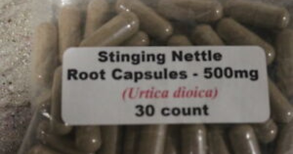
Orthosiphon
Cystinuria
Oxalobacter formigenes
Hyperoxaluria, Primary
Citrates
Renal Tubular Transport, Inborn Errors
Citric Acid
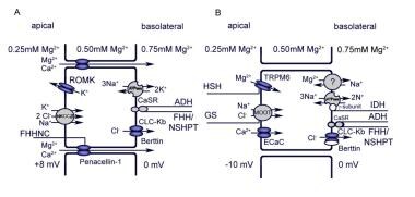
Uromodulin
Potassium Citrate
Receptors, Calcium-Sensing
Oxalic Acid
Glucose Transport Proteins, Facilitative
Pyelonephritis, Xanthogranulomatous
Sodium-Phosphate Cotransporter Proteins, Type IIc
Hyperparathyroidism, Primary
Acidosis, Renal Tubular
Gout
Crystallization
Ureteral Calculi
Lithotripsy
Calcium Phosphates
Sodium Chloride Symporter Inhibitors
Urine
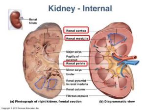
Kidney
Calcium
Factors increasing the risk for stone formation in adult patients with cystic fibrosis. (1/120)
BACKGROUND: Patients with cystic fibrosis (CF) are at high risk of nephrolithiasis (NL), but controversy still exists in terms of causes, including low urine output, hypercalciuria, hyperoxaluria, hyperuricosuria and hypocitraturia. Moreover, heterozygotes (H-CF), which may exhibit altered renal concentrating and diluting ability, have never studied so far. We, therefore, evaluated the metabolic and physicochemical data of adult CF and H-CF patients, comparing them to controls (C). METHODS: Twenty-nine CF patients (16 females, aged 28.4 +/- 7.1 years), 20 H-CF (12 females, aged 58.6 +/- 6.3 years) and 30 C (19 females, aged 39.1 +/- 11.5 years) underwent kidney ultrasound and metabolic evaluation to assess stone risk profile. RESULTS: There was a 21% prevalence of NL in CF vs 15% in H-CF. The CF group had elevated uric acid, but no other serological differences compared with the H-CF and C group. Conversely, the citrate and oxalate content in the urine differed significantly, being lower and higher, respectively. These changes held after correction for urine creatinine. Consequently, urine specimens were more supersaturated with calcium oxalate, despite exhibiting no differences for other relevant constituents. Uric acid increased only after normalization for the body weight and urine creatinine. Lower urine volume and more acidic pH produced mild supersaturation with uric acid in samples from CF, while urine from both H-CF and C remained undersaturated. H-CF had only minor increases in both urine oxalate and calcium oxalate supersaturation. CONCLUSIONS: This study confirms a high prevalence of kidney stones among CF patients associated with supersaturated urine. Their longer survival justifies diets and/or medications aimed at reducing the risk of forming stones. (+info)Crystal retention in renal stone disease: a crucial role for the glycosaminoglycan hyaluronan? (2/120)
The mechanisms that are involved in renal stone disease are not entirely clear. In this article, the various concepts that have been proposed during the past century are reviewed briefly and integrated into current insights. Much attention is dedicated to hyaluronan (HA), an extremely large glycosaminoglycan that may play a central role in renal stone disease. The precipitation of poorly soluble calcium salts (crystal formation) in the kidney is the inevitable consequence of producing concentrated urine. HA is a major constituent of the extracellular matrix in the renal medullary interstitium and the pericellular matrix of mitogen/stress-activated renal tubular cells. HA is an excellent crystal-binding molecule because of its size, negative ionic charge, and ability to form hydrated gel-like matrices. Crystal binding to HA leads to crystal retention in the renal tubules (nephrocalcinosis) and to the formation of calcified plaques in the renal interstitium (Randall's plaques). It remains to be determined whether one or both forms of renal crystal retention are involved in the development of kidney stones (nephrolithiasis). (+info)R990G polymorphism of the calcium-sensing receptor and renal calcium excretion in patients with primary hyperparathyroidism. (3/120)
CONTEXT: Primary hyperparathyroidism (PHPT) shows a great variability in clinical course and severity. Data concerning the association between polymorphic variants of the gene encoding the calcium-sensing receptor (CaSR) and clinical characteristics of PHPT are not conclusive. OBJECTIVE: To evaluate the frequency of three polymorphisms; A986S, R990G, and Q1011E of CaSR in patients with PHPT and to correlate the genotypes with clinical and biochemical parameters. PATIENTS AND METHODS: The study included 94 consecutive unrelated patients referred to our Departments for PHPT diagnosis and management between 2000 and 2005 and 137 age and sex-matched healthy subjects. Patients and controls were genotyped according to standard procedures. Due to the rarity of 990G allele, homozygous and heterozygous subjects were grouped in R/G+G/G set. All PHPT patients were studied for calcium metabolism parameters and renal and bone complications. RESULTS: The proportion of CaSRvariants was similar in PHPT patients and controls. In PHPT patients, only R990G polymorphism was associated with disease parameters; in comparison with R/R, R/G+G/G patients showed lower mean serum parathyroid hormone (PTH) and phosphate levels (139.9 +/- 62.2 vs 199.9 +/- 136.3 pg/ml, P < 0.05 and 0.69 +/- 0.12 vs 0.81 +/- 0.18 mmol/l, P = 0.031 respectively), higher mean 24-h urine calcium concentration and calcium excretion (9.05 +/- 2.05 vs 6.77 +/- 4.31 mmol/24 h, P = 0.012 and 67 +/- 20 vs 51 +/- 26 mumol/l GF, P = 0.039), and increased prevalence of nephrolithiasis (90.0 vs 44.2%, P = 0.007). CONCLUSIONS: The study showed that patients with PHPT, bearing the 990G allele, had lower serum PTH levels and higher urinary calcium excretion in comparison with the other genotype, suggesting an increased sensitivityof the variant receptor to extracellular calcium. Since this variant was associated with increased occurrence of nephrolithiasis, analysis of this polymorphism might help to predict renal complication of the disease. (+info)Proteomics in renal research. (4/120)
Proteomic technologies are used with increasing frequency in the renal community. In this review, we highlight the use in renal research of a number of available techniques including two-dimensional gel electrophoresis, liquid chromatography/mass spectrometry, surface-enhanced laser desorption/ionization, capillary electrophoresis/mass spectrometry, and antibody and tissue arrays. These techniques have been used to identify proteins or changes in proteins specific to regions of the kidney or associated with renal diseases or toxicity. They have also been used to examine protein expression changes and posttranslational modifications of proteins during signaling. A number of studies have used proteomic methodologies to look for diagnostic biomarkers in body fluids. The rapid rate of development of the technologies along with the combination of classic physiological and biochemical techniques with proteomics will enable new discoveries. (+info)Evidence that postprandial reduction of renal calcium reabsorption mediates hypercalciuria of patients with calcium nephrolithiasis. (5/120)
Idiopathic hypercalciuria (IH) is common among calcium stone formers (IHSF). The increased urinary calcium arises from increased intestinal absorption of calcium, but it is unclear whether increased filtered load or decreased renal tubular reabsorption of calcium is the main mechanism for the increased renal excretion. To explore this question, 10 IHSF and 7 normal subjects (N) were studied for 1 day. Urine and blood samples were collected at 30- to 60-min intervals while subjects were fasting and after they ate three meals providing known amounts of calcium, phosphorus, sodium, protein, and calories. Fasting and fed, ultrafiltrable calcium levels, and filtered load of calcium did not differ between N and IHSF. Urine calcium rose with meals, and fractional reabsorption fell in all subjects, but the change was significantly higher in IHSF. The changes in calcium excretion were independent of sodium excretion. Serum parathyroid hormone levels did not differ between N and IHSF, and they could not account for the greater fall in calcium reabsorption in IHSF. Serum magnesium and phosphorus levels in IHSF were below N throughout the day, and tubule phosphate reabsorption was lower in IHSF than N after meals. The primary mechanism by which kidneys ferry absorbed calcium into the urine after meals is via reduced tubule calcium reabsorption, and IHSF differ from N in the magnitude of the response. Parathyroid hormone is not likely to be a sufficient explanation for this difference. (+info)Hypocitraturia despite potassium citrate tablet supplementation. (6/120)
Citrate supplementation is widely used in the prevention of recurrent nephrolithiasis with hypocitraturia. Potassium citrate is the most commonly used citrate agent for this indication. In patients with chronic diarrheal syndromes, the absorption of potassium citrate can be affected. We describe a patient who presented with recurrent nephrolithiasis and chronic diarrhea and was found to have severe hypocitraturia despite citrate supplementation with potassium citrate tablets, likely due to inadequate gastrointestinal absorption of citrate from the slow-release wax-matrix tablets. (+info)Oxalate intake and the risk for nephrolithiasis. (7/120)
Most kidney stones consist of calcium oxalate, and higher urinary oxalate increases the risk for calcium oxalate nephrolithiasis. However, the relation between dietary oxalate and stone risk is unclear. This study prospectively examined the relation between oxalate intake and incident nephrolithiasis in the Health Professionals Follow-up Study (n = 45,985 men), the Nurses' Health Study I (n = 92,872 older women), and the Nurses' Health Study II (n = 101,824 younger women). Food frequency questionnaires were used to assess oxalate intake every 4 yr. Cox proportional hazards regression was used to adjust for age, body mass index, thiazide use, and dietary factors. A total of 4605 incident kidney stones were documented over a combined 44 yr of follow-up. Mean oxalate intakes were 214 mg/d in men, 185 mg/d in older women, and 183 mg/d in younger women and were similar in stone formers and non-stone formers. Spinach accounted for >40% of oxalate intake. For participants in the highest compared with lowest quintile of dietary oxalate, the relative risks for stones were 1.22 (95% confidence interval [CI] 1.03 to 1.45; P = 0.01 for trend) for men and 1.21 (95% CI 1.01 to 1.44; P = 0.05 for trend) for older women. Risk was higher in men with lower dietary calcium (P = 0.08 for interaction). The relative risks for participants who ate eight or more servings of spinach per month compared with fewer than 1 serving per month were 1.30 (95% CI 1.08 to 1.58) for men and 1.34 (95% CI 1.10 to 1.64) for older women. Oxalate intake and spinach were not associated with risk in younger women. These data do not implicate dietary oxalate as a major risk factor for nephrolithiasis. (+info)Incomplete distal renal tubular acidosis affects growth in children. (8/120)
BACKGROUND: Incomplete distal renal tubular acidosis (idRTA) is recognized as an underlying aetiology in recurrent nephrolithiasis. Until the recently reported high prevalence of idRTA in adults with osteoporosis, the effect of idRTA on skeletal parameters was not known. We hypothesize that idRTA has a potential to affect height in the paediatric population. METHODS: In a cross-sectional study, the children with posterior urethral valves (PUV), with normal estimated glomerular filtration rates, were evaluated for idRTA and complete dRTA. The idRTA evaluation was done by short ammonium chloride acidification test. The height standard deviation scores (SDS) in the idRTA group were compared with PUV children without dRTA, with complete dRTA, and to age and gender matched controls with no renal issue (n = 50). RESULTS: The idRTA group (n = 17) manifested a significantly lower mean height SDS (-1.94 +/- 0.41 vs -0.46 +/- 0.28; P < 0.001) and a higher short stature prevalence (height SDS below 2) (18% vs 0; P = 0.06) as compared with those without dRTA (n = 23). The matched controls showed a significantly higher height SDS as compared with the idRTA group (-0.39 +/- 0.25 vs -1.94 +/- 0.41; P < 0.001). As compared with the complete dRTA group (n = 9), the children with idRTA did have significantly higher height SDS (-1.94 +/- 0.41 vs -5.31 +/- 1.95; P = 0.002), and a lower short stature prevalence (18% vs 78%; P = 0.001). On multivariate analysis, dRTA was significantly associated with the height SDS (= -0.88; P < 0.001). CONCLUSIONS: Incomplete dRTA affects height in children. This observation needs validation in longitudinal studies. (+info)Nephrolithiasis is a medical term that refers to the presence of stones or calculi in the kidney. These stones can form anywhere in the urinary tract, including the kidneys, ureters, bladder, and urethra. Nephrolithiasis is also commonly known as kidney stones.
Kidney stones are hard deposits made up of minerals and salts that crystallize in the urine. They can vary in size from tiny sand-like particles to larger pebble or even golf ball-sized masses. Kidney stones can cause pain, bleeding, and infection if they block the flow of urine through the urinary tract.
The formation of kidney stones is often associated with a variety of factors such as dehydration, high levels of calcium, oxalate, or uric acid in the urine, family history, obesity, and certain medical conditions like gout or inflammatory bowel disease. Treatment for nephrolithiasis depends on the size and location of the stone, as well as the severity of symptoms. Small stones may pass spontaneously with increased fluid intake, while larger stones may require medication, shock wave lithotripsy, or surgical removal.
Kidney calculi, also known as kidney stones, are hard deposits made of minerals and salts that form inside your kidneys. They can range in size from a grain of sand to a golf ball. When they're small enough, they can be passed through your urine without causing too much discomfort. However, larger stones may block the flow of urine, causing severe pain and potentially leading to serious complications such as urinary tract infections or kidney damage if left untreated.
The formation of kidney calculi is often associated with factors like dehydration, high levels of certain minerals in your urine, family history, obesity, and certain medical conditions such as gout or inflammatory bowel disease. Symptoms of kidney stones typically include severe pain in the back, side, lower abdomen, or groin; nausea and vomiting; fever and chills if an infection is present; and blood in the urine. Treatment options depend on the size and location of the stone but may include medications to help pass the stone, shock wave lithotripsy to break up the stone, or surgical removal of the stone in severe cases.
Calcium oxalate is a chemical compound with the formula CaC2O4. It is the most common type of stone found in kidneys, also known as kidney stones. Calcium oxalate forms when there is too much calcium or oxalate in the urine. This can occur due to various reasons such as dietary habits, dehydration, medical conditions like hyperparathyroidism, or genetic factors.
Calcium oxalate stones are hard and crystalline and can cause severe pain during urination or while passing through the urinary tract. They may also lead to other symptoms like blood in the urine, nausea, vomiting, or fever. Prevention strategies for calcium oxalate stones include staying hydrated, following a balanced diet, and taking prescribed medications to control the levels of calcium and oxalate in the body.
Hypercalciuria is a medical condition characterized by an excessive amount of calcium in the urine. It can occur when the body absorbs too much calcium from food, or when the bones release more calcium than usual. In some cases, it may be caused by certain medications, kidney disorders, or genetic factors.
Hypercalciuria can increase the risk of developing kidney stones and other kidney problems. It is often diagnosed through a 24-hour urine collection test that measures the amount of calcium in the urine. Treatment may include changes in diet, increased fluid intake, and medications to help reduce the amount of calcium in the urine.
Hyperoxaluria is a medical condition characterized by an excessive excretion of oxalate in the urine. Oxalate is a naturally occurring substance found in some foods and can also be produced by the body. When oxalate combines with calcium in the urine, it can form kidney stones or calcium oxalate deposits in the kidneys and other tissues, leading to kidney damage or systemic oxalosis. There are three types of hyperoxaluria: primary, secondary, and enteric. Primary hyperoxaluria is caused by genetic defects that affect the body's ability to regulate oxalate production, while secondary hyperoxaluria results from increased dietary intake or absorption of oxalate, or from other medical conditions. Enteric hyperoxaluria occurs in individuals with malabsorption syndromes, such as inflammatory bowel disease or after gastric bypass surgery, where excessive amounts of oxalate are absorbed from the gut into the bloodstream and excreted in the urine.
Nephrocalcinosis is a medical condition characterized by the deposition of calcium salts in the renal parenchyma, specifically within the tubular epithelial cells and interstitium of the kidneys. This process can lead to chronic inflammation, tissue damage, and ultimately impaired renal function if left untreated.
The condition is often associated with metabolic disorders such as hyperparathyroidism, distal renal tubular acidosis, or hyperoxaluria; medications like loop diuretics, corticosteroids, or calcineurin inhibitors; and chronic kidney diseases. The diagnosis of nephrocalcinosis is typically made through imaging studies such as ultrasound, CT scan, or X-ray. Treatment usually involves addressing the underlying cause, modifying dietary habits, and administering medications to control calcium levels in the body.
Oxalates, also known as oxalic acid or oxalate salts, are organic compounds that contain the functional group called oxalate. Oxalates are naturally occurring substances found in various foods such as spinach, rhubarb, nuts, and seeds. They can also be produced by the body as a result of metabolism.
In the body, oxalates can bind with calcium and other minerals to form crystals, which can accumulate in various tissues and organs, including the kidneys. This can lead to the formation of kidney stones, which are a common health problem associated with high oxalate intake or increased oxalate production in the body.
It is important for individuals with a history of kidney stones or other kidney problems to monitor their oxalate intake and limit consumption of high-oxalate foods. Additionally, certain medical conditions such as hyperoxaluria, a rare genetic disorder that causes increased oxalate production in the body, may require medical treatment to reduce oxalate levels and prevent complications.
Urolithiasis is the formation of stones (calculi) in the urinary system, which includes the kidneys, ureters, bladder, and urethra. These stones can be composed of various substances such as calcium oxalate, calcium phosphate, uric acid, or struvite. The presence of urolithiasis can cause symptoms like severe pain in the back or side, nausea, vomiting, fever, and blood in the urine. The condition can be managed with medications, increased fluid intake, and in some cases, surgical intervention may be required to remove the stones.
Uric acid is a chemical compound that is formed when the body breaks down purines, which are substances that are found naturally in certain foods such as steak, organ meats and seafood, as well as in our own cells. After purines are broken down, they turn into uric acid and then get excreted from the body in the urine.
However, if there is too much uric acid in the body, it can lead to a condition called hyperuricemia. High levels of uric acid can cause gout, which is a type of arthritis that causes painful swelling and inflammation in the joints, especially in the big toe. Uric acid can also form crystals that can collect in the kidneys and lead to kidney stones.
It's important for individuals with gout or recurrent kidney stones to monitor their uric acid levels and follow a treatment plan prescribed by their healthcare provider, which may include medications to lower uric acid levels and dietary modifications.
Calcium metabolism disorders refer to a group of medical conditions that affect the body's ability to properly regulate the levels of calcium in the blood and tissues. Calcium is an essential mineral that plays a critical role in many bodily functions, including bone health, muscle contraction, nerve function, and blood clotting.
There are several types of calcium metabolism disorders, including:
1. Hypocalcemia: This is a condition characterized by low levels of calcium in the blood. It can be caused by various factors such as vitamin D deficiency, hypoparathyroidism, and certain medications. Symptoms may include muscle cramps, spasms, and tingling sensations in the fingers and toes.
2. Hypercalcemia: This is a condition characterized by high levels of calcium in the blood. It can be caused by various factors such as hyperparathyroidism, cancer, and certain medications. Symptoms may include fatigue, weakness, confusion, and kidney stones.
3. Osteoporosis: This is a condition characterized by weak and brittle bones due to low calcium levels in the bones. It can be caused by various factors such as aging, menopause, vitamin D deficiency, and certain medications. Symptoms may include bone fractures and loss of height.
4. Paget's disease: This is a condition characterized by abnormal bone growth and deformities due to disordered calcium metabolism. It can be caused by various factors such as genetics, age, and certain medications. Symptoms may include bone pain, fractures, and deformities.
Treatment for calcium metabolism disorders depends on the underlying cause of the condition. It may involve supplements, medication, dietary changes, or surgery. Proper diagnosis and management are essential to prevent complications such as kidney stones, bone fractures, and neurological damage.
Urinary calculi, also known as kidney stones or nephrolithiasis, are hard deposits made of minerals and salts that form inside the urinary system. These calculi can develop in any part of the urinary system, which includes the kidneys, ureters, bladder, and urethra.
The formation of urinary calculi typically occurs when there is a concentration of certain substances, such as calcium, oxalate, uric acid, or struvite, in the urine. When these substances become highly concentrated, they can crystallize and form small seeds that gradually grow into larger stones over time.
The size of urinary calculi can vary from tiny, sand-like particles to large stones that can fill the entire renal pelvis. The symptoms associated with urinary calculi depend on the stone's size, location, and whether it is causing a blockage in the urinary tract. Common symptoms include severe pain in the flank, lower abdomen, or groin; nausea and vomiting; blood in the urine (hematuria); fever and chills; and frequent urge to urinate or painful urination.
Treatment for urinary calculi depends on the size and location of the stone, as well as the severity of symptoms. Small stones may pass spontaneously with increased fluid intake and pain management. Larger stones may require medical intervention, such as extracorporeal shock wave lithotripsy (ESWL), ureteroscopy, or percutaneous nephrolithotomy (PCNL) to break up or remove the stone. Preventive measures include maintaining adequate hydration, modifying dietary habits, and taking medications to reduce the risk of stone formation.
Ethylene glycol is a colorless, odorless, syrupy liquid with a sweet taste, which makes it appealing to animals and children. It is commonly used in the manufacture of antifreeze, coolants, deicers, hydraulic brake fluids, solvents, and other industrial products. Ethylene glycol is also found in some household items such as certain types of wood stains, paints, and cosmetics.
Ingesting even small amounts of ethylene glycol can be harmful or fatal to humans and animals. It is metabolized by the body into toxic substances that can cause damage to the central nervous system, heart, kidneys, and other organs. Symptoms of ethylene glycol poisoning may include nausea, vomiting, abdominal pain, decreased level of consciousness, seizures, coma, acidosis, increased heart rate, low blood pressure, and kidney failure.
If you suspect that someone has ingested ethylene glycol, it is important to seek medical attention immediately. Treatment typically involves administering a medication called fomepizole or ethanol to inhibit the metabolism of ethylene glycol, as well as providing supportive care such as fluid replacement and dialysis to remove the toxic substances from the body.
Orthosiphon is a genus of plants in the family Lamiaceae, also known as the mint or deadnettle family. The most common species is Orthosiphon stamineus, also known as Cat's Whiskers or Java Tea. This plant is native to Southeast Asia and some parts of Australia.
In a medical context, Orthosiphon stamineus is used in traditional medicine for its diuretic, antioxidant, anti-inflammatory, and antibacterial properties. The leaves and stems of the plant are dried and prepared as an herbal infusion or decoction to treat various health conditions such as kidney stones, urinary tract infections, and high blood pressure. However, it is important to note that the scientific evidence supporting these claims is limited, and more research is needed to establish its safety and efficacy.
Cystinuria is a genetic disorder that affects the way the body handles certain amino acids, specifically cystine, arginine, lysine, and ornithine. These amino acids are normally reabsorbed in the kidneys and released into the bloodstream. However, people with cystinuria have a defect in the transport mechanism that causes large amounts of cystine to be excreted in the urine, where it can form stones in the urinary tract. These stones can cause pain, blockages, and infection. Cystinuria is inherited in an autosomal recessive manner, meaning that an individual must inherit two copies of the defective gene, one from each parent, to have the condition.
"Oxalobacter formigenes" is a type of gram-negative, anaerobic bacteria that resides in the human gastrointestinal tract. It is commonly found in the large intestine and plays a role in the metabolism of oxalate, a compound found in many foods that can contribute to kidney stone formation when present in high levels in the body.
"Oxalobacter formigenes" has the ability to break down and utilize oxalate as a source of energy, which can help to reduce the amount of oxalate that is absorbed into the bloodstream and excreted by the kidneys. Some research suggests that the presence of "Oxalobacter formigenes" in the gut may be associated with a lower risk of kidney stone formation, although more studies are needed to confirm this association.
It's worth noting that while "Oxalobacter formigenes" is considered a beneficial bacteria, it is not currently used as a probiotic or therapeutic agent in clinical practice.
Ureteroscopy is a medical procedure that involves the use of a ureteroscope, which is a thin, flexible or rigid fiber-optic tube with a light and camera at the end, to visualize the inside of the ureters and kidneys. The ureteroscope is inserted through the urethra and bladder, and then up into the ureter to examine it for any abnormalities such as stones, tumors, or structural issues.
During the procedure, the doctor can also remove any small stones or take a biopsy of any suspicious tissue. Ureteroscopy is typically performed under general or regional anesthesia and may require hospitalization depending on the complexity of the procedure. It is a minimally invasive alternative to traditional open surgery for diagnosing and treating ureteral and kidney conditions.
Primary hyperoxaluria is a rare genetic disorder characterized by overproduction of oxalate in the body due to mutations in specific enzymes involved in oxalate metabolism. There are three types of primary hyperoxaluria (PH1, PH2, and PH3), with PH1 being the most common and severe form.
In primary hyperoxaluria type 1 (PH1), there is a deficiency or dysfunction in the enzyme alanine-glyoxylate aminotransferase (AGT), which leads to an accumulation of glyoxylate. Glyoxylate is then converted to oxalate, resulting in increased oxalate production. Oxalate is a compound that naturally occurs in the body but is primarily excreted through the kidneys. When there is an overproduction of oxalate, it can lead to the formation of calcium oxalate crystals in various tissues, including the kidneys. This can cause recurrent kidney stones, nephrocalcinosis (calcium deposits in the kidneys), and eventually chronic kidney disease or end-stage renal failure.
Primary hyperoxaluria type 2 (PH2) is caused by a deficiency or dysfunction in the enzyme glyoxylate reductase/hydroxypyruvate reductase (GRHPR), leading to an accumulation of glyoxylate, which is subsequently converted to oxalate. PH2 has a milder clinical presentation compared to PH1.
Primary hyperoxaluria type 3 (PH3) is a rare form caused by mutations in the gene HOGA1, which encodes for 4-hydroxy-2-oxoglutarate aldolase. This enzyme deficiency results in an increase in glyoxylate and, subsequently, oxalate production.
Early diagnosis and management of primary hyperoxaluria are crucial to prevent or slow down the progression of kidney damage. Treatment options include increased fluid intake, medications to reduce stone formation (such as potassium citrate), and in some cases, liver-kidney transplantation.
Citrates are the salts or esters of citric acid, a weak organic acid that is naturally found in many fruits and vegetables. In a medical context, citrates are often used as a buffering agent in intravenous fluids to help maintain the pH balance of blood and other bodily fluids. They are also used in various medical tests and treatments, such as in urine alkalinization and as an anticoagulant in kidney dialysis solutions. Additionally, citrate is a component of some dietary supplements and medications.
Inborn errors of renal tubular transport refer to genetic disorders that affect the normal functioning of the kidney tubules. The kidney tubules are responsible for the reabsorption and secretion of various substances, including electrolytes and nutrients, as urine is formed. Inherited defects in the proteins that mediate these transport processes can lead to abnormal levels of these substances in the body and may result in a variety of clinical symptoms.
These disorders can affect different parts of the renal tubule, including the proximal tubule, loop of Henle, distal tubule, and collecting duct. Depending on the specific transporter affected, inborn errors of renal tubular transport can present with a range of clinical manifestations, such as electrolyte imbalances, acid-base disorders, growth retardation, kidney stones, nephrocalcinosis, or even kidney failure.
Examples of inborn errors of renal tubular transport include:
1. Distal renal tubular acidosis (dRTA): A genetic disorder that affects the ability of the distal tubule to acidify urine, leading to metabolic acidosis, hypokalemia, and nephrocalcinosis.
2. Bartter syndrome: A group of autosomal recessive disorders characterized by impaired sodium reabsorption in the loop of Henle, resulting in hypokalemia, metabolic alkalosis, and hyperreninemic hyperaldosteronism.
3. Gitelman syndrome: An autosomal recessive disorder caused by a defect in the thiazide-sensitive sodium chloride cotransporter in the distal tubule, leading to hypokalemia, metabolic alkalosis, and hypocalciuria.
4. Liddle syndrome: An autosomal dominant disorder characterized by increased sodium reabsorption in the collecting duct due to a gain-of-function mutation in the epithelial sodium channel (ENaC), resulting in hypertension, hypokalemia, and metabolic alkalosis.
5. Dent disease: An X-linked recessive disorder caused by mutations in the CLCN5 gene, which encodes a chloride channel in the proximal tubule, leading to low molecular weight proteinuria, hypercalciuria, and nephrolithiasis.
6. Familial hypomagnesemia with hypercalciuria and nephrocalcinosis (FHHNC): An autosomal recessive disorder caused by mutations in the CLCN5 or CLDN16 genes, which encode chloride channels in the thick ascending limb of Henle's loop, resulting in hypomagnesemia, hypercalciuria, and nephrocalcinosis.
Citric acid is a weak organic acid that is widely found in nature, particularly in citrus fruits such as lemons and oranges. Its chemical formula is C6H8O7, and it exists in a form known as a tribasic acid, which means it can donate three protons in chemical reactions.
In the context of medical definitions, citric acid may be mentioned in relation to various physiological processes, such as its role in the Krebs cycle (also known as the citric acid cycle), which is a key metabolic pathway involved in energy production within cells. Additionally, citric acid may be used in certain medical treatments or therapies, such as in the form of citrate salts to help prevent the formation of kidney stones. It may also be used as a flavoring agent or preservative in various pharmaceutical preparations.
Uromodulin, also known as Tamm-Horsfall protein, is a glycoprotein that is primarily produced in the thick ascending limb of the loop of Henle in the kidney. It is the most abundant protein found in normal urine. Uromodulin plays a role in the protection of the urinary tract by preventing the formation of calcium oxalate and brushite crystals, which can lead to kidney stones. Additionally, it has been implicated in various renal diseases, including chronic kidney disease and kidney transplant rejection.
Potassium citrate is a medication and dietary supplement that contains potassium and citrate. Medically, it is used to treat and prevent kidney stones, as well as to manage metabolic acidosis in people with chronic kidney disease. Potassium citrate works by increasing the pH of urine, making it less acidic, which can help to dissolve certain types of kidney stones and prevent new ones from forming. It is also used as an alkalizing agent in the treatment of various conditions that cause acidosis.
In addition to its medical uses, potassium citrate is also found naturally in some fruits and vegetables, such as oranges, grapefruits, lemons, limes, and spinach. It is often used as a food additive and preservative, and can be found in a variety of processed foods and beverages.
It's important to note that taking too much potassium citrate can lead to high levels of potassium in the blood, which can be dangerous. Therefore, it is important to follow the dosage instructions carefully and talk to your doctor before taking this medication if you have any medical conditions or are taking any other medications.
Calcium-sensing receptors (CaSR) are a type of G protein-coupled receptor that play a crucial role in the regulation of extracellular calcium homeostasis. They are widely expressed in various tissues, including the parathyroid gland, kidney, and bone.
The primary function of CaSR is to detect changes in extracellular calcium concentrations and transmit signals to regulate the release of parathyroid hormone (PTH) from the parathyroid gland. When the concentration of extracellular calcium increases, CaSR is activated, which leads to a decrease in PTH secretion, thereby preventing further elevation of calcium levels. Conversely, when calcium levels decrease, CaSR is inhibited, leading to an increase in PTH release and restoration of normal calcium levels.
In addition to regulating calcium homeostasis, CaSR also plays a role in other physiological processes, including cell proliferation, differentiation, and apoptosis. Dysregulation of CaSR has been implicated in various diseases, such as hyperparathyroidism, hypoparathyroidism, and cancer. Therefore, understanding the function and regulation of CaSR is essential for developing new therapeutic strategies to treat these conditions.
Oxalic acid is not a medical term, but it is a chemical compound with the formula HOOC-COOH. It is a white crystalline solid that is soluble in water and polar organic solvents. Medically, oxalic acid is relevant due to its presence in certain foods and its potential to form calcium oxalate stones in the kidneys when excreted in urine.
Hyperoxaluria is a medical condition characterized by increased levels of oxalate in the urine, which can lead to the formation of kidney stones. This condition can be caused by genetic factors or excessive intake of oxalate-rich foods such as spinach, rhubarb, and certain nuts and beans. In severe cases, it may require medical treatment to reduce oxalate levels in the body.
Glucose Transporter Proteins, Facilitative (GLUTs) are a group of membrane proteins that facilitate the passive transport of glucose and other simple sugars across the cell membrane. They are also known as solute carrier family 2 (SLC2A) members. These proteins play a crucial role in maintaining glucose homeostasis within the body by regulating the uptake of glucose into cells. Unlike active transport, facilitative diffusion does not require energy and occurs down its concentration gradient. Different GLUT isoforms have varying tissue distributions and substrate specificities, allowing them to respond to different physiological needs. For example, GLUT1 is widely expressed and is responsible for basal glucose uptake in most tissues, while GLUT4 is primarily found in insulin-sensitive tissues such as muscle and adipose tissue, where it mediates the increased glucose uptake in response to insulin signaling.
Xanthogranulomatous pyelonephritis (XPN) is a rare and severe form of chronic pyelonephritis, which is an infection and inflammation of the renal pelvis. In XPN, there is a proliferation of lipid-laden macrophages (also known as xanthoma cells) and other inflammatory cells in the kidney parenchyma, leading to the formation of multiple granulomas.
XPN typically affects middle-aged to older women with underlying urologic abnormalities such as obstructive uropathy, calculi (stones), or chronic urinary tract infections. The condition can be difficult to diagnose and often requires a combination of imaging studies, urinalysis, and histopathological examination of renal tissue.
The clinical presentation of XPN is variable and may include fever, flank pain, weight loss, and symptoms related to urinary tract obstruction or infection. Treatment usually involves antibiotic therapy, surgical removal of the affected kidney (nephrectomy), and management of any underlying urologic abnormalities. If left untreated, XPN can lead to irreversible kidney damage and even sepsis.
Sodium-phosphate cotransporter proteins, type IIc (NPTIIc), are a subtype of sodium-dependent phosphate transporters that play a crucial role in the regulation of phosphate homeostasis within the body. They are located primarily in the kidney's proximal tubule cells and intestinal epithelial cells.
NPTIIc proteins facilitate the active transport of inorganic phosphate (Pi) ions across the cell membrane, in conjunction with sodium ions (Na+). This symport mechanism allows for the movement of Pi against its concentration gradient, from areas of low concentration to high concentration. The energy required for this process is derived from the electrochemical gradient of sodium ions.
These transporters are essential for maintaining normal phosphate levels in the body, as they help reabsorb a significant portion of filtered phosphate in the kidneys and absorb dietary phosphate in the intestines. Dysregulation of NPTIIc proteins can lead to various disorders related to phosphate homeostasis, such as hypophosphatemia (low serum phosphate levels) or hyperphosphatemia (high serum phosphate levels), which can have detrimental effects on bone health, mineral metabolism, and overall body function.
Primary hyperparathyroidism is a medical condition characterized by excessive secretion of parathyroid hormone (PTH) from one or more of the parathyroid glands in the neck. These glands are normally responsible for regulating calcium levels in the body by releasing PTH, which helps to maintain an appropriate balance of calcium and phosphate in the bloodstream.
In primary hyperparathyroidism, the parathyroid gland(s) become overactive and produce too much PTH, leading to elevated calcium levels (hypercalcemia) in the blood. This can result in a variety of symptoms, such as fatigue, weakness, bone pain, kidney stones, and cognitive impairment, although some individuals may not experience any symptoms at all.
The most common cause of primary hyperparathyroidism is a benign tumor called an adenoma that develops in one or more of the parathyroid glands. In rare cases, primary hyperparathyroidism can be caused by cancer of the parathyroid gland(s) or by enlargement of all four glands (four-gland hyperplasia). Treatment typically involves surgical removal of the affected parathyroid gland(s), which is usually curative.
Renal tubular acidosis (RTA) is a medical condition that occurs when the kidneys are unable to properly excrete acid into the urine, leading to an accumulation of acid in the bloodstream. This results in a state of metabolic acidosis.
There are several types of RTA, but renal tubular acidosis type 1 (also known as distal RTA) is characterized by a defect in the ability of the distal tubules to acidify the urine, leading to an inability to lower the pH of the urine below 5.5, even in the face of metabolic acidosis. This results in a persistently alkaline urine, which can lead to calcium phosphate stones and bone demineralization.
Type 1 RTA is often caused by inherited genetic defects, but it can also be acquired due to various kidney diseases, drugs, or autoimmune disorders. Symptoms of type 1 RTA may include fatigue, weakness, muscle cramps, decreased appetite, and vomiting. Treatment typically involves alkali therapy to correct the acidosis and prevent complications.
Gout is a type of inflammatory arthritis that occurs when urate crystals accumulate in and around the joints, causing sudden attacks of severe pain, swelling, redness, and tenderness. Urate crystals can form when there are high levels of uric acid in the blood. Uric acid is a waste product that is produced when the body breaks down purines, substances that are found naturally in certain foods, such as steak, organ meats, and seafood. Other foods also promote higher levels of uric acid, such as alcoholic beverages, especially beer, and drinks sweetened with fruit sugar (fructose).
Normally, uric acid dissolves in the blood and passes through the kidneys and out of the body in urine. But sometimes either the body produces too much uric acid or the kidneys excrete too little uric acid. When this happens, uric acid can build up, forming sharp, needle-like urate crystals in a joint or surrounding tissue that cause pain, inflammation and swelling.
Gout most commonly affects the big toe but can also occur in any joint in the body. The symptoms of gout are often acute, occurring suddenly without warning and frequently at night. The attacks are characterized by a rapid onset of pain, swelling, warmth, and redness in the affected joint. An attack of gout can be so painful that it wakes you up from sleep.
Over time, gout can cause permanent damage to the joints and surrounding tissue, resulting in chronic arthritis. If left untreated, gout also can lead to an accumulation of uric acid crystals in the kidneys, which can result in kidney stones.
Hypophosphatemia is a medical condition characterized by abnormally low levels of phosphate (phosphorus) in the blood, specifically below 2.5 mg/dL. Phosphate is an essential electrolyte that plays a crucial role in various bodily functions such as energy production, bone formation, and maintaining acid-base balance.
Hypophosphatemia can result from several factors, including malnutrition, vitamin D deficiency, alcoholism, hormonal imbalances, and certain medications. Symptoms of hypophosphatemia may include muscle weakness, fatigue, bone pain, confusion, and respiratory failure in severe cases. Treatment typically involves correcting the underlying cause and administering phosphate supplements to restore normal levels.
Crystallization is a process in which a substance transitions from a liquid or dissolved state to a solid state, forming a crystal lattice. In the medical context, crystallization can refer to the formation of crystals within the body, which can occur under certain conditions such as changes in pH, temperature, or concentration of solutes. These crystals can deposit in various tissues and organs, leading to the formation of crystal-induced diseases or disorders.
For example, in patients with gout, uric acid crystals can accumulate in joints, causing inflammation, pain, and swelling. Similarly, in nephrolithiasis (kidney stones), minerals in the urine can crystallize and form stones that can obstruct the urinary tract. Crystallization can also occur in other medical contexts, such as in the formation of dental calculus or plaque, and in the development of cataracts in the eye.
Ureteral calculi, also known as ureteric stones or ureteral stones, refer to the presence of solid mineral deposits (calculi) within the ureters, the tubes that transport urine from the kidneys to the bladder. These calculi can vary in size and composition, and their formation is often associated with conditions such as dehydration, urinary tract infections, or metabolic disorders. Ureteral calculi may cause symptoms like severe pain, hematuria (blood in the urine), and obstruction of urine flow, potentially leading to serious complications if left untreated.
Lithotripsy is a medical procedure that uses shock waves or other high-energy sound waves to break down and remove calculi (stones) in the body, particularly in the kidneys, ureters, or gallbladder. The procedure is typically performed on an outpatient basis and does not require any incisions.
During lithotripsy, the patient lies on a cushioned table while a lithotripter, a device that generates shock waves, is positioned around the area of the stone. As the shock waves pass through the body, they break the stone into tiny fragments that can then be easily passed out of the body in urine.
Lithotripsy is generally a safe and effective procedure, but it may not be suitable for everyone. Patients with certain medical conditions, such as bleeding disorders or pregnancy, may not be able to undergo lithotripsy. Additionally, some stones may be too large or too dense to be effectively treated with lithotripsy. In these cases, other treatment options, such as surgery, may be necessary.
Calcium phosphates are a group of minerals that are important components of bones and teeth. They are also found in some foods and are used in dietary supplements and medical applications. Chemically, calcium phosphates are salts of calcium and phosphoric acid, and they exist in various forms, including hydroxyapatite, which is the primary mineral component of bone tissue. Other forms of calcium phosphates include monocalcium phosphate, dicalcium phosphate, and tricalcium phosphate, which are used as food additives and dietary supplements. Calcium phosphates are important for maintaining strong bones and teeth, and they also play a role in various physiological processes, such as nerve impulse transmission and muscle contraction.
Sodium chloride symporter inhibitors are a class of pharmaceutical agents that block the function of the sodium chloride symporter (NCC), which is a protein found in the kidney's distal convoluted tubule. The NCC is responsible for reabsorbing sodium and chloride ions from the filtrate back into the bloodstream, helping to regulate electrolyte balance and blood pressure.
Sodium chloride symporter inhibitors work by selectively binding to and blocking the NCC, preventing it from transporting sodium and chloride ions across the cell membrane. This leads to increased excretion of sodium and chloride in the urine, which can help lower blood pressure in patients with hypertension.
Examples of sodium chloride symporter inhibitors include thiazide diuretics such as hydrochlorothiazide and chlorthalidone, which have been used for many years to treat hypertension and edema associated with heart failure and liver cirrhosis. These medications work by reducing the amount of sodium and fluid in the body, which helps lower blood pressure and reduce swelling.
It's worth noting that while sodium chloride symporter inhibitors can be effective at treating hypertension, they can also cause side effects such as electrolyte imbalances, dehydration, and increased urination. As with any medication, it's important to use them under the guidance of a healthcare provider and to follow dosing instructions carefully.
Urine is a physiological excretory product that is primarily composed of water, urea, and various ions (such as sodium, potassium, chloride, and others) that are the byproducts of protein metabolism. It also contains small amounts of other substances like uric acid, creatinine, ammonia, and various organic compounds. Urine is produced by the kidneys through a process called urination or micturition, where it is filtered from the blood and then stored in the bladder until it is excreted from the body through the urethra. The color, volume, and composition of urine can provide important diagnostic information about various medical conditions.
A kidney, in medical terms, is one of two bean-shaped organs located in the lower back region of the body. They are essential for maintaining homeostasis within the body by performing several crucial functions such as:
1. Regulation of water and electrolyte balance: Kidneys help regulate the amount of water and various electrolytes like sodium, potassium, and calcium in the bloodstream to maintain a stable internal environment.
2. Excretion of waste products: They filter waste products from the blood, including urea (a byproduct of protein metabolism), creatinine (a breakdown product of muscle tissue), and other harmful substances that result from normal cellular functions or external sources like medications and toxins.
3. Endocrine function: Kidneys produce several hormones with important roles in the body, such as erythropoietin (stimulates red blood cell production), renin (regulates blood pressure), and calcitriol (activated form of vitamin D that helps regulate calcium homeostasis).
4. pH balance regulation: Kidneys maintain the proper acid-base balance in the body by excreting either hydrogen ions or bicarbonate ions, depending on whether the blood is too acidic or too alkaline.
5. Blood pressure control: The kidneys play a significant role in regulating blood pressure through the renin-angiotensin-aldosterone system (RAAS), which constricts blood vessels and promotes sodium and water retention to increase blood volume and, consequently, blood pressure.
Anatomically, each kidney is approximately 10-12 cm long, 5-7 cm wide, and 3 cm thick, with a weight of about 120-170 grams. They are surrounded by a protective layer of fat and connected to the urinary system through the renal pelvis, ureters, bladder, and urethra.
Calcium is an essential mineral that is vital for various physiological processes in the human body. The medical definition of calcium is as follows:
Calcium (Ca2+) is a crucial cation and the most abundant mineral in the human body, with approximately 99% of it found in bones and teeth. It plays a vital role in maintaining structural integrity, nerve impulse transmission, muscle contraction, hormonal secretion, blood coagulation, and enzyme activation.
Calcium homeostasis is tightly regulated through the interplay of several hormones, including parathyroid hormone (PTH), calcitonin, and vitamin D. Dietary calcium intake, absorption, and excretion are also critical factors in maintaining optimal calcium levels in the body.
Hypocalcemia refers to low serum calcium levels, while hypercalcemia indicates high serum calcium levels. Both conditions can have detrimental effects on various organ systems and require medical intervention to correct.
 Uric acid
Hyperuricosuria
Gout
Kidney stone disease
Iminoglycinuria
List of OMIM disorder codes
Hypercalciuria
Distal renal tubular acidosis
Renal tubular acidosis
Renal colic
Medullary sponge kidney
Fanconi syndrome
Rectal tenesmus
Oliver Wrong
Pyelonephritis
Melamine
2008 Chinese milk scandal
Nephrocalcinosis
CLCN5
CLCNKB
Mammalian kidney
Visnaga daucoides
Crohn's disease
ZNF365
Sodium-dependent phosphate transport protein 2A
Glyoxylate reductase
Dent's disease
Chlortalidone
Magnesium citrate
Calculus (medicine)
 Kidney Stones | Kidney Stone Symptoms | Nephrolithiasis | MedlinePlus
 PRIME PubMed | Calcium nephrolithiasis: effect of water hardness on urinary electrolytes
 Nephrolithiasis Testing Algorithm | Choose the Right Test
 Postmenopausal hormone use and the risk of nephrolithiasis - International Menopause Society
Uric acid nephrolithiasis - Unbounded Medicine
Biochemical and Clinical Effects of Ethane-1-Hydroxy-1,1-Diphosphonate in Calcium Nephrolithiasis | Clinical Science | Portland...
 Nephrolithiasis | The Infographic Guide to Medicine | AccessMedicine | McGraw Hill Medical
 Uric Acid Nephrolithiasis: Current Concepts and Controversies
Nephrolithiasis Differential Diagnoses
 Nephrolithiasis | Clinical Gate
 Polycystic Kidney Disease Clinical Presentation: History, Physical Examination, Complications
 Kelly C. et al., 2019: Nephrolithiasis in the Obese Patient - STORZ MEDICAL Blog
 Nephrolithiasis (Kidney Stone Disease) | GK Hospital
 Nephrolithiasis Risk Factors - Medical Algorithm | Medicalalgorithms.com
![PGS Catalog - nephrolithiasis [EFO:0004253] (Polygenic Trait)](data:image/png;base64,iVBORw0KGgoAAAANSUhEUgAAABAAAAAQCAYAAAAf8/9hAAACqUlEQVQ4jYXRT2hcVRTH8e+977730kknk3mZqfnjvzYNMcWooEIXSkREN7oRqwvBjW6L0m33FroXcevWrgSDqKBQiLZTqFEippHYIbZmmMn8y3t582bevcdFXEhbyVkezu+z+B3FA+bS8+8iIouPh1MXN9K7F0+Wpnc++P6TB52i712YyhgfnniZ+bHqq8+MPfxOcfn0x1dff+iNVUmZLE3cB6iPzp8Hpc44597XWhcByW3OK9ulF57Oq2e+fe049Tmz1bt64wdjjADKOdvxtPcZsG2arRZRufzeiysrF5aWlnAixO0e4ac3iFPBm5mgXBlfWDn31sJ0NIVSivX1dX5aW9vLhsPLph/HRFFUODU/z5PLywDsbmwTD27SKFn80jH8MCSamWX59CIAvX6fH9fWCnme398BwEG9iXeQ06/4+GMBngh3eh3EuaNLdAjZHw2cCMlcAWMMgUArS0kHg6OBdD/BbTaJC8LokQk0itAJic1p7/ePBtqbO/j1Po1HA3TlOCJCKCDWcrfb+X9AAcODAb0vb5KonPazVfwgQCmFrz1KFm7t3qGfxKj/ACZOYpxz1Go1fv/uOu52g+zsFF7JkDXbCKD+TfSzjC++XmXU6oAI1lpMEsdYa/nr9ibVqIh6zgeVYeu3wFkAPE/hi0aLMAR2/m6SW8sgG2CqUQ04xUtLGU/MZNjZgPr1LknRogcCSjFeCngs9ZBc0Erxm4u5sjsi2P8T8/ab8PPGiIWTFWbGQ9x0yOyiYktS0u4IpTXFcsBcYnC5QytF7hyFXxvMRTnmq2+eohyFndovXTonQmwcsHVtj3xSEeSHBRQ6PnHqIfYQ2GoMSNyx7l5cxpQnz+Jp8/nqtZby1MEERstw5COMUC4HFFpnGFEghw+zym+HxcqVQiHiHygLN77aGgUdAAAAAElFTkSuQmCC) PGS Catalog - nephrolithiasis [EFO:0004253] (Polygenic Trait)
PGS Catalog - nephrolithiasis [EFO:0004253] (Polygenic Trait)
 DailyMed - DIPENTUM- olsalazine sodium capsule, gelatin coated
 Mediterranean Diet and Risk of Nephrolithiasis - AJKD Blog
These highlights do not include all the information needed to use BALSALAZIDE DISODIUM CAPSULES safely and effectively. See...
 View of Medicine and Nephro-urology in Ancient Iran: Part III: Rhazes and His Recommendations for Nephrolithiasis
 A young man presenting with recurrent nephrolithiasis - Calcoli Renali
 Vitamin D status in patients with idiopathic calcium nephrolithiasis
 Obstructive nephrolithiasis and hydronephrosis in a cat - Small Animal Ultrasonography
 Gout and risk of chronic kidney disease and nephrolithiasis: meta-analysis of observational studies | Arthritis Research &...
 Nephrolithiasis ( Kidney Stones ) discussion response 180-200 words APA - Custom Research Writers
Uric acid - Wikipedia
Living Donor Renal Transplantation in Donors with Incidental Nephrolithiasis - Nuffield Department of Surgical Sciences
Risk of chronic and end stage kidney disease in patients with nephrolithiasis<...
Nephrolithiasis In Renal Tubular Acidosis | Best Nephrologist In Delhi | Dr Rajesh Goel | Kidney Care Centre
Bilateral Uric Acid Nephrolithiasis and Ureteral Hypertrophy in a Free-ranging River Otter (Lontra canadensis)
Urolithiasis3
- Kidney stones (nephrolithiasis or urolithiasis) affect approximately 12% of men and 5% of women during their lifetime. (unboundedmedicine.com)
- Kidney stones (also called renal calculi, nephrolithiasis or urolithiasis) are hard deposits made of minerals and salts that form inside your kidneys. (mayoclinic.org)
- You might hear your doctor call them renal calculi, nephrolithiasis, or urolithiasis. (webmd.com)
Calcium Nephrolithiasis1
- Clinical practice instead shows that many patients with idiopathic calcium nephrolithiasis (ICN) are vitamin-D-defective. (unipr.it)
Kidney stone4
- Access to Kidney stone (nephrolithiasis) is restricted. (medicaldatabase.com)
- Kidney stone disease (nephrolithiasis) is a common problem that can be associated with alterations in urinary solute composition including hypercalciuria. (nature.com)
- Kidney stone disease, or nephrolithiasis, is a condition that commonly affects people in the United States. (utrgv.edu)
- We noted statistically significant differences between races in the following areas of nephrolithiasis care: kidney stone analysis, orders for 24-hour urine tests, and dietary histories. (utrgv.edu)
Hypercalciuria1
- Untreated distal or type 1 renal tubular acidosis often results in hypocitraturia, nephrolithiasis, increased urinary pH, hypercalciuria, and bone abnormalities. (kidneycarecentre.in)
Urinary1
- Urinary risk factors for nephrolithiasis and urine saturation with respect to calcium oxalate and uric acid (assessed as supersaturation index [SI]) were correlated with various measures of adiposity. (elsevierpure.com)
Disease4
- Large scale prospective studies are needed to further characterize the relationship between nephrolithiasis and chronic kidney disease. (northwestern.edu)
- Direct and indirect costs of nephrolithiasis in an employed population: Opportunity for disease management? (calcoli-renali.it)
- The prevalence of nephrolithiasis is concentrated among working age adults, yet little prior work has examined the economic burden of the disease on employers and their employees. (calcoli-renali.it)
- This subgroup, in addition to patients in whom prior surgery was unsuccessful, are at risk for disease-associated complications, including symptomatic hypercalcemia, osteoporosis, nephrolithiasis, and unfavorable renal manifestations. (medscape.com)
Uric2
- We report the first case of uric acid nephrolithiasis in a free-ranging river otter ( Lontra canadensis ). (bioone.org)
- Robert A. Grove , Rob Bildfell , Charles J. Henny , and Donald R. Buhler "Bilateral Uric Acid Nephrolithiasis and Ureteral Hypertrophy in a Free-ranging River Otter ( Lontra canadensis )," Journal of Wildlife Diseases 39(4), 914-917, (1 October 2003). (bioone.org)
Stones5
- Nephrolithiasis (kidney stones) accounts for significant morbidity and expense and is increasing in prevalence in the U.S. An initial workup typically begins with a point-of-care urinalysis to assess for hematuria or blood in the urine. (arupconsult.com)
- The majority of patients with nephrolithiasis form calcium containing stones, which may be composed of calcium oxalate or calcium phosphate. (ajkdblog.org)
- The disorder I chose is Nephrolithiasis (kidney stones). (customresearchwriters.com)
- History of nephrolithiasis was assessed with the question, Have you ever had kidney stones? (northwestern.edu)
- Type 1 or distal renal tubular acidosis leads to nephrolithiasis or the formation of kidney stones, apart from causing other symptoms. (kidneycarecentre.in)
Patients3
- These advances, which have increased our understanding of the pathogenesis of nephrolithiasis, will hopefully facilitate the future development of targeted therapies for precision medicine approaches in patients with nephrolithiasis. (nature.com)
- We completed a retrospective chart review of 429 nephrolithiasis patients of 13 urologists affiliated with two academic medical institutions. (utrgv.edu)
- 709 patients volontaires, âgés de 60 ans et plus, qui avaient consulté à la Faculté de Dentisterie de l'Université de Suleyman Demirel pour des soins dentaires de routine entre mars 2008 et avril 2009 à Isparta, en Turquie, ont été interrogés pour obtenir des données démographiques, ainsi qu'au sujet des maladies systémiques et des listes de médicaments qui les concernaient. (who.int)
Outcomes1
- Outcomes of Nephrolithiasis Care by Patient Race: From a Statewide Qua" by Brianna A. Guillen, Fumihiko Nakamura et al. (utrgv.edu)
Incidence1
- Aside from its observational nature, self-reported diagnosis was used to evaluate the incidence of nephrolithiasis. (ajkdblog.org)
Calculus1
- 440 micromol/day]) may cause calcium oxalate calculus formation (hyperuricosuric calcium oxalate nephrolithiasis). (msdmanuals.com)
Diagnosis2
- Consider the possibility of symptomatic AAA in the older patient, and rule out this possibility before pursuing the diagnosis of nephrolithiasis. (medscape.com)
- All participants had no baseline diagnosis of nephrolithiasis, and had follow-up questionnaires. (ajkdblog.org)
Prevention1
- Although not statistically significant, our entire patient population had little to no documentation of physician-led counseling for nephrolithiasis prevention. (utrgv.edu)
Adults3
- We sought to estimate the direct and indirect costs of nephrolithiasis for working age adults (18-64) with employer-provided insurance. (calcoli-renali.it)
- More than 1% of working-age adults were treated for nephrolithiasis in 2000. (calcoli-renali.it)
- DESCRIPTION: The American College of Physicians (ACP) developed this guideline to present the evidence and provide clinical recommendations on the comparative effectiveness and safety of preventive dietary and pharmacologic management of recurrent nephrolithiasis in adults. (bvsalud.org)
Disorders1
- Fig. 2: Monogenic disorders of nephrolithiasis resulting in renal tubular dysfunction. (nature.com)
Algorithm1
- Establish an effective algorithm for the medical management of nephrolithiasis. (auanet.org)
Risk11
- The increased nephrolithiasis risk was independent of progestin co-administration. (imsociety.org)
- This large prospective study is the first to explore the association between the Mediterranean dietary pattern and risk for nephrolithiasis. (ajkdblog.org)
- The authors described an almost 40% reduction in the risk of nephrolithiasis in participants with higher adherence to the Mediterranean diet. (ajkdblog.org)
- Similar to the DASH diet, it has been shown to be associated with lower risk for nephrolithiasis. (ajkdblog.org)
- The impact of dairy products (which are high in calcium) on the risk of nephrolithiasis is still unclear. (ajkdblog.org)
- Leone et al suggest that lower dietary calcium content from dairy products, "in combination" with increased intake of fluids, fruits, and vegetables, lead to a protective effect against risk of nephrolithiasis. (ajkdblog.org)
- In this study, meat consumption was not associated with the risk for nephrolithiasis. (ajkdblog.org)
- The data on the effect of dietary fatty acids on risk of nephrolithiasis is conflicting, to say the least. (ajkdblog.org)
- In contrast, Taylor et al did not find any association between dietary PUFA and risk of nephrolithiasis. (ajkdblog.org)
- The current study shows that a high monounsaturated fatty acid: saturated fatty acid (MUFA: SFA) ratio leads to an increased risk for nephrolithiasis. (ajkdblog.org)
- Background and objectives Obesity is associated with a higher risk of nephrolithiasis. (elsevierpure.com)
Estimate1
- Detailed medical and pharmacy claims from 25 large employers and absentee data from a subset of firms were used to estimate the direct and indirect costs associated with nephrolithiasis in a privately insured, nonelderly population. (calcoli-renali.it)
Common problem1
- Nephrolithiasis is a common problem encountered not only in the nephrology office but also in primary care settings. (ajkdblog.org)
Formation1
- There are several factors that contribute to nephrolithiasis or stone formation in distal renal tubular acidosis. (kidneycarecentre.in)
Years1
- More than 5% of the United States population has been diagnosed with nephrolithiasis and about one half of (first-time) stone formers will have a recurrence within 5 years. (calcoli-renali.it)
Nephrocalcinosis1
- These two disorders, which represent unusual forms of the renal Fanconi syndrome, are characterized by a low molecular weight proteinuria, hypercalciuria, nephrocalcinosis, nephrolithiasis and renal failure. (ox.ac.uk)
Recurrent nephrolithiasis1
- We herein report a case in which an adult cystinuria patient with recurrent nephrolithiasis leading to ESRD was successfully treated by KTx. (biomedcentral.com)
Oxalate3
- Renal disorders, especially nephrolithiasis, are observed in one-third of patients with Crohn's disease, probably related to increased oxalate absorption associated with steatorrhea. (medicinespecifics.com)
- Hydroxycitrate prevents calcium oxalate crystallization and kidney injury in a nephrolithiasis rat model. (bvsalud.org)
- 440 micromol/day]) may cause calcium oxalate calculus formation (hyperuricosuric calcium oxalate nephrolithiasis). (msdmanuals.com)
Computed tomography1
- There is a lack of consensus about whether the initial imaging method for patients with suspected nephrolithiasis should be computed tomography (CT) or ultrasonography. (radiologypaper.com)
Ultrasonography1
- Methods In this multicenter, pragmatic, comparative effectiveness trial, we randomly assigned patients 18 to 76 years of age who presented to the emergency department with suspected nephrolithiasis to undergo initial diagnostic ultrasonography performed by an emergency physician (point-of-care ultrasonography), ultrasonography performed by a radiologist (radiology ultrasonography), or abdominal CT. (elsevierpure.com)
Stones2
- Nephrolithiasis can be asymptomatic, when the stones are small, or manifest with intense pressure on the kidney, when they are larger, due to the obstacles encountered by the urine. (euromedica-rhodes.gr)
- This can cause kidney stones (nephrolithiasis), pain when urinating, and back pain. (poz.com)
Uric1
- Thus, our study provides physiological, physicochemical and clinical validation for the use of potassium citrate in the treatment of hypocitraturic calcium nephrolithiasis and uric acid lithiasis with or without calcium nephrolithiasis. (elsevierpure.com)
Medscape1
- Opioids vs NSAIDs for Nephrolithiasis - Medscape - Jul 15, 2022. (medscape.com)
Pathogenesis1
- Pathogenesis of Dent's disease and related syndromes of X-linked nephrolithiasis. (ox.ac.uk)
Hospitalization1
- He had no prior history of nephrolithiasis, but during hospitalization developed renal colic and was found to have obstructing right proximal ureteral calculi associated with declining renal function. (beaumont.org)
Cystine1
- Management of cystine nephrolithiasis with alpha- mercaptopropionylglycine. (medscape.com)
Epidemiology1
- Epidemiology and economics of nephrolithiasis. (medscape.com)
Chronic Liver D1
- Background and aims: Nephrolithiasis is known to be associated with several systemic diseases including chronic kidney disease and renal failure, which can also occur as a complication of chronic liver disease (CLD). (elsevierpure.com)
Symptoms1
- Write down the symptoms of nephrolithiasis. (mdpathyqa.com)
Diseases1
- A handout on this topic is available at https://familydoctor.org/familydoctor/en/diseases-conditions/gout.html . (aafp.org)
Evaluation1
- Comparative evaluation of ceftriaxone- and cefotaxime-induced biliary pseudolithiasis or nephrolithiasis: A prospective study in 154 children. (nih.gov)
Disease1
- Direct and indirect costs of nephrolithiasis in an employed population: opportunity for disease management? (medscape.com)
Overview1
- During this podcast, Sean George PA-C, MHS and Dr Biruh Workeneh M.D. will be providing a clinical overview of nephrolithiasis. (nephu.org)
Children1
- The condition is present in approximately 1% of adults and 8% of children with nephrolithiasis. (biomedcentral.com)
Common1
- Less common are reports of nephrolithiasis consisting purely of N4-acetyl-sulfamethoxazole, the primary metabolite of trimethoprim-sulfamethoxazole. (beaumont.org)
Initial1
- Eighteen percent had an initial misdiagnosis of nephrolithiasis. (medscape.com)
Large2
- It is estimated that one in ten people will be faced with nephrolithiasis at some point in their life, while a large percentage of them will relapse a few years later. (euromedica-rhodes.gr)
- To a large extent, nephrolithiasis can be prevented through nutrition, but also by hydrating the body. (euromedica-rhodes.gr)