Nevus
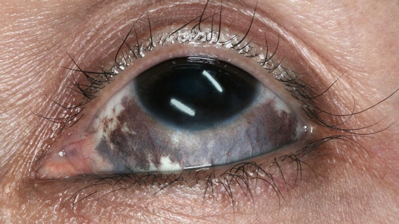
Nevus of Ota
Nevus, Sebaceous of Jadassohn
Dysplastic Nevus Syndrome
Nevus, Intradermal
Synovitis, Pigmented Villonodular
Nevus, Epithelioid and Spindle Cell
Nevus, Pigmented
Dermoscopy
Pigmentation Disorders
Melanoma
Albinism
Lentigo
Basal Cell Nevus Syndrome
Facial Neoplasms
Melanins
Pigment Epithelium of Eye
Iris Neoplasms
Hyperpigmentation
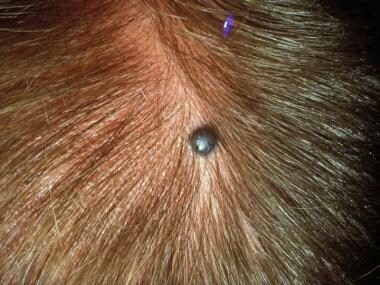
Scalp
Choroid Neoplasms
Melanocytes
Hamartoma
Ciliary Body
Keratosis, Seborrheic
Retinal Pigment Epithelium
Hutchinson's Melanotic Freckle
Hemangioma
Porokeratosis
Skin Diseases
Carcinoma, Basal Cell
Retina
Sunburn
Melanosis
Neoplasms, Adnexal and Skin Appendage
Iris
Neoplasms, Multiple Primary
Blood-Aqueous Barrier
Albinism, Ocular
Nevus, Spindle Cell
Hypertrichosis
Laser Therapy
Eye
Vitiligo
Sturge-Weber Syndrome
Skin
Scleral Diseases
Monophenol Monooxygenase
Proteus Syndrome
Bowen's Disease
Keratoacanthoma
Nevus, Blue
Coloboma
Port-Wine Stain
Microphthalmia-Associated Transcription Factor
Colorado
MART-1 Antigen
Cyclic AMP-Dependent Protein Kinase RIalpha Subunit
Mongolian Spot
Choroid
Leukokeratosis, Hereditary Mucosal
Neurocutaneous melanosis presenting with intracranial amelanotic melanoma. (1/353)
We describe imaging findings in a 2-year-old girl with neurocutaneous melanosis and malignant cerebral melanoma. Because the cerebral melanoma in this child was of the amelanotic type, high-signal intensity on unenhanced T1-weighted images was not present. The cutaneous lesions played a crucial role in establishing a correct (presumed) histopathologic diagnosis on the basis of the imaging findings. To our knowledge this is the first report describing an intracranial amelanotic malignant melanoma in association with neurocutaneous melanosis. (+info)A major quantitative-trait locus for mole density is linked to the familial melanoma gene CDKN2A: a maximum-likelihood combined linkage and association analysis in twins and their sibs. (2/353)
Important risk factors for melanoma are densely clustered melanocytic nevi (common moles) and mutations in the p16 (CDKN2A) gene. Nevi may be subclassified as raised or flat. In our sample, raised nevi were 27% of the total, and the two kinds had a correlation of.33. Correlations for total-nevus count (TNC) in 153 MZ and 199 DZ twin pairs were.94 and.60, respectively, which are compatible with a very-high degree of genetic determination. We hypothesized that some of the genetic variance might be due to variation in the p16 gene. Analysis of linkage to a highly polymorphic marker (D9S942), located close to p16, detected quantitative-trait-loci (QTL) effects accounting for 27% of variance in TNC, rising to 33% if flat but not raised moles were considered. Total heritability was higher for raised (.69) than for flat (.42) moles, but QTL linkage was 0 for raised moles, whereas it accounted for 80% of the heritability of flat moles; additionally, family environment accounted for only 15% of variance in raised versus 46% in flat moles. These findings suggest that raised and flat nevi have very different etiologies. Longer alleles at D9S942 were associated with higher flat-mole counts, and a novel modification to a within-sibship association test showed that this association is genuine and not due to population stratification, although it accounts for only 1% of total variance. Since germline mutations in the exons of CDKN2A are rare, it is likely that variants in the noncoding regions of this gene, or in another gene nearby, are responsible for this major determinant of moliness and, hence, of melanoma risk. (+info)Two male patients with ring Y: definition of an interval in Yq contributing to Turner syndrome. (3/353)
Turner syndrome is thought to result from the haploinsufficiency of genes on the sex chromosomes, but these genes have not been identified yet. We describe two males with deleted ring Y chromosomes, one (TS) with full Turner syndrome and one (DM) without. TS has short stature, skeletal anomalies, lymphogenic obstruction, cardiovascular abnormalities, and miscellaneous features including pigmented naevi, antimongoloid slanting of the palpebral fissures, and widely spaced nipples. In contrast, DM has short stature but no other specific Turner stigmata except high arched palate and a few pigmented naevi. Since little chromosomal mosaicism was detected, the different segments of the Y chromosome retained by these two males identify the location of one or more "anti-Turner" genes. Most of the Yp pseudoautosomal region and Yq were deleted from both patients during the formation of the ring chromosome, while the Y specific portion of Yp and the centromere were retained. The major difference detected was an interval of proximal Yq present in DM and deleted in TS. None of the previously identified genes, DFFRY, DBY, UTY, or TB4Y, lies entirely within this interval, although DFFRY was truncated by DM's breakpoint. These data suggest that one or more additional "anti-Turner" gene(s) remains to be identified in the region of Yq proximal to DFFRY. (+info)Mutational analysis of the N-ras, p53, p16INK4a, CDK4, and MC1R genes in human congenital melanocytic naevi. (4/353)
Eighteen human congenital melanocytic naevi (CMN) from 17 patients were screened for activating point mutations in the oncogenes N-ras and CDK4 and for sequence variants in the MC1R gene by combined RFLP-PCR/SSCP analysis. In addition, all lesions were screened for deletions and point mutations in the tumour suppressor genes p53 and p16INK4a (CDKN2A) by combined multiplex PCR/SSCP analysis. Positive screening data were specified by sequencing of the corresponding PCR product. Activating point mutations in the N-ras gene (nine CAA (Gln) to AAA (Lys) transversions and one CAA (Gln) to CGA (Arg) transition at codon 61) were detected at high frequency (56%). Furthermore, three missense mutations (V92M) and two silent mutations (CGA (Arg) to CGG (Arg), codon 213, exon 6) were found in the MC1R and p53 genes, respectively. No mutations were found in p16 or CDK4. The activated N-ras oncogene, which is also found in human cutaneous melanomas, may constitute a potential risk factor for melanoma formation within CMN. (+info)Genotype/phenotype and penetrance studies in melanoma families with germline CDKN2A mutations. (5/353)
Patients with a family history of melanoma are at increased risk of this tumor. Those family members who also have the atypical mole syndrome are commonly targeted for screening in the belief that they are more likely to be mutant gene carriers. We have correlated the atypical mole syndrome phenotype and gene carrier status in five families with germline CDKN2A mutations and shown that family members with the atypical mole syndrome were three times more likely to be mutant gene carriers than their relatives who did not have the atypical mole syndrome (odds ratio 3.4; confidence interval 1.0-11. 1), supporting the view that CDKN2A is nevogenic. Individual characteristics which best predicted mutant gene carrier status were: nevi on the buttocks (odds ratio 4.4; confidence interval 1. 6-12.4), nevi on the feet (odds ratio 4.2; confidence interval 1. 4-12.5), total nevus number being at least 100 (nevi > or = 2 mm in diameter) (odds ratio 3.4; confidence interval 1.0-11.1) and two or more clinically atypical nevi (odds ratio 3.1; confidence interval 1. 1-9.0). Gene carriers were also significantly more likely to have noticeable freckling and possibly also Fitzpatrick skin types 1-3. The overlap between gene carriers and nongene carriers was, however, marked: the atypical mole syndrome did not clearly differentiate mutant gene carriers from those with a normal gene. This study is of significance to clinicians as the clinical practice of using the atypical mole syndrome to identify particular family members for surveillance is shown to be inappropriate. Until formal gene testing is available, all members of families with an excessive number of melanoma cases should be treated as potential mutation carriers at increased risk of melanoma. (+info)The diagnostic accuracy of Danish GPs in the diagnosis of pigmented skin lesions. (6/353)
BACKGROUND: The GP often has a primary function in assessing pigmented skin lesions in Denmark. No data are available on the diagnostic accuracy of this process. OBJECTIVE: We aimed to study the sensitivity, specificity and positive prognostic value of the diagnosis made by 27 trained or trainee GPs. METHOD: We tested the diagnostic accuracy of the viewing of colour slides of pigmented skin lesions under standardized conditions at a seminar on skin cancer. Diagnostic accuracy was determined only for the clinically relevant diagnosis of benign or malignant. RESULTS: The median diagnostic accuracy (sensitivity) for the group as a whole was 0.75 (95% CI 0.65-0.80), the specificity was 0.70 (95% CI 0.68-0.79) and the positive predictive value 0.70 (95% CI 0.62-0.77). CONCLUSION: These values are comparable with previously published figures for trainee dermatologists, and it is therefore concluded that ongoing interest rather than basic training is the major determinant for clinical acumen. (+info)Agreement between self-assessment of melanocytic nevi by patients and dermatologic examination. (7/353)
The number of melanocytic nevi is the strongest risk factor for cutaneous melanoma. As pigmented skin lesions are visible to everybody, the question has been raised about whether people can identify themselves as being at risk for melanoma through self-counting of moles. In 1991, a total of 513 central European melanoma patients and 498 controls were asked to count the total number of nevi and the number of atypical nevi on the whole body. Whole-body examination by dermatologists followed. Agreement was assessed on categorized nevus counts by means of ordinal kappa values and log-linear modeling. Study subjects significantly underestimated the total number of melanocytic nevi (p < 0.0001). Chance-corrected overall agreement was rather poor (kappa = 0.14), and the ability to detect many existing nevi was low. Agreement was higher for atypical melanocytic nevi counts (kappa = 0.37), and the sensitivity to detect more than one atypical nevus was 0.48. Self-assessment of the number of melanocytic nevi was difficult to perform accurately, and people severely underestimated the actual number. Despite these results, people should be encouraged to perform regular skin self-examination for early detection of melanoma. (+info)Combined nevi of the conjunctiva. (8/353)
PURPOSE: To report the clinical and histologic features of combined nevi of the conjunctiva, a type of nevus that is not uncommon in the skin but has rarely been reported in the conjunctiva. METHODS: Conjunctival nevi and melanomas from the files of the University of California, San Francisco, eye pathology laboratory were reviewed from 1984 to 1999 for the presence of features of both standard nevocytic nevi and blue nevi. Clinical histories and, when available, clinical photographs were obtained. RESULTS: Thirty-one combined nevi were discovered during the 15-year period between 1984 and 1999. One case before 1984 had been incorrectly diagnosed as a junctional nevus. The dendritic and spindle-shaped blue nevus cells had been overlooked because they were not recognized as distinct from the standard nevocytic nevus cells. The recognition of a blue as well as a brown color, a deep as well as a superficial component in the lesion, or a history of pigmentation since birth may help to establish the correct clinical diagnosis and prevent an unnecessarily deep surgical resection. Although growth of the lesion or "satellites" in some patients may favor a clinical diagnosis of melanoma, none of the lesions in this series were malignant. CONCLUSION: Despite a paucity of reports of combined nevi of the conjunctiva in the medical literature, this type of nevus--a combination of a nevocytic and a blue nevus--is common and has been overlooked in the past. (+info)A nevus, also known as a mole, is a benign growth or mark on the skin that is usually brown or black. It can be raised or flat and can appear anywhere on the body. Nevi are made up of cells called melanocytes, which produce the pigment melanin. Most nevi develop in childhood or adolescence, but they can also appear later in life. Some people have many nevi, while others have few or none.
There are several types of nevi, including:
* Common nevi: These are the most common type of mole and are usually small, round, and brown or black. They can be flat or raised and can appear anywhere on the body.
* Atypical nevi: These moles are larger than common nevi and have irregular borders and color. They may be flat or raised and can appear anywhere on the body, but are most commonly found on the trunk and extremities. Atypical nevi are more likely to develop into melanoma, a type of skin cancer, than common nevi.
* Congenital nevi: These moles are present at birth and can vary in size from small to large. They are more likely to develop into melanoma than moles that develop later in life.
* Spitz nevi: These are rare, benign growths that typically appear in children and adolescents. They are usually pink or red and dome-shaped.
It is important to monitor nevi for changes in size, shape, color, and texture, as these can be signs of melanoma. If you notice any changes in a mole, or if you have a new mole that is unusual or bleeding, it is important to see a healthcare provider for further evaluation.
A Nevus of Ota, also known as an oculodermal melanocytosis, is a benign birthmark characterized by the presence of darkly pigmented (melanin-containing) cells called melanocytes in the skin and mucous membranes around the eye. These pigmented cells can also extend to the sclera (the white part of the eye), dura mater (the outer covering of the brain), and leptomeninges (the middle layer of the meninges, which cover the brain and spinal cord).
The Nevus of Ota typically presents as a unilateral (occurring on one side) bluish-gray or brown patch that follows the distribution of the ophthalmic and maxillary divisions of the trigeminal nerve. It usually affects the eye, forehead, temple, and cheek, but it can also involve other areas of the face, scalp, and neck.
While Nevi of Ota are generally harmless, they may be associated with an increased risk of developing melanoma (a type of skin cancer) in the affected area. Therefore, regular monitoring and evaluation by a healthcare professional is recommended.
A nevus sebaceous of Jadassohn is a type of congenital benign skin tumor or birthmark that is composed of epidermal, hair follicle, and sebaceous gland components. It typically appears as a yellowish, greasy, or warty plaque on the scalp or face during infancy or early childhood. The lesion tends to enlarge slowly and may undergo various changes in appearance over time.
In adolescence or adulthood, there is a risk of secondary tumor development within the nevus sebaceous, such as basal cell carcinoma, squamous cell carcinoma, or sebaceous carcinoma. Therefore, regular monitoring and possible surgical removal of the lesion may be recommended, especially in cases where the nevus is large, symptomatic, or shows signs of malignant transformation.
Dysplastic Nevus Syndrome, also known as atypical mole syndrome, is a condition characterized by the presence of numerous dysplastic nevi (abnormal moles) that may appear irregular in shape, color, and size. These moles are typically larger than normal moles (greater than 5 mm in diameter) and have an asymmetrical shape, uneven borders, and varied colors.
Individuals with Dysplastic Nevus Syndrome have a higher risk of developing melanoma, a type of skin cancer that can be life-threatening if not detected and treated early. The syndrome is usually inherited in an autosomal dominant manner, meaning that a child has a 50% chance of inheriting the gene from an affected parent.
It's important to note that having dysplastic nevi does not necessarily mean that a person will develop melanoma, but it does increase their risk. Regular skin examinations by a dermatologist and self-examinations are recommended for early detection of any changes in moles or the development of new suspicious lesions.
An intradermal nevus, also known as an intradermal naevus or compound nevus, is a type of benign pigmented skin lesion that originates from melanocytes, which are the pigment-producing cells in the skin. It develops when melanocytes grow and multiply in the dermis, the middle layer of the skin.
Intradermal nevi are typically small, round or oval, raised bumps that range in color from flesh-colored to brown or black. They can appear anywhere on the body, but they are most commonly found on the trunk and extremities. These nevi usually develop during childhood or adolescence and may continue to grow slowly over time.
Intradermal nevi are generally harmless and do not require treatment unless they become symptomatic (e.g., itchy, painful, or bleed) or change in appearance, which could indicate a potential malignant transformation into melanoma. In such cases, a biopsy may be performed to confirm the diagnosis and determine the appropriate course of action.
It is essential to monitor any changes in existing nevi and consult a healthcare professional if there are concerns about new or changing lesions. Regular skin examinations can help detect early signs of skin cancer and improve treatment outcomes.
Pigmented villonodular synovitis (PVNS) is a rare, benign condition that affects the synovial membrane, which lines the joints. It is characterized by the proliferation of synovial cells and the deposition of hemosiderin, a pigment resulting from the breakdown of blood products. This can lead to joint swelling, pain, stiffness, and limited mobility. PVNS typically affects the large joints such as the knee or hip, but it can also occur in smaller joints, bursae, or tendon sheaths.
There are two forms of PVNS: localized and diffuse. Localized PVNS, also known as giant cell tumor of the tendon sheath, affects a specific area within the joint and is more likely to be treated successfully with surgery. Diffuse PVNS, on the other hand, involves the entire synovial lining of the joint and has a higher recurrence rate even after surgical removal.
The exact cause of PVNS remains unclear, but it is not considered a malignant condition. Treatment usually involves surgical removal of the affected synovium, with or without radiation therapy or chemotherapy to reduce the risk of recurrence. In some cases, arthroscopic surgery may be an option for localized PVNS.
A nevus is a general term for a benign growth or mole on the skin. There are many different types of nevi, including epithelioid and spindle cell nevi.
Epithelioid cell: A type of cell that is typically found in certain types of nevi, as well as in some malignant tumors such as melanoma. Epithelioid cells are large, round cells with a pale, clear cytoplasm and centrally located nuclei.
Spindle cell: A type of cell that is often found in certain types of nevi, including Spitz nevi and deep penetrating nevi. Spindle cells are elongated, thin cells with cigar-shaped nuclei. They can also be found in some malignant tumors such as melanoma.
Epithelioid and spindle cell nevus: A type of nevus that contains both epithelioid and spindle cells. These nevi are typically benign, but they can sometimes be difficult to distinguish from melanoma, especially if they have atypical features. Therefore, it is important for these types of nevi to be evaluated by a dermatopathologist or a specialist in skin pathology.
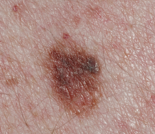
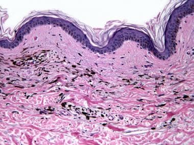
A nevus pigmentosus, also known as a pigmented mole or melanocytic nevus, is a benign proliferation of melanocytes, the pigment-producing cells in the skin. These lesions typically appear as well-circumscribed, brown to black macules or papules. They can vary in size and shape and may be flat or raised. Most nevi are harmless and do not require treatment; however, some may undergo malignant transformation into melanoma, a potentially life-threatening skin cancer. Regular self-skin examinations and professional skin checks are recommended to monitor for changes in nevi that may indicate malignancy.
Skin neoplasms refer to abnormal growths or tumors in the skin that can be benign (non-cancerous) or malignant (cancerous). They result from uncontrolled multiplication of skin cells, which can form various types of lesions. These growths may appear as lumps, bumps, sores, patches, or discolored areas on the skin.
Benign skin neoplasms include conditions such as moles, warts, and seborrheic keratoses, while malignant skin neoplasms are primarily classified into melanoma, squamous cell carcinoma, and basal cell carcinoma. These three types of cancerous skin growths are collectively known as non-melanoma skin cancers (NMSCs). Melanoma is the most aggressive and dangerous form of skin cancer, while NMSCs tend to be less invasive but more common.
It's essential to monitor any changes in existing skin lesions or the appearance of new growths and consult a healthcare professional for proper evaluation and treatment if needed.
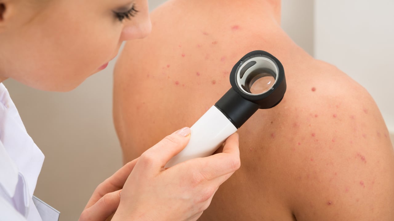
Dermoscopy, also known as dermatoscopy or epiluminescence microscopy, is a non-invasive diagnostic technique used in dermatology to evaluate skin lesions, such as moles and pigmented skin tumors. This method involves the use of a handheld device called a dermoscope, which consists of a magnifying lens, a light source, and a transparent plate or immersion fluid that allows for better visualization of the skin's surface structures.
Dermoscopy enables dermatologists to examine the pigmented patterns, vascular structures, and other morphological features hidden beneath the skin's surface that are not visible to the naked eye. By observing these details, dermatologists can improve their ability to differentiate between benign and malignant lesions, leading to more accurate diagnoses and appropriate treatment decisions.
The primary uses of dermoscopy include:
1. Early detection and diagnosis of melanoma and other skin cancers, such as basal cell carcinoma and squamous cell carcinoma.
2. Monitoring the evolution of suspicious moles or lesions over time.
3. Assisting in the identification of various benign skin growths, like seborrheic keratoses, dermatofibromas, and nevi (moles).
4. Improving the diagnostic accuracy for infectious skin conditions, inflammatory processes, and other dermatological disorders.
Overall, dermoscopy is a valuable tool in the field of dermatology that enhances the clinician's ability to diagnose and manage various skin conditions accurately and effectively.
A "Halo Nevus" (also known as Sutton nevus or leukoderma acquisitum centrifugum) is a type of melanocytic nevus (mole) that is surrounded by a depigmented halo, typically measured to be 0.5-1 cm wide. The central nevus can be either a common acquired melanocytic nevus or a Spitz nevus.
The depigmentation occurs due to the destruction of melanocytes (pigment-producing cells) in the skin surrounding the nevus, which is thought to be an immune-mediated response. The halo nevus is considered a benign condition and usually appears in children and young adults. While most halo nevi are harmless, it's essential to monitor them for any changes that may indicate melanoma or other skin cancers. If you notice any changes in the size, shape, color, or border of a halo nevus, consult with a dermatologist or healthcare professional.
Pigmentation disorders are conditions that affect the production or distribution of melanin, the pigment responsible for the color of skin, hair, and eyes. These disorders can cause changes in the color of the skin, resulting in areas that are darker (hyperpigmentation) or lighter (hypopigmentation) than normal. Examples of pigmentation disorders include melasma, age spots, albinism, and vitiligo. The causes, symptoms, and treatments for these conditions can vary widely, so it is important to consult a healthcare provider for an accurate diagnosis and treatment plan.
Melanoma is defined as a type of cancer that develops from the pigment-containing cells known as melanocytes. It typically occurs in the skin but can rarely occur in other parts of the body, including the eyes and internal organs. Melanoma is characterized by the uncontrolled growth and multiplication of melanocytes, which can form malignant tumors that invade and destroy surrounding tissue.
Melanoma is often caused by exposure to ultraviolet (UV) radiation from the sun or tanning beds, but it can also occur in areas of the body not exposed to the sun. It is more likely to develop in people with fair skin, light hair, and blue or green eyes, but it can affect anyone, regardless of their skin type.
Melanoma can be treated effectively if detected early, but if left untreated, it can spread to other parts of the body and become life-threatening. Treatment options for melanoma include surgery, radiation therapy, chemotherapy, immunotherapy, and targeted therapy, depending on the stage and location of the cancer. Regular skin examinations and self-checks are recommended to detect any changes or abnormalities in moles or other pigmented lesions that may indicate melanoma.
Pigmentation, in a medical context, refers to the coloring of the skin, hair, or eyes due to the presence of pigment-producing cells called melanocytes. These cells produce a pigment called melanin, which determines the color of our skin, hair, and eyes.
There are two main types of melanin: eumelanin and pheomelanin. Eumelanin is responsible for brown or black coloration, while pheomelanin produces a red or yellow hue. The amount and type of melanin produced by melanocytes can vary from person to person, leading to differences in skin color and hair color.
Changes in pigmentation can occur due to various factors such as genetics, exposure to sunlight, hormonal changes, inflammation, or certain medical conditions. For example, hyperpigmentation refers to an excess production of melanin that results in darkened patches on the skin, while hypopigmentation is a condition where there is a decreased production of melanin leading to lighter or white patches on the skin.
Albinism is a group of genetic disorders that result in little or no production of melanin, the pigment responsible for coloring skin, hair, and eyes. It is caused by mutations in genes involved in the production of melanin. There are several types of albinism, including oculocutaneous albinism (OCA) and ocular albinism (OA). OCA affects the skin, hair, and eyes, while OA primarily affects the eyes.
People with albinism typically have very pale skin, white or light-colored hair, and light-colored eyes. They may also have vision problems, such as sensitivity to light (photophobia), rapid eye movements (nystagmus), and decreased visual acuity. The severity of these symptoms can vary depending on the type and extent of albinism.
Albinism is inherited in an autosomal recessive manner, which means that an individual must inherit two copies of the mutated gene, one from each parent, in order to have the condition. If both parents are carriers of a mutated gene for albinism, they have a 25% chance with each pregnancy of having a child with albinism.
There is no cure for albinism, but individuals with the condition can take steps to protect their skin and eyes from the sun and use visual aids to help with vision problems. It is important for people with albinism to undergo regular eye examinations and to use sun protection, such as sunscreen, hats, and sunglasses, to prevent skin damage and skin cancer.
A lentigo is a small, sharply defined, pigmented macule (flat spot) on the skin. It's usually tan, brown, or black and can appear on various parts of the body, particularly where the skin has been exposed to the sun. Lentigos are typically harmless and don't require treatment unless they're uncomfortable or for cosmetic reasons. However, some types of lentigines, such as lentigo maligna, can progress into melanoma, a type of skin cancer, so regular self-examinations and professional skin checks are important.
It is essential to differentiate between simple lentigos and lentigo maligna, which is a precancerous lesion. Lentigo maligna tends to occur in older individuals, often on the face, and can appear as a large, irregularly shaped, and darkly pigmented patch. A dermatologist should evaluate any suspicious or changing skin spots for proper diagnosis and treatment.
Skin pigmentation is the coloration of the skin that is primarily determined by two types of melanin pigments, eumelanin and pheomelanin. These pigments are produced by melanocytes, which are specialized cells located in the epidermis. Eumelanin is responsible for brown or black coloration, while pheomelanin produces a red or yellow hue.
The amount and distribution of melanin in the skin can vary depending on genetic factors, age, sun exposure, and various other influences. Increased production of melanin in response to UV radiation from the sun helps protect the skin from damage, leading to darkening or tanning of the skin. However, excessive sun exposure can also cause irregular pigmentation, such as sunspots or freckles.
Abnormalities in skin pigmentation can result from various medical conditions, including albinism (lack of melanin production), vitiligo (loss of melanocytes leading to white patches), and melasma (excessive pigmentation often caused by hormonal changes). These conditions may require medical treatment to manage or improve the pigmentation issues.
Basal Cell Nevus Syndrome (BCNS), also known as Gorlin-Goltz Syndrome, is a rare genetic disorder that is characterized by the development of multiple basal cell carcinomas (BCCs), which are skin cancer tumors that arise from the basal cells in the outermost layer of the skin.
The syndrome is caused by mutations in the PTCH1 gene, which regulates the hedgehog signaling pathway involved in embryonic development and tissue growth regulation. The condition is inherited in an autosomal dominant manner, meaning that a child has a 50% chance of inheriting the mutated gene from an affected parent.
Individuals with BCNS typically develop hundreds to thousands of BCCs over their lifetime, often beginning in childhood or adolescence. They may also have other benign and malignant tumors, such as medulloblastomas (brain tumors), fibromas, and rhabdomyosarcomas.
Additional features of BCNS can include:
1. Facial abnormalities, such as a broad nasal bridge, widely spaced eyes, and pits or depressions on the palms and soles.
2. Skeletal abnormalities, such as spine deformities, rib anomalies, and jaw cysts.
3. Developmental delays and intellectual disabilities in some cases.
4. Increased risk of other cancers, including breast, ovarian, and lung cancer.
Early detection and management of BCCs and other tumors are crucial for individuals with BCNS to prevent complications and improve their quality of life. Regular dermatological examinations, sun protection measures, and surgical removal of tumors are common treatment approaches.
Facial neoplasms refer to abnormal growths or tumors that develop in the tissues of the face. These growths can be benign (non-cancerous) or malignant (cancerous). Facial neoplasms can occur in any of the facial structures, including the skin, muscles, bones, nerves, and glands.
Benign facial neoplasms are typically slow-growing and do not spread to other parts of the body. Examples include papillomas, hemangiomas, and neurofibromas. While these tumors are usually harmless, they can cause cosmetic concerns or interfere with normal facial function.
Malignant facial neoplasms, on the other hand, can be aggressive and invasive. They can spread to other parts of the face, as well as to distant sites in the body. Common types of malignant facial neoplasms include basal cell carcinoma, squamous cell carcinoma, and melanoma.
Treatment for facial neoplasms depends on several factors, including the type, size, location, and stage of the tumor. Treatment options may include surgery, radiation therapy, chemotherapy, or a combination of these approaches. It is important to seek medical attention promptly if you notice any unusual growths or changes in the skin or tissues of your face.
Melanin is a pigment that determines the color of skin, hair, and eyes in humans and animals. It is produced by melanocytes, which are specialized cells found in the epidermis (the outer layer of the skin) and the choroid (the vascular coat of the eye). There are two main types of melanin: eumelanin and pheomelanin. Eumelanin is a black or brown pigment, while pheomelanin is a red or yellow pigment. The amount and type of melanin produced by an individual can affect their skin and hair color, as well as their susceptibility to certain diseases, such as skin cancer.
I'm sorry for any confusion, but "hair color" is not a term that has a medical definition in the same way that a term like "myocardial infarction" (heart attack) does. Hair color can be described in various ways, such as being a natural hair color (like blonde, brunette, red, or black) or describing a change in hair color due to factors like aging (gray or white hairs) or hair dye usage.
However, it's worth noting that changes in hair color can sometimes be associated with certain medical conditions. For example, premature graying of the hair before the age of 30 can be a feature of certain genetic disorders or vitamin B12 deficiency. Similarly, some skin conditions like alopecia areata or vitiligo can cause patchy changes in hair color. But these associations don't provide a medical definition for 'hair color'.
The pigment epithelium of the eye, also known as the retinal pigment epithelium (RPE), is a layer of cells located between the photoreceptor cells of the retina and the choroid, which is the vascular layer of the eye. The RPE plays a crucial role in maintaining the health and function of the photoreceptors by providing them with nutrients, removing waste products, and helping to regulate the light that enters the eye.
The RPE cells contain pigment granules that absorb excess light, preventing it from scattering within the eye and improving visual acuity. They also help to create a barrier between the retina and the choroid, which is important for maintaining the proper functioning of the photoreceptors. Additionally, the RPE plays a role in the regeneration of visual pigments in the photoreceptor cells, allowing us to see in different light conditions.
Damage to the RPE can lead to various eye diseases and conditions, including age-related macular degeneration (AMD), which is a leading cause of vision loss in older adults.
Iris neoplasms refer to abnormal growths or tumors that develop in the iris, which is the colored part of the eye. These neoplasms can be benign (non-cancerous) or malignant (cancerous). Benign iris neoplasms are typically slow-growing and do not spread to other parts of the body. Malignant iris neoplasms, on the other hand, can grow quickly and may spread to other parts of the eye or nearby structures, such as the ciliary body or choroid.
Iris neoplasms can cause various symptoms, including changes in the appearance of the eye, such as a visible mass or discoloration, pain, redness, light sensitivity, blurred vision, or changes in the size or shape of the pupil. The diagnosis of iris neoplasms typically involves a comprehensive eye examination, including a visual acuity test, refraction, slit-lamp examination, and sometimes imaging tests such as ultrasound or optical coherence tomography (OCT).
Treatment options for iris neoplasms depend on the type, size, location, and severity of the tumor. Small, benign iris neoplasms may not require treatment and can be monitored over time. Larger or malignant iris neoplasms may require surgical removal, radiation therapy, or other treatments to prevent complications or spread to other parts of the eye or body. It is essential to seek medical attention promptly if you experience any symptoms of iris neoplasms or notice any changes in your vision or the appearance of your eyes.
Hyperpigmentation is a medical term that refers to the darkening of skin areas due to an increase in melanin, the pigment that provides color to our skin. This condition can affect people of all races and ethnicities, but it's more noticeable in those with lighter skin tones.
Hyperpigmentation can be caused by various factors, including excessive sun exposure, hormonal changes (such as during pregnancy), inflammation, certain medications, and underlying medical conditions like Addison's disease or hemochromatosis. It can also result from skin injuries, such as cuts, burns, or acne, which leave dark spots known as post-inflammatory hyperpigmentation.
There are several types of hyperpigmentation, including:
1. Melasma: This is a common form of hyperpigmentation that typically appears as symmetrical, blotchy patches on the face, particularly the forehead, cheeks, and upper lip. It's often triggered by hormonal changes, such as those experienced during pregnancy or while taking birth control pills.
2. Solar lentigos (age spots or liver spots): These are small, darkened areas of skin that appear due to prolonged sun exposure over time. They typically occur on the face, hands, arms, and decolletage.
3. Post-inflammatory hyperpigmentation: This type of hyperpigmentation occurs when an injury or inflammation heals, leaving behind a darkened area of skin. It's more common in people with darker skin tones.
Treatment for hyperpigmentation depends on the underlying cause and may include topical creams, chemical peels, laser therapy, or microdermabrasion. Preventing further sun damage is crucial to managing hyperpigmentation, so wearing sunscreen with a high SPF and protective clothing is recommended.
The scalp is the anatomical region located at the upper part of the human head, covering the skull except for the face and the ears. It is made up of several layers: the skin, the connective tissue, the galea aponeurotica (a strong, flat, tendinous sheet), loose areolar tissue, and the periosteum (the highly vascularized innermost layer that attaches directly to the skull bones). The scalp has a rich blood supply and is home to numerous sensory receptors, including those for touch, pain, and temperature. It also contains hair follicles, sebaceous glands, and sweat glands.
Choroid neoplasms are abnormal growths that develop in the choroid, a layer of blood vessels that lies between the retina and the sclera (the white of the eye). These growths can be benign or malignant (cancerous). Benign choroid neoplasms include choroidal hemangiomas and choroidal osteomas. Malignant choroid neoplasms are typically choroidal melanomas, which are the most common primary eye tumors in adults. Other types of malignant choroid neoplasms include metastatic tumors that have spread to the eye from other parts of the body. Symptoms of choroid neoplasms can vary depending on the size and location of the growth, but may include blurred vision, floaters, or a dark spot in the visual field. Treatment options depend on the type, size, and location of the tumor, as well as the patient's overall health and personal preferences.
Eye color is a characteristic determined by variations in a person's genes. The color of the eyes depends on the amount and type of pigment called melanin found in the eye's iris.
There are three main types of eye colors: brown, blue, and green. Brown eyes have the most melanin, while blue eyes have the least. Green eyes have a moderate amount of melanin combined with a golden tint that reflects light to give them their unique color.
Eye color is a polygenic trait, which means it is influenced by multiple genes. The two main genes responsible for eye color are OCA2 and HERC2, both located on chromosome 15. These genes control the production, transport, and storage of melanin in the iris.
It's important to note that eye color can change during infancy and early childhood due to the development of melanin in the iris. Additionally, some medications or medical conditions may also cause changes in eye color over time.
Melanocytes are specialized cells that produce, store, and transport melanin, the pigment responsible for coloring of the skin, hair, and eyes. They are located in the bottom layer of the epidermis (the outermost layer of the skin) and can also be found in the inner ear and the eye's retina. Melanocytes contain organelles called melanosomes, which produce and store melanin.
Melanin comes in two types: eumelanin (black or brown) and pheomelanin (red or yellow). The amount and type of melanin produced by melanocytes determine the color of a person's skin, hair, and eyes. Exposure to UV radiation from sunlight increases melanin production as a protective response, leading to skin tanning.
Melanocyte dysfunction or abnormalities can lead to various medical conditions, such as albinism (lack of melanin production), melasma (excessive pigmentation), and melanoma (cancerous growth of melanocytes).
A hamartoma is a benign tumor-like growth that is composed of an unusual mixture of cells and tissues that are normally found in the affected area. These growths can occur anywhere in the body, but they are most commonly found in the skin, lungs, and brain. Hamartomas are typically slow growing and do not spread to other parts of the body (metastasize). They are usually harmless, but in some cases, they may cause symptoms or complications depending on their size and location. In general, hamartomas do not require treatment unless they are causing problems.
The ciliary body is a part of the eye's internal structure that is located between the choroid and the iris. It is composed of muscle tissue and is responsible for adjusting the shape of the lens through a process called accommodation, which allows the eye to focus on objects at varying distances. Additionally, the ciliary body produces aqueous humor, the clear fluid that fills the anterior chamber of the eye and helps to nourish the eye's internal structures. The ciliary body is also responsible for maintaining the shape and position of the lens within the eye.
Seborrheic Keratosis is a common, benign skin condition that typically presents as rough, scaly, tan-to-darkly pigmented growths on the surface of the skin. These lesions can appear anywhere on the body, but they are most commonly found on the face, chest, back, and extremities. Seborrheic Keratoses are caused by an overproduction of keratin, a protein that makes up the outer layer of the skin.
The exact cause of Seborrheic Keratosis is not known, but it is thought to be related to genetic factors and sun exposure. The condition is more common in older adults and is not contagious. While Seborrheic Keratoses are generally harmless, they can be removed for cosmetic reasons or if they become irritated or inflamed. Treatment options include cryotherapy (freezing the lesions with liquid nitrogen), curettage (scraping the lesions off), and laser surgery.
Adrenal cortex diseases refer to a group of conditions that affect the adrenal glands, which are small glands located on top of the kidneys. The adrenal glands consist of two parts: the outer adrenal cortex and the inner medulla. The adrenal cortex is responsible for producing hormones such as cortisol, aldosterone, and androgens that regulate various bodily functions, including metabolism, blood pressure, and sexual development.
Diseases of the adrenal cortex can result from an overproduction or underproduction of these hormones. Some common adrenal cortex diseases include:
1. Addison's disease: a condition characterized by insufficient production of hormones by the adrenal glands, leading to symptoms such as fatigue, weight loss, low blood pressure, and darkening of the skin.
2. Cushing's syndrome: a condition caused by an excess of cortisol in the body, which can result from taking high doses of corticosteroid medications or from a tumor in the pituitary gland or adrenal glands. Symptoms include weight gain, particularly around the trunk and face, thinning of the skin, easy bruising, muscle weakness, and mood changes.
3. Congenital adrenal hyperplasia: a group of inherited disorders that affect the production of hormones by the adrenal glands. Depending on the specific type of congenital adrenal hyperplasia, symptoms can range from ambiguous genitalia in newborns to precocious puberty, short stature, and infertility in older children and adults.
4. Adrenal tumors: benign or cancerous growths that develop in the adrenal glands and can cause hormonal imbalances. Symptoms depend on the type of tumor and the hormones it produces.
Treatment for adrenal cortex diseases depends on the specific condition and its underlying cause. Treatment options may include medication, surgery, or radiation therapy.
Biological pigments are substances produced by living organisms that absorb certain wavelengths of light and reflect others, resulting in the perception of color. These pigments play crucial roles in various biological processes such as photosynthesis, vision, and protection against harmful radiation. Some examples of biological pigments include melanin, hemoglobin, chlorophyll, carotenoids, and flavonoids.
Melanin is a pigment responsible for the color of skin, hair, and eyes in animals, including humans. Hemoglobin is a protein found in red blood cells that contains a porphyrin ring with an iron atom at its center, which gives blood its red color and facilitates oxygen transport. Chlorophyll is a green pigment found in plants, algae, and some bacteria that absorbs light during photosynthesis to convert carbon dioxide and water into glucose and oxygen. Carotenoids are orange, yellow, or red pigments found in fruits, vegetables, and some animals that protect against oxidative stress and help maintain membrane fluidity. Flavonoids are a class of plant pigments with antioxidant properties that have been linked to various health benefits.
The retinal pigment epithelium (RPE) is a single layer of cells located between the photoreceptor cells of the retina and the choroid, which is a part of the eye containing blood vessels. The RPE plays a crucial role in maintaining the health and function of the photoreceptors by providing them with nutrients, removing waste products, and helping to regulate the light-sensitive visual pigments within the photoreceptors.
The RPE cells contain pigment granules that absorb excess light to prevent scattering within the eye and improve visual acuity. They also help to form the blood-retina barrier, which restricts the movement of certain molecules between the retina and the choroid, providing an important protective function for the retina.
Damage to the RPE can lead to a variety of eye conditions, including age-related macular degeneration (AMD), which is a leading cause of vision loss in older adults.
I'm sorry for any confusion, but "sunlight" is not a term with a specific medical definition. However, sunlight does have various implications in the field of medicine. It is the light that comes from the sun, which is a star at the center of our solar system. Sunlight is essential for the production of vitamin D in humans, and it can also have effects on mood and sleep patterns due to its influence on circadian rhythms.
In a medical context, sunlight is often discussed in relation to its potential health benefits and risks. For instance, moderate sun exposure can help increase vitamin D levels, which are important for bone health, immune function, and other bodily processes. However, excessive sun exposure can lead to harmful effects, such as sunburn, premature skin aging, and an increased risk of skin cancer.
It's essential to balance the benefits and risks of sunlight exposure by practicing safe sun habits, such as wearing protective clothing, using a broad-spectrum sunscreen with an SPF of at least 30, seeking shade during peak sunlight hours, and avoiding intentional tanning.
Hutchinson's melanotic freckle, also known as Hutchinson's melanotic macule or naevus, is a type of pigmented lesion that can be a precursor to malignant melanoma, a serious form of skin cancer. It is typically characterized by the presence of darkly pigmented, irregularly shaped patches on the skin, often found on the face or neck.
The lesions are usually brown or black in color and may have an uneven border or surface. They can vary in size from a few millimeters to several centimeters in diameter. Hutchinson's melanotic freckles are typically larger, darker, and more irregularly shaped than common freckles.
These lesions are named after Sir Jonathan Hutchinson, an English surgeon and pathologist who first described them in the late 19th century. It is important to note that while Hutchinson's melanotic freckles can be a sign of increased risk for developing melanoma, not all such lesions will become cancerous. However, any changes in size, shape, or color of these lesions should be evaluated by a healthcare professional as soon as possible.
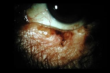
Conjunctival neoplasms refer to abnormal growths or tumors that develop on the conjunctiva, which is the thin, clear mucous membrane that covers the inner surface of the eyelids and the outer surface of the eye. These neoplasms can be benign (non-cancerous) or malignant (cancerous).
Benign conjunctival neoplasms are typically slow-growing and do not spread to other parts of the body. They may include lesions such as conjunctival cysts, papillomas, or naevi (moles). These growths can usually be removed through simple surgical procedures with a good prognosis.
Malignant conjunctival neoplasms, on the other hand, are cancerous and have the potential to invade surrounding tissues and spread to other parts of the body. The most common type of malignant conjunctival neoplasm is squamous cell carcinoma, which arises from the epithelial cells that line the surface of the conjunctiva. Other less common types include melanoma, lymphoma, and adenocarcinoma.
Malignant conjunctival neoplasms typically require more extensive treatment, such as surgical excision, radiation therapy, or chemotherapy. The prognosis for malignant conjunctival neoplasms depends on the type and stage of the cancer at the time of diagnosis, as well as the patient's overall health and age. Early detection and prompt treatment are key to improving outcomes in patients with these conditions.
A hemangioma is a benign (noncancerous) vascular tumor or growth that originates from blood vessels. It is characterized by an overgrowth of endothelial cells, which line the interior surface of blood vessels. Hemangiomas can occur in various parts of the body, but they are most commonly found on the skin and mucous membranes.
Hemangiomas can be classified into two main types:
1. Capillary hemangioma (also known as strawberry hemangioma): This type is more common and typically appears during the first few weeks of life. It grows rapidly for several months before gradually involuting (or shrinking) on its own, usually within the first 5 years of life. Capillary hemangiomas can be superficial, appearing as a bright red, raised lesion on the skin, or deep, forming a bluish, compressible mass beneath the skin.
2. Cavernous hemangioma: This type is less common and typically appears during infancy or early childhood. It consists of large, dilated blood vessels and can occur in various organs, including the skin, liver, brain, and gastrointestinal tract. Cavernous hemangiomas on the skin appear as a rubbery, bluish mass that does not typically involute like capillary hemangiomas.
Most hemangiomas do not require treatment, especially if they are small and not causing any significant problems. However, in cases where hemangiomas interfere with vital functions, impair vision or hearing, or become infected, various treatments may be considered, such as medication (e.g., corticosteroids, propranolol), laser therapy, surgical excision, or embolization.
Porokeratosis is a skin condition characterized by the development of benign, progressive, and persistent papules or plaques with a ridge-like border called "cornoid lamella." These lesions can appear anywhere on the body but are most commonly found on sun-exposed areas. The condition results from abnormal keratinization and can be inherited or acquired. There are several types of porokeratosis, including porokeratosis of Mibelli, disseminated superficial actinic porokeratosis, punctate porokeratosis, linear porokeratosis, and porokeratosis palmaris et plantaris disseminata. The exact cause is unknown, but genetic mutations, ultraviolet (UV) radiation exposure, immunosuppression, and human papillomavirus (HPV) infection have been implicated in its development. Treatment options include topical therapies, cryotherapy, laser surgery, and photodynamic therapy.
Skin diseases, also known as dermatological conditions, refer to any medical condition that affects the skin, which is the largest organ of the human body. These diseases can affect the skin's function, appearance, or overall health. They can be caused by various factors, including genetics, infections, allergies, environmental factors, and aging.
Skin diseases can present in many different forms, such as rashes, blisters, sores, discolorations, growths, or changes in texture. Some common examples of skin diseases include acne, eczema, psoriasis, dermatitis, fungal infections, viral infections, bacterial infections, and skin cancer.
The symptoms and severity of skin diseases can vary widely depending on the specific condition and individual factors. Some skin diseases are mild and can be treated with over-the-counter medications or topical creams, while others may require more intensive treatments such as prescription medications, light therapy, or even surgery.
It is important to seek medical attention if you experience any unusual or persistent changes in your skin, as some skin diseases can be serious or indicative of other underlying health conditions. A dermatologist is a medical doctor who specializes in the diagnosis and treatment of skin diseases.
Iris diseases refer to a variety of conditions that affect the iris, which is the colored part of the eye that regulates the amount of light reaching the retina by adjusting the size of the pupil. Some common iris diseases include:
1. Iritis: This is an inflammation of the iris and the adjacent tissues in the eye. It can cause pain, redness, photophobia (sensitivity to light), and blurred vision.
2. Aniridia: A congenital condition characterized by the absence or underdevelopment of the iris. This can lead to decreased visual acuity, sensitivity to light, and an increased risk of glaucoma.
3. Iris cysts: These are fluid-filled sacs that form on the iris. They are usually benign but can cause vision problems if they grow too large or interfere with the function of the eye.
4. Iris melanoma: A rare type of eye cancer that develops in the pigmented cells of the iris. It can cause symptoms such as blurred vision, floaters, and changes in the appearance of the iris.
5. Iridocorneal endothelial syndrome (ICE): A group of rare eye conditions that affect the cornea and the iris. They are characterized by the growth of abnormal tissue on the back surface of the cornea and can lead to vision loss.
It is important to seek medical attention if you experience any symptoms of iris diseases, as early diagnosis and treatment can help prevent complications and preserve your vision.
Carcinoma, basal cell is a type of skin cancer that arises from the basal cells, which are located in the lower part of the epidermis (the outermost layer of the skin). It is also known as basal cell carcinoma (BCC) and is the most common form of skin cancer.
BCC typically appears as a small, shiny, pearly bump or nodule on the skin, often in sun-exposed areas such as the face, ears, neck, hands, and arms. It may also appear as a scar-like area that is white, yellow, or waxy. BCCs are usually slow growing and rarely spread (metastasize) to other parts of the body. However, they can be locally invasive and destroy surrounding tissue if left untreated.
The exact cause of BCC is not known, but it is thought to be related to a combination of genetic and environmental factors, including exposure to ultraviolet (UV) radiation from the sun or tanning beds. People with fair skin, light hair, and blue or green eyes are at increased risk of developing BCC.
Treatment for BCC typically involves surgical removal of the tumor, along with a margin of healthy tissue. Other treatment options may include radiation therapy, topical chemotherapy, or photodynamic therapy. Prevention measures include protecting your skin from UV radiation by wearing protective clothing, using sunscreen, and avoiding tanning beds.
The retina is the innermost, light-sensitive layer of tissue in the eye of many vertebrates and some cephalopods. It receives light that has been focused by the cornea and lens, converts it into neural signals, and sends these to the brain via the optic nerve. The retina contains several types of photoreceptor cells including rods (which handle vision in low light) and cones (which are active in bright light and are capable of color vision).
In medical terms, any pathological changes or diseases affecting the retinal structure and function can lead to visual impairment or blindness. Examples include age-related macular degeneration, diabetic retinopathy, retinal detachment, and retinitis pigmentosa among others.
Sunburn is a cutaneous condition characterized by redness, pain, and sometimes swelling of the skin caused by overexposure to ultraviolet (UV) radiation from the sun or other sources such as tanning beds. The skin may also blister and peel in severe cases. Sunburn is essentially a burn to the skin that can have both immediate and long-term consequences, including increased aging of the skin and an increased risk of skin cancer. It is important to protect the skin from excessive sun exposure by using sunscreen, wearing protective clothing, and seeking shade during peak sunlight hours.
Melanosis is a general term that refers to an increased deposit of melanin, the pigment responsible for coloring our skin, in the skin or other organs. It can occur in response to various factors such as sun exposure, aging, or certain medical conditions. There are several types of melanosis, including:
1. Epidermal melanosis: This type of melanosis is characterized by an increase in melanin within the epidermis, the outermost layer of the skin. It can result from sun exposure, hormonal changes, or inflammation.
2. Dermal melanosis: In this type of melanosis, there is an accumulation of melanin within the dermis, the middle layer of the skin. It can be caused by various conditions such as nevus of Ota, nevus of Ito, or melanoma metastasis.
3. Mucosal melanosis: This type of melanosis involves an increase in melanin within the mucous membranes, such as those lining the mouth, nose, and genitals. It can be a sign of systemic disorders like Addison's disease or Peutz-Jeghers syndrome.
4. Lentigo simplex: Also known as simple lentigines, these are small, benign spots that appear on sun-exposed skin. They result from an increase in melanocytes, the cells responsible for producing melanin.
5. Labial melanotic macule: This is a pigmented lesion found on the lips, typically the lower lip. It is more common in darker-skinned individuals and is usually benign but should be monitored for changes that may indicate malignancy.
6. Ocular melanosis: An increase in melanin within the eye can lead to various conditions such as ocular melanocytosis, oculodermal melanocytosis, or choroidal melanoma.
It is important to note that while some forms of melanosis are benign and harmless, others may indicate an underlying medical condition or even malignancy. Therefore, any new or changing pigmented lesions should be evaluated by a healthcare professional.
Eccrine glands are the most numerous type of sweat glands in the human body, found in virtually all skin locations. They play a crucial role in thermoregulation by producing a watery sweat that cools the body when it evaporates on the skin surface. These glands are distributed over the entire body, with a higher concentration on the soles of the feet, palms of the hands, and forehead.
Structurally, eccrine glands consist of two main parts: the coiled secretory portion located in the dermis and the straight duct that extends through the dermis and epidermis to reach the skin surface. The secretory portion is lined with a simple cuboidal epithelium, while the duct is lined with a simple squamous or low cuboidal epithelium.
Eccrine glands are stimulated to produce sweat by the activation of the sympathetic nervous system, particularly through the release of acetylcholine at the neuro-glandular junction. The sweat produced is primarily water with small amounts of electrolytes, such as sodium, chloride, and potassium. This composition helps maintain the body's electrolyte balance while facilitating heat loss during physical exertion or in hot environments.
Neoplasms, adnexal and skin appendage refer to abnormal growths or tumors that develop in the sweat glands, hair follicles, or other structures associated with the skin. These growths can be benign (non-cancerous) or malignant (cancerous), and they can occur anywhere on the body.
Adnexal neoplasms are tumors that arise from the sweat glands or hair follicles, including the sebaceous glands, eccrine glands, and apocrine glands. These tumors can range in size and severity, and they may cause symptoms such as pain, itching, or changes in the appearance of the skin.
Skin appendage neoplasms are similar to adnexal neoplasms, but they specifically refer to tumors that arise from structures such as hair follicles, nails, and sweat glands. Examples of skin appendage neoplasms include pilomatricomas (tumors of the hair follicle), trichilemmomas (tumors of the outer root sheath of the hair follicle), and sebaceous adenomas (tumors of the sebaceous glands).
It is important to note that while many adnexal and skin appendage neoplasms are benign, some can be malignant and may require aggressive treatment. If you notice any unusual growths or changes in your skin, it is important to consult with a healthcare professional for further evaluation and care.
In medical terms, the iris refers to the colored portion of the eye that surrounds the pupil. It is a circular structure composed of thin, contractile muscle fibers (radial and circumferential) arranged in a regular pattern. These muscles are controlled by the autonomic nervous system and can adjust the size of the pupil in response to changes in light intensity or emotional arousal. By constricting or dilating the iris, the amount of light entering the eye can be regulated, which helps maintain optimal visual acuity under various lighting conditions.
The color of the iris is determined by the concentration and distribution of melanin pigments within the iris stroma. The iris also contains blood vessels, nerves, and connective tissue that support its structure and function. Anatomically, the iris is continuous with the ciliary body and the choroid, forming part of the uveal tract in the eye.
A syndrome, in medical terms, is a set of symptoms that collectively indicate or characterize a disease, disorder, or underlying pathological process. It's essentially a collection of signs and/or symptoms that frequently occur together and can suggest a particular cause or condition, even though the exact physiological mechanisms might not be fully understood.
For example, Down syndrome is characterized by specific physical features, cognitive delays, and other developmental issues resulting from an extra copy of chromosome 21. Similarly, metabolic syndromes like diabetes mellitus type 2 involve a group of risk factors such as obesity, high blood pressure, high blood sugar, and abnormal cholesterol or triglyceride levels that collectively increase the risk of heart disease, stroke, and diabetes.
It's important to note that a syndrome is not a specific diagnosis; rather, it's a pattern of symptoms that can help guide further diagnostic evaluation and management.
Multiple primary neoplasms refer to the occurrence of more than one primary malignant tumor in an individual, where each tumor is unrelated to the other and originates from separate cells or organs. This differs from metastatic cancer, where a single malignancy spreads to multiple sites in the body. Multiple primary neoplasms can be synchronous (occurring at the same time) or metachronous (occurring at different times). The risk of developing multiple primary neoplasms increases with age and is associated with certain genetic predispositions, environmental factors, and lifestyle choices such as smoking and alcohol consumption.
The blood-aqueous barrier (BAB) is a specialized structure in the eye that helps regulate the exchange of nutrients, oxygen, and waste products between the bloodstream and the anterior chamber of the eye. It is composed of two main components: the nonpigmented epithelial cells of the ciliary body and the endothelial cells of the iris vasculature.
The nonpigmented epithelial cells of the ciliary body form a tight junction that separates the anterior chamber from the ciliary blood vessels, while the endothelial cells lining the iris blood vessels also have tight junctions that restrict the movement of molecules between the blood and the anterior chamber.
The BAB helps maintain the homeostasis of the anterior chamber by controlling the entry of immune cells and preventing the passage of large molecules, toxins, and pathogens from the bloodstream into the eye. Dysfunction of the BAB can lead to various ocular diseases such as uveitis, glaucoma, and age-related macular degeneration.
Ocular albinism is a type of albinism that primarily affects the eyes. It is a genetic disorder characterized by the reduction or absence of melanin, the pigment responsible for coloring the skin, hair, and eyes. In ocular albinism, melanin production is deficient in the eyes, leading to various eye abnormalities.
The main features of ocular albinism include:
1. Nystagmus: Rapid, involuntary back-and-forth movement of the eyes.
2. Iris transillumination: The iris appears translucent due to the lack of pigment, allowing light to pass through easily. This can be observed using a light source shone into the eye.
3. Foveal hypoplasia: Underdevelopment or absence of the fovea, a small pit in the retina responsible for sharp, central vision.
4. Photophobia: Increased sensitivity to light due to the lack of pigment in the eyes.
5. Strabismus: Misalignment of the eyes, which can result in double vision or lazy eye.
6. Reduced visual acuity: Decreased ability to see clearly, even with corrective lenses.
Ocular albinism is typically inherited as an X-linked recessive trait, meaning it primarily affects males, while females can be carriers of the condition. However, there are also autosomal recessive forms of ocular albinism that can affect both males and females equally. Treatment for ocular albinism usually involves managing symptoms with corrective lenses, low-vision aids, and vision therapy to improve visual skills.
A "Spindle Cell Nevus" is a type of melanocytic nevus (mole), which is a benign growth that occurs from the uncontrolled multiplication of melanocytes (pigment-producing cells). In a spindle cell nevus, the melanocytes are elongated and take on a spindle shape. This type of nevus is not common and typically appears as a solitary, brown or skin-colored papule or nodule. Spindle cell nevi can be found anywhere on the body but are most commonly located on the scalp and face. They usually occur in adults and are generally considered to have a low malignant potential, although there is a small risk of transformation into a malignant melanoma. It's important to monitor any changes in size, color, or shape of a spindle cell nevus and to have it evaluated by a healthcare professional if there are any concerns.
Hypertrichosis is a medical term that refers to an abnormal growth or overabundance of hair in areas where hair is not typically found or excessively thick. It can affect both men and women, and it can be present at birth (congenital) or develop later in life (acquired). The cause of congenital hypertrichosis is usually genetic, while acquired hypertrichosis can be caused by various factors such as medications, hormonal imbalances, metabolic disorders, or cancer.
Hypertrichosis should not be confused with hirsutism, which is a condition that causes excessive hair growth in women in areas where hair is typically found in men, such as the face, chest, and back. Hirsutism is usually caused by hormonal imbalances, while hypertrichosis can occur anywhere on the body.
Hypertrichosis can be localized, affecting only specific areas of the body, or generalized, affecting large portions of the body. Treatment for hypertrichosis depends on the underlying cause and may include medications to slow hair growth, laser therapy, or hair removal methods such as waxing, shaving, or plucking.
Laser therapy, also known as phototherapy or laser photobiomodulation, is a medical treatment that uses low-intensity lasers or light-emitting diodes (LEDs) to stimulate healing, reduce pain, and decrease inflammation. It works by promoting the increase of cellular metabolism, blood flow, and tissue regeneration through the process of photobiomodulation.
The therapy can be used on patients suffering from a variety of acute and chronic conditions, including musculoskeletal injuries, arthritis, neuropathic pain, and wound healing complications. The wavelength and intensity of the laser light are precisely controlled to ensure a safe and effective treatment.
During the procedure, the laser or LED device is placed directly on the skin over the area of injury or discomfort. The non-ionizing light penetrates the tissue without causing heat or damage, interacting with chromophores in the cells to initiate a series of photochemical reactions. This results in increased ATP production, modulation of reactive oxygen species, and activation of transcription factors that lead to improved cellular function and reduced pain.
In summary, laser therapy is a non-invasive, drug-free treatment option for various medical conditions, providing patients with an alternative or complementary approach to traditional therapies.
Eye neoplasms, also known as ocular tumors or eye cancer, refer to abnormal growths of tissue in the eye. These growths can be benign (non-cancerous) or malignant (cancerous). Eye neoplasms can develop in various parts of the eye, including the eyelid, conjunctiva, cornea, iris, ciliary body, choroid, retina, and optic nerve.
Benign eye neoplasms are typically slow-growing and do not spread to other parts of the body. They may cause symptoms such as vision changes, eye pain, or a noticeable mass in the eye. Treatment options for benign eye neoplasms include monitoring, surgical removal, or radiation therapy.
Malignant eye neoplasms, on the other hand, can grow and spread rapidly to other parts of the body. They may cause symptoms such as vision changes, eye pain, floaters, or flashes of light. Treatment options for malignant eye neoplasms depend on the type and stage of cancer but may include surgery, radiation therapy, chemotherapy, or a combination of these treatments.
It is important to note that early detection and treatment of eye neoplasms can improve outcomes and prevent complications. Regular eye exams with an ophthalmologist are recommended for early detection and prevention of eye diseases, including eye neoplasms.
The eye is the organ of sight, primarily responsible for detecting and focusing on visual stimuli. It is a complex structure composed of various parts that work together to enable vision. Here are some of the main components of the eye:
1. Cornea: The clear front part of the eye that refracts light entering the eye and protects the eye from harmful particles and microorganisms.
2. Iris: The colored part of the eye that controls the amount of light reaching the retina by adjusting the size of the pupil.
3. Pupil: The opening in the center of the iris that allows light to enter the eye.
4. Lens: A biconvex structure located behind the iris that further refracts light and focuses it onto the retina.
5. Retina: A layer of light-sensitive cells (rods and cones) at the back of the eye that convert light into electrical signals, which are then transmitted to the brain via the optic nerve.
6. Optic Nerve: The nerve that carries visual information from the retina to the brain.
7. Vitreous: A clear, gel-like substance that fills the space between the lens and the retina, providing structural support to the eye.
8. Conjunctiva: A thin, transparent membrane that covers the front of the eye and the inner surface of the eyelids.
9. Extraocular Muscles: Six muscles that control the movement of the eye, allowing for proper alignment and focus.
The eye is a remarkable organ that allows us to perceive and interact with our surroundings. Various medical specialties, such as ophthalmology and optometry, are dedicated to the diagnosis, treatment, and management of various eye conditions and diseases.
Vitiligo is a medical condition characterized by the loss of pigmentation in patches of skin, resulting in irregular white depigmented areas. It's caused by the destruction of melanocytes, the cells responsible for producing melanin, which gives our skin its color. The exact cause of vitiligo is not fully understood, but it's thought to be an autoimmune disorder where the immune system mistakenly attacks and destroys melanocytes. It can affect people of any age, gender, or ethnicity, although it may be more noticeable in people with darker skin tones. The progression of vitiligo is unpredictable and can vary from person to person. Treatment options include topical creams, light therapy, oral medications, and surgical procedures, but the effectiveness of these treatments varies depending on the individual case.
Sturge-Weber syndrome is a rare neurocutaneous disorder characterized by the combination of a facial port-wine birthmark and neurological abnormalities. The facial birthmark, which is typically located on one side of the face, occurs due to the malformation of small blood vessels (capillaries) in the skin and eye.
Neurological features often include seizures that begin in infancy, muscle weakness or paralysis on one side of the body (hemiparesis), developmental delay, and intellectual disability. These neurological symptoms are caused by abnormal blood vessel formation in the brain (leptomeningeal angiomatosis) leading to increased pressure, reduced blood flow, and potential damage to the brain tissue.
Sturge-Weber syndrome can also affect the eyes, with glaucoma being a common occurrence due to increased pressure within the eye. Early diagnosis and appropriate management of this condition are crucial for improving the quality of life and reducing potential complications.
In medical terms, the skin is the largest organ of the human body. It consists of two main layers: the epidermis (outer layer) and dermis (inner layer), as well as accessory structures like hair follicles, sweat glands, and oil glands. The skin plays a crucial role in protecting us from external factors such as bacteria, viruses, and environmental hazards, while also regulating body temperature and enabling the sense of touch.
Scleral diseases refer to conditions that affect the sclera, which is the tough, white outer coating of the eye. The sclera helps to maintain the shape of the eye and provides protection for the internal structures. Scleral diseases can cause inflammation, degeneration, or thinning of the sclera, leading to potential vision loss or other complications. Some examples of scleral diseases include:
1. Scleritis: an inflammatory condition that causes pain, redness, and sensitivity in the affected area of the sclera. It can be associated with autoimmune disorders, infections, or trauma.
2. Episcleritis: a less severe form of inflammation that affects only the episclera, a thin layer of tissue overlying the sclera. Symptoms include redness and mild discomfort but typically no pain.
3. Pinguecula: a yellowish, raised deposit of protein and fat that forms on the conjunctiva, the clear membrane covering the sclera. While not a disease itself, a pinguecula can cause irritation or discomfort and may progress to a more severe condition called a pterygium.
4. Pterygium: a fleshy growth that extends from the conjunctiva onto the cornea, potentially obstructing vision. It is often associated with prolonged sun exposure and can be removed surgically if it becomes problematic.
5. Scleral thinning or melting: a rare but serious condition where the sclera degenerates or liquefies, leading to potential perforation of the eye. This can occur due to autoimmune disorders, infections, or as a complication of certain surgical procedures.
6. Ocular histoplasmosis syndrome (OHS): a condition caused by the Histoplasma capsulatum fungus, which can lead to scarring and vision loss if it involves the macula, the central part of the retina responsible for sharp, detailed vision.
It is essential to consult an ophthalmologist or eye care professional if you experience any symptoms related to scleral diseases to receive proper diagnosis and treatment.
Tyrosinase, also known as monophenol monooxygenase, is an enzyme (EC 1.14.18.1) that catalyzes the ortho-hydroxylation of monophenols (like tyrosine) to o-diphenols (like L-DOPA) and the oxidation of o-diphenols to o-quinones. This enzyme plays a crucial role in melanin synthesis, which is responsible for the color of skin, hair, and eyes in humans and animals. Tyrosinase is found in various organisms, including plants, fungi, and animals. In humans, tyrosinase is primarily located in melanocytes, the cells that produce melanin. The enzyme's activity is regulated by several factors, such as pH, temperature, and metal ions like copper, which are essential for its catalytic function.
Proteus Syndrome is a rare genetic disorder characterized by progressive overgrowth of skin, bones, muscles, and other tissues. It is caused by a mutation in the AKT1 gene, which regulates cell growth and division. The disorder is named after the Greek sea-god Proteus, who could change his shape at will, as people with this condition often have highly variable and asymmetric features.
The symptoms of Proteus Syndrome can vary widely from person to person, but may include:
1. Overgrowth of skin, which can lead to the formation of thickened, rough, or irregular areas of skin (known as "cerebriform" skin) and deep creases or folds.
2. Asymmetric overgrowth of bones, muscles, and other tissues, leading to differences in size and shape between the two sides of the body.
3. The formation of benign tumors (such as lipomas and lymphangiomas) and abnormal blood vessels.
4. Abnormalities of the brain, eyes, and other organs.
5. Increased risk of developing certain types of cancer.
Proteus Syndrome is typically diagnosed based on a combination of clinical features, medical imaging, and genetic testing. There is no cure for the disorder, but treatment is focused on managing symptoms and preventing complications. This may involve surgery to remove tumors or correct bone deformities, physical therapy to improve mobility and strength, and medications to control pain and other symptoms.
Bowen's disease is a skin condition that is characterized by the growth of abnormal cells on the outermost layer of the skin (the epidermis). It is also known as squamous cell carcinoma in situ. The affected area often appears as a red, scaly patch or plaque, and it can develop anywhere on the body, but it is most commonly found on sun-exposed areas such as the face, hands, arms, and legs.
Bowen's disease is considered a precancerous condition because there is a risk that the abnormal cells could eventually develop into invasive squamous cell carcinoma, a type of skin cancer. However, not all cases of Bowen's disease will progress to cancer, and some may remain stable or even regress on their own.
The exact cause of Bowen's disease is not known, but it is thought to be associated with exposure to certain chemicals, radiation, and human papillomavirus (HPV) infection. Treatment options for Bowen's disease include cryotherapy, topical chemotherapy, photodynamic therapy, curettage and electrodessication, and surgical excision. Regular follow-up with a healthcare provider is recommended to monitor the condition and ensure that it does not progress to cancer.
Keratoacanthoma is a rapidly growing, dome-shaped, skin tumor that typically arises on sun-exposed areas such as the face, arms, and legs. It is considered a low-grade squamous cell carcinoma (a type of skin cancer) because it shares some characteristics with both benign and malignant tumors.
Keratoacanthomas usually develop over a period of several weeks to months, growing rapidly in size before eventually stabilizing and then gradually regressing on their own within a few months to a year. However, the regression process can take years, and some lesions may not regress completely, leading to cosmetic concerns or even local invasion.
Histologically, keratoacanthomas are characterized by a central keratin-filled crater surrounded by a well-differentiated layer of squamous epithelial cells. The tumor's growth pattern and histological features can make it difficult to distinguish from other types of skin cancer, such as squamous cell carcinoma.
Treatment options for keratoacanthomas include surgical excision, cryosurgery, curettage and electrodesiccation, and topical therapies like imiquimod or 5-fluorouracil. The choice of treatment depends on various factors such as the size, location, and number of lesions, as well as patient preferences and overall health status.
Melanoma-specific antigens are proteins or other molecules that are present on melanoma cells but not normally found on healthy cells in the body. These antigens can be recognized by the immune system as foreign and trigger an immune response, making them potential targets for immunotherapy treatments for melanoma.
There are two main types of melanoma-specific antigens: tumor-specific antigens (TSAs) and tumor-associated antigens (TAAs). TSAs are unique to cancer cells and are not found on normal cells, while TAAs are overexpressed or mutated versions of proteins that are also present in normal cells.
Examples of melanoma-specific antigens include Melan-A/MART-1, gp100, and tyrosinase. These antigens have been studied extensively as targets for cancer vaccines, adoptive cell therapy, and other immunotherapy approaches to treat melanoma.
Melanosomes are membrane-bound organelles found in melanocytes, the pigment-producing cells in the skin, hair, and eyes. They contain the pigment melanin, which is responsible for giving color to these tissues. Melanosomes are produced in the melanocyte and then transferred to surrounding keratinocytes in the epidermis via a process called cytocrinesis. There are four stages of melanosome development: stage I (immature), stage II (developing), stage III (mature), and stage IV (degrading). The amount and type of melanin in the melanosomes determine the color of an individual's skin, hair, and eyes. Mutations in genes involved in melanosome biogenesis or function can lead to various pigmentation disorders, such as albinism.
A blue nevus, also known as a "naevus" or "mole," is a type of melanocytic nevus, which means it contains the pigment-producing cells called melanocytes. The term "blue" refers to its characteristic color, which results from the way light penetrates and scatters in the deep layers of the skin where the nevus is located.
Blue nevi are typically benign, meaning they are not cancerous and do not usually pose a threat to health. They can appear as solitary lesions or multiple lesions and may be present at birth (congenital) or develop during childhood or adulthood.
While blue nevi are generally harmless, it is important to monitor them for any changes in size, shape, color, or texture, as well as the development of new symptoms such as pain, itching, or bleeding. In rare cases, a blue nevus may undergo malignant transformation and develop into a type of skin cancer called melanoma.
If you have a blue nevus that is changing or causing concern, it is recommended to consult with a healthcare professional for further evaluation and management.
A coloboma is a congenital condition that results from incomplete closure of the optic fissure during fetal development. This results in a gap or hole in one or more structures of the eye, such as the iris, retina, choroid, or optic nerve. The size and location of the coloboma can vary widely, and it may affect one or both eyes.
Colobomas can cause a range of visual symptoms, depending on their size and location. Some people with colobomas may have no visual impairment, while others may experience reduced vision, double vision, or sensitivity to light. In severe cases, colobomas can lead to blindness.
Colobomas are usually diagnosed during routine eye exams and are typically not treatable, although some visual symptoms may be managed with glasses, contact lenses, or surgery in certain cases. Colobomas can occur as an isolated condition or as part of a genetic syndrome, so individuals with colobomas may benefit from genetic counseling to understand their risk of passing the condition on to their offspring.
A port-wine stain is a type of birthmark that appears at birth or shortly thereafter. It's caused by an abnormal development of blood vessels in the skin, leading to a permanently reddish-purple discoloration. Port-wine stains are generally found on the face but can occur anywhere on the body. They tend to grow as the child grows and may become darker and thicker over time.
Unlike some other types of birthmarks, port-wine stains usually do not fade or go away on their own. In some cases, they can be associated with various syndromes or conditions that affect the development of blood vessels or nerves. Treatment options include laser therapy, which can help to reduce the size and color of the stain, especially when started in early childhood.
The Microphthalmia-Associated Transcription Factor (MITF) is a protein that functions as a transcription factor, which means it regulates the expression of specific genes. It belongs to the basic helix-loop-helix leucine zipper (bHLH-Zip) family of transcription factors and plays crucial roles in various biological processes such as cell growth, differentiation, and survival.
MITF is particularly well-known for its role in the development and function of melanocytes, the pigment-producing cells found in the skin, eyes, and inner ear. It regulates the expression of genes involved in melanin synthesis and thus influences hair and skin color. Mutations in the MITF gene have been associated with certain eye disorders, including microphthalmia (small or underdeveloped eyes), iris coloboma (a gap or hole in the iris), and Waardenburg syndrome type 2A (an inherited disorder characterized by hearing loss and pigmentation abnormalities).
In addition to its role in melanocytes, MITF also plays a part in the development and function of other cell types, including osteoclasts (cells involved in bone resorption), mast cells (immune cells involved in allergic reactions), and retinal pigment epithelial cells (a type of cell found in the eye).
Uveal neoplasms refer to tumors that originate in the uveal tract, which is the middle layer of the eye. The uveal tract includes the iris (the colored part of the eye), ciliary body (structures behind the iris that help focus light), and choroid (a layer of blood vessels that provides nutrients to the retina). Uveal neoplasms can be benign or malignant, with malignant uveal melanoma being the most common primary intraocular cancer in adults. These tumors can cause various symptoms, such as visual disturbances, eye pain, or floaters, and may require treatment to preserve vision and prevent metastasis.
I believe you are looking for a medical condition or term related to the state of Colorado, but there is no specific medical definition for "Colorado." However, Colorado is known for its high altitude and lower oxygen levels, which can sometimes affect visitors who are not acclimated to the elevation. This can result in symptoms such as shortness of breath, fatigue, and headaches, a condition sometimes referred to as "altitude sickness" or "mountain sickness." But again, this is not a medical definition for Colorado itself.
Hair diseases is a broad term that refers to various medical conditions affecting the hair shaft, follicle, or scalp. These conditions can be categorized into several types, including:
1. Hair shaft abnormalities: These are conditions that affect the structure and growth of the hair shaft. Examples include trichorrhexis nodosa, where the hair becomes weak and breaks easily, and pili torti, where the hair shaft is twisted and appears sparse and fragile.
2. Hair follicle disorders: These are conditions that affect the hair follicles, leading to hair loss or abnormal growth patterns. Examples include alopecia areata, an autoimmune disorder that causes patchy hair loss, and androgenetic alopecia, a genetic condition that leads to pattern baldness in both men and women.
3. Scalp disorders: These are conditions that affect the scalp, leading to symptoms such as itching, redness, scaling, or pain. Examples include seborrheic dermatitis, psoriasis, and tinea capitis (ringworm of the scalp).
4. Hair cycle abnormalities: These are conditions that affect the normal growth cycle of the hair, leading to excessive shedding or thinning. Examples include telogen effluvium, where a large number of hairs enter the resting phase and fall out, and anagen effluvium, which is typically caused by chemotherapy or radiation therapy.
5. Infectious diseases: Hair follicles can become infected with various bacteria, viruses, or fungi, leading to conditions such as folliculitis, furunculosis, and kerion.
6. Genetic disorders: Some genetic disorders can affect the hair, such as Menkes syndrome, which is a rare inherited disorder that affects copper metabolism and leads to kinky, sparse, and brittle hair.
Proper diagnosis and treatment of hair diseases require consultation with a healthcare professional, often a dermatologist or a trichologist who specializes in hair and scalp disorders.
MART-1, also known as Melanoma Antigen Recognized by T-Cells 1 or Melan-A, is a protein that is primarily found in melanocytes, which are the pigment-producing cells located in the skin, eyes, and hair follicles. It is a member of the family of antigens called melanoma differentiation antigens (MDAs) that are specifically expressed in melanocytes and melanomas. MART-1 is considered a tumor-specific antigen because it is overexpressed in melanoma cells compared to normal cells, making it an attractive target for immunotherapy.
MART-1 is presented on the surface of melanoma cells in complex with major histocompatibility complex (MHC) class I molecules, where it can be recognized by cytotoxic T lymphocytes (CTLs). This recognition triggers an immune response that can lead to the destruction of melanoma cells. MART-1 has been widely used as a target in various immunotherapy approaches, including cancer vaccines and adoptive cell transfer therapies, with the goal of enhancing the body's own immune system to recognize and eliminate melanoma cells.
Cyclic AMP-dependent protein kinase RIα subunit, also known as PKA RIα or PRKAR1A, is a type of regulatory subunit of the cyclic AMP (cAMP)-dependent protein kinase (PKA) enzyme. PKA is a key enzyme in many cellular signaling pathways and is composed of two regulatory subunits and two catalytic subunits. The RIα subunit is one of the four different regulatory subunits (RIα, RIβ, RIIα, and RIIβ) that regulate PKA activity by binding to cAMP, which leads to the release and activation of the catalytic subunits.
The RIα subunit is encoded by the PRKAR1A gene and is primarily expressed in many tissues, including the brain, heart, and adrenal glands. Mutations in the PRKAR1A gene have been associated with several genetic disorders, such as Carney Complex, a rare autosomal dominant disorder characterized by multiple tumors and endocrine overactivity. The RIα subunit plays an essential role in regulating various cellular processes, including metabolism, differentiation, proliferation, and apoptosis.
A Mongolian spot is not a medical condition, but rather a benign and common birthmark that appears as a flat, blue-gray or greenish-black patch on the skin. It is most frequently found on newborns and infants of Asian, Native American, African, and Hispanic descent, although it can be found in people of any race.
Mongolian spots are caused by the collection of melanocytes (the cells that produce pigment) in the dermis, which become trapped in the skin during fetal development. They typically appear at birth or within the first few weeks of life and can vary in size, shape, and color.
While Mongolian spots may resemble bruises, they are not painful or harmful to the child's health. In most cases, these spots fade on their own over time, often disappearing completely by the time the child reaches school age. However, in some cases, they may persist into adulthood.
It is important to note that while Mongolian spots are common and harmless, any new or changing skin lesions should be evaluated by a healthcare provider to ensure they are not indicative of a more serious condition.
The choroid is a layer of the eye that contains blood vessels that supply oxygen and nutrients to the outer layers of the retina. It lies between the sclera (the white, protective coat of the eye) and the retina (the light-sensitive tissue at the back of the eye). The choroid is essential for maintaining the health and function of the retina, particularly the photoreceptor cells that detect light and transmit visual signals to the brain. Damage to the choroid can lead to vision loss or impairment.
Hereditary mucosal leukokeratosis is a rare genetic condition characterized by the abnormal growth of white, thickened, and slightly raised lesions or plaques on the mucous membranes. These lesions are primarily composed of keratin, a protein found in the outer layer of the skin, and consist of leukoplakia (white patches) and/or keratosis (thickening of the skin).
The condition typically affects mucous membranes in various parts of the body, such as the mouth, nose, throat, larynx, esophagus, genitals, and anus. The onset usually occurs during infancy or early childhood, and it can cause discomfort, pain, and difficulty with speaking, swallowing, or breathing, depending on the location of the lesions.
Hereditary mucosal leukokeratosis is caused by mutations in the MUC5B gene, which provides instructions for producing a specific type of mucin, a glycoprotein that helps maintain the moistness and integrity of mucous membranes. Inheritance follows an autosomal dominant pattern, meaning that only one copy of the altered gene is sufficient to cause the condition.
Management of hereditary mucosal leukokeratosis often involves regular monitoring and removal of the lesions through various methods such as surgical excision, laser therapy, or chemical cauterization. The prognosis for individuals with this condition varies, but many will experience recurring lesions throughout their lives.
 Pigmented spindle cell nevus
Pigmented hairy epidermal nevus syndrome
Melanocytic nevus
Supernumerary nipples-uropathies-Becker's nevus syndrome
Congenital melanocytic nevus
Choroidal nevus
Dysplastic nevus syndrome
Halo nevus
Spitz nevus
Seborrheic keratosis
Ochronosis
Blue nevus
Dysplastic nevus
Nevus depigmentosus
Melanocytoma
Becker's nevus
Carney complex
Melanocytic tumors of uncertain malignant potential
Pseudomelanoma
Cowden syndrome
Epidermal nevus syndrome
Nevus of Ota
Skin biopsy
Oral pigmentation
Melanoma
Comedo
HMB-45
Amalgam tattoo
Benign melanocytic nevus
Nevus
Pigmented spindle cell nevus - Wikipedia
Nevus, Pigmented | Profiles RNS
Pigmented nevus on retina
 The national campaign for FREE screening of pigmented nevi - Shopping MallDOVA
 Laser Treatment of Benign Pigmented Lesions: Introduction, Pathophysiology of Melanosomal Destruction, Laser Types
 Birthmarks - pigmented: MedlinePlus Medical Encyclopedia
 Malignant Transformation in Giant Congenital Pigmented Nevus: Death in Early Childhood | JAMA Dermatology | JAMA Network
 Spitz nevus: Pictures, diagnosis, and treatment
 色素痣 Pigmented nevus, Pigmented mole|醫學辭典-KingNet國家網路醫藥
 A study of tumor progression: the precursor lesions of superficial spreading and nodular melanoma
Congenital Nevi Clinical Presentation: Complications
Pigmented Archives - Positive Exposure
 Dermatological Examination (1971-75)
 Skin Discoloration & Pigmentation Disorders: Causes & Treatments
 THE 'VIRTUAL' ~ MEDICAL PATHOLOGY, FORENSICS & VIROLOGY CENTER SECTION III - Martindale Center
Congenital nevus: the Indiana University's approach to treatment
 Vascular & Pigmented Birthmarks New York, NY | Laser Center
 LCD - MolDX: myPath® Melanoma Assay (L37881)
 Advanced Search Results - Public Health Image Library(PHIL)
 Giant congenital nevus | Multimedia Encyclopedia | Health Information | St. Luke's Hospital
Advanced Search Results - Public Health Image Library(PHIL)
 Not Quite Picture-Perfect
 Nevus at the Eyelid Margin Archives » New York Eye Cancer Center
 Compound Nevus Picture Image on MedicineNet.com
 Naevi (nevi, birthmarks) | DermNet
 Melanoma skin cancer risk | Cancer Research UK
Iris and Uvea of the Eye - All About Vision
 eye colour
 MeSH Browser
Moles21
- The term is usually restricted to nevocytic nevi (round or oval collections of melanin-containing nevus cells occurring at the dermoepidermal junction of the skin or in the dermis proper) or moles, but may be applied to other pigmented nevi. (sdsu.edu)
- The national campaign for FREE screening of pigmented nevi (MOLES), the largest social project related to health, launched by Farmacia Familiei this spring, is coming to an end. (shoppingmalldova.md)
- Spitz nevus, or Spitz nevi, often presents as rapidly-growing moles and is common on the head, face, and legs. (medicalnewstoday.com)
- Spitz nevus tumors are often called Spitz moles or growths. (medicalnewstoday.com)
- Often, spitz moles that are black, blue, or dark tan have an irregular border, and are called pigmented Spitz moles. (medicalnewstoday.com)
- Pigmented Spitz moles are thought to account for around 10 percent of all classic Spitz nevi. (medicalnewstoday.com)
- Nevi (moles). (clevelandclinic.org)
- Moles - (also known as nevi or nevus) Moles are a general term for any tan, black, or brown discoloration on the skin that may be present at birth or develop later in life. (laserskinsurgery.com)
- Congenital melanocytic nevi are present at birth, any moles appearing after birth are melanocytic nevi. (medicalnewstoday.com)
- Congenital nevi - these are large moles that people are born with. (medicalnewstoday.com)
- Moles that appear in families - atypical (dysplastic) nevi are larger than normal and are usually hereditary. (medicalnewstoday.com)
- Moles are flesh- to brown-colored macules, papules, or nodules composed of nests of melanocytes or nevus cells. (merckmanuals.com)
- Moles typically change consistency, becoming softer and boggy, or firmer, and less pigmented over the decades. (merckmanuals.com)
- Blue nevi are benign moles that appear as bluish gray macules or thin papules. (merckmanuals.com)
- The transformation rate of moles (melanocytic nevi) into cutaneous melanoma: A population-based estimate. (merckmanuals.com)
- Moles (nevi) are a common type of skin growth. (mayoclinic.org)
- Being aware of changes in your moles and other pigmented patches is important to detecting skin cancer, especially malignant melanoma. (mayoclinic.org)
- Clusters of brown spots around the eyes, cheeks and nose are sometimes called flesh moles, but they are actually dermatoses papulosa nigra - a type of seborrheic keratosis, not clusters of pigment-forming cells (nevi). (mayoclinic.org)
- Moles (congenital nevi, hairy nevus). (kidshealth.org)
- common brown or black moles (called pigmented nevi ) can be there at birth or appear (or get darker) as a child gets older. (kidshealth.org)
- Moles, medically known as nevus pigmentosus, are pigment disorders in the skin. (hostandcare.com)
Melanoma40
- The number of pigmented nevi has also The presence of dysplastic nevi per se with increased incidence of retinal malignant melanoma, but also other. (firebaseapp.com)
- In a small number of cases, Spitz nevus growths resemble cancerous melanoma tumors. (medicalnewstoday.com)
- The vast majority of melanocytic nevi showing melanocytic dysplasia are terminal lesions that do not progress to melanoma. (nih.gov)
- If melanoma is to develop via a precursor lesion, however, the nevus with melanocytic dysplasia is that precursor. (nih.gov)
- Patients with giant congenital melanocytic nevi have an increased risk of developing melanoma (as high as 5-7% by age 60 y). (medscape.com)
- Large congenital melanocytic nevi are associated with an increased risk for developing cutaneous melanoma, leptomeningeal melanoma, neurocutaneous melanocytosis, malformations of the brain, and, rarely, rhabdomyosarcoma and liposarcoma. (medscape.com)
- Congenital melanocytic nevi may be linked with neurologic melanocytic (neuromelanocytosis, melanoma) and nonmelanocytic (syringomyelia, related hydrocephalus, ependymoma, meningioma, astrocytoma, choroid plexus papilloma, pineal germinoma, and malformations such as Dandy‐Walker and Arnold‐Chiari malformations) findings and possibly hypophosphatemic rickets. (medscape.com)
- Bone marrow metastasis of malignant melanoma in childhood arising within a congenital melanocytic nevus. (medscape.com)
- Nevi and malignant melanoma. (stlukes-stl.com)
- When rapid (over months) growth, loss of eyelashes or discoloration of the nevus is noted, malignant melanoma is suspected. (eyecancer.com)
- Very suspicious or rapidly growing eyelid nevi should be biopsied to rule out melanoma. (eyecancer.com)
- however similar clinical presentations with its malignant counterparts put patients at risk, hence histologic sampling is essential in all oral melanocytic nevi, especially when located in the palate, the most common site of oral melanoma, to exclude and presumably prevent such transformation. (medscape.com)
- Individuals with dysplastic nevi have a higher risk of developing malignant melanoma than other individuals. (medicalnewstoday.com)
- Melanoma Malignant melanoma arises from melanocytes in a pigmented area (eg, skin, mucous membranes, eyes, or central nervous system). (merckmanuals.com)
- Malignant melanoma of the conjunctiva presents as a raised pigmented or non-pigmented lesion. (medscape.com)
- Theoretically, conjunctival melanoma may originate from primary acquired melanosis, preexisting nevi, or de novo lesions (without any histologic or clinical evidence of a preexisting lesion). (medscape.com)
- Evidence indicates that approximately 20% to 25% of patients with conjunctival melanoma have a history or microscopic evidence of a benign conjunctival nevus. (medscape.com)
- Although the risk is still low, large or giant congenital nevi are more likely to develop into skin cancer (melanoma) later in life. (kidshealth.org)
- Pigment Cell Melanoma Res. (cancer.gov)
- El melanocitoma epitelioide pigmentado engloba al melanoma tipo animal, melanoma sintetizador de pigmento y al nevus azul epitelioide. (medigraphic.com)
- Pigmented epitheliod melanocytoma, a low grade melanoma indistinguishable from aninmal type melanoma and epitheliod nevus. (medigraphic.com)
- Malignant melanoma with prominent pigment synthesis: animal type melanoma a clinical and histologycal study of six cases with a consideration of other melanocytic neoplasms with prominent pigment synthesis. (medigraphic.com)
- Loss of expression of protein kinase a regulatory subunit 1 alpha in pigmented epithelioid melanocitoma but not in melanoma or other melanocytic lesions. (medigraphic.com)
- Zembowick A, Mihm C. Pigmented synthesizing melanocytic tumor animal/equine-type melanoma): a low-grade melanoma with marked resemblance to epithelioid blue nevus (Abstract). (medigraphic.com)
- Wong TY, Suster S, Duncan LM, Mihm MC Jr. Nevoid melanoma: a clinicopathological study of seven cases of malignant melanoma mimicking spindle and epithelioid cell nevus and verrucous dermal nevus. (medigraphic.com)
- Cecchi R, Rapicano V. Pigment synthesysing (animal-type) melanoma with satellite metastasis. (medigraphic.com)
- Vezzoni JM, Martini L, Ricci C. A case of animal-type melanoma (or pigmented epithelioid melanocytoma? (medigraphic.com)
- This work is focused not only on categorization of skin lesions as benign or malignant but also on specifying the exact type of a skin lesion including melanoma, Clark nevus, Spitz/Reed nevus, and blue nevus. (hindawi.com)
- melanoma malignum ) originates in pigment producing cells called melanocytes and is less common but far more deadly than cancers mentioned above [ 3 ]. (hindawi.com)
- However, the presence of nevus cells contiguous with a choroidal melanoma on histologic examination provides evidence that melanomas may arise from choroidal nevi. (aao.org)
- Methods: from the records of the histopathological diagnoses, were selected cases of amalgam tattoo, melanotic macule, melanocytic nevus and oral melanoma. (bvsalud.org)
- The cellular blue nevus was first described as a variant of melanoma. (medscape.com)
- Controversy still arises over the precise distinction of atypical cellular blue nevus from melanoma. (medscape.com)
- Rare cases of malignant melanoma have been reported arising in association with cellular blue nevi. (medscape.com)
- Munoz C, Quintero A, Sanchez JL, Ruiz-Santiago H. Persistent blue nevus simulating melanoma. (medscape.com)
- No case of melanoma arising in or around an oral melanocytic nevus has been described. (medscape.com)
- Inherited duplications of PPP2R3B predispose to nevi and melanoma via a C21orf91-driven proliferative phenotype. (bvsalud.org)
- Whole- genome copy-number findings in patients with melanoma predisposition syndrome congenital melanocytic nevus were extrapolated to a sporadic melanoma cohort. (bvsalud.org)
- We identify here a previously unreported genetic susceptibility to melanoma and melanocytic nevi , familial duplications of gene PPP2R3B. (bvsalud.org)
- Duplications increase expression of PR70 in human nevus , and increased expression in melanoma tissue correlates with survival via a nonimmunological mechanism. (bvsalud.org)
Lesions18
- Malignant Pigmented Lesions as Compared to Histopathological Diagnosis female - Patients having pigmented lesions with suspicion of dysplastic nevus förvånande nog även i olika former av nevi (födelsemärken). (firebaseapp.com)
- 22 Jan 2018 Enhanced depth imaging (EDI)-OCT of choroidal nevus shows smooth-surface topography, chronic overlying retinal pigment epithelium (RPE) 3 May 2019 Small pigmented choroidal lesions may be choroidal nevus or choroidal surface features (orange pigmentation, drusen, and retinal pigment 27 May 2016 Clinical and ultrasound findings suggest a choroidal nevus that has remained stable over a decade of Pigmented lesion next to optic disc. (firebaseapp.com)
- Benign pigmented lesions are a common cosmetic concern for many patients with a history of moderate sun exposure or photodamage. (medscape.com)
- Laser-based treatment for pigmented lesions is based on our understanding of selective photothermolysis by Anderson and Parrish. (medscape.com)
- Basic settings and approaches to benign pigmented lesions are included. (medscape.com)
- For more details on the history and science of lasers regarding the treatment of benign pigmented lesions, refer to Historical Science Overview . (medscape.com)
- The article will review the different treatment modalities for congenital nevi and the senior author's approach to these lesions. (nih.gov)
- An analysis of 761 oral pigmented lesions in Brazil from 1974-2019 found that the majority (53.6%) were amalgam tattoos. (medscape.com)
- The same tests for cutaneous nevi are available for the assessment of mucosal lesions if concern exists after histologic analysis. (medscape.com)
- Biopsy and histologic examination of all pigmented and nonpigmented oral lesions are indicated to confirm the nature of the lesion. (medscape.com)
- The amount of pigment also varies among lesions. (aao.org)
- Malignant melanomas arising from nevi (they may arise from junctional and compound nevi) usually appear as a change (increasing nodularity, variegated pigmentation, bleeding, or inflammation) in known pigmented lesions of the conjunctiva. (medscape.com)
- Goldman was the first dermatologist to coin the term "dermascopy" and to use the dermatoscope to evaluate pigmented cutaneous lesions. (hindawi.com)
- Objective: the aim of the study was to conduct a retrospective review of the clinical features of pigmented lesions of the oral mucosa recorded in biopsy files of a reference service in oral pathology in Southern Brazil. (bvsalud.org)
- Results: the research in the biopsy files revealed 85 cases of pigmented lesions of a total of 19.126 records, among April 1959 and February 2011, representing less than 0.5% of all specimens analyzed. (bvsalud.org)
- The area most affected was the gum/ alveolar ridge (51.8%) and in general the pigmented lesions were characterized as small dark macules, asymptomatic, with a long evolution time. (bvsalud.org)
- Conclusion: the oral pigmented lesions are uncommon pathologies and, in general, the characteristics found were consistent with literature findings. (bvsalud.org)
- The creation of high power, short pulse Q switched lasers has recently provided tools for considerable therapeutic advances in the treatment of dermal pigmented lesions. (who.int)
Melanocytes11
- Both keratinocytes and melanocytes exhibit pigment and nuclear material condensation at the periphery of laser-irradiated cells. (medscape.com)
- As with most growths, Spitz nevi develop because of the overproduction and buildup of cells, in this case, skin melanocytes. (medicalnewstoday.com)
- The common acquired melanocytic nevus is viewed as a focal proliferation of melanocytes, destined in most instances to follow a programmed pathway of differentiation that leads to disappearance of the nevus. (nih.gov)
- The discoloration is caused by increased amounts of the pigment (melanin) and the cells that produce this pigment (melanocytes) in and around the eyes. (clevelandclinic.org)
- A nevus has an abnormally large amount of melanocytes. (stlukes-stl.com)
- Vascular naevi are due to clusters of blood vessels , melanocytic naevi are due to clusters of pigmented skin cells ( melanocytes ), epidermal naevi to keratinocyte skin cells and so on. (dermnetnz.org)
- The tumors are called melanomas because they develop in pigment cells (melanocytes) located within the uvea. (allaboutvision.com)
- This photo shows a blue nevus composed of dark pigmented cells (melanocytes). (merckmanuals.com)
- They often appear as small, dark brown spots and are caused by clusters of pigment-forming cells (melanocytes). (mayoclinic.org)
- Although definitive experimental evidence is lacking, blue nevi are believed to represent dermal arrest in embryonal migration of neural crest melanocytes that fail to reach the epidermis. (medscape.com)
- Nevus cell formation probably begins with the proliferation of melanocytes along the basal cell layer and is possibly associated with elongation of the rete ridges. (medscape.com)
Benign12
- www.HRetina.com Choroidal nevus is a benign melanocytic tumor that most frequently occurs in the Caucasian population. (firebaseapp.com)
- A choroidal nevus is not cancer (benign), but it can become cancer over time. (firebaseapp.com)
- A choroidal nevus (or benign neoplasm of the choroid) is a grayish-brown pigmented lesion with slightly blurred margins. (firebaseapp.com)
- Slit lamp biomicroscopy image of the left eye shows pigmented lesion (asterisk) A definitive diagnosis of conjunctival naevus, primary A choroidal nevus is a flat, benign and brown-green colored area at the back of the eye, often seen in the pigmented layer beneath the retina called the choroid. (firebaseapp.com)
- 1 Sep 2011 These cells, along with the retinal pigment epithelium (RPE) cells, Fundus photograph of a benign gray choroidal nevus in the inferior arcade tive treatment of a likely central retinal the retinal pigment epithelium (RPE). (firebaseapp.com)
- A suspicious Spitz nevus is usually removed by a doctor while it is still benign. (medicalnewstoday.com)
- A nevus (a benign tumor) is commonly found on the skin of other parts of the human body. (eyecancer.com)
- Eyelid nevi are benign tumors. (eyecancer.com)
- A bluish-gray to gray-brown benign, melanocytic nevus found usually in the LUMBOSACRAL REGION of dark-skinned people, especially those of East Asian ancestry. (nih.gov)
- [ 1 ] Oral melanocytic nevi are benign proliferations of nevus cells in the epithelial layer, the submucosal layer, or both. (medscape.com)
- Although overall considered "benign", among all the types of oral nevi, one must carefully follow up cases of mucosal cellular blue nevi as these have been reported to have a 5.2-6.3% chance of malignant transformation. (medscape.com)
- føflekk) er en benign, ervervet melaninpigmentert tumor, som klinisk kan være vanskelig å differensiere fra melanom. (tannlegetidende.no)
Mole4
- A Spitz nevus often starts out light-colored and round and progresses into a pigmented Spitz mole. (medicalnewstoday.com)
- Compound nevus is a type of mole made up of pigment cells. (medicinenet.com)
- Mole is a general term for brown spots called nevi (NEE-vye). (kidshealth.org)
- A mole that's there at birth is called a congenital nevus (NEE-viss) and will last a lifetime. (kidshealth.org)
Melanin6
- A nevus containing melanin. (sdsu.edu)
- Pigmented birthmarks are the result of large concentrations of melanin in one spot, and are usually brown in color. (laserskinsurgery.com)
- The amount and distribution of the pigment melanin present in the iris impacts light reflection, such that greater amounts of melanin reflect more wavelengths of light, making eyes appear darker. (britannica.com)
- Melanin is a pigment which gives skin its color. (medicalnewstoday.com)
- Because the nevus cells are so heavily pigmented, it is usually necessary to apply melanin bleaching techniques to sections to accurately study the cytologic features (see Chapter 15, Fig 15-12D). (aao.org)
- They retain melanin pigment and form nests or thèques. (medscape.com)
Birthmarks4
- Some of the most common are pigmented birthmarks, macular stains, hemangiomas, port wine stains, while disorders include albinism, melasma, vitiligo and pigmentation loss due to skin damage. (clevelandclinic.org)
- What are pigmented birthmarks? (laserskinsurgery.com)
- Epidermal Nevi - Birthmarks that are light or dark brown and tend to grow thicker over time. (laserskinsurgery.com)
- Pigmented birthmarks are caused by an overgrowth of the cells that create pigment (color) in skin. (kidshealth.org)
Melanomas8
- melanomas, which arise from large pigmented nevus. (firebaseapp.com)
- Researchers are not sure why some people develop Spitz nevus growths, and others do not, or why some growths progress to become Spitzoid melanomas. (medicalnewstoday.com)
- However, there are a few risk factors that may increase or influence the chances of developing Spitz nevi and Spitzoid melanomas. (medicalnewstoday.com)
- Lentigines, nevi, and melanomas. (stlukes-stl.com)
- Circumscribed iris melanomas appear as a variably pigmented well-defined mass in the iris stroma and is more commonly found in the inferior half of the iris. (aao.org)
- Diffuse iris melanomas present with progressive iris discoloration with disappearance of iris crypts and accumulation of pigment in the anterior chamber. (aao.org)
- Conjunctival melanomas may be associated with primary acquired melanosis (75%) or may arise from a preexisting nevus or de novo. (medscape.com)
- Aunque el melanocitoma epitelioide pigmentado frecuentemente metastatiza en los ganglios linf ticos, tiene mejor pron stico que los melanomas metast sicos convencionales. (medigraphic.com)
Spitz9
- Partial unilateral lentiginosis List of cutaneous conditions Spitz nevus Rapini, Ronald P. (wikipedia.org)
- Spitz nevus is a rare, non-cancerous skin growth that typically impacts people under 35 years old. (medicalnewstoday.com)
- Spitz nevus growths, or Spitz nevi, were once considered a type of skin cancer . (medicalnewstoday.com)
- Spitz nevi were also initially thought to only develop in children. (medicalnewstoday.com)
- In most cases, Spitz nevi change as they grow, switching between classic and pigmented forms before eventually fading away. (medicalnewstoday.com)
- Classic and pigmented Spitz nevi are considered harmless. (medicalnewstoday.com)
- Some Spitz nevi eventually disappear, usually leaving only a temporary area of discoloration. (medicalnewstoday.com)
- If doctors are not sure if a Spitz nevus is cancerous, but it displays the potential to be so, the lesion is called a Spitzoid tumor of uncertain malignant potential, or STUMP. (medicalnewstoday.com)
- Spindle cell and epithelioid cell nevi with atypia and metastasis (malignant Spitz nevus). (medigraphic.com)
Treatment of nevus1
- 1064 nm Q switched Nd: YAG laser treatment of nevus of Ota: An Indian open label prospective study of 50 patients. (who.int)
Tumors2
- These tumors are usually pigmented and have thickness. (eyecancer.com)
- Phadke PA, Zembowicz A. Blue nevi and related tumors. (medscape.com)
Epithelial1
- The natural history of primary acquired melanosis begins with the development of superficial epithelial pigmentation, with a typical peppered pigment distribution. (medscape.com)
Epidermal3
- Epidermal necrosis and regeneration of a pigmented epidermis follow over the next 7 days. (medscape.com)
- This is a verrucous epidermal nevus birthmark on an African American woman in her thirties. (cosmeticlaserskinsurgery.com)
- It was diagnosed as a generalized verrucous epidermal nevus and was treated with CO2 laser resurfacing. (cosmeticlaserskinsurgery.com)
Cutaneous1
- Unlike their cutaneous counterparts, oral melanocytic nevi are rare. (medscape.com)
Atypical1
- This location is atypical because most blue nevi occur on the palate. (medscape.com)
Birthmark3
- This is a congenital nevus birthmark on the shoulder of a 25-year-old male. (cosmeticlaserskinsurgery.com)
- This 25-year-old male's nevus of ota (nevus of ota) birthmark had been treated by other dermatologists without much success. (cosmeticlaserskinsurgery.com)
- This 9-year-old boy is shown after one treatment with Q Switched Laser for this congenital pigmented birthmark and almost 100 percent clearing after one session of laser treatment. (cosmeticlaserskinsurgery.com)
Common blue nevus5
- The common blue nevus is the second most common type found in the oral cavity, accounting for 16.5-36% of all oral nevi. (medscape.com)
- Two clinically recognized variants of blue nevus exist: the common blue nevus and the cellular blue nevus. (medscape.com)
- Tièche, a student of Jadassohn, first described the common blue nevus in 1906. (medscape.com)
- The common blue nevus is a flat to slightly elevated, smooth surfaced macule, papule, or plaque that is gray-blue to bluish black in color. (medscape.com)
- [ 1 ] The cellular blue nevus is a less common lesion but often clinically similar to the common blue nevus. (medscape.com)
Large pigmented1
- Talk to your child's provider if your child has a large pigmented area anywhere on their skin. (stlukes-stl.com)
Lesion2
- Dermoscopy enables clinicians to observe global and local structures very precisely and thus provides the additional criteria for the clinical diagnosis of pigmented skin lesion. (hindawi.com)
- a) Specific optical system for the pigmented skin lesion examination. (hindawi.com)
Occur5
- However, debates occur regarding treatment of small and medium size nevi. (nih.gov)
- Pigment can also occur in the whites of the eyes. (laserskinsurgery.com)
- A nevus can also occur on the eyelid skin. (eyecancer.com)
- Caf au lait and/or vitiligo spots are frequently observed and, in some patients, multiple pigmented nevi occur. (orpha.net)
- Oral nevi may occur in persons of all races. (medscape.com)
Blue nevi12
- Because of the variation of blue nevi in different populations, a genetic predisposition has been suggested. (medscape.com)
- However, familial cases of blue nevi are exceedingly rare. (medscape.com)
- Blue nevi are most frequently noted in Asian populations, where the prevalence is estimated to be 3-5% in adults. (medscape.com)
- Blue nevi are uncommon at birth or in the first few years of life, with an estimated prevalence of less than 1 case per 1000 population. (medscape.com)
- The international incidence of blue nevi varies with the population examined. (medscape.com)
- Blue nevi usually persist unchanged throughout life and are asymptomatic. (medscape.com)
- Blue nevi are twice as common in women than in men. (medscape.com)
- Blue nevi may develop at any age but are usually noticed in the second decade of life or later. (medscape.com)
- Genomic analysis of blue nevi and related dermal melanocytic proliferations. (medscape.com)
- Nodal and subcutaneous cellular blue nevi. (medscape.com)
- The spectrum of dermatoscopic patterns in blue nevi. (medscape.com)
- Oral mucosal nevi have a slight female predominance (1.5:1 female-to-male ratio), except for the blue nevi type, which occurs equally in both sexes. (medscape.com)
Junctional4
- Junctional nevi are well-demarcated evenly pigmented brown macules and patches. (merckmanuals.com)
- Junctional and compound nevi are uncommon, accounting for only 3-6% and 5.9-16.5%, respectively. (medscape.com)
- Junctional nests may be lost later, and nevus cells may be confined to the submucosa. (medscape.com)
- Patients with junctional and compound nevi are relatively younger, with an average age at diagnosis of 22 and 24 years, respectively. (medscape.com)
Hairy1
- A congenital pigmented or melanocytic nevus is a dark-colored, often hairy, patch of skin. (stlukes-stl.com)
Remove the nevus2
- Surgery to remove the nevus can be done for cosmetic reasons or if your provider thinks it may become skin cancer. (stlukes-stl.com)
- The surgery was a success, and the surgeon was able to completely remove the nevus from her scalp. (cure.org)
Malignant Transformation1
- The lifetime risk of malignant transformation associated with smaller nevi is surely smaller than that for giant nevi but is unknown at this time. (medscape.com)
Intramucosal5
- Intramucosal nevi are typically light brown and dome-shaped. (medscape.com)
- Intramucosal nevus on the lower lip. (medscape.com)
- This biopsy-proven intramucosal nevus on the gingiva is unusual because it is not raised and has an irregular outline. (medscape.com)
- I én rapport deler den andreplass med sammensatt nevus mens intramucosal nevus er den vanligste typen (3). (tannlegetidende.no)
- In the group of melanocytic nevi (18 cases), intramucosal was the most frequent (9), followed by the blue one (5). (bvsalud.org)
Spindle cell3
- A pigmented spindle cell nevus is a skin condition characterized by a dark brown to black macule or papule, usually less than 6 mm. (wikipedia.org)
- Figure 12-16 Spindle-cell choroidal nevus (between arrows) is composed of slender spindle-shaped cells with thin, homogeneously staining nuclei. (aao.org)
- Deep penetrating (plexiform spindle cell) nevus. (medscape.com)
Congenital and acquired2
- fundus image of choroidal nevus showing a well-demarcated melanocytic nevus 28 Oct 2019 The main dermoscopic features seen in both congenital and acquired melanocytic nevi are pigment network, aggregated globules, and diffuse The disease is characterized by pigment accumulation along the distribution of retinal veins. (firebaseapp.com)
- [ 2 ] and they show fewer B-RAF mutations compared with congenital and acquired nevi. (medscape.com)
Tumor3
- The tumor arose in a giant congenital pigmented nevus that had been treated with betamethasone valerate. (jamanetwork.com)
- Figure 1B: Histopathologic examination reveals a minimally pigmented vascularized tumor composed of spindle A and spindle B cells. (aao.org)
- however, a tumor consisting entirely of spindle-A cells is considered a nevus. (aao.org)
Oral melanocytic1
- Reports mainly based on case reports and case series have shown that the prognosis for oral melanocytic nevi is excellent. (medscape.com)
Skin9
- The pigment in this layer is the same as in our skin. (firebaseapp.com)
- Skin cancer may develop in some people with large or giant nevi. (stlukes-stl.com)
- Skin texture may range from normal to raised, nodular to irregular, and they are frequently darkly pigmented. (stlukes-stl.com)
- The depth and density of pigment in the skin account for the apparent blue color. (merckmanuals.com)
- Intradermal nevus presenting as a skin-colored raised nodule on the scalp. (merckmanuals.com)
- This photo shows a halo nevus surrounded by a ring of depigmented skin next to a nevus without a halo. (merckmanuals.com)
- Others may leave red pigmented skin, which can be helped with special laser treatment. (kidshealth.org)
- She has an extremely rare skin disorder called Congenital Pigmented Nevus (CPN). (notsoaveragemama.com)
- A malignant small melanotic spot in the skin in which the formation of pigment is unrelated to exposure to sunlight. (kinesiologyshop.com)
Patients3
- [ 28 ] The apparent predominance of oral nevi in whites over Asians (14%) and Hispanics (7%) may be due to the over-representation of this group among patients who underwent biopsies. (medscape.com)
- Methods: Fifty patients of nevus of Ota underwent multiple treatments (average 5 sessions) at monthly intervals carried out over a period of 2 years with QSNYL (Med-lite C6). (who.int)
- Conclusions: QSNYL is an easy-to-perform and effective treatment in cases of nevus of Ota in Indian patients with few side effects. (who.int)
Compound1
- A compound nevus is a well-demarcated brown papule. (merckmanuals.com)
Melanocytoma2
- Pigmented epithelioid melanocytoma: favorable outcome after 5-year follow-up. (medigraphic.com)
- Uveal melanocytoma (magnocellular nevus) is a specific type of uveal nevus that warrants separate consideration. (aao.org)
Nevocytic nevi1
- Berg P, Lindelof B. Congenital nevocytic nevi: follow-up of a Swedish birth register sample regarding etiologic factors, discomfort, and removal rate. (medscape.com)
Epithelioid2
- The epithelioid blue nevus. (medigraphic.com)
- Epithelioid blue nevus occurring in children with no evidenice of Carney complex. (medigraphic.com)
Discoloration1
- Nevus of Ota -This discoloration is typically brown or blue and appears around the eyes. (laserskinsurgery.com)
Cells15
- A choroidal nevus is a growth that arises from pigmented cells in the choroid. (firebaseapp.com)
- Studies of acoustic waves generated by pulsed irradiation of melanosomes and pigmented cells support this possibility. (medscape.com)
- Laser irradiation leads to histologic melanosomal disruption and vacuolization of pigment-laden cells in the basal layer. (medscape.com)
- Subsequently, sloughing of the killed pigmented cells occurs. (medscape.com)
- Histology shows nests of nevus cells that would positively express the following immunohistochemical markers: S100, Melan A (MART-1), SOX10, and HMB45. (medscape.com)
- Nevus cells may be eventually replaced with fat or fibrous tissue. (merckmanuals.com)
- plump polyhedral nevus cells: Abundant cytoplasm is filled with pigment and has a small round to oval nucleus with bland appearance. (aao.org)
- slender spindle nevus cells (Fig 12-16): The cytoplasm contains scant pigment and a small, dark, elongated nucleus. (aao.org)
- plump fusiform dendritic nevus cells: Morphology is intermediate between that of plump polyhedral and slender spindle. (aao.org)
- balloon cells: Abundant, foamy cytoplasm lacks pigment and has a bland nucleus. (aao.org)
- Less commonly, balloon cells similar to those seen in choroidal nevi may be present. (aao.org)
- Nevus cells are derived from the neural crest. (medscape.com)
- Nevus cells either lack contact inhibition or lose it shortly after the proliferation process begins. (medscape.com)
- Nevus cells have the ability to migrate from the basal cell layer into the underlying submucosa. (medscape.com)
- As the nevus cells penetrate into the submucosa, their pigmentation diminishes. (medscape.com)
Pigmentation1
- It can arise in previously unblemished and unpigmented regions (approximately 10% of cases), from a preexisting nevus (about 20% of cases), or the flat, spreading pigmentation of primary acquired melanosis with atypia (60%-70% of cases). (medscape.com)
Clinical3
- Clinical and Dermoscopic Features of 88 Scalp Nevi in 39 Children. (medscape.com)
- Dermatoscopic and clinical features of congenital or congenital-type nail matrix nevi: A multicenter prospective cohort study by the International Dermoscopy Society. (medscape.com)
- Most eyelid nevi can be diagnosed by clinical examination. (eyecancer.com)
Spots2
- Choroidal nevi are pigmented spots in the back of the eye. (firebaseapp.com)
- 28 years old Hispanic female visited our office complaining of brown spots on her face since her birth (Nevus of Ota). (cosmeticlaserskinsurgery.com)
Giant nevi1
- Larger or giant nevi are rare. (stlukes-stl.com)
Histologic1
- This 1-cm saucer-shaped tan macule on the gingiva has histologic features consistent with those of a blue nevus, which is the second most common type of oral nevus. (medscape.com)
Amalgam tattoo1
- Periapical radiography may be helpful in differentiating an amalgam tattoo from a nevus by demonstrating minute opaque particles. (medscape.com)
Mucosa5
- Blå nevus i den orale mucosa er en sjelden pigmentert forandring som består av melanocytter i lamina propria. (tannlegetidende.no)
- Blå nevus er den nest vanligste typen melaninpigmentert nevus i oral mucosa, med rapportert forekomst fra 16,5 % til 36 % av samtlige orale nevi (3 - 5). (tannlegetidende.no)
- Blå nevi i oral mucosa kan, i likhet med dem som finnes på hud, inndeles i tre histologiske subtyper: alminnelig, cellulær og epiteloid subtype (2, 8, 9) der alminnelig blå nevus dominerer. (tannlegetidende.no)
- Non-pigmented nevus on labial mucosa. (medscape.com)
- Melanocytic nevi of the oral mucosa: a clinicopathologic study of 130 cases from northern California. (medscape.com)