Nose
Electronic Nose
Pancreatic Neoplasms
Neoplasms
Neoplasms, Cystic, Mucinous, and Serous
Rhinoplasty
Neoplasms, Multiple Primary
Nasal Cavity
Neoplasms, Second Primary
Otorhinolaryngologic Diseases
Adenocarcinoma, Mucinous
Myeloproliferative Disorders
Electronics
Cystadenoma
Neoplasms, Connective and Soft Tissue
Neoplasms, Plasma Cell
Nasal Mucosa
Cystadenoma, Mucinous
Ovarian Neoplasms
Nasal Septum
Immunohistochemistry
Gastrointestinal Neoplasms
Otolaryngology
Carcinoma, Pancreatic Ductal
Neoplasms, Experimental
Neoplasms, Vascular Tissue
Nasal Obstruction
Neoplasms, Radiation-Induced
Adenocarcinoma, Papillary
Carcinoma, Papillary
Testicular Neoplasms
Neoplasms, Muscle Tissue
Neoplasms, Glandular and Epithelial
Cystadenocarcinoma, Mucinous
Soft Tissue Neoplasms
Hematologic Neoplasms
Neoplasm Proteins
Neoplasms, Adnexal and Skin Appendage
Neoplasm Staging
Vascular Neoplasms
Sweat Gland Neoplasms
Palatal Neoplasms
Antigens, Neoplasm
Cystadenocarcinoma
Heart Neoplasms
Cystadenoma, Serous
Odors
Tumor Markers, Biological
Complete sequence of enzootic nasal tumor virus, a retrovirus associated with transmissible intranasal tumors of sheep. (1/506)
The sequence of the complete genome of ovine enzootic nasal tumor virus, an exogenous retrovirus associated exclusively with contagious intranasal tumors of sheep, was determined. The genome is 7,434 nucleotides long and exhibits a genetic organization characteristic of type B and D oncoviruses. Enzootic nasal tumor virus is closely related to the Jaagsiekte sheep retrovirus and to sheep endogenous retroviruses. (+info)Toxicology and carcinogenesis studies of pentachlorophenol in rats. (2/506)
Pentachlorophenol (PCP) has been used as an herbicide, algaecide, defoliant, wood preservative, germicide, fungicide, and molluscicide. A 28-day toxicity study of PCP in F344/N rats of both sexes was conducted to select dose levels for a carcinogenicity study. Groups of 10 male and 10 female rats were given 0, 200, 400, 800, 1600, or 3200 ppm PCP in feed for 28 days. The incidences of minimal to mild hepatocyte degeneration in males and females exposed to 400 ppm or greater and the incidences of centrilobular hepatocyte hypertrophy in the 3200-ppm groups were increased. For carcinogenicity studies, groups of 50 male and 50 female F344/N rats were fed diets containing 200, 400, or 600 PCP for 2 years. A stop-exposure group of 60 male and 60 female rats received 1000 ppm of PCP in feed for 52 weeks and control feed thereafter for the remainder of the 2-year studies; 10 male and 10 female rats were evaluated at 7 months. Survival of 600-ppm males was significantly greater than that of the controls; survival of all other exposed groups was similar to that of the control groups. Mean body weights of the 400- and 600-ppm groups were generally less than those of the controls throughout the studies. There was no evidence of carcinogenic activity of PCP in male or female rats fed diets containing 200, 400, or 600 ppm for 2 years. Stop-exposure study males and females regained a transitory body weight reduction by the end of the 2 year study, and males had better survival than the controls. At a 7-month interim evaluation, the incidences of centrilobular hypertrophy in stop-exposure males and females exceeded those in the controls. At 2 years, malignant mesothelioma originating from the tunica vaginalis was present in 9 1000-ppm males and 1 control male (p = 0.014). Nasal squamous cell carcinomas were present in five 1000-ppm males and 1 control male. This incidence was not significantly increased but exceeded the historical control range (0-4%). Based on the increased incidences of mesotheliomas and nasal tumors, there was some evidence of carcinogenic activity of PCP in male rats given a diet containing 1000 ppm for 1 year followed by control diet for 1 year. There was no evidence of PCP carcinogenic activity in stop-exposure female rats. (+info)Hypercalcemia and parathyroid hormone-related protein in a dog with undifferentiated nasal carcinoma. (3/506)
Hypercalcemia was discovered in a 7-year-old, castrated male basset hound with a suspected nasal tumor. The dog died the day after admission and nasal carcinoma and disseminated intravascular coagulation were diagnosed on postmortem. Detectable levels of serum PTHrP support a diagnosis of hypercalcemia of malignancy. (+info)Strong nasal carcinogenicity and genotoxicity of 1-nitroso-4-methylpiperazine after low dose inhalation in rats. (4/506)
Sprague-Dawley rats were exposed by inhalation to 1-nitroso-4-methylpiperazine (NMPz) vapor at 2.4 p.p.m. for 15 h/day for 74 days over a 7.5 month period. After a dose of 1.1 mg/day NMPz (total dose 340 mg/kg body wt) 10/10 animals developed tumors of the nasal cavity, mostly invasive muco-epidermoidal carcinomas; no such tumors were observed in sham-exposed controls. This high tumor yield was seen at an 80 times lower dose and a shorter latency period when compared with rat carcinogenicity studies reported earlier. The single cell microgel electrophoresis (Comet) assay was used to determine genotoxicity in target tissues. Short-term in vitro exposure of rat and human nasal epithelial tissues to NMPz caused genotoxic effects in cells of both species. Short-term in vivo exposure of rats to NMPz vapor for 1 h induced DNA damage in nasal epithelial cells. Our results revealed NMPz as a potent genotoxic nitrosamine in rat and human nasal cells, the carcinogenicity of inhaled NMPz vapor in rats being remarkably higher as compared with oral uptake. (+info)A biologically based risk assessment for vinyl acetate-induced cancer and noncancer inhalation toxicity. (5/506)
The 1990 Clean Air Act Amendments require that health risk from exposure to vinyl acetate be assessed. Vinyl acetate is a nasal carcinogen in rats, but not mice, and induces olfactory degeneration in both species. A biologically based approach to extrapolating risks of inhalation exposure from rats to humans was developed, which incorporates critical determinants of interspecies dosimetry. A physiologically based pharmacokinetic (PBPK) model describing uptake and metabolism of vinyl acetate in rat nose was validated against nasal deposition data collected at three airflow rates. The model was also validated against observations of metabolically derived acetaldehyde. Modifying the rat nose model to reflect human anatomy created a PBPK model of the human nose. Metabolic constants from both rats and humans specific for vinyl acetate and acetaldehyde metabolism enabled predictions of various olfactory tissue dosimeters related to the mode of action. Model predictions of these dosimeters in rats corresponded well with observations of vinyl acetate toxicity. Intracellular pH (pHi) of olfactory epithelial cells was predicted to drop significantly at airborne exposure concentrations above the NOAEL of 50 ppm. Benchmark dose methods were used to estimate the ED10 and LED10 for olfactory degeneration, the precursor lesion thought to drive cellular proliferation and eventually tumor development at excess cellular acetaldehyde levels. A concentration x time adjustment was applied to the benchmark dose values. Human-equivalent concentrations were calculated by using the human PBPK model to predict concentrations that yield similar cellular levels of acetic acid, acetaldehyde, and pHi. After the application of appropriate uncertainty factors, an ambient air value of 0.4 to 1.0 ppm was derived. The biologically based approach supports a workplace standard of 10 ppm. (+info)Recurrent inverted papilloma: diagnosis with pharmacokinetic dynamic gadolinium-enhanced MR imaging. (6/506)
BACKGROUND AND PURPOSE: Dynamic gadolinium-enhanced MR imaging has been used successfully to identify post-treatment recurrence or postoperative changes in rectal and cervical carcinoma. Our purpose was to evaluate the usefulness of dynamic gadolinium-enhanced MR imaging for distinguishing recurrent inverted papilloma (IP) from postoperative changes. METHODS: Fifteen patients with 20 pathologically proved lesions (recurrent IP, 12; fibrosis or granulation tissue, eight) were enrolled in the study. Three observers, blinded to pathologic results, independently evaluated conventional MR images, including T1-weighted (unenhanced and postcontrast), proton-density-weighted, and T2-weighted spin-echo images. Results then were determined by consensus. Dynamic images were obtained using fast spin-echo sequences at 5, 30, 60, 90, 120, 150, 180, and 300 seconds after the injection of gadolinium-diethylene-triamine penta-acetic acid. Time-signal intensity curves of suspected lesions were analyzed by a pharmacokinetic model. The calculated amplitude and tissue distribution time were used to characterize tissue, and their values were displayed as a color-coded overlay. RESULTS: T2-weighted images yielded a sensitivity of 67%, a specificity of 75%, and an accuracy of 70% in the diagnosis of recurrent IP. Contrast-enhanced T1-weighted images yielded a sensitivity of 75%, a specificity of 50%, and an accuracy of 65%. Pharmacokinetic analysis showed that recurrent IP had faster (distribution time, 41 versus 88 seconds) and higher (amplitude, 2.4 versus 1.2 arbitrary units) enhancement than did fibrosis or granulation tissue. A cut-off of 65 seconds for distribution time and 1.6 units for amplitude yielded a sensitivity of 100% and a specificity of 100% for diagnosing recurrent IP. CONCLUSION: Dynamic MR imaging can differentiate accurately recurrent IP from postoperative changes and seems to be a valuable diagnostic tool. (+info)Carcinogenicity of inhaled butadiene diepoxide in female B6C3F1 mice and Sprague-Dawley rats. (7/506)
Previous studies suggest that the greater sensitivity of mice, compared to rats, to the carcinogenicity of 1,3-butadiene (BD) is linked to higher rates of BD metabolism to butadiene diepoxide (BDO2) by mice than rats. The purpose of this study was to determine the tumorigenicity of BDO2 in mice and rats exposed by inhalation to the same concentrations of the agent. Female B6C3F1 mice and Sprague-Dawley rats, 10-11 weeks old, 56/group, were exposed to 0, 2.5, or 5.0 ppm BDO2, 6 h/day, 5 days/week for 6 weeks. At the end of the BDO2 exposure, 8 animals/group were evaluated for toxicity. The remainder of the exposed rats and mice were held for up to 18 months for observation of tumor development. At the end of the exposure, rats had no biologically significant alteration in standard hematological parameters, but mice had a dose-dependent increase in neutrophils and decrease in lymphocytes. Most of the significant lesions in both species were in the nose, concentrated around the main airflow pathway. Necrosis, inflammation, and squamous metaplasia of the nasal mucosa, as well as atrophy of the turbinates, were all present in animals exposed to 5.0 ppm. In mice, necrosis and inflammation subsided within 6 months, but squamous metaplasia remained. In rats that died after exposure, squamous metaplasia was seen in areas of earlier inflammation and extended beyond those areas with time. The metaplasia was severe enough to restrict and occlude the nasopharyngeal duct. Later, keratinizing squamous-cell carcinomas developed from metaplastic foci in rats, but these were not seen in mice. At the end of 18 months, the only significant increase in neoplasia in the exposed rats was a dose-dependent increase in neoplasms of the nasal mucosa (0/47, 12/48, and 21/48 for the control, 2.5 ppm, and 5.0 ppm exposures, respectively). Neoplasia of the nasal mucosa did not increase significantly in the mice. Neoplastic lesions in the mice were observed in reproductive organs, lymph nodes, bone, liver, Harderian gland, pancreas, and lung, but the only significant increase in neoplasms in a single organ in the mice was in the Harderian gland (0/40, 2/42, and 5/36 for the control, 2.5 ppm, and 5.0 ppm exposures, respectively). This tumor accounts for the apparent trend toward an increase in total neoplastic lesions in mice as a function of dose (10/40, 7/42, and 16/36 for control, 2.5 ppm, and 5.0 ppm, respectively). These findings indicate that the metabolite of BD, BDO2, is carcinogenic in the upper respiratory tract of rats. An increase in upper respiratory tract tumors was not observed in similarly exposed mice, despite the fact that preliminary studies indicated mice should have received twice the dose to tissue than did the rats. Higher cytosolic activity of detoxication enzymes has been reported in the liver and lung cells of the mouse compared to the rat, and this may account, in part, for the differences in response. The transport of externally administered BDO2, into the cell and through the cytoplasm, might allow detoxication of the molecule before it reaches critical sites on the DNA. The results indicate that the site of formation of the BDO2 is important for tumor induction. (+info)Inverted sinonasal papilloma : a molecular genetic appraisal of its putative status as a Precursor to squamous cell carcinoma. (8/506)
Inverted papilloma (IP) is a proliferative lesion of the epithelium lining the sinonasal tract. Although IP often recurs after surgical excision and is sometimes associated with squamous cell carcinoma of the sinonasal cavity (SNSCC), its presumed neoplastic nature and putative role as a precursor to squamous cell carcinoma have not been confirmed at the molecular genetic level. We analyzed the pattern of X chromosome inactivation in IPs from nine female patients. Inactivation of a single allele is seen in monoclonal proliferations and may be indicative of a neoplastic process. We also analyzed 28 IPs and 6 concurrent SNSCCs for loss of heterozygosity (LOH) on chromosomal arms 3p, 9p21, 11q13, 13q11, and 17p13. Losses at these loci occur frequently during neoplastic transformation of the upper respiratory tract and can be detected in squamous cell carcinomas and the progenitor lesions from which they arise. X chromosome analysis was informative in four of the nine IPs. All four lesions demonstrated a monoclonal pattern of inactivation. LOH was not detected in any nondysplastic areas from the 28 IPs, but LOH at one or more chromosomal loci was present in all six of the concurrent SNSCCs. We conclude that IPs are monoclonal proliferations, yet they do not fit the profile of a prototypic precursor lesion. Unlike squamous epithelial dysplasia, IPs do not routinely harbor several of the key genetic alterations that are associated with malignant transformation of the upper respiratory tract. (+info)Nose neoplasms refer to abnormal growths or tumors in the nasal cavity or paranasal sinuses. These growths can be benign (non-cancerous) or malignant (cancerous). Benign neoplasms are typically slow-growing and do not spread to other parts of the body, while malignant neoplasms can invade surrounding tissues and have the potential to metastasize.
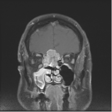
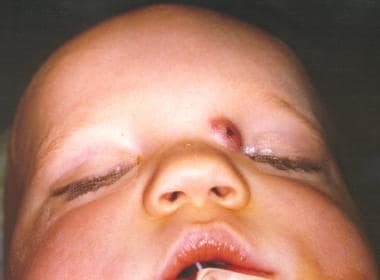
Nose neoplasms can cause various symptoms such as nasal congestion, nosebleeds, difficulty breathing through the nose, loss of smell, facial pain or numbness, and visual changes if they affect the eye. The diagnosis of nose neoplasms usually involves a combination of physical examination, imaging studies (such as CT or MRI scans), and biopsy to determine the type and extent of the growth. Treatment options depend on the type, size, location, and stage of the neoplasm and may include surgery, radiation therapy, chemotherapy, or a combination of these approaches.
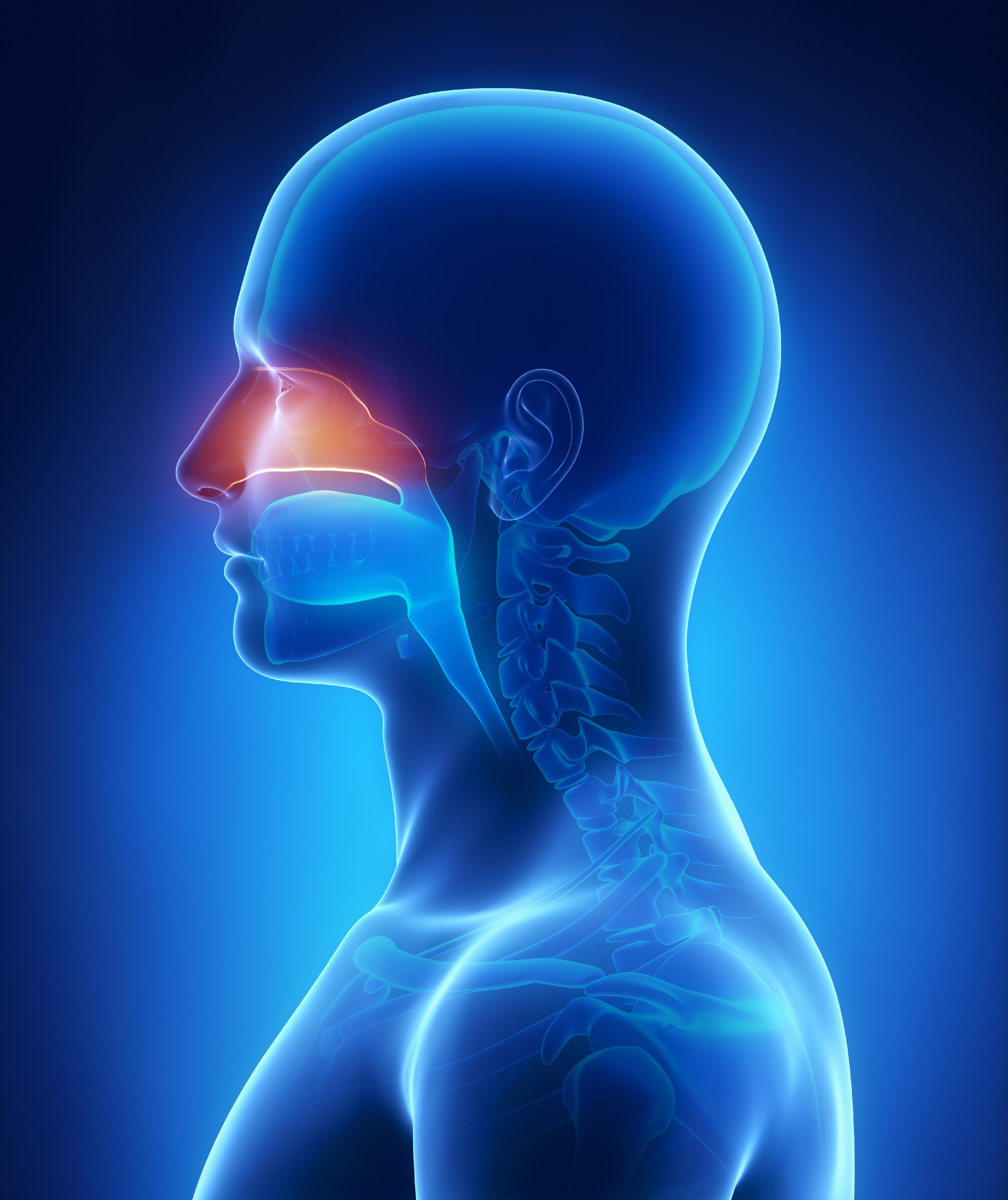
A nose, in a medical context, refers to the external part of the human body that is located on the face and serves as the primary organ for the sense of smell. It is composed of bone and cartilage, with a thin layer of skin covering it. The nose also contains nasal passages that are lined with mucous membranes and tiny hairs known as cilia. These structures help to filter, warm, and moisturize the air we breathe in before it reaches our lungs. Additionally, the nose plays an essential role in the process of verbal communication by shaping the sounds we make when we speak.
An "Electronic Nose" is a device that analytically detects, identifies, and quantifies volatile organic compounds (VOCs) in gaseous samples to identify specific odors or chemical compositions. It typically consists of an array of electronic gas sensors with partial specificity and pattern recognition software to analyze the response patterns of these sensors. The device mimics the functioning of a human nose, which can recognize a wide range of smells based on the unique pattern of activation of its olfactory receptors. Electronic noses have applications in various fields, including medical diagnostics, food quality control, environmental monitoring, and security.
Acquired nose deformities refer to structural changes or abnormalities in the shape of the nose that occur after birth, as opposed to congenital deformities which are present at birth. These deformities can result from various factors such as trauma, injury, infection, tumors, or surgical procedures. Depending on the severity and cause of the deformity, it may affect both the aesthetic appearance and functionality of the nose, potentially causing difficulty in breathing, sinus problems, or sleep apnea. Treatment options for acquired nose deformities may include minimally invasive procedures, such as fillers or laser surgery, or more extensive surgical interventions, such as rhinoplasty or septoplasty, to restore both form and function to the nose.
Nose diseases, also known as rhinologic disorders, refer to a wide range of conditions that affect the nose and its surrounding structures. These may include:
1. Nasal Allergies (Allergic Rhinitis): An inflammation of the inner lining of the nose caused by an allergic reaction to substances such as pollen, dust mites, or mold.
2. Sinusitis: Inflammation or infection of the sinuses, which are air-filled cavities in the skull that surround the nasal cavity.
3. Nasal Polyps: Soft, fleshy growths that develop on the lining of the nasal passages or sinuses.
4. Deviated Septum: A condition where the thin wall (septum) between the two nostrils is displaced to one side, causing difficulty breathing through the nose.
5. Rhinitis Medicamentosa: Nasal congestion caused by overuse of decongestant nasal sprays.
6. Nosebleeds (Epistaxis): Bleeding from the nostrils, which can be caused by a variety of factors including dryness, trauma, or underlying medical conditions.
7. Nasal Fractures: Breaks in the bone structure of the nose, often caused by trauma.
8. Tumors: Abnormal growths that can occur in the nasal passages or sinuses. These can be benign or malignant.
9. Choanal Atresia: A congenital condition where the back of the nasal passage is blocked, often by a thin membrane or bony partition.
10. Nasal Valve Collapse: A condition where the side walls of the nose collapse inward during breathing, causing difficulty breathing through the nose.
These are just a few examples of the many diseases that can affect the nose.
Pancreatic neoplasms refer to abnormal growths in the pancreas that can be benign or malignant. The pancreas is a gland located behind the stomach that produces hormones and digestive enzymes. Pancreatic neoplasms can interfere with the normal functioning of the pancreas, leading to various health complications.
Benign pancreatic neoplasms are non-cancerous growths that do not spread to other parts of the body. They are usually removed through surgery to prevent any potential complications, such as blocking the bile duct or causing pain.
Malignant pancreatic neoplasms, also known as pancreatic cancer, are cancerous growths that can invade and destroy surrounding tissues and organs. They can also spread (metastasize) to other parts of the body, such as the liver, lungs, or bones. Pancreatic cancer is often aggressive and difficult to treat, with a poor prognosis.
There are several types of pancreatic neoplasms, including adenocarcinomas, neuroendocrine tumors, solid pseudopapillary neoplasms, and cystic neoplasms. The specific type of neoplasm is determined through various diagnostic tests, such as imaging studies, biopsies, and blood tests. Treatment options depend on the type, stage, and location of the neoplasm, as well as the patient's overall health and preferences.
Neoplasms are abnormal growths of cells or tissues in the body that serve no physiological function. They can be benign (non-cancerous) or malignant (cancerous). Benign neoplasms are typically slow growing and do not spread to other parts of the body, while malignant neoplasms are aggressive, invasive, and can metastasize to distant sites.
Neoplasms occur when there is a dysregulation in the normal process of cell division and differentiation, leading to uncontrolled growth and accumulation of cells. This can result from genetic mutations or other factors such as viral infections, environmental exposures, or hormonal imbalances.
Neoplasms can develop in any organ or tissue of the body and can cause various symptoms depending on their size, location, and type. Treatment options for neoplasms include surgery, radiation therapy, chemotherapy, immunotherapy, and targeted therapy, among others.
Neoplasms: Neoplasms refer to abnormal growths of tissue that can be benign (non-cancerous) or malignant (cancerous). They occur when the normal control mechanisms that regulate cell growth and division are disrupted, leading to uncontrolled cell proliferation.
Cystic Neoplasms: Cystic neoplasms are tumors that contain fluid-filled sacs or cysts. These tumors can be benign or malignant and can occur in various organs of the body, including the pancreas, ovary, and liver.
Mucinous Neoplasms: Mucinous neoplasms are a type of cystic neoplasm that is characterized by the production of mucin, a gel-like substance produced by certain types of cells. These tumors can occur in various organs, including the ovary, pancreas, and colon. Mucinous neoplasms can be benign or malignant, and malignant forms are often aggressive and have a poor prognosis.
Serous Neoplasms: Serous neoplasms are another type of cystic neoplasm that is characterized by the production of serous fluid, which is a thin, watery fluid. These tumors commonly occur in the ovary and can be benign or malignant. Malignant serous neoplasms are often aggressive and have a poor prognosis.
In summary, neoplasms refer to abnormal tissue growths that can be benign or malignant. Cystic neoplasms contain fluid-filled sacs and can occur in various organs of the body. Mucinous neoplasms produce a gel-like substance called mucin and can also occur in various organs, while serous neoplasms produce thin, watery fluid and commonly occur in the ovary. Both mucinous and serous neoplasms can be benign or malignant, with malignant forms often being aggressive and having a poor prognosis.
Rhinoplasty is a surgical procedure performed on the nose to reshape its structure or improve its function. This may involve altering the bone, cartilage, or soft tissues of the nose to change its appearance, straighten its bridge, reduce or increase its size, narrow its width at the nostrils, or change the angle between the nose and upper lip. It can also be done to correct birth defects, injuries, or help relieve breathing problems. The procedure is usually performed by an otolaryngologist (ear, nose, and throat specialist) or a plastic surgeon, and it requires a thorough understanding of nasal anatomy and function.
Skin neoplasms refer to abnormal growths or tumors in the skin that can be benign (non-cancerous) or malignant (cancerous). They result from uncontrolled multiplication of skin cells, which can form various types of lesions. These growths may appear as lumps, bumps, sores, patches, or discolored areas on the skin.
Benign skin neoplasms include conditions such as moles, warts, and seborrheic keratoses, while malignant skin neoplasms are primarily classified into melanoma, squamous cell carcinoma, and basal cell carcinoma. These three types of cancerous skin growths are collectively known as non-melanoma skin cancers (NMSCs). Melanoma is the most aggressive and dangerous form of skin cancer, while NMSCs tend to be less invasive but more common.
It's essential to monitor any changes in existing skin lesions or the appearance of new growths and consult a healthcare professional for proper evaluation and treatment if needed.
Multiple primary neoplasms refer to the occurrence of more than one primary malignant tumor in an individual, where each tumor is unrelated to the other and originates from separate cells or organs. This differs from metastatic cancer, where a single malignancy spreads to multiple sites in the body. Multiple primary neoplasms can be synchronous (occurring at the same time) or metachronous (occurring at different times). The risk of developing multiple primary neoplasms increases with age and is associated with certain genetic predispositions, environmental factors, and lifestyle choices such as smoking and alcohol consumption.
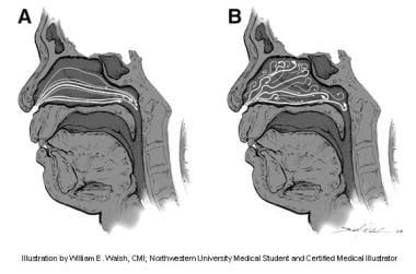
The nasal cavity is the air-filled space located behind the nose, which is divided into two halves by the nasal septum. It is lined with mucous membrane and is responsible for several functions including respiration, filtration, humidification, and olfaction (smell). The nasal cavity serves as an important part of the upper respiratory tract, extending from the nares (nostrils) to the choanae (posterior openings of the nasal cavity that lead into the pharynx). It contains specialized structures such as turbinate bones, which help to warm, humidify and filter incoming air.
Kidney neoplasms refer to abnormal growths or tumors in the kidney tissues that can be benign (non-cancerous) or malignant (cancerous). These growths can originate from various types of kidney cells, including the renal tubules, glomeruli, and the renal pelvis.
Malignant kidney neoplasms are also known as kidney cancers, with renal cell carcinoma being the most common type. Benign kidney neoplasms include renal adenomas, oncocytomas, and angiomyolipomas. While benign neoplasms are generally not life-threatening, they can still cause problems if they grow large enough to compromise kidney function or if they undergo malignant transformation.
Early detection and appropriate management of kidney neoplasms are crucial for improving patient outcomes and overall prognosis. Regular medical check-ups, imaging studies, and urinalysis can help in the early identification of these growths, allowing for timely intervention and treatment.
A "second primary neoplasm" is a distinct, new cancer or malignancy that develops in a person who has already had a previous cancer. It is not a recurrence or metastasis of the original tumor, but rather an independent cancer that arises in a different location or organ system. The development of second primary neoplasms can be influenced by various factors such as genetic predisposition, environmental exposures, and previous treatments like chemotherapy or radiation therapy.
It is important to note that the definition of "second primary neoplasm" may vary slightly depending on the specific source or context. In general medical usage, it refers to a new, separate cancer; however, in some research or clinical settings, there might be more precise criteria for defining and diagnosing second primary neoplasms.
Otorhinolaryngologic diseases, also known as ear, nose, and throat (ENT) diseases, refer to a group of medical conditions that affect the ears, nose, and/or throat. These specialized areas are closely related both anatomically and functionally, and disorders in one area can often have impacts on the others.
Here are some examples of otorhinolaryngologic diseases categorized by the affected area:
1. Otologic diseases - affecting the ear:
* Otitis media (ear infection)
* Otitis externa (swimmer's ear)
* Tinnitus (ringing in the ears)
* Hearing loss
* Meniere's disease (inner ear disorder causing vertigo, tinnitus, and hearing loss)
* Acoustic neuroma (noncancerous tumor on the vestibular nerve)
2. Rhinologic diseases - affecting the nose:
* Allergic rhinitis (hay fever)
* Non-allergic rhinitis
* Sinusitis (sinus infection)
* Deviated septum
* Nasal polyps
* Epistaxis (nosebleed)
3. Laryngologic diseases - affecting the throat and voice box:
* Laryngitis (inflammation of the larynx, causing hoarseness or voice loss)
* Vocal cord nodules or polyps
* Reflux laryngitis (acid reflux irritating the throat)
* Subglottic stenosis (narrowing of the airway below the vocal cords)
* Laryngeal cancer
4. Common otorhinolaryngologic diseases:
* Tonsillitis (inflammation of the tonsils, often causing sore throat and difficulty swallowing)
* Adenoiditis (inflammation of the adenoids, commonly seen in children)
* Obstructive sleep apnea (OSA, a disorder characterized by pauses in breathing during sleep)
* Pharyngitis (inflammation of the pharynx or throat)
Otorhinolaryngologists, also known as ENT specialists, diagnose and treat these conditions. They may use various methods such as physical examination, imaging studies, endoscopy, and laboratory tests to determine the best course of treatment for each individual patient.
Adenocarcinoma, mucinous is a type of cancer that begins in the glandular cells that line certain organs and produce mucin, a substance that lubricates and protects tissues. This type of cancer is characterized by the presence of abundant pools of mucin within the tumor. It typically develops in organs such as the colon, rectum, lungs, pancreas, and ovaries.
Mucinous adenocarcinomas tend to have a distinct appearance under the microscope, with large pools of mucin pushing aside the cancer cells. They may also have a different clinical behavior compared to other types of adenocarcinomas, such as being more aggressive or having a worse prognosis in some cases.
It is important to note that while a diagnosis of adenocarcinoma, mucinous can be serious, the prognosis and treatment options may vary depending on several factors, including the location of the cancer, the stage at which it was diagnosed, and the individual's overall health.
Thyroid neoplasms refer to abnormal growths or tumors in the thyroid gland, which can be benign (non-cancerous) or malignant (cancerous). These growths can vary in size and may cause a noticeable lump or nodule in the neck. Thyroid neoplasms can also affect the function of the thyroid gland, leading to hormonal imbalances and related symptoms. The exact causes of thyroid neoplasms are not fully understood, but risk factors include radiation exposure, family history, and certain genetic conditions. It is important to note that most thyroid nodules are benign, but a proper medical evaluation is necessary to determine the nature of the growth and develop an appropriate treatment plan.
Myeloproliferative disorders (MPDs) are a group of rare, chronic blood cancers that originate from the abnormal proliferation or growth of one or more types of blood-forming cells in the bone marrow. These disorders result in an overproduction of mature but dysfunctional blood cells, which can lead to serious complications such as blood clots, bleeding, and organ damage.
There are several subtypes of MPDs, including:
1. Chronic Myeloid Leukemia (CML): A disorder characterized by the overproduction of mature granulocytes (a type of white blood cell) in the bone marrow, leading to an increased number of these cells in the blood. CML is caused by a genetic mutation that results in the formation of the BCR-ABL fusion protein, which drives uncontrolled cell growth and division.
2. Polycythemia Vera (PV): A disorder characterized by the overproduction of all three types of blood cells - red blood cells, white blood cells, and platelets - in the bone marrow. This can lead to an increased risk of blood clots, bleeding, and enlargement of the spleen.
3. Essential Thrombocythemia (ET): A disorder characterized by the overproduction of platelets in the bone marrow, leading to an increased risk of blood clots and bleeding.
4. Primary Myelofibrosis (PMF): A disorder characterized by the replacement of normal bone marrow tissue with scar tissue, leading to impaired blood cell production and anemia, enlargement of the spleen, and increased risk of infections and bleeding.
5. Chronic Neutrophilic Leukemia (CNL): A rare disorder characterized by the overproduction of neutrophils (a type of white blood cell) in the bone marrow, leading to an increased number of these cells in the blood. CNL can lead to an increased risk of infections and organ damage.
MPDs are typically treated with a combination of therapies, including chemotherapy, targeted therapy, immunotherapy, and stem cell transplantation. The choice of treatment depends on several factors, including the subtype of MPD, the patient's age and overall health, and the presence of any comorbidities.
I believe there might be a misunderstanding in your question. "Electronics" is not a medical term, but rather a branch of physics and engineering that deals with the design, construction, and operation of electronic devices and systems. It involves the study and application of electrical properties of materials, components, and systems, and how they can be used to process, transmit, and store information and energy.
However, electronics have numerous applications in the medical field, such as in diagnostic equipment, monitoring devices, surgical tools, and prosthetics. In these contexts, "electronics" refers to the specific electronic components or systems that are used for medical purposes.
The term "DNA, neoplasm" is not a standard medical term or concept. DNA refers to deoxyribonucleic acid, which is the genetic material present in the cells of living organisms. A neoplasm, on the other hand, is a tumor or growth of abnormal tissue that can be benign (non-cancerous) or malignant (cancerous).
In some contexts, "DNA, neoplasm" may refer to genetic alterations found in cancer cells. These genetic changes can include mutations, amplifications, deletions, or rearrangements of DNA sequences that contribute to the development and progression of cancer. Identifying these genetic abnormalities can help doctors diagnose and treat certain types of cancer more effectively.
However, it's important to note that "DNA, neoplasm" is not a term that would typically be used in medical reports or research papers without further clarification. If you have any specific questions about DNA changes in cancer cells or neoplasms, I would recommend consulting with a healthcare professional or conducting further research on the topic.
Lung neoplasms refer to abnormal growths or tumors in the lung tissue. These tumors can be benign (non-cancerous) or malignant (cancerous). Malignant lung neoplasms are further classified into two main types: small cell lung carcinoma and non-small cell lung carcinoma. Lung neoplasms can cause symptoms such as cough, chest pain, shortness of breath, and weight loss. They are often caused by smoking or exposure to secondhand smoke, but can also occur due to genetic factors, radiation exposure, and other environmental carcinogens. Early detection and treatment of lung neoplasms is crucial for improving outcomes and survival rates.
Parotid neoplasms refer to abnormal growths or tumors in the parotid gland, which is the largest of the salivary glands and is located in front of the ear and extends down the neck. These neoplasms can be benign (non-cancerous) or malignant (cancerous).
Benign parotid neoplasms are typically slow-growing, painless masses that may cause facial asymmetry or difficulty in chewing or swallowing if they become large enough to compress surrounding structures. The most common type of benign parotid tumor is a pleomorphic adenoma.
Malignant parotid neoplasms, on the other hand, are more aggressive and can invade nearby tissues and spread to other parts of the body. They may present as rapidly growing masses that are firm or fixed to surrounding structures. Common types of malignant parotid tumors include mucoepidermoid carcinoma, adenoid cystic carcinoma, and squamous cell carcinoma.
The diagnosis of parotid neoplasms typically involves a thorough clinical evaluation, imaging studies such as CT or MRI scans, and fine-needle aspiration biopsy (FNAB) to determine the nature of the tumor. Treatment options depend on the type, size, and location of the neoplasm but may include surgical excision, radiation therapy, and chemotherapy.
Cystadenoma is a type of benign tumor (not cancerous), which arises from glandular epithelial cells and is covered by a thin layer of connective tissue. These tumors can develop in various locations within the body, including the ovaries, pancreas, and other organs that contain glands.
There are two main types of cystadenomas: serous and mucinous. Serous cystadenomas are filled with a clear or watery fluid, while mucinous cystadenomas contain a thick, gelatinous material. Although they are generally not harmful, these tumors can grow quite large and cause discomfort or other symptoms due to their size or location. In some cases, cystadenomas may undergo malignant transformation and develop into cancerous tumors, known as cystadenocarcinomas. Regular medical follow-up and monitoring are essential for individuals diagnosed with cystadenomas to ensure early detection and treatment of any potential complications.
Neoplasms of connective and soft tissue are abnormal growths or tumors that develop in the body's supportive tissues, such as cartilage, tendons, ligaments, fascia, and fat. These neoplasms can be benign (non-cancerous) or malignant (cancerous).
Benign connective and soft tissue neoplasms include:
- Lipomas: slow-growing, fatty tumors that develop under the skin.
- Fibromas: firm, benign tumors that develop in connective tissue such as tendons or ligaments.
- Nevi (plural of nevus): benign growths made up of cells called melanocytes, which produce pigment.
Malignant connective and soft tissue neoplasms include:
- Sarcomas: a type of cancer that develops in the body's supportive tissues such as muscle, bone, fat, cartilage, or blood vessels. There are many different types of sarcomas, including liposarcoma (fatty tissue), rhabdomyosarcoma (muscle), and osteosarcoma (bone).
- Desmoid tumors: a rare type of benign tumor that can become aggressive and invade surrounding tissues. While not considered cancerous, desmoid tumors can cause significant morbidity due to their tendency to grow and infiltrate nearby structures.
Connective and soft tissue neoplasms can present with various symptoms depending on their location and size. Treatment options include surgery, radiation therapy, chemotherapy, or a combination of these modalities. Regular follow-up care is essential to monitor for recurrence or metastasis (spread) of the tumor.
Plasma cell neoplasms are a type of cancer that originates from plasma cells, which are a type of white blood cell found in the bone marrow. These cells are responsible for producing antibodies to help fight off infections. When plasma cells become cancerous and multiply out of control, they can form a tumor called a plasmacytoma.
There are two main types of plasma cell neoplasms: solitary plasmacytoma and multiple myeloma. Solitary plasmacytoma is a localized tumor that typically forms in the bone, while multiple myeloma is a systemic disease that affects multiple bones and can cause a variety of symptoms such as bone pain, fatigue, and anemia.
Plasma cell neoplasms are diagnosed through a combination of tests, including blood tests, imaging studies, and bone marrow biopsy. Treatment options depend on the stage and extent of the disease, but may include radiation therapy, chemotherapy, and stem cell transplantation.
Appendiceal neoplasms refer to various types of tumors that can develop in the appendix, a small tube-like structure attached to the large intestine. These neoplasms can be benign or malignant and can include:
1. Adenomas: These are benign tumors that arise from the glandular cells lining the appendix. They are usually slow-growing and may not cause any symptoms.
2. Carcinoids: These are neuroendocrine tumors that arise from the hormone-producing cells in the appendix. They are typically small and slow-growing, but some can be aggressive and spread to other parts of the body.
3. Mucinous neoplasms: These are tumors that produce mucin, a slippery substance that can cause the appendix to become distended and filled with mucus. They can be low-grade (less aggressive) or high-grade (more aggressive) and may spread to other parts of the abdomen.
4. Adenocarcinomas: These are malignant tumors that arise from the glandular cells lining the appendix. They are relatively rare but can be aggressive and spread to other parts of the body.
5. Pseudomyxoma peritonei: This is a condition in which mucin produced by an appendiceal neoplasm leaks into the abdominal cavity, causing a jelly-like accumulation of fluid and tissue. It can be caused by both benign and malignant tumors.
Treatment for appendiceal neoplasms depends on the type and stage of the tumor, as well as the patient's overall health. Treatment options may include surgery, chemotherapy, or radiation therapy.
Liver neoplasms refer to abnormal growths in the liver that can be benign or malignant. Benign liver neoplasms are non-cancerous tumors that do not spread to other parts of the body, while malignant liver neoplasms are cancerous tumors that can invade and destroy surrounding tissue and spread to other organs.
Liver neoplasms can be primary, meaning they originate in the liver, or secondary, meaning they have metastasized (spread) to the liver from another part of the body. Primary liver neoplasms can be further classified into different types based on their cell of origin and behavior, including hepatocellular carcinoma, cholangiocarcinoma, and hepatic hemangioma.
The diagnosis of liver neoplasms typically involves a combination of imaging studies, such as ultrasound, CT scan, or MRI, and biopsy to confirm the type and stage of the tumor. Treatment options depend on the type and extent of the neoplasm and may include surgery, radiation therapy, chemotherapy, or liver transplantation.
Nasal mucosa refers to the mucous membrane that lines the nasal cavity. It is a delicate, moist, and specialized tissue that contains various types of cells including epithelial cells, goblet cells, and glands. The primary function of the nasal mucosa is to warm, humidify, and filter incoming air before it reaches the lungs.
The nasal mucosa produces mucus, which traps dust, allergens, and microorganisms, preventing them from entering the respiratory system. The cilia, tiny hair-like structures on the surface of the epithelial cells, help move the mucus towards the back of the throat, where it can be swallowed or expelled.
The nasal mucosa also contains a rich supply of blood vessels and immune cells that help protect against infections and inflammation. It plays an essential role in the body's defense system by producing antibodies, secreting antimicrobial substances, and initiating local immune responses.
Mucinous cystadenoma is a type of benign tumor that arises from the epithelial cells lining the mucous membranes of the body. It is most commonly found in the ovary, but can also occur in other locations such as the pancreas or appendix.
Mucinous cystadenomas are characterized by the production of large amounts of mucin, a slippery, gel-like substance that accumulates inside the tumor and causes it to grow into a cystic mass. These tumors can vary in size, ranging from a few centimeters to over 20 centimeters in diameter.
While mucinous cystadenomas are generally benign, they have the potential to become cancerous (mucinous cystadenocarcinoma) if left untreated. Symptoms of mucinous cystadenoma may include abdominal pain or swelling, bloating, and changes in bowel movements or urinary habits. Treatment typically involves surgical removal of the tumor.
Ovarian neoplasms refer to abnormal growths or tumors in the ovary, which can be benign (non-cancerous) or malignant (cancerous). These growths can originate from various cell types within the ovary, including epithelial cells, germ cells, and stromal cells. Ovarian neoplasms are often classified based on their cell type of origin, histological features, and potential for invasive or metastatic behavior.
Epithelial ovarian neoplasms are the most common type and can be further categorized into several subtypes, such as serous, mucinous, endometrioid, clear cell, and Brenner tumors. Some of these epithelial tumors have a higher risk of becoming malignant and spreading to other parts of the body.
Germ cell ovarian neoplasms arise from the cells that give rise to eggs (oocytes) and can include teratomas, dysgerminomas, yolk sac tumors, and embryonal carcinomas. Stromal ovarian neoplasms develop from the connective tissue cells supporting the ovary and can include granulosa cell tumors, thecomas, and fibromas.
It is essential to diagnose and treat ovarian neoplasms promptly, as some malignant forms can be aggressive and potentially life-threatening if not managed appropriately. Regular gynecological exams, imaging studies, and tumor marker tests are often used for early detection and monitoring of ovarian neoplasms. Treatment options may include surgery, chemotherapy, or radiation therapy, depending on the type, stage, and patient's overall health condition.
The nasal septum is the thin, flat wall of bone and cartilage that separates the two sides (nostrils) of the nose. Its primary function is to support the structures of the nose, divide the nostrils, and regulate airflow into the nasal passages. The nasal septum should be relatively centered, but it's not uncommon for a deviated septum to occur, where the septum is displaced to one side, which can sometimes cause blockage or breathing difficulties in the more affected nostril.
Endocrine gland neoplasms refer to abnormal growths (tumors) that develop in the endocrine glands. These glands are responsible for producing hormones, which are chemical messengers that regulate various functions and processes in the body. Neoplasms can be benign or malignant (cancerous). Benign neoplasms tend to grow slowly and do not spread to other parts of the body. Malignant neoplasms, on the other hand, can invade nearby tissues and organs and may also metastasize (spread) to distant sites.
Endocrine gland neoplasms can occur in any of the endocrine glands, including:
1. Pituitary gland: located at the base of the brain, it produces several hormones that regulate growth and development, as well as other bodily functions.
2. Thyroid gland: located in the neck, it produces thyroid hormones that regulate metabolism and calcium balance.
3. Parathyroid glands: located near the thyroid gland, they produce parathyroid hormone that regulates calcium levels in the blood.
4. Adrenal glands: located on top of each kidney, they produce hormones such as adrenaline, cortisol, and aldosterone that regulate stress response, metabolism, and blood pressure.
5. Pancreas: located behind the stomach, it produces insulin and glucagon, which regulate blood sugar levels, and digestive enzymes that help break down food.
6. Pineal gland: located in the brain, it produces melatonin, a hormone that regulates sleep-wake cycles.
7. Gonads (ovaries and testicles): located in the pelvis (ovaries) and scrotum (testicles), they produce sex hormones such as estrogen, progesterone, and testosterone that regulate reproductive function and secondary sexual characteristics.
Endocrine gland neoplasms can cause various symptoms depending on the type and location of the tumor. For example, a pituitary gland neoplasm may cause headaches, vision problems, or hormonal imbalances, while an adrenal gland neoplasm may cause high blood pressure, weight gain, or mood changes.
Diagnosis of endocrine gland neoplasms typically involves a combination of medical history, physical examination, imaging studies such as CT or MRI scans, and laboratory tests to measure hormone levels. Treatment options may include surgery, radiation therapy, chemotherapy, or hormonal therapy, depending on the type and stage of the tumor.
Immunohistochemistry (IHC) is a technique used in pathology and laboratory medicine to identify specific proteins or antigens in tissue sections. It combines the principles of immunology and histology to detect the presence and location of these target molecules within cells and tissues. This technique utilizes antibodies that are specific to the protein or antigen of interest, which are then tagged with a detection system such as a chromogen or fluorophore. The stained tissue sections can be examined under a microscope, allowing for the visualization and analysis of the distribution and expression patterns of the target molecule in the context of the tissue architecture. Immunohistochemistry is widely used in diagnostic pathology to help identify various diseases, including cancer, infectious diseases, and immune-mediated disorders.
Gastrointestinal (GI) neoplasms refer to abnormal growths in the gastrointestinal tract, which can be benign or malignant. The gastrointestinal tract includes the mouth, esophagus, stomach, small intestine, large intestine, rectum, and anus.
Benign neoplasms are non-cancerous growths that do not invade nearby tissues or spread to other parts of the body. They can sometimes be removed completely and may not cause any further health problems.
Malignant neoplasms, on the other hand, are cancerous growths that can invade nearby tissues and organs and spread to other parts of the body through the bloodstream or lymphatic system. These types of neoplasms can be life-threatening if not diagnosed and treated promptly.
GI neoplasms can cause various symptoms, including abdominal pain, bloating, changes in bowel habits, nausea, vomiting, weight loss, and anemia. The specific symptoms may depend on the location and size of the neoplasm.
There are many types of GI neoplasms, including adenocarcinomas, gastrointestinal stromal tumors (GISTs), lymphomas, and neuroendocrine tumors. The diagnosis of GI neoplasms typically involves a combination of medical history, physical examination, imaging studies, and biopsy. Treatment options may include surgery, radiation therapy, chemotherapy, targeted therapy, or immunotherapy.
Otolaryngology is a specialized branch of medicine that deals with the diagnosis, management, and treatment of disorders related to the ear, nose, throat (ENT), and head and neck region. It's also known as ENT (Ear, Nose, Throat) specialty. Otolaryngologists are physicians trained in the medical and surgical management of conditions such as hearing and balance disorders, nasal congestion, sinusitis, allergies, sleep apnea, snoring, swallowing difficulties, voice and speech problems, and head and neck tumors.
Pancreatic ductal carcinoma (PDC) is a specific type of cancer that forms in the ducts that carry digestive enzymes out of the pancreas. It's the most common form of exocrine pancreatic cancer, making up about 90% of all cases.
The symptoms of PDC are often vague and can include abdominal pain, jaundice (yellowing of the skin and eyes), unexplained weight loss, and changes in bowel movements. These symptoms can be similar to those caused by other less serious conditions, which can make diagnosis difficult.
Pancreatic ductal carcinoma is often aggressive and difficult to treat. The prognosis for PDC is generally poor, with a five-year survival rate of only about 9%. Treatment options may include surgery, chemotherapy, radiation therapy, or a combination of these approaches. However, because PDC is often not detected until it has advanced, treatment is frequently focused on palliative care to relieve symptoms and improve quality of life.
Experimental neoplasms refer to abnormal growths or tumors that are induced and studied in a controlled laboratory setting, typically in animals or cell cultures. These studies are conducted to understand the fundamental mechanisms of cancer development, progression, and potential treatment strategies. By manipulating various factors such as genetic mutations, environmental exposures, and pharmacological interventions, researchers can gain valuable insights into the complex processes underlying neoplasm formation and identify novel targets for cancer therapy. It is important to note that experimental neoplasms may not always accurately represent human cancers, and further research is needed to translate these findings into clinically relevant applications.
A neoplasm of vascular tissue is an abnormal growth or mass of cells in the blood vessels or lymphatic vessels. These growths can be benign (non-cancerous) or malignant (cancerous). Benign neoplasms, such as hemangiomas and lymphangiomas, are typically not harmful and may not require treatment. However, they can cause symptoms if they grow large enough to press on nearby organs or tissues. Malignant neoplasms, such as angiosarcomas, are cancerous and can invade and destroy surrounding tissue, as well as spread (metastasize) to other parts of the body. Treatment for vascular tissue neoplasms depends on the type, size, location, and stage of the growth, and may include surgery, radiation therapy, chemotherapy, or a combination of these.
Eye neoplasms, also known as ocular tumors or eye cancer, refer to abnormal growths of tissue in the eye. These growths can be benign (non-cancerous) or malignant (cancerous). Eye neoplasms can develop in various parts of the eye, including the eyelid, conjunctiva, cornea, iris, ciliary body, choroid, retina, and optic nerve.
Benign eye neoplasms are typically slow-growing and do not spread to other parts of the body. They may cause symptoms such as vision changes, eye pain, or a noticeable mass in the eye. Treatment options for benign eye neoplasms include monitoring, surgical removal, or radiation therapy.
Malignant eye neoplasms, on the other hand, can grow and spread rapidly to other parts of the body. They may cause symptoms such as vision changes, eye pain, floaters, or flashes of light. Treatment options for malignant eye neoplasms depend on the type and stage of cancer but may include surgery, radiation therapy, chemotherapy, or a combination of these treatments.
It is important to note that early detection and treatment of eye neoplasms can improve outcomes and prevent complications. Regular eye exams with an ophthalmologist are recommended for early detection and prevention of eye diseases, including eye neoplasms.
Nasal obstruction is a medical condition that refers to any blockage or restriction in the normal flow of air through the nasal passages. This can be caused by various factors such as inflammation, swelling, or physical abnormalities in the nasal cavity. Common causes of nasal obstruction include allergies, sinusitis, deviated septum, enlarged turbinates, and nasal polyps. Symptoms may include difficulty breathing through the nose, nasal congestion, and nasal discharge. Treatment options depend on the underlying cause and may include medications, surgery, or lifestyle changes.
Salivary gland neoplasms refer to abnormal growths or tumors that develop in the salivary glands. These glands are responsible for producing saliva, which helps in digestion, lubrication of food and maintaining oral health. Salivary gland neoplasms can be benign (non-cancerous) or malignant (cancerous).
Benign neoplasms are slow-growing and typically do not spread to other parts of the body. They may cause symptoms such as swelling, painless lumps, or difficulty swallowing if they grow large enough to put pressure on surrounding tissues.
Malignant neoplasms, on the other hand, can be aggressive and have the potential to invade nearby structures and metastasize (spread) to distant organs. Symptoms of malignant salivary gland neoplasms may include rapid growth, pain, numbness, or paralysis of facial nerves.
Salivary gland neoplasms can occur in any of the major salivary glands (parotid, submandibular, and sublingual glands) or in the minor salivary glands located throughout the mouth and throat. The exact cause of these neoplasms is not fully understood, but risk factors may include exposure to radiation, certain viral infections, and genetic predisposition.
Radiation-induced neoplasms are a type of cancer or tumor that develops as a result of exposure to ionizing radiation. Ionizing radiation is radiation with enough energy to remove tightly bound electrons from atoms or molecules, leading to the formation of ions. This type of radiation can damage DNA and other cellular structures, which can lead to mutations and uncontrolled cell growth, resulting in the development of a neoplasm.
Radiation-induced neoplasms can occur after exposure to high levels of ionizing radiation, such as that received during radiation therapy for cancer treatment or from nuclear accidents. The risk of developing a radiation-induced neoplasm depends on several factors, including the dose and duration of radiation exposure, the type of radiation, and the individual's genetic susceptibility to radiation-induced damage.
Radiation-induced neoplasms can take many years to develop after initial exposure to ionizing radiation, and they often occur at the site of previous radiation therapy. Common types of radiation-induced neoplasms include sarcomas, carcinomas, and thyroid cancer. It is important to note that while ionizing radiation can increase the risk of developing cancer, the overall risk is still relatively low, especially when compared to other well-established cancer risk factors such as smoking and exposure to certain chemicals.
Adenocarcinoma, papillary is a type of cancer that begins in the glandular cells and grows in a finger-like projection (called a papilla). This type of cancer can occur in various organs, including the lungs, pancreas, thyroid, and female reproductive system. The prognosis and treatment options for papillary adenocarcinoma depend on several factors, such as the location and stage of the tumor, as well as the patient's overall health. It is important to consult with a healthcare professional for an accurate diagnosis and personalized treatment plan.
Carcinoma, papillary is a type of cancer that begins in the cells that line the glandular structures or the lining of organs. In a papillary carcinoma, the cancerous cells grow and form small finger-like projections, called papillae, within the tumor. This type of cancer most commonly occurs in the thyroid gland, but can also be found in other organs such as the lung, breast, and kidney. Papillary carcinoma of the thyroid gland is usually slow-growing and has a good prognosis, especially when it is diagnosed at an early stage.
Testicular neoplasms are abnormal growths or tumors in the testicle that can be benign (non-cancerous) or malignant (cancerous). They are a type of genitourinary cancer, which affects the reproductive and urinary systems. Testicular neoplasms can occur in men of any age but are most commonly found in young adults between the ages of 15 and 40.
Testicular neoplasms can be classified into two main categories: germ cell tumors and non-germ cell tumors. Germ cell tumors, which arise from the cells that give rise to sperm, are further divided into seminomas and non-seminomas. Seminomas are typically slow-growing and have a good prognosis, while non-seminomas tend to grow more quickly and can spread to other parts of the body.
Non-germ cell tumors are less common than germ cell tumors and include Leydig cell tumors, Sertoli cell tumors, and lymphomas. These tumors can have a variety of clinical behaviors, ranging from benign to malignant.
Testicular neoplasms often present as a painless mass or swelling in the testicle. Other symptoms may include a feeling of heaviness or discomfort in the scrotum, a dull ache in the lower abdomen or groin, and breast enlargement (gynecomastia).
Diagnosis typically involves a physical examination, imaging studies such as ultrasound or CT scan, and blood tests to detect tumor markers. Treatment options depend on the type and stage of the neoplasm but may include surgery, radiation therapy, chemotherapy, or a combination of these modalities. Regular self-examinations of the testicles are recommended for early detection and improved outcomes.
Neoplasms in muscle tissue refer to abnormal and excessive growths of muscle cells that can be benign or malignant. These growths can arise from any of the three types of muscle tissue: skeletal, cardiac, or smooth muscle. Neoplasms in muscle tissue are classified based on their origin, behavior, and histological features.
Benign neoplasms in muscle tissue include leiomyomas (smooth muscle), rhabdomyomas (skeletal muscle), and myxomas (cardiac muscle). These tumors are usually slow-growing and do not invade surrounding tissues or spread to other parts of the body.
Malignant neoplasms in muscle tissue, also known as sarcomas, include leiomyosarcoma (smooth muscle), rhabdomyosarcoma (skeletal muscle), and angiosarcoma (cardiac muscle). These tumors are aggressive, invasive, and have the potential to metastasize to other parts of the body.
Symptoms of neoplasms in muscle tissue depend on their location, size, and type. They may include a painless or painful mass, weakness, fatigue, weight loss, and difficulty swallowing or breathing. Treatment options for neoplasms in muscle tissue include surgery, radiation therapy, chemotherapy, and targeted therapy. The choice of treatment depends on the type, stage, location, and patient's overall health condition.
Neoplasms are abnormal growths of cells or tissues that serve no purpose and can be benign (non-cancerous) or malignant (cancerous). Glandular and epithelial neoplasms refer to specific types of tumors that originate from the glandular and epithelial tissues, respectively.
Glandular neoplasms arise from the glandular tissue, which is responsible for producing and secreting substances such as hormones, enzymes, or other fluids. These neoplasms can be further classified into adenomas (benign) and adenocarcinomas (malignant).
Epithelial neoplasms, on the other hand, develop from the epithelial tissue that lines the outer surfaces of organs and the inner surfaces of cavities. These neoplasms can also be benign or malignant and are classified as papillomas (benign) and carcinomas (malignant).
It is important to note that while both glandular and epithelial neoplasms can become cancerous, not all of them do. However, if they do, the malignant versions can invade surrounding tissues and spread to other parts of the body, making them potentially life-threatening.
Paranasal sinus neoplasms refer to abnormal growths or tumors that develop within the paranasal sinuses, which are air-filled cavities located inside the skull near the nasal cavity. These tumors can be benign (noncancerous) or malignant (cancerous), and they can arise from various types of tissue within the sinuses, such as the lining of the sinuses (mucosa), bone, or other soft tissues.
Paranasal sinus neoplasms can cause a variety of symptoms, including nasal congestion, nosebleeds, facial pain or numbness, and visual disturbances. The diagnosis of these tumors typically involves a combination of imaging studies (such as CT or MRI scans) and biopsy to determine the type and extent of the tumor. Treatment options may include surgery, radiation therapy, chemotherapy, or a combination of these approaches, depending on the specific type and stage of the neoplasm.
Mucinous cystadenocarcinoma is a type of cancer that arises from the mucin-producing cells in the lining of a cyst. It is a subtype of cystadenocarcinoma, which is a malignant tumor that develops within a cyst. Mucinous cystadenocarcinomas are typically found in the ovary or pancreas but can also occur in other organs such as the appendix and the respiratory tract.
These tumors are characterized by the production of large amounts of mucin, a gel-like substance that can accumulate within the cyst and cause it to grow. Mucinous cystadenocarcinomas tend to grow slowly but can become quite large and may eventually spread (metastasize) to other parts of the body if left untreated.
Symptoms of mucinous cystadenocarcinoma depend on the location and size of the tumor, but they may include abdominal pain or discomfort, bloating, changes in bowel movements, or vaginal bleeding. Treatment typically involves surgical removal of the tumor, followed by chemotherapy or radiation therapy to kill any remaining cancer cells. The prognosis for mucinous cystadenocarcinoma depends on several factors, including the stage of the disease at diagnosis and the patient's overall health.
An adenoma is a benign (noncancerous) tumor that develops from glandular epithelial cells. These types of cells are responsible for producing and releasing fluids, such as hormones or digestive enzymes, into the surrounding tissues. Adenomas can occur in various organs and glands throughout the body, including the thyroid, pituitary, adrenal, and digestive systems.
Depending on their location, adenomas may cause different symptoms or remain asymptomatic. Some common examples of adenomas include:
1. Colorectal adenoma (also known as a polyp): These growths occur in the lining of the colon or rectum and can develop into colorectal cancer if left untreated. Regular screenings, such as colonoscopies, are essential for early detection and removal of these polyps.
2. Thyroid adenoma: This type of adenoma affects the thyroid gland and may result in an overproduction or underproduction of hormones, leading to conditions like hyperthyroidism (overactive thyroid) or hypothyroidism (underactive thyroid).
3. Pituitary adenoma: These growths occur in the pituitary gland, which is located at the base of the brain and controls various hormonal functions. Depending on their size and location, pituitary adenomas can cause vision problems, headaches, or hormonal imbalances that affect growth, reproduction, and metabolism.
4. Liver adenoma: These rare benign tumors develop in the liver and may not cause any symptoms unless they become large enough to press on surrounding organs or structures. In some cases, liver adenomas can rupture and cause internal bleeding.
5. Adrenal adenoma: These growths occur in the adrenal glands, which are located above the kidneys and produce hormones that regulate stress responses, metabolism, and blood pressure. Most adrenal adenomas are nonfunctioning, meaning they do not secrete excess hormones. However, functioning adrenal adenomas can lead to conditions like Cushing's syndrome or Conn's syndrome, depending on the type of hormone being overproduced.
It is essential to monitor and manage benign tumors like adenomas to prevent potential complications, such as rupture, bleeding, or hormonal imbalances. Treatment options may include surveillance with imaging studies, medication to manage hormonal issues, or surgical removal of the tumor in certain cases.
Soft tissue neoplasms refer to abnormal growths or tumors that develop in the soft tissues of the body. Soft tissues include muscles, tendons, ligaments, fascia, nerves, blood vessels, fat, and synovial membranes (the thin layer of cells that line joints and tendons). Neoplasms can be benign (non-cancerous) or malignant (cancerous), and their behavior and potential for spread depend on the specific type of neoplasm.
Benign soft tissue neoplasms are typically slow-growing, well-circumscribed, and rarely spread to other parts of the body. They can often be removed surgically with a low risk of recurrence. Examples of benign soft tissue neoplasms include lipomas (fat tumors), schwannomas (nerve sheath tumors), and hemangiomas (blood vessel tumors).
Malignant soft tissue neoplasms, on the other hand, can grow rapidly, invade surrounding tissues, and may metastasize (spread) to distant parts of the body. They are often more difficult to treat than benign neoplasms and require a multidisciplinary approach, including surgery, radiation therapy, and chemotherapy. Examples of malignant soft tissue neoplasms include sarcomas, such as rhabdomyosarcoma (arising from skeletal muscle), leiomyosarcoma (arising from smooth muscle), and angiosarcoma (arising from blood vessels).
It is important to note that soft tissue neoplasms can occur in any part of the body, and their diagnosis and treatment require a thorough evaluation by a healthcare professional with expertise in this area.
Hematologic neoplasms, also known as hematological malignancies, are a group of diseases characterized by the uncontrolled growth and accumulation of abnormal blood cells or bone marrow cells. These disorders can originate from the myeloid or lymphoid cell lines, which give rise to various types of blood cells, including red blood cells, white blood cells, and platelets.
Hematologic neoplasms can be broadly classified into three categories:
1. Leukemias: These are cancers that primarily affect the bone marrow and blood-forming tissues. They result in an overproduction of abnormal white blood cells, which interfere with the normal functioning of the blood and immune system. There are several types of leukemia, including acute lymphoblastic leukemia (ALL), chronic lymphocytic leukemia (CLL), acute myeloid leukemia (AML), and chronic myeloid leukemia (CML).
2. Lymphomas: These are cancers that develop from the lymphatic system, which is a part of the immune system responsible for fighting infections. Lymphomas can affect lymph nodes, spleen, bone marrow, and other organs. The two main types of lymphoma are Hodgkin lymphoma (HL) and non-Hodgkin lymphoma (NHL).
3. Myelomas: These are cancers that arise from the plasma cells, a type of white blood cell responsible for producing antibodies. Multiple myeloma is the most common type of myeloma, characterized by an excessive proliferation of malignant plasma cells in the bone marrow, leading to the production of abnormal amounts of monoclonal immunoglobulins (M proteins) and bone destruction.
Hematologic neoplasms can have various symptoms, such as fatigue, weakness, frequent infections, easy bruising or bleeding, weight loss, swollen lymph nodes, and bone pain. The diagnosis typically involves a combination of medical history, physical examination, laboratory tests, imaging studies, and sometimes bone marrow biopsy. Treatment options depend on the type and stage of the disease and may include chemotherapy, radiation therapy, targeted therapy, immunotherapy, stem cell transplantation, or a combination of these approaches.
A neoplasm is a tumor or growth that is formed by an abnormal and excessive proliferation of cells, which can be benign or malignant. Neoplasm proteins are therefore any proteins that are expressed or produced in these neoplastic cells. These proteins can play various roles in the development, progression, and maintenance of neoplasms.
Some neoplasm proteins may contribute to the uncontrolled cell growth and division seen in cancer, such as oncogenic proteins that promote cell cycle progression or inhibit apoptosis (programmed cell death). Others may help the neoplastic cells evade the immune system, allowing them to proliferate undetected. Still others may be involved in angiogenesis, the formation of new blood vessels that supply the tumor with nutrients and oxygen.
Neoplasm proteins can also serve as biomarkers for cancer diagnosis, prognosis, or treatment response. For example, the presence or level of certain neoplasm proteins in biological samples such as blood or tissue may indicate the presence of a specific type of cancer, help predict the likelihood of cancer recurrence, or suggest whether a particular therapy will be effective.
Overall, understanding the roles and behaviors of neoplasm proteins can provide valuable insights into the biology of cancer and inform the development of new diagnostic and therapeutic strategies.
Uterine neoplasms refer to abnormal growths in the uterus, which can be benign (non-cancerous) or malignant (cancerous). These growths can originate from different types of cells within the uterus, leading to various types of uterine neoplasms. The two main categories of uterine neoplasms are endometrial neoplasms and uterine sarcomas.
Endometrial neoplasms develop from the endometrium, which is the inner lining of the uterus. Most endometrial neoplasms are classified as endometrioid adenocarcinomas, arising from glandular cells in the endometrium. Other types include serous carcinoma, clear cell carcinoma, and mucinous carcinoma.
Uterine sarcomas, on the other hand, are less common and originate from the connective tissue (stroma) or muscle (myometrium) of the uterus. Uterine sarcomas can be further divided into several subtypes, such as leiomyosarcoma, endometrial stromal sarcoma, and undifferentiated uterine sarcoma.
Uterine neoplasms can cause various symptoms, including abnormal vaginal bleeding or discharge, pelvic pain, and difficulty urinating or having bowel movements. The diagnosis typically involves a combination of imaging tests (such as ultrasound, CT, or MRI scans) and tissue biopsies to determine the type and extent of the neoplasm. Treatment options depend on the type, stage, and patient's overall health but may include surgery, radiation therapy, chemotherapy, or hormone therapy.
Intestinal neoplasms refer to abnormal growths in the tissues of the intestines, which can be benign or malignant. These growths are called neoplasms and they result from uncontrolled cell division. In the case of intestinal neoplasms, these growths occur in the small intestine, large intestine (colon), rectum, or appendix.
Benign intestinal neoplasms are not cancerous and often do not invade surrounding tissues or spread to other parts of the body. However, they can still cause problems if they grow large enough to obstruct the intestines or cause bleeding. Common types of benign intestinal neoplasms include polyps, leiomyomas, and lipomas.
Malignant intestinal neoplasms, on the other hand, are cancerous and can invade surrounding tissues and spread to other parts of the body. The most common type of malignant intestinal neoplasm is adenocarcinoma, which arises from the glandular cells lining the inside of the intestines. Other types of malignant intestinal neoplasms include lymphomas, sarcomas, and carcinoid tumors.
Symptoms of intestinal neoplasms can vary depending on their size, location, and type. Common symptoms include abdominal pain, bloating, changes in bowel habits, rectal bleeding, weight loss, and fatigue. If you experience any of these symptoms, it is important to seek medical attention promptly.
Neoplasms, adnexal and skin appendage refer to abnormal growths or tumors that develop in the sweat glands, hair follicles, or other structures associated with the skin. These growths can be benign (non-cancerous) or malignant (cancerous), and they can occur anywhere on the body.
Adnexal neoplasms are tumors that arise from the sweat glands or hair follicles, including the sebaceous glands, eccrine glands, and apocrine glands. These tumors can range in size and severity, and they may cause symptoms such as pain, itching, or changes in the appearance of the skin.
Skin appendage neoplasms are similar to adnexal neoplasms, but they specifically refer to tumors that arise from structures such as hair follicles, nails, and sweat glands. Examples of skin appendage neoplasms include pilomatricomas (tumors of the hair follicle), trichilemmomas (tumors of the outer root sheath of the hair follicle), and sebaceous adenomas (tumors of the sebaceous glands).
It is important to note that while many adnexal and skin appendage neoplasms are benign, some can be malignant and may require aggressive treatment. If you notice any unusual growths or changes in your skin, it is important to consult with a healthcare professional for further evaluation and care.
Neoplasm staging is a systematic process used in medicine to describe the extent of spread of a cancer, including the size and location of the original (primary) tumor and whether it has metastasized (spread) to other parts of the body. The most widely accepted system for this purpose is the TNM classification system developed by the American Joint Committee on Cancer (AJCC) and the Union for International Cancer Control (UICC).
In this system, T stands for tumor, and it describes the size and extent of the primary tumor. N stands for nodes, and it indicates whether the cancer has spread to nearby lymph nodes. M stands for metastasis, and it shows whether the cancer has spread to distant parts of the body.
Each letter is followed by a number that provides more details about the extent of the disease. For example, a T1N0M0 cancer means that the primary tumor is small and has not spread to nearby lymph nodes or distant sites. The higher the numbers, the more advanced the cancer.
Staging helps doctors determine the most appropriate treatment for each patient and estimate the patient's prognosis. It is an essential tool for communication among members of the healthcare team and for comparing outcomes of treatments in clinical trials.
Vascular neoplasms are a type of tumor that develops from cells that line the blood vessels or lymphatic vessels. These tumors can be benign (non-cancerous) or malignant (cancerous). Benign vascular neoplasms, such as hemangiomas and lymphangiomas, are usually harmless and may not require treatment unless they cause symptoms or complications. Malignant vascular neoplasms, on the other hand, are known as angiosarcomas and can be aggressive, spreading to other parts of the body and potentially causing serious health problems.
Angiosarcomas can develop in any part of the body but are most commonly found in the skin, particularly in areas exposed to radiation or chronic lymph edema. They can also occur in the breast, liver, spleen, and heart. Treatment for vascular neoplasms depends on the type, location, size, and stage of the tumor, as well as the patient's overall health. Treatment options may include surgery, radiation therapy, chemotherapy, or a combination of these approaches.
Sweat gland neoplasms are abnormal growths that develop in the sweat glands. These growths can be benign (noncancerous) or malignant (cancerous). Benign sweat gland neoplasms include hidradenomas and syringomas, which are usually slow-growing and cause little to no symptoms. Malignant sweat gland neoplasms, also known as sweat gland carcinomas, are rare but aggressive cancers that can spread to other parts of the body. They may cause symptoms such as a lump or mass under the skin, pain, swelling, and redness. Treatment typically involves surgical removal of the growth.
Lymphoma is a type of cancer that originates from the white blood cells called lymphocytes, which are part of the immune system. These cells are found in various parts of the body such as the lymph nodes, spleen, bone marrow, and other organs. Lymphoma can be classified into two main types: Hodgkin lymphoma (HL) and non-Hodgkin lymphoma (NHL).
HL is characterized by the presence of a specific type of abnormal lymphocyte called Reed-Sternberg cells, while NHL includes a diverse group of lymphomas that lack these cells. The symptoms of lymphoma may include swollen lymph nodes, fever, night sweats, weight loss, and fatigue.
The exact cause of lymphoma is not known, but it is believed to result from genetic mutations in the lymphocytes that lead to uncontrolled cell growth and division. Exposure to certain viruses, chemicals, and radiation may increase the risk of developing lymphoma. Treatment options for lymphoma depend on various factors such as the type and stage of the disease, age, and overall health of the patient. Common treatments include chemotherapy, radiation therapy, immunotherapy, and stem cell transplantation.
Bone neoplasms are abnormal growths or tumors that develop in the bone. They can be benign (non-cancerous) or malignant (cancerous). Benign bone neoplasms do not spread to other parts of the body and are rarely a threat to life, although they may cause problems if they grow large enough to press on surrounding tissues or cause fractures. Malignant bone neoplasms, on the other hand, can invade and destroy nearby tissue and may spread (metastasize) to other parts of the body.
There are many different types of bone neoplasms, including:
1. Osteochondroma - a benign tumor that develops from cartilage and bone
2. Enchondroma - a benign tumor that forms in the cartilage that lines the inside of the bones
3. Chondrosarcoma - a malignant tumor that develops from cartilage
4. Osteosarcoma - a malignant tumor that develops from bone cells
5. Ewing sarcoma - a malignant tumor that develops in the bones or soft tissues around the bones
6. Giant cell tumor of bone - a benign or occasionally malignant tumor that develops from bone tissue
7. Fibrosarcoma - a malignant tumor that develops from fibrous tissue in the bone
The symptoms of bone neoplasms vary depending on the type, size, and location of the tumor. They may include pain, swelling, stiffness, fractures, or limited mobility. Treatment options depend on the type and stage of the tumor but may include surgery, radiation therapy, chemotherapy, or a combination of these treatments.
Palatal neoplasms refer to abnormal growths or tumors that occur on the palate, which is the roof of the mouth. These growths can be benign (non-cancerous) or malignant (cancerous). Benign neoplasms are typically slower growing and less likely to spread, while malignant neoplasms are more aggressive and can invade nearby tissues and organs.
Palatal neoplasms can have various causes, including genetic factors, environmental exposures, and viral infections. They may present with symptoms such as mouth pain, difficulty swallowing, swelling or lumps in the mouth, bleeding, or numbness in the mouth or face.
The diagnosis of palatal neoplasms typically involves a thorough clinical examination, imaging studies, and sometimes biopsy to determine the type and extent of the growth. Treatment options depend on the type, size, location, and stage of the neoplasm but may include surgery, radiation therapy, chemotherapy, or a combination of these approaches. Regular follow-up care is essential to monitor for recurrence or spread of the neoplasm.
Neoplasms are abnormal growths of cells or tissues in the body that can be benign (non-cancerous) or malignant (cancerous). When referring to "Complex and Mixed Neoplasms," it is typically used in the context of histopathology, where it describes tumors with a mixture of different types of cells or growth patterns.
A complex neoplasm usually contains areas with various architectural patterns, cell types, or both, making its classification challenging. It may require extensive sampling and careful examination to determine its nature and behavior. These neoplasms can be either benign or malignant, depending on the specific characteristics of the tumor cells and their growth pattern.
A mixed neoplasm, on the other hand, is a tumor that contains more than one type of cell or tissue component, often arising from different germ layers (the three primary layers of embryonic development: ectoderm, mesoderm, and endoderm). A common example of a mixed neoplasm is a teratoma, which can contain tissues derived from all three germ layers, such as skin, hair, teeth, bone, and muscle. Mixed neoplasms can also be benign or malignant, depending on the specific components of the tumor.
It's important to note that the classification and behavior of complex and mixed neoplasms can vary significantly based on their location in the body, cellular composition, and other factors. Accurate diagnosis typically requires a thorough examination by an experienced pathologist and may involve additional tests, such as immunohistochemistry or molecular analysis, to determine the appropriate treatment and management strategies.
Neoplasm antigens, also known as tumor antigens, are substances that are produced by cancer cells (neoplasms) and can stimulate an immune response. These antigens can be proteins, carbohydrates, or other molecules that are either unique to the cancer cells or are overexpressed or mutated versions of normal cellular proteins.
Neoplasm antigens can be classified into two main categories: tumor-specific antigens (TSAs) and tumor-associated antigens (TAAs). TSAs are unique to cancer cells and are not expressed by normal cells, while TAAs are present at low levels in normal cells but are overexpressed or altered in cancer cells.
TSAs can be further divided into viral antigens and mutated antigens. Viral antigens are produced when cancer is caused by a virus, such as human papillomavirus (HPV) in cervical cancer. Mutated antigens are the result of genetic mutations that occur during cancer development and are unique to each patient's tumor.
Neoplasm antigens play an important role in the immune response against cancer. They can be recognized by the immune system, leading to the activation of immune cells such as T cells and natural killer (NK) cells, which can then attack and destroy cancer cells. However, cancer cells often develop mechanisms to evade the immune response, allowing them to continue growing and spreading.
Understanding neoplasm antigens is important for the development of cancer immunotherapies, which aim to enhance the body's natural immune response against cancer. These therapies include checkpoint inhibitors, which block proteins that inhibit T cell activation, and therapeutic vaccines, which stimulate an immune response against specific tumor antigens.
Mandibular neoplasms refer to abnormal growths or tumors that develop in the mandible, which is the lower jawbone. These growths can be benign (non-cancerous) or malignant (cancerous). Benign neoplasms are typically slow-growing and rarely spread to other parts of the body, while malignant neoplasms can invade surrounding tissues and may metastasize (spread) to distant sites.
Mandibular neoplasms can have various causes, including genetic mutations, exposure to certain chemicals or radiation, and infection with certain viruses. The symptoms of mandibular neoplasms may include swelling or pain in the jaw, difficulty chewing or speaking, numbness in the lower lip or chin, loose teeth, and/or a lump or mass in the mouth or neck.
The diagnosis of mandibular neoplasms typically involves a thorough clinical examination, imaging studies such as X-rays, CT scans, or MRI scans, and sometimes a biopsy to confirm the type and extent of the tumor. Treatment options depend on the type, stage, and location of the neoplasm, and may include surgery, radiation therapy, chemotherapy, or a combination of these approaches. Regular follow-up care is essential to monitor for recurrence or metastasis.
Cystadenocarcinoma is a type of tumor that arises from the epithelial lining of a cyst, and it has the potential to invade surrounding tissues and spread (metastasize) to other parts of the body. It typically affects glandular organs such as the ovaries, pancreas, and salivary glands.
Cystadenocarcinomas can be classified into two types: serous and mucinous. Serous cystadenocarcinomas produce a watery fluid, while mucinous cystadenocarcinomas produce a thick, mucus-like fluid. Both types of tumors can be benign or malignant, but malignant cystadenocarcinomas are more aggressive and have a higher risk of metastasis.
Symptoms of cystadenocarcinoma depend on the location and size of the tumor. In some cases, there may be no symptoms until the tumor has grown large enough to cause pain or other problems. Treatment typically involves surgical removal of the tumor, along with any affected surrounding tissue. Chemotherapy and radiation therapy may also be used in some cases to help prevent recurrence or spread of the cancer.
Bile duct neoplasms, also known as cholangiocarcinomas, refer to a group of malignancies that arise from the bile ducts. These are the tubes that carry bile from the liver to the gallbladder and small intestine. Bile duct neoplasms can be further classified based on their location as intrahepatic (within the liver), perihilar (at the junction of the left and right hepatic ducts), or distal (in the common bile duct).
These tumors are relatively rare, but their incidence has been increasing in recent years. They can cause a variety of symptoms, including jaundice, abdominal pain, weight loss, and fever. The diagnosis of bile duct neoplasms typically involves imaging studies such as CT or MRI scans, as well as blood tests to assess liver function. In some cases, a biopsy may be necessary to confirm the diagnosis.
Treatment options for bile duct neoplasms depend on several factors, including the location and stage of the tumor, as well as the patient's overall health. Surgical resection is the preferred treatment for early-stage tumors, while chemotherapy and radiation therapy may be used in more advanced cases. For patients who are not candidates for surgery, palliative treatments such as stenting or bypass procedures may be recommended to relieve symptoms and improve quality of life.
Neoplasm invasiveness is a term used in pathology and oncology to describe the aggressive behavior of cancer cells as they invade surrounding tissues and organs. This process involves the loss of cell-to-cell adhesion, increased motility and migration, and the ability of cancer cells to degrade the extracellular matrix (ECM) through the production of enzymes such as matrix metalloproteinases (MMPs).
Invasive neoplasms are cancers that have spread beyond the original site where they first developed and have infiltrated adjacent tissues or structures. This is in contrast to non-invasive or in situ neoplasms, which are confined to the epithelial layer where they originated and have not yet invaded the underlying basement membrane.
The invasiveness of a neoplasm is an important prognostic factor in cancer diagnosis and treatment, as it can indicate the likelihood of metastasis and the potential effectiveness of various therapies. In general, more invasive cancers are associated with worse outcomes and require more aggressive treatment approaches.
Thymus neoplasms are abnormal growths in the thymus gland that result from uncontrolled cell division. The term "neoplasm" refers to any new and abnormal growth of tissue, also known as a tumor. Thymus neoplasms can be benign or malignant (cancerous).
Malignant thymus neoplasms are called thymomas or thymic carcinomas. Thymomas are the most common type and tend to grow slowly, invading nearby tissues and organs. They can also spread (metastasize) to other parts of the body. Thymic carcinomas are rarer and more aggressive, growing and spreading more quickly than thymomas.
Symptoms of thymus neoplasms may include coughing, chest pain, difficulty breathing, or swelling in the neck or upper chest. Treatment options for thymus neoplasms depend on the type, size, location, and stage of the tumor, as well as the patient's overall health. Treatment may include surgery, radiation therapy, chemotherapy, or a combination of these approaches.
Splenic neoplasms refer to abnormal growths or tumors in the spleen, which can be benign (non-cancerous) or malignant (cancerous). These growths can arise from various cell types present within the spleen, including hematopoietic cells (red and white blood cells, platelets), stromal cells (supporting tissue), or lymphoid cells (part of the immune system).
There are several types of splenic neoplasms:
1. Hematologic malignancies: These are cancers that affect the blood and bone marrow, such as leukemias, lymphomas, and multiple myeloma. They often involve the spleen, causing enlargement (splenomegaly) and neoplastic infiltration of splenic tissue.
2. Primary splenic tumors: These are rare and include benign lesions like hemangiomas, lymphangiomas, and hamartomas, as well as malignant tumors such as angiosarcoma, littoral cell angiosarcoma, and primary splenic lymphoma.
3. Metastatic splenic tumors: These occur when cancer cells from other primary sites spread (metastasize) to the spleen. Common sources of metastasis include lung, breast, colon, and ovarian cancers, as well as melanomas and sarcomas.
Symptoms of splenic neoplasms may vary depending on the type and extent of the disease but often include abdominal pain or discomfort, fatigue, weight loss, and anemia. Diagnosis typically involves imaging studies (such as ultrasound, CT, or MRI scans) and sometimes requires a biopsy for confirmation. Treatment options depend on the type of neoplasm and may include surgery, chemotherapy, radiation therapy, targeted therapy, or immunotherapy.
Heart neoplasms are abnormal growths or tumors that develop within the heart tissue. They can be benign (noncancerous) or malignant (cancerous). Benign tumors, such as myxomas and rhabdomyomas, are typically slower growing and less likely to spread, but they can still cause serious complications if they obstruct blood flow or damage heart valves. Malignant tumors, such as angiosarcomas and rhabdomyosarcomas, are fast-growing and have a higher risk of spreading to other parts of the body. Symptoms of heart neoplasms can include shortness of breath, chest pain, fatigue, and irregular heart rhythms. Treatment options depend on the type, size, and location of the tumor, and may include surgery, radiation therapy, or chemotherapy.
A serous cystadenoma is a type of benign tumor that arises from the epithelial cells lining the serous glands, which are glands that produce a watery, lubricating fluid. This type of tumor typically develops in the ovary or the pancreas.
Serous cystadenomas of the ovary are usually filled with a clear, watery fluid and have multiple loculations (compartments). They can vary in size from a few millimeters to several centimeters in diameter. Although these tumors are benign, they can cause symptoms if they become large enough to press on surrounding organs or if they rupture and release their contents into the abdominal cavity.
Serous cystadenomas of the pancreas are less common than ovarian serous cystadenomas. They typically occur in the tail of the pancreas and can range in size from a few millimeters to several centimeters in diameter. These tumors are usually asymptomatic, but they can cause symptoms such as abdominal pain or discomfort if they become large enough to press on surrounding organs.
It is important to note that while serous cystadenomas are generally benign, there is a small risk that they may undergo malignant transformation and develop into a type of cancer known as a serous cystadenocarcinoma. For this reason, it is important for patients with these tumors to be followed closely by a healthcare provider and to have regular imaging studies and/or surgical excision to monitor for any changes in the tumor.
Colonic neoplasms refer to abnormal growths in the large intestine, also known as the colon. These growths can be benign (non-cancerous) or malignant (cancerous). The two most common types of colonic neoplasms are adenomas and carcinomas.
Adenomas are benign tumors that can develop into cancer over time if left untreated. They are often found during routine colonoscopies and can be removed during the procedure.
Carcinomas, on the other hand, are malignant tumors that invade surrounding tissues and can spread to other parts of the body. Colorectal cancer is the third leading cause of cancer-related deaths in the United States, and colonic neoplasms are a significant risk factor for developing this type of cancer.
Regular screenings for colonic neoplasms are recommended for individuals over the age of 50 or those with a family history of colorectal cancer or other risk factors. Early detection and removal of colonic neoplasms can significantly reduce the risk of developing colorectal cancer.
Maxillary neoplasms refer to abnormal growths or tumors in the maxilla, which is the upper jaw bone. These growths can be benign (non-cancerous) or malignant (cancerous). Benign neoplasms are slow-growing and do not spread to other parts of the body, while malignant neoplasms can invade surrounding tissues and spread to distant sites.
Maxillary neoplasms can cause various symptoms such as swelling, pain, numbness, loose teeth, or difficulty in chewing or swallowing. They may also cause nasal congestion, nosebleeds, or visual changes if they affect the eye or orbit. The diagnosis of maxillary neoplasms usually involves a combination of clinical examination, imaging studies such as CT or MRI scans, and biopsy to determine the type and extent of the tumor.
Treatment options for maxillary neoplasms depend on several factors, including the type, size, location, and stage of the tumor, as well as the patient's overall health and preferences. Treatment may include surgery, radiation therapy, chemotherapy, or a combination of these modalities. Regular follow-up care is essential to monitor for recurrence or metastasis and ensure optimal outcomes.
In the context of medicine, "odors" refer to smells or scents that are produced by certain medical conditions, substances, or bodily functions. These odors can sometimes provide clues about underlying health issues. For example, sweet-smelling urine could indicate diabetes, while foul-smelling breath might suggest a dental problem or gastrointestinal issue. However, it's important to note that while odors can sometimes be indicative of certain medical conditions, they are not always reliable diagnostic tools and should be considered in conjunction with other symptoms and medical tests.
Tumor markers are substances that can be found in the body and their presence can indicate the presence of certain types of cancer or other conditions. Biological tumor markers refer to those substances that are produced by cancer cells or by other cells in response to cancer or certain benign (non-cancerous) conditions. These markers can be found in various bodily fluids such as blood, urine, or tissue samples.
Examples of biological tumor markers include:
1. Proteins: Some tumor markers are proteins that are produced by cancer cells or by other cells in response to the presence of cancer. For example, prostate-specific antigen (PSA) is a protein produced by normal prostate cells and in higher amounts by prostate cancer cells.
2. Genetic material: Tumor markers can also include genetic material such as DNA, RNA, or microRNA that are shed by cancer cells into bodily fluids. For example, circulating tumor DNA (ctDNA) is genetic material from cancer cells that can be found in the bloodstream.
3. Metabolites: Tumor markers can also include metabolic products produced by cancer cells or by other cells in response to cancer. For example, lactate dehydrogenase (LDH) is an enzyme that is released into the bloodstream when cancer cells break down glucose for energy.
It's important to note that tumor markers are not specific to cancer and can be elevated in non-cancerous conditions as well. Therefore, they should not be used alone to diagnose cancer but rather as a tool in conjunction with other diagnostic tests and clinical evaluations.
 Nose prosthesis
Osteoma
Polyp (medicine)
Nasoendoscopy
Hidrocystoma
Intention tremor
List of MeSH codes (C05)
List of MeSH codes (C04)
Inverted papilloma
Endoscope
Angiofibroma
Low-grade myofibroblastic sarcoma
Sebaceous lymphadenoma
Glioblastoma
List of diseases (T)
Library of Congress Classification:Class R -- Medicine
Diet and cancer
Johanson-Blizzard syndrome
List of diseases (B)
Nasolacrimal duct obstruction
Telangiectasia
Centrofacial lentiginosis
Giant-cell carcinoma of the lung
Photoaging
Keratoacanthoma
Mucoepidermoid carcinoma
Bloom syndrome
Brachytherapy
Route of administration
Corticosteroid
 Short Nose Rhinoplasty: History of the Procedure, Problem, Etiology
 Regulations - WTC Health Program
 Nasal Cancer | Paranasal Sinuses | MedlinePlus
 Esthesioneuroblastoma
95001001 - Neoplasm of uncertain behavior of pharynx - SNOMED CT
Nose prosthesis - Wikipedia
 rectal disease - Ontology Report - Rat Genome Database
 Primary Sphenoidal Sinus Lymphoma with Initial Presentation as Unilateral Abducens Nerve Palsy Symptom
Epistaxis: Practice Essentials, Anatomy, Pathophysiology
 Laparoscopic Middle Rectal Cancer Resection with Transanal Specimen Extraction (NOSES II) | SpringerLink
Maxillary Sinus Neoplasms | Colorado PROFILES
 Otolaryngology
 Linear trichilemmomas: An unusual clinical presentation - Indian Journal of Dermatology, Venereology and Leprology
 National Coverage Determination: Cytogenetic Studies
 Benign neoplasm of other and unspecified sites - Protoi Healing
Salivary Abnormalities in Dentistry: Overview, Salivary Gland Anatomy, Salivation and Age
 Neoplasm staging. Medical search. Definitions
HuGE Navigator|Genopedia|PHGKB
Ectodermal Neoplasms | Midwest Sinus and Allergy
 Welcome from the Chair - Otolaryngology | UAB
 2023 ICD-10-CM Diagnosis Code C00.6: Malignant neoplasm of commissure of lip, unspecified
 Nikolas Blevins, MD's Profile | Stanford Profiles
 Formats: Text / Genre: Letter / Copyright: Public domain - Digital Collections - National Library of Medicine Search Results
 Saiama Waqar - Research output
- Research Profiles at Washington University School of Medicine
Saiama Waqar - Research output
- Research Profiles at Washington University School of Medicine
 Dr. Miguel Conde, MD, Medical Oncology Specialist - Livingston, NJ | Sharecare
 Baby’s Pregnancy Calendar
 Abstract for TR-558
 Colorectal Neoplasms Follow-Up after Surgery and Prognosis | health.am
Tumors5
- For multiple neoplasms of the same site that are not contiguous, such as tumors in different quadrants of the same breast, codes for each site should be assigned. (icd10data.com)
- Although researchers have learned much from the study of this diverse group of tumors over the years, the diagnosis and treatment of salivary gland neoplasms remain complex and challenging problems for the head and neck surgeon. (medscape.com)
- Salivary gland neoplasms make up 6% of all head and neck tumors. (medscape.com)
- Benign neoplasms occur more frequently in women than in men, but malignant tumors are distributed equally between the sexes. (medscape.com)
- Almost half of all submandibular gland neoplasms and most sublingual and minor salivary gland tumors are malignant. (medscape.com)
Benign and Malignant Neoplasms1
- The department's 17 board-certified otolaryngologists offer a wide range of surgical and nonsurgical multidisciplinary treatment options for benign and malignant neoplasms, sinus disease, hearing and balance disorders, and other diseases of the ear, nose, and throat. (uab.edu)
Cancer11
- Purpose: The objectives of this study were 1) to assess PV reporting to the PCR in 2006-2009, 2) to determine whether a cancer cluster persisted, and 3) to determine whether other myeloproliferative neoplasms (MPNs), including essential thrombocytopenia (ET), were subject to similar reporting problems. (cdc.gov)
- Doctors diagnose nasal cancer with imaging tests, lighted tube-like instruments that look inside the nose, and biopsies. (medlineplus.gov)
- In the end, the main procedures of laparoscopic middle rectal cancer resection with transanal specimen extraction (NOSES II) are presented. (springer.com)
- A series of video about NOSES in colorectal surgery has been made with the purpose of comprehensively understanding this technique in treatment of colorectal cancer. (springer.com)
- He serves as the director of Colorectal Surgery Department in National Cancer Center, Cancer Hospital Chinese Academy of Medical Sciences and Peking Union Medical College, the chairman of Chinese Medical Doctor Association, Colorectal Cancer Committee, the chairman of Chinese Anti-cancer Association, Colorectal Cancer Committee, the Chairman of International NOSES Alliance and Chinese NOSES Alliance, et cetera. (springer.com)
- According to the roots of specimen extraction, NOSES in colorectal cancer is divided into transanal specimen extraction and transvaginal specimen extraction. (springer.com)
- For the lower rectal cancer, we further propose five methods, and we will show them in detail in other videos of our CRC-NOSES series. (springer.com)
- With cancer, the cells multiply uncontrollably, which leads to a malignant neoplasm (abnormal growth of tissue) developing. (bund.de)
- Head and neck cancer is cancer that starts in the lip, oral cavity (mouth), nasal cavity (inside the nose), paranasal sinuses, pharynx, larynx or parotid glands. (icd.codes)
- Can teach you knowledge and thorough research for each chance that a follicular neoplasm will turn out to be cancer. (tools4thegarden.co.uk)
- The word "cancer" is often used instead of neoplasia, but only malignant neoplasms are true cancers. (westernanimalhosp.com)
Salivary gland neoplasms10
- In general, salivary gland neoplasms respond poorly to chemotherapy, and adjuvant chemotherapy is currently indicated only for palliation. (medscape.com)
- Recent reports have shown that neutron-based radiation therapy may be more effective than photon-based radiation therapy for the treatment of malignant salivary gland neoplasms with gross disease and provides excellent local and regional control of microscopic disease. (medscape.com)
- Some common salivary gland neoplasms are listed in the image below. (medscape.com)
- [ 1 ] The incidence of salivary gland neoplasms as a whole is approximately 1.5 cases per 100,000 individuals in the United States. (medscape.com)
- Salivary gland neoplasms most commonly appear in the sixth decade of life. (medscape.com)
- Among salivary gland neoplasms, 80% arise in the parotid glands, 10-15% arise in the submandibular glands, and the remainder arise in the sublingual and minor salivary glands. (medscape.com)
- Salivary gland neoplasms are rare in children. (medscape.com)
- In children, 35% of salivary gland neoplasms are malignant. (medscape.com)
- The etiology of salivary gland neoplasms is not fully understood. (medscape.com)
- Recent evidence suggests that the bicellular stem cell theory is the more probable etiology of salivary gland neoplasms. (medscape.com)
Myeloproliferative neoplasms1
- Increased risk of lymphoid neoplasms in patients with Philadelphia chromosome-negative myeloproliferative neoplasms. (who.int)
Diseases2
- Similarly, KTP: (Nd:YAG) and Ho:YAG lasers have been used to manage diseases in the nose, paranasal sinuses, larynx, and trachea. (aslms.org)
- Exploring the genetic factors involved in otorhinolaryngologic diseases, which affect the ear, nose, and throat, can lead to a better understanding of these conditions and their underlying mechanisms. (genet.ca)
Nasal11
- The root, or radix, of the nose is located where the nasal bones project from the frontal bone. (medscape.com)
- Nasal height (NH) measures the distance from the radix to the base of the nose. (medscape.com)
- A saddle nose has a concave nasal dorsum. (medscape.com)
- Paradoxically, in the short nose with an upwardly rotated tip, nasal height may be perfectly normal. (medscape.com)
- Although the nasal tip has several ideal qualities, the 2 most important qualities of the short nose, for purposes of discussion, are critical-tip projection and tip rotation. (medscape.com)
- The nasal cavity is the passageway just behind your nose. (medlineplus.gov)
- A benign tumor or a malignant neoplasm forms within the nasal cavity. (wikipedia.org)
- Trauma: Trauma to nose, para nasal sinuses or head may result in bleed which usually stops in a short time. (tandurust.com)
- Post operative: After any operative procedure of nose and para nasal sinuses, primary or reactionary bleeding may be seen. (tandurust.com)
- Neoplasms: Angiomas from nasal septum may bleed profusely. (tandurust.com)
- The goal is to increase the nasal skeleton to eliminate the external snub nose effect. (pl.ua)
Throat3
- Nose prostheses are designed by anaplastologists who have their patients referred to them by ear, nose, and throat doctors and plastic surgeons. (wikipedia.org)
- DRG Group #146-148 - Ear, nose, mouth and throat malignancy with MCC. (icd.codes)
- Neoplasm of ear, nose, throat. (ijhns.com)
Larynx1
- Pattern of ear, nose, pharynx, larynx and esophagus (ENPLO) cancers in rural-based hospital. (ijhns.com)
Abnormal2
- Abnormal growths of tissue that follow a previous neoplasm but are not metastases of the latter. (lookformedical.com)
- Once developed, the abnormal growth is called a neoplasm or tumor. (westernanimalhosp.com)
Incidence2
Surgical5
- prior to getting a nose prosthesis, the original nose is usually left partially intact, so a rhinectomy, surgical removal of the nose, has to be performed before a prosthetic nose can be used. (wikipedia.org)
- However, computed tomography (CT) scanning, magnetic resonance imaging (MRI), or both may be indicated to evaluate the surgical anatomy and to determine the presence and extent of rhinosinusitis, foreign bodies, and neoplasms. (medscape.com)
- The book introduced 10 different techniques of NOSES for colorectal neoplasms based on extensive high-quality surgical images. (springer.com)
- NOSES is defined as the surgical procedures, including specimen resection or reconstruction, are performed intracorporeally by laparoscopic, robotic device or flexible endoscope followed by specimen extraction through a natural orifice, anus, vagina, or mouth. (springer.com)
- How to fix a nose with potatoes: surgical and non-invasive methods. (pl.ua)
Primary3
- Transfer of a neoplasm from its primary site to lymph nodes or to distant parts of the body by way of the lymphatic system. (lookformedical.com)
- In some countries, maxillofacial surgeons are also involved in the primary treatment of neoplasm involving the salivary glands. (medscape.com)
- A primary malignant neoplasm that overlaps two or more contiguous (next to each other) sites should be classified to the subcategory/code .8 ('overlapping lesion'), unless the combination is specifically indexed elsewhere. (icd10data.com)
Disease3
- Fungal sinusitis is one example of a disease that can destroy the nose. (wikipedia.org)
- The common causes of unilateral abducens nerve palsy are neoplasm and vascular disease in middle-aged people [ 1 ]. (hindawi.com)
- Significant abnormality or disease of the salivary glands, such as that associated with Sjögren syndrome or neoplasm, necessitates additional evaluation by an otolaryngologist or an otolaryngologist/head and neck surgeon for comprehensive treatment of the gland pathology itself. (medscape.com)
Diagnosis3
- eliminate a diagnosis of mycobacteriosis or neoplasm. (cdc.gov)
- A membrane forms in the nose due to presence of corny bacterium diphtheria which confirms the diagnosis. (tandurust.com)
- C08.9 is a billable ICD code used to specify a diagnosis of malignant neoplasm of major salivary gland, unspecified. (icd.codes)
Reconstruction3
- Ten patients underwent complex reconstruction of the nose, including 8 men with a mean age of 53 years. (rbcp.org.br)
- It is particularly exposed to traumas and skin neoplasms, making its reconstruction challenging for plastic surgeons 1 . (rbcp.org.br)
- Often, this type of operative treatment is carried out for aesthetic reasons or is the final stage of the reconstruction of the tip or dorsum of the snub nose. (pl.ua)
Histological type1
- The second neoplasm may have the same or different histological type and can occur in the same or different organs as the previous neoplasm but in all cases arises from an independent oncogenic event. (lookformedical.com)
Lesions2
Tissue7
- Bartonella species in tissue specimens has become the cases) or neoplasm (3 cases). (cdc.gov)
- Head computed tomography (CT) and magnetic resonance imaging (MRI) scans revealed soft-tissue density neoplasms that occupied the sphenoidal sinus and further invaded to destroy the clivus. (hindawi.com)
- Computed tomography (CT) scanning revealed soft-tissue density neoplasms filling the sphenoidal sinus (Figure 1 ). (hindawi.com)
- Ability of neoplasms to infiltrate and actively destroy surrounding tissue. (lookformedical.com)
- Malignant neoplasms of ectopic tissue are to be coded to the site mentioned, e.g., ectopic pancreatic malignant neoplasms are coded to pancreas, unspecified ( C25.9 ). (icd10data.com)
- In some cases, taking a tissue sample (biopsy) from the neoplasm for microscopic examination may also be necessary. (westernanimalhosp.com)
- Additional tissue samples from other organs such as the lymph nodes may be necessary to determine the rate and extent of spread of a malignant neoplasm. (westernanimalhosp.com)
Tumor2
- According to the tumor locations, 10 different NOSES techniques are proposed. (springer.com)
- The most common tumor of the parotid gland is the pleomorphic adenoma , which represents about 60% of all parotid neoplasms, as seen in the image below. (medscape.com)
Epistaxis1
- Epistaxis, or bleeding from the nose, is a common complaint. (medscape.com)
Colorectal1
- Patients who did not undergo full colonoscopy preoperatively should undergo colonoscopy within 3-6 months postoperatively to exclude other synchronous colorectal neoplasms and 1 year thereafter. (health.am)
Surgery6
- Instead of the traditional plastic surgery to realign it, they decide to get an entirely new nose. (wikipedia.org)
- As one of the representatives, Natural Orifice Specimen Extraction Surgery, also known as NOSES, has attracted widespread attention in recent years. (springer.com)
- Nose/surgery. (rbcp.org.br)
- Nose correction (rhinoplasty) is a surgery during which the doctor corrects birth defects of the nose and those that were obtained as a result of injury or corrects its shape (for example, a bulbous nose) that does not suit the client. (pl.ua)
- The purpose of the surgery is to give the dorsum of the nose the correct shape (to align the axis, to eliminate the hump, to correct the shape of the snub nose). (pl.ua)
- Common etiologies include trauma, neoplasms, and prior surgery, although the condition may occur spontaneously. (bvsalud.org)
Deformity1
- Typical short nose deformity. (medscape.com)
Dorsum of the nose1
- The dorsum of the nose usually extends in a straight line in a caudal direction from the nasofrontal angle to the tip. (medscape.com)
Breast1
- In dogs, 50% of all breast neoplasms are malignant, whereas in cats, more than 85% of breast neoplasms are malignant. (westernanimalhosp.com)
Skull4
- Skull Neoplasms" is a descriptor in the National Library of Medicine's controlled vocabulary thesaurus, MeSH (Medical Subject Headings) . (umassmed.edu)
- Neoplasms of the bony part of the skull. (umassmed.edu)
- This graph shows the total number of publications written about "Skull Neoplasms" by people in this website by year, and whether "Skull Neoplasms" was a major or minor topic of these publications. (umassmed.edu)
- Below are the most recent publications written about "Skull Neoplasms" by people in Profiles. (umassmed.edu)
Rhinoplasty2
- The short nose has represented one of the greatest challenges in rhinoplasty. (medscape.com)
- When skillfully executed, the short nose rhinoplasty can produce dramatic improvement. (medscape.com)
Vascular2
- The nose has a rich vascular supply, with substantial contributions from the internal carotid artery (ICA) and the external carotid artery (ECA). (medscape.com)
- As facial plastic surgeons, otolaryngologists also apply various laser wavelengths to resurface the face, reshape cartilage of the ear and nose, and manage vascular malformations. (aslms.org)
Malignancy1
- Most series report that about 80% of parotid neoplasms are benign, with the relative proportion of malignancy increasing in the smaller glands. (medscape.com)