Odontogenic Cysts
Odontogenic Cyst, Calcifying
Maxillary Diseases
Mandibular Diseases
Jaw Neoplasms
Radicular Cyst
Dentigerous Cyst
Ameloblastoma
Cysts
Odontoma
Pilomatrixoma
Nonodontogenic Cysts
Jaw Diseases
Periodontal Cyst
Radiography, Panoramic
Tooth, Impacted
Fibroma, Ossifying
Fibrous Dysplasia of Bone
Focal Infection, Dental
Granuloma, Giant Cell
Periodontal Abscess
Odontogenesis
Odontogenic Tumor, Squamous
Mandible
Epidermal Cyst
Keratins
Myxoma
Mediastinal Cyst
Gingival Neoplasms
Synovial Cyst
Bone Cysts
Bronchogenic Cyst
B cell- and monocyte-activating chemokine (BMAC), a novel non-ELR alpha-chemokine. (1/122)
A novel alpha-chemokine, designated KS1, was identified from an EST database of a murine immature keratinocyte cDNA library. The EST has 94% similarity to a recently cloned human gene, BRAK, that has no demonstrated function. Northern analysis of mouse and human genes showed detectable mRNA in brain, intestine, muscle and kidney. Tumour panel blots showed that BRAK was down-regulated in cervical adenocarcinoma and uterine leiomyoma, but was up-regulated in breast invasive ductal carcinoma. KS1 bound specifically to B cells and macrophages, as well as two B cell lines, CESS and A20, and a monocyte line, THP-1. KS1 showed no binding to naive or activated T cells. In addition, KS1 stimulated the chemotaxis of CESS and THP-1 cells but not T cells. The s.c. injection of KS1 creates a mixed inflammatory response in Nude and C3H/HeJ mice. The above data indicates that KS1 and its human homologue represents a novel non-ELR alpha-chemokine that may have important roles in trafficking of B cells and monocytes. We propose the name B cell- and monocyte-activating chemokine (BMAC) for this molecule to reflect the described biological functions. (+info)Unusual CT appearance in an odontogenic keratocyst of the mandible: case report. (2/122)
An expansile lesion in the body of the left mandible had high attenuation (225 HU) on nonenhanced CT scans. Histologic examination revealed an odontogenic keratocyst with no evidence of mineralization or calcification within the lesion. The high attenuation was considered to be due to highly concentrated protein of thick, viscous keratin in the lumen of the keratocyst. (+info)Odontogenic cysts. Analysis of 856 cases. (3/122)
Odontogenic cysts (OC) are one of the main causes of jaw destruction. Information about these lesions in the Mexican population is scant. And for this reason the purpose of this work is to describe the frequency of the different varieties of OC recorded in two oral pathology services in Mexico City. As well as to compare the findings with those previously reported in other studies and to analyze the association of these lesions with the gender of the affected patients and the type of oral pathology service. There were a total of 856 OC; of these, 449 (52.5%) occurred in men, 403 in women (47%), and in 4 cases (0.5%) gender was not stated. There were 8 out of the 10 different types of OC recognized by the WHO. The most frequently diagnosed OC were radicular cyst (342 cases), dentigerous cyst (283 cases) and odontogenic keratocyst (184 cases). Together, these three entities represented 94.5% of all OC. Both the gender and the type of oral pathology service showed a significant association with radicular and dentigerous cysts (p<0.01). The knowledge of the origin, clinico-pathological features and the biological behavior of these lesions are basic aspects to achieve an early diagnosis and a proper treatment. (+info)Odontogenic cysts, odontogenic tumors, fibroosseous, and giant cell lesions of the jaws. (4/122)
Odontogenic cysts that can be problematic because of recurrence and/or aggressive growth include odontogenic keratocyst (OKC), calcifying odontogenic cyst, and the recently described glandular odontogenic cyst. The OKC has significant growth capacity and recurrence potential and is occasionally indicative of the nevoid basal cell carcinoma syndrome. There is also an orthokeratinized variant, the orthokeratinized odontogenic cyst, which is less aggressive and is not syndrome associated. Ghost cell keratinization, which typifies the calcifying odontogenic cyst, can be seen in solid lesions that have now been designated odontogenic ghost cell tumor. The glandular odontogenic cyst contains mucous cells and ductlike structures that may mimic central mucoepidermoid carcinoma. Several odontogenic tumors may provide diagnostic challenges, particularly the cystic ameloblastoma. Identification of this frequently underdiagnosed cystic tumor often comes after one or more recurrences and a destructive course. Other difficult lesions include malignant ameloblastomas, calcifying epithelial odontogenic tumor, squamous odontogenic tumor, and clear-cell odontogenic tumor. Histologic identification of myxofibrous lesions of the jaws (odontogenic myxoma, odontogenic fibroma, desmoplastic fibroma) is necessary to avoid the diagnostic pitfall of overdiagnosis of similar-appearing follicular sacs and dental pulps. Fibroosseous lesions of the jaws show considerable microscopic overlap and include fibrous dysplasia, ossifying fibroma, periapical cementoosseous dysplasia, and low-grade chronic osteomyelitis. The term fibrous dysplasia is probably overused in general practice and should be reserved for the rare lesion that presents as a large, expansile, diffuse opacity of children and young adults. The need to use clinicopathologic correlation in assessing these lesions is of particular importance. Central giant cell granuloma is a relatively common jaw lesion of young adults that has an unpredictable behavior. Microscopic diagnosis is relatively straightforward; however, this lesion continues to be somewhat controversial because of its disputed classification (reactive versus neoplastic) and because of its management (surgical versus. medical). Its relationship to giant cell tumor of long bone remains undetermined. (+info)Malignant transformation in odontogenic keratocysts. Case report. (5/122)
Squamous cell carcinoma arising in the epithelial lining of an odontogenic keratocyst is a rare finding. Up to now, only 12 cases have been reported in the literature. The present work reports a new case diagnosed in a 70 year old man. The clinical, radiographic, and histopathological findings and the treatment are described. (+info)Malignant odontogenic tumors. A retrospective and collaborative study of seven cases. (6/122)
The frequency, clinico-pathologic features and outcome of malignant odontogenic tumors diagnosed according to the current WHO classification in three pathology services in Mexico City are presented. There were seven cases (5 male and 2 female patients), which represent less than 4% of all odontogenic tumors diagnosed in these services. There were six odontogenic carcinomas (two malignant ameloblastomas, two clear cell odontogenic carcinomas, one primary intraosseous carcinoma and one carcinoma arising in an odontogenic cyst) and one ameloblastic fibrosarcoma. Age ranged from 25 to 72 years (mean: 43.8). Clear cell odontogenic carcinomas occurred in the canine-premolar region, one in the maxilla and one in the mandible (one ia a man and one in a woman), while the remaining lesions affected the posterior region of the mandible, with a male predominance (4:1), which agrees with previously reported cases. Surgical resection was the treatment employed in all carcinomas, while the ameloblastic fibrosarcoma was treated with chemotherapy due to its large extension, but without favorable response. The patient with primary intraosseous carcinoma had submaxillary and cervical metastases and the neoplasm was the cause of death. In spite of their extremely low frequency, malignant odontogenic tumors are an important cause of extensive surgical procedures in the oral and maxillofacial region. (+info)A spontaneous mutation: amelogenesis imperfecta with cysts in rats. (7/122)
Amelogenesis imperfecta (AI) is an inherited dental disease of enamel formation in humans, and there are various phenotypes due to the combination of enamel quality and quantity. We encountered four female IGS rats with spontaneous AI including odontogenic cysts in the incisor teeth. Histopathologically, in the incisors of the rats, the enamel organ was disorganized with the remaining enamel matrix residing within the enamel space. The expanding cysts derived from the enamel organ were formed in the periosteal connective tissue on the labial side. At the bottom of the tooth germs, the precursor cells of the epithelial root sheath were arranged regularly and the enamel organs were preserved to the same degree as those of normal rats. In the molar teeth of the affected rats an enamel matrix remained on the neck and crown of the erupted teeth; however, no abnormality was observed at the tooth root. Although an animal model of AI has been developed from mutants of the SHR-SP rat strain, the present cases represent another potential model of the disease because of the differences in the way the enamel matured and the odontogenic cyst formation in the incisors. (+info)A survey of epithelial odontogenic tumors and cysts in dogs and cats. (8/122)
A retrospective histologic study of 12 canine and eight feline epithelial odontogenic tumors and cysts was conducted from oral masses (n = 3,917) obtained between 1980 and 1990. No sex or breed predilection was identified. Ameloblastoma was observed in two dogs (case Nos. 1, 2) 6 and 8 months of age. Calcifying epithelial odontogenic tumors were seen in a dog (case No. 3) and in two cats (case Nos. 4, 5) between 8 and 16 years of age. Ameloblastic fibroma (or fibroameloblastoma) was observed in cats (case Nos. 6-10) only. Inductive fibroameloblastoma was observed in four cats (case Nos. 6-9) up to 1 year of age, whereas ameloblastic fibroma was seen in a 14-year-old cat (case No. 10). A single ameloblastic odontoma was identified in a 20-month-old dog (case No. 11). Two complex odontomas occurred in a 6-month-old (case No. 12) and a 4-year-old (case No. 13) dog. Odontogenic cysts were identified in five dogs (case Nos. 14-18) aged 4.5 months to 16 years and in a 1-year-old cat (case No. 19) and have not been previously reported in these species. These cysts were lined by a stratified epithelium reminiscent of the appearance of ameloblastic epithelium. An odontogenic keratocyst with prominent central parakeratotic keratinization was identified in one 9-year-old female dog (case No. 20). Almost all epithelial odontogenic tumors were circumscribed, benign tumors that warranted a good prognosis for survival, although local recurrence may have followed (or may follow) incomplete excision. Calcifying epithelial odontogenic tumors may be locally invasive. Of six odontogenic cysts (case Nos. 14-19), two (case Nos. 15, 18) gave rise to basi-squamous carcinomas. The classification and behavior of epithelial odontogenic tumors and cysts in human beings, dogs, and cats are discussed. (+info)Odontogenic cysts are a type of cyst that originates from the dental tissues or odontogenic apparatus. They are typically found in the jawbones, and can be classified as developmental or inflammatory in origin. Developmental odontogenic cysts arise from remnants of the tooth-forming structures, while inflammatory odontogenic cysts result from an infection or injury to a tooth.
The most common types of odontogenic cysts include:
1. Periapical cyst - an inflammatory cyst that forms at the tip of the root of a dead or non-vital tooth.
2. Dentigerous cyst - a developmental cyst that surrounds the crown of an unerupted or impacted tooth.
3. Follicular cyst - a type of dentigerous cyst that forms around the crown of an unerupted wisdom tooth.
4. Odontogenic keratocyst - a developmental cyst that arises from the dental lamina and has a high recurrence rate.
5. Lateral periodontal cyst - a rare, developmental cyst that forms in the periodontal ligament of a vital tooth.
Odontogenic cysts can cause various symptoms such as swelling, pain, or numbness in the affected area. They may also displace or resorb adjacent teeth. Diagnosis is typically made through radiographic imaging and histopathological examination of tissue samples obtained through biopsy. Treatment options include surgical excision, marsupialization (a procedure that creates an opening between the cyst and oral cavity), or enucleation (removal of the cyst lining).
An Odontogenic Cyst, Calcifying is a specific type of cyst that originates from the dental tissues. It's also known as a calcifying odontogenic cyst or Gorlin cyst. This cyst is characterized by the presence of calcified structures within its lining.
The calcifications can appear as flecks or more complex structures, such as teeth-like formations. The lining of this cyst often contains ghost cells, which are the remains of epithelial cells that have undergone calcification.
These cysts are typically slow-growing and asymptomatic, although they can sometimes cause swelling or pain if they become large enough to compress adjacent tissues. They are most commonly found in the jaw bones, particularly the mandible.
While the exact cause of calcifying odontogenic cysts is not fully understood, they are thought to arise from developmental abnormalities in the tissues that form teeth. Treatment typically involves surgical removal of the cyst.
Maxillary diseases refer to conditions that affect the maxilla, which is the upper bone of the jaw. This bone plays an essential role in functions such as biting, chewing, and speaking, and also forms the upper part of the oral cavity, houses the upper teeth, and supports the nose and the eyes.
Maxillary diseases can be caused by various factors, including infections, trauma, tumors, congenital abnormalities, or systemic conditions. Some common maxillary diseases include:
1. Maxillary sinusitis: Inflammation of the maxillary sinuses, which are air-filled cavities located within the maxilla, can cause symptoms such as nasal congestion, facial pain, and headaches.
2. Periodontal disease: Infection and inflammation of the tissues surrounding the teeth, including the gums and the alveolar bone (which is part of the maxilla), can lead to tooth loss and other complications.
3. Maxillary fractures: Trauma to the face can result in fractures of the maxilla, which can cause pain, swelling, and difficulty breathing or speaking.
4. Maxillary cysts and tumors: Abnormal growths in the maxilla can be benign or malignant and may require surgical intervention.
5. Oral cancer: Cancerous lesions in the oral cavity, including the maxilla, can cause pain, swelling, and difficulty swallowing or speaking.
Treatment for maxillary diseases depends on the specific condition and its severity. Treatment options may include antibiotics, surgery, radiation therapy, or chemotherapy. Regular dental check-ups and good oral hygiene practices can help prevent many maxillary diseases.
Odontogenic tumors are a group of neoplasms that originate from the dental tissues or their remnants, including the odontogenic epithelium, ectomesenchyme, and/or their derivatives. These tumors can be benign or malignant and may affect the jaw bones and surrounding structures. They can cause various symptoms, such as swelling, pain, loosening of teeth, and altered bite. The classification of odontogenic tumors includes a wide range of entities with different biological behaviors, clinical features, and treatment approaches. Accurate diagnosis is essential for proper management and prognosis.
Mandibular diseases refer to conditions that affect the mandible, or lower jawbone. These diseases can be classified as congenital (present at birth) or acquired (developing after birth). They can also be categorized based on the tissues involved, such as bone, muscle, or cartilage. Some examples of mandibular diseases include:
1. Mandibular fractures: These are breaks in the lower jawbone that can result from trauma or injury.
2. Osteomyelitis: This is an infection of the bone and surrounding tissues, which can affect the mandible.
3. Temporomandibular joint (TMJ) disorders: These are conditions that affect the joint that connects the jawbone to the skull, causing pain and limited movement.
4. Mandibular tumors: These are abnormal growths that can be benign or malignant, and can develop in any of the tissues of the mandible.
5. Osteonecrosis: This is a condition where the bone tissue dies due to lack of blood supply, which can affect the mandible.
6. Cleft lip and palate: This is a congenital deformity that affects the development of the face and mouth, including the lower jawbone.
7. Mandibular hypoplasia: This is a condition where the lower jawbone does not develop properly, leading to a small or recessed chin.
8. Developmental disorders: These are conditions that affect the growth and development of the mandible, such as condylar hyperplasia or hemifacial microsomia.
Jaw neoplasms refer to abnormal growths or tumors in the jawbone (mandible) or maxilla (upper jaw). These growths can be benign (non-cancerous) or malignant (cancerous). Benign neoplasms are not considered life-threatening, but they can still cause problems by invading nearby tissues and causing damage. Malignant neoplasms, on the other hand, can spread to other parts of the body and can be life-threatening if not treated promptly and effectively.
Jaw neoplasms can present with various symptoms such as swelling, pain, loose teeth, numbness or tingling in the lips or tongue, difficulty chewing or swallowing, and jaw stiffness or limited movement. The diagnosis of jaw neoplasms typically involves a thorough clinical examination, imaging studies such as X-rays, CT scans, or MRI, and sometimes a biopsy to determine the type and extent of the tumor.
Treatment options for jaw neoplasms depend on several factors, including the type, size, location, and stage of the tumor, as well as the patient's overall health and medical history. Treatment may involve surgery, radiation therapy, chemotherapy, or a combination of these modalities. Regular follow-up care is essential to monitor for recurrence or metastasis (spread) of the neoplasm.
A radicular cyst is a type of dental cyst that forms around the root of a tooth, usually as a result of chronic infection or inflammation. It is also known as a periapical cyst. The cyst develops from the accumulation of fluid and cells in the periodontal ligament, which is the tissue that connects the tooth to the jawbone.
Radicular cysts are often caused by untreated dental caries or trauma to the tooth that allows bacteria to enter the pulp chamber of the tooth and cause an infection. Over time, the infection can spread to the surrounding tissues, leading to the formation of a cyst. Symptoms of a radicular cyst may include pain, swelling, and tenderness in the affected area. Treatment typically involves removing the affected tooth and the cyst through a surgical procedure.
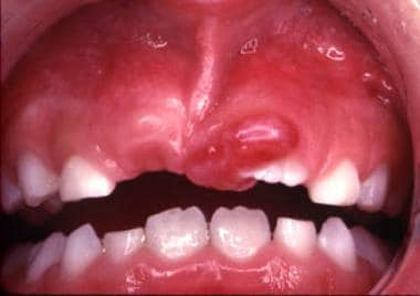
A dentigerous cyst is a type of odontogenic cyst that forms around the crown of an unerupted tooth. It is typically slow-growing and often asymptomatic, but it can cause displacement or resorption of adjacent teeth if it becomes large enough. Dentigerous cysts are more common in permanent teeth than primary teeth, and they are more likely to occur in the mandible (lower jaw) than the maxilla (upper jaw). They are usually diagnosed through radiographic examination and can be treated by surgical removal of the cyst along with the affected tooth. If left untreated, dentigerous cysts can continue to grow and may eventually develop into a tumor or cancer.
Ameloblastoma is a slow-growing, non-cancerous tumor that develops in the jawbone, typically in the lower jaw. It originates from the cells that form the enamel (the hard, outer surface of the teeth). This tumor can cause swelling, pain, and displacement or loosening of teeth. In some cases, it may also lead to fractures of the jawbone.
There are different types of ameloblastomas, including solid or multicystic, unicystic, and peripheral ameloblastoma. Treatment usually involves surgical removal of the tumor, with careful monitoring to ensure that it does not recur. In rare cases, more aggressive treatment may be necessary if the tumor is large or has invaded surrounding tissues.
It's important to note that while ameloblastomas are generally benign, they can still cause significant morbidity and should be treated promptly by an oral and maxillofacial surgeon or other qualified healthcare professional.
Mandibular neoplasms refer to abnormal growths or tumors that develop in the mandible, which is the lower jawbone. These growths can be benign (non-cancerous) or malignant (cancerous). Benign neoplasms are typically slow-growing and rarely spread to other parts of the body, while malignant neoplasms can invade surrounding tissues and may metastasize (spread) to distant sites.
Mandibular neoplasms can have various causes, including genetic mutations, exposure to certain chemicals or radiation, and infection with certain viruses. The symptoms of mandibular neoplasms may include swelling or pain in the jaw, difficulty chewing or speaking, numbness in the lower lip or chin, loose teeth, and/or a lump or mass in the mouth or neck.
The diagnosis of mandibular neoplasms typically involves a thorough clinical examination, imaging studies such as X-rays, CT scans, or MRI scans, and sometimes a biopsy to confirm the type and extent of the tumor. Treatment options depend on the type, stage, and location of the neoplasm, and may include surgery, radiation therapy, chemotherapy, or a combination of these approaches. Regular follow-up care is essential to monitor for recurrence or metastasis.
Maxillary neoplasms refer to abnormal growths or tumors in the maxilla, which is the upper jaw bone. These growths can be benign (non-cancerous) or malignant (cancerous). Benign neoplasms are slow-growing and do not spread to other parts of the body, while malignant neoplasms can invade surrounding tissues and spread to distant sites.
Maxillary neoplasms can cause various symptoms such as swelling, pain, numbness, loose teeth, or difficulty in chewing or swallowing. They may also cause nasal congestion, nosebleeds, or visual changes if they affect the eye or orbit. The diagnosis of maxillary neoplasms usually involves a combination of clinical examination, imaging studies such as CT or MRI scans, and biopsy to determine the type and extent of the tumor.
Treatment options for maxillary neoplasms depend on several factors, including the type, size, location, and stage of the tumor, as well as the patient's overall health and preferences. Treatment may include surgery, radiation therapy, chemotherapy, or a combination of these modalities. Regular follow-up care is essential to monitor for recurrence or metastasis and ensure optimal outcomes.
A cyst is a closed sac, having a distinct membrane and division between the sac and its surrounding tissue, that contains fluid, air, or semisolid material. Cysts can occur in various parts of the body, including the skin, internal organs, and bones. They can be caused by various factors, such as infection, genetic predisposition, or blockage of a duct or gland. Some cysts may cause symptoms, such as pain or discomfort, while others may not cause any symptoms at all. Treatment for cysts depends on the type and location of the cyst, as well as whether it is causing any problems. Some cysts may go away on their own, while others may need to be drained or removed through a surgical procedure.
Odontoma is a type of odontogenic tumor, which means it arises from the tissues that form teeth. It is considered a benign or non-cancerous tumor and is typically slow-growing. Odontomas are usually asymptomatic and are often discovered on routine dental X-rays or during procedures such as wisdom tooth removal.
Odontomas can be classified into two types: complex and compound. Complex odontomas are composed of a haphazard mixture of dental tissue, including enamel, dentin, and cementum, while compound odontomas contain small tooth-like structures called denticles.
These tumors typically occur in the posterior region of the jaw, and while they are usually asymptomatic, some patients may experience symptoms such as swelling, pain, or displacement of teeth. Treatment for odontomas typically involves surgical removal of the tumor.
Pilomatrixoma is a benign skin tumor that originates from the hair follicle's matrix. It is also known as calcifying epithelioma of Malherbe. This slow-growing tumor typically appears as a hard, mobile, small nodule, often on the head or neck region. Pilomatrixomas are usually painless but can become inflamed or infected. They are more common in children and young adults and are slightly more prevalent in females than males. Histologically, pilomatrixoma is characterized by the presence of shadow cells, basaloid cells, and calcifications. Surgical excision is the standard treatment for this condition.
Nonodontogenic cysts are a type of cyst that occur in the oral and maxillofacial region, but they do not originate from tooth-forming tissues. These cysts can develop in various locations within the jaws, including the bone or soft tissues. They are typically classified into several categories based on their origin, such as developmental, inflammatory, or neoplastic.
Examples of nonodontogenic cysts include:
1. Nasopalatine duct cyst - This is the most common type of nonodontogenic cyst and arises from remnants of the nasopalatine duct, which is a structure present during embryonic development. It typically appears in the anterior region of the maxilla (upper jaw).
2. Nasolabial cyst - This rare cyst develops near the nasolabial fold, between the nose and the upper lip. Its origin is unclear but may be related to embryonic remnants or developmental abnormalities.
3. Median mandibular cyst - Also known as a median mental cyst, this rare cyst forms in the midline of the mandible (lower jaw) and may originate from remnants of the dental lamina or other developmental structures.
4. Lateral periodontal cyst - This inflammatory cyst arises from the periodontal ligament, which supports the teeth within their sockets. It is usually found near the roots of lower molars and premolars.
5. Glandular odontogenic cyst - This developmental cyst originates from remnants of minor salivary glands or epithelial rests in the jawbone. It can appear as a unilocular (single-chambered) or multilocular (multi-chambered) cyst and may have a more aggressive behavior than other nonodontogenic cysts.
6. Dentigerous cyst - Although technically classified as an odontogenic cyst, the dentigerous cyst is sometimes considered a borderline case because it arises from the crowns of unerupted teeth rather than their roots. It can grow quite large and may cause significant bone resorption.
Nonodontogenic cysts are less common than odontogenic cysts, but they still require proper diagnosis and treatment to prevent complications such as tooth displacement, jaw deformation, or infection. Treatment options for nonodontogenic cysts depend on their size, location, and histological features and may include enucleation (complete removal), marsupialization (creating a communication between the cyst and oral cavity to allow for gradual reduction), or more extensive surgical procedures. Regular follow-up appointments with your dentist or oral surgeon are essential to monitor healing and ensure that the cyst does not recur.
Jaw diseases refer to a variety of conditions that affect the temporomandibular joint (TMJ) and the surrounding muscles, as well as dental disorders that can impact the jaw. Some common examples include:
1. Temporomandibular Joint Disorders (TMD): These are problems with the TMJ and the muscles that control jaw movement. Symptoms may include pain, clicking or popping sounds, and limited movement of the jaw.
2. Osteonecrosis of the Jaw: This is a condition where bone in the jaw dies due to lack of blood supply. It can be caused by radiation therapy, chemotherapy, or certain medications.
3. Dental Cavities: These are holes in the teeth caused by bacteria. If left untreated, they can cause pain, infection, and damage to the jawbone.
4. Periodontal Disease: This is an infection of the gums and bones that support the teeth. Advanced periodontal disease can lead to loss of teeth and damage to the jawbone.
5. Jaw Fractures: These are breaks in the jawbone, often caused by trauma.
6. Oral Cancer: This is a type of cancer that starts in the mouth or throat. If not treated early, it can spread to the jaw and other parts of the body.
7. Cysts and Tumors: These are abnormal growths in the jawbone or surrounding tissues. While some are benign (non-cancerous), others can be malignant (cancerous).
8. Osteomyelitis: This is an infection of the bone, often occurring in the lower jaw. It can cause pain, swelling, and fever.
9. Oral Thrush: This is a fungal infection that causes white patches on the inside of the mouth. If left untreated, it can spread to the jaw and other parts of the body.
10. Sinusitis: Inflammation of the sinuses can sometimes cause pain in the upper jaw.
A periodontal cyst, also known as a radicular cyst or dental cyst, is a type of odontogenic cyst that forms from the tissue of the periodontium, which surrounds and supports the teeth. It typically develops at the apex (tip) of a dead or non-vital tooth root and is filled with fluid. The cyst can grow slowly and painlessly, often going unnoticed until it becomes quite large or causes symptoms such as swelling, tenderness, or tooth mobility.
Periodontal cysts are usually asymptomatic and are often discovered during routine dental x-rays. If left untreated, they can eventually lead to the destruction of surrounding bone and tissue, potentially causing teeth to become loose or even fall out. Treatment typically involves surgical removal of the cyst along with the affected tooth, followed by careful monitoring to ensure that the cyst does not recur.
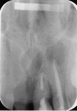
Panoramic radiography is a specialized type of dental X-ray imaging that captures a panoramic view of the entire mouth, including the teeth, upper and lower jaws, and surrounding structures. It uses a special machine that rotates around the head, capturing images as it moves. This technique provides a two-dimensional image that is helpful in diagnosing and planning treatment for various dental conditions such as impacted teeth, bone abnormalities, and jaw disorders.
The panoramic radiograph can also be used to assess the development and positioning of wisdom teeth, detect cysts or tumors in the jaws, and evaluate the effects of trauma or injury to the mouth. It is a valuable tool for dental professionals as it allows them to see a comprehensive view of the oral structures, which may not be visible with traditional X-ray techniques.
It's important to note that while panoramic radiography provides valuable information, it should be used in conjunction with other diagnostic tools and clinical examinations to ensure accurate diagnosis and treatment planning.
An impacted tooth is a condition where a tooth fails to erupt into the oral cavity within its expected time frame, resulting in its partial or complete entrapment within the jawbone or soft tissues. This commonly occurs with wisdom teeth (third molars) but can affect any tooth. Impacted teeth may cause problems such as infection, decay of adjacent teeth, gum disease, or cyst formation, and they may require surgical removal.
A fibroma, ossifying is a benign (non-cancerous) tumor that typically develops in the periodontal ligament, which is the tissue that connects the tooth to the jawbone. This type of fibroma is characterized by the formation of bone-like tissue within the tumor. It usually appears as a firm, slow-growing nodule or mass that can cause pain or discomfort, particularly when biting down on the affected tooth.
The exact cause of ossifying fibromas is not well understood, but they are thought to arise from an overgrowth of cells in the periodontal ligament. They are more common in women than men and typically occur in people between the ages of 20 and 40. Treatment usually involves surgical removal of the tumor, along with any affected tissue or teeth. In some cases, recurrence may occur, so regular follow-up appointments with a dental professional are recommended.
Fibrous Dysplasia of Bone is a rare, benign bone disorder that is characterized by the replacement of normal bone tissue with fibrous (scar-like) and immature bone tissue. This results in weakened bones that are prone to fractures, deformities, and pain. The condition can affect any bone in the body but most commonly involves the long bones of the legs, arms, and skull. It can occur as an isolated finding or as part of a genetic disorder called McCune-Albright syndrome. The exact cause of fibrous dysplasia is not fully understood, but it is believed to result from a genetic mutation that occurs during early bone development. There is no cure for fibrous dysplasia, and treatment typically focuses on managing symptoms and preventing complications.
A focal infection is a focus or source of infection that can spread and cause harm to other parts of the body. A "focal infection, dental" refers to an infection that originates in the teeth or surrounding tissues of the mouth and then spreads to other parts of the body. This can occur when bacteria or other pathogens from a dental infection enter the bloodstream and travel to distant sites, where they can cause inflammation, tissue damage, and illness.
Dental focal infections can be caused by various conditions, such as tooth decay, periodontal disease, abscesses, or other oral infections. The bacteria involved in dental infections are often part of the normal oral flora but can become pathogenic under certain circumstances, such as when they gain access to deeper tissues or the bloodstream due to trauma, surgery, or poor oral hygiene.
If left untreated, dental focal infections can lead to serious health complications, including heart disease, brain abscesses, and other systemic infections. It is essential to maintain good oral hygiene and seek professional dental care to prevent and treat dental infections, reducing the risk of developing focal infections and related health issues.
Cyst fluid refers to the fluid accumulated within a cyst, which is a closed sac-like or capsular structure, typically filled with liquid or semi-solid material. Cysts can develop in various parts of the body for different reasons, and the composition of cyst fluid may vary depending on the type of cyst and its location.
In some cases, cyst fluid might contain proteins, sugars, hormones, or even cells from the surrounding tissue. Infected cysts may have pus-like fluid, while cancerous or precancerous cysts might contain abnormal cells or tumor markers. The analysis of cyst fluid can help medical professionals diagnose and manage various medical conditions, including infections, inflammatory diseases, genetic disorders, and cancers.
It is important to note that the term 'cyst fluid' generally refers to the liquid content within a cyst, but the specific composition and appearance of this fluid may vary significantly depending on the underlying cause and type of cyst.
Hair diseases is a broad term that refers to various medical conditions affecting the hair shaft, follicle, or scalp. These conditions can be categorized into several types, including:
1. Hair shaft abnormalities: These are conditions that affect the structure and growth of the hair shaft. Examples include trichorrhexis nodosa, where the hair becomes weak and breaks easily, and pili torti, where the hair shaft is twisted and appears sparse and fragile.
2. Hair follicle disorders: These are conditions that affect the hair follicles, leading to hair loss or abnormal growth patterns. Examples include alopecia areata, an autoimmune disorder that causes patchy hair loss, and androgenetic alopecia, a genetic condition that leads to pattern baldness in both men and women.
3. Scalp disorders: These are conditions that affect the scalp, leading to symptoms such as itching, redness, scaling, or pain. Examples include seborrheic dermatitis, psoriasis, and tinea capitis (ringworm of the scalp).
4. Hair cycle abnormalities: These are conditions that affect the normal growth cycle of the hair, leading to excessive shedding or thinning. Examples include telogen effluvium, where a large number of hairs enter the resting phase and fall out, and anagen effluvium, which is typically caused by chemotherapy or radiation therapy.
5. Infectious diseases: Hair follicles can become infected with various bacteria, viruses, or fungi, leading to conditions such as folliculitis, furunculosis, and kerion.
6. Genetic disorders: Some genetic disorders can affect the hair, such as Menkes syndrome, which is a rare inherited disorder that affects copper metabolism and leads to kinky, sparse, and brittle hair.
Proper diagnosis and treatment of hair diseases require consultation with a healthcare professional, often a dermatologist or a trichologist who specializes in hair and scalp disorders.
A giant cell granuloma is a type of non-cancerous (benign) lesion characterized by the presence of large collections of immune cells called macrophages, which have fused together to form multinucleated giant cells. These lesions can occur in various tissues throughout the body but are most commonly found in the oral cavity and jawbone.
Giant cell granulomas can be further classified into two types: central (or bone) giant cell granuloma and peripheral giant cell granuloma. Central giant cell granulomas arise from the bone, while peripheral giant cell granulomas occur in the soft tissues of the gingiva or mouth lining.
Central giant cell granulomas are more aggressive than peripheral ones and can cause significant bone destruction if left untreated. They typically affect younger individuals, with a higher prevalence in females than males. The exact cause of central giant cell granulomas is not well understood but may be associated with local trauma, hormonal imbalances, or genetic factors.
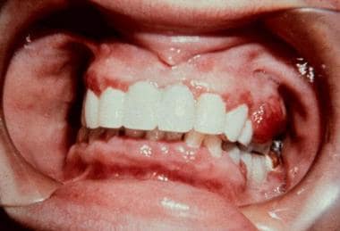
Peripheral giant cell granulomas are less aggressive and usually present as painless, slow-growing nodules on the gums. They typically affect adults, with a higher prevalence in females than males. Peripheral giant cell granulomas may be associated with local irritants such as plaque, calculus, or dental restorations.
Treatment for giant cell granulomas depends on their size, location, and aggressiveness. Surgical excision is the most common treatment approach, but other options such as curettage, corticosteroid injections, or medication therapy may also be considered. Regular follow-up appointments with a healthcare provider are essential to monitor for recurrence.
A periodontal abscess is a localized collection of pus in the tissues surrounding and supporting the teeth, caused by an infection. It's typically characterized by symptoms such as pain, swelling, redness, and sometimes drainage of pus from the affected area. The infection usually arises from dental plaque that accumulates on the teeth and gums, leading to periodontal disease. If left untreated, a periodontal abscess can result in tissue destruction, bone loss, and even tooth loss. Treatment typically involves draining the abscess, removing any infected tissue, and providing oral hygiene instruction to prevent future infections. In some cases, antibiotics may also be prescribed to help clear up the infection.
An ovarian cyst is a sac or pouch filled with fluid that forms on the ovary. Ovarian cysts are quite common in women during their childbearing years, and they often cause no symptoms. In most cases, ovarian cysts disappear without treatment over a few months. However, larger or persistent cysts may require medical intervention, including surgical removal.
There are various types of ovarian cysts, such as functional cysts (follicular and corpus luteum cysts), which develop during the menstrual cycle due to hormonal changes, and non-functional cysts (dermoid cysts, endometriomas, and cystadenomas), which can form due to different causes.
While many ovarian cysts are benign, some may have malignant potential or indicate an underlying medical condition like polycystic ovary syndrome (PCOS). Regular gynecological check-ups, including pelvic examinations and ultrasounds, can help detect and monitor ovarian cysts.
Odontogenesis is the process of tooth development that involves the formation and calcification of teeth. It is a complex process that requires the interaction of several types of cells, including epithelial cells, mesenchymal cells, and odontoblasts. The process begins during embryonic development with the formation of dental lamina, which gives rise to the tooth bud. As the tooth bud grows and differentiates, it forms the various structures of the tooth, including the enamel, dentin, cementum, and pulp. Odontogenesis is completed when the tooth erupts into the oral cavity. Abnormalities in odontogenesis can result in developmental dental anomalies such as tooth agenesis, microdontia, or odontomas.
An odontogenic tumor, squamous type, refers to a specific category of oral tumors that originate from the dental tissues. These tumors are characterized by the abnormal growth of squamous epithelial cells, which are normally found in the outermost layer of the skin and the mucous membranes, including those inside the mouth.
Odontogenic tumors can arise from various components of the dental tissues, such as the odontoblasts, dentin, enamel, cementum, or the epithelial rests of Malassez. The squamous type of odontogenic tumor is typically classified under either one of the following entities:
1. Squamous Odontogenic Tumor (SOT): This is a rare, benign (non-cancerous) neoplasm that primarily affects the tooth-bearing areas of the jaws. SOT is composed of well-differentiated squamous epithelial cells arranged in nests, strands, or sheets, often surrounded by a fibrous stroma. This tumor typically occurs in adults during their third to fifth decades of life and has a slight female predilection.
2. Ameloblastoma with Squamous Metaplasia: Ameloblastoma is a more common odontogenic tumor that usually affects the mandible (lower jaw). In some cases, ameloblastomas may undergo squamous metaplasia, where the original epithelial cells transform into squamous epithelial cells. This variant of ameloblastoma is still considered a benign neoplasm; however, it has a higher recurrence rate compared to conventional ameloblastomas.
It is essential to differentiate these entities from other oral lesions and malignancies through histopathological examination, as the treatment and prognosis may vary depending on the specific diagnosis.
The mandible, also known as the lower jaw, is the largest and strongest bone in the human face. It forms the lower portion of the oral cavity and plays a crucial role in various functions such as mastication (chewing), speaking, and swallowing. The mandible is a U-shaped bone that consists of a horizontal part called the body and two vertical parts called rami.
The mandible articulates with the skull at the temporomandibular joints (TMJs) located in front of each ear, allowing for movements like opening and closing the mouth, protrusion, retraction, and side-to-side movement. The mandible contains the lower teeth sockets called alveolar processes, which hold the lower teeth in place.
In medical terminology, the term "mandible" refers specifically to this bone and its associated structures.
An epidermal cyst is a common benign skin condition characterized by the growth of a sac-like structure filled with keratin, a protein found in the outermost layer of the skin (epidermis). These cysts typically appear as round, firm bumps just under the surface of the skin, often on the face, neck, trunk, or scalp. They can vary in size from a few millimeters to several centimeters in diameter.
Epidermal cysts usually develop as a result of the accumulation of dead skin cells that become trapped within a hair follicle or a pilosebaceous unit (a structure that contains a hair follicle and an oil gland). The keratin produced by the skin cells then collects inside the sac, causing it to expand gradually.
These cysts are generally slow-growing, painless, and rarely cause any symptoms. However, they may become infected or inflamed, leading to redness, tenderness, pain, or pus formation. In such cases, medical attention might be necessary to drain the cyst or administer antibiotics to treat the infection.
Epidermal cysts can be removed surgically if they cause cosmetic concerns or become frequently infected. The procedure typically involves making an incision in the skin and removing the entire sac along with its contents to prevent recurrence.
Keratins are a type of fibrous structural proteins that constitute the main component of the integumentary system, which includes the hair, nails, and skin of vertebrates. They are also found in other tissues such as horns, hooves, feathers, and reptilian scales. Keratins are insoluble proteins that provide strength, rigidity, and protection to these structures.
Keratins are classified into two types: soft keratins (Type I) and hard keratins (Type II). Soft keratins are found in the skin and simple epithelial tissues, while hard keratins are present in structures like hair, nails, horns, and hooves.
Keratin proteins have a complex structure consisting of several domains, including an alpha-helical domain, beta-pleated sheet domain, and a non-repetitive domain. These domains provide keratin with its unique properties, such as resistance to heat, chemicals, and mechanical stress.
In summary, keratins are fibrous structural proteins that play a crucial role in providing strength, rigidity, and protection to various tissues in the body.
A fibroma is a benign (non-cancerous) tumor that consists primarily of fibrous or connective tissue. It can occur in various parts of the body, including the skin, mouth, and internal organs. The term "fibroma" is often used to describe any benign fibrous growth, but there are specific types of fibromas such as dermatofibroma (found in the skin), oral fibroma (found in the mouth), and benign fibrous histiocytoma (found in soft tissues).
It's important to note that while fibromas are generally harmless, they can cause discomfort or problems depending on their size and location. If a fibroma is causing issues or there's concern about its growth or malignancy, it should be evaluated by a healthcare professional for potential removal or further assessment.
A myxoma is a type of benign (non-cancerous) tumor that develops in the heart, specifically in the heart's chambers or valves. It is the most common primary cardiac tumor in adults and typically affects the left atrium. Myxomas are composed of gelatinous, mucoid material and may have a stalk-like attachment to the endocardium (the inner lining of the heart).
Myxomas can vary in size and may cause symptoms such as shortness of breath, fatigue, chest pain, coughing, and fever. These symptoms are due to obstruction of blood flow within the heart or embolization (detachment and travel) of tumor fragments to other parts of the body. Surgical removal is usually required to treat myxomas, as they can lead to serious complications if left untreated.
A mediastinal cyst is a rare, abnormal fluid-filled sac located in the mediastinum, which is the central part of the chest cavity that separates the lungs and contains various organs such as the heart, esophagus, trachea, thymus gland, and lymph nodes. Mediastinal cysts can be congenital (present at birth) or acquired (develop later in life). They are usually asymptomatic but can cause symptoms depending on their size and location. Symptoms may include chest pain, cough, difficulty breathing, or swallowing. Treatment typically involves surgical removal of the cyst to prevent complications such as infection, bleeding, or pressure on surrounding structures.
Gingival neoplasms refer to abnormal growths or tumors that occur in the gingiva, which are the part of the gums that surround the teeth. These growths can be benign (non-cancerous) or malignant (cancerous). Benign neoplasms include conditions such as fibromas, papillomas, and hemangiomas, while malignant neoplasms are typically squamous cell carcinomas.
Gingival neoplasms can present with a variety of symptoms, including swelling, bleeding, pain, and loose teeth. They may also cause difficulty with chewing, speaking, or swallowing. The exact cause of these neoplasms is not always known, but risk factors include tobacco use, alcohol consumption, poor oral hygiene, and certain viral infections.
Diagnosis of gingival neoplasms typically involves a thorough clinical examination, including a dental exam and biopsy. Treatment options depend on the type and stage of the neoplasm, but may include surgery, radiation therapy, chemotherapy, or a combination of these approaches. Regular dental check-ups and good oral hygiene practices can help to detect gingival neoplasms at an early stage and improve treatment outcomes.
A Synovial Cyst is a type of benign cyst that typically develops in the synovium, which is the membrane that lines and lubricates joint capsules. These cysts are filled with synovial fluid, which is the same lubricating fluid found inside joints. They usually form as a result of degenerative changes, trauma, or underlying joint diseases such as osteoarthritis.
Synovial cysts commonly occur in the spine (particularly in the facet joints), but they can also develop in other areas of the body, including the knees, hips, and hands. While synovial cysts are generally not harmful, they may cause discomfort or pain if they press on nearby nerves or restrict movement in the affected joint. Treatment options for synovial cysts range from conservative measures like physical therapy and pain management to surgical intervention in severe cases.
A bone cyst is a fluid-filled sac that develops within a bone. It can be classified as either simple (unicameral) or aneurysmal. Simple bone cysts are more common in children and adolescents, and they typically affect the long bones of the arms or legs. These cysts are usually asymptomatic unless they become large enough to weaken the bone and cause a fracture. Aneurysmal bone cysts, on the other hand, can occur at any age and can affect any bone, but they are most common in the leg bones and spine. They are characterized by rapidly growing blood-filled sacs that can cause pain, swelling, and fractures.
Both types of bone cysts may be treated with observation, medication, or surgery depending on their size, location, and symptoms. It is important to note that while these cysts can be benign, they should still be evaluated and monitored by a healthcare professional to ensure proper treatment and prevention of complications.
A tooth is a hard, calcified structure found in the jaws (upper and lower) of many vertebrates and used for biting and chewing food. In humans, a typical tooth has a crown, one or more roots, and three layers: the enamel (the outermost layer, hardest substance in the body), the dentin (the layer beneath the enamel), and the pulp (the innermost layer, containing nerves and blood vessels). Teeth are essential for proper nutrition, speech, and aesthetics. There are different types of teeth, including incisors, canines, premolars, and molars, each designed for specific functions in the mouth.
A Follicular Cyst is a type of cyst that forms within a follicle, which is the sac-like structure in the skin that contains and protects a hair root. In particular, it refers to a specific condition in the ovary where a follicle fails to rupture or release an egg after maturation, instead continuing to grow and fill with fluid, forming a cyst. These cysts are usually asymptomatic but can become large and cause symptoms such as pelvic pain or discomfort, irregular menstrual cycles, or abnormal vaginal bleeding. In most cases, follicular cysts resolve on their own within 2-3 menstrual cycles, but in rare cases, they may require medical intervention if they become complicated or do not resolve.
A bronchogenic cyst is a type of congenital cyst that develops from abnormal budding or development of the bronchial tree during fetal growth. These cysts are typically filled with mucus or fluid and can be found in the mediastinum (the area between the lungs) or within the lung tissue itself.
Bronchogenic cysts are usually asymptomatic, but they can cause symptoms if they become infected, rupture, or compress nearby structures such as airways or blood vessels. Symptoms may include cough, chest pain, difficulty breathing, and recurrent respiratory infections.
Diagnosis of bronchogenic cysts is typically made through imaging tests such as chest X-rays, CT scans, or MRI scans. Treatment usually involves surgical removal of the cyst to prevent complications.
Aneurysmal bone cyst (ABC) is a benign but locally aggressive tumor that typically involves the metaphysis of long bones in children and adolescents. It is characterized by blood-filled spaces or cysts separated by fibrous septa containing osteoclast-type giant cells, spindle cells, and capillary vessels.
ABCs can also arise in other locations such as the vertebral column, pelvis, and skull. They may cause bone pain, swelling, or pathologic fractures. The exact cause of ABC is unknown, but it is thought to be related to a reactive process to a primary bone lesion or trauma.
Treatment options for ABC include curettage and bone grafting, intralesional injection of corticosteroids or bone marrow aspirate, and adjuvant therapy with phenol or liquid nitrogen. In some cases, radiation therapy may be used, but it is generally avoided due to the risk of secondary malignancies. Recurrence rates after treatment range from 10-30%.
 Odontogenic cyst
Glandular odontogenic cyst
Calcifying odontogenic cyst
Botryoid odontogenic cyst
Lateral periodontal cyst
Ameloblastic carcinoma
Gingival cyst
Index of oral health and dental articles
Paradental cyst
Catenin beta-1
Dentigerous cyst
Periapical cyst
Dental emergency
Cysts of the jaws
Primordial cyst
Odontogenic keratocyst
Wisdom tooth
Cell counting
Cyst
Ghost cell
Nasopalatine duct cyst
Carnoy's solution
Thyroglossal cyst
Median mandibular cyst
Dental follicle
ITK-SNAP
Pilomatricoma
Pericoronitis
Ameloblastoma
Adenomatoid odontogenic tumor
Eruption cyst
Odontogenic cyst - Wikipedia
 Mandibular Cysts and Odontogenic Tumors: Overview, Odontogenic Mandibular Cysts, Nonodontogenic Mandibular Cysts
Odontogenic tumors and cysts - Vrije Universiteit Amsterdam
 Clinico pathological study of Odontogenic cysts and tumors in a Tertiary care Dental hospital of Nepal
| Journal of...
Clinico pathological study of Odontogenic cysts and tumors in a Tertiary care Dental hospital of Nepal
| Journal of...
 Odontogenic tumours and cysts - Libre Pathology
 Adenomatoid odontogenic tumour mimetizing a radicular cyst
 Chapter 15: Odontogenic Cysts and Tumors Flashcards - Easy Notecards
 Odontogenic myxofibroma resembling dentigerous cyst - IIUM Repository (IRep)
 oral surgeon St. George UT - What Are Odontogenic Cysts? | Precision Dental Specialties
Maxillary dentigerous cyst showing squamous odontogenic tumor-like proliferation: surgical approach and literature review
 Pubblicazione | SILVIA TORTORICI | Università degli Studi di Palermo
 The molecular basis of odontogenic cysts and tumours. | J Oral Pathol Med;52(4): 351-356, 2023 Apr. | MEDLINE | BVS...
Evaluation of collagen in connective tissue walls of odontogenic cysts -A histochemical study - Fingerprint - Manipal...
Unicameral (Simple) Bone Cyst Workup: Laboratory Studies, Plain Radiography, Magnetic Resonance Imaging
 Clinical and histologic features of botryoid odontogenic cyst: a case report | Journal of Medical Case Reports | Peer Review
Odontogenic keratocyst (okc) or keratocystic odontogenic tumor (kcot) - journey of okc from cyst to tumor to cyst again : ...
Clinicopathological Features and Expression of Ki-67 in Odontogenic Keratocyst, Dentigerous Cyst and Radicular Cyst
|...
 Multiple Choice Questions in Oral Medicine and Radiology - A Rapid Review in Dentistry - Nova Science Publishers
 S Patel | West Indian Medical Journal
 References | Chronic Inflammation on the Right Side of the Maxilla | DentalCare.com
 JETO 12.1, p. 35-42 - Old City Publishing
 The Journal of Contemporary Dental Practice
 Dentigerous Cyst | Harvard Catalyst Profiles | Harvard Catalyst
 Amina Kozaric | IntechOpen
 Healthcare | Topical Collection : Dentistry, Oral Health and Maxillofacial Surgery
 Equine Dental Exams in Five Easy Steps - The Horse
 Ameloblastoma
Summary Report | CureHunter
Ameloblastoma
Summary Report | CureHunter
 Mineral metabolism, dental bioch212 1 | PPT
![PTCH1 patched 1 [Homo sapiens (human)] - Gene - NCBI](data:image/png;base64,iVBORw0KGgoAAAANSUhEUgAAABAAAAAQCAYAAAAf8/9hAAAB1ElEQVQ4jaWSPWgTcRjGf/ehuWhobO2JxGJRY3taTTRV2yoqSpW6iIWO4iAoUsRBioNDKUWKLU7i4KA4OfhVREQnETRia03k7IdiS0LaQYKJQg3mLtfc30GySNUDn/V5nx/vy/vAf0pqad3db2xquiBJku93s2Tb2eEHdw1rTcsxol23sObTjN7oIp9KVmaU9kMdTxcLAyiqGtA0bfms+XKQULSdQG2EmnUx0q9ughAA8p/CFW0IN3Sv0vUI5p2zIMpUrd5JeP/Jii//80ZJUlrb9lyV8qn3zI5dB8A4MoBWtcITAKBmZe3eRmPzccYf9uIUsyzx6zQd7fMMAIjFdgxpkuPy4clFANbu6qa6fouybXtznxeAoqoBn0/zz5kvBqVQ5DBasJ5gXaPnDQAWFpwCkiwLZekyAMp2wTPAsqy5d8nEZcIHThPQo7jlIua9854BibdvekqKX8PouARAOn6F+c8pT4Bc7svz6U8f77O1cwDVV439PcPU4yHw8AUhhDPyOn4OfWOMuuZfBZp41INTLACorhC2/Jc2zsxMX8vl8lMcPBUHFL5mnpEZGa748sS42esKYS8WLtl2NjE22s/6fScIhtr48W2S5O0zIFwvp3vST6Z+myCvkaonAAAAAElFTkSuQmCC) PTCH1 patched 1 [Homo sapiens (human)] - Gene - NCBI
PTCH1 patched 1 [Homo sapiens (human)] - Gene - NCBI
 General Dentistry Details
Keratocyst13
- Odontogenic keratocyst iii. (wikipedia.org)
- trichinosis Buccal bifurcation cyst Calcifying odontogenic cyst Dentigerous cyst (associated with the crowns of non-erupted teeth) Glandular odontogenic cyst Keratocyst (in the jaws, these can appear solitary or associated with the Gorlin-Goltz or Nevoid basal cell carcinoma syndrome. (wikipedia.org)
- However, sometimes these masses are considered neoplasm: Keratocyst Calcifying odontogenic cyst According to the current (2005) classification of the World Health Organization, both (parakeratizied) odontogenic keratocyst and calcifying odontogenic cyst have neoplastic characteristics, thus renamed as Keratocystic odontogenic tumor and Calcifying odontogenic tumor, respectively. (wikipedia.org)
- Cystic ameloblastoma Long standing dentigerous cyst, odontogenic keratocyst, and residual cyst may have neoplastic potential converting into the locally aggressive ameloblastoma, or the malignant squamous cell carcinoma and mucoepidermoid carcinoma. (wikipedia.org)
- The cyst was excised, and histopathologic examination revealed an odontogenic keratocyst (OKC). (easynotecards.com)
- There is debate over reclassification of the odontogenic keratocyst (OKC) as keratocystic odontogenic tumor (KCOT). (easynotecards.com)
- common ones include radicular, dentigerous, and odontogenic keratocyst. (precisionstg.com)
- On the other hand, odontogenic keratocyst occurs less frequently compared to others, but their high recurrence rate is associated with remnants of the dental lamina, traumatic implantation, or down growth. (precisionstg.com)
- PTCH1 mutations have been reported in a high proportion of odontogenic keratocyst . (bvsalud.org)
- The odontogenic keratocyst (OKC) is a dilemmatic odontogenic developmental cyst of oral and maxillofacial region which has gained very special attention since last two decades. (journalcra.com)
- Biological behaviour of Odontogenic Keratocyst (OKC) is aggressiveness than others Odontogenic Cysts (OCs) like Dentigerous Cyst (DCs) and Periapical Cyst/Residual Cysts (RCs). (org.pk)
- The Odontogenic Keratocyst is known for its aggressiveness, high recurrence rate and transformation of keratinized epithelia to non-keratinized squamous epithelium for which inflammation has been suggested to be responsible. (uwi.edu)
- We note in particular the gingival cyst of the newborn, the gingival cyst of the adult (uncommon), the odontogenic keratocyst, the dentigerous cyst (formed within the organ of the enamel of a tooth still included), eruptive cyst (which surrounds the crown of a tooth ready to erupt), lateral periodontal cyst, calcifying odontogenic cyst and odontogenic glandular cyst. (calendarena.com)
Tumor31
- Adenomatoid Odontogenic Tumor (AOT) was so classified as a new tumor in 1969 by Philipsen and Birn. (bvsalud.org)
- This odontogenic tumor is benign, asymptomatic, has a slow growing and rarely reach a size greater than 3 cm. (bvsalud.org)
- Odontogenic myxoma is a rare benign odontogenic tumor with an infiltrative local growth. (iium.edu.my)
- Squamous odontogenic tumor (SOT) is a rare benign neoplasm of the jaw that likely arises from remnants of the dental lamina. (autopsyandcasereports.org)
- In rare cases, squamous odontogenic tumor-like proliferation (SOT-LP) can be observed arising from odontogenic cysts (SOT-LPOC). (autopsyandcasereports.org)
- Squamous odontogenic tumor. (autopsyandcasereports.org)
- 2 Elmuradi S, Mair Y, Suresh L, DeSantis J, Neiders M, Aguirre A. Multicentric squamous odontogenic tumor: a case report and review of the literature. (autopsyandcasereports.org)
- Squamous odontogenic tumor and squamous odontogenic tumor-like proliferations in odontogenic cysts: an updated analysis of 170 cases reported in the literature. (autopsyandcasereports.org)
- Squamous odontogenic tumor-like proliferations in radicular cysts: a clinicopathologic study of forty-two cases. (autopsyandcasereports.org)
- 6 Leventon GS, Happonen RP, Newland JR. Squamous odontogenic tumor. (autopsyandcasereports.org)
- Squamous odontogenic tumor-like proliferations in periapical cysts. (autopsyandcasereports.org)
- 9 Sala-Pérez S, Marco-Molina V, Gay-Escoda C. Squamous odontogenic tumor-like proliferation in a radicular cyst: a case report. (autopsyandcasereports.org)
- 11 Unal T, Gomel M, Gunel O. Squamous odontogenic tumor-like islands in a radicular cyst: report of a case. (autopsyandcasereports.org)
- Management of a rare case of peripheral squamous odontogenic tumor of the gingiva. (autopsyandcasereports.org)
- 14 Oliveira JA, Costa IM, Loyola AM. Squamous odontogenic tumor-like proliferations (SOT-LP) versus intraosseous squamous cell carcinoma in residual cyst. (autopsyandcasereports.org)
- Previously classified under developmental odontogenic cyst of jaw by WHO in 1971 & 1992, OKC has been reclassified and renamed as keratocystic odontogenic tumor (KCOT) in the WHO classifications of head and neck tumors in 2005 due to its aggressive behavior, high recurrence rates and specific histological characterstics. (journalcra.com)
- Most common odontogenic cyst and tumor reported was dentigerous cyst and ameloblastoma respectively. (oldcitypublishing.com)
- Multilocularity as a radiographic marker of the keratocystic odontogenic tumor. (harvard.edu)
- Odontogenic myxoma is a benign but invasive tumor that has a high rate of recurrence after surgical removal (Speight, 2013). (pleasefireme.com)
- Central odontogenic fibroma (COF) is an extremely rare benign tumor that accounts for 0.1% of all odontogenic tumors. (pleasefireme.com)
- What is the most common odontogenic tumor? (pleasefireme.com)
- Peripheral odontogenic fibroma (formerly known as fibromatous and ossifying epulis) is a benign, often slow-growing tumor that arises from periodontal structures (gums, ligaments, and bone). (pleasefireme.com)
- What is the difference between cyst and tumor? (pleasefireme.com)
- Ameloblastoma is a benign epithelial odontogenic tumor. (biomedcentral.com)
- The lesion histologically shows typical ameloblastomatous epithelium lining part of the cyst cavity with or without and/or mural tumor growth. (biomedcentral.com)
- Ameloblastoma is a local invasive tumor which originates from remnants of the dental lamina and odontogenic epithelium and it accounts for only 1 % of all oral tumors [ 1 , 2 ]. (biomedcentral.com)
- UA refers to those cystic lesions with clinical, radiographic, or gross features of a jaw cyst, with which they are usually differentially diagnosed, but on histological examination the UA shows a typical ameloblastomatous epithelium lining part of the cyst cavity, with or without luminal and/or mural tumor growth [ 5 ]. (biomedcentral.com)
- The cyst is a tumor since it corresponds well to an increase in volume of a clearly delimited tissue without specifying the cause. (calendarena.com)
- The cyst is a benign tumor insofar as it is not serious, and cannot give rise to metastases. (calendarena.com)
- Odontoma, the most common odontogenic tumor, affects the dental follicle or the dental tissues and usually appears in the mandibles of young people. (msdmanuals.com)
- Ameloblastoma, the most common epithelial odontogenic tumor, usually arises in the posterior mandible. (msdmanuals.com)
Tumours22
- Diagnosis of odontogenic cysts and tumours requires detailed clinical, radiographical, and histopathological findings. (nepjol.info)
- Fourth edition WHO 2017, classification of Head and Neck lesions, reclassified odontogenic cysts and tumours. (nepjol.info)
- To know relative frequency of odontogenic cysts and tumours according to WHO 2017 classification and to know their clinico-pathological characteristics in selected population of Nepal. (nepjol.info)
- Data were obtained conveniently from records of patients diagnosed with odontogenic cysts and tumours from April 2014-2021. (nepjol.info)
- In total of 163 biopsies, 120 (73.62%) cases were of odontogenic cysts and 43 (26.38%) cases were of odontogenic tumours. (nepjol.info)
- The mean age of occurrence for cysts was 33.35 ± 16.67 years and for tumours was 28.91 ± 13.96 years. (nepjol.info)
- Radicular cyst (49/120, 40.83%) and conventional ameloblastoma (23/43 53.48%) were the commonest cysts and tumours. (nepjol.info)
- Male (67/120, 55.83%) and female (24/43, 55.81%) predisposition was seen in cysts and tumours. (nepjol.info)
- Increased frequency of radicular cysts and conventional ameloblastoma were appreciated with male predisposition in tumours and female predisposition in cysts. (nepjol.info)
- Both cysts and tumours were common in second to third decade of life affecting middle and posterior region of mandible. (nepjol.info)
- Prevalence of odontogenic cysts and tumours: A retrospective clinico-pathological study of 204 cases. (nepjol.info)
- Prevalence of odontogenic cysts and tumours on turkish sample according to latest classification of world health organisation: A 10-year retrospective study. (nepjol.info)
- Wright JM, Vered M. Update from the 4th edition of the World Health Organization classification of head and neck tumours: odontogenic and maxillofacial bone tumours. (nepjol.info)
- Prevalence of odontogenic cysts and tumours among UAE population. (nepjol.info)
- This article covers odontogenic tumours and cysts , which is a subset of oral pathology and can be grouped under the heading of head and neck pathology . (librepathology.org)
- The molecular basis of odontogenic cysts and tumours. (bvsalud.org)
- The advances in molecular technologies have allowed a better understanding of the molecular basis of odontogenic cysts and tumours. (bvsalud.org)
- CTNNB1 mutations also occur in other ghost- cell -containing tumours, including calcifying odontogenic cysts , dentinogenic ghost cell tumours and odontogenic carcinoma with dentinoid, but the link between CTNNB1 mutations and ghost cell formation in these lesions remains unclear. (bvsalud.org)
- Since the functional effect of pathogenic mutations is context and tissue -dependent, a clear role for the reported mutations in odontogenic cysts and tumours in their pathogenesis remains to be elucidated. (bvsalud.org)
- In:Kramer IRH, Pindborg JJ, Shear M. Histological Typing of Odontogenic Tumours, 2nd edn. (thejcdp.com)
- WHO classification odontogenic Tumours? (pleasefireme.com)
- The anterior teeth, consisting of the incisors and odontogenic tumours and cyst. (bvsalud.org)
Ameloblastoma4
- The removed cyst must be evaluated by pathologist to confirm the diagnosis, and to rule out other neoplastic lesions with similar clinical or radiographic features (e.g., cystic or solid ameloblastoma, central mucoepidermoid carcinoma). (wikipedia.org)
- BRAF p.V600E are recurrent in ameloblastoma and KRAS p.G12V/R in adenomatoid odontogenic tumour, dysregulating the MAPK/ ERK pathway . (bvsalud.org)
- Unicystic ameloblastoma is a rare odontogenic lesion, with clinical, radiographic and gross features of jaw cysts. (biomedcentral.com)
- Preoperative diagnosis of the lesion was made as dentigerous cyst based on the age of the patient, location of the swelling, clinical and radiographic findings, but the unicystic ameloblastoma was also taken into consideration. (biomedcentral.com)
Radicular cysts6
- Common odontogenic cysts are dentigerous cysts , and radicular cysts . (librepathology.org)
- Radicular cysts emerge from a tooth experiencing pulp necrosis, often related to untreated cavities, tooth roots, or trauma. (precisionstg.com)
- Inflammatory radicular cysts were observed more in male gender, younger age at diagnosis and anterior maxilla as site of presentation. (unipa.it)
- Unlike dentigerous cysts, the frequency of radicular cysts decreased from 10.4% in 1986-1995 to about 8% in 1996-2005 (P (unipa.it)
- Inflammatory radicular cysts are the most represented group among OCs in our area with a higher prevalence than that reported in other countries. (unipa.it)
- Among odontogenic cysts with benign pathology, up to 60% of all jaw cysts are radicular cysts, which originate from root canal infection. (intechopen.com)
Benign6
- Radicular cyst (associated with the roots of non-vital teeth, also known as Periapical cyst) Residual cyst Most cysts in the body are benign (dysfunctional) tumors, the result of plugged ducts or other natural body outlets for secretions. (wikipedia.org)
- Odontogenic myxoma is a rare benign tumour of the jaw and characteristically presents as a slow, painless, bony expansion with resultant facial deformity. (pleasefireme.com)
- Odontogenic myxoma (OM) is a rare benign painless, slow-growing lesion with local aggressive behavior. (pleasefireme.com)
- Is Odontogenic myxoma benign or malignant? (pleasefireme.com)
- Odontogenic myxomas are rare benign neoplasm of mesenchymal origin, comprising 3-6% of all odontogenic tumors. (pleasefireme.com)
- BENIGN ODONTOGENIC TUMORS, EPITHELIAL Ameloblastomas were classified as solid/multicystic, extraosseous/peripheral, desmoplastic, and unicystic types in the 2005 classification. (pleasefireme.com)
Epithelium9
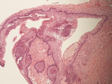
- Odontogenic cysts are closed sacs, and have a distinct membrane derived from rests of odontogenic epithelium. (wikipedia.org)
- That odontogenic epithelium is critical in normal tooth development. (wikipedia.org)
- Oral cysts with gastric or intestinal epithelium (oral alimentary tract cyst) 6. (wikipedia.org)
- Odontogenic cysts are defined as epithelial-lined structures derived from odontogenic epithelium. (medscape.com)
- Eventually, this epithelium undergoes necrosis caused by a lack of blood supply, and the granuloma becomes a cyst. (medscape.com)
- Although these cysts arise from a mature resting epithelium and thus have a relatively low growth potential, a squamous cell carcinoma occasionally may arise de novo in a radicular cyst, thus the recommendation for histopathologic examination of all tissues removed. (medscape.com)
- Keratocystic odontogenic tumour - keratinized epithelium. (librepathology.org)
- A lymphoepithelial cyst (LC) is a rare occurrence in the oral cavity, lined by a keratinized stratified squamous epithelium that is surrounded by diffuse lymphoid tissue, which often contains lymphoid follicles. (agd.org)
- These cysts which derive from the odontogenic epithelium can appear during the development of a tooth or after its eruption. (calendarena.com)
Dentigerous cysts5
- Dentigerous cysts usually manifest around emerging teeth trapped inside gums leading to crowding bone loss resulting from delayed/undetected development, besides other complications. (precisionstg.com)
- What factors differentiate dentigerous cysts from other pericoronal lesions? (harvard.edu)
- A clinicopathological study of 338 dentigerous cysts. (harvard.edu)
- Patient with nonsyndromic bilateral and multiple impacted teeth and dentigerous cysts. (harvard.edu)
- Fluid sacs called dentigerous cysts or non-cancerous tumors like odontogenic keratocysts may arise from impacted wisdom tooth follicles. (cdhp.org)
Tumour2
- Most common odontogenic tumour - considered to be a hamartoma . (librepathology.org)
- Keratocystic odontogenic tumour - parakeratosis, ribbon like, (artefactual) clefting. (librepathology.org)
Glandular1
- Glandular odontogenic cyst ix. (wikipedia.org)
Inflammatory3
- Inflammatory collateral cyst B. Non-epithelial-lined cysts 1. (wikipedia.org)
- This inflamed cyst shows a thick epithelial lining with hyperplastic rete ridges and diffuse chronic inflammatory infiltrate. (easynotecards.com)
- It is a well-established fact that all inflammatory and developmental cysts of odontogenic origin have squamous epithelial linings. (oldcitypublishing.com)
Keratocystic1
- Volumetric analysis of keratocystic odontogenic tumors and non-neoplastic jaw cysts - Comparison and its clinical relevance. (harvard.edu)
Developmental4
- Developmental origin (a) Odontogenic i. (wikipedia.org)
- Developmental lateral periodontal cyst vii. (wikipedia.org)
- What is the most common type of developmental odontogenic cyst? (easynotecards.com)
- Dentigerous cyst represents is the most common developmental odontogenic cyst usually associated with an impacted tooth after complete formation of the crown. (oldcitypublishing.com)
Mandibular3
- instead, it confines itself to an overview of the major odontogenic cysts and tumors with a brief discussion of other mandibular lesions that are often called cysts but are not true cystic lesions. (medscape.com)
- A panoramic radiograph revealed a well-defined unilocular radiolucency with corticated border associated with the unerupted right mandibular third molar resembling a dentigerous cyst. (iium.edu.my)
- Acquired conditions include pre-eruptive intracoronal resorption and mandibular infected buccal cyst. (medscape.com)
Residual cyst2
- Residual cyst iii. (wikipedia.org)
- There are the lateral radicular cyst, the apical cyst, the residual cyst (2 to 10% of dental cysts) and the paradental cyst. (calendarena.com)
Periapical cyst1
- Paradental cyst Periapical cyst (The periapical cyst, otherwise known as radicular cyst, is the most common odontogenic cyst. (wikipedia.org)
Paradental cyst3
- Paradental cyst and juvenile paradental cyst iv. (wikipedia.org)
- Ackermann G, Cohen MA, Altini M. The paradental cyst. (dentalcare.com)
- http://www.ncbi.nlm.nih.gov/pubmed?term=The paradental cyst. (dentalcare.com)
Tumors and cysts1
- Tumors and cysts of the jaws. (thejcdp.com)
Calcifying2
- Calcifying odontogenic cyst (b) Non-odontogenic i. (wikipedia.org)
- Calcifying odontogenic cyst. (dentalcare.com)
Radiographic3
- In this paper, adenomatoid odontogenic tumor's clinical, radiographic and histological aspects will be discussed, as well the recommended treatment. (bvsalud.org)
- The diagnosis of a unicameral (simple) bone cyst (UBC) is strongly suspected on the basis of the lesion's typical radiographic appearance and is confirmed when an appropriate cyst fluid is demonstrated. (medscape.com)
- 15] However, the classical radiographic features of the odontogenic myxoma, where the bony trabeculae of a multilocular radiolucency intersect at right angles and the lesion causes root resorption, resulting in tooth mobility,[2,5,10] were readily identifiable in our case. (pleasefireme.com)
Distribution of odontogenic1
- Prevalence and distribution of odontogenic cyst in Indian population: A 10-year retrospective study. (nepjol.info)
Tooth10
- Odontogenic cyst are a group of jaw cysts that are formed from tissues involved in odontogenesis (tooth development). (wikipedia.org)
- Many cysts resolve with endodontic therapy of the involved tooth. (medscape.com)
- The second most common odontogenic cyst is the dentigerous cyst, which develops within the normal dental follicle that surrounds an unerupted tooth. (medscape.com)
- Gross specimen of a cyst involving a maxillary canine tooth. (easynotecards.com)
- A unilocular radiolucent cyst involving the crown of an unerupted tooth, with the crown projecting into the cystic cavity. (easynotecards.com)
- A unilocular radiolucent cyst extending along the mesial and distal roots of the unerupted tooth. (easynotecards.com)
- Occurs in relation to a partially erupted or unerupted tooth with at least the crown of the tooth to which the cyst is attached protruding into the cystic cavity. (harvard.edu)
- Dentigerous cyst associated with a displaced tooth in the maxillary sinus: an unusual cause of recurrent sinusitis in an adolescent. (harvard.edu)
- Odontogenic cysts (cysts formed from tissues involved in tooth development). (thehorse.com)
- A dentigerous cyst encloses is seen attached to the neck of the tooth at the cementoenamel junction associated with an unerupted/ impacted tooth and grows by expansion of the dental follicles. (oldcitypublishing.com)
Lesion3
- Radiographically, distinguishing between a granuloma and a cyst is impossible, although some say that if the lesion is quite large it is more likely to be a cyst. (medscape.com)
- A mixed radiolucent-radiopaque lesion of the jaws with features of both a cyst and a solid neoplasm. (nih.gov)
- We emphasize the importance of differential diagnosis of an odontogenic lesion with common clinical and radiological features that will impact the treatment planning and follow up. (biomedcentral.com)
Case of odontogenic myxoma1
- A case of odontogenic myxoma on the mandible in a 44-year-old female was discovered accidentally. (iium.edu.my)
Mandible2
- Intra-bony cysts are most common in the jaws, because the mandible and maxilla are the only bones with epithelial components. (wikipedia.org)
- The aim of this study is to report an atipic case of AOT in the anterior mandible mimetizing a radicular cyst, affecting a 45 years-old male patient, who was treated by surgical enucleation. (bvsalud.org)
Squamous cell carc1
- Squamous cell carcinoma arising in a residual odontogenic cyst: case report. (autopsyandcasereports.org)
Tissues3
- Cysts of the soft tissues of the mouth, face and neck 1. (wikipedia.org)
- Complex odontoma - disorganized mass of odontogenic tissues. (librepathology.org)
- Upon requiring surgery, the cysts removal procedure involves the removal of healthy tissue around the infected tissues promoting proper cavity restoration. (precisionstg.com)
Jaws3
- I. Cysts of the jaws A. Epithelial-lined cysts 1. (wikipedia.org)
- Cabrini RL, Barros RE, Albano H. Cysts of the jaws: a statistical analysis. (dentalcare.com)
- http://www.ncbi.nlm.nih.gov/pubmed?term=Cysts of the jaws: a statistical analysis. (dentalcare.com)
Pathology1
- Data for the study were obtained from the archives of the Department of Oral and Maxillofacial Pathology, diagnosed as the cases of Odontogenic cysts and tumors histopathologically, reported from January 2007 to March 2016. (oldcitypublishing.com)
Eruption2
- Eruption cyst v. Gingival cyst of adults vi. (wikipedia.org)
- What is the treatment and prognosis for eruption cyst? (easynotecards.com)
Clinicopathologic1
- Orthokeratinized odontogenic cysts: A clinicopathologic study of 159 cases and molecular evidence for the absence of PTCH1 mutations. (nih.gov)
Diagnosis2
- The histopathological biopsy confirmed the diagnosis of odontogenic myxofibroma. (iium.edu.my)
- Rajasekaran RB, Krishnamoorthy V, Gulia A. Unicameral Bone Cysts: Review of Etiopathogenesis and Current Concepts in Diagnosis and Management. (medscape.com)
Maxillary3
- Cysts associated with the maxillary antrum 1. (wikipedia.org)
- Postoperative maxillary cyst III. (wikipedia.org)
- These non-odontogenic cysts represent only 5% of maxillary cysts. (calendarena.com)
Prevalence3
- The objective of this study was to assess the prevalence of odontogenic cysts (OCs) in Sicily and evaluate their distribution during a 20-year period. (unipa.it)
- The purpose of this study was to determine the prevalence of odontogenic cysts and tumors over a period of 10 years and to compare with other data reported around the world. (oldcitypublishing.com)
- The relative frequency of these cysts and tumors can be analyzed at a global level to understand their prevalence, incidence, biological behaviour, and distribution. (oldcitypublishing.com)
Mucous cells1
- This cyst shows scattered mucous cells within the thin epithelial nonkeratinized epithelial lining. (easynotecards.com)
Clinical4
- Cases with complete clinical details were included whereas non-odontogenic cysts, oral soft tissue, and salivary gland lesions were excluded. (nepjol.info)
- Odontogenic lesions of the jaw: A clinical? (nepjol.info)
- It has characteristic histopathological and clinical features but still the cyst is very special due to its aggressive behavior and high recurrence rate. (journalcra.com)
- The clinical significance of anaerobic bacteria in acute orofacial odontogenic infections. (medscape.com)
Cavity2
- The aim of the study was to determine clinicopathological features and expression of Ki-67 in Odontogenic Cysts of the oral cavity. (org.pk)
- From the Greek " Kustis ", which means "bladder", the cyst is a pathological cavity which contains a liquid, semi-liquid, even airy content. (calendarena.com)
Maxillofacial1
- Cysts of the oral and maxillofacial regions (4th ed. (wikipedia.org)
Gingival1
- Gingival cyst of infants ii. (wikipedia.org)
Lesions of the jaw1
- Odontogenic cysts are a group of common pathological lesions of the jaw. (intechopen.com)
Neoplastic potential1
- Neoplastic potential of odontogenic cysts. (librepathology.org)
Aneurysmal4
- ranging from anatomic variants such as Stafne static bone cyst, to the aggressive aneurysmal bone cyst. (wikipedia.org)
- Aneurysmal bone cyst II. (wikipedia.org)
- Nadimi H, Bronny AT, Sbigoli A, Gatti WM, Hasiakos P. Aneurysmal bone cyst associated with a dentigerous cyst: report of a case. (dentalcare.com)
- http://www.ncbi.nlm.nih.gov/pubmed?term=Aneurysmal bone cyst associated with a dentigerous cyst: report of a case. (dentalcare.com)
Botryoid1
- Botryoid odontogenic cyst viii. (wikipedia.org)
Buccal1
- There are cysts, e.g. buccal bifurcation cyst with self-resolation nature, in which close observation can be employed unless the cyst is infected and symptomatic. (wikipedia.org)
Teeth3
- These cysts usually form around dental roots or in the areas around teeth and jawbones. (precisionstg.com)
- The cyst includes or is attached to the roots of the right second and third premolar teeth. (avma.org)
- Jaw destruction from expanding cysts can affect other teeth. (cdhp.org)
Enucleation3
- Treatment ranges from simple enucleation of the cyst to curettage to resection. (wikipedia.org)
- However, the conservative enucleation is the treatment of choice for most odontogenic cysts. (wikipedia.org)
- Decompression, enucleation, and implant placement in the management of a large dentigerous cyst. (harvard.edu)
Bone14
- Solitary bone cyst 2. (wikipedia.org)
- The interior of the bone cyst may contain complete or nearly complete thin bony septations within it. (medscape.com)
- they choose to imagine the broken fragment of bone gently wafting down from the top of the cyst to the bottom of the cyst as if it were a leaf slowly falling to earth from a tree. (medscape.com)
- Evans J, Shamrock AG, Blake J. Unicameral Bone Cyst . (medscape.com)
- Jaffe HL, Lichtenstein L. Solitary unicameral bone cyst with emphasis on the roentgen picture, the pathologic appearance and the pathogenesis. (medscape.com)
- Komiya S, Inoue A. Development of a solitary bone cyst--a report of a case suggesting its pathogenesis. (medscape.com)
- Stanton RP, Abdel-Mota'al MM. Growth arrest resulting from unicameral bone cyst. (medscape.com)
- Chondrosarcoma arising in histologically proved unicameral bone cyst. (medscape.com)
- Ewing's sarcoma arising in a unicameral bone cyst. (medscape.com)
- COHEN J. Simple bone cysts. (medscape.com)
- Chigira M, Maehara S, Arita S, Udagawa E. The aetiology and treatment of simple bone cysts. (medscape.com)
- Gebhart M, Blaimont P. Contribution to the vascular origin of the unicameral bone cyst. (medscape.com)
- Cysts may arise from debris from osteogenesis, the process by which bone tissue develops. (calendarena.com)
- It is also known that the formation and development of a cyst depends both on cell proliferation, on the accumulation of fluid in its lumen and on bone resorption. (calendarena.com)
Cystic1
- Using CT texture analysis to differentiate cystic and cystic-appearing odontogenic lesions. (harvard.edu)
Computed tomography1
- Morphologic analysis of odontogenic cysts with computed tomography. (librepathology.org)