Olfactory Nerve Injuries
Olfactory Nerve
Olfactory Nerve Diseases
Olfactory Bulb
Olfactory Receptor Neurons
Cranial Nerve Injuries
Olfactory Marker Protein
Sciatic Nerve
Olfactory Mucosa
Odors
Olfactory Pathways
Olfaction Disorders
Optic Nerve Injuries
Rhinotopy is disrupted during the re-innervation of the olfactory bulb that follows transection of the olfactory nerve. (1/12)
Re-innervation of the olfactory bulb was investigated after transection of the olfactory nerve using monoclonal antibody RB-8 to assess whether rhinotopy of the primary olfactory projection is restored. In normal animals RB-8 heavily stains the axons, and their terminals, that project from the ventrolateral olfactory epithelium onto glomeruli of the ventrolateral bulb (termed RB-8(+)). In contrast, axons from dorsomedial epithelium are unlabeled (RB-8(-)) and normally terminate in the dorsomedial bulb. Sprague-Dawley rats underwent unilateral olfactory nerve transection and survived for 6 weeks prior to perfusion, sectioning and immunostaining with RB-8. Nerve lesion does not shift the position of the boundary between RB-8(+) and RB-8(-) regions of the epithelium. However, following transection and bulb re-innervation, the distribution of RB-8(+) and RB-8(-) axons is markedly abnormal. First, in all 10 experimental animals RB-8(-) axons displace RB-8(+) axons from anterior glomeruli. Furthermore, the usual target of the RB-8(-) fibers, i.e. the dorsomedial bulb at more posterior levels of the bulb, remains denervated, judging by the lack of staining with antibodies that label axons derived from all epithelial zones. Finally, RB-8(+) fibers invade foreign territory in the dorsolateral bulb on the lesioned side in some cases. The shifts in terminal territory in the bulb after transection contrast with the restoration of the normal zonal patterning of the projection after recovery from methyl bromide lesion, but is consistent with reports of mistargeting by a receptor-defined subset of neurons after transection. (+info)Volumetric and horseradish peroxidase tracing analysis of rat olfactory bulb following reversible olfactory nerve lesions. (2/12)
Olfactory receptor neurons can regenerate from basal stem cells. Receptor neuron lesion causes degenerative changes in the olfactory bulb followed by regeneration as new olfactory receptor axons innervate the olfactory bulb. To our knowledge, parametric analyses of morphometric changes in the olfactory bulb during degeneration and regeneration do not exist except in abstract form. To better characterize olfactory bulb response, we performed morphometric analysis in rats following reversible olfactory nerve lesion with diethyldithiocarbamate. We also performed anterograde tracing of the olfactory nerve with wheatgerm agglutinin linked to horseradish peroxidase. Results of morphometry and tracing were complementary. The glomerular layer and external plexiform layer showed shrinkage of 45 and 26%, respectively, at 9 days. No significant shrinkage occurred in any other layer. Individual glomeruli shrank by 40-50% at 3 and 9 days following lesion. These data show that degenerative changes occur both in the glomeruli and transneuronally in the external plexiform layer. Olfactory nerve regeneration (identified by WGA-HRP transport) paralleled volumetric recovery. Recovery occurred first in ventral and lateral glomeruli between 9 and 16 days followed by recovery in medial and dorsal glomeruli. These data indicate substantial transynaptic degeneration in the olfactory bulb and a heretofore unrecognized gradient in olfactory nerve regeneration that can be used to systematically study recovery of a cortical structure. (+info)Neural regeneration and the peripheral olfactory system. (3/12)
The peripheral olfactory system is able to recover after injury, i.e., the olfactory epithelium reconstitutes, the olfactory nerve regenerates, and the olfactory bulb is reinnervated, with a facility that is unique within the mammalian nervous system. Cell renewal in the epithelium is directed to replace neurons when they die in normal animals and does so at an accelerated pace after damage to the olfactory nerve. Neurogenesis persists because neuron-competent progenitor cells, including transit amplifying and immediate neuronal precursors, are maintained within the population of globose basal cells. Notwithstanding events in the neuron-depleted epithelium, the death of both non-neuronal cells and neurons directs multipotent globose basal cell progenitors, to give rise individually to sustentacular cells and horizontal basal cells as well as neurons. Multiple growth factors, including TGF-alpha, FGF2, BMPs, and TGF-betas, are likely to be central in regulating choice points in epitheliopoiesis. Reinnervation of the bulb is rapid and robust. When the nerve is left undisturbed, i.e., by lesioning the epithelium directly, the projection of the reconstituted epithelium onto the bulb is restored to near-normal with respect to rhinotopy and in the targeting of odorant receptor-defined neuronal classes to small clusters of glomeruli in the bulb. However, at its ultimate level, i.e., the convergence of axons expressing the same odorant receptor onto one or a few glomeruli, specificity is not restored unless a substantial number of fibers of the same type are spared. Rather, odorant receptor-defined subclasses of neurons innervate an excessive number of glomeruli in the rough vicinity of their original glomerular targets. (+info)Thallium transport and the evaluation of olfactory nerve connectivity between the nasal cavity and olfactory bulb. (4/12)
Little is known regarding how alkali metal ions are transported in the olfactory nerve following their intranasal administration. In this study, we show that an alkali metal ion, thallium is transported in the olfactory nerve fibers to the olfactory bulb in mice. The olfactory nerve fibers of mice were transected on both sides of the body under anesthesia. A double tracer solution (thallium-201, (201)Tl; manganese-54, (54)Mn) was administered into the nasal cavity the following day. Radioactivity in the olfactory bulb and nasal turbinate was analyzed with gamma spectrometry. Auto radiographic images were obtained from coronal slices of frozen heads of mice administered with (201)Tl or (54)Mn. The transection of the olfactory nerve fibers was confirmed with a neuronal tracer. The transport of intranasal administered (201)Tl/(54)Mn to the olfactory bulb was significantly reduced by the transection of olfactory nerve fibers. The olfactory nerve transection also significantly inhibited the accumulation of fluoro-ruby in the olfactory bulb. Findings indicate that thallium is transported by the olfactory nerve fibers to the olfactory bulb in mice. The assessment of thallium transport following head injury may provide a new diagnostic method for the evaluation of olfactory nerve injury. (+info)Peak in matrix metaloproteinases-2 levels observed during recovery from olfactory nerve injury. (5/12)
(+info)BIG-2 mediates olfactory axon convergence to target glomeruli. (6/12)
(+info)Odor detection ability and thallium-201 transport in the olfactory nerve of traumatic olfactory-impaired mice. (7/12)
(+info)Olfactory nerve recovery following mild and severe injury and the efficacy of dexamethasone treatment. (8/12)
(+info)Olfactory nerve injuries refer to damages or trauma inflicted on the olfactory nerve, which is the first cranial nerve (CN I) responsible for the sense of smell. The olfactory nerve has sensory receptors in the nasal cavity that detect and transmit smell signals to the brain.
Olfactory nerve injuries can occur due to various reasons, such as head trauma, viral infections, exposure to toxic chemicals, or neurodegenerative diseases like Parkinson's and Alzheimer's. The injury may result in a reduced or complete loss of the sense of smell (anosmia) or distorted smells (parosmia).
The diagnosis of olfactory nerve injuries typically involves a thorough clinical evaluation, including a detailed medical history, physical examination, and specific tests like those assessing the ability to identify and discriminate between various odors. Treatment options depend on the underlying cause and may include medications, surgery, or rehabilitation strategies aimed at improving sensory function.
The olfactory nerve, also known as the first cranial nerve (I), is a specialized sensory nerve that is responsible for the sense of smell. It consists of thin, delicate fibers called olfactory neurons that are located in the upper part of the nasal cavity. These neurons have hair-like structures called cilia that detect and transmit information about odors to the brain.
The olfactory nerve has two main parts: the peripheral process and the central process. The peripheral process extends from the olfactory neuron to the nasal cavity, where it picks up odor molecules. These molecules bind to receptors on the cilia, which triggers an electrical signal that travels along the nerve fiber to the brain.
The central process of the olfactory nerve extends from the olfactory bulb, a structure at the base of the brain, to several areas in the brain involved in smell and memory, including the amygdala, hippocampus, and thalamus. Damage to the olfactory nerve can result in a loss of smell (anosmia) or distorted smells (parosmia).
Olfactory nerve diseases refer to conditions that affect the olfactory nerve, which is the first cranial nerve responsible for the sense of smell. These diseases can result in impaired or loss of smell (anosmia) and taste (ageusia), as well as distorted perception of smells (parosmia). The causes of olfactory nerve diseases can include trauma, infection, inflammation, neurological disorders, and exposure to certain chemicals. Some examples of specific olfactory nerve diseases include sinusitis, upper respiratory infections, head injuries, and neurodegenerative disorders such as Parkinson's disease and Alzheimer's disease. Treatment for these conditions depends on the underlying cause and may include medications, surgery, or lifestyle changes.
The olfactory bulb is the primary center for the sense of smell in the brain. It's a structure located in the frontal part of the brain, specifically in the anterior cranial fossa, and is connected to the nasal cavity through tiny holes called the cribriform plates. The olfactory bulb receives signals from olfactory receptors in the nose that detect different smells, processes this information, and then sends it to other areas of the brain for further interpretation and perception of smell.
Peripheral nerve injuries refer to damage or trauma to the peripheral nerves, which are the nerves outside the brain and spinal cord. These nerves transmit information between the central nervous system (CNS) and the rest of the body, including sensory, motor, and autonomic functions. Peripheral nerve injuries can result in various symptoms, depending on the type and severity of the injury, such as numbness, tingling, weakness, or paralysis in the affected area.
Peripheral nerve injuries are classified into three main categories based on the degree of damage:
1. Neuropraxia: This is the mildest form of nerve injury, where the nerve remains intact but its function is disrupted due to a local conduction block. The nerve fiber is damaged, but the supporting structures remain intact. Recovery usually occurs within 6-12 weeks without any residual deficits.
2. Axonotmesis: In this type of injury, there is damage to both the axons and the supporting structures (endoneurium, perineurium). The nerve fibers are disrupted, but the connective tissue sheaths remain intact. Recovery can take several months or even up to a year, and it may be incomplete, with some residual deficits possible.
3. Neurotmesis: This is the most severe form of nerve injury, where there is complete disruption of the nerve fibers and supporting structures (endoneurium, perineurium, epineurium). Recovery is unlikely without surgical intervention, which may involve nerve grafting or repair.
Peripheral nerve injuries can be caused by various factors, including trauma, compression, stretching, lacerations, or chemical exposure. Treatment options depend on the type and severity of the injury and may include conservative management, such as physical therapy and pain management, or surgical intervention for more severe cases.
Olfactory receptor neurons (ORNs) are specialized sensory nerve cells located in the olfactory epithelium, a patch of tissue inside the nasal cavity. These neurons are responsible for detecting and transmitting information about odors to the brain. Each ORN expresses only one type of olfactory receptor protein, which is specific to certain types of odor molecules. When an odor molecule binds to its corresponding receptor, it triggers a signal transduction pathway that generates an electrical impulse in the neuron. This impulse is then transmitted to the brain via the olfactory nerve, where it is processed and interpreted as a specific smell. ORNs are continuously replaced throughout an individual's lifetime due to their exposure to environmental toxins and other damaging agents.
Cranial nerve injuries refer to damages or trauma to one or more of the twelve cranial nerves (CN I through CN XII). These nerves originate from the brainstem and are responsible for transmitting sensory information (such as vision, hearing, smell, taste, and balance) and controlling various motor functions (like eye movement, facial expressions, swallowing, and speaking).
Cranial nerve injuries can result from various causes, including head trauma, tumors, infections, or neurological conditions. The severity of the injury may range from mild dysfunction to complete loss of function, depending on the extent of damage to the nerve. Treatment options vary based on the type and location of the injury but often involve a combination of medical management, physical therapy, surgical intervention, or rehabilitation.
In medical terms, the sense of smell is referred to as olfaction. It is the ability to detect and identify different types of chemicals in the air through the use of the olfactory system. The olfactory system includes the nose, nasal passages, and the olfactory bulbs located in the brain.
When a person inhales air containing volatile substances, these substances bind to specialized receptor cells in the nasal passage called olfactory receptors. These receptors then transmit signals to the olfactory bulbs, which process the information and send it to the brain's limbic system, including the hippocampus and amygdala, as well as to the cortex. The brain interprets these signals and identifies the various scents or smells.
Impairment of the sense of smell can occur due to various reasons such as upper respiratory infections, sinusitis, nasal polyps, head trauma, or neurodegenerative disorders like Parkinson's disease and Alzheimer's disease. Loss of smell can significantly impact a person's quality of life, including their ability to taste food, detect dangers such as smoke or gas leaks, and experience emotions associated with certain smells.
The olfactory marker protein (OMP) is a specific type of protein that is primarily found in the olfactory sensory neurons of the nose. These neurons are responsible for detecting and transmitting information about odors to the brain. The OMP plays a crucial role in the function of these neurons, as it helps to maintain their structure and stability. It also contributes to the process of odor detection by helping to speed up the transmission of signals from the olfactory receptors to the brain.
The presence of OMP is often used as a marker for mature olfactory sensory neurons, as it is not typically found in other types of cells. Additionally, changes in the expression levels of OMP have been associated with various neurological conditions, such as Alzheimer's disease and Parkinson's disease, making it a potential target for diagnostic and therapeutic purposes.
The sciatic nerve is the largest and longest nerve in the human body, running from the lower back through the buttocks and down the legs to the feet. It is formed by the union of the ventral rami (branches) of the L4 to S3 spinal nerves. The sciatic nerve provides motor and sensory innervation to various muscles and skin areas in the lower limbs, including the hamstrings, calf muscles, and the sole of the foot. Sciatic nerve disorders or injuries can result in symptoms such as pain, numbness, tingling, or weakness in the lower back, hips, legs, and feet, known as sciatica.
The olfactory mucosa is a specialized mucous membrane that is located in the upper part of the nasal cavity, near the septum and the superior turbinate. It contains the olfactory receptor neurons, which are responsible for the sense of smell. These neurons have hair-like projections called cilia that are covered in a mucus layer, which helps to trap and identify odor molecules present in the air we breathe. The olfactory mucosa also contains supporting cells, blood vessels, and nerve fibers that help to maintain the health and function of the olfactory receptor neurons. Damage to the olfactory mucosa can result in a loss of smell or anosmia.
In the context of medicine, "odors" refer to smells or scents that are produced by certain medical conditions, substances, or bodily functions. These odors can sometimes provide clues about underlying health issues. For example, sweet-smelling urine could indicate diabetes, while foul-smelling breath might suggest a dental problem or gastrointestinal issue. However, it's important to note that while odors can sometimes be indicative of certain medical conditions, they are not always reliable diagnostic tools and should be considered in conjunction with other symptoms and medical tests.
The olfactory pathways refer to the neural connections and structures involved in the sense of smell. The process begins with odor molecules that are inhaled through the nostrils, where they bind to specialized receptor cells located in the upper part of the nasal cavity, known as the olfactory epithelium.
These receptor cells then transmit signals via the olfactory nerve (cranial nerve I) to the olfactory bulb, a structure at the base of the brain. Within the olfactory bulb, the signals are processed and relayed through several additional structures, including the olfactory tract, lateral olfactory striae, and the primary olfactory cortex (located within the piriform cortex).
From there, information about odors is further integrated with other sensory systems and cognitive functions in higher-order brain regions, such as the limbic system, thalamus, and hippocampus. This complex network of olfactory pathways allows us to perceive and recognize various scents and plays a role in emotional responses, memory formation, and feeding behaviors.
Nerve regeneration is the process of regrowth and restoration of functional nerve connections following damage or injury to the nervous system. This complex process involves various cellular and molecular events, such as the activation of support cells called glia, the sprouting of surviving nerve fibers (axons), and the reformation of neural circuits. The goal of nerve regeneration is to enable the restoration of normal sensory, motor, and autonomic functions impaired due to nerve damage or injury.
Olfaction disorders, also known as smell disorders, refer to conditions that affect the ability to detect or interpret odors. These disorders can be categorized into two main types:
1. Anosmia: This is a complete loss of the sense of smell. It can be caused by various factors such as nasal polyps, sinus infections, head injuries, and degenerative diseases like Alzheimer's and Parkinson's.
2. Hyposmia: This is a reduced ability to detect odors. Like anosmia, it can also be caused by similar factors including aging and exposure to certain chemicals.
Other olfaction disorders include parosmia, which is a distortion of smell where individuals may perceive a smell as being different from its original scent, and phantosmia, which is the perception of a smell that isn't actually present.
Hypoglossal nerve injuries refer to damages or impairments to the twelfth cranial nerve, also known as the hypoglossal nerve. This nerve is primarily responsible for controlling the movements of the tongue.
An injury to this nerve can result in various symptoms, depending on the severity and location of the damage. These may include:
1. Deviation of the tongue to one side when protruded (usually away from the side of the lesion)
2. Weakness or paralysis of the tongue muscles
3. Difficulty with speaking, swallowing, and articulation
4. Changes in taste and sensation on the back of the tongue (in some cases)
Hypoglossal nerve injuries can occur due to various reasons, such as trauma, surgical complications, tumors, or neurological disorders like stroke or multiple sclerosis. Treatment for hypoglossal nerve injuries typically focuses on managing symptoms and may involve speech and language therapy, exercises to strengthen the tongue muscles, and, in some cases, surgical intervention.
Optic nerve injuries refer to damages or trauma inflicted on the optic nerve, which is a crucial component of the visual system. The optic nerve transmits visual information from the retina to the brain, enabling us to see. Injuries to the optic nerve can result in various visual impairments, including partial or complete vision loss, decreased visual acuity, changes in color perception, and reduced field of view.
These injuries may occur due to several reasons, such as:
1. Direct trauma to the eye or head
2. Increased pressure inside the eye (glaucoma)
3. Optic neuritis, an inflammation of the optic nerve
4. Ischemia, or insufficient blood supply to the optic nerve
5. Compression from tumors or other space-occupying lesions
6. Intrinsic degenerative conditions affecting the optic nerve
7. Toxic exposure to certain chemicals or medications
Optic nerve injuries are diagnosed through a comprehensive eye examination, including visual acuity testing, slit-lamp examination, dilated fundus exam, and additional diagnostic tests like optical coherence tomography (OCT) and visual field testing. Treatment options vary depending on the cause and severity of the injury but may include medications, surgery, or vision rehabilitation.
The ethmoid bone is a paired, thin, and lightweight bone that forms part of the skull's anterior cranial fossa and contributes to the formation of the orbit and nasal cavity. It is located between the frontal bone above and the maxilla and palatine bones below. The ethmoid bone has several important features:
1. Cribriform plate: This is the horizontal, sieve-like portion that forms part of the anterior cranial fossa and serves as the roof of the nasal cavity. It contains small openings (foramina) through which olfactory nerves pass.
2. Perpendicular plate: The perpendicular plate is a vertical structure that projects downward from the cribriform plate, forming part of the nasal septum and separating the left and right nasal cavities.
3. Superior and middle nasal conchae: These are curved bony projections within the lateral walls of the nasal cavity that help to warm, humidify, and filter incoming air.
4. Lacrimal bone: The ethmoid bone articulates with the lacrimal bone, forming part of the medial wall of the orbit.
5. Frontal process: This is a thin, vertical plate that articulates with the frontal bone above the orbit.
6. Sphenoidal process: The sphenoidal process connects the ethmoid bone to the sphenoid bone posteriorly.
The ethmoid bone plays a crucial role in protecting the brain and providing structural support for the eyes, as well as facilitating respiration by warming, humidifying, and filtering incoming air.
 Olfactory System Anatomy: Overview, Olfactory Epithelium, Olfactory Nerve and the Cribriform Plate
 Olfactory nerve - Wikipedia
 What are the side effects of low dose prednisone? | Sjögren's Foundation
 Changes in Taste, Smell, and Hormones After Brain Injury | BrainLine
 Police deny beating 'frothing' former bank executive
 What are the Causes of Sudden Loss of Taste and Smell?
 Mending the Spinal Cord - Scientific American
 Electrical impulses to the olfactory bulb can provide sense of smell, research shows
![BrainCranialNerves [Autosaved].ppt](data:image/png;base64,iVBORw0KGgoAAAANSUhEUgAAABAAAAAQCAYAAAAf8/9hAAACcUlEQVQ4jaVSS0hUURj+zrl37r1qD4leVjgolIYmSDSEj41I4cqV0CKSdq2iIgiqxWCPjfSwhYs2ponkRAsfSD4WkkkURCRYFJSFNmr4GGecuefee+5/WqRiOlDQvzrnP9/3/f/h+4D/LLZ66OnpLySiQd3gB4goLZhzDqnUDJg6WVtTMwYA+uojMb/BsIwbru+Oc2IsnYBkvjJ0PU+68rpS6hRj7PeksFK8q6u3va2tLetf1u7u7W3u6+szAYCvNhVnZFmW+TdyJBIxmNLW/sjTgcSTikOq83i1egATAPI7vlRse/jpWDosB4DwymXCLPGclqOFcJ2XSMUGyc2+ld36s37J0UZSKYyicay2rrmZACAYXFZrAhOPzhqkAuzuSAFpTiLftOd2yaUkXJuHRHy+fD4FSGgBCKcUw8MwdYnb73ItYMWFnORQlc+SVa9D3bu1p5/7F0r331cOjvBlLyxY8DtofC9cZUN4zaGWiZ2Z0bnqVwlrBEAnAwAVKTLk9OJVlpFRT3rmGxXIes8Ng+uphQ+WNySNLKP4nN3OW3HicFzoZUJ6HeB6A84fdP7wO3VvX7lmWo+NDD0PioOEjWdUidiWXDgSuBA/M+mTexqXS16kdSHzYnSUzO1Rsh24y0kkBU3NJ+3Jj7MS12K18B17Zj0ZWBdlAFDhIsPOwVhAJgrIV0gK9XYHH/ChmSF4KcBOfMVWFONSmZ0+B+FxD+ROaZxBSkCkmM6Z0EECIAIcGUV8wNmUg3XrKE6ySXjSDSgO5nnTpMwf0AxACB/Sb0I4TBs4myvWGKzSSNUFFmfvWHu+SXhzVxCPdeFm5fON2F/8gyi8FPu3twAAAABJRU5ErkJggg==) BrainCranialNerves [Autosaved].ppt
BrainCranialNerves [Autosaved].ppt 'I felt my work was done': Former WP chief Low Thia Khiang who will not stand in GE2020, Singapore News -...
 Integration of the Peripheral and Central Nervous System During Development of the Murine Olfactory Nerve
 Identification of a Papain-Like Protease Inhibitor with Potential for Repurposing in Combination with an Mpro Protease...
 Olfactory nerve. Medical search
 Loss of Smell - Ear, Nose, and Throat Disorders - MSD Manual Consumer Version
 Epidural Injection: Cervical Transforaminal - dark skin - Medivisuals Inc.
 Tail nerve electrical stimulation combined with scar ablation and neural transplantation promotes locomotor recovery in rats...
 No. 4 (2006)
| Ukrainian Neurosurgical Journal
No. 4 (2006)
| Ukrainian Neurosurgical Journal
 Could nose cells treat spinal cord injuries?
 Channelopathy-associated congenital insensitivity to pain: MedlinePlus Genetics
 COVID can cause long-term injuries to the brain. Here's what scientists have learned | Northern Public Radio: WNIJ and WNIU
 Spouse Coma Nightmare - Severe Brain Injury Vigil
 Paralyzed Man Walks Following Cutting Edge Therapy (VIDEO)
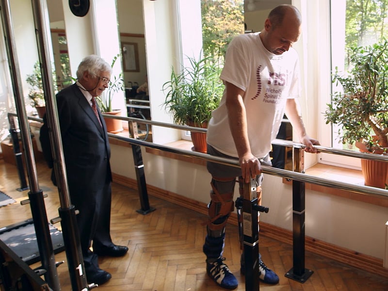
 Baby’s Pregnancy Calendar
 NEW learning series: Anatomy Dissected | Complete Anatomy