Orbital Pseudotumor
Granuloma, Plasma Cell
Pseudotumor Cerebri
Tolosa-Hunt Syndrome
Cranial Fossa, Middle
Orbital Myositis
Encyclopedias as Topic
Skin Diseases
Dermatology
Sex Factors
Sex Distribution
Orbital myositis and rheumatoid arthritis: case report. (1/32)
Orbital myositis implies orbital inflammation confined to one or more of the extraocular muscles. The acute form responds well to high doses of oral corticosteroids tapered gradually, but it may recur or become chronic. We describe a 38 years old female who has been suffering from rheumatoid arthritis for six years. She developed diplopia as a result of a paralysis of the right and left rectus medialis muscle. MRI showed inflammatory process and thickness of the referred muscles. The patient had a total recovery with oral use of 80 mg methylprednisolone daily. Two months after the first episode she developed a bilateral ophthalmoplegy. The patient improved with oral use of steroids the second time, but a paresis of the left rectus lateralis muscle remained. From the 156 cases we reviewed only three have been related to rheumatic diseases and none has been previously related to rheumatoid arthritis. (+info)A role for methotrexate in the management of non-infectious orbital inflammatory disease. (2/32)
AIM: To evaluate the clinical usefulness of methotrexate for patients with non-infectious orbital inflammatory disease who fail to respond to systemic corticosteroids and/or orbital irradiation. METHODS: The medical records of patients with non-infectious orbital inflammatory disease who were treated with methotrexate at Oregon Health Sciences University between June 1993 and June 2000 were examined. Methotrexate was administered at a median maximum dose of 20 mg per week (range 15-25 mg per week) in conjunction with folate supplementation. Patients were followed with regular ophthalmic examinations, as well as serum liver enzyme levels and blood cell counts. Clinical signs of regression of the orbital inflammation, visual acuity, dosage and duration of methotrexate therapy, requirement for concurrent corticosteroid administration, and adverse drug reactions were recorded. RESULTS: The study cohort included 14 patients (24 eyes) with diagnoses including non-specific orbital inflammation (n=7), Tolosa-Hunt syndrome (n=1), thyroid orbitopathy (n=3), Wegener's granulomatosis (n=1), sarcoidosis (n=1), and Erdheim-Chester disease (n=1). In all cases, methotrexate was commenced as a corticosteroid sparing agent. 10 patients (71%) completed a 4 month therapeutic trial of methotrexate. Median duration of treatment for the nine (64%) patients who experienced clinical benefit was 25 months (range 10-47 months). Six responders were ultimately able to cease methotrexate, including the single patient who required concurrent long term corticosteroid therapy. Complications included fatigue, gastrointestinal disturbance, hair thinning and mild, reversible serum liver enzyme elevation. Two patients (14%) discontinued treatment because of adverse effects. CONCLUSION: Methotrexate is a well tolerated immunosuppressive medication which may benefit patients with recalcitrant non-infectious orbital inflammatory disease. (+info)Orbital myositis in scleritis. (3/32)
AIMS: To investigate the association between scleritis and myositis. METHODS: Retrospective, non-comparative case series. Records and ultrasonograms were examined of 132 patients, with a diagnosis of episcleritis or scleritis, who attended the ophthalmology department at Leiden University Medical Center between 1997 and 2000. 103 were eligible for comprehensive examination. Medical records were evaluated. Ultrasonography was performed in all patients diagnosed with episcleritis or scleritis. Clinical features, precipitating factors, systemic associations, ocular complications, treatment, and outcome of each patient were assessed. RESULTS: Of the 103 patients, 27 (26.2%) had episcleritis and 76 (73.8%) had scleritis. Myositis was found to be present in 11 patients. It was present in 14.5% of all patients with scleritis and 30.5% of those in whom the posterior sclera was affected. The presence of the associated myositis did not worsen the visual prognosis and the presence of myositis was not associated with other systemic diseases. There were no cases of unilateral scleritis with bilateral orbital myositis. During an attack ocular complications were more common in patients with scleritis and myositis (64%) than in patients with scleritis alone (30.4%), indicating a more diffuse and potentially dangerous inflammation. There was no evidence that the inflammatory changes in the orbit had spread to involve the sclera, so it is assumed that the muscle changes are an extension of a generalised response to intense inflammation of the episclera and sclera. CONCLUSION: This study found a frequent association between myositis and scleritis. Prognosis for vision was not affected by coexistence of myositis. (+info)Technical modification in the intracarotid chemotherapy and osmotic blood-brain barrier disruption procedure to prevent the relapse of carboplatin-induced orbital pseudotumor. (4/32)
The blood-brain barrier disruption (BBBD) procedure is an established strategy to enhance drug delivery to brain tumors. Complication rates associated with this procedure are usually low, but when complications do occur, they usually mandate discontinuation of treatment. Orbital pseudotumor is an inflammatory condition of one or more extraocular muscles that produces limitation of ocular motility. Patients usually experience sudden diplopia associated with orbital pain, conjunctival chemosis and injection, and proptosis. Imaging of the orbit shows diffuse enlargement of the extraocular muscles, exophthalmia, and, rarely, sinusal or intracranial infiltration. On pathologic examinations, the soft tissues of the orbit are infiltrated with a mixture of eosinophils, lymphocytes, and plasma cells. Many etiologies can induce this syndrome, including the intracarotid infusion of platinum molecules. As part of a phase II study, a total of 110 patients were treated for malignant brain tumors with intra-arterial carboplatin, enhanced by the BBBD procedure, at the Sherbrooke University Hospital. Here we report on three patients who developed orbital pseudotumor ipsilateral to the carotid infused a few hours to days after the procedure. After the occurrence of this syndrome in the first patient, we developed a technical modification to the procedure that enabled uninterrupted treatment in the other two patients. This modification was as follows: after the mannitol infusion, and before carboplatin, the catheter was changed for a 3.5 tracker and was repositioned just above the emergence of the ophthalmic artery. (+info)Diplopia and periorbital mass associated with Miragel buckling explant. (5/32)
A 28-year-old female presented with a palpable mass lesion on the superonasal aspect of her right globe and she had a progressive diplopia. She had a scleral encircling surgery with a Miragel explant (MIRA, Waltham, Mass, USA) for the tractional retinal detachment associated with pars planitis 9 years previously. On examination, she revealed restricted eye movements of her right eye. The magnetic resonance imaging documented a swelling of the Miragel explant that mimicked a periorbital mass lesion. The Miragel explant was removed and fragmentation of the explant was found intraoperatively. The removed Miragel explant was examined by a scanning electron microscopy, and this demonstrated a disintergrated and swollen structural composition of the Miragel explant. Postoperatively, her extraocular movement was almost restored and the retina remained well attached. Alterations in the structural composition of the Miragel explant results in an excessive swelling that causes a restriction of the extraocular movement, and this can mimick a periorbital mass lesion. (+info)Bilateral idiopathic sclerosing inflammation of the orbit: report of three cases. (6/32)
We report 3 cases of bilateral orbital sclerosing inflammation and review the literatures concerning bilateral idiopathic orbital inflammation. We found that when idiopathic orbital inflammation presents as a bilaterally diffuse retrobulbar apical mass, the sclerosing subtype must be considered first, and an orbital biopsy should be performed. The present cases were characterized by chronic-onset, cicatricial inflammation with a mass effect. The most common ocular findings in our study were vision decrease, proptosis, and restriction of extraocular muscle movement. We found that current treatment, using systemic corticosteroids and radiotherapy, was nonspecific, incomplete, or frequently resulted in relapse. For cases refractory to corticosteroids and radiotherapy, early and aggressive multiagent immunosuppressive therapy should be used to minimize visual and ocular disabilities. (+info)5-fluorouracil as a phosensitiser. (7/32)
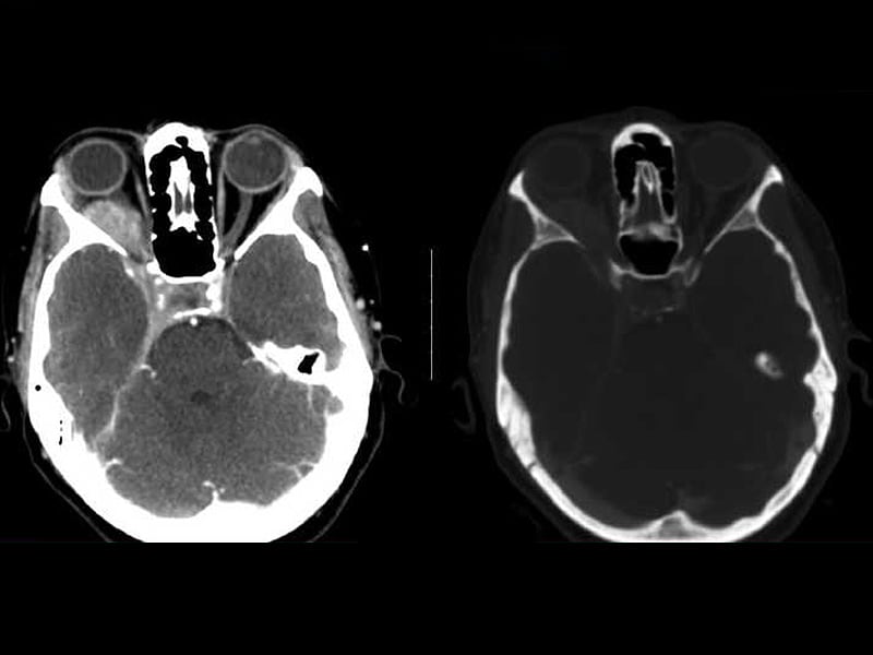
5-FU exhibits a high fluorescence after irradiation with UV-vis light. An enhancement of the cytostatic activity of 5-FU under UV-vis irradiation was observed on an in vivo experimental model. (+info)MR imaging of orbital inflammatory pseudotumors with extraorbital extension. (8/32)
OBJECTIVE: To demonstrate a variety of MR imaging findings of orbital inflammatory pseudotumors with extraorbital extension. MATERIALS AND METHODS: We retrospectively reviewed the MR features of five patients, who were diagnosed clinically and radiologically as having an orbital inflammatory pseudotumor with extraorbital extension. RESULTS: The types of orbital pseudotumors were a mass in the orbital apex (n = 3), diffuse form (n = 2), and myositis (n = 1). The extraorbital extension of the orbital pseudotumor passed through the superior orbital fissure in all cases, through the inferior orbital fissure in two cases, and through the optic canal in one case. The orbital lesions extended into the following areas: the cavernous sinus (n = 4), the middle cranial fossa (n = 4), Meckel's cave (n = 2), the petrous apex (n = 2), the clivus (n = 2), the pterygopalatine fossa and infratemporal fossa (n = 2), the foramen rotundum (n = 1), the paranasal sinus (n = 1), and the infraorbital foramen (n = 1). On MR imaging, the lesions appeared as an isosignal intensity with gray matter on the T1-weighted images, as a low signal intensity on the T2-weighted images and showed a marked enhancement on the post-gadoliniumdiethylene triamine pentaacetic acid (post-Gd-DTPA) T1-sequences. The symptoms of all of the patients improved when they were given high doses of steroids. Three of the five patients experienced a recurrence. CONCLUSION: MR imaging is useful for demonstrating the presence of a variety of extraorbital extensions of orbital inflammatory pseudotumors. (+info)Orbital pseudotumor, also known as orbital inflammatory syndrome or idiopathic orbital inflammation, is a non-specific term used to describe a group of conditions characterized by inflammation in the orbit (the bony cavity surrounding the eye) without any identifiable cause. It is not a true tumor, but rather an inflammatory reaction that can mimic the symptoms and signs of a tumor.
The condition can affect people of any age, although it is more common in middle-aged adults. The exact cause of orbital pseudotumor is unknown, but it is believed to be related to an abnormal immune response or inflammation triggered by various factors such as infections, trauma, or autoimmune disorders.
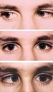
Symptoms of orbital pseudotumor may include eye pain, redness, swelling, protrusion of the eyeball (proptosis), double vision, and decreased vision. Diagnostic tests such as imaging studies (CT or MRI scans) and biopsy may be used to rule out other causes of orbital inflammation. Treatment typically involves corticosteroids to reduce inflammation, although other immunosuppressive medications may be necessary in severe cases. In some cases, the condition may resolve on its own without treatment.
A "Plasma Cell Granuloma" is a specific type of granulomatous inflammation that is characterized by the presence of numerous plasma cells. Plasma cells are white blood cells that produce antibodies, which are proteins that help the body fight off infections and diseases. In a Plasma Cell Granuloma, there is an excessive accumulation of these cells, leading to the formation of a nodular lesion or mass.
Plasma Cell Granulomas can occur in various organs, including the skin, lungs, gastrointestinal tract, and oral cavity. They are often associated with chronic inflammation, autoimmune disorders, or malignancies. The exact cause of Plasma Cell Granulomas is not always known, but they may be triggered by infections, foreign bodies, or other stimuli that induce an immune response.
Histologically, a Plasma Cell Granuloma is composed of a central area of plasma cells surrounded by a rim of lymphocytes and macrophages. The lesion may also contain multinucleated giant cells, eosinophils, and other inflammatory cells. Treatment options for Plasma Cell Granulomas depend on the location and extent of the lesion, as well as the underlying cause. Surgical excision is often curative, but medical therapy may be necessary in some cases.
Pseudotumor cerebri, also known as idiopathic intracranial hypertension, is a condition characterized by increased pressure around the brain without any identifiable cause such as a tumor or other space-occupying lesion. The symptoms mimic those of a brain mass, hence the term "pseudotumor."
The primary manifestation of this condition is headaches, often accompanied by vision changes like blurry vision, double vision, or temporary loss of vision, and pulsatile tinnitus (a rhythmic whooshing sound in the ears). Other symptoms can include neck pain, nausea, vomiting, and papilledema (swelling of the optic nerve disc). If left untreated, pseudotumor cerebri can lead to permanent vision loss.
The exact cause of pseudotumor cerebri remains unknown, but it has been associated with certain factors such as obesity, rapid weight gain, female gender (particularly during reproductive years), sleep apnea, and the use of certain medications like tetracyclines, vitamin A derivatives, and steroid withdrawal. Diagnosis typically involves a series of tests including neurological examination, imaging studies (such as MRI or CT scan), and lumbar puncture to measure cerebrospinal fluid pressure. Treatment usually focuses on lowering intracranial pressure through medications, weight loss, and sometimes surgical interventions like optic nerve sheath fenestration or shunting procedures.
Tolosa-Hunt syndrome is a rare disorder characterized by the inflammation of the nerve structures (including the fifth and sixth cranial nerves) within the cavernous sinus, a venous space near the base of the skull. This inflammation can lead to various symptoms such as:
1. Unilateral or bilateral orbital pain, which may be severe and deep, often radiating around the eye and temple.
2. Ophthalmoplegia (paralysis of the eye muscles), causing double vision (diplopia) and limited eye movement in specific directions.
3. Ptosis (drooping of the eyelid).
4. Other possible symptoms include decreased sensation around the forehead, cheek, or upper jaw, and loss of taste on the anterior part of the tongue.
The exact cause of Tolosa-Hunt syndrome is unknown, but it's believed to be related to an autoimmune response or a non-specific inflammatory process. It can also occur in conjunction with other medical conditions like neoplasms (tumors) or infections. The diagnosis typically involves imaging studies such as MRI and CT scans, along with blood tests and a thorough neurological examination.
Treatment usually includes corticosteroids to reduce inflammation and alleviate symptoms. In some cases, immunosuppressive medications or radiation therapy may be necessary. If left untreated, Tolosa-Hunt syndrome can lead to permanent visual impairment or other neurological deficits.
The middle cranial fossa is a depression or hollow in the skull that forms the upper and central portion of the cranial cavity. It is located between the anterior cranial fossa (which lies anteriorly) and the posterior cranial fossa (which lies posteriorly). The middle cranial fossa contains several important structures, including the temporal lobes of the brain, the pituitary gland, the optic chiasm, and the cavernous sinuses. It is also where many of the cranial nerves pass through on their way to the brain.
The middle cranial fossa can be further divided into two parts: the anterior and posterior fossae. The anterior fossa contains the optic chiasm and the pituitary gland, while the posterior fossa contains the temporal lobes of the brain and the cavernous sinuses.
The middle cranial fossa is formed by several bones of the skull, including the sphenoid bone, the temporal bone, and the parietal bone. The shape and size of the middle cranial fossa can vary from person to person, and abnormalities in its structure can be associated with various medical conditions, such as pituitary tumors or aneurysms.
Orbital diseases refer to a group of medical conditions that affect the orbit, which is the bony cavity in the skull that contains the eye, muscles, nerves, fat, and blood vessels. These diseases can cause various symptoms such as eyelid swelling, protrusion or displacement of the eyeball, double vision, pain, and limited extraocular muscle movement.
Orbital diseases can be broadly classified into inflammatory, infectious, neoplastic (benign or malignant), vascular, traumatic, and congenital categories. Some examples of orbital diseases include:
* Orbital cellulitis: a bacterial or fungal infection that causes swelling and inflammation in the orbit
* Graves' disease: an autoimmune disorder that affects the thyroid gland and can cause protrusion of the eyeballs (exophthalmos)
* Orbital tumors: benign or malignant growths that develop in the orbit, such as optic nerve gliomas, lacrimal gland tumors, and lymphomas
* Carotid-cavernous fistulas: abnormal connections between the carotid artery and cavernous sinus, leading to pulsatile proptosis and other symptoms
* Orbital fractures: breaks in the bones surrounding the orbit, often caused by trauma
* Congenital anomalies: structural abnormalities present at birth, such as craniofacial syndromes or dermoid cysts.
Proper diagnosis and management of orbital diseases require a multidisciplinary approach involving ophthalmologists, neurologists, radiologists, and other specialists.
Orbital myositis is a medical condition characterized by inflammation of the extraocular muscles, which are the muscles responsible for eye movement. These muscles are located within the orbit, the bony cavity that contains and protects the eye. Orbital myositis can cause symptoms such as painful eye movements, double vision, redness, swelling, and decreased visual acuity.
The condition is often associated with other systemic inflammatory or autoimmune disorders, such as rheumatoid arthritis, granulomatosis with polyangiitis (GPA), and sarcoidosis. However, it can also occur as an isolated phenomenon, known as idiopathic orbital myositis.
Diagnosis of orbital myositis typically involves a combination of clinical examination, imaging studies such as MRI or CT scans, and blood tests to evaluate for underlying systemic conditions. Treatment usually includes corticosteroids to reduce inflammation and alleviate symptoms, as well as addressing any underlying systemic disorders if present.
Orbital neoplasms refer to abnormal growths or tumors that develop in the orbit, which is the bony cavity that contains the eyeball, muscles, nerves, fat, and blood vessels. These neoplasms can be benign (non-cancerous) or malignant (cancerous), and they can arise from various types of cells within the orbit.
Orbital neoplasms can cause a variety of symptoms depending on their size, location, and rate of growth. Common symptoms include protrusion or displacement of the eyeball, double vision, limited eye movement, pain, swelling, and numbness in the face. In some cases, orbital neoplasms may not cause any noticeable symptoms, especially if they are small and slow-growing.
There are many different types of orbital neoplasms, including:
1. Optic nerve glioma: a rare tumor that arises from the optic nerve's supportive tissue.
2. Orbital meningioma: a tumor that originates from the membranes covering the brain and extends into the orbit.
3. Lacrimal gland tumors: benign or malignant growths that develop in the lacrimal gland, which produces tears.
4. Orbital lymphangioma: a non-cancerous tumor that arises from the lymphatic vessels in the orbit.
5. Rhabdomyosarcoma: a malignant tumor that develops from the skeletal muscle cells in the orbit.
6. Metastatic tumors: cancerous growths that spread to the orbit from other parts of the body, such as the breast, lung, or prostate.
The diagnosis and treatment of orbital neoplasms depend on several factors, including the type, size, location, and extent of the tumor. Imaging tests, such as CT scans and MRI, are often used to visualize the tumor and determine its extent. A biopsy may also be performed to confirm the diagnosis and determine the tumor's type and grade. Treatment options include surgery, radiation therapy, chemotherapy, or a combination of these approaches.
An encyclopedia is a comprehensive reference work containing articles on various topics, usually arranged in alphabetical order. In the context of medicine, a medical encyclopedia is a collection of articles that provide information about a wide range of medical topics, including diseases and conditions, treatments, tests, procedures, and anatomy and physiology. Medical encyclopedias may be published in print or electronic formats and are often used as a starting point for researching medical topics. They can provide reliable and accurate information on medical subjects, making them useful resources for healthcare professionals, students, and patients alike. Some well-known examples of medical encyclopedias include the Merck Manual and the Stedman's Medical Dictionary.
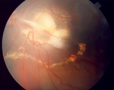
Eye diseases are a range of conditions that affect the eye or visual system, causing damage to vision and, in some cases, leading to blindness. These diseases can be categorized into various types, including:
1. Refractive errors: These include myopia (nearsightedness), hyperopia (farsightedness), astigmatism, and presbyopia, which affect the way light is focused on the retina and can usually be corrected with glasses or contact lenses.
2. Cataracts: A clouding of the lens inside the eye that leads to blurry vision, glare, and decreased contrast sensitivity. Cataract surgery is the most common treatment for this condition.
3. Glaucoma: A group of diseases characterized by increased pressure in the eye, leading to damage to the optic nerve and potential blindness if left untreated. Treatment includes medications, laser therapy, or surgery.
4. Age-related macular degeneration (AMD): A progressive condition that affects the central part of the retina called the macula, causing blurry vision and, in advanced stages, loss of central vision. Treatment may include anti-VEGF injections, laser therapy, or nutritional supplements.
5. Diabetic retinopathy: A complication of diabetes that affects the blood vessels in the retina, leading to bleeding, leakage, and potential blindness if left untreated. Treatment includes laser therapy, anti-VEGF injections, or surgery.
6. Retinal detachment: A separation of the retina from its underlying tissue, which can lead to vision loss if not treated promptly with surgery.
7. Amblyopia (lazy eye): A condition where one eye does not develop normal vision, often due to a misalignment or refractive error in childhood. Treatment includes correcting the underlying problem and encouraging the use of the weaker eye through patching or other methods.
8. Strabismus (crossed eyes): A misalignment of the eyes that can lead to amblyopia if not treated promptly with surgery, glasses, or other methods.
9. Corneal diseases: Conditions that affect the transparent outer layer of the eye, such as keratoconus, Fuchs' dystrophy, and infectious keratitis, which can lead to vision loss if not treated promptly.
10. Uveitis: Inflammation of the middle layer of the eye, which can cause vision loss if not treated promptly with anti-inflammatory medications or surgery.
Skin diseases, also known as dermatological conditions, refer to any medical condition that affects the skin, which is the largest organ of the human body. These diseases can affect the skin's function, appearance, or overall health. They can be caused by various factors, including genetics, infections, allergies, environmental factors, and aging.
Skin diseases can present in many different forms, such as rashes, blisters, sores, discolorations, growths, or changes in texture. Some common examples of skin diseases include acne, eczema, psoriasis, dermatitis, fungal infections, viral infections, bacterial infections, and skin cancer.
The symptoms and severity of skin diseases can vary widely depending on the specific condition and individual factors. Some skin diseases are mild and can be treated with over-the-counter medications or topical creams, while others may require more intensive treatments such as prescription medications, light therapy, or even surgery.
It is important to seek medical attention if you experience any unusual or persistent changes in your skin, as some skin diseases can be serious or indicative of other underlying health conditions. A dermatologist is a medical doctor who specializes in the diagnosis and treatment of skin diseases.
Dermatology is a medical specialty that focuses on the diagnosis, treatment, and prevention of diseases and conditions related to the skin, hair, nails, and mucous membranes. A dermatologist is a medical doctor who has completed specialized training in this field. They are qualified to treat a wide range of skin conditions, including acne, eczema, psoriasis, skin cancer, and many others. Dermatologists may also perform cosmetic procedures to improve the appearance of the skin or to treat signs of aging.
Dermatologic agents are medications, chemicals, or other substances that are applied to the skin (dermis) for therapeutic or cosmetic purposes. They can be used to treat various skin conditions such as acne, eczema, psoriasis, fungal infections, and wounds. Dermatologic agents include topical corticosteroids, antibiotics, antifungals, retinoids, benzoyl peroxide, salicylic acid, and many others. They can come in various forms such as creams, ointments, gels, lotions, solutions, and patches. It is important to follow the instructions for use carefully to ensure safety and effectiveness.
"Sex factors" is a term used in medicine and epidemiology to refer to the differences in disease incidence, prevalence, or response to treatment that are observed between males and females. These differences can be attributed to biological differences such as genetics, hormones, and anatomy, as well as social and cultural factors related to gender.
For example, some conditions such as autoimmune diseases, depression, and osteoporosis are more common in women, while others such as cardiovascular disease and certain types of cancer are more prevalent in men. Additionally, sex differences have been observed in the effectiveness and side effects of various medications and treatments.
It is important to consider sex factors in medical research and clinical practice to ensure that patients receive appropriate and effective care.
"Sex distribution" is a term used to describe the number of males and females in a study population or sample. It can be presented as a simple count, a percentage, or a ratio. This information is often used in research to identify any differences in health outcomes, disease prevalence, or response to treatment between males and females. Additionally, understanding sex distribution can help researchers ensure that their studies are representative of the general population and can inform the design of future studies.
Mikulicz disease is a rare condition characterized by the symmetrical enlargement of the salivary and lacrimal glands. It is named after Jan Mikulicz-Radecki, a Polish surgeon who first described it in 1892. The enlarged glands are typically painless, and the condition can be associated with other systemic diseases such as Sjogren's syndrome, sarcoidosis, lymphoma, and tuberculosis.
In Mikulicz disease, there is a benign infiltration of the salivary and lacrimal glands with immune cells, particularly lymphocytes, which can lead to their enlargement. The exact cause of the condition is not known, but it is thought to be related to an autoimmune response.
Mikulicz disease is often treated with medications that suppress the immune system, such as corticosteroids or immunosuppressive drugs. In some cases, surgical removal of the affected glands may be necessary. The prognosis for Mikulicz disease is generally good, but it can vary depending on the underlying cause and any associated medical conditions.
 Idiopathic orbital inflammatory disease
Dacryoadenitis
Jeffrey W. Berger
Tenonitis
Exposure keratopathy
Exophthalmos
Trochleitis
Tenon's capsule
Optic neuropathy
Tolosa-Hunt syndrome
Fibromatosis
IgG4-related ophthalmic disease
Trochlear nerve
Jerry A. Shields
Papilledema
Abducens nerve
Epstein-Barr virus-associated lymphoproliferative diseases
 The Role of Radiotherapy in Orbital Pseudotumor: A Systematic Review of Literature
 특발성 경화성 안와염증, 안와의 경화성 가성종양, Idiopathic Sclerosing Orbital Inflammation, Sclerosing Pseudotumor of Orbit
Idiopathic orbital inflammatory disease - Wikipedia
 Turmeric and Health: The Latest Research | NutritionFacts.org
 Optic Neuritis Clinical Guide | Cleveland Clinic
![Moseley I[au] - Search Results - PubMed](data:image/png;base64,iVBORw0KGgoAAAANSUhEUgAAABAAAAAQCAMAAAAoLQ9TAAAARVBMVEVHcEwoU45gYmYAUpQAUpRPYGVgYmZLXnJgYmYAUZUAUpRJXnIAUpQAUpRgYmYAUpRgYmZgYmZhYmYAUpQAUpQAUpRgYmaDiPJuAAAAFXRSTlMADOJ+6QewGO8/uTRqtH7GdFJ11p1bCL3TAAAAZUlEQVQYlV2PVw7AIAxDTeney7n/UcsoldX3E+VJOAboEi7MBpHWMs1ADlG8u7UYWauwyZFeRQVPOhG2o+aiwhByJxUx91Jxhje3iJSqGfHuLKI0+0TpXvY1twCOPlFh5pa/++MB0vIOBm+1zaoAAAAASUVORK5CYII=) Moseley I[au] - Search Results - PubMed
Moseley I[au] - Search Results - PubMed
 IgG4 as a Biomarker in Graves' Orbitopathy
 Lacrimal Gland Tumors: Background, Epidemiology
 Around the Eye in 365 Days - SLACK Books
 EyeRounds.org: Emily Birkholz, MD
 Inflammation of the Orbit - Eye Disorders - MSD Manual Consumer Version
 GenElute Blood Genomic DNA Kit sufficient for 70 purifications Gen Elute
 Bioline International Official Site (site up-dated regularly)
Ocular Lymphoma: ACRONYMS, Overview, Epidemiology
 Immunomodulatory Therapy (IMT) for Ocular Inflammation - EyeWiki
 A dermatologic riddle: How is IgG4-related ophthalmic disease parliamentary?
 Toddler With Progressive Proptosis From Acute Myelogenous Leukemia
 Glucocorticoid - Induced Osteoporosis: Considerations in Ophthalmology - Uveitis.org | OIUF
Vasculitis: Vessel Size Matters
 Jeanmarie Perrone, MD | Children's Hospital of Philadelphia
 turmeric - Total Health Magazine
Granulomatosis with Polyangiitis (GPA) - Musculoskeletal and Connective Tissue Disorders - Merck Manuals Professional Edition
 Concomitant Intraocular and Orbital Space-Occupied Lesions
 Optic Nerve Decompression Surgery - Medical Clinical Policy Bulletins | Aetna
 Opthamology Data (1971-75)
Cataflam Causing Nausea - Pseudotumor Cerebri
Clinical Profile of Unilateral Proptosis in a Tertiary Care Centre
 Diagnostic Features Of Feline Restrictive Orbital Myofibroblastic Sarcoma | Advanced Veterinary Medical Imaging
Orbit14
- Orbital pseudotumor is the swelling of tissue behind the eye in an area called the orbit. (medlineplus.gov)
- Idiopathic orbital inflammatory (IOI) disease refers to a marginated mass-like enhancing soft tissue involving any area of the orbit. (wikipedia.org)
- Inflammation affecting any or all parts of the orbit is called inflammatory orbital pseudotumor (which is not really a tumor and is not a cancer) or nonspecific orbital inflammation. (msdmanuals.com)
- Is the Tolosa-Hunt syndrome comparable to the pseudotumor of the orbit? (nih.gov)
- Orbital and retro-orbital pain are relatively common clinical conditions that are associated with such disorders as trigeminal, lacrimal, and ciliary neuralgia, cluster headaches, paroxysmal hemicrania, inflammatory orbital pseudotumor, trochleitis, and herpetic neuralgia ophthalmicus, thus making the nerves supplying the orbit of great clinical importance. (ouchuk.org)
- An understanding of the variability and frequency of these neural connections could lead to safer surgical procedures of the orbit and effective treatments for patients with orbital pain. (ouchuk.org)
- Idiopathic orbital inflammatory disease "pseudotumor" is a nonspecific inflammation involving the orbit. (drvladimirkratky.com)
- The orbit , which protects, supports, and maximizes the function of the eye, is shaped like a quadrilateral pyramid, with its base in plane with the orbital rim. (medscape.com)
- The superficial bony orbit is defined by the orbital margin, which is rectangular with rounded corners. (medscape.com)
- This image of the right orbit shows superficial landmarks, optic canal, and superior and inferior orbital fissures. (medscape.com)
- The greater wing of the sphenoid, the maxilla, and the palatine bones of the orbit form the boundaries of the inferior orbital fissure. (medscape.com)
- The infraorbital sulcus crosses the floor of the orbit and carries the infraorbital artery, infraorbital vein, and infraorbital nerve from the inferior orbital fissure to the infraorbital foramen. (medscape.com)
- Orbital dermoids are common lesions noted of the anterior orbit in children. (entokey.com)
- Inflammation and thickening of the sclera seen in thyroid eye disease, inflammatory pseudotumour of the orbit and rheumatoid posterior scleritis may also cause choroidal folds [6]. (uk.com)
Lacrimal gland7
- Orbital magnetic resonance imaging revealed left lacrimal gland enlargement with homogeneous contrast enhancement and diffuse mild enlargement of the left lateral and superior rectus muscles. (nih.gov)
- The lesion involved the lacrimal gland and orbital soft tissue in all with extraocular muscle involvement also in three cases. (uk.com)
- It can range from a diffuse inflammatory process to a more localized inflammation of muscle, lacrimal gland or orbital fat. (wikipedia.org)
- They include inflammation of the extraocular muscles (myositis) with tendinous involvement, orbital fat stranding, lacrimal gland inflammation and enlargement (dacryoadenitis), involvement of the optic sheath complex, uvea, and sclera, a focal intraorbital mass or even diffuse orbital involvement. (wikipedia.org)
- The 2 lobes of the lacrimal gland, the orbital lobe and the much smaller palpebral lobe, are separated anatomically by the lateral horn of the levator aponeurosis. (medscape.com)
- Malignant epithelial neoplasms of the lacrimal gland account for approximately 2% of all orbital neoplasms. (medscape.com)
- Tumours commonly associated with choroidal folds include mucoceles, dermoids, lacrimal gland tumours and orbital meningiomas [5]. (uk.com)
Cases of orbital pseudotumor2
- Severe cases of orbital pseudotumor may push the eye forward so much that the lids cannot cover and protect the cornea. (medlineplus.gov)
- Trauma has also been seen to precede some cases of orbital pseudotumor. (wikipedia.org)
MRNA COVID-19 vaccination2
- Yucel Gencoglu A, Mangan MS. Orbital inflammatory pseudotumor following mRNA COVID-19 vaccination. (medlineplus.gov)
- The authors present a case of orbital pseudotumor after mRNA COVID-19 vaccination. (nih.gov)
Granulomatosis with polyangiitis2
- A differential diagnosis includes lymphoproliferative lesions, thyroid ophthalmopathy, IgG4-related ophthalmic disease, sarcoidosis, granulomatosis with polyangiitis, orbital cellulitis and carotid-cavernous fistula. (wikipedia.org)
- IgG4-related orbital inflammation can affect the same structures as granulomatosis with polyangiitis but typically has fewer symptoms. (msdmanuals.com)
Inflammatory syndrome3
- Idiopathic orbital inflammatory syndrome, also known as orbital pseudotumor, was first described by Gleason in 1903 and by Busse and Hochheim. (wikipedia.org)
- Overall, radiographic features for idiopathic orbital inflammatory syndrome vary widely. (wikipedia.org)
- Nonspecific Orbital best dark web marketplaces 2023 Inflammation (Idiopathic Orbital Inflammation, Orbital Inflammatory Syndrome, Orbital Pseudotumor). (darkwebmarketme.com)
Dacryoadenitis1
- Orbital pseudotumors are often associated with inflammation of the extraocular muscles ( ORBITAL MYOSITIS ) or inflammation of the lacrimal glands ( DACRYOADENITIS ). (nih.gov)
Cellulitis5
- Orbital cellulitis is an infection of the fat and muscles around the eye. (nih.gov)
- Orbital cellulitis is a dangerous infection, which can cause lasting problems. (nih.gov)
- Both preseptal cellulitis and orbital. (msdmanuals.com)
- Fezza JChaudhry IAKwon YHGrannum EESinard JWolfley DE Orbital melanoma presenting as orbital cellulitis: a clinicopathologic report. (jamanetwork.com)
- Infectious causes of proptosis may also be acute (e.g., bacterial orbital cellulitis) or chronic (e.g., parasitic cyst). (entokey.com)
Systemic7
- Systemic corticosteroids were started for the orbital pseudotumor. (nih.gov)
- The authors describe four children in whom idiopathic orbital pseudotumor (IOP) was the initial solitary finding with systemic inflammatory disease developing later. (uk.com)
- Idiopathic orbital pseudotumour preceding systemic inflammatory disease in children. (uk.com)
- It is a benign, nongranulomatous orbital inflammatory process characterized by extraocular orbital and adnexal inflammation with no known local or systemic cause. (wikipedia.org)
- Orbital pseudotumor has also been observed in association with Crohn's disease, systemic lupus erythematosus, rheumatoid arthritis, diabetes mellitus, myasthenia gravis, and ankylosing spondylitis all of which strengthen the basis of IOI being an immune-mediated disease. (wikipedia.org)
- In the latter, if there is a lymphangiomatous component, compression may only occur when infection, for example, an upper respiratory viral systemic infection, results in temporary swelling of the orbital lesion. (entokey.com)
- Orbital and adnexal lymphoma is associated with systemic lymphoma in 30-35% of cases. (medscape.com)
Superior orbital2
- Why is the process confined to the superior orbital fissure? (nih.gov)
- The superior orbital fissure is bounded by the lesser and greater wings of the sphenoid. (medscape.com)
Idiopathic orbital inflammatory2
- The best imaging modality for idiopathic orbital inflammatory disease is contrast-enhanced thin section magnetic resonance with fat suppression. (wikipedia.org)
- To characterize the clinical and pathological features of 4 patients with histopathology-confirmed idiopathic orbital inflammatory disease (OID) initially diagnosed as an orbital neoplasm and 9 patients with histopathology-confirmed orbital neoplasm that presented as idiopathic OID. (donsantiagomd.com)
Extraocular1
- A massive area of extraocular extension forms a contiguous orbital mass. (jamanetwork.com)
Adnexal1
- The median age at presentation for orbital and adnexal lymphoma is older than 60 years. (medscape.com)
Tissue4
- The presented case of orbital pseudotumor development after the mRNA vaccine may be considered to be an immunological process targeting the orbital tissue following immunization, although the cause-effect relationship remains uncertain. (nih.gov)
- Idiopathic orbital inflammation has a varied clinical presentation depending on the involved tissue. (wikipedia.org)
- In the setting of extensive sclerosis there may be restriction, compression, and destruction of orbital tissue. (wikipedia.org)
- Tumor of virtually any orbital tissue can cause optic nerve compression. (entokey.com)
Malignancy2
- Histopathology and immunohistochemistry of the orbital masses revealed malignancy in 80% (7/9) of these cases. (donsantiagomd.com)
- Biopsy is recommended when there is poor or equivocal response to steroids or suspicion of orbital malignancy. (donsantiagomd.com)
Anatomy1
- He is co-author of the book MRI and CT, Clinical Neuro-Orbital Anatomy, published by the American Academy of Ophthalmology. (dentalimplantsroc.com)
Neoplasms3
- A detailed history, comprehensive physical examination, and appropriate radiological evaluation are essential to differentiate OID and non-inflammatory orbital conditions such as neoplasms. (donsantiagomd.com)
- This article presents a case series (19 orbits from 18 patients) of lateral orbitotomies for excision biopsy of orbital neoplasms, over a 10-year period (from September 2001 to October 2011). (donsantiagomd.com)
- Intraconal orbital tumours such as cavernous hemangiomas, metastatic neoplasms and optic nerve meningiomas, can press on the globe producing exophthalmos, flattening of the globe and shifting of the refractive error towards hyperopia [4]. (uk.com)
Diagnosis of orbital1
- There were 4 patients in the histopathology-confirmed idiopathic OID group with preoperative diagnosis of orbital neoplasm. (donsantiagomd.com)
Patients with orbital1
- The medical records of 13 patients with orbital mass were reviewed. (donsantiagomd.com)
Fibrous1
- These are called septum and include the fibrous orbital septum and tarsi. (eyelidsbybrown.com)
Neoplasm2
- Its former name, orbital pseudotumor, is derived due to resemblance to a neoplasm. (wikipedia.org)
- In the histopathology-confirmed orbital neoplasm with preoperative diagnosis of idiopathic OID group, there were 9 patients with mean age at presentation of 52 years. (donsantiagomd.com)
Inferior orbital1
- The left image shows a child at baseline with an inferior orbital mass. (entokey.com)
Malignant3
- Levine RAPutterman AMKorey MS Recurrent orbital malignant melanoma after the evisceration of an unsuspected choroidal melanoma. (jamanetwork.com)
- Starr HJZimmerman LE Extrascleral extension and orbital recurrence of malignant melanomas of the choroid and ciliary body. (jamanetwork.com)
- Rose GEHoh HBHarrad RAHungerford JL Intraocular malignant melanomas presenting with orbital inflammation. (jamanetwork.com)
Histopathology1
- The histopathology of idiopathic orbital inflammation is described as nondiagnostic and diverse. (wikipedia.org)
Surgeon1
- Knowledge of these distances safely guides the surgeon along the medial orbital wall. (medscape.com)
Tumours2
- Orbital pain , particularly in rapidly growing tumours. (symptoma.com)
- Choroidal folds and refractive errors associated with orbital tumours. (uk.com)
Uveitis1
- It is the most common painful orbital mass in the adult population, and is associated with proptosis, cranial nerve palsy (Tolosa-Hunt syndrome), uveitis, and retinal detachment. (wikipedia.org)
Choroidal1
- Tabassian AZuravleff JJ Necrotic choroidal melanoma with orbital inflammation. (jamanetwork.com)
Lateral1
- No patient needed the use of miniplate hardware in repositioning the lateral orbital wall nor complained of a palpable deformity of the lateral orbital wall. (donsantiagomd.com)
Disease3
- People with this condition need regular follow-up care with an eye doctor who is familiar with the treatment of orbital disease. (medlineplus.gov)
- Thus, disease processes that solely affect the orbital lobe may not manifest until later in the course of the illness. (medscape.com)
- Acute inflammatory disease includes pseudotumor. (entokey.com)
Infections1
- Several studies have described cases where onset of orbital pseudotumor was seen simultaneously or several weeks after upper respiratory infections. (wikipedia.org)
Infection1
- Orbital infection and inflammation. (medlineplus.gov)
Nerves1
- In this project, we reviewed all of the reported connections between the orbital nerves and V1 in order to understand how pain from this region is transmitted to the brain. (ouchuk.org)
Symptoms2
- 11. Metastatic Orbital Tumor From Breast Ductal Carcinoma With Neuroendocrine Differentiation Initially Presenting as Ocular Symptoms: A Case Report and Literature Review. (nih.gov)
- The presenting symptoms and signs included proptosis (4/9), inflammation (3/9), orbital pain (1/9), and epiphora (1/9). (donsantiagomd.com)
Proptosis1
- Proptosis occurs when there is an increase in retrobulbar orbital volume. (entokey.com)