Osteoblastoma
Osteoma, Osteoid
Spinal Neoplasms
Hamate Bone
Curettage
Frontal Sinus
Osteoblastoma of dorsal spine: a case report. (1/52)
A case of benign osteoblastoma affecting posterior element of spine with pain and paraplegia in a female is being presented with brief review of literature. Early diagnosis and surgical excision remains the mainstay of treatment. (+info)Benign osteoblastoma mimicking malignancy of the spine. (2/52)
A case of osteoblastoma of the spine in a sixty year old female is presented. These lesions usually get misdiagnosed as tuberculosis or malignancy of the spine. Salient diagnostic features have been discussed. (+info)Skeletal changes in rats given daily subcutaneous injections of recombinant human parathyroid hormone (1-34) for 2 years and relevance to human safety. (3/52)
Fischer 344 rats (60/sex/group) were given daily subcutaneous injections of recombinant human parathyroid hormone (PTH)(1-34) for 2 years at doses of 0, 5, 30, or 75 microg/kg. Treatment caused substantial increases in bone mass consistent with the known pharmacologic effects of once-daily administration. As determined by quantitative computed tomography (QCT) and histomorphometry, bone mass was markedly increased. Substantial new bone formation resulted in a large decrease in marrow space accompanied by altered bone architecture. Bone proliferative lesions were observed in all PTH( 1-34)-treated groups. Osteosarcoma occurred in 3, 21, and 31 male rats and in 4, 12, and 23 female rats in the 5-, 30-, and 75-microg/kg treatment groups, respectively. Focal osteoblast hyperplasia, osteoma, and osteoblastoma were much less frequent. Although the specific cellular or molecular mechanisms responsible for the rat bone tumors have not been fully elucidated, the data suggest that these lesions resulted from the long duration of treatment and the exaggerated pharmacologic response of the rat skeleton to daily treatment with PTH(1-34). Important differences between the rat study and clinical use in adult humans suggest that the increased incidence of bone neoplasia in rats treated for 2 years is likely not predictive of an increased risk of bone cancer in skeletally mature adult humans being given PTH(1-34) for a limited period of time in the treatment of osteoporosis. (+info)MR imaging features of pigmented villonodular synovitis of the cervical spine. (4/52)
Pigmented villonodular synovitis (PVNS) is a benign proliferative disorder primarily occurring in the large joints of the appendicular skeleton such as the knee and hip joints. We present an unusual case of PVNS involving the cervical spine in an adult. MR imaging showed an enhancing mass lesion arising from the posterior elements of the cervical spine and hyperintensity on the T2-weighted images, without evidence of T2 susceptibility effects. Gross total excision of the tumor was performed, and the diagnosis was established by histopathology. (+info)Progression of a lumbar spinal osteoblastoma. (5/52)
A 24-year-old woman presented with a lumbar spinal osteoblastoma manifesting as a 5-year history of low back pain radiating to the left foot. Neuroimaging showed suspicious hypertrophy of the left L4-5 facet which transformed in 3 years to an expansile mass lesion that compressed the dura mater and neural structures. Primary benign bone tumors such as osteoblastoma and osteoid osteoma should be considered in the differential diagnosis of back pain and the patients should be followed up carefully. (+info)Bisphosphonates for treatment and prevention of bone metastases. (6/52)
Bone metastases are a major cause of morbidity for men with prostate cancer. Complications of bone metastases include pain, fractures, and spinal cord compression. Although they appear osteoblastic by radiographic imaging, most bone metastases are characterized by excess osteoclast number and activity. In addition, pathologic osteoclast activation is associated with increased risk of skeletal complications. Zoledronic acid, a potent inhibitor of osteoclast activity, differentiation, and survival, decreases the risk of skeletal complications in men with androgen-independent prostate cancer and bone metastases. Other bisphosphonates, including pamidronate and clodronate, seem to be ineffective in this setting. The reduction in risk of skeletal complications with zoledronic acid must be weighed against potential adverse effects. Additional studies are needed to determine the optimal timing, schedule, and duration of treatment in men with bone metastases as well as the potential role of bisphosphonates in other settings including the prevention of bone metastases. (+info)A complementary method for the detection of osteoblastic metastases on digitized radiographs. (7/52)
PURPOSE: This study was conducted to evaluate the diagnostic usefulness of gray level parameters in order to distinguish healthy bone from osteoblastic metastases on digitized radiographs. MATERIALS AND METHODS: Skeletal radiographs of healthy bone (n = 144) and osteoblastic metastases (n = 35) were digitized using pixels 0.175 mm in size and 4,096 gray levels. We obtained an optimized healthy bone classification to compare with pathological bone: cortical, trabecular, and flat bone. The osteoblastic metastases (OM) were classified in nonflat and flat bone. These radiological images were analyzed by using a computerized method. The parameters (gray scale) calculated were: mean, standard deviation, and coefficient of variation (MGL, SDGL, and CVGL, respectively) based on gray level histogram analysis. Diagnostic utility was quantified by measurement of parameters on healthy and pathological bone, yielding quantification of area under the receiver operating characteristic (ROC) curve, AUC. RESULTS: All three image parameters showed high and significant values of AUC when comparing healthy trabecular bone and nonflat bone OM, showing MGL the best discriminatory ability (0.97). As for flat bones, MGL showed no ability to distinguish between healthy and flat bone OM (0.50). This could be achieved by using SDGL or CVGL, with both showing a similar diagnostic ability (0.85 and 0.83, respectively). CONCLUSION: Our results show that the use of gray level parameters quantify healthy bone and osteoblastic metastases zones on digitized radiographs. This may be helpful as a complementary method for differential diagnosis. Moreover, our method will allow us to study the evolution of osteoblastic metastases under medical treatment. (+info)Utility of immunohistochemical analysis for cyclo-oxygenase 2 in the differential diagnosis of osteoblastoma and osteosarcoma. (8/52)
AIMS: To study the immunoexpression of cyclo-oxygenase (COX) 2 in osteoblastomas (OBs) and osteosarcomas (OSs), and to assess the utility of immunohistochemical analysis for COX 2 in the differential diagnosis of the two tumour forms. METHODS: The immunohistochemical features of COX 2 were studied in 11 OBs and 30 OSs, including 26 high-grade OSs (16 osteoblastic, 7 chondroblastic, and 3 fibroblastic) and 4 low-grade OSs. RESULTS: Tumour cells from all 11 OBs unequivocally showed diffuse, intense and cytoplasmic immunoreactivity for COX 2. Strong cytoplasmic expression of COX 2 was observed in 5 of 26 (19%) high-grade OSs, all chondroblastic. In one osteoblastic-type OS, COX 2 was expressed in the chondroblastic component, but this tumour was considered to be COX 2 negative. No COX 2 expression was noted in atypical osteoblastic cells. Staining in the four low-grade OSs was negative. CONCLUSION: The results of immunohistochemical analysis of COX 2 suggest that in addition to the routine histopathological evaluation, COX 2 is a valuable diagnostic marker in the distinction between OB and OS. (+info)Osteoblastoma is a rare, benign (non-cancerous) bone tumor that originates from osteoblasts, which are cells responsible for bone formation. It typically affects children and young adults, with around two-thirds of cases occurring in individuals under 30 years old.
Osteoblastomas usually develop in the long bones of the body, such as the femur (thigh bone) or tibia (shin bone), but they can also occur in the vertebrae of the spine. The tumor tends to grow slowly and may cause symptoms like pain, swelling, or tenderness in the affected area. In some cases, it can lead to pathological fractures (fractures caused by weakened bone structure).
While osteoblastomas are generally not life-threatening, they can be locally aggressive and cause significant morbidity if left untreated. Treatment typically involves surgical removal of the tumor, followed by curettage (scraping) and bone grafting to fill the void created by the tumor excision. In some cases, adjuvant therapies like cryosurgery or radiation therapy may be used to ensure complete tumor eradication.
Osteoma is a benign bone tumor that usually develops on the surface of the bone and is composed of mature lamellar bone. On the other hand, osteoid osteoma is a type of benign bone-forming tumor that is made up of osteoid tissue (immature bone) and is surrounded by a highly vascularized fibrous connective tissue.
Osteoid osteomas are typically smaller than osteomas and can cause significant pain, especially at night, which can be relieved with the use of nonsteroidal anti-inflammatory drugs (NSAIDs). They usually affect young people, particularly males under 30 years old, and commonly involve the long bones of the lower extremities.
While osteomas are generally asymptomatic and do not require treatment unless they cause functional or aesthetic problems, osteoid osteomas may require surgical intervention to alleviate pain and prevent potential complications such as bone deformity or fracture.
The trapezoid bone is a carpal bone located in the wrist, more specifically in the proximal row of carpals. It is situated between the trapezium bone (also known as the greater multangular bone) and the capitate bone, and articulates proximally with the scaphoid bone. The trapezoid bone has a quadrilateral shape, with its lateral surface being convex and articulating with the trapezium, while its medial surface is concave and articulates with the capitate. Its distal surface articulates with the second metacarpal bone. This bone plays an essential role in wrist movements and stability.
The triquetral bone, also known as the triquetrum, is one of the eight carpal bones in the human wrist. It is located on the ulnar side of the wrist and articulates with the lunate bone proximally, the pisiform bone distally, and the hamate bone medially. The triquetral bone has a pyramidal shape and plays an essential role in wrist movements, particularly in pronation and supination. It is named "triquetral" because of its three articular facets, which create a triangular shape.
Spinal neoplasms refer to abnormal growths or tumors found within the spinal column, which can be benign (non-cancerous) or malignant (cancerous). These tumors can originate in the spine itself, called primary spinal neoplasms, or they can spread to the spine from other parts of the body, known as secondary or metastatic spinal neoplasms. Spinal neoplasms can cause various symptoms, such as back pain, neurological deficits, and even paralysis, depending on their location and size. Early diagnosis and treatment are crucial to prevent or minimize long-term complications and improve the patient's prognosis.
Bone neoplasms are abnormal growths or tumors that develop in the bone. They can be benign (non-cancerous) or malignant (cancerous). Benign bone neoplasms do not spread to other parts of the body and are rarely a threat to life, although they may cause problems if they grow large enough to press on surrounding tissues or cause fractures. Malignant bone neoplasms, on the other hand, can invade and destroy nearby tissue and may spread (metastasize) to other parts of the body.
There are many different types of bone neoplasms, including:
1. Osteochondroma - a benign tumor that develops from cartilage and bone
2. Enchondroma - a benign tumor that forms in the cartilage that lines the inside of the bones
3. Chondrosarcoma - a malignant tumor that develops from cartilage
4. Osteosarcoma - a malignant tumor that develops from bone cells
5. Ewing sarcoma - a malignant tumor that develops in the bones or soft tissues around the bones
6. Giant cell tumor of bone - a benign or occasionally malignant tumor that develops from bone tissue
7. Fibrosarcoma - a malignant tumor that develops from fibrous tissue in the bone
The symptoms of bone neoplasms vary depending on the type, size, and location of the tumor. They may include pain, swelling, stiffness, fractures, or limited mobility. Treatment options depend on the type and stage of the tumor but may include surgery, radiation therapy, chemotherapy, or a combination of these treatments.
The hamate bone is one of the eight carpal bones located in the wrist. It is shaped like a hook and is situated on the medial side of the distal row of carpals, near the pisiform bone. The hamate bone plays an essential role in the function of the wrist joint, providing attachment sites for various muscles, ligaments, and tendons that contribute to hand and finger movements. Its unique shape also forms part of the Guyon's canal, through which the ulnar nerve and artery pass into the hand. Injuries to the hamate bone can significantly impact grip strength and overall hand function.
The mastoid is a term used in anatomy and refers to the bony prominence located at the base of the skull, posterior to the ear. More specifically, it's part of the temporal bone, one of the bones that forms the side and base of the skull. The mastoid process provides attachment for various muscles involved in chewing and moving the head.
In a medical context, "mastoid" can also refer to conditions or procedures related to this area. For example, mastoiditis is an infection of the mastoid process, while a mastoidectomy is a surgical procedure that involves removing part or all of the mastoid process.
The metacarpus is the medical term for the part of the hand located between the carpus (wrist) and the digits (fingers). It consists of five bones, known as the metacarpal bones, which are numbered 1 to 5 from the thumb side to the little finger side. Each metacarpal bone has a base, a shaft, and a head. The bases of the metacarpal bones articulate with the carpal bones to form the wrist joint, while the heads of the metacarpal bones form the knuckles at the back of the hand.
The metacarpus plays an essential role in hand function as it provides stability and support for the movement of the fingers and thumb. Injuries or conditions affecting the metacarpus can significantly impact hand function, causing pain, stiffness, weakness, or deformity.
Maxillary neoplasms refer to abnormal growths or tumors in the maxilla, which is the upper jaw bone. These growths can be benign (non-cancerous) or malignant (cancerous). Benign neoplasms are slow-growing and do not spread to other parts of the body, while malignant neoplasms can invade surrounding tissues and spread to distant sites.
Maxillary neoplasms can cause various symptoms such as swelling, pain, numbness, loose teeth, or difficulty in chewing or swallowing. They may also cause nasal congestion, nosebleeds, or visual changes if they affect the eye or orbit. The diagnosis of maxillary neoplasms usually involves a combination of clinical examination, imaging studies such as CT or MRI scans, and biopsy to determine the type and extent of the tumor.
Treatment options for maxillary neoplasms depend on several factors, including the type, size, location, and stage of the tumor, as well as the patient's overall health and preferences. Treatment may include surgery, radiation therapy, chemotherapy, or a combination of these modalities. Regular follow-up care is essential to monitor for recurrence or metastasis and ensure optimal outcomes.
The humerus is the long bone in the upper arm that extends from the shoulder joint (glenohumeral joint) to the elbow joint. It articulates with the glenoid cavity of the scapula to form the shoulder joint and with the radius and ulna bones at the elbow joint. The proximal end of the humerus has a rounded head that provides for movement in multiple planes, making it one of the most mobile joints in the body. The greater and lesser tubercles are bony prominences on the humeral head that serve as attachment sites for muscles that move the shoulder and arm. The narrow shaft of the humerus provides stability and strength for weight-bearing activities, while the distal end forms two articulations: one with the ulna (trochlea) and one with the radius (capitulum). Together, these structures allow for a wide range of motion in the shoulder and elbow joints.
Curettage is a medical procedure that involves scraping or removing tissue from the lining of an organ or body cavity, typically performed using a curette, which is a long, thin surgical instrument with a looped or sharp end. In gynecology, curettage is often used to remove tissue from the uterus during a procedure called dilation and curettage (D&C) to diagnose or treat abnormal uterine bleeding, or to remove residual placental or fetal tissue following a miscarriage or abortion. Curettage may also be used in other medical specialties to remove damaged or diseased tissue from areas such as the nose, throat, or skin.
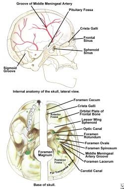
A frontal sinus is a paired, air-filled paranasal sinus located in the frontal bone of the skull, above the eyes and behind the forehead. It is one of the four pairs of sinuses found in the human head. The frontal sinuses are lined with mucous membrane and are interconnected with the nasal cavity through small openings called ostia. They help to warm, humidify, and filter the air we breathe, and contribute to the resonance of our voice. Variations in size, shape, and asymmetry of frontal sinuses are common among individuals.
Osteosarcoma is defined as a type of cancerous tumor that arises from the cells that form bones (osteoblasts). It's the most common primary bone cancer, and it typically develops in the long bones of the body, such as the arms or legs, near the growth plates. Osteosarcoma can metastasize (spread) to other parts of the body, including the lungs, making it a highly malignant form of cancer. Symptoms may include bone pain, swelling, and fractures. Treatment usually involves a combination of surgery, chemotherapy, and/or radiation therapy.
 Osteoblastoma
Hamate bone
Osteoid osteoma
Brodie abscess
Curettage
Chondroblast
Alan L. Schiller
Brachial plexus
Edmontosaurus
Bactrosaurus
Brachylophosaurus
Bone
Bone tumor
Aneurysmal bone cyst
Metaphysis
Gilmoreosaurus
Hadrosaurus
Giant-cell tumor of bone
List of MeSH codes (C04)
International Classification of Diseases for Oncology
Index of trauma and orthopaedics articles
Osteoblastoma - Wikipedia
 Osteoblastoma Clinical Presentation: History and Physical Examination
 FOS-ANKH and FOS-RUNX2 Fusion Genes in Osteoblastoma - PubMed
Spinal Tumors Treatment & Management: Approach Considerations, Primary Benign Spinal Tumors, Primary Malignant Spinal Tumors
Spinal Tumors Treatment & Management: Approach Considerations, Primary Benign Spinal Tumors, Primary Malignant Spinal Tumors
 Osteoblastoma | Boston Children's Hospital
 Osteoblastoma in the retromolar region: a case report
 Dr. Jim F. Vellara - Amrita Vishwa Vidyapeetham
 Understanding Bone Cancer | American Cancer Society
 Search Strategy Used to Create the PubMed Cancer Filter
 Temporal bone osteoblastoma involving temporomandibular joint diagnosed as simple disc disorders: A case report. | Front Surg...
Biomarkers Search
 Benign Bone Tumors and Cysts - Musculoskeletal and Connective Tissue Disorders - MSD Manual Professional Edition
 Bone cancer: Survival rate, causes, types, and treatment
id|key id|primary name|gopher term class|icd9 code id|word synonyms|excluded from phr|excluded from cms|excluded from phr2...
 DailyMed - FORTEO- teriparatide injection, solution
From the archives of the AFIP. Primary tumors of the spine: radiologic pathologic correlation
Chondroblastoma Imaging and Diagnosis: Practice Essentials, Radiography, Computed Tomography
 Jaw tumors and cysts - Symptoms and causes - Mayo Clinic
 Oncogenic Osteomalacia - Basic Science - Orthobullets
 Subchronic Pulmonary Pathology, Iron Overload, and Transcriptional Activity after Libby Amphibole Exposure in Rat Models of...
 C42637|Pharmaceutical Excipient|modify|27-OCT-06|(null)|(null
 Spine Flashcards by hussein baydoun | Brainscape
IDH2 | Cancer Genetics Web
FOS | Cancer Genetics Web
 MeSH Browser
MeSH Browser
![Best Clinics for Paranasal Sinus and Nasal Cavity Cancer Treatment in South Korea [2023 Prices]](data:image/png;base64,iVBORw0KGgoAAAANSUhEUgAAABAAAAAQCAMAAAAoLQ9TAAAA7VBMVEVHcEwrodw3p95goc2e0u4qrOMkot0WZqJsjaNQhbAzr+QtqOA9WHtkyfMiaaQdl9Yoq+ImquIvQWMmm9iByuQnq+Izr+RIY4UrcqxLoc8AACZFuOs6TWzN5eg3sON0WmUnqOEdmNc9s+UhmdcfmdceZJym3PMZOWB/ip89frUTJk0sbKMmbaczRmUjOl5AUnA/XH42odtSueciHj44t+w8QFw/U3MrVXw9WHoWQmseSHE5caQ3r+QfOF5XY35uosUcPGRNaooxQmIxS20fmdc/qN9tfpoemNclqeIEMFoAGkMAH0gAV5YFXZwmm9jSTIdTAAAAR3RSTlMAMJBTA/5c+g5y+aSEQNHjQWunvCCzwmymFmuZnRLoHbvM3/GmZQnSQH703rTik4p2zX9cfnpNsVX92YrV2WF1+YDB9VBtNmSKSzQAAADZSURBVBiVPY7XcsIwEEWvq2Tjggu9d9ITSkIqKYDkpv//HBDJ5Lzc2Z3dOReQTHdBUMY/tM+NYdfg6t9c5m+xTNfmVKbCFngtCkLqqHK5YCq0nHje9cUc3QpgCnhWTnQQKw07vIFI7eTb/ArQLrO0NwwwcieF1Ti96vQma7ZKYMqkqP/q7rLmqgJfo9bT/QZ4vG2ny9PFuISX2vMn8J45KWwTikAtcb7iw0/SXruyyOABTiL5/oBdPTeL0Atns5DqI27o0ueL/RSIVcYYN8++3YAJwSLFHPv9I8hRG0SWb3LsAAAAAElFTkSuQmCC) Best Clinics for Paranasal Sinus and Nasal Cavity Cancer Treatment in South Korea [2023 Prices]
Best Clinics for Paranasal Sinus and Nasal Cavity Cancer Treatment in South Korea [2023 Prices]
 Dorsal access Decompression - Open Operating Theatre (OOT)
Osteoid osteoma8
- therefore, some consider the two tumors to be variants of the same disease, with osteoblastoma representing a giant osteoid osteoma. (wikipedia.org)
- In contrast to the pain associated with osteoid osteoma, the pain of osteoblastoma usually is less intense, usually not worse at night, and not relieved readily with salicylatesfluid (aspirin and related compounds). (wikipedia.org)
- Spinal lesions can cause painful scoliosis, although this is less common with osteoblastoma than with osteoid osteoma. (wikipedia.org)
- Osteoblastoma is a rare primary neoplasm of bone, categorized as a benign bone tumor that is closely related to osteoid osteoma . (medscape.com)
- [ 1 ] Osteoblastoma differs from osteoid osteoma in its ability to grow larger than 2.0 cm in diameter and its aggressive behavior in bone. (medscape.com)
- Other diagnoses that share similar clinical, radiographic, and histologic features with conventional osteoblastoma include osteoid osteoma. (medscape.com)
- Osteoblastoma, and the related entity osteoid osteoma, are the most common benign bone-forming tumours. (ncri.org.uk)
- We suggest that FOS or FOSB rearrangements define osteoblastoma and osteoid osteoma. (ncri.org.uk)
Conventional osteoblastoma2
- citation needed] In regards to morbidity and mortality, conventional osteoblastoma is a benign lesion with little associated morbidity. (wikipedia.org)
- There are two main clinicopathologic subtypes of osteoblastoma: conventional osteoblastoma characterized by slow growth and well-defined sclerotic margins, which is fairly well vascularized and exhibits a mild inflammatory response, and the aggressive form characterized by a locally aggressive behavior and the propensity to recur. (bvsalud.org)
Tumor5
- ABSTRACT: Background: Osteoblastoma, a rare osteoblastic tumor, constitutes approximately 1% of all primary bone tu- mors. (scirp.org)
- Osteoblastoma is a non-cancerous (benign) tumor bone. (agileortho.in)
- Osteoblastoma is a rare benign tumor that accounts for less than 1% of all bone tumors. (bvsalud.org)
- Osteoblastoma é um tumor benigno raro que representa menos de 1% de todos os tumores ósseos e 10% estão localizadas nos ossos do crânio e quase metade destes casos afeta a mandíbula, especialmente os segmentos posteriores. (bvsalud.org)
- Osteoblastoma is a benign but aggressive tumor of bone (it can attain a large size and is not. (orthofixar.com)
Osteosarcoma2
- Osteoblastoma is about 20 times less common than osteosarcoma is. (medscape.com)
- Osteoblastoma-Like Osteosarcoma of the Proximal Humerus. (jsmcentral.org)
Malignant1
- Other primary osseous and cartilaginous tumors that have been reported rarely in the orbit include both benign entities (eg, osteoblastoma) and malignant sarcomas (eg, Ewing sarcoma, osteogenic sarcoma). (aao.org)
Aneurysmal2
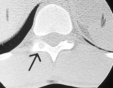
- Standard radiograph of osteoblastoma with secondary aneurysmal bone cyst of lumbar spine. (medscape.com)
- Osteoblastoma with secondary aneurysmal bone cyst. (medscape.com)
Fibroma1
- In his original description of osteoblastoma, Lichtenstein termed the lesion an "osteogenic fibroma of bone. (medscape.com)
Tumors3
- Osteoblastoma also arises in the talus more frequently than other benign tumors do (see the image below). (medscape.com)
- These tumors are also called osteoblastoma or giant cell reparative granuloma. (medicinenet.com)
- [ 1 ] Locally aggressive stage III benign tumors extend beyond natural borders and often require en bloc resection for cure. (medscape.com)
Complete excision2
- This type of complete excision is usually curative for osteoblastoma. (wikipedia.org)
- The appropriate surgical treatment goal for osteoblastoma is complete excision of the lesion. (medscape.com)
Mandible2
- Aim: The purpose of this report is to present an osteoblastoma of the mandible, with particular emphasis on the differential diagnosis of this rare tu- mor. (scirp.org)
- Osteoblastoma is quite rare in the oromaxillo-facial region, while the mandible is always the predilection. (bvsalud.org)
Lesion2
- However, computed tomography (CT) is often necessary to support clinical and plain radiographic findings suggestive of osteoblastoma and to better define the margins of the lesion for potential surgery. (wikipedia.org)
- If the nidus measures over 2cm in length the lesion should be termed osteoblastoma . (radiopaedia.org)
Radiologic2
- When diagnosing osteoblastoma, the preliminary radiologic workup should consist of radiography of the site of the patient's pain. (wikipedia.org)
- Many bone-producing lesions have clinical, radiologic and histopathologic features that resemble osteoblastoma. (bvsalud.org)
Diagnosis2
- The clinical course of osteoblastoma often makes diagnosis difficult. (medscape.com)
- A definitive diagnosis and appropriate treatment can only be established using the combination of clinical, histologic and radiographic components 4 .The objective of this study was to report the case of a young adult female patient with an osteoblastoma in the mandibular region. (bvsalud.org)
Aggressive1
- However, an aggressive type of osteoblastoma has been recognized, making the relationship less clear. (wikipedia.org)
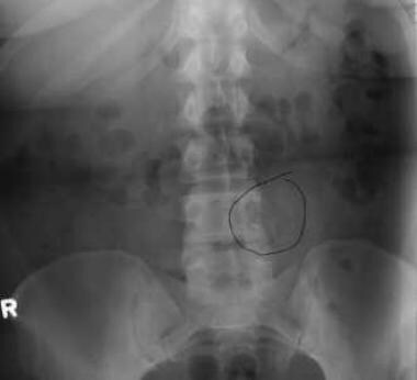
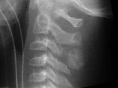
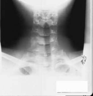
Spine2
- Osteoblastoma commonly affects the vertebral column, with approximately 30% of these lesions arising within the posterior elements of the spine. (medscape.com)
- An osteoblastoma usually involves the spine. (medicoapps.org)
Indications1
- There are two primary indications for surgical management of an osteoblastoma within the musculoskeletal system. (medscape.com)
Nonspecific2
- MRI aids in the detection of nonspecific reactive marrow and soft-tissue edema, and MRI best defines soft tissue extension, although this finding is not typical of osteoblastoma. (wikipedia.org)
- Although osteoblastoma demonstrates increased radiotracer accumulation, its appearance is nonspecific, and differentiating these lesions from those due to other causes involving increased radiotracer accumulation in the bone is difficult. (wikipedia.org)
Define1
- We sought to define the somatic changes that underpin osteoblastoma. (ncri.org.uk)
Controversial2
- Although necessary, radiation therapy (or chemotherapy) is controversial in the treatment of osteoblastoma. (wikipedia.org)
- The use of radiation or chemotherapeutic measures to treat osteoblastoma has been controversial. (medscape.com)
Lesions1
- However, we found a mass of bone lesions at the left temporal articular tubercle in MRI and cone beam CT , and it turned out to be an osteoblastoma after surgery . (bvsalud.org)
Rare3
- Except in rare cases, your doctor will not recommend these treatment options for osteoblastoma. (agileortho.in)
- Osteoblastoma is very rare in the temporomandibular joint region. (bvsalud.org)
- Our report provides experience in the identification of osteoblastoma in rare sites. (bvsalud.org)
Bones2
- Other documented locations of osteoblastoma include the pelvic bones, the small bones of the hands and feet, the skull and facial bones, the clavicle, the scapula, and the ribs. (medscape.com)
- Mild pain and swelling are the most usual symptoms of osteoblastoma in arm bones and legs. (agileortho.in)
Rarely1
- Osteoblastoma is rarely found in the hand or wrist. (ac.ir)
Clinical1
- Bone scintigraphy (bone scan) demonstrates abnormal radiotracer accumulation at the affected site, substantiating clinical suspicion, but this finding is not specific for osteoblastoma. (wikipedia.org)
Citation needed2
- citation needed] The cause of osteoblastoma is unknown. (wikipedia.org)
- citation needed] The first route of treatment in Osteoblastoma is via medical means. (wikipedia.org)
Case1
- Temporal bone osteoblastoma involving temporomandibular joint diagnosed as simple disc disorders: A case report. (bvsalud.org)
Treatment1
- Fortunately, treatment of osteoblastoma resolves this curve. (agileortho.in)
Tissue4
- Osteoblastoma is an uncommon osteoid tissue-forming primary neoplasm of the bone. (wikipedia.org)
- Histologically, osteoblastoma are similar to osteoid osteomas, producing both osteoid and primitive woven bone amidst fibrovascular connective tissue, the difference being that osteoblastoma can grow larger than 2.0 cm in diameter while osteoid osteomas cannot. (wikipedia.org)
- In this test, your doctor may take a part of the tissue from osteoblastoma. (agileortho.in)
- Without damaging any surrounding tissue, your doctor will try to remove your osteoblastoma. (agileortho.in)
Cases1
- Review of literature evealed that only five cases of osteoblastoma have been reported in scaphoid bone. (ac.ir)
Cancer1
- However, several osteoblastoma may affect cancer as well. (agileortho.in)


![10 Best Clinics for Esophageal Cancer Treatment in Thailand [2023 Prices]](https://www.mymeditravel.com/cdn-cgi/image/f=auto,fit=contain,quality=75/uploads/property/gallery/5af2758efa6b7e04401f8c27/5af51dadfa6b7e4212052361/preview.jpg)