Osteochondritis Dissecans
Joint Loose Bodies
Osteochondrosis
Range of Motion, Articular
Cartilage, Articular
Transplantation, Autologous
The pathogenesis of Perthes' disease. (1/142)
It has been shown that in the puppy, two infarcts separated by an interval of four weeks produce a disorder of long duration which results in flattening and broadening of the femoral head and which reproduces the radiological changes seen in Perthes' disease in man. The histological appearances produced by two infarcts are characteristic. In this study the histological appearance of fifty-seven femoral head biopsy specimens in Perthes' disease in man have been studied. In 51 per cent of hips histopathological changes characteristic of double infarction were present, and there were grounds for postulating that double infarction might eventually occur in all cases. The findings support the concept that the deformation of the femoral head and the chronicity of Perthes' disease in man may be due at least as much or even more to repeated episodes of infarction and the ensuing abnormalities of growth as to mechanical factors related to weight-bearing. (+info)Osteochondrosis and epiphyseal bone abnormalities associated with copper deficiency in bison calves. (2/142)
Two bison calves were submitted to the Western College of Veterinary Medicine to confirm suspected copper deficiency. In addition to clinical signs, there were pathologic changes in the cartilage and subchondral bone of several joints. Water analysis indicated high levels of sulfate in the drinking water, contributing to a secondary copper deficiency. (+info)Influence of development and joint pathology on stromelysin enzyme activity in equine synovial fluid. (3/142)
OBJECTIVE: To investigate the role of stromelysin (MMP-3) activity in synovial fluid (SF) at different stages of development and in common joint disorders in the horse. METHODS: Stromelysin activity was determined with a fluorogenic enzyme activity assay in SF of normal joints of fetal, juvenile and adult horses, and in SF of horses suffering from the developmental orthopaedic disease osteochondrosis (OC) or osteoarthritis (OA). Additionally, MMP-3 activity was expressed as a ratio of previously reported general MMP activity in the same SF samples. RESULTS: The levels of active stromelysin were 30-fold to 80-fold higher in SF from fetal horses than in SF from juvenile and mature animals (p<0.001). Juvenile horses (5 and 11 months of age) showed a twofold to threefold higher stromelysin activity than adult horses ( p<0.05). In OC joints, stromelysin activity was not significantly different from the activity in normal, age matched, control joints. In OA joints the activity was about four times higher than in normal joints (p<0.001). The ratio MMP-3 activity/general MMP activity did not change with age in normal, healthy joints. This ratio was more then twofold increased in OA joints compared with normal joints, indicating selective upregulation of gene expression or activation of proMMP-3, or both, in OA pathology. CONCLUSIONS: The significantly higher stromelysin activity in young individuals parallels the higher metabolic activity occurring at rapid growth and differentiation at early age. In OC, MMP-3 mediated matrix degradation appears to be not different from normal joints. The increased stromelysin activity in OA joints is in agreement with pathological matrix degradation. In these joints MMP-3 activity is selectively increased compared with normal joints. (+info)Role of Fc receptor gamma chain in inflammation and cartilage damage during experimental antigen-induced arthritis. (4/142)
OBJECTIVE: To study the role of Fc receptor (FcR) gamma chain in inflammation and cartilage destruction during antigen-induced arthritis (AIA). METHODS: FcR gamma-/- mice and controls were immunized with methylated bovine serum albumin (mBSA) in Freund's complete adjuvant, followed by induction of arthritis by local injection of mBSA into the right knee joint. Joint inflammation was studied by 99mTc uptake and by histology. Breakdown of proteoglycans from the cartilage matrix was determined by loss of red staining in Safranin O-stained knee joint sections, and matrix metalloproteinase (MMP)-mediated aggrecan degradation was determined by immunolocalization using anti-VDIPEN antibodies. Chondrocyte death was measured by determining empty lacunae in hematoxylin-stained sections and with the TUNEL assay in cryostat sections. Erosion was detected as ruffling of the cartilage surface. RESULTS: Joint swelling, as measured by 99mTc uptake on days 1, 3, and 7, was significantly decreased in FcR gamma-/- mice compared with controls. On day 7 after AIA induction, sustained joint inflammation, as seen histologically, was not significantly lower in FcR gamma-/- deficient mice. In various cartilage layers (femur, tibia, patella) of central arthritic knee joints, marked depletion of proteoglycans (40-70%), chondrocyte death (25-50%), and mild surface erosion were found. In FcR gamma-/- knee joints, depletion of proteoglycans was comparable (40-70%). Strikingly, chondrocyte death and matrix erosion were absent. Furthermore, MMP-induced aggrecan neoepitopes, which were abundantly found in controls, were also absent in FcR gamma-/-. Nevertheless, latent MMPs were present in the cartilage matrix as seen in APMA-activated patellae. CONCLUSION: FcR gamma chain is involved in the severity of acute and sustained inflammation and is a crucial factor in cartilage erosion during AIA, probably by regulating activation of latent MMPs present in the cartilage matrix. (+info)Kienbock's disease: conservative management versus radial shortening. (5/142)
Avascular necrosis of the lunate, first described by Kienbock, can be treated either conservatively or by various surgical procedures. We compared the results of 18 conservatively treated patients, all of whom had stage-2 or stage-3 disease, with those of 15 who underwent a radial shortening procedure. We evaluated pain, range of movement, grip strength and functional disability, and determined the progression of the disease by assessing radiologically carpal height, the width and flattening of the lunate, the radioscaphoid angle, the pattern of the fracture and sclerosis and cysts. The mean follow-up was for 3.6 years (1.5 to 9). Patients treated by radial shortening had less pain and better grip strength than those managed conservatively. In some patients with stage-3 disease treated conservatively there was rapid deterioration to carpal collapse. Although radial shortening did not reverse or prevent carpal collapse, it slowed down the process in patients with stage-3 disease. We recommend a radial shortening procedure for patients with severe pain and radiological signs of progressive carpal collapse. (+info)Some aspects of orthopaedic surgery in childhood. (6/142)
The aetiological role of immunodeficiency in acute septic arthritis of the hip in infancy, the management of the condition after the acute infection has subsided, and the special hazards of infection in the region of the hip joint in the older child are discussed. The principles of treatment of congenital dislocation of the hip are examined in relation to the maintenance of acetabular growth potential. The factors determining the outcome of treatment in Perthes' disease are discussed and a comparison of the result in a series of cases treated by femoral osteotomy with those in untreated controls is presented. (+info)The role of cumulative physical work load in lumbar spine disease: risk factors for lumbar osteochondrosis and spondylosis associated with chronic complaints. (7/142)
OBJECTIVES: To investigate the relation with a case-control study between symptomatic osteochondrosis or spondylosis of the lumbar spine and cumulative occupational exposure to lifting or carrying and to working postures with extreme forward bending. METHODS: From two practices and four clinics were recruited 229 male patients with radiographically confirmed osteochondrosis or spondylosis of the lumbar spine associated with chronic complaints. Of these 135 had additionally had acute lumbar disc herniation. A total of 197 control subjects was recruited: 107 subjects with anamnestic exclusion of lumbar spine disease were drawn as a random population control group and 90 patients admitted to hospital for urolithiasis who had no osteochondrosis or spondylosis of the lumbar spine radiographically were recruited as a hospital based control group. Data were gathered in a structured personal interview and analysed using logistic regression to control for age, region, nationality, and other diseases affecting the lumbar spine. To calculate cumulative forces to the lumbar spine over the entire working life, the Mainz-Dortmund dose model (MDD), which is based on an overproportional weighting of the lumbar disc compression force relative to the respective duration of the lifting process was applied with modifications: any objects weighing >or=5 kg were included in the calculation and no minimum daily exposure limits were established. Calculation of forces to the lumbar spine was based on self reported estimates of occupational lifting, trunk flexion, and duration. RESULTS: For a lumbar spine dose >9 x 10(6) Nh (Newton x hours), the risk of having radiographically confirmed osteochondrosis or spondylosis of the lumbar spine as measured by the odds ratio (OR) was 8.5 (95% confidence interval (95% CI) 4.1 to 17.5) compared with subjects with a load of 0 Nh. To avoid differential bias, forces to the lumbar spine were also calculated on the basis of an internal job exposure matrix based on the control subjects' exposure assessments for their respective job groups. Although ORs were lower with this approach, they remained significant. CONCLUSIONS: The calculation of the sum of forces to the lumbar spine is a useful tool for risk assessment for symptomatic osteochondrosis or spondylosis of the lumbar spine. The results suggest that cumulative occupational exposure to lifting or carrying and extreme forward bending increases the risk for developing symptomatic osteochondrosis or spondylosis of the lumbar spine. (+info)Osteochondrosis of the superior pole of the patella: two cases with histologic correlation. (8/142)
Two cases of osteochondrosis of the superior pole of the patella are reported with histologic findings. Both patients were young girls; one had mild cerebral palsy. Sixteen cases of this disorder have been documented but without histologic study. The histologic features of these two cases showed osteonecrosis with reparative changes. These findings support that this entity is similar to other osteochondroses of the quadriceps mechanism: Osgood-Schlatter disease and Sinding-Larsen-Johansson disease. (+info)Osteochondritis dissecans (OCD) is a joint condition that occurs when a piece of cartilage or bone in the joint separates from its underlying bone due to a lack of blood supply. This condition most commonly affects the knee, but it can also occur in other joints such as the elbow, ankle, and wrist.
In OCD, the affected area of cartilage and bone may form a loose body that can move around within the joint, causing pain, swelling, and limited mobility. In some cases, the loose body may eventually heal on its own, but in other cases, surgical intervention may be necessary to remove or repair the damaged tissue.
OCD is more common in children and adolescents, particularly those who participate in sports that involve repetitive joint trauma. Treatment for OCD typically involves a combination of rest, physical therapy, and possibly surgery, depending on the severity of the condition.
Osteochondritis is a joint condition where a piece of cartilage or bone in the joint separates from its attachment due to a lack of blood supply. This can cause pain, stiffness, and potentially restricted movement in the affected joint. It often occurs in weight-bearing joints like the knee or ankle, and is more common in children and adolescents. The separated piece may sometimes float around in the joint space, causing further damage to the cartilage and bone. If left untreated, it can lead to long-term joint problems. Also known as osteochondrosis or osteochondritis dissecans.
'Joint loose bodies' refer to free-floating fragments or particles within the joint space. These can be composed of cartilage, bone, or other synovial tissue debris. They can vary in size and number and may cause symptoms such as pain, locking, catching, or decreased range of motion due to mechanical interference with joint movement. Joint loose bodies are often associated with degenerative joint diseases like osteoarthritis but can also result from trauma or previous surgeries.
A cartilage fracture is not a common injury because cartilage itself does not have bones, and it is difficult to fracture something that is not hard. However, there are situations where the term "cartilage fracture" can be used. One such situation is when the articular cartilage, which covers the ends of bones in joints, gets damaged or injured. This type of injury is also known as a chondral fracture or osteochondral fracture (if the bone beneath the cartilage is also involved). These injuries can occur due to trauma, such as a fall or a direct blow to the joint, and can cause pain, swelling, and limited mobility in the affected joint.
Arthroscopy is a minimally invasive surgical procedure where an orthopedic surgeon uses an arthroscope (a thin tube with a light and camera on the end) to diagnose and treat problems inside a joint. The surgeon makes a small incision, inserts the arthroscope into the joint, and then uses the attached camera to view the inside of the joint on a monitor. They can then insert other small instruments through additional incisions to repair or remove damaged tissue.
Arthroscopy is most commonly used for joints such as the knee, shoulder, hip, ankle, and wrist. It offers several advantages over traditional open surgery, including smaller incisions, less pain and bleeding, faster recovery time, and reduced risk of infection. The procedure can be used to diagnose and treat a wide range of conditions, including torn ligaments or cartilage, inflamed synovial tissue, loose bone or cartilage fragments, and joint damage caused by arthritis.
The elbow joint, also known as the cubitus joint, is a hinge joint that connects the humerus bone of the upper arm to the radius and ulna bones of the forearm. It allows for flexion and extension movements of the forearm, as well as some degree of rotation. The main articulation occurs between the trochlea of the humerus and the trochlear notch of the ulna, while the radial head of the radius also contributes to the joint's stability and motion. Ligaments, muscles, and tendons surround and support the elbow joint, providing strength and protection during movement.
The knee joint, also known as the tibiofemoral joint, is the largest and one of the most complex joints in the human body. It is a synovial joint that connects the thighbone (femur) to the shinbone (tibia). The patella (kneecap), which is a sesamoid bone, is located in front of the knee joint and helps in the extension of the leg.
The knee joint is made up of three articulations: the femorotibial joint between the femur and tibia, the femoropatellar joint between the femur and patella, and the tibiofibular joint between the tibia and fibula. These articulations are surrounded by a fibrous capsule that encloses the synovial membrane, which secretes synovial fluid to lubricate the joint.
The knee joint is stabilized by several ligaments, including the medial and lateral collateral ligaments, which provide stability to the sides of the joint, and the anterior and posterior cruciate ligaments, which prevent excessive forward and backward movement of the tibia relative to the femur. The menisci, which are C-shaped fibrocartilaginous structures located between the femoral condyles and tibial plateaus, also help to stabilize the joint by absorbing shock and distributing weight evenly across the articular surfaces.
The knee joint allows for flexion, extension, and a small amount of rotation, making it essential for activities such as walking, running, jumping, and sitting.
Bone transplantation, also known as bone grafting, is a surgical procedure in which bone or bone-like material is transferred from one part of the body to another or from one person to another. The graft may be composed of cortical (hard outer portion) bone, cancellous (spongy inner portion) bone, or a combination of both. It can be taken from different sites in the same individual (autograft), from another individual of the same species (allograft), or from an animal source (xenograft). The purpose of bone transplantation is to replace missing bone, provide structural support, and stimulate new bone growth. This procedure is commonly used in orthopedic, dental, and maxillofacial surgeries to repair bone defects caused by trauma, tumors, or congenital conditions.
Osteochondrosis is a group of orthopedic disorders that primarily affect the epiphyseal growth plates (the areas of growing tissue at the ends of long bones) and adjacent articular (joint) cartilage in children and adolescents. These disorders are characterized by abnormal development, degeneration, or fragmentation of the affected bone and/or cartilage, which can lead to pain, stiffness, and, in some cases, restricted mobility.
The term "osteochondrosis" is often used interchangeably with "osteochondritis dissecans," but they are not identical conditions. Osteochondrosis refers to the general category of disorders, while osteochondritis dissecans is a specific type of osteochondrosis that primarily affects the subchondral bone (the layer of bone directly beneath the articular cartilage) and results in the formation of loose fragments or "joint mice."
Examples of osteochondrosis include:
1. Legg-Calvé-Perthes disease, which affects the hip joint
2. Köhler's disease, which affects the navicular bone in the foot
3. Panner's disease, which affects the elbow joint
4. Scheuermann's disease, which affects the vertebral bodies in the spine
5. Freiberg's infarction, which affects the metatarsal heads in the foot
The exact cause of osteochondrosis remains unclear, but it is believed to involve a combination of genetic, biomechanical, and environmental factors that contribute to the abnormal growth and development of the affected bone and cartilage. Treatment typically involves rest, physical therapy, bracing or casting, and, in some cases, surgery to remove loose fragments or promote healing.
The patella, also known as the kneecap, is a sesamoid bone located at the front of the knee joint. It is embedded in the tendon of the quadriceps muscle and serves to protect the knee joint and increase the leverage of the extensor mechanism, allowing for greater extension force of the lower leg. The patella moves within a groove on the femur called the trochlea during flexion and extension of the knee.
Articular Range of Motion (AROM) is a term used in physiotherapy and orthopedics to describe the amount of movement available in a joint, measured in degrees of a circle. It refers to the range through which synovial joints can actively move without causing pain or injury. AROM is assessed by measuring the degree of motion achieved by active muscle contraction, as opposed to passive range of motion (PROM), where the movement is generated by an external force.
Assessment of AROM is important in evaluating a patient's functional ability and progress, planning treatment interventions, and determining return to normal activities or sports participation. It is also used to identify any restrictions in joint mobility that may be due to injury, disease, or surgery, and to monitor the effectiveness of rehabilitation programs.
Articular cartilage is the smooth, white tissue that covers the ends of bones where they come together to form joints. It provides a cushion between bones and allows for smooth movement by reducing friction. Articular cartilage also absorbs shock and distributes loads evenly across the joint, protecting the bones from damage. It is avascular, meaning it does not have its own blood supply, and relies on the surrounding synovial fluid for nutrients. Over time, articular cartilage can wear down or become damaged due to injury or disease, leading to conditions such as osteoarthritis.
The femur is the medical term for the thigh bone, which is the longest and strongest bone in the human body. It connects the hip bone to the knee joint and plays a crucial role in supporting the weight of the body and allowing movement during activities such as walking, running, and jumping. The femur is composed of a rounded head, a long shaft, and two condyles at the lower end that articulate with the tibia and patella to form the knee joint.
Autologous transplantation is a medical procedure where cells, tissues, or organs are removed from a person, stored and then returned back to the same individual at a later time. This is different from allogeneic transplantation where the tissue or organ is obtained from another donor. The term "autologous" is derived from the Greek words "auto" meaning self and "logos" meaning study.
In autologous transplantation, the patient's own cells or tissues are used to replace or repair damaged or diseased ones. This reduces the risk of rejection and eliminates the need for immunosuppressive drugs, which are required in allogeneic transplants to prevent the body from attacking the foreign tissue.
Examples of autologous transplantation include:
* Autologous bone marrow or stem cell transplantation, where stem cells are removed from the patient's blood or bone marrow, stored and then reinfused back into the same individual after high-dose chemotherapy or radiation therapy to treat cancer.
* Autologous skin grafting, where a piece of skin is taken from one part of the body and transplanted to another area on the same person.
* Autologous chondrocyte implantation, where cartilage cells are harvested from the patient's own knee, cultured in a laboratory and then implanted back into the knee to repair damaged cartilage.
Chondrocytes are the specialized cells that produce and maintain the extracellular matrix of cartilage tissue. They are responsible for synthesizing and secreting the collagen fibers, proteoglycans, and other components that give cartilage its unique properties, such as elasticity, resiliency, and resistance to compression. Chondrocytes are located within lacunae, or small cavities, in the cartilage matrix, and they receive nutrients and oxygen through diffusion from the surrounding tissue fluid. They are capable of adapting to changes in mechanical stress by modulating the production and organization of the extracellular matrix, which allows cartilage to withstand various loads and maintain its structural integrity. Chondrocytes play a crucial role in the development, maintenance, and repair of cartilaginous tissues throughout the body, including articular cartilage, costal cartilage, and growth plate cartilage.
 Osteochondritis
Osteochondritis dissecans
Bone disease
Bone
Franz König (surgeon)
Knee examination
Knee pain
Osteitis
Osteochondrosis
Chondrolysis
Joint locking (medicine)
Elbow dysplasia
Kevin R. Stone
Herbert screw
List of OMIM disorder codes
National Register of Historic Places listings in Red River County, Texas
National Register of Historic Places listings in the Upper East region of Texas
Akhal-Teke
Panner disease
Epiphysis
Legg-Calvé-Perthes disease
Chondritis
Kaylia Nemour
Polina Tsurskaya
Kristina Vaculik
Lauren Beers
South German Coldblood
Ian Gunther
Nathan Stein
Nicolás Larcamón
Osteochondritis - Wikipedia
 Knee Osteochondritis Dissecans: Background, Etiology, Epidemiology
 How Osteochondritis Dissecans Affects the Knee
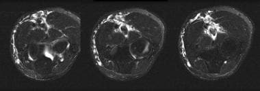
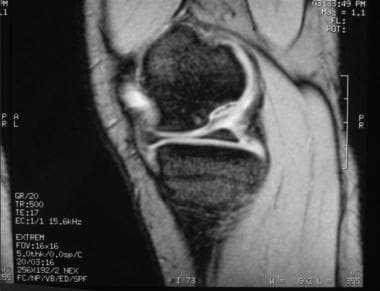
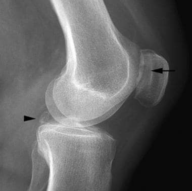
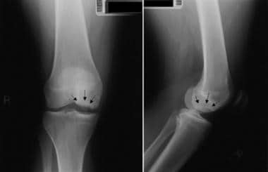
 Osteochondritis dissecans radiology tests: X-ray, MRI, and more
 Osteochondritis Dissecans in Dogs: A Detailed Guide | PetCareRx
Osteochondritis dissecans of the knee
 Arthrex - Treatment of Capitellar Osteochondritis Dissecans Using Precut Fresh Osteochondral Allografts
 Go to the Osteochondritis Dissecans/surgery Veterinary Medicine archives.
 Osteochondritis (medical concept explorer)
 OCD - Osteochondritis | Centre Vétérinaire Rive Sud
 Osteochondritis Dissecans | Children's Hospital Colorado
 Osteochondritis Dissecans (OCD) in Dogs - Veterinary Partner - VIN
 Juvenile osteochondritis dissecans of the knee: current concepts in diagnosis and management. | Read by QxMD
 Elbow Pain: Causes, Symptoms, Treatment, Medications, Prevention
 Patient Education - Osteochondritis Dissecans
 Osteochondritis - Clinique du pied
 Osteochondritis dissecans | Rare Diseases | RareGuru
 Osteochondritis dessecans - or OCD - NZHS
Aaron Krych, MD - Osteochondritis Dissecans
 Osteochondritis Dissecans | Concise Medical Knowledge
77 Osteochondritis Dissecans | Radiology Key
Humeral Capitellum Osteochondritis Dissecans Differential Diagnoses
 Osteochondritis Dissecans | Absolute Foot Care Specialists
Treating Osteochondritis Dissecans In The Elbow
 Osteochondritis Dissecans Causes & Treatment Ventura CA
 Osteochondritis Dissecans - West Coast Veterinary Clinic
 Osteochondritis Dissecans | American College of Radiology
 Osteochondritis Dissecans | Lee's Hill Pet Hospital
Osteochondritis Dissecans in a Female Soccer Player
Lesions9
- A characteristic feature of familial osteochondritis dissecans is areas of bone damage (lesions) caused by detachment of cartilage and a piece of the underlying bone from the end of the bone at a joint. (medlineplus.gov)
- It is unclear how the abnormal cartilage leads to the lesions and osteoarthritis characteristic of familial osteochondritis dissecans. (medlineplus.gov)
- Fixation of juvenile osteochondritis dissecans lesions of the knee using poly 96L/4D-lactide copolymer bioabsorbable implants. (medscape.com)
- Healing predictors of stable juvenile osteochondritis dissecans knee lesions after 6 and 12 months of nonoperative treatment. (medscape.com)
- Juvenile osteochondritis dissecans (JOCD) lesions contain cartilaginous, fibrous and osseous tissues which are difficult to distinguish with clinical, morphological magnetic resonance imaging (MRI). (healthpartners.com)
- The chapter reviews the latest operative treatment for osteochondritis dissecans (OCD lesions) of the Knee. (atlantasportsmedicine.com)
- These areas of dead bone are referred to as osteochondritis lesions. (shouldersandknees.com)
- The natural history of some osteochondritis dissecans lesions is the separation of these structures from the capitellum, leading to the development of an osteochondral fragment of articular cartilage on the underlying bone at the superficial surface of the diarthrodial joint. (medscape.com)
- 6. Osteochondritis Dissecans Lesions in Family Members: Does a Positive Family History Impact Phenotypic Potency? (nih.gov)
Treating Osteochondritis dissecans2
- When it comes to treating Osteochondritis Dissecans with Dr Spector's guidance you can trust that your recovery will be on track with a plan that takes into account your specific needs and goals for recovery. (myfootspecialist.com)
- Is there a protocol for treating Osteochondritis dissecans, right knee? (thereadystate.com)
Diagnosis1
- Juvenile osteochondritis dissecans of the knee: current concepts in diagnosis and management. (medscape.com)
Juvenile8
- Adachi N, Deie M, Nakamae A, Ishikawa M, Motoyama M, Ochi M. Functional and radiographic outcome of stable juvenile osteochondritis dissecans of the knee treated with retroarticular drilling without bone grafting. (medscape.com)
- Juvenile versus adult osteochondritis dissecans of the knee: appropriate MR imaging criteria for instability. (medscape.com)
- Nakayama H, Iseki T, Kambara S, Yoshiya S. Analysis of risk factors for poor prognosis in conservatively managed juvenile osteochondritis dissecans of the lateral femoral condyle. (medscape.com)
- Correlation of magnetic resonance imaging to arthroscopic findings of stability in juvenile osteochondritis dissecans. (medscape.com)
- Treatment of juvenile osteochondritis dissecans and osteochondritis dissecans of the knee. (nih.gov)
- Juvenile osteochondritis dissecans. (nih.gov)
- Children and teens can experience juvenile osteochondritis dissecans, which can lead to adult osteochondritis dissecans, or OCD. (shouldersandknees.com)
- The pathogenesis of human juvenile osteochondritis dissecans (JOCD) remains poorly understood, with multiple factors implicated, including ischemia, repetitive trauma, and genetic predisposition. (umn.edu)
Lesion8
- A similar condition called sporadic osteochondritis dissecans is associated with a single lesion in one joint, most often the knee. (medlineplus.gov)
- Some people with sporadic osteochondritis dissecans develop osteoarthritis in the affected joint, especially if the lesion occurs later in life after the bone has stopped growing. (medlineplus.gov)
- Osteochondritis Dissecans, also commonly referred to as an osteochondral lesion (OCL) or osteochondral defect (OCD), is a condition in which there is a cartilaginous defect within one of the joints in your foot or ankle. (myfootspecialist.com)
- While this can sometimes confirm Osteochondritis Dissecans (an OCD lesion), often times advanced imaging studies such as CT or MRI are required. (myfootspecialist.com)
- Osteochondritis is a lesion that usually causes pain and stiffness of the ankle joint and affects all age groups. (tiptonandunroe.com)
- A similar disease called osteochondritis dissecans sporadic associated with a single lesion in a joint, most often knee. (ivami.com)
- Osteochondritis dissecans is a disease that leads to cartilage/bone damage (osteochondral lesion) in the joint. (knieschmerzen-wien.at)
- The term "osteochondritis" is not appropriate since inflammation is not a characteristic feature of the lesion. (nih.gov)
ACAN6
- in these cases, osteochondritis dissecans is caused by changes in the ACAN gene and is inherited in an autosomal dominant manner. (nih.gov)
- Mutation of the ACAN gene can cause familial osteochondritis dissecans. (medlineplus.gov)
- The ACAN gene mutation associated with familial osteochondritis dissecans results in an abnormal protein that is unable to attach to the other components of cartilage. (medlineplus.gov)
- It has identified at least one mutation in the ACAN gene in people with familial osteochondritis dissecans. (ivami.com)
- in IVAMI perform detection of mutations associated with familial osteochondritis dissecans, by complete PCR amplification of the exons of the gene ACAN and subsequent sequencing. (ivami.com)
- Some affected individuals also manifested osteochondritis dissecans and early-onset osteoarthritis.ACAN encodes a proteoglycan that is an important component of cartilage extracellular matrix. (nih.gov)
Familial7
- When Do Symptoms of Familial osteochondritis dissecans Begin? (nih.gov)
- Familial osteochondritis dissecans is a genetic disease. (nih.gov)
- Familial osteochondritis dissecans is a condition that affects the joints and is associated with abnormal cartilage. (medlineplus.gov)
- Other characteristic features of familial osteochondritis dissecans include short stature and development of a joint disorder called osteoarthritis at an early age. (medlineplus.gov)
- Familial osteochondritis dissecans is a rare condition, although the prevalence is unknown. (medlineplus.gov)
- Stattin EL, Tegner Y, Domellof M, Dahl N. Familial osteochondritis dissecans associated with early osteoarthritis and disproportionate short stature. (medlineplus.gov)
- Familial osteochondritis dissecans: a dysplasia of articular cartilage? (nih.gov)
Adult osteochondritis dissecans1
- Midterm results of surgical treatment for adult osteochondritis dissecans of the knee. (medscape.com)
Bone18
- Osteochondritis dissecans is a joint condition that occurs when a piece of cartilage and the thin layer of bone beneath it, separates from the end of the bone. (nih.gov)
- Osteochondritis is a painful type of osteochondrosis where the cartilage or bone in a joint is inflamed. (wikipedia.org)
- Osteochondritis dissecans (OCD) is a condition consisting of aseptic bone necrosis at articular surfaces, such as the medial femoral condyle, talar dome, or capitellum humeri. (medscape.com)
- Arthroscopic debridement of the osteochondritis dissecans bed to bleeding bone. (medscape.com)
- Osteochondritis is the abnormal development of cartilage on the bone. (sitstay.com)
- Osteochondritis Dissecans, most often occurs in the knees and is a bone and cartilage condition that can cause swelling and pain. (sub-4.co.uk)
- Osteochondritis dissecans (OCD) is a condition in which a piece of bone or cartilage (or both) inside a joint loses blood supply and dies. (medicalrecords.com)
- Osteochondritis dissecans is a joint condition in which a piece of cartilage, along with a thin layer of the bone separates from the end of the bone because of inadequate blood supply. (robertfullickmd.com)
- Osteochondritis dissecans may be caused by restricted blood supply to the end of the affected bone that usually occurs in conjunction with repetitive trauma. (robertfullickmd.com)
- Osteochondritis dissecans is a bone disease that can impact the knee joint , affecting the lower end of the femur. (shouldersandknees.com)
- Osteochondritis dissecans usually impacts the lower femoral condyles, which are the rounded ends of the thigh bone. (shouldersandknees.com)
- Osteochondritis dissecans (aka "OCD") is a condition in which the bone that supports the cartilage inside a joint undergoes softening. (brianwatermanmd.com)
- Humeral capitellum osteochondritis dissecans occurs after the capitellum has ossified and is the result of "injury" to the subchondral bone. (medscape.com)
- Osteochondritis Dissecans (OCD) is a joint disorder involving the subchondral bone and the overlying articular cartilage. (orthobangalore.com)
- Osteochondritis dissecans, also called OCD, is a joint condition that occurs when there is insufficient blood flow to the joint, causing part of the bone to die and cracks to form in the bone and cartilage. (orthopaedic-surgery-md.com)
- When diagnosing osteochondritis dissecans, a healthcare provider will likely start with a physical examination of the affected joint to check for swelling, tenderness, and/or a loose bone fragment. (orthopaedic-surgery-md.com)
- If you have osteochondritis dissecans and your healthcare provider finds a loose piece of bone in your joint, you may need treatment. (orthopaedic-surgery-md.com)
- A characteristic feature of family osteochondritis dissecans are areas of bone injury due to detachment of the cartilage and underlying bone from the bone end in a joint. (ivami.com)
Etiology2
- Osteochondritis Dissecans of the Knee: Etiology and Pathogenetic Mechanisms. (nih.gov)
- The exact etiology of osteochondritis dissecans is unclear. (medscape.com)
Term osteochondritis1
- Although no inflammatory cells have been identified on histologic sections of excised fragments, the term osteochondritis dissecans has persisted and since been broadened to describe a similar process occurring in many other joints , including the knee, hip, ankle, elbow, and metatarsophalangeal joints. (medscape.com)
Cartilage2
- In 1889, Francis Konig described osteochondritis dissecans as a subchondral inflammatory process of the knee resulting in a loose fragment of cartilage from the femoral condyle. (medscape.com)
- The trapdoor procedure is Dr. Kennedy's approach to preserving the cartilage in his younger patients that suffer Osteochondritis dissecans. (sportsmedicinenewyork.com)
Ankle9
- Having osteochondritis dissecans in the ankle is a painful condition that can lead to arthritis if left untreated. (healthline.com)
- Osteochondritis dissecans (OCD) in the ankle is a condition that can lead to swelling, difficulty moving your ankle, weakness, and increasing pain over time. (healthline.com)
- How long can it take to heal from osteochondritis in the ankle? (healthline.com)
- If you're suffering from Osteochondritis Dissecans, consulting with a specialist like Dr. Jason Spector at Florida Foot and Ankle Specialists is essential for ensuring that you receive the best possible care for your condition. (myfootspecialist.com)
- Osteochondritis is caused by a twisting-type injury to the ankle. (tiptonandunroe.com)
- What is Ankle Osteochondritis? (orthobangalore.com)
- When OCD affects the ankle it is called Ankle Osteochondritis or OCD of the ankle. (orthobangalore.com)
- In Ankle Osteochondritis, it typically occurs in the inner or medial portion of the ankle (Talus). (orthobangalore.com)
- Ankle Osteochondritis is often found in young athletes involved in high impact sports like running, gymnastics, hockey, cricket, lacrosse, squash, tennis, weightlifting, football and basketball, which needs a high level of intense training daily and with minimal rest between the activities. (orthobangalore.com)
Symptoms1
- OCD (osteochondritis dissecans), on the other hand, is considered a chronic process that can go on for months to years before any symptoms are felt. (brianwatermanmd.com)
Talus2
- The video shows osteochondritis dissecans of talus together with cure and preoperative evaluation. (medtube.net)
- Higuera J, Laguna R, Peral M, Aranda E, Soleto J. Osteochondritis dissecans of the talus during childhood and adolescence. (legehandboka.no)
Occurs5
- Osteochondritis dissecans occurs within the lateral aspect of the medial femoral condyle. (robertfullickmd.com)
- In the United States, humeral capitellum osteochondritis dissecans most commonly occurs in the second decade of life and is rare in individuals younger than 10 years or older than 50 years. (medscape.com)
- Osteochondritis dissecans also occurs in females, most notably gymnasts. (medscape.com)
- Humeral capitellum osteochondritis dissecans usually occurs in the dominant arm. (medscape.com)
- Osteochondritis dissecans most commonly occurs in the knee joint (at the medial femoral condyle). (knieschmerzen-wien.at)
Sporadic1
- Sporadic osteochondritis dissecans is not caused by genetic changes and is not inherited. (medlineplus.gov)
Bilateral1
- Bilateral osteochondritis dissecans in a female pitcher. (nih.gov)
Radiographs2
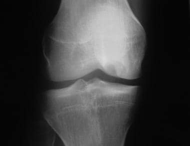
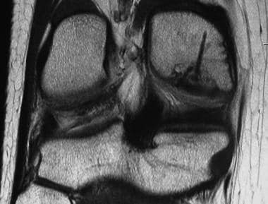
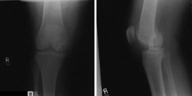
- Anteroposterior and lateral radiographs of medial femoral condyle osteochondritis dissecans. (medscape.com)
- Radiographs: Osteochondritis, diaphyseal osteomyelitis, periostitis. (cdc.gov)
Condition is inherited1
- The appearance of osteochondritis dissecans in several family members may indicate that the condition is inherited. (robertfullickmd.com)
Knees2
- She had a history of 4 operations, 1 each at both knees and both elbows, for the treatment of osteochondritis dissecans (OCD). (medscape.com)
- Dr. Steven Struhl at Shoulders & Knees offers osteochondritis dissecans treatments at our clinics in NYC and Westchester. (shouldersandknees.com)
Medial femoral3
- Herbert screw stabilization of medial femoral condyle osteochondritis dissecans. (medscape.com)
- Arthroscopic view of medial femoral condyle osteochondritis dissecans, hinged medially. (medscape.com)
- Arthroscopic view of osteochondritis dissecans of the medial femoral condyle. (medscape.com)
Fracture1
- This image shows a "fracture line" of osteochondritis dissecans (OCD) in yellow. (ballaratosm.com.au)
Subchondral1
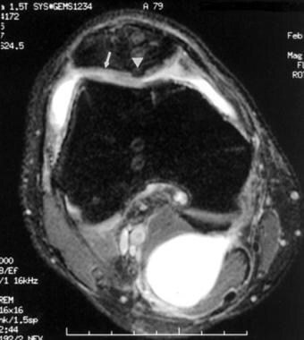
- These forces are believed to lead to fibrillation on the articular surface and subchondral osseous changes, with the possible production of osteocartilaginous fragments and the development of humeral capitellum osteochondritis dissecans. (medscape.com)
Knee Pain2
- Osteochondritis Dissecans, Knee Pain? (sub-4.co.uk)
- If you or your child are suffering from knee pain related to osteochondritis dissecans, contact our clinic in NYC or Westchester. (shouldersandknees.com)
Joints2
- Osteochondritis dissecans (OCD) is a condition that develops in joints, most often in children and adolescents. (cassurgery.com)
- Osteochondritis dissecans can occur in any of the joints including your elbows, ankles, shoulders, and hips. (drjeanyun.com)
Femur1
- The other recognized types of osteochondritis are osteochondritis deformans juvenilis (osteochondritis of the capitular head of the epiphysis of the femur) and osteochondritis deformans juvenilis dorsi (osteochondrosis of the spinal vertebrae, also known as Scheuermann's disease). (wikipedia.org)
Lameness1
- Hindlimb lameness localized to the tarsal joint is most commonly caused by osteochondritis dissecans (OCD). (dvm360.com)
Treatment5
- Andriolo L, Candrian C, Papio T, Cavicchioli A, Perdisa F, Filardo G. Osteochondritis Dissecans of the Knee - Conservative Treatment Strategies: A Systematic Review. (medscape.com)
- The purpose of a Sub-4 free assessment, is to help explore the pain issue you have and determine an appropriate Osteochondritis Dissecans treatment. (sub-4.co.uk)
- If you think that you may be suffering from an Osteochondritis Dissecans issue, don't hesitate to contact Dr Jason Spector today at 941-241-5333 for a proper evaluation and a recommended treatment plan that is tailored specifically to your needs! (myfootspecialist.com)
- Treatment of Osteochondritis Dissecans depends on the location and severity. (myfootspecialist.com)
- While osteochondritis dissecans can be painful and inconvenient, treatment is available that can reduce your pain and restore movement to your aching joint. (orthopaedic-surgery-md.com)
Surgical4
- Surgical management of osteochondritis dissecans of the knee in the paediatric population: a systematic review addressing surgical techniques. (medscape.com)
- Surgical management of osteochondritis dissecans of the knee. (medscape.com)
- Surgical correction of osteochondritis dissecans can be done using by open technique or arthroscopic techniques. (robertfullickmd.com)
- Surgical correction of osteochondritis dissecans can be done using the open technique or arthroscopic technique. (drjeanyun.com)
Osteochondrosis1
- Historically, osteochondrosis has also been recorded as osteochondritis and osteochondritis dissecans. (nih.gov)
Joint pain3
- Patients with osteochondritis dissecans usually have joint pain, swelling, stiffness, decreased range of motion, and joint popping or locking. (robertfullickmd.com)
- If you are experiencing joint pain and are hearing a crackling sound when you move your joint, you may have osteochondritis dissecans. (orthopaedic-surgery-md.com)
- Osteochondritis dissecans usually leads to elbow joint pain, swelling, stiffness and decreased range of motion. (drjeanyun.com)
Elbow1
- Osteochondritis, and especially osteochondritis dissecans, can manifest in animals as a primary cause of elbow dysplasia, a chronic condition in some dog breeds. (wikipedia.org)