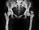
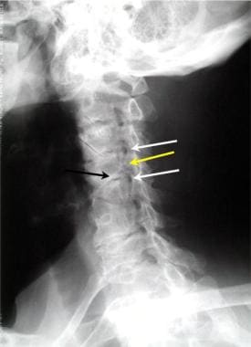
Osteopetrosis
Osteoclasts
Acidosis, Renal Tubular
Tooth Eruption
Vacuolar Proton-Translocating ATPases
Maxillofacial Abnormalities
Oral Fistula
Cathepsin K
Macrophage Colony-Stimulating Factor
Avian Leukosis
SLC4A Proteins
Satellite Viruses
Bone and Bones
Mandibular Diseases
Anemia, Myelophthisic
Avian leukosis virus
Osteomyelitis
Carbonic Anhydrase II
Chloride Channels
Acid Phosphatase
Granulocyte/macrophage colony-stimulating factor and interleukin-3 correct osteopetrosis in mice with osteopetrosis mutation. (1/298)
Although young mice homozygous for the osteopetrosis (op) mutation usually developed prominent osteopetrosis, its severity was markedly reduced in aged op/op mice. This age-associated reversal of osteopetrosis was accompanied by the expansion of bone marrow cavities and increased numbers of tartrate-resistant acid phosphatase (TRAP)-positive cells and of macrophages in the bone marrow. The TRAP-positive cells were mononuclear and developed ruffled borders and numerous vesicles, vacuoles, and granules. Enzyme-linked immunosorbent assay demonstrated a significant elevation of serum granulocyte/ macrophage colony-stimulating factor (GM-CSF) and interleukin (IL)-3 levels in the aged op/op mice. To examine whether GM-CSF and/or IL-3 could correct osteopetrosis in young op/op mice, 5 ng of recombinant murine (rm)GM-CSF and/or 100 ng of rmIL-3 were injected daily into young op/op mice. In these treated young op/op mice, the bone marrow cavities were expanded significantly at 2 weeks after administration, associated with significantly increased numbers of TRAP-positive cells and bone marrow macrophages. TRAP-positive cells increased in number with days after injection. These results suggest that GM-CSF and IL-3 induce the development of osteoclasts to correct osteopetrosis in the op/op mice with aging. (+info)Midpalatal suture of osteopetrotic (op/op) mice exhibits immature fusion. (2/298)
The midpalatal suture was observed histologically in both toothless osteopetrotic (op/op) and normal (control) mice. The normal mice had a mature sutural structure, which consists of a well-developed cartilage cell zone and palatal bone. In contrast, the thickness of the cartilage cell zone was substantially greater in the op/op mice than that in the controls. Moreover, the cartilage cells in the op/op mice were frequently found in the palatal bone as well as in the sutural space, exhibiting an imperfect fusion. It seems that immature fusion at the sutural interface in the op/op mice is related to a decrease in biting or masticatory force accompanied by the failure of tooth eruption in addition to an essential defect in osteoclast differentiation, which is a congenital symptom in op/op mice. (+info)TRAF6 deficiency results in osteopetrosis and defective interleukin-1, CD40, and LPS signaling. (3/298)
Bone resorption and remodeling is an intricately controlled, physiological process that requires the function of osteoclasts. The processes governing both the differentiation and activation of osteoclasts involve signals induced by osteoprotegerin ligand (OPGL), a member of tumor necrosis factor (TNF) superfamily, and its cognate receptor RANK. The molecular mechanisms of the intracellular signal transduction remain to be elucidated. Here we report that mice deficient in TNF receptor-associated factor 6 (TRAF6) are osteopetrotic with defects in bone remodeling and tooth eruption due to impaired osteoclast function. Using in vitro assays, we demonstrate that TRAF6 is crucial not only in IL-1 and CD40 signaling but also, surprisingly, in LPS signaling. Furthermore, like TRAF2 and TRAF3, TRAF6 is essential for perinatal and postnatal survival. These findings establish unexpectedly diverse and critical roles for TRAF6 in perinatal and postnatal survival, bone metabolism, LPS, and cytokine signaling. (+info)Allogeneic bone marrow transplantation in an osteopetrosis patient: first report in Thailand. (4/298)
We described the successful allogeneic matched sibling bone marrow transplantation (BMT) in a 5-year-old Thai boy in whom osteopetrosis was diagnosed on the basis of anemia, thrombocytopenia, leukoerythroblastosis, sclerotic bone, hepatosplenomegaly, and visual deficit from an encroachment of cranial nerve foramina. The preparative regimen included 4 days of busulfan 4 mg/kg/day, and 4 days of cyclophosphamide 50 mg/kg/day. Complete hematopoietic engraftment and no evidence of graft versus host disease were shown after BMT. Complete hematologic findings were corrected. His hematopoietic chimerism was changed to that of his donor. Post BMT, he has no hepatosplenomegaly. His bone radiographic findings revealed normal after BMT. Bone marrow biopsy showed normalized bone and bone marrow matrix. However, his vision remained impaired. We believe that this is the first case of successful bone marrow transplantation in an osteopetrosis patient in Thailand. (+info)Importance of neurological assessment before bone marrow transplantation for osteopetrosis. (5/298)
Neurological complications of malignant infantile osteopetrosis are well recognised; successful bone marrow transplantation, when performed early in life, can prevent or halt some of them. In a subgroup of infants osteopetrosis is associated with primary retinal degeneration and/or generalised neurodegeneration. Bone marrow transplantation, in spite of being successful in correcting the osseous and haematological abnormalities, does not influence the progressive course of the neurodegenerative disorder. Thus, the recognition of this subgroup of infants with a very poor prognosis is essential before deciding on bone marrow transplantation. (+info)Facial development and type III collagen RNA expression: concurrent repression in the osteopetrotic (Toothless,tl) rat and rescue after treatment with colony-stimulating factor-1. (6/298)
The toothless (osteopetrotic) mutation in the rat is characterized by retarded development of the anterior facial skeleton. Growth of the anterior face in rats occurs at the premaxillary-maxillary suture (PMMS). To identify potential mechanisms for stunted facial growth in this mutation we compared the temporospatial expression of collagen I (Col I) and collagen III (Col III) RNA around this suture in toothless (tl) rats and normal littermates by in situ hybridization of specific riboprobes in sagittal sections of the head. In normal rats, the suture is S shaped at birth and becomes highly convoluted by 10 days with cells in the center (fibroblasts and osteoblast progenitors) expressing Col III RNA and those at the periphery (osteoblasts) expressing no Col III RNA but high amounts of Col I RNA throughout the growth phase (the first 2 postnatal weeks). In the mutant PMMS, cells were reduced in number, less differentiated, and fewer osteoblasts were encountered. Expression of Col I RNA was at normal levels, but centrosutural cells expressed Col III RNA only after day 6 and then only weakly. A highly convoluted sutural shape was never achieved in mutants during the first 2 postnatal weeks. Treatment of tl rats with the cytokine CSF-1 improved facial growth and restored cellular diversity and Col III RNA expression in the PMMS to normal levels. Taken together, these data suggest that normal facial growth in rats is related to expression of Col III RNAby osteoblast precursors in the PMMS, that these cells are deficient in the tl mutation and are rescued following treatment with CSF-1. (+info)Congenital osteoclast deficiency in osteopetrotic (op/op) mice is improved by ovariectomy and orchiectomy. (7/298)
We examined the changes in the appearance of osteoclasts in the femora of ovariectomized (OVX) or orchiectomized (ORX) op/op mice. Osteoclasts on the trabecular bone surface of the OVX or ORX op/op mice significantly increased in number seven or eight times in comparison with sham-operated op/op mice. Furthermore, TRAP-positive cells increased about four times in 100-week-old females and males, compared with sham-operated groups. These results have indicated that a sex hormone reduction due to OVX or ORX induces prominent recruitment of osteoclasts in op/op mice. (+info)The M cell as a portal of entry to the lung for the bacterial pathogen Mycobacterium tuberculosis. (8/298)
M. tuberculosis accesses the terminal lung and is phagocytosed by alveolar macrophages. Utilizing a mouse intratracheal challenge model, we demonstrate that M. tuberculosis rapidly enters through M cells as well. From there, bacilli are deposited within associated intraepithelial leukocytes and subsequently conveyed to the draining lymph nodes early after infection. Osteopetrotic (Csfm(op)/Csfm(op)) mice, null mutants for macrophage colony-stimulating factor, possess diminished numbers of circulating monocytes and tissue macrophages. Csfm(op)/Csfm(op) mice were highly susceptible to challenge with M. tuberculosis. In contrast to controls, tubercle bacilli were not conveyed to draining lymph nodes early after infection but were instead retained within the mucosa. These results indicate that M cells represent an alternate portal of entry for M. tuberculosis, which may contribute to the rapid development of protective lung immune responses. (+info)Osteopetrosis, also known as Albers-Schönberg disease or marble bone disease, is a group of rare genetic disorders characterized by increased bone density due to impaired bone resorption by osteoclasts. This results in brittle bones that are more susceptible to fractures and can also lead to various complications such as anemia, hearing loss, and vision problems. There are several types of osteopetrosis, which vary in severity and age of onset.
The medical definition of osteopetrosis is:
A genetic disorder characterized by defective bone resorption due to impaired osteoclast function, resulting in increased bone density, susceptibility to fractures, and potential complications such as anemia, hearing loss, and vision problems.
Osteosclerosis is a medical term that refers to an abnormal thickening and increased density of bone tissue. This condition can occur as a result of various diseases or conditions, such as certain types of bone cancer, Paget's disease of bone, fluoride poisoning, or chronic infection of the bone. Osteosclerosis can also be seen in some benign conditions, such as osteopetrosis, which is a rare genetic disorder characterized by an excessively hard and dense skeleton.
In some cases, osteosclerosis may not cause any symptoms and may only be discovered on X-rays or other imaging studies. However, in other cases, it can lead to complications such as bone pain, fractures, or deformities. Treatment for osteosclerosis depends on the underlying cause of the condition and may include medications, surgery, or other therapies.
Osteoclasts are large, multinucleated cells that are primarily responsible for bone resorption, a process in which they break down and dissolve the mineralized matrix of bones. They are derived from monocyte-macrophage precursor cells of hematopoietic origin and play a crucial role in maintaining bone homeostasis by balancing bone formation and bone resorption.
Osteoclasts adhere to the bone surface and create an isolated microenvironment, called the "resorption lacuna," between their cell membrane and the bone surface. Here, they release hydrogen ions into the lacuna through a process called proton pumping, which lowers the pH and dissolves the mineral component of the bone matrix. Additionally, osteoclasts secrete proteolytic enzymes, such as cathepsin K, that degrade the organic components, like collagen, in the bone matrix.
An imbalance in osteoclast activity can lead to various bone diseases, including osteoporosis and Paget's disease, where excessive bone resorption results in weakened and fragile bones.
Renal tubular acidosis (RTA) is a medical condition that occurs when the kidneys are unable to properly excrete acid into the urine, leading to an accumulation of acid in the bloodstream. This results in a state of metabolic acidosis.
There are several types of RTA, but renal tubular acidosis type 1 (also known as distal RTA) is characterized by a defect in the ability of the distal tubules to acidify the urine, leading to an inability to lower the pH of the urine below 5.5, even in the face of metabolic acidosis. This results in a persistently alkaline urine, which can lead to calcium phosphate stones and bone demineralization.
Type 1 RTA is often caused by inherited genetic defects, but it can also be acquired due to various kidney diseases, drugs, or autoimmune disorders. Symptoms of type 1 RTA may include fatigue, weakness, muscle cramps, decreased appetite, and vomiting. Treatment typically involves alkali therapy to correct the acidosis and prevent complications.
Bone resorption is the process by which bone tissue is broken down and absorbed into the body. It is a normal part of bone remodeling, in which old or damaged bone tissue is removed and new tissue is formed. However, excessive bone resorption can lead to conditions such as osteoporosis, in which bones become weak and fragile due to a loss of density. This process is carried out by cells called osteoclasts, which break down the bone tissue and release minerals such as calcium into the bloodstream.
Tooth eruption is the process by which a tooth emerges from the gums and becomes visible in the oral cavity. It is a normal part of dental development that occurs in a predictable sequence and timeframe. Primary or deciduous teeth, also known as baby teeth, begin to erupt around 6 months of age and continue to emerge until approximately 2-3 years of age. Permanent or adult teeth start to erupt around 6 years of age and can continue to emerge until the early twenties.
The process of tooth eruption involves several stages, including the formation of the tooth within the jawbone, the movement of the tooth through the bone and surrounding tissues, and the final emergence of the tooth into the mouth. Proper tooth eruption is essential for normal oral function, including chewing, speaking, and smiling. Any abnormalities in the tooth eruption process, such as delayed or premature eruption, can indicate underlying dental or medical conditions that require further evaluation and treatment.
Vacuolar Proton-Translocating ATPases (V-ATPases) are complex enzyme systems that are found in the membranes of various intracellular organelles, such as vacuoles, endosomes, lysosomes, and Golgi apparatus. They play a crucial role in the establishment and maintenance of electrochemical gradients across these membranes by actively pumping protons (H+) from the cytosol to the lumen of the organelles.
The V-ATPases are composed of two major components: a catalytic domain, known as V1, which contains multiple subunits and is responsible for ATP hydrolysis; and a membrane-bound domain, called V0, which consists of several subunits and facilitates proton translocation. The energy generated from ATP hydrolysis in the V1 domain is used to drive conformational changes in the V0 domain, resulting in the vectorial transport of protons across the membrane.
These electrochemical gradients established by V-ATPases are essential for various cellular processes, including secondary active transport, maintenance of organellar pH, protein sorting and trafficking, and regulation of cell volume. Dysfunction in V-ATPases has been implicated in several human diseases, such as neurodegenerative disorders, renal tubular acidosis, and certain types of cancer.
Maxillofacial abnormalities, also known as craniofacial anomalies, refer to a broad range of structural and functional disorders that affect the development of the skull, face, jaws, and related soft tissues. These abnormalities can result from genetic factors, environmental influences, or a combination of both. They can vary in severity, from minor cosmetic issues to significant impairments of vital functions such as breathing, speaking, and eating.
Examples of maxillofacial abnormalities include cleft lip and palate, craniosynostosis (premature fusion of the skull bones), hemifacial microsomia (underdevelopment of one side of the face), and various other congenital anomalies. These conditions may require multidisciplinary treatment involving surgeons, orthodontists, speech therapists, and other healthcare professionals to address both functional and aesthetic concerns.
An oral fistula is an abnormal connection or tunnel that links the oral cavity (the mouth) to another structure, usually the skin of the face or the neck. This condition can occur as a result of various factors such as infection, trauma, surgery, or congenital abnormalities. Oral fistulas may cause symptoms like pain, discomfort, difficulty in swallowing or speaking, and leakage of saliva or food from the opening of the fistula. Treatment typically involves surgical closure of the fistulous tract to restore normal anatomy and function.
Cathepsin K is a proteolytic enzyme, which belongs to the family of papain-like cysteine proteases. It is primarily produced by osteoclasts, which are specialized cells responsible for bone resorption. Cathepsin K plays a crucial role in the degradation and remodeling of the extracellular matrix, particularly in bone tissue.
This enzyme is capable of breaking down various proteins, including collagen, elastin, and proteoglycans, which are major components of the bone matrix. By doing so, cathepsin K helps osteoclasts to dissolve and remove mineralized and non-mineralized bone matrix during the process of bone resorption.
Apart from its function in bone metabolism, cathepsin K has also been implicated in several pathological conditions, such as osteoporosis, rheumatoid arthritis, and tumor metastasis to bones. Inhibitors of cathepsin K are being investigated as potential therapeutic agents for the treatment of these disorders.
Macrophage Colony-Stimulating Factor (M-CSF) is a growth factor that belongs to the family of colony-stimulating factors (CSFs). It is a glycoprotein hormone that plays a crucial role in the survival, proliferation, and differentiation of mononuclear phagocytes, including macrophages. M-CSF binds to its receptor, CSF1R, which is expressed on the surface of monocytes, macrophages, and their precursors.
M-CSF stimulates the production of mature macrophages from monocyte precursors in the bone marrow and enhances the survival and function of mature macrophages in peripheral tissues. It also promotes the activation of macrophages, increasing their ability to phagocytize and destroy foreign particles, microorganisms, and tumor cells.
In addition to its role in the immune system, M-CSF has been implicated in various physiological processes, including hematopoiesis, bone remodeling, angiogenesis, and female reproduction. Dysregulation of M-CSF signaling has been associated with several pathological conditions, such as inflammatory diseases, autoimmune disorders, and cancer.
Avian leukosis is a group of viral diseases that primarily affect chickens and other birds. It is caused by retroviruses known as avian leukosis viruses (ALVs) and leads to a variety of clinical signs, including immunosuppression, growth retardation, and the development of tumors in various organs. The disease can be transmitted both horizontally (through direct contact with infected birds or their secretions) and vertically (from infected hens to their offspring through the egg).
There are several subgroups of ALVs, each associated with specific types of tumors and clinical manifestations. For example:
1. ALV-J (Japanese strain): This subgroup is responsible for myelocytomatosis, a condition characterized by the proliferation of immature blood cells in the bone marrow, leading to anemia, leukopenia, and enlarged spleens and livers.
2. ALV-A, ALV-B, and ALV-C (American strains): These subgroups are associated with various types of lymphoid tumors, such as B-cell and T-cell lymphomas, which can affect the bursa of Fabricius, thymus, spleen, and other organs.
3. ALV-E (European strain): This subgroup is linked to erythroblastosis, a condition in which there is an excessive proliferation of red blood cell precursors, resulting in the formation of tumors in the bone marrow and other organs.
Avian leukosis poses significant economic challenges for the poultry industry due to its impact on growth, feed conversion efficiency, and mortality rates. Additionally, some countries have regulations in place to prevent the spread of avian leukosis viruses through the trade of infected birds or their products. Prevention measures include strict biosecurity protocols, vaccination programs, and rigorous screening and eradication strategies for infected flocks.
Avian myeloblastosis virus (AMV) is a type of retrovirus that primarily infects birds, particularly chickens. It is named after the disease it causes, avian myeloblastosis, which is a malignant condition affecting the bone marrow and blood cells of infected birds.
AMV is classified as an alpharetrovirus and has a single-stranded RNA genome. When the virus infects a host cell, its RNA genome is reverse transcribed into DNA, which then integrates into the host's chromosomal DNA. This integrated viral DNA, known as a provirus, can then direct the production of new virus particles.
AMV has been extensively studied as a model system for retroviruses and has contributed significantly to our understanding of their replication and pathogenesis. The virus is also used in laboratory research as a tool for generating genetically modified animals and for studying the regulation of gene expression. However, it is not known to infect or cause disease in humans or other mammals.
Solute carrier family 4A (anion exchanger) proteins, also known as SLC4A proteins, are a group of membrane transport proteins that facilitate the exchange of bicarbonate (HCO3-) and chloride (Cl-) ions across biological membranes. They play crucial roles in various physiological processes, including pH regulation, intracellular signaling, and fluid secretion/absorption in different tissues such as the kidney, brain, and red blood cells.
There are several members of this protein family, including:
1. SLC4A1 (AE1): Also known as band 3 anion transport protein, it is primarily expressed in the erythrocyte membrane and facilitates chloride-bicarbonate exchange. It also plays a role in carbon dioxide transport and maintaining the stability of red blood cells.
2. SLC4A2 (AE2): Expressed in various tissues, including the kidney, gastrointestinal tract, and brain. AE2 mediates chloride-bicarbonate exchange in these tissues and is involved in pH regulation and fluid secretion/absorption.
3. SLC4A3 (AE3): Found mainly in the heart, skeletal muscle, and brain, where it facilitates chloride-bicarbonate exchange. AE3 plays a role in regulating intracellular pH during muscle contraction and neuronal activity.
4. SLC4A4 (NBCe1): Expressed primarily in the kidney and brain, NBCe1 is a sodium-bicarbonate cotransporter that mediates the uptake of bicarbonate into cells. It plays a critical role in maintaining acid-base balance by reabsorbing bicarbonate from the urine filtrate in the kidney.
5. SLC4A5 (NBCe2): Found in various tissues, including the kidney and brain, NBCe2 is another sodium-bicarbonate cotransporter that facilitates bicarbonate uptake into cells. It contributes to pH regulation and acid-base balance.
6. SLC4A7 (NBCn1): Present in various tissues, including the eye, brain, and heart, NBCn1 is a sodium-bicarbonate cotransporter that mediates bicarbonate efflux from cells. It plays a role in maintaining intracellular pH homeostasis and has been implicated in certain diseases such as epilepsy and glaucoma.
7. SLC4A8 (NDCBE): Expressed mainly in the brain, NDCBE is a sodium-dependent chloride-bicarbonate exchanger that plays a role in regulating intracellular pH during neuronal activity.
8. SLC4A9 (AE4): Found primarily in the gastrointestinal tract and kidney, AE4 is a chloride-bicarbonate exchanger involved in pH regulation and fluid secretion/absorption.
9. SLC4A10 (NBCn2): Expressed mainly in the eye, NBCn2 is a sodium-bicarbonate cotransporter that plays a role in maintaining intracellular pH homeostasis and has been implicated in certain diseases such as epilepsy.
10. SLC4A11 (BTR1): Present in various tissues, including the eye and inner ear, BTR1 is a sodium-dependent borate cotransporter that plays a role in maintaining intracellular pH homeostasis and has been implicated in certain diseases such as Fuchs endothelial corneal dystrophy.
"Satellite viruses" are a type of viruses that require the presence of another virus, known as a "helper virus," to complete their replication cycle. They lack certain genes that are essential for replication and therefore depend on the helper virus to provide these functions. Satellite viruses can either be satellite RNA or satellite DNA viruses, and they can affect plants, animals, and bacteria.
Satellite viruses can influence the severity of the disease caused by the helper virus, either increasing or decreasing it. They can also interfere with the replication of the helper virus and affect its transmission. The relationship between satellite viruses and their helper viruses is complex and can vary depending on the specific viruses involved.
It's important to note that the term "satellite virus" is not used consistently in the scientific literature, and some researchers may use it to refer to other types of dependent or defective viruses. Therefore, it's always a good idea to consult the original research when interpreting the use of this term.
"Bone" is the hard, dense connective tissue that makes up the skeleton of vertebrate animals. It provides support and protection for the body's internal organs, and serves as a attachment site for muscles, tendons, and ligaments. Bone is composed of cells called osteoblasts and osteoclasts, which are responsible for bone formation and resorption, respectively, and an extracellular matrix made up of collagen fibers and mineral crystals.
Bones can be classified into two main types: compact bone and spongy bone. Compact bone is dense and hard, and makes up the outer layer of all bones and the shafts of long bones. Spongy bone is less dense and contains large spaces, and makes up the ends of long bones and the interior of flat and irregular bones.
The human body has 206 bones in total. They can be further classified into five categories based on their shape: long bones, short bones, flat bones, irregular bones, and sesamoid bones.
Mandibular diseases refer to conditions that affect the mandible, or lower jawbone. These diseases can be classified as congenital (present at birth) or acquired (developing after birth). They can also be categorized based on the tissues involved, such as bone, muscle, or cartilage. Some examples of mandibular diseases include:
1. Mandibular fractures: These are breaks in the lower jawbone that can result from trauma or injury.
2. Osteomyelitis: This is an infection of the bone and surrounding tissues, which can affect the mandible.
3. Temporomandibular joint (TMJ) disorders: These are conditions that affect the joint that connects the jawbone to the skull, causing pain and limited movement.
4. Mandibular tumors: These are abnormal growths that can be benign or malignant, and can develop in any of the tissues of the mandible.
5. Osteonecrosis: This is a condition where the bone tissue dies due to lack of blood supply, which can affect the mandible.
6. Cleft lip and palate: This is a congenital deformity that affects the development of the face and mouth, including the lower jawbone.
7. Mandibular hypoplasia: This is a condition where the lower jawbone does not develop properly, leading to a small or recessed chin.
8. Developmental disorders: These are conditions that affect the growth and development of the mandible, such as condylar hyperplasia or hemifacial microsomia.
Myelophthisic anemia is a type of anemia that occurs when the bone marrow becomes replaced or damaged by fibrosis, tumor infiltration, or other disorders, leading to decreased production of blood cells. This results in a decrease in all three types of blood cells - red blood cells, white blood cells, and platelets.
The symptoms of myelophthisic anemia may include fatigue, weakness, shortness of breath, frequent infections, and easy bruising or bleeding. The diagnosis is typically made through a combination of medical history, physical examination, complete blood count (CBC), and bone marrow aspiration and biopsy. Treatment for myelophthisic anemia depends on the underlying cause and may include chemotherapy, radiation therapy, surgery, or supportive care with transfusions of red blood cells or platelets.
Avian leukosis virus (ALV) is a type of retrovirus that primarily affects chickens and other birds. It is responsible for a group of diseases known as avian leukosis, which includes various types of tumors and immunosuppressive conditions. The virus is transmitted horizontally through the shedder's dander, feathers, and vertical transmission through infected eggs.
There are several subgroups of ALV (A, B, C, D, E, and J), each with different host ranges and pathogenicity. Some strains can cause rapid death in young chickens, while others may take years to develop clinical signs. The most common form of the disease is neoplastic, characterized by the development of various types of tumors such as lymphomas, myelomas, and sarcomas.
Avian leukosis virus infection can have significant economic impacts on the poultry industry due to decreased growth rates, increased mortality, and condemnation of infected birds at processing. Control measures include eradication programs, biosecurity practices, vaccination, and breeding for genetic resistance.
Recessive genes refer to the alleles (versions of a gene) that will only be expressed when an individual has two copies of that particular allele, one inherited from each parent. If an individual inherits one recessive allele and one dominant allele for a particular gene, the dominant allele will be expressed and the recessive allele will have no effect on the individual's phenotype (observable traits).
Recessive genes can still play a role in determining an individual's genetic makeup and can be passed down through generations even if they are not expressed. If two carriers of a recessive gene have children, there is a 25% chance that their offspring will inherit two copies of the recessive allele and exhibit the associated recessive trait.
Examples of genetic disorders caused by recessive genes include cystic fibrosis, sickle cell anemia, and albinism.
Osteomyelitis is a medical condition characterized by an infection that involves the bone or the bone marrow. It can occur as a result of a variety of factors, including bacterial or fungal infections that spread to the bone from another part of the body, or direct infection of the bone through trauma or surgery.
The symptoms of osteomyelitis may include pain and tenderness in the affected area, fever, chills, fatigue, and difficulty moving the affected limb. In some cases, there may also be redness, swelling, and drainage from the infected area. The diagnosis of osteomyelitis typically involves imaging tests such as X-rays, CT scans, or MRI scans, as well as blood tests and cultures to identify the underlying cause of the infection.
Treatment for osteomyelitis usually involves a combination of antibiotics or antifungal medications to eliminate the infection, as well as pain management and possibly surgical debridement to remove infected tissue. In severe cases, hospitalization may be necessary to monitor and manage the condition.
Carbonic anhydrase II (CA-II) is a specific isoform of the carbonic anhydrase enzyme, which catalyzes the reversible reaction between carbon dioxide and water to form carbonic acid. This enzyme plays a crucial role in various physiological processes, including pH regulation, electrolyte balance, and biosynthetic reactions.
CA-II is widely distributed in the body, with high concentrations found in erythrocytes (red blood cells), the gastric mucosa, and renal tubules. In erythrocytes, CA-II facilitates the rapid conversion of carbon dioxide generated during cellular respiration to bicarbonate and protons, which can then be transported across the cell membrane for excretion or used in other metabolic processes.
In the gastric mucosa, CA-II helps regulate acid secretion by catalyzing the formation of carbonic acid from water and carbon dioxide, which subsequently dissociates into bicarbonate and a proton. The generated proton can then participate in the production of hydrochloric acid in the stomach.
In renal tubules, CA-II is involved in the reabsorption of bicarbonate ions from the filtrate back into the bloodstream, helping maintain electrolyte balance and pH homeostasis. Additionally, CA-II has been implicated in several pathological conditions, such as neurological disorders, cancer, and osteoporosis, making it a potential therapeutic target for drug development.
Chloride channels are membrane proteins that form hydrophilic pores or gaps, allowing the selective passage of chloride ions (Cl-) across the lipid bilayer of cell membranes. They play crucial roles in various physiological processes, including regulation of neuronal excitability, maintenance of resting membrane potential, fluid and electrolyte transport, and pH and volume regulation of cells.
Chloride channels can be categorized into several groups based on their structure, function, and mechanism of activation. Some of the major classes include:
1. Voltage-gated chloride channels (ClC): These channels are activated by changes in membrane potential and have a variety of functions, such as regulating neuronal excitability and transepithelial transport.
2. Ligand-gated chloride channels: These channels are activated by the binding of specific ligands or messenger molecules, like GABA (gamma-aminobutyric acid) or glycine, and are involved in neurotransmission and neuromodulation.
3. Cystic fibrosis transmembrane conductance regulator (CFTR): This is a chloride channel primarily located in the apical membrane of epithelial cells, responsible for secreting chloride ions and water to maintain proper hydration and mucociliary clearance in various organs, including the lungs and pancreas.
4. Calcium-activated chloride channels (CaCCs): These channels are activated by increased intracellular calcium concentrations and participate in various physiological processes, such as smooth muscle contraction, neurotransmitter release, and cell volume regulation.
5. Swelling-activated chloride channels (ClSwells): Also known as volume-regulated anion channels (VRACs), these channels are activated by cell swelling or osmotic stress and help regulate cell volume and ionic homeostasis.
Dysfunction of chloride channels has been implicated in various human diseases, such as cystic fibrosis, myotonia congenita, epilepsy, and certain forms of cancer.
Acid phosphatase is a type of enzyme that is found in various tissues and organs throughout the body, including the prostate gland, red blood cells, bone, liver, spleen, and kidneys. This enzyme plays a role in several biological processes, such as bone metabolism and the breakdown of molecules like nucleotides and proteins.
Acid phosphatase is classified based on its optimum pH level for activity. Acid phosphatases have an optimal activity at acidic pH levels (below 7.0), while alkaline phosphatases have an optimal activity at basic or alkaline pH levels (above 7.0).
In clinical settings, measuring the level of acid phosphatase in the blood can be useful as a tumor marker for prostate cancer. Elevated acid phosphatase levels may indicate the presence of metastatic prostate cancer or disease progression. However, it is important to note that acid phosphatase is not specific to prostate cancer and can also be elevated in other conditions, such as bone diseases, liver disorders, and some benign conditions. Therefore, acid phosphatase should be interpreted in conjunction with other diagnostic tests and clinical findings for a more accurate diagnosis.
Consanguinity is a medical and genetic term that refers to the degree of genetic relationship between two individuals who share common ancestors. Consanguineous relationships exist when people are related by blood, through a common ancestor or siblings who have children together. The closer the relationship between the two individuals, the higher the degree of consanguinity.
The degree of consanguinity is typically expressed as a percentage or fraction, with higher values indicating a closer genetic relationship. For example, first-degree relatives, such as parents and children or full siblings, share approximately 50% of their genes and have a consanguinity coefficient of 0.25 (or 25%).
Consanguinity can increase the risk of certain genetic disorders and birth defects in offspring due to the increased likelihood of sharing harmful recessive genes. The risks depend on the degree of consanguinity, with closer relationships carrying higher risks. It is important for individuals who are planning to have children and have a history of consanguinity to consider genetic counseling and testing to assess their risk of passing on genetic disorders.
 Osteopetrosis - Wikipedia
 Etiology
 CLCN7-Related Osteopetrosis
 Autosomal dominant osteopetrosis type 1 - About the Disease - Genetic and Rare Diseases Information Center
 Peer-Reviewed Publication of Positive Preclinical Data of SiSaf's SIS-ADO2 siRNA Program to Treat Rare Genetic Bone Disorder...
 A Comparison of Osteoclast-Rich and Osteoclast-Poor Osteopetrosis in Adult Mice Sheds Light on the Role of the Osteoclast in...
 ITGB3 gene: MedlinePlus Genetics
 531: Pediatrics⎪Osteopetrosis - Orthohub
 Osteopetrosis autosomal recessive 1 | Rare Diseases | RareGuru
 Clinical Management of Malignant Infantile Osteopetrosis - Hematology Advisor
 Orthopaedic Challenges in Arthroplasty for Osteoarthritis in Osteopetrosis
![Osteopetrosis[Clinical Features] OR 18223[uid] - MedGen -...](data:image/png;base64,iVBORw0KGgoAAAANSUhEUgAAABAAAAAQCAYAAAAf8/9hAAAB1ElEQVQ4jaWSPWgTcRjGf/ehuWhobO2JxGJRY3taTTRV2yoqSpW6iIWO4iAoUsRBioNDKUWKLU7i4KA4OfhVREQnETRia03k7IdiS0LaQYKJQg3mLtfc30GySNUDn/V5nx/vy/vAf0pqad3db2xquiBJku93s2Tb2eEHdw1rTcsxol23sObTjN7oIp9KVmaU9kMdTxcLAyiqGtA0bfms+XKQULSdQG2EmnUx0q9ughAA8p/CFW0IN3Sv0vUI5p2zIMpUrd5JeP/Jii//80ZJUlrb9lyV8qn3zI5dB8A4MoBWtcITAKBmZe3eRmPzccYf9uIUsyzx6zQd7fMMAIjFdgxpkuPy4clFANbu6qa6fouybXtznxeAoqoBn0/zz5kvBqVQ5DBasJ5gXaPnDQAWFpwCkiwLZekyAMp2wTPAsqy5d8nEZcIHThPQo7jlIua9854BibdvekqKX8PouARAOn6F+c8pT4Bc7svz6U8f77O1cwDVV439PcPU4yHw8AUhhDPyOn4OfWOMuuZfBZp41INTLACorhC2/Jc2zsxMX8vl8lMcPBUHFL5mnpEZGa748sS42esKYS8WLtl2NjE22s/6fScIhtr48W2S5O0zIFwvp3vST6Z+myCvkaonAAAAAElFTkSuQmCC) "Osteopetrosis"[Clinical Features] OR 18223[uid] - MedGen -...
"Osteopetrosis"[Clinical Features] OR 18223[uid] - MedGen -... Refractory mandibular osteomyelitis in a young patient with osteopetrosis: Case report
 Overview of Osteopetroses - Pediatrics - MSD Manual Professional Edition
 No evidence of an association between lethal recessive osteopetrosis and performance in dairy cattle.
 Autosomal Recessive Osteopetrosis Type 2 (Autosomal Recessive Osteopetrosis 2): Symptoms, Diagnosis and Treatment - Symptoma
 Article - Billing and Coding: Vitamin D Assay Testing (A57484)
 RNA Interference Technology: FDA Approves SiSaf's siRNA Therapy, SIS-101-ADO | Autosomal Dominant Osteopetrosis Treatment
 lil bub - SFist - San Francisco News, Restaurants, Events, & Sports
 Aqueductal stenosis with optic atrophy in case of malignant osteopetrosis - Journal of Natural Science, Biology and Medicine
 Genericos Tadalafil Cialis, Venta de tadalafil generica / Genericos tadalafil cialis, Comprar tadalafil online en españa....