Paralysis
Respiratory Paralysis
Vocal Cord Paralysis
Paralyses, Familial Periodic
Facial Paralysis
Sleep Paralysis
Paralysis, Hyperkalemic Periodic
Muscle Hypotonia
Hypokalemia
Poliomyelitis
Thyrotoxicosis
NAV1.4 Voltage-Gated Sodium Channel
Paraplegia
Hyperkalemia
Bell Palsy
Facial Nerve Diseases
Neuromuscular Blocking Agents
Quadriplegia
Recurrent Laryngeal Nerve
Paralysis, Obstetric
Poliovirus
Poliovirus Vaccine, Oral
Phrenic Nerve
Botulinum Toxins, Type A
Pancuronium
Diaphragm
Botulinum Toxins
Enterovirus Infections
Andersen Syndrome
Myotonia
Laryngoscopy
Neuromuscular Nondepolarizing Agents
Spinal Cord
Ophthalmoplegia
Acidosis, Renal Tubular
Neuromuscular Blockade
Vocal Cords
Facial Nerve
Botulism
Succinylcholine
Spinal Cord Diseases
Brachial Plexus Neuritis
Electromyography
Recurrent Laryngeal Nerve Injuries
Guillain-Barre Syndrome
Myelitis
Muscle, Skeletal
Spinal Cord Injuries
Channelopathies
Muscle Weakness
Thyroid Cartilage
Poliovirus Vaccines
Myotonic Disorders
Facial Nerve Injuries
Hypoglossal Nerve Diseases
Larynx
Cataplexy
Bungarus
Myotonia Congenita
Narcolepsy
Sodium Channels
Doxapram
Cranial Nerve Diseases
Bees
Facial Muscles
Polyradiculoneuropathy
Nerve Compression Syndromes
Brachial Plexus Neuropathies
Tubocurarine
Neuromuscular Monitoring
Encephalomyelitis
Amyotrophic Lateral Sclerosis
Intercostal Muscles
Picornaviridae
Ribs
Melkersson-Rosenthal Syndrome
Laryngeal Nerves
Mutation
Impairment of skeletal muscle adenosine triphosphate-sensitive K+ channels in patients with hypokalemic periodic paralysis. (1/55)
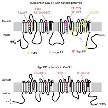
The adenosine triphosphate (ATP)-sensitive K+ (KATP) channel is the most abundant K+ channel active in the skeletal muscle fibers of humans and animals. In the present work, we demonstrate the involvement of the muscular KATP channel in a skeletal muscle disorder known as hypokalemic periodic paralysis (HOPP), which is caused by mutations of the dihydropyridine receptor of the Ca2+ channel. Muscle biopsies excised from three patients with HOPP carrying the R528H mutation of the dihydropyridine receptor showed a reduced sarcolemma KATP current that was not stimulated by magnesium adenosine diphosphate (MgADP; 50-100 microM) and was partially restored by cromakalim. In contrast, large KATP currents stimulated by MgADP were recorded in the healthy subjects. At channel level, an abnormal KATP channel showing several subconductance states was detected in the patients with HOPP. None of these were surveyed in the healthy subjects. Transitions of the KATP channel between subconductance states were also observed after in vitro incubation of the rat muscle with low-K+ solution. The lack of the sarcolemma KATP current observed in these patients explains the symptoms of the disease, i.e., hypokalemia, depolarization of the fibers, and possibly the paralysis following insulin administration. (+info)Activation and inactivation of the voltage-gated sodium channel: role of segment S5 revealed by a novel hyperkalaemic periodic paralysis mutation. (2/55)
Hyperkalaemic periodic paralysis, paramyotonia congenita, and potassium-aggravated myotonia are three autosomal dominant skeletal muscle disorders linked to the SCN4A gene encoding the alpha-subunit of the human voltage-sensitive sodium channel. To date, approximately 20 point mutations causing these disorders have been described. We have identified a new point mutation, in the SCN4A gene, in a family with a hyperkalaemic periodic paralysis phenotype. This mutation predicts an isoleucine-to-phenylalanine substitution at position 1495 located in the transmembrane segment S5 in the fourth homologous domain of the human alpha-subunit sodium channel. Introduction of the I1495F mutation into the wild-type channels disrupted the macroscopic current inactivation decay and shifted both steady-state activation and inactivation to the hyperpolarizing direction. The recovery from fast inactivation was slowed, and there was no effect on channel deactivation. Additionally, a significant enhancement of slow inactivation was observed in the I1495F mutation. In contrast, the T704M mutation, a hyperkalaemic periodic paralysis mutation located in the cytoplasmic interface of the S5 segment of the second domain, also shifted activation in the hyperpolarizing direction but had little effect on fast inactivation and dramatically impaired slow inactivation. These results, showing that the I1495F and T704M hyperkalaemic periodic paralysis mutations both have profound effects on channel activation and fast-slow inactivation, suggest that the S5 segment maybe in a location where fast and slow inactivation converge. (+info)A novel syndrome of episodic muscle weakness maps to xp22.3. (3/55)
We describe a family with a novel disorder characterized by episodic muscle weakness and X-linked inheritance. Eight males in three generations demonstrate the characteristic features of the disorder. Episodes of severe muscle weakness are typically precipitated by febrile illness and affect the facial and extraocular musculature, as well as the trunk and limbs, and resolve spontaneously over a period of weeks to months. Younger members of the family are normal between episodes but during relapses show generalized weakness, ptosis, and fluctuations in strength. In some cases, fatigability can be demonstrated. The proband has late-onset chronic weakness and fatigability. The clinical phenotype has features suggestive both of the congenital myasthenic syndromes and of ion-channel disorders such as the periodic paralyses. We have localized the responsible gene to chromosome Xp22.3, with a maximum two-point LOD score of 4. 52 at a recombination fraction of.0, between OACA2 and DXS9985. (+info)Spectrum of sodium channel disturbances in the nondystrophic myotonias and periodic paralyses. (4/55)
Several heritable forms of myotonia and periodic paralysis are caused by missense mutations in the voltage-gated sodium channel of skeletal muscle. Mutations produce gain-of-function defects, either disrupted inactivation or enhanced activation. Both defects result in too much inward Na current which may either initiate pathologic bursts of action potentials (myotonia) or cause flaccid paralysis by depolarizing fibers to a refractory inexcitable state. Myotonic stiffness and periodic paralysis occur as paroxysmal attacks often triggered by environmental factors such as serum K+, cold, or exercise. Many gaps remain in our understanding of the interactions between genetic predisposition and these environmental influences. Targeted gene manipulation in animals may provide the tools to fill in these gaps. (+info)A double mutation in families with periodic paralysis defines new aspects of sodium channel slow inactivation. (5/55)
Hyperkalemic periodic paralysis (HyperKPP) is an autosomal dominant skeletal muscle disorder caused by single mutations in the SCN4A gene, encoding the human skeletal muscle voltage-gated Na(+) channel. We have now identified one allele with two novel mutations occurring simultaneously in the SCN4A gene. These mutations are found in two distinct families that had symptoms of periodic paralysis and malignant hyperthermia susceptibility. The two nucleotide transitions predict phenylalanine 1490-->leucine and methionine 1493-->isoleucine changes located in the transmembrane segment S5 in the fourth repeat of the alpha-subunit Na(+) channel. Surprisingly, this mutation did not affect fast inactivation parameters. The only defect produced by the double mutant (F1490L-M1493I, expressed in human embryonic kidney 293 cells) is an enhancement of slow inactivation, a unique behavior not seen in the 24 other disease-causing mutations. The behavior observed in these mutant channels demonstrates that manifestation of HyperKPP does not necessarily require disruption of slow inactivation. Our findings may also shed light on the molecular determinants and mechanism of Na(+) channel slow inactivation and help clarify the relationship between Na(+) channel defects and the long-term paralytic attacks experienced by patients with HyperKPP. (+info)MiRP2 forms potassium channels in skeletal muscle with Kv3.4 and is associated with periodic paralysis. (6/55)
The subthreshold, voltage-gated potassium channel of skeletal muscle is shown to contain MinK-related peptide 2 (MiRP2) and the pore-forming subunit Kv3.4. MiRP2-Kv3.4 channels differ from Kv3.4 channels in unitary conductance, voltage-dependent activation, recovery from inactivation, steady-state open probability, and block by a peptide toxin. Thus, MiRP2-Kv3.4 channels set resting membrane potential (RMP) and do not produce afterhyperpolarization or cumulative inactivation to limit action potential frequency. A missense mutation is identified in the gene for MiRP2 (KCNE3) in two families with periodic paralysis and found to segregate with the disease. Mutant MiRP2-Kv3.4 complexes exhibit reduced current density and diminished capacity to set RMP. Thus, MiRP2 operates with a classical potassium channel subunit to govern skeletal muscle function and pathophysiology. (+info)Mutations in Kir2.1 cause the developmental and episodic electrical phenotypes of Andersen's syndrome. (7/55)
Andersen's syndrome is characterized by periodic paralysis, cardiac arrhythmias, and dysmorphic features. We have mapped an Andersen's locus to chromosome 17q23 near the inward rectifying potassium channel gene KCNJ2. A missense mutation in KCNJ2 (encoding D71V) was identified in the linked family. Eight additional mutations were identified in unrelated patients. Expression of two of these mutations in Xenopus oocytes revealed loss of function and a dominant negative effect in Kir2.1 current as assayed by voltage-clamp. We conclude that mutations in Kir2.1 cause Andersen's syndrome. These findings suggest that Kir2.1 plays an important role in developmental signaling in addition to its previously recognized function in controlling cell excitability in skeletal muscle and heart. (+info)Channelopathies: Kir2.1 mutations jeopardize many cell functions. (8/55)
Andersen's syndrome is caused by mutations in the potassium channel Kir2.1, a major determinant of resting membrane potential. The clinical features of this disease illustrate the importance of a stable resting membrane potential for many cell functions. (+info)Paralysis is a loss of muscle function in part or all of your body. It can be localized, affecting only one specific area, or generalized, impacting multiple areas or even the entire body. Paralysis often occurs when something goes wrong with the way messages pass between your brain and muscles. In most cases, paralysis is caused by damage to the nervous system, especially the spinal cord. Other causes include stroke, trauma, infections, and various neurological disorders.
It's important to note that paralysis doesn't always mean a total loss of movement or feeling. Sometimes, it may just cause weakness or numbness in the affected area. The severity and extent of paralysis depend on the underlying cause and the location of the damage in the nervous system.
Respiratory paralysis is a condition characterized by the inability to breathe effectively due to the failure or weakness of the muscles involved in respiration. This can include the diaphragm, intercostal muscles, and other accessory muscles.
In medical terms, it's often associated with conditions that affect the neuromuscular junction, such as botulism, myasthenia gravis, or spinal cord injuries. It can also occur as a complication of general anesthesia, sedative drugs, or certain types of poisoning.
Respiratory paralysis is a serious condition that requires immediate medical attention, as it can lead to lack of oxygen (hypoxia) and buildup of carbon dioxide (hypercapnia) in the body, which can be life-threatening if not treated promptly.
Vocal cord paralysis is a medical condition characterized by the inability of one or both vocal cords to move or function properly due to nerve damage or disruption. The vocal cords are two bands of muscle located in the larynx (voice box) that vibrate to produce sound during speech, singing, and breathing. When the nerves that control the vocal cord movements are damaged or not functioning correctly, the vocal cords may become paralyzed or weakened, leading to voice changes, breathing difficulties, and other symptoms.
The causes of vocal cord paralysis can vary, including neurological disorders, trauma, tumors, surgery, or infections. The diagnosis typically involves a physical examination, including a laryngoscopy, to assess the movement and function of the vocal cords. Treatment options may include voice therapy, surgical procedures, or other interventions to improve voice quality and breathing functions.
Familial periodic paralysis is a group of rare genetic disorders characterized by episodes of muscle weakness or paralysis that recur over time. There are several types of familial periodic paralysis, including hypokalemic periodic paralysis, hyperkalemic periodic paralysis, and normokalemic periodic paralysis, each with its own specific genetic cause and pattern of symptoms.
In general, these disorders are caused by mutations in genes that regulate ion channels in muscle cells, leading to abnormalities in the flow of ions such as potassium in and out of the cells. This can result in changes in muscle excitability and contractility, causing episodes of weakness or paralysis.
The episodes of paralysis in familial periodic paralysis can vary in frequency, duration, and severity. They may be triggered by factors such as rest after exercise, cold or hot temperatures, emotional stress, alcohol consumption, or certain medications. During an episode, the affected muscles may become weak or completely paralyzed, often affecting the limbs but sometimes also involving the muscles of the face, throat, and trunk.
Familial periodic paralysis is typically inherited in an autosomal dominant pattern, meaning that a child has a 50% chance of inheriting the disorder if one parent is affected. However, some cases may arise from new mutations in the affected gene and occur in people with no family history of the disorder.
Treatment for familial periodic paralysis typically involves avoiding triggers and managing symptoms during episodes. In some cases, medications such as potassium-binding agents or diuretics may be used to help prevent or reduce the severity of episodes. Lifestyle modifications, such as a low-carbohydrate or high-sodium diet, may also be recommended in some cases.
Facial paralysis is a loss of facial movement due to damage or dysfunction of the facial nerve (cranial nerve VII). This nerve controls the muscles involved in facial expressions, such as smiling, frowning, and closing the eyes. Damage to one side of the facial nerve can cause weakness or paralysis on that side of the face.
Facial paralysis can result from various conditions, including:
1. Bell's palsy - an idiopathic (unknown cause) inflammation of the facial nerve
2. Trauma - skull fractures, facial injuries, or surgical trauma to the facial nerve
3. Infections - Lyme disease, herpes zoster (shingles), HIV/AIDS, or bacterial infections like meningitis
4. Tumors - benign or malignant growths that compress or invade the facial nerve
5. Stroke - damage to the brainstem where the facial nerve originates
6. Congenital conditions - some people are born with facial paralysis due to genetic factors or birth trauma
Symptoms of facial paralysis may include:
* Inability to move one or more parts of the face, such as the eyebrows, eyelids, mouth, or cheeks
* Drooping of the affected side of the face
* Difficulty closing the eye on the affected side
* Changes in saliva and tear production
* Altered sense of taste
* Pain around the ear or jaw
* Speech difficulties due to weakened facial muscles
Treatment for facial paralysis depends on the underlying cause. In some cases, such as Bell's palsy, spontaneous recovery may occur within a few weeks to months. However, physical therapy, medications, and surgical interventions might be necessary in other situations to improve function and minimize complications.
Sleep paralysis is a temporary inability to move or speak while falling asleep or waking up, often accompanied by frightening hallucinations. These episodes typically last a few seconds to several minutes. During sleep paralysis, a person's body is immobile and cannot perform voluntary muscle movements even though they are fully conscious and awake. This condition can be quite alarming, but it is generally harmless and does not pose any serious threat to one's health. Sleep paralysis is often associated with certain sleep disorders, such as narcolepsy, or other medical conditions, as well as stress, lack of sleep, and changes in sleep patterns.
Tick paralysis is a condition caused by the saliva of certain species of tick that contains neurotoxins. When the tick feeds on the host's blood, the toxin is absorbed, leading to progressive ascending muscle weakness and eventually respiratory failure if not promptly treated. The symptoms typically begin in the lower extremities and progress upward, often within 2-7 days after attachment of the tick. Removal of the attached tick usually leads to improvement in symptoms and full recovery within a few days. It is more commonly seen in children and pets than adults.
Hyperkalemic periodic paralysis (HypPK) is a rare genetic disorder characterized by episodes of muscle weakness or paralysis due to an abnormality in the ion channels that regulate the movement of potassium into and out of muscle fibers. This results in an excessive accumulation of potassium in the blood (hyperkalemia) during attacks, which can interfere with the ability of nerve cells to communicate with muscles and cause temporary muscle weakness or paralysis.
HypPK is caused by mutations in the SCN4A gene, which encodes a sodium channel protein found in skeletal muscle. These genetic changes disrupt the normal functioning of the ion channels, leading to an imbalance in the flow of ions across the muscle cell membrane and affecting muscle excitability.
Episodes of paralysis in HypPK typically begin in childhood or adolescence and can last from several hours to days. Triggers for attacks may include rest following exercise, cold or hot weather, stress, alcohol consumption, infection, or certain medications. Between episodes, individuals with HypPK usually have normal muscle strength and function.
Management of HypPK includes avoiding triggers, maintaining a low-potassium diet, and using medications to prevent or treat attacks. Medications such as thiazide diuretics, acetazolamide, or dichlorphenamide can help lower potassium levels in the blood and reduce the frequency and severity of episodes. In some cases, intravenous glucose and insulin may be used to drive potassium into cells during an attack, thereby reducing its concentration in the blood and alleviating symptoms.
Muscle hypotonia, also known as decreased muscle tone, refers to a condition where the muscles appear to be flaccid or lacking in tension and stiffness. This results in reduced resistance to passive movements, making the limbs feel "floppy" or "like a rag doll." It can affect any muscle group in the body and can be caused by various medical conditions, including neurological disorders, genetic diseases, and injuries to the nervous system. Hypotonia should not be confused with muscle weakness, which refers to the inability to generate normal muscle strength.
Hypokalemia is a medical condition characterized by abnormally low potassium levels in the blood, specifically when the concentration falls below 3.5 milliequivalents per liter (mEq/L). Potassium is an essential electrolyte that helps regulate heart function, nerve signals, and muscle contractions.
Hypokalemia can result from various factors, including inadequate potassium intake, increased potassium loss through the urine or gastrointestinal tract, or shifts of potassium between body compartments. Common causes include diuretic use, vomiting, diarrhea, certain medications, kidney diseases, and hormonal imbalances.
Mild hypokalemia may not cause noticeable symptoms but can still affect the proper functioning of muscles and nerves. More severe cases can lead to muscle weakness, fatigue, cramps, paralysis, heart rhythm abnormalities, and in rare instances, respiratory failure or cardiac arrest. Treatment typically involves addressing the underlying cause and replenishing potassium levels through oral or intravenous (IV) supplementation, depending on the severity of the condition.
Poliomyelitis, also known as polio, is a highly infectious disease caused by a virus that invades the body through the mouth, usually from contaminated water or food. The virus multiplies in the intestine and can invade the nervous system, causing paralysis.
The medical definition of Poliomyelitis includes:
1. An acute viral infection caused by the poliovirus.
2. Characterized by inflammation of the gray matter of the spinal cord (poliomyelitis), leading to muscle weakness, and in some cases, paralysis.
3. The disease primarily affects children under 5 years of age.
4. Transmission occurs through the fecal-oral route or, less frequently, by respiratory droplets.
5. The virus enters the body via the mouth, multiplies in the intestines, and can invade the nervous system.
6. There are three types of poliovirus (types 1, 2, and 3), each capable of causing paralytic polio.
7. Infection with one type does not provide immunity to the other two types.
8. The disease has no cure, but vaccination can prevent it.
9. Two types of vaccines are available: inactivated poliovirus vaccine (IPV) and oral poliovirus vaccine (OPV).
10. Rare complications of OPV include vaccine-associated paralytic polio (VAPP) and circulating vaccine-derived polioviruses (cVDPVs).
Thyrotoxicosis is a medical condition that results from an excess of thyroid hormones in the body, leading to an overactive metabolic state. It can be caused by various factors such as Graves' disease, toxic adenoma, Plummer's disease, or excessive intake of thyroid hormone medication. Symptoms may include rapid heart rate, weight loss, heat intolerance, tremors, and increased sweating, among others. Thyrotoxicosis is not a diagnosis itself but a manifestation of various underlying thyroid disorders. Proper diagnosis and management are crucial to prevent complications and improve quality of life.
NAV1.4, also known as SCN4A, is a gene that encodes for the α subunit of the voltage-gated sodium channel in humans. This channel, specifically located in the skeletal muscle, is responsible for the rapid influx of sodium ions during the initiation and propagation of action potentials, which are critical for muscle contraction.
The NAV1.4 Voltage-Gated Sodium Channel plays a crucial role in the functioning of skeletal muscles. Mutations in this gene can lead to various neuromuscular disorders such as hyperkalemic periodic paralysis, paramyotonia congenita, and potassium-aggravated myotonia, which are characterized by muscle stiffness, cramps, and episodes of weakness or paralysis.
Dicistroviridae is a family of small, non-enveloped, positive-sense single-stranded RNA viruses that infect insects. These viruses are characterized by having two open reading frames (ORFs) in their genome, separated by an intergenic region (IGR). The first ORF encodes for the non-structural proteins involved in replication, while the second ORF encodes for the structural proteins needed for virion assembly.
The family Dicistroviridae includes several important insect pathogens, such as Cricket paralysis virus (CrPV), Rhopalisphum padi virus (RhPV), and Triatoma virus (TrV). These viruses can cause significant economic losses in agriculture by infecting and killing beneficial insects, such as honeybees and silkworms.
Dicistroviridae viruses are transmitted horizontally between hosts through various routes, including oral-fecal transmission, contact with contaminated surfaces, or via vectors such as parasitoids. They have a wide host range within the Insecta class, but they do not infect vertebrates or higher plants.
In terms of medical relevance, Dicistroviridae viruses are not known to infect humans or other mammals. However, understanding their replication and transmission strategies can provide valuable insights into the development of novel antiviral therapies and insect control methods.
Paraplegia is a medical condition characterized by partial or complete loss of motor function and sensation in the lower extremities, typically affecting both legs. This results from damage to the spinal cord, often due to trauma such as accidents, falls, or gunshot wounds, or from diseases like spina bifida, polio, or tumors. The specific area and extent of the injury on the spinal cord determine the severity and location of paralysis. Individuals with paraplegia may require assistive devices for mobility, such as wheelchairs, and may face various health challenges, including pressure sores, urinary tract infections, and chronic pain.
Hyperkalemia is a medical condition characterized by an elevated level of potassium (K+) in the blood serum, specifically when the concentration exceeds 5.0-5.5 mEq/L (milliequivalents per liter). Potassium is a crucial intracellular ion that plays a significant role in various physiological processes, including nerve impulse transmission, muscle contraction, and heart rhythm regulation.
Mild to moderate hyperkalemia might not cause noticeable symptoms but can still have harmful effects on the body, particularly on the cardiovascular system. Severe cases of hyperkalemia (potassium levels > 6.5 mEq/L) can lead to potentially life-threatening arrhythmias and heart failure.
Hyperkalemia may result from various factors, such as kidney dysfunction, hormonal imbalances, medication side effects, trauma, or excessive potassium intake. Prompt identification and management of hyperkalemia are essential to prevent severe complications and ensure proper treatment.
Bell palsy is a peripheral facial nerve palsy, which means that it is a weakness or paralysis of the facial nerves (cranial nerve VII) that causes sudden asymmetric weakness on one side of the face. The symptoms can vary from mild to severe and may include:
* Sudden weakness or paralysis on one side of the face
* Drooping of the mouth, causing difficulty with smiling, eating, drinking, or speaking
* Inability to close one eye
* Dryness of the eye and mouth
* Changes in taste sensation
* Discomfort around the jaw and behind the ear
* Headache
* Increased sensitivity to sound
The exact cause of Bell palsy is not known, but it is believed to be related to inflammation or swelling of the facial nerve. It may also be associated with viral infections such as herpes simplex virus or HIV. In most cases, Bell palsy resolves on its own within a few weeks to months, although some people may experience residual symptoms such as facial weakness or asymmetry. Treatment typically involves corticosteroids and antiviral medications, which can help reduce inflammation and speed up recovery.
Facial nerve diseases refer to a group of medical conditions that affect the function of the facial nerve, also known as the seventh cranial nerve. This nerve is responsible for controlling the muscles of facial expression, and it also carries sensory information from the taste buds in the front two-thirds of the tongue, and regulates saliva flow and tear production.
Facial nerve diseases can cause a variety of symptoms, depending on the specific location and extent of the nerve damage. Common symptoms include:
* Facial weakness or paralysis on one or both sides of the face
* Drooping of the eyelid and corner of the mouth
* Difficulty closing the eye or keeping it closed
* Changes in taste sensation or dryness of the mouth and eyes
* Abnormal sensitivity to sound (hyperacusis)
* Twitching or spasms of the facial muscles
Facial nerve diseases can be caused by a variety of factors, including:
* Infections such as Bell's palsy, Ramsay Hunt syndrome, and Lyme disease
* Trauma or injury to the face or skull
* Tumors that compress or invade the facial nerve
* Neurological conditions such as multiple sclerosis or Guillain-Barre syndrome
* Genetic disorders such as Moebius syndrome or hemifacial microsomia
Treatment for facial nerve diseases depends on the underlying cause and severity of the symptoms. In some cases, medication, physical therapy, or surgery may be necessary to restore function and relieve symptoms.
Neuromuscular blocking agents (NMBAs) are a class of drugs that act on the neuromuscular junction, the site where nerve impulses transmit signals to muscles to cause contraction. NMBAs prevent the transmission of these signals, leading to muscle paralysis. They are used in medical settings during surgical procedures and mechanical ventilation to facilitate intubation, control ventilation, and prevent patient movement. It is important to note that NMBAs do not have any effect on consciousness or pain perception; therefore, they are always used in conjunction with anesthetics and analgesics.
NMBAs can be classified into two main categories based on their mechanism of action:
1. Depolarizing Neuromuscular Blocking Agents: These drugs, such as succinylcholine, cause muscle fasciculations (brief, involuntary contractions) before inducing paralysis. They work by binding to the acetylcholine receptors at the neuromuscular junction and depolarizing the membrane, which results in muscle paralysis. However, the continuous depolarization also causes desensitization of the receptors, leading to a loss of effectiveness over time. Depolarizing NMBAs have a relatively short duration of action.
2. Non-depolarizing Neuromuscular Blocking Agents: These drugs, such as rocuronium, vecuronium, and pancuronium, do not cause muscle fasciculations. They work by binding to the acetylcholine receptors at the neuromuscular junction without depolarizing the membrane, which prevents the transmission of nerve impulses to muscles and leads to paralysis. Non-depolarizing NMBAs have a longer duration of action compared to depolarizing NMBAs.
Close monitoring of neuromuscular function is essential when using NMBAs to ensure adequate reversal of their effects before the patient regains consciousness. This can be achieved through the use of nerve stimulators, which assess the degree of blockade and help guide the administration of reversal agents when necessary.
Quadriplegia, also known as tetraplegia, is a medical condition characterized by paralysis affecting all four limbs and the trunk of the body. It results from damage to the cervical spinal cord, typically at levels C1-C8, which controls signals to the muscles in the arms, hands, trunk, legs, and pelvic organs. The extent of quadriplegia can vary widely, ranging from weakness to complete loss of movement and sensation below the level of injury. Other symptoms may include difficulty breathing, bowel and bladder dysfunction, and sexual dysfunction. The severity and prognosis depend on the location and extent of the spinal cord injury.
The Recurrent Laryngeal Nerve (RLN) is a branch of the vagus nerve (cranial nerve X), which is a mixed sensory, motor, and autonomic nerve. The RLN has important functions in providing motor innervation to the intrinsic muscles of the larynx, except for the cricothyroid muscle, which is supplied by the external branch of the superior laryngeal nerve.
The recurrent laryngeal nerve supplies all the muscles that are responsible for adduction (bringing together) of the vocal cords, including the vocalis muscle, lateral cricoarytenoid, thyroarytenoid, and interarytenoid muscles. These muscles play a crucial role in voice production, coughing, and swallowing.
The right recurrent laryngeal nerve has a longer course than the left one. It loops around the subclavian artery in the chest before ascending to the larynx, while the left RLN hooks around the arch of the aorta. This anatomical course makes them vulnerable to injury during various surgical procedures, such as thyroidectomy and neck dissection, leading to potential voice impairment or vocal cord paralysis.
Obstetric paralysis is a specific type of paralysis that can occur as a result of complications during childbirth. It is also known as "birth paralysis" or "puerperal paralysis."
The condition is typically caused by nerve damage or trauma to the brachial plexus, which is a network of nerves that runs from the spinal cord in the neck and provides movement and sensation to the shoulders, arms, and hands. Obstetric paralysis can occur when the brachial plexus is stretched or compressed during childbirth, particularly in difficult deliveries where forceps or vacuum extraction may be used.
There are several types of obstetric paralysis, including:
* Erb's palsy: This type of obstetric paralysis affects the upper brachial plexus and can cause weakness or paralysis in the arm, particularly the shoulder and elbow.
* Klumpke's palsy: This type of obstetric paralysis affects the lower brachial plexus and can cause weakness or paralysis in the hand and forearm.
* Total brachial plexus injury: This is a rare but severe form of obstetric paralysis that involves injury to all of the nerves in the brachial plexus, resulting in complete paralysis of the arm.
The severity of obstetric paralysis can vary widely, from mild weakness to complete paralysis. In some cases, the condition may resolve on its own within a few months, while in other cases, surgery or physical therapy may be necessary to help restore function.
Poliovirus is a human enterovirus, specifically a type of picornavirus, that is the causative agent of poliomyelitis (polio). It is a small, non-enveloped, single-stranded, positive-sense RNA virus. There are three serotypes of Poliovirus (types 1, 2 and 3) which can cause different degrees of severity in the disease. The virus primarily spreads through the fecal-oral route and infects the gastrointestinal tract, from where it can invade the nervous system and cause paralysis.
The Poliovirus has an icosahedral symmetry, with a diameter of about 30 nanometers. It contains a single stranded RNA genome which is encapsidated in a protein shell called capsid. The capsid is made up of 60 units of four different proteins (VP1, VP2, VP3 and VP4).
Poliovirus has been eradicated from most countries of the world through widespread vaccination with inactivated poliovirus vaccine (IPV) or oral poliovirus vaccine (OPV). However, it still remains endemic in a few countries and is considered a major public health concern.
Poliovirus Vaccine, Oral (OPV) is a vaccine used to prevent poliomyelitis (polio). It contains live attenuated (weakened) polioviruses, which stimulate an immune response in the body and provide protection against all three types of wild, infectious polioviruses. OPV is given by mouth, usually in drops, and it replicates in the gastrointestinal tract, where it induces a strong immune response. This response not only protects the individual who receives the vaccine but also helps to stop the spread of poliovirus in the community, providing indirect protection (herd immunity) to those who are not vaccinated. OPV is safe, effective, and easy to administer, making it an important tool for global polio eradication efforts. However, due to the risk of vaccine-associated paralytic polio (VAPP), inactivated poliovirus vaccine (IPV) is recommended for routine immunization in some countries.
The phrenic nerve is a motor nerve that originates from the cervical spine (C3-C5) and descends through the neck to reach the diaphragm, which is the primary muscle used for breathing. The main function of the phrenic nerve is to innervate the diaphragm and control its contraction and relaxation, thereby enabling respiration.
Damage or injury to the phrenic nerve can result in paralysis of the diaphragm, leading to difficulty breathing and potentially causing respiratory failure. Certain medical conditions, such as neuromuscular disorders, spinal cord injuries, and tumors, can affect the phrenic nerve and impair its function.
Botulinum toxins type A are neurotoxins produced by the bacterium Clostridium botulinum and related species. These toxins act by blocking the release of acetylcholine at the neuromuscular junction, leading to muscle paralysis. Botulinum toxin type A is used in medical treatments for various conditions characterized by muscle spasticity or excessive muscle activity, such as cervical dystonia, blepharospasm, strabismus, and chronic migraine. It is also used cosmetically to reduce the appearance of wrinkles by temporarily paralyzing the muscles that cause them. The commercial forms of botulinum toxin type A include Botox, Dysport, and Xeomin.
Pancuronium is defined as a non-depolarizing neuromuscular blocking agent, which is used in anesthesia practice to provide skeletal muscle relaxation during surgery. It works by competitively inhibiting the binding of acetylcholine to nicotinic receptors at the motor endplate, thereby preventing muscle contraction. Pancuronium has a intermediate duration of action and is often used for routine surgical procedures requiring muscle relaxation. It is administered intravenously and is typically reversed with an anticholinesterase agent such as neostigmine at the conclusion of surgery.
Laryngoplasty is a surgical procedure that involves reconstructing or reinforcing the larynx, specifically the vocal cords. The goal of this procedure can be to improve voice quality, restore breathing function, or manage airway obstructions caused by various conditions such as vocal cord paralysis, vocal fold bowing, or scarring.
There are different types of laryngoplasties, including:
1. Type I Thyroplasty (Medialization Laryngoplasty): This procedure involves placing an implant made of silicone, Gore-Tex, or other materials in the thyroid cartilage to medialize (move towards the midline) and support the paralyzed vocal cord. This helps improve voice quality and airway closure during speech and swallowing.
2. Arytenoid Adduction: In this procedure, the arytenoid cartilage is repositioned or fixed in place to help approximate (bring together) the vocal cords. It is often performed along with a Type I Thyroplasty for better voice and airway outcomes.
3. Laryngeal Framework Surgery: This is a more extensive procedure that involves reshaping the laryngeal framework, including the thyroid and cricoid cartilages, to improve voice, swallowing, or breathing function.
The choice of surgical technique depends on the underlying condition, its severity, and the patient's individual needs and goals.
A diaphragm is a thin, dome-shaped muscle that separates the chest cavity from the abdominal cavity. It plays a vital role in the process of breathing as it contracts and flattens to draw air into the lungs (inhalation) and relaxes and returns to its domed shape to expel air out of the lungs (exhalation).
In addition, a diaphragm is also a type of barrier method of birth control. It is a flexible dome-shaped device made of silicone that fits over the cervix inside the vagina. When used correctly and consistently, it prevents sperm from entering the uterus and fertilizing an egg, thereby preventing pregnancy.
Botulinum toxins are neurotoxic proteins produced by the bacterium Clostridium botulinum and related species. They are the most potent naturally occurring toxins, and are responsible for the paralytic illness known as botulism. There are seven distinct botulinum toxin serotypes (A-G), each of which targets specific proteins in the nervous system, leading to inhibition of neurotransmitter release and subsequent muscle paralysis.
In clinical settings, botulinum toxins have been used for therapeutic purposes due to their ability to cause temporary muscle relaxation. Botulinum toxin type A (Botox) is the most commonly used serotype in medical treatments, including management of dystonias, spasticity, migraines, and certain neurological disorders. Additionally, botulinum toxins are widely employed in aesthetic medicine for reducing wrinkles and fine lines by temporarily paralyzing facial muscles.
It is important to note that while botulinum toxins have therapeutic benefits when used appropriately, they can also pose significant health risks if misused or improperly handled. Proper medical training and supervision are essential for safe and effective utilization of these powerful toxins.
Enterovirus infections are viral illnesses caused by enteroviruses, which are a type of picornavirus. These viruses commonly infect the gastrointestinal tract and can cause a variety of symptoms depending on the specific type of enterovirus and the age and overall health of the infected individual.
There are over 100 different types of enteroviruses, including polioviruses, coxsackieviruses, echoviruses, and newer enteroviruses such as EV-D68 and EV-A71. Some enterovirus infections may be asymptomatic or cause only mild symptoms, while others can lead to more severe illnesses.
Common symptoms of enterovirus infections include fever, sore throat, runny nose, cough, muscle aches, and skin rashes. In some cases, enteroviruses can cause more serious complications such as meningitis (inflammation of the membranes surrounding the brain and spinal cord), encephalitis (inflammation of the brain), myocarditis (inflammation of the heart muscle), and paralysis.
Enterovirus infections are typically spread through close contact with an infected person, such as through respiratory droplets or fecal-oral transmission. They can also be spread through contaminated surfaces or objects. Preventive measures include good hygiene practices, such as washing hands frequently and avoiding close contact with sick individuals.
There are no specific antiviral treatments for enterovirus infections, and most cases resolve on their own within a few days to a week. However, severe cases may require hospitalization and supportive care, such as fluids and medication to manage symptoms. Prevention efforts include vaccination against poliovirus and surveillance for emerging enteroviruses.
Andersen Syndrome is a rare genetic disorder characterized by the presence of three major features:
1. Periodic episodes of muscle weakness (periodic paralysis)
2. Potassium-sensitive ventricular arrhythmias
3. Physical deformities of the face and skeleton
The periodic paralysis in Andersen Syndrome is typically less severe than other forms of periodic paralysis, and it can be triggered by factors such as cold, emotional stress, or infection. The potassium-sensitive ventricular arrhythmias can be life-threatening and may require treatment with medications or an implantable cardioverter-defibrillator (ICD).
The physical deformities associated with Andersen Syndrome can include a short stature, low-set ears, a broad nose, widely spaced eyes, a cleft palate, and skeletal abnormalities such as scoliosis or clubfoot. These features may vary in severity among individuals with the disorder.
Andersen Syndrome is caused by mutations in the gene for the protein called the inward rectifier potassium channel (Kir2.1), which is involved in regulating the flow of potassium ions across cell membranes. This gene is located on chromosome 17 and is designated KCNJ2. The disorder is inherited in an autosomal dominant manner, meaning that a person has a 50% chance of inheriting the mutated gene from an affected parent. However, some cases of Andersen Syndrome are due to new (de novo) mutations and occur in people with no family history of the disorder.
Myotonia is a condition characterized by the delayed relaxation of a muscle after voluntary contraction or electrical stimulation, resulting in stiffness or difficulty with relaxing the muscles. It's often associated with certain neuromuscular disorders such as myotonic dystrophy and myotonia congenita. The prolonged muscle contraction can cause stiffness, especially after periods of rest, and may improve with repeated contractions (warm-up phenomenon).
Laryngoscopy is a medical procedure that involves the examination of the larynx, which is the upper part of the windpipe (trachea), and the vocal cords using a specialized instrument called a laryngoscope. The laryngoscope is inserted through the mouth or nose to provide a clear view of the larynx and surrounding structures. This procedure can be performed for diagnostic purposes, such as identifying abnormalities like growths, inflammation, or injuries, or for therapeutic reasons, such as removing foreign objects or taking tissue samples for biopsy. There are different types of laryngoscopes and techniques used depending on the reason for the examination and the patient's specific needs.
Neuromuscular non-depolarizing agents are a type of muscle relaxant medication used in anesthesia and critical care settings to facilitate endotracheal intubation, mechanical ventilation, and to prevent muscle contractions during surgery. These agents work by competitively binding to the acetylcholine receptors at the neuromuscular junction, without activating them, thereby preventing the initiation of muscle contraction.
Examples of non-depolarizing neuromuscular blocking agents include:
* Vecuronium
* Rocuronium
* Pancuronium
* Atracurium
* Cisatracurium
* Mivacurium
These medications have a reversible effect and their duration of action can be prolonged in patients with impaired renal or hepatic function, acid-base imbalances, electrolyte abnormalities, or in those who are taking other medications that interact with these agents. Therefore, it is important to monitor the patient's neuromuscular function during and after the administration of non-depolarizing neuromuscular blocking agents.
The spinal cord is a major part of the nervous system, extending from the brainstem and continuing down to the lower back. It is a slender, tubular bundle of nerve fibers (axons) and support cells (glial cells) that carries signals between the brain and the rest of the body. The spinal cord primarily serves as a conduit for motor information, which travels from the brain to the muscles, and sensory information, which travels from the body to the brain. It also contains neurons that can independently process and respond to information within the spinal cord without direct input from the brain.
The spinal cord is protected by the bony vertebral column (spine) and is divided into 31 segments: 8 cervical, 12 thoracic, 5 lumbar, 5 sacral, and 1 coccygeal. Each segment corresponds to a specific region of the body and gives rise to pairs of spinal nerves that exit through the intervertebral foramina at each level.
The spinal cord is responsible for several vital functions, including:
1. Reflexes: Simple reflex actions, such as the withdrawal reflex when touching a hot surface, are mediated by the spinal cord without involving the brain.
2. Muscle control: The spinal cord carries motor signals from the brain to the muscles, enabling voluntary movement and muscle tone regulation.
3. Sensory perception: The spinal cord transmits sensory information, such as touch, temperature, pain, and vibration, from the body to the brain for processing and awareness.
4. Autonomic functions: The sympathetic and parasympathetic divisions of the autonomic nervous system originate in the thoracolumbar and sacral regions of the spinal cord, respectively, controlling involuntary physiological responses like heart rate, blood pressure, digestion, and respiration.
Damage to the spinal cord can result in various degrees of paralysis or loss of sensation below the level of injury, depending on the severity and location of the damage.
Ophthalmoplegia is a medical term that refers to the paralysis or weakness of the eye muscles, which can result in double vision (diplopia) or difficulty moving the eyes. It can be caused by various conditions, including nerve damage, muscle disorders, or neurological diseases such as myasthenia gravis or multiple sclerosis. Ophthalmoplegia can affect one or more eye muscles and can be partial or complete. Depending on the underlying cause, ophthalmoplegia may be treatable with medications, surgery, or other interventions.
Renal tubular acidosis (RTA) is a medical condition that occurs when the kidneys are unable to properly excrete acid into the urine, leading to an accumulation of acid in the bloodstream. This results in a state of metabolic acidosis.
There are several types of RTA, but renal tubular acidosis type 1 (also known as distal RTA) is characterized by a defect in the ability of the distal tubules to acidify the urine, leading to an inability to lower the pH of the urine below 5.5, even in the face of metabolic acidosis. This results in a persistently alkaline urine, which can lead to calcium phosphate stones and bone demineralization.
Type 1 RTA is often caused by inherited genetic defects, but it can also be acquired due to various kidney diseases, drugs, or autoimmune disorders. Symptoms of type 1 RTA may include fatigue, weakness, muscle cramps, decreased appetite, and vomiting. Treatment typically involves alkali therapy to correct the acidosis and prevent complications.
Neuromuscular blockade (NMB) is a pharmacological state in which the communication between nerves and muscles is interrupted by blocking the neuromuscular junction, thereby preventing muscle contraction. This condition can be achieved through the use of certain medications called neuromuscular blocking agents (NMBAs). These drugs are commonly used during surgical procedures to facilitate endotracheal intubation, mechanical ventilation, and to prevent patient movement and minimize potential injury during surgery. NMBs are classified into two main categories based on their mechanism of action: depolarizing and non-depolarizing agents.
Depolarizing neuromuscular blocking agents, such as succinylcholine, work by activating the nicotinic acetylcholine receptors at the neuromuscular junction, causing a sustained depolarization and muscle contraction followed by flaccid paralysis. Non-depolarizing neuromuscular blocking agents, such as rocuronium, vecuronium, pancuronium, and atracurium, bind to the receptors without activating them, thereby preventing acetylcholine from binding and transmitting the signal for muscle contraction.
Clinical monitoring of neuromuscular blockade is essential to ensure proper dosing and avoid complications such as residual curarization, which can lead to respiratory compromise in the postoperative period. Monitoring techniques include peripheral nerve stimulation and train-of-four (TOF) assessment to evaluate the depth of neuromuscular blockade and guide the administration of reversal agents when appropriate.
Vocal cords, also known as vocal folds, are specialized bands of muscle, membrane, and connective tissue located within the larynx (voice box). They are essential for speech, singing, and other sounds produced by the human voice. The vocal cords vibrate when air from the lungs is passed through them, creating sound waves that vary in pitch and volume based on the tension, length, and mass of the vocal cords. These sound waves are then further modified by the resonance chambers of the throat, nose, and mouth to produce speech and other vocalizations.
The facial nerve, also known as the seventh cranial nerve (CN VII), is a mixed nerve that carries both sensory and motor fibers. Its functions include controlling the muscles involved in facial expressions, taste sensation from the anterior two-thirds of the tongue, and secretomotor function to the lacrimal and salivary glands.
The facial nerve originates from the brainstem and exits the skull through the internal acoustic meatus. It then passes through the facial canal in the temporal bone before branching out to innervate various structures of the face. The main branches of the facial nerve include:
1. Temporal branch: Innervates the frontalis, corrugator supercilii, and orbicularis oculi muscles responsible for eyebrow movements and eyelid closure.
2. Zygomatic branch: Supplies the muscles that elevate the upper lip and wrinkle the nose.
3. Buccal branch: Innervates the muscles of the cheek and lips, allowing for facial expressions such as smiling and puckering.
4. Mandibular branch: Controls the muscles responsible for lower lip movement and depressing the angle of the mouth.
5. Cervical branch: Innervates the platysma muscle in the neck, which helps to depress the lower jaw and wrinkle the skin of the neck.
Damage to the facial nerve can result in various symptoms, such as facial weakness or paralysis, loss of taste sensation, and dry eyes or mouth due to impaired secretion.
Botulism is a rare but serious condition caused by the toxin produced by the bacterium Clostridium botulinum. The neurotoxin causes muscle paralysis, which can lead to respiratory failure and death if not treated promptly. Botulism can occur in three main forms: foodborne, wound, and infant.
Foodborne botulism is caused by consuming contaminated food, usually home-canned or fermented foods with low acid content. Wound botulism occurs when the bacterium infects a wound and produces toxin in the body. Infant botulism affects babies under one year of age who have ingested spores of the bacterium, which then colonize the intestines and produce toxin.
Symptoms of botulism include double vision, drooping eyelids, slurred speech, difficulty swallowing, dry mouth, muscle weakness, and paralysis that progresses downward from the head to the limbs. Treatment typically involves supportive care such as mechanical ventilation, intensive care unit monitoring, and antitoxin therapy. Prevention measures include proper food handling and canning techniques, prompt wound care, and avoiding consumption of known sources of contaminated food.
Colony Collapse Disorder (CCD) is a phenomenon in which the majority of worker bees in a honeybee colony disappear, leaving behind the queen, immature bees, and enough food to survive. The exact cause of CCD is unknown, but it's believed to be due to a combination of factors such as pests, pathogens, poor nutrition, and exposure to environmental stressors like pesticides. This disorder has been a major concern for the honeybee population and agriculture industry because honeybees play a crucial role in pollinating crops.
The neuromuscular junction (NMJ) is the specialized synapse or chemical communication point, where the motor neuron's nerve terminal (presynaptic element) meets the muscle fiber's motor end plate (postsynaptic element). This junction plays a crucial role in controlling muscle contraction and relaxation.
At the NMJ, the neurotransmitter acetylcholine is released from the presynaptic nerve terminal into the synaptic cleft, following an action potential. Acetylcholine then binds to nicotinic acetylcholine receptors on the postsynaptic membrane of the muscle fiber, leading to the generation of an end-plate potential. If sufficient end-plate potentials are generated and summate, they will trigger an action potential in the muscle fiber, ultimately causing muscle contraction.
Dysfunction at the neuromuscular junction can result in various neuromuscular disorders, such as myasthenia gravis, where autoantibodies attack acetylcholine receptors, leading to muscle weakness and fatigue.
Insect viruses, also known as entomoviruses, are viruses that specifically infect and replicate in insect hosts. These viruses can be found in various insect species, including those of medical and agricultural importance. Insect viruses can cause diseases in insect populations, leading to significant impacts on their growth, development, and survival. Some insect viruses have been studied as potential biological control agents for managing pest insects that affect crops or transmit diseases. Examples of insect viruses include Baculoviridae, Reoviridae, and Picornaviridae families.
Motor neurons are specialized nerve cells in the brain and spinal cord that play a crucial role in controlling voluntary muscle movements. They transmit electrical signals from the brain to the muscles, enabling us to perform actions such as walking, talking, and swallowing. There are two types of motor neurons: upper motor neurons, which originate in the brain's motor cortex and travel down to the brainstem and spinal cord; and lower motor neurons, which extend from the brainstem and spinal cord to the muscles. Damage or degeneration of these motor neurons can lead to various neurological disorders, such as amyotrophic lateral sclerosis (ALS) and spinal muscular atrophy (SMA).
Succinylcholine is a neuromuscular blocking agent, a type of muscle relaxant used in anesthesia during surgical procedures. It works by inhibiting the transmission of nerve impulses at the neuromuscular junction, leading to temporary paralysis of skeletal muscles. This facilitates endotracheal intubation and mechanical ventilation during surgery. Succinylcholine has a rapid onset of action and is metabolized quickly, making it useful for short surgical procedures. However, its use may be associated with certain adverse effects, such as increased heart rate, muscle fasciculations, and potentially life-threatening hyperkalemia in susceptible individuals.
Spinal cord diseases refer to a group of conditions that affect the spinal cord, which is a part of the central nervous system responsible for transmitting messages between the brain and the rest of the body. These diseases can cause damage to the spinal cord, leading to various symptoms such as muscle weakness, numbness, pain, bladder and bowel dysfunction, and difficulty with movement and coordination.
Spinal cord diseases can be congenital or acquired, and they can result from a variety of causes, including infections, injuries, tumors, degenerative conditions, autoimmune disorders, and genetic factors. Some examples of spinal cord diseases include multiple sclerosis, spina bifida, spinal cord injury, herniated discs, spinal stenosis, and motor neuron diseases such as amyotrophic lateral sclerosis (ALS).
The treatment for spinal cord diseases varies depending on the underlying cause and severity of the condition. Treatment options may include medication, physical therapy, surgery, and rehabilitation. In some cases, the damage to the spinal cord may be irreversible, leading to permanent disability or paralysis.
Brachial plexus neuritis, also known as Parsonage-Turner syndrome or neuralgic amyotrophy, is a medical condition characterized by inflammation and damage to the brachial plexus. The brachial plexus is a network of nerves that originates from the spinal cord in the neck and travels down the arm, controlling movement and sensation in the shoulder, arm, and hand.
In Brachial plexus neuritis, the insulating covering of the nerves (myelin sheath) is damaged or destroyed, leading to impaired nerve function. The exact cause of this condition is not fully understood, but it can be associated with viral infections, trauma, surgery, or immunological disorders.
Symptoms of Brachial plexus neuritis may include sudden onset of severe pain in the shoulder and arm, followed by weakness or paralysis of the affected muscles. There may also be numbness, tingling, or loss of sensation in the affected areas. In some cases, recovery can occur spontaneously within a few months, while others may experience persistent weakness or disability. Treatment typically involves pain management, physical therapy, and in some cases, corticosteroids or other medications to reduce inflammation.
Electromyography (EMG) is a medical diagnostic procedure that measures the electrical activity of skeletal muscles during contraction and at rest. It involves inserting a thin needle electrode into the muscle to record the electrical signals generated by the muscle fibers. These signals are then displayed on an oscilloscope and may be heard through a speaker.
EMG can help diagnose various neuromuscular disorders, such as muscle weakness, numbness, or pain, and can distinguish between muscle and nerve disorders. It is often used in conjunction with other diagnostic tests, such as nerve conduction studies, to provide a comprehensive evaluation of the nervous system.
EMG is typically performed by a neurologist or a physiatrist, and the procedure may cause some discomfort or pain, although this is usually minimal. The results of an EMG can help guide treatment decisions and monitor the progression of neuromuscular conditions over time.
Recurrent laryngeal nerve injuries refer to damages or trauma inflicted on the recurrent laryngeal nerve, which is a branch of the vagus nerve that supplies motor function to the intrinsic muscles of the larynx, except for the cricothyroid muscle. This nerve plays a crucial role in controlling vocal fold movement and swallowing.
Injuries to this nerve can result in voice changes, hoarseness, or even complete loss of voice, depending on the severity and location of the injury. Additionally, it may also lead to breathing difficulties, coughing, and choking while swallowing due to impaired laryngeal function.
Recurrent laryngeal nerve injuries can occur due to various reasons, such as surgical complications (particularly during thyroid or neck surgeries), tumors, infections, inflammation, or direct trauma to the neck region. In some cases, these injuries may be temporary and resolve on their own or through appropriate treatment; however, severe or prolonged injuries might require medical intervention, including possible surgical repair.
Guillain-Barré syndrome (GBS) is a rare autoimmune disorder in which the body's immune system mistakenly attacks the peripheral nervous system, leading to muscle weakness, tingling sensations, and sometimes paralysis. The peripheral nervous system includes the nerves that control our movements and transmit signals from our skin, muscles, and joints to our brain.
The onset of GBS usually occurs after a viral or bacterial infection, such as respiratory or gastrointestinal infections, or following surgery, vaccinations, or other immune system triggers. The exact cause of the immune response that leads to GBS is not fully understood.
GBS typically progresses rapidly over days or weeks, with symptoms reaching their peak within 2-4 weeks after onset. Most people with GBS experience muscle weakness that starts in the lower limbs and spreads upward to the upper body, arms, and face. In severe cases, the diaphragm and chest muscles may become weakened, leading to difficulty breathing and requiring mechanical ventilation.
The diagnosis of GBS is based on clinical symptoms, nerve conduction studies, and sometimes cerebrospinal fluid analysis. Treatment typically involves supportive care, such as pain management, physical therapy, and respiratory support if necessary. In addition, plasma exchange (plasmapheresis) or intravenous immunoglobulin (IVIG) may be used to reduce the severity of symptoms and speed up recovery.
While most people with GBS recover completely or with minimal residual symptoms, some may experience long-term disability or require ongoing medical care. The prognosis for GBS varies depending on the severity of the illness and the individual's age and overall health.
Myelitis is a medical term that refers to inflammation of the spinal cord. This inflammation can cause damage to the myelin sheath, which is the protective covering of nerve fibers in the spinal cord. As a result, the transmission of nerve impulses along the spinal cord may be disrupted, leading to various neurological symptoms.
Myelitis can affect any part of the spinal cord and can have many different causes, including infections (such as viral or bacterial infections), autoimmune disorders (such as multiple sclerosis), and other conditions (such as spinal cord injuries or tumors). The specific symptoms of myelitis depend on the location and severity of the inflammation. They may include muscle weakness, numbness or tingling sensations, pain, bladder or bowel dysfunction, and difficulty with coordination and balance.
Myelitis can be a serious condition that requires prompt medical attention and treatment. Treatment typically focuses on addressing the underlying cause of the inflammation, as well as managing symptoms and supporting recovery.
Skeletal muscle, also known as striated or voluntary muscle, is a type of muscle that is attached to bones by tendons or aponeuroses and functions to produce movements and support the posture of the body. It is composed of long, multinucleated fibers that are arranged in parallel bundles and are characterized by alternating light and dark bands, giving them a striped appearance under a microscope. Skeletal muscle is under voluntary control, meaning that it is consciously activated through signals from the nervous system. It is responsible for activities such as walking, running, jumping, and lifting objects.
Spinal cord injuries (SCI) refer to damage to the spinal cord that results in a loss of function, such as mobility or feeling. This injury can be caused by direct trauma to the spine or by indirect damage resulting from disease or degeneration of surrounding bones, tissues, or blood vessels. The location and severity of the injury on the spinal cord will determine which parts of the body are affected and to what extent.
The effects of SCI can range from mild sensory changes to severe paralysis, including loss of motor function, autonomic dysfunction, and possible changes in sensation, strength, and reflexes below the level of injury. These injuries are typically classified as complete or incomplete, depending on whether there is any remaining function below the level of injury.
Immediate medical attention is crucial for spinal cord injuries to prevent further damage and improve the chances of recovery. Treatment usually involves immobilization of the spine, medications to reduce swelling and pressure, surgery to stabilize the spine, and rehabilitation to help regain lost function. Despite advances in treatment, SCI can have a significant impact on a person's quality of life and ability to perform daily activities.
Channelopathies are genetic disorders that are caused by mutations in the genes that encode for ion channels. Ion channels are specialized proteins that regulate the flow of ions, such as sodium, potassium, and calcium, across cell membranes. These ion channels play a crucial role in various physiological processes, including the generation and transmission of electrical signals in the body.
Channelopathies can affect various organs and systems in the body, depending on the type of ion channel that is affected. For example, mutations in sodium channel genes can cause neuromuscular disorders such as epilepsy, migraine, and periodic paralysis. Mutations in potassium channel genes can cause cardiac arrhythmias, while mutations in calcium channel genes can cause neurological disorders such as episodic ataxia and hemiplegic migraine.
The symptoms of channelopathies can vary widely depending on the specific disorder and the severity of the mutation. Treatment typically involves managing the symptoms and may include medications, lifestyle modifications, or in some cases, surgery.
Muscular diseases, also known as myopathies, refer to a group of conditions that affect the functionality and health of muscle tissue. These diseases can be inherited or acquired and may result from inflammation, infection, injury, or degenerative processes. They can cause symptoms such as weakness, stiffness, cramping, spasms, wasting, and loss of muscle function.
Examples of muscular diseases include:
1. Duchenne Muscular Dystrophy (DMD): A genetic disorder that results in progressive muscle weakness and degeneration due to a lack of dystrophin protein.
2. Myasthenia Gravis: An autoimmune disease that causes muscle weakness and fatigue, typically affecting the eyes and face, throat, and limbs.
3. Inclusion Body Myositis (IBM): A progressive muscle disorder characterized by muscle inflammation and wasting, typically affecting older adults.
4. Polymyositis: An inflammatory myopathy that causes muscle weakness and inflammation throughout the body.
5. Metabolic Myopathies: A group of inherited disorders that affect muscle metabolism, leading to exercise intolerance, muscle weakness, and other symptoms.
6. Muscular Dystonias: Involuntary muscle contractions and spasms that can cause abnormal postures or movements.
It is important to note that muscular diseases can have a significant impact on an individual's quality of life, mobility, and overall health. Proper diagnosis and treatment are crucial for managing symptoms and improving outcomes.
Muscle weakness is a condition in which muscles cannot develop the expected level of physical force or power. This results in reduced muscle function and can be caused by various factors, including nerve damage, muscle diseases, or hormonal imbalances. Muscle weakness may manifest as difficulty lifting objects, maintaining posture, or performing daily activities. It is essential to consult a healthcare professional for proper diagnosis and treatment of muscle weakness.
Hoarseness is a condition characterized by an abnormal change in the quality of voice, making it sound rough, breathy, strained, or weak. Medically, it's described as a disorder of phonation, which is the process of producing sound by vibrating the vocal cords in the larynx (voice box). Hoarseness can be caused by various factors, such as inflammation, irritation, or injury to the vocal cords, and may result in symptoms like altered voice pitch, volume, and clarity. It's essential to consult a healthcare professional if hoarseness persists for more than two weeks, especially if it's accompanied by other concerning symptoms like difficulty swallowing or breathing.
Thyroid cartilage is the largest and most superior of the laryngeal cartilages, forming the front and greater part of the larynx, also known as the "Adam's apple" in humans. It serves to protect the vocal cords and provides attachment for various muscles involved in voice production. The thyroid cartilage consists of two laminae that join in front at an angle, creating a noticeable prominence in the anterior neck. This structure is crucial in speech formation and swallowing functions.
Poliovirus vaccines are preparations used for active immunization against poliomyelitis, a highly infectious disease caused by the poliovirus. The two types of poliovirus vaccines available are:
1. Inactivated Poliovirus Vaccine (IPV): This vaccine contains inactivated (killed) poliovirus strains of all three serotypes. IPV is typically administered through an injection, usually in combination with other vaccines. It provides a strong immune response and does not carry the risk of vaccine-associated paralytic polio (VAPP), which is a rare but serious adverse event associated with the oral poliovirus vaccine (OPV).
2. Oral Poliovirus Vaccine (OPV): This vaccine contains live attenuated (weakened) poliovirus strains of all three serotypes. OPV is administered orally and induces both humoral and intestinal immunity, which helps prevent the spread of the virus in a community. However, there is a small risk of VAPP associated with this vaccine, especially after multiple doses. In rare cases, the weakened virus can revert to its virulent form and cause paralytic polio in the vaccinated individual or their close contacts.
Both IPV and OPV have been instrumental in global efforts to eradicate polio. The World Health Organization (WHO) recommends using IPV in routine immunization programs, while using OPV during supplementary immunization activities in areas with a high risk of poliovirus transmission.
Myotonic disorders are a group of genetic muscle diseases characterized by the inability to relax muscles (myotonia) after contraction. Myotonia can cause symptoms such as stiffness, muscle spasms, and prolonged muscle contractions or cramps. These disorders may also be associated with other symptoms, including muscle weakness, wasting, and various systemic features.
The most common myotonic disorder is myotonic dystrophy type 1 (DM1), which is caused by a mutation in the DMPK gene. Myotonic dystrophy type 2 (DM2) is another form of myotonic dystrophy, resulting from a mutation in the CNBP gene. These two forms of myotonic dystrophy have distinct genetic causes but share similar clinical features, such as myotonia and muscle weakness.
Other less common myotonic disorders include:
1. Myotonia congenita - A group of inherited conditions characterized by muscle stiffness from birth or early childhood. There are two main types: Thomsen's disease (autosomal dominant) and Becker's disease (autosomal recessive).
2. Paramyotonia congenita - An autosomal dominant disorder characterized by muscle stiffness triggered by cold temperatures or physical exertion.
3. Potassium-aggravated myotonia (PAM) - A rare, autosomal dominant condition with symptoms similar to paramyotonia congenita but without the cold sensitivity.
4. Myotonia fluctuans - A rare, autosomal dominant disorder characterized by fluctuating muscle stiffness and cramps.
5. Acquired myotonia - Rare cases of myotonia caused by factors other than genetic mutations, such as medication side effects or underlying medical conditions.
Myotonic disorders can significantly impact a person's quality of life, making daily activities challenging. Proper diagnosis and management are essential to help alleviate symptoms and improve overall well-being.
Facial nerve injuries refer to damages or trauma inflicted on the facial nerve, also known as the seventh cranial nerve (CN VII). This nerve is responsible for controlling the muscles involved in facial expressions, eyelid movement, and taste sensation in the front two-thirds of the tongue.
There are two main types of facial nerve injuries:
1. Peripheral facial nerve injury: This type of injury occurs when damage affects the facial nerve outside the skull base, usually due to trauma from cuts, blunt force, or surgical procedures in the parotid gland or neck region. The injury may result in weakness or paralysis on one side of the face, known as Bell's palsy, and may also impact taste sensation and salivary function.
2. Central facial nerve injury: This type of injury occurs when damage affects the facial nerve within the skull base, often due to stroke, brain tumors, or traumatic brain injuries. Central facial nerve injuries typically result in weakness or paralysis only on the lower half of the face, as the upper motor neurons responsible for controlling the upper face receive innervation from both sides of the brain.
Treatment for facial nerve injuries depends on the severity and location of the damage. For mild to moderate injuries, physical therapy, protective eyewear, and medications like corticosteroids and antivirals may be prescribed. Severe cases might require surgical intervention, such as nerve grafts or muscle transfers, to restore function. In some instances, facial nerve injuries may heal on their own over time, particularly when the injury is mild and there is no ongoing compression or tension on the nerve.
The hypoglossal nerve, also known as the 12th cranial nerve (CN XII), is primarily responsible for controlling tongue movements. Hypoglossal nerve diseases refer to conditions that affect this nerve and result in various tongue-related symptoms. These disorders can be congenital or acquired, and they may stem from different causes such as trauma, tumors, infections, inflammation, or degenerative processes.
Hypoglossal nerve diseases can present with the following symptoms:
1. Weakness or paralysis of the tongue muscles on one or both sides.
2. Deviation of the tongue towards the affected side when protruded.
3. Fasciculations (involuntary muscle twitches) or atrophy (wasting) of the tongue muscles.
4. Difficulty with speaking, swallowing, and chewing due to tongue weakness.
5. Changes in taste and sensation on the back of the tongue and throat.
Some specific hypoglossal nerve diseases include:
1. Hypoglossal nerve palsy: A condition characterized by unilateral or bilateral weakness or paralysis of the tongue due to damage to the hypoglossal nerve. Causes can include trauma, tumors, stroke, multiple sclerosis, or other neurological disorders.
2. Hypoglossal neuritis: Inflammation of the hypoglossal nerve, often caused by viral infections or autoimmune processes, leading to tongue weakness and atrophy.
3. Congenital hypoglossal nerve anomalies: Abnormal development of the hypoglossal nerve during fetal growth can result in various tongue-related symptoms and difficulties with speech and swallowing.
4. Tumors affecting the hypoglossal nerve: Both benign and malignant tumors, such as schwannomas or neurofibromas, can compress or infiltrate the hypoglossal nerve, causing weakness or paralysis.
5. Hypoglossal-facial anastomosis: A surgical procedure that connects the hypoglossal nerve to the facial nerve to restore facial movement in cases of facial nerve palsy. This connection can lead to tongue weakness as a side effect.
The larynx, also known as the voice box, is a complex structure in the neck that plays a crucial role in protection of the lower respiratory tract and in phonation. It is composed of cartilaginous, muscular, and soft tissue structures. The primary functions of the larynx include:
1. Airway protection: During swallowing, the larynx moves upward and forward to close the opening of the trachea (the glottis) and prevent food or liquids from entering the lungs. This action is known as the swallowing reflex.
2. Phonation: The vocal cords within the larynx vibrate when air passes through them, producing sound that forms the basis of human speech and voice production.
3. Respiration: The larynx serves as a conduit for airflow between the upper and lower respiratory tracts during breathing.
The larynx is located at the level of the C3-C6 vertebrae in the neck, just above the trachea. It consists of several important structures:
1. Cartilages: The laryngeal cartilages include the thyroid, cricoid, and arytenoid cartilages, as well as the corniculate and cuneiform cartilages. These form a framework for the larynx and provide attachment points for various muscles.
2. Vocal cords: The vocal cords are thin bands of mucous membrane that stretch across the glottis (the opening between the arytenoid cartilages). They vibrate when air passes through them, producing sound.
3. Muscles: There are several intrinsic and extrinsic muscles associated with the larynx. The intrinsic muscles control the tension and position of the vocal cords, while the extrinsic muscles adjust the position and movement of the larynx within the neck.
4. Nerves: The larynx is innervated by both sensory and motor nerves. The recurrent laryngeal nerve provides motor innervation to all intrinsic laryngeal muscles, except for one muscle called the cricothyroid, which is innervated by the external branch of the superior laryngeal nerve. Sensory innervation is provided by the internal branch of the superior laryngeal nerve and the recurrent laryngeal nerve.
The larynx plays a crucial role in several essential functions, including breathing, speaking, and protecting the airway during swallowing. Dysfunction or damage to the larynx can result in various symptoms, such as hoarseness, difficulty swallowing, shortness of breath, or stridor (a high-pitched sound heard during inspiration).
Cataplexy is a medical condition characterized by sudden and temporary loss of muscle tone or strength, typically triggered by strong emotions such as laughter, anger, or surprise. This can result in symptoms ranging from a slight slackening of the muscles to complete collapse. Cataplexy is often associated with narcolepsy, which is a neurological disorder that affects sleep-wake cycles. It's important to note that cataplexy is different from syncope (fainting), as it specifically involves muscle weakness rather than loss of consciousness.
'Bungarus' is a genus of venomous elapid snakes commonly known as kraits, which are native to South and Southeast Asia. The term 'Bungarus' comes from the natural history classification system used in biology, specifically in the field of herpetology (the study of amphibians and reptiles).
Kraits are known for their highly potent neurotoxic venom, which can cause respiratory failure and death if left untreated. They are typically nocturnal and have a distinctive pattern of alternating black, white, and yellow bands. Some of the more well-known species in this genus include the banded krait (Bungarus fasciatus) and the Malayan krait (Bungarus candidus).
It's worth noting that 'Bungarus' is not a medical term per se, but rather a taxonomic designation used by biologists to classify a group of related organisms. However, understanding the properties and behaviors of venomous snakes like kraits can be important for medical professionals who may encounter patients who have been bitten or envenomated by these creatures.
Myotonia Congenita is a genetic muscle disorder characterized by delayed relaxation after voluntary muscle contraction, leading to stiffness or difficulty in relaxing the muscles following use. This muscle stiffness is called myotonia and can be aggravated by voluntary muscle action, such as handgrip or walking, and also occurs after periods of rest.
There are two main forms of Myotonia Congenita: Thomsen's disease (autosomal dominant inheritance) and Becker's disease (autosomal recessive inheritance). The disorder is caused by mutations in the CLCN1 gene, which encodes a chloride channel that helps regulate muscle excitability.
Myotonia Congenita primarily affects skeletal muscles, causing stiffness and cramping, but it does not typically affect muscle strength or size. Symptoms usually begin in childhood and may improve with repeated muscle use (warm-up phenomenon). Treatment options include medication to reduce muscle stiffness and physical therapy to maintain muscle flexibility and strength.
Narcolepsy is a chronic neurological disorder that affects the control of sleep and wakefulness. It's characterized by excessive daytime sleepiness (EDS), where people experience sudden, uncontrollable episodes of falling asleep during the day. These "sleep attacks" can occur at any time - while working, talking, eating, or even driving.
In addition to EDS, narcolepsy often includes cataplexy, a condition that causes loss of muscle tone, leading to weakness and sometimes collapse, often triggered by strong emotions like laughter or surprise. Other common symptoms are sleep paralysis (a temporary inability to move or speak while falling asleep or waking up), vivid hallucinations during the transitions between sleep and wakefulness, and fragmented nighttime sleep.
The exact cause of narcolepsy is not fully understood, but it's believed to involve genetic and environmental factors, as well as problems with certain neurotransmitters in the brain, such as hypocretin/orexin, which regulate sleep-wake cycles. Narcolepsy can significantly impact a person's quality of life, making it essential to seek medical attention for proper diagnosis and management.
Sodium channels are specialized protein structures that are embedded in the membranes of excitable cells, such as nerve and muscle cells. They play a crucial role in the generation and transmission of electrical signals in these cells. Sodium channels are responsible for the rapid influx of sodium ions into the cell during the initial phase of an action potential, which is the electrical signal that travels along the membrane of a neuron or muscle fiber. This sudden influx of sodium ions causes the membrane potential to rapidly reverse, leading to the depolarization of the cell. After the action potential, the sodium channels close and become inactivated, preventing further entry of sodium ions and helping to restore the resting membrane potential.
Sodium channels are composed of a large alpha subunit and one or two smaller beta subunits. The alpha subunit forms the ion-conducting pore, while the beta subunits play a role in modulating the function and stability of the channel. Mutations in sodium channel genes have been associated with various inherited diseases, including certain forms of epilepsy, cardiac arrhythmias, and muscle disorders.
Doxapram is a central stimulant drug that acts on the respiratory system. It is primarily used to stimulate breathing and promote wakefulness in patients who have reduced levels of consciousness or are experiencing respiratory depression due to various causes, such as anesthesia or medication overdose.
Doxapram works by stimulating the respiratory center in the brainstem, increasing the rate and depth of breathing. It also has a mild stimulant effect on the central nervous system, which can help to promote wakefulness and alertness.
The drug is available in various forms, including injectable solutions and inhaled powders. It is typically administered under medical supervision in a hospital or clinical setting due to its potential for causing adverse effects such as agitation, anxiety, and increased heart rate and blood pressure.
It's important to note that doxapram should only be used under the direction of a healthcare professional, as improper use can lead to serious complications.
Cranial nerve diseases refer to conditions that affect the cranial nerves, which are a set of 12 pairs of nerves that originate from the brainstem and control various functions in the head and neck. These functions include vision, hearing, taste, smell, movement of the eyes and face, and sensation in the face.
Diseases of the cranial nerves can result from a variety of causes, including injury, infection, inflammation, tumors, or degenerative conditions. The specific symptoms that a person experiences will depend on which cranial nerve is affected and how severely it is damaged.
For example, damage to the optic nerve (cranial nerve II) can cause vision loss or visual disturbances, while damage to the facial nerve (cranial nerve VII) can result in weakness or paralysis of the face. Other common symptoms of cranial nerve diseases include pain, numbness, tingling, and hearing loss.
Treatment for cranial nerve diseases varies depending on the underlying cause and severity of the condition. In some cases, medication or surgery may be necessary to treat the underlying cause and relieve symptoms. Physical therapy or rehabilitation may also be recommended to help individuals regain function and improve their quality of life.
Dysphonia is a medical term that refers to difficulty or discomfort in producing sounds or speaking, often characterized by hoarseness, roughness, breathiness, strain, or weakness in the voice. It can be caused by various conditions such as vocal fold nodules, polyps, inflammation, neurological disorders, or injuries to the vocal cords. Dysphonia can affect people of all ages and may impact their ability to communicate effectively, causing social, professional, and emotional challenges. Treatment for dysphonia depends on the underlying cause and may include voice therapy, medication, surgery, or lifestyle modifications.
Laryngeal nerve injuries refer to damages or injuries to the recurrent laryngeal nerve (RLN) and/or the superior laryngeal nerve (SLN), which are the primary nerves that supply the larynx, or voice box. These nerves play crucial roles in controlling the vocal cord movements and protecting the airway during swallowing.
The recurrent laryngeal nerve provides motor function to all intrinsic muscles of the larynx, except for the cricothyroid muscle, which is innervated by the superior laryngeal nerve. The RLN also carries sensory fibers from a small area of the mucous membrane below the vocal folds.
Injuries to these nerves can result in voice changes, breathing difficulties, and swallowing problems. Depending on the severity and location of the injury, patients may experience hoarseness, weak voice, breathy voice, coughing while swallowing, or even complete airway obstruction in severe cases. Laryngeal nerve injuries can occur due to various reasons, such as surgical complications (e.g., thyroid, esophageal, and cardiovascular surgeries), neck trauma, tumors, infections, or iatrogenic causes.
"Bees" are not a medical term, as they refer to various flying insects belonging to the Apidae family in the Apoidea superfamily. They are known for their role in pollination and honey production. If you're looking for medical definitions or information, please provide relevant terms.
Muscle denervation is a medical term that refers to the loss of nerve supply to a muscle or group of muscles. This can occur due to various reasons, such as injury to the nerves, nerve compression, or certain medical conditions like neuromuscular disorders. When the nerve supply to the muscle is interrupted, it can lead to muscle weakness, atrophy (wasting), and ultimately, paralysis.
In denervation, the communication between the nervous system and the muscle is disrupted, which means that the muscle no longer receives signals from the brain to contract and move. Over time, this can result in significant muscle wasting and disability, depending on the severity and extent of the denervation.
Denervation may be treated with various therapies, including physical therapy, medication, or surgical intervention, such as nerve grafting or muscle transfers, to restore function and prevent further muscle wasting. The specific treatment approach will depend on the underlying cause and severity of the denervation.
Facial muscles, also known as facial nerves or cranial nerve VII, are a group of muscles responsible for various expressions and movements of the face. These muscles include:
1. Orbicularis oculi: muscle that closes the eyelid and raises the upper eyelid
2. Corrugator supercilii: muscle that pulls the eyebrows down and inward, forming wrinkles on the forehead
3. Frontalis: muscle that raises the eyebrows and forms horizontal wrinkles on the forehead
4. Procerus: muscle that pulls the medial ends of the eyebrows downward, forming vertical wrinkles between the eyebrows
5. Nasalis: muscle that compresses or dilates the nostrils
6. Depressor septi: muscle that pulls down the tip of the nose
7. Levator labii superioris alaeque nasi: muscle that raises the upper lip and flares the nostrils
8. Levator labii superioris: muscle that raises the upper lip
9. Zygomaticus major: muscle that raises the corner of the mouth, producing a smile
10. Zygomaticus minor: muscle that raises the nasolabial fold and corner of the mouth
11. Risorius: muscle that pulls the angle of the mouth laterally, producing a smile
12. Depressor anguli oris: muscle that pulls down the angle of the mouth
13. Mentalis: muscle that raises the lower lip and forms wrinkles on the chin
14. Buccinator: muscle that retracts the cheek and helps with chewing
15. Platysma: muscle that depresses the corner of the mouth and wrinkles the skin of the neck.
These muscles are innervated by the facial nerve, which arises from the brainstem and exits the skull through the stylomastoid foramen. Damage to the facial nerve can result in facial paralysis or weakness on one or both sides of the face.
Polyradiculoneuropathy is a medical term that refers to a condition affecting multiple nerve roots and peripheral nerves. It's a type of neuropathy, which is damage or disease affecting the peripheral nerves, and it involves damage to the nerve roots as they exit the spinal cord.
The term "poly" means many, "radiculo" refers to the nerve root, and "neuropathy" indicates a disorder of the nerves. Therefore, polyradiculoneuropathy implies that multiple nerve roots and peripheral nerves are affected.
This condition can result from various causes, such as infections (like Guillain-Barre syndrome), autoimmune disorders (such as lupus or rheumatoid arthritis), diabetes, cancer, or exposure to toxins. Symptoms may include weakness, numbness, tingling, or pain in the limbs, which can progress and become severe over time. Proper diagnosis and management are crucial for improving outcomes and preventing further nerve damage.
Nerve compression syndromes refer to a group of conditions characterized by the pressure or irritation of a peripheral nerve, causing various symptoms such as pain, numbness, tingling, and weakness in the affected area. This compression can occur due to several reasons, including injury, repetitive motion, bone spurs, tumors, or swelling. Common examples of nerve compression syndromes include carpal tunnel syndrome, cubital tunnel syndrome, radial nerve compression, and ulnar nerve entrapment at the wrist or elbow. Treatment options may include physical therapy, splinting, medications, injections, or surgery, depending on the severity and underlying cause of the condition.
Brachial plexus neuropathies refer to a group of conditions that affect the brachial plexus, which is a network of nerves that originates from the spinal cord in the neck and travels down the arm. These nerves are responsible for providing motor and sensory function to the shoulder, arm, and hand.
Brachial plexus neuropathies can occur due to various reasons, including trauma, compression, inflammation, or tumors. The condition can cause symptoms such as pain, numbness, weakness, or paralysis in the affected arm and hand.
The specific medical definition of brachial plexus neuropathies is:
"A group of conditions that affect the brachial plexus, characterized by damage to the nerves that results in motor and/or sensory impairment of the upper limb. The condition can be congenital or acquired, with causes including trauma, compression, inflammation, or tumors."
Tubocurarine is a type of neuromuscular blocking agent, specifically a non-depolarizing skeletal muscle relaxant. It works by competitively binding to the nicotinic acetylcholine receptors at the motor endplate, thereby preventing the binding of acetylcholine and inhibiting muscle contraction. Tubocurarine is derived from the South American curare plant and has been used in anesthesia to facilitate intubation and mechanical ventilation during surgery. However, its use has largely been replaced by newer, more selective agents due to its potential for histamine release and cardiovascular effects.
Neuromuscular monitoring is a medical procedure that involves the assessment and measurement of the functioning of the neuromuscular junction, which is the site where nerve impulses are transmitted to muscles. This type of monitoring is often used during surgeries that require the use of muscle relaxants, such as during certain types of orthopedic or neurological procedures.
During neuromuscular monitoring, electrodes are placed on the skin over a peripheral nerve and a muscle that is innervated by that nerve. The electrical stimulation of the nerve causes a contraction of the muscle, which is then measured and displayed on a monitor. This allows the anesthesiologist to assess the degree of neuromuscular blockade (the suppression of nerve impulses) caused by the muscle relaxant and adjust the dosage as needed to ensure that the patient receives an adequate amount of relaxation while avoiding overdose.
Neuromuscular monitoring is important for ensuring the safety and efficacy of muscle relaxants during surgery, as it helps prevent complications such as respiratory failure, prolonged paralysis, and nerve damage. It can also be used in the intensive care unit to monitor patients who are receiving mechanical ventilation and have been administered muscle relaxants.
Encephalomyelitis is a medical term that refers to inflammation of both the brain (encephalitis) and spinal cord (myelitis). This condition can be caused by various infectious agents, such as viruses, bacteria, fungi, or parasites, or it can be due to an autoimmune response where the body's own immune system attacks the nervous tissue.
The symptoms of encephalomyelitis can vary widely depending on the extent and location of the inflammation, but they may include fever, headache, stiff neck, seizures, muscle weakness, sensory changes, and difficulty with coordination or walking. In severe cases, encephalomyelitis can lead to permanent neurological damage or even death.
Treatment for encephalomyelitis typically involves addressing the underlying cause, such as administering antiviral medications for viral infections or immunosuppressive drugs for autoimmune reactions. Supportive care, such as pain management, physical therapy, and rehabilitation, may also be necessary to help manage symptoms and promote recovery.
A muscle is a soft tissue in our body that contracts to produce force and motion. It is composed mainly of specialized cells called muscle fibers, which are bound together by connective tissue. There are three types of muscles: skeletal (voluntary), smooth (involuntary), and cardiac. Skeletal muscles attach to bones and help in movement, while smooth muscles are found within the walls of organs and blood vessels, helping with functions like digestion and circulation. Cardiac muscle is the specific type that makes up the heart, allowing it to pump blood throughout the body.
Amyotrophic Lateral Sclerosis (ALS) is a progressive neurodegenerative disorder that affects nerve cells in the brain and spinal cord responsible for controlling voluntary muscle movements, such as speaking, walking, breathing, and swallowing. The condition is characterized by the degeneration of motor neurons in the brain (upper motor neurons) and spinal cord (lower motor neurons), leading to their death.
The term "amyotrophic" comes from the Greek words "a" meaning no or negative, "myo" referring to muscle, and "trophic" relating to nutrition. When a motor neuron degenerates and can no longer send impulses to the muscle, the muscle becomes weak and eventually atrophies due to lack of use.
The term "lateral sclerosis" refers to the hardening or scarring (sclerosis) of the lateral columns of the spinal cord, which are primarily composed of nerve fibers that carry information from the brain to the muscles.
ALS is often called Lou Gehrig's disease, named after the famous American baseball player who was diagnosed with the condition in 1939. The exact cause of ALS remains unknown, but it is believed to involve a combination of genetic and environmental factors. There is currently no cure for ALS, and treatment primarily focuses on managing symptoms and maintaining quality of life.
The progression of ALS varies from person to person, with some individuals experiencing rapid decline over just a few years, while others may have a more slow-progressing form of the disease that lasts several decades. The majority of people with ALS die from respiratory failure within 3 to 5 years after the onset of symptoms. However, approximately 10% of those affected live for 10 or more years following diagnosis.
Pseudocholinesterase, also known as butyrylcholinesterase or plasma cholinesterase, is an enzyme found in the blood plasma. It is responsible for breaking down certain types of drugs and muscle relaxants that are used during general anesthesia, such as succinylcholine and mivacurium.
Pseudocholinesterase deficiency can lead to prolonged neuromuscular blockade and difficulty in reversing the effects of these muscle relaxants, which can result in respiratory complications and other adverse effects during or after surgery. This deficiency can be inherited or acquired due to various factors such as liver disease, malnutrition, or exposure to certain chemicals.
It is important to test the patient's pseudocholinesterase levels before administering succinylcholine or mivacurium to ensure that they have adequate enzyme activity to metabolize these drugs properly.
The intercostal muscles are a group of muscles located between the ribs (intercostal spaces) in the thoracic region of the body. They play a crucial role in the process of breathing by assisting in the expansion and contraction of the chest wall during inspiration and expiration.
There are two sets of intercostal muscles: the external intercostals and the internal intercostals. The external intercostals run from the lower edge of one rib to the upper edge of the next lower rib, forming a layer that extends from the tubercles of the ribs down to the costochondral junctions (where the rib meets the cartilage). These muscles help elevate the ribcage during inspiration.
The internal intercostals are deeper and run in the opposite direction, originating at the lower edge of a rib and inserting into the upper edge of the next higher rib. They assist in lowering the ribcage during expiration.
Additionally, there is a third layer called the innermost intercostal muscles, which are even deeper than the internal intercostals and have similar functions. The intercostal membranes connect the ends of the ribs and complete the muscle layers between the ribs. Together, these muscles help maintain the structural integrity of the chest wall and contribute to respiratory function.
Picornaviridae is a family of small, single-stranded RNA viruses that are non-enveloped and have an icosahedral symmetry. The name "picornavirus" is derived from "pico," meaning small, and "RNA." These viruses are responsible for a variety of human and animal diseases, including the common cold, poliomyelitis, hepatitis A, hand-foot-and-mouth disease, and myocarditis. The genome of picornaviruses is around 7.5 to 8.5 kilobases in length and encodes a single polyprotein that is processed into structural and nonstructural proteins by viral proteases. Picornaviridae includes several important genera, such as Enterovirus, Rhinovirus, Hepatovirus, Cardiovirus, Aphthovirus, and Erbovirus.
In medical terms, ribs are the long, curved bones that make up the ribcage in the human body. They articulate with the thoracic vertebrae posteriorly and connect to the sternum anteriorly via costal cartilages. There are 12 pairs of ribs in total, and they play a crucial role in protecting the lungs and heart, allowing room for expansion and contraction during breathing. Ribs also provide attachment points for various muscles involved in respiration and posture.
Melkersson-Rosenthal Syndrome is a rare neurological disorder characterized by recurrent orofacial swelling, most commonly involving the lips (cheilitis granulomatosa), facial nerve palsy (usually unilateral), and fissured tongue (scrotal tongue). These symptoms may not always occur together, and some individuals may only experience one or two of these features. The onset typically occurs in young adults, and it can have a significant impact on an individual's quality of life due to its chronic and relapsing nature.
The exact cause of Melkersson-Rosenthal Syndrome is unknown, but it is believed to be related to an abnormal immune response or genetic factors. Treatment usually involves managing the symptoms with medications such as corticosteroids, anti-inflammatory drugs, or immunomodulatory therapies. In some cases, surgery may be required to relieve severe swelling or nerve compression.
The laryngeal nerves are a pair of nerves that originate from the vagus nerve (cranial nerve X) and provide motor and sensory innervation to the larynx. There are two branches of the laryngeal nerves: the superior laryngeal nerve and the recurrent laryngeal nerve.
The superior laryngeal nerve has two branches: the external branch, which provides motor innervation to the cricothyroid muscle and sensation to the mucous membrane of the laryngeal vestibule; and the internal branch, which provides sensory innervation to the mucous membrane of the laryngeal vestibule.
The recurrent laryngeal nerve provides motor innervation to all the intrinsic muscles of the larynx, except for the cricothyroid muscle, and sensation to the mucous membrane below the vocal folds. The right recurrent laryngeal nerve has a longer course than the left one, as it hooks around the subclavian artery before ascending to the larynx.
Damage to the laryngeal nerves can result in voice changes, difficulty swallowing, and respiratory distress.
A mutation is a permanent change in the DNA sequence of an organism's genome. Mutations can occur spontaneously or be caused by environmental factors such as exposure to radiation, chemicals, or viruses. They may have various effects on the organism, ranging from benign to harmful, depending on where they occur and whether they alter the function of essential proteins. In some cases, mutations can increase an individual's susceptibility to certain diseases or disorders, while in others, they may confer a survival advantage. Mutations are the driving force behind evolution, as they introduce new genetic variability into populations, which can then be acted upon by natural selection.
An enterovirus is a type of virus that primarily infects the gastrointestinal tract. There are over 100 different types of enteroviruses, including polioviruses, coxsackieviruses, echoviruses, and newer enteroviruses such as EV-D68 and EV-A71. These viruses are typically spread through close contact with an infected person, or by consuming food or water contaminated with the virus.
While many people infected with enteroviruses may not experience any symptoms, some may develop mild to severe illnesses such as hand, foot and mouth disease, herpangina, meningitis, encephalitis, myocarditis, and paralysis (in case of poliovirus). Infection can occur in people of all ages, but young children are more susceptible to infection and severe illness.
Prevention measures include practicing good hygiene, such as washing hands frequently with soap and water, avoiding close contact with sick individuals, and not sharing food or drinks with someone who is ill. There are also vaccines available to prevent poliovirus infection.
 Hypokalemic periodic paralysis
Thyrotoxic periodic paralysis
Kir2.1
Mary Broadfoot Walker
Periodic paralysis
Louis Ptáček
List of diseases (N)
List of MeSH codes (C10)
Familial hemiplegic migraine
List of MeSH codes (C16)
FHPP
Myopathy
List of ICD-9 codes 320-389: diseases of the nervous system and sense organs
List of diseases (H)
Ion channel
Periodic fever syndrome
List of diseases (F)
List of diseases (T)
List of MeSH codes (C18)
List of OMIM disorder codes
Sleep disorder
List of neurological conditions and disorders
Hyperkalemia
Hobart Reimann
Peritonitis
Drowning
Acetazolamide
Dementia with Lewy bodies
Long QT syndrome
January-March 2022 in science
 Familial Periodic Paralyses
 Familial Periodic Paralysis - Pediatrics - MSD Manual Professional Edition
Familial Periodic Paralysis - Pediatrics - MSD Manual Professional Edition
 Hyperkalemic periodic paralysis: MedlinePlus Genetics
Health Library | Rutgers Cancer Institute of New Jersey
 Paralysis, Hyperkalemic Periodic; Hyperkalemic Periodic Paralysis; Myotonic Periodic Paralysis; Paralysis, Periodic,...
Hypokalemic periodic paralysis - Wikipedia
 Periodic Paralyses: Background, Pathophysiology, Epidemiology
Periodic Paralyses Follow-up: Prognosis, Patient Education
 Hypokalemic periodic paralysis Information | Mount Sinai - New York
 KLOR-CON M - Renal and Urology News
 Klor-Con M: Package Insert - Drugs.com
 Evaluating the Efficacy of Primary Treatment for Graves' Disease Complicated by Thyrotoxic Periodic Paralysis
 An Unusual Malfunction of an Anesthetic Machine | Anesthesiology | American Society of Anesthesiologists
JCI - A sodium channel knockin mutant (NaV1.4-R669H) mouse model of hypokalemic periodic paralysis
Wolff-Parkinson-White Syndrome: Practice Essentials, Background, Pathophysiology
![KCNJ12 potassium inwardly rectifying channel subfamily J member 12 [Homo sapiens (human)] - Gene - NCBI](data:image/png;base64,iVBORw0KGgoAAAANSUhEUgAAABAAAAAQCAYAAAAf8/9hAAAB1ElEQVQ4jaWSPWgTcRjGf/ehuWhobO2JxGJRY3taTTRV2yoqSpW6iIWO4iAoUsRBioNDKUWKLU7i4KA4OfhVREQnETRia03k7IdiS0LaQYKJQg3mLtfc30GySNUDn/V5nx/vy/vAf0pqad3db2xquiBJku93s2Tb2eEHdw1rTcsxol23sObTjN7oIp9KVmaU9kMdTxcLAyiqGtA0bfms+XKQULSdQG2EmnUx0q9ughAA8p/CFW0IN3Sv0vUI5p2zIMpUrd5JeP/Jii//80ZJUlrb9lyV8qn3zI5dB8A4MoBWtcITAKBmZe3eRmPzccYf9uIUsyzx6zQd7fMMAIjFdgxpkuPy4clFANbu6qa6fouybXtznxeAoqoBn0/zz5kvBqVQ5DBasJ5gXaPnDQAWFpwCkiwLZekyAMp2wTPAsqy5d8nEZcIHThPQo7jlIua9854BibdvekqKX8PouARAOn6F+c8pT4Bc7svz6U8f77O1cwDVV439PcPU4yHw8AUhhDPyOn4OfWOMuuZfBZp41INTLACorhC2/Jc2zsxMX8vl8lMcPBUHFL5mnpEZGa748sS42esKYS8WLtl2NjE22s/6fScIhtr48W2S5O0zIFwvp3vST6Z+myCvkaonAAAAAElFTkSuQmCC) KCNJ12 potassium inwardly rectifying channel subfamily J member 12 [Homo sapiens (human)] - Gene - NCBI
KCNJ12 potassium inwardly rectifying channel subfamily J member 12 [Homo sapiens (human)] - Gene - NCBI
 ACIP Grading for Japanese Encephalitis Vaccine in Children | CDC
 International Classification of Diseases - Endocrine, Nutritional and Metabolic Diseases, and Immunity Disorders
 Functional and clinical characterization of a mutation in KCNJ2 associated with Andersen-Tawil syndrome
 January 1, 1985 Issue of JAMA Neurology | JAMA Network
 Six faculty elected to the IOM - Harvard Gazette
DeCS - Changed terms
DeCS - Changed terms
DeCS - Changed terms
DeCS - Changed terms
DeCS - Changed terms
 DeCS - Changed terms
Hyperkalemic11
- Some patients may develop chronic muscle weakness later in life.Hyperkalemic periodic paralysis is characterized by a rise in potassium levels in the blood. (brainfacts.org)
- Hyperkalemic periodic paralysis is a condition that causes episodes of extreme muscle weakness or paralysis, usually beginning in infancy or early childhood. (medlineplus.gov)
- Most people with hyperkalemic periodic paralysis have increased levels of potassium in their blood (hyperkalemia) during attacks. (medlineplus.gov)
- Hyperkalemic periodic paralysis affects an estimated 1 in 200,000 people. (medlineplus.gov)
- Mutations in the SCN4A gene can cause hyperkalemic periodic paralysis. (medlineplus.gov)
- In 30 to 40 percent of cases, the cause of hyperkalemic periodic paralysis is unknown. (medlineplus.gov)
- Jurkat-Rott K, Lehmann-Horn F. Genotype-phenotype correlation and therapeutic rationale in hyperkalemic periodic paralysis. (medlineplus.gov)
- citation needed] In contrast, hyperkalemic periodic paralysis refers to gain-of-function mutations in sodium channels that maintain muscle depolarisation and therefore are aggravated by high potassium ion concentrations. (wikipedia.org)
- HypoPP is one of a group of genetic disorders that includes hyperkalemic periodic paralysis and thyrotoxic periodic paralysis . (mountsinai.org)
- Mutations of Na V 1.4 give rise to a heterogeneous group of muscle disorders, with gain-of-function defects causing myotonia or hyperkalemic periodic paralysis. (jci.org)
- Hyperkalemic Periodic Paralysis" Encyclopedia , https://encyclopedia.pub/entry/4131 (accessed December 08, 2023). (encyclopedia.pub)
Hypokalemic Familial Periodic2
- in patients with hypokalemic familial periodic paralysis and for prevention of potassium depletion . (wikidoc.org)
- in digitalis intoxication and in patients with hypokalemic familial periodic paralysis . (wikidoc.org)
Thyrotoxic hypokalemic periodic3
- Mutations in potassium channel Kir2.6 cause susceptibility to thyrotoxic hypokalemic periodic paralysis. (medscape.com)
- A mutation in the KCNE3 potassium channel gene is associated with susceptibility to thyrotoxic hypokalemic periodic paralysis. (medscape.com)
- the most common causes are Familial Hypokalemic Periodic Paralysis (FHypoKPP), an autosomal dominant disease, and Thyrotoxic Hypokalemic Periodic Paralysis (THypoKPP), secondary to thyrotoxicosis. (unifesp.br)
Hyperthyroidism5
- Some people only develop symptoms of periodic paralysis due to hyperthyroidism (overactive thyroid). (wikipedia.org)
- In addition, significant hypokalemia and hypokalemic paralysis develop in 2-8% of Asians with hyperthyroidism. (medscape.com)
- Thyrotoxic periodic paralysis (TPP) is a complication of hyperthyroidism. (ugm.ac.id)
- Thyrotoxic periodic paralysis: An unusual presentation of hyperthyroidism. (ugm.ac.id)
- 13.Penev D, Rotering C, Pusl T. Sudden night palsy in a young man : thyrotoxic periodic paralysis as a first manifestation of hyperthyroidism. (ugm.ac.id)
Autosomal dominant channelopathy characterized2
- Hypokalemic periodic paralysis (hypoKPP), also known as familial hypokalemic periodic paralysis (FHPP), is a rare, autosomal dominant channelopathy characterized by muscle weakness or paralysis when there is a fall in potassium levels in the blood. (wikipedia.org)
- Familial hypokalemic periodic paralysis (HOKPP) is an autosomal dominant channelopathy characterized by episodic attacks of muscle weakness and hypokalemia. (e-cep.org)
Mutations8
- Familial periodic paralyses are a group of inherited neurological disorders caused by mutations in genes that regulate sodium and calcium channels in nerve cells. (brainfacts.org)
- Mutations in the following genes can cause hypokalemic periodic paralysis: An association with KCNE3 (voltage-gated potassium channel) has also been described, but is currently disputed, and excluded from the disease definition in OMIM. (wikipedia.org)
- citation needed] Mutations altering the usual structure and function of these channels therefore disrupt regulation of muscle contraction, leading to episodes of severe muscle weakness or paralysis. (wikipedia.org)
- Mutations in KCNJ2 lead to hypokalemic periodic paralysis with cardiac arrhythmias called Andersen-Tawil syndrome. (wikipedia.org)
- Leaky sodium channels from voltage sensor mutations in periodic paralysis, but not paramyotonia. (medscape.com)
- Andersen-Tawil syndrome (ATS) is a rare inherited disorder, characterised by periodic paralysis, cardiac dysarrhythmias, and dysmorphic features, and is caused by mutations in the gene KCNJ2, which encodes the inward rectifier potassium channel, Kir2.1. (nih.gov)
- Since both diseases are similar, we tested the hypothesis that THypoKPP could carry the same mutations described in FHypoKPP, being the paralysis a genetically conditioned complication of thyrotoxicosis. (unifesp.br)
- Voltage-sensor sodium channel mutations cause hypokalemic periodic paralysis type 2 by enhanced inactivation and reduced current. (schoen-clinic.com)
Hyperaldosteronism1
- Racial differences may be present in predisposing conditions such as Bartter syndrome, Gitelman syndrome, Conn syndrome (ie, hyperaldosteronism), Cushing syndrome, and familial hypokalemic paralysis. (medscape.com)
Weakness or paralysis3
- These changes in ion transport reduce the ability of skeletal muscles to contract, leading to episodes of muscle weakness or paralysis. (medlineplus.gov)
- Attacks cause severe weakness or paralysis that usually lasts from hours to days. (wikipedia.org)
- The diagnosis of PP is important to consider in patients with sudden recurring episodes of weakness or paralysis, especially in otherwise healthy adolescents/young adults. (imaginedanceacademy.com)
Genetic3
- Kir2.2 p.Thr140Met: a genetic susceptibility to sporadic periodic paralysis. (nih.gov)
- Brown has studies under way to define the genetic mechanisms that cause paralysis. (harvard.edu)
- Genetic mutation analysis confirmed the diagnosis of familial hypokalemic periodic paralysis (FHPP) with a p.Arg672His mutation in the sodium channel, gene SCN4A (17q23.3) in both siblings. (imaginedanceacademy.com)
Hypokalaemic1
- Chalissery AJ, Munteanu T, Langan Y, Brett F, Redmond J. Diverse phenotype of hypokalaemic periodic paralysis within a family. (medscape.com)
HypoKPP1
- Submitted by deb on Sun, 06/26/2011 - 23:58 Part Two Case Studies - HypoKPP Patient #1 Three Pregnancies: Familial HypoKPP Symptoms began at age of 13 years with three paralytic episodes of 48-72 hour duration. (hkpp.org)
Susceptibility1
- To address the question of specificity for the allele encoding the Na V 1.4-R669H variant as a cause of HypoPP and to produce a model system in which to characterize functional defects of the mutant channel and susceptibility to paralysis, we generated knockin mice carrying the ortholog of the gene encoding the Na V 1.4-R669H variant (referred to herein as R669H mice). (jci.org)
Thyrotoxicosis3
- Siddiqui FA, Sheikh A. Muscle paralysis in thyrotoxicosis. (medscape.com)
- Thyrotoxic periodic paralysis (TPP) is a rare but potentially life-threatening complication of thyrotoxicosis characterized by muscle paralysis and serum hypokalemia due to massive shifting of potassium into the intracellular space [ 1 , 2 ]. (hindawi.com)
- Symptoms of paralysis are similar in both diseases, distinguished by thyrotoxicosis present in THypoKPP. (unifesp.br)
Disorders6
- The NINDS conducts and supports research on neuromuscular disorders such as the familial periodic paralyses. (brainfacts.org)
- Periodic paralyses (PP) are a rare group of disorders. (epnet.com)
- Available at: https://www.ninds.nih.gov/Disorders/All-Disorders/Familial-Periodic-Paralyses-Information-Page. (epnet.com)
- His studies of the molecular defects underlying selected inherited neuromuscular diseases, including amyotrophic lateral sclerosis (Lou Gehrig's disease), a form of muscular dystrophy, one form of periodic paralysis, and a familial nerve disease, have resulted in identifying the gene defects triggering these disorders. (harvard.edu)
- Here, we review the most important differential diagnoses ( Mimics ) for patients presenting with acute flaccid paralysis and brainstem syndromes and highlight some of the more unusual presentations ( Chameleons ) of GBS-related disorders. (bmj.com)
- The periodic paralyses (PPs) are a group of rare neuromuscular disorders related to muscle ion channels, presenting with episodes of sudden muscle weakness. (imaginedanceacademy.com)
Symptoms2
- Treatment of the periodic paralyses focuses on preventing further attacks and relieving acute symptoms. (brainfacts.org)
- Symptoms include attacks of muscle weakness or loss of muscle movement ( paralysis ) that come and go. (mountsinai.org)
Hypokalemia1
- During an acute attack of TPP with marked hypokalemia, cautious potassium supplementation is of paramount importance as it prevents major cardiopulmonary complications [ 6 ] but, at the same time, one must bear in mind that overadministration of potassium could lead to rebound hyperkalemia and fatal dysrhythmia during recovery from paralysis [ 7 ]. (hindawi.com)
Flaccid paralysis2
- Familial periodic paralysis is a rare autosomal dominant condition with considerable variation in penetrance characterized by episodes of flaccid paralysis with loss of deep tendon reflexes and failure of muscle to respond to electrical stimulation. (msdmanuals.com)
- Potassium depletion may produce weakness, fatigue, disturbances or cardiac rhythm (primarily ectopic beats), prominent U-waves in the electrocardiogram, and in advanced cases, flaccid paralysis and/or impaired ability to concentrate urine. (drugs.com)
Prognosis1
- The prognosis for the familial periodic paralyses varies. (brainfacts.org)
Patients4
- This study sought to analyse KCNJ2 in patients with familial ATS and to determine the functional characteristics of the mutated gene. (nih.gov)
- Impact of first-line treatment choice on long-term outcomes of hyperthyroid Graves' disease patients with thyrotoxic periodic paralysis. (ugm.ac.id)
- Patients who report muscle pain in association with their episodes are too often told that the periodic paralyses are not painful despite many authoritative reports to the contrary. (hkpp.org)
- Submitted by deb on Mon, 06/27/2011 - 00:31 In July of 1998 we conducted a survey of 64 self-reported clinically diagnosed periodic paralysis patients, all members of the HKPP Listserv. (hkpp.org)
HypoPP4
- Hypokalemic periodic paralysis (hypoPP) is a disorder that causes occasional episodes of muscle weakness and sometimes a lower than normal level of potassium in the blood. (mountsinai.org)
- HypoPP is the most common form of periodic paralysis. (mountsinai.org)
- Unlike other forms of periodic paralysis, people with hypoPP have normal thyroid function. (mountsinai.org)
- Hypokalemic periodic paralysis (HypoPP) is an ion channelopathy of skeletal muscle characterized by attacks of muscle weakness associated with low serum K + . HypoPP results from a transient failure of muscle fiber excitability. (jci.org)
Acute2
- 9.Naji W. Acute respiratory failure and ventricular arrhythmia secondary to thyrotoxic periodic paralysis : when k is the key. (ugm.ac.id)
- Hypokalemic Periodic Paralyses comprise diverse diseases characterized by acute and reversible attacks of severe muscle weakness, associated with low serum potassium. (unifesp.br)
Muscle3
- Jurkat-Rott K, Lehmann-Horn F. Paroxysmal muscle weakness: the familial periodic paralyses. (medlineplus.gov)
- Weakness may be mild and limited to certain muscle groups, or more severe full-body paralysis. (wikipedia.org)
- As a result, the muscle cannot contract efficiently (paralysis). (wikipedia.org)
Diagnosis2
- This entity is distinguished with thyroid function tests, and the diagnosis is instead called thyrotoxic periodic paralysis. (wikipedia.org)
- Review of the Diagnosis and Treatment of Periodic Paralysis. (medscape.com)
Complication1
- Thyrotoxic periodic paralysis (TPP) is a potentially life-threatening complication of Graves' disease (GD). (hindawi.com)
Attacks2
- Normokalemic periodic paralysis is a closely related disorder marked by a lack of alterations in potassium levels during attacks of weakness. (lookfordiagnosis.com)
- Arzel-Hezode M, McGoey S, Sternberg D, Vicart S, Eymard B, Fontaine B. Glucocorticoids may trigger attacks in several types of periodic paralysis. (medscape.com)
Review of the Literature2
- Chaudhry MA, Wayangankar S. Thyrotoxic Periodic Paralysis: A Concise Review of the Literature. (medscape.com)
- Thyrotoxic periodic paralysis: case report and review of the literature. (ugm.ac.id)
Sodium channel1
- Mutation in the neuronal voltage-gated sodium channel SCN1A in familial hemiplegic migraine. (schoen-clinic.com)
Infancy1
- An autosomal dominant familial disorder which presents in infancy or childhood and is characterized by episodes of weakness associated with hyperkalemia . (lookfordiagnosis.com)
Common2
- The two most common types of periodic paralyses are:Hypokalemic periodic paralysis is characterized by a fall in potassium levels in the blood. (brainfacts.org)
- Although the hypokalemic form is the most common form of familial periodic paralysis, it is nonetheless quite rare, with a prevalence of 1/100,000. (msdmanuals.com)
Physician1
- Submitted by deb on Sun, 06/26/2011 - 22:54 Part One There is little available medical literature to guide the physician in the care of the pregnant patient who has periodic paralysis. (hkpp.org)
Rare1
- BACKGROUND: Thyrotoxic periodic paralysis (TPP) is a rare and most often acquired subtype of hypokalemic periodic paralysis. (bvsalud.org)
Treatment1
- Treatment for periodic paralysis. (epnet.com)
Form1
- Each form of familial periodic paralysis involves a different gene and electrolyte channel. (msdmanuals.com)