Paresthesia
Trigeminal Nerve Injuries
Lingual Nerve Injuries
Mandibular Nerve
Lip Diseases
Tongue Diseases
Chin
Ecchymosis
Trigeminal Nerve Diseases
Peripheral Nervous System Diseases
Sensation Disorders
Spinal Cord Stimulation
Nerve Compression Syndromes
Nerve Block
Paramethasone
Cranial Nerve Injuries
Radiculopathy
Maxillary Nerve
Electric Stimulation Therapy
Carpal Tunnel Syndrome
Thoracic Outlet Syndrome
Prilocaine
Shoulder
Pain Measurement
Root Canal Therapy
Peripheral Nerves
Treatment Outcome
Muscle Weakness
Tooth, Impacted
Angiostrongylus cantonensis
Anesthetics, Local
Hematoma, Epidural, Spinal
Electrodiagnosis
Ulnar Nerve
Peripheral Nervous System Neoplasms
Venous Insufficiency
Decompression, Surgical
Iatrogenic Disease
Orthopedic Procedures
Hand
Neuralgia
Sciatica
Migraine Disorders
Clinical profile of multiple sclerosis in Bengal. (1/316)
Forty five patients of multiple sclerosis diagnosed on the basis of Poser's criteria from West Bengal were studied. The male-female ratio was 1:1.5, mean age of onset 31.83 years in male and 29.11 years in females. The maximum cases were between the 3rd and 4th decade. Definite MS comprised of 60%, while remaining 40% were probable. Visual impairment (53.33%), weakness of limbs (31.11%) and sensory paraesthesia (20%) were the common presenting symptoms whereas pyramidal tract involvement (93.33%) with absent abdominal reflexes (90%) and optic pallor (64.44%) were common signs. Posterior column and spinothalamic sensations were involved in 55% and 51% of cases respectively. Inter-nuclear ophthalmoplegia was present in 6.66% of cases. Pattern of involvement commonly showed three or more sites of lesion. Optico-spinal affection was present in 22.2% of cases. Relapsing and remitting course was found in 48. 91%, relapsing and progressive course in 33.33% and chronic progressive in 17.8%. MRI of brain showed positive results in 16 out of 23 cases. CSF study showed increased positivity in estimation of immunoglobulin level than oligoclonal band. Findings revalidate the disease pattern as being similar to that in other parts of India as well as Asia. (+info)Conduction block in carpal tunnel syndrome. (2/316)
Wrist extension was performed in six healthy subjects to establish, first, whether it would be sufficient to produce conduction block and, secondly, whether the excitability changes associated with this manoeuvre are similar to those produced by focal nerve compression. During maintained wrist extension to 90 degrees, all subjects developed conduction block in cutaneous afferents distal to the wrist, with a marked reduction in amplitude of the maximal potential by >50%. This was associated with changes in axonal excitability at the wrist: a prolongation in latency, a decrease in supernormality and an increase in refractoriness. These changes indicate axonal depolarization. Similar studies were then performed in seven patients with carpal tunnel syndrome. The patients developed conduction block, again with evidence of axonal depolarization prior to block. Mild paraesthesiae were reported by all subjects (normals and patients) during wrist extension, and more intense paraesthesiae were reported following the release of wrist extension. In separate experiments, conduction block was produced by ischaemic compression, but its development could not be altered by hyperpolarizing currents. It is concluded that wrist extension produces a 'depolarization' block in both normal subjects and patients with carpal tunnel syndrome, much as occurs with ischaemic compression, but that this block cannot be altered merely by compensating for the axonal depolarization. It is argued that conduction slowing need not always be attributed to disturbed myelination, and that ischaemic compression may be sufficient to explain some of the intermittent symptoms and electrodiagnostic findings in patients with carpal tunnel syndrome, particularly when it is of mild or moderate severity. (+info)Movement-related cerebellar activation in the absence of sensory input. (3/316)
Movement-related cerebellar activation may be due to sensory or motor processing. Ordinarily, sensory and motor processing are obligatorily linked, but in patients who have severe pansensory neuropathies with normal muscle strength, motor activity occurs in isolation. In the present study, positron emission tomography and functional magnetic resonance imaging in such patients showed no cerebellar activation with passive movement, whereas there was prominent movement-related cerebellar activation despite absence of proprioceptive or visual input. The results indicate that motor processing occurs within the cerebellum and do not support the recently advanced view that the cerebellum is primarily a sensory organ. (+info)The wrist of the formula 1 driver. (4/316)
OBJECTIVES: During formula 1 driving, repetitive cumulative trauma may provoke nerve disorders such as nerve compression syndrome as well as osteoligament injuries. A study based on interrogatory and clinical examination of 22 drivers was carried out during the 1998 formula 1 World Championship in order to better define the type and frequency of these lesions. METHODS: The questions investigated nervous symptoms, such as paraesthesia and diminishment of sensitivity, and osteoligamentous symptoms, such as pain, specifying the localisation (ulnar side, dorsal aspect of the wrist, snuff box) and the effect of the wrist position on the intensity of the pain. Clinical examination was carried out bilaterally and symmetrically. RESULTS: Fourteen of the 22 drivers reported symptoms. One suffered cramp in his hands at the end of each race and one described a typical forearm effort compartment syndrome. Six drivers had effort "osteoligamentous" symptoms: three scapholunate pain; one medial hypercompression of the wrist; two sequellae of a distal radius fracture. Seven reported nerve disorders: two effort carpal tunnel syndromes; one typical carpal tunnel syndrome; one effort cubital tunnel syndrome; three paraesthesia in all fingers at the end of a race, without any objective signs. CONCLUSIONS: This appears to be the first report of upper extremity disorders in competition drivers. The use of a wrist pad to reduce the effects of vibration may help to prevent trauma to the wrist in formula 1 drivers. (+info)Sensory sequelae of medullary infarction: differences between lateral and medial medullary syndrome. (5/316)
BACKGROUND AND PURPOSE: A comparison between long-term sensory sequelae of lateral medullary infarction (LMI) and medial medullary infarction (MMI) has never been made. METHODS: We studied 55 patients with medullary infarction (41 with LMI and 14 with MMI) who were followed up for >6 months. We examined and interviewed the patients with the use of a structured format regarding the most important complaints, functional disabilities, and the presence of sensory symptoms. The nature and the intensity of sensory symptoms were assessed with the modified McGill-Melzack Pain Questionnaire and the visual analog scale, respectively. RESULTS: There were 43 men and 12 women, with an average age of 59 years. Mean follow-up period was 21 months. The sensory symptoms were the most important residual sequelae in LMI patients and the second most important in MMI patients. In LMI patients, the severity of residual sensory symptoms was significantly related to the initial severity of objective sensory deficits (P<0.05). Sensory symptoms were most often described by LMI patients as numbness (39%), burning (35%), and cold (22%) in the face, and cold (38%), numbness (29%), and burning (27%) in the body/limbs, whereas they were described as numbness (60%), squeezing (30%) and cold (10%), but never as burning, in their body/limbs by MMI patients. LMI patients significantly (P<0.05) more often cited a cold environment as an aggravating factor for the sensory symptoms than did the MMI patients without spinothalamic sensory impairment. The subjective sensory symptoms were frequently of a delayed onset (up to 6 months) in LMI patients, whereas they usually started immediately after the onset in MMI patients. CONCLUSIONS: Our study shows that sensory symptoms are major sequelae in both LMI and MMI patients. However, the nature, the mode of onset, and aggravating factors are different between the 2 groups, which probably is related to a selective involvement of the spinothalamic tract by the former and the medial lemniscus by the latter. We suggest that the mechanisms for the central poststroke pain or paresthesia may differ according to the site of damages on the sensory tracts (spinothalamic tract versus medial lemniscal tract). (+info)Methylmercury: a new look at the risks. (6/316)
In the US, exposure to methylmercury, a neurotoxin, occurs primarily through consumption of fish. Data from recent studies assessing the health impact of methylmercury exposure due to consumption of fish and other sources in the aquatic food web (shellfish, crustacea, and marine mammals) suggest adverse effects at levels previously considered safe. There is substantial variation in human methylmercury exposure based on differences in the frequency and amount of fish consumed and in the fish's mercury concentration. Although virtually all fish and other seafood contain at least trace amounts of methylmercury, large predatory fish species have the highest concentrations. Concerns have been expressed about mercury exposure levels in the US, particularly among sensitive populations, and discussions are underway about the standards used by various federal agencies to protect the public. In the 1997 Mercury Study Report to Congress, the US Environmental Protection Agency summarized the current state of knowledge on methylmercury's effects on the health of humans and wildlife; sources of mercury; and how mercury is distributed in the environment. This article summarizes some of the major findings in the Report to Congress and identifies issues of concern to the public health community. (+info)Patients treated with antitumor drugs displaying neurological deficits are characterized by a low circulating level of nerve growth factor. (7/316)
The aim of our study was to explore whether nerve growth factor (NGF) plays any role in the development of peripheral neuropathy induced by anticancer treatment. We measured the circulating NGF levels in 23 cancer patients before and after chemotherapy. We evaluated whether the development of peripheral neurotoxicity was associated with changes in basal NGF concentrations in patients studied with a comprehensive neurological and neurophysiological examination. The results of these studies showed that the circulating levels of NGF, which are about 20 pg/ml in plasma of controls, decrease during chemotherapy and in some cases completely disappeared after prolonged treatment with antitumor agents. The decrease in NGF levels seems to be correlated with the severity of neurotoxicity. These results clearly suggest that NGF might become a useful agent to prevent neuropathies induced by antineoplastic drugs and restore peripheral nerve dysfunction induced by these pharmacological compounds. (+info)Radiofrequency electrocution (196 MHz). (8/316)
Radiofrequency (RF) electrocutions are uncommon. A case of electrocution at 196 MHz is presented partly because there are no previous reports with frequencies as high as this, and partly to assist in safety standard setting. A 53-year-old technician received two brief exposures to both hands of 2A current at 196 MHz. He did not experience shock or burn. Progressively over the next days and months he developed joint pains in the hands, wrists and elbows, altered temperature and touch sensation and parasthesiae. Extensive investigation found no frank neurological abnormality, but there were changes in temperature perception in the palms and a difference in temperature between hands. His symptoms were partly alleviated with ultra-sound therapy, phenoxybenzamine and glyceryl trinitrate patches locally applied, but after several months he continues to have some symptoms. The biophysics and clinical aspects are discussed. It is postulated that there was mainly surface flow of current and the micro-vasculature was effected. Differences to 50 Hz electrocution are noted. Electrocution at 196 MHz, even in the absence of burns may cause long-term morbidity to which physicians should be alerted. Safety standards should consider protection from electrocution at these frequencies. (+info)Paresthesia is a medical term that describes an abnormal sensation such as tingling, numbness, prickling, or burning, usually in the hands, feet, arms, or legs. These sensations can occur without any obvious cause, often described as "pins and needles" or falling asleep in a limb. However, persistent paresthesia can be a sign of an underlying medical condition, such as nerve damage, diabetes, multiple sclerosis, or a vitamin deficiency. It is important to consult with a healthcare professional if experiencing persistent paresthesia to determine the cause and appropriate treatment.
Trigeminal nerve injuries refer to damages or traumas affecting the trigeminal nerve, also known as the fifth cranial nerve. This nerve is responsible for sensations in the face and motor functions such as biting and chewing. Trigeminal nerve injuries can result in various symptoms depending on the severity and location of the injury, including:
1. Loss or reduction of sensation in the face, lips, gums, teeth, or tongue.
2. Pain, often described as burning, aching, or stabbing, in the affected areas.
3. Numbness or tingling sensations.
4. Difficulty with biting, chewing, or performing other motor functions.
5. Impaired taste sensation.
6. Headaches or migraines.
7. Eye dryness or excessive tearing.
Trigeminal nerve injuries can occur due to various reasons, such as trauma during facial surgeries, accidents, tumors, infections, or neurological conditions like multiple sclerosis. Treatment options depend on the cause and severity of the injury and may include medication, physical therapy, surgical intervention, or pain management strategies.
A lingual nerve injury refers to damage or trauma to the lingual nerve, which is a branch of the mandibular nerve (itself a branch of the trigeminal nerve). The lingual nerve provides sensation to the anterior two-thirds of the tongue and the floor of the mouth. It also contributes to taste perception on the front two-thirds of the tongue through its connection with the chorda tympani nerve.
Lingual nerve injuries can result from various causes, such as surgical procedures (e.g., dental extractions, implant placements, or third molar surgeries), pressure from tumors or cysts, or direct trauma to the mouth and tongue area. The injury may lead to symptoms like numbness, altered taste sensation, pain, or difficulty speaking and swallowing. Treatment for lingual nerve injuries typically involves a combination of symptom management and possible surgical intervention, depending on the severity and cause of the injury.
The mandibular nerve is a branch of the trigeminal nerve (the fifth cranial nerve), which is responsible for sensations in the face and motor functions such as biting and chewing. The mandibular nerve provides both sensory and motor innervation to the lower third of the face, below the eye and nose down to the chin.
More specifically, it carries sensory information from the lower teeth, lower lip, and parts of the oral cavity, as well as the skin over the jaw and chin. It also provides motor innervation to the muscles of mastication (chewing), which include the masseter, temporalis, medial pterygoid, and lateral pterygoid muscles.
Damage to the mandibular nerve can result in numbness or loss of sensation in the lower face and mouth, as well as weakness or difficulty with chewing and biting.
Lip diseases refer to various medical conditions that affect the lips, which can be caused by different factors such as infections, inflammation, allergies, or autoimmune disorders. Some examples of lip diseases include:
1. Cheilitis: It is an inflammation of the lips, which can cause dryness, cracking, and soreness. It can be caused by various factors, including irritants, allergies, or infections.
2. Angular cheilitis: It is a condition that causes inflammation and redness at the corners of the mouth. It can be caused by fungal or bacterial infections, ill-fitting dentures, or vitamin deficiencies.
3. Herpes simplex labialis: Also known as cold sores, it is a viral infection that causes painful blisters on the lips and around the mouth. The virus can be spread through close contact with an infected person.
4. Actinic cheilitis: It is a precancerous condition caused by excessive exposure to the sun, which leads to dry, scaly, or thickened patches on the lips.
5. Fordyce spots: These are small, painless, white or yellowish bumps that appear on the lips and inside the mouth. They are harmless and do not require treatment.
6. Lip cancer: It is a type of skin cancer that affects the lips, usually caused by excessive exposure to the sun. The symptoms include a sore or lump on the lip that does not heal, bleeding, pain, or numbness.
If you experience any symptoms related to lip diseases, it is recommended to consult a healthcare professional for proper diagnosis and treatment.
Tongue diseases refer to various medical conditions that affect the structure, function, or appearance of the tongue. These conditions can be categorized into several types, including:
1. Infections: Bacterial, viral, or fungal infections can cause tongue inflammation (glossitis), pain, and ulcers. Common causes include streptococcus, herpes simplex, and candida albicans.
2. Traumatic injuries: These can result from accidental bites, burns, or irritation caused by sharp teeth, dental appliances, or habitual habits like tongue thrusting or chewing.
3. Neoplasms: Both benign and malignant growths can occur on the tongue, such as papillomas, fibromas, and squamous cell carcinoma.
4. Congenital disorders: Some individuals may be born with abnormalities of the tongue, like ankyloglossia (tongue-tie) or macroglossia (enlarged tongue).
5. Neurological conditions: Certain neurological disorders can affect tongue movement and sensation, such as Bell's palsy, stroke, or multiple sclerosis.
6. Systemic diseases: Various systemic conditions can have symptoms that manifest on the tongue, like diabetes mellitus (which can cause dryness and furring), iron deficiency anemia (which may lead to atrophic glossitis), or Sjögren's syndrome (which can result in xerostomia).
7. Idiopathic: In some cases, the cause of tongue symptoms remains unknown, leading to a diagnosis of idiopathic glossitis or burning mouth syndrome.
Proper diagnosis and treatment of tongue diseases require a thorough examination by a healthcare professional, often involving a dental or medical specialist such as an oral pathologist, otolaryngologist, or dermatologist.
Hyperesthesia is a medical term that refers to an increased sensitivity to sensory stimuli, including touch, pain, or temperature. It can affect various parts of the body and can be caused by different conditions, such as nerve damage, multiple sclerosis, or complex regional pain syndrome. Hyperesthesia can manifest as a heightened awareness of sensations, which can be painful or uncomfortable, and may interfere with daily activities. It is essential to consult a healthcare professional for an accurate diagnosis and appropriate treatment if experiencing symptoms of hyperesthesia.
The "chin" is the lower, prominent part of the front portion of the jaw in humans and other animals. In medical terms, it is often referred to as the mentum or the symphysis of the mandible. The chin helps in protecting the soft tissues of the mouth and throat during activities such as eating, speaking, and swallowing. It also plays a role in shaping the overall appearance of the face. Anatomically, the chin is formed by the fusion of the two halves of the mandible (lower jawbone) at the symphysis menti.
Ecchymosis is a medical term that refers to a discoloration of the skin caused by the leakage of blood from ruptured blood vessels into the tissues beneath. It is typically caused by trauma or injury to the affected area, which results in the escape of blood from the damaged blood vessels. The escaped blood collects under the skin, causing a bruise or a purple, blue, or blackish patch on the skin's surface.
Ecchymosis can occur anywhere on the body and can vary in size and shape depending on the extent of the injury. While ecchymosis is generally harmless and resolves on its own within a few days to a week, it can be a sign of an underlying medical condition, such as a bleeding disorder or a blood vessel abnormality. In these cases, further evaluation and treatment may be necessary.
Trigeminal nerve diseases refer to conditions that affect the trigeminal nerve, which is one of the cranial nerves responsible for sensations in the face and motor functions such as biting and chewing. The trigeminal nerve has three branches: ophthalmic, maxillary, and mandibular, which innervate different parts of the face and head.
Trigeminal nerve diseases can cause various symptoms, including facial pain, numbness, tingling, or weakness. Some common trigeminal nerve diseases include:
1. Trigeminal neuralgia: A chronic pain condition that affects the trigeminal nerve, causing intense, stabbing, or electric shock-like pain in the face.
2. Hemifacial spasm: A neuromuscular disorder that causes involuntary muscle spasms on one side of the face, often affecting the muscles around the eye and mouth.
3. Trigeminal neuropathy: Damage or injury to the trigeminal nerve, which can result in numbness, tingling, or weakness in the face.
4. Herpes zoster oticus (Ramsay Hunt syndrome): A viral infection that affects the facial nerve and geniculate ganglion of the trigeminal nerve, causing facial paralysis, ear pain, and a rash around the ear.
5. Microvascular compression: Compression of the trigeminal nerve by a blood vessel, which can cause symptoms similar to trigeminal neuralgia.
Treatment for trigeminal nerve diseases depends on the specific condition and its severity. Treatment options may include medication, surgery, or radiation therapy.
Peripheral Nervous System (PNS) diseases, also known as Peripheral Neuropathies, refer to conditions that affect the functioning of the peripheral nervous system, which includes all the nerves outside the brain and spinal cord. These nerves transmit signals between the central nervous system (CNS) and the rest of the body, controlling sensations, movements, and automatic functions such as heart rate and digestion.
PNS diseases can be caused by various factors, including genetics, infections, toxins, metabolic disorders, trauma, or autoimmune conditions. The symptoms of PNS diseases depend on the type and extent of nerve damage but often include:
1. Numbness, tingling, or pain in the hands and feet
2. Muscle weakness or cramps
3. Loss of reflexes
4. Decreased sensation to touch, temperature, or vibration
5. Coordination problems and difficulty with balance
6. Sexual dysfunction
7. Digestive issues, such as constipation or diarrhea
8. Dizziness or fainting due to changes in blood pressure
Examples of PNS diseases include Guillain-Barre syndrome, Charcot-Marie-Tooth disease, diabetic neuropathy, and peripheral nerve injuries. Treatment for these conditions varies depending on the underlying cause but may involve medications, physical therapy, lifestyle changes, or surgery.
Sensation disorders are conditions that affect the nervous system's ability to receive and interpret sensory information from the environment. These disorders can affect any of the five senses, including sight, hearing, touch, taste, and smell. They can result in symptoms such as numbness, tingling, pain, or loss of sensation in various parts of the body.
Some common types of sensation disorders include:
1. Neuropathy: A disorder that affects the nerves, often causing numbness, tingling, or pain in the hands and feet.
2. Central pain syndrome: A condition that results from damage to the brain or spinal cord, leading to chronic pain.
3. Tinnitus: A ringing or buzzing sound in the ears that can be a symptom of an underlying hearing disorder.
4. Ageusia: The loss of taste sensation, often caused by damage to the tongue or nerves that transmit taste information to the brain.
5. Anosmia: The loss of smell sensation, which can result from a variety of causes including injury, infection, or neurological disorders.
Sensation disorders can have significant impacts on a person's quality of life and ability to perform daily activities. Treatment may involve medication, physical therapy, or other interventions aimed at addressing the underlying cause of the disorder.
A neuroma is not a specific type of tumor, but rather refers to a benign (non-cancerous) growth or swelling of nerve tissue. The most common type of neuroma is called a Morton's neuroma, which typically occurs between the third and fourth toes in the foot. It develops as a result of chronic irritation, compression, or trauma to the nerves leading to the toes, causing them to thicken and enlarge.
Morton's neuroma can cause symptoms such as pain, numbness, tingling, or burning sensations in the affected area. Treatment options for Morton's neuroma may include rest, ice, orthotics, physical therapy, medication, or in some cases, surgery. It is essential to consult a healthcare professional if you suspect you have a neuroma or are experiencing related symptoms.
Spinal cord stimulation (SCS) is a medical procedure that involves the use of an implanted device to deliver electrical pulses to the spinal cord. The pulses are intended to interrupt or mask the transmission of pain signals to the brain, thereby reducing the perception of pain. SCS is typically offered as a treatment option for patients with chronic pain who have not found relief from other therapies, such as medication or surgery.
During the procedure, electrodes are placed in the epidural space of the spinal cord, and connected to a pulse generator that is implanted under the skin, usually in the abdomen or buttocks. The patient can use a remote control to adjust the intensity and location of the stimulation, allowing them to customize the therapy to their individual pain patterns.
SCS is generally considered safe, although there are some risks associated with the procedure, such as infection, bleeding, and nerve damage. It is important for patients to discuss these risks with their healthcare provider before deciding whether to undergo SCS.
Nerve compression syndromes refer to a group of conditions characterized by the pressure or irritation of a peripheral nerve, causing various symptoms such as pain, numbness, tingling, and weakness in the affected area. This compression can occur due to several reasons, including injury, repetitive motion, bone spurs, tumors, or swelling. Common examples of nerve compression syndromes include carpal tunnel syndrome, cubital tunnel syndrome, radial nerve compression, and ulnar nerve entrapment at the wrist or elbow. Treatment options may include physical therapy, splinting, medications, injections, or surgery, depending on the severity and underlying cause of the condition.
A nerve block is a medical procedure in which an anesthetic or neurolytic agent is injected near a specific nerve or bundle of nerves to block the transmission of pain signals from that area to the brain. This technique can be used for both diagnostic and therapeutic purposes, such as identifying the source of pain, providing temporary or prolonged relief, or facilitating surgical procedures in the affected region.
The injection typically contains a local anesthetic like lidocaine or bupivacaine, which numbs the nerve, preventing it from transmitting pain signals. In some cases, steroids may also be added to reduce inflammation and provide longer-lasting relief. Depending on the type of nerve block and its intended use, the injection might be administered close to the spine (neuraxial blocks), at peripheral nerves (peripheral nerve blocks), or around the sympathetic nervous system (sympathetic nerve blocks).
While nerve blocks are generally safe, they can have side effects such as infection, bleeding, nerve damage, or in rare cases, systemic toxicity from the anesthetic agent. It is essential to consult with a qualified medical professional before undergoing this procedure to ensure proper evaluation, technique, and post-procedure care.
I apologize for any confusion, but "Carticaine" is not a recognized medical term or a commonly used medication in the field of medicine. It's possible that there may be some misunderstanding or misspelling in the term. If you have more context or information about where this term came from, I would be happy to help you further clarify or research the correct term.
However, if you are referring to "Articaine," it is a type of local anesthetic that is used in dental and medical procedures to numb specific areas of the body. Articaine works by blocking nerve signals in the area where it is administered, which helps to reduce pain and discomfort during various procedures.
If you have any questions about "Articaine" or other local anesthetics, I would be happy to help answer them for you.
I'm sorry for any confusion, but "Paramethasone" does not appear to be a recognized medication or substance in modern medical practice. It's possible that there may be a spelling error or it could be an outdated or less-known term.
If you meant "DEXAMETHASONE," however, I can provide a definition. Dexamethasone is a type of corticosteroid medication used to reduce inflammation and suppress the immune system. It's often used in the treatment of various conditions such as allergies, asthma, rheumatoid arthritis, and certain skin diseases. It can also be used to treat cancer and to prevent nausea and vomiting caused by chemotherapy or radiation therapy. Please confirm if this is the medication you intended to inquire about.
Varicose veins are defined as enlarged, swollen, and twisting veins often appearing blue or dark purple, which usually occur in the legs. They are caused by weakened valves and vein walls that can't effectively push blood back toward the heart. This results in a buildup of blood, causing the veins to bulge and become varicose.
The condition is generally harmless but may cause symptoms like aching, burning, muscle cramp, or a feeling of heaviness in the legs. In some cases, varicose veins can lead to more serious problems, such as skin ulcers, blood clots, or chronic venous insufficiency. Treatment options include lifestyle changes, compression stockings, and medical procedures like sclerotherapy, laser surgery, or endovenous ablation.
Cranial nerve injuries refer to damages or trauma to one or more of the twelve cranial nerves (CN I through CN XII). These nerves originate from the brainstem and are responsible for transmitting sensory information (such as vision, hearing, smell, taste, and balance) and controlling various motor functions (like eye movement, facial expressions, swallowing, and speaking).
Cranial nerve injuries can result from various causes, including head trauma, tumors, infections, or neurological conditions. The severity of the injury may range from mild dysfunction to complete loss of function, depending on the extent of damage to the nerve. Treatment options vary based on the type and location of the injury but often involve a combination of medical management, physical therapy, surgical intervention, or rehabilitation.
Radiculopathy is a medical term that refers to the condition where there is damage or disturbance in the nerve roots as they exit the spinal column. These nerve roots, also known as radicles, can become damaged due to various reasons such as compression, inflammation, or injury, leading to a range of symptoms.
Radiculopathy may occur in any part of the spine, but it is most commonly found in the cervical (neck) and lumbar (lower back) regions. When the nerve roots in the cervical region are affected, it can result in symptoms such as neck pain, shoulder pain, arm pain, numbness, tingling, or weakness in the arms or fingers. On the other hand, when the nerve roots in the lumbar region are affected, it can cause lower back pain, leg pain, numbness, tingling, or weakness in the legs or feet.
The symptoms of radiculopathy can vary depending on the severity and location of the damage to the nerve roots. In some cases, the condition may resolve on its own with rest and conservative treatment. However, in more severe cases, medical intervention such as physical therapy, medication, or surgery may be necessary to alleviate the symptoms and prevent further damage.
The maxillary nerve, also known as the second division of the trigeminal nerve (cranial nerve V2), is a primary sensory nerve that provides innervation to the skin of the lower eyelid, side of the nose, part of the cheek, upper lip, and roof of the mouth. It also supplies sensory fibers to the mucous membranes of the nasal cavity, maxillary sinus, palate, and upper teeth. Furthermore, it contributes motor innervation to the muscles involved in chewing (muscles of mastication), specifically the tensor veli palatini and tensor tympani. The maxillary nerve originates from the trigeminal ganglion and passes through the foramen rotundum in the skull before reaching its target areas.
I'm happy to help, but I must clarify that the term "voluntary programs" is not a medical term per se. Instead, it is a more general term used in various fields, including healthcare and medicine, to refer to initiatives or activities that individuals willingly choose to participate in.
In the context of healthcare and medicine, voluntary programs might include wellness initiatives, smoking cessation support groups, or community health education classes. These programs are typically designed to promote healthy behaviors, prevent illnesses, or manage existing medical conditions. Participants engage in these activities voluntarily, without any external pressure or coercion, and can choose to discontinue their involvement at any time.
It is essential to understand that the specifics of a "voluntary program" may vary depending on the context and setting. If you are looking for information about a particular type of voluntary program in healthcare or medicine, please provide more details so I can offer a more precise response.
Tooth extraction is a dental procedure in which a tooth that is damaged or poses a threat to oral health is removed from its socket in the jawbone. This may be necessary due to various reasons such as severe tooth decay, gum disease, fractured teeth, crowded teeth, or for orthodontic treatment purposes. The procedure is performed by a dentist or an oral surgeon, under local anesthesia to numb the area around the tooth, ensuring minimal discomfort during the extraction process.
Electric stimulation therapy, also known as neuromuscular electrical stimulation (NMES) or electromyostimulation, is a therapeutic treatment that uses electrical impulses to stimulate muscles and nerves. The electrical signals are delivered through electrodes placed on the skin near the target muscle group or nerve.
The therapy can be used for various purposes, including:
1. Pain management: Electric stimulation can help reduce pain by stimulating the release of endorphins, which are natural painkillers produced by the body. It can also help block the transmission of pain signals to the brain.
2. Muscle rehabilitation: NMES can be used to prevent muscle atrophy and maintain muscle tone in individuals who are unable to move their muscles due to injury or illness, such as spinal cord injuries or stroke.
3. Improving circulation: Electric stimulation can help improve blood flow and reduce swelling by contracting the muscles and promoting the movement of fluids in the body.
4. Wound healing: NMES can be used to promote wound healing by increasing blood flow, reducing swelling, and improving muscle function around the wound site.
5. Muscle strengthening: Electric stimulation can be used to strengthen muscles by causing them to contract and relax repeatedly, which can help improve muscle strength and endurance.
It is important to note that electric stimulation therapy should only be administered under the guidance of a trained healthcare professional, as improper use can cause harm or discomfort.
Carpal Tunnel Syndrome (CTS) is a common peripheral nerve disorder that affects the median nerve, which runs from the forearm into the hand through a narrow tunnel-like structure in the wrist called the carpal tunnel. The condition is caused by compression or pinching of the median nerve as it passes through this tunnel, leading to various symptoms such as numbness, tingling, and weakness in the hand and fingers.
The median nerve provides sensation to the thumb, index finger, middle finger, and half of the ring finger. It also controls some small muscles in the hand that allow for fine motor movements. When the median nerve is compressed or damaged due to CTS, it can result in a range of symptoms including:
1. Numbness, tingling, or burning sensations in the fingers (especially the thumb, index finger, middle finger, and half of the ring finger)
2. Pain or discomfort in the hand, wrist, or forearm
3. Weakness in the hand, leading to difficulty gripping objects or making a fist
4. A sensation of swelling or inflammation in the fingers, even if there is no visible swelling present
5. Nighttime symptoms that may disrupt sleep patterns
The exact cause of Carpal Tunnel Syndrome can vary from person to person, but some common risk factors include:
1. Repetitive hand and wrist motions (such as typing, writing, or using tools)
2. Prolonged exposure to vibrations (from machinery or power tools)
3. Wrist trauma or fractures
4. Pregnancy and hormonal changes
5. Certain medical conditions like diabetes, rheumatoid arthritis, and thyroid disorders
6. Obesity
7. Smoking
Diagnosis of Carpal Tunnel Syndrome typically involves a physical examination, medical history review, and sometimes specialized tests like nerve conduction studies or electromyography to confirm the diagnosis and assess the severity of the condition. Treatment options may include splinting, medication, corticosteroid injections, and in severe cases, surgery to relieve pressure on the median nerve.
Thoracic outlet syndrome (TOS) is a group of disorders that occur when the blood vessels or nerves in the thoracic outlet, the space between the collarbone (clavicle) and the first rib, become compressed. This compression can cause pain, numbness, and weakness in the neck, shoulder, arm, and hand.
There are three types of TOS:
1. Neurogenic TOS: This is the most common type and occurs when the nerves (brachial plexus) that pass through the thoracic outlet become compressed, causing symptoms such as pain, numbness, tingling, and weakness in the arm and hand.
2. Venous TOS: This type occurs when the veins that pass through the thoracic outlet become compressed, leading to swelling, pain, and discoloration of the arm.
3. Arterial TOS: This is the least common type and occurs when the arteries that pass through the thoracic outlet become compressed, causing decreased blood flow to the arm, which can result in pain, numbness, and coldness in the arm and hand.
TOS can be caused by a variety of factors, including an extra rib (cervical rib), muscle tightness or spasm, poor posture, repetitive motions, trauma, or tumors. Treatment for TOS may include physical therapy, pain management, and in some cases, surgery.
Prilocaine is an amide local anesthetic that is often used in topical, injectable, and regional anesthesia. It is commonly combined with lidocaine to reduce the risk of methhemoglobinemia, a rare but potentially serious side effect that can occur with prilocaine use.
Prilocaine works by blocking sodium channels in nerve cell membranes, which prevents the transmission of nerve impulses and results in local anesthesia. It has a rapid onset of action and a relatively short duration of effect.
In addition to its use as a local anesthetic, prilocaine is also used in some dental procedures and for the treatment of premature ejaculation. As with any medication, prilocaine can have side effects, including allergic reactions, numbness, tingling, and pain at the injection site. It should be used with caution in patients with certain medical conditions, such as heart disease, liver or kidney dysfunction, and in pregnant or breastfeeding women.
In anatomical terms, the shoulder refers to the complex joint of the human body that connects the upper limb to the trunk. It is formed by the union of three bones: the clavicle (collarbone), scapula (shoulder blade), and humerus (upper arm bone). The shoulder joint is a ball-and-socket type of synovial joint, allowing for a wide range of movements such as flexion, extension, abduction, adduction, internal rotation, and external rotation.
The shoulder complex includes not only the glenohumeral joint but also other structures that contribute to its movement and stability, including:
1. The acromioclavicular (AC) joint: where the clavicle meets the acromion process of the scapula.
2. The coracoclavicular (CC) ligament: connects the coracoid process of the scapula to the clavicle, providing additional stability to the AC joint.
3. The rotator cuff: a group of four muscles (supraspinatus, infraspinatus, teres minor, and subscapularis) that surround and reinforce the shoulder joint, contributing to its stability and range of motion.
4. The biceps tendon: originates from the supraglenoid tubercle of the scapula and passes through the shoulder joint, helping with flexion, supination, and stability.
5. Various ligaments and capsular structures that provide additional support and limit excessive movement in the shoulder joint.
The shoulder is a remarkable joint due to its wide range of motion, but this also makes it susceptible to injuries and disorders such as dislocations, subluxations, sprains, strains, tendinitis, bursitis, and degenerative conditions like osteoarthritis. Proper care, exercise, and maintenance are essential for maintaining shoulder health and function throughout one's life.
Pain measurement, in a medical context, refers to the quantification or evaluation of the intensity and/or unpleasantness of a patient's subjective pain experience. This is typically accomplished through the use of standardized self-report measures such as numerical rating scales (NRS), visual analog scales (VAS), or categorical scales (mild, moderate, severe). In some cases, physiological measures like heart rate, blood pressure, and facial expressions may also be used to supplement self-reported pain ratings. The goal of pain measurement is to help healthcare providers better understand the nature and severity of a patient's pain in order to develop an effective treatment plan.
Root canal therapy, also known as endodontic treatment, is a dental procedure that involves the removal of infected or damaged pulp tissue from within a tooth's root canal system. The root canal system is a series of narrow channels that run from the center of the tooth (pulp chamber) down to the tip of the tooth roots, containing nerves, blood vessels, and connective tissues.
During the procedure, the dentist or endodontist will gain access to the pulp chamber, carefully clean and shape the root canals using specialized instruments, and then fill and seal them with a rubber-like material called gutta-percha. This helps prevent reinfection and preserves the structural integrity of the tooth. In many cases, a crown or other restoration is placed over the treated tooth to protect it and restore its function and appearance.
Root canal therapy is typically recommended when the pulp tissue becomes inflamed or infected due to deep decay, repeated dental procedures, cracks, or chips in the teeth. The goal of this treatment is to alleviate pain, preserve natural tooth structure, and prevent the need for extraction.
Peripheral nerves are nerve fibers that transmit signals between the central nervous system (CNS, consisting of the brain and spinal cord) and the rest of the body. These nerves convey motor, sensory, and autonomic information, enabling us to move, feel, and respond to changes in our environment. They form a complex network that extends from the CNS to muscles, glands, skin, and internal organs, allowing for coordinated responses and functions throughout the body. Damage or injury to peripheral nerves can result in various neurological symptoms, such as numbness, weakness, or pain, depending on the type and severity of the damage.
Peripheral nerve injuries refer to damage or trauma to the peripheral nerves, which are the nerves outside the brain and spinal cord. These nerves transmit information between the central nervous system (CNS) and the rest of the body, including sensory, motor, and autonomic functions. Peripheral nerve injuries can result in various symptoms, depending on the type and severity of the injury, such as numbness, tingling, weakness, or paralysis in the affected area.
Peripheral nerve injuries are classified into three main categories based on the degree of damage:
1. Neuropraxia: This is the mildest form of nerve injury, where the nerve remains intact but its function is disrupted due to a local conduction block. The nerve fiber is damaged, but the supporting structures remain intact. Recovery usually occurs within 6-12 weeks without any residual deficits.
2. Axonotmesis: In this type of injury, there is damage to both the axons and the supporting structures (endoneurium, perineurium). The nerve fibers are disrupted, but the connective tissue sheaths remain intact. Recovery can take several months or even up to a year, and it may be incomplete, with some residual deficits possible.
3. Neurotmesis: This is the most severe form of nerve injury, where there is complete disruption of the nerve fibers and supporting structures (endoneurium, perineurium, epineurium). Recovery is unlikely without surgical intervention, which may involve nerve grafting or repair.
Peripheral nerve injuries can be caused by various factors, including trauma, compression, stretching, lacerations, or chemical exposure. Treatment options depend on the type and severity of the injury and may include conservative management, such as physical therapy and pain management, or surgical intervention for more severe cases.
Treatment outcome is a term used to describe the result or effect of medical treatment on a patient's health status. It can be measured in various ways, such as through symptoms improvement, disease remission, reduced disability, improved quality of life, or survival rates. The treatment outcome helps healthcare providers evaluate the effectiveness of a particular treatment plan and make informed decisions about future care. It is also used in clinical research to compare the efficacy of different treatments and improve patient care.
Muscle weakness is a condition in which muscles cannot develop the expected level of physical force or power. This results in reduced muscle function and can be caused by various factors, including nerve damage, muscle diseases, or hormonal imbalances. Muscle weakness may manifest as difficulty lifting objects, maintaining posture, or performing daily activities. It is essential to consult a healthcare professional for proper diagnosis and treatment of muscle weakness.
An impacted tooth is a condition where a tooth fails to erupt into the oral cavity within its expected time frame, resulting in its partial or complete entrapment within the jawbone or soft tissues. This commonly occurs with wisdom teeth (third molars) but can affect any tooth. Impacted teeth may cause problems such as infection, decay of adjacent teeth, gum disease, or cyst formation, and they may require surgical removal.
The Cervical Atlas, also known as C1 or the atlas vertebra, is the uppermost and most superior of the seven cervical vertebrae in the human spine. It plays a crucial role in supporting and facilitating the movement of the head, as it articulates with both the occipital bone (forming the joint called the atlanto-occipital joint) and the axis (or C2) vertebra (forming the atlantoaxial joint). The unique structure of the cervical atlas lacks a body, instead having an anterior and posterior arch with two lateral masses that form the facet joints for articulation with the axis. This arrangement allows for a wide range of motion in the neck, including flexion, extension, lateral bending, and rotation.
Angiostrongylus cantonensis is a parasitic nematode, also known as the rat lungworm, which can cause eosinophilic meningitis in humans. The life cycle of this parasite involves rats as the definitive host and various mollusks, such as snails and slugs, as intermediate hosts. Humans can become accidentally infected by consuming raw or undercooked mollusks, contaminated vegetables, or through accidental ingestion of larvae present on produce. The parasite then migrates to the central nervous system, causing inflammation and potentially severe neurological symptoms.
Local anesthetics are a type of medication that is used to block the sensation of pain in a specific area of the body. They work by temporarily numbing the nerves in that area, preventing them from transmitting pain signals to the brain. Local anesthetics can be administered through various routes, including topical application (such as creams or gels), injection (such as into the skin or tissues), or regional nerve blocks (such as epidural or spinal anesthesia).
Some common examples of local anesthetics include lidocaine, prilocaine, bupivacaine, and ropivacaine. These medications can be used for a variety of medical procedures, ranging from minor surgeries (such as dental work or skin biopsies) to more major surgeries (such as joint replacements or hernia repairs).
Local anesthetics are generally considered safe when used appropriately, but they can have side effects and potential complications. These may include allergic reactions, toxicity (if too much is administered), and nerve damage (if the medication is injected into a nerve). It's important to follow your healthcare provider's instructions carefully when using local anesthetics, and to report any unusual symptoms or side effects promptly.
A third molar is the most posterior of the three molars present in an adult human dental arch. They are also commonly known as wisdom teeth, due to their late eruption period which usually occurs between the ages of 17-25, a time traditionally associated with gaining maturity and wisdom.
Anatomically, third molars have four cusps, making them the largest of all the teeth. However, not everyone develops third molars; some people may have one, two, three or no third molars at all. In many cases, third molars do not have enough space to fully erupt and align properly with the rest of the teeth, leading to impaction, infection, or other dental health issues. As a result, third molars are often extracted if they cause problems or if there is a risk they will cause problems in the future.
An epidural spinal hematoma is a rare but potentially serious medical condition characterized by the accumulation of blood in the epidural space of the spinal canal. The epidural space is the outermost layer of the spinal canal and it contains fat, blood vessels, and nerve roots.
In an epidural spinal hematoma, blood collects in this space, often as a result of trauma or injury to the spine, or due to complications from medical procedures such as spinal taps or epidural anesthesia. The buildup of blood can put pressure on the spinal cord and nerves, leading to symptoms such as back pain, muscle weakness, numbness, or paralysis below the level of the hematoma.
Epidural spinal hematomas require immediate medical attention and may necessitate surgical intervention to relieve the pressure on the spinal cord and prevent further nerve damage. Risk factors for developing an epidural spinal hematoma include bleeding disorders, anticoagulant medication use, and spinal trauma or surgery.
Electrodiagnosis, also known as electromyography (EMG), is a medical diagnostic procedure that evaluates the health and function of muscles and nerves. It measures the electrical activity of skeletal muscles at rest and during contraction, as well as the conduction of electrical signals along nerves.
The test involves inserting a thin needle electrode into the muscle to record its electrical activity. The physician will ask the patient to contract and relax the muscle while the electrical activity is recorded. The resulting data can help diagnose various neuromuscular disorders, such as nerve damage or muscle diseases, by identifying abnormalities in the electrical signals.
Electrodiagnosis can be used to diagnose conditions such as carpal tunnel syndrome, peripheral neuropathy, muscular dystrophy, and amyotrophic lateral sclerosis (ALS), among others. It is a valuable tool in the diagnosis and management of neuromuscular disorders, helping physicians to develop appropriate treatment plans for their patients.
The Ulnar nerve is one of the major nerves in the forearm and hand, which provides motor function to the majority of the intrinsic muscles of the hand (except for those innervated by the median nerve) and sensory innervation to the little finger and half of the ring finger. It originates from the brachial plexus, passes through the cubital tunnel at the elbow, and continues down the forearm, where it runs close to the ulna bone. The ulnar nerve then passes through the Guyon's canal in the wrist before branching out to innervate the hand muscles and provide sensation to the skin on the little finger and half of the ring finger.
Peripheral nervous system (PNS) neoplasms refer to tumors that originate in the peripheral nerves, which are the nerves outside the brain and spinal cord. These tumors can be benign or malignant (cancerous). Benign tumors, such as schwannomas and neurofibromas, grow slowly and do not spread to other parts of the body. Malignant tumors, such as malignant peripheral nerve sheath tumors (MPNSTs), can invade nearby tissues and may metastasize (spread) to other organs.
PNS neoplasms can cause various symptoms depending on their location and size. Common symptoms include pain, weakness, numbness, or tingling in the affected area. In some cases, PNS neoplasms may not cause any symptoms until they become quite large. Treatment options for PNS neoplasms depend on several factors, including the type, size, and location of the tumor, as well as the patient's overall health. Treatment options may include surgery, radiation therapy, chemotherapy, or a combination of these approaches.
Venous insufficiency is a medical condition that occurs when the veins, particularly in the legs, have difficulty returning blood back to the heart due to impaired valve function or obstruction in the vein. This results in blood pooling in the veins, leading to symptoms such as varicose veins, swelling, skin changes, and ulcers. Prolonged venous insufficiency can cause chronic pain and affect the quality of life if left untreated.
Surgical decompression is a medical procedure that involves relieving pressure on a nerve or tissue by creating additional space. This is typically accomplished through the removal of a portion of bone or other tissue that is causing the compression. The goal of surgical decompression is to alleviate symptoms such as pain, numbness, tingling, or weakness caused by the compression.
In the context of spinal disorders, surgical decompression is often used to treat conditions such as herniated discs, spinal stenosis, or bone spurs that are compressing nerves in the spine. The specific procedure used may vary depending on the location and severity of the compression, but common techniques include laminectomy, discectomy, and foraminotomy.
It's important to note that surgical decompression is a significant medical intervention that carries risks such as infection, bleeding, and injury to surrounding tissues. As with any surgery, it should be considered as a last resort after other conservative treatments have been tried and found to be ineffective. A thorough evaluation by a qualified medical professional is necessary to determine whether surgical decompression is appropriate in a given case.
Iatrogenic disease refers to any condition or illness that is caused, directly or indirectly, by medical treatment or intervention. This can include adverse reactions to medications, infections acquired during hospitalization, complications from surgical procedures, or injuries caused by medical equipment. It's important to note that iatrogenic diseases are unintended and often preventable with proper care and precautions.
The saphenous vein is a term used in anatomical description to refer to the great or small saphenous veins, which are superficial veins located in the lower extremities of the human body.
The great saphenous vein (GSV) is the longest vein in the body and originates from the medial aspect of the foot, ascending along the medial side of the leg and thigh, and drains into the femoral vein at the saphenofemoral junction, located in the upper third of the thigh.
The small saphenous vein (SSV) is a shorter vein that originates from the lateral aspect of the foot, ascends along the posterior calf, and drains into the popliteal vein at the saphenopopliteal junction, located in the popliteal fossa.
These veins are often used as conduits for coronary artery bypass grafting (CABG) surgery due to their consistent anatomy and length.
Orthopedic procedures are surgical or nonsurgical methods used to treat musculoskeletal conditions, including injuries, deformities, or diseases of the bones, joints, muscles, ligaments, and tendons. These procedures can range from simple splinting or casting to complex surgeries such as joint replacements, spinal fusions, or osteotomies (cutting and repositioning bones). The primary goal of orthopedic procedures is to restore function, reduce pain, and improve the quality of life for patients.
In medical terms, a hand is the part of the human body that is attached to the forearm and consists of the carpus (wrist), metacarpus, and phalanges. It is made up of 27 bones, along with muscles, tendons, ligaments, and other soft tissues. The hand is a highly specialized organ that is capable of performing a wide range of complex movements and functions, including grasping, holding, manipulating objects, and communicating through gestures. It is also richly innervated with sensory receptors that provide information about touch, temperature, pain, and proprioception (the sense of the position and movement of body parts).
Neuralgia is a type of pain that occurs along the pathway of a nerve, often caused by damage or irritation to the nerve. It is typically described as a sharp, stabbing, burning, or electric-shock like pain that can be severe and debilitating. Neuralgia can affect any nerve in the body, but it most commonly occurs in the facial area (trigeminal neuralgia) or in the nerves related to the spine (postherpetic neuralgia). The pain associated with neuralgia can be intermittent or constant and may be worsened by certain triggers such as touch, temperature changes, or movement. Treatment for neuralgia typically involves medications to manage pain, as well as other therapies such as nerve blocks, surgery, or lifestyle modifications.
Sciatica is not a medical condition itself but rather a symptom of an underlying medical problem. It's typically described as pain that radiates along the sciatic nerve, which runs from your lower back through your hips and buttocks and down each leg.
The pain can vary widely, from a mild ache to a sharp, burning sensation or excruciating discomfort. Sometimes, the pain is severe enough to make moving difficult. Sciatica most commonly occurs when a herniated disk, bone spur on the spine, or narrowing of the spine (spinal stenosis) compresses part of the nerve.
While sciatica can be quite painful, it's not typically a sign of permanent nerve damage and can often be relieved with non-surgical treatments. However, if the pain is severe or persists for a long period, it's essential to seek medical attention as it could indicate a more serious underlying condition.
A migraine disorder is a neurological condition characterized by recurrent headaches that often involve one side of the head and are accompanied by various symptoms such as nausea, vomiting, sensitivity to light and sound, and visual disturbances. Migraines can last from several hours to days and can be severely debilitating. The exact cause of migraines is not fully understood, but they are believed to result from a combination of genetic and environmental factors that affect the brain and blood vessels. There are different types of migraines, including migraine without aura, migraine with aura, chronic migraine, and others, each with its own specific set of symptoms and diagnostic criteria. Treatment typically involves a combination of lifestyle changes, medications, and behavioral therapies to manage symptoms and prevent future attacks.
Pain is an unpleasant sensory and emotional experience associated with actual or potential tissue damage, or described in terms of such damage. It is a complex phenomenon that can result from various stimuli, such as thermal, mechanical, or chemical irritation, and it can be acute or chronic. The perception of pain involves the activation of specialized nerve cells called nociceptors, which transmit signals to the brain via the spinal cord. These signals are then processed in different regions of the brain, leading to the conscious experience of pain. It's important to note that pain is a highly individual and subjective experience, and its perception can vary widely among individuals.
 Paresthesia
Nintendo thumb
Obdormition
Glossary of medicine
Toothlessness
Methyl-DMA
Articaine
Local anesthetic
2,α-Dimethyltryptamine
Tramadol
Somatosensory system
Antimigraine drug
Morton's neuroma
Metastatic tumor of jaws
Central giant-cell granuloma
Valentin Magnan
Ameloblastic fibro-odontoma
Free flap breast reconstruction
Nerve glide
Nerve decompression
Atypical facial pain
1971 Iraq poison grain disaster
Hypoesthesia
Neuroprosthetics
De Quervain syndrome
Ischial bursitis
Protriptyline
Nerve compression syndrome
Hydroxy-alpha-sanshool
Compartment syndrome
Paresthesia - Wikipedia
 Paresthesia: What Causes 'Pins and Needles' and How to Treat It | livestrong
 Dermatomes, Myotomes, And Associated Paresthesias - ProProfs Quiz
paraesthesia: Microsoft Certified Database Administrator
Unilateral Paresthesia after Isolated Infarct of the Splenium: Case Report|airiti Library 華藝線上圖書館
 Paraesthesia's Karma Give - Galactic Civilizations II: Dread Lords
 Paraesthesia.Tools.NAntTasks 2.0.0.0 Released
 PARESTHESIA - Lymphatic Surgery
 Paresthesia: Types, Causes, Symptoms and Treatment
 Anterior Endodontic Treatment Leading to Paresthesia
 What is paresthesia? - Lone Star Neurology
 Journal Glossary 'Paraesthesia'
 Dr. Skand Kumar's Ortho Clinic - Meralgia paresthesia
 The Various Treatments For Paresthesia - Pain Management
 paresthesia Archives - EP Wellness & Functional Medicine Clinic
 What is Paresthesia? Definition, Causes, Treatment, Symptoms, ICD 9
 Weakness, tingling, hand coordination, paresthesia | Page 2 | Mayo Clinic Connect
 Paraesthesia Archives | Balkan Journal of Dental Medicine
Paresthesia During Naltrexone Treatment: A Case Report<...
 Paresthesias During Brachial: Plexus Block | Regional Anesthesia & Pain Medicine
 Post-COVID Conditions: Information for Healthcare Providers
 Use of acupuncture in a case of paresthesia of the left lower alveolar nerve
CDC - NIOSH Pocket Guide to Chemical Hazards -
Mercury (organo) alkyl compounds (as Hg)
 Hanny Newton, Paresthesia F Detail, 2017, 90cmx90cm, Gold braid on cotton linen - TextileArtist.org
 3 Tips To Reduce The Risk Of Dental Paresthesia - The Truth About Your Tooth
 Nervous System Diseases | Neurologic Diseases | MedlinePlus
 Primary cervical spine carcinoid tumor in a woman with arm paresthesias and weakness: a case report | Journal of Medical Case...
TPLC - Total Product Life Cycle
 Pain and Paresthesia of the Upper Extremity: Carpal and Cubital Tunnel Syndromes for DOCTOR | MIMS CPD
Numbness5
- Paresthesia is an abnormal sensation of the skin (tingling, pricking, chilling, burning, numbness) with no apparent physical cause. (wikipedia.org)
- Paresthesias refer to abnormal sensations like tingling or numbness, and in this case, there are no reported paresthesias associated with C1. (proprofs.com)
- Paresthesias refer to abnormal sensations such as tingling or numbness. (proprofs.com)
- Paresthesia is a feeling of tickling, tingling, prickling, burning or numbness of a skin region, usually the hands, arms, feet or legs, but not restricted to the limbs.The sensation arises without warning and is painless. (firstaidhalifax.ca)
- A 50-year-old African-American woman presented with a 4-month history of numbness, paresthesias, and mild left-hand weakness. (biomedcentral.com)
Symptoms13
- The correct answer is C6 because the symptoms described in the question, paresthesias in the thumb and index finger, are associated with nerve impingement at the C6 level. (proprofs.com)
- The causes of paresthesia are different and varied, depending on the place of manifestation of symptoms . (lonestarneurology.net)
- Treatment of paresthesia is aimed at eliminating the cause of paresthesia and relieving symptoms. (lonestarneurology.net)
- Although paresthesia is rarely a call for serious alarm, it is better to be aware of the symptoms, as it may signify chronic conditions. (firstaidhalifax.ca)
- Sign up for first aid classes to recognize symptoms of not just paresthesia, but other diseases that may be considered a medical emergency. (firstaidhalifax.ca)
- Paresthesia is an interesting condition in that its symptoms originate from a wide range of possibilities. (dieutridau.com)
- Many of the aforementioned causes make people with paresthesia develop symptoms. (dieutridau.com)
- While paresthesia is considered a symptom of many conditions, the condition itself makes people experience symptoms. (dieutridau.com)
- in rare cases, those symptoms might worsen their paresthesia symptoms. (dieutridau.com)
- Treatment for paresthesia is based on the diagnosis of the condition, meaning people with different diagnoses relating to paresthesia will have different recommended treatments for their symptoms. (dieutridau.com)
- Let's learn more about Paresthesia causes, treatment, symptoms, and ICD 9-10 medical documentation. (healthncare.info)
- Some people report improvement in paresthesia symptoms after receiving a massage. (healthline.com)
- This usually presents with paresthesias which can be accompanied by muscle aches, occasionally muscular weakness, and can progress to more severe symptoms such as ataxia [1]. (who.int)
People have experienced temporary paresthesia2
- Most people have experienced temporary paresthesia -- a feeling of 'pins and needles' -- at some time in their lives when they have sat with legs crossed for too long. (xeragenx.com)
- Most people have experienced temporary paresthesia at some point. (healthline.com)
Sensation11
- A less well-known and uncommon paresthesia is formication, the sensation of insects crawling on the skin. (wikipedia.org)
- Anyone who's ever sat on their foot for too long has likely felt the sensation of paresthesia - the medical term for that tingling, pins-and-needles feeling that can spring up when you accidentally put pressure on a nerve. (livestrong.com)
- Since there are no paresthesias reported, it suggests that there is no abnormal sensation or tingling in this area, further supporting the answer of C5. (proprofs.com)
- Paresthesia refers to a burning or prickling sensation that is usually felt in the hands, arms, legs, or feet, but can also occur in other parts of the body. (xeragenx.com)
- ĐTĐ ) - Paresthesia is a term referring to a burning or prickling sensation that people may experience within their legs, arms, hands or feet. (dieutridau.com)
- Though paresthesia is loss sensation, paralysis, for the most part, includes both loss movement and the loss of sensations. (healthncare.info)
- Paresthesia is an abnormal sensation of tingling or prickling (like pins and needles), caused chiefly by pressure on or damage to peripheral nerves. (healthncare.info)
- Paresthesia is a sensation of discomfort and "tingling" that indicates irritation of sensory peripheral nerves or posterior roots. (bvsalud.org)
- Dental paresthesia refers to altered sensation in and around the oral cavity. (vermetteco.com)
- Doctors call this pins and needles sensation " paresthesia . (healthline.com)
- [3] It is known as transient paresthesia when sensation is temporarily abnormal. (wikipedia.org)
Chronic9
- Paresthesia may be transient or chronic, and may have many possible underlying causes. (wikipedia.org)
- Chronic paresthesia (Berger's paresthesia, Sinagesia, or Bernhardt paresthesia) indicates either a problem with the functioning of neurons, or poor circulation. (wikipedia.org)
- Chronic paresthesia is often a symptom of an underlying neurological disease or traumatic nerve damage. (xeragenx.com)
- The chronic version of paresthesia doesn't fade away as fast, and it's often a symptom of an underlying neurological condition or even traumatic nerve damage. (dieutridau.com)
- Chronic paresthesia, also known as intermittent paresthesia, may originate from disorders that affect the central nervous system, such as transient ischemic attacks or strokes, multiple sclerosis or encephalitis. (dieutridau.com)
- Of course, treatment for paresthesia helps people avoid contracting permanent or chronic complications from the condition. (dieutridau.com)
- Transient paresthesia and Chronic paresthesia . (healthncare.info)
- Paresthesia that's chronic, or lasts a long time, may be a symptom of an underlying medical condition. (healthline.com)
- Chronic paresthesia could be triggered by nerve, spinal cord, or brain damage. (healthline.com)
Transient1
- Transient paresthesia will go away on its own without treatment. (firstaidhalifax.ca)
Types of Paresthesia1
- There are two types of paresthesia i.e. (healthncare.info)
Development of paresthesia2
- It is recommended to stop drinking alcohol, as long-term intake of alcoholic beverages provokes the development of paresthesia. (lonestarneurology.net)
- However, the development of paresthesia from those conditions is considered rare. (dieutridau.com)
Oral paresthesia1
- Precursors can accompany migraine attacks in the form of oral paresthesia. (lonestarneurology.net)
Cause paresthesia2
- Benzodiazepine withdrawal may also cause paresthesia, as the drug removal leaves the GABA receptors stripped bare and possibly malformed. (wikipedia.org)
- Vascular lesions or tumor-like growths may press against a person's spinal cord or brain and cause paresthesia to develop. (dieutridau.com)
Sensations1
- Paresthesia is a type of sensitivity disorder that includes subjective tingling and burning sensations. (lonestarneurology.net)
Symptom3
- Because of this, paresthesia can also be a symptom of vitamin deficiency or other malnutrition, as well as metabolic disorders like diabetes, hypothyroidism, or hypoparathyroidism. (wikipedia.org)
- Paresthesia, although a disease of its own, is also considered a symptom for many diseases. (firstaidhalifax.ca)
- Paresthesia was defined as present if the symptom occurred in the lower extremities with frequency "often" or "almost continuous. (cdc.gov)
Temporary5
- Sometimes, however, that pins-and-needles tingling isn't temporary - rather, paresthesia is a sign of a more permanent condition. (livestrong.com)
- Most people actually experience a temporary form of paresthesia when they sit with their legs crossed or sleep with their arm underneath their head for too long. (dieutridau.com)
- Though, paresthesia is a condition that doesn't fade away like its temporary version. (dieutridau.com)
- The majority of individuals experience temporary or transitory paresthesia, with the related feelings of pins and needles eventually in their lives when they have remained in a sitting position with their legs crossed for a really long time, or has fallen asleep with an arm underneath of their head. (healthncare.info)
- Simply changing your position or moving around can relieve temporary paresthesia. (healthline.com)
Asleep2
- People with limbs that fall asleep, due to paresthesia, may be recommended stretching, exercising or massaging for their affected limbs. (dieutridau.com)
- Paresthesia can happen temporarily when a body part "falls asleep. (healthline.com)
Painless1
- Paresthesias are usually painless and can occur anywhere on the body, but most commonly occur in the arms and legs. (wikipedia.org)
Intermittent1
- The acute phase of rabies infection is characterized by intermittent fever, paresthesia, and hallucinations. (cdc.gov)
Nerve damage4
- In most cases, paresthesia develops after a person experiences nerve damage from infections, trauma, inflammation or other conditions. (dieutridau.com)
- Since it often develops from diseases affecting the nervous system or nerve damage, people with paresthesia need to seek immediate treatment to prevent themselves from experiencing any complications that might lead to permanent damage. (dieutridau.com)
- Dental paresthesia stems from nerve damage. (vermetteco.com)
- Any form of physical trauma can cause nerve damage and trigger paresthesia. (vermetteco.com)
Spinal2
- Paresthesia occurs when the nerve root, nerve endings, spinal cord, or brain are damaged or irritated. (lonestarneurology.net)
- A C7 corpectomy, en bloc resection of the tumor, and anterior C6-T1 fusion were performed to decompress the spinal cord and nerves and provide stability. (biomedcentral.com)
Mild1
- The good news: The majority of paresthesia cases are mild and easily treated, says Kristin Brown, MD , a neurologist with McGovern Medical School at UTHealth Houston. (livestrong.com)
Edema1
- The most prevalent clinical manifestations were pain, edema and paresthesias. (scielo.br)
Typically3
- Removing the pressure typically results in gradual relief of these paresthesias. (wikipedia.org)
- Additionally, the absence of paresthesias further supports the involvement of the C2 dermatome, as paresthesias would typically be present if there was nerve dysfunction in this area. (proprofs.com)
- Doctors typically diagnose paresthesia based on a person's medical history, a complete physical examination and/or laboratory testing. (dieutridau.com)
Limb1
- Paresthesia can also be caused simply by putting pressure on a nerve by applying weight (or pressure) to the limb for extended periods of time. (wikipedia.org)
Brain1
- So, for example, with a stroke, paresthesia develops on the opposite side of the body (from the part of the brain where the circulatory disorder has occurred). (lonestarneurology.net)
Nerves3
- The distance between the treatment site and nearby nerves determines the risk of dental paresthesia. (vermetteco.com)
- The nearer the nerves are to the treatment site, the higher the risk of paresthesia. (vermetteco.com)
- For example, an impacted tooth with oddly shaped and complex roots near nerves increases the risk of post-extraction paresthesia. (vermetteco.com)
Define1
- citation needed] In older individuals[define], paresthesia is often the result of poor circulation in the limbs (such as in peripheral vascular disease), most often caused by atherosclerosis, the build-up of plaque within artery walls over decades, with eventual plaque ruptures, internal clots over the ruptures, and subsequent clot healing, but leaving behind narrowing or closure of the artery openings, locally and/or in downstream smaller branches. (wikipedia.org)
Paralysis1
- Paresthesia is different from paralysis. (firstaidhalifax.ca)
Complications3
- Therefore, it is essential to carry out a complete examination to prescribe the necessary paresthesia treatment in time and prevent the development of complications. (lonestarneurology.net)
- Careful monitoring will help prevent complications such as diabetic polyneuropathy, which, in turn, is accompanied by paresthesia. (lonestarneurology.net)
- Paresthesia may cause complications. (dieutridau.com)
Legs1
- The authors describe the treatment of a 28‐year‐old woman intravenous heroin user who developed paresthesia of the arms and legs after a standard 50‐mg dose. (herts.ac.uk)
Stroke1
- People who've experienced sustained pressure on - or trauma to - a nerve can be at increased risk for paresthesia, according to the National Institute of Neurological Disorders and Stroke , as can people with specific diseases. (livestrong.com)
Treatment1
- You might experience dental paresthesia as a side effect of dental treatment, such as dental extractions, surgeries, and injections. (vermetteco.com)
Treatments2
- That way, you will get early treatments, which are likely to be simple and carry lower risks of paresthesia. (vermetteco.com)
- Hopefully, you will never experience dental paresthesia after dental treatments. (vermetteco.com)
Describe2
- Purpose: We describe a patient who presented with unilateral paresthesia after acute isolated infarct of the splenium. (airitilibrary.com)
- Some people describe paresthesia as uncomfortable or painful. (healthline.com)
Occur1
- Paresthesia may either occur spontaneously or due to a disease. (firstaidhalifax.ca)
Intake1
- Cases of paresthesia have also been reported at varying frequencies following anthrax, flu, HPV and COVID-19 vaccine intake. (wikipedia.org)
Neck1
- Similar brief shocks can be experienced when any other nerve is tweaked (e.g. a pinched neck nerve may cause a brief shock-like paresthesia toward the scalp). (wikipedia.org)
Tooth1
- When paresthesia is caused by filling the tooth canal with filling material, the dentist is engaged in diagnostics. (lonestarneurology.net)
Pressure3
- Most pressure-induced paraesthesia results from awkward posture, such as engaging in cross-legged sitting for prolonged periods of time. (wikipedia.org)
- Paresthesia is something that may happen when sustained pressure is put on a nerve. (healthncare.info)
- Such paraesthesia vanishes step by step as the pressure is relieved. (healthncare.info)
Long1
- But there is also a continuous course, for example, with multiple sclerosis with a long history of the disease, when paresthesia constantly appears. (lonestarneurology.net)
Dental3
- This paper describes a case of patient I.F.S.S., 51, with paresthesia of the left lower alveolar nerve, which occurred after a surgery for bone graft taken in order to perform dental implants, there was about two years and was treated with acupuncture. (bvsalud.org)
- Below are three major ways to limit your risk of dental paresthesia. (vermetteco.com)
- Your dentist's skills can also reduce the risk of dental paresthesia. (vermetteco.com)
Risk1
- We also used Cox regression analysis to examine risk factors for time-to-paresthesia after 9/11/2001 and calculate hazard ratios adjusted for potential confounders. (cdc.gov)
Disease2
- Reactive hyperaemia, which occurs when blood flow is restored after a period of ischemia, may be accompanied by paresthesia, e.g. when patients with Raynaud's disease rewarm after a cold episode. (wikipedia.org)
- The next stage is the search for the disease against the background of which paresthesia has developed. (lonestarneurology.net)

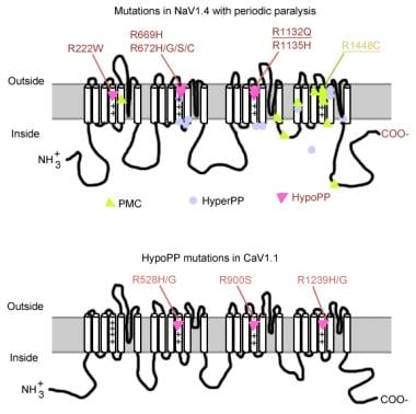


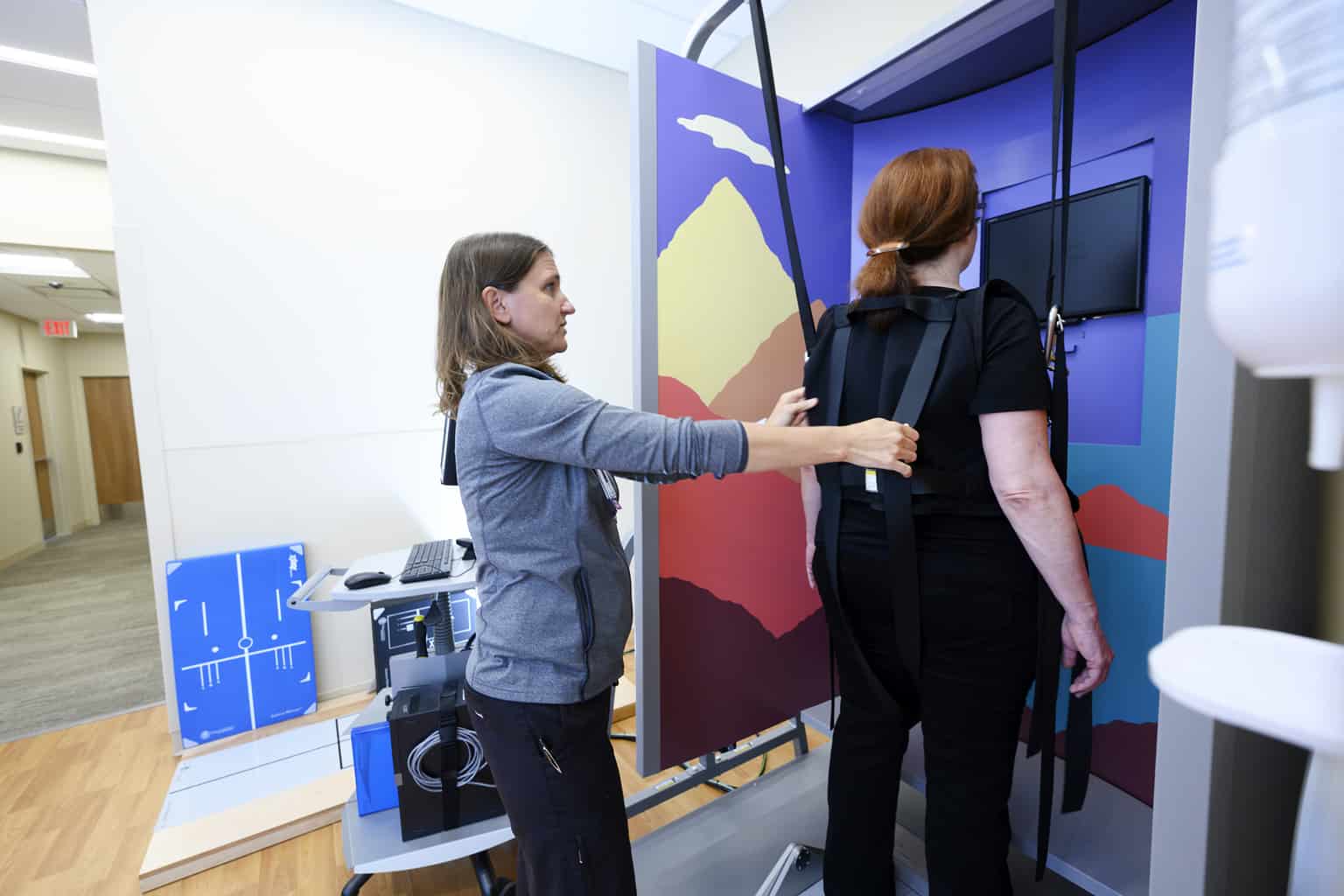

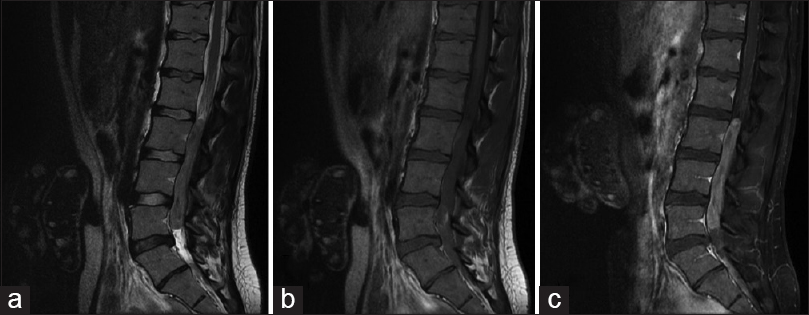

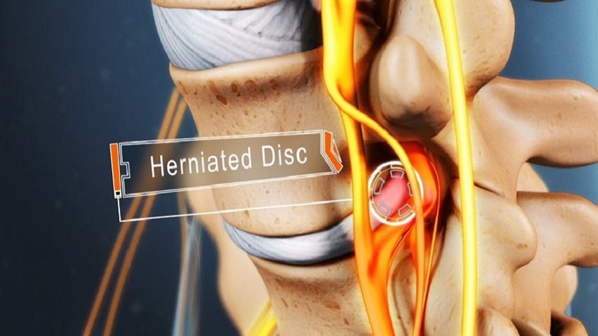
![Buy Finalgon® [Nicoboxil, Nonivamide]](https://pillbuys.com/24086-large_default/finalgon-nicoboxil-nonivamide-.jpg)

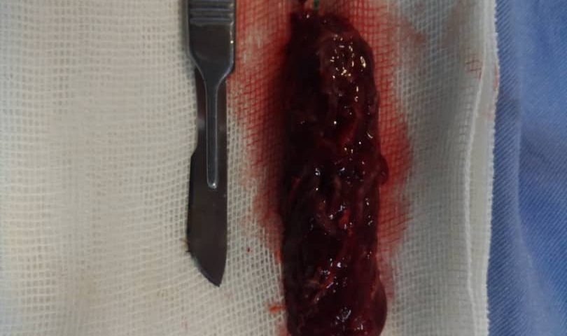


![Emla Patch [Topical Anesthetic] - RFID & NFC Chip Implants and Biohacking products](https://dangerousthings.com/wp-content/uploads/product_85129400724_patch_quarter.png)