Dislocations
Patellar Ligament
Patellofemoral Joint
Chondromalacia Patellae
Joint Instability
Hip Dislocation, Congenital
Traction
Lens Subluxation
Reconstruction of the medial patellofemoral ligament for the treatment of habitual or recurrent dislocation of the patella in children. (1/50)
We investigated the clinical outcome of a reconstructive procedure of the medial patellofemoral ligament for the treatment of habitual or recurrent dislocation of the patella in four children (6 knees), with a minimum follow-up of four years. The technique involves transfer of the tendon of semitendinosus to the patella using the posterior one-third of the femoral insertion of the medial collateral ligament as a pulley. There was no recurrence of dislocation after surgery. The mean Kujala score at follow-up was 96.3 points. Radiological assessment showed that the congruence angle, the tilt angle and the lateral shift radio were restored to normal. The lateral and medial stress shift ratios and the Insall-Salvati ratio remained abnormal. We conclude that this technique can be recommended for the treatment of habitual or recurrent patellar dislocation in children, although hypermobility and patella alta are not fully corrected. (+info)Evaluation of patients presenting with knee pain: Part II. Differential diagnosis. (2/50)
Knee pain is a common presenting complaint with many possible causes. An awareness of certain patterns can help the family physician identify the underlying cause more efficiently. Teenage girls and young women are more likely to have patellar tracking problems such as patellar subluxation and patellofemoral pain syndrome, whereas teenage boys and young men are more likely to have knee extensor mechanism problems such as tibial apophysitis (Osgood-Schlatter lesion) and patellar tendonitis. Referred pain resulting from hip joint pathology, such as slipped capital femoral epiphysis, also may cause knee pain. Active patients are more likely to have acute ligamentous sprains and overuse injuries such as pes anserine bursitis and medial plica syndrome. Trauma may result in acute ligamentous rupture or fracture, leading to acute knee joint swelling and hemarthrosis. Septic arthritis may develop in patients of any age, but crystal-induced inflammatory arthropathy is more likely in adults. Osteoarthritis of the knee joint is common in older adults. (+info)Avulsion of the common origin of the medial collateral and medial patello-femoral ligaments. (3/50)
This study documents for the first time avulsion of the common origin of the medial collateral and medial patello-femoral ligaments. (+info)The management of superior dislocation of the patella with interlocking osteophytes--an update on a rare problem. (4/50)
The superior dislocation of the patella with interlocking osteophytes is a rare condition. A review of the English literature revealed only 12 reported cases. The purpose of reviewing these case reports is to highlight the unusual presentation and the injury mechanism in 2 of our patients, and to present our treatment algorithm. Closed reduction with manipulation of the patella, with or without anaesthesia, was performed without difficulty. We recommend an intermediate step of trying a regional nerve block before proceeding to general anaesthesia. Our patients had full range-of-motion after reduction and they were symptom-free after 3 years of follow-up. There were no recurrent dislocations in our patients. (+info)The effects of articular, retinacular, or muscular deficiencies on patellofemoral joint stability: a biomechanical study in vitro. (5/50)
Normal function of the patellofemoral joint is maintained by a complex interaction between soft tissues and articular surfaces. No quantitative data have been found on the relative contributions of these structures to patellar stability. Eight knees were studied using a materials testing machine to displace the patella 10 mm laterally and medially and measure the force required. Patellar stability was tested from 0 degrees to 90 degrees knee flexion with the quadriceps tensed to 175 N. Four conditions were examined: intact, vastus medialis obliquus relaxed, flat lateral condyle, and ruptured medial retinaculae. Abnormal trochlear geometry reduced the lateral stability by 70% at 30 degrees flexion, while relaxation of vastus medialis obliquus caused a 30% reduction. Ruptured medial retinaculae had the largest effect at 0 degrees flexion with 49% reduction. There was no effect on medial stability. There is a complex interaction between these structures, with their contributions to loss of lateral patellar stability varying with knee flexion. (+info)Ultrastructural study of the extra-articular Leeds-Keio ligament prosthesis. (6/50)
BACKGROUND: There have been several histological studies of the Leeds-Keio ligament in anterior cruciate ligament reconstruction, but there have been few of the Leeds-Keio ligament in the extra-articular portion. AIMS/METHODS: To report the histological and ultrastructural findings of two cases of medial patellofemoral ligament reconstruction using the Leeds-Keio ligament, removed 6.1 years and 8.7 years after implantation. RESULTS: In both cases, the tissue over the Leeds-Keio ligament was a ligament-like tissue. Electron microscopy showed that the diameter of the collagen fibrils in the tissue over the Leeds-Keio ligament was unimodal in the case investigated 6.1 years after implantation but bimodal in the case investigated after 8.7 years. CONCLUSIONS: The tissue over the Leeds-Keio ligament may continue to grow with prolonged periods of mechanical stress. (+info)Trochleoplasty in lateral patellar luxation in two calves. (7/50)
Two Holstein Friesian calves were referred to the Animal Medical Center of Chonbuk National University with severe lameness on the hind limb(s), flexed stifle joint and an inability to walk since birth. Based on the clinical history, clinical findings and radiography of the stifle joint, the cases were diagnosed as grade III lateral patellar luxation (LPL). Trochleoplasty along with medial retinacular reinforcement and lateral release were performed in both calves. The calves recovered 3 to 4 weeks after surgery. Trochleoplasty along with medial retinacular reinforcement and lateral release can be an effective technique for the treatment of LPL in calves. (+info)Early results of one-stage knee extensor realignment and autologous osteochondral grafting. (8/50)
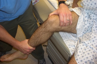
We treated 49 patients with recurrent patellar dislocations or persistent patellar subluxations. Chondral defects were graded according to the International Cartilage Repair Society (ICRS). Thirty patients (group I) had chondral defects grade I or II, and 19 patients (group II) had chondral defects grade III or IV. All patients were treated with proximal and distal realignment of the knee extensor mechanism, but group II also had a simultaneous autologous osteochondral grafting of the chondral defect. Patients were followed for 2 years and clinically assessed using the Marshall score comparing the two groups. Apart from a slower recovery in group II, the clinical and functional results were almost the same at the final follow-up. (+info)Patellar dislocation is a medical condition characterized by the displacement of the patella (kneecap) from its normal position in the femoral groove, which is a part of the femur (thighbone). This displacement usually occurs laterally, meaning that the patella moves toward the outer side of the knee.
Patellar dislocation can happen as a result of direct trauma or due to various factors that increase the laxity of the medial patellofemoral ligament and tightness of the lateral structures, leading to abnormal tracking of the patella. These factors include anatomical variations, muscle imbalances, genetic predisposition, or degenerative changes in the knee joint.
Dislocation of the patella can cause pain, swelling, and difficulty in moving the knee. In some cases, it might be associated with other injuries such as fractures or damage to the articular cartilage and surrounding soft tissues. Immediate medical attention is required for proper diagnosis and treatment, which may involve reduction, immobilization, physical therapy, bracing, or even surgery in severe cases.
A dislocation is a condition in which a bone slips out of its normal position in a joint. This can happen as a result of trauma or injury, such as a fall or direct blow to the body. Dislocations can cause pain, swelling, and limited mobility in the affected area. In some cases, a dislocation may also damage surrounding tissues, such as ligaments, tendons, and nerves.
Dislocations are typically treated by reducing the dislocation, which means putting the bone back into its normal position. This is usually done with the help of medication to relieve pain and relaxation techniques to help the person stay still during the reduction. In some cases, surgery may be necessary to repair damaged tissues or if the dislocation cannot be reduced through other methods. After the dislocation has been reduced, the joint may be immobilized with a splint or sling to allow it to heal properly.
It is important to seek medical attention promptly if you suspect that you have a dislocation. If left untreated, a dislocation can lead to further complications, such as joint instability and chronic pain.
The patellar ligament, also known as the patellar tendon, is a strong band of tissue that connects the bottom part of the kneecap (patella) to the top part of the shinbone (tibia). This ligament plays a crucial role in enabling the extension and straightening of the leg during activities such as walking, running, and jumping. Injuries to the patellar ligament, such as tendonitis or tears, can cause pain and difficulty with mobility.
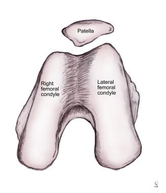
The patella, also known as the kneecap, is a sesamoid bone located at the front of the knee joint. It is embedded in the tendon of the quadriceps muscle and serves to protect the knee joint and increase the leverage of the extensor mechanism, allowing for greater extension force of the lower leg. The patella moves within a groove on the femur called the trochlea during flexion and extension of the knee.
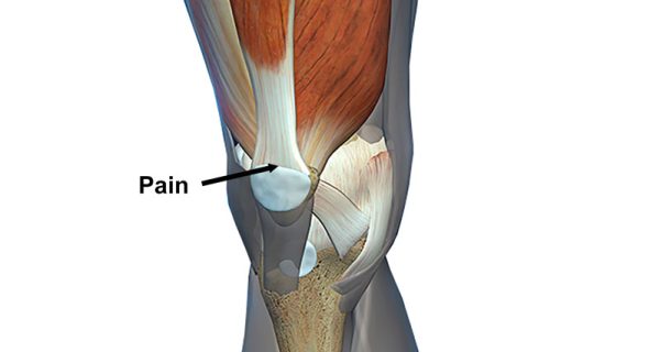
The patellofemoral joint is the articulation between the patella (kneecap) and the femur (thigh bone). It is a synovial joint, which means it is surrounded by a joint capsule containing synovial fluid to lubricate the joint. This joint is responsible for providing stability to the knee extensor mechanism and allows for smooth movement of the patella during activities like walking, running, and jumping. Pain or dysfunction in this joint can result in various conditions such as patellofemoral pain syndrome, chondromalacia patella, or patellar dislocation.
Chondromalacia patellae is a medical condition that refers to the softening and degeneration of the articular cartilage on the undersurface of the patella, or kneecap. This cartilage, which provides a smooth, lubricated surface for joint movement, can become damaged due to various reasons such as overuse, misalignment of the patella, or direct trauma. The resulting damage can cause pain and inflammation in the knee, particularly during activities that involve bending or straightening the leg. In some cases, chondromalacia patellae may also lead to the formation of bone spurs or osteophytes, which can further exacerbate the symptoms and limit joint mobility. Treatment for chondromalacia patellae typically involves a combination of rest, physical therapy, and pain management strategies, such as anti-inflammatory medications or corticosteroid injections. In severe cases, surgery may be required to repair or replace the damaged cartilage.
A hip dislocation is a medical emergency that occurs when the head of the femur (thighbone) slips out of its socket in the pelvis. This can happen due to high-energy trauma, such as a car accident or a severe fall. Hip dislocations can also occur in people with certain health conditions that make their hips more prone to displacement, such as developmental dysplasia of the hip.
There are two main types of hip dislocations: posterior and anterior. In a posterior dislocation, the femur head moves out of the back of the socket, which is the most common type. In an anterior dislocation, the femur head moves out of the front of the socket. Both types of hip dislocations can cause severe pain, swelling, and difficulty moving the affected leg.
Immediate medical attention is necessary for a hip dislocation to realign the bones and prevent further damage. Treatment typically involves sedation or anesthesia to relax the muscles around the joint, followed by a closed reduction procedure to gently guide the femur head back into the socket. In some cases, surgery may be required to repair any associated injuries, such as fractures or damaged ligaments. After treatment, physical therapy and rehabilitation are usually necessary to restore strength, mobility, and function to the affected hip joint.
Tenodesis is a surgical procedure where a damaged or torn tendon is attached to a nearby bone using sutures, anchors, or screws. The term specifically refers to the surgical fixation of a tendon to a bone. This procedure is often performed to treat injuries of the shoulder or wrist, such as rotator cuff tears or distal biceps tendon ruptures.
The goal of tenodesis is to provide stability and restore function to the affected joint by creating a new, stable attachment point for the tendon. This procedure can help reduce pain, improve strength, and enhance overall joint mobility. It is typically recommended when non-surgical treatments have failed or are not appropriate for the patient's injury.
It is important to note that tenodesis should not be confused with tenotomy, which is a surgical procedure where a tendon is cut to release tension and improve mobility in a joint.
Joint instability is a condition characterized by the loss of normal joint function and increased risk of joint injury due to impaired integrity of the supporting structures, such as ligaments, muscles, or cartilage. This can result in excessive movement or laxity within the joint, leading to decreased stability and increased susceptibility to dislocations or subluxations. Joint instability may cause pain, swelling, and limited range of motion, and it can significantly impact a person's mobility and quality of life. It is often caused by trauma, degenerative conditions, or congenital abnormalities and may require medical intervention, such as physical therapy, bracing, or surgery, to restore joint stability.
A cartilage fracture is not a common injury because cartilage itself does not have bones, and it is difficult to fracture something that is not hard. However, there are situations where the term "cartilage fracture" can be used. One such situation is when the articular cartilage, which covers the ends of bones in joints, gets damaged or injured. This type of injury is also known as a chondral fracture or osteochondral fracture (if the bone beneath the cartilage is also involved). These injuries can occur due to trauma, such as a fall or a direct blow to the joint, and can cause pain, swelling, and limited mobility in the affected joint.
Shoulder dislocation is a medical condition where the head of the humerus (upper arm bone) gets displaced from its normal position in the glenoid fossa of the scapula (shoulder blade). This can occur anteriorly, posteriorly, or inferiorly, with anterior dislocations being the most common. It is usually caused by trauma or forceful movement and can result in pain, swelling, bruising, and limited range of motion in the shoulder joint. Immediate medical attention is required to relocate the joint and prevent further damage.
Knee injuries refer to damages or harm caused to the structures surrounding or within the knee joint, which may include the bones (femur, tibia, and patella), cartilage (meniscus and articular cartilage), ligaments (ACL, PCL, MCL, and LCL), tendons (patellar and quadriceps), muscles, bursae, and other soft tissues. These injuries can result from various causes, such as trauma, overuse, degeneration, or sports-related activities. Symptoms may include pain, swelling, stiffness, instability, reduced range of motion, and difficulty walking or bearing weight on the affected knee. Common knee injuries include fractures, dislocations, meniscal tears, ligament sprains or ruptures, and tendonitis. Proper diagnosis and treatment are crucial to ensure optimal recovery and prevent long-term complications.
Arthroscopy is a minimally invasive surgical procedure where an orthopedic surgeon uses an arthroscope (a thin tube with a light and camera on the end) to diagnose and treat problems inside a joint. The surgeon makes a small incision, inserts the arthroscope into the joint, and then uses the attached camera to view the inside of the joint on a monitor. They can then insert other small instruments through additional incisions to repair or remove damaged tissue.
Arthroscopy is most commonly used for joints such as the knee, shoulder, hip, ankle, and wrist. It offers several advantages over traditional open surgery, including smaller incisions, less pain and bleeding, faster recovery time, and reduced risk of infection. The procedure can be used to diagnose and treat a wide range of conditions, including torn ligaments or cartilage, inflamed synovial tissue, loose bone or cartilage fragments, and joint damage caused by arthritis.
Knee dislocation is a serious and uncommon orthopedic injury that occurs when the bones that form the knee joint (femur, tibia, and patella) are forced out of their normal position due to extreme trauma or force. This injury often requires immediate medical attention and reduction (repositioning) by a healthcare professional. If left untreated, it can lead to serious complications such as compartment syndrome, nerve damage, and long-term joint instability. It's important to note that knee dislocation is different from a kneecap (patellar) dislocation, which involves the patella sliding out of its groove in the femur.
The knee joint, also known as the tibiofemoral joint, is the largest and one of the most complex joints in the human body. It is a synovial joint that connects the thighbone (femur) to the shinbone (tibia). The patella (kneecap), which is a sesamoid bone, is located in front of the knee joint and helps in the extension of the leg.
The knee joint is made up of three articulations: the femorotibial joint between the femur and tibia, the femoropatellar joint between the femur and patella, and the tibiofibular joint between the tibia and fibula. These articulations are surrounded by a fibrous capsule that encloses the synovial membrane, which secretes synovial fluid to lubricate the joint.
The knee joint is stabilized by several ligaments, including the medial and lateral collateral ligaments, which provide stability to the sides of the joint, and the anterior and posterior cruciate ligaments, which prevent excessive forward and backward movement of the tibia relative to the femur. The menisci, which are C-shaped fibrocartilaginous structures located between the femoral condyles and tibial plateaus, also help to stabilize the joint by absorbing shock and distributing weight evenly across the articular surfaces.
The knee joint allows for flexion, extension, and a small amount of rotation, making it essential for activities such as walking, running, jumping, and sitting.
Congenital hip dislocation, also known as developmental dysplasia of the hip (DDH), is a condition where the hip joint fails to develop normally in utero or during early infancy. In a healthy hip, the head of the femur (thigh bone) fits snugly into the acetabulum (hip socket). However, in congenital hip dislocation, the femoral head is not held firmly in place within the acetabulum due to abnormal development or laxity of the ligaments that support the joint.
There are two types of congenital hip dislocations:
1. Teratologic dislocation: This type is present at birth and occurs due to abnormalities in the development of the hip joint during fetal growth. The femoral head may be completely outside the acetabulum or partially dislocated.
2. Developmental dysplasia: This type develops after birth, often within the first few months of life, as a result of ligamentous laxity and shallow acetabulum. In some cases, it can progress to a complete hip dislocation if left untreated.
Risk factors for congenital hip dislocation include family history, breech presentation during delivery, and female gender. Early diagnosis and treatment are crucial to prevent long-term complications such as pain, limited mobility, and osteoarthritis. Treatment options may include bracing, closed reduction, or surgical intervention, depending on the severity and age of the child at diagnosis.
The femur is the medical term for the thigh bone, which is the longest and strongest bone in the human body. It connects the hip bone to the knee joint and plays a crucial role in supporting the weight of the body and allowing movement during activities such as walking, running, and jumping. The femur is composed of a rounded head, a long shaft, and two condyles at the lower end that articulate with the tibia and patella to form the knee joint.
Recurrence, in a medical context, refers to the return of symptoms or signs of a disease after a period of improvement or remission. It indicates that the condition has not been fully eradicated and may require further treatment. Recurrence is often used to describe situations where a disease such as cancer comes back after initial treatment, but it can also apply to other medical conditions. The likelihood of recurrence varies depending on the type of disease and individual patient factors.
Traction, in medical terms, refers to the application of a pulling force to distract or align parts of the body, particularly bones, joints, or muscles, with the aim of immobilizing, reducing displacement, or realigning them. This is often achieved through the use of various devices such as tongs, pulleys, weights, or specialized traction tables. Traction may be applied manually or mechanically and can be continuous or intermittent, depending on the specific medical condition being treated. Common indications for traction include fractures, dislocations, spinal cord injuries, and certain neurological conditions.
Lens subluxation, also known as lens dislocation or ectopia lentis, is a condition where the lens of the eye becomes partially or completely displaced from its normal position. The lens is held in place by tiny fibers called zonules, which can become weakened or broken due to various reasons such as genetic disorders (like Marfan syndrome, homocystinuria, and Weill-Marchesani syndrome), trauma, inflammation, or cataract surgery complications. This displacement can lead to symptoms like blurry vision, double vision, sensitivity to light, or the appearance of a shadow in the peripheral vision. In some cases, lens subluxation may not cause any noticeable symptoms and can be discovered during routine eye examinations. Treatment options depend on the severity and underlying cause of the subluxation and may include eyeglasses, contact lenses, or surgical intervention to remove and replace the displaced lens with an intraocular lens (IOL).
The acromioclavicular (AC) joint is the joint located between the acromion process of the scapula (shoulder blade) and the clavicle (collarbone). It allows for a small amount of movement between these two bones and participates in shoulder motion. Injuries to this joint, such as AC joint separations or sprains, are common and can occur due to falls, direct blows, or repetitive motions that cause the ligaments that support the AC joint to become stretched or torn.
 Patellar dislocation - Wikipedia
 Reduction of Patellar Dislocation: Background, Indications, Contraindications
 Dynamic versus static medial patellofemoral ligament reconstruction technique in the treatment of recurrent patellar...
Dislocated Knee & Patellar Dislocation Treatment in Ramsey, NJ
 Patellar dislocation
Patellar Dislocation | Profiles RNS
 Patellar Dislocation | OrthoFixar 2023
 Patellar Dislocation Melbourne | Patellofemoral Dislocation Heidelberg, Melbourne
 Patellar Dislocation - MPFL reconstruction
Patellar Dislocation Denver CO | Patellofemoral Dislocation Aurora
 Anatomical Factors Linked to Recurring Patellar Dislocation
Dislocated Knee & Patellar Dislocation Treatment in Arlington, TX
 Patellar Dislocation Denver, Colorado | Patellofemoral Dislocation Aurora, Englewood
Acute Lateral Patellar Dislocation-MRI - Sumer's Radiology Blog
Patellar Dislocation Treatment ACT | Kneecap Injury Treatment Canberra
 Patellar Dislocations - Injuries; Poisoning - MSD Manual Professional Edition
Patellar Dislocation New York City | Patellofemoral Dislocation Manhattan, NYC
Patellar Instability Virginia Beach, VA | Patellar Dislocation Chesapeake, VA
Patellar Instability Treatment Denver CO | Patellar Dislocation Denver CO
Patellar Instability Treatment Alexandria VA | Knee Dislocation Arlington, Lorton VA
 Peak Physical Therapy | Patellar Dislocations and Subluxations
 Minimally invasive surgical technique in treating recurrent patellar dislocation<...
 First-time patellar dislocation with resultant habitual dislocation two years later, which was not demonstrated on plain X-rays...
Editorial for "T2 Mapping of Patellar Cartilage After a Single First-Time Episode of Traumatic Lateral Patellar Dislocation". -...
 Functional outcome of isolated medial patellofemoral ligament reconstruction for recurrent patellar dislocation
 Dr. David Crawford, MD - Orthopedic Surgery Specialist in New Albany, OH | Healthgrades
 Kneecap dislocation: MedlinePlus Medical Encyclopedia
Treatment of patellar dislocation with arthroscopic medial patellofemoral ligament reconstruction using gracilis tendon...
 DWP to provide up to £691 a month to people with any of these muscle or joint conditions - Cambridgeshire Live
 Dr. Michael Sirota, MD, Orthopedic Surgery Specialist - San Diego, CA | Sharecare
Ligament14
- Displacement of the patella laterally out of its groove strains the medial stabilizing connective tissues, particularly the medial patellofemoral ligament (supporting 50-80% of the knee mechanisms in lateral patellar glide), which is torn usually at its femoral attachment. (wikipedia.org)
- Traumatic patellar dislocation may cause bleeding into the joint space, ligament and muscle attachment tearing, and fracture of the medial wing of the patella. (wikipedia.org)
- Surgical repair of the patellar stabilizing structures - the medial patellofemoral ligament and vastus medialis muscle - may be needed for athletes. (wikipedia.org)
- The medial patellofemoral ligament is the primary stabiliser (53-67%) against lateral displacement/dislocation of the patella. (ramtan.co)
- In the case of lateral patellar dislocations, there is tenderness along the medial joint due to tears in the medial retinaculum at the point where the medial patellofemoral ligament attaches to the femur. (orthofixar.com)
- The Medial Patellofemoral Ligament, which is usually damaged by an acute lateral patellar dislocation, serves as the main barrier against excessive lateral displacement. (orthopedicsjournal.in)
- The safe, effective treatment option for recurrent patellar dislocation in individuals without any predisposed anatomic features is medial patellofemoral ligament restoration utilizing hamstring tendon. (orthopedicsjournal.in)
- What are the different causes of patellar ligament pain? (newsbasis.com)
- The patellar ligament connects the bottom (apex) of your patella to your tibia (shin bone) . (clevelandclinic.org)
- Rupture of the patellar ligament after use of its central third for anterior cruciate reconstruction. (medscape.com)
- Ismail AM, Balakrishnan R, Rajakumar MK, Lumpur K. Rupture of patellar ligament after steroid infiltration. (medscape.com)
- Arthrosis of the patella is often triggered by an injury, e.g., if the kneecap is broken or dislocated, or after a cruciate ligament injury, or due to a patellar tracking disorder. (ottobock.com)
- The extensor mechanism of the knee consists of the quadriceps muscle group, quadriceps tendon, patella, patellar retinaculum, patellar ligament, and adjacent soft tissues. (medscape.com)
- The patellar tendon, occasionally termed the patellar ligament, originates at the inferior pole of the patella and inserts onto the tibial tuberosity. (medscape.com)
Instability14
- Value of the tibial tuberosity-trochlear groove distance in patellar instability in the young athlete. (medscape.com)
- Moreover, chronic cartilage damage has been described at 13-year follow up with patellofemoral osteoarthritis in 22% in patellar instability knees compared to 11% in contralateral healthy knees [ 6 ]. (springer.com)
- What is Patellar Instability? (kevinbonnermd.com)
- Patellar (kneecap) instability results from one or more complete or partial dislocations (subluxations). (kevinbonnermd.com)
- Various factors and conditions may cause patellar instability. (kevinbonnermd.com)
- This case illustrates that first-time patellar dislocation can gradually lead to habitual dislocation subsequently, and that cautious physical examinations in regard to patella tracking are essential since radiological examinations do not always reveal the pathophysiology of patellar instability. (biomedcentral.com)
- Patients who experience frequent episodes of lateral patellar instability and fail a thorough non operative therapy are candidates for surgery. (orthopedicsjournal.in)
- The study comprised 34 individuals with repeated unilateral patellar dislocation and persistent patellar instability without any anatomical predisposing factors. (orthopedicsjournal.in)
- Sherman SL, Hinckel BB, Farr J. Patellar instability. (medlineplus.gov)
- The scope of this guidance is to provide recommendations for the surgical management of skeletally mature patients* with recurrent patellar instability and no significant degenerative change. (boa.ac.uk)
- Isolated lateral retinacular release is never indicated for patellar instability. (boa.ac.uk)
- Isolated medialisation of the tibial tuberosity is not recommended in pure patellar instability, although reducing excessive lateralisation may occasionally be indicated. (boa.ac.uk)
- All surgical procedures for patellar instability must be carried out by surgeons with appropriate experience and training. (boa.ac.uk)
- Physiotherapy following surgery for patellar instability, should be performed by physiotherapists with training and experience in this pathology, or under the supervision of an appropriately skilled and experienced physiotherapist. (boa.ac.uk)
Kneecap13
- A patellar dislocation is a knee injury in which the patella (kneecap) slips out of its normal position. (wikipedia.org)
- If the kneecap partially comes out of the groove, it is called as subluxation and if the kneecap completely comes out, it is called as dislocation (luxation). (kneesurgeonmelbourne.com.au)
- It is done to loosen or release the tight lateral ligaments that pull the kneecap from its groove which increases pressure on the cartilage and causes dislocation. (sportsandshoulderdoc.com)
- The tight lateral ligaments that pull the kneecap from its groove, increase pressure on the cartilage and cause dislocation are loosened or released. (stephenthonmd.com)
- Close-up comparing the normal right knee (left) and swollen left knee of a male patient following dislocation of the kneecap (patella) as a result of an injury. (msdmanuals.com)
- A dislocation of your patella, or kneecap, often occurs from non-contact knee injuries, especially with the foot/lower leg in a twisted position, resulting in the kneecap moving out of place. (peaktherapy.com)
- A subluxation is an incomplete or partial dislocation when the kneecap does not fully come out of its normal location in the front of the knee. (peaktherapy.com)
- Kneecap dislocation occurs when the round-shaped bone covering the knee (patella) moves or slides out of place. (medlineplus.gov)
- Kneecap dislocation often occurs after a sudden change in direction when your leg is planted. (medlineplus.gov)
- Kneecap dislocation damages your knee joint. (medlineplus.gov)
- The patellar tendon is located just below the kneecap. (newsbasis.com)
- This involves the kneecap slipping out of the femoral groove (dislocation). (ottobock.com)
- The latter includes patellar chondropathy, chondromalacia and, in the worst-case scenario, retropatellar osteoarthritis (arthrosis of the kneecap). (ottobock.com)
Isolated MPFL reconstruction for recurrent late1
- The objectives of this study was to evaluate the clinical, functional, and radiological results of isolated MPFL reconstruction for recurrent lateral patellar dislocation. (orthopedicsjournal.in)
Quadriceps tendon3
- The more common one involves a horizontal intra-articular dislocation of the patella with detachment of the quadriceps tendon. (orthofixar.com)
- The function of the quadriceps tendon and patellar tendon is to work with the muscles in the front of the thigh to straighten the knee. (newsbasis.com)
- Patellar and quadriceps tendon ruptures--jumper's knee. (medscape.com)
Reconstruction2
- Patients with recurrent patella dislocation requiring isolated MPFL reconstruction will be recruited and randomized to the dynamic or static reconstruction technique. (springer.com)
- Jabłoński JJ, Jarmuziewicz P, Drużbicki M. Reconstruction of chronic patellar tendon rupture with semitendinosus tendon: case report. (medscape.com)
Traumatic patellar dislocation6
- Surgical stabilization significantly reduces the redislocation rate of primary traumatic patellar dislocation in the young adult population, but is associated with a higher risk of patellofemoral joint osteoarthritis. (ramtan.co)
- Traumatic Patellar dislocation is more common in women owing to physiological laxity and in patients with connective tissue disorders (e.g. (orthofixar.com)
- BACKGROUND: The aim of this study was to investigate the risk factors of neglected osteochondral fractures in primary acute traumatic patellar dislocation in the pediatric population. (bvsalud.org)
- METHODS: A total of 113 patients with primary acute traumatic patellar dislocation for whom coincident osteochondral fractures could not be confirmed by X-ray examination at initial diagnosis between January 2010 and February 2022 were retrospectively analyzed. (bvsalud.org)
- CONCLUSIONS: SP effusion is not only associated with an increased risk of neglected osteochondral fractures in primary acute traumatic patellar dislocation but also with larger fragment size. (bvsalud.org)
- Knee radiograph, medical history, and physical examination can predict the need for further imaging examination and even surgery in primary acute traumatic patellar dislocation. (bvsalud.org)
Diagnosis2
- In most instances, the history, the physical examination, and standard radiographs suffice for making a diagnosis of acute patellar tendon rupture. (medscape.com)
- Early diagnosis of bilateral disease in the absence of trauma and breed predisposition supports the concept that patellar luxation results from a congenital or developmental misalignment of the entire extensor mechanism. (acvs.org)
Fracture4
- Fracture of the weight-bearing portion of the lateral femoral condyle occurs in 25% of traumatic patellar dislocations. (wikipedia.org)
- A patellar dislocation associated with a fracture of the proximal tibia or distal femur should not be reduced in this manner. (medscape.com)
- Anteroposterior and lateral knee x-rays and patellar views are taken to exclude fracture, even if the dislocation has obviously reduced. (msdmanuals.com)
- If you ever break your patella - a patellar fracture - your provider might use some of these terms to describe where your bone was damaged. (clevelandclinic.org)
Recurrent dislocation2
- Individuals who are younger and those with a natural looseness in their ligaments are more prone to experiencing this recurrent dislocation. (orthofixar.com)
- In contrast to recurrent dislocation, which occurs as an isolated and intermittent sequela of injury, the transition to a habitual dislocation after an initial dislocation has not yet been clarified. (biomedcentral.com)
Acute primary patellar1
- An algorithm guiding the evaluation and treatment of acute primary patellar dislocations. (medscape.com)
History of patella dislocation2
- It's curable: If you really have a history of patella dislocation, it is something that may be successfully treated with physical therapy, but it may require a surgical procedure to help realign the patella. (newsbasis.com)
- All males with a history of patella dislocation had at least a mild trochlea dysplasia. (bvsalud.org)
Subluxation or dislocation1
- Repeated subluxation or dislocation makes the knee unstable. (kevinbonnermd.com)
Injuries11
- People who have larger Q angles tend to be more prone to having knee injuries such as dislocations, due to the central line of pull found in the quadriceps muscles that run from the anterior superior iliac spine to the center of the patella. (wikipedia.org)
- An immediate attempt at reduction should be made on any dislocation associated with vascular compromise of the distal extremity, though such compromise is exceedingly uncommon in the setting of an isolated lateral patellar dislocation and should prompt further examination into possible concurrent injuries. (medscape.com)
- After patella dislocation, the incidence of acute osteochondral or chondral injuries is up to 95% after initial patella dislocation [ 5 ]. (springer.com)
- A knee dislocation is usually associated with events of severe trauma such as automobile crashes, severe falls or sports injuries. (orthopaedic-surgery-md.com)
- Superior dislocation primarily affects elderly individuals, resulting from hyperextension injuries to the knee, often with the patella locked onto an anterior femoral osteophyte. (orthofixar.com)
- Studies have indicated a prevalence of 39-71 % for osteochondral or chondral injuries following an acute patellar dislocation. (orthofixar.com)
- Knee (Tibiofemoral) Dislocations Knee dislocations are commonly accompanied by arterial or nerve injuries. (msdmanuals.com)
- Lateral dislocation of the patella (LPD) is one of the most common knee joint injuries in adolescents and young adults [ 9 , 28 ]. (springeropen.com)
- Traumas that hurt your knee are the most common patella injuries, including dislocations and bone fractures. (clevelandclinic.org)
- Injuries or your joint being too loose can cause patellar subluxation. (clevelandclinic.org)
- Overall, she said, "nearly all studies" reporting on first-time patella dislocations classify the injuries as traumatic and not syndromic, and incidence is approximately equal among males and females. (aaos.org)
Ligaments5
- In some cases, the injured ligaments involved in patellar dislocation do not allow the leg to flex. (wikipedia.org)
- If a knee dislocation occurs, there is the potential for significant damage to the surrounding tissue and ligaments. (orthopaedic-surgery-md.com)
- Your healthcare provider will also likely begin to address the ligaments, cartilage and meniscus damage that has occurred as a result of the knee dislocation. (orthopaedic-surgery-md.com)
- The ligaments on the inner and outer sides of the patella hold it in the femoral groove and avoid dislocation of the patella from the groove. (kevinbonnermd.com)
- Any damage to the supporting ligaments may cause the patella to slip out of the groove either partially (subluxation) or completely (dislocation). (kevinbonnermd.com)
Occurs8
- A patellar dislocation typically occurs when the knee is straight and the lower leg is bent outwards when twisting. (wikipedia.org)
- Recurrence after an initial dislocation occurs in about 30% of people. (wikipedia.org)
- Patellar dislocation occurs mainly in youths (under age 20) engaged in sports that may involve accidental rotation of the knee while in flexion, a movement clinically called valgus, which is the cause of some 93% of patellar dislocation cases. (wikipedia.org)
- Often confused with a partial dislocation (called a subluxation), a full knee dislocation occurs when your thigh bone completely loses contact with the top of your shin bone. (orthopaedic-surgery-md.com)
- Dislocation of the patella occurs when the patella moves out of the patellofemoral groove, (called as trochlea) onto a bony head of the femur. (kneesurgeonmelbourne.com.au)
- Patellar dislocation occurs when the patella moves out of the patellofemoral groove, (trochlea) onto the bony head of the femur. (stephenthonmd.com)
- Usually, patellar dislocation occurs when people suddenly change direction or twist the knee or when force is applied to the knee (as may occur in soccer, gymnastics, or baseball when swinging a bat). (msdmanuals.com)
- The dislocation often occurs toward the outside of the leg. (medlineplus.gov)
Mechanism of Injury1
- In another study, which included more than 300 patients, the primary mechanism of injury was noncontact, constituting 73 percent of dislocations. (aaos.org)
Luxation7
- Figure 1: Three-dimensional computed tomography illustrating the anatomy of a canine limb with medial patellar luxation, viewed from the front (left) and the outer side (right) of the leg. (acvs.org)
- Patellar luxation (dislocation) is a condition where the knee cap rides outside the femoral groove when the knee is flexed (Figure 1). (acvs.org)
- Patellar luxation is one of the most common orthopedic conditions in dogs , diagnosed in 7% of puppies. (acvs.org)
- Patellar luxation affects both knees in half of all cases, potentially resulting in discomfort and loss of function. (acvs.org)
- Figure 3: Preoperative computed tomographic (CAT scan) evaluation of a dog with severe patellar luxation of both knees and malformation (bowing) of the femur in each limb. (acvs.org)
- Patellar luxation occasionally results from a traumatic injury to the knee, causing sudden severe lameness of the limb. (acvs.org)
- The femoral groove into which the knee cap normally rides is commonly shallow (Figure 2a, Figure 2b) or absent in dogs with non-traumatic patellar luxation. (acvs.org)
Occur7
- Dislocations nearly always occur away from the midline. (wikipedia.org)
- Patellar dislocations occur in about 6 per 100,000 people per year. (wikipedia.org)
- Osteochondral fractures may occur in the setting of patellar dislocation. (medscape.com)
- Patellar dislocations occur most often in adolescent females who have an underlying chronic patellofemoral abnormality. (msdmanuals.com)
- Dislocation may also occur as result of direct trauma. (medlineplus.gov)
- Excessive valgus with lateral dislocation of the patella may occur. (medscape.com)
- Genu valgum with patellar dislocation may occur in patients with diastrophic dysplasia. (medscape.com)
Groove2
- Su P, Hu H, Li S, Xu T, Li J, Fu W. Tibial Tubercle-Trochlear Groove/Trochlear Width Is the Optimal Indicator for Diagnosing a Lateralized Tibial Tubercle in Recurrent Patellar Dislocation Requiring Surgical Stabilization. (medscape.com)
- It has an anterior projection on the lateral femoral condyle, lateral to the patellar groove. (ramtan.co)
Retinaculum3
- Injury to the medial stabilizers of the patella is also identified as disruption of the medial retinaculum at its patellar attachment or midsubstance. (indianradiology.com)
- The patellar retinaculum is an important soft tissue stabilizer of the patellofemoral joint. (medscape.com)
- Deep to the medial patellar retinaculum, there are 3 focal capsular thickenings. (medscape.com)
Partial dislocation1
- A subluxation is a partial dislocation. (clevelandclinic.org)
Surgical management1
- A Cochrane review by Smith et al found that although there was some evidence appearing to favor surgical management of primary patellar dislocation, the quality of the currently available evidence was too poor to allow any firm conclusions to be made. (medscape.com)
Tendonitis1
- Patellar tendonitis comes from repetitive stress on the knee, most often from overuse in sports or exercise. (newsbasis.com)
Anatomic2
- 1.2 are the two anatomic patellar risk factors most prevalent in first-time lateral patellar dislocations. (aaos.org)
- She concluded: "The thing I want to emphasize is that we can characterize anatomic factors associated with the first-time dislocation, but we don't know when these factors should be surgically corrected or at what threshold these should be corrected for the best results. (aaos.org)
Patella alta1
- We do know that open growth plates, patella alta, high-grade trochlear dysplasia measured in a couple of different ways, and contralateral patella dislocation are all indicators of high probability of recurrence," Dr. Arendt said. (aaos.org)
Lateral displacement1
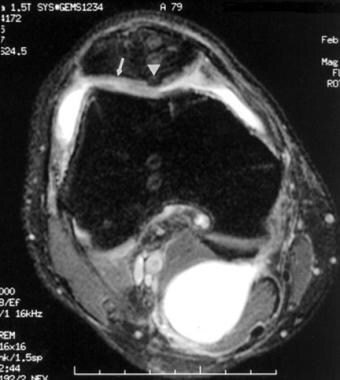
- This anteroposterior view of the knee shows a patellar dislocation, characterized by extreme lateral displacement of the patella, outside its normal location between the femoral condyles. (msdmanuals.com)
Elbow dysplasia1
- Patellar dislocations and elbow dysplasia are more common in Chihuahuas. (thedogman.net)
Pull on the patella1
- Patella dislocation is commonly observed in young athletes between 15 and 20 years and commonly affects women because of the wider pelvis creates lateral pull on the patella. (kneesurgeonmelbourne.com.au)
Patients7
- This is necessary in recurrent patella dislocations and in particular cases of first patella dislocations such as patients with severe anatomical risk factors [ 15 ]. (springer.com)
- Lateral patellar dislocation (LPD) is a common cause of acute traumatic hemarthrosis in young active patients. (indianradiology.com)
- However, dislocation is usually transient, and patients are frequently unaware that it has occurred. (indianradiology.com)
- In young, active patients, a patellar dislocation is a frequent knee injury. (orthopedicsjournal.in)
- Long-term non operative treatment is reportedly becoming less popular in recent years, and patients who fail initial non operative management for recurring patellar dislocations are more frequently advised to undergo surgery. (orthopedicsjournal.in)
- There is a paucity of quality of life (QoL) assessments in studies evaluating patients treated for recurrent lateral patellar dislocation (LPD). (springeropen.com)
- One meta-analysis of first-time lateral patella dislocation (LPD) in 70 patients showed a distribution of 46 percent male and 54 percent female, with an average age of 24.5 years. (aaos.org)
Injury1
- For patient information resources, see the Breaks, Fractures, and Dislocations Center, as well as Knee Injury and Magnetic Resonance Imaging (MRI). (medscape.com)
Hemarthrosis2
- One of the common findings related to acute, primary, traumatic patellar dislocations is hemarthrosis of the knee, caused by rupture of the medial restraints of the patella. (ramtan.co)
- If the dislocation has spontaneously reduced, hemarthrosis is often present, and the peripatellar area is usually tender. (msdmanuals.com)
Primary acute1
- In a systematic review that included 2134 primary acute patellar dislocations (N = 2086) treated either conservatively or surgically, Longo et al found that whereas surgical treatment was associated with a significantly lower rate of recurrence and better results in the short-to-medium term, the results of the two approaches were not significantly different in the long term. (medscape.com)
Contralateral2
- We present an instructive case of habitual left patellar dislocation in which the patella had appeared odd due to lateral tilt relative to contralateral side, but had been radiologically confirmed to be on the trochlea at 1 year prior to the referral. (biomedcentral.com)
- We report a case of habitual patellar dislocation that appeared odd to the patient's family due to lateral tilt compared with contralateral patella, but was left untreated because plain X-rays (including skyline view) did not demonstrate significant patellofemoral malalignment 1 year prior to the referral. (biomedcentral.com)
Arthroscopic1
- Arthroscopic reduction of a locked patellar dislocation has been described. (medscape.com)
Treatment4
- Make sure to understand what is happening throughout your knee dislocation treatment even if this means asking your healthcare provider questions often. (orthopaedic-surgery-md.com)
- Conservative treatment is the most common treatment after primary patellar dislocation. (ramtan.co)
- Surgical treatment is recommended for those individuals who have recurrent patella dislocation. (kneesurgeonmelbourne.com.au)
- Your physical therapy treatment will focus on improving muscle activation to restore normal patellar joint mechanics as well as functional strengthening for your hips, knees, and ankles. (peaktherapy.com)
MPFL1
- After initial patellar dislocation, the MPFL is injured in 94% of the cases [ 11 ]. (springer.com)
Symptoms3
- citation needed] Another cause of patellar symptoms is lateral patellar compression syndrome, which can be caused from lack of balance or inflammation in the joints. (wikipedia.org)
- The patellar symptoms cause knee extensor dysplasia, and sensitive small variations affect the muscular mechanism that controls the joint movements. (wikipedia.org)
- Contact your provider if you injure your knee and have symptoms of dislocation. (medlineplus.gov)
Orthopedic surgeon1
- Any superior, intercondylar, or horizontal dislocation should be examined by an orthopedic surgeon, as should any dislocation with suspected locked osteophyte. (medscape.com)