Patellofemoral Pain Syndrome
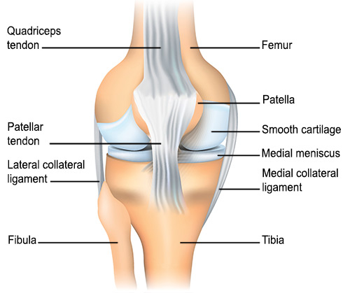
Patellofemoral Joint
Complex Regional Pain Syndromes
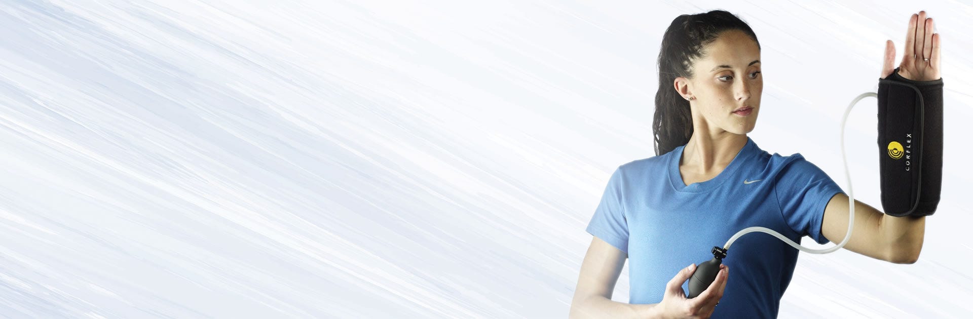
Orthotic Devices
Pain Measurement
Biomechanical Phenomena
Torque
Range of Motion, Articular
Muscle Strength
Foot
Exercise Therapy
Pain
Myofascial Pain Syndromes
Physical Therapy Modalities
Athletic Tape
Reflex Sympathetic Dystrophy
Patellar Ligament
Pelvic Pain
Hip
Prostatitis
Quadriceps Muscle
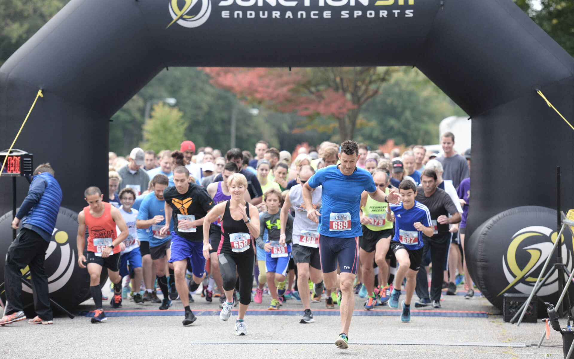
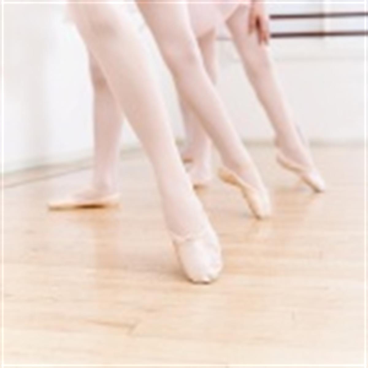
Running
Quadriceps atrophy: to what extent does it exist in patellofemoral pain syndrome? (1/80)
BACKGROUND: Quadriceps atrophy is a commonly cited accompaniment to patellofemoral pain syndrome (PFPS), yet there is little valid, objective evidence for its existence. OBJECTIVE: To investigate atrophy and weakness of the quadriceps femoris muscle group in patients with PFPS using measures of cross-sectional area and peak extension torque. METHODS: A total of 57 patients with insidious onset of PFPS and 10 healthy control subjects had ultrasound scanning of the quadriceps femoris. The scans were analysed using computerised planimetry to estimate the cross-sectional area of the quadriceps femoris. Lower limb peak torque was also measured using a Biodex dynamometer. RESULTS: The mean of % differences revealed a 3.38% (95% confidence interval (CI) 1.3 to 5.45) difference in cross-sectional area (CSA) between the affected and unaffected limb in PFPS patients and a 1.31% (95% CI 0.06 to 2.55) difference in the dominant and non-dominant limb of the control group; the between-groups difference was not significant (p = 0.409). There was a 18.4% (95% CI 13 to 23.8) difference between the affected and unaffected limb in peak torque in PFPS patients and a 7.6% (95% CI 3.2 to 12) difference between the dominant and non-dominant limb in the control group; the between-groups difference was significant (p = 0.002). CONCLUSIONS: The mean of % differences of 3.38% quadriceps atrophy between limbs was considerably less than the only other study using ultrasound scanning on the quadriceps in PFPS and was not significant between the groups. There were greater and more significant between-group differences in lower limb peak torque indicating that muscle strength may not be related to muscle size. These results help to re-appraise of the amount of quadriceps atrophy in PFPS. (+info)Relation between running injury and static lower limb alignment in recreational runners. (2/80)
OBJECTIVES: To determine if measurements of static lower limb alignment are related to lower limb injury in recreational runners. METHODS: Static lower limb alignment was prospectively measured in 87 recreational runners. They were observed for the following six months for any running related musculoskeletal injuries of the lower limb. Injuries were defined according to six types: R1, R2, and R3 injuries caused a reduction in running mileage for one day, two to seven days, or more than seven days respectively; S1, S2, and S3 injuries caused stoppage of running for one day, two to seven days, or more than seven days respectively. RESULTS: At least one lower limb injury was suffered by 79% of the runners during the observation period. When the data for all runners were pooled, 95% confidence intervals calculated for the differences in the measurements of lower limb alignment between the injured and non-injured runners suggested that there were no differences. However, when only runners diagnosed with patellofemoral pain syndrome (n = 6) were compared with non-injured runners, differences were found in right ankle dorsiflexion (0.3 to 6.1), right knee genu varum (-0.9 to -0.3), and left forefoot varus (-0.5 to -0.4). CONCLUSIONS: In recreational runners, there is no evidence that static biomechanical alignment measurements of the lower limbs are related to lower limb injury except patellofemoral pain syndrome. However, the effect of static lower limb alignment may be injury specific. (+info)Diffusely increased bone scintigraphic uptake in patellofemoral pain syndrome. (3/80)
OBJECTIVES: Painful disorders of the patellofemoral joint are one of the most frequent complaints in orthopaedic and sports medicine. The aims of this study were to determine whether bone scintigrams of patients suffering from patellofemoral pain syndrome (PFPS) show diffuse uptake and in what bony compartment of the knee uptake, if any, was localised. METHODS: Fifty eight patients with chronic PFPS were examined. All patients underwent a detailed clinical history and a thorough physical examination of the knee. Anterior and lateral static images of both knees were made using a gamma camera 3 h after injection of 550 MBq of (99m)Tc-HMDP. Two experienced radiologists visually evaluated the scans blindly and separately. As 51 patients had bilateral pain, 109 painful knees are included in the results. RESULTS: Diffuse uptake on bone scintigrams was found in 48 knees in 30 of the patients. In 33 knees the uptake was localised to only one bone compartment, in 10 knees diffuse uptake was found in two of the bones forming the knee joint, and in six knees all three bone compartments (the distal femur, the patella, and the proximal tibia) exhibited diffuse uptake. CONCLUSIONS: Scintigrams of approximately half of the patients with PFPS will show diffuse uptake in one or more of the bony compartments of the knee joint and radioactive tracer accumulation will occur as often in the proximal tibia as in the patella. (+info)Patellofemoral pain and asymmetrical hip rotation. (4/80)
BACKGROUND AND PURPOSE: Patellofemoral joint problems are the most common overuse injury of the lower extremity, and altered femoral or hip rotation may play a role in patellofemoral pain. The purpose of this case report is to describe the evaluation of and intervention for a patient with asymmetrical hip rotation and patellofemoral pain. CASE DESCRIPTION: The patient was a 15-year-old girl with an 8-month history of anterior right knee pain, without known trauma or injury. Prior to intervention, her score on the Western Ontario and McMaster Universities Osteoarthritis Index (WOMAC) was 24%. Right hip medial (internal) rotation was less than left hip medial rotation, and manual muscle testing showed weakness of the right hip internal rotator and abductor muscles. The intervention was aimed at increasing right hip medial rotation, improving right hip muscle strength (eg, the muscle force exerted by a muscle or a group of muscles to overcome a resistance), and eliminating anterior right knee pain. OUTCOMES: After 6 visits (14 days), passive left and right hip medial rotations were symmetrical, and her right hip internal rotator and abductor muscle grades were Good plus. Her WOMAC score was 0%. DISCUSSION: The patient had right patellofemoral pain and an uncommon pattern of asymmetrical hip rotation, with diminished hip medial rotation and excessive hip lateral (external) rotation on the right side. The patient's outcomes suggest that femoral or hip joint asymmetry may be related to patellofemoral joint pain. (+info)How evidence based is the management of two common sports injuries in a sports injury clinic? (5/80)
OBJECTIVES: To examine the diagnosis and management of adults attending a sports injury clinic, to establish to what extent the management of the two most common injuries treated at this clinic is evidence based, and to explore factors that affect management. METHODS: A retrospective examination of 100 random case notes extracted age, sex, sport, type and site of injury, treatment, and outcome. Systematic literature reviews examined the extent and quality of scientific evidence for the management of the two most commonly presenting injuries. A clinical attachment period and practitioner interviews allowed recognition of factors impinging on management decisions. RESULTS: Patellofemoral pain syndrome (PFPS; 10% of all injuries) and Achilles tendinopathy (6% of all injuries) were the most commonly presenting injuries. The mean (SD) number of treatments used for PFPS was 2.8 (0.9). The mean number of treatments used for Achilles tendinopathy was 3.7 (1.0). Clinicians reported that personal experience formed the basis of management plans in 44% of PFPS cases and 59% of Achilles tendinopathy cases, and that primary research evidence only accounted for 24% of management plans in PFPS and 14% in Achilles tendinopathy. Practitioners were unaware of literature supporting over 50% of the treatment modalities they used. However, clinicians were often using evidence based treatments, unaware of the supporting research data. CONCLUSIONS: This study highlights a lack of evidence base, a lack of knowledge of the research evidence, and a lack of management based on the current evidence that is available for these conditions. Practitioners practised evidence based medicine in under 50% of cases. (+info)Patellar taping does not change the amplitude of electromyographic activity of the vasti in a stair stepping task. (6/80)
OBJECTIVES: To investigate the effect of patellar taping on the amplitude of electromyographic activity (EMG) of vasti activation in subjects with and without patellofemoral pain (PFP). METHODS: Ten participants with PFP and 12 asymptomatic controls were recruited to the study. The study was designed as a randomised crossover trial. Participants completed a stair stepping task. Three experimental conditions were assessed: no tape, therapeutic medially directed tape, and placebo vertically directed tape. The main outcome measure was the EMG amplitude of the vastus medialis obliquus and vastus lateralis during the concentric phase of stair stepping. RESULTS: The application of medially directed therapeutic tape significantly decreased pain in subjects with PFP. However, application of tape over the patella (therapeutic or placebo) did not alter the amplitude of vasti EMG when either the PFP or control participants completed the concentric stair stepping task. CONCLUSION: The results of this study indicate that the positive clinical effects of medially directed therapeutic tape are not due to changes in EMG amplitude of the vasti muscle. Thus other effects such as changes in timing of contraction of the vasti are more likely candidates for the mechanism of efficacy. (+info)Reliability of measures of impairments associated with patellofemoral pain syndrome. (7/80)
BACKGROUND: The reliability and measurement error of several impairment measures used during the clinical examination of patients with patellofemoral pain syndrome (PFPS) has not been established. The purpose was to determine the inter-tester reliability and measurement error of measures of impairments associated with PFPS in patients with PFPS. METHODS: A single group repeated measures design was used. Two pairs of physical therapists participated in data collection. Examiners were blinded to each others' measurements. RESULTS: Thirty patients (age 29 +/- 8; 17 female) with PFPS participated in this study. Inter-tester reliability coefficients were substantial for measures of hamstrings, quadriceps, plantarflexors, and ITB/TFL complex length, hip abductors strength, and foot pronation (ICCs from .85 to .97); moderate for measures of Q-angle, tibial torsion, hip external rotation strength, lateral retinacular tightness, and quality of movement during a step down task (ICCs from .67 to .79); and poor for femoral anteversion (ICC of .45). Standard error of measurement (SEM) for measures of muscle length ranged from 1.6 degrees to 4.3 degrees. SEM for Q-angle, tibial torsion, and femoral anteversion were 2.4 degrees, 2.9 degrees, and 4.5 degrees respectively. SEM for foot pronation was 1 mm. SEM for measures of muscle strength was 1.8 Kg for abduction and 2.4 Kg for external rotation. CONCLUSION: Several of the impairments associated with PFPS had sufficient reliability and low measurement error. Further investigation is needed to test if these impairment measurements are related to physical function and whether or not they are useful for decision-making. (+info)The knee skyline radiograph: its usefulness in the diagnosis of patello-femoral osteoarthritis. (8/80)
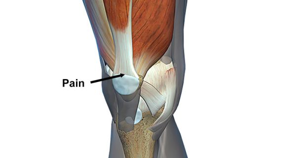
The aim of this study was to determine the usefulness of the skyline radiograph in the diagnosis of patellofemoral osteoarthritis. Additionally, we wanted to assess the usefulness of patello-femoral crepitus as a clinical sign of this condition. Seventy-seven patients scheduled to undergo knee surgery had standard antero-posterior, lateral and skyline X-rays of their affected knee. The presence of clinical patello-femoral crepitus was also documented preoperatively. At the operation, their patellofemoral joints were graded into two groups according to the presence or absence of osteoarthritis. The lateral and skyline view X-rays as well as patello-femoral crepitus were compared individually against the operative findings. The skyline view had a sensitivity of 79% and a specificity of 80%. The lateral view had a sensitivity of 82% and specificity of 65%. Patello-femoral crepitus as a sign had a sensitivity of 89% and a specificity of 82%. There was no statistically significant difference between the two radiological views in terms of sensitivity and specificity in the diagnosis of patellofemoral osteoarthritis. Hence, we cannot recommend the skyline view as a routine radiological investigation in all cases of suspected patellofemoral osteoarthritis. (+info)Patellofemoral Pain Syndrome (PFPS) is a broad term used to describe pain arising from the front of the knee, specifically where the patella (kneecap) meets the femur (thigh bone). It is often described as a diffuse, aching pain in the anterior knee, typically worsening with activities that load the patellofemoral joint such as climbing stairs, running, jumping or prolonged sitting.
PFPS can be caused by various factors including overuse, muscle imbalances, poor biomechanics, or abnormal tracking of the patella. Treatment usually involves a combination of physical therapy to improve strength and flexibility, activity modification, and sometimes bracing or orthotics for better alignment.
The patellofemoral joint is the articulation between the patella (kneecap) and the femur (thigh bone). It is a synovial joint, which means it is surrounded by a joint capsule containing synovial fluid to lubricate the joint. This joint is responsible for providing stability to the knee extensor mechanism and allows for smooth movement of the patella during activities like walking, running, and jumping. Pain or dysfunction in this joint can result in various conditions such as patellofemoral pain syndrome, chondromalacia patella, or patellar dislocation.
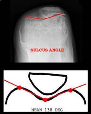
The patella, also known as the kneecap, is a sesamoid bone located at the front of the knee joint. It is embedded in the tendon of the quadriceps muscle and serves to protect the knee joint and increase the leverage of the extensor mechanism, allowing for greater extension force of the lower leg. The patella moves within a groove on the femur called the trochlea during flexion and extension of the knee.
Surgical tape, also known as surgical adhesive tape or hypoallergenic tape, is a type of adhesive tape that is specifically designed for use in surgical settings. It is typically made from a thin, porous material such as rayon, cotton, or polyester, which allows air to circulate and moisture to escape. The adhesive used in surgical tape is designed to be gentle on the skin and to minimize the risk of allergic reactions or irritation.
Surgical tape is used to hold dressings or bandages in place, to close wounds or incisions, or to secure IV lines or other medical devices to the skin. It is available in a variety of sizes, shapes, and colors, and can be cut or shaped to fit the specific needs of the patient.
When applied properly, surgical tape can provide a secure and comfortable hold, while also minimizing the risk of damage to the skin or infection. It is important to follow proper technique when applying and removing surgical tape, as improper use can lead to discomfort, irritation, or other complications.
Complex Regional Pain Syndromes (CRPS) are a group of chronic pain conditions that typically affect a limb after an injury or trauma. They are characterized by prolonged, severe and often debilitating pain that is out of proportion to the severity of the initial injury. CRPS is divided into two types:
1. CRPS-1 (also known as Reflex Sympathetic Dystrophy): This type occurs without a clearly defined nerve injury. It usually develops after an illness or injury that didn't directly damage the nerves.
2. CRPS-2 (also known as Causalgia): This type is associated with a confirmed nerve injury.
The symptoms of CRPS include:
* Continuous, burning or throbbing pain in the affected limb
* Changes in skin temperature, color and texture
* Swelling and stiffness in the joints
* Decreased range of motion and weakness in the affected limb
* Sensitivity to touch or cold
* Abnormal sweating pattern in the affected area
* Changes in nail and hair growth patterns
The exact cause of CRPS is not fully understood, but it is thought to be related to a dysfunction in the nervous system's response to injury. Treatment for CRPS typically involves a combination of medications, physical therapy, and psychological support. In some cases, more invasive treatments such as nerve blocks or spinal cord stimulation may be recommended.
Orthotic devices are custom-made or prefabricated appliances designed to align, support, prevent deformity, or improve the function of movable body parts. They are frequently used in the treatment of various musculoskeletal disorders, such as foot and ankle conditions, knee problems, spinal alignment issues, and hand or wrist ailments. These devices can be adjustable or non-adjustable and are typically made from materials like plastic, metal, leather, or fabric. They work by redistributing forces across joints, correcting alignment, preventing unwanted movements, or accommodating existing deformities. Examples of orthotic devices include ankle-foot orthoses, knee braces, back braces, wrist splints, and custom-made foot insoles.
The knee joint, also known as the tibiofemoral joint, is the largest and one of the most complex joints in the human body. It is a synovial joint that connects the thighbone (femur) to the shinbone (tibia). The patella (kneecap), which is a sesamoid bone, is located in front of the knee joint and helps in the extension of the leg.
The knee joint is made up of three articulations: the femorotibial joint between the femur and tibia, the femoropatellar joint between the femur and patella, and the tibiofibular joint between the tibia and fibula. These articulations are surrounded by a fibrous capsule that encloses the synovial membrane, which secretes synovial fluid to lubricate the joint.
The knee joint is stabilized by several ligaments, including the medial and lateral collateral ligaments, which provide stability to the sides of the joint, and the anterior and posterior cruciate ligaments, which prevent excessive forward and backward movement of the tibia relative to the femur. The menisci, which are C-shaped fibrocartilaginous structures located between the femoral condyles and tibial plateaus, also help to stabilize the joint by absorbing shock and distributing weight evenly across the articular surfaces.
The knee joint allows for flexion, extension, and a small amount of rotation, making it essential for activities such as walking, running, jumping, and sitting.
Pain measurement, in a medical context, refers to the quantification or evaluation of the intensity and/or unpleasantness of a patient's subjective pain experience. This is typically accomplished through the use of standardized self-report measures such as numerical rating scales (NRS), visual analog scales (VAS), or categorical scales (mild, moderate, severe). In some cases, physiological measures like heart rate, blood pressure, and facial expressions may also be used to supplement self-reported pain ratings. The goal of pain measurement is to help healthcare providers better understand the nature and severity of a patient's pain in order to develop an effective treatment plan.
Biomechanics is the application of mechanical laws to living structures and systems, particularly in the field of medicine and healthcare. A biomechanical phenomenon refers to a observable event or occurrence that involves the interaction of biological tissues or systems with mechanical forces. These phenomena can be studied at various levels, from the molecular and cellular level to the tissue, organ, and whole-body level.
Examples of biomechanical phenomena include:
1. The way that bones and muscles work together to produce movement (known as joint kinematics).
2. The mechanical behavior of biological tissues such as bone, cartilage, tendons, and ligaments under various loads and stresses.
3. The response of cells and tissues to mechanical stimuli, such as the way that bone tissue adapts to changes in loading conditions (known as Wolff's law).
4. The biomechanics of injury and disease processes, such as the mechanisms of joint injury or the development of osteoarthritis.
5. The use of mechanical devices and interventions to treat medical conditions, such as orthopedic implants or assistive devices for mobility impairments.
Understanding biomechanical phenomena is essential for developing effective treatments and prevention strategies for a wide range of medical conditions, from musculoskeletal injuries to neurological disorders.
A syndrome, in medical terms, is a set of symptoms that collectively indicate or characterize a disease, disorder, or underlying pathological process. It's essentially a collection of signs and/or symptoms that frequently occur together and can suggest a particular cause or condition, even though the exact physiological mechanisms might not be fully understood.
For example, Down syndrome is characterized by specific physical features, cognitive delays, and other developmental issues resulting from an extra copy of chromosome 21. Similarly, metabolic syndromes like diabetes mellitus type 2 involve a group of risk factors such as obesity, high blood pressure, high blood sugar, and abnormal cholesterol or triglyceride levels that collectively increase the risk of heart disease, stroke, and diabetes.
It's important to note that a syndrome is not a specific diagnosis; rather, it's a pattern of symptoms that can help guide further diagnostic evaluation and management.
"Torque" is not a term that has a specific medical definition. It is a physical concept used in the fields of physics and engineering, referring to a twisting force that causes rotation around an axis. However, in certain medical contexts, such as in discussions of spinal or joint biomechanics, the term "torque" may be used to describe a rotational force applied to a body part. But generally speaking, "torque" is not a term commonly used in medical terminology.
Articular Range of Motion (AROM) is a term used in physiotherapy and orthopedics to describe the amount of movement available in a joint, measured in degrees of a circle. It refers to the range through which synovial joints can actively move without causing pain or injury. AROM is assessed by measuring the degree of motion achieved by active muscle contraction, as opposed to passive range of motion (PROM), where the movement is generated by an external force.
Assessment of AROM is important in evaluating a patient's functional ability and progress, planning treatment interventions, and determining return to normal activities or sports participation. It is also used to identify any restrictions in joint mobility that may be due to injury, disease, or surgery, and to monitor the effectiveness of rehabilitation programs.
Muscle strength, in a medical context, refers to the amount of force a muscle or group of muscles can produce during contraction. It is the maximum amount of force that a muscle can generate through its full range of motion and is often measured in units of force such as pounds or newtons. Muscle strength is an important component of physical function and mobility, and it can be assessed through various tests, including manual muscle testing, dynamometry, and isokinetic testing. Factors that can affect muscle strength include age, sex, body composition, injury, disease, and physical activity level.
In medical terms, the foot is the part of the lower limb that is distal to the leg and below the ankle, extending from the tarsus to the toes. It is primarily responsible for supporting body weight and facilitating movement through push-off during walking or running. The foot is a complex structure made up of 26 bones, 33 joints, and numerous muscles, tendons, ligaments, and nerves that work together to provide stability, balance, and flexibility. It can be divided into three main parts: the hindfoot, which contains the talus and calcaneus (heel) bones; the midfoot, which includes the navicular, cuboid, and cuneiform bones; and the forefoot, which consists of the metatarsals and phalanges that form the toes.
Exercise therapy is a type of medical treatment that uses physical movement and exercise to improve a patient's physical functioning, mobility, and overall health. It is often used as a component of rehabilitation programs for individuals who have experienced injuries, illnesses, or surgeries that have impaired their ability to move and function normally.
Exercise therapy may involve a range of activities, including stretching, strengthening, balance training, aerobic exercise, and functional training. The specific exercises used will depend on the individual's needs, goals, and medical condition.
The benefits of exercise therapy include:
* Improved strength and flexibility
* Increased endurance and stamina
* Enhanced balance and coordination
* Reduced pain and inflammation
* Improved cardiovascular health
* Increased range of motion and joint mobility
* Better overall physical functioning and quality of life.
Exercise therapy is typically prescribed and supervised by a healthcare professional, such as a physical therapist or exercise physiologist, who has experience working with individuals with similar medical conditions. The healthcare professional will create an individualized exercise program based on the patient's needs and goals, and will provide guidance and support to ensure that the exercises are performed safely and effectively.
Patellar dislocation is a medical condition characterized by the displacement of the patella (kneecap) from its normal position in the femoral groove, which is a part of the femur (thighbone). This displacement usually occurs laterally, meaning that the patella moves toward the outer side of the knee.
Patellar dislocation can happen as a result of direct trauma or due to various factors that increase the laxity of the medial patellofemoral ligament and tightness of the lateral structures, leading to abnormal tracking of the patella. These factors include anatomical variations, muscle imbalances, genetic predisposition, or degenerative changes in the knee joint.
Dislocation of the patella can cause pain, swelling, and difficulty in moving the knee. In some cases, it might be associated with other injuries such as fractures or damage to the articular cartilage and surrounding soft tissues. Immediate medical attention is required for proper diagnosis and treatment, which may involve reduction, immobilization, physical therapy, bracing, or even surgery in severe cases.
Pain is an unpleasant sensory and emotional experience associated with actual or potential tissue damage, or described in terms of such damage. It is a complex phenomenon that can result from various stimuli, such as thermal, mechanical, or chemical irritation, and it can be acute or chronic. The perception of pain involves the activation of specialized nerve cells called nociceptors, which transmit signals to the brain via the spinal cord. These signals are then processed in different regions of the brain, leading to the conscious experience of pain. It's important to note that pain is a highly individual and subjective experience, and its perception can vary widely among individuals.
Arthralgia is a medical term that refers to pain in the joints. It does not involve inflammation, which would be referred to as arthritis. The pain can range from mild to severe and may occur in one or multiple joints. Arthralgia can have various causes, including injuries, infections, degenerative conditions, or systemic diseases. In some cases, the underlying cause of arthralgia remains unknown. Treatment typically focuses on managing the pain and addressing the underlying condition if it can be identified.
Myofascial pain syndromes (MPS) are a group of chronic pain disorders characterized by the presence of trigger points in the musculoskeletal system. A trigger point is a hyperirritable spot within a taut band of skeletal muscle, which is often tender to palpation and can cause referred pain, meaning that the pain is felt in a different location than where the trigger point is located.
MPS can affect any muscle in the body, but they are most commonly found in the muscles of the neck, back, shoulders, and hips. The symptoms of MPS may include local or referred pain, stiffness, weakness, and reduced range of motion. The pain is often described as a deep, aching, or throbbing sensation that can be aggravated by physical activity, stress, or anxiety.
The exact cause of MPS is not fully understood, but it is believed to be related to muscle overuse, injury, or chronic tension. Other factors that may contribute to the development of MPS include poor posture, vitamin deficiencies, hormonal imbalances, and emotional stress.
Treatment for MPS typically involves a combination of physical therapy, trigger point release techniques, pain management strategies, and self-care practices such as stretching, relaxation, and stress reduction. In some cases, medication may be prescribed to help manage the pain and reduce muscle spasms.
The femur is the medical term for the thigh bone, which is the longest and strongest bone in the human body. It connects the hip bone to the knee joint and plays a crucial role in supporting the weight of the body and allowing movement during activities such as walking, running, and jumping. The femur is composed of a rounded head, a long shaft, and two condyles at the lower end that articulate with the tibia and patella to form the knee joint.
Physical therapy modalities refer to the various forms of treatment that physical therapists use to help reduce pain, promote healing, and restore function to the body. These modalities can include:
1. Heat therapy: This includes the use of hot packs, paraffin baths, and infrared heat to increase blood flow, relax muscles, and relieve pain.
2. Cold therapy: Also known as cryotherapy, this involves the use of ice packs, cold compresses, or cooling gels to reduce inflammation, numb the area, and relieve pain.
3. Electrical stimulation: This uses electrical currents to stimulate nerves and muscles, which can help to reduce pain, promote healing, and improve muscle strength and function.
4. Ultrasound: This uses high-frequency sound waves to penetrate deep into tissues, increasing blood flow, reducing inflammation, and promoting healing.
5. Manual therapy: This includes techniques such as massage, joint mobilization, and stretching, which are used to improve range of motion, reduce pain, and promote relaxation.
6. Traction: This is a technique that uses gentle pulling on the spine or other joints to help relieve pressure and improve alignment.
7. Light therapy: Also known as phototherapy, this involves the use of low-level lasers or light-emitting diodes (LEDs) to promote healing and reduce pain and inflammation.
8. Therapeutic exercise: This includes a range of exercises that are designed to improve strength, flexibility, balance, and coordination, and help patients recover from injury or illness.
Physical therapy modalities are often used in combination with other treatments, such as manual therapy and therapeutic exercise, to provide a comprehensive approach to rehabilitation and pain management.
Athletic tape, also known as sports tape or physiotherapy tape, is a type of adhesive tape that is commonly used in the field of sports medicine and physical therapy to provide support and stability to joints, muscles, and tendons during athletic activities. It is typically made from a cotton or synthetic fabric material with a strong adhesive backing.
The main purpose of athletic tape is to limit excessive movement or provide compression to an injured area, which can help to reduce pain, swelling, and the risk of further injury. Athletic tape can be used to support a wide variety of body parts, including the ankles, knees, wrists, elbows, and fingers.
There are several different types of athletic tape available, including rigid and flexible options. Rigid tapes, such as zinc oxide tape, are designed to provide maximum support and stability to joints and muscles, while flexible tapes, such as cohesive bandage or kinesiology tape, allow for a greater range of motion and can be used to provide more gentle support or to help facilitate muscle activation and movement.
It is important to note that athletic tape should only be applied by trained professionals, as improper application can lead to further injury or skin irritation. Additionally, athletes should always consult with their healthcare provider before using athletic tape to treat an injury, as it may not be appropriate for all types of injuries or medical conditions.
Reflex Sympathetic Dystrophy (RSD), also known as Complex Regional Pain Syndrome (CRPS), is a chronic pain condition that most often affects a limb after an injury or trauma. It is characterized by prolonged or excessive pain and sensitivity, along with changes in skin color, temperature, and swelling.
The symptoms of RSD/CRPS are thought to be caused by an overactive sympathetic nervous system, which controls involuntary bodily functions such as heart rate, blood pressure, and sweating. In RSD/CRPS, the sympathetic nerves are believed to send incorrect signals to the brain, causing it to perceive intense pain even in the absence of any actual tissue damage.
RSD/CRPS can be classified into two types: Type 1, which occurs after an injury or trauma that did not directly damage the nerves, and Type 2, which occurs after a distinct nerve injury. The symptoms of both types are similar, but Type 2 is typically more severe and may involve more widespread nerve damage.
Treatment for RSD/CRPS usually involves a combination of medications, physical therapy, and other therapies such as spinal cord stimulation or sympathetic nerve blocks. Early diagnosis and treatment can help improve outcomes and reduce the risk of long-term complications.
The torso refers to the central part of the human body, which is composed of the spine, ribcage, and the abdomen. It does not include the head, neck, arms, or legs. In anatomical terms, it is often used to describe the area between the neck and the pelvis.
The patellar ligament, also known as the patellar tendon, is a strong band of tissue that connects the bottom part of the kneecap (patella) to the top part of the shinbone (tibia). This ligament plays a crucial role in enabling the extension and straightening of the leg during activities such as walking, running, and jumping. Injuries to the patellar ligament, such as tendonitis or tears, can cause pain and difficulty with mobility.
Pelvic pain is defined as discomfort or unpleasant sensation in the lower abdominal region, below the belly button, and between the hips. It can be acute (sudden and lasting for a short time) or chronic (persisting for months or even years), and it may be steady or intermittent, mild or severe. The pain can have various causes, including musculoskeletal issues, nerve irritation, infection, inflammation, or organic diseases in the reproductive, urinary, or gastrointestinal systems. Accurate diagnosis often requires a thorough medical evaluation to determine the underlying cause and develop an appropriate treatment plan.
In medical terms, the hip is a ball-and-socket joint where the rounded head of the femur (thigh bone) fits into the cup-shaped socket, also known as the acetabulum, of the pelvis. This joint allows for a wide range of movement in the lower extremities and supports the weight of the upper body during activities such as walking, running, and jumping. The hip joint is surrounded by strong ligaments, muscles, and tendons that provide stability and enable proper functioning.
Prostatitis is a medical condition that refers to inflammation of the prostate gland, which can be caused by bacterial or non-bacterial factors. It can present with various symptoms such as pain in the lower abdomen, pelvis, or genital area, difficulty and/or painful urination, ejaculation pain, and flu-like symptoms. Prostatitis can be acute or chronic, and it is important to seek medical attention for proper diagnosis and treatment.
The Quadriceps muscle, also known as the Quadriceps Femoris, is a large muscle group located in the front of the thigh. It consists of four individual muscles - the Rectus Femoris, Vastus Lateralis, Vastus Intermedius, and Vastus Medialis. These muscles work together to extend the leg at the knee joint and flex the thigh at the hip joint. The Quadriceps muscle is crucial for activities such as walking, running, jumping, and kicking.
I couldn't find a specific medical definition for "running" as an exercise or physical activity. However, in a medical or clinical context, running usually refers to the act of moving at a steady speed by lifting and setting down each foot in turn, allowing for a faster motion than walking. It is often used as a form of exercise, recreation, or transportation.
Running can be described medically in terms of its biomechanics, physiological effects, and potential health benefits or risks. For instance, running involves the repetitive movement of the lower extremities, which can lead to increased heart rate, respiratory rate, and metabolic demand, ultimately improving cardiovascular fitness and burning calories. However, it is also associated with potential injuries such as runner's knee, shin splints, or plantar fasciitis, especially if proper precautions are not taken.
It is important to note that before starting any new exercise regimen, including running, individuals should consult their healthcare provider, particularly those with pre-existing medical conditions or concerns about their ability to engage in physical activity safely.
Treatment outcome is a term used to describe the result or effect of medical treatment on a patient's health status. It can be measured in various ways, such as through symptoms improvement, disease remission, reduced disability, improved quality of life, or survival rates. The treatment outcome helps healthcare providers evaluate the effectiveness of a particular treatment plan and make informed decisions about future care. It is also used in clinical research to compare the efficacy of different treatments and improve patient care.
 Patellofemoral pain syndrome
Chondromalacia patellae
Knee pain
Medial knee injuries
Patellar subluxation syndrome
Iliotibial band syndrome
Clarke's test
Plica syndrome
Quadriceps tendon
Plantaris muscle
Lateral retinaculum
Running injuries
Knee taping
Vastus medialis
Running
Patellofemoral
Movement assessment
Runner's knee
High-heeled shoe
List of syndromes
Patellar dislocation
Knee examination
Patellar tendinitis
Osgood-Schlatter disease
Sports injury
Ischiopatellar dysplasia
Munjed Al Muderis
Glossary of medicine
Patellofemoral pain syndrome - Wikipedia
 Patellofemoral Pain Syndrome | Southern California Orthopedic Institute
 Patellofemoral Pain Syndrome - HSS.edu
 Predictors of short and long term outcome in patellofemoral pain syndrome: a prospective longitudinal study
 RACGP - Exercise for patellofemoral pain syndrome
 "Reliability of Electromyographic Methods Used for Assessing Hip and Knee Neuromuscular Activity in Females Diagnosed with...
 Foot orthoses and physiotherapy in the treatment of patellofemoral pain syndrome: randomised clinical trial | British Journal...
Greater peak rearfoot eversion predicts foot orthoses efficacy in individuals with patellofemoral pain syndrome | British...
 Patellofemoral Pain Syndrome | Kintec
Journal of Rehabilitation Medicine - Abstract - Predictors of pain and function outcome after rehabilitation in patients with...
 Patellofemoral pain syndrome: Video & Anatomy | Osmosis
 Patellofemoral Syndrome Follow-up: Further Outpatient Care, Further Inpatient Care, Inpatient & Outpatient Medications
 Reliability of measures of impairments associated with patellofemoral pain syndrome | BMC Musculoskeletal Disorders | Full Text
 Clinical Solutions :: Patellofemoral pain syndrome
 Patellofemoral Pain Syndrome the Osteopath
 Patellofemoral Pain Syndrome - Kramer Orthopedics
 Patellofemoral Pain Syndrome | Runners Knee
 Patellofemoral pain syndrome Archives - OrthoWashington
 Running Injuries: Patellofemoral Pain Syndrome (PFPS)
 IT Band Exercises | Patellofemoral Pain Syndrome Exercises
 Patellofemoral Pain Syndrome - Kelowna Bone & Joint Health
Patellofemoral Pain Syndrome and Epicondylitis: Myofascial Treatment
 Patellofemoral Pain Syndrome Raleigh | Knee Pain & Injury Garner
 Patellofemoral Pain Syndrome | Injury Management | Back To Sport
 Patello-Femoral Joint Pain Syndrome (knee cap pain)
 Patellofemoral Pain Syndrome (PFPS): Chronic Knee Pain - PT Solutions
 SSMI CASE: Patellofemoral Pain Syndrome - Scottsdale Sports Medicine Institute
Runner's Knee Milwaukee, Madison, WI | Patellofemoral Pain Syndrome Brookfield
 Everything You Need to Know About Patellofemoral Pain Syndrome
patellofemoral pain syndrome Archives - Page 15 of 18 - [P]rehab
PFPS24
- The medical cause of PFPS is thought to be increased pressure on the patellofemoral joint. (wikipedia.org)
- Patellofemoral pain syndrome (PFPS) is one of the most common, yet misunderstood, knee pathologies. (bepress.com)
- Patellofemoral Pain Syndrome, also known as PFPS or Runners Knee, is one of the most common musculoskeletal injuries seen by healthcare professionals. (kintec.net)
- Could I ask how can I make difference between Iliotibial friction syndrome and PFPS? (kintec.net)
- Hi Judit, The presentation of pain is often quite different between ITBFS and PFPS. (kintec.net)
- Patellofemoral pain syndrome (PFPS) is a knee injury characterized by knee pain originating from the contact of the posterior surface of the patella with the femur . (osmosis.org)
- PFPS pain is usually felt in the front of the knee and may be worse when sitting with bent knees for a long time, going up or down stairs, or when getting up from a seated position. (osmosis.org)
- The reliability and measurement error of several impairment measures used during the clinical examination of patients with patellofemoral pain syndrome (PFPS) has not been established. (biomedcentral.com)
- It has been proposed that PFPS may arise from abnormal muscular and biomechanical factors that alter tracking of the patella within the femoral trochlear notch contributing to increased patellofemoral contact pressures that result in pain and dysfunction [ 4 , 5 ]. (biomedcentral.com)
- Also known as 'Runners Knee' or PFPS, patellofemoral pain syndrome is a painful condition that affects the front of the knee. (james-mccormack.com)
- Patellofemoral Pain Syndrome (or PFPS for those not interested in a tongue twister! (blueberrytherapy.ca)
- Pain from a PFPS injury is typically localized to the front of the knee and tends to increase with more activity or with sitting for long periods of time. (blueberrytherapy.ca)
- Patellofemoral pain syndrome (PFPS) is a common cause of knee pain . (dcpracticeinsights.com)
- Certain authors theorize that PFPS is caused by a degeneracy of the cartilage, but it is well-known that cartilage has no nerve cells, and that many people have no pain even if the cartilage is substantially altered, while others have a lot of pain with normal cartilage. (dcpracticeinsights.com)
- Patellofemoral pain syndrome (PFPS) is a common cause of anterior knee pain in adults younger than 60 yo. (scottsdalesportsmedicine.com)
- PFPS is thought to be caused by increased, pressures on the patellofemoral joint, one of which can be malalignment of the patella during motion. (scottsdalesportsmedicine.com)
- Runner's World reports on the causes, treatment and prevention of patellofemoral pain syndrome (PFPS), also known as "runner's knee. (hss.edu)
- Patellofemoral pain syndrome (PFPS) is a frequent overuse disorder that affects the patellofemoral region and is present as anterior knee pain. (jscimedcentral.com)
- PFPS is the most common cause of knee pain seen by primary care physicians, traumatology, rehabilitation and sports medicine specialists [1,2]. (jscimedcentral.com)
- PFPS involves pain on the patella and retinaculum once intraarticular and peripatellar pathology is excluded [3]. (jscimedcentral.com)
- Patellofemoral pain syndrome (PFPS) is one of the most common, but least understood, knee disorders. (uky.edu)
- PFPS subjects demonstrated quadriceps dysfunction but even greater hip weakness that was correlated more with pain. (uky.edu)
- The typical term for it is "patellofemoral pain syndrome" (PFPS). (ottobock.com)
- BACKGROUND: Increased knee abduction angle during activity is suggested to be a risk factor for sustaining an anterior cruciate ligament (ACL) injury or developing patellofemoral pain syndrome (PFPS). (lu.se)
Patella34
- Lastly, lateral instability can be assessed via the patellar apprehension test, which is deemed positive when there is pain or discomfort associated with lateral translation of the patella. (wikipedia.org)
- According to Beth Shubin Stein, MD, associate attending orthopedic surgeon at HSS, upon examination, the pain is often found at the lower and outer areas of the kneecap - underneath the patella and at the outside of the knee. (hss.edu)
- Patellofemoral pain syndrome - as well as other problems with the patella - are seen far more frequently in women than in men. (hss.edu)
- Conversely, women with hyper mobile, loose soft tissues can also develop the syndrome owing to weakness and the failure of supporting muscles to balance or unload the patella, thereby allowing it to be pulled laterally away from the trochlea. (hss.edu)
- In addition to a tilted patella, pain can be exacerbated by other factors that place extra stress on the bone including flat feet, abnormal rotation of the hips, tightness of the IT band, and hip flexors. (hss.edu)
- The Patellofemoral Joint is compromised of 2 bones: the 1st being the Patella (kneecap) and the 2nd being the Femur (upper thigh bone). (kintec.net)
- He has pain below the right side of the patella, and right femoral epicondyle which fits in both pathologies' feature. (kintec.net)
- Treatment involves pain management, patella bracing, lower extremity muscle strengthening, and stretching the hamstrings . (osmosis.org)
- Patellofemoral Pain Syndrome is a common cause of pain or discomfort around and behind the patella (kneecap). (theosteopath.net)
- This is called Patellofemoral Maltracking and produces abnormal stresses on the under-surface of the patella that can cause knee cap pain. (theosteopath.net)
- share_title="Iliotibial Band and Patella Femoral Syndrome " facebook="true" twitter="true" google_plus="true" linkedin="true" pinterest="true" reddit="false" email="true" email_subject="Hey, thought you might enjoy this 4 Week Core Challenge! (forgept.com)
- the examiner grasps the patella with one hand on the other, and applies a compressive pressure on the patella vertically into the patellofemoral joint space. (dcpracticeinsights.com)
- Patellofemoral syndrome is different than arthritis of the patella, caused by long term wear. (matthewboesmd.com)
- The pain is described as an ache around the patella and is exacerbated by stairs, squatting, and running. (scottsdalesportsmedicine.com)
- Risk factors include female sex, increased physical training, and quadricep weakness.The patellofemoral is composed of the patella acting as a lever above the trochlea of the femur. (scottsdalesportsmedicine.com)
- Patellofemoral pain is associated with a number of medical conditions such as anterior knee pain syndrome, patellofemoral malalignment, and chondromalacia patella. (orthowisconsin.com)
- Pain usually occurs under or around the front of the kneecap (patella) where it attaches to the lower end of the thighbone (femur). (orthowisconsin.com)
- Taping of the patella may reduce pain. (orthowisconsin.com)
- Patellofemoral pain syndrome is a broad term used to describe pain in the front of the knee and around the patella, or kneecap. (healthncare.info)
- Patellar taping techniques are used in patients with Patello Femoral Syndrome to reduce the friction on the patella. (healthncare.info)
- Patellofemoral pain syndrome is pain at the front of your knee, around your kneecap (patella). (westminstercoloradochiro.com)
- Patellofemoral pain syndrome, also known as chondromalacia patella, runner's knee, or moviegoer's knee, is a condition characterized by discomfort in the patellofemoral joint, which includes the kneecap and the front part of the femur. (kneecares.com)
- Dr. Amit Meena emphasizes that, upon examination, the pain is frequently located at the lower and outer edges of the kneecap, beneath the patella, and on the outer side of the knee. (kneecares.com)
- Abnormal tissue homeostasis that include inflamed synovial lining and fat pad tissues, retinacular neuromas, increased intraosseous pressure and increased osseous metabolic activity of the patella are believed to cause pain and dysfunction [8]. (jscimedcentral.com)
- Patellofemoral" refers to the area between the kneecap (patella) and the thigh bone (femur). (ottobock.com)
- Patellofemoral pain syndrome - arising from patellar tracking disorder, patella dislocation or patella subluxation - is reported to be one of the most common diseases affecting the locomotor system in sports medicine and is the most common cause of knee pain in general. (ottobock.com)
- Ultimately, this malalignment will result in pain and can lead to patellofemoral pain syndrome or even to dislocation of the patella. (ottobock.com)
- A sharp, burning sensation around the edges of your kneecap may be due to patellofemoral stress syndrome (runner's knee), a condition where the kneecap (patella) abnormally rubs on the end of the thigh bone (femur). (spine-health.com)
- Otherwise known as anterior knee pain, it's typically caused by maltracking of the patella as the knee flexes and extends. (medscape.com)
- The patella should be sitting between the heads of the femur, and you can tell if it tracks off to one side, or you may see jagged osteophytes, which could also cause pain. (medscape.com)
- The term chondromalacia is used to describe early alterations in the articular cartilage of the patella that may eventually lead to patellofemoral arthritis. (medscape.com)
- Patellofemoral arthritis usually affects patients who have patellofemoral laxity, subluxation, malalignment, and a high-riding patella, as well as the same patient population affected by arthritis of other joints. (medscape.com)
- Subsequent research has focused on anatomic and biomechanical causes of damage to the patellofemoral joint, such as shear and compressive forces, abnormal patellar tracking, and patella subluxation and tilting. (medscape.com)
- A syndrome of multiple abnormalities characterized by the absence or hypoplasia of the PATELLA and congenital nail dystrophy. (bvsalud.org)
Kneecap23
- not to be confused with jumper's knee) is knee pain as a result of problems between the kneecap and the femur. (wikipedia.org)
- If pushing the kneecap into the femur increases the pain, the diagnosis is more likely. (wikipedia.org)
- The most common symptom is diffuse vague pain around the kneecap (peripatellar) and localized pain focused behind the kneecap (retropatellar). (wikipedia.org)
- If you have this syndrome, you may have injured the soft tissues that support and cushion your kneecap. (scoi.com)
- Patellofemoral pain syndrome (commonly called runner's knee ) describes pain in the patellofemoral joint (kneecap and front part of femur) that is caused by overuse rather than by a traumatic injury. (hss.edu)
- Patients with this syndrome have an uneven distribution of stress or load underneath the kneecap that is causing pain," Dr. Shubin Stein explains. (hss.edu)
- Because patellofemoral pain inhibits the quadriceps muscle (the major muscle in front of the thigh) from doing its "job" of unloading stress on the kneecap, once pain occurs, it often progresses. (hss.edu)
- Patellofemoral Pain Syndrome is commonly characterized by generalized pain around or under the kneecap. (kintec.net)
- Patellofemoral pain syndrome also called runner's knee refers to pain under and around your kneecap. (orthowashington.com)
- is a blanket term that simply means pain of the patellofemoral joint (where your kneecap connects with your thigh bone). (blueberrytherapy.ca)
- Most people with PFJP complain of pain under, behind or around the kneecap. (kinetixphysiotherapy.com)
- Patellofemoral pain can result from poor alignment of the kneecap, complete or partial dislocation, overuse, tight or weak thigh muscles, flat feet or direct trauma to the knee. (orthowisconsin.com)
- Patellofemoral pain often occurs from strained tendons, and irritation or softening of the cartilage that lines the underside of the kneecap. (orthowisconsin.com)
- The most common symptom includes a dull aching pain underneath the kneecap while walking up or down stairs, squatting, kneeling down, and sitting with your knees bent for a long period of time. (orthowisconsin.com)
- Patellofemoral pain can occur when the muscles around your hip and knee don't keep your kneecap properly aligned. (westminstercoloradochiro.com)
- Trauma to the kneecap, such as a dislocation or fracture, has been linked to patellofemoral pain syndrome. (westminstercoloradochiro.com)
- The syndrome is often associated with an uneven distribution of stress under the kneecap, leading to discomfort. (kneecares.com)
- When patellofemoral pain sets in, it inhibits the quadriceps muscle, the major muscle in the front of the thigh, from effectively unloading stress on the kneecap. (kneecares.com)
- Anterior knee pain is generally described as pain around the kneecap. (ottobock.com)
- Kneeling aggravates knee pain because of increased mechanical compression in the knee joint - which means the kneecap pushes forcefully against the thigh bone. (medicalnewstoday.com)
- Patellar tendonitis - also known as jumper's knee - occurs when the tendons connecting the kneecap to the shinbone become inflamed and result in pain. (medicalnewstoday.com)
- Osgood-Schlatter disease causes knee pain due to inflammation of the area just below the knee, where the kneecap tendon attaches to the shinbone. (medicalnewstoday.com)
- Patellofemoral pain syndrome , or runner's knee , is a common knee condition that causes pain at the front of the knee around the patellofemoral joint - between the kneecap and the thigh bone (femur). (medicalnewstoday.com)
Patients with patellofemoral pain syn3
- The fact that patients who decreased their fear-avoidance beliefs improved function and decreased pain indicates that perhaps fear-avoidance beliefs should be targeted during the treatment of patients with patellofemoral pain syndrome. (medicaljournals.se)
- Associates of physical function and pain in patients with patellofemoral pain syndrome. (medscape.com)
- Ozone is safe and improves significantly pain, stiffness and function in patients with Patellofemoral Pain Syndrome and Chondromalacia. (jscimedcentral.com)
Runner's6
- Runner's knee is commonly described as a feeling of pain underneath or behind the knee cap , and can be in one or both knees. (james-mccormack.com)
- Runner's knee is caused by an irritation to the patellofemoral joint. (james-mccormack.com)
- The most common cause of knee pain in athletes who haven't experienced an injury is patellofemoral pain syndrome, or runner's knee. (matthewboesmd.com)
- At the first sign of pain, runner's are advised to cut back on mileage to lessen the knee's workload. (hss.edu)
- It is also known as runner's kneeorjumper's kneebecause it is common in people who participate in sportsparticularly females and young adultsbut patellofemoral pain syndrome can occur in nonathleticas well. (healthncare.info)
- Burning in the front of the knee is often caused by an overuse injury known as runner's knee - also referred to as chondromalacia or patellofemoral pain syndrome (PFS) . (healthline.com)
Patellar9
- The Active Instability Test, knee pain during stair climbing, Clarke's test, pain with prolonged sitting, patellar inferior pole tilt, and pain during squatting have demonstrated the best accuracy. (wikipedia.org)
- The most specific test is the patellar tilt test and the most sensitive is pain during squatting. (scottsdalesportsmedicine.com)
- Besides pain and patellar tracking issues, the physical exam is typically benign. (scottsdalesportsmedicine.com)
- During this period, KT tape to help relieve patellar stresses has been shown to reduce pain. (scottsdalesportsmedicine.com)
- Knee surgery, particularly repair to the anterior cruciate ligament using your own patellar tendon as a graft, increases the risk of patellofemoral pain. (westminstercoloradochiro.com)
- Patellofemoral pain syndrome, as well as other patellar issues, is more commonly observed in women than in men. (kneecares.com)
- Knee pain when kneeling can be due to a number of conditions, such as bursitis, arthritis, and patellar tendonitis. (medicalnewstoday.com)
- An increase in Q-angle beyond the normal range is considered as indicative of extensor mechanism misalignment," write the authors, who studied the variability of the Q-angle in adults in India, "and has been associated with patellofemoral pain syndrome, knee joint hypermobility and patellar instability. (acefitness.org)
- Through anamnesis and directed physical examination, it were considered the possible differential diagnosis of pain in the anterior portion of the knee: Diseases in menisci, anterior and posterior cruciate ligament injuries, diseases of knee collateral ligaments, diseases of knee cartilage, diseases of patellar tendon tendinitis and patellar chondromalacia. (up.pt)
Malalignment2
- Patellofemoral pain is seen in number of medical conditions such as anterior knee pain syndrome, patellofemoral malalignment, and chondromalacia. (orthowashington.com)
- Patellofemoral arthritis can also occur in younger patients as a result of malalignment or trauma a. (medscape.com)
Musculoskeletal1
- Patellofemoral pain syndrome (PFP) is a common musculoskeletal condition that has a tendency to become chronic and problematic in a proportion of affected individuals. (nih.gov)
Exercises4
- Changes in activity patterns such as excessive increases in running mileage, repetitions such as running up steps and the addition of strength exercises that affect the patellofemoral joint are commonly associated with symptom onset. (wikipedia.org)
- There is low quality but consistent evidence across several systematic reviews that hip and knee strengthening exercises can lead to improvement in pain and function. (racgp.org.au)
- If you're still doing exercises that make the pain worse, think about putting them on hold until your pain improves. (blueberrytherapy.ca)
- Ice packs frequently are used to decrease pain and inflammation associated with PFS, especially after completing the exercises. (healthncare.info)
Diagnosis2
- Remember, early diagnosis and appropriate treatment can make a significant difference in managing patellofemoral pain syndrome and enhancing your overall quality of life. (kneecares.com)
- After a correct diagnosis of patellofemoral pain syndrome, a conservative treatment was performed using bodybuilding. (up.pt)
Joint18
- citation needed] The cause of pain and dysfunction often results from either abnormal forces (e.g. increased pull of the lateral quadriceps retinaculum with acute or chronic lateral PF subluxation/dislocation) or prolonged repetitive compressive or shearing forces (running or jumping) on the PF joint. (wikipedia.org)
- Other names for patellofemoral pain syndrome include "chondromalacia patellae" (a reference to the degeneration of cartilage in the joint) and "moviegoer's knee" (since some people feel pain during periods of prolonged sitting). (hss.edu)
- However, patients may also experience pain over the whole joint, or in severe cases, in the back of the knee. (hss.edu)
- This causes abnormal stress on the tissues within and surrounding the joint, eventually leading to pain. (kintec.net)
- The forces across the patellofemoral joint, therefore, are affected by the angle and impact of the activity. (james-mccormack.com)
- The duration of will vary depending on several factors: pain severity, duration of aggravation and condition of the patellofemoral joint. (james-mccormack.com)
- As well as pain, a common complaint is stiffness or clicking of the joint. (james-mccormack.com)
- Some sports and activities put high levels of stress and load through the patellofemoral joint, such as those sports that include running, jumping and squatting (see above in the anatomy of the patellofemoral joint section). (james-mccormack.com)
- Specific biomechanical movement patterns have a greater risk of developing this condition of overload of the patellofemoral joint. (james-mccormack.com)
- How did I injure my Patellofemoral joint? (blueberrytherapy.ca)
- Stretching the large muscles In front of the thigh can also help relieve excessive pressure in the patellofemoral joint, which contributes to this type of pain. (matthewboesmd.com)
- Patellofemoral joint pain (PFJP) is a very common condition causing pain in the front of the knee. (kinetixphysiotherapy.com)
- While it may be more noticeable during activities that place extra stress on the patellofemoral joint, the pain is generally less pronounced when walking on level ground. (kneecares.com)
- In cases of heightened inflammation, patients may experience diffuse pain throughout the entire joint. (kneecares.com)
- Because the knee one of the most actively used joints in the human body, pain in this joint isn't an uncommon complaint. (healthline.com)
- It is possible that your knee pain may originate due to a nerve or joint injury other than the typical causes of sciatica, but may feel like sciatic nerve pain. (spine-health.com)
- This patient had severe medial compartment arthritis but a relatively normal patellofemoral joint. (medscape.com)
- Whether these differences help prevent or promote arthritic changes in the patellofemoral joint is not yet clear. (medscape.com)
Symptom1
- The most common symptom of patellofemoral pain syndrome is a dull, aching pain in the front of the knee. (healthncare.info)
Treatment10
- Objective: To identify changes in impairments associated with functional and pain outcome in patients with patello-femoral pain syndrome following a standardized physiotherapy treatment. (medicaljournals.se)
- Subjects: Seventy-four patients (median age 27 years, 52% female) diagnosed with patellofemoral pain syndrome and referred to physiotherapy treatment. (medicaljournals.se)
- The standard procedure for treatment of individuals with patellofemoral syndrome is performed on an outpatient basis. (medscape.com)
- Treatment of Patellofemoral pain is changing. (clinicalsolutions.co.za)
- Treatment of patellofemoral syndrome is often crucial for an athlete to return to their maximum ability. (matthewboesmd.com)
- Our expert providers are experienced and skilled at treating patellofemoral pain syndrome and are your source for expert treatment of patellofemoral pain syndrome in Westminster and Denver Colorado. (westminstercoloradochiro.com)
- We are your Premier destination for patellofemoral pain syndrome treatment in Westminster Colorado and Denver Colorado Chiropractor. (westminstercoloradochiro.com)
- Pain and quality of life were measured by Visual Analogical Scale (VAS) and Western Ontario and Mc Master Universities Index for Osteoarthritis (WOMAC) at the beginning / end of treatment. (jscimedcentral.com)
- Expert Lee Herrington, lead clinician at the Knee-Rehab UK knee rehabilitation clinic, explains the causes and treatment approaches for this knee pain.About Lee Herrington & contact details What is Patellofemoral Pain Syndrome and what causes it? (physioroom.com)
- See Pain Management: Concepts, Evaluation, and Therapeutic Options , a Critical Images slideshow, to help assess pain and establish efficacious treatment plans. (medscape.com)
Injury6
- Many athletes will suffer from some form of knee pain even without having experienced an injury. (matthewboesmd.com)
- The best way to treat a jumper's knee is to stop any activity causing the pain until the injury is healed. (medicalnewstoday.com)
- In recreational runners, there is no evidence that static biomechanical alignment measurements of the lower limbs are related to lower limb injury except patellofemoral pain syndrome. (nih.gov)
- Many have postulated that a larger Q-angle, therefore, predisposes one to increased risk of knee injury and patellofemoral tracking problems. (acefitness.org)
- In fact, several studies have shown that females have a greater incidence of knee pain and an average of 3.5 times greater risk of a non-contact ACL injury compared to males. (acefitness.org)
- Knee pain that cannot be traced back to a physical injury may be caused by a problem in your lower back. (spine-health.com)
Arthritis5
- Learn more about the symptoms and treatments of patellofemoral arthritis. (medicalnewstoday.com)
- Do your Knee Joints Suffer from Pain from Arthritis? (physioroom.com)
- Manifestations of this form of arthritis range from no symptoms to vague anterior knee pain to severe difficulties with stair climbing and ambulation. (medscape.com)
- Patellectomy was one of the first surgical procedures performed for patellofemoral arthritis. (medscape.com)
- See the images of patellofemoral arthritis below. (medscape.com)
Osteoarthritis2
- Secondary causes of PF Syndrome are fractures, internal knee derangement, osteoarthritis of the knee and bony tumors in or around the knee. (wikipedia.org)
- Osteoarthritis (OA) of the knee is a common cause of knee pain. (medicalnewstoday.com)
Affects3
- Patellofemoral pain syndrome typically affects adolescents and young adults. (westminstercoloradochiro.com)
- Anterior knee pain affects more than twice as many women as men. (ottobock.com)
- If your knee pain does not subside with self-care, affects your activities of daily living, or worsens over time, consult a doctor for a diagnostic workup. (spine-health.com)
Typically7
- Affected individuals typically have difficulty describing the location of the pain. (wikipedia.org)
- The pain is typically aching and occasionally sharp. (wikipedia.org)
- Pain is typically felt with activities such as walking, running, walking down stairs or walking downhill . (james-mccormack.com)
- The pain will almost always affect one leg at a time, so knee pain in sciatica typically does not affect both knees together. (spine-health.com)
- If your knee pain is caused due to sciatica, your doctor will typically focus on treating the underlying cause. (spine-health.com)
- While this condition typically does not cause pain in other regions, such as the thigh or calf, the hot, burning sensation in the knee along with the resulting weakness may be mistaken for sciatica. (spine-health.com)
- Children with chondromalacia patellae should avoid pain-causing activities (typically, those that involve bending the knee) for several days. (msdmanuals.com)
Thigh5
- When the knee bends or straightens, the knee cap glides in a special groove on the thigh bone called the 'patellofemoral groove', controlled by the quadriceps (thigh) muscles. (theosteopath.net)
- When knee pain is a part of your sciatica symptoms, you may also experience pain in your buttock, thigh, calf, and/or foot. (spine-health.com)
- When the L4 nerve root is affected, you may also experience pain in your thigh and calf. (spine-health.com)
- Another possible cause for knee pain due to sciatica is tightness in your hamstrings, the group of muscles located at the back of your thigh. (spine-health.com)
- You may also experience pain in the front of your thigh, side of your hip, and the groin region. (spine-health.com)
Orthoses1
- The physical therapist can evaluate the patient's biomechanics and recommend proper shoes and orthoses, which in turn can lessen knee pain. (healthncare.info)
Chondromalacia patellae2
- Acute pain due to chondromalacia patellae is treated by doing physical therapy to improve the mechanics, applying ice, and taking analgesics. (msdmanuals.com)
- Persistent or recurrent pain due to chondromalacia patellae may rarely require arthroscopic smoothing of the patella's undersurface. (msdmanuals.com)
Abstract1
- An abstract has been accepted for the third International Patellofemoral Pain Research Retreat, Vancouver, September 2013. (uea.ac.uk)
Stiffness1
- These changes may cause lower back pain and stiffness, with radiating pain from your lower back into your knee and leg. (spine-health.com)
Treatments2
- Simple treatments - such as rest and ice - often help, but sometimes physical therapy is needed to ease patellofemoral pain. (westminstercoloradochiro.com)
- Medical treatments commonly include pain-relieving medications, guided physical therapy and exercise programs, and/or epidural steroid injections . (spine-health.com)
Injuries1
- Reflex Sympathetic Dystrophy Syndrome (RSDS) is relatively common and has been reported to occur after 5% of all traumatic injuries. (physioroom.com)
Rehabilitation1
- For example, a study published in the American Journal of Physical Medicine & Rehabilitation showed that an "abnormal Q-angle" is one of the most common factors that correlates with patellofemoral pain. (acefitness.org)
Knee cap1
- When we have poor knee biomechanics it is thought the knee cap moves to the outside of the groove which can cause pain. (kinetixphysiotherapy.com)
Tightness2
- Additional factors that can exacerbate pain include flat feet, abnormal hip rotation, IT band tightness, and issues with hip flexors. (kneecares.com)
- Influence of Hamstring Tightness in Pelvic, Lumbar and Trunk Range of Motion in Low Back Pain and Asymptomatic Volunteers during Forward Bending. (spine-health.com)
Aggravate2
- Educate the patient so that he/she understands which activities aggravate patellofemoral syndrome. (medscape.com)
- To diagnose patellofemoral pain, your doctor will review your symptoms, medical history, sports participation, and other activities that may aggravate your knee pain. (orthowisconsin.com)
Stairs7
- Pain may worsen with sitting, excessive use, or climbing and descending stairs. (wikipedia.org)
- Pain is usually initiated when weight is put on the knee extensor mechanism, such as when ascending or descending stairs or slopes, squatting, kneeling, cycling, or running. (wikipedia.org)
- It is characterised by retropatellar or peripatellar pain with knee loading activities particularly stairs, squatting and prolonged sitting. (racgp.org.au)
- They often report that the pain is exacerbated during activities such as kneeling, squatting, running, walking/running downhill and descending stairs. (kintec.net)
- The patient who suffers from this condition feels pain at the anterior knee level, at the peripatellar and retropatellar regions, especially when they squat, go up or down the stairs, or sit down for a long time (cinema sign). (dcpracticeinsights.com)
- Knee pain - especially when sitting for long periods of time, climbing or descending stairs, jumping and engaging in other activities that cause you to quickly bend and straighten your knees. (matthewboesmd.com)
- The knee pain often increases when you run, walk up or down stairs, sit for long periods, or squat. (westminstercoloradochiro.com)
Patient5
- When the patient is asked to perform squats, there is significant pain in the left knee. (osmosis.org)
- As well as patellofemoral pain syndrome, many patients have TrPs somewhere around the knee, so it is best to treat all the problematic areas, especially the ones pinpointed by the patient. (dcpracticeinsights.com)
- In the case in analysis, the patient presented (at rest) localized pain (twinges) in the anterior face of the left knee, with an intensity of five (0-10 scale), without phlogistic signs, which worsens when performing the squat, hindering him from leaving the bed without pain. (up.pt)
- It's nice to show a visual to the patient about what may be causing their pain. (medscape.com)
- The last thing that I want to talk about, because this is a bit more vexing and complicated, is the patient who has knee pain after knee replacement. (medscape.com)
Common5
- Patellofemoral pain syndrome is the most common cause of knee pain, affecting more than 20% of young adults. (wikipedia.org)
- Patellofemoral pain (PFP) is common in adolescents and young adults, especially women. (racgp.org.au)
- Outpatient medications for individuals with patellofemoral syndrome include common analgesics or NSAIDs (see Medication). (medscape.com)
- Read on to learn about how knee pain may feel in sciatica as well as common examples of lower back and other conditions that mimic sciatica pain in the knee. (spine-health.com)
- It's fitting that we're going to be talking about knee pain , a very common complaint in your primary care clinic. (medscape.com)
Imbalances1
- Orthopedists evaluate patellofemoral pain with a thorough a physical exam which includes the assessment of any imbalances that may be present from the feet up. (hss.edu)
Help relieve1
- Special shoe inserts may be prescribed for those with flat feet, which may help relieve the pain. (orthowisconsin.com)
Dull1
- The resulting pain can be sharp and sudden or dull and chronic, and it may disappear while you're running, only to return again. (hss.edu)
Trauma1
- Usually the pain comes on gradually but can sometimes be caused by trauma and may affect both knees. (kinetixphysiotherapy.com)
Dysfunction1
- pain relief and dysfunction in knee OA [9,10]. (jscimedcentral.com)
Knees4
- Additionally, those suffering from patellofemoral syndrome are encouraged to give their knees a break. (matthewboesmd.com)
- If a daily activity results in a lot of pain, find simple ways to make it less severe on your knees. (matthewboesmd.com)
- Symptoms may include pain and swelling when bending or straightening knees due to repetitive motion. (medicalnewstoday.com)
- The purpose of this study was to investigate the extent of torque reduction in the quadriceps femoris in subjects complaining of patellofemoral pain syndrome compared to subjects with sound knees. (haifa.ac.il)
Neck Pain1
- I am designated an inventor of a therapeutic device for neck pain (FLEXOR): European patent number 13195184.0-1654. (ul.ie)
Factors2
- The objective of this study was to identify prognostic factors that may have clinical utility in predicting poor outcome on measures of pain and function in individuals with PFP. (nih.gov)
- Experts therefore believe that the pain is the result of a combination of multiple factors. (ottobock.com)
Evaluate2
- Orthopedic surgeons and sports medicine specialists conduct a comprehensive physical examination to evaluate patellofemoral pain. (kneecares.com)
- The researchers of a 2007 study , which was done to evaluate the relationship between anterior knee pain and the Q-angle, concluded that their results "substantiate the fact that patients with anterior knee pain have larger Q-angles than healthy individuals. (acefitness.org)