Pemphigus
Desmoglein 3
Acantholysis
Desmoglein 1
Desmogleins
Autoantibodies
Skin Diseases, Vesiculobullous
Paraneoplastic Syndromes
Desmosomes
Fluorescent Antibody Technique, Direct
Plakins
Pemphigoid, Bullous
Pemphigus, Benign Familial
Keratinocytes
Immunoglobulin G
Autoantigens
Epidermis
Mouth Diseases
Cadherins
Desmoplakins
Oral Ulcer
Desmoglein 2
Explanations for the clinical and microscopic localization of lesions in pemphigus foliaceus and vulgaris. (1/417)
Patients with pemphigus foliaceus (PF) have blisters on skin, but not mucous membranes, whereas patients with pemphigus vulgaris (PV) develop blisters on mucous membranes and/or skin. PF and PV blisters are due to loss of keratinocyte cell-cell adhesion in the superficial and deep epidermis, respectively. PF autoantibodies are directed against desmoglein (Dsg) 1; PV autoantibodies bind Dsg3 or both Dsg3 and Dsg1. In this study, we test the hypothesis that coexpression of Dsg1 and Dsg3 in keratinocytes protects against pathology due to antibody-induced dysfunction of either one alone. Using passive transfer of pemphigus IgG to normal and DSG3(null) neonatal mice, we show that in the areas of epidermis and mucous membrane that coexpress Dsg1 and Dsg3, antibodies against either desmoglein alone do not cause spontaneous blisters, but antibodies against both do. In areas (such as superficial epidermis of normal mice) where Dsg1 without Dsg3 is expressed, anti-Dsg1 antibodies alone can cause blisters. Thus, the anti-desmoglein antibody profiles in pemphigus sera and the normal tissue distributions of Dsg1 and Dsg3 determine the sites of blister formation. These studies suggest that pemphigus autoantibodies inhibit the adhesive function of desmoglein proteins, and demonstrate that either Dsg1 or Dsg3 alone is sufficient to maintain keratinocyte adhesion. (+info)Feasibility and safety of a new technique of extracorporeal photochemotherapy: experience of 240 procedures. (2/417)
BACKGROUND AND OBJECTIVE: Extracorporeal photochemotherapy (ECP) is a therapeutic approach based on the biological effects of ultraviolet light (UV) - A and psoralens on mononuclear cells collected by apheresis. Recently, ECP has been under investigation as an alternative treatment for various immune and autoimmune diseases. The aim of this study was to evaluate the safety and feasibility of a new three-step ECP technique, in terms of reproducibility, acceptance, tolerability, and short and long term side effects. DESIGN AND METHODS: Seventeen patients affected by acute or chronic graft-versus-host disease (GvHD), pemphigus vulgaris, or interferon-resistant chronic hepatitis C and one patient being treated for prevention of heart transplant rejection underwent 240 ECP procedures. MNC collection and processing parameters were recorded, biological effects of UV-A/8 methoxy-psoralen (8-MOP) were evaluated, and short and long term side effects were monitored. RESULTS: At a mean follow up of 7 months (range 2-19) 240 ECP had been completed, a mean of 7,136 mL (range 1,998-10,591) of whole blood having beenprocessed per procedure. The mean of total nucleated cells collected per procedure was 6.5x10⁹ (range 0.65-23.8), with a mean MNC percentage of 85% (41. 4-98%) in a mean final volume of 115.5 mL (37-160). No severe side effects were documented and no infectious episodes occurred throughout the course of the treatment. INTERPRETATION AND CONCLUSIONS: The new ECP technique was highly reproducible as regards the collection and each processing step. Short and long term side effects were mild. No increase in infectious episodes was recorded. All patients willingly underwent ECP, demonstrating an excellent tolerability for the procedure even after several courses. (+info)The anti-desmoglein 1 autoantibodies in pemphigus vulgaris sera are pathogenic. (3/417)
Pemphigus vulgaris and pemphigus foliaceus are two closely related, but clinically and histologically distinct, autoimmune skin diseases. The autoantigens for pemphigus vulgaris and pemphigus foliaceus are desmoglein 3 and desmoglein 1, respectively. The anti-desmoglein 1 antibodies in pemphigus foliaceus and anti-desmoglein 3 antibodies in pemphigus vulgaris are pathogenic as determined by immunoglobulin G passive transfer animal models. More than 50% of pemphigus vulgaris sera also contain anti-desmoglein 1 autoantibodies; however, the pathogenicity of the anti-desmoglein 1 autoantibodies in pemphigus vulgaris remains unknown. In this study, we used soluble recombinant extracellular domains of desmoglein 1 and desmoglein 3 to obtain affinity-purified anti-desmoglein 1 and anti-desmoglein 3 autoantibodies from pemphigus vulgaris sera and examined the pathogenicity of each fraction separately using the passive transfer mouse model. By immunoprecipitation, the purified anti-desmoglein 1 and anti-desmoglein 3 showed no cross-reactivity. The anti-desmoglein 1 autoantibodies in pemphigus vulgaris induced typical pemphigus foliaceus lesions in neonatal mice, whereas the anti-desmoglein 3 fraction induced pemphigus vulgaris-like lesions. In addition, the pathogenic anti-desmoglein 1 and anti-desmoglein 3 autoantibodies in pemphigus vulgaris had predominant IgG4 subclass specificity. These findings suggest that the anti-desmoglein 1 antibodies in pemphigus vulgaris are pathogenic. (+info)Envoplakin and periplakin are the paraneoplastic pemphigus antigens. (4/417)
Paraneoplastic pemphigus (PNP) sera have been reported to immunoprecipitate multiple proteins, including the 250 kDa and 210 kDa proteins believed to correspond to desmoplakins I/II, BP230, and two unidentified proteins of 190 kDa and 170 kDa. We have recently provided evidence that the presence of the 210 kDa and 190 kDa proteins is the most prominent feature of PNP, and have suggested that the major 210 kDa antigen may not correspond to desmoplakin II. Using immunoprecipitation and immunoblotting, we found that some PNP sera identified a doublet protein migrating at 210 kDa, with the larger protein corresponding to desmoplakin II, and the smaller protein corresponding to envoplakin, a recently described precursor of the keratinocyte cornified envelope. In contrast to desmoplakin II, envoplakin was detected by all PNP sera analyzed. Using immunoblotting and immunoprecipitation, we further showed that the 190 kDa PNP antigen is identical to periplakin, another recently identified envelope precursor that can form complexes with envoplakin. Like desmoplakin and BP230, envoplakin and periplakin belong to the plakin family of proteins. (+info)Constrictive bronchiolitis obliterans and paraneoplastic pemphigus. (5/417)
Constrictive bronchiolitis obliterans is rare, and the pathogenesis of the disease often remains unknown. This study reports on the case of a 38 yr-old female with constrictive bronchiolitis obliterans and paraneoplastic pemphigus associated with malignant lymphoma. The patient developed progressive obstructive lung disease. The chest radiograph showed almost normal lungs. Paraneoplastic pemphigus is a newly described syndrome in which patients have autoantibodies binding to some epithelia, including in the respiratory tract. The disease develops in association with non-Hodgkin's lymphomas or other malignant neoplasms. The case presented here suggests that constrictive bronchiolitis obliterans associated with paraneoplastic pemphigus may be one of the facets of autoimmune responses in this context. (+info)Pemphigus vulgaris and pemphigus foliaceus antibodies are pathogenic in plasminogen activator knockout mice. (6/417)
Previous studies have suggested that urokinase plasminogen activator is required for blister formation in pemphigus vulgaris and pemphigus foliaceus. Other studies, however, have shown that downregulation of plasminogen activator does not inhibit blisters induced by pemphigus immunoglobulin G. To eliminate the possibility that small amounts of urokinase plasminogen activator might be sufficient for blister formation, we passively transferred pemphigus immunoglobulin G to urokinase plasminogen activator knockout neonatal mice. Pemphigus foliaceus and pemphigus vulgaris immunoglobulin G caused gross blisters and acantholysis in the superficial and suprabasal epidermis, respectively, to the same degree in knockout and control mice, demonstrating that urokinase plasminogen activator is not absolutely required for antibody-induced blisters. Some studies have shown elevated tissue-type plasminogen activator in pemphigus lesions. Tissue-type plasminogen activator, however, is not necessary for blister formation, because pemphigus foliaceus and pemphigus vulgaris immunoglobulin G caused blisters to the same degree in tissue-type plasminogen activator knockout and control mice. To rule out that one plasminogen activator might compensate for the other in the knockout mice, we bred urokinase plasminogen activator, tissue-type plasminogen activator double knockouts. After passive transfer of pemphigus foliaceus and pemphigus vulgaris immunoglobulin G these mice blistered to the same degree as the single knockout and control mice, and histology indicated blisters at the expected level of the epidermis. These data definitively demonstrate that plasminogen activator is not necessary for pemphigus immunoglobulin G to induce acantholysis in the neonatal mouse model of pemphigus. (+info)Common human leukocyte antigen alleles in pemphigus vulgaris and pemphigus foliaceus Italian patients. (7/417)
Pemphigus refers to a group of autoimmune blistering skin diseases, mainly identified as pemphigus vulgaris and pemphigus foliaceus, both characterized by the presence of autoantibodies against keratinocyte adhesion molecules, leading to loss of cell-cell adhesion with consequent blister formation. Pemphigus vulgaris is reported to be associated with human leukocyte antigen DR4 and/or DR6 whereas no data are available on pemphigus foliaceus, except for the endemic Brazilian form (fogo selvagem), which is reported to be associated with DR1 and DR4. We here report human leukocyte antigen molecular typing on a total of 87 patients, 61 with pemphigus vulgaris and 26 with pemphigus foliaceus, versus 128 healthy matched controls. Generic typing showed an increase of DRB1*04 and DRB1*14 and a decrease of DRB1*07 in both pemphigus vulgaris and pemphigus foliaceus patients. Molecular subtyping of DR4+ and DR14+ subjects showed a highly significant association between the DRB1*1401 and both pemphigus vulgaris (p < 0.0001) and pemphigus foliaceus patients (p < 0.0001) together with a significant increase of the linked DQB1*0503 (pemphigus vulgaris p < 0.0001; pemphigus foliaceus p < 0.0001). Moreover, whereas the association between DRB1*0402 and pemphigus vulgaris (p < 0.0001) has been confirmed, no significant association between a specific allele of the DR4 group and pemphigus foliaceus, has been found. Therefore, at least in Italian patients, pemphigus vulgaris and pemphigus foliaceus share DRB1*1401 and DQB1*0503, as susceptible human leukocyte antigen alleles, whereas DRB1*0402 is only found associated with pemphigus vulgaris. The observation that both diseases, pemphigus vulgaris and pemphigus foliaceus, carry the same susceptible human leukocyte antigen alleles has been interpreted as a common genetic background predisposing to pemphigus as, like in other autoimmune disorders, it is not sufficient to explain the onset of the disease on the basis of the sole aforementioned alleles. Other linked genes and/or environmental factors should play a facilitating role in the outbreak of pemphigus, either pemphigus vulgaris or pemphigus foliaceus. (+info)The role of antibody to human beta4 integrin in conjunctival basement membrane separation: possible in vitro model for ocular cicatricial pemphigoid. (8/417)
PURPOSE: To demonstrate the specific binding of autoantibodies present in the sera of patients with ocular cicatricial pemphigoid (OCP) to human beta4 integrin present in the normal human conjunctiva (NHC) and to study the role of OCP autoantibodies and antibody to human beta4 integrin in the pathogenesis of subepithelial lesion formation in OCP. METHODS: Indirect immunofluorescence assay and in vitro organ culture method using NHC were used. Sera and IgG fractions from 10 patients with OCP; immunoaffinity-purified OCP autoantibody; antibodies to human beta4, beta1, alpha6, and alpha5 integrins; and sera from patients with pemphigus vulgaris, bullous pemphigoid (BP), and chronic atopic and chronic ocular rosacea cicatrizing conjunctivitis; and normal human serum (NHS) were used. RESULTS: Nine of 10 OCP sera or IgG fractions, immunoaffinity-purified OCP autoantibody, antibodies to human beta4 and alpha6 integrins, and sera from patients with BP showed homogenous, smooth linear binding along the basement membrane zone (BMZ) of the NHC. NHS, antibodies to other integrins, and sera from patients with chronic cicatrizing conjunctivitis from other causes showed no such binding. When NHC was first absorbed with OCP sera and then reacted with anti-beta4 antibodies or vice versa, the intensity of the BMZ binding was dramatically reduced or completely eliminated, indicating that there were autoantibodies in OCP sera specific for the beta4 integrin. BMZ separation developed 48 to 72 hours after addition of total OCP sera, IgG fractions from OCP sera, immunoaffinity-purified autoantibodies from sera of patients with OCP, or anti-beta4 antibodies to the NHC cultures, but not after addition of normal control sera, sera from patients with chronic cicatrizing conjunctivitis from causes other than OCP, or sera from patients with OCP in clinical remission. CONCLUSION: Circulating anti-beta4 integrin antibody may have an important role in the pathogenesis of OCP. (+info)Pemphigus is a group of rare, autoimmune blistering diseases that affect the skin and mucous membranes. In these conditions, the immune system mistakenly produces antibodies against desmoglein proteins, which are crucial for maintaining cell-to-cell adhesion in the epidermis (outermost layer of the skin). This results in the loss of keratinocyte cohesion and formation of flaccid blisters filled with serous fluid.
There are several types of pemphigus, including:
1. Pemphigus vulgaris - The most common form, primarily affecting middle-aged to older adults, with widespread erosions and flaccid blisters on the skin and mucous membranes (e.g., mouth, nose, genitals).
2. Pemphigus foliaceus - A more superficial form, mainly involving the skin, causing crusted erosions and scaly lesions without mucosal involvement. It is more prevalent in older individuals and in certain geographical regions like the Middle East.
3. Paraneoplastic pemphigus - A rare type associated with underlying neoplasms (cancers), such as lymphomas or carcinomas, characterized by severe widespread blistering of both skin and mucous membranes, along with antibodies against additional antigens besides desmogleins.
4. IgA pemphigus - A less common form characterized by localized or generalized erosions and blisters, with IgA autoantibodies targeting the basement membrane zone.
Treatment for pemphigus typically involves high-dose systemic corticosteroids, often in combination with immunosuppressive agents (e.g., azathioprine, mycophenolate mofetil, rituximab) to control the disease activity and prevent complications. Regular follow-ups with dermatologists and oral specialists are essential for monitoring treatment response and managing potential side effects.
Desmoglein 3 is a type of desmoglein protein that is primarily found in the upper layers of the epidermis, specifically in the desmosomes of the skin. Desmogleins are part of the cadherin family of cell adhesion molecules and play a crucial role in maintaining the structural integrity and cohesion of tissues, particularly in areas subjected to mechanical stress.
Desmoglein 3 is essential for the formation and maintenance of desmosomal junctions in stratified squamous epithelia, such as the skin and mucous membranes. It is involved in cell-to-cell adhesion by forming calcium-dependent homophilic interactions with other Desmoglein 3 molecules on adjacent cells.
Mutations in the gene encoding Desmoglein 3 have been associated with several skin disorders, including pemphigus vulgaris, a severe autoimmune blistering disease that affects the mucous membranes and skin. In pemphigus vulgaris, autoantibodies target Desmoglein 3 (and sometimes Desmoglein 1) molecules, leading to loss of cell-to-cell adhesion and formation of blisters and erosions.
Acantholysis is a medical term that refers to the separation of the cells in the upper layer of the skin (the epidermis), specifically between the pickle cell layer (stratum spinosum) and the granular cell layer (stratum granulosum). This separation results in the formation of distinct, round, or oval cells called acantholytic cells, which are typically seen in certain skin conditions.
Acantholysis is a characteristic feature of several skin disorders, including:
1. Pemphigus vulgaris: A rare autoimmune blistering disorder where the immune system produces antibodies against desmoglein-1 and -3 proteins, leading to acantholysis and formation of flaccid blisters.
2. Pemphigus foliaceus: Another autoimmune blistering disorder that specifically targets desmoglein-1 protein, causing superficial blisters and erosions on the skin.
3. Hailey-Hailey disease (familial benign chronic pemphigus): An autosomal dominant genetic disorder affecting ATP2C1 gene, leading to defective calcium transport and abnormal keratinocyte adhesion, resulting in acantholysis and recurrent skin eruptions.
4. Darier's disease (keratosis follicularis): An autosomal dominant genetic disorder affecting ATP2A2 gene, causing dysfunction of calcium transport and abnormal keratinocyte adhesion, resulting in acantholysis and characteristic papular or keratotic skin lesions.
5. Grover's disease (transient acantholytic dermatosis): An acquired skin disorder of unknown cause, characterized by the development of pruritic, red, and scaly papules and vesicles due to acantholysis.
The presence of acantholysis in these conditions can be confirmed through histopathological examination of skin biopsies.
Desmoglein 1 is a type of desmosomal cadherin, which is a transmembrane protein involved in cell-to-cell adhesion. It is primarily expressed in the upper layers of the epidermis and plays a crucial role in maintaining the integrity and stability of the skin. Desmoglein 1 forms desmosomes, specialized intercellular junctions that connect adjacent keratinocytes and help to resist shearing forces.
Desmoglein 1 is also a target for autoantibodies in certain blistering diseases, such as pemphigus foliaceus, where the binding of these antibodies to desmoglein 1 results in the loss of cell-to-cell adhesion and formation of skin blisters.
Desmogleins are a group of proteins that are part of the desmosomes, which are structures that help to strengthen and maintain the integrity of epithelial tissues. Desmogleins play a crucial role in cell-to-cell adhesion by forming intercellular junctions known as desmoglein adherens junctions. These junctions help to anchor intermediate filaments, such as keratin, to the plasma membrane and provide structural support to epithelial cells.
There are four main types of desmogleins (Dsg1-4), each with distinct expression patterns in different tissues. For example, Dsg1 is primarily expressed in the upper layers of the epidermis, while Dsg3 is found in the lower layers and in mucous membranes. Mutations in desmoglein genes have been associated with several skin disorders, including pemphigus vulgaris and pemphigus foliaceus, which are autoimmune blistering diseases characterized by the loss of cell-to-cell adhesion in the epidermis.
A blister is a small fluid-filled bubble that forms on the skin due to friction, burns, or contact with certain chemicals or irritants. Blisters are typically filled with a clear fluid called serum, which is a component of blood. They can also be filled with blood (known as blood blisters) if the blister is caused by a more severe injury.
Blisters act as a natural protective barrier for the underlying skin and tissues, preventing infection and promoting healing. It's generally recommended to leave blisters intact and avoid breaking them, as doing so can increase the risk of infection and delay healing. If a blister is particularly large or painful, medical attention may be necessary to prevent complications.
Autoantibodies are defined as antibodies that are produced by the immune system and target the body's own cells, tissues, or organs. These antibodies mistakenly identify certain proteins or molecules in the body as foreign invaders and attack them, leading to an autoimmune response. Autoantibodies can be found in various autoimmune diseases such as rheumatoid arthritis, lupus, and thyroiditis. The presence of autoantibodies can also be used as a diagnostic marker for certain conditions.
Vesiculobullous skin diseases are a group of disorders characterized by the formation of blisters (vesicles) and bullae (larger blisters) on the skin. These blisters form when there is a separation between the epidermis (outer layer of the skin) and the dermis (layer beneath the epidermis) due to damage in the area where they join, known as the dermo-epidermal junction.
There are several types of vesiculobullous diseases, each with its own specific causes and symptoms. Some of the most common types include:
1. Pemphigus vulgaris: an autoimmune disorder where the immune system mistakenly attacks proteins that help to hold the skin together, causing blisters to form.
2. Bullous pemphigoid: another autoimmune disorder, but in this case, the immune system attacks a different set of proteins, leading to large blisters and inflammation.
3. Dermatitis herpetiformis: a skin condition associated with celiac disease, where gluten ingestion triggers an immune response that leads to the formation of itchy blisters.
4. Pemphigoid gestationis: a rare autoimmune disorder that occurs during pregnancy and causes blisters on the abdomen and other parts of the body.
5. Epidermolysis bullosa: a group of inherited disorders where there is a fragile skin structure, leading to blistering and wound formation after minor trauma or friction.
Treatment for vesiculobullous diseases depends on the specific diagnosis and may include topical or systemic medications, such as corticosteroids, immunosuppressants, or antibiotics, as well as wound care and prevention of infection.
Paraneoplastic syndromes refer to a group of rare disorders that are caused by an abnormal immune system response to a cancerous (malignant) tumor. These syndromes are characterized by symptoms or signs that do not result directly from the growth of the tumor itself, but rather from substances produced by the tumor or the body's immune system in response to the tumor.
Paraneoplastic syndromes can affect various organs and systems in the body, including the nervous system, endocrine system, skin, and joints. Examples of paraneoplastic syndromes include Lambert-Eaton myasthenic syndrome (LEMS), which affects nerve function and causes muscle weakness; cerebellar degeneration, which can cause difficulty with coordination and balance; and dermatomyositis, which is an inflammatory condition that affects the skin and muscles.
Paraneoplastic syndromes can occur in association with a variety of different types of cancer, including lung cancer, breast cancer, ovarian cancer, and lymphoma. Treatment typically involves addressing the underlying cancer, as well as managing the symptoms of the paraneoplastic syndrome.
Desmosomes are specialized intercellular junctions that provide strong adhesion between adjacent epithelial cells and help maintain the structural integrity and stability of tissues. They are composed of several proteins, including desmoplakin, plakoglobin, and cadherins, which form complex structures that anchor intermediate filaments (such as keratin) to the cell membrane. This creates a network of interconnected cells that can withstand mechanical stresses. Desmosomes are particularly abundant in tissues subjected to high levels of tension, such as the skin and heart.
The Fluorescent Antibody Technique (FAT), Direct is a type of immunofluorescence assay used in laboratory diagnostic tests. It is a method for identifying and locating specific antigens in cells or tissues by using fluorescent-labeled antibodies that directly bind to the target antigen.
In this technique, a sample (such as a tissue section or cell smear) is prepared and then treated with a fluorescently labeled primary antibody that specifically binds to the antigen of interest. After washing away unbound antibodies, the sample is examined under a fluorescence microscope. If the antigen is present in the sample, it will be visible as distinct areas of fluorescence, allowing for the direct visualization and localization of the antigen within the cells or tissues.
Direct FAT is commonly used in diagnostic laboratories to identify and diagnose various infectious diseases, including bacterial, viral, and fungal infections. It can also be used to detect specific proteins or antigens in research and clinical settings.
Plakins are a family of proteins that play important roles in maintaining the structure and function of various types of cells, particularly in epithelial tissues. They are large, multidomain proteins that interact with several other cellular components, including the cytoskeleton, cell adhesion molecules, and extracellular matrix proteins.
The name "plakin" comes from the Greek word "plax," which means "plate" or "plaque." This reflects the fact that these proteins help to form and maintain cell-cell and cell-matrix junctions, which are often referred to as "plaques" due to their plate-like appearance.
There are several different types of plakins, including:
1. BP230 (also known as BPAG1-e): This plakin is a component of hemidesmosomes, which are structures that help to anchor epithelial cells to the underlying basement membrane.
2. Plectin: This plakin is a large protein that interacts with several different components of the cytoskeleton, including intermediate filaments, microtubules, and actin filaments. It is found in many different types of cells, including epithelial cells, muscle cells, and neurons.
3. Desmoplakin: This plakin is a component of desmosomes, which are structures that help to anchor adjacent epithelial cells together.
4. Periplakin: This plakin is found in the upper layers of the skin, where it helps to form and maintain cell-cell junctions called corneodesmosomes.
5. Microtubule actin crosslinking factor 1 (MACF1): This plakin interacts with both microtubules and actin filaments, and is involved in regulating the organization and dynamics of these cytoskeletal components.
Mutations in genes encoding plakins have been associated with a variety of human diseases, including epidermolysis bullosa, a group of inherited skin disorders characterized by fragile skin and blistering.
According to the American Academy of Ophthalmology and the National Organization for Rare Disorders, bullous pemphigoid is an autoimmune blistering disorder characterized by the formation of large, fluid-filled blisters (bullae) on the skin and mucous membranes. This condition primarily affects older adults, with most cases occurring in individuals over 60 years of age.
In bullous pemphigoid, the immune system mistakenly produces antibodies against proteins called BP230 and BP180, which are found in the basement membrane zone – a layer that separates the epidermis (outer skin layer) from the dermis (inner skin layer). This autoimmune response leads to the formation of blisters, causing significant discomfort and potential complications if left untreated.
The symptoms of bullous pemphigoid typically include:
1. Large, fluid-filled blisters on the skin, often appearing on the trunk, arms, or legs. These blisters may be itchy or painful.
2. Blisters that rupture easily, leading to raw, open sores.
3. Mucous membrane involvement, such as blisters in the mouth, nose, eyes, or genital area.
4. Skin redness and irritation.
5. Fluid-filled bumps (papules) or pus-filled bumps (pustules).
6. Scarring and skin discoloration after blisters heal.
Treatment for bullous pemphigoid usually involves a combination of medications to control the immune response, reduce inflammation, and promote healing. These may include corticosteroids, immunosuppressants, or other targeted therapies. In some cases, antibiotics may also be prescribed to help manage secondary infections that can occur due to blister formation.
It is essential to consult with a healthcare professional for an accurate diagnosis and treatment plan if you suspect you have bullous pemphigoid or are experiencing related symptoms.
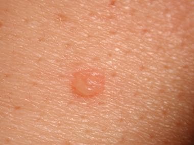
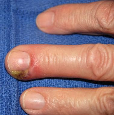
Benign familial pemphigus is a rare, autosomal dominant blistering disorder that primarily affects the mucous membranes. It is characterized by the presence of flaccid blisters and erosions on the skin and mucous membranes. The lesions are usually painless and heal without scarring.
The condition is caused by mutations in the desmoglein-1 (DSG1) gene, which provides instructions for making a protein called desmoglein 1. This protein is a component of desmosomes, which are structures that help bind cells together. Mutations in the DSG1 gene lead to the production of an abnormal desmoglein 1 protein, which disrupts the formation of desmosomes and causes the cells in the epidermis to separate from each other, resulting in blister formation.
Benign familial pemphigus is typically a milder form of pemphigus and has a good prognosis. Treatment usually involves the use of topical corticosteroids to reduce inflammation and promote healing of the lesions. In severe cases, systemic corticosteroids or other immunosuppressive medications may be necessary.
Keratinocytes are the predominant type of cells found in the epidermis, which is the outermost layer of the skin. These cells are responsible for producing keratin, a tough protein that provides structural support and protection to the skin. Keratinocytes undergo constant turnover, with new cells produced in the basal layer of the epidermis and older cells moving upward and eventually becoming flattened and filled with keratin as they reach the surface of the skin, where they are then shed. They also play a role in the immune response and can release cytokines and other signaling molecules to help protect the body from infection and injury.
Immunoglobulin G (IgG) is a type of antibody, which is a protective protein produced by the immune system in response to foreign substances like bacteria or viruses. IgG is the most abundant type of antibody in human blood, making up about 75-80% of all antibodies. It is found in all body fluids and plays a crucial role in fighting infections caused by bacteria, viruses, and toxins.
IgG has several important functions:
1. Neutralization: IgG can bind to the surface of bacteria or viruses, preventing them from attaching to and infecting human cells.
2. Opsonization: IgG coats the surface of pathogens, making them more recognizable and easier for immune cells like neutrophils and macrophages to phagocytose (engulf and destroy) them.
3. Complement activation: IgG can activate the complement system, a group of proteins that work together to help eliminate pathogens from the body. Activation of the complement system leads to the formation of the membrane attack complex, which creates holes in the cell membranes of bacteria, leading to their lysis (destruction).
4. Antibody-dependent cellular cytotoxicity (ADCC): IgG can bind to immune cells like natural killer (NK) cells and trigger them to release substances that cause target cells (such as virus-infected or cancerous cells) to undergo apoptosis (programmed cell death).
5. Immune complex formation: IgG can form immune complexes with antigens, which can then be removed from the body through various mechanisms, such as phagocytosis by immune cells or excretion in urine.
IgG is a critical component of adaptive immunity and provides long-lasting protection against reinfection with many pathogens. It has four subclasses (IgG1, IgG2, IgG3, and IgG4) that differ in their structure, function, and distribution in the body.
Autoantigens are substances that are typically found in an individual's own body, but can stimulate an immune response because they are recognized as foreign by the body's own immune system. In autoimmune diseases, the immune system mistakenly attacks and damages healthy tissues and organs because it recognizes some of their components as autoantigens. These autoantigens can be proteins, DNA, or other molecules that are normally present in the body but have become altered or exposed due to various factors such as infection, genetics, or environmental triggers. The immune system then produces antibodies and activates immune cells to attack these autoantigens, leading to tissue damage and inflammation.
The epidermis is the outermost layer of the skin, composed mainly of stratified squamous epithelium. It forms a protective barrier that prevents water loss and inhibits the entry of microorganisms. The epidermis contains no blood vessels, and its cells are nourished by diffusion from the underlying dermis. The bottom-most layer of the epidermis, called the stratum basale, is responsible for generating new skin cells that eventually move up to replace dead cells on the surface. This process of cell turnover takes about 28 days in adults.
The most superficial part of the epidermis consists of dead cells called squames, which are constantly shed and replaced. The exact rate at which this happens varies depending on location; for example, it's faster on the palms and soles than elsewhere. Melanocytes, the pigment-producing cells, are also located in the epidermis, specifically within the stratum basale layer.
In summary, the epidermis is a vital part of our integumentary system, providing not only physical protection but also playing a crucial role in immunity and sensory perception through touch receptors called Pacinian corpuscles.
Mouth diseases refer to a variety of conditions that affect the oral cavity, including the lips, gums, teeth, tongue, palate, and lining of the mouth. These diseases can be caused by bacteria, viruses, fungi, or other organisms. They can also result from injuries, chronic illnesses, or genetic factors.
Some common examples of mouth diseases include dental caries (cavities), periodontal disease (gum disease), oral herpes, candidiasis (thrush), lichen planus, and oral cancer. Symptoms may include pain, swelling, redness, bleeding, bad breath, difficulty swallowing or speaking, and changes in the appearance of the mouth or teeth. Treatment depends on the specific diagnosis and may involve medications, dental procedures, or lifestyle changes.
Cadherins are a type of cell adhesion molecule that play a crucial role in the development and maintenance of intercellular junctions. They are transmembrane proteins that mediate calcium-dependent homophilic binding between adjacent cells, meaning that they bind to identical cadherin molecules on neighboring cells.
There are several types of cadherins, including classical cadherins, desmosomal cadherins, and protocadherins, each with distinct functions and localization in tissues. Classical cadherins, also known as type I cadherins, are the most well-studied and are essential for the formation of adherens junctions, which help to maintain cell-to-cell contact and tissue architecture.
Desmosomal cadherins, on the other hand, are critical for the formation and maintenance of desmosomes, which are specialized intercellular junctions that provide mechanical strength and stability to tissues. Protocadherins are a diverse family of cadherin-related proteins that have been implicated in various developmental processes, including neuronal connectivity and tissue patterning.
Mutations in cadherin genes have been associated with several human diseases, including cancer, neurological disorders, and heart defects. Therefore, understanding the structure, function, and regulation of cadherins is essential for elucidating their roles in health and disease.
Desmoplakins are important proteins that play a crucial role in the structural integrity and function of certain types of cell-to-cell junctions called desmosomes. Desmosomes are specialized structures that connect adjacent cells in tissues that undergo significant mechanical stress, such as the skin, heart, and gut.
Desmoplakins are large proteins that are composed of several domains, including a plakin domain, which interacts with other desmosomal components, and a spectrin-like repeat domain, which binds to intermediate filaments. By linking desmosomes to the intermediate filament network, desmoplakins help to provide mechanical strength and stability to tissues.
Mutations in the genes that encode desmoplakins have been associated with several human genetic disorders, including arrhythmogenic right ventricular cardiomyopathy (ARVC), a heart condition characterized by abnormal heart rhythms and structural changes in the heart muscle, and epidermolysis bullosa simplex (EBS), a skin disorder characterized by blistering and fragility of the skin.
An oral ulcer is a defect or break in the continuity of the epithelium, the tissue that lines the inner surface of the mouth, leading to an inflamed, painful, and sometimes bleeding lesion. They can be classified as primary (e.g., aphthous ulcers, traumatic ulcers) or secondary (e.g., those caused by infections, underlying systemic conditions, or reactions to medications). Oral ulcers may cause discomfort, impacting speech and food consumption, and their presence might indicate an underlying medical issue that requires further evaluation.
Pulse therapy, in the context of drug treatment, refers to a therapeutic regimen where a medication is administered in large doses for a short period of time, followed by a break or "drug-free" interval before the next dose. This cycle is then repeated at regular intervals. The goal of pulse therapy is to achieve high concentrations of the drug in the body to maximize its therapeutic effect while minimizing overall exposure and potential side effects.
This approach is often used for drugs that have a long half-life or slow clearance, as it allows for periodic "washing out" of the drug from the body. Pulse therapy can also help reduce the risk of developing drug resistance in certain conditions like rheumatoid arthritis and tuberculosis. Common examples include pulse methotrexate for rheumatoid arthritis and intermittent preventive treatment with anti-malarial drugs.
It is important to note that the use of pulse therapy should be based on a thorough understanding of the drug's pharmacokinetics, therapeutic index, and potential adverse effects. Close monitoring of patients undergoing pulse therapy is essential to ensure safety and efficacy.
Desmoglein 2 is a type of desmoglein protein that is primarily found in the desmosomes of epithelial cells. Desmosomes are specialized structures that help to anchor intermediate filaments to the cell membrane and provide strength and stability to tissues that undergo mechanical stress, such as the skin and heart.
Desmoglein 2 plays a critical role in maintaining cell-cell adhesion by forming intercellular junctions called desmosomal cadherins. These junctions help to hold adjacent cells together and contribute to the integrity of epithelial tissues. Mutations in the gene that encodes Desmoglein 2 have been associated with several skin disorders, including pemphigus vulgaris, a blistering autoimmune disease that affects mucous membranes and the skin. In this condition, antibodies target Desmoglein 2, leading to loss of cell-cell adhesion and formation of blisters.
Desmocollins are a type of cadherin, which is a transmembrane protein involved in cell-cell adhesion. Specifically, desmocollins are found in the desmosomes, which are specialized structures that help to mechanically connect adjacent epithelial cells. There are three main isoforms of desmocollin (Desmocollin-1, -2, and -3) that are encoded by different genes. Mutations in the genes encoding desmocollins have been associated with several skin blistering disorders, including certain forms of epidermolysis bullosa.
 Pemphigus
 Pemphigus
 Pemphigus | Nature Reviews Disease Primers
 Pemphigus vulgaris: MedlinePlus Medical Encyclopedia
 Pandemic Dictates Reconsideration of Pemphigus Therapy
 Pemphigus Vulgaris | Johns Hopkins Medicine
 Pemphigus: Signs and symptoms
 Pemphigus - wikidoc
 Paraneoplastic pemphigus pathology | DermNet
 Paraneoplastic Pemphigus - Causes, Symptoms & Treatment!
 Image: Pemphigus foliaceus, pinnal lesions - Merck Veterinary Manual
 Feline Pemphigus - A Review • MSPCA-Angell
 Pemphigus Complex (Wildfire): Epidemiology and Clinics - WSAVA2009 - VIN
 Efgartigimod alfa by Argenx for Pemphigus Vulgaris: Likelihood of Approval
 "Periplakin, an autoantigen in paraneoplastic pemphigus and pemphigus f" by Shideh Kazerounian
 A Clinician's Guide to Pemphigus Vulgaris, Differential Diagnosis of Cutaneous Lesions | DeepDyve
 Cyclophosphamide pulses with oral prednisolone in the treatment of pemphigus: A pilot study
 Pemphigus & Pemphigoid Treatment in Salt Lake City, UT | University of Utah Health | University of Utah Health
 Description: Characterization of cellular and humoral immune responses in pemphigus patients and an HLA-transgenic mouse model ...
 Study of pemphigus vegetans finds no correlation between clinical findings and prognostic outcome
 Pemphigus vulgaris localised exclusively to the penis - Indian Journal of Dermatology, Venereology and Leprology
 Phase III PEMPHIX Study Shows Genentech's Rituxan (Rituximab) Superior to Mycophenolate Mofetil in Patients With Pemphigus...
 Validation of 5-Point Investigator Global Assessments for Pemphigus - Full Text View - ClinicalTrials.gov
 Herbal Medicine for Pemphigus - Avnayt & Waltham's Herbal Treatments