Pericarditis
Pericarditis, Constrictive
Pericarditis, Tuberculous
Pericardiectomy
Pericardial Effusion
Cardiac Tamponade
Cardiomyopathy, Restrictive
Pericardium
Tuberculosis, Cardiovascular
Pleurodynia, Epidemic
Myocarditis
Carcinoid heart disease from ovarian primary presenting with acute pericarditis and biventricular failure. (1/361)
A case is described of a 54 year old woman who had acute pericarditis with large exudative effusion accompanied by severe right and left ventricular failure. The patient was finally diagnosed with carcinoid heart disease from an ovarian carcinoid teratoma. She was treated with octreotide--a somatostatin analogue--followed by radical surgical resection of the neoplasm. At one year follow up only mild carcinoid tricuspid regurgitation remained. Only 16 cases of carcinoid heart disease from an ovarian primary have been described in literature. Moreover clinically manifest acute, nonmetastatic pericarditis and left heart failure are not considered as possible presentations of carcinoid heart disease, whatever the origin. In a recent series a small pericardial effusion was considered an infrequent and unexpected echocardiographic finding in carcinoid heart patients. One case of "carcinoid pericarditis" has previously been described as a consequence of pericardial metastasis. Left sided heart involvement is usually caused by bronchial carcinoids or patency of foramen ovale; both were excluded in the case presented. (+info)Primary right atrial angiosarcoma mimicking acute pericarditis, pulmonary embolism, and tricuspid stenosis. (2/361)
A 29 year old white man presented to the emergency room with new onset pleuritic chest pain and shortness of breath. He was initially diagnosed as having viral pericarditis and was treated with non-steroidal anti-inflammatory drugs. A few weeks later he developed recurrent chest pain with cough and haemoptysis. Chest radiography, cardiac examination, transthoracic and transoesophageal echocardiography pointed to a mass that arose from the posterior wall of the right atrium, not attached to the interatrial septum, which protruded into the lumen of the right atrium causing intermittent obstruction of inflow across the tricuspid valve. Contrast computed tomography of the chest showed a right atrial mass extending to the anterior chest wall. The lung fields were studded with numerous pulmonary nodules suggestive of metastases. A fine needle aspiration of the pulmonary nodule revealed histopathology consistent with spindle cell sarcoma thought to originate in the right atrium. Immunohistochemical stains confirmed that this was an angiosarcoma. There was no evidence of extracardiac origin of the tumour. The patient was treated with chemotherapy and radiation. This case highlights the clinical presentation, rapid and aggressive course of cardiac angiosarcomas, and the diagnostic modalities available for accurate diagnosis. (+info)One-step reverse transcriptase PCR method for detection of Borrelia burgdorferi mRNA in mouse Lyme arthritis tissue samples. (3/361)
A one-step reverse transcriptase PCR (RT-PCR) method for detection of Borrelia burgdorferi mRNA in infected C3H mice is described. This simple procedure, less prone to nucleic acid cross-contamination than the standard method, was found to be 10-fold more sensitive than a classical two-step RT-PCR assay. By using one-step RT-PCR, flagellin mRNAs were detected in synovial and heart tissues from all seven infected mice tested. (+info)Intrapericardial streptokinase in purulent pericarditis. (4/361)
Six consecutive children with proven purulent pericarditis were treated with pericardial irrigation with streptokinase. Mean (SD) 861 (678) ml (range 240-2000) of thick purulent fluid was drained, and five children had complete clearance of the pus within 3-8 days. One child developed intrapericardial haemorrhage with a submitral pseudoaneurysm and underwent patch closure of the neck of the aneurysm as well as anterior pericardiectomy. Follow up of 13 to 30 months revealed no pericardial constriction. (+info)Restrictive pericarditis. (5/361)
BACKGROUND: Pericardial thickening is an uncommon complication of cardiac surgery. OBJECTIVES: To study pericardial thickening as the cause of severe postoperative venous congestion. SUBJECTS: Two men, one with severe aortic stenosis and single coronary artery disease, and one with coronary artery disease after an old inferior infarction. Both had coronary artery bypass grafting surgery. METHODS: Magnetic resonance imaging (MRI), Doppler echocardiography, and cardiac catheterisation. RESULTS: Venous pressure was raised in both patients. MRI showed mildly thickened pericardium, and cardiac catheterisation indicated diastolic equalization of pressures in the four chambers. Jugular venous pulse showed a dominant "Y" descent coinciding with early diastolic flow in the superior vena cava, and mitral and tricuspid Doppler forward flow proved restrictive physiology. The clinical background suggested pericardial disease so both patients had pericardiectomy. This proved the pericardium to be thickened; the extent of fibrosis also involved the epicardium. CONCLUSIONS: Although rare, restrictive pericarditis (restrictive ventricular physiology resulting from pericardial disease) should be considered to be a separate diagnostic entity because its pathological basis and treatment are different from intrinsic myocardial disease. This diagnosis may be confirmed by standard investigational techniques or may require diagnostic thoracotomy. (+info)Left ventricular pseudoaneurysm complicating infective pericarditis. (6/361)
Cross sectional echocardiography demonstrated a pseudoaneurysm of the left ventricular posterolateral wall close to the atrioventricular junction in a 4 year old girl with infective pericarditis complicating lobar pneumonia. Colour flow Doppler demonstrated bidirectional flow across the communication hole. Surgical resection was successful. (+info)Coxiella burnetii pericarditis: report of 15 cases and review. (7/361)
Q fever is characterized by its clinical polymorphism, and pericarditis associated with Q fever has occasionally been described. Herein we report 15 cases of Coxiella burnetii pericarditis, 9 from our data bank and 6 encountered within the past 12 months. Three patients presented with life-threatening tamponade. We compare our cases with the 18 previously reported and with 60 Q fever-matched controls at our center. This study showed that Q fever pericarditis can present as acute as well as chronic disease; we describe relapse after 6 months in association with a serological profile compatible with the chronic form of disease (phase I C. burnetii IgG titer of > or = 800). Discriminant factors among patients and controls are age of > 52 years (adjusted odds ratio [OR], 5.66), the occurrence of general symptoms such as arthralgias or myalgias (adjusted OR, 6.54), and a normal erythrocyte sedimentation rate (adjusted OR, 16.37). No specific symptoms or underlying cardiac predispositions are observed. (+info)New insights regarding the atrial flutter reentrant circuit : studies in the canine sterile pericarditis model. (8/361)
Background-We studied atrial activation during induced atrial flutter in the canine sterile pericarditis model to test the hypothesis that the atrial flutter reentrant circuit includes a septal component. Methods and Results-We studied 10 episodes of induced, sustained (>5 minutes) atrial flutter in 9 dogs. In all episodes, the reentrant circuit included a septal component. In 6 episodes, there were 2 reentrant circuits, one in the right atrial free wall and the second involving the atrial septum, Bachmann's bundle, and the right atrial free wall; both circuits shared a pathway in the right atrial free wall (figure-of-eight). The direction (superior or inferior) of the septal wave front of the second circuit correlated with the direction (clockwise or counterclockwise, respectively) of the right atrial free-wall circuit. A line of functional block in the right atrial free wall was part of both reentrant circuits. In the other 4 atrial flutter episodes, only 1 reentrant circuit was present, with activation in an inferior-to-superior direction in the septum and a superior-to-inferior direction in the right atrial free wall in 2 episodes and in the opposite direction in the other 2 episodes. In all atrial flutter episodes, the flutter wave polarity in ECG lead II was determined by the direction of activation in the left atrium; polarity was positive when the direction was superior to inferior and negative when the direction was inferior to superior. Conclusions-In this model of atrial flutter, the reentrant circuit (1) always included a septal component, (2) did not always require a right atrial free-wall reentrant circuit, (3) demonstrated figure-of-eight reentry when a reentrant circuit was present in the right atrial free wall, and (4) was associated with a line of functional block in the right atrial free wall. (+info)Pericarditis is a medical condition characterized by inflammation of the pericardium, which is the thin sac-like membrane that surrounds the heart and contains serous fluid to reduce friction during heartbeats. The inflammation can cause symptoms such as chest pain, shortness of breath, and sometimes fever.
The pericardium has two layers: the visceral pericardium, which is tightly adhered to the heart's surface, and the parietal pericardium, which lines the inner surface of the chest cavity. Normally, there is a small amount of fluid between these two layers, allowing for smooth movement of the heart within the chest cavity.
In pericarditis, the inflammation causes the pericardial layers to become irritated and swollen, leading to an accumulation of excess fluid in the pericardial space. This can result in a condition called pericardial effusion, which can further complicate the situation by putting pressure on the heart and impairing its function.
Pericarditis may be caused by various factors, including viral or bacterial infections, autoimmune disorders, heart attacks, trauma, or cancer. Treatment typically involves addressing the underlying cause, managing symptoms, and reducing inflammation with medications such as nonsteroidal anti-inflammatory drugs (NSAIDs), colchicine, or corticosteroids. In severe cases, pericardiocentesis (removal of excess fluid from the pericardial space) or surgical intervention may be necessary.
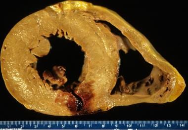
Constrictive pericarditis is a medical condition characterized by the inflammation and thickening of the pericardium, which is the sac-like membrane that surrounds the heart. This inflammation leads to scarring and thickening of the pericardium, causing it to become stiff and inflexible. As a result, the heart's ability to fill with blood between beats is restricted, leading to symptoms such as shortness of breath, fatigue, and fluid retention.
In contrastive pericarditis, the thickened and scarred pericardium restricts the normal movement of the heart within the chest cavity, leading to a characteristic pattern of hemodynamic abnormalities. These include equalization of diastolic pressures in all cardiac chambers, increased systemic venous pressure, and decreased cardiac output.
The most common causes of constrictive pericarditis include prior infection, radiation therapy, autoimmune disorders, and previous heart surgery. Diagnosis typically involves a combination of medical history, physical examination, imaging studies such as echocardiography or MRI, and sometimes invasive testing such as cardiac catheterization. Treatment may involve medications to manage symptoms and reduce inflammation, as well as surgical removal of the pericardium (pericardiectomy) in severe cases.
Tuberculous pericarditis is a specific form of pericarditis (inflammation of the pericardium, the thin sac-like membrane that surrounds the heart) that is caused by the bacterial infection of Mycobacterium tuberculosis. This type of pericarditis is more common in areas where tuberculosis is prevalent and can lead to serious complications if not diagnosed and treated promptly.
In tuberculous pericarditis, the bacteria typically spread from the lungs (the most common site of TB infection) or other infected organs through the bloodstream to the pericardium. The infection causes an inflammatory response, leading to the accumulation of fluid in the pericardial space (pericardial effusion), which can put pressure on the heart and impair its function. In some cases, the inflammation may lead to the formation of scar tissue, causing the pericardium to thicken and constrict, a condition known as constrictive pericarditis.
Symptoms of tuberculous pericarditis can include chest pain, cough, fever, fatigue, weight loss, and difficulty breathing. Diagnosis typically involves a combination of medical history, physical examination, imaging tests (such as echocardiography, CT scan, or MRI), and laboratory tests (including analysis of the pericardial fluid). Treatment usually consists of a long course of antibiotics specific to TB, along with anti-inflammatory medications and close monitoring for potential complications.
Pericardiectomy is a surgical procedure that involves the removal of all or part of the pericardium, which is the sac-like membrane surrounding the heart. This surgery is typically performed to treat chronic or recurrent pericarditis, constrictive pericarditis, or pericardial effusions that do not respond to other treatments. Pericardiectomy can help reduce symptoms such as chest pain, shortness of breath, and fluid buildup around the heart, improving the patient's quality of life and overall prognosis.
Pericardial effusion is an abnormal accumulation of fluid in the pericardial space, which is the potential space between the two layers of the pericardium - the fibrous and serous layers. The pericardium is a sac that surrounds the heart to provide protection and lubrication for the heart's movement during each heartbeat. Normally, there is only a small amount of fluid (5-15 mL) in this space to ensure smooth motion of the heart. However, when an excessive amount of fluid accumulates, it can cause increased pressure on the heart, leading to various complications such as decreased cardiac output and even cardiac tamponade, a life-threatening condition that requires immediate medical attention.
Pericardial effusion may result from several causes, including infections (viral, bacterial, or fungal), inflammatory conditions (such as rheumatoid arthritis, lupus, or cancer), trauma, heart surgery, kidney failure, or iatrogenic causes. The symptoms of pericardial effusion can vary depending on the rate and amount of fluid accumulation. Slowly developing effusions may not cause any symptoms, while rapid accumulations can lead to chest pain, shortness of breath, cough, palpitations, or even hypotension (low blood pressure). Diagnosis is usually confirmed through imaging techniques such as echocardiography, CT scan, or MRI. Treatment depends on the underlying cause and severity of the effusion, ranging from close monitoring to drainage procedures or medications to address the root cause.
Cardiac tamponade is a serious medical condition that occurs when there is excessive fluid or blood accumulation in the pericardial sac, which surrounds the heart. This accumulation puts pressure on the heart, preventing it from filling properly and reducing its ability to pump blood effectively. As a result, cardiac output decreases, leading to symptoms such as low blood pressure, shortness of breath, chest pain, and a rapid pulse. If left untreated, cardiac tamponade can be life-threatening, requiring emergency medical intervention to drain the fluid and relieve the pressure on the heart.
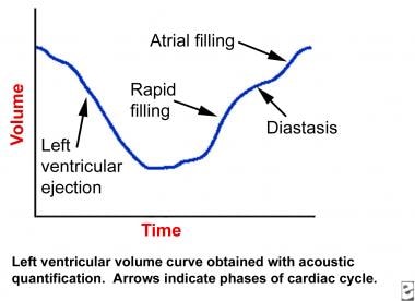
Restrictive cardiomyopathy (RCM) is a type of heart muscle disorder characterized by impaired relaxation and filling of the lower chambers of the heart (the ventricles), leading to reduced pump function. This is caused by stiffening or rigidity of the heart muscle, often due to fibrosis or scarring. The stiffness prevents the ventricles from filling properly with blood during the diastolic phase, which can result in symptoms such as shortness of breath, fatigue, and fluid retention.
RCM is a less common form of cardiomyopathy compared to dilated or hypertrophic cardiomyopathies. It can be idiopathic (no known cause) or secondary to other conditions like amyloidosis, sarcoidosis, or storage diseases. Diagnosis typically involves a combination of medical history, physical examination, echocardiography, and sometimes cardiac MRI or biopsy. Treatment is focused on managing symptoms and addressing underlying causes when possible.
Pericardiocentesis is a medical procedure where a needle or a catheter is inserted into the pericardial sac, the thin fluid-filled space surrounding the heart, to remove excess fluids or air that has accumulated. This buildup can put pressure on the heart and impede its function, leading to various cardiac symptoms such as chest pain, shortness of breath, and palpitations. The procedure is often guided by echocardiography or fluoroscopy to ensure proper placement and minimize risks. Pericardiocentesis may be performed as an emergency treatment or a scheduled intervention, depending on the patient's condition.
The pericardium is the double-walled sac that surrounds the heart. It has an outer fibrous layer and an inner serous layer, which further divides into two parts: the parietal layer lining the fibrous pericardium and the visceral layer (epicardium) closely adhering to the heart surface.
The space between these two layers is filled with a small amount of lubricating serous fluid, allowing for smooth movement of the heart within the pericardial cavity. The pericardium provides protection, support, and helps maintain the heart's normal position within the chest while reducing friction during heart contractions.
"Cardiovascular Tuberculosis" refers to a form of tuberculosis (TB) where the bacteria (Mycobacterium tuberculosis) infects the heart or the blood vessels. This is a less common manifestation of TB, but it can have serious consequences if left untreated.
In cardiovascular TB, the bacteria can cause inflammation and damage to the heart muscle (myocarditis), the sac surrounding the heart (pericarditis), or the coronary arteries that supply blood to the heart muscle. This can lead to symptoms such as chest pain, shortness of breath, coughing, fatigue, and fever. In severe cases, it can cause heart failure or life-threatening arrhythmias.
Cardiovascular TB is usually treated with a combination of antibiotics that are effective against the TB bacteria. The treatment may last for several months to ensure that all the bacteria have been eliminated. In some cases, surgery may be necessary to repair or replace damaged heart valves or vessels. Early diagnosis and treatment can help prevent serious complications and improve outcomes in patients with cardiovascular TB.
Suppuration is the process of forming or discharging pus. It is a condition that results from infection, tissue death (necrosis), or injury, where white blood cells (leukocytes) accumulate to combat the infection and subsequently die, forming pus. The pus consists of dead leukocytes, dead tissue, debris, and microbes (bacteria, fungi, or protozoa). Suppuration can occur in various body parts such as the lungs (empyema), brain (abscess), or skin (carbuncle, furuncle). Treatment typically involves draining the pus and administering appropriate antibiotics to eliminate the infection.
Cardiovascular infections, also known as infective endocarditis, are infections that affect the inner layer of the heart, including the heart valves. These infections are usually caused by bacteria, but they can also be caused by fungi or other microorganisms. They can occur when bacteria or other germs enter the bloodstream and then settle in the heart.
There are several types of cardiovascular infections, including:
* Native Valve Endocarditis: This occurs when an infection affects the heart valves that are present at birth.
* Prosthetic Valve Endocarditis: This occurs when an infection affects an artificial heart valve.
* Intracardiac Device-Related Infections: These infections can occur in people who have devices such as pacemakers or implantable defibrillators.
* Infectious Myocarditis: This is an inflammation of the heart muscle caused by an infection.
Symptoms of cardiovascular infections may include fever, chills, fatigue, shortness of breath, chest pain, and a new or changing heart murmur. Treatment typically involves several weeks of antibiotics, and in some cases, surgery may be necessary to remove the infected tissue. Prevention measures include good oral hygiene, prompt treatment of skin infections, and prophylactic antibiotics for certain high-risk individuals undergoing dental or surgical procedures.
Epidemic pleurodynia, also known as Bornholm disease or devils' grip, is a self-limiting viral illness characterized by sudden onset of severe, stabbing chest or upper abdominal pain. It is caused most commonly by an enterovirus, often Coxsackie A or B.
The hallmark of epidemic pleurodynia is the pleuritic nature of the pain, which is aggravated by deep breathing, coughing, or movement. The muscle spasms can be so intense that they cause the patient to assume a fetal position in order to minimize the discomfort. Other symptoms may include fever, headache, nausea, vomiting, and generalized weakness.
The term "epidemic" refers to the fact that this disease tends to occur in outbreaks, particularly during the summer and fall months. However, sporadic cases can also occur throughout the year. The illness typically lasts for 5-10 days but may rarely persist for several weeks.
Treatment is generally supportive and includes rest, hydration, and analgesics for pain relief. Antiviral medications are not usually recommended, as they have not been shown to significantly affect the course of the illness.
Myocarditis is an inflammation of the myocardium, which is the middle layer of the heart wall. The myocardium is composed of cardiac muscle cells and is responsible for the heart's pumping function. Myocarditis can be caused by various infectious and non-infectious agents, including viruses, bacteria, fungi, parasites, autoimmune diseases, toxins, and drugs.
In myocarditis, the inflammation can damage the cardiac muscle cells, leading to decreased heart function, arrhythmias (irregular heart rhythms), and in severe cases, heart failure or even sudden death. Symptoms of myocarditis may include chest pain, shortness of breath, fatigue, palpitations, and swelling in the legs, ankles, or abdomen.
The diagnosis of myocarditis is often based on a combination of clinical presentation, laboratory tests, electrocardiogram (ECG), echocardiography, cardiac magnetic resonance imaging (MRI), and endomyocardial biopsy. Treatment depends on the underlying cause and severity of the disease and may include medications to support heart function, reduce inflammation, control arrhythmias, and prevent further damage to the heart muscle. In some cases, hospitalization and intensive care may be necessary.
Cardanolides are a type of steroid compound that are found in certain plants, particularly in the family Apocynaceae. These compounds have a characteristic structure that includes a five-membered lactone ring attached to a steroid nucleus, and they are known for their ability to inhibit the sodium-potassium pump (Na+/K+-ATPase) in animal cells. This property makes cardanolides toxic to many organisms, including humans, and they have been used as heart poisons and insecticides.
One of the most well-known cardanolides is ouabain, which is found in the seeds of several African plants and has been used traditionally as a medicine for various purposes, including as a heart stimulant and a poison for hunting. Other examples of cardanolides include digoxin and digitoxin, which are derived from the foxglove plant (Digitalis purpurea) and are used in modern medicine to treat heart failure and atrial arrhythmias.
It's worth noting that while cardanolides have important medical uses, they can also be highly toxic if ingested or otherwise introduced into the body in large amounts. Therefore, it's essential to use these compounds only under the supervision of a qualified healthcare professional.
 Pericarditis
Acute pericarditis
Tuberculous pericarditis
Constrictive pericarditis
Uremic pericarditis
Camptodactyly-arthropathy-coxa vara-pericarditis syndrome
Obstructive shock
Myocarditis
Bongani Mayosi
Davis Evan Bedford
Fredrick Arthur Willius
ST elevation
Pericardial friction rub
Colchicine
Rilonacept
Dressler syndrome
Pulsus paradoxus
Edwin Herman Lennette
Myocardial infarction complications
Ibuprofen
Mitral valve prolapse
Rheumatoid pleuritis
Infectious mononucleosis
Ewart's sign
Cardiac muscle
Max Barrett
Edward Delos Churchill
Roberto Ferrari (cardiologist)
Brucella
Mumps
Viktor Schmieden
Postpericardiotomy syndrome
Acute42
- Acute myocardial infarction can also cause pericarditis, but the presenting symptoms often differ enough to warrant diagnosis. (wikipedia.org)
- Acute and subacute forms of pericarditis (which may or may not be symptomatic) may deposit fibrin, which, in turn, can evoke a pericardial effusion. (medscape.com)
- Viral infections (especially infections with coxsackieviruses and echoviruses but also influenza, Epstein-Barr, varicella, hepatitis, mumps, and HIV viruses) are the commonest cause of acute pericarditis and probably are responsible for many cases classified as idiopathic. (health.am)
- Acute Pericarditis can result in Pericardial Tamponade and can lead to chronic or constrictive Pericarditis . (ecureme.com)
- and analyze the conformity of the management of acute pericarditis according to the recommendations, a retrospective inclusion of all patients admitted to our hospital from January 2010 to December 2018 with the diagnosis of acute pericarditis was conducted. (nih.gov)
- There was no link between the decision to conduct etiology exams and the age, gender, a history of acute pericarditis or relapse. (nih.gov)
- Acute pericarditis is a self-limiting disease without significant complications or recurrences in 70% to 90% of patients. (escardio.org)
- Regarding clinical management and therapy of acute pericarditis, it is not mandatory to search for the aetiology in all patients, especially in countries with a low prevalence of tuberculosis (TB) because of the relatively benign course associated with the common causes of pericarditis and the relatively low yield of diagnostic investigations [1]. (escardio.org)
- The major risk factors associated with poor prognosis in acute pericarditis include high fever, subacute course, evidence of large pericardial effusion, cardiac tamponade and failure to respond within seven days to non-steroidal anti-inflammatory drugs (NSAIDs). (escardio.org)
- These are based on expert opinion and literature review, including acute pericarditis associated with immunodeficiency, trauma, anticoagulant therapy and myocarditis (myopericarditis). (escardio.org)
- On this basis, a triage for acute pericarditis is proposed (Figure 1). (escardio.org)
- Proposed Triage of Acute Pericarditis. (escardio.org)
- Proposed triage of acute pericarditis according to epidemiological background and predictors of poor prognosis at presentation (at least one predictor of poor prognosis is sufficient to identify a high-risk case). (escardio.org)
- Most patients with acute pericarditis (generally those with presumed viral or idiopathic pericarditis) have a good long-term prognosis [6]. (escardio.org)
- Acute pericarditis typically presents with sharp pleuritic chest pain, exacerbated by the supine position and improved with leaning forward. (logicalimages.com)
- There are two types of pericarditis - acute and chronic. (heartfoundation.org.nz)
- Acute pericarditis develops suddenly, with symptoms starting rapidly. (heartfoundation.org.nz)
- Recurrent pericarditis is probably the most common and troublesome complication of pericarditis, affecting about 20% to 30% of patients after a first attack of acute pericarditis. (revespcardiol.org)
- Acute Pericarditis Acute pericarditis is inflammation of the pericardium (the flexible two-layered sac that envelops the heart) that begins suddenly, is often painful, and causes fluid and blood components such. (merckmanuals.com)
- Is acute idiopathic pericarditis associated with recent upper respiratory tract infection or gastroenteritis? (unige.ch)
- On the 11th day of hospitalization, the patient developed acute chest pain, diaphoresis, and hypotension, prompting an ECG that showed diffuse ST elevations compatible with acute pericarditis. (acc.org)
- Our patient was subsequently diagnosed with acute aortic valve endocarditis on a native aortic valve that was complicated by both an aortic root abscess and a contained aortic root rupture, leading to hemorrhagic pericarditis. (acc.org)
- Case report: Acute pericarditis following hepatic microwave ablation for liver metastasis. (unil.ch)
- ESR, CRP, and Troponin have all been shown to be elevated with acute pericarditis. (pedemmorsels.com)
- Colchicine has been recently shown to be helpful in adults with acute pericarditis (See CMC Core Concept ), but this is not yet an often used therapy in children. (pedemmorsels.com)
- Acute MI - post MI pericarditis occurs in about 20% of MI patients. (almostadoctor.co.uk)
- Compared with pericardial effusion and tamponade, acute pericarditis can be generally self-limiting and not life-threatening, with the possibility of causing significant disabilities. (eminencepapers.com)
- Acute pericarditis always has a high chance of reoccurrence compared to chronic pericarditis. (eminencepapers.com)
- The symptoms relapse after 4-6 weeks of an acute episode of pericarditis, which is defined as incessant pericarditis (Imazio et al. (eminencepapers.com)
- There is a lack of accurate epidemiological data for acute pericarditis. (eminencepapers.com)
- However, acute pericarditis is always diagnosed in 0.2% of all cardiovascular disease hospital admissions. (eminencepapers.com)
- Acute pericarditis also accounts for 5% of emergency admissions for chest pain in North America. (eminencepapers.com)
- Around 9% of individuals experiencing acute pericarditis develop constrictive physiology, with infectious causes remaining the primary culprits in the developing world. (medtigo.com)
- The pathophysiology of chronic constrictive pericarditis implicates the gradual closure of the pericardial cavity due to the formation of granulation tissue, which occurs during the healing process of an acute episode of fibrinous through the absorption of a chronic pericardial effusion. (medtigo.com)
- This is an example of acute pericarditis Murmurs are generated by turbulent blood flow across incompetent or stenotic valves. (practicalclinicalskills.com)
- The ST/T ratio can also be used to differentiate early repolarization from acute pericarditis. (cardiacbootcamp.org)
- My diagnosis in this case is that this is probably acute pericarditis on the background of BER. (cardiacbootcamp.org)
- Among 13,759 patients with acute pericarditis , 1550 were subsequently diagnosed with cancer. (medscape.com)
- Ratnapalan S, Brown K, Benson L. Children presenting with acute pericarditis to the emergency department. (medscape.com)
- Ananthasubramaniam K, Farha A. Primary right atrial angiosarcoma mimicking acute pericarditis, pulmonary embolism, and tricuspid stenosis. (medscape.com)
- Differential diagnosis of acute pericarditis from normal variant early repolarization and left ventricular hypertrophy with early repolarization: an electrocardiographic study. (medscape.com)
- Colchicine in addition to conventional therapy for acute pericarditis: results of the COlchicine for acute PEricarditis (COPE) trial. (medscape.com)
Cause pericarditis3
- Fungi such as Candida spp and parasites such as Toxoplasma gondii also cause pericarditis. (logicalimages.com)
- malignancy: Certain kinds of malignancy, such as lymphoma and mesothelioma, can cause pericarditis in cats. (petcarerx.com)
- Anything that can cause pericarditis can also cause recurrent pericarditis . (osmosis.org)
Recurrent14
- Recurrent pericarditis is seen in 15%-30% of patients. (logicalimages.com)
- Recurrent Pericarditis: Can Anakinra Offer a Promising Therapy in Adults With Refractory Symptoms? (revespcardiol.org)
- 1-3 Recurrent pericarditis is defined as the recurrence of symptoms and signs of pericarditis after an arbitrary symptom-free interval of 6 weeks. (revespcardiol.org)
- Proposed diagnostic criteria for recurrent pericarditis include recurrent chest pain and 1 or more of the following signs: fever, pericardial friction rubs, electrocardiographic changes, echocardiographic evidence of new or worsening pericardial effusion, or elevated markers of inflammation (ie, elevated leukocyte count, erythrocyte sedimentation rate, or C-reactive protein level). (revespcardiol.org)
- 5 Many patients with a previous attack of pericarditis may experience recurrent pain but without a true recurrence documented by objective evidence of disease activity (ie, elevated C-reactive protein). (revespcardiol.org)
- Recurrent pericarditis is a disease characterized by recurring episodes of swelling or inflammation of the fluid filled sac surrounding the heart, the pericardium . (osmosis.org)
- Pericarditis is considered recurrent when an episode occurs at least four to six weeks after the end of a previous episode. (osmosis.org)
- Symptoms of a recurrent pericarditis episode are usually similar to, but less severe than, the first episode. (osmosis.org)
- Recurrent pericarditis can also be caused by pericardial infections from viruses, drugs that target the pericardium , cancer, heart attacks and cardiac surgery. (osmosis.org)
- Recurrent pericarditis is a condition characterized by multiple episodes of inflammation of the pericardium , the sac that surrounds the heart. (osmosis.org)
- Go Back Meet Matt Matt's story of recurrent pericarditis (RP) is one of an unexpected illness with no advance warning, no known cause and what we now know as a diagnostic odyssey that included receiving the wrong diagnosis, and delays in getting care.Matt's story of. (pericarditisalliance.org)
- Pericarditis is defined as recurrent when there is a relapse of the symptoms after a minimum symptom relief interval of 4-6 weeks. (eminencepapers.com)
- Colchicine treatment for recurrent pericarditis. (medscape.com)
- Colchicine for recurrent pericarditis in children. (medscape.com)
Cases of myocarditis3
- There were roughly 19.7 million doses of the mRNA vaccines administered and 417 cases of myocarditis or pericarditis found in the Ontario registry. (acsh.org)
- Among males 18-24 years old, 219 cases of myocarditis/pericarditis were reported to the CDC within 7 days of receiving a second mRNA vaccine dose ( page 28 here ). (stackexchange.com)
- Since June, the CDC has been investigating rare cases of myocarditis and pericarditis in vaccinated young people. (connecticutchildrens.org)
COVID-19 Vaccination5
- CDC and its partners are actively monitoring reports of myocarditis and pericarditis after COVID-19 vaccination. (cdc.gov)
- Myocarditis and pericarditis after COVID-19 vaccination are rare. (cdc.gov)
- Most patients with myocarditis or pericarditis after COVID-19 vaccination responded well to medicine and rest and felt better quickly. (cdc.gov)
- Seek medical care if you or your child have any symptoms of myocarditis or pericarditis after COVID-19 vaccination. (cdc.gov)
- An increasing number of myocarditis/pericarditis incidences has been reported after coronavirus disease-19 (COVID-19) vaccination in adolescents and young adults. (mdpi.com)
Myocarditis or pericarditis1
- In Canada there have been a small number of reports of myocarditis or pericarditis in people who have received a COVID mRNA vaccine but - so far at least - we have not seen higher rates than would occur in the population from other causes. (fnha.ca)
Chest19
- Pericarditis is an uncommon cause of chest pain. (wikipedia.org)
- Substernal or left precordial pleuritic chest pain with radiation to the trapezius ridge (the bottom portion of scapula on the back) is the characteristic pain of pericarditis. (wikipedia.org)
- The presentation and course of inflammatory pericarditis depend on its cause, but all syndromes are often (not always) associated with chest pain , which is usually pleuritic and postural (relieved by sitting). (health.am)
- Pericarditis is an important diagnosis to consider in a patient presenting with chest pain. (racgp.org.au)
- Characteristic clinical findings in pericarditis include pleuritic chest pain and a pericardial friction rub on auscultation of the left lower sternal border. (racgp.org.au)
- Pericarditis is an uncommon pathology that represents 0.1% of patients hospitalized for chest pain with a wide etiological spectrum and whose cause is uncommonly highlighted. (nih.gov)
- The exact incidence of pericarditis is unknown, although data suggests it represents 5% of emergency department (ED) visits admitted for chest pain. (logicalimages.com)
- To make the diagnosis of pericarditis, the patient must have 2 of 4 criteria: pericardial rub, chest pain, ECG changes, and/or presence of a pericardial effusion. (logicalimages.com)
- The main symptom of a pericarditis episode is a sharp chest pain that increases when taking deep breaths and lying down. (osmosis.org)
- Pericarditis is a common cause of chest pain, and may mimic the signs and symptoms of myocardial infarction . (almostadoctor.co.uk)
- Pericarditis can lead to painful breathing or sharp chest pain that may feel better when sitting upright and leaning forward. (medicalnewstoday.com)
- Pericarditis is the inflammation of the pericardial layer of the chest cavity. (eminencepapers.com)
- The inflammation caused by pericarditis often stimulates a stereotyped immune response that is characterized by chest pain that is always associated with peculiar electromagnetic changes that are accomplished by pleural effusion. (eminencepapers.com)
- The most common symptom of pericarditis is chest pain. (eminencepapers.com)
- Other diagnostic tests in pericarditis include an echocardiogram, chest X-ray, and ultrasound. (eminencepapers.com)
- In today's post, we will review a less common cause of chest pain seen in the ED: pericarditis and myocarditis. (emottawablog.com)
- Focus on POCUS: Pleuritic Chest Pain with Tachycardia - Pericarditis or PE? (emottawablog.com)
- Plain chest radiograph in a 2-year-old boy with viral pericarditis and massive pericardial effusion. (medscape.com)
- Left: Chest radiograph in a patient with bacterial pericarditis revealing cardiomegaly and left lower lobe infiltrate with marked increase in pulmonary vascular markings. (medscape.com)
Pericardium23
- Pericarditis is inflammation of the pericardium, the fibrous sac surrounding the heart. (wikipedia.org)
- Constrictive pericarditis is a process where the sac-like covering of the heart (the pericardium) becomes thickened and scarred. (medlineplus.gov)
- Constrictive pericarditis occurs when a thickened fibrotic pericardium, of whatever cause, impedes normal diastolic filling. (medscape.com)
- [ 1 ] This usually involves the parietal pericardium, although it can involve the visceral pericardium (see Constrictive-Effusive Pericarditis ). (medscape.com)
- This often leads to pericardial organization, chronic fibrotic scarring, and calcification, most often involving the parietal pericardium (see Constrictive-Effusive Pericarditis for visceral pericardial disease). (medscape.com)
- In constrictive Pericarditis there is a thickening of the pericardium and attachment to the heart that may restrict its normal movements. (ecureme.com)
- Pericarditis is inflammation of the lining around the heart, called the pericardium. (heartfoundation.org.nz)
- If you have pericarditis, the sac becomes inflamed, and the amount of fluid between the two layers of the pericardium can increase. (heartfoundation.org.nz)
- Pericarditis is a disorder that arises when the pericardium, the sac that surrounds the heart, gets inflamed. (petcarerx.com)
- A condition known as pericarditis causes inflammation of the sac that surrounds the heart, or pericardium. (petcarerx.com)
- Symptoms in pericarditis cats can vary depending on the intensity of the inflammation and the amount of fluid that has been collected in the pericardium. (petcarerx.com)
- If pericarditis is suspected, your veterinarian may also suggest additional testing, such as a pericardiocentesis (a technique in which fluid from the pericardium is evacuated for study) or a biopsy to determine the underlying cause of the inflammation. (petcarerx.com)
- Chronic pericarditis is inflammation of the pericardium (the flexible two-layered sac that envelops the heart) that begins gradually, is long-lasting, and results in fluid accumulation in the pericardial space or thickening of the pericardium. (merckmanuals.com)
- In chronic effusive pericarditis, fluid slowly accumulates in the pericardial space, between the two layers of the pericardium. (merckmanuals.com)
- Chronic constrictive pericarditis, which is rare, usually results when scarlike (fibrous) tissue forms throughout the pericardium. (merckmanuals.com)
- Pericarditis is a swelling and irritation of a heart membrane called the pericardium. (connecticutchildrens.org)
- In cases of constrictive pericarditis, the thickened, fibrotic pericardium hinders the filling of the heart's ventricles due to spatial constraint. (symptoma.com)
- Uremia Uremic pericarditis is thought to result from inflammation of the visceral and parietal layers of the pericardium by metabolic toxins that accumulate in the body owing to kidney failure . (symptoma.com)
- Pericarditis is an inflammation of the pericardium, which is a fluid-filled sac that surrounds and protects the heart. (medicalnewstoday.com)
- The features responsible for the pathophysiology of pericarditis include cardiac pressure transmission through the pericardium and heightened vesicular independence. (eminencepapers.com)
- Pericarditis occurs when the sac that surrounds the heart, called the pericardium, becomes inflamed. (medicalnewstoday.com)
- Constrictive pericarditis is characterized by the inflammation and stiffening of the pericardium. (medtigo.com)
- Constrictive pericarditis rarely occurs secondary to severe asbestos-induced fibrosis or calcification of the pericardium. (cdc.gov)
Idiopathic pericarditis3
- Idiopathic: In many situations, the underlying cause of pericarditis in cats is unclear, and the condition is classed as idiopathic pericarditis. (petcarerx.com)
- Idiopathic pericarditis and pericardial effusion in children: contemporary epidemiology and management. (pedemmorsels.com)
- Although the causes of pericarditis are unknown (idiopathic), pericarditis can always be caused by noninfectious causes such as trauma, autoimmune diseases, auto-inflammatory diseases, and cancers. (eminencepapers.com)
Reports of myocarditis and pericarditis2
- The recent reports of myocarditis and pericarditis provide a good example to let you know how the vaccine safety system works, ensuring that COVID-19 vaccines are safe, effective - and saving lives. (fnha.ca)
- The reports of myocarditis and pericarditis after vaccination have been very rare, and for the most part, mild. (connecticutchildrens.org)
Clinical6
- The following table organizes the clinical presentation of pericarditis differential to myocardial infarction: The classic sign of pericarditis is a friction rub heard with a stethoscope on the cardiovascular examination, usually on the lower left sternal border. (wikipedia.org)
- The suspect of tuberculous pericarditis is usually clinical and anamnestic. (diagnosticimaging.com)
- Abstract The diagnosis of constrictive pericarditis requires a high degree of clinical suspicion, for the signs and symptoms of this disease can be falsely attributed to other causes. (cam.ac.uk)
- Pericarditis is initially a clinical diagnosis. (cardiacbootcamp.org)
- His clinical presentation points to a pericarditis. (cardiacbootcamp.org)
- These findings indicate that pericarditis may be a first clinical manifestation of a hidden cancer, most frequently lung cancer, lymphoma, leukemia, and unspecified metastatic cancer, and that the risk of having a cancer diagnosed is most pronounced with wet pericarditis, although seen with dry pericarditis," Bøtker said. (medscape.com)
Patient4
- Pericarditis should also be considered in any patient with recent cardiothoracic surgery or underlying rheumatologic disease. (logicalimages.com)
- Staphylococcus aureus pericarditis in a patient with AIDS. (bmj.com)
- The patient was subsequently started on anti-inflammatory medications as well as colchicine for pericarditis. (acc.org)
- If you are a pericarditis patient or caregiver who would like to share your experience to help others, please contact us. (pericarditisalliance.org)
Common causes of pericarditis1
- The two most common causes of pericarditis are viral infection and secondary to myocardial infarction, although normally when people talk about pericarditis, they are referring to the viral variety. (almostadoctor.co.uk)
Tamponade6
- Complications can include cardiac tamponade, myocarditis, and constrictive pericarditis. (wikipedia.org)
- citation needed] Pericarditis can progress to pericardial effusion and eventually cardiac tamponade. (wikipedia.org)
- The investigators used a factorial design to investigate the effects of the two substances on a composite endpoint of death, cardiac tamponade requiring pericardiocentesis, or constrictive pericarditis. (medpagetoday.com)
- The complications of pericarditis include effusion, tamponade and myopericarditis. (racgp.org.au)
- In both chronic constrictive pericarditis and cardiac tamponade, there is a convergence of pressures in the right ventricle (RV), right atrium (RA), left ventricle (LV), and pulmonary wedge pressure. (medtigo.com)
- In cardiac tamponade, the pressures decrease with inspiration, whereas in constrictive pericarditis, the RA pressure remains relatively steady while the pulmonary wedge pressure declines. (medtigo.com)
Chronic6
- Chronic pericarditis lasts for three months or longer. (heartfoundation.org.nz)
- With chronic pericarditis, which can last for three months or longer, fluid can build up, and scarring and constriction (tightness) can occur. (heartfoundation.org.nz)
- Pericarditis is considered chronic if it lasts longer than 6 months. (merckmanuals.com)
- Usually, the cause of chronic effusive pericarditis is unknown. (merckmanuals.com)
- Due to chronic constrictive pericarditis, there is a decrease in end-diastolic volume and, subsequently, a reduction in cardiac output and stroke volume. (medtigo.com)
- In cases of chronic constrictive pericarditis, there is a notable disparity between intrathoracic and intracardiac pressures. (medtigo.com)
Constrictive pericarditis occurs1
- Occasionally, constrictive pericarditis occurs more quickly (for example, within a few weeks after heart surgery) and is considered subacute. (merckmanuals.com)
Effusion10
- MR features of tuberculous pericarditis include presence of a diffuse edematous imbibition of visceral layers which can be recognized using T2-weighted sequences and is usually associate with a variable amount of effusion and irregular thickening of the membrane. (diagnosticimaging.com)
- Also noted was a pericardial effusion with mixed density encapsulated by a rim of mild 18 F-FDG uptake, representing a proteinaceous/hemorrhagic component to the effusion and ongoing pericarditis (Figure 1). (acc.org)
- Multicenter studies on idiopathic or viral pericarditis and pericardial effusion (PPE) have not been reported in children. (pedemmorsels.com)
- On a global scale, tuberculosis is the primary cause of constrictive pericarditis, contributing to approximately 50% of cases in individuals with tuberculous pericardial effusion, even when undergoing antitubercular treatment. (medtigo.com)
- While this is most pronounced when pericarditis manifests with pericardial effusion , it is also important to note that the increased risk is not restricted to wet pericarditis," Bøtker said. (medscape.com)
- Pericarditis may be "wet" or "dry," depending on whether or not there is pericardial effusion in the cardiac sac, he noted. (medscape.com)
- Pericarditis with massive pericardial effusion in a cytomegalovirus-infected infant. (medscape.com)
- Constrictive Pericarditis Presenting as Bilateral Pleural Effusion: A Report of Two Cases. (bvsalud.org)
- In hospital settings, constrictive pericarditis is not usually considered as a differential in patients presenting with pleural effusion . (bvsalud.org)
- We suggest that when dealing with cases of bilateral pleural effusion , the etiology of constrictive pericarditis should be considered. (bvsalud.org)
Forms of pericarditis1
- Constrictive pericarditis usually arises as a consequence of other forms of pericarditis, but may also develop after a heart attack or heart surgery . (symptoma.com)
Symptoms of pericarditis1
- Anyone with symptoms of pericarditis should seek medical attention. (medicalnewstoday.com)
Diagnosis of pericarditis1
- The researchers cross-linked Danish hospital registries to identify patients without a history of cancer who were admitted to the hospital with a first-time diagnosis of pericarditis between 1994 and 2013. (medscape.com)
Management of Pericarditis2
- The bottom line of the Investigation of the Management of Pericarditis trial was that "we need to be selective in the use of steroids," Mayosi said, because despite some benefits, there remains the risk of cancers in people with concomitant HIV. (medpagetoday.com)
- This article describes the common features and management of pericarditis in the general practice setting. (racgp.org.au)
Patients with pericarditis3
- Is exercise restriction necessary in patients with pericarditis? (pericarditisalliance.org)
- However, when associated with systemic diseases, patients with pericarditis always present with additional symptoms such as loss of weight, skin rash, night sweats, and even arthritis. (eminencepapers.com)
- The risk of having a cancer diagnosed during that 3-month time period was approximately 12-fold higher among patients with pericarditis than would be expected in the general population. (medscape.com)
Systemic1
- As described above, viral infections often precede pericarditis, and patients may report systemic signs and symptoms of infection. (logicalimages.com)
Causes pericarditis2
- Find out what causes pericarditis and learn about its diagnosis and treatment. (heartfoundation.org.nz)
- What causes pericarditis? (heartfoundation.org.nz)
Diagnose pericarditis1
- A physical examination, diagnostic testing, and imaging studies are commonly used to diagnose pericarditis in cats. (petcarerx.com)
Indicate pericarditis1
- Electrocardiogram (ECG): An ECG is a non-invasive test that can detect aberrant heart rhythms that may indicate pericarditis. (petcarerx.com)
Incessant pericarditis1
- Such incessant cases are also named as "incessant pericarditis" and do not represent a real recurrence. (revespcardiol.org)
Echocardiogram1
- The echocardiogram may disclose pericardial effusions and indicate their hemodynamic significance, but it is often normal in inflammatory pericarditis. (health.am)
Inflammatory1
- Doctors can usually treat pericarditis with anti-inflammatory medications. (medicalnewstoday.com)
Vaccination2
- Post-marketing surveillance of Pfizer's and Moderna's COVID vaccination has identified a possible association between its use and subsequent episodes of myocarditis and pericarditis - two forms of heart inflammation. (acsh.org)
- As part of the surveillance system that monitors new medicines, doctors have been asked to specifically look for myocarditis and pericarditis after vaccination to make sure we are not missing any cases. (fnha.ca)
Viral infections1
- The joint contagious agent of pericarditis is viral infections. (eminencepapers.com)
Myocardial5
- citation needed] Due to its similarity to the pain of myocardial infarction (heart attack), pericarditis can be misdiagnosed as a heart attack. (wikipedia.org)
- Constrictive pericarditis symptoms overlap those of diseases as diverse as myocardial infarction (MI), aortic dissection, pneumonia, influenza, and connective tissue disorders. (medscape.com)
- The preservation of myocardial function in early diastole aids in distinguishing constrictive pericarditis from restrictive cardiomyopathy . (medscape.com)
- Differentiating between Pericarditis, Benign Early Depolarisation and ST Elevation Myocardial Infarction can sometimes be difficult. (cardiacbootcamp.org)
- Is it pericarditis, or Benign Early Repolarisation(BER) or ST elevation myocardial Infarction(STEMI)? (cardiacbootcamp.org)
Incidence5
- the data, in this case, some reliable findings on the incidence of myocarditis and pericarditis associated with the COVID vaccinations. (acsh.org)
- The incidence of myocarditis and pericarditis was higher after the second dose, more so with Moderna's vaccine, and more frequently among males. (acsh.org)
- The incidence of myocarditis and pericarditis decreased as the interval between vaccine dosages increased. (acsh.org)
- True incidence of pericarditis is likely under-estimated in children. (pedemmorsels.com)
- Compared with the 1070 new cancer diagnoses that would be expected to occur in the general population, those who had pericarditis were 1.5 times more likely to be diagnosed with cancer (overall standardized incidence ratio [SIR] 1.5, 95% CI 1.4-1.5). (medscape.com)
Bacterial4
- citation needed] Pericarditis may be caused by viral, bacterial, or fungal infection. (wikipedia.org)
- Pneumococcus or tuberculous pericarditis are the most common bacterial forms. (wikipedia.org)
- Do use if concern for bacterial pericarditis. (pedemmorsels.com)
- If bacterial pericarditis is suspected, initial treatment should include antistaphylococcal agents. (pedemmorsels.com)
Malignancy2
- AARHUS, DENMARK - Pericarditis may be a sign of a hidden malignancy, a new study suggests [ 1 ] . (medscape.com)
- In a Danish cohort study, people had a higher-than-expected risk of being diagnosed with a malignancy-specifically lung cancer, non-Hodgkin lymphoma , and myeloid leukemia-within the first 3 months after developing pericarditis. (medscape.com)
Disease4
- Tuberculous pericarditis "is the most important and the most serious form of pericardial disease in the world," Mayosi said, occurring in about 10% of the 10 million new TB patients every year. (medpagetoday.com)
- Much of the disease burden of tuberculous pericarditis is in developing countries, where many TB patients are also co-infected with HIV, Mayosi noted. (medpagetoday.com)
- I have an illness which can cause vasculitis (Behcet's disease) and we many of us have pericarditis or costochondritis . (patient.info)
- Rarely, constrictive pericarditis can occur secondary to asbestos-associated disease. (cdc.gov)
Infection5
- The cause of pericarditis often remains unknown but is believed to be most often due to a viral infection. (wikipedia.org)
- The most common cause of pericarditis is a viral infection such as the flu or Covid-19. (heartfoundation.org.nz)
- Blood tests: Blood tests may be performed to look for evidence of infection, inflammation, or any underlying problems that may be contributing to pericarditis. (petcarerx.com)
- Both pericarditis and myocarditis are associated with viral infection, although the underlying cause is unknown in many cases. (acsh.org)
- Tapparel C, L'Huillier AG, Rougemont AL, Beghetti M, Barazzone-Argiroffo C, Kaiser L. Pneumonia and pericarditis in a child with HRV-C infection: a case report. (medscape.com)
Tuberculous3
- Tuberculous pericarditis has become rare in developed countries but remains common in other areas. (health.am)
- BARCELONA -- Adding steroids to anti-tuberculosis treatment for patients with tuberculous pericarditis can reduce the risk of an important complication and the resulting hospital admissions, researchers said here. (medpagetoday.com)
- Some 1,400 patients with probably or confirmed tuberculous pericarditis were randomly assigned to get prednisolone or placebo for 6 weeks. (medpagetoday.com)
Recurrences3
- Rare patients will continue to experience recurrences chronically, sometimes leading to constrictive pericarditis, when pericardial resection may be required. (health.am)
- 4 A minimal symptom-free interval of 4 to 6 weeks after the index attack is important to avoid labeling incessant cases without resolution of the first attack of pericarditis as recurrences. (revespcardiol.org)
- Efficacy and safety of colchicine for treatment of multiple recurrences of pericarditis (CORP-2): a multicentre, double-blind, placebo-controlled, randomised trial. (emottawablog.com)
Complication2
- Pericarditis can also be a secondary complication of diseases of the gastrointestinal system or condition of the respiratory system. (eminencepapers.com)
- This is a rare presentation because, normally, constrictive pericarditis is a late complication of tuberculosis . (bvsalud.org)
Aortic1
- They also identified hemorrhagic pericarditis and an infected aortic root especially over the right coronary sinus and the non-coronary sinus. (acc.org)
Myocardium1
- Viral pericarditis may also involve the myocardium, causing myopericarditis . (logicalimages.com)
Therapy1
- An increased suspicion of constriction helps move constrictive pericarditis to the top of a lengthy differential diagnosis list and facilitates correct diagnosis and timely therapy. (medscape.com)