Periprosthetic Fractures
Fracture Healing
Prosthesis Failure
Reoperation
Fracture Fixation, Internal
Hip Fractures
Prosthesis-Related Infections
Hemiarthroplasty
Humeral Head
Fracture Fixation, Intramedullary
Cementation
Fracture Fixation
Bone Cements
Joint Prosthesis
Durapatite
Osteoporotic Fractures
Radius Fractures
Coated Materials, Biocompatible
Femoral Neck Fractures
Fractures, Spontaneous
Fractures, Stress
Hip Joint
Treatment Outcome
Postoperative Complications
Retrospective Studies
Bone Plates
Minnesota
Rib Fractures
Follow-Up Studies
Skull Fractures
Recovery of Function
Osseointegration
Polyethylenes
Bone Nails
Peptic Ulcer
Polyethylene
Prospective Studies
Bone Density
Fractures, Compression
Risk Assessment
Risk Factors
Incidence
Titanium
Bone Remodeling
Polymethyl Methacrylate
Osteoporosis
Debridement
Bone Wires
Orbital Fractures
Registries
Prostheses and Implants
Colles' Fracture
Bony Callus
Bone Density Conservation Agents
Bone Demineralization, Pathologic
Awards and Prizes
Range of Motion, Articular
Absorptiometry, Photon
Casts, Surgical
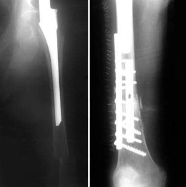
Management of late periprosthetic femur fractures: a retrospective cohort of 72 patients. (1/48)
(+info)Does femoral component loosening predispose to femoral fracture?: an in vitro comparison of cemented hips. (2/48)
(+info)The long modified extended sliding trochanteric osteotomy. (3/48)
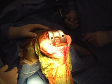
(+info)Retrograde intramedullary nailing for periprosthetic supracondylar fractures of the femur after total knee arthroplasty. (4/48)
(+info)The Modular Universal Tumour And Revision System (MUTARS(R)) in endoprosthetic revision surgery. (5/48)
(+info)Reasons for revision of first-generation highly cross-linked polyethylenes. (6/48)
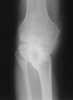
(+info)Periprosthetic fractures of the femur after total knee arthroplasty. (7/48)
(+info)Biomechanical testing of rectangular humeral shaft prosthesis: higher torsional stability without increased fracture risk. (8/48)
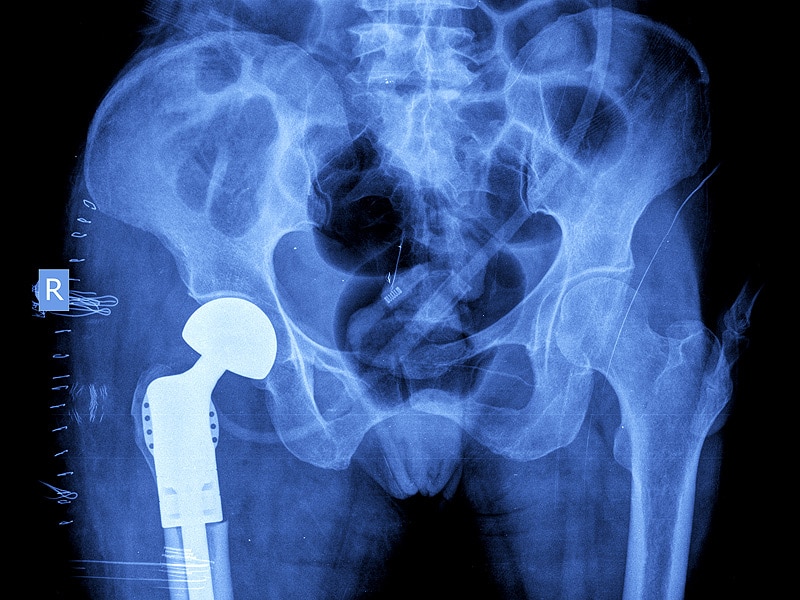
(+info)Periprosthetic fractures are defined as fractures that occur in close proximity to a prosthetic joint, such as those found in total hip or knee replacements. These types of fractures typically occur as a result of low-energy trauma, and can be caused by a variety of factors including osteoporosis, bone weakness, or loosening of the prosthetic implant.
Periprosthetic fractures are classified based on the location of the fracture in relation to the prosthesis, as well as the stability of the implant. Treatment options for periprosthetic fractures may include non-surgical management, such as immobilization with a brace or cast, or surgical intervention, such as open reduction and internal fixation (ORIF) or revision arthroplasty.
The management of periprosthetic fractures can be complex and requires careful consideration of various factors, including the patient's age, overall health status, bone quality, and functional needs. As such, these types of fractures are typically managed by orthopedic surgeons with experience in joint replacement surgery and fracture care.
A femoral fracture is a medical term that refers to a break in the thigh bone, which is the longest and strongest bone in the human body. The femur extends from the hip joint to the knee joint and is responsible for supporting the weight of the upper body and allowing movement of the lower extremity. Femoral fractures can occur due to various reasons such as high-energy trauma, low-energy trauma in individuals with weak bones (osteoporosis), or as a result of a direct blow to the thigh.
Femoral fractures can be classified into different types based on their location, pattern, and severity. Some common types of femoral fractures include:
1. Transverse fracture: A break that occurs straight across the bone.
2. Oblique fracture: A break that occurs at an angle across the bone.
3. Spiral fracture: A break that occurs in a helical pattern around the bone.
4. Comminuted fracture: A break that results in multiple fragments of the bone.
5. Open or compound fracture: A break in which the bone pierces through the skin.
6. Closed or simple fracture: A break in which the bone does not pierce through the skin.
Femoral fractures can cause severe pain, swelling, bruising, and difficulty walking or bearing weight on the affected leg. Diagnosis typically involves a physical examination, medical history, and imaging tests such as X-rays or CT scans. Treatment may involve surgical intervention, including the use of metal rods, plates, or screws to stabilize the bone, followed by rehabilitation and physical therapy to restore mobility and strength.
Fracture healing is the natural process by which a broken bone repairs itself. When a fracture occurs, the body responds by initiating a series of biological and cellular events aimed at restoring the structural integrity of the bone. This process involves the formation of a hematoma (a collection of blood) around the fracture site, followed by the activation of inflammatory cells that help to clean up debris and prepare the area for repair.
Over time, specialized cells called osteoblasts begin to lay down new bone matrix, or osteoid, along the edges of the broken bone ends. This osteoid eventually hardens into new bone tissue, forming a bridge between the fracture fragments. As this process continues, the callus (a mass of newly formed bone and connective tissue) gradually becomes stronger and more compact, eventually remodeling itself into a solid, unbroken bone.
The entire process of fracture healing can take several weeks to several months, depending on factors such as the severity of the injury, the patient's age and overall health, and the location of the fracture. In some cases, medical intervention may be necessary to help promote healing or ensure proper alignment of the bone fragments. This may include the use of casts, braces, or surgical implants such as plates, screws, or rods.
Prosthesis failure is a term used to describe a situation where a prosthetic device, such as an artificial joint or limb, has stopped functioning or failed to meet its intended purpose. This can be due to various reasons, including mechanical failure, infection, loosening of the device, or a reaction to the materials used in the prosthesis.
Mechanical failure can occur due to wear and tear, manufacturing defects, or improper use of the prosthetic device. Infection can also lead to prosthesis failure, particularly in cases where the prosthesis is implanted inside the body. The immune system may react to the presence of the foreign material, leading to inflammation and infection.
Loosening of the prosthesis can also cause it to fail over time, as the device becomes less stable and eventually stops working properly. Additionally, some people may have a reaction to the materials used in the prosthesis, leading to tissue damage or other complications that can result in prosthesis failure.
In general, prosthesis failure can lead to decreased mobility, pain, and the need for additional surgeries or treatments to correct the problem. It is important for individuals with prosthetic devices to follow their healthcare provider's instructions carefully to minimize the risk of prosthesis failure and ensure that the device continues to function properly over time.
Hip arthroplasty, also known as hip replacement surgery, is a medical procedure where the damaged or diseased joint surfaces of the hip are removed and replaced with artificial components. These components typically include a metal or ceramic ball that replaces the head of the femur (thigh bone), and a polyethylene or ceramic socket that replaces the acetabulum (hip socket) in the pelvis.
The goal of hip arthroplasty is to relieve pain, improve joint mobility, and restore function to the hip joint. This procedure is commonly performed in patients with advanced osteoarthritis, rheumatoid arthritis, hip fractures, or other conditions that cause significant damage to the hip joint.
There are several types of hip replacement surgeries, including traditional total hip arthroplasty, partial (hemi) hip arthroplasty, and resurfacing hip arthroplasty. The choice of procedure depends on various factors, such as the patient's age, activity level, overall health, and the extent of joint damage.
After surgery, patients typically require rehabilitation to regain strength, mobility, and function in the affected hip. With proper care and follow-up, most patients can expect significant pain relief and improved quality of life following hip arthroplasty.
A hip prosthesis, also known as a total hip replacement, is a surgical implant designed to replace the damaged or diseased components of the human hip joint. The procedure involves replacing the femoral head (the ball at the top of the thigh bone) and the acetabulum (the socket in the pelvis) with artificial parts, typically made from materials such as metal, ceramic, or plastic.
The goal of a hip prosthesis is to relieve pain, improve joint mobility, and restore function, allowing patients to return to their normal activities and enjoy an improved quality of life. The procedure is most commonly performed in individuals with advanced osteoarthritis, rheumatoid arthritis, or other degenerative conditions that have caused significant damage to the hip joint.
There are several different types of hip prostheses available, each with its own unique design and set of benefits and risks. The choice of prosthesis will depend on a variety of factors, including the patient's age, activity level, overall health, and specific medical needs. In general, however, all hip prostheses are designed to provide a durable, long-lasting solution for patients suffering from debilitating joint pain and stiffness.
A bone fracture is a medical condition in which there is a partial or complete break in the continuity of a bone due to external or internal forces. Fractures can occur in any bone in the body and can vary in severity from a small crack to a shattered bone. The symptoms of a bone fracture typically include pain, swelling, bruising, deformity, and difficulty moving the affected limb. Treatment for a bone fracture may involve immobilization with a cast or splint, surgery to realign and stabilize the bone, or medication to manage pain and prevent infection. The specific treatment approach will depend on the location, type, and severity of the fracture.
A reoperation is a surgical procedure that is performed again on a patient who has already undergone a previous operation for the same or related condition. Reoperations may be required due to various reasons, such as inadequate initial treatment, disease recurrence, infection, or complications from the first surgery. The nature and complexity of a reoperation can vary widely depending on the specific circumstances, but it often carries higher risks and potential complications compared to the original operation.
Fracture fixation, internal, is a surgical procedure where a fractured bone is fixed using metal devices such as plates, screws, or rods that are implanted inside the body. This technique helps to maintain the alignment and stability of the broken bone while it heals. The implants may be temporarily or permanently left inside the body, depending on the nature and severity of the fracture. Internal fixation allows for early mobilization and rehabilitation, which can result in a faster recovery and improved functional outcome.
A hip fracture is a medical condition referring to a break in the upper part of the femur (thigh) bone, which forms the hip joint. The majority of hip fractures occur due to falls or direct trauma to the area. They are more common in older adults, particularly those with osteoporosis, a condition that weakens bones and makes them more prone to breaking. Hip fractures can significantly impact mobility and quality of life, often requiring surgical intervention and rehabilitation.
Prosthesis-related infections, also known as prosthetic joint infections (PJIs), are infections that occur around or within a prosthetic device, such as an artificial joint. These infections can be caused by bacteria, fungi, or other microorganisms and can lead to serious complications if not treated promptly and effectively.
Prosthesis-related infections can occur soon after the implantation of the prosthetic device (early infection) or months or even years later (late infection). Early infections are often caused by bacteria that enter the surgical site during the procedure, while late infections may be caused by hematogenous seeding (i.e., when bacteria from another source spread through the bloodstream and settle in the prosthetic device) or by contamination during a subsequent medical procedure.
Symptoms of prosthesis-related infections can include pain, swelling, redness, warmth, and drainage around the affected area. In some cases, patients may also experience fever, chills, or fatigue. Diagnosis typically involves a combination of clinical evaluation, laboratory tests (such as blood cultures, joint fluid analysis, and tissue biopsy), and imaging studies (such as X-rays, CT scans, or MRI).
Treatment of prosthesis-related infections usually involves a combination of antibiotics and surgical intervention. The specific treatment approach will depend on the type and severity of the infection, as well as the patient's overall health status. In some cases, it may be necessary to remove or replace the affected prosthetic device.
Hemiarthroplasty is a surgical procedure where only one half (hemi-) of a joint is replaced with an artificial component, usually a metal ball attached to a stem that fits into the bone. This procedure is most commonly performed on the shoulder or hip joints. In a hip hemiarthroplasty, it involves replacing the femoral head (the ball part of the thighbone) which has been damaged due to fracture or arthritis. The acetabulum (socket part of the pelvis) is not replaced and remains as it is. This procedure aims to relieve pain, restore mobility, and improve joint function.
The humeral head is the rounded, articular surface at the proximal end of the humerus bone in the human body. It forms the upper part of the shoulder joint and articulates with the glenoid fossa of the scapula to form the glenohumeral joint, allowing for a wide range of motion in the arm. The humeral head is covered with cartilage that helps to provide a smooth, lubricated surface for movement and shock absorption.
Intramedullary fracture fixation is a surgical technique used to stabilize and align bone fractures. In this procedure, a metal rod or nail is inserted into the marrow cavity (intramedullary canal) of the affected bone, spanning the length of the fracture. The rod is then secured to the bone using screws or other fixation devices on either side of the fracture. This provides stability and helps maintain proper alignment during the healing process.
The benefits of intramedullary fixation include:
1. Load sharing: The intramedullary rod shares some of the load bearing capacity with the bone, which can help reduce stress on the healing bone.
2. Minimal soft tissue dissection: Since the implant is inserted through the medullary canal, there is less disruption to the surrounding muscles, tendons, and ligaments compared to other fixation methods.
3. Biomechanical stability: Intramedullary fixation provides rotational and bending stiffness, which helps maintain proper alignment of the fracture fragments during healing.
4. Early mobilization: Patients with intramedullary fixation can often begin weight bearing and rehabilitation exercises earlier than those with other types of fixation, leading to faster recovery times.
Common indications for intramedullary fracture fixation include long bone fractures in the femur, tibia, humerus, and fibula, as well as certain pelvic and spinal fractures. However, the choice of fixation method depends on various factors such as patient age, fracture pattern, location, and associated injuries.
A knee prosthesis, also known as a knee replacement or artificial knee joint, is a medical device used to replace the damaged or diseased weight-bearing surfaces of the knee joint. It typically consists of three components: the femoral component (made of metal) that fits over the end of the thighbone (femur), the tibial component (often made of metal and plastic) that fits into the top of the shinbone (tibia), and a patellar component (usually made of plastic) that replaces the damaged surface of the kneecap.
The primary goal of knee prosthesis is to relieve pain, restore function, and improve quality of life for individuals with advanced knee joint damage due to conditions such as osteoarthritis, rheumatoid arthritis, or traumatic injuries. The procedure to implant a knee prosthesis is called knee replacement surgery or total knee arthroplasty (TKA).
A spinal fracture, also known as a vertebral compression fracture, is a break in one or more bones (vertebrae) of the spine. This type of fracture often occurs due to weakened bones caused by osteoporosis, but it can also result from trauma such as a car accident or a fall.
In a spinal fracture, the front part of the vertebra collapses, causing the height of the vertebra to decrease, while the back part of the vertebra remains intact. This results in a wedge-shaped deformity of the vertebra. Multiple fractures can lead to a hunched forward posture known as kyphosis or dowager's hump.
Spinal fractures can cause pain, numbness, tingling, or weakness in the back, legs, or arms, depending on the location and severity of the fracture. In some cases, spinal cord compression may occur, leading to more severe symptoms such as paralysis or loss of bladder and bowel control.
Prosthesis design is a specialized field in medical device technology that involves creating and developing artificial substitutes to replace a missing body part, such as a limb, tooth, eye, or internal organ. The design process typically includes several stages: assessment of the patient's needs, selection of appropriate materials, creation of a prototype, testing and refinement, and final fabrication and fitting of the prosthesis.
The goal of prosthesis design is to create a device that functions as closely as possible to the natural body part it replaces, while also being comfortable, durable, and aesthetically pleasing for the patient. The design process may involve collaboration between medical professionals, engineers, and designers, and may take into account factors such as the patient's age, lifestyle, occupation, and overall health.
Prosthesis design can be highly complex, particularly for advanced devices such as robotic limbs or implantable organs. These devices often require sophisticated sensors, actuators, and control systems to mimic the natural functions of the body part they replace. As a result, prosthesis design is an active area of research and development in the medical field, with ongoing efforts to improve the functionality, comfort, and affordability of these devices for patients.
In the medical field, cementation refers to the process of using a type of dental cement or bonding agent to attach a dental restoration (such as a crown, bridge, or false tooth) to a natural tooth or implant. The cement helps to create a strong and secure attachment, while also helping to seal the restoration and prevent the entry of bacteria and saliva.
Dental cement can be made from various materials, including glass ionomers, resin-modified glass ionomers, zinc phosphate, and polycarboxylate cements. The choice of cement depends on several factors, such as the type of restoration being attached, the location in the mouth, and the patient's individual needs and preferences.
Cementation is an important step in many dental procedures, as it helps to ensure the longevity and success of the restoration. Proper technique and material selection are crucial for achieving a successful cementation that will last for years to come.
Arthroplasty, replacement, knee is a surgical procedure where the damaged or diseased joint surface of the knee is removed and replaced with an artificial joint or prosthesis. The procedure involves resurfacing the worn-out ends of the femur (thigh bone) and tibia (shin bone) with metal components, and the back of the kneecap with a plastic button. This surgery is usually performed to relieve pain and restore function in patients with severe knee osteoarthritis, rheumatoid arthritis, or traumatic injuries that have damaged the joint beyond repair. The goal of knee replacement surgery is to improve mobility, reduce pain, and enhance the quality of life for the patient.
Fracture fixation is a surgical procedure in orthopedic trauma surgery where a fractured bone is stabilized using various devices and techniques to promote proper healing and alignment. The goal of fracture fixation is to maintain the broken bone ends in correct anatomical position and length, allowing for adequate stability during the healing process.
There are two main types of fracture fixation:
1. Internal fixation: In this method, metal implants like plates, screws, or intramedullary rods are inserted directly into the bone to hold the fragments in place. These implants can be either removed or left in the body once healing is complete, depending on the type and location of the fracture.
2. External fixation: This technique involves placing pins or screws through the skin and into the bone above and below the fracture site. These pins are then connected to an external frame that maintains alignment and stability. External fixators are typically used when there is significant soft tissue damage, infection, or when internal fixation is not possible due to the complexity of the fracture.
The choice between internal and external fixation depends on various factors such as the type and location of the fracture, patient's age and overall health, surgeon's preference, and potential complications. Both methods aim to provide a stable environment for bone healing while minimizing the risk of malunion, nonunion, or deformity.
Bone cements are medical-grade materials used in orthopedic and trauma surgery to fill gaps between bone surfaces and implants, such as artificial joints or screws. They serve to mechanically stabilize the implant and provide a smooth, load-bearing surface. The two most common types of bone cement are:
1. Polymethylmethacrylate (PMMA) cement: This is a two-component system consisting of powdered PMMA and liquid methyl methacrylate monomer. When mixed together, they form a dough-like consistency that hardens upon exposure to air. PMMA cement has been widely used for decades in joint replacement surgeries, such as hip or knee replacements.
2. Calcium phosphate (CP) cement: This is a two-component system consisting of a powdered CP compound and an aqueous solution. When mixed together, they form a paste that hardens through a chemical reaction at body temperature. CP cement has lower mechanical strength compared to PMMA but demonstrates better biocompatibility, bioactivity, and the ability to resorb over time.
Both types of bone cements have advantages and disadvantages, and their use depends on the specific surgical indication and patient factors.
The femur is the medical term for the thigh bone, which is the longest and strongest bone in the human body. It connects the hip bone to the knee joint and plays a crucial role in supporting the weight of the body and allowing movement during activities such as walking, running, and jumping. The femur is composed of a rounded head, a long shaft, and two condyles at the lower end that articulate with the tibia and patella to form the knee joint.
A comminuted fracture is a type of bone break where the bone is shattered into three or more pieces. This type of fracture typically occurs after high-energy trauma, such as a car accident or a fall from a great height. Commminuted fractures can also occur in bones that are weakened by conditions like osteoporosis or cancer. Because of the severity and complexity of comminuted fractures, they often require extensive treatment, which may include surgery to realign and stabilize the bone fragments using metal screws, plates, or rods.
A joint prosthesis, also known as an artificial joint or a replacement joint, is a surgical implant used to replace all or part of a damaged or diseased joint. The most common types of joint prostheses are total hip replacements and total knee replacements. These prostheses typically consist of a combination of metal, plastic, and ceramic components that are designed to replicate the movement and function of a natural joint.
Joint prostheses are usually recommended for patients who have severe joint pain or mobility issues that cannot be adequately managed with other treatments such as physical therapy, medication, or lifestyle changes. The goal of joint replacement surgery is to relieve pain, improve joint function, and enhance the patient's quality of life.
Joint prostheses are typically made from materials such as titanium, cobalt-chrome alloys, stainless steel, polyethylene plastic, and ceramics. The choice of material depends on a variety of factors, including the patient's age, activity level, weight, and overall health.
While joint replacement surgery is generally safe and effective, there are risks associated with any surgical procedure, including infection, blood clots, implant loosening or failure, and nerve damage. Patients who undergo joint replacement surgery typically require several weeks of rehabilitation and physical therapy to regain strength and mobility in the affected joint.
Dura Mater: The tough, outer membrane that covers the brain and spinal cord.
Hydroxyapatite: A naturally occurring mineral form of calcium apatite, also known as dahllite, with the formula Ca5(PO4)3(OH), is the primary mineral component of biological apatites found in bones and teeth.
Therefore, "Durapatite" isn't a recognized medical term, but it seems like it might be a combination of "dura mater" and "hydroxyapatite." If you meant to ask about a material used in medical or dental applications that combines properties of both dura mater and hydroxyapatite, please provide more context.
Osteolysis is a medical term that refers to the loss or resorption of bone tissue. It's a process where the body's normal bone remodeling cycle is disrupted, leading to an imbalance between bone formation and bone breakdown. This results in the progressive deterioration and destruction of bone.
Osteolysis can occur due to various reasons such as chronic inflammation, mechanical stress, or certain medical conditions like rheumatoid arthritis, Paget's disease, or bone tumors. It can also be a side effect of some medications, such as those used in cancer treatment or for managing osteoporosis.
In severe cases, osteolysis can lead to weakened bones, increased risk of fractures, and deformities. Treatment typically aims to address the underlying cause and may include medication, surgery, or lifestyle changes.
Osteoporotic fractures are breaks or cracks in bones that occur as a result of osteoporosis, a condition characterized by weak and brittle bones. Osteoporosis causes bones to lose density and strength, making them more susceptible to fractures, even from minor injuries or falls.
The most common types of osteoporotic fractures are:
1. Hip fractures: These occur when the upper part of the thigh bone (femur) breaks, often due to a fall. Hip fractures can be serious and may require surgery and hospitalization.
2. Vertebral compression fractures: These occur when the bones in the spine (vertebrae) collapse, causing height loss, back pain, and deformity. They are often caused by everyday activities, such as bending or lifting.
3. Wrist fractures: These occur when the bones in the wrist break, often due to a fall. Wrist fractures are common in older adults with osteoporosis.
4. Other fractures: Osteoporotic fractures can also occur in other bones, such as the pelvis, ribs, and humerus (upper arm bone).
Prevention is key in managing osteoporosis and reducing the risk of osteoporotic fractures. This includes getting enough calcium and vitamin D, engaging in regular weight-bearing exercise, avoiding smoking and excessive alcohol consumption, and taking medications as prescribed by a healthcare provider.
A radius fracture is a break in the bone that runs from the wrist to the elbow, located on the thumb side of the forearm. Radius fractures can occur as a result of a fall, direct blow to the forearm, or a high-energy collision such as a car accident. There are various types of radius fractures, including:
1. Distal radius fracture: A break at the end of the radius bone, near the wrist joint, which is the most common type of radius fracture.
2. Radial shaft fracture: A break in the middle portion of the radius bone.
3. Radial head and neck fractures: Breaks in the upper part of the radius bone, near the elbow joint.
4. Comminuted fracture: A complex radius fracture where the bone is broken into multiple pieces.
5. Open (compound) fracture: A radius fracture with a wound or laceration in the skin, allowing for communication between the outside environment and the fractured bone.
6. Intra-articular fracture: A radius fracture that extends into the wrist joint or elbow joint.
7. Torus (buckle) fracture: A stable fracture where one side of the bone is compressed, causing it to buckle or bend, but not break completely through.
Symptoms of a radius fracture may include pain, swelling, tenderness, bruising, deformity, limited mobility, and in some cases, numbness or tingling in the fingers. Treatment options depend on the type and severity of the fracture but can range from casting to surgical intervention with implant fixation.
Biocompatible coated materials refer to surfaces or substances that are treated or engineered with a layer or film designed to interact safely and effectively with living tissues or biological systems, without causing harm or adverse reactions. The coating material is typically composed of biomaterials that can withstand the conditions of the specific application while promoting a positive response from the body.
The purpose of these coatings may vary depending on the medical device or application. For example, they might be used to enhance the lubricity and wear resistance of implantable devices, reduce the risk of infection, promote integration with surrounding tissues, control drug release, or prevent the formation of biofilms.
Biocompatible coated materials must undergo rigorous testing and evaluation to ensure their safety and efficacy in various clinical settings. This includes assessing potential cytotoxicity, genotoxicity, sensitization, hemocompatibility, carcinogenicity, and other factors that could impact the body's response to the material.
Examples of biocompatible coating materials include:
1. Hydrogels: Cross-linked networks of hydrophilic polymers that can be used for drug delivery, tissue engineering, or as lubricious coatings on medical devices.
2. Self-assembling monolayers (SAMs): Organosilane or thiol-based molecules that form a stable, well-ordered film on surfaces, which can be further functionalized to promote specific biological interactions.
3. Poly(ethylene glycol) (PEG): A biocompatible polymer often used as a coating material due to its ability to reduce protein adsorption and cell attachment, making it useful for preventing biofouling or thrombosis on medical devices.
4. Bioactive glass: A type of biomaterial composed of silica-based glasses that can stimulate bone growth and healing when used as a coating material in orthopedic or dental applications.
5. Drug-eluting coatings: Biocompatible polymers impregnated with therapeutic agents, designed to release the drug over time to promote healing, prevent infection, or inhibit restenosis in various medical devices.
A femoral neck fracture is a type of hip fracture that occurs in the narrow, vertical section of bone just below the ball of the femur (thigh bone) that connects to the hip socket. This area is called the femoral neck. Femoral neck fractures can be categorized into different types based on their location and the direction of the fractured bone.
These fractures are typically caused by high-energy trauma, such as car accidents or falls from significant heights, in younger individuals. However, in older adults, particularly those with osteoporosis, femoral neck fractures can also result from low-energy trauma, like a simple fall from standing height.
Femoral neck fractures are often serious and require prompt medical attention. Treatment usually involves surgery to realign and stabilize the broken bone fragments, followed by rehabilitation to help regain mobility and strength. Potential complications of femoral neck fractures include avascular necrosis (loss of blood flow to the femoral head), nonunion or malunion (improper healing), and osteoarthritis in the hip joint.
Spontaneous fractures are bone breaks that occur without any identifiable trauma or injury. They are typically caused by underlying medical conditions that weaken the bones, making them more susceptible to breaking under normal stress or weight. The most common cause of spontaneous fractures is osteoporosis, a condition characterized by weak and brittle bones. Other potential causes include various bone diseases, certain cancers, long-term use of corticosteroids, and genetic disorders affecting bone strength.
It's important to note that while the term "spontaneous" implies that the fracture occurred without any apparent cause, it is usually the result of an underlying medical condition. Therefore, if you experience a spontaneous fracture, seeking medical attention is crucial to diagnose and manage the underlying cause to prevent future fractures and related complications.
Arthroplasty, replacement, is a surgical procedure where a damaged or diseased joint surface is removed and replaced with an artificial implant or device. The goal of this surgery is to relieve pain, restore function, and improve the quality of life for patients who have severe joint damage due to arthritis or other conditions.
During the procedure, the surgeon removes the damaged cartilage and bone from the joint and replaces them with a metal, plastic, or ceramic component that replicates the shape and function of the natural joint surface. The most common types of joint replacement surgery are hip replacement, knee replacement, and shoulder replacement.
The success rate of joint replacement surgery is generally high, with many patients experiencing significant pain relief and improved mobility. However, as with any surgical procedure, there are risks involved, including infection, blood clots, implant loosening or failure, and nerve damage. Therefore, it's essential to discuss the potential benefits and risks of joint replacement surgery with a healthcare provider before making a decision.
Stress fractures are defined as small cracks or severe bruising in bones that occur from repetitive stress or overuse. They most commonly occur in weight-bearing bones, such as the legs and feet, but can also occur in the arms, hips, and back. Stress fractures differ from regular fractures because they typically do not result from a single, traumatic event. Instead, they are caused by repeated stress on the bone that results in microscopic damage over time. Athletes, military personnel, and individuals who engage in high-impact activities or have weak bones (osteoporosis) are at increased risk of developing stress fractures. Symptoms may include pain, swelling, tenderness, and difficulty walking or bearing weight on the affected bone.
The hip joint, also known as the coxal joint, is a ball-and-socket type synovial joint that connects the femur (thigh bone) to the pelvis. The "ball" is the head of the femur, while the "socket" is the acetabulum, a concave surface on the pelvic bone.
The hip joint is surrounded by a strong fibrous capsule and is reinforced by several ligaments, including the iliofemoral, ischiofemoral, and pubofemoral ligaments. The joint allows for flexion, extension, abduction, adduction, medial and lateral rotation, and circumduction movements, making it one of the most mobile joints in the body.
The hip joint is also supported by various muscles, including the gluteus maximus, gluteus medius, gluteus minimus, iliopsoas, and other hip flexors and extensors. These muscles provide stability and strength to the joint, allowing for weight-bearing activities such as walking, running, and jumping.
Treatment outcome is a term used to describe the result or effect of medical treatment on a patient's health status. It can be measured in various ways, such as through symptoms improvement, disease remission, reduced disability, improved quality of life, or survival rates. The treatment outcome helps healthcare providers evaluate the effectiveness of a particular treatment plan and make informed decisions about future care. It is also used in clinical research to compare the efficacy of different treatments and improve patient care.
Postoperative complications refer to any unfavorable condition or event that occurs during the recovery period after a surgical procedure. These complications can vary in severity and may include, but are not limited to:
1. Infection: This can occur at the site of the incision or inside the body, such as pneumonia or urinary tract infection.
2. Bleeding: Excessive bleeding (hemorrhage) can lead to a drop in blood pressure and may require further surgical intervention.
3. Blood clots: These can form in the deep veins of the legs (deep vein thrombosis) and can potentially travel to the lungs (pulmonary embolism).
4. Wound dehiscence: This is when the surgical wound opens up, which can lead to infection and further complications.
5. Pulmonary issues: These include atelectasis (collapsed lung), pneumonia, or respiratory failure.
6. Cardiovascular problems: These include abnormal heart rhythms (arrhythmias), heart attack, or stroke.
7. Renal failure: This can occur due to various reasons such as dehydration, blood loss, or the use of certain medications.
8. Pain management issues: Inadequate pain control can lead to increased stress, anxiety, and decreased mobility.
9. Nausea and vomiting: These can be caused by anesthesia, opioid pain medication, or other factors.
10. Delirium: This is a state of confusion and disorientation that can occur in the elderly or those with certain medical conditions.
Prompt identification and management of these complications are crucial to ensure the best possible outcome for the patient.
An ulna fracture is a break in the ulna bone, which is one of the two long bones in the forearm. The ulna is located on the pinky finger side of the forearm and functions to support the elbow joint and assist in rotation and movement of the forearm. Ulna fractures can occur at various points along the bone, including the shaft, near the wrist, or at the elbow end of the bone. Symptoms may include pain, swelling, bruising, tenderness, deformity, limited mobility, and in some cases, numbness or tingling in the fingers. Treatment typically involves immobilization with a cast or splint, followed by rehabilitation exercises to restore strength and range of motion. In severe cases, surgery may be required to realign and stabilize the fractured bone.
Retrospective studies, also known as retrospective research or looking back studies, are a type of observational study that examines data from the past to draw conclusions about possible causal relationships between risk factors and outcomes. In these studies, researchers analyze existing records, medical charts, or previously collected data to test a hypothesis or answer a specific research question.
Retrospective studies can be useful for generating hypotheses and identifying trends, but they have limitations compared to prospective studies, which follow participants forward in time from exposure to outcome. Retrospective studies are subject to biases such as recall bias, selection bias, and information bias, which can affect the validity of the results. Therefore, retrospective studies should be interpreted with caution and used primarily to generate hypotheses for further testing in prospective studies.
Bone plates are medical devices used in orthopedic surgery to stabilize and hold together fractured or broken bones during the healing process. They are typically made of surgical-grade stainless steel, titanium, or other biocompatible materials. The plate is shaped to fit the contour of the bone and is held in place with screws that are inserted through the plate and into the bone on either side of the fracture. This provides stability and alignment to the broken bones, allowing them to heal properly. Bone plates can be used to treat a variety of fractures, including those that are complex or unstable. After healing is complete, the bone plate may be left in place or removed, depending on the individual's needs and the surgeon's recommendation.
I'm sorry for any confusion, but "Minnesota" is not a medical term or concept. It is a state located in the Midwestern United States, known for its cold winters, beautiful lakes, and friendly people. If you have any questions about medical terms or concepts, I would be happy to help!
Rib fractures are breaks or cracks in the bones that make up the rib cage, which is the protective structure around the lungs and heart. Rib fractures can result from direct trauma to the chest, such as from a fall, motor vehicle accident, or physical assault. They can also occur from indirect forces, such as during coughing fits in people with weakened bones (osteoporosis).
Rib fractures are painful and can make breathing difficult, particularly when taking deep breaths or coughing. In some cases, rib fractures may lead to complications like punctured lungs (pneumothorax) or collapsed lungs (atelectasis), especially if multiple ribs are broken in several places.
It is essential to seek medical attention for suspected rib fractures, as proper diagnosis and management can help prevent further complications and promote healing. Treatment typically involves pain management, breathing exercises, and, in some cases, immobilization or surgery.
Follow-up studies are a type of longitudinal research that involve repeated observations or measurements of the same variables over a period of time, in order to understand their long-term effects or outcomes. In medical context, follow-up studies are often used to evaluate the safety and efficacy of medical treatments, interventions, or procedures.
In a typical follow-up study, a group of individuals (called a cohort) who have received a particular treatment or intervention are identified and then followed over time through periodic assessments or data collection. The data collected may include information on clinical outcomes, adverse events, changes in symptoms or functional status, and other relevant measures.
The results of follow-up studies can provide important insights into the long-term benefits and risks of medical interventions, as well as help to identify factors that may influence treatment effectiveness or patient outcomes. However, it is important to note that follow-up studies can be subject to various biases and limitations, such as loss to follow-up, recall bias, and changes in clinical practice over time, which must be carefully considered when interpreting the results.
A skull fracture is a break in one or more of the bones that form the skull. It can occur from a direct blow to the head, penetrating injuries like gunshot wounds, or from strong rotational forces during an accident. There are several types of skull fractures, including:
1. Linear Skull Fracture: This is the most common type, where there's a simple break in the bone without any splintering, depression, or displacement. It often doesn't require treatment unless it's near a sensitive area like an eye or ear.
2. Depressed Skull Fracture: In this type, a piece of the skull is pushed inward toward the brain. Surgery may be needed to relieve pressure on the brain and repair the fracture.
3. Diastatic Skull Fracture: This occurs along the suture lines (the fibrous joints between the skull bones) that haven't fused yet, often seen in infants and young children.
4. Basilar Skull Fracture: This involves fractures at the base of the skull. It can be serious due to potential injury to the cranial nerves and blood vessels located in this area.
5. Comminuted Skull Fracture: In this severe type, the bone is shattered into many pieces. These fractures usually require extensive surgical repair.
Symptoms of a skull fracture can include pain, swelling, bruising, bleeding (if there's an open wound), and in some cases, clear fluid draining from the ears or nose (cerebrospinal fluid leak). Severe fractures may cause brain injury, leading to symptoms like confusion, loss of consciousness, seizures, or neurological deficits. Immediate medical attention is necessary for any suspected skull fracture.
"Recovery of function" is a term used in medical rehabilitation to describe the process in which an individual regains the ability to perform activities or tasks that were previously difficult or impossible due to injury, illness, or disability. This can involve both physical and cognitive functions. The goal of recovery of function is to help the person return to their prior level of independence and participation in daily activities, work, and social roles as much as possible.
Recovery of function may be achieved through various interventions such as physical therapy, occupational therapy, speech-language therapy, and other rehabilitation strategies. The specific approach used will depend on the individual's needs and the nature of their impairment. Recovery of function can occur spontaneously as the body heals, or it may require targeted interventions to help facilitate the process.
It is important to note that recovery of function does not always mean a full return to pre-injury or pre-illness levels of ability. Instead, it often refers to the person's ability to adapt and compensate for any remaining impairments, allowing them to achieve their maximum level of functional independence and quality of life.
A mandibular fracture is a break or crack in the lower jaw (mandible) bone. It can occur at any point along the mandible, but common sites include the condyle (the rounded end near the ear), the angle (the curved part of the jaw), and the symphysis (the area where the two halves of the jaw meet in the front). Mandibular fractures are typically caused by trauma, such as a direct blow to the face or a fall. Symptoms may include pain, swelling, bruising, difficulty chewing or speaking, and malocclusion (misalignment) of the teeth. Treatment usually involves immobilization with wires or screws to allow the bone to heal properly.
Osseointegration is a direct structural and functional connection between living bone and the surface of an implant. It's a process where the bone grows in and around the implant, which is typically made of titanium or another biocompatible material. This process provides a solid foundation for dental prosthetics, such as crowns, bridges, or dentures, or for orthopedic devices like artificial limbs. The success of osseointegration depends on various factors, including the patient's overall health, the quality and quantity of available bone, and the surgical technique used for implant placement.
A foreign-body reaction is an immune response that occurs when a non-native substance, or "foreign body," is introduced into the human body. This can include things like splinters, surgical implants, or even injected medications. The immune system recognizes these substances as foreign and mounts a response to try to eliminate them.
The initial response to a foreign body is often an acute inflammatory reaction, characterized by the release of chemical mediators that cause vasodilation, increased blood flow, and the migration of white blood cells to the site. This can result in symptoms such as redness, swelling, warmth, and pain.
If the foreign body is not eliminated, a chronic inflammatory response may develop, which can lead to the formation of granulation tissue, fibrosis, and encapsulation of the foreign body. In some cases, this reaction can cause significant tissue damage or impede proper healing.
It's worth noting that not all foreign bodies necessarily elicit a strong immune response. The nature and size of the foreign body, as well as its location in the body, can all influence the severity of the reaction.
I believe there may be some confusion in your question as Polyethylenes are not a medical term, but rather a category of synthetic polymers commonly used in various industrial and medical applications. Here's a brief overview:
Polyethylene (PE) is a type of thermoplastic polymer made from the monomer ethylene. It is a versatile material with numerous applications due to its chemical resistance, durability, and flexibility. There are several types of polyethylenes, including:
1. Low-density polyethylene (LDPE): This type has a lower density and more branching in its molecular structure, which results in less crystallinity. LDPE is known for its flexibility and is often used in packaging films, bags, and containers.
2. High-density polyethylene (HDPE): HDPE has a higher density and less branching, resulting in greater crystallinity. It is more rigid than LDPE and is commonly used in applications such as bottles, pipes, and containers.
3. Linear low-density polyethylene (LLDPE): This type combines the flexibility of LDPE with some of the strength and rigidity of HDPE. LLDPE has fewer branches than LDPE but more than HDPE. It is often used in film applications, such as stretch wrap and agricultural films.
4. Ultra-high molecular weight polyethylene (UHMWPE): UHMWPE has an extremely high molecular weight, resulting in exceptional wear resistance, impact strength, and chemical resistance. It is commonly used in medical applications, such as orthopedic implants and joint replacements, due to its biocompatibility and low friction coefficient.
While polyethylenes are not a medical term per se, they do have significant medical applications, particularly UHMWPE in orthopedic devices.
A tooth fracture is a dental health condition characterized by a break or crack in the tooth structure. It can occur in different parts of the tooth, including the crown (the visible part), root, or filling. Tooth fractures can result from various factors such as trauma, biting or chewing on hard objects, grinding or clenching teeth, and having large, old amalgam fillings that weaken the tooth structure over time. Depending on the severity and location of the fracture, it may cause pain, sensitivity, or affect the tooth's functionality and appearance. Treatment options for tooth fractures vary from simple bonding to root canal treatment or even extraction in severe cases. Regular dental check-ups are essential for early detection and management of tooth fractures.
I believe you are referring to "bone pins" or "bone nails" rather than "bone nails." These terms are used in the medical field to describe surgical implants made of metal or biocompatible materials that are used to stabilize and hold together fractured bones during the healing process. They can also be used in spinal fusion surgery to provide stability and promote bone growth between vertebrae.
Bone pins or nails typically have a threaded or smooth shaft, with a small diameter that allows them to be inserted into the medullary canal of long bones such as the femur or tibia. They may also have a head or eyelet on one end that allows for attachment to external fixation devices or other surgical instruments.
The use of bone pins and nails has revolutionized orthopedic surgery, allowing for faster healing times, improved stability, and better functional outcomes for patients with fractures or spinal deformities.
A peptic ulcer is a sore or erosion in the lining of your stomach and the first part of your small intestine (duodenum). The most common causes of peptic ulcers are bacterial infection and long-term use of nonsteroidal anti-inflammatory drugs (NSAIDs) such as aspirin, ibuprofen, or naproxen.
The symptoms of a peptic ulcer include abdominal pain, often in the upper middle part of your abdomen, which can be dull, sharp, or burning and may come and go for several days or weeks. Other symptoms can include bloating, burping, heartburn, nausea, vomiting, loss of appetite, and weight loss. Severe ulcers can cause bleeding in the digestive tract, which can lead to anemia, black stools, or vomit that looks like coffee grounds.
If left untreated, peptic ulcers can result in serious complications such as perforation (a hole through the wall of the stomach or duodenum), obstruction (blockage of the digestive tract), and bleeding. Treatment for peptic ulcers typically involves medications to reduce acid production, neutralize stomach acid, and kill the bacteria causing the infection. In severe cases, surgery may be required.
I'm sorry for any confusion, but "Polyethylene" is not a medical term. It is a type of synthetic plastic that is commonly used in various industrial and consumer products. Medical definitions typically refer to substances or conditions related to human health, treatment, or disease processes. If you have any questions related to medical terminology or concepts, I'd be happy to help!
Prospective studies, also known as longitudinal studies, are a type of cohort study in which data is collected forward in time, following a group of individuals who share a common characteristic or exposure over a period of time. The researchers clearly define the study population and exposure of interest at the beginning of the study and follow up with the participants to determine the outcomes that develop over time. This type of study design allows for the investigation of causal relationships between exposures and outcomes, as well as the identification of risk factors and the estimation of disease incidence rates. Prospective studies are particularly useful in epidemiology and medical research when studying diseases with long latency periods or rare outcomes.
Bone density refers to the amount of bone mineral content (usually measured in grams) in a given volume of bone (usually measured in cubic centimeters). It is often used as an indicator of bone strength and fracture risk. Bone density is typically measured using dual-energy X-ray absorptiometry (DXA) scans, which provide a T-score that compares the patient's bone density to that of a young adult reference population. A T-score of -1 or above is considered normal, while a T-score between -1 and -2.5 indicates osteopenia (low bone mass), and a T-score below -2.5 indicates osteoporosis (porous bones). Regular exercise, adequate calcium and vitamin D intake, and medication (if necessary) can help maintain or improve bone density and prevent fractures.
A compression fracture is a type of bone fracture that occurs when there is a collapse of a vertebra in the spine. This type of fracture is most commonly seen in the thoracic and lumbar regions of the spine. Compression fractures are often caused by weakened bones due to osteoporosis, but they can also result from trauma or tumors that weaken the bone.
In a compression fracture, the front part (anterior) of the vertebra collapses, while the back part (posterior) remains intact, causing the height of the vertebra to decrease. This can lead to pain, deformity, and decreased mobility. In severe cases, multiple compression fractures can result in a condition called kyphosis, which is an abnormal curvature of the spine that leads to a hunchback appearance.
Compression fractures are typically diagnosed through imaging tests such as X-rays, CT scans, or MRI scans. Treatment may include pain medication, bracing, physical therapy, or in some cases, surgery. Preventive measures such as maintaining a healthy diet, getting regular exercise, and taking medications to prevent or treat osteoporosis can help reduce the risk of compression fractures.
An intra-articular fracture is a type of fracture that involves the joint surface or articular cartilage of a bone. These types of fractures can occur in any joint, but they are most commonly seen in the weight-bearing joints such as the knee, ankle, and wrist.
Intra-articular fractures can be caused by high-energy trauma, such as motor vehicle accidents or falls from significant heights, or by low-energy trauma, such as a simple fall in older adults with osteoporosis.
These types of fractures are often complex and may involve displacement or depression of the joint surface, which can increase the risk of developing post-traumatic arthritis. Therefore, prompt diagnosis and appropriate treatment are essential to ensure optimal outcomes and minimize long-term complications. Treatment options for intra-articular fractures may include surgical fixation with plates, screws, or pins, as well as joint replacement in some cases.
Risk assessment in the medical context refers to the process of identifying, evaluating, and prioritizing risks to patients, healthcare workers, or the community related to healthcare delivery. It involves determining the likelihood and potential impact of adverse events or hazards, such as infectious diseases, medication errors, or medical devices failures, and implementing measures to mitigate or manage those risks. The goal of risk assessment is to promote safe and high-quality care by identifying areas for improvement and taking action to minimize harm.
Medical Definition:
"Risk factors" are any attribute, characteristic or exposure of an individual that increases the likelihood of developing a disease or injury. They can be divided into modifiable and non-modifiable risk factors. Modifiable risk factors are those that can be changed through lifestyle choices or medical treatment, while non-modifiable risk factors are inherent traits such as age, gender, or genetic predisposition. Examples of modifiable risk factors include smoking, alcohol consumption, physical inactivity, and unhealthy diet, while non-modifiable risk factors include age, sex, and family history. It is important to note that having a risk factor does not guarantee that a person will develop the disease, but rather indicates an increased susceptibility.
In epidemiology, the incidence of a disease is defined as the number of new cases of that disease within a specific population over a certain period of time. It is typically expressed as a rate, with the number of new cases in the numerator and the size of the population at risk in the denominator. Incidence provides information about the risk of developing a disease during a given time period and can be used to compare disease rates between different populations or to monitor trends in disease occurrence over time.
Titanium is not a medical term, but rather a chemical element (symbol Ti, atomic number 22) that is widely used in the medical field due to its unique properties. Medically, it is often referred to as a biocompatible material used in various medical applications such as:
1. Orthopedic implants: Titanium and its alloys are used for making joint replacements (hips, knees, shoulders), bone plates, screws, and rods due to their high strength-to-weight ratio, excellent corrosion resistance, and biocompatibility.
2. Dental implants: Titanium is also commonly used in dental applications like implants, crowns, and bridges because of its ability to osseointegrate, or fuse directly with bone tissue, providing a stable foundation for replacement teeth.
3. Cardiovascular devices: Titanium alloys are used in the construction of heart valves, pacemakers, and other cardiovascular implants due to their non-magnetic properties, which prevent interference with magnetic resonance imaging (MRI) scans.
4. Medical instruments: Due to its resistance to corrosion and high strength, titanium is used in the manufacturing of various medical instruments such as surgical tools, needles, and catheters.
In summary, Titanium is a chemical element with unique properties that make it an ideal material for various medical applications, including orthopedic and dental implants, cardiovascular devices, and medical instruments.
In the field of medicine, "time factors" refer to the duration of symptoms or time elapsed since the onset of a medical condition, which can have significant implications for diagnosis and treatment. Understanding time factors is crucial in determining the progression of a disease, evaluating the effectiveness of treatments, and making critical decisions regarding patient care.
For example, in stroke management, "time is brain," meaning that rapid intervention within a specific time frame (usually within 4.5 hours) is essential to administering tissue plasminogen activator (tPA), a clot-busting drug that can minimize brain damage and improve patient outcomes. Similarly, in trauma care, the "golden hour" concept emphasizes the importance of providing definitive care within the first 60 minutes after injury to increase survival rates and reduce morbidity.
Time factors also play a role in monitoring the progression of chronic conditions like diabetes or heart disease, where regular follow-ups and assessments help determine appropriate treatment adjustments and prevent complications. In infectious diseases, time factors are crucial for initiating antibiotic therapy and identifying potential outbreaks to control their spread.
Overall, "time factors" encompass the significance of recognizing and acting promptly in various medical scenarios to optimize patient outcomes and provide effective care.
Bone remodeling is the normal and continuous process by which bone tissue is removed from the skeleton (a process called resorption) and new bone tissue is formed (a process called formation). This ongoing cycle allows bones to repair microdamage, adjust their size and shape in response to mechanical stress, and maintain mineral homeostasis. The cells responsible for bone resorption are osteoclasts, while the cells responsible for bone formation are osteoblasts. These two cell types work together to maintain the structural integrity and health of bones throughout an individual's life.
During bone remodeling, the process can be divided into several stages:
1. Activation: The initiation of bone remodeling is triggered by various factors such as microdamage, hormonal changes, or mechanical stress. This leads to the recruitment and activation of osteoclast precursor cells.
2. Resorption: Osteoclasts attach to the bone surface and create a sealed compartment called a resorption lacuna. They then secrete acid and enzymes that dissolve and digest the mineralized matrix, creating pits or cavities on the bone surface. This process helps remove old or damaged bone tissue and releases calcium and phosphate ions into the bloodstream.
3. Reversal: After resorption is complete, the osteoclasts undergo apoptosis (programmed cell death), and mononuclear cells called reversal cells appear on the resorbed surface. These cells prepare the bone surface for the next stage by cleaning up debris and releasing signals that attract osteoblast precursors.
4. Formation: Osteoblasts, derived from mesenchymal stem cells, migrate to the resorbed surface and begin producing a new organic matrix called osteoid. As the osteoid mineralizes, it forms a hard, calcified structure that gradually replaces the resorbed bone tissue. The osteoblasts may become embedded within this newly formed bone as they differentiate into osteocytes, which are mature bone cells responsible for maintaining bone homeostasis and responding to mechanical stress.
5. Mineralization: Over time, the newly formed bone continues to mineralize, becoming stronger and more dense. This process helps maintain the structural integrity of the skeleton and ensures adequate calcium storage.
Throughout this continuous cycle of bone remodeling, hormones, growth factors, and mechanical stress play crucial roles in regulating the balance between resorption and formation. Disruptions to this delicate equilibrium can lead to various bone diseases, such as osteoporosis, where excessive resorption results in weakened bones and increased fracture risk.
Sonication is a medical and laboratory term that refers to the use of ultrasound waves to agitate particles in a liquid. This process is often used in medical and scientific research to break down or disrupt cells, tissue, or other substances that are being studied. The high-frequency sound waves create standing waves that cause the particles in the liquid to vibrate, which can lead to cavitation (the formation and collapse of bubbles) and ultimately result in the disruption of the cell membranes or other structures. This technique is commonly used in procedures such as sonication of blood cultures to release microorganisms from clots, enhancing their growth in culture media and facilitating their identification.
Polymethyl methacrylate (PMMA) is a type of synthetic resin that is widely used in the medical field due to its biocompatibility and versatility. It is a transparent, rigid, and lightweight material that can be easily molded into different shapes and forms. Here are some of the medical definitions of PMMA:
1. A biocompatible acrylic resin used in various medical applications such as bone cement, intraocular lenses, dental restorations, and drug delivery systems.
2. A type of synthetic material that is used as a bone cement to fix prosthetic joint replacements and vertebroplasty for the treatment of spinal fractures.
3. A transparent and shatter-resistant material used in the manufacture of medical devices such as intravenous (IV) fluid bags, dialyzer housings, and oxygenators.
4. A drug delivery system that can be used to administer drugs locally or systemically, such as intraocular sustained-release drug implants for the treatment of chronic eye diseases.
5. A component of dental restorations such as fillings, crowns, and bridges due to its excellent mechanical properties and esthetic qualities.
Overall, PMMA is a versatile and valuable material in the medical field, with numerous applications that take advantage of its unique properties.
Osteoporosis is a systemic skeletal disease characterized by low bone mass, deterioration of bone tissue, and disruption of bone architecture, leading to increased risk of fractures, particularly in the spine, wrist, and hip. It mainly affects older people, especially postmenopausal women, due to hormonal changes that reduce bone density. Osteoporosis can also be caused by certain medications, medical conditions, or lifestyle factors such as smoking, alcohol abuse, and a lack of calcium and vitamin D in the diet. The diagnosis is often made using bone mineral density testing, and treatment may include medication to slow bone loss, promote bone formation, and prevent fractures.
Debridement is a medical procedure that involves the removal of dead, damaged, or infected tissue to improve the healing process or prevent further infection. This can be done through various methods such as surgical debridement (removal of tissue using scalpel or scissors), mechanical debridement (use of wound irrigation or high-pressure water jet), autolytic debridement (using the body's own enzymes to break down and reabsorb dead tissue), and enzymatic debridement (application of topical enzymes to dissolve necrotic tissue). The goal of debridement is to promote healthy tissue growth, reduce the risk of infection, and improve overall wound healing.
I'm not aware of a medical term called "bone wires." The term "wiring" is used in orthopedic surgery to describe the use of metal wire to hold bones or fractures in place during healing. However, I couldn't find any specific medical definition or term related to "bone wires." It may be a colloquialism, a term used in a specific context, or a term from science fiction. If you could provide more context about where you encountered this term, I might be able to give a more accurate answer.
Orbital fractures refer to breaks in the bones that make up the eye socket, also known as the orbit. These bones include the maxilla, zygoma, frontal bone, and palatine bone. Orbital fractures can occur due to trauma, such as a blunt force injury or a penetrating wound.
There are several types of orbital fractures, including:
1. Blowout fracture: This occurs when the thin bone of the orbital floor is broken, often due to a direct blow to the eye. The force of the impact can cause the eyeball to move backward, breaking the bone and sometimes trapping the muscle that moves the eye (the inferior rectus).
2. Blow-in fracture: This type of fracture involves the breakage of the orbital roof, which is the bone that forms the upper boundary of the orbit. It typically occurs due to high-impact trauma, such as a car accident or a fall from a significant height.
3. Direct fracture: A direct fracture happens when there is a break in one or more of the bones that form the walls of the orbit. This type of fracture can result from a variety of traumas, including motor vehicle accidents, sports injuries, and assaults.
4. Indirect fracture: An indirect fracture occurs when the force of an injury is transmitted to the orbit through tissues surrounding it, causing the bone to break. The most common type of indirect orbital fracture is a blowout fracture.
Orbital fractures can cause various symptoms, including pain, swelling, bruising, and double vision. In some cases, the fracture may also lead to enophthalmos (sinking of the eye into the orbit) or telecanthus (increased distance between the inner corners of the eyes). Imaging tests, such as CT scans, are often used to diagnose orbital fractures and determine the best course of treatment. Treatment may include observation, pain management, and in some cases, surgery to repair the fracture and restore normal function.
A registry in the context of medicine is a collection or database of standardized information about individuals who share a certain condition or attribute, such as a disease, treatment, exposure, or demographic group. These registries are used for various purposes, including:
* Monitoring and tracking the natural history of diseases and conditions
* Evaluating the safety and effectiveness of medical treatments and interventions
* Conducting research and generating hypotheses for further study
* Providing information to patients, clinicians, and researchers
* Informing public health policy and decision-making
Registries can be established for a wide range of purposes, including disease-specific registries (such as cancer or diabetes registries), procedure-specific registries (such as joint replacement or cardiac surgery registries), and population-based registries (such as birth defects or cancer registries). Data collected in registries may include demographic information, clinical data, laboratory results, treatment details, and outcomes.
Registries can be maintained by a variety of organizations, including hospitals, clinics, academic medical centers, professional societies, government agencies, and industry. Participation in registries is often voluntary, although some registries may require informed consent from participants. Data collected in registries are typically de-identified to protect the privacy of individuals.
Bone screws are medical devices used in orthopedic and trauma surgery to affix bone fracture fragments or to attach bones to other bones or to metal implants such as plates, rods, or artificial joints. They are typically made of stainless steel or titanium alloys and have a threaded shaft that allows for purchase in the bone when tightened. The head of the screw may have a hexagonal or star-shaped design to allow for precise tightening with a screwdriver. Bone screws come in various shapes, sizes, and designs, including fully threaded, partially threaded, cannulated (hollow), and headless types, depending on their intended use and location in the body.
Prostheses: Artificial substitutes or replacements for missing body parts, such as limbs, eyes, or teeth. They are designed to restore the function, appearance, or mobility of the lost part. Prosthetic devices can be categorized into several types, including:
1. External prostheses: Devices that are attached to the outside of the body, like artificial arms, legs, hands, and feet. These may be further classified into:
a. Cosmetic or aesthetic prostheses: Primarily designed to improve the appearance of the affected area.
b. Functional prostheses: Designed to help restore the functionality and mobility of the lost limb.
2. Internal prostheses: Implanted artificial parts that replace missing internal organs, bones, or tissues, such as heart valves, hip joints, or intraocular lenses.
Implants: Medical devices or substances that are intentionally placed inside the body to replace or support a missing or damaged biological structure, deliver medication, monitor physiological functions, or enhance bodily functions. Examples of implants include:
1. Orthopedic implants: Devices used to replace or reinforce damaged bones, joints, or cartilage, such as knee or hip replacements.
2. Cardiovascular implants: Devices that help support or regulate heart function, like pacemakers, defibrillators, and artificial heart valves.
3. Dental implants: Artificial tooth roots that are placed into the jawbone to support dental prostheses, such as crowns, bridges, or dentures.
4. Neurological implants: Devices used to stimulate nerves, brain structures, or spinal cord tissues to treat various neurological conditions, like deep brain stimulators for Parkinson's disease or cochlear implants for hearing loss.
5. Ophthalmic implants: Artificial lenses that are placed inside the eye to replace a damaged or removed natural lens, such as intraocular lenses used in cataract surgery.
A Colles' fracture is a specific type of fracture in the distal end of the radius bone in the forearm, which is the larger of the two bones in the lower arm. This type of fracture occurs when the wrist is forcefully bent backward (dorsiflexion), often as a result of falling onto an outstretched hand.
In a Colles' fracture, the distal end of the radius bone breaks and is displaced downward and angulated backward, resulting in a characteristic "dinner fork" deformity. This type of fracture is more common in older individuals, particularly women with osteoporosis, but can also occur in younger people as a result of high-energy trauma.
Colles' fractures are typically treated with immobilization using a cast or splint to hold the bones in proper alignment while they heal. In some cases, surgery may be necessary to realign and stabilize the fracture, particularly if there is significant displacement or instability of the bone fragments.
Bony callus is a medical term that refers to the specialized tissue that forms in response to a bone fracture. It is a crucial part of the natural healing process, as it helps to stabilize and protect the broken bone while it mends.
When a bone is fractured, the body responds by initiating an inflammatory response, which triggers the production of various cells and signaling molecules that promote healing. As part of this process, specialized cells called osteoblasts begin to produce new bone tissue at the site of the fracture. This tissue is initially soft and pliable, allowing it to bridge the gap between the broken ends of the bone.
Over time, this soft callus gradually hardens and calcifies, forming a bony callus that helps to stabilize the fracture and provide additional support as the bone heals. The bony callus is typically composed of a mixture of woven bone (which is less organized than normal bone) and more structured lamellar bone (which is similar in structure to normal bone).
As the bone continues to heal, the bony callus may be gradually remodeled and reshaped by osteoclasts, which are specialized cells that break down and remove excess or unwanted bone tissue. This process helps to restore the bone's original shape and strength, allowing it to function normally again.
It is worth noting that excessive bony callus formation can sometimes lead to complications, such as stiffness, pain, or decreased range of motion in the affected limb. In some cases, surgical intervention may be necessary to remove or reduce the size of the bony callus and promote proper healing.
Bone density conservation agents, also known as anti-resorptive agents or bone-sparing drugs, are a class of medications that help to prevent the loss of bone mass and reduce the risk of fractures. They work by inhibiting the activity of osteoclasts, the cells responsible for breaking down and reabsorbing bone tissue during the natural remodeling process.
Examples of bone density conservation agents include:
1. Bisphosphonates (e.g., alendronate, risedronate, ibandronate, zoledronic acid) - These are the most commonly prescribed class of bone density conservation agents. They bind to hydroxyapatite crystals in bone tissue and inhibit osteoclast activity, thereby reducing bone resorption.
2. Denosumab (Prolia) - This is a monoclonal antibody that targets RANKL (Receptor Activator of Nuclear Factor-κB Ligand), a key signaling molecule involved in osteoclast differentiation and activation. By inhibiting RANKL, denosumab reduces osteoclast activity and bone resorption.
3. Selective estrogen receptor modulators (SERMs) (e.g., raloxifene) - These medications act as estrogen agonists or antagonists in different tissues. In bone tissue, SERMs mimic the bone-preserving effects of estrogen by inhibiting osteoclast activity and reducing bone resorption.
4. Hormone replacement therapy (HRT) - Estrogen hormone replacement therapy has been shown to preserve bone density in postmenopausal women; however, its use is limited due to increased risks of breast cancer, cardiovascular disease, and thromboembolic events.
5. Calcitonin - This hormone, secreted by the thyroid gland, inhibits osteoclast activity and reduces bone resorption. However, it has largely been replaced by other more effective bone density conservation agents.
These medications are often prescribed for individuals at high risk of fractures due to conditions such as osteoporosis or metabolic disorders that affect bone health. It is essential to follow the recommended dosage and administration guidelines to maximize their benefits while minimizing potential side effects. Regular monitoring of bone density, blood calcium levels, and other relevant parameters is also necessary during treatment with these medications.
Pathologic bone demineralization is a condition characterized by the loss of minerals, such as calcium and phosphate, from the bones. This process makes the bones more porous, weaker, and more susceptible to fractures. It can occur due to various medical conditions, including osteoporosis, hyperparathyroidism, Paget's disease of bone, and cancer that has spread to the bones (metastatic cancer).
In a healthy individual, the body constantly remodels the bones by removing old bone tissue (resorption) and replacing it with new tissue. This process is regulated by two types of cells: osteoclasts, which are responsible for bone resorption, and osteoblasts, which produce new bone tissue. In pathologic bone demineralization, there is an imbalance between the activity of these two cell types, with excessive resorption and inadequate formation of new bone tissue.
Pathologic bone demineralization can lead to a range of symptoms, including bone pain, fractures, loss of height, and a decreased ability to perform daily activities. Treatment for this condition depends on the underlying cause but may include medications that slow down bone resorption or promote bone formation, as well as lifestyle changes such as exercise and dietary modifications.
Wrist injuries refer to damages or traumas affecting the structures of the wrist, including bones, ligaments, tendons, muscles, and cartilage. These injuries can occur due to various reasons such as falls, accidents, sports-related impacts, or repetitive stress. Common types of wrist injuries include fractures (such as scaphoid fracture), sprains (like ligament tears), strains (involving muscles or tendons), dislocations, and carpal tunnel syndrome. Symptoms may include pain, swelling, tenderness, bruising, limited mobility, and in severe cases, deformity or numbness. Immediate medical attention is necessary for proper diagnosis and treatment to ensure optimal recovery and prevent long-term complications.
"Awards and prizes" in a medical context generally refer to recognitions given to individuals or organizations for significant achievements, contributions, or advancements in the field of medicine. These can include:
1. Research Awards: Given to researchers who have made significant breakthroughs or discoveries in medical research.
2. Lifetime Achievement Awards: Recognizing individuals who have dedicated their lives to advancing medicine and healthcare.
3. Humanitarian Awards: Presented to those who have provided exceptional service to improving the health and well-being of underserved populations.
4. Innovation Awards: Given to recognize groundbreaking new treatments, technologies, or approaches in medicine.
5. Educator Awards: Honoring medical educators for their contributions to teaching and mentoring future healthcare professionals.
6. Patient Care Awards: Recognizing excellence in patient care and advocacy.
7. Public Health Awards: Given for outstanding work in preventing disease and promoting health at the population level.
8. Global Health Awards: Honoring those who have made significant contributions to improving health outcomes in low-resource settings around the world.
These awards can be given by various organizations, including medical societies, hospitals, universities, pharmaceutical companies, and government agencies.
Articular Range of Motion (AROM) is a term used in physiotherapy and orthopedics to describe the amount of movement available in a joint, measured in degrees of a circle. It refers to the range through which synovial joints can actively move without causing pain or injury. AROM is assessed by measuring the degree of motion achieved by active muscle contraction, as opposed to passive range of motion (PROM), where the movement is generated by an external force.
Assessment of AROM is important in evaluating a patient's functional ability and progress, planning treatment interventions, and determining return to normal activities or sports participation. It is also used to identify any restrictions in joint mobility that may be due to injury, disease, or surgery, and to monitor the effectiveness of rehabilitation programs.
Photon Absorptiometry is a medical technique used to measure the absorption of photons (light particles) by tissues or materials. In clinical practice, it is often used as a non-invasive method for measuring bone mineral density (BMD). This technique uses a low-energy X-ray beam or gamma ray to penetrate the tissue and then measures the amount of radiation absorbed by the bone. The amount of absorption is related to the density and thickness of the bone, allowing for an assessment of BMD. It can be used to diagnose osteoporosis and monitor treatment response in patients with bone diseases. There are two types of photon absorptiometry: single-photon absorptiometry (SPA) and dual-photon absorptiometry (DPA). SPA uses one energy level, while DPA uses two different energy levels to measure BMD, providing more precise measurements.
Surgical casts are medical devices used to immobilize and protect injured body parts, typically fractured or broken bones, during the healing process. They are usually made of plaster or fiberglass materials that harden when wet and conform to the shape of the affected area once applied. The purpose of a surgical cast is to restrict movement and provide stability to the injured site, allowing for proper alignment and healing of the bones.
The casting process involves first aligning the broken bone fragments into their correct positions, often through manual manipulation or surgical intervention. Once aligned, the cast material is applied in layers, with each layer being allowed to dry before adding the next. This creates a rigid structure that encases and supports the injured area. The cast must be kept dry during the healing process to prevent it from becoming weakened or damaged.
Surgical casts come in various shapes and sizes depending on the location and severity of the injury. They may also include additional components such as padding, Velcro straps, or window openings to allow for regular monitoring of the skin and underlying tissue. In some cases, removable splints or functional braces may be used instead of traditional casts, providing similar support while allowing for limited movement and easier adjustments.
It is essential to follow proper care instructions when wearing a surgical cast, including elevating the injured limb, avoiding excessive weight-bearing, and monitoring for signs of complications such as swelling, numbness, or infection. Regular check-ups with a healthcare provider are necessary to ensure proper healing and adjust the cast if needed.
 Munjed Al Muderis
Bone fracture
Vancouver classification
Patella fracture
Periprosthetic
Titanium
Indium-111 WBC scan
Knee replacement
Single photon absorptiometry
Bone scintigraphy
William H. Harris (orthopaedic surgeon)
 Periprosthetic and Peri-implant Fractures: Practice Essentials, Pathophysiology, Etiology
 Fixation of Periprosthetic Femur Fractures Around Total Hip... : Techniques in Orthopaedics
 Merete Launches New Design for Periprosthetic Fracture Plate | ORTHOWORLD
 Implants for periprosthetic femoral fractures
JKU-FoDok Forschungsdokumentation der Universit t Linz - Publikation - Cumulative Long-Term Incidence of Postoperative...
 JCDR - Citation Manager Cite this Article Alkaptonuric Arthropathy
with Periprosthetic Fracture:
A Case Report
JCDR - Citation Manager Cite this Article Alkaptonuric Arthropathy
with Periprosthetic Fracture:
A Case Report
 periprosthetic humerus fracture - Consult QD
Early Definitive Fixation of an Open Periprosthetic Femur Fracture in the Polytraumatized Patient: A Case Report and Review of...
 Adverse Events in the Treatment of Periprosthetic Fractures Around the Knee - a Clinical and Radiological Outcome Analysis
 Peri-prosthetic fractures
periprosthetic fractures - the UCTV BLOG
 Computed tomography for managing periprosthetic femoral fractures. A retrospective analysis | BMC Musculoskeletal Disorders |...
 Periprosthetic Knee Fractures Leicester | Knee Replacement Leicester
Periprosthetic Knee Fractures Waxahachie | Knee Replacement Midlothian, Mansfield
Periprosthetic Hip Fractures Asheville | Hip Replacement Greenville, Charlotte
INTERNATIONAL - Hip - Periprosthetic Fractures - English Language | WST Hands-On
Periprosthetic Knee Fractures Richfield, UT | Knee Replacement Gunnison, Ephraim
 Sequencing of a Periprosthetic Fracture - AQuity Solutions
Periprosthetic Knee Fractures Broward County, FL | Knee Arthroplasty Palm Beach County
Periprosthetic Shoulder Fracture Murray, UT | Shoulder Prosthesis Murray, UT
 Surgical and pharmacological management of periprosthetic atypical femoral fractures: a narrative literature review
Distal femur periprosthetic knee fractures in elderly patients: Clinical and radiographic outcome after internal fixation
Mortality after Periprosthetic Femur Fractures during and after Primary and Revision Total Hip Arthroplasty - Fingerprint ...
 Christopher Melnic, MD - Department of Orthopaedics
 OTA Online | Orthopaedic Trauma Association (OTA)
 Influence of stem design parameters on periprosthetic femoral fractures examined by subject specific finite element analyses. ...
Arthroplasty | Medscape
 The treatment of periprosthetic fractures with locking plates : effect of drill and screw type on cement mantles : a...
Management and Outcomes of Femoral Periprosthetic Fractures: Data from the Characteristics, Outcomes and Management of...
Optimal surgical treatment for periprosthetic distal femoral fractures after total knee arthroplasty: a Bayesian-based network...
Femoral15
- Periprosthetic fractures around total hip stems are challenging fractures because it is difficult to gain proximal fixation in the area of the femoral stem. (lww.com)
- Implants for periprosthetic femoral fractures are used in combination with proximal femoral plates and straight plates for periprosthetic femoral fractures. (medin.cz)
- Implants for periprosthetic femoral fractures consist of 3 types of locking clamps and a locking screw for clamp, which allows attachment of the locking clamp to the plate with a Ø 5 mm locking screw hole. (medin.cz)
- Cumulative Long-Term Incidence of Postoperative Periprosthetic Femoral Fractures Using an Uncemented Tapered Titanium Hip Stem: 26- to 32-Year Results. (uni-linz.ac.at)
- David Peitgen , M. M. Innmann , Christian Merle , Tobias Gotterbarm , Babak Moradi , Marcus Streit , "Cumulative Long-Term Incidence of Postoperative Periprosthetic Femoral Fractures Using an Uncemented Tapered Titanium Hip Stem: 26- to 32-Year Results. (uni-linz.ac.at)
- Computed tomography for managing periprosthetic femoral fractures. (biomedcentral.com)
- Periprosthetic femoral fractures (PFFs) are one of the worst complications of THA associated with high rates of unfavorable prognosis. (unisi.it)
- Besides, in the last decade, a new independent disease entity called "atypical femoral fracture" (AFF) has been identified and defined by the American Society for Bone and Mineral Research (ASBMR) task force. (unisi.it)
- However, there is an increasing number of published studies suggesting that periprosthetic atypical femoral fractures (PAFFs) exist and should not be excluded by definition. (unisi.it)
- Influence of stem design parameters on periprosthetic femoral fractures examined by subject specific finite element analyses. (bvsalud.org)
- Due to the increasing number of periprosthetic femoral fractures (PFF), the optimisation of implant design gains importance. (bvsalud.org)
- The surgical methods for periprosthetic distal femoral fractures (PDFFs) after total knee arthroplasty included locking compression plate (LCP), retrograde intramedullary nailing (RIMN), and distal femoral replacement (DFR). (biomedcentral.com)
- One form of malunion is the malrotation of a bone, which is especially common after femoral and tibial fractures. (wikipedia.org)
- No symptomatic pulmonary embolization or femoral fractures were observed. (researchgate.net)
- Femoral Neck Fractures in the 'Elderly': What to Do, When and Why? (vumedi.com)
Fixation22
- More than 300,000 hip fractures occur yearly, and almost all are treated operatively with internal fixation or prosthetic replacement. (medscape.com)
- Improvements in cancer treatment also have resulted in longer life spans with increased likelihood of metastatic bone lesions and impending or actual pathologic fractures that require internal fixation. (medscape.com)
- The ability of tumor to "outgrow" a fixation device and the reduced ability of irradiated or tumor-replaced bone to heal fractures also result in an increasing frequency of peri-implant fractures. (medscape.com)
- Fractures around implants pose unique fixation challenges. (medscape.com)
- The original placement of the implant may predispose to later fracture, the long-term presence of the device may change the structure of the bone and increase susceptibility to fracture, and the implant itself may interfere with healing or the placement of other fixation devices. (medscape.com)
- Fixation of Periprosthetic Femur Fractures Around Total Hip. (lww.com)
- This paper describes the implants and techniques to accomplish this fixation and stabilize these complex fractures in a more biologically friendly manner. (lww.com)
- Early Definitive Fixation of an Open Periprosthetic Femur Fracture in the Polytraumatized Patient: A Case Report and Review of the Literature. (mcmaster.ca)
- X-ray images of a femur fracture and its fixation with plate and screws. (drsunilreddy.com.au)
- Some fractures require fixation with bone graft from donor (allograft struts) to facilitate healing and provide additional stability. (drsunilreddy.com.au)
- Periprosthetic fractures occur when there is a fracture around an orthopedic implant, whether a replacement or an internal fixation device. (aquitysolutions.com)
- When the fracture line crosses the tip of the prosthesis and extends towards the elbow, open reduction and internal fixation (ORIF) of the fracture may be required. (bradleysmithmd.com)
- However, fixation of periprosthetic shoulder fractures can be very complicated, and each case is treated individually, depending on the severity of the fracture and the condition of the prosthesis. (bradleysmithmd.com)
- Conclusion: Internal fixation is a valuable and safe option for distal femur periprosthetic fracture treatment in the elderly. (units.it)
- This study analyzes risk factors associated with mechanical failure following plate fixation of humeral shaft fractures in order to further elucidate the mode and location of failure. (researchsquare.com)
- Eleven of eighty-five humeral shaft fractures had aseptic mechanical failure requiring revision (12.9%), following initial plate fixation. (researchsquare.com)
- Fracture characteristics (AO type, comminution, location) and fracture fixation (plate type, multiplanar, number of screws proximal and distal to the fracture) were compared between aseptic mechanical failure and those without failure. (researchsquare.com)
- In contrast, constructs with 4 bicortical screws or more of fixation on either side of the fracture had lower failure rates and may help to reduce the risk of mechanical failure. (researchsquare.com)
- Common complications for open reduction internal fixation (ORIF) of humeral shaft fractures include radial nerve injury, infection, and hardware failure 3 . (researchsquare.com)
- Ideal plate and screw fixation construct for open reduction and internal fixation of humeral shaft fractures remains controversial. (researchsquare.com)
- Although 6-8 cortices of proximal and distal fixation have been recommended based on expert opinion 1 , there is considerable variation in plating techniques and fixation constructs to match the variability in patient and fracture characteristics. (researchsquare.com)
- We reviewed all patients who underwent humeral shaft ORIF over a 9-year period to evaluate modes and location of failure by fracture type and fixation construct, as well as to determine the risk factors contributing to failure. (researchsquare.com)
Proximal5
- [ 13 ] Rates of nonunion for supracondylar fractures proximal to total knee prostheses are higher than those for supracondylar fractures without the implant. (medscape.com)
- Lastly, a higher fracture load was reached with an elongated stem , but the bending and change in fracture path indicate a more distal force transmission and subsequent stress shielding in the proximal femur . (bvsalud.org)
- The current dogma of humeral shaft fracture stabilization is to use a minimum of 3 screws proximal and distal to the fracture, however the current study demonstrates this is associated with higher rates of mechanical failure. (researchsquare.com)
- Proximal Periprosthetic Fractures About THA: When/How to Fix? (vumedi.com)
- [ 6 ] Other stem-related complications include proximal stress shielding, as well as revision difficulties including stem extraction, cement mantle resection, bone loss and periprosthetic fracture. (medscape.com)
Arthroplasty2
- His current research interests include peri-prosthetic fractures, infected arthroplasty. (nuffieldhealth.com)
- Although fairly rare, previous reports of stemmed implants have estimated the rate of intraoperative humeral fracture to be 1.5%, [ 4 ] and the rate of periprosthetic humeral fracture after anatomic shoulder arthroplasty to be 1.6% [ 5 ] to 2.4. (medscape.com)
Distal1
- Methods: All patients treated at the Orthopedic and Traumatology Unit of Cattinara Hospital-ASUGI (Trieste, Italy) between September 2014 and September 2019 for distal femur periprosthetic fracture after total knee replacement were included in the study. (units.it)
Infection1
- Periprosthetic infection and fracture surgery. (nuffieldhealth.com)
Trauma4
- The OTA Fracturebook: Current Practice of Trauma and Fracture Management will be the new official textbook of the OTA. (ota.org)
- We are happy to invite you to embark on a learning journey with our forthcoming Trauma webinar, centered around the key to mastering the intricate art of managing segmental fractures of the lower extremities. (sicot.org)
- [1] A bone fracture may be the result of high force impact or stress , or a minimal trauma injury as a result of certain medical conditions that weaken the bones, such as osteoporosis , osteopenia , bone cancer , or osteogenesis imperfecta , where the fracture is then properly termed a pathologic fracture . (wikipedia.org)
- A retrospective review of 351 humeral shaft fractures was completed at a single level I trauma center. (researchsquare.com)
Implant17
- Fractures around joint replacement prostheses are commonly called periprosthetic fractures, whereas fractures around plates, rods, or prostheses can be more generally termed peri-implant fractures. (medscape.com)
- Nonprosthetic peri-implant fractures may be considered to represent a separate entity. (medscape.com)
- The implant may impair fracture healing because of endosteal ischemia. (medscape.com)
- The techniques for treating peri-implant fractures may be more difficult, with more limited options and more frequent complications than the techniques used in treating fractures without the presence of an implant. (medscape.com)
- The V-Duo Monoaxial/Polyaxial locking hole is intended to increase the stability of polyaxial screws in fractures that occur close to an implant after a hip replacement. (orthoworld.com)
- Any resulting fractures or breaks in the bone around the implant are called periprosthetic knee fractures. (randeepaujla.com)
- Some cases of periprosthetic knee fracture are caused by a loose implant. (randeepaujla.com)
- Joint-revision involves the removal of the old implant and placement of a new implant with special components to treat the fracture. (randeepaujla.com)
- Some cases of periprosthetic knee fractures do not require replacement of the implant and the fractured bone fragments are fixed together with the help of screws. (randeepaujla.com)
- Some cases of periprosthetic hip fracture do not require replacement of the implant. (taylorharrismd.com)
- However, most unstable peri-implant fractures require surgical stabilization, implant replacement, or both to restore function. (aquitysolutions.com)
- Periprosthetic fractures can be classified based on the location of the fracture in relation to the implant stem tip within the arm bone (humerus). (bradleysmithmd.com)
- Replacement with a long-stemmed implant that extends beyond the fracture line may also be required. (bradleysmithmd.com)
- Revision surgery to replace the implant should be considered when the fracture line encompasses most of the shoulder prosthesis or if there is a loosening of the prosthesis. (bradleysmithmd.com)
- The results were compared in terms of fracture load, subsidence and fracture pattern to analyse the influence of friction at the implant- bone interface, implant size and stem length. (bvsalud.org)
- Also, the usage of an oversized implant has a negligible effect while an undersized implant reduces the fracture load by 48.9% for the investigated femur . (bvsalud.org)
- Newer modular implants provide options to adapt the implant to the patient's anatomy, but there are still limitations, especially in cases of malunion, humeral canal deformities, or post-fracture sclerosis. (medscape.com)
Acetabular Fractures1
- Management of Periprosthetic Acetabular Fractures: A Critical Analysis and Review of the Literature. (nih.gov)
Occur3
- [ 1 ] Interprosthetic fractures are fractures that occur between two prostheses or implants (usually a femur fracture between a total knee replacement and a total hip replacement. (medscape.com)
- These fractures may occur during surgery (intraoperative) or after surgery (postoperative), and usually involve the patella, tibia or the femur (kneecap, shinbone, and thighbone). (randeepaujla.com)
- As these fractures typically occur in older adults with poor bone quality and medical comorbidities, treatment is challenging and outcomes remain dismal, with a 1-year mortality rate up to 27% [ 5 ]. (biomedcentral.com)
Stem2
- Mayo Clinic and Swedish registry studies found that 94% and 70% of patients with periprosthetic fracture had a loose stem before fracture. (medscape.com)
- As well, this has the potential to avoid humeral stem-related complications, such as intraoperative fracture, periprosthetic fracture, and difficulties with revisions and stem extraction. (medscape.com)
Surgical2
- Surgical Management of Complex Periprosthetic Humerus Fractures: Replace or Fix? (clevelandclinic.org)
- This can be achieved with the surgical placement of plates and screws or surgical wires to stabilize the fracture. (bradleysmithmd.com)
Humeral1
- Operative treatment of humeral shaft fractures (AO/OTA 12) is being performed more frequently. (researchsquare.com)
Incidence3
- Periprosthetic fractures through pin sites have been reported infrequently, but as the frequency of computer-assisted techniques increases, the incidence of these fractures will likely also increase. (medscape.com)
- [ 9 ] Abdel et al reported a 12% incidence of intraoperative fracture in revision total hip replacement. (medscape.com)
- Background and aim of the work: The incidence of periprosthetic knee fractures is steadily increasing, especially in the geriatric population. (units.it)
Screws1
- The fractured bone fragments are fixed with the help of screws. (taylorharrismd.com)
Shoulder4
- Fractures of the humerus or/and glenoid (socket of shoulder joint) around a shoulder replacement prosthesis. (drsunilreddy.com.au)
- Urgent appointments for fractures and acute knee and shoulder injuries are available. (drsunilreddy.com.au)
- What is a Periprosthetic Shoulder Fracture? (bradleysmithmd.com)
- A periprosthetic shoulder fracture is a fracture that occurs in the bone adjacent to a shoulder prosthesis. (bradleysmithmd.com)
Fragments2
- In more severe cases, the bone may be broken into several fragments, known as a comminuted fracture . (wikipedia.org)
- Les fractures mandibulaires sont une solution brutale et accidentelle de la continuité complète ou incomplète, avec ou sans déplacement des fragments de la fracture de l'os mandibulaire. (bvsalud.org)
Dislocations1
- Our highly trained surgeons treat simple and complex fractures, dislocations, and injuries of ligaments, muscles, tendons and cartilage. (ucsfhealth.org)
Trochanteric1
- The arrows indicate fracture site, severe osteolysis is shown in the oval area and star mark points to the fractured greater trochanteric fragment. (drsunilreddy.com.au)
Bone fracture3
- A ductile damage model with element deletion was applied to simulate bone fracture in a load case re-enacting a stumbling scenario. (bvsalud.org)
- A bone fracture (abbreviated FRX or Fx , F x , or # ) is a medical condition in which there is a partial or complete break in the continuity of any bone in the body. (wikipedia.org)
- Stages in Fracture Repair: The healing of a bone fracture follows a series of progressive steps: (a) A fracture hematoma forms. (wikipedia.org)
Implants1
- As the number of implants placed and life expectancy increases, it is inevitable that associated fractures also become more common. (medscape.com)
Surgically2
- Two cases illustrate the complex decision-making required when surgically treating periprosthetic humerus fractures. (clevelandclinic.org)
- Periprosthetic knee fractures are usually treated surgically, under general or regional anaesthesia. (randeepaujla.com)
Traumatic1
- A 64-year-old female patient with a history of bilateral replacement presented to the hospital after having tripped and fallen sustaining a traumatic periprosthetic fracture of the lower end of the right femur. (aquitysolutions.com)
Injuries1
- Peri-prosthetic fractures relate to a distinct group of injuries occurring around Joint replacement prosthesis. (drsunilreddy.com.au)
Plate1
- Previous biomechanical work on optimal screw configurations, plate type and the use of locking constructs have been studied as a result, but they investigate a very narrow range of fracture patterns and configurations 4 , 5 . (researchsquare.com)
Prosthesis1
- For people undergoing HA for intracapsular hip fracture, it is likely that a cemented prosthesis will yield an improved global outcome, particularly in terms of HRQoL and mortality. (cochrane.org)
Occurs3
- [ 12 ] If malalignment occurs after a periprosthetic fracture, the abnormal joint biomechanics may cause a high rate of revision secondary to loosening. (medscape.com)
- Fracture Night occurs the first three Wednesdaysw of the month. (ota.org)
- Immediate complications - occurs at the time of the fracture. (wikipedia.org)
Replacement4
- [ 3 ] Additionally, the indications for joint replacement have expanded significantly over the past few decades to include much older and younger patients, both of which have a higher risk of periprosthetic fracture. (medscape.com)
- Learn more about various fractures around a total hip replacement - visit American Academy of Orthopaedic Surgeon website . (drsunilreddy.com.au)
- Periprosthetic fractures after cemented hip replacement are a challenging problem to manage. (unibas.ch)
- This review assessed evidence from randomised controlled trials (RCTs) and quasi-RCTs, on the benefits and harms of different types of hip replacement used to treat hip fracture in adults. (cochrane.org)
Treatment2
- Based on the flight simulator used in aviation, this practice-oriented course series combines state-of-the-art technologies and teaches the treatment of various complex periprosthetic fractures of the hip in an impressively realistic manner. (wst-hands-on.com)
- All periprosthetic fractures require some form of treatment. (aquitysolutions.com)
Management2
- Immediate feedback from experienced instructors will guide you every step of the way, so that after the course you will have extensive expert knowledge in the management of periprosthetic fractures. (wst-hands-on.com)
- Seize the opportunity to connect with the vanguard of fracture management techniques. (sicot.org)
Surgery3
- These fractures can be simple ones that may be managed non-operatively, but many can be complex and challenging requiring major reconstructive or revision surgery. (drsunilreddy.com.au)
- Both types of surgery noted above may include the transplant of bone tissue to fractured or weakened areas - a procedure called bone grafting. (randeepaujla.com)
- 3D printed spine fracture models can help physicians identify complex spine fracture markers, practice prior to spine surgery, and develop standardized training programs [ 3 , 4 ]. (hindawi.com)
Nonoperatively1
- Diaphyseal humerus fractures (AO/OTA 12) have traditionally been managed nonoperatively with splinting and functional bracing 1 . (researchsquare.com)
Malunion1
- Other complications may include non-union, where the fractured bone fails to heal, or malunion, where the fractured bone heals in a deformed manner. (wikipedia.org)