Pituitary Apoplexy
Pituitary Neoplasms
Pituitary Diseases
Pituitary Gland
Adenoma, Chromophobe
Oculomotor Nerve Diseases
Blepharoptosis
Prolactinoma
Sphenoid Sinus
Sella Turcica
Adenoma, Acidophil
Central Nervous System Cysts
Pituitary Gland, Anterior
Remission, Spontaneous
Acromegaly
Magnetic Resonance Imaging
Thyrotropin-Releasing Hormone
Adrenocorticotropic Hormone
Headache
Cushing Syndrome
Adrenal Insufficiency
Encyclopedias as Topic
Intensive Care
Hydrocortisone
Post-traumatic pituitary apoplexy--two case reports. (1/67)
A 60-year-old female and a 66-year-old male presented with post-traumatic pituitary apoplexy associated with clinically asymptomatic pituitary macroadenoma manifesting as severe visual disturbance that had not developed immediately after the head injury. Skull radiography showed a unilateral linear occipital fracture. Magnetic resonance imaging revealed pituitary tumor with dumbbell-shaped suprasellar extension and fresh intratumoral hemorrhage. Transsphenoidal surgery was performed in the first patient, and the visual disturbance subsided. Decompressive craniectomy was performed in the second patient to treat brain contusion and part of the tumor was removed to decompress the optic nerves. The mechanism of post-traumatic pituitary apoplexy may occur as follows. The intrasellar part of the tumor is fixed by the bony structure forming the sella, and the suprasellar part is free to move, so a rotational force acting on the occipital region on one side will create a shearing strain between the intra- and suprasellar part of the tumor, resulting in pituitary apoplexy. Recovery of visual function, no matter how severely impaired, can be expected if an emergency operation is performed to decompress the optic nerves. Transsphenoidal surgery is the most advantageous procedure, as even partial removal of the tumor may be adequate to decompress the optic nerves in the acute stage. Staged transsphenoidal surgery is indicated to achieve total removal later. (+info)Pituitary metastasis from carcinoma of the urinary bladder mimicking pituitary apoplexy--case report. (2/67)
A 70-year-old male presented with pituitary metastasis from transitional cell carcinoma of the urinary bladder manifesting as sudden headache, transient unconsciousness, and visual disturbance mimicking apoplexy of pituitary adenoma. Computed tomography showed a suprasellar tumor with intratumoral and intraventricular hemorrhage. Magnetic resonance imaging demonstrated an intra- and suprasellar mass lesion mimicking pituitary adenoma. Diabetes insipidus developed soon after. The tumor was subtotally removed. Histological examination revealed transitional cell carcinoma. An intratumoral hemorrhage may be associated with a pituitary metastasis if the patient presents with symptoms such as pituitary apoplexy. (+info)Repeated hemorrhage in ciliated craniopharyngioma--case report. (3/67)
A 49-year-old female presented with a ciliated craniopharyngioma manifesting as repeated intratumoral hemorrhage. Histological examination suggested that the hemorrhage originated from the many thin blood vessels in the cyst wall stroma associated with inflammation. Symptomatic hemorrhage in cystic craniopharyngioma may mimic pituitary apoplexy but the etiology is quite different. Minor hemorrhage may recur unless the cyst wall is totally removed. (+info)Spontaneous resolution of a non-functioning pituitary adenoma following an apoplexy. (4/67)
A rare case of non-functioning pituitary adenoma, which completely resolved following an apoplectic event without producing hypopituitarism, is described. (+info)Apoplexy of pituitary macroadenoma after combined test of anterior pituitary function. (5/67)
Pituitary apoplexy has been reported as a very rare complication of combined tests of anterior pituitary function and of TRH or gonadotropin-releasing hormone (GnRH) administration in pituitary tumor. A 34-year-old man with a GH-secreting pituitary macroadenoma and diabetes mellitus received an injection of 400 microg TRH, 100 microg GnRH, and 0.15 U/Kg regular insulin. Twenty minutes later, he complained of a severe headache and vomited. Visual acuity and visual field did not change and his headache was persistent during the next 24 hours of conservative management. Magnetic resonance imaging (MRI) of the sella turcica done the day after the event showed definitive elevation of the optic chiasm and slight enlargement of tumor and focal areas of mixed high signal and low signal intensities in the macroadenoma on noncontrast T1-weighted images. Headache subsided markedly within a day of octreotide therapy. Transsphenoidal removal of the pituitary tumor was performed 9 days after the hormone study. Ischemic necrosis and hemorrhage were confirmed in the acidophilic adenoma with positive immunostaining for GH. Postoperative course was uneventful and his serum insulin-like growth factor-1 (IGF-1) level and blood glucose levels were normalized. Three months after the surgery the dynamic test was repeated without adverse effects. To our knowledge, this is a very rare case of apoplexy of GH-secreting pituitary adenoma after a combined stimulation test of anterior pituitary function. (+info)Perioperative management of a patient requiring surgery for pituitary apoplexy and severe angina pectoris. (6/67)
We describe the management of a 71-yr-old man with pituitary apoplexy and severe angina pectoris who underwent treatment of an intra-cranial haemorrhage and open-heart surgery requiring anticoagulant therapy within a very short period. Subtotal removal of the pituitary tumour was undertaken under stable cardiovascular conditions. But ventricular fibrillation occurred after the neurosurgery in the intensive care unit. After the patient was defibrillated, intra-aortic balloon pumping was necessary to assist coronary artery blood flow. Twenty hours after neurosurgery, oozing from the surgical wound stopped and coronary artery bypass grafting with full heparinization was performed uneventfully. (+info)Pituitary apoplexy caused by ruptured internal carotid artery aneurysm. (7/67)
BACKGROUND AND PURPOSE: We report the first case of pituitary apoplexy caused by the rupture of an intracavernous carotid artery aneurysm embedded in a pituitary adenoma. CASE DESCRIPTION: A 46-year-old man presented with clinical and CT findings typical of pituitary apoplexy. MRI showed an unusual flow-void protrusion into the intratumoral hematoma, which, however, was not diagnosed as a ruptured aneurysm until severe intraoperative bleeding occurred. Angiography after surgery revealed an intracavernous carotid artery aneurysm. CONCLUSIONS: The possible association of adenoma and aneurysmal rupture should be kept in mind when assessing any case of pituitary apoplexy. (+info)Recurrence of Cushing's disease after long-term remission due to pituitary apoplexy. (8/67)
We encountered a case with long-term remission of Cushing's disease due to pituitary apoplexy. The apoplexy of pituitary adenoma secreting adrenocorticotropin hormone was diagnosed by successive and timely magnetic resonance imaging when the symptoms of the patient were not yet severe and anterior pituitary dysfunction was only a transient reduction of growth hormone secretion. Seven years after the first episode of pituitary apoplexy, hypercorticism recurred, and pituitary magnetic resonance imaging showed a regrowth of the pituitary adenoma. A spontaneous remission of Cushing's disease without significant visual, neurologic or hormonal defects seems to be a much more common phenomenon than has been previously suggested. Cases with relapse after spontaneous remission of Cushing's disease are rare and the duration of remission in previous reports was within 5 years. We observed such a patient with a 7 year-remission caused by pituitary apoplexy. We consider that a careful long-term follow-up is required for patients with Cushing's disease whose remission was due to pituitary apoplexy. (+info)Pituitary apoplexy is a medical emergency that involves bleeding into the pituitary gland (a small gland at the base of the brain) and/or sudden swelling of the pituitary gland. This can lead to compression of nearby structures, such as the optic nerves and the hypothalamus, causing symptoms like severe headache, visual disturbances, hormonal imbalances, and altered mental status. It is often associated with a pre-existing pituitary tumor (such as a pituitary adenoma), but can also occur in individuals without any known pituitary abnormalities. Immediate medical attention is required to manage this condition, which may include surgical intervention, hormone replacement therapy, and supportive care.
Pituitary neoplasms refer to abnormal growths or tumors in the pituitary gland, a small endocrine gland located at the base of the brain. These neoplasms can be benign (non-cancerous) or malignant (cancerous), with most being benign. They can vary in size and may cause various symptoms depending on their location, size, and hormonal activity.
Pituitary neoplasms can produce and secrete excess hormones, leading to a variety of endocrine disorders such as Cushing's disease (caused by excessive ACTH production), acromegaly (caused by excessive GH production), or prolactinoma (caused by excessive PRL production). They can also cause local compression symptoms due to their size, leading to headaches, vision problems, and cranial nerve palsies.
The exact causes of pituitary neoplasms are not fully understood, but genetic factors, radiation exposure, and certain inherited conditions may increase the risk of developing these tumors. Treatment options for pituitary neoplasms include surgical removal, radiation therapy, and medical management with drugs that can help control hormonal imbalances.
Pituitary diseases refer to a group of conditions that affect the pituitary gland, a small endocrine gland located at the base of the brain. The pituitary gland is responsible for producing and secreting several important hormones that regulate various bodily functions, including growth and development, metabolism, stress response, and reproduction.
Pituitary diseases can be classified into two main categories:
1. Pituitary tumors: These are abnormal growths in or around the pituitary gland that can affect its function. Pituitary tumors can be benign (non-cancerous) or malignant (cancerous), and they can vary in size. Some pituitary tumors produce excess hormones, leading to a variety of symptoms, while others may not produce any hormones but can still cause problems by compressing nearby structures in the brain.
2. Pituitary gland dysfunction: This refers to conditions that affect the normal function of the pituitary gland without the presence of a tumor. Examples include hypopituitarism, which is a condition characterized by decreased production of one or more pituitary hormones, and Sheehan's syndrome, which occurs when the pituitary gland is damaged due to severe blood loss during childbirth.
Symptoms of pituitary diseases can vary widely depending on the specific condition and the hormones that are affected. Treatment options may include surgery, radiation therapy, medication, or a combination of these approaches.
The pituitary gland is a small, endocrine gland located at the base of the brain, in the sella turcica of the sphenoid bone. It is often called the "master gland" because it controls other glands and makes the hormones that trigger many body functions. The pituitary gland measures about 0.5 cm in height and 1 cm in width, and it weighs approximately 0.5 grams.
The pituitary gland is divided into two main parts: the anterior lobe (adenohypophysis) and the posterior lobe (neurohypophysis). The anterior lobe is further divided into three zones: the pars distalis, pars intermedia, and pars tuberalis. Each part of the pituitary gland has distinct functions and produces different hormones.
The anterior pituitary gland produces and releases several important hormones, including:
* Growth hormone (GH), which regulates growth and development in children and helps maintain muscle mass and bone strength in adults.
* Thyroid-stimulating hormone (TSH), which controls the production of thyroid hormones by the thyroid gland.
* Adrenocorticotropic hormone (ACTH), which stimulates the adrenal glands to produce cortisol and other steroid hormones.
* Follicle-stimulating hormone (FSH) and luteinizing hormone (LH), which regulate reproductive function in both males and females.
* Prolactin, which stimulates milk production in pregnant and lactating women.
The posterior pituitary gland stores and releases two hormones that are produced by the hypothalamus:
* Antidiuretic hormone (ADH), which helps regulate water balance in the body by controlling urine production.
* Oxytocin, which stimulates uterine contractions during childbirth and milk release during breastfeeding.
Overall, the pituitary gland plays a critical role in maintaining homeostasis and regulating various bodily functions, including growth, development, metabolism, and reproductive function.
An adenoma is a benign (noncancerous) tumor that develops from glandular epithelial cells. These types of cells are responsible for producing and releasing fluids, such as hormones or digestive enzymes, into the surrounding tissues. Adenomas can occur in various organs and glands throughout the body, including the thyroid, pituitary, adrenal, and digestive systems.
Depending on their location, adenomas may cause different symptoms or remain asymptomatic. Some common examples of adenomas include:
1. Colorectal adenoma (also known as a polyp): These growths occur in the lining of the colon or rectum and can develop into colorectal cancer if left untreated. Regular screenings, such as colonoscopies, are essential for early detection and removal of these polyps.
2. Thyroid adenoma: This type of adenoma affects the thyroid gland and may result in an overproduction or underproduction of hormones, leading to conditions like hyperthyroidism (overactive thyroid) or hypothyroidism (underactive thyroid).
3. Pituitary adenoma: These growths occur in the pituitary gland, which is located at the base of the brain and controls various hormonal functions. Depending on their size and location, pituitary adenomas can cause vision problems, headaches, or hormonal imbalances that affect growth, reproduction, and metabolism.
4. Liver adenoma: These rare benign tumors develop in the liver and may not cause any symptoms unless they become large enough to press on surrounding organs or structures. In some cases, liver adenomas can rupture and cause internal bleeding.
5. Adrenal adenoma: These growths occur in the adrenal glands, which are located above the kidneys and produce hormones that regulate stress responses, metabolism, and blood pressure. Most adrenal adenomas are nonfunctioning, meaning they do not secrete excess hormones. However, functioning adrenal adenomas can lead to conditions like Cushing's syndrome or Conn's syndrome, depending on the type of hormone being overproduced.
It is essential to monitor and manage benign tumors like adenomas to prevent potential complications, such as rupture, bleeding, or hormonal imbalances. Treatment options may include surveillance with imaging studies, medication to manage hormonal issues, or surgical removal of the tumor in certain cases.
A chromophobe adenoma is a type of benign (non-cancerous) tumor that typically arises in the pituitary gland, which is a small endocrine gland located at the base of the brain. The term "chromophobe" refers to the appearance of the cells under a microscope - they lack pigment and have a characteristic appearance with abundant clear or lightly stained cytoplasm.
Chromophobe adenomas are slow-growing tumors that can vary in size, and they may cause symptoms due to pressure on surrounding structures or by producing excess hormones. The most common hormone produced by chromophobe adenomas is prolactin, leading to symptoms such as menstrual irregularities, milk production (galactorrhea), and decreased sexual function in women, and decreased libido, erectile dysfunction, and infertility in men.
Treatment for chromophobe adenomas typically involves surgical removal of the tumor, often through a transsphenoidal approach (through the nose and sphenoid sinus). In some cases, radiation therapy or medical management with hormone-blocking drugs may also be necessary. Regular follow-up with an endocrinologist is important to monitor for any recurrence or hormonal imbalances.
The oculomotor nerve, also known as the third cranial nerve (CN III), is responsible for controlling several important eye movements and functions. Oculomotor nerve diseases refer to conditions that affect this nerve and can lead to various symptoms related to eye movement and function. Here's a medical definition of oculomotor nerve diseases:
Oculomotor nerve diseases are a group of medical disorders characterized by the dysfunction or damage to the oculomotor nerve (CN III), resulting in impaired eye movements, abnormalities in pupillary response, and potential effects on eyelid position. These conditions can be congenital, acquired, or traumatic in nature and may lead to partial or complete paralysis of the nerve. Common oculomotor nerve diseases include oculomotor nerve palsy, third nerve ganglionopathies, and compressive oculomotor neuropathies caused by various pathologies such as aneurysms, tumors, or infections.
Blepharoptosis is a medical term that refers to the drooping or falling of the upper eyelid. It is usually caused by weakness or paralysis of the muscle that raises the eyelid, known as the levator palpebrae superioris. This condition can be present at birth or acquired later in life due to various factors such as aging, nerve damage, eye surgery complications, or certain medical conditions like myasthenia gravis or brain tumors. Blepharoptosis may obstruct vision and cause difficulty with daily activities, and treatment options include eyedrops, eye patches, or surgical correction.
Pituitary function tests are a group of diagnostic exams that evaluate the proper functioning of the pituitary gland, a small endocrine gland located at the base of the brain. The pituitary gland is responsible for producing and releasing several essential hormones that regulate various bodily functions, including growth, metabolism, stress response, reproduction, and lactation.
These tests typically involve measuring the levels of different hormones in the blood, stimulating or suppressing the pituitary gland with specific medications, and assessing the body's response to these challenges. Some common pituitary function tests include:
1. Growth hormone (GH) testing: Measures GH levels in the blood, often after a provocative test using substances like insulin, arginine, clonidine, or glucagon to stimulate GH release.
2. Thyroid-stimulating hormone (TSH) and free thyroxine (FT4) testing: Assesses the function of the thyroid gland by measuring TSH and FT4 levels in response to TRH (thyrotropin-releasing hormone) stimulation.
3. Adrenocorticotropic hormone (ACTH) and cortisol testing: Evaluates the hypothalamic-pituitary-adrenal axis by measuring ACTH and cortisol levels after a CRH (corticotropin-releasing hormone) stimulation test or an insulin tolerance test.
4. Prolactin (PRL) testing: Measures PRL levels in the blood, which can be elevated due to pituitary tumors or other conditions affecting the hypothalamus.
5. Follicle-stimulating hormone (FSH) and luteinizing hormone (LH) testing: Assesses reproductive function by measuring FSH and LH levels, often in conjunction with estradiol or testosterone levels.
6. Gonadotropin-releasing hormone (GnRH) stimulation test: Evaluates gonadal function by measuring FSH and LH levels after GnRH administration.
7. Growth hormone (GH) testing: Measures GH levels in response to various stimuli, such as insulin-like growth factor-1 (IGF-1), glucagon, or arginine.
8. Vasopressin (ADH) testing: Assesses the posterior pituitary function by measuring ADH levels and performing a water deprivation test.
These tests can help diagnose various pituitary disorders, such as hypopituitarism, hyperpituitarism, or pituitary tumors, and guide appropriate treatment strategies.
A prolactinoma is a type of pituitary tumor that produces an excess amount of the hormone prolactin, leading to various symptoms. The pituitary gland, located at the base of the brain, is responsible for producing and releasing several hormones that regulate different bodily functions. Prolactin is one such hormone, primarily known for its role in stimulating milk production in women during lactation (breastfeeding).
Prolactinoma tumors can be classified into two types: microprolactinomas and macroprolactinomas. Microprolactinomas are smaller tumors, typically less than 10 millimeters in size, while macroprolactinomas are larger tumors, generally greater than 10 millimeters in size.
The overproduction of prolactin caused by these tumors can lead to several clinical manifestations, including:
1. Galactorrhea: Unusual and often spontaneous milk production or leakage from the nipples, which can occur in both men and women who do not have a recent history of pregnancy or breastfeeding.
2. Menstrual irregularities: In women, high prolactin levels can interfere with the normal functioning of other hormones, leading to menstrual irregularities such as infrequent periods (oligomenorrhea) or absent periods (amenorrhea), and sometimes infertility.
3. Sexual dysfunction: In both men and women, high prolactin levels can cause decreased libido and sexual desire. Men may also experience erectile dysfunction and reduced sperm production.
4. Bone loss: Over time, high prolactin levels can lead to decreased bone density and an increased risk of osteoporosis due to the disruption of other hormones that regulate bone health.
5. Headaches and visual disturbances: As the tumor grows, it may put pressure on surrounding structures in the brain, leading to headaches and potential vision problems such as blurred vision or decreased peripheral vision.
Diagnosis typically involves measuring prolactin levels in the blood and performing imaging tests like an MRI (magnetic resonance imaging) scan to assess the size of the tumor. Treatment usually consists of medication to lower prolactin levels, such as dopamine agonists (e.g., bromocriptine or cabergoline), which can also help shrink the tumor. In some cases, surgery may be necessary if medication is ineffective or if the tumor is large and causing severe symptoms.
The sphenoid sinuses are air-filled spaces located within the sphenoid bone, which is one of the bones that make up the skull base. These sinuses are located deep inside the skull, behind the eyes and nasal cavity. They are paired and separated by a thin bony septum, and each one opens into the corresponding nasal cavity through a small opening called the sphenoethmoidal recess. The sphenoid sinuses vary greatly in size and shape between individuals. They develop during childhood and continue to grow until early adulthood. The function of the sphenoid sinuses, like other paranasal sinuses, is not entirely clear, but they may contribute to reducing the weight of the skull, resonating voice during speech, and insulating the brain from trauma.
The Sella Turcica, also known as the Turkish saddle, is a depression or fossa in the sphenoid bone located at the base of the skull. It forms a housing for the pituitary gland, which is a small endocrine gland often referred to as the "master gland" because it controls other glands and makes several essential hormones. The Sella Turcica has a saddle-like shape, with its anterior and posterior clinoids forming the front and back of the saddle, respectively. This region is of significant interest in neuroimaging and clinical settings, as various conditions such as pituitary tumors or other abnormalities may affect the size, shape, and integrity of the Sella Turcica.
An adenoma is a benign tumor that forms in glandular tissue. When referring to "acidophil," it describes the appearance of the cells under a microscope. Acidophils are cells that take up acidic dyes, giving them a distinct appearance. In the context of an adenoma, an acidophil adenoma would be a benign tumor composed of acidophil cells.
Acidophil adenomas are most commonly found in the pituitary gland and are also known as lactotroph or mammosomatotroph adenomas. These tumors can produce and release prolactin, growth hormone, or both, leading to various endocrine disorders such as hyperprolactinemia, acromegaly, or gigantism. Treatment options typically include surgical removal of the tumor or medical management with dopamine agonists or somatostatin analogs.
Central nervous system (CNS) cysts are abnormal fluid-filled sacs that develop in the brain or spinal cord. These cysts can be congenital, meaning they are present at birth and develop as a result of abnormal embryonic development, or they can be acquired later in life due to injury, infection, or disease.
CNS cysts can vary in size and may cause symptoms depending on their location and the amount of pressure they place on surrounding brain or spinal cord tissue. Symptoms may include headaches, seizures, weakness, numbness, or difficulty with coordination and balance. In some cases, CNS cysts may not cause any symptoms and may be discovered incidentally during imaging studies performed for other reasons.
There are several types of CNS cysts, including:
1. Arachnoid cysts: These are the most common type of CNS cyst and occur between the layers of the arachnoid membrane that covers the brain and spinal cord.
2. Colloid cysts: These cysts typically develop at the junction of the third and fourth ventricles in the brain and can obstruct the flow of cerebrospinal fluid (CSF), leading to increased intracranial pressure.
3. Ependymal cysts: These cysts arise from the ependymal cells that line the ventricular system of the brain and can cause symptoms by compressing surrounding brain tissue.
4. Neuroglial cysts: These cysts are composed of glial cells, which support and protect nerve cells in the CNS.
5. Pineal cysts: These cysts develop in the pineal gland, a small endocrine gland located near the center of the brain.
Treatment for CNS cysts depends on their size, location, and symptoms. In some cases, observation and monitoring may be all that is necessary. However, if the cyst is causing significant symptoms or is at risk of rupturing or obstructing CSF flow, surgical intervention may be required to remove or reduce the size of the cyst.
The anterior pituitary, also known as the adenohypophysis, is the front portion of the pituitary gland. It is responsible for producing and secreting several important hormones that regulate various bodily functions. These hormones include:
* Growth hormone (GH), which stimulates growth and cell reproduction in bones and other tissues.
* Thyroid-stimulating hormone (TSH), which regulates the production of thyroid hormones by the thyroid gland.
* Adrenocorticotropic hormone (ACTH), which stimulates the adrenal glands to produce cortisol and other steroid hormones.
* Follicle-stimulating hormone (FSH) and luteinizing hormone (LH), which regulate reproductive function in both males and females by controlling the development and release of eggs or sperm.
* Prolactin, which stimulates milk production in pregnant and nursing women.
* Melanocyte-stimulating hormone (MSH), which regulates skin pigmentation and appetite.
The anterior pituitary gland is controlled by the hypothalamus, a small region of the brain located just above it. The hypothalamus produces releasing and inhibiting hormones that regulate the secretion of hormones from the anterior pituitary. These hormones are released into a network of blood vessels called the portal system, which carries them directly to the anterior pituitary gland.
Damage or disease of the anterior pituitary can lead to hormonal imbalances and various medical conditions, such as growth disorders, thyroid dysfunction, adrenal insufficiency, reproductive problems, and diabetes insipidus.
Spontaneous remission in a medical context refers to the disappearance or significant improvement of symptoms of a disease or condition without any specific treatment being administered. In other words, it's a situation where the disease resolves on its own, without any apparent cause. While spontaneous remission can occur in various conditions, it is relatively rare and not well understood. It's important to note that just because a remission occurs without treatment doesn't mean that medical care should be avoided, as many conditions can worsen or lead to complications if left untreated.
Acromegaly is a rare hormonal disorder that typically occurs in middle-aged adults. It results from the pituitary gland producing too much growth hormone (GH) during adulthood. The excessive production of GH leads to abnormal growth of body tissues, particularly in the hands, feet, and face.
The term "acromegaly" is derived from two Greek words: "akros," meaning extremities, and "megaly," meaning enlargement. In most cases, acromegaly is caused by a benign tumor (adenoma) of the pituitary gland, which results in overproduction of GH.
Common symptoms include enlarged hands and feet, coarse facial features, deepened voice, joint pain, and sweating. If left untreated, acromegaly can lead to serious complications such as diabetes, hypertension, heart disease, and arthritis. Treatment usually involves surgical removal of the tumor, radiation therapy, or medication to control GH production.
Medical Definition:
Magnetic Resonance Imaging (MRI) is a non-invasive diagnostic imaging technique that uses a strong magnetic field and radio waves to create detailed cross-sectional or three-dimensional images of the internal structures of the body. The patient lies within a large, cylindrical magnet, and the scanner detects changes in the direction of the magnetic field caused by protons in the body. These changes are then converted into detailed images that help medical professionals to diagnose and monitor various medical conditions, such as tumors, injuries, or diseases affecting the brain, spinal cord, heart, blood vessels, joints, and other internal organs. MRI does not use radiation like computed tomography (CT) scans.
Thyrotropin-Releasing Hormone (TRH) is a tripeptide hormone that is produced and released by the hypothalamus in the brain. Its main function is to regulate the release of thyroid-stimulating hormone (TSH) from the anterior pituitary gland. TRH acts on the pituitary gland to stimulate the synthesis and secretion of TSH, which then stimulates the thyroid gland to produce and release thyroid hormones (triiodothyronine (T3) and thyroxine (T4)) into the bloodstream.
TRH is a tripeptide amino acid sequence with the structure of pGlu-His-Pro-NH2, and it is synthesized as a larger precursor molecule called preprothyrotropin-releasing hormone (preproTRH) in the hypothalamus. PreproTRH undergoes post-translational processing to produce TRH, which is then stored in secretory vesicles and released into the hypophyseal portal system, where it travels to the anterior pituitary gland and binds to TRH receptors on thyrotroph cells.
In addition to its role in regulating TSH release, TRH has been shown to have other physiological functions, including modulation of feeding behavior, body temperature, and neurotransmitter release. Dysregulation of the TRH-TSH axis can lead to various thyroid disorders, such as hypothyroidism or hyperthyroidism.
Adrenocorticotropic Hormone (ACTH) is a hormone produced and released by the anterior pituitary gland, a small endocrine gland located at the base of the brain. ACTH plays a crucial role in the regulation of the body's stress response and has significant effects on various physiological processes.
The primary function of ACTH is to stimulate the adrenal glands, which are triangular-shaped glands situated on top of the kidneys. The adrenal glands consist of two parts: the outer cortex and the inner medulla. ACTH specifically targets the adrenal cortex, where it binds to specific receptors and initiates a series of biochemical reactions leading to the production and release of steroid hormones, primarily cortisol (a glucocorticoid) and aldosterone (a mineralocorticoid).
Cortisol is involved in various metabolic processes, such as regulating blood sugar levels, modulating the immune response, and helping the body respond to stress. Aldosterone plays a vital role in maintaining electrolyte and fluid balance by promoting sodium reabsorption and potassium excretion in the kidneys.
ACTH release is controlled by the hypothalamus, another part of the brain, which produces corticotropin-releasing hormone (CRH). CRH stimulates the anterior pituitary gland to secrete ACTH, which in turn triggers cortisol production in the adrenal glands. This complex feedback system helps maintain homeostasis and ensures that appropriate amounts of cortisol are released in response to various physiological and psychological stressors.
Disorders related to ACTH can lead to hormonal imbalances, resulting in conditions such as Cushing's syndrome (excessive cortisol production) or Addison's disease (insufficient cortisol production). Proper diagnosis and management of these disorders typically involve assessing the function of the hypothalamic-pituitary-adrenal axis and addressing any underlying issues affecting ACTH secretion.
A headache is defined as pain or discomfort in the head, scalp, or neck. It can be a symptom of various underlying conditions such as stress, sinus congestion, migraine, or more serious issues like meningitis or concussion. Headaches can vary in intensity, ranging from mild to severe, and may be accompanied by other symptoms such as nausea, vomiting, or sensitivity to light and sound. There are over 150 different types of headaches, including tension headaches, cluster headaches, and sinus headaches, each with their own specific characteristics and causes.
Cushing syndrome is a hormonal disorder that occurs when your body is exposed to high levels of the hormone cortisol for a long time. This can happen due to various reasons such as taking high doses of corticosteroid medications or tumors that produce cortisol or adrenocorticotropic hormone (ACTH).
The symptoms of Cushing syndrome may include:
* Obesity, particularly around the trunk and upper body
* Thinning of the skin, easy bruising, and purple or red stretch marks on the abdomen, thighs, breasts, and arms
* Weakened bones, leading to fractures
* High blood pressure
* High blood sugar
* Mental changes such as depression, anxiety, and irritability
* Increased fatigue and weakness
* Menstrual irregularities in women
* Decreased fertility in men
Cushing syndrome can be diagnosed through various tests, including urine and blood tests to measure cortisol levels, saliva tests, and imaging tests to locate any tumors. Treatment depends on the cause of the condition but may include surgery, radiation therapy, chemotherapy, or adjusting medication dosages.
Adrenal insufficiency is a condition in which the adrenal glands do not produce adequate amounts of certain hormones, primarily cortisol and aldosterone. Cortisol helps regulate metabolism, respond to stress, and suppress inflammation, while aldosterone helps regulate sodium and potassium levels in the body to maintain blood pressure.
Primary adrenal insufficiency, also known as Addison's disease, occurs when there is damage to the adrenal glands themselves, often due to autoimmune disorders, infections, or certain medications. Secondary adrenal insufficiency occurs when the pituitary gland fails to produce enough adrenocorticotropic hormone (ACTH), which stimulates the adrenal glands to produce cortisol.
Symptoms of adrenal insufficiency may include fatigue, weakness, weight loss, decreased appetite, nausea, vomiting, diarrhea, abdominal pain, low blood pressure, dizziness, and darkening of the skin. Treatment typically involves replacing the missing hormones with medications taken orally or by injection.
An encyclopedia is a comprehensive reference work containing articles on various topics, usually arranged in alphabetical order. In the context of medicine, a medical encyclopedia is a collection of articles that provide information about a wide range of medical topics, including diseases and conditions, treatments, tests, procedures, and anatomy and physiology. Medical encyclopedias may be published in print or electronic formats and are often used as a starting point for researching medical topics. They can provide reliable and accurate information on medical subjects, making them useful resources for healthcare professionals, students, and patients alike. Some well-known examples of medical encyclopedias include the Merck Manual and the Stedman's Medical Dictionary.
Intensive care is a specialized level of medical care that is provided to critically ill patients. It's usually given in a dedicated unit of a hospital called the Intensive Care Unit (ICU) or Critical Care Unit (CCU). The goal of intensive care is to closely monitor and manage life-threatening conditions, stabilize vital functions, and support organs until they recover or the patient can be moved to a less acute level of care.
Intensive care involves advanced medical equipment and technologies, such as ventilators to assist with breathing, dialysis machines for kidney support, intravenous lines for medication administration, and continuous monitoring devices for heart rate, blood pressure, oxygen levels, and other vital signs.
The ICU team typically includes intensive care specialists (intensivists), critical care nurses, respiratory therapists, and other healthcare professionals who work together to provide comprehensive, round-the-clock care for critically ill patients.
Hydrocortisone is a synthetic glucocorticoid, which is a class of steroid hormones. It is identical to the naturally occurring cortisol, a hormone produced by the adrenal gland that helps regulate metabolism and helps your body respond to stress. Hydrocortisone has anti-inflammatory effects and is used to treat various inflammatory conditions such as allergies, skin disorders, and autoimmune diseases. It works by suppressing the immune system's response to reduce swelling, redness, itching, and other symptoms caused by inflammation.
Hydrocortisone is available in different forms, including oral tablets, topical creams, lotions, gels, and ointments, as well as injectable solutions. The specific use and dosage depend on the condition being treated and the individual patient's medical history and current health status.
As with any medication, hydrocortisone can have side effects, especially when used in high doses or for extended periods. Common side effects include increased appetite, weight gain, mood changes, insomnia, and skin thinning. Long-term use of hydrocortisone may also increase the risk of developing osteoporosis, diabetes, cataracts, and other health problems. Therefore, it is essential to follow your healthcare provider's instructions carefully when using this medication.
Hypotension is a medical term that refers to abnormally low blood pressure, usually defined as a systolic blood pressure less than 90 millimeters of mercury (mm Hg) or a diastolic blood pressure less than 60 mm Hg. Blood pressure is the force exerted by the blood against the walls of the blood vessels as the heart pumps blood.
Hypotension can cause symptoms such as dizziness, lightheadedness, weakness, and fainting, especially when standing up suddenly. In severe cases, hypotension can lead to shock, which is a life-threatening condition characterized by multiple organ failure due to inadequate blood flow.
Hypotension can be caused by various factors, including certain medications, medical conditions such as heart disease, endocrine disorders, and dehydration. It is important to seek medical attention if you experience symptoms of hypotension, as it can indicate an underlying health issue that requires treatment.
 Pituitary apoplexy - Wikipedia
 Pituitary Apoplexy: Practice Essentials, Background, Pathophysiology
 Pituitary Apoplexy - Pituitary & Skull Base Tumor | UCLA Health
 Pituitary Apoplexy Care at Tufts Medical Center
 Determinants of visual and endocrinological outcome after early endoscopic endonasal surgery for pituitary apoplexy. -...
 Visual and Endocrine Recovery Following Conservative versus Surgical Treatment of Pituitary Apoplexy: A Meta-Analysis - cns.org
 Pituitary Apoplexy
 PITUITARY APOPLEXY - pediagenosis
Pituitary Apoplexy Differential Diagnoses
 Pituitary Apoplexy Archives - PT Master Guide
Pituitary disease: presentation, diagnosis, and management | Journal of Neurology, Neurosurgery & Psychiatry
ACTH-producing remnants following apoplexy of an ACTH-secreting pituitary macroadenoma - Fingerprint - Icahn School of...
 Triptodur Side Effects: Common, Severe, Long Term
 Apoplexy: MedlinePlus Medical Encyclopedia
 DailyMed - LEUPROLIDE ACETATE DEPOT- leuprolide acetate kit
 Brain Tumor Treatment Services UPMC in Central Pa.
Anovulation Guidelines: Guidelines Summary
DailyMed - ZOLADEX- goserelin implant
 Rie Murakami - NeL.edu
 Pituitary tumor headache location, symptoms, and more
 Brouh Yapo - Articles - Scientific Research Publishing
 Pituitary tumors, adenoma, craniopharyngioma | Mayfield Brain & Spine, Cincinnati, OH
 Active Clinical Trials | Ohio State College of Medicine
Neurology Archives | BMJ Case Reports
Search - NeL.edu
Abstract Search
 Thunderclap Headache: What It Is, Causes & Symptoms
Aaron Clark | UCSF Profiles
 Personensuche
Adenoma25
- Almost all cases of pituitary apoplexy arise from a pituitary adenoma, a benign tumor of the pituitary gland. (wikipedia.org)
- An existing pituitary adenoma usually is present. (medscape.com)
- Pituitary apoplexy stems from an acute expansion of a pituitary adenoma or, less commonly, in a nonadenomatous gland from infarction or hemorrhage. (medscape.com)
- Pituitary apoplexy (PA) most commonly manifests as a spontaneous hemorrhage or infarct of a pituitary adenoma and can present with visual and endocrine defects. (cns.org)
- Pituitary apoplexy occurs when a pituitary adenoma either spontaneously hemorrhages or grows in such a way as to compress and cut off its own blood supply, resulting in tumor cell death, bleeding, and acute swelling . (symptoma.com)
- In this report, we present a 42-year-old man who had been diagnosed of pituitary adenoma presented with a sudden onset of unconsciousness , left hemiplegia and right ptosis. (symptoma.com)
- Rarely, the pituitary tumor apoplexy may be induced by the administration of a hypothalamic-releasing hormone (e.g., gonadotropin-releasing hormone agonist in a patient with a gonadotropin-secreting adenoma) or by the administration of an agent used to treat the pituitary tumor (e.g., bromocriptine for a prolactin-secreting pituitary tumor). (pediagenosis.com)
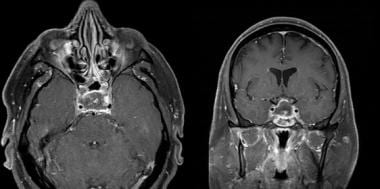
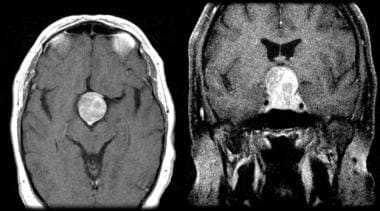
- Pituitary imaging with magnetic resonance imaging (MRI) is diagnostic and typically shows signs of intra-pituitary or intra-adenoma hemorrhage, fluid-fluid level, and compression of normal pituitary tissue. (pediagenosis.com)
- Isolated postoperative hyponatremia resistant to medical correction consider a central cause, in particular pituitary adenoma and/or apoplexy. (medscape.com)
- A tumor that grows from the pituitary gland is called an adenoma. (mayfieldclinic.com)
- We presented a case with a 54 × 40 × 40 mm pituitary adenoma and optic chiasmatic compression with left sphenoid sinus invasion. (nel.edu)
- Histology of the pituitary lesion showed a typical eosinophilic adenoma which only secreted GH when tested with specific immunostain. (nel.edu)
- We describe a patient with an unusual mass presentation of a pituitary adenoma in whom there was also a marked discrepancy between the clinical and laboratory findings in the assessment of suspected Cushing's d. (endocrine-abstracts.org)
- Magnetic resonance imaging was consistent with a pituitary adenoma in one case and RCC in the other. (thieme-connect.com)
- Intraoperative findings and pathological work-up identified RCC along with adenomatous tissue displaying hemorrhagic pituitary adenoma in one and hemorrhagic RCC in the other. (thieme-connect.com)
- RCC and concomitant pituitary adenoma are a rare intraoperative finding that must be considered as a differential diagnosis in patients with symptoms of pituitary adenoma apoplexy. (thieme-connect.com)
- 2 Sumida M, Migita K, Tominaga A, Iida K, Kurisu K. Concomitant pituitary adenoma and Rathke's cleft cyst. (thieme-connect.com)
- Rathke cleft cyst concomitant with pituitary adenoma. (thieme-connect.com)
- 8 Noh S J, Ahn J Y, Lee K S, Kim S H. Pituitary adenoma and concomitant Rathke's cleft cyst. (thieme-connect.com)
- 11 Nourizadeh A R, Pitts F W. Hemorrhage into pituitary adenoma during anticoagulant therapy. (thieme-connect.com)
- Headache may result from an enlarging pituitary adenoma, even when intracranial pressure is not increased. (merckmanuals.com)
- Gigantism and Acromegaly Gigantism and acromegaly are syndromes of excessive secretion of growth hormone (hypersomatotropism) that are nearly always due to a pituitary adenoma. (merckmanuals.com)
- Pituitary apoplexy occurs due to infarction or hemorrhage, within a pituitary adenoma or a nontumorous pituitary gland and can have catastrophic consequences. (surgicalneurologyint.com)
- From a prospective registry of patients who underwent endoscopic transnasal transsphenoidal surgery (TSS) for pituitary adenoma, patients with postoperative DI and SIADH were identified. (uzh.ch)
- Broad categories of etiologies should be considered: pituitary adenoma, nonpituitary tumors, vascular lesions, infiltrative disorders, and others (see Table 2). (the-hospitalist.org)
Adenomas22
- Mohr and Hardy reviewed hospital records of 664 patients who had surgery for pituitary adenomas. (medscape.com)
- All types of pituitary adenomas can be associated with apoplexy, particularly larger tumors (macroadenomas). (uclahealth.org)
- The incidence of pituitary apoplexy ranges from 1 to 20% in surgically verified pituitary adenomas, with a slight male predominance. (symptoma.com)
- It should be noted that necrosis and hemorrhage within a pituitary tumor occur much more frequently than the clinical syndrome of pituitary apoplexy, especially in silent corticotroph adenomas, in which hemorrhage occurs in more than 50% of the tumors. (pediagenosis.com)
- This is despite the high prevalence of occult pituitary adenomas in the general population, the widespread use of high definition imaging techniques, and the broad range of intra- and perisellar lesions that can mimic pituitary adenomas. (bmj.com)
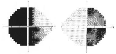
- Inappropriate pituitary hormone secretion and visual field deficits are the most characteristic presenting features of pituitary adenomas. (bmj.com)
- Pituitary adenomas are classified by size and hormone secretory subtype. (bmj.com)
- There are various kinds of pituitary tumors: adenomas, craniopharyngiomas, and Rathke's cleft cysts. (mayfieldclinic.com)
- Closely related to pituitary adenomas are craniopharyngiomas and Rathke's cleft cysts. (mayfieldclinic.com)
- Craniopharyngiomas typically grow from the pituitary stalk upward into the third ventricle and cause symptoms similar to pituitary adenomas. (mayfieldclinic.com)
- When apoplexy occurs in functioning adenomas, it may cause spontaneous remission. (nel.edu)
- We therefore hypothesized that the physiological IGF-I-GH negative feedback loop may be reset in somatotroph adenomas, and we investigated the role of type 1 IGF receptor (IGF-R) and GH receptor (GHR) by quantifying mRNA expression in somatotroph tumours, and investigated the possible presence of mutations of the GHR gene.Methods: Pituitary t. (endocrine-abstracts.org)
- Large corticotroph adenomas are uncommon pituitary mass lesions, representing around 10% of cases of Cushing's disease, and are often found to be locally invasive. (endocrine-abstracts.org)
- Most pituitary tumors are adenomas. (merckmanuals.com)
- Most tumors of the pituitary and suprasellar region are pituitary adenomas. (merckmanuals.com)
- Patients with occult pituitary adenomas infrequently present with pituitary apoplexy. (stanford.edu)
- The use of leuprolide or other gonadotropin releasing hormone agonists for pituitary down-regulation in conjunction with ovarian stimulation can have serious consequences in women harboring unrecognized pituitary adenomas. (stanford.edu)
- Not only is Barrow a place to receive the most advanced treatment and care for your pituitary tumor or disorder, we are also a place where you can help contribute to the scientific understanding and therapeutic research surrounding acromegaly, adenomas, gigantism, and other disorders that implicate the pituitary gland. (barrowneuro.org)
- The vascular supply to nonfunctioning pituitary adenomas (NFPAs) differs compared with that of the anterior lobe of the normal pituitary gland. (surgicalneurologyint.com)
- The vascular supply of pituitary adenomas (PA) is not well understood. (surgicalneurologyint.com)
- Previously, we reported three blood supply patterns (descending, monophasic, and ascending) in nonfunctioning pituitary adenomas (NFPAs) by the time-intensity curve (TIC) analysis using pituitary dynamic magnetic resonance imaging (MRI). (surgicalneurologyint.com)
- Mutation analysis of inhibitory guanine nucleotide binding protein alpha (GNAI) loci in young and familial pituitary adenomas. (cdc.gov)
Cases of pituitary apoplexy4
- In more than 50% of cases of pituitary apoplexy, the apoplectic event is the initial clinical presentation of a pituitary tumor. (pediagenosis.com)
- As with other GnRH agonist, other adverse reactions, including decreased bone density and rare cases of pituitary apoplexy may occur. (nih.gov)
- In cases of pituitary apoplexy, the headache will typically feel sudden and severe and manifest in the front of the head or behind the eyes. (medicalnewstoday.com)
- In cases of pituitary apoplexy, the pain occurs when tumors block a blood vessel to the pituitary gland or cause a bleed. (medicalnewstoday.com)
Hemorrhage14
- Pituitary apoplexy is characterized by a sudden onset of headache, visual symptoms, altered mental status, and hormonal dysfunction due to acute hemorrhage or infarction of a pituitary gland. (medscape.com)
- Frequency of intratumoral hemorrhage increases to 26% if using only MRI criteria without clinical evidence of apoplexy. (medscape.com)
- Kurisu et al described a 68-year-old man who developed pituitary apoplexy resulting in massive intracerebral hemorrhage and death 1 month later. (medscape.com)
- Pituitary apoplexy describes a condition in which the pituitary gland is subject to necrotic changes that may or may not be due to hemorrhage . (symptoma.com)
- This is generally the result of sudden hemorrhage and necrosis in the lateral pituitary fossa, leading to the displacement of the oculomotor nerves. (symptoma.com)
- NCI Thesaurus A rare, potentially life-threatening disorder caused by acute ischemic infarction or hemorrhage in the pituitary gland. (symptoma.com)
- Although pituitary apoplexy, acute hemorrhage of the pituitary gland, is an uncommon event, it is an endocrine emergency, and prompt diagnosis and treatment are critical. (pediagenosis.com)
- This may be a potential source of confusion in differentiating pituitary apoplexy from meningitis or subarachnoid hemorrhage. (pediagenosis.com)
- Pituitary apoplexy occurs most often in the setting of a preexisting pituitary macroadenoma or cyst, and the hemorrhage may be spontaneous or triggered by head trauma, coagulation disorders (e.g., idiopathic thrombocytopenic purpura), or anticoagulant (e.g., heparin, warfarin) administration. (pediagenosis.com)
- Kumar V, Kataria R, Mehta VS. Dengue hemorrhagic fever: A rare cause of pituitary tumor hemorrhage and reversible vision loss. (medscape.com)
- Conditions such as subarachnoid hemorrhage or stroke are sometimes called apoplexy. (medlineplus.gov)
- Pituitary apoplexy (PA) is an acute clinical syndrome characterized by sudden headache, vomiting, visual disturbances, ophthalmoplegia, and/or altered consciousness, secondary to infarction or hemorrhage within a pituitary tumor or nontumorous pituitary gland. (surgicalneurologyint.com)
- Sheehan syndrome refers to hypopituitarism caused by postpartum pituitary necrosis, usually following severe postpartum hemorrhage. (tomwademd.net)
- Consequently, the cells of the anterior pituitary are more prone to necrosis in pregnancies complicated by significant postpartum hemorrhage. (tomwademd.net)
Hypopituitarism10
- This is also commonly associated with pituitary hypopituitarism that requires hormonal replacement for correction. (iasp-pain.org)
- An unusual case of hypopituitarism and transient thyrotoxicosis following asymptomatic pituitary apoplexy. (nel.edu)
- Yoshida M, Murakami M, Ueda H, Miyata M, Takahashi N, Oiso Y. An unusual case of hypopituitarism and transient thyrotoxicosis following asymptomatic pituitary apoplexy. (nel.edu)
- These tumors may compress the normal pituitary gland decrease hormone production (hypopituitarism). (mayfieldclinic.com)
- Generalized Hypopituitarism Generalized hypopituitarism refers to endocrine deficiency syndromes due to partial or complete loss of anterior lobe pituitary function. (merckmanuals.com)
- Patients found with pituitary incidentalomas can be susceptible to several types of adverse outcomes: hormonal hypersecretion, hypopituitarism, neurologic morbidity due to tumor size, and malignancy in rare cases. (the-hospitalist.org)
- Additionally, up to 41% of patients with macroadenomas were found to have varying degrees of hypopituitarism due to compression of the hypothalamus, the hypothalamic-pituitary stalk, or the pituitary itself. (the-hospitalist.org)
- Sheehan syndrome is the clinical manifestation of anterior pituitary cell necrosis and may present as pan-hypopituitarism or as selective loss of pituitary function. (tomwademd.net)
- Pan-hypopituitarism is a result when many cells of the pituitary are affected, as opposed to only a few cells. (tomwademd.net)
- Pituitary stalk dysgenesis-induced hypopituitarism in adult patients: prevalence, evolution of hormone dysfunction and genetic analysis. (cdc.gov)
Prolactin6
- Large pituitary tumors can slightly elevate blood prolactin levels. (uclahealth.org)
- Hormonal evaluation typically shows complete anterior pituitary failure (including prolactin). (pediagenosis.com)
- Prolactin levels were moderately elevated: 1539 (AB), 1186 (LS) (NR 59-619 mU/L). The remainder of the pituitary profile was normal. (endocrine-abstracts.org)
- Generalized condition caused by deficiency of anterior pituitary hormones: adrenocorticotropic hormone (ACTH), thyroid-stimulating hormone (TSH), luteinizing hormone (LH), follicle-stimulating hormone (FSH), growth hormone (GH), and prolactin. (unboundmedicine.com)
- This increase is caused principally by hyperplasia of prolactin-producing cells (lactotrophs) and hyperplasia of other cells in the anterior pituitary gland. (tomwademd.net)
- Prolactin and growth hormone are the most common hormones affected by selective pituitary necrosis and hypofunction. (tomwademd.net)
Tumor apoplexy2
- Pituitary tumor apoplexy: a review. (medscape.com)
- A conservative management is preferable in milder forms of pituitary tumor apoplexy. (cns.org)
Posterior pituitary5
- This may be caused by low cortisol levels or by inappropriate release of antidiuretic hormone (ADH) from the posterior pituitary. (wikipedia.org)
- Because of the anatomy of the pituitary circulation and the sparing of the infundibular circulation (inferior hypophysial arteries), the posterior pituitary is infrequently affected by pituitary apoplexy. (pediagenosis.com)
- Less commonly, the posterior pituitary gland's hormones can be affected: vasopressin (AVP) (antidiuretic hormone [ADH]) and oxytocin ( 1 )[ A ]. (unboundmedicine.com)
- The posterior pituitary gland has its blood supply which functions under higher pressure than the anterior pituitary, so it is not usually affected by shock or hypovolemia. (tomwademd.net)
- The posterior pituitary function is usually not affected, as stated above. (tomwademd.net)
Symptoms16
- The initial symptoms of pituitary apoplexy are related to the increased pressure in and around the pituitary gland. (wikipedia.org)
- The term apoplexy usually describes larger bleeds leading to the sudden onset of symptoms. (uclahealth.org)
- By the conclusion of this session, participants should be able to 1) Describe the various possible presenting symptoms of pituitary apoplexy, 2) Discuss, in small groups, pros and cons of surgery versus conservative management in light of the presenting results, and 3) Identify an effective approach towards a case-by-case treatment decision. (cns.org)
- Fatigue Seizures Pituitary Apoplexy: Diagnosis Your doctor will conduct a thorough physical exam and ask you about your symptoms and medical history. (symptoma.com)
- Less specific symptoms such as headache, and subtle signs of pituitary hormone deficiency with peripheral endocrine organ hypofunction characterised by amenorrhoea, loss of libido, and lethargy, are also common. (bmj.com)
- Symptoms and signs of pituitary hormone deficiency are more subtle than those seen in primary end organ failure (table 1). (bmj.com)
- Apoplexy most often refers to stroke symptoms that occur suddenly. (medlineplus.gov)
- Functional apoplexy is when a person appears to be having stroke-like symptoms. (medlineplus.gov)
- Read on to learn more about pituitary tumor headaches, including their location, what they feel like, and other symptoms that may indicate someone has one. (medicalnewstoday.com)
- If a person has these symptoms, they should seek immediate medical help, as pituitary apoplexy can be serious. (medicalnewstoday.com)
- Not all pituitary tumors cause symptoms. (medicalnewstoday.com)
- When pituitary tumors grow they can compress the above-mentioned structures and cause symptoms. (mayfieldclinic.com)
- Both patients presented at the university hospital with pituitary apoplexy symptoms of sudden-onset headache while undergoing treatment with Coumadin (warfarin). (thieme-connect.com)
- Clinical symptoms of pituitary apoplexy were present in both cases, making pituitary and RCC apoplexy clinically indistinguishable. (thieme-connect.com)
- Pituitary Lesions Patients with hypothalamic-pituitary lesions generally present with some combination of Symptoms and signs of a mass lesion: headaches, altered appetite, thirst, visual field defects-particularly. (merckmanuals.com)
- Other symptoms are associated with loss of pituitary gland hormone production and may include amenorrhea or oligomenorrhea, hot flashes, and/or decreased sex drive. (tomwademd.net)
Treatment of pituitary3
- With the possible equally obtainable visual and endocrine improvements in both surgical and conservative treatment of pituitary apoplexy, physicians and patients can be open to discussing both invasive and non-invasive forms of symptom management. (cns.org)
- At the Pituitary Center at Barrow Neurological Institute, our specialists believe that patient education and involvement form a vital foundation for the successful treatment of pituitary tumors and disorders. (barrowneuro.org)
- Recently, the Endocrine Society released consensus recommendations to guide the evaluation and treatment of pituitary incidentalomas, which are included in the approach outlined below. (the-hospitalist.org)
Blood flow to the pituitary gland1
- If a tumor prevents blood flow to the pituitary gland, it can also lead to pituitary apoplexy, which causes a sudden, severe type of headache . (medicalnewstoday.com)
Condition in which the pituitary1
- Pituitary apoplexy is a condition in which the pituitary tumor spontaneously hemorrhages (bleeds). (uclahealth.org)
Case of pituitary apoplexy1
- We describe a case of pituitary apoplexy in a young woman receiving leuprolide in preparation for ovum donation. (stanford.edu)
Management of pituitary apoplexy2
- The UCLA Pituitary Tumor Program offers comprehensive management of pituitary apoplexy. (uclahealth.org)
- 14 Lubina A, Olchovsky D, Berezin M, Ram Z, Hadani M, Shimon I. Management of pituitary apoplexy: clinical experience with 40 patients. (thieme-connect.com)
Clinical6
- This study was undertaken to evaluate the clinical recovery of 45 patients diagnosed with symptomatic pituitary apoplexy who underwent early (within 72 h of symptom onset) endoscopic transsphenoidal surgical resection with an emphasis on visual, ocular craniopathy, and endocrinological outcome. (iasp-pain.org)
- 2. Giritharan S, Gnanalingham K, Kearney T. Pituitary apoplexy - bespoke patient management allows good clinical outcome. (cns.org)
- The clinical course of pituitary apoplexy varies widely in duration and severity. (pediagenosis.com)
- Mou C, Han T, Zhao H, Wang S, Qu Y. Clinical features and immunohistochemical changes of pituitary apoplexy. (medscape.com)
- Pituitary apoplexy is a rare clinical syndrome associated with rapid enlargement of a pituitary mass. (nel.edu)
- the clinical diagnosis of prolactinoma depends on the degree of hyperprolactinaemia in the context of pituitary tumour size. (endocrine-abstracts.org)
Diagnosis4
- Pituitary apoplexy is rarely life threatening, if you receive prompt and accurate diagnosis and treatment. (uclahealth.org)
- Learn more about our Neuroendocrine and Pituitary Program located in downtown Boston, we specialize in the diagnosis and treatment of various neuroendocrine disorders. (tuftsmedicalcenter.org)
- The diagnosis of pituitary disease is generally uncomplicated. (bmj.com)
- In this brief overview, the presentation, classification, and general investigation of pituitary lesions is followed by a discussion of the diagnosis and management of specific secretory subtypes. (bmj.com)
Adrenal2
- In pituitary apoplexy, the main initial problem is a lack of secretion of adrenocorticotropic hormone (ACTH, corticotropin), which stimulates the secretion of cortisol by the adrenal gland. (wikipedia.org)
- 1 Three commonly discovered lesions by hospitalists are pituitary, thyroid, and adrenal incidentalomas. (the-hospitalist.org)
Symptomatic pituitary apoplexy1
- [ 3 ] Typical symptomatic pituitary apoplexy occurred in only 0.6% of patients with significant hemorrhagic and necrotic changes in 9.5% of surgical specimens. (medscape.com)
Endocrine2
- Okuda O, Umezawa H, Miyaoka M. Pituitary apoplexy caused by endocrine stimulation tests: a case report. (medscape.com)
- Known as the master gland, the pituitary controls the other endocrine glands in the body. (mayfieldclinic.com)
Types of pituitary tumors2
- It was previously thought that particular types of pituitary tumors were more prone to apoplexy than others, but this has not been confirmed. (wikipedia.org)
- There are two broad types of pituitary tumors: nonfunctional and functional. (medicalnewstoday.com)
Necrosis2
- This deprives the anterior pituitary gland and the tumor itself of its vascular supply, apoplectically causing ischemia and subsequent necrosis. (medscape.com)
- Sheehan syndrome which is also called post-partum pituitary necrosis refers to the necrosis of cells of the anterior pituitary gland following significant post-partum bleeding, hypovolemia, and shock. (tomwademd.net)
Occurs5
- This usually occurs in the presence of a tumor of the pituitary, although in 80% of cases this has not been diagnosed previously. (wikipedia.org)
- This occurs in 70% of those with pituitary apoplexy. (wikipedia.org)
- Doctors think this occurs because of compression of the pituitary stalk, the connection between the brain and pituitary gland. (uclahealth.org)
- Pituitary apoplexy is a medical emergency that occurs when a pituitary tumor starts bleeding or cuts off its own blood supply. (upmc.com)
- Sheehan syndrome occurs when the anterior pituitary gland is damaged due to significant blood loss. (tomwademd.net)
Cavernous sinus1
- Adjacent to the pituitary lies a part of the skull base known as the cavernous sinus. (wikipedia.org)
Hormones5
- The pituitary produces many of the hormones that control essential body processes. (tuftsmedicalcenter.org)
- In contrast, functional pituitary tumors produce hormones, some of which can affect females and males in different ways. (medicalnewstoday.com)
- Functioning pituitary tumors secrete high levels of hormones and interfere with other body organs. (mayfieldclinic.com)
- Nonfunctioning pituitary tumors do not secrete hormones. (mayfieldclinic.com)
- This blood loss results in the pituitary gland not being able to produce hormones. (tomwademd.net)
Stalk4
- The anterior pituitary gland is perfused by its portal venous system, which passes down the hypophyseal stalk. (medscape.com)
- Some postulate that a gradually enlarging pituitary tumor becomes impacted at the diaphragmatic notch, compressing and distorting the hypophyseal stalk and its vascular supply. (medscape.com)
- The gland is connected to the hypothalamus in the brain by the pituitary stalk. (mayfieldclinic.com)
- Hyperprolactinaemia in the presence of pituitary tumour can occur from tumour secretion or from stalk compression causing loss of dopaminergic inhibition. (endocrine-abstracts.org)
Combined pituitary hormone deficiency1
- Genetic analyses of bone morphogenetic protein 2, 4 and 7 in congenital combined pituitary hormone deficiency. (cdc.gov)
Macroadenoma with suprasellar extension1
- MRIs (without contrast) reported pituitary macroadenoma with suprasellar extension but no optic chiasm compression. (endocrine-abstracts.org)
Surgery for pituitary1
- Determinants of visual and endocrinological outcome after early endoscopic endonasal surgery for pituitary apoplexy. (iasp-pain.org)
Ischemic3
- [ 1 ] It is important to note that pituitary apoplexy may be divided into hemorrhagic or ischemic, each with unique neuroimaging findings, and some patients have elements of both. (medscape.com)
- Enhanced T1-weighted axial and coronal MRI showing a large pituitary tumor that has recently undergone ischemic apoplexy showing a necrotic (hypointense) center and ring of gadolinium enhancement (hyperintense), ie, the "pituitary ring sign. (medscape.com)
- Pituitary metastasis presenting as ischemic pituitary apoplexy following heparin-induced thrombocytopenia. (medscape.com)
People with pituitary2
- 70% of people with pituitary apoplexy experience double vision due to compression of one of the nerves. (wikipedia.org)
- And, because our doctors and nurses treat more people with pituitary disorders than any other team in the Southwest, you can rest assured that you will be in experienced hands once you decide on a course of treatment. (barrowneuro.org)
Subarachnoid1
- Pituitary apoplexy is rarely associated with subarachnoid bleed and vasospasm, leading to cerebral infarcts and consequent focal neurologic deficits. (medscape.com)
Patients9
- Patients diagnosed with pituitary apoplexy and presenting with acute visual deterioration require urgent surgical resection. (iasp-pain.org)
- Erythrocytes and an increased protein concentration are found in the cerebrospinal fluid of many patients with pituitary apoplexy. (pediagenosis.com)
- Thus, diabetes insipidus is rare in patients with pituitary apoplexy. (pediagenosis.com)
- Stress dosages of glucocorticoids should be initiated in all patients with pituitary apoplexy. (pediagenosis.com)
- The Pituitary Foundation (internet search term: "Pit Pat") provides important opinion and information, and reassures patients that they are not alone. (bmj.com)
- Various precipitating factors have been reported in 25-30% of pituitary apoplexy patients. (nel.edu)
- Pituitary tumors are suspected in patients with unexplained headaches, characteristic visual abnormalities, or endocrinopathies. (merckmanuals.com)
- Although patients suffering from DHF harbor multiple factors, which may be precipitants of pituitary apoplexy, the association between these two conditions is rare and only few case reports document their coexistence. (surgicalneurologyint.com)
- Functional characterization of a heterozygous GLI2 missense mutation in patients with multiple pituitary hormone deficiency. (cdc.gov)
Secretion1
- Tumors may also compress or destroy pituitary or hypothalamic tissue, impairing hormone production or secretion. (merckmanuals.com)
Optic5
- Humphrey computerised visual fields are useful even if there appears to be no contact between the optic pathways and pituitary mass. (bmj.com)
- Directly above the pituitary gland is the optic chiasm, which is responsible for vision. (mayfieldclinic.com)
- A cross-section of the pituitary gland (green) shows its relationship to the optic chiasm, the sphenoid sinus, and the cavernous sinuses on each side. (mayfieldclinic.com)
- The pituitary gland is related to the optic chiasm above and the sphenoid sinus below. (mayfieldclinic.com)
- Any tumor that grows out of the pituitary can compress optic nerve tracts, including the chiasm. (merckmanuals.com)
Cysts2
- Hemorrhagic and nonhemorrhagic Rathke cleft cysts mimicking pituitary apoplexy. (medscape.com)
- 1 Binning M J, Liu J K, Gannon J, Osborn A G, Couldwell W T. Hemorrhagic and nonhemorrhagic Rathke cleft cysts mimicking pituitary apoplexy. (thieme-connect.com)
Hemorrhagic pituitary1
- However, hemorrhagic pituitary apoplexy may be fatal. (medscape.com)
Hypothalamus4
- Pituitary apoplexy or infarction is the death of an area of tissue in the pituitary gland, a small gland joined to the hypothalamus (part of the brain). (tuftsmedicalcenter.org)
- The hypothalamus then regulates pituitary hormone levels, depending on the needs of the body. (mayfieldclinic.com)
- Luteinizing hormone (LH) is a glycoprotein gonadotropin secreted by the anterior pituitary in response to gonadotropin-releasing hormone (GnRH), which is released by the hypothalamus. (medscape.com)
- LH and FSH play central roles in the hypothalamic-pituitary-gonadal axis, and, thus, conditions related to LH and FSH deficiency can be caused by pathology of either the hypothalamus or pituitary. (medscape.com)
Internal caroti1
- Pituitary apoplexy resulting in internal carotid artery occlusion has been reported due to the mass compressing the bilateral cavernous sinuses, resulting in obliteration of the cavernous portion of the right internal carotid artery. (medscape.com)
Syndrome4
- Kaplun J, Fratila C, Ferenczi A, Yang WC, Lantos G, Fleckman AM. Sequential pituitary MR imaging in Sheehan syndrome: report of 2 cases. (medscape.com)
- 9 Nagarajan D V, Bird D, Papouchado M. Pituitary apoplexy following anticoagulation for acute coronary syndrome. (thieme-connect.com)
- These include: idiopathic intracranial hypertension, eclampsia, reversible cerebral vasoconstriction syndrome, cerebral venous thrombosis, pituitary apoplexy and postdural puncture headache. (bmj.com)
- in acute Sheehan syndrome, MRI shows an enlarged pituitary with only a thin rim of enhancement with gadolinium. (tomwademd.net)
Sudden3
- The word apoplexy is defined as a sudden neurologic impairment, usually due to a vascular process. (medscape.com)
- Doctors may suspect pituitary apoplexy because there is a sudden increase in the size of the tumor. (uclahealth.org)
- The compression may also lead to a loss of blood supply (pituitary infarct), which can cause tumor cell death, bleeding and sudden tumor swelling. (uclahealth.org)
Anterior3
- The pituitary gland consists of two parts, the anterior (front) and posterior (back) pituitary. (wikipedia.org)
- This hyperplasia leads to increased nutritional and metabolic demand by the anterior pituitary gland as a whole, but the blood supply that feeds the anterior pituitary does not increase. (tomwademd.net)
- The blood supply that feeds the anterior pituitary gland is a relatively low-pressure system. (tomwademd.net)
Sella3
- Side view of the pituitary gland sitting inside the bony sella. (mayfieldclinic.com)
- The pituitary gland (green) is located deep within the skull in an area called the sella. (mayfieldclinic.com)
- After one year, MRI shows atrophy of the pituitary and a partially empty sella. (tomwademd.net)
Occur2
- Although hemorrhagic areas of the pituitary are absorbed over time, reabsorption alone may not occur fast enough for recovery of visual acuity. (pediagenosis.com)
- An increase in pituitary volume and cell count occur in pregnant women in the weeks preceding delivery. (tomwademd.net)
Gland is a small2
- The pituitary gland is a small gland that sits behind the bridge of the nose, beneath the brain. (medicalnewstoday.com)
- The pituitary gland is a small, bean-shaped organ that sits at the base of the brain, behind the bridge of the nose (Fig. 1 and 2). (mayfieldclinic.com)
Gonadotropin releasi2
- Precipitation of pituitary apoplexy by gonadotropin releasing hormone or gonadotropin releasing hormone agonists has been described. (stanford.edu)
- To our knowledge, pituitary apoplexy after gonadotropin releasing hormone agonist use for ovum donation has not been previously described. (stanford.edu)
Benign2
- Pituitary tumors are usually benign (noncancerous) and develop in the pituitary gland, which controls hormone levels throughout your body and is located at the base of your brain. (upmc.com)
- Most pituitary tumors are benign, meaning they are not cancerous. (medicalnewstoday.com)
Cerebral2
- Association of degenerative change in pituitary ademona with radiotherapy and detection by cerebral computed tomography. (medscape.com)
- Ahmed SK, Semple PL. Cerebral ischemia in pituitary apoplexy. (medscape.com)
Headaches4
- Pituitary apoplexy has a variable presentation , although it most commonly presents with headaches , visual changes , changes in consciousness and ophthalmoplegia . (symptoma.com)
- In addition to headaches, pituitary apoplexy can also present with confusion , vomiting , nausea and visual changes. (symptoma.com)
- Follow up and optimisation of pituitary hormone replacement is also relatively straightforward, but management of visual impairment, reduced fertility, coarsened facial features, arthritis, obesity, headaches, and obstructive sleep apnoea is often much more troublesome. (bmj.com)
- Pituitary tumors do not always cause headaches. (medicalnewstoday.com)
Rarely1
- Rarely, pituitary tumors are carcinomas. (merckmanuals.com)
Sphenoid Sinus1
- The pituitary gland is bordered on either side by the cavernous sinuses and below by the sphenoid sinus. (mayfieldclinic.com)
Computerized tomography1
- You may also undergo a computerized tomography (CT) scan of the pituitary gland, which will also show if there is an abnormality. (uclahealth.org)