Pityriasis Lichenoides
Pityriasis
Pityriasis Rosea
Parapsoriasis
Pityriasis Rubra Pilaris
Malassezia
Lichenoid Eruptions
Enhanced expression of human metalloelastase (MMP-12) in cutaneous granulomas and macrophage migration. (1/12)
Accumulation of inflammatory cells such as macrophages may lead to degeneration of connective tissue matrix in various skin diseases. Macrophage metalloelastase, is a matrix metalloproteinase (MMP-12) capable of degrading elastin as well as various basement membrane components. To investigate the role of human macrophage metalloelastase in skin, we assessed by in situ hybridization and immunohistochemistry 66 specimens representing skin diseases characterized either by changes in elastic fibers or by pronounced infiltrations of extravasating and migrating macrophages. CD68 immunostaining was performed to identify the human macrophage metalloelastase-positive cells and Weigert's Resorcin-Fuchsin staining to reveal the status of elastic fibers. We found abundant expression of human macrophage metalloelastase mRNA in macrophages in areas devoid of normal elastic fibers in granulomatous skin diseases sarcoidosis, necrobiosis lipoidica diabeticorum, and granuloma annulare. Positive cells for human macrophage metalloelastase protein could be detected in the same regions as well as positive immunostaining for urokinase plasminogen activator. Of the other matrix metalloproteinases capable of degrading elastin, 92 kDa gelatinase colocalized with human macrophage metalloelastase, while 72 kDa gelatinase was produced by surrounding fibroblast-like cells. Furthermore, human macrophage metalloelastase was expressed by macrophages in areas with disrupted basement membrane, as assessed by type IV collagen staining, in pityriasis lichenoides and dermatitis herpetiformis. Specimens of anetoderma, acrodermatitis chronica atrophicans and pseudoxanthoma elasticum showed no signal for human macrophage metalloelastase. Matrilysin was not detected in any of the samples investigated. Our study suggests that human macrophage metalloelastase may contribute to elastin degradation occurring in granulomatous skin diseases and may aid macrophage migration through the epidermal and vascular basement membranes in inflammatory disorders. (+info)Febrile ulceronecrotic Mucha-Habermann disease: a case report and a review of the literature. (2/12)
This report describes the case of a 76 year old man who suffered from febrile ulceronecrotic Mucha-Habermann disease (FUMHD). Despite this patient's typical clinical and histological findings, the fulminating course led to death. Polymerase chain reaction (PCR) analysis of the skin lesions showed that the infiltrating cells were monoclonal in origin and were from an aberrant clone. FUMHD is a very rare, febrile variant type of pityriasis lichenoides et varioliformis acuta, and is characterised by necrotic cutaneous ulcerations associated with high fever and systemic manifestations. Including this present case, only 18 cases of FUMHD have been reported. FUMHD can occur in both adults and children, although there are several differences between the manifestations of the disease in the two groups. One major difference is prognosis: all cases resulting in fatality are of the adult type, whereas no fatal cases have been reported among children. The aberrant clone detected by PCR may be responsible for host responses, resulting in the severe symptoms observed in this disorder. (+info)Pityriasis lichenoides chronica. (3/12)
A 19-year-old woman presented with a five-year history of guttate macules and yellow to skin-colored papules with collarette of fine scale on the trunk and the upper and lower extremities. Guttate pityriasis lichenoides chronica is an uncommon presentation of this T-cell-mediated disease. It is of unknown etiology; however, an infectious agent has been suspected. It is considered in a spectrum with pityriasis lichenoides et varioloformis acuta and rarely mycosis fungoides and CD30 lymphomas. Improvement has been shown after treatment with oral tetracyclines, ultraviolet B and UVA1 phototherapy, and PUVA photochemotherapy. (+info)Presence of chimeric maternally derived keratinocytes in cutaneous inflammatory diseases of children: the example of pityriasis lichenoides. (4/12)
During pregnancy, maternal cells may enter the fetal circulation and persist until adulthood. The fate of these cells remains unknown. As unexplained T-cell-mediated conditions such as pityriasis lichenoides (PL) may occur in children, we aimed at identifying maternal cells in lesional skin of PL and controls. Archived skin biopsy specimens from young males with PL, atopic dermatitis, or normal skin were scanned for the presence of female (presumably maternal) cells using fluorescence in situ hybridization (FISH) with X and Y chromosome-specific probes. Phenotyping of maternal cells relied on FISH combined with anti-CD45, anti-CD1a, or anti-cytokeratin labelling, identifying leukocytes, Langerhans cells, and keratinocytes, respectively. Maternal cells were found in PL (11/12) and controls (4/7), but their average frequency was higher in PL: 99 per million cells as compared to 5 per million cells in controls (P = 0.005). In the epidermis, the maternal microchimeric cells were labelled by anti-cytokeratin in all cases. We identified maternally derived keratinocytes in the skin of male children with inflammatory skin disorders. These cells may either help repair the damaged skin or home initially in the skin and trigger a host (child) versus graft (mother) disease. (+info)Febrile ulceronecrotic Mucha-Habermann disease: a case report and a review of the literature. (5/12)
We report a case of febrile ulceronecrotic Mucha-Habermann disease (FUMHD) in a 27-year-old woman. After 20 days of a mild eruption, extensive polymorphous, papular and ulcerohemorrhagic skin lesions gradually developed, associated with intermittent high temperature, and constitutional symptoms. The initial treatment with acyclovir was not successful, the skin lesions still progressed distally and individual lesions evolved from necrotic papules and bullae to erosions and ulcers. Skin biopsies showed the typical histopathological changes of PLEVA. The patient was treated with systemic prednisolone but dosage was limited in order to avoid sepsis. Despite corticosteroid therapy and supportive therapy, the fulminating course led to death. Including this present case, only 31 cases of FUMHD have been reported in English literature. Our case is the second report from Turkey. (+info)A clinical and histopathological study of pityriasis lichenoides. (6/12)
BACKGROUND: Pityriasis lichenoides is a papulosquamous disorder of unknown etiology with remissions and exacerbations. Histopathology helps greatly in the diagnosis of this condition. AIM: We studied clinical and histopathological features of pityriasis lichenoides in our patients. METHODS: This is a 3-year retrospective and prospective, descriptive study of all patients clinically diagnosed as pityriasis lichenoides and confirmed by histopathology. All patients were studied clinically and histopathologically. RESULTS: There were 51 (30 males and 21 females) cases of pityriasis lichenoides in the study period. The maximum number of cases, 14 (27.45%) were in their second decade of life. Pityriasis lichenoides chronica was diagnosed in 39 cases (76.47%) and pityriasis lichenoides et varioliformis acuta (PLEVA) in 12 cases (23.53%). Histopathologically, basal cell vacuolation and perivascular infiltrate were seen in all the cases. Exocytosis was seen in 45.1% of the cases. All the cases of PLEVA showed lymphocytic vasculitis albeit without fibrinoid deposition in the vessel walls. CONCLUSION: Pityriasis lichenoides is not a rare disorder, and is not a true lymphocytic vasculitis as blood vessel damage and fibrinoid deposition in the blood vessel walls were not seen in this study. (+info)Febrile ulceronecrotic Mucha-Habermann disease: a case report and review of the literature. (7/12)
(+info)Pityriasis lichenoides with ulceronecrosis and hyperthermia: a rare variant of pityriasis lichenoides et varioliformis acuta. (8/12)
(+info)Pityriasis lichenoides is a group of skin disorders characterized by scaly, red or purple-red papules (small, raised bumps) that can progress to vesicles or pustules and often leave behind a tan or brown discoloration as they resolve. The two main types are pityriasis lichenoides chronica (PLC) and pityriasis lichenoides et varioliformis acuta (PLEVA). PLC is a more chronic, milder form with scaly pink-red papules that can last for months to years, while PLEVA is a more acute form characterized by sudden onset of widespread, eruptive papules and vesicles that may heal with varioliform (pockmarked) scarring. The exact cause of pityriasis lichenoides is unknown, but it's thought to be related to an immune response to an infectious agent or other trigger. Treatment options include topical corticosteroids, phototherapy, and systemic medications such as antibiotics or immunosuppressants.
Pityriasis is a general term used to describe a group of skin conditions characterized by scaling. It includes several specific types, the most common being Pityriasis rosea and Pityriasis simplex capillitii (also known as dandruff).
1. Pityriasis rosea: This is a temporary skin rash that often begins with a single, round, scaly patch on the chest, abdomen, or back. A few days to weeks later, more patches appear. These patches are oval and scaly, and they may be pink, red, or tan. The rash usually lasts about 6-8 weeks.
2. Pityriasis simplex capillitii: This is a very common condition characterized by flaking or scaling of the scalp, which is often referred to as dandruff.
The term "pityriasis" comes from the Greek word "pitýrios," which means "bran."
Pityriasis rosea is a common, self-limited skin condition characterized by the development of oval or round, scaly, pinkish, inflamed patches on the skin. The initial lesion, known as the "herald patch," often appears before other lesions and measures 2-10 cm in diameter. It usually starts as a single, solitary, scaly, raised patch on the trunk that precedes the generalized eruption by about 1-2 weeks. The rash typically spreads to involve the chest, abdomen, back, arms, and legs, sparing the face, palms, and soles.
The rash is often asymptomatic but can be pruritic (itchy) in some cases. It usually resolves within 6-12 weeks without any treatment, although topical treatments such as corticosteroids or antihistamines may be used to relieve itching. The exact cause of pityriasis rosea is not known, but it is thought to be caused by a viral infection. It is more common in young adults and is more prevalent in the spring and fall seasons.
Parapsoriasis is a term used to describe two uncommon, chronic, and relatively benign inflammatory skin conditions. These are small plaque parapsoriasis (SPP) and large plaque parapsoriasis (LPP), also known as retiform or digitate dermatosis of Köbner.
Small plaque parapsoriasis is characterized by scaly, thin, pink to red patches or plaques, usually less than 3-5 cm in diameter. The lesions are often asymptomatic or mildly pruritic and can be found on the trunk and proximal extremities.
Large plaque parapsoriasis presents as larger, irregularly shaped, scaly patches or thin plaques, typically greater than 5 cm in diameter. The lesions are often asymptomatic but may occasionally be pruritic. LPP is considered a precursor to a rare cutaneous T-cell lymphoma called mycosis fungoides, especially when the lesions become thicker or more numerous over time.
It's important to note that these conditions can sometimes be challenging to diagnose and may require a skin biopsy for accurate diagnosis. Dermatologists and pathologists should carefully evaluate the clinical presentation, histopathological features, and any potential progression to ensure appropriate management.
Pityriasis rubra pilaris (PRP) is a rare, chronic inflammatory skin disorder characterized by the development of reddish orange scaly patches and thickened plaques on the skin. It primarily affects the scalp, face, knees, elbows, and palms and soles. The condition can also cause reddening and thickening of the skin over the entire body in severe cases.
The key features of PRP include follicular papules (small bumps around hair follicles) that may be surrounded by a white collarette of scale, and areas of skin that are redder than normal (erythema). The condition can also cause nail changes, such as thickening and ridging.
PRP is thought to be caused by an abnormal immune response, although the exact cause is not known. It can affect people of all ages, but it is most commonly seen in children and adults between 40-60 years old. The condition typically progresses through several stages, with periods of worsening symptoms followed by periods of improvement or remission.
Treatment for PRP may include topical therapies, such as corticosteroids or retinoids, as well as systemic treatments, such as immunosuppressive drugs or biologics. The choice of treatment depends on the severity and extent of the condition, as well as other factors.
Malassezia is a genus of fungi (specifically, yeasts) that are commonly found on the skin surfaces of humans and other animals. They are part of the normal flora of the skin, but under certain conditions, they can cause various skin disorders such as dandruff, seborrheic dermatitis, pityriasis versicolor, and atopic dermatitis.
Malassezia species require lipids for growth, and they are able to break down the lipids present in human sebum into fatty acids, which can cause irritation and inflammation of the skin. Malassezia is also associated with fungal infections in people with weakened immune systems.
The genus Malassezia includes several species, such as M. furfur, M. globosa, M. restricta, M. sympodialis, and others. These species can be identified using various laboratory methods, including microscopy, culture, and molecular techniques.
Lichenoid eruptions are skin reactions that resemble the appearance of lichen, a type of slow-growing fungus. These eruptions are characterized by flat, scaly bumps (papules) and rough, discolored patches (plaques) on the skin. They can be caused by various factors, including medications, medical conditions, or as a reaction to certain chemicals or substances that come into contact with the skin.
The term "lichenoid" refers to the resemblance of these eruptions to lichen, which is characterized by its distinctive appearance and growth pattern. Lichenoid eruptions can occur anywhere on the body but are most commonly found on sun-exposed areas such as the arms, legs, and trunk.
The exact cause of lichenoid eruptions can vary, but they are often associated with an autoimmune response in which the body's immune system mistakenly attacks healthy skin cells. This can lead to inflammation, redness, itching, and other symptoms associated with these eruptions. Treatment for lichenoid eruptions typically involves identifying and addressing the underlying cause, as well as managing symptoms with topical medications or other therapies.
Keratosis, in general, refers to a skin condition characterized by the abnormal growth or development of keratin, a protein that forms part of the outer layer of the skin (epidermis). There are several types of keratosis, including:
1. Seborrheic Keratosis: benign, often pigmented, rough, and scaly growths that can appear anywhere on the body. They tend to increase in number with age.
2. Actinic Keratosis: rough, scaly patches or spots on the skin that are caused by long-term exposure to sunlight or artificial UV light. These have the potential to develop into squamous cell carcinoma, a type of skin cancer.
3. Solar Keratosis: another term for actinic keratosis, as it is primarily caused by sun damage.
4. Keratosis Pilaris: a common condition where small, rough bumps appear on the skin, often on the arms, thighs, or cheeks. These are caused by excess keratin blocking hair follicles.
5. Follicular Keratosis: a disorder characterized by the formation of horny plugs within the hair follicles, leading to rough, sandpaper-like bumps on the skin.
6. Intraepidermal Keratosis: a term used to describe the abnormal accumulation of keratin in the epidermis, which can lead to various skin conditions.
It's important to consult with a healthcare professional or dermatologist for proper diagnosis and treatment if you suspect having any form of keratosis.
 Pityriasis lichenoides
Pityriasis lichenoides chronica
Pityriasis lichenoides et varioliformis acuta
Viktor Mucha
PLC
Lymphoproliferative disorders
List of diseases (P)
Histopathologic diagnosis of dermatitis
Parapsoriasis
List of MeSH codes (C17)
Cutaneous T-cell lymphoma
List of skin conditions
Pityriasis
Lichen planus
Pityriasis lichenoides - Wikipedia
 Pityriasis Lichenoides: Practice Essentials, Pathophysiology, Etiology
 Pityriasis Lichenoides Chronica (PLC)
 Hyperbaric oxygen therapy (HBOT) | Pityriasis lichenoides | India
 Pityriasis Lichenoides - Dermatologic Disorders - MSD Manual Professional Edition
Pityriasis Lichenoides - Dermatologic Disorders - MSD Manual Professional Edition
 Life | Free Full-Text | Bromelain a Potential Bioactive Compound: A Comprehensive Overview from a Pharmacological Perspective
 Pityriasis lichenoides et varioliformis acuta (Mucha- Haberman) - Image | Evidence-Based Medicine Guidelines
 View of Pityriasis Lichenoides; A Case Series With Clinical And Histological Evaluation In A Sample Of Sixteen Iraqi Patients...
 Tags
Tags
 Advanced Search Results - Public Health Image Library(PHIL)
 My Lyme Disease Diagnosis
 Scaly signs in dermatology - Indian Journal of Dermatology, Venereology and Leprology
 babies and children with eczema - talkhealth forums
Lymphomatoid papulosis
 Ulcerative Colitis
 Pityriasis Rosea | AAFP
 38 CFR § 4.118 - Schedule of ratings-skin. | Electronic Code of Federal Regulations (e-CFR) | US Law | LII / Legal Information...
 Pink-to-red scaly raised lesions - Clinical Advisor
 Diagnostic Pathology Nonneoplastic Pediatrics, 2nd edition - Angelica R. Putnam - 1020 - ELSEVIER HEALTH SCIENCES -...
 Pathology
 A clinician's opinion on ineffective dermatologic therapies
 Dermoscopy of Inflammatory Conditions: The Journey So Far - European Medical Journal
 Pattern of skin diseases at Riyadh Military Hospital, EDOJ4(2):4
 UVA1 Phototherapy: A Concise and Practical Review
 Anal carcinoma - Altmeyers Encyclopedia - Department Dermatology
 Search
Specific PHGKB|Rare Diseases PHGKB|PHGKB
 PALLER AND MANCINI HURWITZ CLINICAL PEDIATRIC DERMATOLOGY 6TH EDITION PDF FREE DOWNLOAD: - Medical Students Corner
Varioliformis acuta9
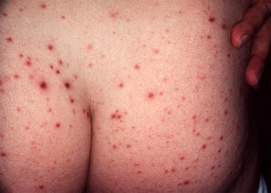
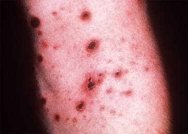
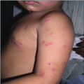
- Pityriasis lichenoides encompasses a spectrum of clinical presentations ranging from acute papular lesions that rapidly evolve into pseudovesicles and central necrosis (pityriasis lichenoides et varioliformis acuta or PLEVA) to small, scaling, benign-appearing papules (pityriasis lichenoides chronica or PLC). (medscape.com)
- Typical hemorrhagic crusted papules of pityriasis lichenoides et varioliformis acuta. (medscape.com)
- Rare reports of death from the febrile-ulceronecrotic variant have been attributed to secondary pulmonary thromboembolism, pneumonia, cardiac arrest, and sepsis, among others.3 Ulceronecrotic pityriasis lichenoides et varioliformis acuta (PLEVA) can lead to scarring. (medscape.com)
- A patient presenting with febrile ulceronecrotic pityriasis lichenoides et varioliformis acuta (PLEVA) requires an entirely different approach than a patient presenting with pityriasis lichenoides chronica (PLC). (medscape.com)
- The more acute (sudden onset) form is known as pityriasis lichenoides et varioliformis acuta ( PLEVA ), also known as Mucha-Habermann disease . (childrens.com)
- Evidence Central , evidence.unboundmedicine.com/evidence/view/EBMG/450119/all/Pityriasis_lichenoides_et_varioliformis_acuta__Mucha__Haberman____Image. (unboundmedicine.com)
- In this review, the dermoscopic patterns of psoriasis, lichen planus (LP), pityriasis rosea (PR), prurigo nodularis (PN), eczema, pityriasis lichenoides et varioliformis acuta (PLEVA), pityriasis lichenoides chronica (PLC), and discoid lupus erythematosus (DLE) are described and their importance is highlighted. (emjreviews.com)
- Pityriasis Lichenoides et Varioliformis Acuta (PLEVA) is also known as Mucha-Habermann disease. (acadderm.com)
- PLEVA (pityriasis lichenoides et varioliformis acuta) is a minor cutaneous lymphoid dyscrasia that can appear suddenly and persist for weeks to months. (mhmedical.com)
Chronica4
- Dermoscopy of an Indian girl with pityriasis lichenoides chronica of the trunk revealed structureless areas with yellowish-orange to light brown color with focal superficial scales and multiple scattered dark brown to black granules. (medscape.com)
- Possible Pityriasis Lichenoides Chronica? (talkhealthpartnership.com)
- NLRP1 hyperactivity has been reported to cause inherited autoinflammatory diseases including familial keratosis lichenoides chronica and NLRP1-associated autoinflammation with arthritis and dyskeratosis. (frontiersin.org)
- Role of bromelain in the treatment of patients with pityriasis lichenoides chronica. (ulyclinic.com)
Acuta1
- Reichel A, Grothaus J , Ott H . Pityriasis lichenoides acuta (PLEVA) pemphigoides: A rare bullous variant of PLEVA. (auf-der-bult.de)
Rosea15
- Pityriasis rosea is a common, acute exanthem of uncertain etiology. (aafp.org)
- Pityriasis rosea typically affects children and young adults. (aafp.org)
- Pityriasis rosea is difficult to identify until the appearance of characteristic smaller secondary lesions that follow Langer's lines (cleavage lines). (aafp.org)
- Several medications can cause a rash similar to pityriasis rosea, and several diseases, including secondary syphilis, are included in the differential diagnosis. (aafp.org)
- Pityriasis rosea is a common skin condition characterized by a herald patch and the later appearance of lesions arrayed along Langer's lines (cleavage lines). (aafp.org)
- Several large case series from dermatology practices indicate that the incidence of pityriasis rosea peaks in persons 20 to 29 years of age, with no consistent gender predilection ( Table 1 ). (aafp.org)
- Although the etiology of pityriasis rosea is unclear, several factors indicate an infectious cause. (aafp.org)
- 4 Second, recurrence of pityriasis rosea outside the acute phase is rare, suggesting that there is long-lasting immunity after the infection. (aafp.org)
- Third, up to 69 percent of patients with pityriasis rosea have a prodromal illness before the herald patch appears. (aafp.org)
- 5 Finally, some patients with pityriasis rosea show an increase in B lymphocytes, a decrease in T lymphocytes, and an elevated sedimentation rate. (aafp.org)
- The results of one study 7 showed elevated levels of human herpesvirus 7 in patients with pityriasis rosea. (aafp.org)
- Chlamydia pneumoniae, Legionella pneumophila , and Mycoplasma pneumoniae also have been suggested as potential infectious agents in pityriasis rosea. (aafp.org)
- Identification of pityriasis rosea can be challenging for a number of reasons. (aafp.org)
- In at least one half of patients, the first symptoms of pityriasis rosea are nonspecific and consistent with a viral upper respiratory infection. (aafp.org)
- Other Medscape pityriasis articles include Dermatologic Manifestations of Pityriasis Alba , Pityriasis Lichenoides , Pityriasis Rosea , and Pityriasis Rotunda . (medscape.com)
PLEVA1
- The dermatologist called these PLEVA (pityriasis lichenoides), a rare condition sometimes associated with bacterial infections like Lyme. (healthline.com)
Parapsoriasis4
- Pityriasis lichenoides variants describe scaly dermatoses with necrotic papules that are clinically and histologically different from parapsoriasis. (medscape.com)
- El-Darouti et al reported on a 7-year study of a hypopigmented disorder that the researchers believe should be classified as a new variant of parapsoriasis en plaque. (medscape.com)
- MF presents as erythematous skin patches that are often confused for immune-related diseases like eczema, psoriasis, parapsoriasis, or pityriasis lichenoides. (targretinhcp.com)
- Proposed nomenclature divides parapsoriasis into two distinct subgroups, PITYRIASIS LICHENOIDES and parapsoriasis en plaques (small- and large-plaque parapsoriasis). (wakehealth.edu)
Case of pityriasis1
- The febrile-ulceronecrotic variant may arise de novo or from a preexisting case of pityriasis lichenoides. (medscape.com)
Rubra20
- Pityriasis rubra pilaris (PRP) was first described in 1828 by Tarral and was named by Besnier in 1889. (medscape.com)
- Griffiths divided pityriasis rubra pilaris into 5 categories: classic adult type, atypical adult type, classic juvenile type, circumscribed juvenile type, and atypical juvenile type. (medscape.com)
- Type V pityriasis rubra pilaris has been linked to mutations in the gene, CARD . (medscape.com)
- [ 12 ] One hypothesis is that pityriasis rubra pilaris may be related to an abnormal immune response to an antigenic trigger. (medscape.com)
- Case reports have described pityriasis rubra pilaris occurring after streptococcal infections. (medscape.com)
- The incidence of pityriasis rubra pilaris has been reported to be 1 case in 3500-5000 patients presenting to dermatologic clinics. (medscape.com)
- Pityriasis rubra pilaris occurs equally among men and women. (medscape.com)
- The familial form of pityriasis rubra pilaris typically begins in early childhood and has an autosomal dominant inheritance pattern. (medscape.com)
- The acquired form of pityriasis rubra pilaris has a bimodal age distribution, with peaks in the first and fifth decades of life, but it can begin at any age. (medscape.com)
- Each type of pityriasis rubra pilaris has its own prognosis. (medscape.com)
- Patients with pityriasis rubra pilaris can have painful and disabling palmoplantar keratoderma. (medscape.com)
- However, most of the morbidity associated with pityriasis rubra pilaris is associated with the erythroderma (see Complications ). (medscape.com)
- Their Web site is currently offline, but it directs the reader to the PRP Community on RareConnect (see https://www.rareconnect.org/en/community/pityriasis-rubra-pilaris ). (medscape.com)
- Pityriasis rubra pilaris: the problem of its classification. (medscape.com)
- Auffret N, Quint L, Domart P, Dubertret L, Lecam JY, Binet O. Pityriasis rubra pilaris in a patient with human immunodeficiency virus infection. (medscape.com)
- Pityriasis rubra pilaris and HIV infection. (medscape.com)
- Pityriasis rubra pilaris in the setting of HIV infection: clinical behaviour and association with explosive cystic acne. (medscape.com)
- Miralles ES, Nunez M, De Las Heras ME, Perez B, Moreno R, Ledo A. Pityriasis rubra pilaris and human immunodeficiency virus infection. (medscape.com)
- Kurzydlo AM, Gillespie R. Paraneoplastic pityriasis rubra pilaris in association with bronchogenic carcinoma. (medscape.com)
- Remedios IM, Jensen JD, Beckum K, McKay K, Kissel R. Paraneoplastic pityriasis rubra pilaris as the presenting manifestation of metastatic squamous cell carcinoma. (medscape.com)
Etiology1
- Pityriasis lichenoides is a rare cutaneous disorder of unknown etiology. (medscape.com)
Papules2
- Pityriasis lichenoides is a rare skin disorder characterized by the appearance of small, scaly, and inflamed papules or spots on the skin, often with unknown causes and varying degrees of severity. (hbot-india.com)
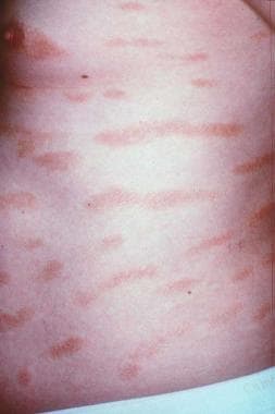
- The chronic form of pityriasis lichenoides initially manifests as flatter, reddish brown, scaling papules that may take months or longer to resolve. (msdmanuals.com)
Cutaneous1
- Pityriasis lichenoides is a clonal T-cell disorder that may develop in response to foreign antigens (eg, infections or substances) and may be associated with cutaneous T-cell lymphoma. (msdmanuals.com)
Diagnosis1
- Diagnosis of pityriasis lichenoides is based on clinical appearance and distribution. (msdmanuals.com)
Tetracycline1
- Since there has been somewhat of a consensus that there are no good options for hidradenitis suppurativa and pityriasis lichenoides and since tetracycline derivatives are quite safe (although no longer inexpensive), they are often used as first-line treatment. (dermatologytimes.com)
Rash1
- Pityriasis lichenoides is the name given to an uncommon rash of unknown cause. (childrens.com)
Rare1
- One rare cause is a disease called pityriasis lichenoides. (kamagra-fast.com)
Skin1
- The skin-limited form of pityriasis lichenoides is a self-limited disease. (medscape.com)
Diseases1
- These diseases generally are benign and undergo spontaneous resolution but, at times, may have a protracted course (see Pityriasis Lichenoides for further discussion). (medscape.com)
Affects1
- Pityriasis lichenoides most often affects adolescents and young adults. (childrens.com)
Infection1
- Pityriasis lichenoides is probably a hypersensitivity reaction to a mild infection, but no specific bacteria or virus has yet been identified. (childrens.com)
Form1
- Pityriasis lichenoides is a form of pityriasis. (wikipedia.org)
Immune1
- Hyperbaric oxygen therapy may potentially accelerate healing and reduce inflammation in pityriasis lichenoides by enhancing tissue oxygenation and promoting immune system function. (hbot-india.com)
Cases1
- Several studies have shown a significant portion of pityriasis lichenoides cases have a T-cell clone. (medscape.com)
Treatment1
- Pityriasis lichenoides may not always respond to treatment and it may return when treatment is stopped. (childrens.com)
Patches1
- Patches caused by exczema steroid cream or Pityriasis? (talkhealthpartnership.com)
Varioliformis9
- Pityriasis lichenoides et varioliformis acuta: a disease spectrum. (medscape.com)
- Brem CE, Abbas O, Bhawan J. Diagnostic Value of Plasmacytoid Dendritic Cells in Differentiating Pityriasis Lichenoides et Varioliformis Acuta From Lymphomatoid Papulosis. (medscape.com)
- When Do Symptoms of Pityriasis lichenoides et varioliformis acuta Begin? (nih.gov)
- Ueber die akut verlaufende, nekrotisierende Unterart der Pityriasis lichenoides (Pityriasis lichenoides et varioliformis acuta. (nih.gov)
- 2. Pityriasis lichenoides et varioliformis acuta with numerous CD30(+) cells: a variant mimicking lymphomatoid papulosis and other cutaneous lymphomas. (nih.gov)
- 7. Expression of cutaneous lymphocyte-associated antigen and TIA-1 by lymphocytes in pityriasis lichenoides et varioliformis acuta and lymphomatoid papulosis: immunohistochemical study. (nih.gov)
- 8. Immunohistochemical distinction of lymphomatoid papulosis and pityriasis lichenoides et varioliformis acuta. (nih.gov)
- 12. T-Cell clonality in pityriasis lichenoides et varioliformis acuta: a heteroduplex analysis of 20 cases. (nih.gov)
- Nosologic relationship to Pityriasis lichenoides et varioliformis acuta (PLEVA), to other CD30 positive malignant T-cell lymphomas and to Hodgkin's disease. (cyberderm.net)
Lichen planus1
- UVB is used to treat common skin conditions such as psoriasis, atopic eczema, other forms of dermatitis, polymorphic light eruption, generalised itching, pityriasis lichenoides, cutaneous T cell lymphoma, lichen planus, vitiligo and other less common conditions. (advancedskinclinicuganda.com)
Clonal3
- Magro C, Crowson AN, Kovatich A, Burns F. Pityriasis lichenoides: a clonal T-cell lymphoproliferative disorder. (medscape.com)
- 5. Pityriasis lichenoides: a clonal T-cell lymphoproliferative disorder. (nih.gov)
- 10. The clonal nature of pityriasis lichenoides. (nih.gov)
Acute1
- Pityriasis lichenoides is an acute or chronic lesion of the skin of unknown etiology, which is associated with a violation of the functioning of certain clones of T-lymphocytes. (medic-journal.com)
Phototherapy1
- Aydogan K, Saricaoglu H, Turan H. Narrowband UVB (311 nm, TL01) phototherapy for pityriasis lichenoides. (medscape.com)
Diagnosis2
- Clinical, Dermatoscopic, and Histological Findings in a Diagnosis of Pityriasis Lichenoides. (medscape.com)
- Diagnosis of pityriasis lichenoides is based on clinical appearance and distribution. (msdmanuals.com)
Clinical1
- Pityriasis lichenoides: a clinical and pathological case series of 49 patients with an emphasis on follow-up. (amazonaws.com)
Gibert2
Chronic form1
- The chronic form of pityriasis lichenoides initially manifests as flatter, reddish brown, scaling papules that may take months or longer to resolve. (msdmanuals.com)
Treatments1
- Jung F, Sibbald C, Bohdanowicz M, Ingram JR, Piguet V. Systematic review of the efficacies and adverse effects of treatments for pityriasis lichenoides. (medscape.com)
Disorder1
- 1. Pityriasis lichenoides: a cytotoxic T-cell-mediated skin disorder. (nih.gov)