Pityriasis Rubra Pilaris
Pityriasis Rosea
Pityriasis
Keratoderma, Palmoplantar
Carcinoma, Basal Cell
Dermatology
Cross-Sectional Studies
Tattooing
Photosensitivity Disorders
Meningitis, Aseptic
The relationship between pityriasis rubra pilaris and inflammatory arthritis: case report and response of the arthritis to anti-tumor necrosis factor immunotherapy. (1/12)
Pityriasis rubra pilaris (PRP) refers to a group of erythematous, scaling dermatologic conditions that have been associated with seronegative arthritis. We report a case of polyarthritis in a young man with PRP in which magnetic resonance imaging suggested an entheseal-based pathology for the joint disease. The arthritis, but not the skin condition, demonstrated dramatic response to anti-tumor necrosis factor immunotherapy. (+info)Pityriasis rubra pilaris. (2/12)
Pityriasis rubra pilaris is a chronic, papulosquamous dermatosis of unclear etiology. The case of a 61-year-old man with pityriasis rubra pilaris is presented. The clinical forms, histopathologic features, and treatment options of pityriasis rubra pilaris are reviewed. (+info)A review of pityriasis rubra pilaris and rheumatologic associations. (3/12)
Pityriasis rubra pilaris (PRP) is a rare group of hyperkeratotic, papulosquamous disease that can be acquired or inherited. There have been reported cases of rheumatologic associations, mainly arthritis and dermatomyositis. In this review article, we will explore the clinical presentation and classification, rheumatologic associations and treatment modalities of PRP. In addition, we will also report a case of PRP with seronegative arthritis. (+info)Pityriasis rubra pilaris, type 1. (4/12)
A 57-year-old woman presented with a history of dry skin with an associated sensation of burning and itching. It had been previously diagnosed as psoriasis. Clinical and histopathologic examination were consistent with pityriasis rubra pilaris, and treatment consisted of acitretin and narrow-band ultraviolet B phototherapy. Pityriasis rubra pilaris is a papulosquamous disorder of unknown etiology, which can be treated with retinoids, methotrexate, cyclosporine, and narrow-band phototherapy. (+info)Pityriasis rubra pilaris, type IV. (5/12)
A 4-year-old girl presented with a 3-year history of demarcated, salmon-pink, hyperkeratotic plaques, which were symmetrically distributed on the elbows, knees, ankles, and dorsal aspects of the hands and feet. A diffuse, orange-pink palmoplantar keratoderma was also evident. Clinical and histologic findings were consistent with a diagnosis of pityriasis rubra pilaris (PRP), type IV (circumscribed juvenile). Type IV PRP develops in prepubertal children, is typically localized to the distal aspects of the extremities, and has an unpredictable course. Although ultraviolet (UV) radiation can potentially exacerbate PRP, our patient has improved with broad-band UVB phototherapy. (+info)Adult onset pityriasis rubra pilaris. (6/12)
Pityriasis rubra pilaris (PRP) has always been an intriguing topic ever since its inception. It is a group of chronic disorders characterized by reddish orange plaques with pityriasiform scaling showing follicular keratoses, palmoplantar keratoderma, and sometimes, erythroderma. It occurs all over the world but with racial variations. Its incidence might vary and the age at onset, behavior, clinical appearance, and prognosis are considered to be very important for its classification. It may manifest either as Type I classical adult onset PRP, Type II atypical adult (onset) PRP, or Type VI PRP (HIV-associated PRP pityriasis rubra pilaris) in contrast to classical juvenile (Type III) and circumscribed juvenile (Type IV) encountered among children. Its diagnosis is largely clinical with microscopic pathology being a useful supplement, but it continues to be a therapeutic dilemma. We review the epidemiology of adult onset PRP here and take stock of the prevalent treatment options. (+info)Systemic sclerosis in a patient with pityriasis rubra pilaris. (7/12)
Pityriasis rubra pilaris (PRP) is a rare, chronic erythematous squamous disorder of unknown etiology. It has been found in association with several autoimmune diseases, including thyroiditis, myositis, myasthenia gravis and vitiligo. Herein we report a case of systemic sclerosis in a patient with classic adult pityriasis rubra pilaris. A 38 year old woman with classic adult type 1 pityriasis rubra pilaris (PRP) developed progressive skin thickening of the trunk, face, upper and lower extremities after 2 years of PRP treatment with topical emollients and steroids. Clinical examination and immunological findings were consistent with SSc. Co-existence of these two rare conditions is documented for the first time. (+info)Letter: Adenocarcinoma of the lung associated with pityriasis rubra pilaris. (8/12)
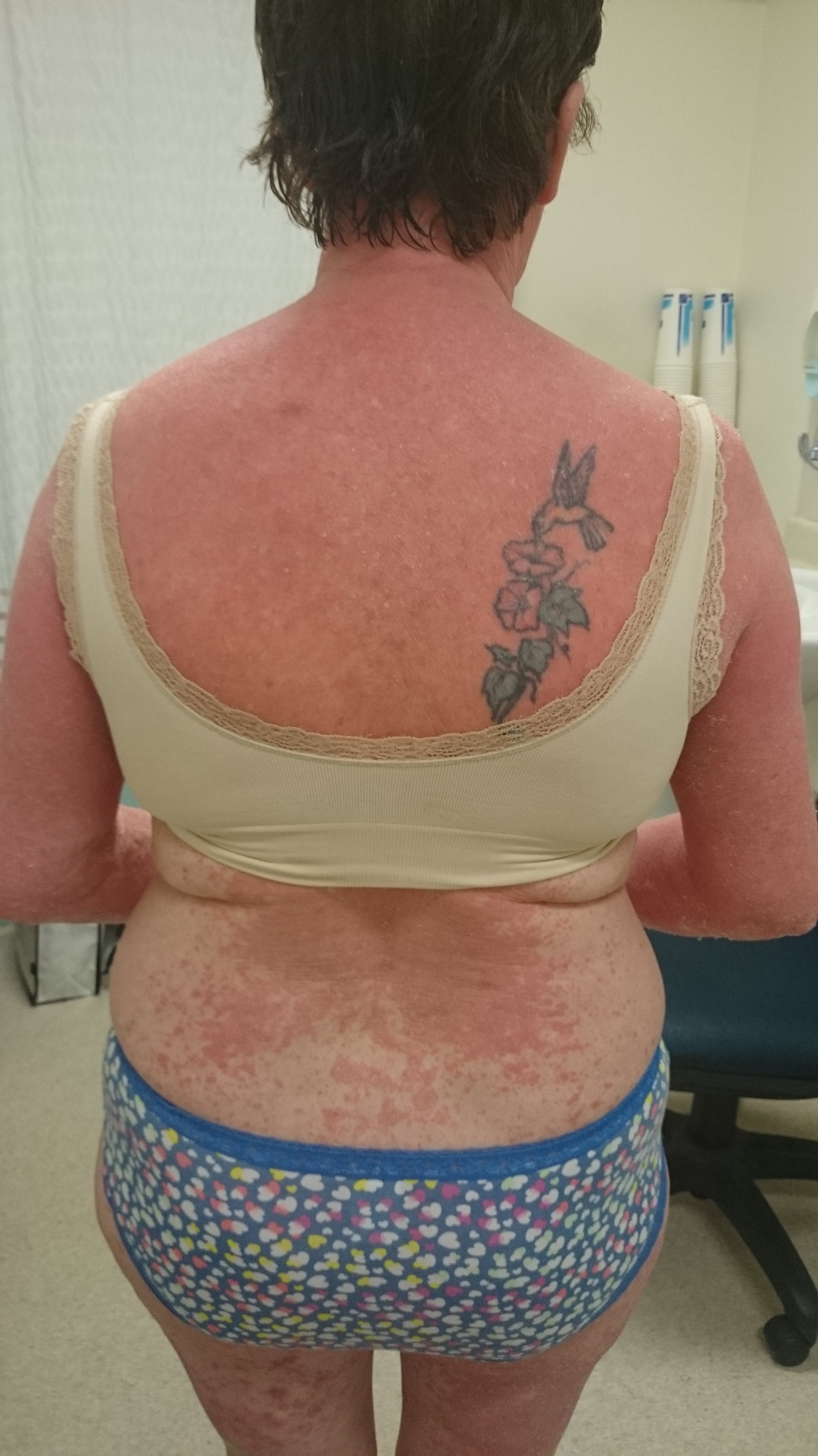
We describe a case of pityriasis rubra pilaris refractory to conventional treatment, found to be associated with an unrecognized primary adenocarcinoma of the lung. Complete resolution of the eruption followed surgical resection of the tumor. (+info)Pityriasis rubra pilaris (PRP) is a rare, chronic inflammatory skin disorder characterized by the development of reddish orange scaly patches and thickened plaques on the skin. It primarily affects the scalp, face, knees, elbows, and palms and soles. The condition can also cause reddening and thickening of the skin over the entire body in severe cases.
The key features of PRP include follicular papules (small bumps around hair follicles) that may be surrounded by a white collarette of scale, and areas of skin that are redder than normal (erythema). The condition can also cause nail changes, such as thickening and ridging.
PRP is thought to be caused by an abnormal immune response, although the exact cause is not known. It can affect people of all ages, but it is most commonly seen in children and adults between 40-60 years old. The condition typically progresses through several stages, with periods of worsening symptoms followed by periods of improvement or remission.
Treatment for PRP may include topical therapies, such as corticosteroids or retinoids, as well as systemic treatments, such as immunosuppressive drugs or biologics. The choice of treatment depends on the severity and extent of the condition, as well as other factors.
Pityriasis rosea is a common, self-limited skin condition characterized by the development of oval or round, scaly, pinkish, inflamed patches on the skin. The initial lesion, known as the "herald patch," often appears before other lesions and measures 2-10 cm in diameter. It usually starts as a single, solitary, scaly, raised patch on the trunk that precedes the generalized eruption by about 1-2 weeks. The rash typically spreads to involve the chest, abdomen, back, arms, and legs, sparing the face, palms, and soles.
The rash is often asymptomatic but can be pruritic (itchy) in some cases. It usually resolves within 6-12 weeks without any treatment, although topical treatments such as corticosteroids or antihistamines may be used to relieve itching. The exact cause of pityriasis rosea is not known, but it is thought to be caused by a viral infection. It is more common in young adults and is more prevalent in the spring and fall seasons.
Pityriasis is a general term used to describe a group of skin conditions characterized by scaling. It includes several specific types, the most common being Pityriasis rosea and Pityriasis simplex capillitii (also known as dandruff).
1. Pityriasis rosea: This is a temporary skin rash that often begins with a single, round, scaly patch on the chest, abdomen, or back. A few days to weeks later, more patches appear. These patches are oval and scaly, and they may be pink, red, or tan. The rash usually lasts about 6-8 weeks.
2. Pityriasis simplex capillitii: This is a very common condition characterized by flaking or scaling of the scalp, which is often referred to as dandruff.
The term "pityriasis" comes from the Greek word "pitýrios," which means "bran."
Keratoderma, palmoplantar is a medical term that refers to a group of skin conditions characterized by thickening and hardening (hyperkeratosis) of the skin on the palms of the hands and soles of the feet. This condition can affect people of all ages, but it's most commonly seen in children.
The thickening of the skin is caused by an overproduction of keratin, a protein that helps to form the tough, outer layer of the skin. In palmoplantar keratoderma, this excess keratin accumulates in the stratum corneum, the outermost layer of the epidermis, leading to the formation of rough, scaly, and thickened patches on the palms and soles.
There are several different types of palmoplantar keratoderma, each with its own specific symptoms and causes. Some forms of the condition are inherited and present at birth or develop in early childhood, while others may be acquired later in life as a result of an underlying medical condition, such as atopic dermatitis, lichen planus, or psoriasis.
Treatment for palmoplantar keratoderma typically involves the use of emollients and keratolytic agents to help soften and remove the thickened skin. In some cases, oral retinoids or other systemic medications may be necessary to manage more severe symptoms. It's important to consult with a healthcare provider for an accurate diagnosis and treatment plan.
Carcinoma, basal cell is a type of skin cancer that arises from the basal cells, which are located in the lower part of the epidermis (the outermost layer of the skin). It is also known as basal cell carcinoma (BCC) and is the most common form of skin cancer.
BCC typically appears as a small, shiny, pearly bump or nodule on the skin, often in sun-exposed areas such as the face, ears, neck, hands, and arms. It may also appear as a scar-like area that is white, yellow, or waxy. BCCs are usually slow growing and rarely spread (metastasize) to other parts of the body. However, they can be locally invasive and destroy surrounding tissue if left untreated.
The exact cause of BCC is not known, but it is thought to be related to a combination of genetic and environmental factors, including exposure to ultraviolet (UV) radiation from the sun or tanning beds. People with fair skin, light hair, and blue or green eyes are at increased risk of developing BCC.
Treatment for BCC typically involves surgical removal of the tumor, along with a margin of healthy tissue. Other treatment options may include radiation therapy, topical chemotherapy, or photodynamic therapy. Prevention measures include protecting your skin from UV radiation by wearing protective clothing, using sunscreen, and avoiding tanning beds.
Dermatology is a medical specialty that focuses on the diagnosis, treatment, and prevention of diseases and conditions related to the skin, hair, nails, and mucous membranes. A dermatologist is a medical doctor who has completed specialized training in this field. They are qualified to treat a wide range of skin conditions, including acne, eczema, psoriasis, skin cancer, and many others. Dermatologists may also perform cosmetic procedures to improve the appearance of the skin or to treat signs of aging.
A cross-sectional study is a type of observational research design that examines the relationship between variables at one point in time. It provides a snapshot or a "cross-section" of the population at a particular moment, allowing researchers to estimate the prevalence of a disease or condition and identify potential risk factors or associations.
In a cross-sectional study, data is collected from a sample of participants at a single time point, and the variables of interest are measured simultaneously. This design can be used to investigate the association between exposure and outcome, but it cannot establish causality because it does not follow changes over time.
Cross-sectional studies can be conducted using various data collection methods, such as surveys, interviews, or medical examinations. They are often used in epidemiology to estimate the prevalence of a disease or condition in a population and to identify potential risk factors that may contribute to its development. However, because cross-sectional studies only provide a snapshot of the population at one point in time, they cannot account for changes over time or determine whether exposure preceded the outcome.
Therefore, while cross-sectional studies can be useful for generating hypotheses and identifying potential associations between variables, further research using other study designs, such as cohort or case-control studies, is necessary to establish causality and confirm any findings.
Skin neoplasms refer to abnormal growths or tumors in the skin that can be benign (non-cancerous) or malignant (cancerous). They result from uncontrolled multiplication of skin cells, which can form various types of lesions. These growths may appear as lumps, bumps, sores, patches, or discolored areas on the skin.
Benign skin neoplasms include conditions such as moles, warts, and seborrheic keratoses, while malignant skin neoplasms are primarily classified into melanoma, squamous cell carcinoma, and basal cell carcinoma. These three types of cancerous skin growths are collectively known as non-melanoma skin cancers (NMSCs). Melanoma is the most aggressive and dangerous form of skin cancer, while NMSCs tend to be less invasive but more common.
It's essential to monitor any changes in existing skin lesions or the appearance of new growths and consult a healthcare professional for proper evaluation and treatment if needed.
Tattooing is defined medically as the process of inserting pigment into the skin's dermis layer to change its color. This procedure creates a permanent design or image. The equipment used for tattooing includes an electrically powered tattoo machine, needles, and ink. Tattooing can carry potential risks such as infection, allergic reactions, and scarring. It is essential to ensure that all tattooing procedures are performed under sterile conditions and by a licensed professional to minimize these risks.
Photosensitivity disorders refer to conditions that cause an abnormal reaction to sunlight or artificial light. This reaction can take the form of various skin changes, such as rashes, inflammation, or pigmentation, and in some cases, it can also lead to systemic symptoms like fatigue, fever, or joint pain.
The two main types of photosensitivity disorders are:
1. Phototoxic reactions: These occur when a substance (such as certain medications, chemicals, or plants) absorbs light energy and transfers it to skin cells, causing damage and inflammation. The reaction typically appears within 24 hours of exposure to the light source and can resemble a sunburn.
2. Photoallergic reactions: These occur when the immune system responds to the combination of light and a particular substance, leading to an allergic response. The reaction may not appear until several days after initial exposure and can cause redness, itching, and blistering.
It is important for individuals with photosensitivity disorders to avoid excessive sun exposure, wear protective clothing, and use broad-spectrum sunscreens with a high SPF rating to minimize the risk of phototoxic or photoallergic reactions.
Aseptic meningitis is a type of meningitis (inflammation of the membranes covering the brain and spinal cord) that is not caused by bacterial infection. Instead, it can be due to viral infections, fungal infections, or non-infectious causes such as certain medications, chemical irritants, or underlying medical conditions. In aseptic meningitis, the cerebrospinal fluid (CSF) analysis may show increased white blood cells, typically lymphocytes, but no bacterial growth on culture. Common viral causes include enteroviruses, herpes simplex virus, and varicella-zoster virus. Treatment depends on the underlying cause and may include supportive care, antiviral medications, or immunosuppressive therapy in some cases.
 Pityriasis rubra pilaris
Marie-Guillaume-Alphonse Devergie
Granular parakeratosis
Erythroderma
Annella Zervas
Harold Wordsworth Barber
Celalettin Muhtar Ozden
Onychauxis
Rubra
PRP
List of ICD-9 codes 680-709: diseases of the skin and subcutaneous tissue
List of diseases (P)
List of MeSH codes (C17)
Koebner phenomenon
Pityriasis
List of skin conditions
List of birds of Asia
Pityriasis rubra pilaris - Wikipedia
 Pityriasis rubra pilaris: MedlinePlus Medical Encyclopedia
 Pityriasis Rubra Pilaris: Background, Pathophysiology, Epidemiology
 Pityriasis rubra pilaris | DermNet
Pityriasis Rubra Pilaris: Background, Pathophysiology, Epidemiology
 Laddered treatment for pityriasis rubra pilaris
Pityriasis Rubra Pilaris Differential Diagnoses
Pityriasis Rubra Pilaris: Background, Pathophysiology, Epidemiology
 ACD A-Z of Skin - Pityriasis Rubra Pilaris
 Pityriasis Rubra Pilaris - Dermatologic Disorders - MSD Manual Professional Edition
 Red Rash - Gluten Free Works: TREATMENT GUIDE
 Ingrown Hair Removal Treatment, Home Remedies, Types, Pictures
 Treatment of Pityriasis Rubra Pilaris with Daily Low-Dose Methotrexate: A Retrospective Cohort Study | SKIN The Journal of...
 Biomedicines | Free Full-Text | Efficacy of Low-Dose Naltrexone and Predictors of Treatment Success or Discontinuation in...
 Erythroderma
 Resources
 Table of Contents - June 20, 1983, 50 (2) | Cleveland Clinic Journal of Medicine
Overview of Skin Cancer - Skin Disorders - MSD Manual Consumer Version
 Salicylic Acid Monograph for Professionals - Drugs.com
 Atypical Presentation of Neurosyphilis - Indian Journal of Dermatology, Venereology and Leprology
 Gyd. Gintarė Ulianskaitė | SUGIHARA
 Psoriatic Arthritis Rash: Symptoms, Treatment, and Pictures
 Carlos Antonio Torres-Cabala | MD Anderson Cancer Center
Woo Cheal Cho | MD Anderson Cancer Center
 Indian Journal of Dermatology: Table of Contents
 38 CFR § 4.118 - Schedule of ratings-skin. | Electronic Code of Federal Regulations (e-CFR) | US Law | LII / Legal Information...
 Frontiers | UVB-Induced Skin Autoinflammation Due to Nlrp1b Mutation and Its Inhibition by Anti-IL-1β Antibody
 RACGP - Facial rash - a case study
Rosea4
- Pityriasis rosea, pityriasis rubra pilaris, and other papulosquamous and hyperkeratotic diseases. (medlineplus.gov)
- Other Medscape pityriasis articles include Dermatologic Manifestations of Pityriasis Alba , Pityriasis Lichenoides , Pityriasis Rosea , and Pityriasis Rotunda . (medscape.com)
- Pityriasis rosea usually begins with the appearance of a solitary, red, round to oval-shaped, itchy "mother patch" on the trunk. (myacare.com)
- To learn more about Pityriasis Rosea , please read our blog. (myacare.com)
Keratosis2
- Also used topically for management of other hyperkeratotic skin disorders such as ichthyoses vulgaris, palmoplantar keratosis, pityriasis rubra pilaris, and keratosis pilaris. (drugs.com)
- Keratosis pilaris - Central keratin plugs, preferentially affects extensor aspects of arms and legs, and often has seasonal variation. (logicalimages.com)
Atopic2
- In a double-blind, placebo-controlled trial, Patrizi and colleagues concluded that sorbityl furfural palmitate (AR-GG27®) cream was effective against mild to moderate atopic dermatitis in patients with pityriasis alba. (medscape.com)
- Extensive pityriasis alba in a child with atopic dermatitis. (medscape.com)
Diagnoses1
- He had been treated with oral retinoids, steroids, and phototherapy for the diagnoses of drug eruption, pityriasis rubra pilaris, and exfoliative dermatitis at other hospitals. (medicaljournals.se)
Scaly2
- Pityriasis rubra pilaris - Central keratin plugs, papules spread cephalocaudally, scaly orange-red plaques (with "islands of sparing"), and palmoplantar keratoderma. (logicalimages.com)
- A chronic skin condition, Pityriasis rubra pilaris appears as orange-red scaly plaques that can affect any part of the body but are usually found on the palms and soles. (myacare.com)
Biologic3
- For some patients with pityriasis rubra pilaris (PRP) who do not respond to standard systemic treatments, biologic therapy may result in marked clinical improvement, according to Scott Worswick, M.D., a UCLA dermatologist speaking at the American Academy of Dermatology annual meeting in San Diego on Feb. 18. (dermatologytimes.com)
- While there are no randomized clinical trials of biologic treatments for pityriasis rubra pilaris, case reports and clinical experience suggest an eight-week course may be sufficient to see if a patient may respond, he said. (dermatologytimes.com)
- Treatment options for pityriasis rubra pilaris including biologic agents: A retrospective analysis from an academic medical center. (msdmanuals.com)
Disorders2
- Pityriasis rubra pilaris refers to a group of chronic disorders characterized by reddish orange, scaling plaques and keratotic follicular papules. (wikipedia.org)
- Pityriasis rubra pilaris (PRP) is the name given to a group of rare skin disorders that present with reddish-orange coloured scaling patches with well-defined borders. (dermnetnz.org)
Spontaneously1
- Pityriasis rubra pilaris type 1 spontaneously resolving after 20 years. (medscape.com)
Palmoplantar keratoderma1
- Patients with pityriasis rubra pilaris can have painful and disabling palmoplantar keratoderma. (medscape.com)
Erythroderma1
- However, most of the morbidity associated with pityriasis rubra pilaris is associated with the erythroderma (see Complications ). (medscape.com)
Chronic1
- Pityriasis rubra pilaris is a rare chronic disorder that causes hyperkeratotic yellowing of the skin, including the trunk, extremities, and, particularly, the palms and soles. (msdmanuals.com)
Systemic1
- Systemic therapies of pityriasis rubra pilaris: a systematic review. (jofskin.org)
Alba8
- Lesions of pityriasis alba do not repigment well upon sun exposure, and darkening of the surrounding skin may worsen the cosmetic appearance. (medscape.com)
- Because pityriasis alba is usually self-limited and asymptomatic, pharmacologic treatment is often unnecessary. (medscape.com)
- In another study, an open-label, noncontrolled, nonrandomized trial by Bhat et al involving patients with eczema associated with pityriasis alba, as well as persons with various forms of irritant dermatitis, RV 2427B cream was found to be effective in 84% of patients as evaluated by an investigator, and in 76% of patients as evaluated through self-assessments. (medscape.com)
- Treatment with a 308-nm excimer laser twice a week for 12 weeks has been shown to be effective against pityriasis alba. (medscape.com)
- Extensive pityriasis alba may warrant a referral to a dermatologist for possible oral psoralen plus ultraviolet-A (PUVA) photochemotherapy. (medscape.com)
- Miazek N, Michalek I, Pawlowska-Kisiel M, Olszewska M, Rudnicka L. Pityriasis Alba--Common Disease, Enigmatic Entity: Up-to-Date Review of the Literature. (medscape.com)
- Vinod S, Singh G, Dash K, Grover S. Clinico epidemiological study of pityriasis alba. (medscape.com)
- Di Lernia V, Ricci C. Progressive and extensive hypomelanosis and extensive pityriasis alba: same disease, different names? (medscape.com)
Clinical5
- Pityriasis rubra pilaris in the setting of HIV infection: clinical behaviour and association with explosive cystic acne. (medscape.com)
- Mohrenschlager M, Abeck D. Further clinical evidence for involvement of bacterial superantigens in juvenile pityriasis rubra pilaris (PRP): report of two new cases. (medscape.com)
- The clinical and histomorphological features of pityriasis rubra pilaris. (medscape.com)
- Diagnosis of pityriasis rubra pilaris is by clinical appearance and may be supported by biopsy. (msdmanuals.com)
- Initial management consisted of en una paciente con topical corticosteroids and oral antihistamines with little clinical response. (bvsalud.org)
Hyperkeratotic1
- Pityriasis rubra pilaris can manifest as hyperkeratotic yellowing of the palms and soles. (msdmanuals.com)
Familial2
- The familial form of pityriasis rubra pilaris typically begins in early childhood and has an autosomal dominant inheritance pattern. (medscape.com)
- Familial Pityriasis Rubra Pilaris Is Caused by Mutations in CARD14. (medscape.com)
Retrospective2
- Pityriasis rubra pilaris-a retrospective single center analysis over eight years. (jofskin.org)
- Treatment of pityriasis rubra pilaris: a retrospective study of 14 patients. (jofskin.org)
Diagnosis and treatment2
- Pityriasis rubra pilaris: a review of diagnosis and treatment. (medscape.com)
- 5. Roenneberg S, Biedermann T. Pityriasis rubra pilaris: algorithms for diagnosis and treatment. (jofskin.org)
Occur2
- Why does pityriasis rubra pilaris occur and who is at risk? (dermnetnz.org)
- What other problems can occur with pityriasis rubra pilaris? (edu.au)
Lesions1
- Pityriasis rubra pilaris can manifest as red follicular papules that coalesce to form red-orange scaling plaques and confluent areas of erythema with islands of normal skin between lesions. (msdmanuals.com)
Incidence2
- The incidence of pityriasis rubra pilaris has been reported to be 1 case in 3500-5000 patients presenting to dermatologic clinics. (medscape.com)
- Avitan-Hersh E, Bergman R. The Incidence of Acantholysis in Pityriasis Rubra Pilaris-Histopathological Study Using Multiple-Step Sections and Clinicopathologic Correlations. (medscape.com)
Juvenile3
- Griffiths divided pityriasis rubra pilaris into 5 categories: classic adult type, atypical adult type, classic juvenile type, circumscribed juvenile type, and atypical juvenile type. (medscape.com)
- Karimian-Teherani D, Parissa M, Tanew A. Response of juvenile circumscribed pityriasis rubra pilaris to topical tazarotene treatment. (medscape.com)
- Juvenile pityriasis rubra pilaris: successful treatment with methotrexate. (jofskin.org)
Methotrexate2
- Pityriasis rubra pilaris: prolonged treatment with methotrexate. (jofskin.org)
- Methotrexate treatment for pityriasis rubra pilaris: a case series and literature review. (jofskin.org)
Treatment6
- Van de Kerkhof PC, Steijlen PM. Topical treatment of pityriasis rubra pilaris with calcipotriol. (medscape.com)
- Treatment of pityriasis rubra pilaris is exceedingly difficult and empiric. (msdmanuals.com)
- Biologics for pityriasis rubra pilaris treatment: A review of the literature. (msdmanuals.com)
- Treatment of pityriasis rubra pilaris type I: A systematic review. (msdmanuals.com)
- Pityriasis rubra pilaris: treatment with folic acid antagonists. (jofskin.org)
- Case report: Successful treatment of Pityriasis Rubra Pilaris with Isotretinoin. (sugihara.lt)
Classic adult1
- Scarring Alopecia in Classic Adult Type I Pityriasis Rubra Pilaris. (medscape.com)
Disorder1
- Pityriasis rubra pilaris (PRP) is a rare skin disorder that causes inflammation and scaling (exfoliation) of the skin. (medlineplus.gov)
Mutations2
- Type V pityriasis rubra pilaris has been linked to mutations in the gene, CARD . (medscape.com)
- Pityriasis Rubra Pilaris Type V as an Autoinflammatory Disease by CARD14 Mutations. (medscape.com)
Treatments2
- What treatments are available for pityriasis rubra pilaris? (dermnetnz.org)
- Scott Worswick, MD. "Hidradenitis Suppurativa and Pityriasis Rubra Pilaris: Updates on Treatments for Two Conditions that are Difficult to Manage. (dermatologytimes.com)
Infection3
- Auffret N, Quint L, Domart P, Dubertret L, Lecam JY, Binet O. Pityriasis rubra pilaris in a patient with human immunodeficiency virus infection. (medscape.com)
- Pityriasis rubra pilaris and HIV infection. (medscape.com)
- Miralles ES, Nunez M, De Las Heras ME, Perez B, Moreno R, Ledo A. Pityriasis rubra pilaris and human immunodeficiency virus infection. (medscape.com)
Occurs1
- Pityriasis rubra pilaris occurs equally among men and women. (medscape.com)
Oral1
- Mohamed M, Belhadjali H, Hammedi F, Meriem CB, Zili J. Pityriasis rubra pilaris occurring after vaccination with diphtheria-pertussis-tetanus and oral poliovirus vaccines. (medscape.com)
Trigger1
- [ 12 ] One hypothesis is that pityriasis rubra pilaris may be related to an abnormal immune response to an antigenic trigger. (medscape.com)
Case2
- Case reports have described pityriasis rubra pilaris occurring after streptococcal infections. (medscape.com)
- Epidemiologic, clinicopathologic, diagnostic, and management challenges of pityriasis rubra pilaris: a case series of 100 patients. (jofskin.org)
Disease1
- Pityriasis rubra pilaris (PRP) is a rare disease that is difficult to manage. (jofskin.org)
Symptoms1
- 442 Symptoms may include reddish-orange patches (Latin: rubra) on the skin, severe flaking (Latin: pityriasis), uncomfortable itching, thickening of the skin on the feet and hands, and thickened bumps around hair follicles (Latin: pilus for hair). (wikipedia.org)
Type1
- Each type of pityriasis rubra pilaris has its own prognosis. (medscape.com)
Children1
- Allison DS, El-Azhary RA, Calobrisi SD, Dicken CH. Pityriasis rubra pilaris in children. (medscape.com)