Placenta Accreta
Gynatresia
Placenta Previa
Placenta
Placenta, Retained
Postpartum Hemorrhage
Uterine Artery Embolization
Obstetric Surgical Procedures
Pregnancy
Pregnancy Trimester, Third
Ultrasonography, Prenatal
Uterine Rupture
Placental Circulation
Pregnancy Trimester, First
Ultrasonography, Doppler, Color
Complicated third stage of labor: time to switch on the scanner. (1/60)
This Editorial chronicles the current experience in ultrasound usage during pathologic events occurring in the third stage of labor. Further improvement in the technology for carrying out clinical research will improve our knowledge so that more information can be gleaned from this modality to bestow optimal management for such potentially dangerous conditions. Awareness of the capabilities of sonography may provide the motivation for its use, and obstetricians are encouraged to scan the third stage of normal deliveries for better recognition of normal findings and improved assessment of abnormal ones. Although final decisions should be based mainly upon sound clinical judgement, we contend that complicated third stage of labor warrants turning on the scanner. (+info)Gray scale and color Doppler sonography in the third stage of labor for early detection of failed placental separation. (2/60)
OBJECTIVE: The purpose of this study was the characterization of normal and abnormal third stage placental separation using gray scale and color Doppler sonography. METHODS: The third stage of labor was examined in 62 patients using gray scale and color Doppler sonography. After identification of placental basal plate vessels by color Doppler sonography, the placentation site was examined throughout the third stage with combined gray scale and color Doppler mode. Placental separation from the myometrium was defined clinically and correlated to cessation of color Doppler detected blood flow in basal plate vessels. RESULTS: Three sonographic phases of placental separation were: (1) latent = interval between delivery of the fetus and beginning placental separation; (2) detachment = mono- or multiphasic shearing off of the placenta and (3) expulsion = interval between completed separation and vaginal delivery of the placenta. In 57 cases with clinically normal placental separation blood flow between placenta and myometrium ceased immediately after delivery of the fetus during the latent period. In five cases manual or instrumental removal was necessary because of placenta adhaerens in one case and placenta accreta in four cases. The latter showed maternal blood flow from the myometrium deep into the placenta beyond the latent phase. CONCLUSION: Cessation of blood flow between the basal placenta and myometrium following delivery of the baby is the sonographic hallmark of normal placental separation. Persistent blood flow demonstrated by color Doppler sonography is suggestive of placenta accreta. (+info)Prenatal diagnosis of placenta previa accreta by transabdominal color Doppler ultrasound. (3/60)
OBJECTIVE: The aim of this study was to evaluate the efficacy of transabdominal color Doppler ultrasound in diagnosing placenta previa accreta. DESIGN: Eighty patients with persistent placenta previa underwent transabdominal B-mode and color Doppler ultrasound evaluation in the second and third trimesters because they had a high risk of placenta accreta. Color Doppler imaging criteria used included diffuse intraparenchymal placental lacunar flow; focal intraparenchymal placental lacunar flow; bladder-uterine serosa interphase hypervascularity; prominent subplacental venous complex; and loss of subplacental Doppler vascular signals. The color Doppler images were interpreted prospectively for signs of placenta previa accreta according to the exhibited color Doppler sonographic features. RESULTS: Sixteen of the 80 patients exhibited characteristic color Doppler imaging patterns highly specific for placenta accreta according to the preceding criteria, and 14 of these had histopathological proof of placenta accreta. Two patients had false-positive color Doppler imaging evidence mistaken for interphase hypervascularity caused by bladder varices. Thirteen patients underwent hysterectomy in the group suspicious for accreta. Of the 64 patients with negative color Doppler imaging results, three had placenta accreta, while two required cesarean hysterectomy; the remaining patient underwent uterine artery ligation for bleeding from the lower uterine segment. The sensitivity of color Doppler imaging in the diagnosis of placenta previa accreta was 82.4% (14/17) and the specificity was 96.8% (61/63). The positive and negative predictive values were 87.5% (14/16) and 95.3% (61/64), respectively. CONCLUSIONS: Variable vascular morphological patterns of placenta previa accreta were exhibited and categorized by transabdominal color Doppler sonography in the antenatal period. The identification of these specific vascular patterns had a positive impact on the peripartum clinical management of the affected patients. (+info)Identification of mtDNA mutation in a pedigree with gestational diabetes, deafness, Wolff-Parkinson-White syndrome and placenta accreta. (4/60)
Mitochondrial DNA (mtDNA) defects are associated with a number of human disorders. Although many occur sporadically, maternal transmission is the hallmark of diseases due to mtDNA point mutations. The same mutation may manifest strikingly different phenotypes; for example, the A to G substitution at np 3243 was first reported in patients with mitochondrial encephalomyopathy, lactic acidosis and stroke-like episodes (the MELAS syndrome), but is also found in patients with diabetes and deafness. Here we present a case of gestational diabetes, deafness, premature greying, placenta accreta and Wolff-Parkinson-White (WPW) syndrome associated with a mtDNA mutation. Although this is the first report of such an association, study of 27 other patients with WPW syndrome failed to confirm that this mtDNA mutation is a common cause of such pre-excitation disorders. (+info)Myometrial fibers in the placental basal plate can confirm but do not necessarily indicate clinical placenta accreta. (5/60)
Placental basal plate myometrial fibers reflect mild placenta accreta. We tested the hypotheses that a macroscopically disrupted area relates to an area where the placenta is focally adherent and that the incidence of placenta accreta is higher than stated in the literature. Sagittal blocks were taken from the basal plate from macroscopically intact, disrupted, and mixed (viz, at the junction of intactness and disruption) areas, together with an en face block from 90 singleton placentas. Histologic examination revealed that 11 of 23 placentas with a macroscopically disrupted maternal surface and 16 of 67 with a macroscopically intact maternal surface had placental basal plate myometrial fibers, a significant difference. More cases were detected with sampling from the mixed than from the intact area, while sampling from the wholly disrupted area was unrewarding. The en face block also was helpful for detecting myometrial fibers. Extensive and selective sampling of the basal plate revealed a much higher incidence of placental basal plate myometrial fibers. Clinical chart review affirmed that their presence can confirm but does not necessarily correlate with a clinical diagnosis of mild placenta accreta. (+info)Placenta accreta diagnosed at 9 weeks' gestation. (6/60)
The majority of cases of placenta accreta are unanticipated and initially identified intraoperatively. Although color Doppler ultrasound is adequate for the evaluation of placenta accreta in the third trimester, ultrasound diagnosis in the first trimester has never been reported. To our knowledge, this is the first case of placenta accreta detected at 9 weeks' gestation by ultrasound. Placenta accreta with intraplacental lacunae can be identified together with a loss of the hypoechogenic retroplacental myometrial zone. Based on this case, we found that early diagnosis of placenta accreta in the first trimester by ultrasound is possible. (+info)Antenatal diagnosis of placenta percreta with planned in situ retention and methotrexate therapy in a woman infected with HIV. (7/60)
Placenta percreta is a rare obstetric condition associated with potentially life-threatening hemorrhage. Diagnosis in advance of delivery permits a planned delivery and preparation for blood transfusions and planned Cesarean hysterectomy, which is the common treatment. We report a case of placenta percreta in an HIV-positive patient which was diagnosed in the second trimester using conventional and extended field of view ultrasound imaging and color Doppler. At 36 weeks the infant was delivered by Cesarean section and the placenta was left in situ. Postoperatively the patient was treated with methotrexate. Four weeks later, the patient delivered the placenta spontaneously. Early or late postpartum hemorrhage did not occur and postoperative recovery was uneventful. (+info)The early sonographic appearance of placenta accreta. (8/60)
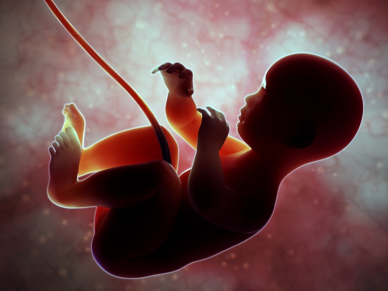
OBJECTIVE: To determine whether any sonographic findings in the first trimester predict placenta accreta. METHODS: Patients who had a diagnosis of placenta accreta, increta, or percreta by clinical course or pathologic examination of the uterus and who had had a sonographic examination at 10 weeks or earlier were included in this study. RESULTS: Seven patients met the study criteria. In 6 of these, who had had at least 1 previous cesarean delivery, the gestational sac was located in the lower uterine segment at the time of the early scan. Two of these pregnancies failed shortly after the early scan, and the patients underwent dilation and curettage, at which time severe bleeding necessitated a hysterectomy. The other 4 continued to term but had sonographic findings typical of placenta accreta during subsequent scans. In the seventh patient (who had had no previous cesarean deliveries), the gestational sac was located in the uterine fundus. CONCLUSIONS: In a patient with a previous cesarean delivery, a sac lying in the lower uterine segment on a scan at 10 weeks or earlier suggests the possibility of placenta accreta. (+info)Placenta accreta is a medical condition where the placenta grows too deeply into the uterine wall, beyond the normal depth. In a healthy pregnancy, the placenta attaches to the uterus and provides oxygen and nutrients to the growing fetus through the umbilical cord. However, in placenta accreta, the placental tissue invades the muscle of the uterus, which can cause complications during childbirth.
There are three types of placenta accreta:
1. Placenta Accreta: The placenta attaches too deeply into the uterine wall but does not penetrate the uterine muscle.
2. Placenta Increta: The placenta grows into and partially penetrates the uterine muscle.
3. Placenta Percreta: The placenta fully penetrates the uterine muscle and can grow into nearby organs, such as the bladder or bowel.
Placenta accreta is a serious condition that can cause severe bleeding during childbirth, which may require an emergency hysterectomy (removal of the uterus) to control the bleeding. It is more common in women who have had previous cesarean sections or other uterine surgeries.
Gynatresia is not a recognized medical term. However, it seems like you might be looking for the definition of "agATRESia," which is a term used in anatomy and medicine. Atresia refers to the congenital absence or closure of a natural body opening, canal, or lumen. So, when we add "a" before atresia, it becomes a specific type of atresia that occurs in female genitalia.
agenital atresia (also known as agATRESia): A congenital abnormality where there is an absence or closure of the vaginal opening, which may also include the absence of the uterus and/or cervix. This condition can vary in severity and may require surgical intervention to correct.
Placenta previa is a medical condition that occurs during pregnancy where the placenta partially or fully covers the cervix, which is the lower part of the uterus that opens into the birth canal. This condition can cause severe bleeding during pregnancy and delivery, and it may lead to other complications such as preterm labor and delivery. Placenta previa is typically diagnosed through an ultrasound exam and managed with close monitoring, bed rest, and sometimes cesarean delivery.
The placenta is an organ that develops in the uterus during pregnancy and provides oxygen and nutrients to the growing baby through the umbilical cord. It also removes waste products from the baby's blood. The placenta attaches to the wall of the uterus, and the baby's side of the placenta contains many tiny blood vessels that connect to the baby's circulatory system. This allows for the exchange of oxygen, nutrients, and waste between the mother's and baby's blood. After the baby is born, the placenta is usually expelled from the uterus in a process called afterbirth.
Retained placenta is a medical condition that occurs when all or part of the placenta remains in the uterus after delivery, instead of being expelled naturally. Normally, the placenta separates from the uterine wall and is delivered within 30 minutes of childbirth. However, if the placenta is not completely delivered, it can lead to complications such as infection, heavy bleeding, and in rare cases, infertility or even death.
Retained placenta can be caused by various factors, including a weakened uterine muscle tone, an abnormally attached placenta, or a retained portion of the membranes. Treatment for retained placenta typically involves manual removal of the remaining tissue by a healthcare professional, often under anesthesia. In some cases, medication may be used to help promote contraction of the uterus and expulsion of the placenta.
It is important to seek medical attention promptly if a retained placenta is suspected, as timely treatment can help prevent potentially serious complications.
A uterine myomectomy is a surgical procedure that involves removing uterine fibroids, which are noncancerous growths that develop in the muscular wall of the uterus. The surgery aims to preserve the uterus, unlike a hysterectomy, where the entire uterus is removed.
During the myomectomy procedure, the surgeon makes an incision in the uterus, carefully extracts the fibroids, and then closes the incision. The approach to the surgery can vary depending on factors such as the size, number, and location of the fibroids:
1. Abdominal myomectomy: An open surgical procedure where an incision is made in the lower abdomen to access the uterus directly. This method is typically used when there are numerous or large fibroids.
2. Laparoscopic myomectomy: A minimally invasive procedure that involves making several small incisions in the abdomen, through which a laparoscope (a thin, lighted tube with a camera) and specialized surgical instruments are inserted to perform the surgery. This method is often preferred for smaller fibroids and when preserving the uterus is essential.
3. Hysteroscopic myomectomy: Another minimally invasive procedure that involves inserting a hysteroscope through the cervix into the uterine cavity to remove submucosal fibroids (fibroids that grow into the inner cavity of the uterus). No abdominal incisions are required for this approach.
Recovery time and postoperative discomfort will vary depending on the type of myomectomy performed, but generally, minimally invasive procedures result in quicker recoveries and fewer complications compared to open surgeries.
Postpartum hemorrhage (PPH) is a significant obstetrical complication defined as the loss of more than 500 milliliters of blood within the first 24 hours after childbirth, whether it occurs vaginally or through cesarean section. It can also be defined as a blood loss of more than 1000 mL in relation to the amount of blood lost during the procedure and the patient's baseline hematocrit level.
Postpartum hemorrhage is classified into two types: primary (early) PPH, which occurs within the first 24 hours after delivery, and secondary (late) PPH, which happens between 24 hours and 12 weeks postpartum. The most common causes of PPH are uterine atony, trauma to the genital tract, retained placental tissue, and coagulopathy.
Uterine atony is the inability of the uterus to contract effectively after delivery, leading to excessive bleeding. Trauma to the genital tract can occur during childbirth, causing lacerations or tears that may result in bleeding. Retained placental tissue refers to the remnants of the placenta left inside the uterus, which can cause infection and heavy bleeding. Coagulopathy is a condition where the blood has difficulty clotting, leading to uncontrolled bleeding.
Symptoms of PPH include excessive vaginal bleeding, low blood pressure, increased heart rate, decreased urine output, and signs of shock such as confusion, rapid breathing, and pale skin. Treatment for PPH includes uterotonics, manual removal of retained placental tissue, repair of genital tract lacerations, blood transfusions, and surgery if necessary.
Preventing PPH involves proper antenatal care, monitoring high-risk pregnancies, active management of the third stage of labor, and prompt recognition and treatment of any bleeding complications during or after delivery.
A Cesarean section, often referred to as a C-section, is a surgical procedure used to deliver a baby. It involves making an incision through the mother's abdomen and uterus to remove the baby. This procedure may be necessary when a vaginal delivery would put the mother or the baby at risk.
There are several reasons why a C-section might be recommended, including:
* The baby is in a breech position (feet first) or a transverse position (sideways) and cannot be turned to a normal head-down position.
* The baby is too large to safely pass through the mother's birth canal.
* The mother has a medical condition, such as heart disease or high blood pressure, that could make vaginal delivery risky.
* The mother has an infection, such as HIV or herpes, that could be passed to the baby during a vaginal delivery.
* The labor is not progressing and there are concerns about the health of the mother or the baby.
C-sections are generally safe for both the mother and the baby, but like any surgery, they do carry some risks. These can include infection, bleeding, blood clots, and injury to nearby organs. In addition, women who have a C-section are more likely to experience complications in future pregnancies, such as placenta previa or uterine rupture.
If you have questions about whether a C-section is necessary for your delivery, it's important to discuss your options with your healthcare provider.
A hysterectomy is a surgical procedure that involves the removal of the uterus (womb). Depending on the specific medical condition and necessity, a hysterectomy may also include the removal of the ovaries, fallopian tubes, and surrounding tissues. There are different types of hysterectomies, including:
1. Total hysterectomy: The uterus and cervix are removed.
2. Supracervical (or subtotal) hysterectomy: Only the upper part of the uterus is removed, leaving the cervix intact.
3. Radical hysterectomy: This procedure involves removing the uterus, cervix, surrounding tissues, and the upper part of the vagina. It is typically performed in cases of cervical cancer.
4. Oophorectomy: The removal of one or both ovaries can be performed along with a hysterectomy depending on the patient's medical condition and age.
5. Salpingectomy: The removal of one or both fallopian tubes can also be performed along with a hysterectomy if needed.
The reasons for performing a hysterectomy may include but are not limited to: uterine fibroids, heavy menstrual bleeding, endometriosis, adenomyosis, pelvic prolapse, cervical or uterine cancer, and chronic pelvic pain. The choice of the type of hysterectomy depends on the patient's medical condition, age, and personal preferences.
Uterine artery embolization (UAE) is a minimally invasive procedure used to treat certain conditions related to the uterus and uterine fibroids. The procedure involves blocking or reducing the blood flow to the fibroids, causing them to shrink and alleviating symptoms such as heavy menstrual bleeding, pain, and pressure.
During the procedure, an interventional radiologist makes a small incision in the groin area and inserts a catheter into the femoral artery. The catheter is then guided to the uterine arteries using fluoroscopic imaging. Once in place, tiny particles are injected through the catheter to block or reduce the blood flow to the fibroids. This process may be performed on one or both uterine arteries, depending on the location and size of the fibroids.
UAE is typically an outpatient procedure, and most women can return home the same day. Recovery time varies but is generally shorter than that of a hysterectomy, which is the surgical removal of the uterus. Potential risks associated with UAE include infection, bleeding, damage to nearby organs, and premature menopause in some cases. However, these complications are relatively rare.
Obstetric surgical procedures are operations that are performed on the female reproductive system during pregnancy, labor, delivery, or after childbirth to address various medical conditions and complications. Some common obstetric surgical procedures include:
1. Cesarean section (C-section): A surgical delivery of a baby through incisions in the abdomen and uterus.
2. Induction of labor: The use of medication or other methods to stimulate labor.
3. Dilation and curettage (D&C): A procedure to remove tissue from the uterus using a thin, sharp instrument called a curette.
4. Hysterectomy: The surgical removal of the uterus.
5. Myomectomy: The surgical removal of fibroids, which are noncancerous growths in the muscular wall of the uterus.
6. Ovarian cystectomy: The surgical removal of a cyst from the ovary.
7. Tubal ligation: A permanent form of birth control in which the fallopian tubes are tied, cut, or sealed to prevent pregnancy.
8. Ectopic pregnancy surgery: Removal of an ectopic pregnancy, which is a pregnancy that develops outside of the uterus, usually in the fallopian tube.
These procedures may be necessary to save the life of the mother or baby, to treat medical conditions, or to prevent future complications. They should only be performed by trained medical professionals in a hospital setting.
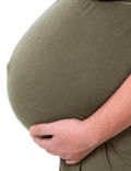
Pregnancy is a physiological state or condition where a fertilized egg (zygote) successfully implants and grows in the uterus of a woman, leading to the development of an embryo and finally a fetus. This process typically spans approximately 40 weeks, divided into three trimesters, and culminates in childbirth. Throughout this period, numerous hormonal and physical changes occur to support the growing offspring, including uterine enlargement, breast development, and various maternal adaptations to ensure the fetus's optimal growth and well-being.
The third trimester of pregnancy is the final stage of pregnancy that lasts from week 29 until birth, which typically occurs around the 40th week. During this period, the fetus continues to grow and mature, gaining weight rapidly. The mother's body also prepares for childbirth by dilating the cervix and producing milk in preparation for breastfeeding. Regular prenatal care is crucial during this time to monitor the health of both the mother and the developing fetus, as well as to prepare for delivery.
Prenatal ultrasonography, also known as obstetric ultrasound, is a medical diagnostic procedure that uses high-frequency sound waves to create images of the developing fetus, placenta, and amniotic fluid inside the uterus. It is a non-invasive and painless test that is widely used during pregnancy to monitor the growth and development of the fetus, detect any potential abnormalities or complications, and determine the due date.
During the procedure, a transducer (a small handheld device) is placed on the mother's abdomen and moved around to capture images from different angles. The sound waves travel through the mother's body and bounce back off the fetus, producing echoes that are then converted into electrical signals and displayed as images on a screen.
Prenatal ultrasonography can be performed at various stages of pregnancy, including early pregnancy to confirm the pregnancy and detect the number of fetuses, mid-pregnancy to assess the growth and development of the fetus, and late pregnancy to evaluate the position of the fetus and determine if it is head down or breech. It can also be used to guide invasive procedures such as amniocentesis or chorionic villus sampling.
Overall, prenatal ultrasonography is a valuable tool in modern obstetrics that helps ensure the health and well-being of both the mother and the developing fetus.
Uterine rupture is a serious obstetrical complication characterized by the disruption or tearing of all layers of the uterine wall, including the serosa (outer covering), myometrium (middle layer of muscle), and endometrium (inner lining). This can occur during pregnancy, labor, or delivery. In some cases, it may also involve the rupture of the adjacent structures such as bladder or broad ligament. Uterine rupture is a medical emergency that requires immediate surgical intervention to prevent maternal and fetal mortality or morbidity.
The symptoms of uterine rupture might include severe abdominal pain, vaginal bleeding, loss of fetal heart rate, changes in the mother's vital signs, and shock. The risk factors for uterine rupture include previous cesarean delivery, grand multiparity (having given birth to five or more pregnancies), use of labor-inducing drugs like oxytocin, and instrumental deliveries with vacuum extractors or forceps.
The management of uterine rupture typically involves an emergency laparotomy (open abdominal surgery) to repair the tear and stop any bleeding. In some cases, a hysterectomy (removal of the uterus) may be necessary if the damage is too severe or if there are other complications. The prognosis for both mother and baby depends on various factors like the extent of the injury, timeliness of treatment, and the overall health status of the patient before the event.
Placental circulation refers to the specialized circulatory system that develops during pregnancy to allow for the exchange of nutrients, oxygen, and waste products between the mother's blood and the fetal blood in the placenta. The placenta is a highly vascular organ that grows within the uterus and is connected to the developing fetus via the umbilical cord.
In the maternal side of the placenta, the spiral arteries branch into smaller vessels called the intervillous spaces, where they come in close contact with the fetal blood vessels within the villi (finger-like projections) of the placenta. The intervillous spaces are filled with maternal blood that flows around the villi, allowing for the exchange of gases and nutrients between the two circulations.
On the fetal side, the umbilical cord contains two umbilical arteries that carry oxygen-depleted blood from the fetus to the placenta, and one umbilical vein that returns oxygenated blood back to the fetus. The umbilical arteries branch into smaller vessels within the villi, where they exchange gases and nutrients with the maternal blood in the intervillous spaces.
Overall, the placental circulation is a crucial component of fetal development, allowing for the growing fetus to receive the necessary oxygen and nutrients to support its growth and development.
The first trimester of pregnancy is defined as the period of gestational development that extends from conception (fertilization of the egg by sperm) to the end of the 13th week. This critical phase marks significant transformations in both the mother's body and the growing embryo/fetus.
During the first trimester, the fertilized egg implants into the uterine lining (implantation), initiating a series of complex interactions leading to the formation of the placenta - an organ essential for providing nutrients and oxygen to the developing fetus while removing waste products. Simultaneously, the embryo undergoes rapid cell division and differentiation, giving rise to various organs and systems. By the end of the first trimester, most major structures are present, although they continue to mature and grow throughout pregnancy.
The mother may experience several physiological changes during this time, including:
- Morning sickness (nausea and vomiting)
- Fatigue
- Breast tenderness
- Frequent urination
- Food aversions or cravings
- Mood swings
Additionally, hormonal shifts can cause various symptoms and prepare the body for potential changes in lactation, posture, and pelvic alignment as pregnancy progresses. Regular prenatal care is crucial during this period to monitor both maternal and fetal wellbeing, identify any potential complications early on, and provide appropriate guidance and support throughout the pregnancy.
Ultrasonography, Doppler, color is a type of diagnostic ultrasound technique that uses the Doppler effect to produce visual images of blood flow in vessels and the heart. The Doppler effect is the change in frequency or wavelength of a wave in relation to an observer who is moving relative to the source of the wave. In this context, it refers to the change in frequency of the ultrasound waves as they reflect off moving red blood cells.
In color Doppler ultrasonography, different colors are used to represent the direction and speed of blood flow. Red typically represents blood flowing toward the transducer (the device that sends and receives sound waves), while blue represents blood flowing away from the transducer. The intensity or brightness of the color is proportional to the velocity of blood flow.
Color Doppler ultrasonography is often used in conjunction with grayscale ultrasound imaging, which provides information about the structure and composition of tissues. Together, these techniques can help diagnose a wide range of conditions, including heart disease, blood clots, and abnormalities in blood flow.
 Placenta accreta spectrum
Vasa praevia
Asherman's syndrome
Decidua
Placenta praevia
Eclampsia
T-shaped uterus
Marzieh Vahid-Dastjerdi
High-risk pregnancy
Uterine atony
Kim Kardashian
Asynclitic birth
Vaginal delivery
Postpartum bleeding
Retained placenta
Hysterotomy abortion
Interleukin 11
Placenta
List of MeSH codes (C13)
Placental disease
Birth injury
Caesarean section
Sally Collins
Delivery after previous caesarean section
Trophoblast
Antepartum bleeding
Placental expulsion
Placenta accreta spectrum - Wikipedia
 Information for "Placenta accreta" - wikidoc
 Effectiveness of Ultrasound Screening for a Placenta Accreta Spectrum Using Standard Ultrasound Criteria in a Secondary Care...
 Trophoblast invasion and spiral artery transformation in placenta accreta | ADC Fetal & Neonatal Edition
 The role of transvaginal ultrasound in the third-trimester evaluation of patients at high-risk of placenta accreta spectrum at...
 Placenta Accreta - Women's Health Issues - MSD Manual Consumer Version
 Placenta Accreta - MRI Online
 Care • Placenta Accreta Ireland
Placenta accreta spectrum - variations in clinical practice and maternal morbidity between UK and France: a population-based...
 Placenta Accreta Spectrum - FMF Courses
 placenta accreta spectrum
 Placenta Accreta - Brown Trial Firm
 Placenta Accreta | Loma Linda University Children's Health
The Well-Rounded Mama: Placenta Accreta: Brandy's Story
 Pregnancy | Topic | NICE
 Placenta accreta | Journal of Anaesthesia & Critical Care Case Reports
 Placenta Accreta: A Life-Threatening Attachment - Stanford Blood Center
 Total Health Solutions (Unit of Catch Creative Concepts): placenta accreta
 Placenta Accreta - When The Adherence Of The Placenta Is Disturbed
Alila Medical Media | Placenta accreta, feto 20 semanas | Ilustración médica
 placenta accrete spectrum (PAS)
 Placenta complications | Tommy's
 NI Maternal Health: Topic: Placenta Previa/Accreta/Increta/Percreta & Vasa Previa
 Placenta Accreta Spectrum; Different
Management Approach. A Report of Two Cases
Placenta Accreta Spectrum; Different
Management Approach. A Report of Two Cases
 The Antenatal Diagnosis of Placenta Accreta Spectrum | Baylor College of Medicine
 View of A TYPICAL PRESENTATION OF PLACENTA ACCRETA SYNDROME- A CASE REPORT
 Interventional Radiology Procedure Preserves Uterus in Patients With Placenta Accreta - OGP News
 Placenta Accreta and MRI - Placenta Accreta, Increta and Percreta | Forums | What to Expect
Placenta accreta and vaginal bleeding according to gestational age at delivery<...
 View of Successful management of total placenta previa with placenta accreta presenting with massive obstetric hemorrhage: A...
Percreta9
- The most severe form is called percreta, wherein the placenta grows through the uterine wall and invades surrounding organs, such as the bladder and intestine. (stanfordbloodcenter.org)
- They were now dealing with placenta percreta with high vascularity and multi-organ invasion. (stanfordbloodcenter.org)
- In about 5% of women, a placenta percreta develops. (practicalmommy.com)
- Welcome to Placenta Accreta, Increta and Percreta! (whattoexpect.com)
- Placenta accreta spectrum (PAS), formerly known as morbidly adherent placenta, refers to the range of pathologic adherence of the placenta, including placenta increta, placenta percreta, and placenta accreta. (jicajournal.in)
- The placenta had penetrated the lower segment of the uterus and the whole of the cervix and was invading the uterine serosa (percreta), therefore, a decision for cesarean hysterectomy was taken. (jicajournal.in)
- Pathology was defined as accreta, increta, or percreta. (thieme-connect.com)
- Increta and percreta patients were each compared with accreta patients. (thieme-connect.com)
- The relative risk (RR) of SGA for increta and percreta did not differ from accreta after adjusting for confounders (adjusted RR = 0.63, 95% confidence interval [CI]: 0.36-1.10 for increta and aRR = 0.72, 95% CI: 0.45-1.16 for percreta). (thieme-connect.com)
Hysterectomy10
- Methods 16 (12 accreta, 4 non-accreta) caesarean hysterectomy specimens were studied using immunohistochemistry. (bmj.com)
- Two experienced operators, blinded to the clinical data were asked to make a judgement on the likelihood of placenta accreta spectrum as a binary: low or high-probability of placenta accreta spectrum and to predict the main surgical outcome (conservative vs peripartum hysterectomy). (ucl.ac.uk)
- The diagnosis of accreta placentation was confirmed when one or more placental cotyledon(s) could not be digitally separated from the uterine wall at delivery or during the gross examination of the hysterectomy or partial myometrial resection specimens. (ucl.ac.uk)
- If doctors detect placenta accreta before delivery, a cesarean delivery followed by removal of the uterus (cesarean hysterectomy) is typically done. (msdmanuals.com)
- Because the placenta cannot be detached from the uterus after delivery, the definitive treatment is a hysterectomy. (lluch.org)
- Diagnosis of invasion was based on hysterectomy performed for an abnormally adherent placenta with histologic confirmation. (elsevierpure.com)
- A 28-year-old female with amenorrhea of 25 weeks G 2 P 1 L 1, diagnosed case of placenta previa and placenta accreta with previous lower-segment cesarean section and ventriculomegaly in the fetus was planned for hysterotomy ± hysterectomy. (jicajournal.in)
- [1] One of the two leading causes of peripartum hemorrhage is placenta accreta and is also the most common indication for peripartum hysterectomy. (jicajournal.in)
- PAS disorders were diagnosed histologically from the following specimens: placenta, placental-bed specimens, uterine curettage, uterine resection and/or total/partial hysterectomy. (biomedcentral.com)
- The following information used in planning antenatal care and admis- was extracted from the records: peripartum sion for elective or emergency deliveries to hysterectomy including hysterectomy for units that are well equipped to deal with po- placenta praevia accreta, bowel and bladder tential complications. (who.int)
Pregnancies7
- As of 2016, placenta accreta affects an estimated 1 in 272 pregnancies. (wikipedia.org)
- The more pregnancies you've had, the higher your risk for placenta accreta. (lluch.org)
- If you are looking for more technical information about placenta accreta, see Part One (what is accreta, how a placenta works), Part Two (risk factors, symptoms, and incidence of accreta), Part Three (risks to mother, baby, and future pregnancies), and Part Four (diagnosis and treatment) of my prior series on placenta accreta. (blogspot.com)
- In most pregnancies, blood vessels from the umbilical cord insert directly into the placenta. (tommys.org)
- In our series, 71% with accreta were delivered at 36 weeks of gestation or greater with delivery for bleeding in five (11%), and estimated blood loss was not increased in these pregnancies. (elsevierpure.com)
- 001). Conclusion: Likelihood of vaginal bleeding necessitating delivery declined with advancing gestation in pregnancies with placenta accreta as did blood loss. (elsevierpure.com)
- Placenta accreta spectrum disorders in twin pregnancies as an under reported clinical entity: A case series and systematic review. (knepublishing.com)
Case of placenta accreta1
- In the case of placenta accreta, the placenta cannot detach on its own after birth. (practicalmommy.com)
Cases of placenta accreta3
- We cannot stress how important diagnosis is for cases of placenta accreta. (browntrialfirm.com)
- You need a hospital that is fully equipped to handle your placenta accreta-not all facilities have the resources to handle cases of placenta accreta. (browntrialfirm.com)
- Many cases of placenta accreta spectrum are not diagnosed antenatally, despite identified risk factors and improved imaging methods . (bvsalud.org)
Diagnosis of placenta accreta2
- When the antepartum diagnosis of placenta accreta is made, it is usually based on ultrasound findings in the second or third trimester. (wikipedia.org)
- Prenatal diagnosis of placenta accreta spectrum. (knepublishing.com)
Disorders4
- Welcome to the educational course on Placenta Accreta Spectrum disorders. (fetalmedicine.com)
- The placenta data of women with PAS disorders were obtained and identified through a search from the database of the Department of Pathology, University Hospital Basel. (biomedcentral.com)
- Placenta praevia, previous endometritis, previous placenta removal, ART and vaginal operative birth can be considered important risk factors of PPH in women with PAS disorders. (biomedcentral.com)
- Established in 2008, the Abnormal Placentation Program has cared for hundreds of women with uterine and placental disorders, including placenta accreta, a condition in which the placenta attaches too deeply to the uterine wall. (brighamandwomens.org)
Cesarean delivery4
- STUDY DESIGN: This was a retrospective analysis of prospectively collected data of patients presenting with a singleton pregnancy, a history of at least one prior cesarean delivery and diagnosed prenatally with an anterior low-lying/placenta previa delivered electively after 32 weeks. (ucl.ac.uk)
- Having had both a cesarean delivery and placenta previa in a previous pregnancy greatly increases the risk of placenta accreta. (msdmanuals.com)
- Methods: This is a retrospective cohort study of women with prior cesarean delivery and persistent placenta previa delivered at our institution between December 1997 and December 2011. (elsevierpure.com)
- An important risk factor governing the presence of placenta accreta is a history of a prior cesarean delivery. (jicajournal.in)
20221
- Google Académico, fueron incluidas únicamente las publicaciones que se encontraron a texto completo, en español, inglés y portugués durante los años 2014 al 2022. (bvsalud.org)
Spectrum disorder4
- Termination of a second-trimester pregnancy with placenta accreta spectrum disorder. (bvsalud.org)
- The termination of pregnancy in patients with placenta accreta spectrum disorder (PASD) during the second trimester remains uncertain. (bvsalud.org)
- Placenta accreta spectrum disorder complicated with endometriosis: Systematic review and meta-analysis. (knepublishing.com)
- ACR Appropriateness Criteria® Placenta Accreta Spectrum Disorder. (nih.gov)
Ultrasound9
- Ultrasound detection of a placenta accreta spectrum (PAS) among women at risk is a key goal to reduce obstetric morbidity, but there is scarce information on its performance in real clinical settings. (karger.com)
- We report the effectiveness of a standardized ultrasound protocol to detect PAS in women with placenta previa (PP) in a secondary-level hospital. (karger.com)
- All 10 cases of invasive placenta presented more than 3 ultrasound criteria. (karger.com)
- BACKGROUND: Transvaginal ultrasound imaging has become an essential tool in the prenatal evaluation of the lower uterine segment and anatomy of the cervix but there are only limited data on the role of transvaginal ultrasound in the management of patients at high risk of placenta accreta spectrum at birth. (ucl.ac.uk)
- p= 0.002) between transabdominal and transvaginal ultrasound examination but both ultrasound techniques had similar likelihood score in identifying accreta placentation that was confirmed at birth. (ucl.ac.uk)
- Placenta accreta can be detected using ultrasound or MRI. (lluch.org)
- MRI and ultrasound are equally good at identifying placenta accreta in the back of the uterus. (lluch.org)
- The pregnancy was going seemingly well and she felt great when, at 29 weeks, Kristen's ultrasound revealed evidence of placenta accreta, despite the extremely low likelihood of 0.3 percent, according to a study published by The American College of Obstetricians and Gynecologists . (stanfordbloodcenter.org)
- Using a Doppler ultrasound to look for vascularity can help to differential between the two, with clots being avascular and retained placenta often receiving persistent blood flow from the uterus. (medscape.com)
Complications7
- What are the complications of placenta accreta? (lluch.org)
- One of the complications of multiple cesareans is that the placenta in a subsequent pregnancy can implant too low in the uterus ( placenta previa ) or grow into the uterine wall ( placenta accreta ). (blogspot.com)
- During each of the prenatal checkups, the doctor checks the position of the placenta and assesses whether there could be complications during delivery. (practicalmommy.com)
- This is a normal place for the placenta to implant and develop and it is very unlikely to cause any complications. (tommys.org)
- It isn't very common, but a retained placenta can cause complications if it isn't treated. (tommys.org)
- We are funding various research projects that are focusing on the placenta and potential pregnancy complications. (tommys.org)
- Placenta accreta, a condition in which the placenta abnormally implants in the uterus, can lead to additional complications, including massive obstetric hemorrhage at delivery. (ogpnews.com)
Placental abruption2
- Placental abruption (placenta detaching too early) is another potential risk after a prior cesarean. (blogspot.com)
- Placental abruption is a serious condition in which the placenta starts to come away from the inside of the womb wall before the baby has delivered. (tommys.org)
Serious pregnancy complication2
- Placenta accreta is a serious pregnancy complication because it usually leads to heavy bleeding at birth because the placenta cannot detach on its own. (practicalmommy.com)
- Researchers have reported on a procedure that can preserve fertility and potentially save the lives of women with a serious pregnancy complication called placenta accreta. (ogpnews.com)
Vaginal3
- Because of abnormal attachment to the myometrium, placenta accreta is associated with an increased risk of heavy bleeding at the time of attempted vaginal delivery. (wikipedia.org)
- While vaginal bleeding can occur, placenta accreta rarely shows signs or symptoms during pregnancy. (lluch.org)
- Objective: To evaluate the incidence of vaginal bleeding in women with placenta accreta according to gestational age at delivery. (elsevierpure.com)
Detach2
- In the final stage of pregnancy, the decidua and placenta detach and are shed as afterbirth. (browntrialfirm.com)
- Placenta accreta is a high-risk pregnancy complication where the placenta grows deep into the uterine wall and does not detach after childbirth. (lluch.org)
Blood vessels2
- There are three different levels of placenta accreta, depending on how deep the placenta's villi (blood vessels) are implanted. (browntrialfirm.com)
- During the healing and regression phase after the afterbirth is expelled, the blood vessels that connect the mother's body to the placenta close. (practicalmommy.com)
Delivery of the placenta1
- In these cases, delivery of the placenta is delayed, and the risks of bleeding and infection in the uterus are increased. (msdmanuals.com)
Complication4
- If women have risk factors for placenta accreta, doctors do ultrasonography periodically during the pregnancy to check for this complication. (msdmanuals.com)
- Any form of placenta accreta is a high-risk pregnancy complication . (browntrialfirm.com)
- This is a rare complication of pregnancy that makes it difficult to deliver the placenta after you give birth. (tommys.org)
- Back in July 2021, Divine Lee-Go was rushed to the hospital due to placenta previa, a pregnancy complication that occurs when "a baby's placenta partially or totally covers the mother's cervix. (smartparenting.com.ph)
Grow into the uterine1
- The placenta does not just grow into the uterine mucosa - as would be normal - but connects with the uterine muscles. (practicalmommy.com)
Praevia5
- A low-lying placenta (also known as placenta praevia) is when the placenta attaches lower down and may cover a part of or all of the cervix (the entrance to the womb). (tommys.org)
- In vasa praevia, these vessels are not protected by the umbilical cord or the placenta tissue. (tommys.org)
- The surgical procedure caesarean sections are associated with an involved a "standard" transverse lower seg- increased incidence of placenta praevia and ment caesarean section under general an- placenta praevia accreta [5-7], scar dehis- aesthesia. (who.int)
- Placenta praevia accreta was diagnosed of evidence-based information that can be intraoperatively. (who.int)
- Of these, 7 were because of intervals (CI) were calculated to identify the placenta praevia accreta, all of which were association between number of caesarean anterior. (who.int)
Fetal1
- The presence of decidua in focal accretas may help explain continued placental function leading to good fetal outcome in these cases. (bmj.com)
Caesarean2
- Women with placenta accreta spectrum will need to give birth by caesarean section. (paireland.ie)
- If the placenta is near or covering the cervix you won't be able to deliver vaginally and will need a caesarean section . (tommys.org)
Myometrium4
- Placenta accreta occurs when all or part of the placenta attaches abnormally to the myometrium (the muscular layer of the uterine wall). (wikipedia.org)
- Three grades of abnormal placental attachment are defined according to the depth of attachment and invasion into the muscular layers of the uterus: Accreta - chorionic villi attached to the myometrium, rather than being restricted within the decidua basalis. (wikipedia.org)
- In placenta accreta, the placenta attaches itself directly to the uterine wall, the myometrium, without the deciduas to protect it. (browntrialfirm.com)
- In this case, the placenta has penetrated completely or only partially through the decidua (endometrium) to the uterine musculature (myometrium). (practicalmommy.com)
Cervix3
- Placenta Previa Placenta previa is attachment (implantation) of the placenta over the opening of the cervix, in the lower rather than the upper part of the uterus. (msdmanuals.com)
- Placenta previa is a condition where the placenta blocks the opening of the cervix. (lluch.org)
- Unfortunately, this time the fertilized egg implanted low, near the cervix (placenta previa) and the placenta grew into the uterine wall and into the cervix itself (placenta increta). (blogspot.com)
Severity2
- The intermediate form in terms of severity is referred to as increta, where the placenta invades more deeply into the muscles of the uterus. (stanfordbloodcenter.org)
- Objective The aim of the study is to evaluate whether pathologic severity of placenta accreta spectrum (PAS) is correlated with the incidence of small for gestational age (SGA) and neonatal birthweight. (thieme-connect.com)
Abnormally firm2
- The placenta forms an abnormally firm and deep attachment to the uterine wall. (wikipedia.org)
- Placenta accreta is a placenta with an abnormally firm attachment to the uterus. (msdmanuals.com)
Mother's3
- This time, rather than writing about what accreta is and how to manage it, we present the first-hand story of one mother's experience with placenta accreta (increta in her case). (blogspot.com)
- Placenta means the place that provides both protection and nutrition to the baby in the mother's womb. (creativec3c.com)
- In CHI, the mother's immune system reacts abnormally to the pregnancy and causes damage to the placenta, increasing the risk of miscarriage and stillbirth . (tommys.org)
Usually detaches2
- After delivery of the baby, the placenta usually detaches from the uterus, and the woman can push the placenta out by herself or with help from a doctor or midwife. (msdmanuals.com)
- After birth, the placenta usually detaches from the uterine wall and is expelled by the afterbirth (afterbirth). (practicalmommy.com)
Attaches5
- What happens when the placenta attaches too deeply to the uterus wall? (browntrialfirm.com)
- When the placenta attaches too deeply to the uterine wall, but does NOT penetrate the uterine muscle. (browntrialfirm.com)
- When the placenta penetrates through the uterine wall and attaches to another organ (usually the bladder or bowels). (browntrialfirm.com)
- Placenta accreta, in its broader sense, describes the condition during pregnancy in which the placenta attaches too deeply to the uterine wall in varying degrees. (stanfordbloodcenter.org)
- An anterior placenta is when the placenta attaches to the front wall of the uterus. (tommys.org)
Chorionic villi1
- With the chorionic villi, the placenta adheres to the uterine wall. (practicalmommy.com)
Gestational2
- We obtained plasma samples before delivery from 16 participants with placenta accreta spectrum and 10 control subjects with similar gestational ages (35.1 vs 35.5 weeks gestation , respectively). (bvsalud.org)
- a the placenta was at anterior to bottom of the uterus without clear borderline of the placenta and the muscle layer at the 28th gestational week. (biomedcentral.com)
Umbilical1
- Your placenta is the gateway to your baby, filtering and delivery oxygen- and nutrient-rich blood through the umbilical cord . (browntrialfirm.com)
Grows3
- In the placenta accreta, the placenta (placenta) grows into the muscles of the uterus. (practicalmommy.com)
- Placenta accreta is a disorder of placental adhesion in which the tissue of the placenta (placenta) grows into the muscles of the uterus. (practicalmommy.com)
- As pregnancy progresses, not only the child grows, but also the placenta. (practicalmommy.com)
Incidence1
- [ 16 ] The incidence can be overestimated with gross examination of the cord, especially if the portion close to the placenta is examined, because the arteries may fuse close to the placenta. (medscape.com)
Wall of the uterus1
- Placenta accreta is when the placenta is attached and embedded too deeply into the wall of the uterus. (tommys.org)
Decidua4
- In six cases, accreta was focal (i.e., normal decidua in some areas), so sections were assessed for decidua. (bmj.com)
- other variables did not differ between non-accreta cases and areas in focal accreta cases showing normal decidua. (bmj.com)
- Trophoblast giant cells and SA transformation are reduced in accreta areas with no decidua. (bmj.com)
- In the presence of a placenta accreta, this means in medical language that the tissue of the placenta has penetrated the decidua. (practicalmommy.com)
Preterm1
- Thus, although preterm delivery is an important consideration when placenta accreta is suspected, our findings support individualizing delivery planning. (elsevierpure.com)
Ultrasonography2
- If a woman has conditions that increase the risk of placenta accreta, doctors usually do ultrasonography before delivery to check for placenta accreta. (msdmanuals.com)
- An ultrasonography during the pregnancy revealed placenta previa with accreta and increta. (jicajournal.in)
Women12
- Pregnant women above 35 years of age who have had a caesarian section and now have a placenta previa overlying the uterine scar have a 40% chance of placenta accreta. (wikipedia.org)
- Placenta Accreta Ireland work in close collaboration with healthcare providers who look after women with PAS in Ireland. (paireland.ie)
- Women with placenta accreta spectrum should have the opportunity to meet members of the team at various times during the pregnancy. (paireland.ie)
- Some women with placenta accreta will need to be admitted to the hospital. (paireland.ie)
- Most women with placenta accreta spectrum will not need to be admitted to hospital, and can safely be cared for while at home. (paireland.ie)
- OBJECTIVE: To compare the management and outcomes of women with Placenta Accreta Spectrum (PAS) in France and the UK. (ox.ac.uk)
- Women with placenta accreta may experience severe blood loss after a delivery. (lluch.org)
- Women who have had multiple C-sections are at a higher risk of developing placenta accreta. (lluch.org)
- Women older than 35 are more likely to have placenta accreta. (lluch.org)
- It's important to remember that placenta accreta is real and affects real women and babies. (blogspot.com)
- The purpose of this study was to determine if women with placenta accreta spectrum have a distinct plasma protein profile compared with control subjects. (bvsalud.org)
- The Killer Placenta"-a threat to the lives of young women giving birth by cesarean section. (knepublishing.com)
Anterior3
- Other risk factors include low-lying placenta, anterior placenta, congenital or acquired uterine defects (such as uterine septa), leiomyoma, ectopic implantation of placenta (including cornual pregnancy). (wikipedia.org)
- Having an anterior placenta can make it a bit harder to feel your baby move because your baby is cushioned by the placenta lying at the front of your stomach. (tommys.org)
- Find out more about anterior placenta . (tommys.org)
Implantation2
- Placenta accreta, generally speaking, is an abnormal implantation of the placenta . (browntrialfirm.com)
- The following are risk factors for an abnormal implantation of the placenta. (browntrialfirm.com)
Invasion3
- Aim Examine EVT invasion and spiral artery (SA) remodelling in placenta accreta. (bmj.com)
- When doctors delivered the baby, they were surprised to find that the invasion of the placenta was much worse than expected. (stanfordbloodcenter.org)
- Many of the top 50 proteins that significantly dysregulated in participants with placenta accreta spectrum were inflammatory cytokines , factors that regulate vascular remodeling , and extracellular matrix proteins that regulate invasion. (bvsalud.org)
Pregnancy and birth2
- This is the story of Brandy's placenta accreta pregnancy and birth. (blogspot.com)
- There are several things relating to the placenta that may affect your pregnancy and birth. (tommys.org)
Adheres1
- The mildest form is called accreta, where a part of the placenta adheres superficially into the uterine wall. (stanfordbloodcenter.org)
Uterus after delivery1
- When the placenta is too firmly attached, parts of the placenta may remain in the uterus after delivery. (msdmanuals.com)
Risk6
- An important risk factor for placenta accreta is placenta previa in the presence of a uterine scar. (wikipedia.org)
- Placenta previa is an independent risk factor for placenta accreta. (wikipedia.org)
- By opting for a natural birth (especially if you are planning to have more children in the future), you can decrease your risk of placenta accreta. (browntrialfirm.com)
- If you have placenta previa and have had C-sections, the risk for placenta accreta increases. (lluch.org)
- If you have symptoms or risk factors for placenta accreta, talk to your doctor about a referral to a specialist right away. (lluch.org)
- If the pregnancy continues within the uterus, the risk of placenta accrete or uterine rupture is increased.Objective. (bvsalud.org)
Risks2
- The risks of multiple cesareans, including accreta, were never mentioned. (blogspot.com)
- The risks for placenta accreta and its variations increase with a woman's age, previous C-sections and in vitro fertilisation, all of which we expect to see more of in the coming decades," he said. (ogpnews.com)