Placenta
Placenta Previa
Placenta Accreta
Placenta, Retained
Pregnancy
Trophoblasts
Chorionic Villi
Maternal-Fetal Exchange
Pregnancy Proteins
Placentation
Fetus
Pre-Eclampsia
Gestational Age
Extraembryonic Membranes
Chorion
Pregnancy Trimester, Third
Pregnancy, Animal
Pregnancy Trimester, First
Placental Lactogen
Decidua
Focal aneurysmal dilatation of subchorionic vessels simulating chorioangioma. (1/362)
Subchorionic vascular aneurysms of the placenta are rare lesions and may present confusion with chorioangioma or focal mesenchymal dysplasia on sonography. To our knowledge, the findings of placental aneurysms have not been reported in the ultrasound literature. We present a case with detailed sonographic evaluation, including spectral and color Doppler and pathological analysis, that was mistaken for chorioangioma prenatally. Knowledge of this benign entity may allow the sonologist to recommend conservative management in similar cases. (+info)Immunity to placental malaria. I. Elevated production of interferon-gamma by placental blood mononuclear cells is associated with protection in an area with high transmission of malaria. (2/362)
In areas in which malaria is holoendemic, primigravidae and secundigravidae, compared with multigravidae, are highly susceptible to placental malaria (PM). The nature of gravidity-dependent immune protection against PM was investigated by measuring in vitro production of cytokines by placental intervillous blood mononuclear cells (IVBMC). The results demonstrated that interferon (IFN)-gamma may be a critical factor in protection against PM: production of this cytokine by PM-negative multigravid IVBMC was elevated compared with PM-negative primigravid and secundigravid and PM-positive multigravid cells. Low IFN-gamma responsiveness to malarial antigen stimulation, most evident in the latter group, was balanced by increased interleukin (IL)-4 production, suggesting that counter-regulation of these two cytokines may be a crucial determinant in susceptibility to PM. A counter-regulatory relationship between IL-10 and tumor necrosis factor-alpha was also observed in response to malarial antigen stimulation. These data suggest that elevated production of IFN-gamma, as part of a carefully regulated cytokine network, is important in the control of PM. (+info)The factor V Leiden mutation in Japanese couples with recurrent spontaneous abortion. (3/362)
Thrombosis of placental vessels can be a major cause of recurrent spontaneous abortion (RSA). The factor V Leiden (FVL) mutation, a single point mutation in the factor V gene, is the most common genetic predisposition to thrombosis in European countries and the United States. However, even among Caucasian populations, the association between the FVL mutation and RSA is still controversial. The objectives of the present study were to investigate the prevalence of the FVL mutation in Japanese women who have experienced RSA and to clarify the contribution of the FVL mutation to recurrent miscarriages. A total of 52 Japanese women with a history of three or more consecutive idiopathic first trimester miscarriages and 41 of their male partners were studied. The control group consisted of 55 parous women without obstetric complications. Peripheral blood cell DNA was examined for the presence of the FVL alleles by polymerase chain reaction with Mnl I restriction fragment length polymorphisms. None of the 52 women with RSA and the 41 partners carried the mutation. We also found no subject carrying the FVL alleles in the control group. These results suggest that the FVL alleles are not concentrated in women with RSA at least to clinically significant levels and that there is no apparent association between the FVL mutation and RSA in our Japanese population. (+info)Maternal serum insulin-like growth factor binding protein-2 and -3 and fetal growth. (4/362)
This was a prospective observational study of maternal insulin-like growth factor binding protein-2 and -3 and fetal growth in 141 pregnant women after 24 weeks gestation who were scanned and venesected fortnightly. Cases (birthweight <5th centile) were sub-divided into those with growth restriction due to placental dysfunction (n = 25) and normal small (n = 27) and there were 89 normally grown controls. Maternal binding protein-3 was measured by radioimmunoassay and the overall pattern of the binding proteins and their proteolytic modifications were assessed by Western ligand blotting and immunoblotting followed by densitometric analysis. In controls, there was no correlation between binding protein-3 and birthweight, and binding protein-3 was elevated in the normal small but not the placental dysfunction group. Complete proteolysis of the 40 kDa doublet of binding protein-3 was observed in all pregnancies. Maternal serum binding protein-2 concentrations were unchanged in normal pregnancy compared to non-pregnant controls but elevated in the growth-restricted group and in all pregnancies binding protein-2 was predominantly present as a 14 kDa proteolysed fragment. These results suggest that compensatory changes in binding protein-2 and -3 or their proteolysis do not increase bioavailability and so do not confound the low maternal insulin-like growth factor-I in growth restricted pregnancies. (+info)Pathophysiology and treatment of fetal anemia due to placental chorioangioma. (5/362)
Placental chorioangiomas occur in 1% of pregnancies. Large chorioangiomas may cause serious complications such as fetal anemia, hydrops and fetal death. In this case report, a pregnancy complicated by a large placental chorioangioma is described. Severe fetal anemia without the occurrence of hydrops fetalis was suspected using ultrasound and Doppler examinations. Successful intrauterine blood transfusion was performed, with an unusually large amount of blood needed to obtain an adequate rise in fetal hematocrit. Two weeks later, at 32 weeks, the infant was born in good condition. In pregnancies with large chorioangiomas, we advise regular ultrasound and Doppler examinations, with the aim of detecting fetal anemia before hydrops develops. When anemia is suspected, fetal blood sampling is indicated and intrauterine transfusion therapy may be beneficial to preserve fetal health until maturity is reached. (+info)The role of insulin-like growth factor binding protein-1 phosphoisoforms in pregnancies with impaired placental function identified by doppler ultrasound. (6/362)
This study was performed to investigate the hypothesis that insulin-like growth factor binding protein-1 (IGFBP-1) is involved in the pathogenesis of trophoblast invasion and impaired placentation in human pregnancy. The role of total and non-phosphorylated IGFBP-1 in women with fetal growth restriction and in high risk pregnancies identified by uterine artery Doppler ultrasound screening was examined. This was a prospective study of women booked for antenatal care having second trimester anomaly scans and Doppler screening between 22-26 weeks gestation. Women were divided into three groups and compared: normal uterine artery Doppler and normal fetal growth (control group, n = 10); abnormal Doppler and normal fetal growth [bilateral uterine artery notches (BN; n = 16); abnormal Doppler and intrauterine growth restriction (IUGR; n = 8)]. Maternal serum was collected, stored and assayed simultaneously for total and non-phosphorylated IGFBP-1. There was elevated total and non-phosphorylated IGFBP-1 (mean 44.99 +/- 12.19 and 29.61 +/- 10.38 microg/l respectively) in the IUGR group compared with controls (mean 17.96 +/- 3.24 and 12.18 +/- 1.55 microg/l, P < 0.05). This finding suggests that the various IGFBP-1 isoforms, the degree of phosphorylation and the ratios of these different forms locally may be important during trophoblast invasion and may be implicated in clinical manifestations of impaired placentation later in the second trimester. (+info)The metabolic effect of antenatal corticosteroid therapy. (7/362)
The use of antenatal dexamethasone to mature the fetal lung in pregnancies likely to deliver before 34 weeks is almost universal. It reduces the incidence of respiratory distress syndrome in the newborn and results in an overall improvement in neonatal morbidity and mortality. Although considered to be generally safe, there are concerns about adverse maternal and fetal effects. In a series of studies, we have found that antenatal dexamethasone administration is associated with reduced placental hormone production and maternal bone formation, impaired glucose tolerance and altered function of the hypothalamic-pituitary-adrenal axis. In this article, we have compared our data with other reports in the human and reviewed the relevant animal data. We conclude that further studies on the long-term effects of antenatal dexamethasone therapy in the human are warranted with particular emphasis on the long-term effects on the fetus. (+info)Effects of massive doses of ergocalciferol plus cholesterol on pregnant rats and their offspring. (8/362)
Ergocalciferol (320,000 or 480,000 IU/kg) plus cholesterol (60 mg/kg) in olive oil solution was administered daily on 1, 2, or 4 consecutive days to pregnant rats from 9,10, 14, or 18 of gestation. The control animals received only olive oil. Disseminated lesions of metastic calcinosis were found in various tissues, in the coronary arteries and myocardium, in the media of the abnormal aorta, in the lung and pleura, in the gastoinstestinal tract, and in the kidney. This is in contrast to the atherosclerosis described in nonpregnant rats fed a similiar diet. A significant decline in maternal weight as well as a high rate of morbidity and mortality was observed. In mothers killed on day 22 of pregnancy, fetal and placental growths appeared significantly retarded suggesting a direct effect of the steroid or its more active metabolite, 1,25-dihydroxycholecalciferol, on the fetus or the trophoblastic tissue. Fetal bone lesionsassociated with a generalized retardation of ossification, placental edema, or calcification accompanied by a loss of the normal structure of the placenta and degenerative manifestation at this level were observed. Moreover, we noted a striking alteration of the fetal face in 33-39% of experimental fetuses, called by us carnival fetuses. (+info)Placental diseases, also known as placental pathologies, refer to a group of conditions that affect the development and function of the placenta during pregnancy. The placenta is an organ that develops in the uterus during pregnancy and provides oxygen and nutrients to the developing fetus while removing waste products.
Placental diseases can have serious consequences for both the mother and the fetus, including preterm labor, growth restriction, stillbirth, and long-term health problems for the child. Some common placental diseases include:
1. Placental abruption: This occurs when the placenta separates from the uterine wall before delivery, causing bleeding and potentially harming the fetus.
2. Placental previa: This is a condition where the placenta implants in the lower part of the uterus, covering the cervix. It can cause bleeding and may require cesarean delivery.
3. Preeclampsia: This is a pregnancy-related disorder characterized by high blood pressure and damage to organs such as the liver and kidneys. Placental dysfunction is thought to play a role in its development.
4. Intrauterine growth restriction (IUGR): This occurs when the fetus does not grow properly due to poor placental function, leading to low birth weight and potential health problems.
5. Chorioamnionitis: This is an infection of the membranes surrounding the fetus, which can lead to preterm labor and other complications.
6. Placental infarction: This occurs when a portion of the placenta dies due to a lack of blood flow, which can lead to growth restriction or stillbirth.
Prompt diagnosis and treatment of placental diseases are essential for ensuring the best possible outcomes for both the mother and the fetus.
The placenta is an organ that develops in the uterus during pregnancy and provides oxygen and nutrients to the growing baby through the umbilical cord. It also removes waste products from the baby's blood. The placenta attaches to the wall of the uterus, and the baby's side of the placenta contains many tiny blood vessels that connect to the baby's circulatory system. This allows for the exchange of oxygen, nutrients, and waste between the mother's and baby's blood. After the baby is born, the placenta is usually expelled from the uterus in a process called afterbirth.
Placenta previa is a medical condition that occurs during pregnancy where the placenta partially or fully covers the cervix, which is the lower part of the uterus that opens into the birth canal. This condition can cause severe bleeding during pregnancy and delivery, and it may lead to other complications such as preterm labor and delivery. Placenta previa is typically diagnosed through an ultrasound exam and managed with close monitoring, bed rest, and sometimes cesarean delivery.
Placenta accreta is a medical condition where the placenta grows too deeply into the uterine wall, beyond the normal depth. In a healthy pregnancy, the placenta attaches to the uterus and provides oxygen and nutrients to the growing fetus through the umbilical cord. However, in placenta accreta, the placental tissue invades the muscle of the uterus, which can cause complications during childbirth.
There are three types of placenta accreta:
1. Placenta Accreta: The placenta attaches too deeply into the uterine wall but does not penetrate the uterine muscle.
2. Placenta Increta: The placenta grows into and partially penetrates the uterine muscle.
3. Placenta Percreta: The placenta fully penetrates the uterine muscle and can grow into nearby organs, such as the bladder or bowel.
Placenta accreta is a serious condition that can cause severe bleeding during childbirth, which may require an emergency hysterectomy (removal of the uterus) to control the bleeding. It is more common in women who have had previous cesarean sections or other uterine surgeries.
Retained placenta is a medical condition that occurs when all or part of the placenta remains in the uterus after delivery, instead of being expelled naturally. Normally, the placenta separates from the uterine wall and is delivered within 30 minutes of childbirth. However, if the placenta is not completely delivered, it can lead to complications such as infection, heavy bleeding, and in rare cases, infertility or even death.
Retained placenta can be caused by various factors, including a weakened uterine muscle tone, an abnormally attached placenta, or a retained portion of the membranes. Treatment for retained placenta typically involves manual removal of the remaining tissue by a healthcare professional, often under anesthesia. In some cases, medication may be used to help promote contraction of the uterus and expulsion of the placenta.
It is important to seek medical attention promptly if a retained placenta is suspected, as timely treatment can help prevent potentially serious complications.
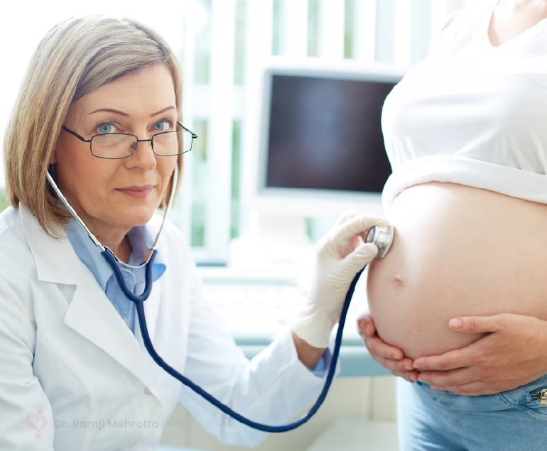
Pregnancy is a physiological state or condition where a fertilized egg (zygote) successfully implants and grows in the uterus of a woman, leading to the development of an embryo and finally a fetus. This process typically spans approximately 40 weeks, divided into three trimesters, and culminates in childbirth. Throughout this period, numerous hormonal and physical changes occur to support the growing offspring, including uterine enlargement, breast development, and various maternal adaptations to ensure the fetus's optimal growth and well-being.
Trophoblasts are specialized cells that make up the outer layer of a blastocyst, which is a hollow ball of cells that forms in the earliest stages of embryonic development. In humans, this process occurs about 5-6 days after fertilization. The blastocyst consists of an inner cell mass (which will eventually become the embryo) and an outer layer of trophoblasts.
Trophoblasts play a crucial role in implantation, which is the process by which the blastocyst attaches to and invades the lining of the uterus. Once implanted, the trophoblasts differentiate into two main layers: the cytotrophoblasts (which are closer to the inner cell mass) and the syncytiotrophoblasts (which form a multinucleated layer that is in direct contact with the maternal tissues).
The cytotrophoblasts proliferate and fuse to form the syncytiotrophoblasts, which have several important functions. They secrete enzymes that help to degrade and remodel the extracellular matrix of the uterine lining, allowing the blastocyst to implant more deeply. They also form a barrier between the maternal and fetal tissues, helping to protect the developing embryo from the mother's immune system.
Additionally, trophoblasts are responsible for the formation of the placenta, which provides nutrients and oxygen to the developing fetus and removes waste products. The syncytiotrophoblasts in particular play a key role in this process by secreting hormones such as human chorionic gonadotropin (hCG), which helps to maintain pregnancy, and by forming blood vessels that allow for the exchange of nutrients and waste between the mother and fetus.
Abnormalities in trophoblast development or function can lead to a variety of pregnancy-related complications, including preeclampsia, intrauterine growth restriction, and gestational trophoblastic diseases such as hydatidiform moles and choriocarcinomas.
Chorionic villi are finger-like projections of the chorion, which is the outermost extraembryonic membrane in a developing embryo. These structures are composed of both fetal and maternal tissues and play a crucial role in the early stages of pregnancy by providing a site for exchange of nutrients and waste products between the mother and the developing fetus.
Chorionic villi contain fetal blood vessels that are surrounded by stromal cells, trophoblasts, and connective tissue. They are formed during the process of implantation, when the fertilized egg attaches to the uterine wall. The chorionic villi continue to grow and multiply as the placenta develops, eventually forming a highly vascular and specialized organ that supports fetal growth and development throughout pregnancy.
One important function of chorionic villi is to serve as the site for the production of human chorionic gonadotropin (hCG), a hormone that can be detected in the mother's blood and urine during early pregnancy. This hormone plays a critical role in maintaining pregnancy by signaling the corpus luteum to continue producing progesterone, which helps to prevent menstruation and support fetal growth.
Abnormalities in chorionic villi can lead to various pregnancy complications, such as miscarriage, stillbirth, or intrauterine growth restriction. For this reason, chorionic villus sampling (CVS) is a diagnostic procedure that may be performed during early pregnancy to obtain fetal cells for genetic testing and diagnosis of chromosomal abnormalities or other genetic disorders.
Maternal-fetal exchange, also known as maternal-fetal transport or placental transfer, refers to the physiological process by which various substances are exchanged between the mother and fetus through the placenta. This exchange includes the transfer of oxygen and nutrients from the mother's bloodstream to the fetal bloodstream, as well as the removal of waste products and carbon dioxide from the fetal bloodstream to the mother's bloodstream.
The process occurs via passive diffusion, facilitated diffusion, and active transport mechanisms across the placental barrier, which is composed of fetal capillary endothelial cells, the extracellular matrix, and the syncytiotrophoblast layer of the placenta. The maternal-fetal exchange is crucial for the growth, development, and survival of the fetus throughout pregnancy.
"Pregnancy proteins" is not a standard medical term, but it may refer to specific proteins that are produced or have increased levels during pregnancy. Two common pregnancy-related proteins are:
1. Human Chorionic Gonadotropin (hCG): A hormone produced by the placenta shortly after fertilization. It is often detected in urine or blood tests to confirm pregnancy. Its primary function is to maintain the corpus luteum, which produces progesterone and estrogen during early pregnancy until the placenta takes over these functions.
2. Pregnancy-Specific beta-1 Glycoprotein (SP1): A protein produced by the placental trophoblasts during pregnancy. Its function is not well understood, but it may play a role in implantation, placentation, and protection against the mother's immune system. SP1 levels increase throughout pregnancy and are used as a marker for fetal growth and well-being.
These proteins have clinical significance in monitoring pregnancy progression, detecting potential complications, and diagnosing certain pregnancy-related conditions.
Placentation is the process by which the placenta, an organ that provides nutrients and oxygen to the developing fetus and removes waste products, is formed and develops during pregnancy. It involves the attachment of the fertilized egg (embryo) to the uterine wall and the development of specialized structures that facilitate the exchange of gases, nutrients, and waste between the mother and the fetus.
In humans, placentation begins when the embryo implants into the endometrium, or the lining of the uterus, about 6-10 days after fertilization. The outer layer of the embryo, called the trophoblast, invades the endometrial tissue and forms a structure called the placenta.
The placenta consists of both maternal and fetal tissues. The fetal portion of the placenta is derived from the chorionic villi, which are finger-like projections that develop on the surface of the embryo and increase the surface area for exchange. The maternal portion of the placenta is made up of modified endometrial tissue called decidua.
The placenta grows and develops throughout pregnancy, providing a vital connection between the mother and fetus. Proper placentation is essential for a healthy pregnancy and fetal development. Abnormalities in placentation can lead to complications such as preeclampsia, preterm labor, and intrauterine growth restriction.
A fetus is the developing offspring in a mammal, from the end of the embryonic period (approximately 8 weeks after fertilization in humans) until birth. In humans, the fetal stage of development starts from the eleventh week of pregnancy and continues until childbirth, which is termed as full-term pregnancy at around 37 to 40 weeks of gestation. During this time, the organ systems become fully developed and the body grows in size. The fetus is surrounded by the amniotic fluid within the amniotic sac and is connected to the placenta via the umbilical cord, through which it receives nutrients and oxygen from the mother. Regular prenatal care is essential during this period to monitor the growth and development of the fetus and ensure a healthy pregnancy and delivery.
Pre-eclampsia is a pregnancy-related disorder, typically characterized by the onset of high blood pressure (hypertension) and damage to organs, such as the kidneys, after the 20th week of pregnancy. It is often accompanied by proteinuria, which is the presence of excess protein in the urine. Pre-eclampsia can lead to serious complications for both the mother and the baby if left untreated or unmanaged.
The exact causes of pre-eclampsia are not fully understood, but it is believed that placental issues, genetic factors, and immune system problems may contribute to its development. Risk factors include first-time pregnancies, history of pre-eclampsia in previous pregnancies, chronic hypertension, obesity, older age (35 or older), and assisted reproductive technology (ART) pregnancies.
Pre-eclampsia can progress to a more severe form called eclampsia, which is characterized by the onset of seizures. HELLP syndrome, another severe complication, involves hemolysis (breaking down of red blood cells), elevated liver enzymes, and low platelet count.
Early detection and management of pre-eclampsia are crucial to prevent severe complications. Regular prenatal care, including frequent blood pressure checks and urine tests, can help identify early signs of the condition. Treatment typically involves close monitoring, medication to lower blood pressure, corticosteroids to promote fetal lung maturity, and, in some cases, delivery of the baby if the mother's or baby's health is at risk.
Gestational age is the length of time that has passed since the first day of the last menstrual period (LMP) in pregnant women. It is the standard unit used to estimate the age of a pregnancy and is typically expressed in weeks. This measure is used because the exact date of conception is often not known, but the start of the last menstrual period is usually easier to recall.
It's important to note that since ovulation typically occurs around two weeks after the start of the LMP, gestational age is approximately two weeks longer than fetal age, which is the actual time elapsed since conception. Medical professionals use both gestational and fetal age to track the development and growth of the fetus during pregnancy.
Extraembryonic membranes are specialized structures that form around the developing embryo in utero and provide vital support and protection during fetal development. There are three main extraembryonic membranes: the amnion, the chorion, and the allantois.
The amnion is the innermost membrane that surrounds the embryo itself, forming a fluid-filled sac known as the amniotic cavity. This sac provides a protective cushion for the developing embryo and helps to regulate its temperature and moisture levels.
The chorion is the outermost of the extraembryonic membranes, and it forms the boundary between the developing fetus and the mother's uterine wall. The chorion contains blood vessels that exchange nutrients and waste products with the mother's circulation, allowing for the growth and development of the fetus.
The allantois is a small membranous sac that arises from the developing fetal gut and eventually becomes part of the umbilical cord. It serves as a reservoir for fetal urine and helps to exchange waste products between the fetal and maternal circulations.
Together, these extraembryonic membranes play a critical role in supporting fetal development and ensuring a healthy pregnancy.
The chorion is the outermost fetal membrane that surrounds the developing conceptus (the embryo or fetus and its supporting structures). It forms early in pregnancy as an extraembryonic structure, meaning it arises from cells that will not become part of the actual body of the developing organism. The chorion plays a crucial role in pregnancy by contributing to the formation of the placenta, which provides nutrients and oxygen to the growing embryo/fetus and removes waste products.
One of the most important functions of the chorion is to produce human chorionic gonadotropin (hCG), a hormone that signals the presence of pregnancy and maintains the corpus luteum, a temporary endocrine structure in the ovary that produces progesterone during early pregnancy. Progesterone is essential for preparing the uterus for implantation and maintaining the pregnancy.
The chorion consists of two layers: an inner cytotrophoblast layer and an outer syncytiotrophoblast layer. The cytotrophoblast layer is made up of individual cells, while the syncytiotrophoblast layer is a multinucleated mass of fused cytotrophoblast cells. These layers interact with the maternal endometrium (the lining of the uterus) to form the placenta and facilitate exchange between the mother and the developing fetus.
In summary, the chorion is a vital extraembryonic structure in pregnancy that contributes to the formation of the placenta, produces hCG, and interacts with the maternal endometrium to support fetal development.
The third trimester of pregnancy is the final stage of pregnancy that lasts from week 29 until birth, which typically occurs around the 40th week. During this period, the fetus continues to grow and mature, gaining weight rapidly. The mother's body also prepares for childbirth by dilating the cervix and producing milk in preparation for breastfeeding. Regular prenatal care is crucial during this time to monitor the health of both the mother and the developing fetus, as well as to prepare for delivery.
"Animal pregnancy" is not a term that is typically used in medical definitions. However, in biological terms, animal pregnancy refers to the condition where a fertilized egg (or eggs) implants and develops inside the reproductive tract of a female animal, leading to the birth of offspring (live young).
The specific details of animal pregnancy can vary widely between different species, with some animals exhibiting phenomena such as placental development, gestation periods, and hormonal changes that are similar to human pregnancy, while others may have very different reproductive strategies.
It's worth noting that the study of animal pregnancy and reproduction is an important area of biological research, as it can provide insights into fundamental mechanisms of embryonic development, genetics, and evolution.
The first trimester of pregnancy is defined as the period of gestational development that extends from conception (fertilization of the egg by sperm) to the end of the 13th week. This critical phase marks significant transformations in both the mother's body and the growing embryo/fetus.
During the first trimester, the fertilized egg implants into the uterine lining (implantation), initiating a series of complex interactions leading to the formation of the placenta - an organ essential for providing nutrients and oxygen to the developing fetus while removing waste products. Simultaneously, the embryo undergoes rapid cell division and differentiation, giving rise to various organs and systems. By the end of the first trimester, most major structures are present, although they continue to mature and grow throughout pregnancy.
The mother may experience several physiological changes during this time, including:
- Morning sickness (nausea and vomiting)
- Fatigue
- Breast tenderness
- Frequent urination
- Food aversions or cravings
- Mood swings
Additionally, hormonal shifts can cause various symptoms and prepare the body for potential changes in lactation, posture, and pelvic alignment as pregnancy progresses. Regular prenatal care is crucial during this period to monitor both maternal and fetal wellbeing, identify any potential complications early on, and provide appropriate guidance and support throughout the pregnancy.
Placental lactogen is a hormone produced by the placenta during pregnancy in humans and some other mammals. It is similar in structure to human growth hormone and prolactin, and has both growth-promoting and lactogenic (milk-producing) properties. Placental lactogen plays an important role in regulating maternal metabolism during pregnancy, promoting the growth and development of the fetus, and preparing the mother's body for lactation after birth. It helps to stimulate the growth of the mammary glands and the production of milk by increasing the availability of nutrients such as glucose, amino acids, and fatty acids in the mother's bloodstream. Placental lactogen also helps to regulate the mother's insulin sensitivity, which can affect her energy levels and the growth of the fetus.
The decidua is a specialized type of tissue that lines the uterus during pregnancy. It forms after the implantation of a fertilized egg (embryo) into the uterine lining, and it plays an important role in supporting the growth and development of the embryo and fetus.
The decidua is composed of several layers, including the decidual capsularis, which surrounds the embryo, and the decidual parietalis, which lines the rest of the uterus. The tissue is rich in blood vessels and contains a variety of immune cells that help to protect the developing fetus from infection.
During pregnancy, the decidua produces various hormones and growth factors that support the growth of the placenta, which provides nutrients and oxygen to the fetus. After the birth of the baby, the decidua is shed along with the placenta in a process called childbirth or parturition.
It's worth noting that abnormalities in the decidua can contribute to pregnancy complications such as preeclampsia, preterm labor, and miscarriage.
 Antithrombin III deficiency
 Frontiers | The influence of early environment and micronutrient availability on developmental epigenetic programming: lessons...
 Best Placenta Stem Cell Therapy at PRMEDICA in Cabo San Lucas, Mexico
 Hydatidiform Mole: Practice Essentials, Background, Pathophysiology
 Retained placenta in homozygous sickle cell disease. | Obstet Gynecol;114(4): 825-828, 2009 Oct. | MEDLINE | BVS CLAP/SMR...
 Impact of the nitric oxide-donor pentaerythrityl-tetranitrate on perinatal outcome in risk pregnancies: a prospective,...
 Advanced Search Results - Public Health Image Library(PHIL)
 Placenta abruptio: MedlinePlus Medical Encyclopedia
![CCN3 cellular communication network factor 3 [Homo sapiens (human)] - Gene - NCBI](data:image/png;base64,iVBORw0KGgoAAAANSUhEUgAAABAAAAAQCAYAAAAf8/9hAAAB1ElEQVQ4jaWSPWgTcRjGf/ehuWhobO2JxGJRY3taTTRV2yoqSpW6iIWO4iAoUsRBioNDKUWKLU7i4KA4OfhVREQnETRia03k7IdiS0LaQYKJQg3mLtfc30GySNUDn/V5nx/vy/vAf0pqad3db2xquiBJku93s2Tb2eEHdw1rTcsxol23sObTjN7oIp9KVmaU9kMdTxcLAyiqGtA0bfms+XKQULSdQG2EmnUx0q9ughAA8p/CFW0IN3Sv0vUI5p2zIMpUrd5JeP/Jii//80ZJUlrb9lyV8qn3zI5dB8A4MoBWtcITAKBmZe3eRmPzccYf9uIUsyzx6zQd7fMMAIjFdgxpkuPy4clFANbu6qa6fouybXtznxeAoqoBn0/zz5kvBqVQ5DBasJ5gXaPnDQAWFpwCkiwLZekyAMp2wTPAsqy5d8nEZcIHThPQo7jlIua9854BibdvekqKX8PouARAOn6F+c8pT4Bc7svz6U8f77O1cwDVV439PcPU4yHw8AUhhDPyOn4OfWOMuuZfBZp41INTLACorhC2/Jc2zsxMX8vl8lMcPBUHFL5mnpEZGa748sS42esKYS8WLtl2NjE22s/6fScIhtr48W2S5O0zIFwvp3vST6Z+myCvkaonAAAAAElFTkSuQmCC) CCN3 cellular communication network factor 3 [Homo sapiens (human)] - Gene - NCBI
CCN3 cellular communication network factor 3 [Homo sapiens (human)] - Gene - NCBI Anja Health Raises $4.5M Seed Funding Round to Help Pregnant Parents Get a Stem Cell Safe For Their Umbilical Cord and Placenta
 Fetal origins of coronary heart disease | The BMJ
 Pregnancy Complications: Miscarriage, Eclampsia, and More
 Early Life Nutritional Programming of Obesity: Mother-Child Cohort Studies | Annals of Nutrition and Metabolism | Karger...
 Biblio | Page 9 | Linus Pauling Institute | Oregon State University
 Multiple Sclerosis - 3X higher incidence in women, previously 1X, wonder why | VitaminDWiki
 Center for Developmental Health Research Areas | OHSU
 Due Date Calculator: How Many Weeks Pregnant? | Bambino Mio
- Bambino Mio (ROW)
Due Date Calculator: How Many Weeks Pregnant? | Bambino Mio
- Bambino Mio (ROW) Twin Terms and Acronyms
 RP-School of Health Sciences
 Services - Pregnancy & Delivery | Lexington Medical Center
 Jack Rychik, MD | Children's Hospital of Philadelphia
 International Classification Of Diseases - 9, Abbreviated Titles