Amyloid
Amyloid beta-Peptides
Serum Amyloid A Protein
Amyloid beta-Protein Precursor
Islet Amyloid Polypeptide
Dental Plaque
Amyloidosis
Cerebral Amyloid Angiopathy
Serum Amyloid P-Component
Amyloid Neuropathies
Amyloid Precursor Protein Secretases
Alzheimer Disease
Congo Red
Viral Plaque Assay
Amyloid Neuropathies, Familial
Prealbumin
Peptide Fragments
Mice, Transgenic
Aspartic Acid Endopeptidases
Brain
Atherosclerosis
Presenilin-1
Thiazoles
Carotid Artery Diseases
Arteriosclerosis
Rupture, Spontaneous
Carotid Stenosis
Cerebral Amyloid Angiopathy, Familial
Apolipoproteins E
Ultrasonography, Interventional
Prions
Disease Models, Animal
Carotid Arteries
Protein Structure, Secondary
Amino Acid Sequence
Coronary Artery Disease
tau Proteins
Molecular Sequence Data
Immunohistochemistry
Neurofibrillary Tangles
Protease Nexins
Microscopy, Electron, Transmission
Microscopy, Electron
Hemolytic Plaque Technique
Protein Multimerization
Neurons
Aniline Compounds
Microscopy, Atomic Force
Mutation
Mice, Inbred C57BL
Neurofibrils
Endopeptidases
Cells, Cultured
beta 2-Microglobulin
Insulysin
Circular Dichroism
Protein Binding
Protein Conformation
Hippocampus
Gerstmann-Straussler-Scheinker Disease
Cerebral Cortex
Macrophages
Peptides
Endarterectomy, Carotid
Microglia
Protein Structure, Tertiary
Biological Markers
Aging
Models, Molecular
Presenilin-2
Protein Structure, Quaternary
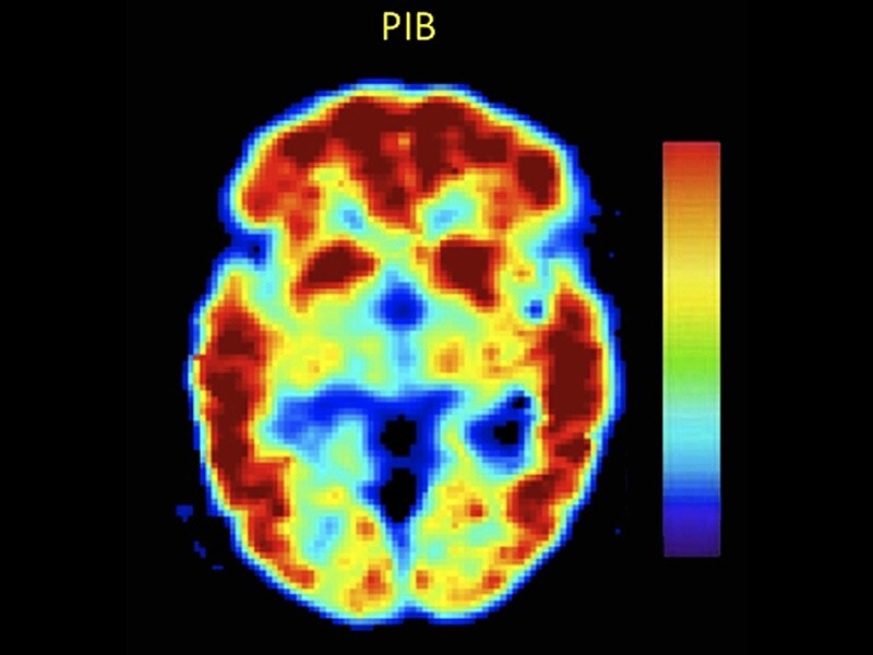
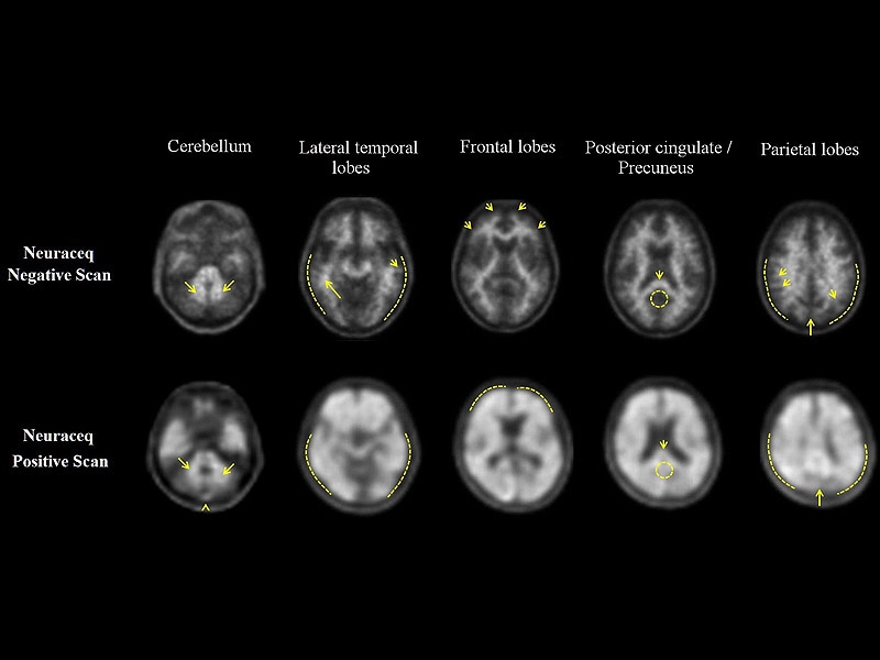
Positron-Emission Tomography
Inflammation
Mice, Knockout
Disease Progression
Enzyme-Linked Immunosorbent Assay
alpha-Synuclein
Apolipoprotein E4
Spectroscopy, Fourier Transform Infrared
Staining and Labeling
Gliosis
Magnetic Resonance Imaging
Presenilins
Coloring Agents
Nerve Tissue Proteins
Neprilysin
Membrane Proteins
Blotting, Western
C-Reactive Protein
Risk Factors
Predictive Value of Tests
Coronary Angiography
Maze Learning
Solubility
Protein Processing, Post-Translational
Neurodegenerative Diseases
Brain Chemistry
Lipids
Peptide Termination Factors
Tunica Intima
Cholesterol
Vascular Calcification
Cricetinae
Tomography, X-Ray Computed
RNA, Messenger
Hydrogen-Ion Concentration
Necrosis
Receptors, Islet Amyloid Polypeptide
Prion Diseases
Rabbits
Islets of Langerhans
Mouthwashes
Severity of Illness Index
Protein Denaturation
PC12 Cells
Gene Expression Regulation
Apolipoprotein E3
Nuclear Magnetic Resonance, Biomolecular
Psoriasis
Memory Disorders
Image Processing, Computer-Assisted
Nerve Degeneration
Acute-Phase Reaction
Base Sequence
Periodontal Index
Astrocytes
Intracranial Arteriosclerosis
Dementia
Protein Stability
Brachiocephalic Trunk
Protein Precursors
Trifluoroethanol
Tunica Media
Transfection
Reproducibility of Results
Microscopy, Fluorescence
Models, Biological
Clioquinol
Aorta, Abdominal
Pleural Diseases
Alzheimer's disease: clues from flies and worms. (1/883)
Presenilin mutations give rise to familial Alzheimer's disease and result in elevated production of amyloid beta peptide. Recent evidence that presenilins act in developmental signalling pathways may be the key to understanding how senile plaques, neurofibrillary tangles and apoptosis are all biochemically linked. (+info)Dynamics of plaque formation in Alzheimer's disease. (2/883)
Plaques that form in the brains of Alzheimer patients are made of deposits of the amyloid-beta peptide. We analyze the time evolution of amyloid-beta deposition in immunostained brain slices from transgenic mice. We find that amyloid-beta deposits appear in clusters whose characteristic size increases from 14 microm in 8-month-old mice to 22 microm in 12-month-old mice. We show that the clustering has implications for the biological growth of amyloid-beta by presenting a growth model that accounts for the experimentally observed structure of individual deposits and predicts the formation of clusters of deposits and their time evolution. (+info)Plaque-induced neurite abnormalities: implications for disruption of neural networks in Alzheimer's disease. (3/883)
The brains of Alzheimer's disease patients contain extracellular Abeta amyloid deposits (senile plaques). Although genetic evidence causally links Abeta deposition to the disease, the mechanism by which Abeta disrupts cortical function is unknown. Using triple immunofluorescent confocal microscopy and three-dimensional reconstructions, we found that neuronal processes that cross through an Abeta deposit are likely to have a radically changed morphology. We modeled the electrophysiological effect of this changed morphology and found a predicted delay of several milliseconds over an average plaque. We propose that this type of delay, played out among thousands of plaques throughout neocortical areas, disrupts the precise temporal firing patterns of action potentials, contributing directly to neural system failure and dementia. (+info)Age-related changes in the brain of the dog. (4/883)
Although many age-related changes have been described in the nervous system of different species, few authors have specifically studied the topic. Knowledge of such changes is essential to veterinary pathologists, who must distinguish the lesions of specific pathologic processes from those arising as a result of normal aging. The brains of 20 old dogs, ranging in age from 8 to 18 years, were compared with those of 10 young dogs using routine staining techniques (hematoxilin and eosin, periodic acid-Schiff), special staining techniques (periodic acid-methenamine silver stain), and immunohistochemical techniques to detect glial fibrillary acid protein, neurofilaments, ubiquitin, and beta-amyloid. Changes affected meninges and choroid plexuses, meningeal and parenchymal vessels, neurons, and glial cells. Of special interest was the presence of polyglucosan bodies, cerebrovascular amyloid deposition, senile plaques, and ubiquitinated bodies. Some of the age-related changes found, particularly lipofuscin, polyglucosan bodies, and beta-amyloid protein deposition, may play a role in the pathogenesis of the canine cognitive dysfunction syndrome. The dog could be used as a natural animal model for the study of normal aging and human neurodegenerative diseases. (+info)Histochemically reactive zinc in plaques of the Swedish mutant beta-amyloid precursor protein transgenic mice. (5/883)
Endogenous metals such as zinc may contribute to beta-amyloid (Abeta) aggregation and hence the plaque formation. In the present study, we examined brains of four Swedish mutant amyloid precursor protein (APP) transgenic mice at 12 months of age for histochemically reactive zinc in the plaques. Here, we report that all the Congo red (+) mature plaques contained chelatable zinc, as demonstrated by staining with the zinc-specific fluorescent dye 6-methoxy-8-quinolyl-para-toluenesulfonamide (TSQ). On the other hand, Congo red (-) preamyloid Abeta deposits were not stained with TSQ. Interestingly, although cerebellum contained similar degree of preamyloid Abeta deposits as cerebral cortex, it was completely devoid of Congo red- or TSQ-stained mature plaques. Although zinc from plaques was only slowly and partially removed by a specific zinc remover, dithizone, treatment of brain sections with heparinase-III, which degrades heparan sulfate proteoglycan (HSPG), another major constituent of plaques, greatly fastened the zinc removal with dithizone. The present study has demonstrated the presence of histochemically reactive zinc in plaques, but not preamyloid Abeta deposits, of the Swedish mutant APP transgenic mice. Because preamyloid Abeta deposits fail to develop into congophilic plaques in cerebellum where synaptic vesicle zinc is deficient, the synaptic zinc may be a necessary element in the plaque formation. In holding zinc inside plaques, HSPG may contribute in addition to Abeta. (+info)Association of microglia with amyloid plaques in brains of APP23 transgenic mice. (6/883)
Microglia are a key component of the inflammatory response in the brain and are associated with senile plaques in Alzheimer's disease (AD). Although there is evidence that microglial activation is important for the pathogenesis of AD, the role of microglia in cerebral amyloidosis remains obscure. The present study was undertaken to investigate the relationship between beta-amyloid deposition and microglia activation in APP23 transgenic mice which express human mutated amyloid-beta precursor protein (betaPP) under the control of a neuron-specific promoter element. Light microscopic analysis revealed that the majority of the amyloid plaques in neocortex and hippocampus of 14- to 18- month-old APP23 mice are congophilic and associated with clusters of hypertrophic microglia with intensely stained Mac-1- and phosphotyrosine-positive processes. No association of such activated microglia was observed with diffuse plaques. In young APP23 mice, early amyloid deposits were already of dense core nature and were associated with a strong microglial response. Ultrastructurally, bundles of amyloid fibrils, sometimes surrounded by an incomplete membrane, were observed within the microglial cytoplasm. However, microglia with the typical characteristics of phagocytosis were associated more frequently with dystrophic neurites than with amyloid fibrils. Although the present observations cannot unequivocally determine whether microglia are causal, contributory, or consequential to cerebral amyloidosis, our results suggest that microglia are involved in cerebral amyloidosis either by participating in the processing of neuron-derived betaPP into amyloid fibrils and/or by ingesting amyloid fibrils via an uncommon phagocytotic mechanism. In any case, our observations demonstrate that neuron-derived betaPP is sufficient to induce not only amyloid plaque formation but also amyloid-associated microglial activation similar to that reported in AD. Moreover, our results are consistent with the idea that microglia activation may be important for the amyloid-associated neuron loss previously reported in these mice. (+info)The AMY antigen co-occurs with abeta and follows its deposition in the amyloid plaques of Alzheimer's disease and down syndrome. (7/883)
Novel plaque-like "AMY" lesions were recently described in the brains of patients with Alzheimer's disease (AD). Using three Abeta antibodies, we now document the co-occurrence of AMY immunoreactivity (IR) with amyloid beta-peptide (Abeta) in the large majority of plaques in AD brain. AMY IR was detected in many compacted plaques, whereas its co-localization with early, diffuse Abeta deposits was rare. AMY IR overlapped considerably or fully with Abeta and, in more severely affected AD brains, decorated the periphery of some plaques. In a temporal series of 29 Down syndrome (DS) brains from patients aged 12 to 73 years, the earliest AMY IR was detected in some plaques at age 15, following the earliest appearance of Abeta plaques (age 12 years), and then accrued within a subset of Abeta deposits, namely, the more spherical, compacted plaques. Brains from DS patients 29 years and older showed AMY staining in many Abeta plaques, as seen in AD. Brains from eight monkeys aged 17 to 34 years and thirty APP transgenic mice aged 8 to 20 months showed Abeta IR but no AMY IR. We conclude that AMY IR represents an amyloid-associated antigen that co-deposits in most but not all Abeta plaques in AD and DS and that accumulation of the AMY antigen follows Abeta deposition in plaques. (+info)Inhibition of NF-kappaB potentiates amyloid beta-mediated neuronal apoptosis. (8/883)
One mechanism leading to neurodegeneration during Alzheimer's disease (AD) is amyloid beta peptide (Abeta) neurotoxicity. Abeta elicits in cultured central nervous system neurons a biphasic response: a low-dose neurotrophic response and a high-dose neurotoxic response. Previously we reported that NF-kappaB is activated by low doses of Abeta only. Here we show that NF-kappaB activation leads to neuroprotection. In primary neurons we found that a pretreatment with 0.1 microM Abeta-(1-40) protects against neuronal death induced with 10 microM Abeta-(1-40). As a known neuroprotective agent we next analyzed the effect of tumor necrosis factor alpha (TNF-alpha). Maximal activation of NF-kappaB was found with 2 ng/ml TNF-alpha. Pretreatment with TNF-alpha protected cerebellar granule cells from cell death induced by 10 microM Abeta-(1-40). This protection is described by an inverted U-shaped dose response and is maximal with a NF-kappaB-activating dose. The molecular specificity of this protective effect was analyzed by specific blockade of NF-kappaB activation. Overexpression of a transdominant negative IkappaB-alpha blocks NF-kappaB activation and potentiates Abeta-mediated neuronal apoptosis. Our findings show that activation of NF-kappaB is the underlying mechanism of the neuroprotective effect of low-dose Abeta and TNF-alpha. In accordance with these in vitro data we find that nuclear NF-kappaB immunoreactivity around various plaque stages of AD patients is reduced in comparison to age-matched controls. Taken together these data suggest that pharmacological NF-kappaB activation may be a useful approach in the treatment of AD and related neurodegenerative disorders. (+info)Amyloid is a term used in medicine to describe abnormally folded protein deposits that can accumulate in various tissues and organs of the body. These misfolded proteins can form aggregates known as amyloid fibrils, which have a characteristic beta-pleated sheet structure. Amyloid deposits can be composed of different types of proteins, depending on the specific disease associated with the deposit.
In some cases, amyloid deposits can cause damage to organs and tissues, leading to various clinical symptoms. Some examples of diseases associated with amyloidosis include Alzheimer's disease (where amyloid-beta protein accumulates in the brain), systemic amyloidosis (where amyloid fibrils deposit in various organs such as the heart, kidneys, and liver), and type 2 diabetes (where amyloid deposits form in the pancreas).
It's important to note that not all amyloid deposits are harmful or associated with disease. However, when they do cause problems, treatment typically involves managing the underlying condition that is leading to the abnormal protein accumulation.
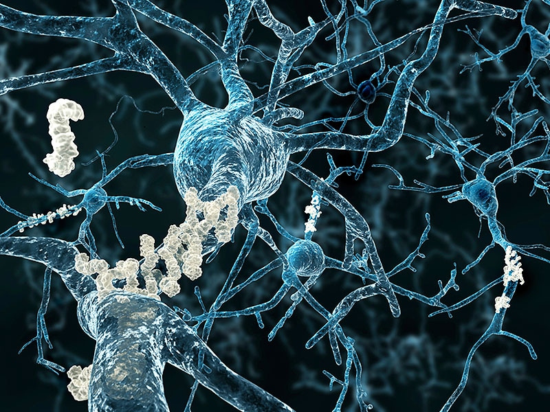
Amyloid beta-peptides (Aβ) are small protein fragments that are crucially involved in the pathogenesis of Alzheimer's disease. They are derived from a larger transmembrane protein called the amyloid precursor protein (APP) through a series of proteolytic cleavage events.
The two primary forms of Aβ peptides are Aβ40 and Aβ42, which differ in length by two amino acids. While both forms can be harmful, Aβ42 is more prone to aggregation and is considered to be the more pathogenic form. These peptides have the tendency to misfold and accumulate into oligomers, fibrils, and eventually insoluble plaques that deposit in various areas of the brain, most notably the cerebral cortex and hippocampus.
The accumulation of Aβ peptides is believed to initiate a cascade of events leading to neuroinflammation, oxidative stress, synaptic dysfunction, and neuronal death, which are all hallmarks of Alzheimer's disease. Although the exact role of Aβ in the onset and progression of Alzheimer's is still under investigation, it is widely accepted that they play a central part in the development of this debilitating neurodegenerative disorder.
Serum Amyloid A (SAA) protein is an acute phase protein produced primarily in the liver, although it can also be produced by other cells in response to inflammation. It is a member of the apolipoprotein family and is found in high-density lipoproteins (HDL) in the blood. SAA protein levels increase rapidly during the acute phase response to infection, trauma, or tissue damage, making it a useful biomarker for inflammation.
In addition to its role as an acute phase protein, SAA has been implicated in several disease processes, including atherosclerosis and amyloidosis. In amyloidosis, SAA can form insoluble fibrils that deposit in various tissues, leading to organ dysfunction. There are four subtypes of SAA in humans (SAA1, SAA2, SAA3, and SAA4), with SAA1 and SAA2 being the most responsive to inflammatory stimuli.
The Amyloid Beta-Protein Precursor (AβPP) is a type of transmembrane protein that is widely expressed in various tissues and organs, including the brain. It plays a crucial role in normal physiological processes, such as neuronal development, synaptic plasticity, and repair.
AβPP undergoes proteolytic processing by enzymes called secretases, resulting in the production of several protein fragments, including the amyloid-beta (Aβ) peptide. Aβ is a small peptide that can aggregate and form insoluble fibrils, which are the main component of amyloid plaques found in the brains of patients with Alzheimer's disease (AD).
The accumulation of Aβ plaques is believed to contribute to the neurodegeneration and cognitive decline observed in AD. Therefore, AβPP and its proteolytic processing have been the focus of extensive research aimed at understanding the pathogenesis of AD and developing potential therapies.
Atherosclerotic plaque is a deposit of fatty (cholesterol and fat) substances, calcium, and other substances in the inner lining of an artery. This plaque buildup causes the artery to narrow and harden, reducing blood flow through the artery, which can lead to serious cardiovascular conditions such as coronary artery disease, angina, heart attack, or stroke. The process of atherosclerosis develops gradually over decades and can start in childhood.
Amyloid plaque is a pathological hallmark of several degenerative diseases, including Alzheimer's disease. It refers to extracellular deposits of misfolded proteins that accumulate in various tissues and organs, but are most commonly found in the brain. The main component of these plaques is an abnormally folded form of a protein called amyloid-beta (Aβ). This protein is produced through the normal processing of the amyloid precursor protein (APP), but in amyloid plaques, it aggregates into insoluble fibrils that form the core of the plaque.
The accumulation of amyloid plaques is thought to contribute to neurodegeneration and cognitive decline in Alzheimer's disease and other related disorders. The exact mechanisms by which this occurs are not fully understood, but it is believed that the aggregation of Aβ into plaques leads to the disruption of neuronal function and viability, as well as the activation of inflammatory responses that can further damage brain tissue.
It's important to note that while amyloid plaques are a key feature of Alzheimer's disease, they are not exclusive to this condition. Amyloid plaques have also been found in other neurodegenerative disorders, as well as in some normal aging brains, although their significance in these contexts is less clear.
Islet Amyloid Polypeptide (IAPP), also known as amylin, is a 37-amino acid peptide co-secreted with insulin from pancreatic beta-cells in response to meals. It plays crucial roles in regulating glucose homeostasis by suppressing glucagon secretion, slowing gastric emptying, and promoting satiety. In type 2 diabetes, IAPP can form amyloid fibrils, which deposit in pancreatic islets, contributing to beta-cell dysfunction and death. This contributes to the progressive nature of type 2 diabetes.
Dental plaque is a biofilm or mass of bacteria that accumulates on the surface of the teeth, restorative materials, and prosthetic devices such as dentures. It is initiated when bacterial colonizers attach to the smooth surfaces of teeth through van der Waals forces and specific molecular adhesion mechanisms.
The microorganisms within the dental plaque produce extracellular polysaccharides that help to stabilize and strengthen the biofilm, making it resistant to removal by simple brushing or rinsing. Over time, if not regularly removed through oral hygiene practices such as brushing and flossing, dental plaque can mineralize and harden into tartar or calculus.
The bacteria in dental plaque can cause tooth decay (dental caries) by metabolizing sugars and producing acid that demineralizes the tooth enamel. Additionally, certain types of bacteria in dental plaque can cause periodontal disease, an inflammation of the gums that can lead to tissue damage and bone loss around the teeth. Regular professional dental cleanings and good oral hygiene practices are essential for preventing the buildup of dental plaque and maintaining good oral health.
Amyloidosis is a medical condition characterized by the abnormal accumulation of insoluble proteins called amyloid in various tissues and organs throughout the body. These misfolded protein deposits can disrupt the normal function of affected organs, leading to a range of symptoms depending on the location and extent of the amyloid deposition.
There are different types of amyloidosis, classified based on the specific proteins involved:
1. Primary (AL) Amyloidosis: This is the most common form, accounting for around 80% of cases. It results from the overproduction and misfolding of immunoglobulin light chains, typically by clonal plasma cells in the bone marrow. The amyloid deposits can affect various organs, including the heart, kidneys, liver, and nervous system.
2. Secondary (AA) Amyloidosis: This form is associated with chronic inflammatory diseases, such as rheumatoid arthritis, tuberculosis, or familial Mediterranean fever. The amyloid fibrils are composed of serum amyloid A protein (SAA), an acute-phase reactant produced during the inflammatory response. The kidneys are commonly affected in this type of amyloidosis.
3. Hereditary or Familial Amyloidosis: These forms are caused by genetic mutations that result in the production of abnormal proteins prone to misfolding and amyloid formation. Examples include transthyretin (TTR) amyloidosis, fibrinogen amyloidosis, and apolipoprotein AI amyloidosis. These forms can affect various organs, including the heart, nerves, and kidneys.
4. Dialysis-Related Amyloidosis: This form is seen in patients undergoing long-term dialysis for chronic kidney disease. The amyloid fibrils are composed of beta-2 microglobulin, a protein that accumulates due to impaired clearance during dialysis. The joints and bones are commonly affected in this type of amyloidosis.
The diagnosis of amyloidosis typically involves a combination of clinical evaluation, imaging studies, and tissue biopsy with the demonstration of amyloid deposition using special stains (e.g., Congo red). Treatment depends on the specific type and extent of organ involvement and may include supportive care, medications to target the underlying cause (e.g., chemotherapy, immunomodulatory agents), and organ transplantation in some cases.
Cerebral amyloid angiopathy (CAA) is a medical condition characterized by the accumulation of beta-amyloid protein in the walls of small to medium-sized blood vessels in the brain. This protein buildup can cause damage to the vessel walls, leading to bleeding (cerebral hemorrhage), cognitive decline, and other neurological symptoms.
CAA is often associated with aging and is a common finding in older adults. It can also be seen in people with Alzheimer's disease and other forms of dementia. The exact cause of CAA is not fully understood, but it is believed to result from the abnormal processing and clearance of beta-amyloid protein in the brain.
The diagnosis of CAA typically involves a combination of clinical evaluation, imaging studies such as MRI or CT scans, and sometimes cerebrospinal fluid analysis. Treatment for CAA is generally supportive and focused on managing symptoms and preventing complications. There are currently no approved disease-modifying treatments for CAA.
Serum Amyloid P-component (SAP) is a protein that is normally present in the blood and other bodily fluids. It is a part of the larger family of pentraxin proteins, which are involved in the innate immune response, meaning they provide immediate defense against foreign invaders without needing to adapt over time. SAP plays a role in inflammation, immune complex clearance, and complement activation.
In the context of amyloidosis, SAP binds to misfolded proteins called amyloid fibrils, which can deposit in various tissues and organs, leading to their dysfunction and failure. The accumulation of these amyloid fibrils with SAP is a hallmark of systemic amyloidosis.
It's important to note that while SAP plays a role in the pathogenesis of amyloidosis, it is not directly responsible for causing the disease. Instead, its presence can serve as a useful marker for diagnosing and monitoring the progression of amyloidosis.
Amyloid neuropathies are a group of peripheral nerve disorders caused by the abnormal accumulation of amyloid proteins in the nerves. Amyloid is a protein that can be produced in various diseases and can deposit in different organs, including nerves. When this occurs in the nerves, it can lead to damage and dysfunction, resulting in symptoms such as numbness, tingling, pain, and weakness in the affected limbs.
There are several types of amyloid neuropathies, with the two most common being:
1. Transthyretin (TTR)-related hereditary amyloidosis: This is an inherited disorder caused by mutations in the TTR gene, which leads to the production of abnormal TTR protein that can form amyloid deposits in various organs, including nerves.
2. Immunoglobulin light chain (AL) amyloidosis: This is a disorder in which abnormal plasma cells produce excessive amounts of immunoglobulin light chains, which can form amyloid deposits in various organs, including nerves.
The diagnosis of amyloid neuropathies typically involves a combination of clinical evaluation, nerve conduction studies, and tissue biopsy to confirm the presence of amyloid deposits. Treatment options depend on the underlying cause of the disorder and may include medications, chemotherapy, stem cell transplantation, or supportive care to manage symptoms.
Amyloid precursor protein (APP) secretases are enzymes that are responsible for cleaving the amyloid precursor protein into various smaller proteins. There are two types of APP secretases: α-secretase and β-secretase.
α-Secretase is a member of the ADAM (a disintegrin and metalloproteinase) family, specifically ADAM10 and ADAM17. When APP is cleaved by α-secretase, it produces a large ectodomain called sAPPα and a membrane-bound C-terminal fragment called C83. This pathway is known as the non-amyloidogenic pathway because it prevents the formation of amyloid-β (Aβ) peptides, which are associated with Alzheimer's disease.
β-Secretase, also known as β-site APP cleaving enzyme 1 (BACE1), is a type II transmembrane aspartic protease. When APP is cleaved by β-secretase, it produces a large ectodomain called sAPPβ and a membrane-bound C-terminal fragment called C99. Subsequently, C99 is further cleaved by γ-secretase to generate Aβ peptides, including the highly neurotoxic Aβ42. This pathway is known as the amyloidogenic pathway because it leads to the formation of Aβ peptides and the development of Alzheimer's disease.
Therefore, APP secretases play a crucial role in the regulation of APP processing and have been the focus of extensive research in the context of Alzheimer's disease and other neurodegenerative disorders.
Alzheimer's disease is a progressive disorder that causes brain cells to waste away (degenerate) and die. It's the most common cause of dementia — a continuous decline in thinking, behavioral and social skills that disrupts a person's ability to function independently.
The early signs of the disease include forgetting recent events or conversations. As the disease progresses, a person with Alzheimer's disease will develop severe memory impairment and lose the ability to carry out everyday tasks.
Currently, there's no cure for Alzheimer's disease. However, treatments can temporarily slow the worsening of dementia symptoms and improve quality of life.
Congo Red is a synthetic diazo dye that is commonly used in histology and pathology for stainings and tests. It is particularly useful in identifying amyloid deposits in tissues, which are associated with various diseases such as Alzheimer's disease, type 2 diabetes, and systemic amyloidosis.
When Congo Red binds to amyloid fibrils, it exhibits a characteristic apple-green birefringence under polarized light microscopy. Additionally, Congo Red stained amyloid deposits show a shift in their emission spectrum when excited with circularly polarized light, a phenomenon known as dichroism. These properties make Congo Red a valuable tool for the diagnosis and study of amyloidosis and other protein misfolding disorders.
It is important to note that Congo Red staining should be performed with care, as it can be toxic and carcinogenic if not handled properly.
A viral plaque assay is a laboratory technique used to measure the infectivity and concentration of viruses in a sample. This method involves infecting a monolayer of cells (usually in a petri dish or multi-well plate) with a known volume of a virus-containing sample, followed by overlaying the cells with a nutrient-agar medium to restrict viral spread and enable individual plaques to form.
After an incubation period that allows for viral replication and cell death, the cells are stained, and clear areas or "plaques" become visible in the monolayer. Each plaque represents a localized region of infected and lysed cells, caused by the progeny of a single infectious virus particle. The number of plaques is then counted, and the viral titer (infectious units per milliliter or PFU/mL) is calculated based on the dilution factor and volume of the original inoculum.
Viral plaque assays are essential for determining viral titers, assessing virus-host interactions, evaluating antiviral agents, and studying viral pathogenesis.
Familial amyloid neuropathies are a group of inherited disorders characterized by the accumulation of abnormal deposits of amyloid proteins in various tissues and organs of the body. These abnormal deposits can cause damage to nerves, leading to a peripheral neuropathy that affects sensation, movement, and organ function.
There are several types of familial amyloid neuropathies, each caused by different genetic mutations. The most common type is known as transthyretin-related hereditary amyloidosis (TTR-HA), which is caused by mutations in the TTR gene. Other types include apolipoprotein A1-related hereditary amyloidosis (APOA1-HA) and gelsolin-related amyloidosis (AGel-HA).
Symptoms of familial amyloid neuropathies can vary depending on the type and severity of the disorder. Common symptoms include:
* Numbness, tingling, or pain in the hands and feet
* Weakness or loss of muscle strength in the legs and arms
* Autonomic nervous system dysfunction, leading to problems with digestion, heart rate, blood pressure, and temperature regulation
* Carpal tunnel syndrome
* Eye abnormalities, such as vitreous opacities or retinal deposits
* Kidney disease
Familial amyloid neuropathies are typically inherited in an autosomal dominant manner, meaning that a child has a 50% chance of inheriting the mutated gene from an affected parent. Diagnosis is usually made through genetic testing and confirmation of the presence of amyloid deposits in tissue samples.
Treatment for familial amyloid neuropathies typically involves managing symptoms and slowing the progression of the disease. This may include medications to control pain, physical therapy to maintain muscle strength and mobility, and devices such as braces or wheelchairs to assist with mobility. In some cases, liver transplantation may be recommended to remove the source of the mutated transthyretin protein.
Prealbumin, also known as transthyretin, is a protein produced primarily in the liver and circulates in the blood. It plays a role in transporting thyroid hormones and vitamin A throughout the body. Prealbumin levels are often used as an indicator of nutritional status and liver function. Low prealbumin levels may suggest malnutrition or inflammation, while increased levels can be seen in certain conditions like hyperthyroidism. It is important to note that prealbumin levels should be interpreted in conjunction with other clinical findings and laboratory tests for a more accurate assessment of a patient's health status.
A peptide fragment is a short chain of amino acids that is derived from a larger peptide or protein through various biological or chemical processes. These fragments can result from the natural breakdown of proteins in the body during regular physiological processes, such as digestion, or they can be produced experimentally in a laboratory setting for research or therapeutic purposes.
Peptide fragments are often used in research to map the structure and function of larger peptides and proteins, as well as to study their interactions with other molecules. In some cases, peptide fragments may also have biological activity of their own and can be developed into drugs or diagnostic tools. For example, certain peptide fragments derived from hormones or neurotransmitters may bind to receptors in the body and mimic or block the effects of the full-length molecule.
Transgenic mice are genetically modified rodents that have incorporated foreign DNA (exogenous DNA) into their own genome. This is typically done through the use of recombinant DNA technology, where a specific gene or genetic sequence of interest is isolated and then introduced into the mouse embryo. The resulting transgenic mice can then express the protein encoded by the foreign gene, allowing researchers to study its function in a living organism.
The process of creating transgenic mice usually involves microinjecting the exogenous DNA into the pronucleus of a fertilized egg, which is then implanted into a surrogate mother. The offspring that result from this procedure are screened for the presence of the foreign DNA, and those that carry the desired genetic modification are used to establish a transgenic mouse line.
Transgenic mice have been widely used in biomedical research to model human diseases, study gene function, and test new therapies. They provide a valuable tool for understanding complex biological processes and developing new treatments for a variety of medical conditions.
Aspartic acid endopeptidases are a type of enzyme that cleave peptide bonds within proteins. They are also known as aspartyl proteases or aspartic proteinases. These enzymes contain two catalytic aspartic acid residues in their active site, which work together to hydrolyze the peptide bond.
Aspartic acid endopeptidases play important roles in various biological processes, including protein degradation, processing, and activation. They are found in many organisms, including viruses, bacteria, fungi, plants, and animals. Some well-known examples of aspartic acid endopeptidases include pepsin, cathepsin D, and HIV protease.
Pepsin is a digestive enzyme found in the stomach that helps break down proteins in food. Cathepsin D is a lysosomal enzyme that plays a role in protein turnover and degradation within cells. HIV protease is an essential enzyme for the replication of the human immunodeficiency virus (HIV), which causes AIDS. Inhibitors of HIV protease are used as antiretroviral drugs to treat HIV infection.
The brain is the central organ of the nervous system, responsible for receiving and processing sensory information, regulating vital functions, and controlling behavior, movement, and cognition. It is divided into several distinct regions, each with specific functions:
1. Cerebrum: The largest part of the brain, responsible for higher cognitive functions such as thinking, learning, memory, language, and perception. It is divided into two hemispheres, each controlling the opposite side of the body.
2. Cerebellum: Located at the back of the brain, it is responsible for coordinating muscle movements, maintaining balance, and fine-tuning motor skills.
3. Brainstem: Connects the cerebrum and cerebellum to the spinal cord, controlling vital functions such as breathing, heart rate, and blood pressure. It also serves as a relay center for sensory information and motor commands between the brain and the rest of the body.
4. Diencephalon: A region that includes the thalamus (a major sensory relay station) and hypothalamus (regulates hormones, temperature, hunger, thirst, and sleep).
5. Limbic system: A group of structures involved in emotional processing, memory formation, and motivation, including the hippocampus, amygdala, and cingulate gyrus.
The brain is composed of billions of interconnected neurons that communicate through electrical and chemical signals. It is protected by the skull and surrounded by three layers of membranes called meninges, as well as cerebrospinal fluid that provides cushioning and nutrients.
The dental plaque index (DPI) is a clinical measurement used in dentistry to assess the amount of dental plaque accumulation on a person's teeth. It was first introduced by Silness and Löe in 1964 as a method to standardize the assessment of oral hygiene and the effectiveness of oral hygiene interventions.
The DPI is based on a visual examination of the amount of plaque present on four surfaces of the teeth, including the buccal (cheek-facing) and lingual (tongue-facing) surfaces of both upper and lower first molars and upper and lower incisors. The examiner assigns a score from 0 to 3 for each surface, with higher scores indicating greater plaque accumulation:
* Score 0: No plaque detected, even after probing the area with a dental explorer.
* Score 1: Plaque detected by visual examination and/or probing but is not visible when the area is gently dried with air.
* Score 2: Moderate accumulation of soft deposits that are visible upon visual examination before air drying, but which can be removed by scraping with a dental explorer.
* Score 3: Abundant soft matter, visible upon visual examination before air drying and not easily removable with a dental explorer.
The DPI is calculated as the average score of all surfaces examined, providing an overall measure of plaque accumulation in the mouth. It can be used to monitor changes in oral hygiene over time or to evaluate the effectiveness of different oral hygiene interventions. However, it should be noted that the DPI has limitations and may not accurately reflect the presence of bacterial biofilms or the risk of dental caries and gum disease.
Atherosclerosis is a medical condition characterized by the buildup of plaques, made up of fat, cholesterol, calcium, and other substances found in the blood, on the inner walls of the arteries. This process gradually narrows and hardens the arteries, reducing the flow of oxygen-rich blood to various parts of the body. Atherosclerosis can affect any artery in the body, including those that supply blood to the heart (coronary arteries), brain, limbs, and other organs. The progressive narrowing and hardening of the arteries can lead to serious complications such as coronary artery disease, carotid artery disease, peripheral artery disease, and aneurysms, which can result in heart attacks, strokes, or even death if left untreated.
The exact cause of atherosclerosis is not fully understood, but it is believed to be associated with several risk factors, including high blood pressure, high cholesterol levels, smoking, diabetes, obesity, physical inactivity, and a family history of the condition. Atherosclerosis can often progress without any symptoms for many years, but as the disease advances, it can lead to various signs and symptoms depending on which arteries are affected. Treatment typically involves lifestyle changes, medications, and, in some cases, surgical procedures to restore blood flow.
Presenilin-1 (PSEN1) is a gene that provides instructions for making one part of an enzyme complex called gamma-secretase. This enzyme is involved in the breakdown of certain proteins, most notably the amyloid precursor protein (APP), into smaller fragments called peptides. One of these peptides, called beta-amyloid, can accumulate and form clumps called plaques, which are a characteristic feature of Alzheimer's disease.
Mutations in the PSEN1 gene have been identified as a major cause of early-onset familial Alzheimer's disease (FAD), a rare, inherited form of the disorder that usually develops before age 65. These mutations result in an abnormal gamma-secretase enzyme that produces more toxic beta-amyloid peptides and fewer harmless ones, leading to the formation of amyloid plaques and neurodegeneration.
It's important to note that while mutations in PSEN1 are associated with early-onset FAD, most cases of Alzheimer's disease are sporadic and develop later in life, typically after age 65. The role of PSEN1 and other genes associated with FAD in the more common, late-onset form of Alzheimer's is still being researched.
Thiazoles are organic compounds that contain a heterocyclic ring consisting of a nitrogen atom and a sulfur atom, along with two carbon atoms and two hydrogen atoms. They have the chemical formula C3H4NS. Thiazoles are present in various natural and synthetic substances, including some vitamins, drugs, and dyes. In the context of medicine, thiazole derivatives have been developed as pharmaceuticals for their diverse biological activities, such as anti-inflammatory, antifungal, antibacterial, and antihypertensive properties. Some well-known examples include thiazide diuretics (e.g., hydrochlorothiazide) used to treat high blood pressure and edema, and the antidiabetic drug pioglitazone.
Carotid artery diseases refer to conditions that affect the carotid arteries, which are the major blood vessels that supply oxygen-rich blood to the head and neck. The most common type of carotid artery disease is atherosclerosis, which occurs when fatty deposits called plaques build up in the inner lining of the arteries.
These plaques can cause the arteries to narrow or become blocked, reducing blood flow to the brain and increasing the risk of stroke. Other carotid artery diseases include carotid artery dissection, which occurs when there is a tear in the inner lining of the artery, and fibromuscular dysplasia, which is a condition that affects the muscle and tissue in the walls of the artery.
Symptoms of carotid artery disease may include neck pain or pulsations, transient ischemic attacks (TIAs) or "mini-strokes," and strokes. Treatment options for carotid artery disease depend on the severity and type of the condition but may include lifestyle changes, medications, endarterectomy (a surgical procedure to remove plaque from the artery), or angioplasty and stenting (procedures to open blocked arteries using a balloon and stent).
Arteriosclerosis is a general term that describes the hardening and stiffening of the artery walls. It's a progressive condition that can occur as a result of aging, or it may be associated with certain risk factors such as high blood pressure, high cholesterol, diabetes, smoking, and a sedentary lifestyle.
The process of arteriosclerosis involves the buildup of plaque, made up of fat, cholesterol, calcium, and other substances, in the inner lining of the artery walls. Over time, this buildup can cause the artery walls to thicken and harden, reducing the flow of oxygen-rich blood to the body's organs and tissues.
Arteriosclerosis can affect any of the body's arteries, but it is most commonly found in the coronary arteries that supply blood to the heart, the cerebral arteries that supply blood to the brain, and the peripheral arteries that supply blood to the limbs. When arteriosclerosis affects the coronary arteries, it can lead to heart disease, angina, or heart attack. When it affects the cerebral arteries, it can lead to stroke or transient ischemic attack (TIA). When it affects the peripheral arteries, it can cause pain, numbness, or weakness in the limbs, and in severe cases, gangrene and amputation.
Spontaneous rupture in medical terms refers to the sudden breaking or tearing of an organ, tissue, or structure within the body without any identifiable trauma or injury. This event can occur due to various reasons such as weakening of the tissue over time because of disease or degeneration, or excessive pressure on the tissue.
For instance, a spontaneous rupture of the appendix is called an "appendiceal rupture," which can lead to peritonitis, a serious inflammation of the abdominal cavity. Similarly, a spontaneous rupture of a blood vessel, like an aortic aneurysm, can result in life-threatening internal bleeding.
Spontaneous ruptures are often medical emergencies and require immediate medical attention for proper diagnosis and treatment.
Carotid stenosis is a medical condition that refers to the narrowing or constriction of the lumen (inner space) of the carotid artery. The carotid arteries are major blood vessels that supply oxygenated blood to the head and neck. Carotid stenosis usually results from the buildup of plaque, made up of fat, cholesterol, calcium, and other substances, on the inner walls of the artery. This process is called atherosclerosis.
As the plaque accumulates, it causes the artery to narrow, reducing blood flow to the brain. Severe carotid stenosis can increase the risk of stroke, as a clot or debris from the plaque can break off and travel to the brain, blocking a smaller blood vessel and causing tissue damage or death.
Carotid stenosis is typically diagnosed through imaging tests such as ultrasound, CT angiography, or MRI angiography. Treatment options may include lifestyle modifications (such as quitting smoking, controlling blood pressure, and managing cholesterol levels), medications to reduce the risk of clots, or surgical procedures like endarterectomy or stenting to remove or bypass the blockage.
Cerebral amyloid angiopathy (CAA), familial type, is a genetic disorder characterized by the buildup of beta-amyloid protein in the walls of blood vessels in the brain. This accumulation can lead to bleeding in the brain (cerebral hemorrhage) and cognitive decline. It is caused by mutations in genes associated with the production or clearance of beta-amyloid, such as the APP, PSEN1, and PSEN2 genes. These genetic mutations are typically inherited in an autosomal dominant manner, meaning that a child has a 50% chance of inheriting the mutation from a parent who carries it. The presence of these mutations leads to an increased production or decreased clearance of beta-amyloid, resulting in its accumulation in the blood vessel walls and subsequent complications.
Apolipoprotein E (ApoE) is a protein involved in the metabolism of lipids, particularly cholesterol. It is produced primarily by the liver and is a component of several types of lipoproteins, including very low-density lipoproteins (VLDL) and high-density lipoproteins (HDL).
ApoE plays a crucial role in the transport and uptake of lipids in the body. It binds to specific receptors on cell surfaces, facilitating the delivery of lipids to cells for energy metabolism or storage. ApoE also helps to clear cholesterol from the bloodstream and is involved in the repair and maintenance of tissues.
There are three major isoforms of ApoE, designated ApoE2, ApoE3, and ApoE4, which differ from each other by only a few amino acids. These genetic variations can have significant effects on an individual's risk for developing certain diseases, particularly cardiovascular disease and Alzheimer's disease. For example, individuals who inherit the ApoE4 allele have an increased risk of developing Alzheimer's disease, while those with the ApoE2 allele may have a reduced risk.
In summary, Apolipoprotein E is a protein involved in lipid metabolism and transport, and genetic variations in this protein can influence an individual's risk for certain diseases.
Interventional ultrasonography is a medical procedure that involves the use of real-time ultrasound imaging to guide minimally invasive diagnostic and therapeutic interventions. This technique combines the advantages of ultrasound, such as its non-ionizing nature (no radiation exposure), relatively low cost, and portability, with the ability to perform precise and targeted procedures.
In interventional ultrasonography, a specialized physician called an interventional radiologist or an interventional sonographer uses high-frequency sound waves to create detailed images of internal organs and tissues. These images help guide the placement of needles, catheters, or other instruments used during the procedure. Common interventions include biopsies (tissue sampling), fluid drainage, tumor ablation, and targeted drug delivery.
The real-time visualization provided by ultrasonography allows for increased accuracy and safety during these procedures, minimizing complications and reducing recovery time compared to traditional surgical approaches. Additionally, interventional ultrasonography can be performed on an outpatient basis, further contributing to its appeal as a less invasive alternative in many clinical scenarios.
Prions are misfolded proteins that can induce other normal proteins to also adopt the misfolded shape, leading to the formation of aggregates. These abnormal prion protein aggregates are associated with a group of progressive neurodegenerative diseases known as transmissible spongiform encephalopathies (TSEs). Examples of TSEs include bovine spongiform encephalopathy (BSE or "mad cow disease") in cattle, variant Creutzfeldt-Jakob disease (vCJD) in humans, and scrapie in sheep. The misfolded prion proteins are resistant to degradation by proteases, which contributes to their accumulation and subsequent neuronal damage, ultimately resulting in spongiform degeneration of the brain and other neurological symptoms associated with TSEs.
Animal disease models are specialized animals, typically rodents such as mice or rats, that have been genetically engineered or exposed to certain conditions to develop symptoms and physiological changes similar to those seen in human diseases. These models are used in medical research to study the pathophysiology of diseases, identify potential therapeutic targets, test drug efficacy and safety, and understand disease mechanisms.
The genetic modifications can include knockout or knock-in mutations, transgenic expression of specific genes, or RNA interference techniques. The animals may also be exposed to environmental factors such as chemicals, radiation, or infectious agents to induce the disease state.
Examples of animal disease models include:
1. Mouse models of cancer: Genetically engineered mice that develop various types of tumors, allowing researchers to study cancer initiation, progression, and metastasis.
2. Alzheimer's disease models: Transgenic mice expressing mutant human genes associated with Alzheimer's disease, which exhibit amyloid plaque formation and cognitive decline.
3. Diabetes models: Obese and diabetic mouse strains like the NOD (non-obese diabetic) or db/db mice, used to study the development of type 1 and type 2 diabetes, respectively.
4. Cardiovascular disease models: Atherosclerosis-prone mice, such as ApoE-deficient or LDLR-deficient mice, that develop plaque buildup in their arteries when fed a high-fat diet.
5. Inflammatory bowel disease models: Mice with genetic mutations affecting intestinal barrier function and immune response, such as IL-10 knockout or SAMP1/YitFc mice, which develop colitis.
Animal disease models are essential tools in preclinical research, but it is important to recognize their limitations. Differences between species can affect the translatability of results from animal studies to human patients. Therefore, researchers must carefully consider the choice of model and interpret findings cautiously when applying them to human diseases.
The carotid arteries are a pair of vital blood vessels in the human body that supply oxygenated blood to the head and neck. Each person has two common carotid arteries, one on each side of the neck, which branch off from the aorta, the largest artery in the body.
The right common carotid artery originates from the brachiocephalic trunk, while the left common carotid artery arises directly from the aortic arch. As they ascend through the neck, they split into two main branches: the internal and external carotid arteries.
The internal carotid artery supplies oxygenated blood to the brain, eyes, and other structures within the skull, while the external carotid artery provides blood to the face, scalp, and various regions of the neck.
Maintaining healthy carotid arteries is crucial for overall cardiovascular health and preventing serious conditions like stroke, which can occur when the arteries become narrowed or blocked due to the buildup of plaque or fatty deposits (atherosclerosis). Regular check-ups with healthcare professionals may include monitoring carotid artery health through ultrasound or other imaging techniques.
Secondary protein structure refers to the local spatial arrangement of amino acid chains in a protein, typically described as regular repeating patterns held together by hydrogen bonds. The two most common types of secondary structures are the alpha-helix (α-helix) and the beta-pleated sheet (β-sheet). In an α-helix, the polypeptide chain twists around itself in a helical shape, with each backbone atom forming a hydrogen bond with the fourth amino acid residue along the chain. This forms a rigid rod-like structure that is resistant to bending or twisting forces. In β-sheets, adjacent segments of the polypeptide chain run parallel or antiparallel to each other and are connected by hydrogen bonds, forming a pleated sheet-like arrangement. These secondary structures provide the foundation for the formation of tertiary and quaternary protein structures, which determine the overall three-dimensional shape and function of the protein.
An amino acid sequence is the specific order of amino acids in a protein or peptide molecule, formed by the linking of the amino group (-NH2) of one amino acid to the carboxyl group (-COOH) of another amino acid through a peptide bond. The sequence is determined by the genetic code and is unique to each type of protein or peptide. It plays a crucial role in determining the three-dimensional structure and function of proteins.
Coronary artery disease (CAD) is a medical condition in which the coronary arteries, which supply oxygen-rich blood to the heart muscle, become narrowed or blocked due to the buildup of cholesterol, fatty deposits, and other substances, known as plaque. Over time, this buildup can cause the arteries to harden and narrow (a process called atherosclerosis), reducing blood flow to the heart muscle.
The reduction in blood flow can lead to various symptoms and complications, including:
1. Angina (chest pain or discomfort) - This occurs when the heart muscle doesn't receive enough oxygen-rich blood, causing pain, pressure, or discomfort in the chest, arms, neck, jaw, or back.
2. Shortness of breath - When the heart isn't receiving adequate blood flow, it can't pump blood efficiently to meet the body's demands, leading to shortness of breath during physical activities or at rest.
3. Heart attack - If a piece of plaque ruptures or breaks off in a coronary artery, a blood clot can form and block the artery, causing a heart attack (myocardial infarction). This can damage or destroy part of the heart muscle.
4. Heart failure - Chronic reduced blood flow to the heart muscle can weaken it over time, leading to heart failure, a condition in which the heart can't pump blood efficiently to meet the body's needs.
5. Arrhythmias - Reduced blood flow and damage to the heart muscle can lead to abnormal heart rhythms (arrhythmias), which can be life-threatening if not treated promptly.
Coronary artery disease is typically diagnosed through a combination of medical history, physical examination, and diagnostic tests such as electrocardiograms (ECGs), stress testing, cardiac catheterization, and imaging studies like coronary computed tomography angiography (CCTA). Treatment options for CAD include lifestyle modifications, medications, medical procedures, and surgery.
Tau proteins are a type of microtubule-associated protein (MAP) found primarily in neurons of the central nervous system. They play a crucial role in maintaining the stability and structure of microtubules, which are essential components of the cell's cytoskeleton. Tau proteins bind to and stabilize microtubules, helping to regulate their assembly and disassembly.
In Alzheimer's disease and other neurodegenerative disorders known as tauopathies, tau proteins can become abnormally hyperphosphorylated, leading to the formation of insoluble aggregates called neurofibrillary tangles (NFTs) within neurons. These aggregates disrupt the normal function of microtubules and contribute to the degeneration and death of nerve cells, ultimately leading to cognitive decline and other symptoms associated with these disorders.
Molecular sequence data refers to the specific arrangement of molecules, most commonly nucleotides in DNA or RNA, or amino acids in proteins, that make up a biological macromolecule. This data is generated through laboratory techniques such as sequencing, and provides information about the exact order of the constituent molecules. This data is crucial in various fields of biology, including genetics, evolution, and molecular biology, allowing for comparisons between different organisms, identification of genetic variations, and studies of gene function and regulation.
Familial amyloidosis is a genetic disorder characterized by the buildup of abnormal protein deposits called amyloid fibrils in various tissues and organs throughout the body. These abnormal protein deposits can cause damage to the affected organs, leading to a variety of symptoms.
There are several types of familial amyloidosis, but the most common type is transthyretin-related hereditary amyloidosis (TTR-HA). This form of the disorder is caused by mutations in the TTR gene, which provides instructions for making a protein called transthyretin. Transthyretin is a transport protein that helps move thyroid hormones and vitamin A around the body. In TTR-HA, mutations in the TTR gene cause the transthyretin protein to misfold and form amyloid fibrils.
Symptoms of familial amyloidosis can vary widely depending on which organs are affected. Commonly affected organs include the heart, kidneys, nerves, and gastrointestinal tract. Symptoms may include:
* Heart problems such as arrhythmias (irregular heartbeat), heart failure, or cardiac conduction abnormalities
* Kidney problems such as proteinuria (protein in the urine) or kidney failure
* Nerve damage leading to numbness, tingling, or pain in the hands and feet, or autonomic nervous system dysfunction affecting digestion, bladder function, or blood pressure regulation
* Gastrointestinal problems such as diarrhea, constipation, nausea, vomiting, or abdominal pain
Familial amyloidosis is typically inherited in an autosomal dominant manner, meaning that a child has a 50% chance of inheriting the mutated gene from a parent with the disorder. However, some cases may be due to new (de novo) mutations and occur in people without a family history of the disorder.
Diagnosis of familial amyloidosis typically involves a combination of clinical evaluation, genetic testing, and tissue biopsy to confirm the presence of amyloid fibrils. Treatment may involve medications to manage symptoms, as well as liver transplantation or other experimental therapies aimed at reducing the production of abnormal proteins that form amyloid fibrils.
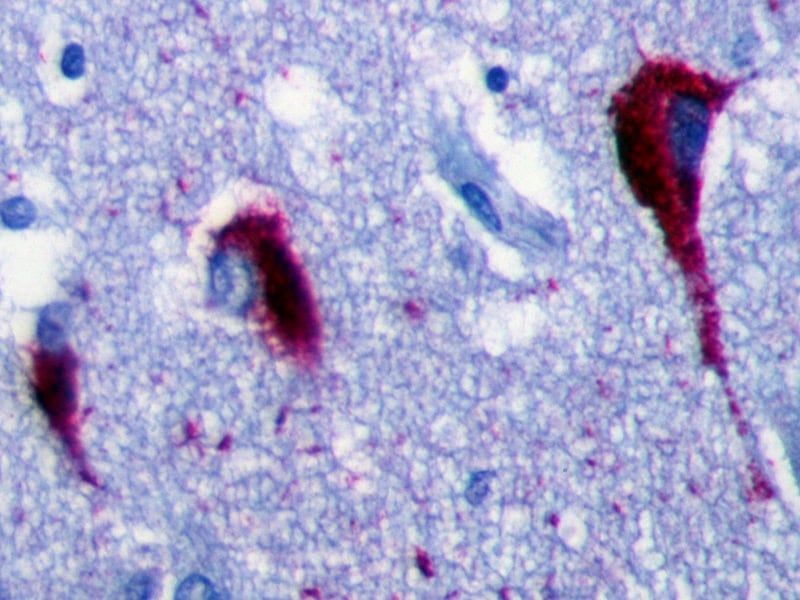
Immunohistochemistry (IHC) is a technique used in pathology and laboratory medicine to identify specific proteins or antigens in tissue sections. It combines the principles of immunology and histology to detect the presence and location of these target molecules within cells and tissues. This technique utilizes antibodies that are specific to the protein or antigen of interest, which are then tagged with a detection system such as a chromogen or fluorophore. The stained tissue sections can be examined under a microscope, allowing for the visualization and analysis of the distribution and expression patterns of the target molecule in the context of the tissue architecture. Immunohistochemistry is widely used in diagnostic pathology to help identify various diseases, including cancer, infectious diseases, and immune-mediated disorders.
Neurofibrillary tangles are a pathological hallmark of several neurodegenerative disorders, most notably Alzheimer's disease. They are intracellular inclusions composed of abnormally phosphorylated and aggregated tau protein, which forms paired helical filaments. These tangles accumulate within the neurons, leading to their dysfunction and eventual death. The presence and density of neurofibrillary tangles are strongly associated with cognitive decline and disease progression in Alzheimer's disease and other related dementias.
Angioscopy is a medical diagnostic procedure that uses a small fiber-optic scope, called an angioscope, to directly visualize the interior of blood vessels. The angioscope is inserted into the vessel through a small incision or catheter and allows physicians to examine the vessel walls for abnormalities such as plaque buildup, inflammation, or damage. This procedure can be used to diagnose and monitor conditions such as coronary artery disease, peripheral artery disease, and vasculitis. It can also be used during surgical procedures to assist with the placement of stents or other devices in the blood vessels.
Protease nexins are a group of proteins that regulate the activity of proteases, which are enzymes that break down other proteins. Proteases play important roles in various biological processes, including blood clotting, immune response, and cell death. However, uncontrolled or excessive protease activity can lead to harmful effects, such as tissue damage and disease progression.
Protease nexins function by forming stable complexes with specific proteases, thereby inhibiting their activity. These complexes also serve as a reservoir of inactive proteases that can be rapidly activated when needed. Protease nexins are involved in various physiological and pathological processes, such as inflammation, neurodegeneration, and cancer.
One well-known example of a protease nexin is the tissue plasminogen activator (tPA) - neuroserpin complex. Neuroserpin is a serine protease inhibitor that forms a complex with tPA, an enzyme that plays a critical role in breaking down blood clots. By forming this complex, neuroserpin regulates the activity of tPA and prevents excessive fibrinolysis, which can lead to bleeding disorders. Mutations in the gene encoding neuroserpin have been associated with familial dementia with Lewy bodies, a form of neurodegenerative disorder.
Transmission electron microscopy (TEM) is a type of microscopy in which an electron beam is transmitted through a ultra-thin specimen, interacting with it as it passes through. An image is formed from the interaction of the electrons with the specimen; the image is then magnified and visualized on a fluorescent screen or recorded on an electronic detector (or photographic film in older models).
TEM can provide high-resolution, high-magnification images that can reveal the internal structure of specimens including cells, viruses, and even molecules. It is widely used in biological and materials science research to investigate the ultrastructure of cells, tissues and materials. In medicine, TEM is used for diagnostic purposes in fields such as virology and bacteriology.
It's important to note that preparing a sample for TEM is a complex process, requiring specialized techniques to create thin (50-100 nm) specimens. These include cutting ultrathin sections of embedded samples using an ultramicrotome, staining with heavy metal salts, and positive staining or negative staining methods.
Electron microscopy (EM) is a type of microscopy that uses a beam of electrons to create an image of the sample being examined, resulting in much higher magnification and resolution than light microscopy. There are several types of electron microscopy, including transmission electron microscopy (TEM), scanning electron microscopy (SEM), and reflection electron microscopy (REM).
In TEM, a beam of electrons is transmitted through a thin slice of the sample, and the electrons that pass through the sample are focused to form an image. This technique can provide detailed information about the internal structure of cells, viruses, and other biological specimens, as well as the composition and structure of materials at the atomic level.
In SEM, a beam of electrons is scanned across the surface of the sample, and the electrons that are scattered back from the surface are detected to create an image. This technique can provide information about the topography and composition of surfaces, as well as the structure of materials at the microscopic level.
REM is a variation of SEM in which the beam of electrons is reflected off the surface of the sample, rather than scattered back from it. This technique can provide information about the surface chemistry and composition of materials.
Electron microscopy has a wide range of applications in biology, medicine, and materials science, including the study of cellular structure and function, disease diagnosis, and the development of new materials and technologies.
I'm sorry for any confusion, but "Hemolytic Plaque Technique" is not a widely recognized or established medical term. It seems like it might be a combination of two different concepts in medical and scientific research: the Hemolytic Assay and the Plaque Assay technique.
A Hemolytic Assay is a method used to measure the amount of hemolysis, or the rupturing of red blood cells, caused by a substance such as a toxin or an antibody. This assay can help determine the concentration of the hemolysin in a sample.
On the other hand, the Plaque Assay Technique is a method used to measure the number of infectious virus particles in a sample. It involves adding a layer of cells (like bacteria) that the virus can infect and then covering it with a nutrient agar overlay. After a period of incubation, clear areas or "plaques" appear in the agar where the viruses have infected and lysed the cells. By counting these plaques, researchers can estimate the number of infectious virus particles present in the original sample.
Therefore, if you're looking for a definition of a Hemolytic Plaque Technique, it might refer to a research method that combines both concepts, possibly measuring the amount of a substance (like an antibody) that causes hemolysis in red blood cells and correlating it with the number of infectious virus particles present. However, I would recommend consulting the original source or author for clarification on their intended meaning.
Protein multimerization refers to the process where multiple protein subunits assemble together to form a complex, repetitive structure called a multimer or oligomer. This can involve the association of identical or similar protein subunits through non-covalent interactions such as hydrogen bonding, ionic bonding, and van der Waals forces. The resulting multimeric structures can have various shapes, sizes, and functions, including enzymatic activity, transport, or structural support. Protein multimerization plays a crucial role in many biological processes and is often necessary for the proper functioning of proteins within cells.
Neurons, also known as nerve cells or neurocytes, are specialized cells that constitute the basic unit of the nervous system. They are responsible for receiving, processing, and transmitting information and signals within the body. Neurons have three main parts: the dendrites, the cell body (soma), and the axon. The dendrites receive signals from other neurons or sensory receptors, while the axon transmits these signals to other neurons, muscles, or glands. The junction between two neurons is called a synapse, where neurotransmitters are released to transmit the signal across the gap (synaptic cleft) to the next neuron. Neurons vary in size, shape, and structure depending on their function and location within the nervous system.
Aniline compounds, also known as aromatic amines, are organic compounds that contain a benzene ring substituted with an amino group (-NH2). Aniline itself is the simplest and most common aniline compound, with the formula C6H5NH2.
Aniline compounds are important in the chemical industry and are used in the synthesis of a wide range of products, including dyes, pharmaceuticals, and rubber chemicals. They can be produced by reducing nitrobenzene or by directly substituting ammonia onto benzene in a process called amination.
It is important to note that aniline compounds are toxic and can cause serious health effects, including damage to the liver, kidneys, and central nervous system. They can also be absorbed through the skin and are known to have carcinogenic properties. Therefore, appropriate safety measures must be taken when handling aniline compounds.
Coronary vessels refer to the network of blood vessels that supply oxygenated blood and nutrients to the heart muscle, also known as the myocardium. The two main coronary arteries are the left main coronary artery and the right coronary artery.
The left main coronary artery branches off into the left anterior descending artery (LAD) and the left circumflex artery (LCx). The LAD supplies blood to the front of the heart, while the LCx supplies blood to the side and back of the heart.
The right coronary artery supplies blood to the right lower part of the heart, including the right atrium and ventricle, as well as the back of the heart.
Coronary vessel disease (CVD) occurs when these vessels become narrowed or blocked due to the buildup of plaque, leading to reduced blood flow to the heart muscle. This can result in chest pain, shortness of breath, or a heart attack.
Atomic Force Microscopy (AFM) is a type of microscopy that allows visualization and measurement of surfaces at the atomic level. It works by using a sharp probe, called a tip, that is mounted on a flexible cantilever. The tip is brought very close to the surface of the sample and as the sample is scanned, the forces between the tip and the sample cause the cantilever to deflect. This deflection is measured and used to generate a topographic map of the surface with extremely high resolution, often on the order of fractions of a nanometer. AFM can be used to study both conductive and non-conductive samples, and can operate in various environments, including air and liquid. It has applications in fields such as materials science, biology, and chemistry.
Calcinosis is a medical condition characterized by the abnormal deposit of calcium salts in various tissues of the body, commonly under the skin or in the muscles and tendons. These calcium deposits can form hard lumps or nodules that can cause pain, inflammation, and restricted mobility. Calcinosis can occur as a complication of other medical conditions, such as autoimmune disorders, kidney disease, and hypercalcemia (high levels of calcium in the blood). In some cases, the cause of calcinosis may be unknown. Treatment for calcinosis depends on the underlying cause and may include medications to manage calcium levels, physical therapy, and surgical removal of large deposits.
A mutation is a permanent change in the DNA sequence of an organism's genome. Mutations can occur spontaneously or be caused by environmental factors such as exposure to radiation, chemicals, or viruses. They may have various effects on the organism, ranging from benign to harmful, depending on where they occur and whether they alter the function of essential proteins. In some cases, mutations can increase an individual's susceptibility to certain diseases or disorders, while in others, they may confer a survival advantage. Mutations are the driving force behind evolution, as they introduce new genetic variability into populations, which can then be acted upon by natural selection.
C57BL/6 (C57 Black 6) is an inbred strain of laboratory mouse that is widely used in biomedical research. The term "inbred" refers to a strain of animals where matings have been carried out between siblings or other closely related individuals for many generations, resulting in a population that is highly homozygous at most genetic loci.
The C57BL/6 strain was established in 1920 by crossing a female mouse from the dilute brown (DBA) strain with a male mouse from the black strain. The resulting offspring were then interbred for many generations to create the inbred C57BL/6 strain.
C57BL/6 mice are known for their robust health, longevity, and ease of handling, making them a popular choice for researchers. They have been used in a wide range of biomedical research areas, including studies of cancer, immunology, neuroscience, cardiovascular disease, and metabolism.
One of the most notable features of the C57BL/6 strain is its sensitivity to certain genetic modifications, such as the introduction of mutations that lead to obesity or impaired glucose tolerance. This has made it a valuable tool for studying the genetic basis of complex diseases and traits.
Overall, the C57BL/6 inbred mouse strain is an important model organism in biomedical research, providing a valuable resource for understanding the genetic and molecular mechanisms underlying human health and disease.
Protein folding is the process by which a protein molecule naturally folds into its three-dimensional structure, following the synthesis of its amino acid chain. This complex process is determined by the sequence and properties of the amino acids, as well as various environmental factors such as temperature, pH, and the presence of molecular chaperones. The final folded conformation of a protein is crucial for its proper function, as it enables the formation of specific interactions between different parts of the molecule, which in turn define its biological activity. Protein misfolding can lead to various diseases, including neurodegenerative disorders such as Alzheimer's and Parkinson's disease.
Neurofibrils are thin, thread-like structures found within the cytoplasm of nerve cells (neurons). They are primarily composed of various proteins and are involved in maintaining the structure and function of neurons. Neurofibrils include two types: neurofilaments and microtubule-associated protein tau (TAU) proteins.
Neurofilaments are intermediate filaments that provide structural support to neurons, while TAU proteins are involved in microtubule assembly, stability, and intracellular transport. Abnormal accumulation and aggregation of these proteins can lead to neurodegenerative disorders, such as Alzheimer's disease, Parkinson's disease, and amyotrophic lateral sclerosis (ALS).
Endopeptidases are a type of enzyme that breaks down proteins by cleaving peptide bonds inside the polypeptide chain. They are also known as proteinases or endoproteinases. These enzymes work within the interior of the protein molecule, cutting it at specific points along its length, as opposed to exopeptidases, which remove individual amino acids from the ends of the protein chain.
Endopeptidases play a crucial role in various biological processes, such as digestion, blood coagulation, and programmed cell death (apoptosis). They are classified based on their catalytic mechanism and the structure of their active site. Some examples of endopeptidase families include serine proteases, cysteine proteases, aspartic proteases, and metalloproteases.
It is important to note that while endopeptidases are essential for normal physiological functions, they can also contribute to disease processes when their activity is unregulated or misdirected. For instance, excessive endopeptidase activity has been implicated in the pathogenesis of neurodegenerative disorders, cancer, and inflammatory conditions.
"Cells, cultured" is a medical term that refers to cells that have been removed from an organism and grown in controlled laboratory conditions outside of the body. This process is called cell culture and it allows scientists to study cells in a more controlled and accessible environment than they would have inside the body. Cultured cells can be derived from a variety of sources, including tissues, organs, or fluids from humans, animals, or cell lines that have been previously established in the laboratory.
Cell culture involves several steps, including isolation of the cells from the tissue, purification and characterization of the cells, and maintenance of the cells in appropriate growth conditions. The cells are typically grown in specialized media that contain nutrients, growth factors, and other components necessary for their survival and proliferation. Cultured cells can be used for a variety of purposes, including basic research, drug development and testing, and production of biological products such as vaccines and gene therapies.
It is important to note that cultured cells may behave differently than they do in the body, and results obtained from cell culture studies may not always translate directly to human physiology or disease. Therefore, it is essential to validate findings from cell culture experiments using additional models and ultimately in clinical trials involving human subjects.
Beta-2 microglobulin (β2M) is a small protein that is a component of the major histocompatibility complex class I molecule, which plays a crucial role in the immune system. It is found on the surface of almost all nucleated cells in the body and is involved in presenting intracellular peptides to T-cells for immune surveillance.
β2M is produced at a relatively constant rate by cells throughout the body and is freely filtered by the glomeruli in the kidneys. Under normal circumstances, most of the filtrated β2M is reabsorbed and catabolized in the proximal tubules of the nephrons. However, when the glomerular filtration rate (GFR) is decreased, as in chronic kidney disease (CKD), the reabsorption capacity of the proximal tubules becomes overwhelmed, leading to increased levels of β2M in the blood and its subsequent appearance in the urine.
Elevated serum and urinary β2M levels have been associated with various clinical conditions, such as CKD, multiple myeloma, autoimmune disorders, and certain infectious diseases. Measuring β2M concentrations can provide valuable information for diagnostic, prognostic, and monitoring purposes in these contexts.
In the field of medicine, "time factors" refer to the duration of symptoms or time elapsed since the onset of a medical condition, which can have significant implications for diagnosis and treatment. Understanding time factors is crucial in determining the progression of a disease, evaluating the effectiveness of treatments, and making critical decisions regarding patient care.
For example, in stroke management, "time is brain," meaning that rapid intervention within a specific time frame (usually within 4.5 hours) is essential to administering tissue plasminogen activator (tPA), a clot-busting drug that can minimize brain damage and improve patient outcomes. Similarly, in trauma care, the "golden hour" concept emphasizes the importance of providing definitive care within the first 60 minutes after injury to increase survival rates and reduce morbidity.
Time factors also play a role in monitoring the progression of chronic conditions like diabetes or heart disease, where regular follow-ups and assessments help determine appropriate treatment adjustments and prevent complications. In infectious diseases, time factors are crucial for initiating antibiotic therapy and identifying potential outbreaks to control their spread.
Overall, "time factors" encompass the significance of recognizing and acting promptly in various medical scenarios to optimize patient outcomes and provide effective care.
I believe there might be a slight confusion in your question. There is no medical definition for "Insulysin" as it seems to be a misspelling of the term "Insulinase" or "Insulysin." I will provide you with the medical definition of Insulinase.
Insulinase, also known as Insulin-degrading enzyme (IDE), is a zinc metalloproteinase found in various tissues, including the liver, brain, and muscle. It is responsible for the intracellular degradation of insulin and other regulatory proteins like amyloid-beta peptide, glucagon, and atrial natriuretic peptide. Insulinase helps regulate blood glucose levels by controlling insulin concentrations in the body. Dysregulation of this enzyme has been implicated in diabetes, Alzheimer's disease, and other neurodegenerative disorders.
Circular dichroism (CD) is a technique used in physics and chemistry to study the structure of molecules, particularly large biological molecules such as proteins and nucleic acids. It measures the difference in absorption of left-handed and right-handed circularly polarized light by a sample. This difference in absorption can provide information about the three-dimensional structure of the molecule, including its chirality or "handedness."
In more technical terms, CD is a form of spectroscopy that measures the differential absorption of left and right circularly polarized light as a function of wavelength. The CD signal is measured in units of millidegrees (mdeg) and can be positive or negative, depending on the type of chromophore and its orientation within the molecule.
CD spectra can provide valuable information about the secondary and tertiary structure of proteins, as well as the conformation of nucleic acids. For example, alpha-helical proteins typically exhibit a strong positive band near 190 nm and two negative bands at around 208 nm and 222 nm, while beta-sheet proteins show a strong positive band near 195 nm and two negative bands at around 217 nm and 175 nm.
CD spectroscopy is a powerful tool for studying the structural changes that occur in biological molecules under different conditions, such as temperature, pH, or the presence of ligands or other molecules. It can also be used to monitor the folding and unfolding of proteins, as well as the binding of drugs or other small molecules to their targets.
There is currently no medical definition for "Alzheimer vaccines" as there are no vaccines that have been approved for use in preventing or curing Alzheimer's disease. However, there are several experimental immunotherapy treatments being investigated in clinical trials. These therapies aim to stimulate the immune system to target and clear beta-amyloid plaques, which are a hallmark pathological feature of Alzheimer's disease.
One type of experimental immunotherapy is known as an active immunization approach, where a vaccine is used to stimulate the patient's own immune system to produce antibodies against beta-amyloid. An example of this approach is the AN1792 vaccine, which was tested in clinical trials but unfortunately showed significant side effects and did not demonstrate clinical benefits.
Another type of experimental immunotherapy is known as a passive immunization approach, where pre-made antibodies are given to the patient through infusions. Several monoclonal antibodies targeting beta-amyloid have been tested in clinical trials, with some showing promise in reducing beta-amyloid levels and slowing cognitive decline. However, further research is needed to determine their safety and efficacy before they can be approved for use as a treatment or prevention for Alzheimer's disease.
Protein binding, in the context of medical and biological sciences, refers to the interaction between a protein and another molecule (known as the ligand) that results in a stable complex. This process is often reversible and can be influenced by various factors such as pH, temperature, and concentration of the involved molecules.
In clinical chemistry, protein binding is particularly important when it comes to drugs, as many of them bind to proteins (especially albumin) in the bloodstream. The degree of protein binding can affect a drug's distribution, metabolism, and excretion, which in turn influence its therapeutic effectiveness and potential side effects.
Protein-bound drugs may be less available for interaction with their target tissues, as only the unbound or "free" fraction of the drug is active. Therefore, understanding protein binding can help optimize dosing regimens and minimize adverse reactions.
Protein conformation refers to the specific three-dimensional shape that a protein molecule assumes due to the spatial arrangement of its constituent amino acid residues and their associated chemical groups. This complex structure is determined by several factors, including covalent bonds (disulfide bridges), hydrogen bonds, van der Waals forces, and ionic bonds, which help stabilize the protein's unique conformation.
Protein conformations can be broadly classified into two categories: primary, secondary, tertiary, and quaternary structures. The primary structure represents the linear sequence of amino acids in a polypeptide chain. The secondary structure arises from local interactions between adjacent amino acid residues, leading to the formation of recurring motifs such as α-helices and β-sheets. Tertiary structure refers to the overall three-dimensional folding pattern of a single polypeptide chain, while quaternary structure describes the spatial arrangement of multiple folded polypeptide chains (subunits) that interact to form a functional protein complex.
Understanding protein conformation is crucial for elucidating protein function, as the specific three-dimensional shape of a protein directly influences its ability to interact with other molecules, such as ligands, nucleic acids, or other proteins. Any alterations in protein conformation due to genetic mutations, environmental factors, or chemical modifications can lead to loss of function, misfolding, aggregation, and disease states like neurodegenerative disorders and cancer.
A cell line is a culture of cells that are grown in a laboratory for use in research. These cells are usually taken from a single cell or group of cells, and they are able to divide and grow continuously in the lab. Cell lines can come from many different sources, including animals, plants, and humans. They are often used in scientific research to study cellular processes, disease mechanisms, and to test new drugs or treatments. Some common types of human cell lines include HeLa cells (which come from a cancer patient named Henrietta Lacks), HEK293 cells (which come from embryonic kidney cells), and HUVEC cells (which come from umbilical vein endothelial cells). It is important to note that cell lines are not the same as primary cells, which are cells that are taken directly from a living organism and have not been grown in the lab.
Aortic diseases refer to conditions that affect the aorta, which is the largest and main artery in the body. The aorta carries oxygenated blood from the heart to the rest of the body. Aortic diseases can weaken or damage the aorta, leading to various complications. Here are some common aortic diseases with their medical definitions:
1. Aortic aneurysm: A localized dilation or bulging of the aortic wall, which can occur in any part of the aorta but is most commonly found in the abdominal aorta (abdominal aortic aneurysm) or the thoracic aorta (thoracic aortic aneurysm). Aneurysms can increase the risk of rupture, leading to life-threatening bleeding.
2. Aortic dissection: A separation of the layers of the aortic wall due to a tear in the inner lining, allowing blood to flow between the layers and potentially cause the aorta to rupture. This is a medical emergency that requires immediate treatment.
3. Aortic stenosis: A narrowing of the aortic valve opening, which restricts blood flow from the heart to the aorta. This can lead to shortness of breath, chest pain, and other symptoms. Severe aortic stenosis may require surgical or transcatheter intervention to replace or repair the aortic valve.
4. Aortic regurgitation: Also known as aortic insufficiency, this condition occurs when the aortic valve does not close properly, allowing blood to leak back into the heart. This can lead to symptoms such as fatigue, shortness of breath, and palpitations. Treatment may include medication or surgical repair or replacement of the aortic valve.
5. Aortitis: Inflammation of the aorta, which can be caused by various conditions such as infections, autoimmune diseases, or vasculitides. Aortitis can lead to aneurysms, dissections, or stenosis and may require medical treatment with immunosuppressive drugs or surgical intervention.
6. Marfan syndrome: A genetic disorder that affects the connective tissue, including the aorta. People with Marfan syndrome are at risk of developing aortic aneurysms and dissections, and may require close monitoring and prophylactic surgery to prevent complications.
The hippocampus is a complex, curved formation in the brain that resembles a seahorse (hence its name, from the Greek word "hippos" meaning horse and "kampos" meaning sea monster). It's part of the limbic system and plays crucial roles in the formation of memories, particularly long-term ones.
This region is involved in spatial navigation and cognitive maps, allowing us to recognize locations and remember how to get to them. Additionally, it's one of the first areas affected by Alzheimer's disease, which often results in memory loss as an early symptom.
Anatomically, it consists of two main parts: the Ammon's horn (or cornu ammonis) and the dentate gyrus. These structures are made up of distinct types of neurons that contribute to different aspects of learning and memory.
Gerstmann-Straussler-Scheinker disease (GSS) is a rare, inherited, progressive neurodegenerative disorder characterized by cerebellar ataxia, pyramidal signs, and distinctive histopathological features in the brain. It is caused by mutations in the PRNP gene, which encodes the prion protein. The disease is transmitted in an autosomal dominant pattern, meaning that a single copy of the mutated gene from either parent is sufficient to cause the disorder.
GSS typically begins in mid-adulthood and progresses over several years to a decade, leading to severe disability and death. The symptoms of GSS include cerebellar ataxia (difficulty with coordination and balance), pyramidal signs (stiffness, spasticity, and hyperreflexia in the limbs), and various other neurological features such as dementia, visual disturbances, and speech difficulties.
Histopathologically, GSS is characterized by the accumulation of abnormal prion protein aggregates in the brain, which can be detected using special staining techniques. These aggregates are thought to be responsible for the neurodegeneration and clinical symptoms of the disease. Currently, there is no cure for GSS and treatment is focused on managing the symptoms of the disorder.
The cerebral cortex is the outermost layer of the brain, characterized by its intricate folded structure and wrinkled appearance. It is a region of great importance as it plays a key role in higher cognitive functions such as perception, consciousness, thought, memory, language, and attention. The cerebral cortex is divided into two hemispheres, each containing four lobes: the frontal, parietal, temporal, and occipital lobes. These areas are responsible for different functions, with some regions specializing in sensory processing while others are involved in motor control or associative functions. The cerebral cortex is composed of gray matter, which contains neuronal cell bodies, and is covered by a layer of white matter that consists mainly of myelinated nerve fibers.
Macrophages are a type of white blood cell that are an essential part of the immune system. They are large, specialized cells that engulf and destroy foreign substances, such as bacteria, viruses, parasites, and fungi, as well as damaged or dead cells. Macrophages are found throughout the body, including in the bloodstream, lymph nodes, spleen, liver, lungs, and connective tissues. They play a critical role in inflammation, immune response, and tissue repair and remodeling.
Macrophages originate from monocytes, which are a type of white blood cell produced in the bone marrow. When monocytes enter the tissues, they differentiate into macrophages, which have a larger size and more specialized functions than monocytes. Macrophages can change their shape and move through tissues to reach sites of infection or injury. They also produce cytokines, chemokines, and other signaling molecules that help coordinate the immune response and recruit other immune cells to the site of infection or injury.
Macrophages have a variety of surface receptors that allow them to recognize and respond to different types of foreign substances and signals from other cells. They can engulf and digest foreign particles, bacteria, and viruses through a process called phagocytosis. Macrophages also play a role in presenting antigens to T cells, which are another type of immune cell that helps coordinate the immune response.
Overall, macrophages are crucial for maintaining tissue homeostasis, defending against infection, and promoting wound healing and tissue repair. Dysregulation of macrophage function has been implicated in a variety of diseases, including cancer, autoimmune disorders, and chronic inflammatory conditions.
Peptides are short chains of amino acid residues linked by covalent bonds, known as peptide bonds. They are formed when two or more amino acids are joined together through a condensation reaction, which results in the elimination of a water molecule and the formation of an amide bond between the carboxyl group of one amino acid and the amino group of another.
Peptides can vary in length from two to about fifty amino acids, and they are often classified based on their size. For example, dipeptides contain two amino acids, tripeptides contain three, and so on. Oligopeptides typically contain up to ten amino acids, while polypeptides can contain dozens or even hundreds of amino acids.
Peptides play many important roles in the body, including serving as hormones, neurotransmitters, enzymes, and antibiotics. They are also used in medical research and therapeutic applications, such as drug delivery and tissue engineering.
Carotid endarterectomy is a surgical procedure to remove plaque buildup (atherosclerosis) from the carotid arteries, which are the major blood vessels that supply oxygen-rich blood to the brain. The surgery involves making an incision in the neck, opening the carotid artery, and removing the plaque from the inside of the artery wall. The goal of the procedure is to restore normal blood flow to the brain and reduce the risk of stroke caused by the narrowing or blockage of the carotid arteries.
Microglia are a type of specialized immune cell found in the brain and spinal cord. They are part of the glial family, which provide support and protection to the neurons in the central nervous system (CNS). Microglia account for about 10-15% of all cells found in the CNS.
The primary role of microglia is to constantly survey their environment and eliminate any potentially harmful agents, such as pathogens, dead cells, or protein aggregates. They do this through a process called phagocytosis, where they engulf and digest foreign particles or cellular debris. In addition to their phagocytic function, microglia also release various cytokines, chemokines, and growth factors that help regulate the immune response in the CNS, promote neuronal survival, and contribute to synaptic plasticity.
Microglia can exist in different activation states depending on the nature of the stimuli they encounter. In a resting state, microglia have a small cell body with numerous branches that are constantly monitoring their surroundings. When activated by an injury, infection, or neurodegenerative process, microglia change their morphology and phenotype, retracting their processes and adopting an amoeboid shape to migrate towards the site of damage or inflammation. Based on the type of activation, microglia can release both pro-inflammatory and anti-inflammatory factors that contribute to either neuroprotection or neurotoxicity.
Dysregulation of microglial function has been implicated in several neurological disorders, including Alzheimer's disease, Parkinson's disease, multiple sclerosis, and Amyotrophic Lateral Sclerosis (ALS). Therefore, understanding the role of microglia in health and disease is crucial for developing novel therapeutic strategies to treat these conditions.
Tertiary protein structure refers to the three-dimensional arrangement of all the elements (polypeptide chains) of a single protein molecule. It is the highest level of structural organization and results from interactions between various side chains (R groups) of the amino acids that make up the protein. These interactions, which include hydrogen bonds, ionic bonds, van der Waals forces, and disulfide bridges, give the protein its unique shape and stability, which in turn determines its function. The tertiary structure of a protein can be stabilized by various factors such as temperature, pH, and the presence of certain ions. Any changes in these factors can lead to denaturation, where the protein loses its tertiary structure and thus its function.
A biological marker, often referred to as a biomarker, is a measurable indicator that reflects the presence or severity of a disease state, or a response to a therapeutic intervention. Biomarkers can be found in various materials such as blood, tissues, or bodily fluids, and they can take many forms, including molecular, histologic, radiographic, or physiological measurements.
In the context of medical research and clinical practice, biomarkers are used for a variety of purposes, such as:
1. Diagnosis: Biomarkers can help diagnose a disease by indicating the presence or absence of a particular condition. For example, prostate-specific antigen (PSA) is a biomarker used to detect prostate cancer.
2. Monitoring: Biomarkers can be used to monitor the progression or regression of a disease over time. For instance, hemoglobin A1c (HbA1c) levels are monitored in diabetes patients to assess long-term blood glucose control.
3. Predicting: Biomarkers can help predict the likelihood of developing a particular disease or the risk of a negative outcome. For example, the presence of certain genetic mutations can indicate an increased risk for breast cancer.
4. Response to treatment: Biomarkers can be used to evaluate the effectiveness of a specific treatment by measuring changes in the biomarker levels before and after the intervention. This is particularly useful in personalized medicine, where treatments are tailored to individual patients based on their unique biomarker profiles.
It's important to note that for a biomarker to be considered clinically valid and useful, it must undergo rigorous validation through well-designed studies, including demonstrating sensitivity, specificity, reproducibility, and clinical relevance.
Gingivitis is a mild form of gum disease (periodontal disease) that causes irritation, redness, swelling and bleeding of the gingiva, or gums. It's important to note that it is reversible with good oral hygiene and professional dental treatment. If left untreated, however, gingivitis can progress to a more severe form of gum disease known as periodontitis, which can result in tissue damage and eventual tooth loss.
Gingivitis is most commonly caused by the buildup of plaque, a sticky film of bacteria that constantly forms on our teeth. When not removed regularly through brushing and flossing, this plaque can harden into tartar, which is more difficult to remove and contributes to gum inflammation. Other factors like hormonal changes, poor nutrition, certain medications, smoking or a weakened immune system may also increase the risk of developing gingivitis.
Aging is a complex, progressive and inevitable process of bodily changes over time, characterized by the accumulation of cellular damage and degenerative changes that eventually lead to increased vulnerability to disease and death. It involves various biological, genetic, environmental, and lifestyle factors that contribute to the decline in physical and mental functions. The medical field studies aging through the discipline of gerontology, which aims to understand the underlying mechanisms of aging and develop interventions to promote healthy aging and extend the human healthspan.
Molecular models are three-dimensional representations of molecular structures that are used in the field of molecular biology and chemistry to visualize and understand the spatial arrangement of atoms and bonds within a molecule. These models can be physical or computer-generated and allow researchers to study the shape, size, and behavior of molecules, which is crucial for understanding their function and interactions with other molecules.
Physical molecular models are often made up of balls (representing atoms) connected by rods or sticks (representing bonds). These models can be constructed manually using materials such as plastic or wooden balls and rods, or they can be created using 3D printing technology.
Computer-generated molecular models, on the other hand, are created using specialized software that allows researchers to visualize and manipulate molecular structures in three dimensions. These models can be used to simulate molecular interactions, predict molecular behavior, and design new drugs or chemicals with specific properties. Overall, molecular models play a critical role in advancing our understanding of molecular structures and their functions.
Presenilin-2 (PSEN2) is a protein that is encoded by the PSEN2 gene in humans. It is a member of the presenilin family, which are integral membrane proteins found in the endoplasmic reticulum and Golgi apparatus. Presenilin-2 is most well-known for its role in the processing of amyloid precursor protein (APP), which is implicated in the pathogenesis of Alzheimer's disease.
In the context of APP processing, presenilin-2 functions as a catalytic subunit of the gamma-secretase complex, which cleaves APP into smaller peptides, including the beta-amyloid peptide (Aβ). Mutations in the PSEN2 gene have been associated with early-onset familial Alzheimer's disease, suggesting that abnormal APP processing and Aβ accumulation play a significant role in the development of this disorder.
However, it is important to note that presenilin-2 has other functions beyond APP processing, including roles in cell signaling, calcium homeostasis, and autophagy. Dysregulation of these processes may also contribute to the pathogenesis of Alzheimer's disease and other neurodegenerative disorders.
Quaternary protein structure refers to the arrangement and interaction of multiple folded protein molecules in a multi-subunit complex. These subunits can be identical or different forms of the same protein or distinctly different proteins that associate to form a functional complex. The quaternary structure is held together by non-covalent interactions, such as hydrogen bonds, ionic bonds, and van der Waals forces. Understanding quaternary structure is crucial for comprehending the function, regulation, and assembly of many protein complexes involved in various cellular processes.
Positron-Emission Tomography (PET) is a type of nuclear medicine imaging that uses small amounts of radioactive material, called a radiotracer, to produce detailed, three-dimensional images. This technique measures metabolic activity within the body, such as sugar metabolism, to help distinguish between healthy and diseased tissue, identify cancerous cells, or examine the function of organs.
During a PET scan, the patient is injected with a radiotracer, typically a sugar-based compound labeled with a positron-emitting radioisotope, such as fluorine-18 (^18^F). The radiotracer accumulates in cells that are metabolically active, like cancer cells. As the radiotracer decays, it emits positrons, which then collide with electrons in nearby tissue, producing gamma rays. A special camera, called a PET scanner, detects these gamma rays and uses this information to create detailed images of the body's internal structures and processes.
PET is often used in conjunction with computed tomography (CT) or magnetic resonance imaging (MRI) to provide both functional and anatomical information, allowing for more accurate diagnosis and treatment planning. Common applications include detecting cancer recurrence, staging and monitoring cancer, evaluating heart function, and assessing brain function in conditions like dementia and epilepsy.
Inflammation is a complex biological response of tissues to harmful stimuli, such as pathogens, damaged cells, or irritants. It is characterized by the following signs: rubor (redness), tumor (swelling), calor (heat), dolor (pain), and functio laesa (loss of function). The process involves the activation of the immune system, recruitment of white blood cells, and release of inflammatory mediators, which contribute to the elimination of the injurious stimuli and initiation of the healing process. However, uncontrolled or chronic inflammation can also lead to tissue damage and diseases.
A "knockout" mouse is a genetically engineered mouse in which one or more genes have been deleted or "knocked out" using molecular biology techniques. This allows researchers to study the function of specific genes and their role in various biological processes, as well as potential associations with human diseases. The mice are generated by introducing targeted DNA modifications into embryonic stem cells, which are then used to create a live animal. Knockout mice have been widely used in biomedical research to investigate gene function, disease mechanisms, and potential therapeutic targets.
Disease progression is the worsening or advancement of a medical condition over time. It refers to the natural course of a disease, including its development, the severity of symptoms and complications, and the impact on the patient's overall health and quality of life. Understanding disease progression is important for developing appropriate treatment plans, monitoring response to therapy, and predicting outcomes.
The rate of disease progression can vary widely depending on the type of medical condition, individual patient factors, and the effectiveness of treatment. Some diseases may progress rapidly over a short period of time, while others may progress more slowly over many years. In some cases, disease progression may be slowed or even halted with appropriate medical interventions, while in other cases, the progression may be inevitable and irreversible.
In clinical practice, healthcare providers closely monitor disease progression through regular assessments, imaging studies, and laboratory tests. This information is used to guide treatment decisions and adjust care plans as needed to optimize patient outcomes and improve quality of life.
An Enzyme-Linked Immunosorbent Assay (ELISA) is a type of analytical biochemistry assay used to detect and quantify the presence of a substance, typically a protein or peptide, in a liquid sample. It takes its name from the enzyme-linked antibodies used in the assay.
In an ELISA, the sample is added to a well containing a surface that has been treated to capture the target substance. If the target substance is present in the sample, it will bind to the surface. Next, an enzyme-linked antibody specific to the target substance is added. This antibody will bind to the captured target substance if it is present. After washing away any unbound material, a substrate for the enzyme is added. If the enzyme is present due to its linkage to the antibody, it will catalyze a reaction that produces a detectable signal, such as a color change or fluorescence. The intensity of this signal is proportional to the amount of target substance present in the sample, allowing for quantification.
ELISAs are widely used in research and clinical settings to detect and measure various substances, including hormones, viruses, and bacteria. They offer high sensitivity, specificity, and reproducibility, making them a reliable choice for many applications.
Alpha-synuclein is a protein that is primarily found in neurons (nerve cells) in the brain. It is encoded by the SNCA gene and is abundantly expressed in presynaptic terminals, where it is believed to play a role in the regulation of neurotransmitter release.
In certain neurological disorders, including Parkinson's disease, dementia with Lewy bodies, and multiple system atrophy, alpha-synuclein can form aggregates known as Lewy bodies and Lewy neurites. These aggregates are a pathological hallmark of these diseases and are believed to contribute to the death of nerve cells, leading to the symptoms associated with these disorders.
The precise function of alpha-synuclein is not fully understood, but it is thought to be involved in various cellular processes such as maintaining the structure of the presynaptic terminal, regulating synaptic vesicle trafficking and neurotransmitter release, and protecting neurons from stress.
A rupture, in medical terms, refers to the breaking or tearing of an organ, tissue, or structure in the body. This can occur due to various reasons such as trauma, injury, increased pressure, or degeneration. A ruptured organ or structure can lead to serious complications, including internal bleeding, infection, and even death, if not treated promptly and appropriately. Examples of ruptures include a ruptured appendix, ruptured eardrum, or a ruptured disc in the spine.
Apolipoprotein E (APOE) is a gene that provides instructions for making a protein involved in the metabolism of fats called lipids. One variant of this gene, APOE4, is associated with an increased risk of developing Alzheimer's disease and other forms of dementia.
The APOE4 allele (variant) is less efficient at clearing beta-amyloid protein, a component of the amyloid plaques found in the brains of people with Alzheimer's disease. This can lead to an accumulation of beta-amyloid and an increased risk of developing Alzheimer's disease.
It is important to note that having one or two copies of the APOE4 allele does not mean that a person will definitely develop Alzheimer's disease, but it does increase the risk. Other factors, such as age, family history, and the presence of other genetic variants, also contribute to the development of this complex disorder.
Fourier Transform Infrared (FTIR) spectroscopy is a type of infrared spectroscopy that uses the Fourier transform mathematical technique to convert the raw data obtained from an interferometer into a more interpretable spectrum. This technique allows for the simultaneous collection of a wide range of wavelengths, resulting in increased sensitivity and speed compared to traditional dispersive infrared spectroscopy.
FTIR spectroscopy measures the absorption or transmission of infrared radiation by a sample as a function of frequency, providing information about the vibrational modes of the molecules present in the sample. This can be used for identification and quantification of chemical compounds, analysis of molecular structure, and investigation of chemical interactions and reactions.
In summary, FTIR spectroscopy is a powerful analytical technique that uses infrared radiation to study the vibrational properties of molecules, with increased sensitivity and speed due to the use of Fourier transform mathematical techniques and an interferometer.
'Staining and labeling' are techniques commonly used in pathology, histology, cytology, and molecular biology to highlight or identify specific components or structures within tissues, cells, or molecules. These methods enable researchers and medical professionals to visualize and analyze the distribution, localization, and interaction of biological entities, contributing to a better understanding of diseases, cellular processes, and potential therapeutic targets.
Medical definitions for 'staining' and 'labeling' are as follows:
1. Staining: A process that involves applying dyes or stains to tissues, cells, or molecules to enhance their contrast and reveal specific structures or components. Stains can be categorized into basic stains (which highlight acidic structures) and acidic stains (which highlight basic structures). Common staining techniques include Hematoxylin and Eosin (H&E), which differentiates cell nuclei from the surrounding cytoplasm and extracellular matrix; special stains, such as PAS (Periodic Acid-Schiff) for carbohydrates or Masson's trichrome for collagen fibers; and immunostains, which use antibodies to target specific proteins.
2. Labeling: A process that involves attaching a detectable marker or tag to a molecule of interest, allowing its identification, quantification, or tracking within a biological system. Labels can be direct, where the marker is directly conjugated to the targeting molecule, or indirect, where an intermediate linker molecule is used to attach the label to the target. Common labeling techniques include fluorescent labels (such as FITC, TRITC, or Alexa Fluor), enzymatic labels (such as horseradish peroxidase or alkaline phosphatase), and radioactive labels (such as ³²P or ¹⁴C). Labeling is often used in conjunction with staining techniques to enhance the specificity and sensitivity of detection.
Together, staining and labeling provide valuable tools for medical research, diagnostics, and therapeutic development, offering insights into cellular and molecular processes that underlie health and disease.
Gliosis is a term used in histopathology and neuroscience to describe the reaction of support cells in the brain, called glial cells, to injury or disease. This response includes an increase in the number and size of glial cells, as well as changes in their shape and function. The most common types of glial cells involved in gliosis are astrocytes and microglia.
Gliosis can be triggered by a variety of factors, including trauma, infection, inflammation, neurodegenerative diseases, and stroke. In response to injury or disease, astrocytes become hypertrophied (enlarged) and undergo changes in their gene expression profile that can lead to the production of various proteins, such as glial fibrillary acidic protein (GFAP). These changes can result in the formation of a dense network of astrocytic processes, which can contribute to the formation of a glial scar.
Microglia, another type of glial cell, become activated during gliosis and play a role in the immune response in the central nervous system (CNS). They can release pro-inflammatory cytokines, chemokines, and reactive oxygen species that contribute to the inflammatory response.
While gliosis is a protective response aimed at containing damage and promoting tissue repair, it can also have negative consequences. For example, the formation of glial scars can impede axonal regeneration and contribute to neurological deficits. Additionally, chronic activation of microglia has been implicated in various neurodegenerative diseases, such as Alzheimer's disease and Parkinson's disease.
An autopsy, also known as a post-mortem examination or obduction, is a medical procedure in which a qualified professional (usually a pathologist) examines a deceased person's body to determine the cause and manner of death. This process may involve various investigative techniques, such as incisions to study internal organs, tissue sampling, microscopic examination, toxicology testing, and other laboratory analyses. The primary purpose of an autopsy is to gather objective evidence about the medical conditions and factors contributing to the individual's demise, which can be essential for legal, insurance, or public health purposes. Additionally, autopsies can provide valuable insights into disease processes and aid in advancing medical knowledge.
Medical Definition:
Magnetic Resonance Imaging (MRI) is a non-invasive diagnostic imaging technique that uses a strong magnetic field and radio waves to create detailed cross-sectional or three-dimensional images of the internal structures of the body. The patient lies within a large, cylindrical magnet, and the scanner detects changes in the direction of the magnetic field caused by protons in the body. These changes are then converted into detailed images that help medical professionals to diagnose and monitor various medical conditions, such as tumors, injuries, or diseases affecting the brain, spinal cord, heart, blood vessels, joints, and other internal organs. MRI does not use radiation like computed tomography (CT) scans.
Presenilins are a group of proteins that play a critical role in the development of early-onset Alzheimer's disease. They are part of the gamma-secretase complex, which is involved in the processing of amyloid precursor protein (APP). This process can result in the formation of beta-amyloid plaques, which are a hallmark of Alzheimer's disease.
Mutations in the presenilin genes (PSEN1 and PSEN2) have been identified as major genetic risk factors for early-onset familial Alzheimer's disease. These mutations can lead to increased production of toxic beta-amyloid fragments, which can accumulate in the brain and cause neuronal damage.
Presenilins also have other functions in the body, including roles in calcium homeostasis, cell signaling, and developmental processes. However, their most well-known function is related to their role in Alzheimer's disease pathogenesis.
In the context of medicine and pharmacology, "kinetics" refers to the study of how a drug moves throughout the body, including its absorption, distribution, metabolism, and excretion (often abbreviated as ADME). This field is called "pharmacokinetics."
1. Absorption: This is the process of a drug moving from its site of administration into the bloodstream. Factors such as the route of administration (e.g., oral, intravenous, etc.), formulation, and individual physiological differences can affect absorption.
2. Distribution: Once a drug is in the bloodstream, it gets distributed throughout the body to various tissues and organs. This process is influenced by factors like blood flow, protein binding, and lipid solubility of the drug.
3. Metabolism: Drugs are often chemically modified in the body, typically in the liver, through processes known as metabolism. These changes can lead to the formation of active or inactive metabolites, which may then be further distributed, excreted, or undergo additional metabolic transformations.
4. Excretion: This is the process by which drugs and their metabolites are eliminated from the body, primarily through the kidneys (urine) and the liver (bile).
Understanding the kinetics of a drug is crucial for determining its optimal dosing regimen, potential interactions with other medications or foods, and any necessary adjustments for special populations like pediatric or geriatric patients, or those with impaired renal or hepatic function.
The aorta is the largest artery in the human body, which originates from the left ventricle of the heart and carries oxygenated blood to the rest of the body. It can be divided into several parts, including the ascending aorta, aortic arch, and descending aorta. The ascending aorta gives rise to the coronary arteries that supply blood to the heart muscle. The aortic arch gives rise to the brachiocephalic, left common carotid, and left subclavian arteries, which supply blood to the head, neck, and upper extremities. The descending aorta travels through the thorax and abdomen, giving rise to various intercostal, visceral, and renal arteries that supply blood to the chest wall, organs, and kidneys.
Coloring agents, also known as food dyes or color additives, are substances that are added to foods, medications, and cosmetics to improve their appearance by giving them a specific color. These agents can be made from both synthetic and natural sources. They must be approved by regulatory agencies such as the U.S. Food and Drug Administration (FDA) before they can be used in products intended for human consumption.
Coloring agents are used for various reasons, including:
* To replace color lost during food processing or preparation
* To make foods more visually appealing
* To help consumers easily identify certain types of food
* To indicate the flavor of a product (e.g., fruit-flavored candies)
It's important to note that while coloring agents can enhance the appearance of products, they do not affect their taste or nutritional value. Some people may have allergic reactions to certain coloring agents, so it's essential to check product labels if you have any known allergies. Additionally, excessive consumption of some synthetic coloring agents has been linked to health concerns, so moderation is key.
Nerve tissue proteins are specialized proteins found in the nervous system that provide structural and functional support to nerve cells, also known as neurons. These proteins include:
1. Neurofilaments: These are type IV intermediate filaments that provide structural support to neurons and help maintain their shape and size. They are composed of three subunits - NFL (light), NFM (medium), and NFH (heavy).
2. Neuronal Cytoskeletal Proteins: These include tubulins, actins, and spectrins that provide structural support to the neuronal cytoskeleton and help maintain its integrity.
3. Neurotransmitter Receptors: These are specialized proteins located on the postsynaptic membrane of neurons that bind neurotransmitters released by presynaptic neurons, triggering a response in the target cell.
4. Ion Channels: These are transmembrane proteins that regulate the flow of ions across the neuronal membrane and play a crucial role in generating and transmitting electrical signals in neurons.
5. Signaling Proteins: These include enzymes, receptors, and adaptor proteins that mediate intracellular signaling pathways involved in neuronal development, differentiation, survival, and death.
6. Adhesion Proteins: These are cell surface proteins that mediate cell-cell and cell-matrix interactions, playing a crucial role in the formation and maintenance of neural circuits.
7. Extracellular Matrix Proteins: These include proteoglycans, laminins, and collagens that provide structural support to nerve tissue and regulate neuronal migration, differentiation, and survival.
Recombinant proteins are artificially created proteins produced through the use of recombinant DNA technology. This process involves combining DNA molecules from different sources to create a new set of genes that encode for a specific protein. The resulting recombinant protein can then be expressed, purified, and used for various applications in research, medicine, and industry.
Recombinant proteins are widely used in biomedical research to study protein function, structure, and interactions. They are also used in the development of diagnostic tests, vaccines, and therapeutic drugs. For example, recombinant insulin is a common treatment for diabetes, while recombinant human growth hormone is used to treat growth disorders.
The production of recombinant proteins typically involves the use of host cells, such as bacteria, yeast, or mammalian cells, which are engineered to express the desired protein. The host cells are transformed with a plasmid vector containing the gene of interest, along with regulatory elements that control its expression. Once the host cells are cultured and the protein is expressed, it can be purified using various chromatography techniques.
Overall, recombinant proteins have revolutionized many areas of biology and medicine, enabling researchers to study and manipulate proteins in ways that were previously impossible.
Neprilysin (NEP), also known as membrane metallo-endopeptidase or CD10, is a type II transmembrane glycoprotein that functions as a zinc-dependent metalloprotease. It is widely expressed in various tissues, including the kidney, brain, heart, and vasculature. Neprilysin plays a crucial role in the breakdown and regulation of several endogenous bioactive peptides, such as natriuretic peptides, bradykinin, substance P, and angiotensin II. By degrading these peptides, neprilysin helps maintain cardiovascular homeostasis, modulate inflammation, and regulate neurotransmission. In the context of heart failure, neprilysin inhibitors have been developed to increase natriuretic peptide levels, promoting diuresis and vasodilation, ultimately improving cardiac function.
Membrane proteins are a type of protein that are embedded in the lipid bilayer of biological membranes, such as the plasma membrane of cells or the inner membrane of mitochondria. These proteins play crucial roles in various cellular processes, including:
1. Cell-cell recognition and signaling
2. Transport of molecules across the membrane (selective permeability)
3. Enzymatic reactions at the membrane surface
4. Energy transduction and conversion
5. Mechanosensation and signal transduction
Membrane proteins can be classified into two main categories: integral membrane proteins, which are permanently associated with the lipid bilayer, and peripheral membrane proteins, which are temporarily or loosely attached to the membrane surface. Integral membrane proteins can further be divided into three subcategories based on their topology:
1. Transmembrane proteins, which span the entire width of the lipid bilayer with one or more alpha-helices or beta-barrels.
2. Lipid-anchored proteins, which are covalently attached to lipids in the membrane via a glycosylphosphatidylinositol (GPI) anchor or other lipid modifications.
3. Monotopic proteins, which are partially embedded in the membrane and have one or more domains exposed to either side of the bilayer.
Membrane proteins are essential for maintaining cellular homeostasis and are targets for various therapeutic interventions, including drug development and gene therapy. However, their structural complexity and hydrophobicity make them challenging to study using traditional biochemical methods, requiring specialized techniques such as X-ray crystallography, nuclear magnetic resonance (NMR) spectroscopy, and single-particle cryo-electron microscopy (cryo-EM).
Western blotting is a laboratory technique used in molecular biology to detect and quantify specific proteins in a mixture of many different proteins. This technique is commonly used to confirm the expression of a protein of interest, determine its size, and investigate its post-translational modifications. The name "Western" blotting distinguishes this technique from Southern blotting (for DNA) and Northern blotting (for RNA).
The Western blotting procedure involves several steps:
1. Protein extraction: The sample containing the proteins of interest is first extracted, often by breaking open cells or tissues and using a buffer to extract the proteins.
2. Separation of proteins by electrophoresis: The extracted proteins are then separated based on their size by loading them onto a polyacrylamide gel and running an electric current through the gel (a process called sodium dodecyl sulfate-polyacrylamide gel electrophoresis or SDS-PAGE). This separates the proteins according to their molecular weight, with smaller proteins migrating faster than larger ones.
3. Transfer of proteins to a membrane: After separation, the proteins are transferred from the gel onto a nitrocellulose or polyvinylidene fluoride (PVDF) membrane using an electric current in a process called blotting. This creates a replica of the protein pattern on the gel but now immobilized on the membrane for further analysis.
4. Blocking: The membrane is then blocked with a blocking agent, such as non-fat dry milk or bovine serum albumin (BSA), to prevent non-specific binding of antibodies in subsequent steps.
5. Primary antibody incubation: A primary antibody that specifically recognizes the protein of interest is added and allowed to bind to its target protein on the membrane. This step may be performed at room temperature or 4°C overnight, depending on the antibody's properties.
6. Washing: The membrane is washed with a buffer to remove unbound primary antibodies.
7. Secondary antibody incubation: A secondary antibody that recognizes the primary antibody (often coupled to an enzyme or fluorophore) is added and allowed to bind to the primary antibody. This step may involve using a horseradish peroxidase (HRP)-conjugated or alkaline phosphatase (AP)-conjugated secondary antibody, depending on the detection method used later.
8. Washing: The membrane is washed again to remove unbound secondary antibodies.
9. Detection: A detection reagent is added to visualize the protein of interest by detecting the signal generated from the enzyme-conjugated or fluorophore-conjugated secondary antibody. This can be done using chemiluminescent, colorimetric, or fluorescent methods.
10. Analysis: The resulting image is analyzed to determine the presence and quantity of the protein of interest in the sample.
Western blotting is a powerful technique for identifying and quantifying specific proteins within complex mixtures. It can be used to study protein expression, post-translational modifications, protein-protein interactions, and more. However, it requires careful optimization and validation to ensure accurate and reproducible results.
C-reactive protein (CRP) is a protein produced by the liver in response to inflammation or infection in the body. It is named after its ability to bind to the C-polysaccharide of pneumococcus, a type of bacteria. CRP levels can be measured with a simple blood test and are often used as a marker of inflammation or infection. Elevated CRP levels may indicate a variety of conditions, including infections, tissue damage, and chronic diseases such as rheumatoid arthritis and cancer. However, it is important to note that CRP is not specific to any particular condition, so additional tests are usually needed to make a definitive diagnosis.
Medical Definition:
"Risk factors" are any attribute, characteristic or exposure of an individual that increases the likelihood of developing a disease or injury. They can be divided into modifiable and non-modifiable risk factors. Modifiable risk factors are those that can be changed through lifestyle choices or medical treatment, while non-modifiable risk factors are inherent traits such as age, gender, or genetic predisposition. Examples of modifiable risk factors include smoking, alcohol consumption, physical inactivity, and unhealthy diet, while non-modifiable risk factors include age, sex, and family history. It is important to note that having a risk factor does not guarantee that a person will develop the disease, but rather indicates an increased susceptibility.
The Predictive Value of Tests, specifically the Positive Predictive Value (PPV) and Negative Predictive Value (NPV), are measures used in diagnostic tests to determine the probability that a positive or negative test result is correct.
Positive Predictive Value (PPV) is the proportion of patients with a positive test result who actually have the disease. It is calculated as the number of true positives divided by the total number of positive results (true positives + false positives). A higher PPV indicates that a positive test result is more likely to be a true positive, and therefore the disease is more likely to be present.
Negative Predictive Value (NPV) is the proportion of patients with a negative test result who do not have the disease. It is calculated as the number of true negatives divided by the total number of negative results (true negatives + false negatives). A higher NPV indicates that a negative test result is more likely to be a true negative, and therefore the disease is less likely to be present.
The predictive value of tests depends on the prevalence of the disease in the population being tested, as well as the sensitivity and specificity of the test. A test with high sensitivity and specificity will generally have higher predictive values than a test with low sensitivity and specificity. However, even a highly sensitive and specific test can have low predictive values if the prevalence of the disease is low in the population being tested.
Coronary angiography is a medical procedure that uses X-ray imaging to visualize the coronary arteries, which supply blood to the heart muscle. During the procedure, a thin, flexible catheter is inserted into an artery in the arm or groin and threaded through the blood vessels to the heart. A contrast dye is then injected through the catheter, and X-ray images are taken as the dye flows through the coronary arteries. These images can help doctors diagnose and treat various heart conditions, such as blockages or narrowing of the arteries, that can lead to chest pain or heart attacks. It is also known as coronary arteriography or cardiac catheterization.
Maze learning is not a medical term per se, but it is a concept that is often used in the field of neuroscience and psychology. It refers to the process by which an animal or human learns to navigate through a complex environment, such as a maze, in order to find its way to a goal or target.
Maze learning involves several cognitive processes, including spatial memory, learning, and problem-solving. As animals or humans navigate through the maze, they encode information about the location of the goal and the various landmarks within the environment. This information is then used to form a cognitive map that allows them to navigate more efficiently in subsequent trials.
Maze learning has been widely used as a tool for studying learning and memory processes in both animals and humans. For example, researchers may use maze learning tasks to investigate the effects of brain damage or disease on cognitive function, or to evaluate the efficacy of various drugs or interventions for improving cognitive performance.
Solubility is a fundamental concept in pharmaceutical sciences and medicine, which refers to the maximum amount of a substance (solute) that can be dissolved in a given quantity of solvent (usually water) at a specific temperature and pressure. Solubility is typically expressed as mass of solute per volume or mass of solvent (e.g., grams per liter, milligrams per milliliter). The process of dissolving a solute in a solvent results in a homogeneous solution where the solute particles are dispersed uniformly throughout the solvent.
Understanding the solubility of drugs is crucial for their formulation, administration, and therapeutic effectiveness. Drugs with low solubility may not dissolve sufficiently to produce the desired pharmacological effect, while those with high solubility might lead to rapid absorption and short duration of action. Therefore, optimizing drug solubility through various techniques like particle size reduction, salt formation, or solubilization is an essential aspect of drug development and delivery.
Post-translational protein processing refers to the modifications and changes that proteins undergo after their synthesis on ribosomes, which are complex molecular machines responsible for protein synthesis. These modifications occur through various biochemical processes and play a crucial role in determining the final structure, function, and stability of the protein.
The process begins with the translation of messenger RNA (mRNA) into a linear polypeptide chain, which is then subjected to several post-translational modifications. These modifications can include:
1. Proteolytic cleavage: The removal of specific segments or domains from the polypeptide chain by proteases, resulting in the formation of mature, functional protein subunits.
2. Chemical modifications: Addition or modification of chemical groups to the side chains of amino acids, such as phosphorylation (addition of a phosphate group), glycosylation (addition of sugar moieties), methylation (addition of a methyl group), acetylation (addition of an acetyl group), and ubiquitination (addition of a ubiquitin protein).
3. Disulfide bond formation: The oxidation of specific cysteine residues within the polypeptide chain, leading to the formation of disulfide bonds between them. This process helps stabilize the three-dimensional structure of proteins, particularly in extracellular environments.
4. Folding and assembly: The acquisition of a specific three-dimensional conformation by the polypeptide chain, which is essential for its function. Chaperone proteins assist in this process to ensure proper folding and prevent aggregation.
5. Protein targeting: The directed transport of proteins to their appropriate cellular locations, such as the nucleus, mitochondria, endoplasmic reticulum, or plasma membrane. This is often facilitated by specific signal sequences within the protein that are recognized and bound by transport machinery.
Collectively, these post-translational modifications contribute to the functional diversity of proteins in living organisms, allowing them to perform a wide range of cellular processes, including signaling, catalysis, regulation, and structural support.
Neurodegenerative diseases are a group of disorders characterized by progressive and persistent loss of neuronal structure and function, often leading to cognitive decline, functional impairment, and ultimately death. These conditions are associated with the accumulation of abnormal protein aggregates, mitochondrial dysfunction, oxidative stress, chronic inflammation, and genetic mutations in the brain. Examples of neurodegenerative diseases include Alzheimer's disease, Parkinson's disease, Huntington's disease, Amyotrophic Lateral Sclerosis (ALS), and Spinal Muscular Atrophy (SMA). The underlying causes and mechanisms of these diseases are not fully understood, and there is currently no cure for most neurodegenerative disorders. Treatment typically focuses on managing symptoms and slowing disease progression.
Brain chemistry refers to the chemical processes that occur within the brain, particularly those involving neurotransmitters, neuromodulators, and neuropeptides. These chemicals are responsible for transmitting signals between neurons (nerve cells) in the brain, allowing for various cognitive, emotional, and physical functions.
Neurotransmitters are chemical messengers that transmit signals across the synapse (the tiny gap between two neurons). Examples of neurotransmitters include dopamine, serotonin, norepinephrine, GABA (gamma-aminobutyric acid), and glutamate. Each neurotransmitter has a specific role in brain function, such as regulating mood, motivation, attention, memory, and movement.
Neuromodulators are chemicals that modify the effects of neurotransmitters on neurons. They can enhance or inhibit the transmission of signals between neurons, thereby modulating brain activity. Examples of neuromodulators include acetylcholine, histamine, and substance P.
Neuropeptides are small protein-like molecules that act as neurotransmitters or neuromodulators. They play a role in various physiological functions, such as pain perception, stress response, and reward processing. Examples of neuropeptides include endorphins, enkephalins, and oxytocin.
Abnormalities in brain chemistry can lead to various neurological and psychiatric conditions, such as depression, anxiety disorders, schizophrenia, Parkinson's disease, and Alzheimer's disease. Understanding brain chemistry is crucial for developing effective treatments for these conditions.
Lipids are a broad group of organic compounds that are insoluble in water but soluble in nonpolar organic solvents. They include fats, waxes, sterols, fat-soluble vitamins (such as vitamins A, D, E, and K), monoglycerides, diglycerides, triglycerides, and phospholipids. Lipids serve many important functions in the body, including energy storage, acting as structural components of cell membranes, and serving as signaling molecules. High levels of certain lipids, particularly cholesterol and triglycerides, in the blood are associated with an increased risk of cardiovascular disease.
Peptide termination factors, also known as release factors, are proteins involved in the process of protein biosynthesis in cells. Specifically, they play a crucial role in the termination step of translation, which is the process by which the genetic code in messenger RNA (mRNA) is translated into a specific sequence of amino acids to form a protein.
During translation, ribosomes move along the mRNA and read the codons (three-nucleotide sequences) to add the corresponding amino acids to the growing polypeptide chain. When the ribosome encounters a stop codon (UAA, UAG, or UGA), peptide termination factors recognize it and bind to the ribosome. The specific factor that recognizes each stop codon is called a class 1 release factor.
In eukaryotic cells, there are two main class 1 release factors: eRF1 (eukaryotic release factor 1) and eRF3. eRF1 recognizes all three stop codons and promotes the hydrolysis of the peptidyl-tRNA bond, releasing the completed polypeptide chain from the ribosome. eRF3 acts as a GTPase and interacts with eRF1 to facilitate its binding to the ribosome.
Once the polypeptide is released, the ribosome dissociates from the mRNA, allowing for another round of translation or degradation of the mRNA. Peptide termination factors are essential for accurate protein synthesis and preventing errors due to premature termination or readthrough of stop codons.
Tunica intima, also known as the intima layer, is the innermost layer of a blood vessel, including arteries and veins. It is in direct contact with the flowing blood and is composed of simple squamous endothelial cells that form a continuous, non-keratinized, stratified epithelium. These cells play a crucial role in maintaining vascular homeostasis by regulating the passage of molecules and immune cells between the blood and the vessel wall, as well as contributing to the maintenance of blood fluidity and preventing coagulation.
The tunica intima is supported by a thin layer of connective tissue called the basement membrane, which provides structural stability and anchorage for the endothelial cells. Beneath the basement membrane lies a loose network of elastic fibers and collagen, known as the internal elastic lamina, that separates the tunica intima from the middle layer, or tunica media.
In summary, the tunica intima is the innermost layer of blood vessels, primarily composed of endothelial cells and a basement membrane, which regulates various functions to maintain vascular homeostasis.
Cholesterol is a type of lipid (fat) molecule that is an essential component of cell membranes and is also used to make certain hormones and vitamins in the body. It is produced by the liver and is also obtained from animal-derived foods such as meat, dairy products, and eggs.
Cholesterol does not mix with blood, so it is transported through the bloodstream by lipoproteins, which are particles made up of both lipids and proteins. There are two main types of lipoproteins that carry cholesterol: low-density lipoproteins (LDL), also known as "bad" cholesterol, and high-density lipoproteins (HDL), also known as "good" cholesterol.
High levels of LDL cholesterol in the blood can lead to a buildup of cholesterol in the walls of the arteries, increasing the risk of heart disease and stroke. On the other hand, high levels of HDL cholesterol are associated with a lower risk of these conditions because HDL helps remove LDL cholesterol from the bloodstream and transport it back to the liver for disposal.
It is important to maintain healthy levels of cholesterol through a balanced diet, regular exercise, and sometimes medication if necessary. Regular screening is also recommended to monitor cholesterol levels and prevent health complications.
Vascular calcification is a pathological process characterized by the deposition of calcium phosphate crystals in the blood vessels, particularly in the tunica intima (the innermost layer) of the arterial wall. This condition can lead to the stiffening and hardening of the arteries, which can impair their ability to expand and contract with each beat of the heart. Vascular calcification is often associated with various cardiovascular diseases such as atherosclerosis, diabetes, chronic kidney disease, and aging. It can contribute to an increased risk of cardiovascular events such as myocardial infarction, stroke, and peripheral artery disease.
Cricetinae is a subfamily of rodents that includes hamsters, gerbils, and relatives. These small mammals are characterized by having short limbs, compact bodies, and cheek pouches for storing food. They are native to various parts of the world, particularly in Europe, Asia, and Africa. Some species are popular pets due to their small size, easy care, and friendly nature. In a medical context, understanding the biology and behavior of Cricetinae species can be important for individuals who keep them as pets or for researchers studying their physiology.
X-ray computed tomography (CT or CAT scan) is a medical imaging method that uses computer-processed combinations of many X-ray images taken from different angles to produce cross-sectional (tomographic) images (virtual "slices") of the body. These cross-sectional images can then be used to display detailed internal views of organs, bones, and soft tissues in the body.
The term "computed tomography" is used instead of "CT scan" or "CAT scan" because the machines take a series of X-ray measurements from different angles around the body and then use a computer to process these data to create detailed images of internal structures within the body.
CT scanning is a noninvasive, painless medical test that helps physicians diagnose and treat medical conditions. CT imaging provides detailed information about many types of tissue including lung, bone, soft tissue and blood vessels. CT examinations can be performed on every part of the body for a variety of reasons including diagnosis, surgical planning, and monitoring of therapeutic responses.
In computed tomography (CT), an X-ray source and detector rotate around the patient, measuring the X-ray attenuation at many different angles. A computer uses this data to construct a cross-sectional image by the process of reconstruction. This technique is called "tomography". The term "computed" refers to the use of a computer to reconstruct the images.
CT has become an important tool in medical imaging and diagnosis, allowing radiologists and other physicians to view detailed internal images of the body. It can help identify many different medical conditions including cancer, heart disease, lung nodules, liver tumors, and internal injuries from trauma. CT is also commonly used for guiding biopsies and other minimally invasive procedures.
In summary, X-ray computed tomography (CT or CAT scan) is a medical imaging technique that uses computer-processed combinations of many X-ray images taken from different angles to produce cross-sectional images of the body. It provides detailed internal views of organs, bones, and soft tissues in the body, allowing physicians to diagnose and treat medical conditions.
Messenger RNA (mRNA) is a type of RNA (ribonucleic acid) that carries genetic information copied from DNA in the form of a series of three-base code "words," each of which specifies a particular amino acid. This information is used by the cell's machinery to construct proteins, a process known as translation. After being transcribed from DNA, mRNA travels out of the nucleus to the ribosomes in the cytoplasm where protein synthesis occurs. Once the protein has been synthesized, the mRNA may be degraded and recycled. Post-transcriptional modifications can also occur to mRNA, such as alternative splicing and addition of a 5' cap and a poly(A) tail, which can affect its stability, localization, and translation efficiency.
Hydrogen-ion concentration, also known as pH, is a measure of the acidity or basicity of a solution. It is defined as the negative logarithm (to the base 10) of the hydrogen ion activity in a solution. The standard unit of measurement is the pH unit. A pH of 7 is neutral, less than 7 is acidic, and greater than 7 is basic.
In medical terms, hydrogen-ion concentration is important for maintaining homeostasis within the body. For example, in the stomach, a high hydrogen-ion concentration (low pH) is necessary for the digestion of food. However, in other parts of the body such as blood, a high hydrogen-ion concentration can be harmful and lead to acidosis. Conversely, a low hydrogen-ion concentration (high pH) in the blood can lead to alkalosis. Both acidosis and alkalosis can have serious consequences on various organ systems if not corrected.
Necrosis is the premature death of cells or tissues due to damage or injury, such as from infection, trauma, infarction (lack of blood supply), or toxic substances. It's a pathological process that results in the uncontrolled and passive degradation of cellular components, ultimately leading to the release of intracellular contents into the extracellular space. This can cause local inflammation and may lead to further tissue damage if not treated promptly.
There are different types of necrosis, including coagulative, liquefactive, caseous, fat, fibrinoid, and gangrenous necrosis, each with distinct histological features depending on the underlying cause and the affected tissues or organs.
Islet amyloid polypeptide receptors (IAPRs) are a type of G protein-coupled receptor (GPCR) that play a role in the regulation of glucose homeostasis and energy balance. They are activated by the hormone islet amyloid polypeptide (IAPP), also known as amylin, which is co-secreted with insulin from pancreatic beta cells in response to meals.
There are two subtypes of IAPRs, named RCS3 (or IA PR1) andRAMP2 (or IA PR2). These receptors can form heterodimers with other GPCRs, such as the calcitonin receptor (CTR), to form functional complexes that bind IAPP with high affinity. Activation of IAPRs by IAPP has been shown to inhibit gastric emptying, reduce food intake, and lower blood glucose levels, suggesting a role in the regulation of satiety and glucose metabolism.
Mutations in the genes encoding IAPP and IAPRs have been associated with the development of type 2 diabetes, suggesting that dysfunction of the IAPP/IAPR system may contribute to the pathogenesis of this disease. Additionally, the accumulation of misfolded IAPP in pancreatic islets can lead to the formation of amyloid deposits, which are a characteristic feature of type 2 diabetes and have been implicated in the destruction of beta cells and the development of insulin resistance.
Prion diseases, also known as transmissible spongiform encephalopathies (TSEs), are a group of progressive neurodegenerative disorders that affect both humans and animals. They are unique in that they are caused by prions, which are misfolded proteins rather than infectious agents like bacteria or viruses. These abnormal prions can cause other normal proteins to misfold and accumulate in the brain, leading to brain damage and neurodegeneration.
Prion diseases can be sporadic, inherited, or acquired. Sporadic forms occur without a known cause and are the most common type. Inherited prion diseases are caused by mutations in the PRNP gene and are often associated with a family history of the disease. Acquired prion diseases can result from exposure to contaminated food (as in variant Creutzfeldt-Jakob disease), medical procedures (iatrogenic Creutzfeldt-Jakob disease), or inherited forms of the disease that cause abnormal prions to be secreted in body fluids (like kuru).
Common prion diseases in humans include:
1. Creutzfeldt-Jakob disease (CJD) - sporadic, inherited, and acquired forms
2. Variant Creutzfeldt-Jakob disease (vCJD) - acquired form linked to consumption of contaminated beef products
3. Gerstmann-Sträussler-Scheinker syndrome (GSS) - inherited form
4. Fatal familial insomnia (FFI) - inherited form
5. Kuru - an acquired form that occurred in a isolated tribe due to cannibalistic practices, now eradicated
Prion diseases are characterized by rapidly progressing dementia, neurological symptoms, and motor dysfunction. There is no known cure for these diseases, and they are universally fatal.
I believe there may be some confusion in your question. "Rabbits" is a common name used to refer to the Lagomorpha species, particularly members of the family Leporidae. They are small mammals known for their long ears, strong legs, and quick reproduction.
However, if you're referring to "rabbits" in a medical context, there is a term called "rabbit syndrome," which is a rare movement disorder characterized by repetitive, involuntary movements of the fingers, resembling those of a rabbit chewing. It is also known as "finger-chewing chorea." This condition is usually associated with certain medications, particularly antipsychotics, and typically resolves when the medication is stopped or adjusted.
The Islets of Langerhans are clusters of specialized cells within the pancreas, an organ located behind the stomach. These islets are named after Paul Langerhans, who first identified them in 1869. They constitute around 1-2% of the total mass of the pancreas and are distributed throughout its substance.
The Islets of Langerhans contain several types of cells, including:
1. Alpha (α) cells: These produce and release glucagon, a hormone that helps to regulate blood sugar levels by promoting the conversion of glycogen to glucose in the liver when blood sugar levels are low.
2. Beta (β) cells: These produce and release insulin, a hormone that promotes the uptake and utilization of glucose by cells throughout the body, thereby lowering blood sugar levels.
3. Delta (δ) cells: These produce and release somatostatin, a hormone that inhibits the release of both insulin and glucagon and helps regulate their secretion in response to changing blood sugar levels.
4. PP cells (gamma or γ cells): These produce and release pancreatic polypeptide, which plays a role in regulating digestive enzyme secretion and gastrointestinal motility.
Dysfunction of the Islets of Langerhans can lead to various endocrine disorders, such as diabetes mellitus, where insulin-producing beta cells are damaged or destroyed, leading to impaired blood sugar regulation.
Benzothiazoles are a class of heterocyclic organic compounds that contain a benzene fused to a thiazole ring. They have the chemical formula C7H5NS. Benzothiazoles and their derivatives have a wide range of applications in various industries, including pharmaceuticals, agrochemicals, dyes, and materials science.
In the medical field, benzothiazoles have been studied for their potential therapeutic properties. Some benzothiazole derivatives have shown promising results as anti-inflammatory, antimicrobial, antiviral, and anticancer agents. However, more research is needed to fully understand the medical potential of these compounds and to develop safe and effective drugs based on them.
It's important to note that while benzothiazoles themselves have some biological activity, most of the medical applications come from their derivatives, which are modified versions of the basic benzothiazole structure. These modifications can significantly alter the properties of the compound, leading to new therapeutic possibilities.
A mouthwash is an antiseptic or therapeutic solution that is held in the mouth and then spit out, rather than swallowed. It is used to improve oral hygiene, to freshen breath, and to help prevent dental cavities, gingivitis, and other periodontal diseases.
Mouthwashes can contain a variety of ingredients, including water, alcohol, fluoride, chlorhexidine, essential oils, and other antimicrobial agents. Some mouthwashes are available over-the-counter, while others require a prescription. It is important to follow the instructions for use provided by the manufacturer or your dentist to ensure the safe and effective use of mouthwash.
A Severity of Illness Index is a measurement tool used in healthcare to assess the severity of a patient's condition and the risk of mortality or other adverse outcomes. These indices typically take into account various physiological and clinical variables, such as vital signs, laboratory values, and co-morbidities, to generate a score that reflects the patient's overall illness severity.
Examples of Severity of Illness Indices include the Acute Physiology and Chronic Health Evaluation (APACHE) system, the Simplified Acute Physiology Score (SAPS), and the Mortality Probability Model (MPM). These indices are often used in critical care settings to guide clinical decision-making, inform prognosis, and compare outcomes across different patient populations.
It is important to note that while these indices can provide valuable information about a patient's condition, they should not be used as the sole basis for clinical decision-making. Rather, they should be considered in conjunction with other factors, such as the patient's overall clinical presentation, treatment preferences, and goals of care.
Protein denaturation is a process in which the native structure of a protein is altered, leading to loss of its biological activity. This can be caused by various factors such as changes in temperature, pH, or exposure to chemicals or radiation. The three-dimensional shape of a protein is crucial for its function, and denaturation causes the protein to lose this shape, resulting in impaired or complete loss of function. Denaturation is often irreversible and can lead to the aggregation of proteins, which can have negative effects on cellular function and can contribute to diseases such as Alzheimer's and Parkinson's.
PC12 cells are a type of rat pheochromocytoma cell line, which are commonly used in scientific research. Pheochromocytomas are tumors that develop from the chromaffin cells of the adrenal gland, and PC12 cells are a subtype of these cells.
PC12 cells have several characteristics that make them useful for research purposes. They can be grown in culture and can be differentiated into a neuron-like phenotype when treated with nerve growth factor (NGF). This makes them a popular choice for studies involving neuroscience, neurotoxicity, and neurodegenerative disorders.
PC12 cells are also known to express various neurotransmitter receptors, ion channels, and other proteins that are relevant to neuronal function, making them useful for studying the mechanisms of drug action and toxicity. Additionally, PC12 cells can be used to study the regulation of cell growth and differentiation, as well as the molecular basis of cancer.
'Gene expression regulation' refers to the processes that control whether, when, and where a particular gene is expressed, meaning the production of a specific protein or functional RNA encoded by that gene. This complex mechanism can be influenced by various factors such as transcription factors, chromatin remodeling, DNA methylation, non-coding RNAs, and post-transcriptional modifications, among others. Proper regulation of gene expression is crucial for normal cellular function, development, and maintaining homeostasis in living organisms. Dysregulation of gene expression can lead to various diseases, including cancer and genetic disorders.
Apolipoprotein E3 (ApoE3) is one of the three major isoforms of apolipoprotein E (ApoE), a protein involved in the metabolism of lipids, particularly cholesterol. ApoE is produced by the APOE gene, which has three common alleles: ε2, ε3, and ε4. These alleles result in three main isoforms of the protein: ApoE2, ApoE3, and ApoE4.
ApoE3 is the most common isoform, found in approximately 77-78% of the population. It has a slightly different amino acid sequence compared to ApoE2 and ApoE4, which can affect its function. ApoE3 is thought to play a neutral or protective role in the risk of developing Alzheimer's disease and cardiovascular diseases, although some studies suggest that it may have a mildly favorable effect on lipid metabolism compared to ApoE4.
Nuclear Magnetic Resonance (NMR) Biomolecular is a research technique that uses magnetic fields and radio waves to study the structure and dynamics of biological molecules, such as proteins and nucleic acids. This technique measures the magnetic properties of atomic nuclei within these molecules, specifically their spin, which can be influenced by the application of an external magnetic field.
When a sample is placed in a strong magnetic field, the nuclei absorb and emit electromagnetic radiation at specific frequencies, known as resonance frequencies, which are determined by the molecular structure and environment of the nuclei. By analyzing these resonance frequencies and their interactions, researchers can obtain detailed information about the three-dimensional structure, dynamics, and interactions of biomolecules.
NMR spectroscopy is a non-destructive technique that allows for the study of biological molecules in solution, which makes it an important tool for understanding the function and behavior of these molecules in their natural environment. Additionally, NMR can be used to study the effects of drugs, ligands, and other small molecules on biomolecular structure and dynamics, making it a valuable tool in drug discovery and development.
Psoriasis is a chronic skin disorder that is characterized by recurrent episodes of red, scaly patches on the skin. The scales are typically silvery-white and often occur on the elbows, knees, scalp, and lower back, but they can appear anywhere on the body. The exact cause of psoriasis is unknown, but it is believed to be related to an immune system issue that causes skin cells to grow too quickly.
There are several types of psoriasis, including plaque psoriasis (the most common form), guttate psoriasis, inverse psoriasis, pustular psoriasis, and erythrodermic psoriasis. The symptoms and severity of the condition can vary widely from person to person, ranging from mild to severe.
While there is no cure for psoriasis, various treatments are available that can help manage the symptoms and improve quality of life. These may include topical medications, light therapy, and systemic medications such as biologics. Lifestyle measures such as stress reduction, quitting smoking, and avoiding triggers (such as certain foods or alcohol) may also be helpful in managing psoriasis.
Memory disorders are a category of cognitive impairments that affect an individual's ability to acquire, store, retain, and retrieve memories. These disorders can be caused by various underlying medical conditions, including neurological disorders, psychiatric illnesses, substance abuse, or even normal aging processes. Some common memory disorders include:
1. Alzheimer's disease: A progressive neurodegenerative disorder that primarily affects older adults and is characterized by a decline in cognitive abilities, including memory, language, problem-solving, and decision-making skills.
2. Dementia: A broader term used to describe a group of symptoms associated with a decline in cognitive function severe enough to interfere with daily life. Alzheimer's disease is the most common cause of dementia, but other causes include vascular dementia, Lewy body dementia, and frontotemporal dementia.
3. Amnesia: A memory disorder characterized by difficulties in forming new memories or recalling previously learned information due to brain damage or disease. Amnesia can be temporary or permanent and may result from head trauma, stroke, infection, or substance abuse.
4. Mild cognitive impairment (MCI): A condition where an individual experiences mild but noticeable memory or cognitive difficulties that are greater than expected for their age and education level. While some individuals with MCI may progress to dementia, others may remain stable or even improve over time.
5. Korsakoff's syndrome: A memory disorder often caused by alcohol abuse and thiamine deficiency, characterized by severe short-term memory loss, confabulation (making up stories to fill in memory gaps), and disorientation.
It is essential to consult a healthcare professional if you or someone you know experiences persistent memory difficulties, as early diagnosis and intervention can help manage symptoms and improve quality of life.
Computer-assisted image processing is a medical term that refers to the use of computer systems and specialized software to improve, analyze, and interpret medical images obtained through various imaging techniques such as X-ray, CT (computed tomography), MRI (magnetic resonance imaging), ultrasound, and others.
The process typically involves several steps, including image acquisition, enhancement, segmentation, restoration, and analysis. Image processing algorithms can be used to enhance the quality of medical images by adjusting contrast, brightness, and sharpness, as well as removing noise and artifacts that may interfere with accurate diagnosis. Segmentation techniques can be used to isolate specific regions or structures of interest within an image, allowing for more detailed analysis.
Computer-assisted image processing has numerous applications in medical imaging, including detection and characterization of lesions, tumors, and other abnormalities; assessment of organ function and morphology; and guidance of interventional procedures such as biopsies and surgeries. By automating and standardizing image analysis tasks, computer-assisted image processing can help to improve diagnostic accuracy, efficiency, and consistency, while reducing the potential for human error.
Nerve degeneration, also known as neurodegeneration, is the progressive loss of structure and function of neurons, which can lead to cognitive decline, motor impairment, and various other symptoms. This process occurs due to a variety of factors, including genetics, environmental influences, and aging. It is a key feature in several neurological disorders such as Alzheimer's disease, Parkinson's disease, Huntington's disease, and multiple sclerosis. The degeneration can affect any part of the nervous system, leading to different symptoms depending on the location and extent of the damage.
The acute-phase reaction is a complex series of physiological responses that occur in response to tissue injury, infection, or stress. It is characterized by the release of pro-inflammatory cytokines such as interleukin-1 (IL-1), IL-6, and tumor necrosis factor-alpha (TNF-α) from activated immune cells, including macrophages and neutrophils.
These cytokines trigger a range of systemic effects, including fever, increased heart rate and respiratory rate, decreased appetite, and changes in white blood cell count. They also stimulate the production of acute-phase proteins (APPs) by the liver, such as C-reactive protein (CRP), fibrinogen, and serum amyloid A.
The acute-phase reaction is an important part of the body's immune response to injury or infection, helping to promote healing and fight off pathogens. However, excessive or prolonged activation of the acute-phase reaction can contribute to the development of chronic inflammatory conditions and diseases such as rheumatoid arthritis, atherosclerosis, and cancer.
A base sequence in the context of molecular biology refers to the specific order of nucleotides in a DNA or RNA molecule. In DNA, these nucleotides are adenine (A), guanine (G), cytosine (C), and thymine (T). In RNA, uracil (U) takes the place of thymine. The base sequence contains genetic information that is transcribed into RNA and ultimately translated into proteins. It is the exact order of these bases that determines the genetic code and thus the function of the DNA or RNA molecule.
The Periodontal Index (PI) is not a current or widely used medical/dental term. However, in the past, it was used to describe a method for assessing and measuring the severity of periodontal disease, also known as gum disease.
Developed by Henry H. Klein and colleagues in 1978, the Periodontal Index was a scoring system that evaluated four parameters: gingival inflammation, gingival bleeding, calculus (tartar) presence, and periodontal pocket depths. The scores for each parameter ranged from 0 to 3, with higher scores indicating worse periodontal health. The overall PI score was the sum of the individual parameter scores, ranging from 0 to 12.
However, due to its limited ability to predict future disease progression and the introduction of more comprehensive assessment methods like the Community Periodontal Index (CPI) and the Basic Periodontal Examination (BPE), the use of the Periodontal Index has become less common in dental practice and research.
Astrocytes are a type of star-shaped glial cell found in the central nervous system (CNS), including the brain and spinal cord. They play crucial roles in supporting and maintaining the health and function of neurons, which are the primary cells responsible for transmitting information in the CNS.
Some of the essential functions of astrocytes include:
1. Supporting neuronal structure and function: Astrocytes provide structural support to neurons by ensheathing them and maintaining the integrity of the blood-brain barrier, which helps regulate the entry and exit of substances into the CNS.
2. Regulating neurotransmitter levels: Astrocytes help control the levels of neurotransmitters in the synaptic cleft (the space between two neurons) by taking up excess neurotransmitters and breaking them down, thus preventing excessive or prolonged activation of neuronal receptors.
3. Providing nutrients to neurons: Astrocytes help supply energy metabolites, such as lactate, to neurons, which are essential for their survival and function.
4. Modulating synaptic activity: Through the release of various signaling molecules, astrocytes can modulate synaptic strength and plasticity, contributing to learning and memory processes.
5. Participating in immune responses: Astrocytes can respond to CNS injuries or infections by releasing pro-inflammatory cytokines and chemokines, which help recruit immune cells to the site of injury or infection.
6. Promoting neuronal survival and repair: In response to injury or disease, astrocytes can become reactive and undergo morphological changes that aid in forming a glial scar, which helps contain damage and promote tissue repair. Additionally, they release growth factors and other molecules that support the survival and regeneration of injured neurons.
Dysfunction or damage to astrocytes has been implicated in several neurological disorders, including Alzheimer's disease, Parkinson's disease, amyotrophic lateral sclerosis (ALS), and multiple sclerosis (MS).
Intracranial arteriosclerosis is a medical condition characterized by the thickening and hardening of the walls of the intracranial arteries, which are the blood vessels that supply blood to the brain. This process is caused by the buildup of plaque, made up of fat, cholesterol, and other substances, within the walls of the arteries.
Intracranial arteriosclerosis can lead to a narrowing or blockage of the affected arteries, reducing blood flow to the brain. This can result in various neurological symptoms, such as headaches, dizziness, seizures, and transient ischemic attacks (TIAs) or strokes.
The condition is more common in older adults, particularly those with a history of hypertension, diabetes, smoking, and high cholesterol levels. Intracranial arteriosclerosis can be diagnosed through imaging tests such as magnetic resonance angiography (MRA) or computed tomographic angiography (CTA). Treatment typically involves managing risk factors and may include medications to control blood pressure, cholesterol levels, and prevent blood clots. In severe cases, surgical procedures such as angioplasty and stenting may be necessary to open up the affected arteries.
Dementia is a broad term that describes a decline in cognitive functioning, including memory, language, problem-solving, and judgment, severe enough to interfere with daily life. It is not a specific disease but rather a group of symptoms that may be caused by various underlying diseases or conditions. Alzheimer's disease is the most common cause of dementia, accounting for 60-80% of cases. Other causes include vascular dementia, Lewy body dementia, frontotemporal dementia, and Huntington's disease.
The symptoms of dementia can vary widely depending on the cause and the specific areas of the brain that are affected. However, common early signs of dementia may include:
* Memory loss that affects daily life
* Difficulty with familiar tasks
* Problems with language or communication
* Difficulty with visual and spatial abilities
* Misplacing things and unable to retrace steps
* Decreased or poor judgment
* Withdrawal from work or social activities
* Changes in mood or behavior
Dementia is a progressive condition, meaning that symptoms will gradually worsen over time. While there is currently no cure for dementia, early diagnosis and treatment can help slow the progression of the disease and improve quality of life for those affected.
Protein stability refers to the ability of a protein to maintain its native structure and function under various physiological conditions. It is determined by the balance between forces that promote a stable conformation, such as intramolecular interactions (hydrogen bonds, van der Waals forces, and hydrophobic effects), and those that destabilize it, such as thermal motion, chemical denaturation, and environmental factors like pH and salt concentration. A protein with high stability is more resistant to changes in its structure and function, even under harsh conditions, while a protein with low stability is more prone to unfolding or aggregation, which can lead to loss of function or disease states, such as protein misfolding diseases.
The brachiocephalic trunk, also known as the brachiocephalic artery or innominate artery, is a large vessel that branches off the aorta and divides into the right common carotid artery and the right subclavian artery. It supplies blood to the head, neck, and arms on the right side of the body.
Protein precursors, also known as proproteins or prohormones, are inactive forms of proteins that undergo post-translational modification to become active. These modifications typically include cleavage of the precursor protein by specific enzymes, resulting in the release of the active protein. This process allows for the regulation and control of protein activity within the body. Protein precursors can be found in various biological processes, including the endocrine system where they serve as inactive hormones that can be converted into their active forms when needed.
Trifluoroethanol (TFE) is not a medical term, but it is a chemical compound with the formula CF3CH2OH. It is a colorless liquid that is used in various scientific and industrial applications. In the context of medical research, TFE has been used as a solvent for spectroscopic studies and as a reagent in organic synthesis.
TFE is known to have strong hydrogen bonding properties due to the electronegativity of the fluorine atoms, which makes it an excellent polar solvent. It can dissolve a wide range of organic compounds, including proteins and nucleic acids, making it useful for studying their structures and interactions.
While TFE is not used as a medication or therapeutic agent, it may have potential applications in medical research and drug development. For example, some studies have investigated the use of TFE as a cryoprotectant to prevent damage to cells and tissues during freezing and thawing. However, more research is needed to fully understand the potential benefits and risks of using TFE in medical contexts.
The tunica media is the middle layer of the wall of a blood vessel or hollow organ in the body. It is primarily composed of smooth muscle cells and elastic fibers, which allow the vessel or organ to expand and contract. This layer helps regulate the diameter of the lumen (the inner space) of the vessel or organ, thereby controlling the flow of fluids such as blood or lymph through it. The tunica media plays a crucial role in maintaining proper organ function and blood pressure regulation.
Transfection is a term used in molecular biology that refers to the process of deliberately introducing foreign genetic material (DNA, RNA or artificial gene constructs) into cells. This is typically done using chemical or physical methods, such as lipofection or electroporation. Transfection is widely used in research and medical settings for various purposes, including studying gene function, producing proteins, developing gene therapies, and creating genetically modified organisms. It's important to note that transfection is different from transduction, which is the process of introducing genetic material into cells using viruses as vectors.
Reproducibility of results in a medical context refers to the ability to obtain consistent and comparable findings when a particular experiment or study is repeated, either by the same researcher or by different researchers, following the same experimental protocol. It is an essential principle in scientific research that helps to ensure the validity and reliability of research findings.
In medical research, reproducibility of results is crucial for establishing the effectiveness and safety of new treatments, interventions, or diagnostic tools. It involves conducting well-designed studies with adequate sample sizes, appropriate statistical analyses, and transparent reporting of methods and findings to allow other researchers to replicate the study and confirm or refute the results.
The lack of reproducibility in medical research has become a significant concern in recent years, as several high-profile studies have failed to produce consistent findings when replicated by other researchers. This has led to increased scrutiny of research practices and a call for greater transparency, rigor, and standardization in the conduct and reporting of medical research.
Fluorescence microscopy is a type of microscopy that uses fluorescent dyes or proteins to highlight and visualize specific components within a sample. In this technique, the sample is illuminated with high-energy light, typically ultraviolet (UV) or blue light, which excites the fluorescent molecules causing them to emit lower-energy, longer-wavelength light, usually visible light in the form of various colors. This emitted light is then collected by the microscope and detected to produce an image.
Fluorescence microscopy has several advantages over traditional brightfield microscopy, including the ability to visualize specific structures or molecules within a complex sample, increased sensitivity, and the potential for quantitative analysis. It is widely used in various fields of biology and medicine, such as cell biology, neuroscience, and pathology, to study the structure, function, and interactions of cells and proteins.
There are several types of fluorescence microscopy techniques, including widefield fluorescence microscopy, confocal microscopy, two-photon microscopy, and total internal reflection fluorescence (TIRF) microscopy, each with its own strengths and limitations. These techniques can provide valuable insights into the behavior of cells and proteins in health and disease.
Biological models, also known as physiological models or organismal models, are simplified representations of biological systems, processes, or mechanisms that are used to understand and explain the underlying principles and relationships. These models can be theoretical (conceptual or mathematical) or physical (such as anatomical models, cell cultures, or animal models). They are widely used in biomedical research to study various phenomena, including disease pathophysiology, drug action, and therapeutic interventions.
Examples of biological models include:
1. Mathematical models: These use mathematical equations and formulas to describe complex biological systems or processes, such as population dynamics, metabolic pathways, or gene regulation networks. They can help predict the behavior of these systems under different conditions and test hypotheses about their underlying mechanisms.
2. Cell cultures: These are collections of cells grown in a controlled environment, typically in a laboratory dish or flask. They can be used to study cellular processes, such as signal transduction, gene expression, or metabolism, and to test the effects of drugs or other treatments on these processes.
3. Animal models: These are living organisms, usually vertebrates like mice, rats, or non-human primates, that are used to study various aspects of human biology and disease. They can provide valuable insights into the pathophysiology of diseases, the mechanisms of drug action, and the safety and efficacy of new therapies.
4. Anatomical models: These are physical representations of biological structures or systems, such as plastic models of organs or tissues, that can be used for educational purposes or to plan surgical procedures. They can also serve as a basis for developing more sophisticated models, such as computer simulations or 3D-printed replicas.
Overall, biological models play a crucial role in advancing our understanding of biology and medicine, helping to identify new targets for therapeutic intervention, develop novel drugs and treatments, and improve human health.
Clioquinol is an antimicrobial drug that contains a combination of clioquinal and hydrocortisone acetate. It is used topically to treat various skin infections and inflammatory conditions. Clioquinol has antibacterial and antifungal properties, while hydrocortisone acetate is a corticosteroid that reduces inflammation and suppresses the immune response.
Clioquinol was first synthesized in the 1930s and was widely used as an antidiarrheal medication until it was banned in many countries due to its association with a neurological disorder called subacute myelooptic neuropathy (SMON). However, topical clioquinol is still available in some countries for the treatment of skin conditions.
It's important to note that topical clioquinol should be used with caution and under the supervision of a healthcare professional, as it can cause skin irritation and sensitization in some individuals. Additionally, prolonged or excessive use of corticosteroids like hydrocortisone acetate can lead to thinning of the skin, increased susceptibility to infection, and other adverse effects.
The abdominal aorta is the portion of the aorta, which is the largest artery in the body, that runs through the abdomen. It originates from the thoracic aorta at the level of the diaphragm and descends through the abdomen, where it branches off into several smaller arteries that supply blood to the pelvis, legs, and various abdominal organs. The abdominal aorta is typically divided into four segments: the suprarenal, infrarenal, visceral, and parietal portions. Disorders of the abdominal aorta can include aneurysms, atherosclerosis, and dissections, which can have serious consequences if left untreated.
Pleural diseases refer to conditions that affect the pleura, which is the thin, double-layered membrane that surrounds the lungs and lines the inside of the chest wall. The space between these two layers contains a small amount of fluid that helps the lungs move smoothly during breathing. Pleural diseases can cause inflammation, infection, or abnormal collections of fluid in the pleural space, leading to symptoms such as chest pain, cough, and difficulty breathing.
Some common examples of pleural diseases include:
1. Pleurisy: Inflammation of the pleura that causes sharp chest pain, often worsened by breathing or coughing.
2. Pleural effusion: An abnormal accumulation of fluid in the pleural space, which can be caused by various underlying conditions such as heart failure, pneumonia, cancer, or autoimmune disorders.
3. Empyema: A collection of pus in the pleural space, usually resulting from a bacterial infection.
4. Pleural thickening: Scarring and hardening of the pleura, which can restrict lung function and cause breathlessness.
5. Mesothelioma: A rare form of cancer that affects the pleura, often caused by exposure to asbestos.
6. Pneumothorax: A collection of air in the pleural space, which can result from trauma or a rupture of the lung tissue.
Proper diagnosis and treatment of pleural diseases require a thorough evaluation by a healthcare professional, often involving imaging tests such as chest X-rays or CT scans, as well as fluid analysis or biopsy if necessary.
Foam cells are a type of cell that form when certain white blood cells, called macrophages, accumulate an excessive amount of lipids (fats) within their cytoplasm. This occurs due to the ingestion and breakdown of low-density lipoproteins (LDL), which then get trapped inside the macrophages, leading to the formation of large lipid-rich vacuoles that give the cells a foamy appearance under the microscope.
Foam cells are commonly found in the early stages of atherosclerosis, a condition characterized by the buildup of plaque in the walls of arteries. Over time, the accumulation of foam cells and other components of plaque can narrow or block the affected artery, leading to serious health problems such as heart attack or stroke.
Cognitive disorders are a category of mental health disorders that primarily affect cognitive abilities including learning, memory, perception, and problem-solving. These disorders can be caused by various factors such as brain injury, degenerative diseases, infection, substance abuse, or developmental disabilities. Examples of cognitive disorders include dementia, amnesia, delirium, and intellectual disability. It's important to note that the specific definition and diagnostic criteria for cognitive disorders may vary depending on the medical source or classification system being used.
 Amyloid plaques
Amyloid beta
Memory and aging
Visual selective attention in dementia
2016 in science
BACE1-AS
Konrad Beyreuther
Colin L. Masters
Role of microglia in disease
Rudolph E. Tanzi
Karen Ashe
Amyloid-related imaging abnormalities
Cell cycle hypothesis of Alzheimer's disease
Blarcamesine
Prevention of dementia
Sleep disorder
P-type calcium channel
Collagen, type XXV, alpha 1
Donanemab
Lary Walker
Alzheimer's research in Australia
Expansion microscopy
Early-onset Alzheimer's disease
Colony stimulating factor 1 receptor
Agrin
Laser capture microdissection
Clinical neurochemistry
Marcus Raichle
Vascular dementia
Concussions in American football
Amyloid-beta precursor protein
Cerebral amyloid angiopathy
Amyloid plaques - Wikipedia
 Loss of TREM2 function increases amyloid seeding but reduces plaque-associated ApoE
 Vitamin D, omega-3 may help clear amyloid plaques found in Alzheimer's | UCLA
 PRIME PubMed | PLCG2 is associated with the inflammatory response and is induced by amyloid plaques in Alzheimer's disease
 In vivo uptake of β-amyloid by non-plaque associated microglia - Open Research Online
 Dementia in hereditary cerebral hemorrhage with amyloidosis-Dutch type is associated with cerebral amyloid angiopathy but is...
 Proteomic Differences in Amyloid Plaques in Rapidly Progressive and Sporadic Alzheimer's Disease
 Validation of Plasma Amyloid-β 42/40 for Detecting Alzheimer Disease Amyloid Plaques | Lund University Publications
 The Amyloid Plaque Proteome in Early Onset Alzheimer's Disease and Down Syndrome | Research Square
 Harnessing the Brain's Immune Cells to Remove Amyloid Plaques in Alzheimer's | Being Patient
 Amyloid Plaque: Causes & Reasons - Symptoma
 Amyloid Plaques Explained By Analogy Metaphor Examples
 plaque (amyloid)
Targeting Alzheimer amyloid plaques in vivo<...
 Amyloid Plaque Review - Avoid Alzheimer's Disease & Brain Memory Loss?
 Amyloid plaques - rarely found in childhood :: CSHL DNA Learning Center
 Amylo-Glo RTD Amyloid Plaque Stain Reagent with EtBr counter stain
Amyloid plaques arise from zinc-enriched cortical layers in APP/PS1 transgenic mice and are paradoxically enlarged with dietary...
 New Alzheimer's drug aducanumab: what we know so far - and why more research is still needed
Press Release: HKUMed identifies an important gene linking the two hallmark pathological features: Amyloid β (Aβ) plaques and...
 CRISPR helps identify potential Alzheimer's-related protein | ScienceDaily
Lack of Neurodegeneration in Transgenic Mice Overexpressing Mutant Amyloid Precursor Protein Is Associated with Increased...
 Silson - Synchrotron X-ray Fluorescence and FTIR Signatures for Amyloid Fibrillary and Nonfibrillary Plaques
 The Biophysical Society - BPS 65th Annual Meeting
 Hippocampal insulin resistance and cognitive dysfunction | Nature Reviews Neuroscience
Validation of Plasma Amyloid-β 42/40 for Detecting Alzheimer Disease Amyloid Plaques | Neurology
The Biophysical Society - BPS 65th Annual Meeting
 Hematopoietic CC-chemokine receptor 2 (CCR2) competent cells are protective for the cognitive impairments and amyloid pathology...
Near-infrared fluorescent probes for imaging of amyloid plaques in Alzheimer's disease<...
Neurofibrillary11
- Some plaques occur in the brain as a result of aging, but large numbers of plaques and neurofibrillary tangles are characteristic features of Alzheimer's disease. (wikipedia.org)
- Alois Alzheimer is often credited with first linking plaques to dementia in a 1906 presentation (published in 1907), but this short report focused mainly on neurofibrillary tangles, and plaques were only briefly mentioned. (wikipedia.org)
- Dementia in hereditary cerebral hemorrhage with amyloidosis-Dutch type is associated with cerebral amyloid angiopathy but is independent of plaques and neurofibrillary tangles. (alzforum.org)
- The only definitive diagnosis for Alzheimer disease (AD) at present is postmortem observation of neuritic plaques and neurofibrillary tangles in brain sections. (elsevierpure.com)
- A research team of LKS Faculty of Medicine, The University of Hong Kong (HKUMed) has identified an important gene linking the two hallmark pathological features: Amyloid β (Aβ) plaques and hyperphosphorylated tau (p-tau) forming neurofibrillary tangles (NFT) in Alzheimer's disease. (hku.hk)
- AD is associated with β-amyloid (Aβ) plaques, neurofibrillary tangles, and large-scale neuronal cell loss. (jneurosci.org)
- AD is traditionally characterised by the presence of two hallmark lesions of abnormal protein aggregates: (i) intracellular neurofibrillary tangles of tau protein, and (ii) extracellular amyloid plaques, primarily comprised of amyloid-β peptides, which are small hydrophobic protein fragments (39 to 42 residues) from the transmembrane protein APP. (diamond.ac.uk)
- These are called amyloid plaque and neurofibrillary tangle. (smh.com)
- Together, amyloid plaques and neurofibrillary tangles disrupt both internal and external neuron function, affecting how systems in the brain operate and contributing in large part to what physicians today understand as Alzheimer's disease. (smh.com)
- Alzheimer disease causes progressive cognitive deterioration and is characterized by beta-amyloid deposits and neurofibrillary tangles in the cerebral cortex and subcortical gray matter. (msdmanuals.com)
- Beta -amyloid may also alter kinase and phosphatase activities in ways that eventually lead to hyperphosphorylation of tau (a protein that stabilizes microtubules) and formation of neurofibrillary tangles. (msdmanuals.com)
Proteins23
- Amyloid plaques contain many proteins in addition to beta amyloid (Aβ). (researchsquare.com)
- The aim of this study was to comprehensively identify proteins that are enriched in amyloid plaques using unbiased proteomics in two subtypes of early onset AD: sporadic early onset AD (EOAD) and Down Syndrome (DS) with AD. (researchsquare.com)
- We focused our study on early onset AD as the drivers of the more aggressive pathology development in these cases is unknown and it is unclear whether amyloid-plaque enriched proteins differ between subtypes of early onset AD. (researchsquare.com)
- Amyloid plaques and neighbouring non-plaque tissue were microdissected from human brain sections using laser capture microdissection and label-free LC-MS was used to quantify the proteins present. (researchsquare.com)
- 48 proteins were consistently enriched in amyloid plaques in EOAD and DS. (researchsquare.com)
- Many of these proteins were more significantly enriched in amyloid plaques than Aβ. (researchsquare.com)
- The most enriched proteins in amyloid plaques in both EOAD and DS were: COL25A1, SMOC1, MDK, NTN1, OLFML3 and HTRA1. (researchsquare.com)
- Endosomal/lysosomal proteins were particularly highly enriched in amyloid plaques. (researchsquare.com)
- Fluorescent immunohistochemistry was used to validate the enrichment of four proteins in amyloid plaques (moesin, ezrin, ARL8B and SMOC1) and to compare the amount of total Aβ, Aβ40, Aβ42, phosphorylated Aβ, pyroglutamate Aβ species and oligomeric species in EOAD and DS. (researchsquare.com)
- Overall, we observed that amyloid plaques in EOAD and DS largely contained the same proteins, however the amount of enrichment of some proteins was different in EOAD and DS. (researchsquare.com)
- Our study highlights the significant enrichment of many proteins in amyloid plaques, many of which may be potential therapeutic targets and/or biomarkers for AD. (researchsquare.com)
- However, it is often overlooked that amyloid plaques also contain hundreds of proteins in addition to Aβ. (researchsquare.com)
- Previous studies have shown that many of these plaque proteins have mechanistic role in AD. (researchsquare.com)
- Therefore, comprehensively profiling the proteins that are enriched in amyloid plaques would increase our understanding about AD pathogenesis, and possibly identify new biomarkers and/or new therapeutic targets for AD. (researchsquare.com)
- Previous studies have typically used immunohistochemistry to identify amyloid plaque proteins. (researchsquare.com)
- Mass spectrometry-based proteomics is an alternative approach that allows efficient quantification of thousands of amyloid plaque proteins simultaneously. (researchsquare.com)
- Alongside beta-amyloid, other proteins, such as tau , are greatly involved in the disease. (theconversation.com)
- Aggregation, or clumping together, and the depositing of two proteins, amyloid beta and tau, throughout a patient's brain are a signature of Alzheimer's disease. (sciencedaily.com)
- More time in that internal compartment allows gamma secretase to produce more amyloid beta proteins. (sciencedaily.com)
- For example, ongoing research and advancements in brain imaging techniques enable scientists to view the living brain and study the development and spread of abnormal amyloid and tau proteins, as well as changes in brain structure and function. (nih.gov)
- A new DNA vaccine tested in mice significantly reduces tau and amyloid beta, two proteins implicated in Alzheimer's disease. (neurosciencenews.com)
- Amyloid in the brain is one of the proteins associated with Alzheimer's disease. (scitechdaily.com)
- Apo E proteins influence beta-amyloid deposition, cytoskeletal integrity, and efficiency of neuronal repair. (msdmanuals.com)
Alzheimer's Disease53
- This discovery led to the generation of new tools to study plaques, particularly antibodies to Aβ, and presented a molecular target for the development of potential therapies for Alzheimer's disease. (wikipedia.org)
- A team of academic researchers has pinpointed how vitamin D3 and omega-3 fatty acids may enhance the immune system's ability to clear the brain of amyloid plaques, one of the hallmarks of Alzheimer's disease. (ucla.edu)
- In a small pilot study published in the Feb. 5 issue of the Journal of Alzheimer's Disease, the scientists identified key genes and signaling networks regulated by vitamin D3 and the omega-3 fatty acid DHA (docosahexaenoic acid) that may help control inflammation and improve plaque clearance. (ucla.edu)
- Both 1alpha, 25-dihydroxyvitamin D3 and resolvin D1 improved the ability of the Alzheimer's disease patients' macrophages to gobble-up amyloid-beta, and they inhibited the cell death that is induced by amyloid-beta. (ucla.edu)
- The role of microglia in β-amyloid (Aβ) deposition or clearance in the Alzheimer's disease (AD) brain remains unclear. (open.ac.uk)
- they provide insight into the factors that drive amyloid plaque development and are potential biomarkers or therapeutic targets for Alzheimer's disease (AD). (researchsquare.com)
- Amyloid plaques are a neuropathological hallmark of Alzheimer's disease and primarily consist of the protein beta amyloid (Aβ). (researchsquare.com)
- Scientists believe that amyloid plaque buildup is one of the key causes of Alzheimer's disease. (beingpatient.com)
- Alzheimer's Disease is characterized by toxic deposition of specific beta-amyloid (Aβ1-42) plaques around the brain cells. (pathfinderclinic.com)
- Amyloid Plaque - Avoid Alzheimer's Disease & Brain Memory Loss? (supplementpolice.com)
- Amyloid Plaque can be described as 1 of the 2 abnormalities in the brain which define the Alzheimer's disease (popularly known as AD). (supplementpolice.com)
- Technically, an individual may exhibit all the cognitive and behavioral symptoms of Alzheimer's disease, but if the brain lacks tangles and amyloid plaque then there is no actual diagnosis of the disease. (supplementpolice.com)
- The protein normally divides improperly in Alzheimer's disease, for reasons that are still unknown, to create a form referred to as beta amyloid that is toxic to the neurons found in the brain. (supplementpolice.com)
- A genetic abnormality called the apoE4 gene that has an implication in Alzheimer's disease may have some involvement in the production of amyloid plaque. (supplementpolice.com)
- Microglial cells are simply described as waste-removal cells that are capable of clearing out all the toxic beta-amyloid clumps which are attributed with the worst symptoms of Alzheimer's disease. (supplementpolice.com)
- Professor Dennis Selkoe explains that amyloid beta oligomers - small assemblies of amyloid beta protein associated with Alzheimer's disease - do not cause plaques but prevent them. (cshl.edu)
- Professor Donna Wilcock describes amyloid plaques as clumps of protein in the brain that are one of the three hallmarks of Alzheimer's disease. (cshl.edu)
- Yet beta-amyloid plaques aren't the sole cause of Alzheimer's disease, nor are they the only cause of cognitive decline. (theconversation.com)
- The gene called Pax6 is upregulated significantly in Alzheimer's disease brains and inhibition of Pax6 could reduce tau phosphorylation and amyloid β-induced neuronal death. (hku.hk)
- The pathological manifestation of Alzheimer's disease is the deposition of amyloid β peptide (Aβ) and abnormal phosphorylation of Tau protein in the brain. (hku.hk)
- The exact causes of Alzheimer's disease remain unknown, but one of the most well- supported theories focuses on a protein called amyloid beta. (sciencedaily.com)
- Nobody knows why the deposition of amyloid beta occurs in Alzheimer's disease patients' brains, but we think a starting point of the process could be CIB1," said Professor Taisuke Tomita, an expert in pathological biochemistry at the University of Tokyo and leader of the research lab that performed the study. (sciencedaily.com)
- Future research projects will uncover more details about the role of CIB1 in the cellular processes that lead to unhealthy levels of amyloid beta and Alzheimer's disease. (sciencedaily.com)
- In an article published by ACS Publications by Alvarez-Marimon et Al that involved the analysis of Amyloid plaques which are hallmarks of Alzheimer's disease, samples were mounted onto Silson's silicon nitride membranes, for the various analysis techniques including XANES and SR-nano-XRF. (silson.com)
- Monocytes emigrate from bone marrow, can infiltrate into brain, differentiate into microglia and clear amyloid β (Aβ) from the brain of mouse models of Alzheimer's disease (AD). (nih.gov)
- In 18-month-old APPNL-F/NL-F knockin mice, with and without the Alzheimer's disease risk mutation Trem2R47H/R47H, we report that expression of 38/55 PIGs have plaque-induced microglial upregulation, with a subset only upregulating in microglia directly contacting plaques. (cngb.org)
- Introduction: One of the most common markers in Alzheimer's disease (AD) is amyloid beta (Aβ) plaques. (ac.ir)
- Effective therapeutics to counteract the formation of amyloid plaques in Alzheimer's disease and type 2 diabetes are not yet available. (besthealtharticle.com)
- Amyloid plaques, which are protein deposits, play a crucial role in the development of Alzheimer's disease and type 2 diabetes. (besthealtharticle.com)
- Everett J., Collingwood J. F., Tjendana-Tjhin V., Brooks J., Lermyte F., Plascencia-Villa G., Hands-Portman I., Dobson J., Perry G. & Telling N. D. Nanoscale synchrotron X-ray speciation of iron and calcium compounds in amyloid plaque cores from Alzheimer's disease subjects. (diamond.ac.uk)
- A hallmark of Alzheimer's disease is the formation of amyloid plaques in the brain that disrupt its normal function. (diamond.ac.uk)
- Researchers wanted to examine Alzheimer's disease plaques to see if there was evidence of the protein-derived amyloid peptide converting non-reactive iron forms into reactive states. (diamond.ac.uk)
- They also wanted to investigate the distribution of calcium in the plaques, as disruptions to brain functions associated with calcium have also been reported in Alzheimer's disease. (diamond.ac.uk)
- This study reveals, in unprecedented detail at nanoscale resolution, the properties of iron and calcium compounds in senile plaques from individuals who had Alzheimer's disease. (diamond.ac.uk)
- Researchers tied the accumulation of the toxic brain protein beta-amyloid to Alzheimer's disease, according to a new study. (sciencedaily.com)
- Researchers tied the accumulation of the toxic brain protein beta-amyloid to Alzheimer's disease, according to a study reported at the 54th Annual Meeting of SNM, the world's largest society for molecular imaging and nuclear medicine professionals. (sciencedaily.com)
- Our findings show that beta-amyloid is associated with brain dysfunction--even in apparently normal elderly individuals--providing further evidence that it is likely related to the fundamental cause of Alzheimer's disease," said Christopher Rowe, director of the nuclear medicine department and Centre for PET at Austin Hospital in Melbourne, Victoria, Australia. (sciencedaily.com)
- Buildup of beta-amyloid in the brain is thought to be the underlying cause of Alzheimer's disease. (sciencedaily.com)
- Extensive deposits of beta-amyloid are found throughout the brains of all patients with Alzheimer's disease," said Rowe. (sciencedaily.com)
- He said that "excess" beta-amyloid is likely related to the fundamental cause of Alzheimer's disease, probably preceding cognitive decline by up to 10 years. (sciencedaily.com)
- Besides providing an accurate diagnosis of early Alzheimer's disease, this research is helpful in providing the possibility of early diagnosis and intervention for individuals who are minimally impaired, subject selection for anti-beta-amyloid clinical trials and monitoring of the effectiveness of anti-beta-amyloid treatments, said Rowe. (sciencedaily.com)
- Importantly, we and other researchers using PIB PET have also observed that by the time dementia has developed--and a diagnosis of Alzheimer's disease can be made by a clinician--the decline in cognitive function then continues without a further increase in amyloid, highlighting the need for early intervention and prevention of dementia," he added. (sciencedaily.com)
- While doctors do not yet have an effective treatment for removing amyloid plaque or curing Alzheimer's disease, there are preventative measures that studies say can help prevent amyloid buildup from ever occurring. (smh.com)
- Setting 14 randomized controlled trials of drugs for the prevention or treatment of Alzheimer's disease that targeted an amyloid mechanism, identified from ClinicalTrials.gov. (bmj.com)
- Inclusion criteria for trials vary, but typically include adults aged 50 years or older with a diagnosis of mild cognitive impairment or Alzheimer's disease, and amyloid positivity at baseline. (bmj.com)
- Amyloid plaques and oligomers are hypothesized to cause a cascade of pathological events resulting in cognitive decline in Alzheimer's disease. (bmj.com)
- 1 2 3 Motivated by the amyloid cascade hypothesis, a primary aim of many new treatments for the prevention or management of Alzheimer's disease has been to reduce amyloid β levels in the brain. (bmj.com)
- 4 Although the presence of amyloid plaques and oligomers in the brain is highly correlated with the progression of Alzheimer's disease, 5 6 the mechanisms by which amyloid might mediate neuronal pathology are currently not well understood. (bmj.com)
- 8 Drugs have targeted various amyloid species-amyloid plaques, amyloid oligomers, and soluble oligomers-and have been performed in populations with mild to moderate Alzheimer's disease, as well as earlier stages of disease (prodromal Alzheimer's disease). (bmj.com)
- In a major breakthrough, researchers have discovered how amyloid beta - the neurotoxin believed to be at the root of Alzheimer's disease (AD) - forms in axons and related structures that connect neurons in the brain, where it causes the most damage. (scitechdaily.com)
- Among his many contributions to research on AD, Rudolph Tanzi, PhD, vice chair of Neurology and co-director of the McCance Center for Brain Health at MGH, led a team in 1986 that discovered the first Alzheimer's disease gene, known as APP, which provides instructions for making amyloid protein precursor (APP). (scitechdaily.com)
- The US Food and Drug Administration (FDA) has approved a third agent for imaging β-amyloid in the setting of Alzheimer's disease (AD), florbetaben F18 injection ( Neuraceq , Piramal Imaging). (medscape.com)
- β-amyloid Peptides and Amyloid Plaques in Alzheimer's Disease. (lu.se)
Amount of amyloid5
- Researchers used the CRISPR/Cas9 system to delete individual genes in mouse cells growing in a dish and then measured the amount of amyloid beta that the cells produced. (sciencedaily.com)
- We found that apparently normal elderly subjects with positive PIB PET scans do have mild--but significant--reduction in memory test scores, and this is related to the amount of amyloid present," explained Rowe. (sciencedaily.com)
- The amount of amyloid present, measured by the PIB scan, related to the severity of memory impairment in these subjects," said Rowe. (sciencedaily.com)
- Using a PET scan, doctors and researchers have long been able to locate, identify and study the amount of amyloid plaque in a patient's brain. (smh.com)
- neuropathological examination has shown this amount of amyloid neuritic plaque is present in patients with AD, but may also be present in patients with other types of neurologic conditions as well as older people with normal cognition. (medscape.com)
Precursor8
- Amyloid beta (Aβ) is a small protein, most often 40 or 42 amino acids in length, that is released from a longer parent protein called the Aβ-precursor protein (APP). (wikipedia.org)
- We report here the identification of Aβ-positive processes throughout the cortex and hippocampus of TgCRND8 mice expressing the human Swedish (KM670/671NL) and Indiana (V717F) amyloid precursor protein mutations, which localized to ionized calcium binding protein-1-positive resident microglia that were not associated with extracellular plaques. (open.ac.uk)
- Professor Dennis Selkoe compares the amyloid precursor (or parent) protein to a Bic pen. (cshl.edu)
- Tg2576 mice overexpress a mutant form of human amyloid precursor protein with the Swedish mutation (APP Sw ), resulting in high β-amyloid (Aβ) levels in the brain. (jneurosci.org)
- For instance, familial AD is linked to mutations in the amyloid precursor protein (APP), presenilin 1, and presenilin 2, all of which lead to increased levels of Aβ. (jneurosci.org)
- Disease progression was exacerbated in APP(Swe)/PS1 mice (transgenic mice expressing a chimeric amyloid precursor protein [APPSwe] and human presenilin 1 [PS1]) harboring CCR2-deficient BMCs. (nih.gov)
- Mutations in genes for the amyloid precursor protein, presenilin I, and presenilin II may lead to autosomal dominant forms of Alzheimer disease, typically with early onset. (msdmanuals.com)
- Cellular and molecular basis of beta-amyloid precursor protein metabolism. (lu.se)
Pathology6
- These findings in the human brain were validated in the 5xFAD amyloid mouse model, which showed disease progression-dependent increases in Plcg2 expression associated with amyloid pathology. (unboundmedicine.com)
- Oral administration of 1-deoxy-1-fluoro-scyllo-inositol, a scyllo-inositol analogue, to TgCRND8 mice improved spatial memory impairments and suppressed amyloid pathology in a dose-dependent manner. (open.ac.uk)
- Plaque contact and unimpaired Trem2 is required for the microglial response to amyloid pathology. (cngb.org)
- Hence, both microglial contact with plaques and functioning TREM2 are necessary for microglia to respond appropriately to amyloid pathology. (cngb.org)
- A clinical diagnosis of mild cognitive impairment (MCI) due to AD or mild AD dementia, both with confirmed presence of amyloid beta pathology consistent with AD. (cms.gov)
- They develop the characteristic plaque and tangle pathology of AD by around 12 months of age. (medicalnewstoday.com)
Alzheimer Disease3
- Background and Objectives To determine the diagnostic accuracy of a plasma Aβ42/Aβ40 assay in classifying amyloid PET status across global research studies using samples collected by multiple centers that utilize different blood collection and processing protocols.MethodsPlasma samples (n = 465) were obtained from 3 large Alzheimer disease (AD) research cohorts in the United States (n = 182), Australia (n = 183), and Sweden (n = 100). (lu.se)
- We determined whether Gram-negative bacterial molecules are associated with Alzheimer disease (AD) neuropathology given that previous studies demonstrate Gram-negative Escherichia coli bacteria can form extracellular amyloid and Gram-negative bacteria have been reported as the predominant bacteria found in normal human brains. (neurology.org)
- A team of biophysicists at Cornell studying beta amyloid plaque formation in Alzheimer disease wanted to learn more about microcapillary blockages in the brain. (berkeley.edu)
Cognitive8
- This technology, when applied to humans, may be used to detect plaques in vivo, allowing early diagnosis of the disease and therapeutic intervention before cognitive decline occurs. (elsevierpure.com)
- This is a non-invasive ultrasound technology which functions to clear the brain of all structures related to the neurotoxic amyloid plaques which are accountable for the decline in cognitive function and memory loss in Alzheimer's patients. (supplementpolice.com)
- These plaques disrupt the function of brain cells and their ability to communicate with each other - which may lead to cognitive issues, such as memory loss. (theconversation.com)
- As such, although a reduction in plaques has been seen, aducanumab hasn't been shown to slow down cognitive decline - although it was hinted. (theconversation.com)
- Lower blood levels of amyloid-beta 42 during mid-life were associated with a higher risk of dementia and a marginally increased risk of mild cognitive impairment later in life, a new study reports. (neurosciencenews.com)
- The accumulation of amyloid plaques in AD brain tissue is thought to be fundamental to the development of the disease, affecting normal neuronal activities with intra and extracellular accumulations associated with impaired cognitive and memory function. (diamond.ac.uk)
- Main outcome measures Analyses included trials for which information could be obtained on both change in brain amyloid levels measured with amyloid positron emission tomography and change in at least one cognitive test score reported for each randomization arm. (bmj.com)
- Florbetaben is indicated for positron emission tomography (PET) of the brain to estimate β-amyloid neuritic plaque density in adults with cognitive impairment who are being evaluated for AD and other causes of cognitive decline. (medscape.com)
Assemblies of amyloid beta protein1
- Three-dimensional and fractal analyses of assemblies of amyloid beta protein subtypes [Abeta40 and Abeta42(43)] in canine senile plaques. (uchicago.edu)
Senile5
- Amyloid plaques (also known as neuritic plaques, amyloid beta plaques or senile plaques) are extracellular deposits of the amyloid beta (Aβ) protein mainly in the grey matter of the brain. (wikipedia.org)
- In 1911, Teofil Simchowicz introduced the term 'senile plaques' to denote their frequent presence in the brains of older individuals. (wikipedia.org)
- In 1968, a quantitative analysis by Gary Blessed, Bernard Tomlinson and Martin Roth confirmed the association of senile plaques with dementia. (wikipedia.org)
- Professor Kenneth Kosik describes senile plaques, an extracellular collection of a-beta protein. (cshl.edu)
- beta-amyloid is the main component of neuritic (senile) plaques, which consist of degenerated axonal or dendritic processes, astrocytes, and glial cells around an amyloid core. (msdmanuals.com)
Clumps4
- They are comprised of clumps of the amyloid protein and degenerating neurons. (supplementpolice.com)
- Study underscores the importance of healthy sleep to prevent the Alzheimer's related amyloid-beta 42 protein from forming clumps in the brain. (neurosciencenews.com)
- Amyloid protein (orange) forms clumps among neurons (blue). (scitechdaily.com)
- Beta-amyloid protein clumps up and forms plaques in between your brain cells. (medlineplus.gov)
Neuritic3
- Henryk Wisniewski and Robert Terry coined the term 'neuritic plaques' in 1973 to designate plaques that include abnormal neuronal processes (neurites). (wikipedia.org)
- Radiolabeled amyloid-β peptide (Aβ), which has been shown to label neuritic plaques in vitro, therefore could provide a diagnostic tool if it also labels neuritic plaques in vivo following intravenous injection. (elsevierpure.com)
- In that study, presented at the American Academy of Neurology annual meeting in 2012, and reported by Medscape Medical News at that time, images were analyzed from 82 patients with post mortem confirmation of the presence or absence of β-amyloid neuritic plaques. (medscape.com)
Prevent amyloid2
- The expression of transthyretin, a protein shown to sequester Aβ and prevent amyloid fibril formation in vitro , and several genes in the insulin-signaling pathway, e.g., insulin-like growth factor-2, were increased selectively in the hippocampus of APP Sw mice. (jneurosci.org)
- Several teams of scientists around the world are working on finding ways to prevent amyloid plaque formation in the human brain. (besthealtharticle.com)
Peptide1
- Furthermore, the MCIPs could also be suitable as templates for the development of small molecule peptidomimetics (molecules mimicking peptide chains), which might also find application as anti-amyloid drugs in Alzheimer's and type 2 diabetes. (besthealtharticle.com)
Peptides3
- Several pathological features of T2DM and AD may contribute to the development of hippocampal IR, including increases in oxidative stress and in the amount of pro-inflammatory cytokines and amyloid-β peptides, as well as hypothalamic-pituitary-adrenal axis dysfunction. (nature.com)
- Scientists at the Technical University of Munich (TUM) have now come a little bit closer to a solution: They have described a new class of designed macrocyclic peptides that are highly potent inhibitors of amyloid formation. (besthealtharticle.com)
- In its new study, the team presents macrocyclic peptides, developed as a new class of amyloid inhibitors. (besthealtharticle.com)
Deposits5
- The smallest plaques (less than 200 µm²), which often consist of diffuse deposits of Aβ, are particularly numerous. (wikipedia.org)
- By 1911 Max Bielschowsky proposed the amyloid-nature of plaque deposits. (wikipedia.org)
- 15 years before dementia onset, beta-amyloid deposits can be detected by amyloid imaging PET scans. (pathfinderclinic.com)
- We also show that, following intravenous injection, radiolabeled, putrescine-modified Aβ labels amyloid deposits in vivo in a transgenic mouse model of AD, as well as in vitro in human AD brain sections. (elsevierpure.com)
- Large deposits of amyloid beta are believed to cause neurological destruction that results in AD. (scitechdaily.com)
Tangles4
- For example, in addition to already existing PET (positron emission tomography) scans for amyloid, there is now an FDA-approved diagnostic agent to detect tau tangles in the brain via PET scan. (nih.gov)
- Many physicians, including Dr. Grindal, believe that the amyloid plaque precedes the tau tangles, and so many physicians maintain that removing this plaque buildup may be the key to intervention for those with Alzheimer's. (smh.com)
- They also analyzed their brain tissue to look for changes in the microglia , amyloid plaques , and tau tangles . (medicalnewstoday.com)
- People with Alzheimer's have plaques and tangles in their brain. (medlineplus.gov)
Accumulation of amyloid1
- Researchers report people who suffer a number of apneas per hour have increased accumulation of amyloid beta in the brain over time. (neurosciencenews.com)
Mice5
- In a report published in the journal EMBO Molecular Medicine , scientists from the German Center for Neurodegenerative Diseases , the Ludwig-Maximilians-Universitaet in Munich and Denali Therapeutics in San Francisco said that mice studies showed the antibody can cause the brain's immune system to attack amyloid plaque more quickly. (beingpatient.com)
- Despite this, amyloid plaques do not develop until 12 months of age, and there is no neuronal loss in mice as old as 16 months. (jneurosci.org)
- The formation of extracellular plaques is common to all lines of transgenic mice overexpressing high levels of mutant APP. (jneurosci.org)
- These TREM2-dependent genes are all involved in phagocytic and degradative processes that we show correspond to a decrease in phagocytic markers and an increase in the density of small plaques in Trem2-mutated mice. (cngb.org)
- The treated mice also showed a reduction in amyloid plaques. (medicalnewstoday.com)
Neuronal1
- Degenerative neuronal elements and an abundance of microglia and astrocytes can be associated with amyloid plaques. (wikipedia.org)
Anti-amyloid drugs1
- Trials of anti-amyloid drugs are underway. (sciencedaily.com)
Oligomers3
- Plaques form when Aβ misfolds and aggregates into oligomers and longer polymers, the latter of which are characteristic of amyloid. (wikipedia.org)
- These studies showed that phosphorylated Aβ, pyroglutamate Aβ species and SMOC1 were significantly higher in DS plaques, while oligomers were significantly higher in EOAD. (researchsquare.com)
- Professor Dennis Selkoe notes amyloid beta oligomers are very potent inhibitors of long-term potentiation (LTP) and can 'short circuit' synapses in the hippocampus. (cshl.edu)
Production of amyloid2
- The study reports the loss of BMI1 triggers and increased production of amyloid beta and decreases neural ability to eliminate the protein. (neurosciencenews.com)
- Conversely, a drug that activated S1R triggered an increase in beta secretase cleavage of palAPP and increased production of amyloid beta in axons. (scitechdaily.com)
Abnormal2
- Abnormal neurites in amyloid plaques are tortuous, often swollen axons and dendrites. (wikipedia.org)
- Previous laboratory work by the team helped clarify key mechanisms involved in helping vitamin D3 clear amyloid-beta, the abnormal protein found in the plaque. (ucla.edu)
Neurons5
- Amyloid plaque is essentially a sticky buildup that accumulates on the outside of the neurons or nerve cells. (supplementpolice.com)
- Beta amyloid effectively kills neurons and plaques commence their formation. (supplementpolice.com)
- Verrucarin A, a secondary metabolite produced by the fungus Myrothecium spp, reduced the amounts of both amyloid-beta 40 and 42 in reprogrammed cortical neurons. (neurosciencenews.com)
- Amyloid plaque is a gradual buildup and accumulation of protein fragments, called beta-amyloid, that forms between neurons, eventually impacting brain function. (smh.com)
- Amyloid beta formed in the brain's axons and nerve endings causes the worst damage in AD by impairing communication between nerve cells (or neurons) in the brain. (scitechdaily.com)
Brain26
- Macrophages are responsible for gobbling up amyloid-beta and other waste products in the brain and body. (ucla.edu)
- We observed significant upregulation of PLCG2 expression in three brain regions of LOAD patients and significant positive correlation of PLCG2 expression with amyloid plaque density. (unboundmedicine.com)
- In 2017, Being Patient spoke with Dr. Roxana Carare, a professor of clinical neuroanatomy and experimental neuropathology at the University of Southampton, about how the body clears protein plaques from the brain. (beingpatient.com)
- Their idea was that the brain was producing amyloid for much the same reason an oyster forms a pearl - for self-defense. (metamia.com)
- Maybe amyloid plaques are a brain pearl,' Moir says, 'a way for our body to trap and permanently sequester these invading pathogens. (metamia.com)
- In normal aging beta-amyloid continues to increase in the brain fluid. (pathfinderclinic.com)
- However, in Alzheimers Dementia brain fluid beta-amyloid is markedly reduced.This is due to reduce clearance of beta-amyloid from the brain to the blood and CSF, as well as increased beta-amyloid plaque deposition in the brain. (pathfinderclinic.com)
- Amyloid plaque's appearance in the brain precedes all the behavioral symptoms by several years. (supplementpolice.com)
- This is the main reason why the beta amyloid that is toxic in nature accumulates in the brain as plaque. (supplementpolice.com)
- If a patient with Down syndrome dies at age 12 of a car accident or of some other complication of Down syndrome, one will already see telltale plaques of a-beta in the brain, so it’s an Alzheimer’s-like picture that begins very early. (cshl.edu)
- Many other conditions in the brain in childhood or during development that can lead to impaired cognition or can even predispose a person to another kind of dementia, not Alzheimer’s [disease] â€" those do not relate to amyloid beta protein. (cshl.edu)
- Aducanumab works by targeting beta-amyloid plaques in the brain. (theconversation.com)
- Every person has beta-amyloid in their brain. (theconversation.com)
- But beta-amyloid can become toxic when it groups together and form plaques in the brain. (theconversation.com)
- Having said this, given beta-amyloid is involved in many important brain functions relating to Alzheimer's, targeting it may prove to be beneficial - but more research will be needed that definitively shows this. (theconversation.com)
- In this case, aducanumab works by targeting the beta-amyloid plaques in the brain by binding to them. (theconversation.com)
- In both human and animal trials , aducanumab has been shown to reduce the amount of plaques in the brain. (theconversation.com)
- But aducanumab's only target is the plaques, meaning other aspects of Alzheimer's (such as neuroinflammation, or the death of brain cells ) remain unchanged. (theconversation.com)
- β (A. β) plaques in the brain. (arizona.edu)
- Researchers have devised a new blood test that can detect if amyloid had begun to accumulate in the brain. (neurosciencenews.com)
- The brain sections were stained using Congo red to confirm formation of Aβ plaques. (ac.ir)
- One of the hallmarks of the always-fatal disease is the accumulation of protein amyloid plaques between nerve cells in the brain. (sciencedaily.com)
- Beta-amyloid is a protein fragment that normally is broken down and eliminated in a healthy brain. (sciencedaily.com)
- As this amyloid serves its regular purpose in a healthy brain, it is broken down and disposed of. (smh.com)
- Correlation of the visual PET interpretation with post mortem histopathology in the 82 brains showed the agent accurately detected moderate to frequent plaques in the brain, the statement adds, "and is a useful tool to estimate the density of these plaques in life. (medscape.com)
- [ 71 , 72 ] The investigators derived a structural brain pattern from MRI that predicts the ratio of total tau to β-amyloid in CSF, to discriminate AD from FTLD. (medscape.com)
Vivo2
- Previous in vivo studies have focused primarily on the association of microglia with Aβ-positive parenchymal plaques, but have given little consideration to the possible interaction between Aβ and non-plaque associated microglia. (open.ac.uk)
- Optical imaging, particularly near-infrared fluorescence (NIRF) imaging, has emerged as a safe, low cost, real-time, and widely available technique, providing an attractive approach for in vivo detection of A. β plaques among many different imaging techniques. (arizona.edu)
Brains4
- LPS colocalized with Aβ 1-40/42 in amyloid plaques and with Aβ 1-40/42 around vessels in AD brains. (neurology.org)
- They used X-ray spectromicroscopy on the Scanning X-ray Microscopy beamline (I08) to characterise the precise distribution and chemical state of iron and calcium compounds within amyloid plaques derived from the brains of Alzheimer's patients. (diamond.ac.uk)
- Alongside amyloid plaque formation, disrupted metal homeostasis is frequently observed in AD brains. (diamond.ac.uk)
- The first abnormality to present itself, and the one most familiar to the general public, is the oft-referenced 'amyloid plaque' present in the brains of patients with Alzheimer's. (smh.com)
Toxic3
- This is because the gene may produce protein which attaches to the toxic beta amyloid, making it impossible for the human body to dissolve it. (supplementpolice.com)
- Researchers have developed a new class of drugs that may be more precise at blocking the production of toxic forms of amyloid beta. (neurosciencenews.com)
- Some of these protein fragments, such as beta-amyloid 42, can be particularly toxic. (smh.com)
Buildup1
- If amyloid buildup can be detected early, maybe it can be disrupted before they harden into a stubborn plaque. (smh.com)
Researchers5
- While researchers found that 1alpha,25-dihydroxyvitamin D3 and resolvin D1 greatly improved the clearance of amyloid-beta by macrophages in patients in both groups, they discovered subtleties in the effects the two substances had on the expression of inflammatory genes in the two groups. (ucla.edu)
- Researchers in Germany and San Francisco believe they have identified an antibody that binds to the brain's immune cells and causes them to live longer, divide more quickly and better detect unwelcome substances such as the plaques believed to contribute to Alzheimer's. (beingpatient.com)
- Researchers tested a total of 19,150 individual genes for their effect on amyloid beta levels and ruled out all but one: calcium and integrin-binding protein 1 (CIB1). (sciencedaily.com)
- Alzheimer's linked protein, amyloid beta, appears to do very little harm to glial cells, at least in fruit flies, researchers report. (neurosciencenews.com)
- Researchers around the world have worked intensely to find ways to block the formation of amyloid beta by preventing cleavage by beta secretase and gamma secretase. (scitechdaily.com)
Cerebral1
- These results reveal a novel, beneficial role for non-plaque associated microglia in the regulation of cerebral Aβ levels in a mouse model of AD. (open.ac.uk)
Diagnosis1
- There has been a tremendous interest in the development of A. β plaques imaging probes for early diagnosis of AD in the past decades. (arizona.edu)
Dementia4
- In 1898, Emil Redlich reported plaques in three patients, two of whom had clinically verified dementia. (wikipedia.org)
- In contrast, Oskar Fischer published a series of comprehensive investigations of plaques and dementia in 1907, 1910 and 1912. (wikipedia.org)
- Beta-amyloid levels in the cerebrospinal fluid (CSF) begin to drop 20 years before the onset of dementia. (pathfinderclinic.com)
- If these prove successful, amyloid imaging will have a vital role in identifying those in need of treatment to prevent the development of Alzheimer's dementia," added Rowe. (sciencedaily.com)
Nerve cells2
- Nerve cells responsible for the production of beta amyloid are also likely to produce extra free radicals. (supplementpolice.com)
- As time passes and more fragments accumulate, these beta-amyloid fragments develop into a "diffuse fibrous material," says Dr. Grindal, "that blocks the synapse - the connection between nerve cells - and blocks the transmissions from one cell to another. (smh.com)
Genes1
- Cells without functional CIB1 genes produced abnormally high levels of amyloid beta protein. (sciencedaily.com)
Beta secretase2
- To liberate Aβ, APP is sequentially cleaved by two enzymes: first, by beta secretase (or β-amyloid cleaving enzyme (BACE)) outside the membrane, and second, by gamma secretase (γ-secretase), an enzyme complex within the membrane. (wikipedia.org)
- When this protein is cut (or cleaved) by enzymes - first, beta secretase, followed by gamma secretase - the byproduct is amyloid beta (sometimes shortened to Abeta). (scitechdaily.com)
Reduction2
- Conclusions Pooled evidence from available trials reporting both reduction in amyloid levels and change in cognition suggests that amyloid reduction strategies do not substantially improve cognition. (bmj.com)
- 1) Monoclonal antibodies directed against amyloid that are approved by FDA for the treatment of AD based upon evidence of efficacy from a change in a surrogate endpoint (e.g., amyloid reduction) considered as reasonably likely to predict clinical benefit may be covered in a randomized controlled trial conducted under an investigational new drug (IND) application. (cms.gov)
Brain's2
- They believe that TREM2 can vary greatly from individual to individual and can increase the risk of developing Alzheimer's by putting the brain's cells-known as microglia-into a dormant state, which prevents them from recognizing, absorbing and breaking down plaques and dead cells. (beingpatient.com)
- This signals that there's a threat to the brain's immune cells, which then come and remove the plaques. (theconversation.com)
Uptake2
- Professor Dennis Selkoe discusses the finding that amyloid beta seems to decrease the uptake of glutamate by synapses. (cshl.edu)
- The pooled estimate for the effect of reducing amyloid levels by 0.1 standardized uptake value ratio units was an improvement in the mini-mental state examination score of 0.03 (95% confidence interval −0.06 to 0.1) points. (bmj.com)
Inhibitors2
- But these are two highly desirable properties for inhibitors of Alzheimer's amyloid. (besthealtharticle.com)
- It's also known that another class of drugs that Kovacs is studying for preventing formation of amyloid beta, called ACAT inhibitors, works directly in MAMs. (scitechdaily.com)
TREM21
- Furthermore, despite the R47H mutation preventing increased Trem2 gene expression, TREM2 protein levels and microglial density are still marginally increased on plaques. (cngb.org)
Fibrils1
- The plaques are normally the accumulation of small fibers referred to as beta amyloid fibrils. (supplementpolice.com)