Platybasia
Odontoid Process
Nasal Cavity
Spinal Cord Compression
Cervical Vertebrae
Basal Cell Nevus Syndrome
Encyclopedias as Topic
Carcinoma, Basal Cell
Spinal Diseases
Ectromelia
Pelvic Bones
Greek World
Caroli Disease
Thalidomide
Basilar impression complicating osteogenesis imperfecta type IV: the clinical and neuroradiological findings in four cases. (1/33)
OBJECTIVES: To describe the clinical and neuroradiological features of basilar impression in patients with osteogenesis imperfecta type IV. METHODS: Four patients with basilar impression were ascertained in a population study of osteogenesis imperfecta. All four had detailed clinical and neuroradiological examination with both CT and MRI of the craniocervical junction and posterior fossa structures. RESULTS: All four showed significant compression of the posterior fossa structures and surgical decompression was performed with relief of symptoms. CONCLUSION: Symptoms of cough headache and trigeminal neuralgia occurring in patients with osteogenesis imperfecta are indications for detailed clinical and neuroradiological investigation to document basilar impression. (+info)Osteogenesis imperfecta at the beginning of bone and joint decade. (2/33)
Osteogenesis imperfecta (OI), or brittle bone disease, is a heritable disorder characterized by increased bone fragility. Four different types of the disease are commonly distinguished, ranging from a mild condition (type I) to a lethal one (type II). Types III and IV are the severe forms surviving the neonatal period. In most cases, there is a reduction in the production of normal type I collagen or the synthesis of abnormal collagen as a result of mutations in the type I collagen genes. These classic forms of OI are described in this review. There are instances, however, where alterations in bone matrix components, other than type I collagen, are the basic abnormalities of the OI. Recently, three such discrete types have been identified by histomorphometric evaluation (types V and VI) and linkage analysis (Rhizomelic OI). They provide evidence for the as yet poorly understood complexity of the phenotype-genotype correlation in OI. We also discuss bisphosphonates treatment as well as fracture management and surgical correction of deformities observed in the patients with OI. However, ultimately, strengthening bone in OI will involve steps to correct the underlying genetic mutations that are responsible for this disorder. Thus, we also describe different genetic therapeutic approaches that have been tested either on OI cells or on available OI murine models. (+info)Disturbances of sexual potency in patients with basilar impression and Arnold-Chiari malformation. (3/33)
Sexual impotence is common in patients with basilar impression and/or Arnold-Chiari malformation. There is no evidence of hypogonadism and testicular biopsy is usually normal. An autonomic disturbance is postulated. (+info)Basilar impression, Chiari malformation and syringomyelia: a retrospective study of 53 surgically treated patients. (4/33)
The present study shows the results of 53 patients who have been treated surgically for basilar impression (BI), Chiari malformation (CM), and syringomyelia (SM). The patients were divided into two groups. Group I (24 patients) underwent osteodural decompression with large inferior occipital craniectomy, laminectomy from C 1 to C 3, dural opening in Y format, dissection of arachnoid adhesion between the cerebellar tonsils, medulla oblongata and spinal cord, large opening of the fourth ventricle and dural grafting with the use of bovine pericardium. Group II patients (29 patients) underwent osteodural-neural decompression with the same procedures described above plus dissection of the arachnoid adherences of the vessels of the region of the cerebellar tonsils, and tonsillectomy (amputation) in 10 cases, and as for the remainning 19 cases, intrapial aspiration of the cerebellar tonsils was performed. The residual pial sac was sutured to the dura in craniolateral position. After completion of the suture of the dural grafting, a thread was run through the graft at the level of the created cisterna magna and fixed to the cervical aponeurosis so as to move the dural graft on a posterior- caudal direction, avoiding, in this way, its adherence to the cerebellum. (+info)Craniocervical CT and MR imaging of Schwartz-Jampel syndrome. (5/33)
Schwartz-Jampel syndrome is a rare, inherited disorder characterized by myotonia, skeletal deformities, facial dysmorphism, and growth retardation. In this report of an adolescent male patient with Schwartz-Jampel syndrome, CT and MR imaging revealed basilar invagination, platybasia, Chiari I malformation, hyperpneumatized mastoids with intramastoid dural sinuses, platyspondyly, bulbous zygoma, and blunted pterygoid processes. (+info)Syringobulbia: a surgical appraisal. (6/33)
Syringobulbia is a term which has been clinically applied to brain stem symptoms or signs in patients with syringomyelia. Syringobulbia clefts are found on investigation or at necropsy caused by cutting outwards of the CSF under pressure from the fourth ventricle into the medulla. These should be differentiated from the ascending syringobulbia which may occur from upward impulsive fluid movements in a previously established syringomyelia. Clinical analysis of 54 patients suggests that bulbar features are most often found with neither of the above mechanisms but are due to the effects of pressure differences acting downward upon the hind-brain with consequent distortion of the cerebellum and brainstem, traction on cranial nerves or indentation of the brain-stem by vascular loops. The commonest symptoms in the 54 patients were headache (35), vertigo (27), dysphonia or dysarthria (21), trigeminal paraesthesiae (27), dysphagia (24), diplopia (16), tinnitus (11), palatal palsy (11) and hypoglossal involvement (11). Careful attention to hydrocephalus is advisable before craniovertebral surgery, but the decompression of the hindbrain and the correction of craniospinal pressure dissociation remains the mainstay of surgical treatment. The results of careful surgery are good, 45 of the 54 cases reported improvement. Most of the reported deterioration occurred in a few patients who did conspicuously badly. (+info)Surgical management of high cervical disc prolapse associated with basilar invagination--two case reports. (7/33)
C3-4 cervical disc prolapse was associated with basilar invagination and short neck in a 21-year-old man and additionally with an extensive Klippel-Feil abnormality and fusion of multiple cervical vertebrae in a 32-year-old man. The transoral surgical route was adopted for cervical discectomy in the latter case and an additional odontoidectomy in the former case. Interbody plate and screw fixation was carried out in the patient with Klippel-Feil abnormality. Both the patients were relieved of symptoms and remained asymptomatic at follow up. Simultaneous fixation procedure is not mandatory after transoral surgery in patients with basilar invagination. (+info)Results of the treatment of syringomyelia associated with Chiari malformation: analysis of 60 cases. (8/33)
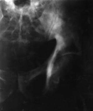
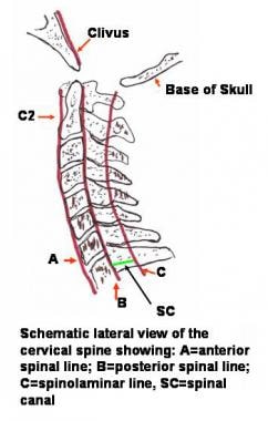
We analyze the results of surgical treatment of 60 patients presenting syringomyelia (SM) associated with Chiari malformation (CM) who were operated in the period 1982-2000. For each case, analysis covered 15 signs and 16 symptoms included in a protocol that separated SM signs and symptoms from those of CM. A score system was established in parallel with the protocol to make the evaluation of treatment results easier. All cases were submitted to craniovertebral decompression by C1 and eventually C2 laminectomy and cerebellar tonsillectomy with duramater graft. To evaluate the results, statistical proportion difference tests and variance analyses were made to a reliability index of 95% (p=0.05). We conclude that the statistical improvement of CM signs and symptoms was very significant (p=0). Syringomyelia signs and symptoms also improved significantly, except for "upper limb hyporeflexia", which did not improve. No statistical difference in the improvement of SM symptoms as compared to CM symptom was found. Syringomyelia signs improved statistically more than CM signs. In half of patients, the percent improvement of signs and symptoms ranged between 40% and 60%. (+info)Platybasia is a medical term that refers to a condition where the base of the skull is flattened or broadened, resulting in an abnormal increase in the angle between the clivus (a part of the sphenoid bone) and the posterior aspect of the upper surface of the palatine bone. This condition can be congenital or acquired and is often associated with other skeletal abnormalities. In some cases, platybasia may lead to neurological symptoms such as headaches, neck pain, or even brainstem compression.
The hard palate is the anterior, bony part of the roof of the mouth, forming a vertical partition between the oral and nasal cavities. It is composed of the maxilla and palatine bones, and provides attachment for the muscles of the soft palate, which functions in swallowing, speaking, and breathing. The hard palate also contains taste buds that contribute to our ability to taste food.
The Atlanto-Occipital Joint, also known as the AO joint or the craniocervical joint, is the articulation between the occiput (the base of the skull) and the atlas (the first cervical vertebra). This joint allows for movements such as nodding your head "yes" and tilting your head from side to side. It is a crucial joint in maintaining the alignment and stability of the head and neck.
The odontoid process, also known as the dens, is a tooth-like projection from the second cervical vertebra (axis). It fits into a ring formed by the first vertebra (atlas), allowing for movement between these two vertebrae. The odontoid process helps to support the head and facilitates movements such as nodding and shaking. It is an essential structure in maintaining stability and mobility of the upper spine.
The atlanto-axial joint is the joint between the first and second cervical vertebrae, also known as C1 (atlas) and C2 (axis). It consists of two separate joints: the median atlanto-axial joint, which is a pivot joint that allows for rotation of the head, and the paired lateral atlanto-axial joints, which are plane joints that allow for limited gliding movements.
The atlanto-axial joint is surrounded by several ligaments that provide stability and limit excessive movement. The transverse ligament, located on the anterior aspect of the joint, is particularly important as it prevents excessive movement of the atlas on the axis and helps to protect the spinal cord.
Abnormalities or injuries to the atlanto-axial joint can result in instability and potentially serious neurological complications.
The nasal cavity is the air-filled space located behind the nose, which is divided into two halves by the nasal septum. It is lined with mucous membrane and is responsible for several functions including respiration, filtration, humidification, and olfaction (smell). The nasal cavity serves as an important part of the upper respiratory tract, extending from the nares (nostrils) to the choanae (posterior openings of the nasal cavity that lead into the pharynx). It contains specialized structures such as turbinate bones, which help to warm, humidify and filter incoming air.
Spinal cord compression is a medical condition that refers to the narrowing of the spinal canal, which puts pressure on the spinal cord and the nerves that branch out from it. This can occur due to various reasons such as degenerative changes in the spine, herniated discs, bone spurs, tumors, or fractures. The compression can lead to a range of symptoms including pain, numbness, tingling, weakness, or loss of bladder and bowel control. In severe cases, it can cause paralysis. Treatment options depend on the underlying cause and may include physical therapy, medication, surgery, or radiation therapy.
Neurosurgical procedures are operations that are performed on the brain, spinal cord, and peripheral nerves. These procedures are typically carried out by neurosurgeons, who are medical doctors with specialized training in the diagnosis and treatment of disorders of the nervous system. Neurosurgical procedures can be used to treat a wide range of conditions, including traumatic injuries, tumors, aneurysms, vascular malformations, infections, degenerative diseases, and congenital abnormalities.
Some common types of neurosurgical procedures include:
* Craniotomy: A procedure in which a bone flap is temporarily removed from the skull to gain access to the brain. This type of procedure may be performed to remove a tumor, repair a blood vessel, or relieve pressure on the brain.
* Spinal fusion: A procedure in which two or more vertebrae in the spine are fused together using bone grafts and metal hardware. This is often done to stabilize the spine and alleviate pain caused by degenerative conditions or spinal deformities.
* Microvascular decompression: A procedure in which a blood vessel that is causing pressure on a nerve is repositioned or removed. This type of procedure is often used to treat trigeminal neuralgia, a condition that causes severe facial pain.
* Deep brain stimulation: A procedure in which electrodes are implanted in specific areas of the brain and connected to a battery-operated device called a neurostimulator. The neurostimulator sends electrical impulses to the brain to help alleviate symptoms of movement disorders such as Parkinson's disease or dystonia.
* Stereotactic radiosurgery: A non-invasive procedure that uses focused beams of radiation to treat tumors, vascular malformations, and other abnormalities in the brain or spine. This type of procedure is often used for patients who are not good candidates for traditional surgery due to age, health status, or location of the lesion.
Neurosurgical procedures can be complex and require a high degree of skill and expertise. Patients considering neurosurgical treatment should consult with a qualified neurosurgeon to discuss their options and determine the best course of action for their individual situation.
The cervical vertebrae are the seven vertebrae that make up the upper part of the spine, also known as the neck region. They are labeled C1 to C7, with C1 being closest to the skull and C7 connecting to the thoracic vertebrae in the chest region. The cervical vertebrae have unique structures to allow for a wide range of motion in the neck while also protecting the spinal cord and providing attachment points for muscles and ligaments.
Basal Cell Nevus Syndrome (BCNS), also known as Gorlin-Goltz Syndrome, is a rare genetic disorder that is characterized by the development of multiple basal cell carcinomas (BCCs), which are skin cancer tumors that arise from the basal cells in the outermost layer of the skin.
The syndrome is caused by mutations in the PTCH1 gene, which regulates the hedgehog signaling pathway involved in embryonic development and tissue growth regulation. The condition is inherited in an autosomal dominant manner, meaning that a child has a 50% chance of inheriting the mutated gene from an affected parent.
Individuals with BCNS typically develop hundreds to thousands of BCCs over their lifetime, often beginning in childhood or adolescence. They may also have other benign and malignant tumors, such as medulloblastomas (brain tumors), fibromas, and rhabdomyosarcomas.
Additional features of BCNS can include:
1. Facial abnormalities, such as a broad nasal bridge, widely spaced eyes, and pits or depressions on the palms and soles.
2. Skeletal abnormalities, such as spine deformities, rib anomalies, and jaw cysts.
3. Developmental delays and intellectual disabilities in some cases.
4. Increased risk of other cancers, including breast, ovarian, and lung cancer.
Early detection and management of BCCs and other tumors are crucial for individuals with BCNS to prevent complications and improve their quality of life. Regular dermatological examinations, sun protection measures, and surgical removal of tumors are common treatment approaches.
The occipital bone is the single, posterior cranial bone that forms the base of the skull and encloses the brain. It articulates with the parietal bones anteriorly and the temporal bones laterally. The occipital bone also contains several important structures such as the foramen magnum, through which the spinal cord connects to the brain, and the external and internal occipital protuberances, which serve as attachment points for neck muscles.
An encyclopedia is a comprehensive reference work containing articles on various topics, usually arranged in alphabetical order. In the context of medicine, a medical encyclopedia is a collection of articles that provide information about a wide range of medical topics, including diseases and conditions, treatments, tests, procedures, and anatomy and physiology. Medical encyclopedias may be published in print or electronic formats and are often used as a starting point for researching medical topics. They can provide reliable and accurate information on medical subjects, making them useful resources for healthcare professionals, students, and patients alike. Some well-known examples of medical encyclopedias include the Merck Manual and the Stedman's Medical Dictionary.
Carcinoma, basal cell is a type of skin cancer that arises from the basal cells, which are located in the lower part of the epidermis (the outermost layer of the skin). It is also known as basal cell carcinoma (BCC) and is the most common form of skin cancer.
BCC typically appears as a small, shiny, pearly bump or nodule on the skin, often in sun-exposed areas such as the face, ears, neck, hands, and arms. It may also appear as a scar-like area that is white, yellow, or waxy. BCCs are usually slow growing and rarely spread (metastasize) to other parts of the body. However, they can be locally invasive and destroy surrounding tissue if left untreated.
The exact cause of BCC is not known, but it is thought to be related to a combination of genetic and environmental factors, including exposure to ultraviolet (UV) radiation from the sun or tanning beds. People with fair skin, light hair, and blue or green eyes are at increased risk of developing BCC.
Treatment for BCC typically involves surgical removal of the tumor, along with a margin of healthy tissue. Other treatment options may include radiation therapy, topical chemotherapy, or photodynamic therapy. Prevention measures include protecting your skin from UV radiation by wearing protective clothing, using sunscreen, and avoiding tanning beds.
Spinal diseases refer to a range of medical conditions that affect the spinal column, which is made up of vertebrae (bones), intervertebral discs, facet joints, nerves, ligaments, and muscles. These diseases can cause pain, discomfort, stiffness, numbness, weakness, or even paralysis, depending on the severity and location of the condition. Here are some examples of spinal diseases:
1. Degenerative disc disease: This is a condition where the intervertebral discs lose their elasticity and height, leading to stiffness, pain, and decreased mobility.
2. Herniated disc: This occurs when the inner material of the intervertebral disc bulges or herniates out through a tear in the outer layer, causing pressure on the spinal nerves and resulting in pain, numbness, tingling, or weakness in the affected area.
3. Spinal stenosis: This is a narrowing of the spinal canal or the neural foramen (the openings where the spinal nerves exit the spinal column), which can cause pressure on the spinal cord or nerves and result in pain, numbness, tingling, or weakness.
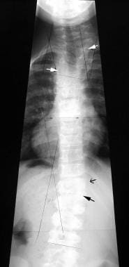
4. Scoliosis: This is a curvature of the spine that can occur in children or adults, leading to an abnormal posture, back pain, and decreased lung function.
5. Osteoarthritis: This is a degenerative joint disease that affects the facet joints in the spine, causing pain, stiffness, and decreased mobility.
6. Ankylosing spondylitis: This is a chronic inflammatory disease that affects the spine and sacroiliac joints, leading to pain, stiffness, and fusion of the vertebrae.
7. Spinal tumors: These are abnormal growths that can occur in the spinal column, which can be benign or malignant, causing pain, neurological symptoms, or even paralysis.
8. Infections: Bacterial or viral infections can affect the spine, leading to pain, fever, and other systemic symptoms.
9. Trauma: Fractures, dislocations, or sprains of the spine can occur due to accidents, falls, or sports injuries, causing pain, neurological deficits, or even paralysis.
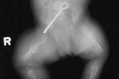
Ectromelia is a medical term that refers to the congenital absence or malformation of a limb or extremity. It is also known as "congenital amputation" or "limb reduction defect." This condition can affect any extremity, including arms, legs, hands, or feet, and can range from mild, such as a missing finger or toe, to severe, such as the absence of an entire limb.
Ectromelia can be caused by various factors, including genetic mutations, environmental factors, or a combination of both. In some cases, the cause may be unknown. Treatment options for ectromelia depend on the severity and location of the malformation and may include prosthetics, physical therapy, or surgery.
The pelvic bones, also known as the hip bones, are a set of three irregularly shaped bones that connect to form the pelvic girdle in the lower part of the human body. They play a crucial role in supporting the spine and protecting the abdominal and pelvic organs.
The pelvic bones consist of three bones:
1. The ilium: This is the largest and uppermost bone, forming the majority of the hip bone and the broad, flaring part of the pelvis known as the wing of the ilium or the iliac crest, which can be felt on the side of the body.
2. The ischium: This is the lower and back portion of the pelvic bone that forms part of the sitting surface or the "sit bones."
3. The pubis: This is the front part of the pelvic bone, which connects to the other side at the pubic symphysis in the midline of the body.
The pelvic bones are joined together at the acetabulum, a cup-shaped socket that forms the hip joint and articulates with the head of the femur (thigh bone). The pelvic bones also have several openings for the passage of blood vessels, nerves, and reproductive and excretory organs.
The shape and size of the pelvic bones differ between males and females due to their different roles in childbirth and locomotion. Females typically have a wider and shallower pelvis than males to accommodate childbirth, while males usually have a narrower and deeper pelvis that is better suited for weight-bearing and movement.
I believe there may be some confusion in your question as "Greek World" is not a medical term. If you are referring to the ancient Greek civilization, it was a significant period in human history that greatly contributed to the development of various fields including medicine. The ancient Greeks, particularly Hippocrates and his followers, are often referred to as the "Fathers of Medicine." They made substantial contributions to the field through their observations, theories, and practices which formed the foundation of much of Western medical thought. However, "Greek World" itself does not have a medical definition.
Caroli disease is a rare genetic disorder that affects the liver and bile ducts. It is characterized by abnormal dilations or sac-like structures in the intrahepatic bile ducts, which are the ducts that carry bile from the liver to the gallbladder and small intestine. These dilations can lead to recurrent cholangitis (inflammation of the bile ducts), stone formation, and liver damage.
Caroli disease is usually diagnosed in childhood or early adulthood, and it can be associated with other congenital anomalies such as polycystic kidney disease. The exact cause of Caroli disease is not fully understood, but it is believed to be inherited in an autosomal recessive manner, meaning that an individual must inherit two copies of the abnormal gene, one from each parent, to develop the condition.
Treatment for Caroli disease may include antibiotics to manage cholangitis, endoscopic procedures to remove stones or dilate strictures, and surgery to bypass or remove affected bile ducts. In severe cases, liver transplantation may be necessary. Regular monitoring of liver function and surveillance for complications are essential in the management of this condition.
Thalidomide is a pharmaceutical drug that was initially developed and marketed as a sedative and treatment for morning sickness in pregnant women. However, it was later found to cause severe birth defects when given during pregnancy, particularly damage to the limbs, ears, and eyes of the developing fetus. As a result, thalidomide was banned in many countries in the 1960s.
In recent years, thalidomide has been reintroduced as a treatment for certain medical conditions, including multiple myeloma (a type of cancer that affects plasma cells) and leprosy. It is also being studied as a potential treatment for other diseases, such as rheumatoid arthritis and Crohn's disease.
Thalidomide works by suppressing the immune system and inhibiting the formation of new blood vessels (angiogenesis). However, its use is tightly regulated due to its teratogenic effects, meaning it can cause birth defects if taken during pregnancy. Women who are pregnant or planning to become pregnant should not take thalidomide, and healthcare providers must follow strict guidelines when prescribing the drug to ensure that it is used safely and effectively.
Genetic research is a branch of biomedical science that involves the study of genes, their functions, and heredity. It aims to understand how genetic variations contribute to human health and disease by using various scientific approaches such as genetics, genomics, molecular biology, biochemistry, and bioinformatics.
Genetic research can be conducted on humans, animals, or plants, and it can focus on a variety of areas including:
1. Identifying genes associated with specific diseases or traits
2. Understanding how genes are regulated and expressed
3. Investigating the role of genetic mutations in disease development
4. Developing new diagnostic tests and treatments based on genetic information
5. Exploring evolutionary relationships between species
6. Examining ethical, legal, and social implications of genetic research.
Genetic research has led to significant advances in our understanding of many diseases, including cancer, diabetes, heart disease, and neurological disorders. It also holds great promise for personalized medicine, which tailors treatments to individual patients based on their genetic makeup.
 Platybasia
Michel aplasia
Chiari malformation
Craniofacial abnormality
List of MeSH codes (C05)
List of MeSH codes (C16)
Empty sella syndrome
Platybasia - Wikipedia
 Syringohydromyelia and Syringomyelia Imaging: Practice Essentials, Radiography, Magnetic Resonance Imaging
 Hajdu-Cheney syndrome: MedlinePlus Genetics
 Puberty, Pregnancy, Parturition, Puerperium-Surveillance by Intertwined Innumerable Neurohumoral Factors; Prevention,...
 Surgical Neurology International
 Baby’s Pregnancy Calendar
 Namespace
 Craniocervical Junction Abnormalities - Neurologic Disorders - MSD Manual Professional Edition
Phocomelia - Wikipedia
ICD-10 Chapter XVII: Congenital malformations, deformations and chromosomal abnormalities - wikidoc
 Medical Terminology - Word Parts: p
 Human Disease Genes - Graph and Chart
Imaging in Chiari I Malformation: Overview, Radiography, Computed Tomography
Neuroradiology: Key Differential Diagnoses and C: 2nd edition | Edited by Juan E. Small | ISBN: 9780323847612 | Elsevier Asia...
 Facet Joint Arthritis - Spine Care | UCLA Health
Pediatric Conditions - Neurosurgery | UCLA Health
'New' Spine Surgery: Looking Beyond Decompression | Grand Rounds/Virtual OR Series | The Neurosurgical...
 22q11 Deletion Syndrome | Harvard Catalyst Profiles | Harvard Catalyst
 Pesquisa | Portal Regional da BVS
 Disease - pl Index | CureHunter
 Playlist 'Manzoor-Rad CNS' by Manzoor Ahmad
Leg Length Inequality | Profiles RNS
 Atlanto-axial rotatory fixation in a girl with Spondylocarpotarsal synostosis syndrome | Scoliosis and Spinal Disorders | Full...
 Distal amyotrophy (Concept Id: C1848736)
- MedGen - NCBI
Distal amyotrophy (Concept Id: C1848736)
- MedGen - NCBI
"Generalized osteoporosis"[Clinical Features] OR 1639139[uid] - MedGen -...
 EurekaMag PDF full texts Chapter 5786
 Bone Diseases, Developmental (medical concept explorer)
CLASSIFICATION OF DISEASES AND INJURIES
 Klippel-Feil syndrome - wikidoc
Basilar invagination2
- The most serious complications of Hajdu-Cheney syndrome, which occur in about half of all affected individuals, are abnormalities known as platybasia and basilar invagination. (medlineplus.gov)
- The risk of developing platybasia and basilar invagination also increases over time. (medlineplus.gov)
Cervical1
- Platybasia is a spinal disease of a malformed relationship between the occipital bone and cervical spine. (wikipedia.org)
Skull1
- Platybasia is a flattening of the base of the skull caused by thinning and softening of the skull bones. (medlineplus.gov)
Basilar impression2
Spinal1
- Platybasia is a spinal disease of a malformed relationship between the occipital bone and cervical spine. (wikipedia.org)
Osteoporosis1
- HCS is characterized by craniofacial developmental defects, including platybasia and wormian bones, osteoporosis with fractures, and acro-osteolysis. (nih.gov)
Base1
- nDessutom erbjuder vi Fri frakt, 90-dagars öppet köp, ångerrätt och en unik Tool for base edge beveling. (netlify.app)