Pleura
Pleural Neoplasms
Solitary Fibrous Tumor, Pleural
Pleural Diseases
Mesothelioma
Pleural Effusion
Pleural Cavity
Neoplasms, Fibrous Tissue
Asbestos
Solitary Fibrous Tumors
Asbestosis
Tomography, X-Ray Computed
Pneumothorax
Pleurisy
Chondrosarcoma, Mesenchymal
Fatal Outcome
Mineral Fibers
Pleurodesis
Thoracic Surgery, Video-Assisted
Pleural Effusion, Malignant
Radiography, Thoracic
Asbestos, Amphibole
Asbestos, Serpentine
Tuberculosis, Pleural
Thoracic Surgical Procedures
Thoracic Neoplasms
Lung
Zeolites
Melanoma, Amelanotic
Mediastinum
Asbestos, Amosite
Calbindin 2
Talc
Hemangiosarcoma
Ribs
Sarcoma, Synovial
Nanowires
Solitary Pulmonary Nodule
Immunohistochemistry
Nanotubes, Carbon
Lung Diseases, Parasitic
Pharmacology of LY315920/S-5920, [[3-(aminooxoacetyl)-2-ethyl-1- (phenylmethyl)-1H-indol-4-yl]oxy] acetate, a potent and selective secretory phospholipase A2 inhibitor: A new class of anti-inflammatory drugs, SPI. (1/631)
LY315920 is a potent, selective inhibitor of recombinant human, group IIA, nonpancreatic secretory PLA2 (sPLA2). In a chromogenic isolated enzyme assay, LY315920 inhibited sPLA2 activity with an IC50 of 9 +/- 1 nM or 7.3 x 10(-6) mole fraction, which approached the stiochiometric limit of this assay. The true potency of LY315920 was defined using a deoxycholate/phosphatidylcholine assay with a mole fraction of 1.5 x 10(-6). LY315920 was 40-fold less active against human, group IB, pancreatic sPLA2 and was inactive against cytosolic PLA2 and the constitutive and inducible forms of cyclooxygenase. Human sPLA2-induced release of thromboxane A2 (TXA2) from isolated guinea pig lung bronchoalveolar lavage cells was inhibited by LY315920 with an IC50 of 0.79 microM. The release of TXA2 from these cells by N-formyl-methionyl-leucyl-phenylalanine or arachidonic acid was not inhibited. The i.v. administration of LY315920, 5 min before harvesting the bronchoalveolar lavage cells, resulted in the inhibition of sPLA2-induced production of TXA2 with an ED50 of 16.1 mg/kg. Challenge of guinea pig lung pleural strips with sPLA2 produced contractile responses that were suppressed in a concentration-dependent manner by LY315920 with an apparent KB of 83 +/- 14 nM. Contractile responses induced by arachidonic acid were not altered. Intravenous or oral administration of LY315920 to transgenic mice expressing the human sPLA2 protein inhibited serum sPLA2 activity in a dose-related manner over a 4-h time course. LY315920 is a potent and selective sPLA2 inhibitor and represents a new class of anti-inflammatory agent designated SPI. This agent is currently undergoing clinical evaluation and should help to define the role of sPLA2 in various inflammatory disease states. (+info)Value of scintigraphy in chronic peritoneal dialysis patients. (2/631)
BACKGROUND: A variety of factors can adversely impact chronic peritoneal dialysis (CPD) as an effective renal replacement therapy for patients with end-stage renal disease. These factors include peritonitis, poor clearances, loss of ultrafiltration, and a variety of anatomic problems, such as hernias, peritoneal fluid leaks, loculations, and catheter-related problems caused by omental blockage. This study reviews our experience with peritoneal scintigraphy for the evaluation of some of these difficulties. METHODS: From 1991 to 1996, 50 peritoneal scintigraphy scans were obtained in 48 CPD patients. Indications for scintigraphy were evaluated, and the patients were placed into four groups: group I, abdominal wall swelling; group II, inguinal or genital swelling; group III, pleural fluid; and group IV, poor drainage and/or poor ultrafiltration. A peritoneal scintigraphy protocol was established and the radiotracer isotope that was used was 2.0 mCi of 99mtechnetium sulfur colloid placed in two liters of 2.5% dextrose peritoneal dialysis solution. RESULTS: Ten scans were obtained to study abdominal wall swelling, with seven scans demonstrating leaks; six of these episodes improved with low-volume exchanges. Twenty scans were obtained to evaluate inguinal or genital swelling, and 10 of these had scintigraphic evidence for an inguinal hernia leak (9 of these were surgically corrected). One of four scans obtained to evaluate a pleural fluid collection demonstrated a peritoneal-pleural leak that corrected with a temporary discontinuation of CPD. Sixteen scans were obtained to assess poor drainage and/or ultrafiltration. Five of these scans demonstrated peritoneal location, and all of these patients required transfer to hemodialysis. The other 11 scans were normal; four patients underwent omentectomies, allowing three patients to continue with CPD. CONCLUSION: Peritoneal scintigraphy is useful in the evaluation and assessment of CPD patients who develop anatomical problems (such as anterior abdominal, pleural-peritoneal, inguinal, and genital leaks) and problems with ultrafiltration and/or drainage. (+info)Penetration of meropenem in lung, bronchial mucosa, and pleural tissues. (3/631)
Lung, bronchial mucosa, and pleural tissue samples were obtained from 14 patients undergoing lung surgery 1 to 5 h after administration of 1 g of meropenem. The mean (range) peak concentrations of meropenem were 3.9 (0.2 to 8.2), 6.6 (3.0 to 13.3), and 2.8 (0.6 to 7.8) mg/kg of tissue, respectively, exceeding the MICs at which 90% of isolates are inhibited for most respiratory pathogens. (+info)The functional anatomy of the bronchial circulation of the domestic fowl. (4/631)
The bronchial circulation was studied in 25 adult domestic fowls. The right and left bronchial arteries originated caudal to the syrinx from a bronchoesophageal artery which is a branch of the right common carotid artery. Each bronchial artery ramified on the wall of the extrapulmonary part of the corresponding primary bronchus and finally anastomosed directly with a branch of the pulmonary artery at the hilus of the lung. Thr bronchial artery did not accompany the intrapulmonary part of the primary bronchus. The branches of each bronchial artery formed an anastomosing network on the wall of the extrapulmonary part of the primary bronchus. The calibre of the bronchial artery at its anastomosis with the branch of the pulmonary artery was greater than at its origin from the bronchoesophageal artery. Intravenous injections of Lycopodium spores indicated that the blood flows from the pulmonary artery into the bronchial artery. Small bronchial veins drained the extrapulmonary part of the primary bronchus into the pulmonary vein and the oesophageal veins. The intrapulmonary part of the primary bronchus was supplied by branches of the pulmonary artery and drained by tributaries of the pulmonary vein. The blood supply to the primary bronchus could constitute a shunt capable of passing blood from the pulmonary artery into the pulmonary vein without going through the exchange tissue. The parabronchial (atrial) muscles received a blood supply directly from the exchange tissue via septal venules which formed a network underneath the muscle bundles, without actually penetrating between the muscle cells. These venules drained into atrial veins which were tributaries of the pulmonary vein. The atrial muscles probably also received oxygen by direct diffusion from the parabronchial lumen. The pleura was supplied by the oesophageal branches of the bronchoesophageal artery, and by small twigs from the internal thoracic and intercostal arteries. (+info)Role of pleural lavage cytology before resection for primary lung carcinoma. (5/631)
OBJECTIVE: To investigate the role of pleural lavage cytology (PLC) in resection for primary lung carcinoma. SUMMARY BACKGROUND DATA: The prognostic significance of PLC before manipulation is still controversial. METHODS: Cytology of pleural lavage immediately after thoracotomy but before any manipulation of the lung was examined in 500 consecutive patients with lung cancer with no pleural effusion who underwent pulmonary resections. Eighteen patients who already had pleural dissemination were excluded from this study. RESULTS: Eighteen of 482 patients (3.7%) had positive cytologic findings. The positivity of PLC was significantly correlated with histology, extension of tumor to pleura, and presence of lymphatic permeation or vascular involvement by tumor. Positive lavage findings were seen only in adenocarcinoma. Because 6.3% of the patients with adenocarcinoma had positive cytologic findings, it is vital to perform PLC before curative resections for lung cancer, especially adenocarcinoma. The 5-year survival rates of the patients having negative and positive lavage findings were 52.9% and 14.6%, respectively. The prognosis of the patients with positive lavage findings was as poor as that of the patients with stage IIIB disease and that of the patients with malignant effusion. CONCLUSIONS: Positive findings on PLC indicate exfoliation of cancer cells into the pleural cavity, which is an essential prognostic factor. In addition, we should regard positive cytologic findings as a subclinical malignant pleural effusion that is pathologic stage T4. (+info)Vagal afferents and active upper airway closure during pulmonary edema in lambs. (6/631)
The present study was undertaken to gain further insight into the mechanisms responsible for the sustained active expiratory upper airway closure previously observed during high-permeability pulmonary edema in lambs. The experiments were conducted in nonsedated lambs, in which airflow and thyroarytenoid and inferior pharyngeal constrictor muscle electromyographic activity were recorded. We first studied the consequences of hemodynamic pulmonary edema (induced by impeding pulmonary venous return) on upper airway dynamics in five lambs; under this condition, a sustained expiratory upper airway closure consistently appeared. We then tested whether expiratory upper airway closure was related to vagal afferent activity from bronchopulmonary receptors. Five bivagotomized lambs underwent high-permeability pulmonary edema: no sustained expiratory upper airway closure was observed. Finally, we studied whether a sustained decrease in lung volume induced a sustained expiratory upper airway closure. Five lambs underwent a 250-ml pleural infusion: no sustained expiratory upper airway closure was observed. We conclude that 1) the sustained expiratory upper airway closure observed during pulmonary edema in nonsedated lambs is related to stimulation of vagal afferents by an increase in lung water and 2) a decrease in lung volume does not seem to be the causal factor. (+info)Thoracic blastomycosis and empyema. (7/631)
Blastomycosis is endemic in river valley areas of the southeastern and Midwestern United States. Pulmonary manifestations include chronic cough and pleuritic pain. Radiographic appearance of the infection can mimic bronchogenic lung carcinoma. Pleural effusion is rarely associated with this pulmonary infection, and empyema has not been previously reported. We report a case of pulmonary and pleural Blastomyces dermatitidis infection presenting as empyema thoracis. Diagnosis and treatment were attained with video-assisted thoracoscopic (VATS) pleural and lung biopsy and debridement. (+info)The mouse GATA-2 gene is expressed in the para-aortic splanchnopleura and aorta-gonads and mesonephros region. (8/631)
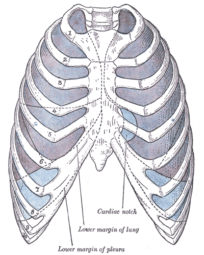
We previously reported that the mouse GATA-2 gene is regulated by two alternative promoters (Minegishi et al, J Biol Chem, 273:3625, 1998). Although the more proximal IG (general) promoter is active in almost all GATA-2-expressing cells, the distal IS (specific) promoter activity was selectively detected in hematopoietic tissues but not in other mesodermal tissues. We report here in vivo analysis of the GATA-2 locus and its regulatory characteristics in hematopoietic tissues of transgenic mice. Transgenes containing 6 or 7 kbp of sequence flanking the 5' end of the IS first exon direct expression of beta-galactosidase or green fluorescent protein (GFP) reporter genes specifically to the para-aortic splanchnopleura, aorta-gonads, and mesonephros (AGM) region, and in the neural tissues. In situ hybridization analysis showed that reporter gene expression specifically recapitulates the endogenous expression profile of GATA-2 in these tissues. The flk-1, CD34, c-kit, and CD45 antigens were identified in the GFP-positive cells from the AGM region and fetal liver, indicating that GATA-2 is expressed in immature hematopoietic cells. Deletion of 3.5 kbp from the 5' end of the 6.0 kbp IS promoter construct, including one of the DNase I hypersensitive sites, completely abolished hematopoietic expression. These experiments describe an early developmental GATA-2 hematopoietic enhancer located between 6.0 and 2.5 kbp 5' to the IS exon. (+info)The pleura is the medical term for the double-layered serous membrane that surrounds the lungs and lines the inside of the chest cavity. The two layers of the pleura are called the parietal pleura, which lines the chest cavity, and the visceral pleura, which covers the surface of the lungs.
The space between these two layers is called the pleural cavity, which contains a small amount of lubricating fluid that allows the lungs to move smoothly within the chest during breathing. The main function of the pleura is to protect the lungs and facilitate their movement during respiration.
Pleural neoplasms refer to abnormal growths or tumors that develop in the pleura, which is the thin, double layered membrane that surrounds the lungs and lines the inside of the chest wall. These neoplasms can be benign (non-cancerous) or malignant (cancerous).
Malignant pleural neoplasms are often associated with lung cancer, mesothelioma, or metastasis from other types of cancer. They can cause symptoms such as chest pain, cough, shortness of breath, and weight loss. Diagnosis typically involves imaging tests like X-rays or CT scans, followed by biopsy to confirm the type of tumor. Treatment options may include surgery, radiation therapy, chemotherapy, or a combination of these approaches.
A Solitary Fibrous Tumor (SFT) is a rare type of soft tissue neoplasm that can occur in various locations throughout the body. When it develops in the pleura, which is the thin layer of tissue that surrounds the lungs, it is referred to as a "Solitary Fibrous Tumor, Pleural."
These tumors are typically slow-growing and can range in size from a few centimeters to over 20 cm in diameter. They are usually asymptomatic and are often discovered incidentally on chest X-rays or CT scans performed for other reasons. However, larger tumors may cause symptoms such as cough, chest pain, or shortness of breath.
SFTs, Pleural are typically well-circumscribed, encapsulated masses that can be removed surgically with curative intent. The histological features of SFTs include a patternless architecture with alternating hypocellular and hypercellular areas, along with a prominent network of thin-walled blood vessels.
Immunohistochemical staining is often used to confirm the diagnosis of SFT, with positivity for CD34 and STAT6 being characteristic features. The prognosis for patients with SFTs, Pleural is generally good, with a low risk of recurrence following surgical resection. However, some cases may exhibit more aggressive behavior, and long-term follow-up is recommended.
Pleural diseases refer to conditions that affect the pleura, which is the thin, double-layered membrane that surrounds the lungs and lines the inside of the chest wall. The space between these two layers contains a small amount of fluid that helps the lungs move smoothly during breathing. Pleural diseases can cause inflammation, infection, or abnormal collections of fluid in the pleural space, leading to symptoms such as chest pain, cough, and difficulty breathing.
Some common examples of pleural diseases include:
1. Pleurisy: Inflammation of the pleura that causes sharp chest pain, often worsened by breathing or coughing.
2. Pleural effusion: An abnormal accumulation of fluid in the pleural space, which can be caused by various underlying conditions such as heart failure, pneumonia, cancer, or autoimmune disorders.
3. Empyema: A collection of pus in the pleural space, usually resulting from a bacterial infection.
4. Pleural thickening: Scarring and hardening of the pleura, which can restrict lung function and cause breathlessness.
5. Mesothelioma: A rare form of cancer that affects the pleura, often caused by exposure to asbestos.
6. Pneumothorax: A collection of air in the pleural space, which can result from trauma or a rupture of the lung tissue.
Proper diagnosis and treatment of pleural diseases require a thorough evaluation by a healthcare professional, often involving imaging tests such as chest X-rays or CT scans, as well as fluid analysis or biopsy if necessary.
Mesothelioma is a rare and aggressive form of cancer that develops in the mesothelial cells, which are the thin layers of tissue that cover many of the internal organs. The most common site for mesothelioma to occur is in the pleura, the membrane that surrounds the lungs. This type is called pleural mesothelioma. Other types include peritoneal mesothelioma (which occurs in the lining of the abdominal cavity) and pericardial mesothelioma (which occurs in the lining around the heart).
Mesothelioma is almost always caused by exposure to asbestos, a group of naturally occurring minerals that were widely used in construction, insulation, and other industries because of their heat resistance and insulating properties. When asbestos fibers are inhaled or ingested, they can become lodged in the mesothelium, leading to inflammation, scarring, and eventually cancerous changes in the cells.
The symptoms of mesothelioma can take many years to develop after exposure to asbestos, and they may include chest pain, coughing, shortness of breath, fatigue, and weight loss. Treatment options for mesothelioma depend on the stage and location of the cancer, but may include surgery, radiation therapy, chemotherapy, or a combination of these approaches. Unfortunately, the prognosis for mesothelioma is often poor, with a median survival time of around 12-18 months after diagnosis.
Pleural effusion is a medical condition characterized by the abnormal accumulation of fluid in the pleural space, which is the thin, fluid-filled space that surrounds the lungs and lines the inside of the chest wall. This space typically contains a small amount of fluid to allow for smooth movement of the lungs during breathing. However, when an excessive amount of fluid accumulates, it can cause symptoms such as shortness of breath, coughing, and chest pain.
Pleural effusions can be caused by various underlying medical conditions, including pneumonia, heart failure, cancer, pulmonary embolism, and autoimmune disorders. The fluid that accumulates in the pleural space can be transudative or exudative, depending on the cause of the effusion. Transudative effusions are caused by increased pressure in the blood vessels or decreased protein levels in the blood, while exudative effusions are caused by inflammation, infection, or cancer.
Diagnosis of pleural effusion typically involves a physical examination, chest X-ray, and analysis of the fluid in the pleural space. Treatment depends on the underlying cause of the effusion and may include medications, drainage of the fluid, or surgery.
The pleural cavity is the potential space between the visceral and parietal pleura, which are the two membranes that surround the lungs. The visceral pleura covers the outside of the lungs, while the parietal pleura lines the inside of the chest wall. Under normal conditions, these two layers are in contact with each other, and the space between them is virtually nonexistent. However, when air, fluid or inflammation accumulates within this space, it results in the formation of a pleural effusion, which can cause discomfort and difficulty breathing.
Neoplasms of fibrous tissue are abnormal growths or tumors that originate from fibroblasts, the cells responsible for producing connective tissue in the body. These neoplasms can be benign or malignant (cancerous). Benign fibrous neoplasms include fibromas and fibrohistiocytic tumors, while malignant fibrous neoplasms are called fibrosarcomas. Fibrosarcomas are aggressive tumors that invade surrounding tissues and can metastasize (spread) to other parts of the body.
Fibrous tissue neoplasms can occur in any part of the body, but they are most commonly found in the soft tissues such as muscles, tendons, and ligaments. They can also develop in bones, where they are called osteosarcomas. Symptoms of fibrous tissue neoplasms depend on their size and location, but may include a painless mass or swelling, limited mobility, or pain if the tumor is pressing on nerves or blood vessels.
Diagnosis of fibrous tissue neoplasms typically involves imaging tests such as X-rays, CT scans, or MRI scans, followed by a biopsy to confirm the type and grade of the tumor. Treatment options may include surgery, radiation therapy, chemotherapy, or a combination of these approaches. Regular follow-up care is important to monitor for recurrence or metastasis.
Asbestos is a group of naturally occurring mineral fibers that are resistant to heat, chemical reactions, and electrical currents. There are six types of asbestos, but the most common ones are chrysotile, amosite, and crocidolite. Asbestos has been widely used in various construction materials, such as roofing shingles, ceiling and floor tiles, paper products, and cement products.
Exposure to asbestos can cause serious health problems, including lung cancer, mesothelioma (a rare form of cancer that affects the lining of the lungs, heart, or abdomen), and asbestosis (a chronic lung disease characterized by scarring of the lung tissue). These health risks are related to the inhalation of asbestos fibers, which can become lodged in the lungs and cause inflammation and scarring over time.
As a result, the use of asbestos has been heavily regulated in many countries, and its use is banned in several others. Despite these regulations, asbestos remains a significant public health concern due to the large number of buildings and products that still contain it.
Thoracoscopy is a surgical procedure in which a thoracoscope, a type of endoscope, is inserted through a small incision between the ribs to examine the lungs and pleural space (the space surrounding the lungs). It allows the surgeon to directly view the chest cavity, take biopsies, and perform various operations. This procedure is often used in the diagnosis and treatment of pleural effusions, lung cancer, and other chest conditions.
Thoracotomy is a surgical procedure that involves making an incision on the chest wall to gain access to the thoracic cavity, which contains the lungs, heart, esophagus, trachea, and other vital organs. The incision can be made on the side (lateral thoracotomy), back (posterolateral thoracotomy), or front (median sternotomy) of the chest wall, depending on the specific surgical indication.
Thoracotomy is performed for various indications, including lung biopsy, lung resection, esophagectomy, heart surgery, and mediastinal mass removal. The procedure allows the surgeon to directly visualize and access the organs within the thoracic cavity, perform necessary procedures, and control bleeding if needed.
After the procedure, the incision is typically closed with sutures or staples, and a chest tube may be placed to drain any accumulated fluid or air from the pleural space around the lungs. The patient will require postoperative care and monitoring in a hospital setting until their condition stabilizes.
Solitary fibrous tumors (SFTs) are rare type of slow-growing neoplasms that typically arise from the pleura, the thin layer of tissue that covers the lungs. However, they can also occur in other locations throughout the body such as the peritoneum, meninges, and deep soft tissues.
SFTs are composed of spindle-shaped cells arranged in a patternless architecture, with a variably collagenous stroma. They are usually well-circumscribed and encapsulated, although they can become invasive in some cases. The cellularity of SFTs varies from low to high, and the tumors can contain staghorn vessels, which are dilated blood vessels with a branching pattern.
The majority of SFTs are benign, but approximately 10-20% of them can be malignant or have aggressive behavior, with potential for local recurrence and distant metastasis. The diagnosis of SFT is usually made by histopathological examination of the tumor tissue, which shows characteristic features such as CD34 and Bcl-2 positivity on immunohistochemistry.
Treatment options for SFTs include surgical resection with wide margins, radiation therapy, and systemic therapy with chemotherapy or targeted agents. The choice of treatment depends on the location, size, and behavior of the tumor, as well as the patient's overall health status. Regular follow-up is necessary to monitor for recurrence or metastasis.
Asbestosis is a chronic lung disease that is caused by the inhalation of asbestos fibers. It is characterized by scarring (fibrosis) of the lung tissue, which can lead to symptoms such as shortness of breath, coughing, and chest pain. The severity of the disease can range from mild to severe, and it is often progressive, meaning that it tends to worsen over time. Asbestosis is not a malignant condition, but it can increase the risk of developing lung cancer or mesothelioma, which are forms of cancer that are associated with asbestos exposure. The disease is typically diagnosed through a combination of medical history, physical examination, and imaging tests such as chest X-rays or CT scans. There is no cure for asbestosis, but treatment can help to manage the symptoms and slow the progression of the disease.
X-ray computed tomography (CT or CAT scan) is a medical imaging method that uses computer-processed combinations of many X-ray images taken from different angles to produce cross-sectional (tomographic) images (virtual "slices") of the body. These cross-sectional images can then be used to display detailed internal views of organs, bones, and soft tissues in the body.
The term "computed tomography" is used instead of "CT scan" or "CAT scan" because the machines take a series of X-ray measurements from different angles around the body and then use a computer to process these data to create detailed images of internal structures within the body.
CT scanning is a noninvasive, painless medical test that helps physicians diagnose and treat medical conditions. CT imaging provides detailed information about many types of tissue including lung, bone, soft tissue and blood vessels. CT examinations can be performed on every part of the body for a variety of reasons including diagnosis, surgical planning, and monitoring of therapeutic responses.
In computed tomography (CT), an X-ray source and detector rotate around the patient, measuring the X-ray attenuation at many different angles. A computer uses this data to construct a cross-sectional image by the process of reconstruction. This technique is called "tomography". The term "computed" refers to the use of a computer to reconstruct the images.
CT has become an important tool in medical imaging and diagnosis, allowing radiologists and other physicians to view detailed internal images of the body. It can help identify many different medical conditions including cancer, heart disease, lung nodules, liver tumors, and internal injuries from trauma. CT is also commonly used for guiding biopsies and other minimally invasive procedures.
In summary, X-ray computed tomography (CT or CAT scan) is a medical imaging technique that uses computer-processed combinations of many X-ray images taken from different angles to produce cross-sectional images of the body. It provides detailed internal views of organs, bones, and soft tissues in the body, allowing physicians to diagnose and treat medical conditions.
Pneumothorax is a medical condition that refers to the presence of air in the pleural space, which is the potential space between the lungs and the chest wall. This collection of air can result in a partial or complete collapse of the lung. The symptoms of pneumothorax may include sudden chest pain, shortness of breath, cough, and rapid heartbeat.
The two main types of pneumothorax are spontaneous pneumothorax, which occurs without any apparent cause or underlying lung disease, and secondary pneumothorax, which is caused by an underlying lung condition such as chronic obstructive pulmonary disease (COPD), asthma, or lung cancer.
Treatment for pneumothorax may include observation, oxygen therapy, needle aspiration, or chest tube insertion to remove the excess air from the pleural space and allow the lung to re-expand. In severe cases, surgery may be required to prevent recurrence.
Pleurisy is a medical condition characterized by inflammation of the pleura, which are the thin membranes that surround the lungs and line the inside of the chest cavity. The pleura normally produce a small amount of lubricating fluid that allows for smooth movement of the lungs during breathing. However, when they become inflamed (a condition known as pleuritis), this can cause pain and difficulty breathing.
The symptoms of pleurisy may include sharp chest pain that worsens with deep breathing or coughing, shortness of breath, cough, fever, and muscle aches. The pain may be localized to one area of the chest or may radiate to other areas such as the shoulders or back.
Pleurisy can have many different causes, including bacterial or viral infections, autoimmune disorders, pulmonary embolism (a blood clot that travels to the lungs), and certain medications or chemicals. Treatment typically involves addressing the underlying cause of the inflammation, as well as managing symptoms such as pain and breathing difficulties with medications such as nonsteroidal anti-inflammatory drugs (NSAIDs) or opioids. In some cases, more invasive treatments such as thoracentesis (removal of fluid from the chest cavity) may be necessary.
Chondrosarcoma, mesenchymal is a type of chondrosarcoma, which is a malignant (cancerous) tumor that arises from cartilaginous tissue. It is a rare and aggressive subtype of chondrosarcoma, accounting for less than 10% of all cases.
Mesenchymal chondrosarcomas are characterized by their undifferentiated small round blue cells intermixed with well-differentiated cartilaginous areas. They can occur in any age group but are more common in children and young adults. These tumors can arise in any bone, but they most commonly involve the long bones of the extremities, pelvis, and spine.
Mesenchymal chondrosarcomas tend to be aggressive with a high risk of local recurrence and metastasis (spread) to other parts of the body, such as the lungs, lymph nodes, or other bones. Treatment typically involves surgical resection of the tumor, often followed by radiation therapy and/or chemotherapy. The prognosis for mesenchymal chondrosarcoma is generally poorer than for other subtypes of chondrosarcoma due to its aggressive behavior and higher likelihood of metastasis.
A fatal outcome is a term used in medical context to describe a situation where a disease, injury, or illness results in the death of an individual. It is the most severe and unfortunate possible outcome of any medical condition, and is often used as a measure of the severity and prognosis of various diseases and injuries. In clinical trials and research, fatal outcome may be used as an endpoint to evaluate the effectiveness and safety of different treatments or interventions.
Empyema is a medical condition characterized by the accumulation of pus in a body cavity, most commonly in the pleural space surrounding the lungs. It is usually caused by a bacterial infection that spreads from the lung tissue to the pleural space. The buildup of pus can cause chest pain, cough, fever, and difficulty breathing. Empyema can be a complication of pneumonia or other respiratory infections, and it may require treatment with antibiotics, drainage of the pus, and sometimes surgery.
Mineral fibers are tiny, elongated particles that occur naturally in the environment. They are made up of minerals such as silica and are often found in rocks and soil. Some mineral fibers, like asbestos, have been widely used in various industries for their heat resistance, insulating properties, and strength. However, exposure to certain types of mineral fibers, particularly asbestos, has been linked to serious health conditions such as lung cancer, mesothelioma, and asbestosis.
Mineral fibers are defined by their physical characteristics, including their length, width, and aspect ratio (the ratio of the fiber's length to its width). According to the International Agency for Research on Cancer (IARC), mineral fibers with a length of at least 5 micrometers, a width of no more than 3 micrometers, and an aspect ratio of at least 3:1 are considered to be "respirable," meaning they can be inhaled and potentially become lodged in the lungs.
It's worth noting that not all mineral fibers are created equal when it comes to health risks. Asbestos, for example, is a known human carcinogen, while other mineral fibers such as fiberglass and rock wool are considered less hazardous, although they can still cause respiratory irritation and other health problems with prolonged exposure.
Pleurodesis is a medical procedure that involves the intentional inflammation and subsequent fusion of the pleural surfaces, which are the thin layers of tissue that separate the lungs from the chest wall. This procedure is typically performed to prevent the recurrence of pneumothorax (a collapsed lung) or pleural effusions (abnormal fluid accumulation in the pleural space).
During the pleurodesis procedure, an irritant such as talc, doxycycline, or silver nitrate is introduced into the pleural space. This causes an inflammatory response, leading to the formation of adhesions between the visceral and parietal pleura. These adhesions obliterate the potential space between the pleural layers, preventing the accumulation of air or fluid within that space.
There are two primary approaches to performing pleurodesis: thoracoscopic (using a video-assisted thoracoscopic surgery or VATS) and chemical (instilling a sclerosing agent through a chest tube). Both methods aim to achieve the same goal of creating adhesions between the pleural layers.
It is essential to note that, while pleurodesis can be an effective treatment for preventing recurrent pneumothorax or pleural effusions, it is not without risks and potential complications. These may include pain, fever, infection, empyema (pus in the pleural space), or acute respiratory distress syndrome (ARDS). Patients should discuss these risks with their healthcare provider before undergoing the procedure.
Thoracic surgery, video-assisted (VATS) is a minimally invasive surgical technique used to diagnose and treat various conditions related to the chest cavity, including the lungs, pleura, mediastinum, esophagus, and diaphragm. In VATS, a thoracoscope, a type of endoscope with a camera and light source, is inserted through small incisions in the chest wall to provide visualization of the internal structures. The surgeon then uses specialized instruments to perform the necessary surgical procedures, such as biopsies, lung resections, or esophageal repairs. Compared to traditional open thoracic surgery, VATS typically results in less postoperative pain, shorter hospital stays, and quicker recoveries for patients.
Malignant pleural effusion is a medical condition characterized by the abnormal accumulation of fluid in the pleural space (the area between the lungs and the chest wall) due to the spread of malignant (cancerous) cells from a primary tumor located elsewhere in the body. This type of effusion is typically associated with advanced-stage cancer, and it can cause symptoms such as shortness of breath, coughing, and chest pain. The presence of malignant pleural effusion often indicates a poor prognosis, and treatment is generally focused on palliating symptoms and improving quality of life.
Thoracic radiography is a type of diagnostic imaging that involves using X-rays to produce images of the chest, including the lungs, heart, bronchi, great vessels, and the bones of the spine and chest wall. It is a commonly used tool in the diagnosis and management of various respiratory, cardiovascular, and thoracic disorders such as pneumonia, lung cancer, heart failure, and rib fractures.
During the procedure, the patient is positioned between an X-ray machine and a cassette containing a film or digital detector. The X-ray beam is directed at the chest, and the resulting image is captured on the film or detector. The images produced can help identify any abnormalities in the structure or function of the organs within the chest.
Thoracic radiography may be performed as a routine screening test for certain conditions, such as lung cancer, or it may be ordered when a patient presents with symptoms suggestive of a respiratory or cardiovascular disorder. It is a safe and non-invasive procedure that can provide valuable information to help guide clinical decision making and improve patient outcomes.
Amphibole asbestos is a type of asbestos mineral that includes several subtypes such as tremolite, actinolite, and crocidolite. These minerals have double-chain structures and are typically composed of iron and magnesium ions. Amphibole asbestos fibers are straight or slightly curved, and they are more brittle than chrysotile (white asbestos) fibers.
Amphibole asbestos is known to be more hazardous to human health than chrysotile asbestos because it is more easily inhaled and can penetrate deeper into the lungs. Amphibole asbestos has been linked to an increased risk of lung cancer, mesothelioma, and other respiratory diseases. Its use has been banned or restricted in many countries due to these health concerns.
'Asbestos, serpentine' is a type of asbestos mineral that belongs to the serpentine group of minerals. The serpentine group of minerals is characterized by its sheet or layered structure, in which each silicate tetrahedron shares three oxygen atoms with adjacent tetrahedra, forming a continuous two-dimensional sheet.
The most common type of asbestos mineral in the serpentine group is chrysotile, also known as white asbestos or serpentine asbestos. Chrysotile fibers are curly and flexible, which makes them easier to weave into textiles and other materials. As a result, chrysotile has been widely used in a variety of industrial and commercial applications, such as insulation, roofing, flooring, and cement products.
However, exposure to chrysotile fibers has been linked to several serious health problems, including lung cancer, mesothelioma, and asbestosis. As a result, the use of chrysotile and other types of asbestos has been banned or restricted in many countries around the world.
Hemothorax is a medical condition characterized by the presence of blood in the pleural space, which is the area between the lungs and the chest wall. This accumulation of blood can occur due to various reasons such as trauma, rupture of a blood vessel, or complications from lung or heart surgery.
The buildup of blood in the pleural space can cause the affected lung to collapse, leading to symptoms such as shortness of breath, chest pain, and cough. In severe cases, hemothorax can be life-threatening if not promptly diagnosed and treated. Treatment options may include chest tube drainage, blood transfusion, or surgery, depending on the severity and underlying cause of the condition.
Pleural Tuberculosis is a form of extrapulmonary tuberculosis (EPTB) that involves the infection and inflammation of the pleura, which are the thin membranes that surround the lungs and line the inside of the chest cavity. This condition is caused by the Mycobacterium tuberculosis bacterium, which can spread through the air when an infected person coughs, sneezes, or talks.
In pleural tuberculosis, the bacteria reach the pleura either through direct extension from a nearby lung infection or via bloodstream dissemination. The infection can cause the pleura to become inflamed and produce excess fluid, leading to pleural effusion. This accumulation of fluid in the pleural space can cause chest pain, coughing, and difficulty breathing.
Diagnosis of pleural tuberculosis typically involves a combination of medical history, physical examination, imaging studies such as chest X-rays or CT scans, and laboratory tests such as acid-fast bacilli (AFB) smear microscopy, culture, and nucleic acid amplification tests (NAATs) to detect the presence of M. tuberculosis in the pleural fluid or tissue samples.
Treatment of pleural tuberculosis typically involves a standard course of anti-tuberculosis therapy (ATT), which includes a combination of multiple antibiotics such as isoniazid, rifampin, ethambutol, and pyrazinamide. The duration of treatment may vary depending on the severity of the infection and the patient's response to therapy. In some cases, surgical intervention may be necessary to drain the pleural effusion or remove the infected pleura.
Thoracic surgical procedures refer to the operations that are performed on the thorax, which is the part of the body that lies between the neck and the abdomen and includes the chest cage, lungs, heart, great blood vessels, esophagus, diaphragm, and other organs in the chest cavity. These surgical procedures can be either open or minimally invasive (using small incisions and specialized instruments) and are performed to diagnose, treat, or manage various medical conditions affecting the thoracic organs, such as:
1. Lung cancer: Thoracic surgeons perform lung resections (lobectomy, segmentectomy, wedge resection) to remove cancerous lung tissue. They may also perform mediastinal lymph node dissection to assess the spread of the disease.
2. Esophageal surgery: Surgeries like esophagectomy are performed to treat esophageal cancer or other conditions affecting the esophagus, such as severe GERD (gastroesophageal reflux disease).
3. Chest wall surgery: This includes procedures to repair or replace damaged ribs, sternum, or chest wall muscles and treat conditions like pectus excavatum or tumors in the chest wall.
4. Heart surgery: Thoracic surgeons collaborate with cardiac surgeons to perform surgeries on the heart, such as coronary artery bypass grafting (CABG), valve repair/replacement, and procedures for treating aneurysms or dissections of the aorta.
5. Diaphragm surgery: Procedures like diaphragm plication are performed to treat paralysis or weakness of the diaphragm that can lead to respiratory insufficiency.
6. Mediastinal surgery: This involves operating on the mediastinum, the area between the lungs, to remove tumors, cysts, or other abnormal growths.
7. Pleural surgery: Procedures like pleurodesis or decortication are performed to manage conditions affecting the pleura (the membrane surrounding the lungs), such as pleural effusions, pneumothorax, or empyema.
8. Lung surgery: Thoracic surgeons perform procedures on the lungs, including lobectomy, segmentectomy, or pneumonectomy to treat lung cancer, benign tumors, or other lung diseases.
9. Tracheal surgery: This includes procedures to repair or reconstruct damaged trachea or remove tumors and growths in the airway.
10. Esophageal surgery: Collaborating with general surgeons, thoracic surgeons perform esophagectomy and other procedures to treat esophageal cancer, benign tumors, or other conditions affecting the esophagus.
Thoracic neoplasms refer to abnormal growths or tumors that develop in the thorax, which is the area of the body that includes the chest and lungs. These neoplasms can be benign (non-cancerous) or malignant (cancerous). Malignant thoracic neoplasms are often referred to as lung cancer, but they can also include other types of cancer such as mesothelioma, thymoma, and esophageal cancer.
Thoracic neoplasms can cause various symptoms depending on their location and size. Common symptoms include coughing, chest pain, shortness of breath, hoarseness, and difficulty swallowing. Treatment options for thoracic neoplasms depend on the type, stage, and location of the tumor, as well as the patient's overall health. Treatment may include surgery, radiation therapy, chemotherapy, targeted therapy, or a combination of these approaches.
A fibroma is a benign (non-cancerous) tumor that consists primarily of fibrous or connective tissue. It can occur in various parts of the body, including the skin, mouth, and internal organs. The term "fibroma" is often used to describe any benign fibrous growth, but there are specific types of fibromas such as dermatofibroma (found in the skin), oral fibroma (found in the mouth), and benign fibrous histiocytoma (found in soft tissues).
It's important to note that while fibromas are generally harmless, they can cause discomfort or problems depending on their size and location. If a fibroma is causing issues or there's concern about its growth or malignancy, it should be evaluated by a healthcare professional for potential removal or further assessment.
A lung is a pair of spongy, elastic organs in the chest that work together to enable breathing. They are responsible for taking in oxygen and expelling carbon dioxide through the process of respiration. The left lung has two lobes, while the right lung has three lobes. The lungs are protected by the ribcage and are covered by a double-layered membrane called the pleura. The trachea divides into two bronchi, which further divide into smaller bronchioles, leading to millions of tiny air sacs called alveoli, where the exchange of gases occurs.
Zeolites are not typically a subject of medical definition, as they are naturally occurring or synthetically produced minerals used in various industrial applications. They are microporous, aluminosilicate minerals with a crystal-like structure, composed of aluminum, silicon, and oxygen tetrahedra. These minerals have a negative charge and can exchange positively charged ions, making them useful for water purification, odor control, and as catalysts in chemical reactions.
However, there is some research into the potential use of zeolites in medical applications, such as drug delivery systems or as adsorbents to remove toxins from the body. In these contexts, the definition of zeolites would be similar to their industrial definition.
Amelanotic melanoma is a type of melanoma, which is the most serious and deadly form of skin cancer. While most melanomas contain dark pigments called melanin, amelanotic melanomas lack melanin, giving them a pink, red, or white color. This absence of color can make amelanotic melanomas harder to detect and diagnose at an early stage compared to other types of melanoma.
Amelanotic melanomas may arise from existing moles or develop on their own in normal skin. They can occur anywhere on the body, but they are more common in sun-exposed areas such as the head, neck, and trunk.
Like other forms of melanoma, amelanotic melanoma can spread quickly to other parts of the body if left untreated. Therefore, it is essential to recognize any changes in the skin and consult a healthcare professional for proper evaluation and diagnosis. Treatment typically involves surgical excision, with additional therapies such as radiation therapy, immunotherapy, or targeted therapy recommended depending on the stage and specific features of the cancer.
The mediastinum is the medical term for the area in the middle of the chest that separates the two lungs. It contains various vital organs and structures, including:
* The heart and its blood vessels
* The trachea (windpipe) and esophagus (tube connecting the throat to the stomach)
* The thymus gland
* Lymph nodes
* Nerves, including the vagus nerve and phrenic nerves
* Connective tissue and fat
The mediastinum is enclosed by the breastbone in front, the spine in back, and the lungs on either side. Abnormalities in the structures contained within the mediastinum can lead to various medical conditions, such as tumors or infections.
Amosite is a type of asbestos also known as "brown asbestos." It is a fibrous mineral that was commonly used in insulation and other building materials due to its heat resistance and fireproof properties. Prolonged exposure to amosite fibers can cause serious health issues, including lung cancer, mesothelioma, and asbestosis. The use of amosite has been banned in many countries due to these health risks.
Calbindin 2 is a calcium-binding protein that belongs to the calbindin family and is found in various tissues, including the brain and intestines. It has a molecular weight of approximately 28 kDa and plays a crucial role in regulating intracellular calcium levels, neurotransmitter release, and protecting neurons from excitotoxicity. Calbindin 2 is also known as calbindin D-28k or calbindin-D9k, depending on the species and its molecular weight. It has multiple isoforms generated by alternative splicing and is involved in various physiological processes, including muscle contraction, hormone secretion, and cell proliferation. In the nervous system, calbindin 2 is expressed in specific populations of neurons and glial cells, where it functions as a neuroprotective agent and modulates synaptic plasticity.
Peritoneal neoplasms refer to tumors or cancerous growths that develop in the peritoneum, which is the thin, transparent membrane that lines the inner wall of the abdomen and covers the organs within it. These neoplasms can be benign (non-cancerous) or malignant (cancerous). Malignant peritoneal neoplasms are often associated with advanced stages of gastrointestinal, ovarian, or uterine cancers and can spread (metastasize) to other parts of the abdomen.
Peritoneal neoplasms can cause various symptoms such as abdominal pain, bloating, nausea, vomiting, loss of appetite, and weight loss. Diagnosis typically involves imaging tests like CT scans or MRIs, followed by a biopsy to confirm the presence of cancerous cells. Treatment options may include surgery, chemotherapy, radiation therapy, or a combination of these approaches, depending on the type, stage, and location of the neoplasm.
Talc is a mineral composed of hydrated magnesium silicate with the chemical formula H2Mg3(SiO3)4 or Mg3Si4O10(OH)2. It is widely used in various industries including pharmaceuticals and cosmetics due to its softness, lubricity, and ability to absorb moisture. In medical contexts, talc is often found in powdered products used for personal hygiene or as a drying agent in medical dressings. However, it should be noted that the use of talcum powder in the genital area has been linked to an increased risk of ovarian cancer, although the overall evidence remains controversial.
Lung neoplasms refer to abnormal growths or tumors in the lung tissue. These tumors can be benign (non-cancerous) or malignant (cancerous). Malignant lung neoplasms are further classified into two main types: small cell lung carcinoma and non-small cell lung carcinoma. Lung neoplasms can cause symptoms such as cough, chest pain, shortness of breath, and weight loss. They are often caused by smoking or exposure to secondhand smoke, but can also occur due to genetic factors, radiation exposure, and other environmental carcinogens. Early detection and treatment of lung neoplasms is crucial for improving outcomes and survival rates.
Empyema is a collection of pus in a body cavity. Pleural empyema refers to the presence of pus in the pleural space, which is the thin fluid-filled space that surrounds the lungs. This condition usually develops as a complication of pneumonia or lung infection, and it can cause symptoms such as chest pain, cough, fever, and difficulty breathing. Treatment typically involves antibiotics to treat the underlying infection, as well as drainage of the pus from the pleural space through procedures such as thoracentesis or chest tube placement. In severe cases, surgery may be necessary to remove the infected pleura and prevent recurrence.
Hemangiosarcoma is a type of cancer that arises from the cells that line the blood vessels (endothelial cells). It most commonly affects middle-aged to older dogs, but it can also occur in cats and other animals, as well as rarely in humans.
This cancer can develop in various parts of the body, including the skin, heart, spleen, liver, and lungs. Hemangiosarcomas of the skin tend to be more benign and have a better prognosis than those that arise internally.
Hemangiosarcomas are highly invasive and often metastasize (spread) to other organs, making them difficult to treat. The exact cause of hemangiosarcoma is not known, but exposure to certain chemicals, radiation, and viruses may increase the risk of developing this cancer. Treatment options typically include surgery, chemotherapy, and/or radiation therapy, depending on the location and stage of the tumor.
In medical terms, ribs are the long, curved bones that make up the ribcage in the human body. They articulate with the thoracic vertebrae posteriorly and connect to the sternum anteriorly via costal cartilages. There are 12 pairs of ribs in total, and they play a crucial role in protecting the lungs and heart, allowing room for expansion and contraction during breathing. Ribs also provide attachment points for various muscles involved in respiration and posture.
An autopsy, also known as a post-mortem examination or obduction, is a medical procedure in which a qualified professional (usually a pathologist) examines a deceased person's body to determine the cause and manner of death. This process may involve various investigative techniques, such as incisions to study internal organs, tissue sampling, microscopic examination, toxicology testing, and other laboratory analyses. The primary purpose of an autopsy is to gather objective evidence about the medical conditions and factors contributing to the individual's demise, which can be essential for legal, insurance, or public health purposes. Additionally, autopsies can provide valuable insights into disease processes and aid in advancing medical knowledge.
Synovial sarcoma is a rare type of cancer that typically develops in the soft tissues surrounding the joints, such as the synovial membrane, which lines the joint capsules. Despite its name, synovial sarcoma does not necessarily arise from the synovium. It is called so due to its resemblance to this tissue under a microscope.
This form of sarcoma primarily affects young adults and can be found in various parts of the body, but it most commonly occurs in the extremities, particularly near the knees. Synovial sarcoma is characterized by specific genetic changes that result in the formation of fusion proteins, which contribute to uncontrolled cell growth and tumor development.
There are two main subtypes of synovial sarcoma: monophasic and biphasic. Monophasic synovial sarcoma is composed of either spindle-shaped (spaghetti-like) cells or epithelioid (roundish) cells, while biphasic synovial sarcoma contains both types of cells. A third subtype, called poorly differentiated synovial sarcoma, has a more aggressive behavior and is composed of small round cells that do not resemble the typical spindle or epithelioid cells.
Treatment for synovial sarcoma usually involves surgical removal of the tumor, often followed by radiation therapy and/or chemotherapy to reduce the risk of recurrence and metastasis. The prognosis varies depending on factors such as the size and location of the tumor, the patient's age, and the presence of metastases at diagnosis.
A pneumonectomy is a surgical procedure in which an entire lung is removed. This type of surgery is typically performed as a treatment for certain types of lung cancer, although it may also be used to treat other conditions such as severe damage or infection in the lung that does not respond to other treatments. The surgery requires general anesthesia and can be quite complex, with potential risks including bleeding, infection, pneumonia, and air leaks. Recovery from a pneumonectomy can take several weeks, and patients may require ongoing rehabilitation to regain strength and mobility.
Lung diseases refer to a broad category of disorders that affect the lungs and other structures within the respiratory system. These diseases can impair lung function, leading to symptoms such as coughing, shortness of breath, chest pain, and wheezing. They can be categorized into several types based on the underlying cause and nature of the disease process. Some common examples include:
1. Obstructive lung diseases: These are characterized by narrowing or blockage of the airways, making it difficult to breathe out. Examples include chronic obstructive pulmonary disease (COPD), asthma, bronchiectasis, and cystic fibrosis.
2. Restrictive lung diseases: These involve stiffening or scarring of the lungs, which reduces their ability to expand and take in air. Examples include idiopathic pulmonary fibrosis, sarcoidosis, and asbestosis.
3. Infectious lung diseases: These are caused by bacteria, viruses, fungi, or parasites that infect the lungs. Examples include pneumonia, tuberculosis, and influenza.
4. Vascular lung diseases: These affect the blood vessels in the lungs, impairing oxygen exchange. Examples include pulmonary embolism, pulmonary hypertension, and chronic thromboembolic pulmonary hypertension (CTEPH).
5. Neoplastic lung diseases: These involve abnormal growth of cells within the lungs, leading to cancer. Examples include small cell lung cancer, non-small cell lung cancer, and mesothelioma.
6. Other lung diseases: These include interstitial lung diseases, pleural effusions, and rare disorders such as pulmonary alveolar proteinosis and lymphangioleiomyomatosis (LAM).
It is important to note that this list is not exhaustive, and there are many other conditions that can affect the lungs. Proper diagnosis and treatment of lung diseases require consultation with a healthcare professional, such as a pulmonologist or respiratory therapist.
A thoracoscope is not a medical condition, but a medical device used in the field of thoracic surgery. It is a type of endoscope that allows surgeons to view the inside of the chest cavity (thorax) through small incisions. The thoracoscope has a light source and a camera at its tip, which transmits images to a video monitor. This enables the surgeon to inspect the lungs, pleura, mediastinum, and diaphragm, take biopsies, and perform various surgical procedures, such as pleurodesis or lung resection, minimizing invasiveness and promoting faster recovery compared to traditional open thoracotomy.
I'm sorry for any confusion, but "nanowires" is not a medical term. It is a term used in the field of materials science and nanotechnology. Nanowires are one-dimensional structures with a diameter of 1-100 nanometers (nm) and an aspect ratio (length/diameter) greater than 1000. They have unique electrical, mechanical, and optical properties that make them useful in various applications such as electronics, sensors, energy storage, and biomedical devices.
Crocidolite is a type of asbestos, which is a naturally occurring fibrous mineral that was widely used in various industrial and commercial applications due to its heat resistance, insulating properties, and strength. Crocidolite, also known as blue asbestos, is made up of fine, straight fibers that can be easily inhaled and become lodged in the lungs.
Prolonged exposure to crocidolite fibers has been linked to serious health problems, including lung cancer, mesothelioma (a rare form of cancer that affects the lining of the lungs and abdomen), and asbestosis (a chronic lung disease characterized by scarring and inflammation of the lung tissue). As a result, the use of crocidolite and other forms of asbestos has been largely banned in many countries.
It is important to note that there is no safe level of exposure to asbestos, and any contact with this mineral should be avoided. If you suspect that you have been exposed to asbestos, it is recommended that you seek medical advice from a healthcare professional.
A Solitary Pulmonary Nodule (SPN) is a single, round or oval-shaped lung shadow that measures up to 3 cm in diameter on a chest radiograph. It is also known as a "coin lesion" due to its appearance. SPNs are usually discovered incidentally during routine chest X-rays or CT scans. They can be benign or malignant, and their nature is determined through further diagnostic tests such as PET scans, biopsies, or follow-up imaging studies.
Mediastinal neoplasms refer to abnormal growths or tumors located in the mediastinum, which is the central compartment of the thoracic cavity that lies between the lungs and contains various vital structures such as the heart, esophagus, trachea, blood vessels, lymph nodes, and nerves. Mediastinal neoplasms can be benign (non-cancerous) or malignant (cancerous), and they can arise from any of the tissues or organs within the mediastinum.
Benign mediastinal neoplasms may include thymomas, lipomas, neurofibromas, or teratomas, among others. These tumors are typically slow-growing and rarely spread to other parts of the body. However, they can still cause symptoms or complications by compressing adjacent structures within the mediastinum, such as the airways, blood vessels, or nerves.
Malignant mediastinal neoplasms are cancerous tumors that can invade and destroy surrounding tissues and may spread (metastasize) to other parts of the body. Common types of malignant mediastinal neoplasms include thymic carcinomas, lymphomas, germ cell tumors, and neuroendocrine tumors. These tumors often require aggressive treatment, such as surgery, radiation therapy, and chemotherapy, to control their growth and spread.
It is important to note that mediastinal neoplasms can present with various symptoms depending on their location, size, and type. Some patients may be asymptomatic, while others may experience cough, chest pain, difficulty breathing, hoarseness, or swallowing difficulties. A thorough diagnostic workup, including imaging studies and biopsies, is necessary to confirm the diagnosis and determine the best course of treatment for mediastinal neoplasms.
Immunohistochemistry (IHC) is a technique used in pathology and laboratory medicine to identify specific proteins or antigens in tissue sections. It combines the principles of immunology and histology to detect the presence and location of these target molecules within cells and tissues. This technique utilizes antibodies that are specific to the protein or antigen of interest, which are then tagged with a detection system such as a chromogen or fluorophore. The stained tissue sections can be examined under a microscope, allowing for the visualization and analysis of the distribution and expression patterns of the target molecule in the context of the tissue architecture. Immunohistochemistry is widely used in diagnostic pathology to help identify various diseases, including cancer, infectious diseases, and immune-mediated disorders.
Carbon nanotubes (CNTs) are defined in medical literature as hollow, cylindrical structures composed of rolled graphene sheets, with diameters typically measuring on the nanoscale (ranging from 1 to several tens of nanometers) and lengths that can reach several micrometers. They can be single-walled (SWCNTs), consisting of a single layer of graphene, or multi-walled (MWCNTs), composed of multiple concentric layers of graphene.
Carbon nanotubes have unique mechanical, electrical, and thermal properties that make them promising for various biomedical applications, such as drug delivery systems, biosensors, and tissue engineering scaffolds. However, their potential toxicity and long-term effects on human health are still under investigation, particularly concerning their ability to induce oxidative stress, inflammation, and genotoxicity in certain experimental settings.
Parasitic lung diseases refer to conditions caused by infection of the lungs by parasites. These are small organisms that live on or in a host organism and derive their sustenance at the expense of the host. Parasitic lung diseases can be caused by various types of parasites, including helminths (worms) and protozoa.
Examples of parasitic lung diseases include:
1. Pulmonary echinococcosis (hydatid disease): This is a rare infection caused by the larval stage of the tapeworm Echinococcus granulosus. The larvae form cysts in various organs, including the lungs.
2. Paragonimiasis: This is a food-borne lung fluke infection caused by Paragonimus westermani and other species. Humans become infected by eating raw or undercooked crustaceans (such as crabs or crayfish) that contain the larval stage of the parasite.
3. Toxocariasis: This is a soil-transmitted helminth infection caused by the roundworm Toxocara canis or T. cati, which are found in the intestines of dogs and cats. Humans become infected through accidental ingestion of contaminated soil, undercooked meat, or through contact with an infected animal's feces. Although the primary site of infection is the small intestine, larval migration can lead to lung involvement in some cases.
4. Amebic lung disease: This is a rare complication of amebiasis, which is caused by the protozoan Entamoeba histolytica. The parasite usually infects the large intestine, but it can spread to other organs, including the lungs, through the bloodstream.
5. Cryptosporidiosis: This is a waterborne protozoan infection caused by Cryptosporidium parvum or C. hominis. Although the primary site of infection is the small intestine, immunocompromised individuals can develop disseminated disease, including pulmonary involvement.
Symptoms of parasitic lung diseases vary depending on the specific organism and the severity of infection but may include cough, chest pain, shortness of breath, fever, and sputum production. Diagnosis typically involves a combination of clinical evaluation, imaging studies, and laboratory tests, such as stool or blood examinations for parasites or their antigens. Treatment depends on the specific organism but may include antiparasitic medications, supportive care, and management of complications.
A fistula is an abnormal connection or passage between two organs, vessels, or body parts that usually do not connect. It can form as a result of injury, infection, surgery, or disease. A fistula can occur anywhere in the body but commonly forms in the digestive system, genital area, or urinary system. The symptoms and treatment options for a fistula depend on its location and underlying cause.


![Concours] Voyez Batman v Superman avant vos amis!](https://geekbecois.com/wp-content/uploads/2016/03/Dom_Adv_Rated_1sht_BVSDJ-low-res_banner.jpg)