Pleural Neoplasms
Pancreatic Neoplasms
Neoplasms
Neoplasms, Cystic, Mucinous, and Serous
Neoplasms, Multiple Primary
Neoplasms, Second Primary
Adenocarcinoma, Mucinous
Myeloproliferative Disorders
Cystadenoma
Neoplasms, Connective and Soft Tissue
Neoplasms, Plasma Cell
Cystadenoma, Mucinous
Ovarian Neoplasms
Gastrointestinal Neoplasms
Carcinoma, Pancreatic Ductal
Neoplasms, Experimental
Neoplasms, Vascular Tissue
Immunohistochemistry
Neoplasms, Radiation-Induced
Adenocarcinoma, Papillary
Carcinoma, Papillary
Testicular Neoplasms
Neoplasms, Muscle Tissue
Neoplasms, Glandular and Epithelial
Cystadenocarcinoma, Mucinous
Soft Tissue Neoplasms
Hematologic Neoplasms
Neoplasm Proteins
Neoplasms, Adnexal and Skin Appendage
Neoplasm Staging
Vascular Neoplasms
Sweat Gland Neoplasms
Palatal Neoplasms
Antigens, Neoplasm
Cystadenocarcinoma
Heart Neoplasms
Cystadenoma, Serous
Tumor Markers, Biological
Dog Diseases
Neoplasms, Germ Cell and Embryonal
Bone Marrow Neoplasms
Neoplasms, Adipose Tissue
Colorectal Neoplasms
Meningeal Neoplasms
Carcinoma, Acinar Cell
Spinal Cord Neoplasms
Adenoma, Oxyphilic
Neoplasm Recurrence, Local
Nervous System Neoplasms
Janus Kinase 2
Neoplasm Metastasis
Muscle Neoplasms
Hemangiosarcoma
Carcinoma
Myelodysplastic-Myeloproliferative Diseases
Peripheral Nervous System Neoplasms
Cerebral Ventricle Neoplasms
Tomography, X-Ray Computed
Common Bile Duct Neoplasms
Abdominal Neoplasms
Cerebellar Neoplasms
Lipoma
Facial Neoplasms
Retrospective Studies
Neoplasms by Site
Histiocytic Disorders, Malignant
Spinal Neoplasms
Neoplasms, Neuroepithelial
Ear Neoplasms
Neoplasm Transplantation
Gingival Neoplasms
The European mesothelioma epidemic. (1/953)
Projections for the period 1995-2029 suggest that the number of men dying from mesothelioma in Western Europe each year will almost double over the next 20 years, from 5000 in 1998 to about 9000 around 2018, and then decline, with a total of about a quarter of a million deaths over the next 35 years. The highest risk will be suffered by men born around 1945-50, of whom about 1 in 150 will die of mesothelioma. Asbestos use in Western Europe remained high until 1980, and substantial quantities are still used in several European countries. These projections are based on the fit of a simple age and birth cohort model to male pleural cancer mortality from 1970 to 1989 for six countries (Britain, France, Germany, Italy, The Netherlands and Switzerland) which together account for three-quarters of the population of Western Europe. The model was tested by comparing observed and predicted numbers of deaths for the period 1990-94. The ratio of mesothelioma to recorded pleural cancer mortality has been 1.6:1 in Britain but was assumed to be 1:1 in other countries. (+info)Telomerase activity in human pleural mesothelioma. (2/953)
BACKGROUND: Gradual telomere erosion eventually limits the replicative life span of somatic cells and is regarded as an ultimate tumour suppressor mechanism, eliminating cells that have accumulated genetic alterations. Telomerase, which has been found in over 85% of human cancers, elongates telomeres and may be required for tumorigenesis by the process of immortalisation. Malignant mesothelioma is an incurable malignancy with a poor prognosis. The disease becomes symptomatic decades after exposure to carcinogenic asbestos fibres, suggesting the long term survival of pre-malignant cell clones. This study investigated the presence of telomerase in pleural malignant mesothelioma, which may be the target for future anti-telomerase drugs. METHODS: Telomerase activity was semiquantitatively measured in extracts from 22 primary pleural mesotheliomas, two benign solitary fibrous tumours of the pleura, four mesothelioma cell lines, and six short term mesothelial cell cultures from normal pleura using a non-isotopic dilution assay of the telomeric repeat amplification protocol. RESULTS: Twenty of the 22 primary mesotheliomas (91%) and all tumour derived mesothelioma cell lines were telomerase positive. Different levels of enzyme activity were observed in the tumours of different histological subtypes. Telomerase activity could not be detected in the six normal mesothelial cell cultures or in the two mesotheliomas. Both benign solitary fibrous tumours showed strong telomerase activity. CONCLUSIONS: Telomerase activity is found in a high proportion of mesotheliomas and anti-telomerase drugs might therefore be useful clinically. The results are consistent with the hypothesis that telomerase activity may be a feature of carcinogenesis in mesotheliomas and possibly in many other cancers. (+info)Invasive thymoma with long-term survival by extensive reoperation. (3/953)
The recurrence of invasive thymoma is often observed; however, no accepted treatment of recurrent invasive thymoma has yet been established. We herein report a 41-year-old woman with invasive thymoma and pleural dissemination who demonstrated long-term survival after undergoing 4 operations. Based on our findings, reoperation is thus suggested in patients with intrathoracic recurrence and long-term survival can be expected. (+info)Phase I trial of methotrexate-albumin in a weekly intravenous bolus regimen in cancer patients. Phase I Study Group of the Association for Medical Oncology of the German Cancer Society. (4/953)
Methotrexate-albumin conjugate (MTX-HSA) is a novel human albumin-based prodrug conjugate of methotrexate (MTX). A low MTX loading rate provided optimal tumor targeting and therapeutic efficacy during preclinical testing. The objectives of this first Phase I study of MTX-HSA were to determine dose-limiting toxicity (DLT) and maximum tolerated dose (MTD) in a weekly regimen. Seventeen cancer patients who were no longer amenable to standard treatment were enrolled and were evaluable for DLT. Up to eight injections were performed in weekly intervals. Dose escalation was as follows: 20, 40, 50, and then 60 mg/m2 MTX-HSA (based on the amount of MTX bound to albumin). Additional MTX-HSA courses were feasible in case of tumor response. DLT (mainly stomatitis, Common Toxicity Criteria grade 3) occurred, beginning at the 50 mg/m2 dose level after repeated administrations; in one case, thrombocytopenia was dose-limiting. Two events of DLT occurred at the 60 mg/m2 dose level within the first two administrations. Mild anemia, transaminitis, and one case of skin toxicity were found. No significant leukopenia, nausea, renal toxicity, or other toxicities were observed. MTX-HSA was well tolerated. Drug accumulation occurred on the weekly schedule. The half-life of the drug was estimated to be up to 3 weeks. Tumor responses were seen in three patients: (a) a partial response was seen in one patient with renal cell carcinoma (response duration, 30 months, ongoing); (b) a minor response was seen in one patient with pleural mesothelioma (response duration, 31 months, ongoing); and (c) a minor response was seen in one patient with renal cell carcinoma (response duration, 14 months until progression). Poststudy treatment was administered at 2-4-week intervals. No signs of toxicity or drug accumulation were seen. Altered pharmacological properties of MTX-HSA such as plasma half-life, tumor targeting, or intracellular metabolism might have contributed to these responses. The MTD for weekly administration was 4 x 50 mg/m2 MTX-HSA during short-term treatment. A regimen with MTX-HSA injections of 50 mg/m2 every 2 weeks was recommended for a further clinical Phase I study. (+info)Malignant mesothelioma due to environmental exposure to erionite: follow-up of a Turkish emigrant cohort. (5/953)
The incidence of malignant mesothelioma is extremely high in some Turkish villages where there is a low-level environmental exposure to erionite, a fibrous zeolite. The best known example is the village of Karain. However, since epidemiological studies are difficult to perform in Turkey, the incidence and the dose-response curve have not been thoroughly examined. A small cohort of immigrants from Karain who have lived in Sweden for many years were studied. Exposure data, i.e. the time residing in Karain, and hospital records including pathological diagnosis, were recorded. The cohort consisted of 162 people. During the observation time, 18 deaths occurred, 14 (78%) of which were due to malignant pleural mesothelioma. In addition, there were five patients with mesothelioma who were still alive, one of whom had a peritoneal mesothelioma. Thus, the risk of mesothelioma is 135-times and 1,336-times greater in males and females, respectively, than for the same sex and age groups in Sweden. The risk increased with duration of residence. (+info)Establishment of four new mesothelioma cell lines: characterization by ultrastructural and immunophenotypic analysis. (6/953)
The aim of this study was to assess the biological characteristics of four new malignant mesothelioma (MM) cell lines. Since simian virus (SV)40 sequences have been recently detected in MM, SV40 large T antigen (Tag) expression was also analysed. MM cell lines were characterized by morphological, ultrastructural and cytogenetic analysis. Expression of Tag and of relevant MM markers was studied by immunocytochemistry, surface antigens by indirect immunofluorescence and immunomodulating cytokines by enzyme-linked immunosorbent assay (ELISA). The four MM cell lines, established from pleural effusions, showed a slow proliferation rate and pleomorphic changes during culture. Cell lines expressed vimentin, cytokeratins 8 and 18, and the mesothelial antigen recognized by HBME-1 monoclonal antibody, but not carcinoembryonic antigen. Surface human leukocyte antigen (HLA)-class I and intercellular adhesion molecule (ICAM)-1 molecules were present on all the cell lines. While HLA class II and CD86 were constitutively undetectable, HLA-class II was present after interferon (IFN)-gamma stimulation. All cell lines displayed abnormal karyotypes with chromosome 6 abnormalities. Transforming growth factor (TGF)-beta2 and interleukin (IL)-6 were constitutively secreted, while tumour necrosis factor (TNF)-alpha was secreted only in response to lipopolysaccharide. Intranuclear Tag was expressed in two cell lines. The persistence of large T antigen with human leukocyte antigen class I and intercellular adhesion molecule-1 positivity may point to large T antigen as a target for cytotoxic T-lymphocyte-based immunotherapy in some malignant mesothelioma patients. (+info)Asbestos related mortality in Northern Ireland: 1985-1994. (7/953)
BACKGROUND: The association between Belfast and research into the hazardous effects of asbestos exposure goes back many years. This paper aims to update previous papers and review the burden of asbestos related disease in Northern Ireland today. METHODS: A study was carried out of all deaths in Northern Ireland between 1985 and 1994 inclusive, in which an asbestos related disease was mentioned anywhere on the death certificate. RESULTS: During this 10 year period, 527 asbestos related deaths were recorded; 88 per cent of these were in men. A total of 410 (77.8 per cent) were registered as the primary cause of death but only 405 (76.9 per cent) of cases were the subject of an autopsy. Standardized rates of pleural cancer in males have been increasing at 3.2 per cent per year though the trend was not significant. Lower rates in the last two years may herald the commencement of a decline. Deaths were clustered around the Belfast estuary, the site of Northern Ireland's shipbuilding industry. High proportional mortality ratios were demonstrated for occupations associated with the shipbuilding and construction industries. Evidence is presented that casts doubt on the attribution of peritoneal cancers in females to asbestos exposure. If lung cancers are included, there may be an average of 81 asbestos related deaths in Northern Ireland every year. CONCLUSION: Asbestos related diseases continue to extract a heavy burden of ill health in Northern Ireland today. There are some indications that the upward trend may be on the wane but confirmation of this will have to await further data. Measures to reduce exposure in the workplace to both asbestos and to tobacco smoke are the only means of reducing this burden. (+info)Percutaneous cervical cordotomy for the control of pain in patients with pleural mesothelioma. (8/953)
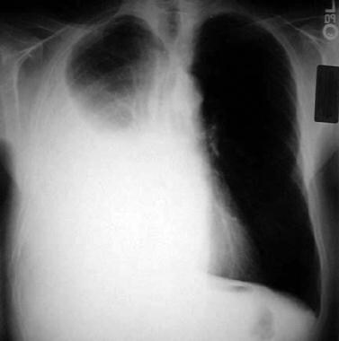
BACKGROUND: Severe chest pain is common in mesothelioma. Percutaneous cervical cordotomy, which interrupts the spinothalamic tract at the C1/C2 level causing contralateral loss of pain sensation, is particularly appropriate in mesothelioma as the tumour is unilateral and systemic analgesia may be ineffective and is limited by harmful side effects. METHOD: A retrospective review was performed to determine the effectiveness and complication rate of this procedure. RESULTS: Fifty two patients were using opioids prior to cordotomy. The median daily dose of morphine before and after cordotomy was 100 mg (range 0-1000 mg) and 20 mg (range 0-520 mg), respectively (p < 0.001). Forty three patients (83%) had a reduction in pain such that their dose of opioid could be at least halved. Twenty patients (38%) were able to stop completely. Recurrence of pain requiring an increase in opioid medication was recorded in 18 patients at a median time of nine weeks (range 0.7-26 weeks). Four patients developed mild weakness, two had troublesome dysaesthesia. The median time from cordotomy to death was 13 weeks (range 0.3-52 weeks). Six early deaths within two weeks of cordotomy occurred early in the series and reflect postoperative chest infection and poor selection as the patients were in the terminal stages of mesothelioma. CONCLUSIONS: Percutaneous cervical cordotomy is successful in treating pain from mesothelioma. There was a low complication rate in this series. Referral to a unit experienced in cordotomy is recommended as soon as pain from chest wall invasion is suspected. (+info)Pleural neoplasms refer to abnormal growths or tumors that develop in the pleura, which is the thin, double layered membrane that surrounds the lungs and lines the inside of the chest wall. These neoplasms can be benign (non-cancerous) or malignant (cancerous).
Malignant pleural neoplasms are often associated with lung cancer, mesothelioma, or metastasis from other types of cancer. They can cause symptoms such as chest pain, cough, shortness of breath, and weight loss. Diagnosis typically involves imaging tests like X-rays or CT scans, followed by biopsy to confirm the type of tumor. Treatment options may include surgery, radiation therapy, chemotherapy, or a combination of these approaches.
Pancreatic neoplasms refer to abnormal growths in the pancreas that can be benign or malignant. The pancreas is a gland located behind the stomach that produces hormones and digestive enzymes. Pancreatic neoplasms can interfere with the normal functioning of the pancreas, leading to various health complications.
Benign pancreatic neoplasms are non-cancerous growths that do not spread to other parts of the body. They are usually removed through surgery to prevent any potential complications, such as blocking the bile duct or causing pain.
Malignant pancreatic neoplasms, also known as pancreatic cancer, are cancerous growths that can invade and destroy surrounding tissues and organs. They can also spread (metastasize) to other parts of the body, such as the liver, lungs, or bones. Pancreatic cancer is often aggressive and difficult to treat, with a poor prognosis.
There are several types of pancreatic neoplasms, including adenocarcinomas, neuroendocrine tumors, solid pseudopapillary neoplasms, and cystic neoplasms. The specific type of neoplasm is determined through various diagnostic tests, such as imaging studies, biopsies, and blood tests. Treatment options depend on the type, stage, and location of the neoplasm, as well as the patient's overall health and preferences.
Neoplasms are abnormal growths of cells or tissues in the body that serve no physiological function. They can be benign (non-cancerous) or malignant (cancerous). Benign neoplasms are typically slow growing and do not spread to other parts of the body, while malignant neoplasms are aggressive, invasive, and can metastasize to distant sites.
Neoplasms occur when there is a dysregulation in the normal process of cell division and differentiation, leading to uncontrolled growth and accumulation of cells. This can result from genetic mutations or other factors such as viral infections, environmental exposures, or hormonal imbalances.
Neoplasms can develop in any organ or tissue of the body and can cause various symptoms depending on their size, location, and type. Treatment options for neoplasms include surgery, radiation therapy, chemotherapy, immunotherapy, and targeted therapy, among others.
Neoplasms: Neoplasms refer to abnormal growths of tissue that can be benign (non-cancerous) or malignant (cancerous). They occur when the normal control mechanisms that regulate cell growth and division are disrupted, leading to uncontrolled cell proliferation.
Cystic Neoplasms: Cystic neoplasms are tumors that contain fluid-filled sacs or cysts. These tumors can be benign or malignant and can occur in various organs of the body, including the pancreas, ovary, and liver.
Mucinous Neoplasms: Mucinous neoplasms are a type of cystic neoplasm that is characterized by the production of mucin, a gel-like substance produced by certain types of cells. These tumors can occur in various organs, including the ovary, pancreas, and colon. Mucinous neoplasms can be benign or malignant, and malignant forms are often aggressive and have a poor prognosis.
Serous Neoplasms: Serous neoplasms are another type of cystic neoplasm that is characterized by the production of serous fluid, which is a thin, watery fluid. These tumors commonly occur in the ovary and can be benign or malignant. Malignant serous neoplasms are often aggressive and have a poor prognosis.
In summary, neoplasms refer to abnormal tissue growths that can be benign or malignant. Cystic neoplasms contain fluid-filled sacs and can occur in various organs of the body. Mucinous neoplasms produce a gel-like substance called mucin and can also occur in various organs, while serous neoplasms produce thin, watery fluid and commonly occur in the ovary. Both mucinous and serous neoplasms can be benign or malignant, with malignant forms often being aggressive and having a poor prognosis.
Skin neoplasms refer to abnormal growths or tumors in the skin that can be benign (non-cancerous) or malignant (cancerous). They result from uncontrolled multiplication of skin cells, which can form various types of lesions. These growths may appear as lumps, bumps, sores, patches, or discolored areas on the skin.
Benign skin neoplasms include conditions such as moles, warts, and seborrheic keratoses, while malignant skin neoplasms are primarily classified into melanoma, squamous cell carcinoma, and basal cell carcinoma. These three types of cancerous skin growths are collectively known as non-melanoma skin cancers (NMSCs). Melanoma is the most aggressive and dangerous form of skin cancer, while NMSCs tend to be less invasive but more common.
It's essential to monitor any changes in existing skin lesions or the appearance of new growths and consult a healthcare professional for proper evaluation and treatment if needed.
Multiple primary neoplasms refer to the occurrence of more than one primary malignant tumor in an individual, where each tumor is unrelated to the other and originates from separate cells or organs. This differs from metastatic cancer, where a single malignancy spreads to multiple sites in the body. Multiple primary neoplasms can be synchronous (occurring at the same time) or metachronous (occurring at different times). The risk of developing multiple primary neoplasms increases with age and is associated with certain genetic predispositions, environmental factors, and lifestyle choices such as smoking and alcohol consumption.
Kidney neoplasms refer to abnormal growths or tumors in the kidney tissues that can be benign (non-cancerous) or malignant (cancerous). These growths can originate from various types of kidney cells, including the renal tubules, glomeruli, and the renal pelvis.
Malignant kidney neoplasms are also known as kidney cancers, with renal cell carcinoma being the most common type. Benign kidney neoplasms include renal adenomas, oncocytomas, and angiomyolipomas. While benign neoplasms are generally not life-threatening, they can still cause problems if they grow large enough to compromise kidney function or if they undergo malignant transformation.
Early detection and appropriate management of kidney neoplasms are crucial for improving patient outcomes and overall prognosis. Regular medical check-ups, imaging studies, and urinalysis can help in the early identification of these growths, allowing for timely intervention and treatment.
A "second primary neoplasm" is a distinct, new cancer or malignancy that develops in a person who has already had a previous cancer. It is not a recurrence or metastasis of the original tumor, but rather an independent cancer that arises in a different location or organ system. The development of second primary neoplasms can be influenced by various factors such as genetic predisposition, environmental exposures, and previous treatments like chemotherapy or radiation therapy.
It is important to note that the definition of "second primary neoplasm" may vary slightly depending on the specific source or context. In general medical usage, it refers to a new, separate cancer; however, in some research or clinical settings, there might be more precise criteria for defining and diagnosing second primary neoplasms.
Adenocarcinoma, mucinous is a type of cancer that begins in the glandular cells that line certain organs and produce mucin, a substance that lubricates and protects tissues. This type of cancer is characterized by the presence of abundant pools of mucin within the tumor. It typically develops in organs such as the colon, rectum, lungs, pancreas, and ovaries.
Mucinous adenocarcinomas tend to have a distinct appearance under the microscope, with large pools of mucin pushing aside the cancer cells. They may also have a different clinical behavior compared to other types of adenocarcinomas, such as being more aggressive or having a worse prognosis in some cases.
It is important to note that while a diagnosis of adenocarcinoma, mucinous can be serious, the prognosis and treatment options may vary depending on several factors, including the location of the cancer, the stage at which it was diagnosed, and the individual's overall health.
Thyroid neoplasms refer to abnormal growths or tumors in the thyroid gland, which can be benign (non-cancerous) or malignant (cancerous). These growths can vary in size and may cause a noticeable lump or nodule in the neck. Thyroid neoplasms can also affect the function of the thyroid gland, leading to hormonal imbalances and related symptoms. The exact causes of thyroid neoplasms are not fully understood, but risk factors include radiation exposure, family history, and certain genetic conditions. It is important to note that most thyroid nodules are benign, but a proper medical evaluation is necessary to determine the nature of the growth and develop an appropriate treatment plan.
Myeloproliferative disorders (MPDs) are a group of rare, chronic blood cancers that originate from the abnormal proliferation or growth of one or more types of blood-forming cells in the bone marrow. These disorders result in an overproduction of mature but dysfunctional blood cells, which can lead to serious complications such as blood clots, bleeding, and organ damage.
There are several subtypes of MPDs, including:
1. Chronic Myeloid Leukemia (CML): A disorder characterized by the overproduction of mature granulocytes (a type of white blood cell) in the bone marrow, leading to an increased number of these cells in the blood. CML is caused by a genetic mutation that results in the formation of the BCR-ABL fusion protein, which drives uncontrolled cell growth and division.
2. Polycythemia Vera (PV): A disorder characterized by the overproduction of all three types of blood cells - red blood cells, white blood cells, and platelets - in the bone marrow. This can lead to an increased risk of blood clots, bleeding, and enlargement of the spleen.
3. Essential Thrombocythemia (ET): A disorder characterized by the overproduction of platelets in the bone marrow, leading to an increased risk of blood clots and bleeding.
4. Primary Myelofibrosis (PMF): A disorder characterized by the replacement of normal bone marrow tissue with scar tissue, leading to impaired blood cell production and anemia, enlargement of the spleen, and increased risk of infections and bleeding.
5. Chronic Neutrophilic Leukemia (CNL): A rare disorder characterized by the overproduction of neutrophils (a type of white blood cell) in the bone marrow, leading to an increased number of these cells in the blood. CNL can lead to an increased risk of infections and organ damage.
MPDs are typically treated with a combination of therapies, including chemotherapy, targeted therapy, immunotherapy, and stem cell transplantation. The choice of treatment depends on several factors, including the subtype of MPD, the patient's age and overall health, and the presence of any comorbidities.
The term "DNA, neoplasm" is not a standard medical term or concept. DNA refers to deoxyribonucleic acid, which is the genetic material present in the cells of living organisms. A neoplasm, on the other hand, is a tumor or growth of abnormal tissue that can be benign (non-cancerous) or malignant (cancerous).
In some contexts, "DNA, neoplasm" may refer to genetic alterations found in cancer cells. These genetic changes can include mutations, amplifications, deletions, or rearrangements of DNA sequences that contribute to the development and progression of cancer. Identifying these genetic abnormalities can help doctors diagnose and treat certain types of cancer more effectively.
However, it's important to note that "DNA, neoplasm" is not a term that would typically be used in medical reports or research papers without further clarification. If you have any specific questions about DNA changes in cancer cells or neoplasms, I would recommend consulting with a healthcare professional or conducting further research on the topic.
Lung neoplasms refer to abnormal growths or tumors in the lung tissue. These tumors can be benign (non-cancerous) or malignant (cancerous). Malignant lung neoplasms are further classified into two main types: small cell lung carcinoma and non-small cell lung carcinoma. Lung neoplasms can cause symptoms such as cough, chest pain, shortness of breath, and weight loss. They are often caused by smoking or exposure to secondhand smoke, but can also occur due to genetic factors, radiation exposure, and other environmental carcinogens. Early detection and treatment of lung neoplasms is crucial for improving outcomes and survival rates.
Parotid neoplasms refer to abnormal growths or tumors in the parotid gland, which is the largest of the salivary glands and is located in front of the ear and extends down the neck. These neoplasms can be benign (non-cancerous) or malignant (cancerous).
Benign parotid neoplasms are typically slow-growing, painless masses that may cause facial asymmetry or difficulty in chewing or swallowing if they become large enough to compress surrounding structures. The most common type of benign parotid tumor is a pleomorphic adenoma.
Malignant parotid neoplasms, on the other hand, are more aggressive and can invade nearby tissues and spread to other parts of the body. They may present as rapidly growing masses that are firm or fixed to surrounding structures. Common types of malignant parotid tumors include mucoepidermoid carcinoma, adenoid cystic carcinoma, and squamous cell carcinoma.
The diagnosis of parotid neoplasms typically involves a thorough clinical evaluation, imaging studies such as CT or MRI scans, and fine-needle aspiration biopsy (FNAB) to determine the nature of the tumor. Treatment options depend on the type, size, and location of the neoplasm but may include surgical excision, radiation therapy, and chemotherapy.
Cystadenoma is a type of benign tumor (not cancerous), which arises from glandular epithelial cells and is covered by a thin layer of connective tissue. These tumors can develop in various locations within the body, including the ovaries, pancreas, and other organs that contain glands.
There are two main types of cystadenomas: serous and mucinous. Serous cystadenomas are filled with a clear or watery fluid, while mucinous cystadenomas contain a thick, gelatinous material. Although they are generally not harmful, these tumors can grow quite large and cause discomfort or other symptoms due to their size or location. In some cases, cystadenomas may undergo malignant transformation and develop into cancerous tumors, known as cystadenocarcinomas. Regular medical follow-up and monitoring are essential for individuals diagnosed with cystadenomas to ensure early detection and treatment of any potential complications.
Neoplasms of connective and soft tissue are abnormal growths or tumors that develop in the body's supportive tissues, such as cartilage, tendons, ligaments, fascia, and fat. These neoplasms can be benign (non-cancerous) or malignant (cancerous).
Benign connective and soft tissue neoplasms include:
- Lipomas: slow-growing, fatty tumors that develop under the skin.
- Fibromas: firm, benign tumors that develop in connective tissue such as tendons or ligaments.
- Nevi (plural of nevus): benign growths made up of cells called melanocytes, which produce pigment.
Malignant connective and soft tissue neoplasms include:
- Sarcomas: a type of cancer that develops in the body's supportive tissues such as muscle, bone, fat, cartilage, or blood vessels. There are many different types of sarcomas, including liposarcoma (fatty tissue), rhabdomyosarcoma (muscle), and osteosarcoma (bone).
- Desmoid tumors: a rare type of benign tumor that can become aggressive and invade surrounding tissues. While not considered cancerous, desmoid tumors can cause significant morbidity due to their tendency to grow and infiltrate nearby structures.
Connective and soft tissue neoplasms can present with various symptoms depending on their location and size. Treatment options include surgery, radiation therapy, chemotherapy, or a combination of these modalities. Regular follow-up care is essential to monitor for recurrence or metastasis (spread) of the tumor.
Plasma cell neoplasms are a type of cancer that originates from plasma cells, which are a type of white blood cell found in the bone marrow. These cells are responsible for producing antibodies to help fight off infections. When plasma cells become cancerous and multiply out of control, they can form a tumor called a plasmacytoma.
There are two main types of plasma cell neoplasms: solitary plasmacytoma and multiple myeloma. Solitary plasmacytoma is a localized tumor that typically forms in the bone, while multiple myeloma is a systemic disease that affects multiple bones and can cause a variety of symptoms such as bone pain, fatigue, and anemia.
Plasma cell neoplasms are diagnosed through a combination of tests, including blood tests, imaging studies, and bone marrow biopsy. Treatment options depend on the stage and extent of the disease, but may include radiation therapy, chemotherapy, and stem cell transplantation.
Appendiceal neoplasms refer to various types of tumors that can develop in the appendix, a small tube-like structure attached to the large intestine. These neoplasms can be benign or malignant and can include:
1. Adenomas: These are benign tumors that arise from the glandular cells lining the appendix. They are usually slow-growing and may not cause any symptoms.
2. Carcinoids: These are neuroendocrine tumors that arise from the hormone-producing cells in the appendix. They are typically small and slow-growing, but some can be aggressive and spread to other parts of the body.
3. Mucinous neoplasms: These are tumors that produce mucin, a slippery substance that can cause the appendix to become distended and filled with mucus. They can be low-grade (less aggressive) or high-grade (more aggressive) and may spread to other parts of the abdomen.
4. Adenocarcinomas: These are malignant tumors that arise from the glandular cells lining the appendix. They are relatively rare but can be aggressive and spread to other parts of the body.
5. Pseudomyxoma peritonei: This is a condition in which mucin produced by an appendiceal neoplasm leaks into the abdominal cavity, causing a jelly-like accumulation of fluid and tissue. It can be caused by both benign and malignant tumors.
Treatment for appendiceal neoplasms depends on the type and stage of the tumor, as well as the patient's overall health. Treatment options may include surgery, chemotherapy, or radiation therapy.
Liver neoplasms refer to abnormal growths in the liver that can be benign or malignant. Benign liver neoplasms are non-cancerous tumors that do not spread to other parts of the body, while malignant liver neoplasms are cancerous tumors that can invade and destroy surrounding tissue and spread to other organs.
Liver neoplasms can be primary, meaning they originate in the liver, or secondary, meaning they have metastasized (spread) to the liver from another part of the body. Primary liver neoplasms can be further classified into different types based on their cell of origin and behavior, including hepatocellular carcinoma, cholangiocarcinoma, and hepatic hemangioma.
The diagnosis of liver neoplasms typically involves a combination of imaging studies, such as ultrasound, CT scan, or MRI, and biopsy to confirm the type and stage of the tumor. Treatment options depend on the type and extent of the neoplasm and may include surgery, radiation therapy, chemotherapy, or liver transplantation.
Mucinous cystadenoma is a type of benign tumor that arises from the epithelial cells lining the mucous membranes of the body. It is most commonly found in the ovary, but can also occur in other locations such as the pancreas or appendix.
Mucinous cystadenomas are characterized by the production of large amounts of mucin, a slippery, gel-like substance that accumulates inside the tumor and causes it to grow into a cystic mass. These tumors can vary in size, ranging from a few centimeters to over 20 centimeters in diameter.
While mucinous cystadenomas are generally benign, they have the potential to become cancerous (mucinous cystadenocarcinoma) if left untreated. Symptoms of mucinous cystadenoma may include abdominal pain or swelling, bloating, and changes in bowel movements or urinary habits. Treatment typically involves surgical removal of the tumor.
Ovarian neoplasms refer to abnormal growths or tumors in the ovary, which can be benign (non-cancerous) or malignant (cancerous). These growths can originate from various cell types within the ovary, including epithelial cells, germ cells, and stromal cells. Ovarian neoplasms are often classified based on their cell type of origin, histological features, and potential for invasive or metastatic behavior.
Epithelial ovarian neoplasms are the most common type and can be further categorized into several subtypes, such as serous, mucinous, endometrioid, clear cell, and Brenner tumors. Some of these epithelial tumors have a higher risk of becoming malignant and spreading to other parts of the body.
Germ cell ovarian neoplasms arise from the cells that give rise to eggs (oocytes) and can include teratomas, dysgerminomas, yolk sac tumors, and embryonal carcinomas. Stromal ovarian neoplasms develop from the connective tissue cells supporting the ovary and can include granulosa cell tumors, thecomas, and fibromas.
It is essential to diagnose and treat ovarian neoplasms promptly, as some malignant forms can be aggressive and potentially life-threatening if not managed appropriately. Regular gynecological exams, imaging studies, and tumor marker tests are often used for early detection and monitoring of ovarian neoplasms. Treatment options may include surgery, chemotherapy, or radiation therapy, depending on the type, stage, and patient's overall health condition.
Endocrine gland neoplasms refer to abnormal growths (tumors) that develop in the endocrine glands. These glands are responsible for producing hormones, which are chemical messengers that regulate various functions and processes in the body. Neoplasms can be benign or malignant (cancerous). Benign neoplasms tend to grow slowly and do not spread to other parts of the body. Malignant neoplasms, on the other hand, can invade nearby tissues and organs and may also metastasize (spread) to distant sites.
Endocrine gland neoplasms can occur in any of the endocrine glands, including:
1. Pituitary gland: located at the base of the brain, it produces several hormones that regulate growth and development, as well as other bodily functions.
2. Thyroid gland: located in the neck, it produces thyroid hormones that regulate metabolism and calcium balance.
3. Parathyroid glands: located near the thyroid gland, they produce parathyroid hormone that regulates calcium levels in the blood.
4. Adrenal glands: located on top of each kidney, they produce hormones such as adrenaline, cortisol, and aldosterone that regulate stress response, metabolism, and blood pressure.
5. Pancreas: located behind the stomach, it produces insulin and glucagon, which regulate blood sugar levels, and digestive enzymes that help break down food.
6. Pineal gland: located in the brain, it produces melatonin, a hormone that regulates sleep-wake cycles.
7. Gonads (ovaries and testicles): located in the pelvis (ovaries) and scrotum (testicles), they produce sex hormones such as estrogen, progesterone, and testosterone that regulate reproductive function and secondary sexual characteristics.
Endocrine gland neoplasms can cause various symptoms depending on the type and location of the tumor. For example, a pituitary gland neoplasm may cause headaches, vision problems, or hormonal imbalances, while an adrenal gland neoplasm may cause high blood pressure, weight gain, or mood changes.
Diagnosis of endocrine gland neoplasms typically involves a combination of medical history, physical examination, imaging studies such as CT or MRI scans, and laboratory tests to measure hormone levels. Treatment options may include surgery, radiation therapy, chemotherapy, or hormonal therapy, depending on the type and stage of the tumor.
Gastrointestinal (GI) neoplasms refer to abnormal growths in the gastrointestinal tract, which can be benign or malignant. The gastrointestinal tract includes the mouth, esophagus, stomach, small intestine, large intestine, rectum, and anus.
Benign neoplasms are non-cancerous growths that do not invade nearby tissues or spread to other parts of the body. They can sometimes be removed completely and may not cause any further health problems.
Malignant neoplasms, on the other hand, are cancerous growths that can invade nearby tissues and organs and spread to other parts of the body through the bloodstream or lymphatic system. These types of neoplasms can be life-threatening if not diagnosed and treated promptly.
GI neoplasms can cause various symptoms, including abdominal pain, bloating, changes in bowel habits, nausea, vomiting, weight loss, and anemia. The specific symptoms may depend on the location and size of the neoplasm.
There are many types of GI neoplasms, including adenocarcinomas, gastrointestinal stromal tumors (GISTs), lymphomas, and neuroendocrine tumors. The diagnosis of GI neoplasms typically involves a combination of medical history, physical examination, imaging studies, and biopsy. Treatment options may include surgery, radiation therapy, chemotherapy, targeted therapy, or immunotherapy.
Pancreatic ductal carcinoma (PDC) is a specific type of cancer that forms in the ducts that carry digestive enzymes out of the pancreas. It's the most common form of exocrine pancreatic cancer, making up about 90% of all cases.
The symptoms of PDC are often vague and can include abdominal pain, jaundice (yellowing of the skin and eyes), unexplained weight loss, and changes in bowel movements. These symptoms can be similar to those caused by other less serious conditions, which can make diagnosis difficult.
Pancreatic ductal carcinoma is often aggressive and difficult to treat. The prognosis for PDC is generally poor, with a five-year survival rate of only about 9%. Treatment options may include surgery, chemotherapy, radiation therapy, or a combination of these approaches. However, because PDC is often not detected until it has advanced, treatment is frequently focused on palliative care to relieve symptoms and improve quality of life.
Experimental neoplasms refer to abnormal growths or tumors that are induced and studied in a controlled laboratory setting, typically in animals or cell cultures. These studies are conducted to understand the fundamental mechanisms of cancer development, progression, and potential treatment strategies. By manipulating various factors such as genetic mutations, environmental exposures, and pharmacological interventions, researchers can gain valuable insights into the complex processes underlying neoplasm formation and identify novel targets for cancer therapy. It is important to note that experimental neoplasms may not always accurately represent human cancers, and further research is needed to translate these findings into clinically relevant applications.
A neoplasm of vascular tissue is an abnormal growth or mass of cells in the blood vessels or lymphatic vessels. These growths can be benign (non-cancerous) or malignant (cancerous). Benign neoplasms, such as hemangiomas and lymphangiomas, are typically not harmful and may not require treatment. However, they can cause symptoms if they grow large enough to press on nearby organs or tissues. Malignant neoplasms, such as angiosarcomas, are cancerous and can invade and destroy surrounding tissue, as well as spread (metastasize) to other parts of the body. Treatment for vascular tissue neoplasms depends on the type, size, location, and stage of the growth, and may include surgery, radiation therapy, chemotherapy, or a combination of these.
Eye neoplasms, also known as ocular tumors or eye cancer, refer to abnormal growths of tissue in the eye. These growths can be benign (non-cancerous) or malignant (cancerous). Eye neoplasms can develop in various parts of the eye, including the eyelid, conjunctiva, cornea, iris, ciliary body, choroid, retina, and optic nerve.
Benign eye neoplasms are typically slow-growing and do not spread to other parts of the body. They may cause symptoms such as vision changes, eye pain, or a noticeable mass in the eye. Treatment options for benign eye neoplasms include monitoring, surgical removal, or radiation therapy.
Malignant eye neoplasms, on the other hand, can grow and spread rapidly to other parts of the body. They may cause symptoms such as vision changes, eye pain, floaters, or flashes of light. Treatment options for malignant eye neoplasms depend on the type and stage of cancer but may include surgery, radiation therapy, chemotherapy, or a combination of these treatments.
It is important to note that early detection and treatment of eye neoplasms can improve outcomes and prevent complications. Regular eye exams with an ophthalmologist are recommended for early detection and prevention of eye diseases, including eye neoplasms.
Immunohistochemistry (IHC) is a technique used in pathology and laboratory medicine to identify specific proteins or antigens in tissue sections. It combines the principles of immunology and histology to detect the presence and location of these target molecules within cells and tissues. This technique utilizes antibodies that are specific to the protein or antigen of interest, which are then tagged with a detection system such as a chromogen or fluorophore. The stained tissue sections can be examined under a microscope, allowing for the visualization and analysis of the distribution and expression patterns of the target molecule in the context of the tissue architecture. Immunohistochemistry is widely used in diagnostic pathology to help identify various diseases, including cancer, infectious diseases, and immune-mediated disorders.
Nose neoplasms refer to abnormal growths or tumors in the nasal cavity or paranasal sinuses. These growths can be benign (non-cancerous) or malignant (cancerous). Benign neoplasms are typically slow-growing and do not spread to other parts of the body, while malignant neoplasms can invade surrounding tissues and have the potential to metastasize.
Nose neoplasms can cause various symptoms such as nasal congestion, nosebleeds, difficulty breathing through the nose, loss of smell, facial pain or numbness, and visual changes if they affect the eye. The diagnosis of nose neoplasms usually involves a combination of physical examination, imaging studies (such as CT or MRI scans), and biopsy to determine the type and extent of the growth. Treatment options depend on the type, size, location, and stage of the neoplasm and may include surgery, radiation therapy, chemotherapy, or a combination of these approaches.
Salivary gland neoplasms refer to abnormal growths or tumors that develop in the salivary glands. These glands are responsible for producing saliva, which helps in digestion, lubrication of food and maintaining oral health. Salivary gland neoplasms can be benign (non-cancerous) or malignant (cancerous).
Benign neoplasms are slow-growing and typically do not spread to other parts of the body. They may cause symptoms such as swelling, painless lumps, or difficulty swallowing if they grow large enough to put pressure on surrounding tissues.
Malignant neoplasms, on the other hand, can be aggressive and have the potential to invade nearby structures and metastasize (spread) to distant organs. Symptoms of malignant salivary gland neoplasms may include rapid growth, pain, numbness, or paralysis of facial nerves.
Salivary gland neoplasms can occur in any of the major salivary glands (parotid, submandibular, and sublingual glands) or in the minor salivary glands located throughout the mouth and throat. The exact cause of these neoplasms is not fully understood, but risk factors may include exposure to radiation, certain viral infections, and genetic predisposition.
Radiation-induced neoplasms are a type of cancer or tumor that develops as a result of exposure to ionizing radiation. Ionizing radiation is radiation with enough energy to remove tightly bound electrons from atoms or molecules, leading to the formation of ions. This type of radiation can damage DNA and other cellular structures, which can lead to mutations and uncontrolled cell growth, resulting in the development of a neoplasm.
Radiation-induced neoplasms can occur after exposure to high levels of ionizing radiation, such as that received during radiation therapy for cancer treatment or from nuclear accidents. The risk of developing a radiation-induced neoplasm depends on several factors, including the dose and duration of radiation exposure, the type of radiation, and the individual's genetic susceptibility to radiation-induced damage.
Radiation-induced neoplasms can take many years to develop after initial exposure to ionizing radiation, and they often occur at the site of previous radiation therapy. Common types of radiation-induced neoplasms include sarcomas, carcinomas, and thyroid cancer. It is important to note that while ionizing radiation can increase the risk of developing cancer, the overall risk is still relatively low, especially when compared to other well-established cancer risk factors such as smoking and exposure to certain chemicals.
Adenocarcinoma, papillary is a type of cancer that begins in the glandular cells and grows in a finger-like projection (called a papilla). This type of cancer can occur in various organs, including the lungs, pancreas, thyroid, and female reproductive system. The prognosis and treatment options for papillary adenocarcinoma depend on several factors, such as the location and stage of the tumor, as well as the patient's overall health. It is important to consult with a healthcare professional for an accurate diagnosis and personalized treatment plan.
Carcinoma, papillary is a type of cancer that begins in the cells that line the glandular structures or the lining of organs. In a papillary carcinoma, the cancerous cells grow and form small finger-like projections, called papillae, within the tumor. This type of cancer most commonly occurs in the thyroid gland, but can also be found in other organs such as the lung, breast, and kidney. Papillary carcinoma of the thyroid gland is usually slow-growing and has a good prognosis, especially when it is diagnosed at an early stage.
Testicular neoplasms are abnormal growths or tumors in the testicle that can be benign (non-cancerous) or malignant (cancerous). They are a type of genitourinary cancer, which affects the reproductive and urinary systems. Testicular neoplasms can occur in men of any age but are most commonly found in young adults between the ages of 15 and 40.
Testicular neoplasms can be classified into two main categories: germ cell tumors and non-germ cell tumors. Germ cell tumors, which arise from the cells that give rise to sperm, are further divided into seminomas and non-seminomas. Seminomas are typically slow-growing and have a good prognosis, while non-seminomas tend to grow more quickly and can spread to other parts of the body.
Non-germ cell tumors are less common than germ cell tumors and include Leydig cell tumors, Sertoli cell tumors, and lymphomas. These tumors can have a variety of clinical behaviors, ranging from benign to malignant.
Testicular neoplasms often present as a painless mass or swelling in the testicle. Other symptoms may include a feeling of heaviness or discomfort in the scrotum, a dull ache in the lower abdomen or groin, and breast enlargement (gynecomastia).
Diagnosis typically involves a physical examination, imaging studies such as ultrasound or CT scan, and blood tests to detect tumor markers. Treatment options depend on the type and stage of the neoplasm but may include surgery, radiation therapy, chemotherapy, or a combination of these modalities. Regular self-examinations of the testicles are recommended for early detection and improved outcomes.
Neoplasms in muscle tissue refer to abnormal and excessive growths of muscle cells that can be benign or malignant. These growths can arise from any of the three types of muscle tissue: skeletal, cardiac, or smooth muscle. Neoplasms in muscle tissue are classified based on their origin, behavior, and histological features.
Benign neoplasms in muscle tissue include leiomyomas (smooth muscle), rhabdomyomas (skeletal muscle), and myxomas (cardiac muscle). These tumors are usually slow-growing and do not invade surrounding tissues or spread to other parts of the body.
Malignant neoplasms in muscle tissue, also known as sarcomas, include leiomyosarcoma (smooth muscle), rhabdomyosarcoma (skeletal muscle), and angiosarcoma (cardiac muscle). These tumors are aggressive, invasive, and have the potential to metastasize to other parts of the body.
Symptoms of neoplasms in muscle tissue depend on their location, size, and type. They may include a painless or painful mass, weakness, fatigue, weight loss, and difficulty swallowing or breathing. Treatment options for neoplasms in muscle tissue include surgery, radiation therapy, chemotherapy, and targeted therapy. The choice of treatment depends on the type, stage, location, and patient's overall health condition.
Neoplasms are abnormal growths of cells or tissues that serve no purpose and can be benign (non-cancerous) or malignant (cancerous). Glandular and epithelial neoplasms refer to specific types of tumors that originate from the glandular and epithelial tissues, respectively.
Glandular neoplasms arise from the glandular tissue, which is responsible for producing and secreting substances such as hormones, enzymes, or other fluids. These neoplasms can be further classified into adenomas (benign) and adenocarcinomas (malignant).
Epithelial neoplasms, on the other hand, develop from the epithelial tissue that lines the outer surfaces of organs and the inner surfaces of cavities. These neoplasms can also be benign or malignant and are classified as papillomas (benign) and carcinomas (malignant).
It is important to note that while both glandular and epithelial neoplasms can become cancerous, not all of them do. However, if they do, the malignant versions can invade surrounding tissues and spread to other parts of the body, making them potentially life-threatening.
Mucinous cystadenocarcinoma is a type of cancer that arises from the mucin-producing cells in the lining of a cyst. It is a subtype of cystadenocarcinoma, which is a malignant tumor that develops within a cyst. Mucinous cystadenocarcinomas are typically found in the ovary or pancreas but can also occur in other organs such as the appendix and the respiratory tract.
These tumors are characterized by the production of large amounts of mucin, a gel-like substance that can accumulate within the cyst and cause it to grow. Mucinous cystadenocarcinomas tend to grow slowly but can become quite large and may eventually spread (metastasize) to other parts of the body if left untreated.
Symptoms of mucinous cystadenocarcinoma depend on the location and size of the tumor, but they may include abdominal pain or discomfort, bloating, changes in bowel movements, or vaginal bleeding. Treatment typically involves surgical removal of the tumor, followed by chemotherapy or radiation therapy to kill any remaining cancer cells. The prognosis for mucinous cystadenocarcinoma depends on several factors, including the stage of the disease at diagnosis and the patient's overall health.
An adenoma is a benign (noncancerous) tumor that develops from glandular epithelial cells. These types of cells are responsible for producing and releasing fluids, such as hormones or digestive enzymes, into the surrounding tissues. Adenomas can occur in various organs and glands throughout the body, including the thyroid, pituitary, adrenal, and digestive systems.
Depending on their location, adenomas may cause different symptoms or remain asymptomatic. Some common examples of adenomas include:
1. Colorectal adenoma (also known as a polyp): These growths occur in the lining of the colon or rectum and can develop into colorectal cancer if left untreated. Regular screenings, such as colonoscopies, are essential for early detection and removal of these polyps.
2. Thyroid adenoma: This type of adenoma affects the thyroid gland and may result in an overproduction or underproduction of hormones, leading to conditions like hyperthyroidism (overactive thyroid) or hypothyroidism (underactive thyroid).
3. Pituitary adenoma: These growths occur in the pituitary gland, which is located at the base of the brain and controls various hormonal functions. Depending on their size and location, pituitary adenomas can cause vision problems, headaches, or hormonal imbalances that affect growth, reproduction, and metabolism.
4. Liver adenoma: These rare benign tumors develop in the liver and may not cause any symptoms unless they become large enough to press on surrounding organs or structures. In some cases, liver adenomas can rupture and cause internal bleeding.
5. Adrenal adenoma: These growths occur in the adrenal glands, which are located above the kidneys and produce hormones that regulate stress responses, metabolism, and blood pressure. Most adrenal adenomas are nonfunctioning, meaning they do not secrete excess hormones. However, functioning adrenal adenomas can lead to conditions like Cushing's syndrome or Conn's syndrome, depending on the type of hormone being overproduced.
It is essential to monitor and manage benign tumors like adenomas to prevent potential complications, such as rupture, bleeding, or hormonal imbalances. Treatment options may include surveillance with imaging studies, medication to manage hormonal issues, or surgical removal of the tumor in certain cases.
Soft tissue neoplasms refer to abnormal growths or tumors that develop in the soft tissues of the body. Soft tissues include muscles, tendons, ligaments, fascia, nerves, blood vessels, fat, and synovial membranes (the thin layer of cells that line joints and tendons). Neoplasms can be benign (non-cancerous) or malignant (cancerous), and their behavior and potential for spread depend on the specific type of neoplasm.
Benign soft tissue neoplasms are typically slow-growing, well-circumscribed, and rarely spread to other parts of the body. They can often be removed surgically with a low risk of recurrence. Examples of benign soft tissue neoplasms include lipomas (fat tumors), schwannomas (nerve sheath tumors), and hemangiomas (blood vessel tumors).
Malignant soft tissue neoplasms, on the other hand, can grow rapidly, invade surrounding tissues, and may metastasize (spread) to distant parts of the body. They are often more difficult to treat than benign neoplasms and require a multidisciplinary approach, including surgery, radiation therapy, and chemotherapy. Examples of malignant soft tissue neoplasms include sarcomas, such as rhabdomyosarcoma (arising from skeletal muscle), leiomyosarcoma (arising from smooth muscle), and angiosarcoma (arising from blood vessels).
It is important to note that soft tissue neoplasms can occur in any part of the body, and their diagnosis and treatment require a thorough evaluation by a healthcare professional with expertise in this area.
Hematologic neoplasms, also known as hematological malignancies, are a group of diseases characterized by the uncontrolled growth and accumulation of abnormal blood cells or bone marrow cells. These disorders can originate from the myeloid or lymphoid cell lines, which give rise to various types of blood cells, including red blood cells, white blood cells, and platelets.
Hematologic neoplasms can be broadly classified into three categories:
1. Leukemias: These are cancers that primarily affect the bone marrow and blood-forming tissues. They result in an overproduction of abnormal white blood cells, which interfere with the normal functioning of the blood and immune system. There are several types of leukemia, including acute lymphoblastic leukemia (ALL), chronic lymphocytic leukemia (CLL), acute myeloid leukemia (AML), and chronic myeloid leukemia (CML).
2. Lymphomas: These are cancers that develop from the lymphatic system, which is a part of the immune system responsible for fighting infections. Lymphomas can affect lymph nodes, spleen, bone marrow, and other organs. The two main types of lymphoma are Hodgkin lymphoma (HL) and non-Hodgkin lymphoma (NHL).
3. Myelomas: These are cancers that arise from the plasma cells, a type of white blood cell responsible for producing antibodies. Multiple myeloma is the most common type of myeloma, characterized by an excessive proliferation of malignant plasma cells in the bone marrow, leading to the production of abnormal amounts of monoclonal immunoglobulins (M proteins) and bone destruction.
Hematologic neoplasms can have various symptoms, such as fatigue, weakness, frequent infections, easy bruising or bleeding, weight loss, swollen lymph nodes, and bone pain. The diagnosis typically involves a combination of medical history, physical examination, laboratory tests, imaging studies, and sometimes bone marrow biopsy. Treatment options depend on the type and stage of the disease and may include chemotherapy, radiation therapy, targeted therapy, immunotherapy, stem cell transplantation, or a combination of these approaches.
A neoplasm is a tumor or growth that is formed by an abnormal and excessive proliferation of cells, which can be benign or malignant. Neoplasm proteins are therefore any proteins that are expressed or produced in these neoplastic cells. These proteins can play various roles in the development, progression, and maintenance of neoplasms.
Some neoplasm proteins may contribute to the uncontrolled cell growth and division seen in cancer, such as oncogenic proteins that promote cell cycle progression or inhibit apoptosis (programmed cell death). Others may help the neoplastic cells evade the immune system, allowing them to proliferate undetected. Still others may be involved in angiogenesis, the formation of new blood vessels that supply the tumor with nutrients and oxygen.
Neoplasm proteins can also serve as biomarkers for cancer diagnosis, prognosis, or treatment response. For example, the presence or level of certain neoplasm proteins in biological samples such as blood or tissue may indicate the presence of a specific type of cancer, help predict the likelihood of cancer recurrence, or suggest whether a particular therapy will be effective.
Overall, understanding the roles and behaviors of neoplasm proteins can provide valuable insights into the biology of cancer and inform the development of new diagnostic and therapeutic strategies.
Uterine neoplasms refer to abnormal growths in the uterus, which can be benign (non-cancerous) or malignant (cancerous). These growths can originate from different types of cells within the uterus, leading to various types of uterine neoplasms. The two main categories of uterine neoplasms are endometrial neoplasms and uterine sarcomas.
Endometrial neoplasms develop from the endometrium, which is the inner lining of the uterus. Most endometrial neoplasms are classified as endometrioid adenocarcinomas, arising from glandular cells in the endometrium. Other types include serous carcinoma, clear cell carcinoma, and mucinous carcinoma.
Uterine sarcomas, on the other hand, are less common and originate from the connective tissue (stroma) or muscle (myometrium) of the uterus. Uterine sarcomas can be further divided into several subtypes, such as leiomyosarcoma, endometrial stromal sarcoma, and undifferentiated uterine sarcoma.
Uterine neoplasms can cause various symptoms, including abnormal vaginal bleeding or discharge, pelvic pain, and difficulty urinating or having bowel movements. The diagnosis typically involves a combination of imaging tests (such as ultrasound, CT, or MRI scans) and tissue biopsies to determine the type and extent of the neoplasm. Treatment options depend on the type, stage, and patient's overall health but may include surgery, radiation therapy, chemotherapy, or hormone therapy.
Intestinal neoplasms refer to abnormal growths in the tissues of the intestines, which can be benign or malignant. These growths are called neoplasms and they result from uncontrolled cell division. In the case of intestinal neoplasms, these growths occur in the small intestine, large intestine (colon), rectum, or appendix.
Benign intestinal neoplasms are not cancerous and often do not invade surrounding tissues or spread to other parts of the body. However, they can still cause problems if they grow large enough to obstruct the intestines or cause bleeding. Common types of benign intestinal neoplasms include polyps, leiomyomas, and lipomas.
Malignant intestinal neoplasms, on the other hand, are cancerous and can invade surrounding tissues and spread to other parts of the body. The most common type of malignant intestinal neoplasm is adenocarcinoma, which arises from the glandular cells lining the inside of the intestines. Other types of malignant intestinal neoplasms include lymphomas, sarcomas, and carcinoid tumors.
Symptoms of intestinal neoplasms can vary depending on their size, location, and type. Common symptoms include abdominal pain, bloating, changes in bowel habits, rectal bleeding, weight loss, and fatigue. If you experience any of these symptoms, it is important to seek medical attention promptly.
Neoplasms, adnexal and skin appendage refer to abnormal growths or tumors that develop in the sweat glands, hair follicles, or other structures associated with the skin. These growths can be benign (non-cancerous) or malignant (cancerous), and they can occur anywhere on the body.
Adnexal neoplasms are tumors that arise from the sweat glands or hair follicles, including the sebaceous glands, eccrine glands, and apocrine glands. These tumors can range in size and severity, and they may cause symptoms such as pain, itching, or changes in the appearance of the skin.
Skin appendage neoplasms are similar to adnexal neoplasms, but they specifically refer to tumors that arise from structures such as hair follicles, nails, and sweat glands. Examples of skin appendage neoplasms include pilomatricomas (tumors of the hair follicle), trichilemmomas (tumors of the outer root sheath of the hair follicle), and sebaceous adenomas (tumors of the sebaceous glands).
It is important to note that while many adnexal and skin appendage neoplasms are benign, some can be malignant and may require aggressive treatment. If you notice any unusual growths or changes in your skin, it is important to consult with a healthcare professional for further evaluation and care.
Neoplasm staging is a systematic process used in medicine to describe the extent of spread of a cancer, including the size and location of the original (primary) tumor and whether it has metastasized (spread) to other parts of the body. The most widely accepted system for this purpose is the TNM classification system developed by the American Joint Committee on Cancer (AJCC) and the Union for International Cancer Control (UICC).
In this system, T stands for tumor, and it describes the size and extent of the primary tumor. N stands for nodes, and it indicates whether the cancer has spread to nearby lymph nodes. M stands for metastasis, and it shows whether the cancer has spread to distant parts of the body.
Each letter is followed by a number that provides more details about the extent of the disease. For example, a T1N0M0 cancer means that the primary tumor is small and has not spread to nearby lymph nodes or distant sites. The higher the numbers, the more advanced the cancer.
Staging helps doctors determine the most appropriate treatment for each patient and estimate the patient's prognosis. It is an essential tool for communication among members of the healthcare team and for comparing outcomes of treatments in clinical trials.
Vascular neoplasms are a type of tumor that develops from cells that line the blood vessels or lymphatic vessels. These tumors can be benign (non-cancerous) or malignant (cancerous). Benign vascular neoplasms, such as hemangiomas and lymphangiomas, are usually harmless and may not require treatment unless they cause symptoms or complications. Malignant vascular neoplasms, on the other hand, are known as angiosarcomas and can be aggressive, spreading to other parts of the body and potentially causing serious health problems.
Angiosarcomas can develop in any part of the body but are most commonly found in the skin, particularly in areas exposed to radiation or chronic lymph edema. They can also occur in the breast, liver, spleen, and heart. Treatment for vascular neoplasms depends on the type, location, size, and stage of the tumor, as well as the patient's overall health. Treatment options may include surgery, radiation therapy, chemotherapy, or a combination of these approaches.
Sweat gland neoplasms are abnormal growths that develop in the sweat glands. These growths can be benign (noncancerous) or malignant (cancerous). Benign sweat gland neoplasms include hidradenomas and syringomas, which are usually slow-growing and cause little to no symptoms. Malignant sweat gland neoplasms, also known as sweat gland carcinomas, are rare but aggressive cancers that can spread to other parts of the body. They may cause symptoms such as a lump or mass under the skin, pain, swelling, and redness. Treatment typically involves surgical removal of the growth.
Lymphoma is a type of cancer that originates from the white blood cells called lymphocytes, which are part of the immune system. These cells are found in various parts of the body such as the lymph nodes, spleen, bone marrow, and other organs. Lymphoma can be classified into two main types: Hodgkin lymphoma (HL) and non-Hodgkin lymphoma (NHL).
HL is characterized by the presence of a specific type of abnormal lymphocyte called Reed-Sternberg cells, while NHL includes a diverse group of lymphomas that lack these cells. The symptoms of lymphoma may include swollen lymph nodes, fever, night sweats, weight loss, and fatigue.
The exact cause of lymphoma is not known, but it is believed to result from genetic mutations in the lymphocytes that lead to uncontrolled cell growth and division. Exposure to certain viruses, chemicals, and radiation may increase the risk of developing lymphoma. Treatment options for lymphoma depend on various factors such as the type and stage of the disease, age, and overall health of the patient. Common treatments include chemotherapy, radiation therapy, immunotherapy, and stem cell transplantation.
Bone neoplasms are abnormal growths or tumors that develop in the bone. They can be benign (non-cancerous) or malignant (cancerous). Benign bone neoplasms do not spread to other parts of the body and are rarely a threat to life, although they may cause problems if they grow large enough to press on surrounding tissues or cause fractures. Malignant bone neoplasms, on the other hand, can invade and destroy nearby tissue and may spread (metastasize) to other parts of the body.
There are many different types of bone neoplasms, including:
1. Osteochondroma - a benign tumor that develops from cartilage and bone
2. Enchondroma - a benign tumor that forms in the cartilage that lines the inside of the bones
3. Chondrosarcoma - a malignant tumor that develops from cartilage
4. Osteosarcoma - a malignant tumor that develops from bone cells
5. Ewing sarcoma - a malignant tumor that develops in the bones or soft tissues around the bones
6. Giant cell tumor of bone - a benign or occasionally malignant tumor that develops from bone tissue
7. Fibrosarcoma - a malignant tumor that develops from fibrous tissue in the bone
The symptoms of bone neoplasms vary depending on the type, size, and location of the tumor. They may include pain, swelling, stiffness, fractures, or limited mobility. Treatment options depend on the type and stage of the tumor but may include surgery, radiation therapy, chemotherapy, or a combination of these treatments.
Palatal neoplasms refer to abnormal growths or tumors that occur on the palate, which is the roof of the mouth. These growths can be benign (non-cancerous) or malignant (cancerous). Benign neoplasms are typically slower growing and less likely to spread, while malignant neoplasms are more aggressive and can invade nearby tissues and organs.
Palatal neoplasms can have various causes, including genetic factors, environmental exposures, and viral infections. They may present with symptoms such as mouth pain, difficulty swallowing, swelling or lumps in the mouth, bleeding, or numbness in the mouth or face.
The diagnosis of palatal neoplasms typically involves a thorough clinical examination, imaging studies, and sometimes biopsy to determine the type and extent of the growth. Treatment options depend on the type, size, location, and stage of the neoplasm but may include surgery, radiation therapy, chemotherapy, or a combination of these approaches. Regular follow-up care is essential to monitor for recurrence or spread of the neoplasm.
Neoplasms are abnormal growths of cells or tissues in the body that can be benign (non-cancerous) or malignant (cancerous). When referring to "Complex and Mixed Neoplasms," it is typically used in the context of histopathology, where it describes tumors with a mixture of different types of cells or growth patterns.
A complex neoplasm usually contains areas with various architectural patterns, cell types, or both, making its classification challenging. It may require extensive sampling and careful examination to determine its nature and behavior. These neoplasms can be either benign or malignant, depending on the specific characteristics of the tumor cells and their growth pattern.
A mixed neoplasm, on the other hand, is a tumor that contains more than one type of cell or tissue component, often arising from different germ layers (the three primary layers of embryonic development: ectoderm, mesoderm, and endoderm). A common example of a mixed neoplasm is a teratoma, which can contain tissues derived from all three germ layers, such as skin, hair, teeth, bone, and muscle. Mixed neoplasms can also be benign or malignant, depending on the specific components of the tumor.
It's important to note that the classification and behavior of complex and mixed neoplasms can vary significantly based on their location in the body, cellular composition, and other factors. Accurate diagnosis typically requires a thorough examination by an experienced pathologist and may involve additional tests, such as immunohistochemistry or molecular analysis, to determine the appropriate treatment and management strategies.
Neoplasm antigens, also known as tumor antigens, are substances that are produced by cancer cells (neoplasms) and can stimulate an immune response. These antigens can be proteins, carbohydrates, or other molecules that are either unique to the cancer cells or are overexpressed or mutated versions of normal cellular proteins.
Neoplasm antigens can be classified into two main categories: tumor-specific antigens (TSAs) and tumor-associated antigens (TAAs). TSAs are unique to cancer cells and are not expressed by normal cells, while TAAs are present at low levels in normal cells but are overexpressed or altered in cancer cells.
TSAs can be further divided into viral antigens and mutated antigens. Viral antigens are produced when cancer is caused by a virus, such as human papillomavirus (HPV) in cervical cancer. Mutated antigens are the result of genetic mutations that occur during cancer development and are unique to each patient's tumor.
Neoplasm antigens play an important role in the immune response against cancer. They can be recognized by the immune system, leading to the activation of immune cells such as T cells and natural killer (NK) cells, which can then attack and destroy cancer cells. However, cancer cells often develop mechanisms to evade the immune response, allowing them to continue growing and spreading.
Understanding neoplasm antigens is important for the development of cancer immunotherapies, which aim to enhance the body's natural immune response against cancer. These therapies include checkpoint inhibitors, which block proteins that inhibit T cell activation, and therapeutic vaccines, which stimulate an immune response against specific tumor antigens.
Mandibular neoplasms refer to abnormal growths or tumors that develop in the mandible, which is the lower jawbone. These growths can be benign (non-cancerous) or malignant (cancerous). Benign neoplasms are typically slow-growing and rarely spread to other parts of the body, while malignant neoplasms can invade surrounding tissues and may metastasize (spread) to distant sites.
Mandibular neoplasms can have various causes, including genetic mutations, exposure to certain chemicals or radiation, and infection with certain viruses. The symptoms of mandibular neoplasms may include swelling or pain in the jaw, difficulty chewing or speaking, numbness in the lower lip or chin, loose teeth, and/or a lump or mass in the mouth or neck.
The diagnosis of mandibular neoplasms typically involves a thorough clinical examination, imaging studies such as X-rays, CT scans, or MRI scans, and sometimes a biopsy to confirm the type and extent of the tumor. Treatment options depend on the type, stage, and location of the neoplasm, and may include surgery, radiation therapy, chemotherapy, or a combination of these approaches. Regular follow-up care is essential to monitor for recurrence or metastasis.
Cystadenocarcinoma is a type of tumor that arises from the epithelial lining of a cyst, and it has the potential to invade surrounding tissues and spread (metastasize) to other parts of the body. It typically affects glandular organs such as the ovaries, pancreas, and salivary glands.
Cystadenocarcinomas can be classified into two types: serous and mucinous. Serous cystadenocarcinomas produce a watery fluid, while mucinous cystadenocarcinomas produce a thick, mucus-like fluid. Both types of tumors can be benign or malignant, but malignant cystadenocarcinomas are more aggressive and have a higher risk of metastasis.
Symptoms of cystadenocarcinoma depend on the location and size of the tumor. In some cases, there may be no symptoms until the tumor has grown large enough to cause pain or other problems. Treatment typically involves surgical removal of the tumor, along with any affected surrounding tissue. Chemotherapy and radiation therapy may also be used in some cases to help prevent recurrence or spread of the cancer.
Bile duct neoplasms, also known as cholangiocarcinomas, refer to a group of malignancies that arise from the bile ducts. These are the tubes that carry bile from the liver to the gallbladder and small intestine. Bile duct neoplasms can be further classified based on their location as intrahepatic (within the liver), perihilar (at the junction of the left and right hepatic ducts), or distal (in the common bile duct).
These tumors are relatively rare, but their incidence has been increasing in recent years. They can cause a variety of symptoms, including jaundice, abdominal pain, weight loss, and fever. The diagnosis of bile duct neoplasms typically involves imaging studies such as CT or MRI scans, as well as blood tests to assess liver function. In some cases, a biopsy may be necessary to confirm the diagnosis.
Treatment options for bile duct neoplasms depend on several factors, including the location and stage of the tumor, as well as the patient's overall health. Surgical resection is the preferred treatment for early-stage tumors, while chemotherapy and radiation therapy may be used in more advanced cases. For patients who are not candidates for surgery, palliative treatments such as stenting or bypass procedures may be recommended to relieve symptoms and improve quality of life.
Neoplasm invasiveness is a term used in pathology and oncology to describe the aggressive behavior of cancer cells as they invade surrounding tissues and organs. This process involves the loss of cell-to-cell adhesion, increased motility and migration, and the ability of cancer cells to degrade the extracellular matrix (ECM) through the production of enzymes such as matrix metalloproteinases (MMPs).
Invasive neoplasms are cancers that have spread beyond the original site where they first developed and have infiltrated adjacent tissues or structures. This is in contrast to non-invasive or in situ neoplasms, which are confined to the epithelial layer where they originated and have not yet invaded the underlying basement membrane.
The invasiveness of a neoplasm is an important prognostic factor in cancer diagnosis and treatment, as it can indicate the likelihood of metastasis and the potential effectiveness of various therapies. In general, more invasive cancers are associated with worse outcomes and require more aggressive treatment approaches.
Thymus neoplasms are abnormal growths in the thymus gland that result from uncontrolled cell division. The term "neoplasm" refers to any new and abnormal growth of tissue, also known as a tumor. Thymus neoplasms can be benign or malignant (cancerous).
Malignant thymus neoplasms are called thymomas or thymic carcinomas. Thymomas are the most common type and tend to grow slowly, invading nearby tissues and organs. They can also spread (metastasize) to other parts of the body. Thymic carcinomas are rarer and more aggressive, growing and spreading more quickly than thymomas.
Symptoms of thymus neoplasms may include coughing, chest pain, difficulty breathing, or swelling in the neck or upper chest. Treatment options for thymus neoplasms depend on the type, size, location, and stage of the tumor, as well as the patient's overall health. Treatment may include surgery, radiation therapy, chemotherapy, or a combination of these approaches.
Splenic neoplasms refer to abnormal growths or tumors in the spleen, which can be benign (non-cancerous) or malignant (cancerous). These growths can arise from various cell types present within the spleen, including hematopoietic cells (red and white blood cells, platelets), stromal cells (supporting tissue), or lymphoid cells (part of the immune system).
There are several types of splenic neoplasms:
1. Hematologic malignancies: These are cancers that affect the blood and bone marrow, such as leukemias, lymphomas, and multiple myeloma. They often involve the spleen, causing enlargement (splenomegaly) and neoplastic infiltration of splenic tissue.
2. Primary splenic tumors: These are rare and include benign lesions like hemangiomas, lymphangiomas, and hamartomas, as well as malignant tumors such as angiosarcoma, littoral cell angiosarcoma, and primary splenic lymphoma.
3. Metastatic splenic tumors: These occur when cancer cells from other primary sites spread (metastasize) to the spleen. Common sources of metastasis include lung, breast, colon, and ovarian cancers, as well as melanomas and sarcomas.
Symptoms of splenic neoplasms may vary depending on the type and extent of the disease but often include abdominal pain or discomfort, fatigue, weight loss, and anemia. Diagnosis typically involves imaging studies (such as ultrasound, CT, or MRI scans) and sometimes requires a biopsy for confirmation. Treatment options depend on the type of neoplasm and may include surgery, chemotherapy, radiation therapy, targeted therapy, or immunotherapy.
Heart neoplasms are abnormal growths or tumors that develop within the heart tissue. They can be benign (noncancerous) or malignant (cancerous). Benign tumors, such as myxomas and rhabdomyomas, are typically slower growing and less likely to spread, but they can still cause serious complications if they obstruct blood flow or damage heart valves. Malignant tumors, such as angiosarcomas and rhabdomyosarcomas, are fast-growing and have a higher risk of spreading to other parts of the body. Symptoms of heart neoplasms can include shortness of breath, chest pain, fatigue, and irregular heart rhythms. Treatment options depend on the type, size, and location of the tumor, and may include surgery, radiation therapy, or chemotherapy.
A serous cystadenoma is a type of benign tumor that arises from the epithelial cells lining the serous glands, which are glands that produce a watery, lubricating fluid. This type of tumor typically develops in the ovary or the pancreas.
Serous cystadenomas of the ovary are usually filled with a clear, watery fluid and have multiple loculations (compartments). They can vary in size from a few millimeters to several centimeters in diameter. Although these tumors are benign, they can cause symptoms if they become large enough to press on surrounding organs or if they rupture and release their contents into the abdominal cavity.
Serous cystadenomas of the pancreas are less common than ovarian serous cystadenomas. They typically occur in the tail of the pancreas and can range in size from a few millimeters to several centimeters in diameter. These tumors are usually asymptomatic, but they can cause symptoms such as abdominal pain or discomfort if they become large enough to press on surrounding organs.
It is important to note that while serous cystadenomas are generally benign, there is a small risk that they may undergo malignant transformation and develop into a type of cancer known as a serous cystadenocarcinoma. For this reason, it is important for patients with these tumors to be followed closely by a healthcare provider and to have regular imaging studies and/or surgical excision to monitor for any changes in the tumor.
Colonic neoplasms refer to abnormal growths in the large intestine, also known as the colon. These growths can be benign (non-cancerous) or malignant (cancerous). The two most common types of colonic neoplasms are adenomas and carcinomas.
Adenomas are benign tumors that can develop into cancer over time if left untreated. They are often found during routine colonoscopies and can be removed during the procedure.
Carcinomas, on the other hand, are malignant tumors that invade surrounding tissues and can spread to other parts of the body. Colorectal cancer is the third leading cause of cancer-related deaths in the United States, and colonic neoplasms are a significant risk factor for developing this type of cancer.
Regular screenings for colonic neoplasms are recommended for individuals over the age of 50 or those with a family history of colorectal cancer or other risk factors. Early detection and removal of colonic neoplasms can significantly reduce the risk of developing colorectal cancer.
Maxillary neoplasms refer to abnormal growths or tumors in the maxilla, which is the upper jaw bone. These growths can be benign (non-cancerous) or malignant (cancerous). Benign neoplasms are slow-growing and do not spread to other parts of the body, while malignant neoplasms can invade surrounding tissues and spread to distant sites.
Maxillary neoplasms can cause various symptoms such as swelling, pain, numbness, loose teeth, or difficulty in chewing or swallowing. They may also cause nasal congestion, nosebleeds, or visual changes if they affect the eye or orbit. The diagnosis of maxillary neoplasms usually involves a combination of clinical examination, imaging studies such as CT or MRI scans, and biopsy to determine the type and extent of the tumor.
Treatment options for maxillary neoplasms depend on several factors, including the type, size, location, and stage of the tumor, as well as the patient's overall health and preferences. Treatment may include surgery, radiation therapy, chemotherapy, or a combination of these modalities. Regular follow-up care is essential to monitor for recurrence or metastasis and ensure optimal outcomes.
Tumor markers are substances that can be found in the body and their presence can indicate the presence of certain types of cancer or other conditions. Biological tumor markers refer to those substances that are produced by cancer cells or by other cells in response to cancer or certain benign (non-cancerous) conditions. These markers can be found in various bodily fluids such as blood, urine, or tissue samples.
Examples of biological tumor markers include:
1. Proteins: Some tumor markers are proteins that are produced by cancer cells or by other cells in response to the presence of cancer. For example, prostate-specific antigen (PSA) is a protein produced by normal prostate cells and in higher amounts by prostate cancer cells.
2. Genetic material: Tumor markers can also include genetic material such as DNA, RNA, or microRNA that are shed by cancer cells into bodily fluids. For example, circulating tumor DNA (ctDNA) is genetic material from cancer cells that can be found in the bloodstream.
3. Metabolites: Tumor markers can also include metabolic products produced by cancer cells or by other cells in response to cancer. For example, lactate dehydrogenase (LDH) is an enzyme that is released into the bloodstream when cancer cells break down glucose for energy.
It's important to note that tumor markers are not specific to cancer and can be elevated in non-cancerous conditions as well. Therefore, they should not be used alone to diagnose cancer but rather as a tool in conjunction with other diagnostic tests and clinical evaluations.
There is no medical definition for "dog diseases" as it is too broad a term. However, dogs can suffer from various health conditions and illnesses that are specific to their species or similar to those found in humans. Some common categories of dog diseases include:
1. Infectious Diseases: These are caused by viruses, bacteria, fungi, or parasites. Examples include distemper, parvovirus, kennel cough, Lyme disease, and heartworms.
2. Hereditary/Genetic Disorders: Some dogs may inherit certain genetic disorders from their parents. Examples include hip dysplasia, elbow dysplasia, progressive retinal atrophy (PRA), and degenerative myelopathy.
3. Age-Related Diseases: As dogs age, they become more susceptible to various health issues. Common age-related diseases in dogs include arthritis, dental disease, cancer, and cognitive dysfunction syndrome (CDS).
4. Nutritional Disorders: Malnutrition or improper feeding can lead to various health problems in dogs. Examples include obesity, malnutrition, and vitamin deficiencies.
5. Environmental Diseases: These are caused by exposure to environmental factors such as toxins, allergens, or extreme temperatures. Examples include heatstroke, frostbite, and toxicities from ingesting harmful substances.
6. Neurological Disorders: Dogs can suffer from various neurological conditions that affect their nervous system. Examples include epilepsy, intervertebral disc disease (IVDD), and vestibular disease.
7. Behavioral Disorders: Some dogs may develop behavioral issues due to various factors such as anxiety, fear, or aggression. Examples include separation anxiety, noise phobias, and resource guarding.
It's important to note that regular veterinary care, proper nutrition, exercise, and preventative measures can help reduce the risk of many dog diseases.
Anal gland neoplasms, also known as anal sac tumors, are abnormal growths that develop from the cells lining the anal glands. These glands are located on either side of the anus in dogs and some other animals, and they produce a scent used for marking territory.
Anal gland neoplasms can be benign or malignant (cancerous). Malignant tumors are more common and tend to grow quickly, invading surrounding tissues and spreading to other parts of the body (metastasis). Common symptoms of anal gland neoplasms include straining to defecate, bleeding from the rectum, and a firm mass that can be felt near the anus.
Treatment for anal gland neoplasms typically involves surgical removal of the tumor. In some cases, radiation therapy or chemotherapy may also be recommended. The prognosis for animals with anal gland neoplasms depends on several factors, including the size and location of the tumor, whether it has spread to other parts of the body, and the overall health of the animal.
Neoplasms, germ cell and embryonal are types of tumors that originate from the abnormal growth of cells. Here's a brief medical definition for each:
1. Neoplasms: Neoplasms refer to abnormal tissue growths or masses, which can be benign (non-cancerous) or malignant (cancerous). They result from uncontrolled cell division and may invade surrounding tissues or spread to other parts of the body through a process called metastasis.
2. Germ Cell Tumors: These are rare tumors that develop from the germ cells, which give rise to sperm and eggs in the reproductive organs (ovaries and testes). They can be benign or malignant and may occur in both children and adults. Germ cell tumors can also arise outside of the reproductive organs, a condition known as extragonadal germ cell tumors.
3. Embryonal Tumors: These are a type of malignant neoplasm that primarily affects infants and young children. They develop from embryonic cells, which are immature cells present during fetal development. Embryonal tumors can occur in various organs, including the brain (medulloblastomas), nervous system (primitive neuroectodermal tumors or PNETs), and other areas like the kidneys and liver.
It is essential to note that these conditions require professional medical evaluation and treatment by healthcare professionals with expertise in oncology and related fields.
Bone marrow neoplasms are a type of cancer that originates in the bone marrow, which is the spongy tissue inside bones where blood cells are produced. These neoplasms can be divided into two main categories: hematologic (or liquid) malignancies and solid tumors.
Hematologic malignancies include leukemias, lymphomas, and multiple myeloma. Leukemias are cancers of the white blood cells, which normally fight infections. In leukemia, the bone marrow produces abnormal white blood cells that do not function properly, leading to an increased risk of infection, anemia, and bleeding.
Lymphomas are cancers of the lymphatic system, which helps to fight infections and remove waste from the body. Lymphoma can affect the lymph nodes, spleen, thymus gland, and bone marrow. There are two main types of lymphoma: Hodgkin's lymphoma and non-Hodgkin's lymphoma.
Multiple myeloma is a cancer of the plasma cells, which are a type of white blood cell that produces antibodies to help fight infections. In multiple myeloma, abnormal plasma cells accumulate in the bone marrow and produce large amounts of abnormal antibodies, leading to bone damage, anemia, and an increased risk of infection.
Solid tumors of the bone marrow are rare and include conditions such as chordomas, Ewing sarcomas, and osteosarcomas. These cancers originate in the bones themselves or in other tissues that support the bones, but they can also spread to the bone marrow.
Treatment for bone marrow neoplasms depends on the type and stage of cancer, as well as the patient's overall health. Treatment options may include chemotherapy, radiation therapy, stem cell transplantation, targeted therapy, or a combination of these approaches.
Neoplasms in adipose tissue refer to abnormal and excessive growths of cells that form tumors within the fatty connective tissue. These neoplasms can be benign or malignant (cancerous). Benign neoplasms, such as lipomas, are slow-growing and typically do not spread to other parts of the body. Malignant neoplasms, on the other hand, are cancerous and can invade surrounding tissues and spread to distant sites in the body (metastasis). An example of a malignant neoplasm in adipose tissue is liposarcoma. It's important to note that while some neoplasms may not cause any symptoms, others can cause pain, swelling or other uncomfortable sensations, and therefore should be evaluated by a medical professional for proper diagnosis and treatment.
Colorectal neoplasms refer to abnormal growths in the colon or rectum, which can be benign or malignant. These growths can arise from the inner lining (mucosa) of the colon or rectum and can take various forms such as polyps, adenomas, or carcinomas.
Benign neoplasms, such as hyperplastic polyps and inflammatory polyps, are not cancerous but may need to be removed to prevent the development of malignant tumors. Adenomas, on the other hand, are precancerous lesions that can develop into colorectal cancer if left untreated.
Colorectal cancer is a malignant neoplasm that arises from the uncontrolled growth and division of cells in the colon or rectum. It is one of the most common types of cancer worldwide and can spread to other parts of the body through the bloodstream or lymphatic system.
Regular screening for colorectal neoplasms is recommended for individuals over the age of 50, as early detection and removal of precancerous lesions can significantly reduce the risk of developing colorectal cancer.
Meningeal neoplasms, also known as malignant meningitis or leptomeningeal carcinomatosis, refer to cancerous tumors that originate in the meninges, which are the membranes covering the brain and spinal cord. These tumors can arise primarily from the meningeal cells themselves, although they more commonly result from the spread (metastasis) of cancer cells from other parts of the body, such as breast, lung, or melanoma.
Meningeal neoplasms can cause a variety of symptoms, including headaches, nausea and vomiting, mental status changes, seizures, and focal neurological deficits. Diagnosis typically involves imaging studies (such as MRI) and analysis of cerebrospinal fluid obtained through a spinal tap. Treatment options may include radiation therapy, chemotherapy, or surgery, depending on the type and extent of the tumor. The prognosis for patients with meningeal neoplasms is generally poor, with a median survival time of several months to a year.
Duodenal neoplasms refer to abnormal growths in the duodenum, which is the first part of the small intestine that receives digestive secretions from the pancreas and bile duct. These growths can be benign or malignant (cancerous).
Benign neoplasms include adenomas, leiomyomas, lipomas, and hamartomas. They are usually slow-growing and do not spread to other parts of the body. However, they may cause symptoms such as abdominal pain, bleeding, or obstruction of the intestine.
Malignant neoplasms include adenocarcinomas, neuroendocrine tumors (carcinoids), lymphomas, and sarcomas. They are more aggressive and can invade surrounding tissues and spread to other parts of the body. Symptoms may include abdominal pain, weight loss, jaundice, anemia, or bowel obstruction.
The diagnosis of duodenal neoplasms is usually made through imaging tests such as CT scans, MRI, or endoscopy with biopsy. Treatment depends on the type and stage of the tumor and may include surgery, chemotherapy, radiation therapy, or a combination of these modalities.
Adrenal cortex neoplasms refer to abnormal growths (tumors) in the adrenal gland's outer layer, known as the adrenal cortex. These neoplasms can be benign or malignant (cancerous). Benign tumors are called adrenal adenomas, while cancerous tumors are called adrenocortical carcinomas.
Adrenal cortex neoplasms can produce various hormones, leading to different clinical presentations. For instance, they may cause Cushing's syndrome (characterized by excessive cortisol production), Conn's syndrome (caused by aldosterone excess), or virilization (due to androgen excess). Some tumors may not produce any hormones and are discovered incidentally during imaging studies for unrelated conditions.
The diagnosis of adrenal cortex neoplasms typically involves a combination of imaging techniques, such as CT or MRI scans, and hormonal assessments to determine if the tumor is functional or non-functional. In some cases, a biopsy may be necessary to confirm the diagnosis and differentiate between benign and malignant tumors. Treatment options depend on the type, size, location, and hormonal activity of the neoplasm and may include surgical excision, radiation therapy, chemotherapy, or a combination of these approaches.
A mouth neoplasm refers to an abnormal growth or tumor in the oral cavity, which can be benign (non-cancerous) or malignant (cancerous). Malignant mouth neoplasms are also known as oral cancer. They can develop on the lips, gums, tongue, roof and floor of the mouth, inside the cheeks, and in the oropharynx (the middle part of the throat at the back of the mouth).
Mouth neoplasms can have various causes, including genetic factors, tobacco use, alcohol consumption, and infection with human papillomavirus (HPV). Symptoms may include a lump or thickening in the oral soft tissues, white or red patches, persistent mouth sores, difficulty swallowing or speaking, and numbness in the mouth. Early detection and treatment of mouth neoplasms are crucial for improving outcomes and preventing complications.
Mediastinal neoplasms refer to abnormal growths or tumors located in the mediastinum, which is the central compartment of the thoracic cavity that lies between the lungs and contains various vital structures such as the heart, esophagus, trachea, blood vessels, lymph nodes, and nerves. Mediastinal neoplasms can be benign (non-cancerous) or malignant (cancerous), and they can arise from any of the tissues or organs within the mediastinum.
Benign mediastinal neoplasms may include thymomas, lipomas, neurofibromas, or teratomas, among others. These tumors are typically slow-growing and rarely spread to other parts of the body. However, they can still cause symptoms or complications by compressing adjacent structures within the mediastinum, such as the airways, blood vessels, or nerves.
Malignant mediastinal neoplasms are cancerous tumors that can invade and destroy surrounding tissues and may spread (metastasize) to other parts of the body. Common types of malignant mediastinal neoplasms include thymic carcinomas, lymphomas, germ cell tumors, and neuroendocrine tumors. These tumors often require aggressive treatment, such as surgery, radiation therapy, and chemotherapy, to control their growth and spread.
It is important to note that mediastinal neoplasms can present with various symptoms depending on their location, size, and type. Some patients may be asymptomatic, while others may experience cough, chest pain, difficulty breathing, hoarseness, or swallowing difficulties. A thorough diagnostic workup, including imaging studies and biopsies, is necessary to confirm the diagnosis and determine the best course of treatment for mediastinal neoplasms.
Tongue neoplasms refer to abnormal growths or tumors that develop in the tongue tissue. These growths can be benign (non-cancerous) or malignant (cancerous).
Benign tongue neoplasms may include entities such as papillomas, fibromas, or granular cell tumors. They are typically slow growing and less likely to spread to other parts of the body.
Malignant tongue neoplasms, on the other hand, are cancers that can invade surrounding tissues and spread to other parts of the body. The most common type of malignant tongue neoplasm is squamous cell carcinoma, which arises from the thin, flat cells (squamous cells) that line the surface of the tongue.
Tongue neoplasms can cause various symptoms such as a lump or thickening on the tongue, pain or burning sensation in the mouth, difficulty swallowing or speaking, and unexplained bleeding from the mouth. Early detection and treatment are crucial for improving outcomes and preventing complications.
Ileal neoplasms refer to abnormal growths in the ileum, which is the final portion of the small intestine. These growths can be benign or malignant (cancerous). Common types of ileal neoplasms include:
1. Adenomas: These are benign tumors that can develop in the ileum and have the potential to become cancerous over time if not removed.
2. Carcinoids: These are slow-growing neuroendocrine tumors that typically start in the ileum. They can produce hormones that cause symptoms such as diarrhea, flushing, and heart problems.
3. Adenocarcinomas: These are malignant tumors that develop from the glandular cells lining the ileum. They are relatively rare but can be aggressive and require prompt treatment.
4. Lymphomas: These are cancers that start in the immune system cells found in the ileum's lining. They can cause symptoms such as abdominal pain, diarrhea, and weight loss.
5. Gastrointestinal stromal tumors (GISTs): These are rare tumors that develop from the connective tissue of the ileum's wall. While most GISTs are benign, some can be malignant and require treatment.
It is important to note that early detection and treatment of ileal neoplasms can significantly improve outcomes and prognosis. Regular screenings and check-ups with a healthcare provider are recommended for individuals at higher risk for developing these growths.
Stomach neoplasms refer to abnormal growths in the stomach that can be benign or malignant. They include a wide range of conditions such as:
1. Gastric adenomas: These are benign tumors that develop from glandular cells in the stomach lining.
2. Gastrointestinal stromal tumors (GISTs): These are rare tumors that can be found in the stomach and other parts of the digestive tract. They originate from the stem cells in the wall of the digestive tract.
3. Leiomyomas: These are benign tumors that develop from smooth muscle cells in the stomach wall.
4. Lipomas: These are benign tumors that develop from fat cells in the stomach wall.
5. Neuroendocrine tumors (NETs): These are tumors that develop from the neuroendocrine cells in the stomach lining. They can be benign or malignant.
6. Gastric carcinomas: These are malignant tumors that develop from the glandular cells in the stomach lining. They are the most common type of stomach neoplasm and include adenocarcinomas, signet ring cell carcinomas, and others.
7. Lymphomas: These are malignant tumors that develop from the immune cells in the stomach wall.
Stomach neoplasms can cause various symptoms such as abdominal pain, nausea, vomiting, weight loss, and difficulty swallowing. The diagnosis of stomach neoplasms usually involves a combination of imaging tests, endoscopy, and biopsy. Treatment options depend on the type and stage of the neoplasm and may include surgery, chemotherapy, radiation therapy, or targeted therapy.
Urinary Bladder Neoplasms are abnormal growths or tumors in the urinary bladder, which can be benign (non-cancerous) or malignant (cancerous). Malignant neoplasms can be further classified into various types of bladder cancer, such as urothelial carcinoma, squamous cell carcinoma, and adenocarcinoma. These malignant tumors often invade surrounding tissues and organs, potentially spreading to other parts of the body (metastasis), which can lead to serious health consequences if not detected and treated promptly and effectively.
Carcinoma, acinar cell is a type of pancreatic cancer that originates in the acinar cells of the pancreas. The acinar cells are responsible for producing digestive enzymes. This type of cancer is relatively rare and accounts for less than 5% of all pancreatic cancers. It typically presents with symptoms such as abdominal pain, weight loss, and jaundice. Treatment options may include surgery, chemotherapy, and radiation therapy.
Spinal cord neoplasms refer to abnormal growths or tumors within the spinal cord. These can be benign (non-cancerous) or malignant (cancerous). They originate from the cells within the spinal cord itself (primary tumors), or they may spread to the spinal cord from other parts of the body (metastatic tumors). Spinal cord neoplasms can cause various symptoms depending on their location and size, including back pain, neurological deficits, and even paralysis. Treatment options include surgery, radiation therapy, and chemotherapy.
Vaginal neoplasms refer to abnormal growths or tumors in the vagina. These growths can be benign (non-cancerous) or malignant (cancerous). The two main types of vaginal neoplasms are:
1. Vaginal intraepithelial neoplasia (VAIN): This is a condition where the cells on the inner lining of the vagina become abnormal but have not invaded deeper tissues. VAIN can be low-grade or high-grade, depending on the severity of the cell changes.
2. Vaginal cancer: This is a malignant tumor that arises from the cells in the vagina. The two main types of vaginal cancer are squamous cell carcinoma and adenocarcinoma. Squamous cell carcinoma is the most common type, accounting for about 85% of all cases.
Risk factors for vaginal neoplasms include human papillomavirus (HPV) infection, smoking, older age, history of cervical cancer or precancerous changes, and exposure to diethylstilbestrol (DES) in utero. Treatment options depend on the type, stage, and location of the neoplasm but may include surgery, radiation therapy, chemotherapy, or a combination of these approaches.
An oxyphilic adenoma is a type of benign tumor that develops in the endocrine glands, specifically in the parathyroid gland. This type of adenoma is characterized by the presence of cells called oxyphils, which have an abundance of mitochondria and appear pink on histological examination due to their high oxidative enzyme activity. Oxyphilic adenomas are a common cause of primary hyperparathyroidism, a condition in which the parathyroid glands produce too much parathyroid hormone (PTH), leading to an imbalance of calcium and phosphorus metabolism. Symptoms of primary hyperparathyroidism may include fatigue, weakness, bone pain, kidney stones, and psychological disturbances. Treatment typically involves surgical removal of the affected parathyroid gland.
Local neoplasm recurrence is the return or regrowth of a tumor in the same location where it was originally removed or treated. This means that cancer cells have survived the initial treatment and started to grow again in the same area. It's essential to monitor and detect any local recurrence as early as possible, as it can affect the prognosis and may require additional treatment.
Nervous system neoplasms are abnormal growths or tumors that occur within the nervous system, which includes the brain, spinal cord, and peripheral nerves. These tumors can be benign (non-cancerous) or malignant (cancerous), and their growth can compress or infiltrate surrounding tissues, leading to various neurological symptoms. The causes of nervous system neoplasms are not fully understood but may involve genetic factors, exposure to certain chemicals or radiation, and certain viral infections. Treatment options depend on the type, location, and size of the tumor and can include surgery, radiation therapy, chemotherapy, or a combination of these approaches.
Janus Kinase 2 (JAK2) is a tyrosine kinase enzyme that plays a crucial role in intracellular signal transduction. It is named after the Roman god Janus, who is depicted with two faces, as JAK2 has two similar phosphate-transferring domains. JAK2 is involved in various cytokine receptor-mediated signaling pathways and contributes to hematopoiesis, immune function, and cell growth.
Mutations in the JAK2 gene have been associated with several myeloproliferative neoplasms (MPNs), including polycythemia vera, essential thrombocythemia, and primary myelofibrosis. The most common mutation is JAK2 V617F, which results in a constitutively active enzyme that promotes uncontrolled cell proliferation and survival, contributing to the development of these MPNs.
Neoplasm metastasis is the spread of cancer cells from the primary site (where the original or primary tumor formed) to other places in the body. This happens when cancer cells break away from the original (primary) tumor and enter the bloodstream or lymphatic system. The cancer cells can then travel to other parts of the body and form new tumors, called secondary tumors or metastases.
Metastasis is a key feature of malignant neoplasms (cancers), and it is one of the main ways that cancer can cause harm in the body. The metastatic tumors may continue to grow and may cause damage to the organs and tissues where they are located. They can also release additional cancer cells into the bloodstream or lymphatic system, leading to further spread of the cancer.
The metastatic tumors are named based on the location where they are found, as well as the type of primary cancer. For example, if a patient has a primary lung cancer that has metastasized to the liver, the metastatic tumor would be called a liver metastasis from lung cancer.
It is important to note that the presence of metastases can significantly affect a person's prognosis and treatment options. In general, metastatic cancer is more difficult to treat than cancer that has not spread beyond its original site. However, there are many factors that can influence a person's prognosis and response to treatment, so it is important for each individual to discuss their specific situation with their healthcare team.
Muscle neoplasms are abnormal growths or tumors that develop in the muscle tissue. They can be benign (non-cancerous) or malignant (cancerous). Benign muscle neoplasms are typically slow-growing and do not spread to other parts of the body, while malignant muscle neoplasms, also known as soft tissue sarcomas, can grow quickly, invade nearby tissues, and metastasize (spread) to distant parts of the body.
Soft tissue sarcomas can arise from any of the muscles in the body, including the skeletal muscles (voluntary muscles that attach to bones and help with movement), smooth muscles (involuntary muscles found in the walls of blood vessels, digestive tract, and other organs), or cardiac muscle (the specialized muscle found in the heart).
There are many different types of soft tissue sarcomas, each with its own set of characteristics and prognosis. Treatment for muscle neoplasms typically involves a combination of surgery, radiation therapy, and chemotherapy, depending on the type, size, location, and stage of the tumor.
Experimental liver neoplasms refer to abnormal growths or tumors in the liver that are intentionally created or manipulated in a laboratory setting for the purpose of studying their development, progression, and potential treatment options. These experimental models can be established using various methods such as chemical induction, genetic modification, or transplantation of cancerous cells or tissues. The goal of this research is to advance our understanding of liver cancer biology and develop novel therapies for liver neoplasms in humans. It's important to note that these experiments are conducted under strict ethical guidelines and regulations to minimize harm and ensure the humane treatment of animals involved in such studies.
Hemangiosarcoma is a type of cancer that arises from the cells that line the blood vessels (endothelial cells). It most commonly affects middle-aged to older dogs, but it can also occur in cats and other animals, as well as rarely in humans.
This cancer can develop in various parts of the body, including the skin, heart, spleen, liver, and lungs. Hemangiosarcomas of the skin tend to be more benign and have a better prognosis than those that arise internally.
Hemangiosarcomas are highly invasive and often metastasize (spread) to other organs, making them difficult to treat. The exact cause of hemangiosarcoma is not known, but exposure to certain chemicals, radiation, and viruses may increase the risk of developing this cancer. Treatment options typically include surgery, chemotherapy, and/or radiation therapy, depending on the location and stage of the tumor.
Carcinoma is a type of cancer that develops from epithelial cells, which are the cells that line the inner and outer surfaces of the body. These cells cover organs, glands, and other structures within the body. Carcinomas can occur in various parts of the body, including the skin, lungs, breasts, prostate, colon, and pancreas. They are often characterized by the uncontrolled growth and division of abnormal cells that can invade surrounding tissues and spread to other parts of the body through a process called metastasis. Carcinomas can be further classified based on their appearance under a microscope, such as adenocarcinoma, squamous cell carcinoma, and basal cell carcinoma.
Adenocarcinoma is a type of cancer that arises from glandular epithelial cells. These cells line the inside of many internal organs, including the breasts, prostate, colon, and lungs. Adenocarcinomas can occur in any of these organs, as well as in other locations where glands are present.
The term "adenocarcinoma" is used to describe a cancer that has features of glandular tissue, such as mucus-secreting cells or cells that produce hormones. These cancers often form glandular structures within the tumor mass and may produce mucus or other substances.
Adenocarcinomas are typically slow-growing and tend to spread (metastasize) to other parts of the body through the lymphatic system or bloodstream. They can be treated with surgery, radiation therapy, chemotherapy, targeted therapy, or a combination of these treatments. The prognosis for adenocarcinoma depends on several factors, including the location and stage of the cancer, as well as the patient's overall health and age.
Myelodysplastic-myeloproliferative diseases (MDS/MPD) are a group of rare and complex bone marrow disorders that exhibit features of both myelodysplastic syndromes (MDS) and myeloproliferative neoplasms (MPN). MDS is characterized by ineffective hematopoiesis, leading to cytopenias, and dysplastic changes in the bone marrow. MPNs are clonal disorders of the hematopoietic stem cells resulting in increased proliferation of one or more cell lines, often leading to elevated blood counts.
MDS/MPD share features of both these entities, with patients showing signs of both ineffective hematopoiesis and increased cell production. These disorders have overlapping clinical, laboratory, and morphological characteristics, making their classification challenging. The World Health Organization (WHO) has recognized several MDS/MPD subtypes, including chronic myelomonocytic leukemia (CMML), juvenile myelomonocytic leukemia (JMML), atypical chronic myeloid leukemia (aCML), and myelodysplastic/myeloproliferative neoplasm, unclassifiable (MDS/MPN, U).
The pathogenesis of MDS/MPD involves genetic mutations that affect various cellular processes, such as signal transduction, epigenetic regulation, and splicing machinery. The prognosis for patients with MDS/MPD varies depending on the specific subtype, age, performance status, and the presence of certain genetic abnormalities. Treatment options may include supportive care, chemotherapy, targeted therapy, or stem cell transplantation.
A pancreatectomy is a surgical procedure in which all or part of the pancreas is removed. There are several types of pancreatectomies, including:
* **Total pancreatectomy:** Removal of the entire pancreas, as well as the spleen and nearby lymph nodes. This type of pancreatectomy is usually done for patients with cancer that has spread throughout the pancreas or for those who have had multiple surgeries to remove pancreatic tumors.
* **Distal pancreatectomy:** Removal of the body and tail of the pancreas, as well as nearby lymph nodes. This type of pancreatectomy is often done for patients with tumors in the body or tail of the pancreas.
* **Partial (or segmental) pancreatectomy:** Removal of a portion of the head or body of the pancreas, as well as nearby lymph nodes. This type of pancreatectomy is often done for patients with tumors in the head or body of the pancreas that can be removed without removing the entire organ.
* **Pylorus-preserving pancreaticoduodenectomy (PPPD):** A type of surgery used to treat tumors in the head of the pancreas, as well as other conditions such as chronic pancreatitis. In this procedure, the head of the pancreas, duodenum, gallbladder, and bile duct are removed, but the stomach and lower portion of the esophagus (pylorus) are left in place.
After a pancreatectomy, patients may experience problems with digestion and blood sugar regulation, as the pancreas plays an important role in these functions. Patients may need to take enzyme supplements to help with digestion and may require insulin therapy to manage their blood sugar levels.
Peripheral nervous system (PNS) neoplasms refer to tumors that originate in the peripheral nerves, which are the nerves outside the brain and spinal cord. These tumors can be benign or malignant (cancerous). Benign tumors, such as schwannomas and neurofibromas, grow slowly and do not spread to other parts of the body. Malignant tumors, such as malignant peripheral nerve sheath tumors (MPNSTs), can invade nearby tissues and may metastasize (spread) to other organs.
PNS neoplasms can cause various symptoms depending on their location and size. Common symptoms include pain, weakness, numbness, or tingling in the affected area. In some cases, PNS neoplasms may not cause any symptoms until they become quite large. Treatment options for PNS neoplasms depend on several factors, including the type, size, and location of the tumor, as well as the patient's overall health. Treatment options may include surgery, radiation therapy, chemotherapy, or a combination of these approaches.
Cerebral ventricle neoplasms refer to tumors that develop within the cerebral ventricles, which are fluid-filled spaces in the brain. These tumors can arise from various types of cells within the ventricular system, including the ependymal cells that line the ventricles, choroid plexus cells that produce cerebrospinal fluid, or other surrounding tissues.
Cerebral ventricle neoplasms can cause a variety of symptoms depending on their size and location, such as headaches, nausea, vomiting, vision changes, imbalance, weakness, or difficulty with mental tasks. The treatment options for these tumors may include surgical resection, radiation therapy, and chemotherapy, depending on the type and extent of the tumor. Regular follow-up care is essential to monitor for recurrence and manage any long-term effects of treatment.
Paranasal sinus neoplasms refer to abnormal growths or tumors that develop within the paranasal sinuses, which are air-filled cavities located inside the skull near the nasal cavity. These tumors can be benign (noncancerous) or malignant (cancerous), and they can arise from various types of tissue within the sinuses, such as the lining of the sinuses (mucosa), bone, or other soft tissues.
Paranasal sinus neoplasms can cause a variety of symptoms, including nasal congestion, nosebleeds, facial pain or numbness, and visual disturbances. The diagnosis of these tumors typically involves a combination of imaging studies (such as CT or MRI scans) and biopsy to determine the type and extent of the tumor. Treatment options may include surgery, radiation therapy, chemotherapy, or a combination of these approaches, depending on the specific type and stage of the neoplasm.
Breast neoplasms refer to abnormal growths in the breast tissue that can be benign or malignant. Benign breast neoplasms are non-cancerous tumors or growths, while malignant breast neoplasms are cancerous tumors that can invade surrounding tissues and spread to other parts of the body.
Breast neoplasms can arise from different types of cells in the breast, including milk ducts, milk sacs (lobules), or connective tissue. The most common type of breast cancer is ductal carcinoma, which starts in the milk ducts and can spread to other parts of the breast and nearby structures.
Breast neoplasms are usually detected through screening methods such as mammography, ultrasound, or MRI, or through self-examination or clinical examination. Treatment options for breast neoplasms depend on several factors, including the type and stage of the tumor, the patient's age and overall health, and personal preferences. Treatment may include surgery, radiation therapy, chemotherapy, hormone therapy, or targeted therapy.
X-ray computed tomography (CT or CAT scan) is a medical imaging method that uses computer-processed combinations of many X-ray images taken from different angles to produce cross-sectional (tomographic) images (virtual "slices") of the body. These cross-sectional images can then be used to display detailed internal views of organs, bones, and soft tissues in the body.
The term "computed tomography" is used instead of "CT scan" or "CAT scan" because the machines take a series of X-ray measurements from different angles around the body and then use a computer to process these data to create detailed images of internal structures within the body.
CT scanning is a noninvasive, painless medical test that helps physicians diagnose and treat medical conditions. CT imaging provides detailed information about many types of tissue including lung, bone, soft tissue and blood vessels. CT examinations can be performed on every part of the body for a variety of reasons including diagnosis, surgical planning, and monitoring of therapeutic responses.
In computed tomography (CT), an X-ray source and detector rotate around the patient, measuring the X-ray attenuation at many different angles. A computer uses this data to construct a cross-sectional image by the process of reconstruction. This technique is called "tomography". The term "computed" refers to the use of a computer to reconstruct the images.
CT has become an important tool in medical imaging and diagnosis, allowing radiologists and other physicians to view detailed internal images of the body. It can help identify many different medical conditions including cancer, heart disease, lung nodules, liver tumors, and internal injuries from trauma. CT is also commonly used for guiding biopsies and other minimally invasive procedures.
In summary, X-ray computed tomography (CT or CAT scan) is a medical imaging technique that uses computer-processed combinations of many X-ray images taken from different angles to produce cross-sectional images of the body. It provides detailed internal views of organs, bones, and soft tissues in the body, allowing physicians to diagnose and treat medical conditions.
Common bile duct neoplasms refer to abnormal growths that can occur in the common bile duct, which is a tube that carries bile from the liver and gallbladder into the small intestine. These growths can be benign or malignant (cancerous).
Benign neoplasms of the common bile duct include papillomas, adenomas, and leiomyomas. Malignant neoplasms are typically adenocarcinomas, which arise from the glandular cells lining the duct. Other types of malignancies that can affect the common bile duct include cholangiocarcinoma, gallbladder carcinoma, and metastatic cancer from other sites.
Symptoms of common bile duct neoplasms may include jaundice (yellowing of the skin and eyes), abdominal pain, dark urine, and light-colored stools. Diagnosis may involve imaging tests such as CT scans or MRCP (magnetic resonance cholangiopancreatography) and biopsy to confirm the type of neoplasm. Treatment options depend on the type and stage of the neoplasm and may include surgery, radiation therapy, chemotherapy, or a combination of these approaches.
Orbital neoplasms refer to abnormal growths or tumors that develop in the orbit, which is the bony cavity that contains the eyeball, muscles, nerves, fat, and blood vessels. These neoplasms can be benign (non-cancerous) or malignant (cancerous), and they can arise from various types of cells within the orbit.
Orbital neoplasms can cause a variety of symptoms depending on their size, location, and rate of growth. Common symptoms include protrusion or displacement of the eyeball, double vision, limited eye movement, pain, swelling, and numbness in the face. In some cases, orbital neoplasms may not cause any noticeable symptoms, especially if they are small and slow-growing.
There are many different types of orbital neoplasms, including:
1. Optic nerve glioma: a rare tumor that arises from the optic nerve's supportive tissue.
2. Orbital meningioma: a tumor that originates from the membranes covering the brain and extends into the orbit.
3. Lacrimal gland tumors: benign or malignant growths that develop in the lacrimal gland, which produces tears.
4. Orbital lymphangioma: a non-cancerous tumor that arises from the lymphatic vessels in the orbit.
5. Rhabdomyosarcoma: a malignant tumor that develops from the skeletal muscle cells in the orbit.
6. Metastatic tumors: cancerous growths that spread to the orbit from other parts of the body, such as the breast, lung, or prostate.
The diagnosis and treatment of orbital neoplasms depend on several factors, including the type, size, location, and extent of the tumor. Imaging tests, such as CT scans and MRI, are often used to visualize the tumor and determine its extent. A biopsy may also be performed to confirm the diagnosis and determine the tumor's type and grade. Treatment options include surgery, radiation therapy, chemotherapy, or a combination of these approaches.
Abdominal neoplasms refer to abnormal growths or tumors in the abdomen that can be benign (non-cancerous) or malignant (cancerous). These growths can occur in any of the organs within the abdominal cavity, including the stomach, small intestine, large intestine, liver, pancreas, spleen, and kidneys.
Abdominal neoplasms can cause various symptoms depending on their size, location, and type. Some common symptoms include abdominal pain or discomfort, bloating, changes in bowel habits, unexplained weight loss, fatigue, and fever. In some cases, abdominal neoplasms may not cause any symptoms until they have grown quite large or spread to other parts of the body.
The diagnosis of abdominal neoplasms typically involves a combination of physical exam, medical history, imaging studies such as CT scans or MRIs, and sometimes biopsy to confirm the type of tumor. Treatment options depend on the type, stage, and location of the neoplasm but may include surgery, radiation therapy, chemotherapy, targeted therapy, or a combination of these approaches.
Cerebellar neoplasms refer to abnormal growths or tumors that develop in the cerebellum, which is the part of the brain responsible for coordinating muscle movements and maintaining balance. These tumors can be benign (non-cancerous) or malignant (cancerous), and they can arise from various types of cells within the cerebellum.
The most common type of cerebellar neoplasm is a medulloblastoma, which arises from primitive nerve cells in the cerebellum. Other types of cerebellar neoplasms include astrocytomas, ependymomas, and brain stem gliomas. Symptoms of cerebellar neoplasms may include headaches, vomiting, unsteady gait, coordination problems, and visual disturbances. Treatment options depend on the type, size, and location of the tumor, as well as the patient's overall health and age. Treatment may involve surgery, radiation therapy, chemotherapy, or a combination of these approaches.
A lipoma is a common, benign (non-cancerous) soft tissue growth. It is composed of adipose or fatty tissue and typically found just beneath the skin, but they can also occur deeper within the body. Lipomas are usually round, moveable, and painless, although they may cause discomfort if they grow large enough to put pressure on nearby nerves or if they're located in a sensitive area. They generally grow slowly over time. Surgical removal is an option if the lipoma becomes bothersome or grows significantly in size. It's important to note that while lipomas are typically harmless, any new lumps or bumps should be evaluated by a healthcare professional to confirm the diagnosis and rule out other more serious conditions.
Facial neoplasms refer to abnormal growths or tumors that develop in the tissues of the face. These growths can be benign (non-cancerous) or malignant (cancerous). Facial neoplasms can occur in any of the facial structures, including the skin, muscles, bones, nerves, and glands.
Benign facial neoplasms are typically slow-growing and do not spread to other parts of the body. Examples include papillomas, hemangiomas, and neurofibromas. While these tumors are usually harmless, they can cause cosmetic concerns or interfere with normal facial function.
Malignant facial neoplasms, on the other hand, can be aggressive and invasive. They can spread to other parts of the face, as well as to distant sites in the body. Common types of malignant facial neoplasms include basal cell carcinoma, squamous cell carcinoma, and melanoma.
Treatment for facial neoplasms depends on several factors, including the type, size, location, and stage of the tumor. Treatment options may include surgery, radiation therapy, chemotherapy, or a combination of these approaches. It is important to seek medical attention promptly if you notice any unusual growths or changes in the skin or tissues of your face.
Retrospective studies, also known as retrospective research or looking back studies, are a type of observational study that examines data from the past to draw conclusions about possible causal relationships between risk factors and outcomes. In these studies, researchers analyze existing records, medical charts, or previously collected data to test a hypothesis or answer a specific research question.
Retrospective studies can be useful for generating hypotheses and identifying trends, but they have limitations compared to prospective studies, which follow participants forward in time from exposure to outcome. Retrospective studies are subject to biases such as recall bias, selection bias, and information bias, which can affect the validity of the results. Therefore, retrospective studies should be interpreted with caution and used primarily to generate hypotheses for further testing in prospective studies.
"Neoplasms by site" refers to the classification and description of abnormal growths or tumors based on their location within the body. This term is often used in pathology reports, medical literature, and research to provide a more specific identification and understanding of the type of neoplasm, its behavior, and potential impact on the patient's health.
Neoplasms can develop in any organ or tissue in the body, and their growth patterns and characteristics may vary depending on the site. For example, a neoplasm arising in the lung will have different clinical symptoms, diagnostic approaches, treatment options, and prognosis compared to a neoplasm found in the breast or colon.
By specifying the site of the neoplasm, healthcare providers can make more informed decisions about diagnosis, staging, and treatment, as well as monitor potential recurrence or metastasis. Additionally, researchers can use this information to better understand the underlying causes and risk factors associated with specific types of neoplasms, which may lead to the development of new prevention strategies and treatments.
Bronchial neoplasms refer to abnormal growths or tumors in the bronchi, which are the large airways that lead into the lungs. These neoplasms can be benign (non-cancerous) or malignant (cancerous). Malignant bronchial neoplasms are often referred to as lung cancer and can be further classified into small cell lung cancer and non-small cell lung cancer, depending on the type of cells involved.
Benign bronchial neoplasms are less common than malignant ones and may include growths such as papillomas, hamartomas, or chondromas. While benign neoplasms are not cancerous, they can still cause symptoms and complications if they grow large enough to obstruct the airways or if they become infected.
Treatment for bronchial neoplasms depends on several factors, including the type, size, location, and stage of the tumor, as well as the patient's overall health and medical history. Treatment options may include surgery, radiation therapy, chemotherapy, or a combination of these approaches.
Peritoneal neoplasms refer to tumors or cancerous growths that develop in the peritoneum, which is the thin, transparent membrane that lines the inner wall of the abdomen and covers the organs within it. These neoplasms can be benign (non-cancerous) or malignant (cancerous). Malignant peritoneal neoplasms are often associated with advanced stages of gastrointestinal, ovarian, or uterine cancers and can spread (metastasize) to other parts of the abdomen.
Peritoneal neoplasms can cause various symptoms such as abdominal pain, bloating, nausea, vomiting, loss of appetite, and weight loss. Diagnosis typically involves imaging tests like CT scans or MRIs, followed by a biopsy to confirm the presence of cancerous cells. Treatment options may include surgery, chemotherapy, radiation therapy, or a combination of these approaches, depending on the type, stage, and location of the neoplasm.
Malignant histiocytic disorders are a group of rare and aggressive cancers that affect the mononuclear phagocyte system, which includes histiocytes or cells that originate from bone marrow precursors called monoblasts. These disorders are characterized by the uncontrolled proliferation of malignant histiocytes, leading to tissue invasion and damage.
There are several types of malignant histiocytic disorders, including:
1. Acute Monocytic Leukemia (AML-M5): This is a subtype of acute myeloid leukemia that affects the monocyte cell lineage and can involve the skin, lymph nodes, and other organs.
2. Langerhans Cell Histiocytosis (LCH): Although primarily considered a benign histiocytic disorder, some cases of LCH can progress to a malignant form with aggressive behavior and poor prognosis.
3. Malignant Histiocytosis (MH): This is a rare and aggressive disorder characterized by the infiltration of malignant histiocytes into various organs, including the liver, spleen, and lymph nodes.
4. Histiocytic Sarcoma (HS): This is a highly aggressive cancer that arises from malignant histiocytes and can affect various organs, such as the skin, lymph nodes, and soft tissues.
Symptoms of malignant histiocytic disorders depend on the type and extent of organ involvement but may include fever, fatigue, weight loss, anemia, and enlarged lymph nodes or organs. Treatment typically involves a combination of chemotherapy, radiation therapy, and/or stem cell transplantation. The prognosis for malignant histiocytic disorders is generally poor, with a high risk of relapse and a low overall survival rate.
Urogenital neoplasms refer to abnormal growths or tumors that occur in the urinary and genital organs. These can include various types of cancer, such as bladder cancer, kidney cancer, prostate cancer, testicular cancer, cervical cancer, ovarian cancer, and others. Some urogenital neoplasms may be benign (non-cancerous), while others are malignant (cancerous) and can spread to other parts of the body.
The term "urogenital" refers to the combined urinary and genital systems in the human body. The urinary system includes the kidneys, ureters, bladder, and urethra, which are responsible for filtering waste from the blood and eliminating it as urine. The genital system includes the reproductive organs such as the ovaries, fallopian tubes, uterus, vagina, prostate gland, testicles, and penis.
Urogenital neoplasms can cause various symptoms depending on their location and size. Common symptoms include blood in urine, pain during urination, difficulty urinating, abnormal discharge, lumps or swelling in the genital area, and unexplained weight loss. If you experience any of these symptoms, it is important to consult a healthcare professional for further evaluation and treatment.
Spinal neoplasms refer to abnormal growths or tumors found within the spinal column, which can be benign (non-cancerous) or malignant (cancerous). These tumors can originate in the spine itself, called primary spinal neoplasms, or they can spread to the spine from other parts of the body, known as secondary or metastatic spinal neoplasms. Spinal neoplasms can cause various symptoms, such as back pain, neurological deficits, and even paralysis, depending on their location and size. Early diagnosis and treatment are crucial to prevent or minimize long-term complications and improve the patient's prognosis.
Skull neoplasms refer to abnormal growths or tumors that develop within the skull. These growths can be benign (non-cancerous) or malignant (cancerous). They can originate from various types of cells, such as bone cells, nerve cells, or soft tissues. Skull neoplasms can cause various symptoms depending on their size and location, including headaches, seizures, vision problems, hearing loss, and neurological deficits. Treatment options include surgery, radiation therapy, and chemotherapy. It is important to note that a neoplasm in the skull can also refer to metastatic cancer, which has spread from another part of the body to the skull.
Vulvar neoplasms refer to abnormal growths or tumors in the vulvar region, which is the exterior female genital area including the mons pubis, labia majora, labia minora, clitoris, and the vaginal vestibule. These neoplasms can be benign (non-cancerous) or malignant (cancerous).
Benign vulvar neoplasms may include conditions such as vulvar cysts, fibromas, lipomas, or condylomas (genital warts). They are typically slow-growing and less likely to spread or invade surrounding tissues.
Malignant vulvar neoplasms, on the other hand, are cancers that can invade nearby tissues and potentially metastasize (spread) to distant parts of the body. The most common types of malignant vulvar neoplasms are squamous cell carcinoma, vulvar melanoma, and adenocarcinoma.
Early detection and treatment of vulvar neoplasms are essential for improving prognosis and reducing the risk of complications or recurrence. Regular gynecological examinations, self-examinations, and prompt attention to any unusual symptoms or changes in the vulvar area can help ensure timely diagnosis and management.
Neuroepithelial neoplasms are a type of tumor that arises from the neuroepithelium, which is the tissue in the developing embryo that gives rise to the nervous system. These tumors can occur anywhere along the nervous system, including the brain and spinal cord (central nervous system) or the peripheral nerves.
Neuroepithelial neoplasms can be benign or malignant, and they can vary widely in their behavior and prognosis. Some common types of neuroepithelial neoplasms include:
1. Astrocytomas: These are tumors that arise from astrocytes, a type of star-shaped glial cell in the brain. Astrocytomas can be low-grade (slow-growing) or high-grade (fast-growing), and they can occur in different parts of the brain.
2. Oligodendrogliomas: These are tumors that arise from oligodendrocytes, a type of glial cell that provides support and insulation to nerve cells in the brain. Oligodendrogliomas are typically low-grade and slow-growing.
3. Ependymomas: These are tumors that arise from the ependyma, which is the tissue that lines the ventricles (fluid-filled spaces) in the brain and the spinal cord canal. Ependymomas can be benign or malignant, and they can occur in the brain or the spinal cord.
4. Medulloblastomas: These are fast-growing tumors that arise from primitive neuroectodermal cells in the cerebellum (the part of the brain that controls balance and coordination). Medulloblastomas are highly malignant and can spread to other parts of the brain and spinal cord.
5. Glioblastomas: These are the most common and aggressive type of primary brain tumor. They arise from astrocytes and can grow rapidly, invading surrounding brain tissue.
Neuroepithelial neoplasms are typically treated with surgery, radiation therapy, and chemotherapy, depending on the type and location of the tumor. The prognosis varies widely depending on the specific type and stage of the tumor.
Ear neoplasms refer to abnormal growths or tumors that occur in the ear. These growths can be benign (non-cancerous) or malignant (cancerous) and can affect any part of the ear, including the outer ear, middle ear, inner ear, and the ear canal.
Benign ear neoplasms are typically slow-growing and do not spread to other parts of the body. Examples include exostoses, osteomas, and ceruminous adenomas. These types of growths are usually removed surgically for cosmetic reasons or if they cause discomfort or hearing problems.
Malignant ear neoplasms, on the other hand, can be aggressive and may spread to other parts of the body. Examples include squamous cell carcinoma, basal cell carcinoma, and adenoid cystic carcinoma. These types of tumors often require more extensive treatment, such as surgery, radiation therapy, and chemotherapy.
It is important to note that any new growth or change in the ear should be evaluated by a healthcare professional to determine the nature of the growth and develop an appropriate treatment plan.
Lip neoplasms refer to abnormal growths or tumors that occur in the lip tissue. These growths can be benign (non-cancerous) or malignant (cancerous). Benign lip neoplasms include conditions such as papillomas, fibromas, and mucocele, while malignant lip neoplasms are typically squamous cell carcinomas.
Squamous cell carcinoma of the lip is the most common type of lip cancer, accounting for about 90% of all lip cancers. It usually develops on the lower lip, and is often associated with prolonged sun exposure, smoking, and alcohol consumption. Symptoms may include a sore or lump on the lip that does not heal, bleeding, pain, numbness, or difficulty moving the lips.
It's important to note that any abnormal growth or change in the lips should be evaluated by a healthcare professional for proper diagnosis and treatment.
A fibroma is a benign (non-cancerous) tumor that consists primarily of fibrous or connective tissue. It can occur in various parts of the body, including the skin, mouth, and internal organs. The term "fibroma" is often used to describe any benign fibrous growth, but there are specific types of fibromas such as dermatofibroma (found in the skin), oral fibroma (found in the mouth), and benign fibrous histiocytoma (found in soft tissues).
It's important to note that while fibromas are generally harmless, they can cause discomfort or problems depending on their size and location. If a fibroma is causing issues or there's concern about its growth or malignancy, it should be evaluated by a healthcare professional for potential removal or further assessment.
Neoplasm transplantation is not a recognized or established medical procedure in the field of oncology. The term "neoplasm" refers to an abnormal growth of cells, which can be benign or malignant (cancerous). "Transplantation" typically refers to the surgical transfer of living cells, tissues, or organs from one part of the body to another or between individuals.
The concept of neoplasm transplantation may imply the transfer of cancerous cells or tissues from a donor to a recipient, which is not a standard practice due to ethical considerations and the potential harm it could cause to the recipient. In some rare instances, researchers might use laboratory animals to study the transmission and growth of human cancer cells, but this is done for scientific research purposes only and under strict regulatory guidelines.
In summary, there is no medical definition for 'Neoplasm Transplantation' as it does not represent a standard or ethical medical practice.
Adrenal gland neoplasms refer to abnormal growths or tumors in the adrenal glands. These glands are located on top of each kidney and are responsible for producing hormones that regulate various bodily functions such as metabolism, blood pressure, and stress response. Adrenal gland neoplasms can be benign (non-cancerous) or malignant (cancerous).
Benign adrenal tumors are called adenomas and are usually small and asymptomatic. However, some adenomas may produce excessive amounts of hormones, leading to symptoms such as high blood pressure, weight gain, and mood changes.
Malignant adrenal tumors are called adrenocortical carcinomas and are rare but aggressive cancers that can spread to other parts of the body. Symptoms of adrenocortical carcinoma may include abdominal pain, weight loss, and hormonal imbalances.
It is important to diagnose and treat adrenal gland neoplasms early to prevent complications and improve outcomes. Diagnostic tests may include imaging studies such as CT scans or MRIs, as well as hormone level testing and biopsy. Treatment options may include surgery, radiation therapy, chemotherapy, or a combination of these approaches.
Pelvic neoplasms refer to abnormal growths or tumors located in the pelvic region. These growths can be benign (non-cancerous) or malignant (cancerous). They can originate from various tissues within the pelvis, including the reproductive organs (such as ovaries, uterus, cervix, vagina, and vulva in women; and prostate, testicles, and penis in men), the urinary system (kidneys, ureters, bladder, and urethra), the gastrointestinal tract (colon, rectum, and anus), as well as the muscles, nerves, blood vessels, and other connective tissues.
Malignant pelvic neoplasms can invade surrounding tissues and spread to distant parts of the body (metastasize). The symptoms of pelvic neoplasms may vary depending on their location, size, and type but often include abdominal or pelvic pain, bloating, changes in bowel or bladder habits, unusual vaginal bleeding or discharge, and unintentional weight loss. Early detection and prompt treatment are crucial for improving the prognosis of malignant pelvic neoplasms.
Gingival neoplasms refer to abnormal growths or tumors that occur in the gingiva, which are the part of the gums that surround the teeth. These growths can be benign (non-cancerous) or malignant (cancerous). Benign neoplasms include conditions such as fibromas, papillomas, and hemangiomas, while malignant neoplasms are typically squamous cell carcinomas.
Gingival neoplasms can present with a variety of symptoms, including swelling, bleeding, pain, and loose teeth. They may also cause difficulty with chewing, speaking, or swallowing. The exact cause of these neoplasms is not always known, but risk factors include tobacco use, alcohol consumption, poor oral hygiene, and certain viral infections.
Diagnosis of gingival neoplasms typically involves a thorough clinical examination, including a dental exam and biopsy. Treatment options depend on the type and stage of the neoplasm, but may include surgery, radiation therapy, chemotherapy, or a combination of these approaches. Regular dental check-ups and good oral hygiene practices can help to detect gingival neoplasms at an early stage and improve treatment outcomes.
Gallbladder neoplasms refer to abnormal growths in the tissue of the gallbladder, which can be benign or malignant. Benign neoplasms are non-cancerous and typically do not spread to other parts of the body. Malignant neoplasms, also known as gallbladder cancer, can invade nearby tissues and organs and may metastasize (spread) to distant parts of the body. Gallbladder neoplasms can cause symptoms such as abdominal pain, jaundice, and nausea, but they are often asymptomatic until they have advanced to an advanced stage. The exact causes of gallbladder neoplasms are not fully understood, but risk factors include gallstones, chronic inflammation of the gallbladder, and certain inherited genetic conditions.
 Efficacy of pleural needle biopsy and pleural fluid cytopathology in the diagnosis of malignant neoplasm involving the pleura
 Open pleural biopsy: MedlinePlus Medical Encyclopedia
 Purpose Malignant pleural mesothelioma (MPM) is a highly lethal neoplasm. cell - Selective and cell-active inhibitors of PI3K
 Rhabdomyoblast - Wikipedia
 Thymic Epithelial Neoplasms Imaging: Practice Essentials, Radiography, Computed Tomography
 Dr. Marc Arkovitz, MD, Pediatric Surgery Specialist - Valhalla, NY | Sharecare
 Serous fluid: Metastatic sarcomas, melanoma, and other non-epithelial neoplasms - CytoJournal
 ATSDR - Asbestos Expert Panel - Premeeting Comments
 Health Library | instituteofliving.org | Institute of Living | Hartford HealthCare | CT
 Pulm III: Neoplasms Cheat Sheet by ksellybelly - Download free from Cheatography - Cheatography.com: Cheat Sheets For Every...
 Pleural effusion pathophysiology - wikidoc
 Pathology Outlines - Metastases
 Glatopa (glatiramer acetate Injection): Uses, Dosage, Side Effects, Interactions, Warning
 Medical Science Monitor | Impact of Factors Secreted by Tumor Cells on Response of Pleural Mesothelial Cells to Different...
 Mycophenolate Mofetil, USP
 Gastric Schwannoma - A Rare Cause of Dyspepsia - SAGES Abstract Archives
 RX List database - use generic or medication brand name - GlobalRPH
 Emily C. Daugherty, MD
 Mesothelioma
 Alessandro Santin, MD | Directory of Faculty Research Interests
 Functional Cancer Genomics TeamRare Cancers Genomics Team (RCG) - IARC