Polychondritis, Relapsing
Ear Deformities, Acquired
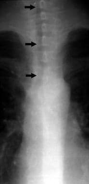
Tracheobronchomalacia
Sweet Syndrome
Matrilin Proteins
A new animal model for relapsing polychondritis, induced by cartilage matrix protein (matrilin-1). (1/60)
Relapsing polychondritis (RP) differs from rheumatoid arthritis (RA) in that primarily cartilage outside diarthrodial joints is affected. The disease usually involves trachea, nose, and outer ears. To investigate whether the tissue distribution of RP may be explained by a specific immune response, we immunized rats with cartilage matrix protein (matrilin-1), a protein predominantly expressed in tracheal cartilage. After 2-3 weeks, some rats developed a severe inspiratory stridor. They had swollen noses and/or epistaxis, but showed neither joint nor outer ear affection. The inflammatory lesions involved chronic active erosions of cartilage. Female rats were more susceptible than males. The disease susceptibility was controlled by both MHC genes (f, l, d, and a haplotypes are high responders, and u, n, and c are resistant) and non-MHC genes (the LEW strain is susceptible; the DA strain is resistant). However, all strains mounted a pronounced IgG response to cartilage matrix protein. The initiation and effector phase of the laryngotracheal involvement causing the clinical symptoms were shown to depend on alphabeta T cells. Taken together, these results represent a novel model for RP: matrilin-1-induced RP. Our findings also suggest that different cartilage proteins are involved in pathogenic models of RP and RA. (+info)Autoreactivity against matrilin-1 in a patient with relapsing polychondritis. (2/60)
Relapsing polychondritis (RP) is a rare inflammatory disease of cartilage. Chondritis of the auricular, nasal, and tracheal cartilages predominates in this disease, suggesting a response to a tissue-specific antigen. One potential antigen is matrilin-1, a cartilage matrix protein found uniquely in the tracheal, auricular, and nasal cartilage of adults. We describe herein a patient with RP who had both a humoral and a cellular immune response directed toward the cartilage matrix protein matrilin-1. (+info)Tracheobronchial involvement in relapsing polychondritis. (3/60)
Relapsing polychondritis (RPC) is a multisystem disorder of chondromalacia involving any cartilage. Respiratory tract involvement is the greatest threat to life. We report a patient with stenosis of the subglottic trachea and left main bronchus who suddenly ceased breathing. As this patient did not have any other clinical features of RPC, the diagnosis was difficult. CT showed circumferential worm-eaten-like thickening suggesting a deformity and edema of the tracheal mucosa. Biopsy of the tracheal and thyroid cartilage revealed mild cartilage degeneration and infiltration with inflammatory cells. Therefore, the patient was diagnosed as having RPC. She is currently well 24 months after Montgomery T tube intubation with systemic steroids. Narrowing of the left main bronchus has not worsened. (+info)Autologous stem-cell transplantation in refractory autoimmune diseases after in vivo immunoablation and ex vivo depletion of mononuclear cells. (4/60)
Autoimmune diseases that are resistant to conventional treatment cause severe morbidity and even mortality. In the present study we demonstrate that complete remissions can be achieved in refractory polychondritis and systemic lupus erythematosus (SLE), even at advanced stage, with the use of autologous stem-cell transplantation (SCT). Remissions persisted after reconstitution of the immune system. In the treatment of advanced systemic sclerosis (SSc), stable disease may be achieved with autologous SCT. (+info)Orbital mucosa-associated lymphoid tissue (MALT)-type lymphoma in a patient with relapsing polychondritis. (5/60)
Relapsing polychondritis is characterized by recurrent inflammation of the cartilaginous tissues of the ears, nose, peripheral joints, and the tracheobronchial tree. The eye is also a frequent target organ in relapsing polychondritis, and proptosis is a well-recognized manifestation of eye involvement. Similar to other rheumatologic diseases, an association of relapsing polychondritis with malignancy has been reported. We describe a patient with relapsing polychondritis who presented with exophthalmos. When treatment directed toward control of her underlying disease was only partially effective, further investigation revealed that she had an orbital mucosa-associated lymphoid tissue (MALT)-type B cell lymphoma. We hypothesize that the lymphoma resulted from malignant transformation of the relapsing polychondritis-induced inflammatory pseudotumor and emphasize that neoplastic disease should be considered in the differential diagnosis in patients with relapsing polychondritis presenting with exophthalmos. (+info)Active aortitis in relapsing polychondritis. (6/60)
Relapsing polychondritis (RP) is a rare inflammatory multiorgan disorder affecting cartilaginous structures and other connective tissues. Serious cardiovascular complications have been reported in patients with RP, the most frequent being aortic or mitral regurgitation and aortic aneurysms. Aortitis is a very rare complication. An unusual case of active aortitis in a patient with RP, despite intensive immunosuppressive treatment, is described with a special emphasis on the pathological findings. (+info)Pulmonary fibrosis with intractable pneumothorax: new pulmonary manifestation of relapsing polychondritis. (7/60)
Relapsing Polychondritis is a rare disease which causes the repetitive inflammation of cartilage and connective tissues. Although the large airway is sometimes involved and the stenosis of them often influences the prognosis of the patients, there have been few reports concerning the manifestation of the peripheral lung. A 60-year-old man with pulmonary fibrosis was admitted to a regional hospital due to sudden deafness, and then he suffered from relapsing polychondritis. During the steroid therapy, he also suffered from bilateral pneumothoraces. His computed tomogram revealed many bilateral bullae, emphysematous changes, and fibrotic changes in bilateral lungs. The mechanism of generating peripheral pulmonary manifestations is also discussed. (+info)Identification of type II collagen peptide 261-273-specific T cell clones in a patient with relapsing polychondritis. (8/60)
OBJECTIVE: To characterize and clone T cells specific for type II collagen (CII) in a patient with relapsing polychondritis (RP) and to establish whether the immunodominant epitope of CII determined in HLA transgenic mice is used in the human autoimmune response to CII. METHODS: T cell responses to CII were examined in a patient with RP, who was heterozygous for the HLA-DR allele DRB1*0101/DRB1*0401. T cell clones were established from this patient and characterized for peptide specificity, class II restriction, cytokine production, and staining with HLA-DRB1*0401 class II tetramers. RESULTS: A response to CII and the peptide 255-273 was present in this patient. T cells specific for the CII epitope 261-273 were cloned. Evaluation of these clones demonstrated a response to CII 261-273 in the context of both DR alleles. HLA-DR4 CII tetramer did not demonstrate staining of either CII-specific DRB1*0401-restricted T cell clones or a polyclonal population of CII-reactive T cells from this individual. CONCLUSION: T cells directed against CII were present in this patient with RP. Also, T cell clones isolated from this individual were found to be specific for the CII peptide 261-273 and were restricted to either the DRB1*0101 or the DRB1*0401 allele. These findings establish that a T cell response directed against CII is present in this patient with RP and that the CII peptide 261-273 plays a role in the human immune response to CII. (+info)Relapsing polychondritis is a rare autoimmune disease characterized by inflammation and damage to the cartilaginous structures in the body. The condition can affect multiple organs and tissues, including the ears, nose, trachea, bronchi, joints, and cardiovascular system. It is called "relapsing" because it tends to involve recurring episodes of inflammation and damage, followed by periods of remission.
The hallmark symptom of relapsing polychondritis is pain and swelling in the ears, nose, or airways. Other symptoms may include:
* Redness, tenderness, and warmth in affected areas
* Hearing loss or tinnitus (ringing in the ears)
* Nasal congestion, runny nose, or nosebleeds
* Hoarseness or difficulty speaking
* Wheezing, shortness of breath, or coughing
* Joint pain, stiffness, or swelling
* Skin rashes or sores
* Eye inflammation or dryness
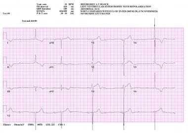
* Heart murmurs or other cardiovascular symptoms
The exact cause of relapsing polychondritis is not known, but it is thought to involve an abnormal immune response in which the body's own antibodies attack and damage cartilage and other tissues. The diagnosis of relapsing polychondritis is typically based on a combination of clinical symptoms, laboratory tests, and imaging studies.
There is no cure for relapsing polychondritis, but treatment can help manage the symptoms and prevent complications. Treatment may include corticosteroids, immunosuppressive drugs, and other medications to reduce inflammation and suppress the immune system. In severe cases, surgery may be necessary to repair or replace damaged tissues.
Ear cartilage, also known as auricular cartilage, refers to the flexible connective tissue that makes up the structural framework of the external ear or pinna. The ear cartilage provides support and shape to the ear, helping to direct sound waves into the ear canal and towards the eardrum.
The ear cartilage is composed of type II collagen fibers and proteoglycans, which give it its flexibility and resiliency. It is covered by a thin layer of skin on both sides and contains no bones. Instead, the ear cartilage is shaped and maintained by the surrounding muscles and connective tissue.
There are three main parts of the ear cartilage: the helix, the antihelix, and the tragus. The helix is the outer rim of the ear, while the antihelix is the curved ridge that runs parallel to the helix. The tragus is the small piece of cartilage that projects from the front of the ear canal.
Ear cartilage can be affected by various conditions, including trauma, infection, and degenerative changes associated with aging. In some cases, surgical procedures may be required to reshape or reconstruct damaged ear cartilage.
Acquired ear deformities refer to abnormal shapes or structures of the ear that result from injury, infection, inflammation, or other external factors after birth. These deformities can affect the appearance and function of the ear, causing symptoms such as hearing loss or discomfort. Examples of acquired ear deformities include:
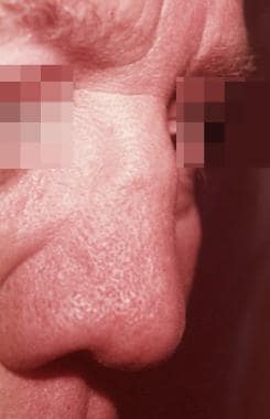
1. Cauliflower ear: a condition characterized by swelling, thickening, and distortion of the ear caused by repeated trauma or injury to the ear cartilage.
2. Microtia: a congenital ear abnormality that can become worse over time due to infection, inflammation, or trauma, resulting in an underdeveloped or absent ear.
3. Macrotia: an abnormally large ear that may result from injury or other external factors.
4. Stenosis: a narrowing of the ear canal that can result from chronic inflammation, infection, or scarring.
5. Hematoma: a collection of blood in the ear tissue caused by trauma or injury, which can lead to deformity if not treated promptly.
6. Keloids: overgrowths of scar tissue that can form after injury or surgery and distort the shape of the ear.
Treatment for acquired ear deformities may include surgical reconstruction, splinting, or other interventions depending on the severity and underlying cause of the condition.
Tracheobronchomalacia is a medical condition that refers to the abnormal softening and weakness of the tracheal and bronchial walls, leading to their collapse or narrowing during breathing, particularly during expiration. This collapse can cause symptoms such as wheezing, shortness of breath, coughing, and recurrent respiratory infections. The condition can be congenital or acquired, with common causes including aging, chronic obstructive pulmonary disease (COPD), and long-term intubation. In severe cases, tracheobronchomalacia may require surgical intervention to stabilize the airway and improve breathing.
Sweet syndrome, also known as acute febrile neutrophilic dermatosis, is a skin condition characterized by the rapid onset of painful, red, and swollen skin lesions. The lesions are often accompanied by fever and elevated white blood cell count, particularly an increase in neutrophils.
The medical definition of Sweet syndrome includes the following criteria:
1. Abrupt onset of painful, erythematous (red), and edematous (swollen) papules, plaques, or nodules.
2. Fever greater than 38°C (100.4°F).
3. Leukocytosis with a predominance of neutrophils in the peripheral blood.
4. Histopathological evidence of a dense dermal infiltrate of neutrophils without evidence of vasculitis.
5. Rapid response to systemic corticosteroids.
Sweet syndrome can be associated with various medical conditions, such as infections, malignancies, and inflammatory diseases, or it can occur without an identifiable underlying cause (idiopathic).
Matrilin proteins are a group of extracellular matrix (ECM) proteins that are predominantly found in cartilaginous tissues, such as articular cartilage, costal cartilage, and intervertebral discs. They belong to the von Willebrand factor A (vWF-A) domain-containing protein family and play important roles in maintaining the structural integrity and organization of the ECM.
Matrilin proteins are composed of multiple domains, including vWF-A domains, coiled-coil domains, and calcium-binding epidermal growth factor (cbEGF)-like domains. They can form multimeric complexes through their coiled-coil domains, which helps to stabilize the ECM network.
There are four known matrilin proteins in humans, designated as Matrilin-1, Matrilin-2, Matrilin-3, and Matrilin-4. Each of these proteins has distinct tissue distribution patterns and functions. For example, Matrilin-1 is primarily found in hyaline cartilage and is involved in regulating chondrocyte differentiation and matrix assembly. Matrilin-2 is widely expressed in various tissues, including cartilage, tendon, and ligament, and plays a role in maintaining the organization of collagen fibrils. Matrilin-3 is specifically expressed in articular cartilage and is involved in regulating the formation and maintenance of the cartilaginous matrix. Matrilin-4 is found in both hyaline and fibrocartilage, as well as in tendons and ligaments, and has been implicated in regulating collagen fibrillogenesis and tissue development.
Mutations in matrilin genes have been associated with various musculoskeletal disorders, such as multiple epiphyseal dysplasia (MED) and spondyloepimetaphyseal dysplasia (SEMD). These genetic defects can lead to abnormalities in the structure and organization of the ECM, resulting in joint pain, stiffness, and reduced mobility.
 Relapsing polychondritis - Wikipedia
 What are treatment options for relapsing polychondritis?
 Relapsing Polychondritis: Practice Essentials, Background, Pathophysiology
 Relapsing Polychondritis: Symptoms, Diagnosis, and Treatment
 Relapsing polychondritis--report of ten cases
 Risk factors for the recurrence of relapsing polychondritis | Arthritis Research & Therapy
 Pages that link to "Ocular Manifestations of Relapsing Polychondritis" - EyeWiki
 Polychondritis, Relapsing | Quick Medical Diagnosis & Treatment 2023 | AccessMedicine | McGraw Hill Medical
 Relapsing Polychondritis - Animation
 the Relapsing Polychondritis Foundation
 Relapsing Polychondritis Testing - HypoGal Blog
 Dan | Relapsing Polychondritis (RP) Foundation
 Driving Awareness for Relapsing Polychondritis - Race for RP
 Relapsing polychondritis: Causes, symptoms, diagnosis, treatment, and diet
 A case of relapsing polychondritis developing tracheomalacia - Current Thoracic Surgery
 Relapsing Polychondritis - Musculoskeletal and Connective Tissue Disorders - MSD Manual Professional Edition
 Vasculitis Translational Research Program | NIAMS
 Med Journal 360 | Localized Tracheobronchial Relapsing Polychondritis With Positive matrilin-1 Staining
 Collagen vascular disease: MedlinePlus Medical Encyclopedia
 Uveitis: The Collaborative Diagnostic Evaluation | AAFP
 Abby Abelson, MD, FACR | Cleveland Clinic
 How People With Weakened Immune Systems Are Navigating The Coronavirus : NPR