Polycythemia Vera
Thrombocythemia, Essential
Janus Kinase 2
Primary Myelofibrosis
Myeloproliferative Disorders
Erythropoietin
Erythroid Precursor Cells
Phlebotomy
Erythropoiesis
Receptors, Erythropoietin
Hematocrit
Receptors, Thrombopoietin
Bloodletting
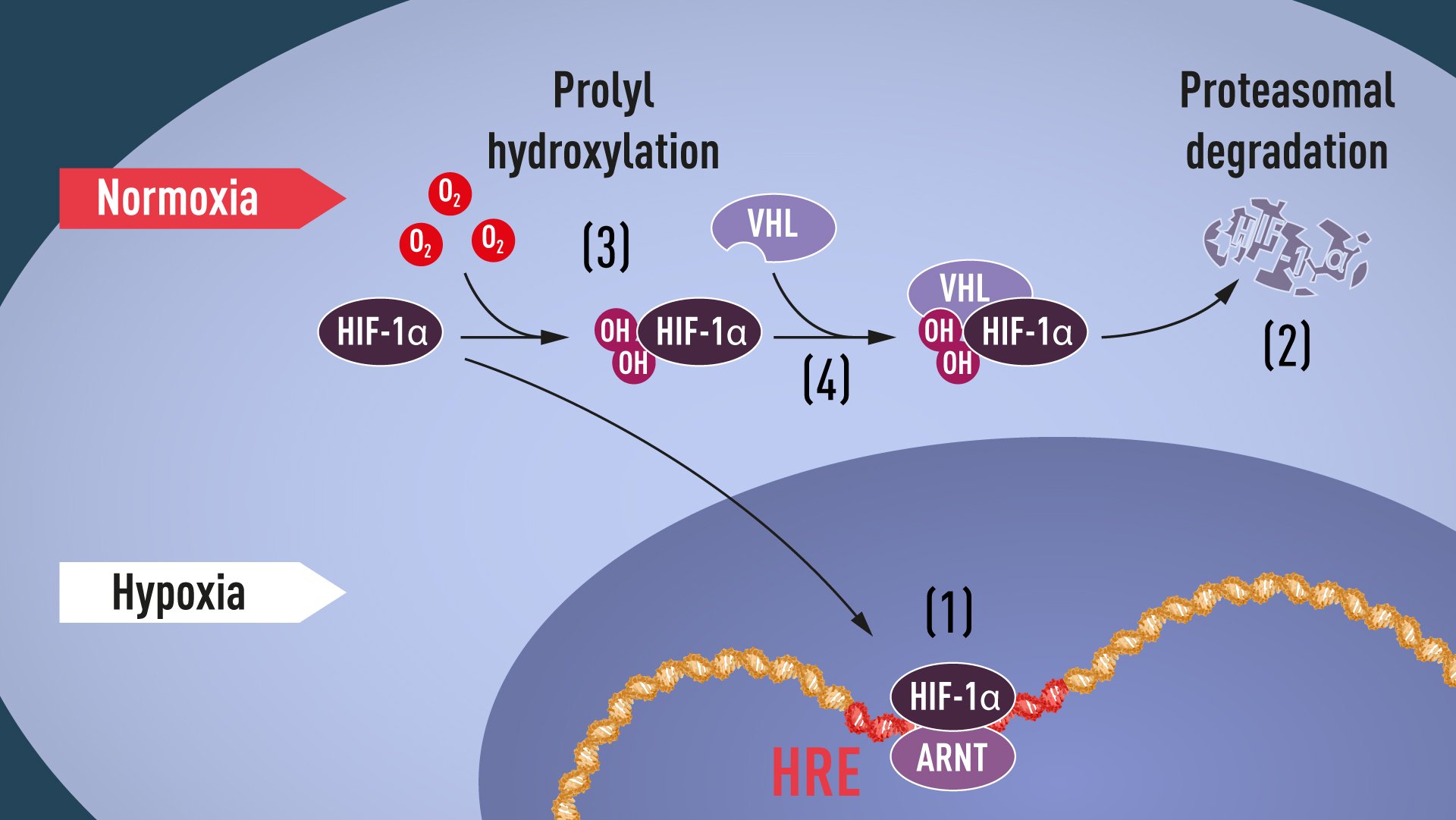
Von Hippel-Lindau Tumor Suppressor Protein
Hematopoiesis, Extramedullary
Granulocytes
Erythrocyte Volume
Somatostatinoma
Russia
GPI-Linked Proteins
Hydroxyurea
Mutation, Missense
Mutation
Amino Acid Substitution
Isoantigens
Cyanosis
Erythroid Cells
Colony-Forming Units Assay
Paraganglioma
Erythrocyte Indices
Bone Marrow
Heterozygote
Budd-Chiari Syndrome
Hemoglobins
Leukemia, Myeloid, Chronic, Atypical, BCR-ABL Negative
Chromosomes, Human, 19-20
Insulin-like growth factor I plays a role in regulating erythropoiesis in patients with end-stage renal disease and erythrocytosis. (1/419)
Erythroid progenitor growth, the serum hormones that regulate erythropoiesis, and the effect of patient's serum on the growth of normal erythroid progenitors were assessed in eight patients with end-stage renal disease (ESRD) and erythrocytosis. All patients were male and had been on maintenance dialysis, they had a hematocrit >50% and/or a red blood cell count >6 x 10(12)/L and an arterial oxygen saturation >95%. Four had acquired cystic disease of the kidney (ACDK), and four other non-ACDK patients did not have known causes of secondary erythrocytosis after appropriate investigations and long-term follow-up. The methylcellulose culture technique was used to assay the erythroid progenitor (BFU-E/CFU-E) growth. Serum erythropoietin (EPO) and insulin-like growth factor I (IGF-I) levels were measured by RIA. Paired experiments were performed to determine the effects of 10% sera from ESRD patients and control subjects on normal marrow CFU-E growth. The numbers of EPO-dependent BFU-E in marrow and/or blood of patients with ESRD and erythrocytosis were higher than those of normal controls. No EPO-independent erythroid colonies were found. Serum EPO levels were constantly normal in one patient and elevated in three patients with ACDK; for non-ACDK patients, EPO levels were normal or low in two patients and persistently increased in one, but fluctuated in the remaining one on serial assays. There was no correlation between serum EPO levels and hematocrit values. The serum IGF-I levels in patients with ESRD and erythrocytosis were significantly increased compared with normal subjects or ESRD patients with anemia. We found an inverse correlation between serum EPO and IGF-I levels. Sera from patients with ESRD and erythrocytosis exhibited a stimulating effect on normal marrow CFU-E growth. The stimulating effect of sera from patients who had a normal serum EPO level and an elevated IGF-I level could be partially blocked by anti-IGF-I. The present study suggests that IGF-I plays an important role in the regulation of erythropoiesis in patients with ESRD and erythrocytosis who did not have an increased EPO production. (+info)Pulmonary prostacyclin synthase overexpression in transgenic mice protects against development of hypoxic pulmonary hypertension. (2/419)
Prostacyclin synthase (PGIS) is the final committed enzyme in the metabolic pathway leading to prostacyclin (PGI2) production. Patients with severe pulmonary hypertension have a PGIS deficiency of their precapillary vessels, but the importance of this deficiency for lung vascular remodeling remains unclear. We hypothesized that selective pulmonary overexpression of PGIS may prevent the development of pulmonary hypertension. To study this hypothesis, transgenic mice were created with selective pulmonary PGIS overexpression using a construct of the 3.7-kb human surfactant protein-C (SP-C) promoter and the rat PGIS cDNA. Transgenic mice (Tg+) and nontransgenic littermates (Tg-) were subjected to a simulated altitude of 17,000 ft for 5 weeks, and right ventricular systolic pressure (RVSP) was measured. Histology was performed on the lungs. The Tg+ mice produced 2-fold more pulmonary 6-keto prostaglandin F1alpha (PGF1alpha) levels than did Tg- mice. After exposure to chronic hypobaric hypoxia, Tg+ mice have lower RVSP than do Tg- mice. Histologic examination of the lungs revealed nearly normal arteriolar vessels in the Tg+ mice in comparison with vessel wall hypertrophy in the Tg- mice. These studies demonstrate that Tg+ mice were protected from the development of pulmonary hypertension after exposure to chronic hypobaric hypoxia. We conclude that PGIS plays a major role in modifying the pulmonary vascular response to chronic hypoxia. This has important implications for the pathogenesis and treatment of severe pulmonary hypertension. (+info)Localization of the gene responsible for familial benign polycythemia to chromosome 11q23. (3/419)
Familial benign polycythemia (FBP) (OMIM 263400) is a rare autosomal recessive condition characterized by erythrocytosis, normal leukocyte and platelet counts, normal uric acid level, and usually increased erythropoietin production. There is a high incidence of this disorder in Chuvashia (Russian Federation), probably due to a founder effect. In an attempt to locate the gene responsible for this disorder, we have carried out linkage studies in 12 Chuvash families, with 35 affected and 32 unaffected members. Linkage to the erythropoietin and erythropoietin receptor loci was excluded, and the FBP gene was assigned to the region of chromosome 11q23 between D11S4142 and D11S1356, with a maximal lod score of 6.61. (+info)Activation of the erythropoietin receptor by the gp55-P viral envelope protein is determined by a single amino acid in its transmembrane domain. (4/419)
The spleen focus forming virus (SFFV) gp55-P envelope glycoprotein specifically binds to and activates murine erythropoietin receptors (EpoRs) coexpressed in the same cell, triggering proliferation of erythroid progenitors and inducing erythroleukemia. Here we demonstrate specific interactions between the single transmembrane domains of the two proteins that are essential for receptor activation. The human EpoR is not activated by gp55-P but by mutation of a single amino acid, L238, in its transmembrane sequence to its murine counterpart serine, resulting in its ability to be activated. The converse mutation in the murine EpoR (S238L) abolishes activation by gp55-P. Computational searches of interactions between the membrane-spanning segments of murine EpoR and gp55-P provide a possible explanation: the face of the EpoR transmembrane domain containing S238 is predicted to interact specifically with gp55-P but not gp55-A, a variant which is much less effective in activating the murine EpoR. Mutational studies on gp55-P M390, which is predicted to interact with S238, provide additional support for this model. Mutation of M390 to isoleucine, the corresponding residue in gp55-A, abolishes activation, but the gp55-P M390L mutation is fully functional. gp55-P is thought to activate signaling by the EpoR by inducing receptor oligomerization through interactions involving specific transmembrane residues. (+info)Erythropoietin receptor mutations associated with familial erythrocytosis cause hypersensitivity to erythropoietin in the heterozygous state. (5/419)
Inherited mutations in the erythropoietin receptor (EPOR) causing premature termination of the receptor cytoplasmic region are associated with dominant familial erythrocytosis (FE), a benign clinical condition characterized by hypersensitivity of erythroid progenitor cells to EPO and low serum EPO (S-EPO) levels. We describe a Swedish family with dominant FE in which erythrocytosis segregates with a new truncation in the negative control domain of the EPOR. We show that cells engineered to concomitantly express the wild-type (WT) EPOR and mutant EPORs associated with FE (FE EPORs) are hypersensitive to EPO-stimulated proliferation and activation of Jak2 and Stat5. These results demonstrate that FE is caused by hyperresponsiveness of receptor-mediated signaling pathways and that this is dominant with respect to WT EPOR signaling. (+info)Role of the spleen in the exaggerated polycythemic response to hypoxia in chronic mountain sickness in rats. (6/419)
In a rat model of chronic mountain sickness, the excessive polycythemic response to hypoxic exposure is associated with profound splenic erythropoiesis. We studied the uptake and distribution of radioactive iron and red blood cell (RBC) morphology in intact and splenectomized rats over a 30-day hypoxic exposure. Retention of (59)Fe in the plasma was correlated with (59)Fe uptake by both spleen and marrow and the appearance of (59)Fe-labeled RBCs in the blood. (59)Fe uptake in both the spleen and the marrow paralleled the production of nucleated RBCs. Splenic (59)Fe uptake was approximately 10% of the total marrow uptake under normoxic conditions but increased to 60% of the total marrow uptake during hypoxic exposure. Peak splenic (59)Fe uptake and splenomegaly occurred at the most intense phase of erythropoiesis and coincided with the rapid appearance of (59)Fe-labeled RBCs in the blood. The bone marrow remains the most important erythropoietic organ under both resting and stimulated states, but inordinate splenic erythropoiesis in this rat strain accounts in large measure for the excessive polycythemia during the development of chronic mountain sickness in chronic hypoxia. (+info)Neutrophil alkaline phosphatase score in chronic granulocytic leukaemia: effects of splenectomy and antileukaemic drugs. (7/419)
Staining with naphthol AS phosphate and Fast Blue BB salt has been used for the estimation of neutrophil alkaline phosphatase (NAP) scores in patients with chronic granulocytic leukaemia (CGL). The very low scores found at diagnosis rise when the disease is treated, and there is some inverse correlation between the NAP score and the absolute neutrophil count. Patients treated intensively developed high NAP scores. Elective splenectomy performed during the chronic phase of CGL is followed by a pronounced but transient neutrophilia and a concurrent striking rise in the NAP score. Similar changes were observed in patients without CGL who underwent splenectomy. These observations can be explained by assuming that newly formed neutrophils in CGL have a normal content of NAP but are rapidly sequestered in non-circulating extramedullary pools, whereas the circulating neutrophil with a typically low NAP content is a relatively aged cell which has lost enzyme activity. In subjects with or without CGL, removal of the spleen, a major site of such pooling, temporarily permits the circulation of newly formed neutrophils but eventually other organs assume the sequestering functions of the spleen. Thus the aberrations of NAP score seen in CGL might be attributable not to an intrinsic cellular defect but to an exaggeration of the granulocyte storage phenomena which also occur in subjects without CGL. (+info)Significance of an abnormally low or high hemoglobin concentration during pregnancy: special consideration of iron nutrition. (8/419)
An association between moderate anemia and poor perinatal outcomes has been found through epidemiologic studies, although available evidence cannot establish this relation as causal. Anemia may not be a direct cause of poor pregnancy outcomes, except in the case of maternal mortality resulting directly from severe anemia due to hypoxia and heart failure. Preventing or treating anemia, whether moderate or severe, is desirable. Because iron deficiency is a common cause of maternal anemia, iron supplementation is a common practice to reduce the incidence of maternal anemia. Nevertheless, the effectiveness of large-scale supplementation programs needs to be improved operationally and, where multiple micronutrient deficiencies are common, supplementation beyond iron and folate can be considered. High hemoglobin concentrations are often mistaken as adequate iron status; however, high hemoglobin is independent of iron status and is often associated with poor health outcomes. Very high hemoglobin concentrations cause high blood viscosity, which results in both compromised oxygen delivery to tissues and cerebrovascular complications. Epidemiologic studies have also found an association between high maternal hemoglobin concentrations and an increased risk of poor pregnancy outcomes. Evidence does not suggest that this association is causal; it could be better attributed to hypertensive disorders of pregnancy and to preeclampsia. The pathophysiologic mechanism of these conditions during pregnancy can produce higher hemoglobin concentrations because of reduced normal plasma expansion and cause fetal stress because of reduced placental-fetal perfusion. Accordingly, higher than normal hemoglobin concentrations should be regarded as an indicator of possible pregnancy complications, not necessarily as a sign of adequate iron nutrition, because iron supplementation does not increase hemoglobin higher than the optimal concentration needed for oxygen delivery. (+info)Polycythemia Vera is a type of myeloproliferative neoplasm, a group of rare blood cancers. In Polycythemia Vera, the body produces too many red blood cells, leading to an increased risk of blood clots and thickening of the blood, which can cause various symptoms such as fatigue, headache, dizziness, and itching. It can also lead to enlargement of the spleen. The exact cause of Polycythemia Vera is not known, but it is associated with genetic mutations in the JAK2 gene in most cases. It is a progressive disease that can lead to complications such as bleeding, thrombosis, and transformation into acute leukemia if left untreated.
Polycythemia is a medical condition characterized by an abnormal increase in the total red blood cell (RBC) mass or hematocrit (the percentage of RBCs in the blood). This results in a higher-than-normal viscosity of the blood, which can lead to various complications such as impaired circulation, increased risk of blood clots, and reduced oxygen supply to the tissues.
There are two main types of polycythemia: primary and secondary. Primary polycythemia, also known as polycythemia vera, is a rare myeloproliferative neoplasm caused by genetic mutations that lead to excessive production of RBCs in the bone marrow. Secondary polycythemia, on the other hand, is a reactive condition triggered by various factors such as chronic hypoxia (low oxygen levels), high altitude, smoking, or certain medical conditions like sleep apnea, heart disease, or kidney tumors.
Symptoms of polycythemia may include fatigue, headaches, dizziness, shortness of breath, itching, and a bluish or reddish tint to the skin (cyanosis). Treatment depends on the underlying cause and severity of the condition and may involve phlebotomy, medications to reduce RBC production, and management of associated complications.
Essential thrombocythemia (ET) is a myeloproliferative neoplasm (MPN), a type of blood cancer characterized by the overproduction of platelets (thrombocytosis) in the bone marrow. In ET, there is an excessive proliferation of megakaryocytes, the precursor cells that produce platelets. This leads to increased platelet counts in the peripheral blood, which can increase the risk of blood clots (thrombosis) and bleeding episodes (hemorrhage).
The term "essential" is used to indicate that the cause of this condition is not known or idiopathic. ET is primarily a disease of older adults, but it can also occur in younger individuals. The diagnosis of essential thrombocythemia requires careful evaluation and exclusion of secondary causes of thrombocytosis, such as reactive conditions, inflammation, or other myeloproliferative neoplasms.
The clinical presentation of ET can vary widely among patients. Some individuals may be asymptomatic and discovered only during routine blood tests, while others may experience symptoms related to thrombosis or bleeding. Common symptoms include headaches, visual disturbances, dizziness, weakness, numbness, or tingling in the extremities, if there are complications due to blood clots in the brain or other parts of the body. Excessive bruising, nosebleeds, or blood in the stool can indicate bleeding complications.
Treatment for essential thrombocythemia is aimed at reducing the risk of thrombosis and managing symptoms. Hydroxyurea is a commonly used medication to lower platelet counts, while aspirin may be prescribed to decrease the risk of blood clots. In some cases, interferon-alpha or ruxolitinib might be considered as treatment options. Regular follow-up with a hematologist and monitoring of blood counts are essential for managing this condition and detecting potential complications early.
Janus Kinase 2 (JAK2) is a tyrosine kinase enzyme that plays a crucial role in intracellular signal transduction. It is named after the Roman god Janus, who is depicted with two faces, as JAK2 has two similar phosphate-transferring domains. JAK2 is involved in various cytokine receptor-mediated signaling pathways and contributes to hematopoiesis, immune function, and cell growth.
Mutations in the JAK2 gene have been associated with several myeloproliferative neoplasms (MPNs), including polycythemia vera, essential thrombocythemia, and primary myelofibrosis. The most common mutation is JAK2 V617F, which results in a constitutively active enzyme that promotes uncontrolled cell proliferation and survival, contributing to the development of these MPNs.
Primary myelofibrosis (PMF) is a rare, chronic bone marrow disorder characterized by the replacement of normal bone marrow tissue with fibrous scar tissue, leading to impaired production of blood cells. This results in cytopenias (anemia, leukopenia, thrombocytopenia), which can cause fatigue, infection susceptibility, and bleeding tendencies. Additionally, PMF is often accompanied by the proliferation of abnormal megakaryocytes (large, atypical bone marrow cells that produce platelets) and extramedullary hematopoiesis (blood cell formation outside the bone marrow, typically in the spleen and liver).
PMF is a type of myeloproliferative neoplasm (MPN), which is a group of clonal stem cell disorders characterized by excessive proliferation of one or more types of blood cells. PMF can present with various symptoms such as fatigue, weight loss, night sweats, abdominal discomfort due to splenomegaly (enlarged spleen), and bone pain. In some cases, PMF may progress to acute myeloid leukemia (AML).
The exact cause of PMF remains unclear; however, genetic mutations are known to play a significant role in its development. The Janus kinase 2 (JAK2), calreticulin (CALR), and MPL genes have been identified as commonly mutated in PMF patients. These genetic alterations contribute to the dysregulated production of blood cells and the activation of signaling pathways that promote fibrosis.
Diagnosis of PMF typically involves a combination of clinical evaluation, complete blood count (CBC), bone marrow aspiration and biopsy, cytogenetic analysis, and molecular testing to identify genetic mutations. Treatment options depend on the individual patient's symptoms, risk stratification, and disease progression. They may include observation, supportive care, medications to manage symptoms and control the disease (such as JAK inhibitors), and stem cell transplantation for eligible patients.
Myeloproliferative disorders (MPDs) are a group of rare, chronic blood cancers that originate from the abnormal proliferation or growth of one or more types of blood-forming cells in the bone marrow. These disorders result in an overproduction of mature but dysfunctional blood cells, which can lead to serious complications such as blood clots, bleeding, and organ damage.
There are several subtypes of MPDs, including:
1. Chronic Myeloid Leukemia (CML): A disorder characterized by the overproduction of mature granulocytes (a type of white blood cell) in the bone marrow, leading to an increased number of these cells in the blood. CML is caused by a genetic mutation that results in the formation of the BCR-ABL fusion protein, which drives uncontrolled cell growth and division.
2. Polycythemia Vera (PV): A disorder characterized by the overproduction of all three types of blood cells - red blood cells, white blood cells, and platelets - in the bone marrow. This can lead to an increased risk of blood clots, bleeding, and enlargement of the spleen.
3. Essential Thrombocythemia (ET): A disorder characterized by the overproduction of platelets in the bone marrow, leading to an increased risk of blood clots and bleeding.
4. Primary Myelofibrosis (PMF): A disorder characterized by the replacement of normal bone marrow tissue with scar tissue, leading to impaired blood cell production and anemia, enlargement of the spleen, and increased risk of infections and bleeding.
5. Chronic Neutrophilic Leukemia (CNL): A rare disorder characterized by the overproduction of neutrophils (a type of white blood cell) in the bone marrow, leading to an increased number of these cells in the blood. CNL can lead to an increased risk of infections and organ damage.
MPDs are typically treated with a combination of therapies, including chemotherapy, targeted therapy, immunotherapy, and stem cell transplantation. The choice of treatment depends on several factors, including the subtype of MPD, the patient's age and overall health, and the presence of any comorbidities.
Thrombocytosis is a medical condition characterized by an abnormally high platelet count (also known as thrombocytes) in the blood. Platelets are small cell fragments that play a crucial role in blood clotting. A normal platelet count ranges from 150,000 to 450,000 platelets per microliter of blood. Thrombocytosis is typically defined as a platelet count exceeding 450,000-500,000 platelets/µL.
Thrombocytosis can be classified into two types: reactive (or secondary) thrombocytosis and primary (or essential) thrombocytosis. Reactive thrombocytosis is more common and occurs as a response to an underlying condition, such as infection, inflammation, surgery, or certain types of cancer. Primary thrombocytosis, on the other hand, is caused by intrinsic abnormalities in the bone marrow cells responsible for platelet production (megakaryocytes), and it is often associated with myeloproliferative neoplasms like essential thrombocythemia.
While mild thrombocytosis may not cause any symptoms, higher platelet counts can increase the risk of blood clots (thrombosis) and bleeding disorders due to excessive platelet aggregation. Symptoms of thrombocytosis may include headaches, dizziness, visual disturbances, or chest pain if a blood clot forms in the brain or heart. Bleeding symptoms can manifest as easy bruising, nosebleeds, or gastrointestinal bleeding.
Treatment for thrombocytosis depends on the underlying cause and the severity of the condition. In cases of reactive thrombocytosis, treating the underlying disorder often resolves the high platelet count. For primary thrombocytosis, medications like aspirin or cytoreductive therapy (such as hydroxyurea) may be used to reduce the risk of blood clots and control platelet production. Regular monitoring of platelet counts is essential for managing this condition and preventing potential complications.
Erythropoietin (EPO) is a hormone that is primarily produced by the kidneys and plays a crucial role in the production of red blood cells in the body. It works by stimulating the bone marrow to produce more red blood cells, which are essential for carrying oxygen to various tissues and organs.
EPO is a glycoprotein that is released into the bloodstream in response to low oxygen levels in the body. When the kidneys detect low oxygen levels, they release EPO, which then travels to the bone marrow and binds to specific receptors on immature red blood cells called erythroblasts. This binding triggers a series of events that promote the maturation and proliferation of erythroblasts, leading to an increase in the production of red blood cells.
In addition to its role in regulating red blood cell production, EPO has also been shown to have neuroprotective effects and may play a role in modulating the immune system. Abnormal levels of EPO have been associated with various medical conditions, including anemia, kidney disease, and certain types of cancer.
EPO is also used as a therapeutic agent for the treatment of anemia caused by chronic kidney disease, chemotherapy, or other conditions that affect red blood cell production. Recombinant human EPO (rhEPO) is a synthetic form of the hormone that is produced using genetic engineering techniques and is commonly used in clinical practice to treat anemia. However, misuse of rhEPO for performance enhancement in sports has been a subject of concern due to its potential to enhance oxygen-carrying capacity and improve endurance.
Pipobroman is an antineoplastic agent, which means it is used to treat cancer. It's a type of alkylating agent, specifically a nitrogen mustard. Alkylating agents work by disrupting the DNA of cancer cells, which can prevent them from dividing and growing. Pipobroman has been used in the treatment of chronic myelogenous leukemia (CML), although it's not widely used today due to the availability of more effective treatments.
Please note that medical definitions can vary based on the source, and this definition is intended to be a general overview. Always refer to the most current prescribing information for any medication.
Erythroid precursor cells, also known as erythroblasts or normoblasts, are early stage cells in the process of producing mature red blood cells (erythrocytes) in the bone marrow. These cells are derived from hematopoietic stem cells and undergo a series of maturation stages, including proerythroblast, basophilic erythroblast, polychromatophilic erythroblast, and orthochromatic erythroblast, before becoming reticulocytes and then mature red blood cells. During this maturation process, the cells lose their nuclei and become enucleated, taking on the biconcave shape and flexible membrane that allows them to move through small blood vessels and deliver oxygen to tissues throughout the body.
Phlebotomy is a medical term that refers to the process of making an incision in a vein, usually in the arm, in order to draw blood. It is also commonly known as venipuncture. This procedure is performed by healthcare professionals for various purposes such as diagnostic testing, blood donation, or therapeutic treatments like phlebotomy for patients with hemochromatosis (a condition where the body absorbs too much iron from food).
The person who performs this procedure is called a phlebotomist. They must be trained in the proper techniques to ensure that the process is safe and relatively pain-free for the patient, and that the blood sample is suitable for laboratory testing.
Erythropoiesis is the process of forming and developing red blood cells (erythrocytes) in the body. It occurs in the bone marrow and is regulated by the hormone erythropoietin (EPO), which is produced by the kidneys. Erythropoiesis involves the differentiation and maturation of immature red blood cell precursors called erythroblasts into mature red blood cells, which are responsible for carrying oxygen to the body's tissues. Disorders that affect erythropoiesis can lead to anemia or other blood-related conditions.
Erythropoietin receptors are cell surface proteins found on immature red blood cell precursors in the bone marrow. They bind to the hormone erythropoietin (EPO), which is produced by the kidneys in response to low oxygen levels in the blood. When EPO binds to its receptor, it activates a signaling pathway that promotes the survival, proliferation, and differentiation of red blood cell precursors, leading to increased production of red blood cells. This process is critical for maintaining adequate oxygen delivery to tissues in the body. Mutations in the erythropoietin receptor gene can lead to various blood disorders, including anemia and polycythemia.
Hematocrit is a medical term that refers to the percentage of total blood volume that is made up of red blood cells. It is typically measured as part of a complete blood count (CBC) test. A high hematocrit may indicate conditions such as dehydration, polycythemia, or living at high altitudes, while a low hematocrit may be a sign of anemia, bleeding, or overhydration. It is important to note that hematocrit values can vary depending on factors such as age, gender, and pregnancy status.
Thrombopoietin receptors are a type of cell surface receptor found on megakaryocytes and platelets. They are also known as MPL (myeloproliferative leukemia virus) receptors. Thrombopoietin is a hormone that regulates the production of platelets in the body, and it binds to these receptors to stimulate the proliferation and differentiation of megakaryocytes, which are large bone marrow cells that produce platelets.
The thrombopoietin receptor is a type I transmembrane protein with an extracellular domain that contains the thrombopoietin-binding site, a single transmembrane domain, and an intracellular domain that contains several tyrosine residues that become phosphorylated upon thrombopoietin binding. This triggers a signaling cascade that leads to the activation of various downstream pathways involved in cell proliferation, differentiation, and survival.
Mutations in the thrombopoietin receptor gene have been associated with certain myeloproliferative neoplasms, such as essential thrombocythemia and primary myelofibrosis, which are characterized by excessive platelet production and bone marrow fibrosis.
Bloodletting is a medical procedure that was commonly used in the past to balance the four humors of the body, which were believed to be blood, phlegm, black bile, and yellow bile. The procedure involved withdrawing blood from a patient through various methods such as venesection (making an incision in a vein), leeches, or cupping.
The theory behind bloodletting was that if one humor became overabundant, it could cause disease or illness. By removing some of the excess humor, practitioners believed they could restore balance and promote healing. Bloodletting was used to treat a wide variety of conditions, including fever, inflammation, and pain.
While bloodletting is no longer practiced in modern medicine, it was once a common treatment for many different ailments. The practice dates back to ancient times and was used by various cultures throughout history, including the Greeks, Romans, Egyptians, and Chinese. However, its effectiveness as a medical treatment has been called into question, and it is now considered an outdated and potentially harmful procedure.
The Von Hippel-Lindau (VHL) tumor suppressor protein is a crucial component in the regulation of cellular growth and division, specifically through its role in oxygen sensing and the ubiquitination of hypoxia-inducible factors (HIFs). The VHL protein forms part of an E3 ubiquitin ligase complex that targets HIFs for degradation under normoxic conditions. In the absence of functional VHL protein or in hypoxic environments, HIFs accumulate and induce the transcription of genes involved in angiogenesis, cell proliferation, and metabolism.
Mutations in the VHL gene can lead to the development of Von Hippel-Lindau syndrome, a rare inherited disorder characterized by the growth of tumors and cysts in various organs, including the central nervous system, retina, kidneys, adrenal glands, and pancreas. These tumors often arise from the overactivation of HIF-mediated signaling pathways due to the absence or dysfunction of VHL protein.
Extramedullary hematopoiesis (EMH) is defined as the production of blood cells outside of the bone marrow in adults. In normal physiological conditions, hematopoiesis occurs within the bone marrow cavities of flat bones such as the pelvis, ribs, skull, and vertebrae. However, certain disease states or conditions can cause EMH to occur in various organs such as the liver, spleen, lymph nodes, and peripheral blood.
EMH can be seen in several pathological conditions, including hematologic disorders such as myeloproliferative neoplasms (e.g., polycythemia vera, essential thrombocytopenia), myelodysplastic syndromes, and leukemias. It can also occur in response to bone marrow failure or infiltration by malignant cells, as well as in some non-hematologic disorders such as fibrocystic disease of the breast and congenital hemolytic anemias.
EMH may lead to organ enlargement, dysfunction, and clinical symptoms depending on the site and extent of involvement. Treatment of EMH is generally directed at managing the underlying condition causing it.
Granulocytes are a type of white blood cell that plays a crucial role in the body's immune system. They are called granulocytes because they contain small granules in their cytoplasm, which are filled with various enzymes and proteins that help them fight off infections and destroy foreign substances.
There are three types of granulocytes: neutrophils, eosinophils, and basophils. Neutrophils are the most abundant type and are primarily responsible for fighting bacterial infections. Eosinophils play a role in defending against parasitic infections and regulating immune responses. Basophils are involved in inflammatory reactions and allergic responses.
Granulocytes are produced in the bone marrow and released into the bloodstream, where they circulate and patrol for any signs of infection or foreign substances. When they encounter a threat, they quickly move to the site of infection or injury and release their granules to destroy the invading organisms or substances.
Abnormal levels of granulocytes in the blood can indicate an underlying medical condition, such as an infection, inflammation, or a bone marrow disorder.
Erythrocyte volume, also known as red cell volume or hematocrit, is the proportion of whole blood that is made up of erythrocytes or red blood cells. It is typically expressed as a percentage and can be measured using a centrifuge to separate the components of a blood sample by density.
The erythrocyte volume is an important clinical parameter because it can provide information about a person's health status, such as their hydration level, altitude acclimatization, and the presence of certain medical conditions like anemia or polycythemia. Changes in erythrocyte volume can also have significant effects on the body's oxygen-carrying capacity and overall cardiovascular function.
Somatostatinoma is a rare type of neuroendocrine tumor that originates from the delta cells (D cells) of the diffuse endocrine system, which are responsible for producing and secreting somatostatin, a hormone that inhibits the release of several other hormones. These tumors can occur in various organs, but they most commonly arise in the pancreas and the small intestine (duodenum).
Somatostatinomas are typically slow-growing and can be functional or nonfunctional. Functional somatostatinomas actively produce and secrete excessive amounts of somatostatin, which can lead to a variety of clinical symptoms due to the inhibition of other hormones' functions. Nonfunctional somatostatinomas do not secrete significant amounts of somatostatin and are often discovered incidentally during imaging studies or when they cause local mass effects.
Common symptoms associated with functional somatostatinomas include diarrhea, abdominal pain, weight loss, fat malabsorption, and steatorrhea (fatty stools). They can also lead to diabetes mellitus due to the inhibition of insulin secretion. Additionally, these tumors may cause symptoms related to hormone deficiencies or the compression of nearby structures, depending on their location.
Diagnosis typically involves imaging studies such as CT scans, MRI, and PET scans, along with biochemical tests to measure somatostatin levels in the blood. A definitive diagnosis usually requires a tissue biopsy or surgical removal of the tumor for histopathological examination. Treatment options include surgery, chemotherapy, radiation therapy, and targeted therapies, depending on the stage and location of the tumor.
I'm sorry for any confusion, but "Russia" is not a medical term or concept. Russia is the largest country in the world by land area, located primarily in Asia with a smaller portion extending into Europe. It is a nation rich in history and culture, known for its diverse landscapes, from tundra and forests to subtropical beaches.
If you have any medical questions or terms that you would like me to define, please feel free to ask!
A homozygote is an individual who has inherited the same allele (version of a gene) from both parents and therefore possesses two identical copies of that allele at a specific genetic locus. This can result in either having two dominant alleles (homozygous dominant) or two recessive alleles (homozygous recessive). In contrast, a heterozygote has inherited different alleles from each parent for a particular gene.
The term "homozygote" is used in genetics to describe the genetic makeup of an individual at a specific locus on their chromosomes. Homozygosity can play a significant role in determining an individual's phenotype (observable traits), as having two identical alleles can strengthen the expression of certain characteristics compared to having just one dominant and one recessive allele.
Thrombosis is the formation of a blood clot (thrombus) inside a blood vessel, obstructing the flow of blood through the circulatory system. When a clot forms in an artery, it can cut off the supply of oxygen and nutrients to the tissues served by that artery, leading to damage or tissue death. If a thrombus forms in the heart, it can cause a heart attack. If a thrombus breaks off and travels through the bloodstream, it can lodge in a smaller vessel, causing blockage and potentially leading to damage in the organ that the vessel supplies. This is known as an embolism.
Thrombosis can occur due to various factors such as injury to the blood vessel wall, abnormalities in blood flow, or changes in the composition of the blood. Certain medical conditions, medications, and lifestyle factors can increase the risk of thrombosis. Treatment typically involves anticoagulant or thrombolytic therapy to dissolve or prevent further growth of the clot, as well as addressing any underlying causes.
GPI-linked proteins are a type of cell surface protein that are attached to the plasma membrane via a glycosylphosphatidylinositol (GPI) anchor. The GPI anchor is a complex glycolipid molecule that acts as a molecular tether, connecting the protein to the outer leaflet of the lipid bilayer of the cell membrane.
The GPI anchor is synthesized in the endoplasmic reticulum (ER) and added to proteins in the ER or Golgi apparatus during protein trafficking. The addition of the GPI anchor to a protein occurs in a post-translational modification process called GPI anchoring, which involves the transfer of the GPI moiety from a lipid carrier to the carboxyl terminus of the protein.
GPI-linked proteins are found on the surface of many different types of cells, including red blood cells, immune cells, and nerve cells. They play important roles in various cellular processes, such as cell signaling, cell adhesion, and enzyme function. Some GPI-linked proteins also serve as receptors for bacterial toxins and viruses, making them potential targets for therapeutic intervention.
Hydroxyurea is an antimetabolite drug that is primarily used in the treatment of myeloproliferative disorders such as chronic myelogenous leukemia (CML), essential thrombocythemia, and polycythemia vera. It works by interfering with the synthesis of DNA, which inhibits the growth of cancer cells.
In addition to its use in cancer therapy, hydroxyurea is also used off-label for the management of sickle cell disease. In this context, it helps to reduce the frequency and severity of painful vaso-occlusive crises by increasing the production of fetal hemoglobin (HbF), which decreases the formation of sickled red blood cells.
The medical definition of hydroxyurea is:
A hydantoin derivative and antimetabolite that inhibits ribonucleoside diphosphate reductase, thereby interfering with DNA synthesis. It has been used as an antineoplastic agent, particularly in the treatment of myeloproliferative disorders, and more recently for the management of sickle cell disease to reduce the frequency and severity of painful vaso-occlusive crises by increasing fetal hemoglobin production.
A missense mutation is a type of point mutation in which a single nucleotide change results in the substitution of a different amino acid in the protein that is encoded by the affected gene. This occurs when the altered codon (a sequence of three nucleotides that corresponds to a specific amino acid) specifies a different amino acid than the original one. The function and/or stability of the resulting protein may be affected, depending on the type and location of the missense mutation. Missense mutations can have various effects, ranging from benign to severe, depending on the importance of the changed amino acid for the protein's structure or function.
A mutation is a permanent change in the DNA sequence of an organism's genome. Mutations can occur spontaneously or be caused by environmental factors such as exposure to radiation, chemicals, or viruses. They may have various effects on the organism, ranging from benign to harmful, depending on where they occur and whether they alter the function of essential proteins. In some cases, mutations can increase an individual's susceptibility to certain diseases or disorders, while in others, they may confer a survival advantage. Mutations are the driving force behind evolution, as they introduce new genetic variability into populations, which can then be acted upon by natural selection.
An amino acid substitution is a type of mutation in which one amino acid in a protein is replaced by another. This occurs when there is a change in the DNA sequence that codes for a particular amino acid in a protein. The genetic code is redundant, meaning that most amino acids are encoded by more than one codon (a sequence of three nucleotides). As a result, a single base pair change in the DNA sequence may not necessarily lead to an amino acid substitution. However, if a change does occur, it can have a variety of effects on the protein's structure and function, depending on the nature of the substituted amino acids. Some substitutions may be harmless, while others may alter the protein's activity or stability, leading to disease.
Isoantigens are antigens that are present on the cells or tissues of one individual of a species, but are absent or different in another individual of the same species. They are also known as "alloantigens." Isoantigens are most commonly found on the surface of red blood cells and other tissues, and they can stimulate an immune response when transplanted into a different individual. This is because the recipient's immune system recognizes the isoantigens as foreign and mounts a defense against them. Isoantigens are important in the field of transplantation medicine, as they must be carefully matched between donor and recipient to reduce the risk of rejection.
Cyanosis is a medical term that refers to the bluish discoloration of the skin and mucous membranes due to an insufficient amount of oxygen in the blood. This occurs when the level of deoxygenated hemoglobin (the form of hemoglobin that has released its oxygen) in the blood is increased, causing a blue or purple tint to appear, especially in the lips, fingertips, and nail beds.
Cyanosis can be central or peripheral. Central cyanosis affects the entire body and results from low levels of oxygen in the arterial blood, often due to heart or lung conditions that impair oxygen exchange. Peripheral cyanosis is localized to the extremities, usually caused by poor circulation or cold exposure, which can lead to sluggish blood flow and slow oxygen uptake in the tissues.
It's important to note that cyanosis may not always be visually apparent, particularly in individuals with darker skin tones. In these cases, other signs of hypoxia (low oxygen levels) should be considered for proper diagnosis and treatment.
Erythroid cells are a type of blood cell that develops in the bone marrow and mature into red blood cells (RBCs), also known as erythrocytes. These cells play a crucial role in the body's oxygen-carrying capacity by transporting oxygen from the lungs to the body's tissues and carbon dioxide from the tissues to the lungs.
The development of erythroid cells begins with hematopoietic stem cells, which can differentiate into various types of blood cells. Through a series of maturation stages, including proerythroblasts, basophilic erythroblasts, polychromatophilic erythroblasts, and orthochromatic erythroblasts, these cells gradually lose their nuclei and organelles to become reticulocytes. Reticulocytes are immature RBCs that still contain some residual ribosomes and are released into the bloodstream. Over time, they mature into fully functional RBCs, which have a biconcave shape and a flexible membrane that allows them to navigate through small blood vessels.
Erythroid cells are essential for maintaining adequate oxygenation of body tissues, and their production is tightly regulated by various hormones and growth factors, such as erythropoietin (EPO), which stimulates the proliferation and differentiation of erythroid progenitor cells. Abnormalities in erythroid cell development or function can lead to various blood disorders, including anemia, polycythemia, and myelodysplastic syndromes.
A platelet count is a laboratory test that measures the number of platelets, also known as thrombocytes, in a sample of blood. Platelets are small, colorless cell fragments that circulate in the blood and play a crucial role in blood clotting. They help to stop bleeding by sticking together to form a plug at the site of an injured blood vessel.
A normal platelet count ranges from 150,000 to 450,000 platelets per microliter (µL) of blood. A lower than normal platelet count is called thrombocytopenia, while a higher than normal platelet count is known as thrombocytosis.
Abnormal platelet counts can be a sign of various medical conditions, including bleeding disorders, infections, certain medications, and some types of cancer. It is important to consult with a healthcare provider if you have any concerns about your platelet count or if you experience symptoms such as easy bruising, prolonged bleeding, or excessive menstrual flow.
A Colony-Forming Units (CFU) assay is a type of laboratory test used to measure the number of viable, or living, cells in a sample. It is commonly used to enumerate bacteria, yeast, and other microorganisms. The test involves placing a known volume of the sample onto a nutrient-agar plate, which provides a solid growth surface for the cells. The plate is then incubated under conditions that allow the cells to grow and form colonies. Each colony that forms on the plate represents a single viable cell from the original sample. By counting the number of colonies and multiplying by the known volume of the sample, the total number of viable cells in the sample can be calculated. This information is useful in a variety of applications, including monitoring microbial populations, assessing the effectiveness of disinfection procedures, and studying microbial growth and survival.
Megakaryocytes are large, specialized bone marrow cells that are responsible for the production and release of platelets (also known as thrombocytes) into the bloodstream. Platelets play an essential role in blood clotting and hemostasis, helping to prevent excessive bleeding during injuries or trauma.
Megakaryocytes have a unique structure with multilobed nuclei and abundant cytoplasm rich in organelles called alpha-granules and dense granules, which store various proteins, growth factors, and enzymes necessary for platelet function. As megakaryocytes mature, they extend long cytoplasmic processes called proplatelets into the bone marrow sinuses, where these extensions fragment into individual platelets that are released into circulation.
Abnormalities in megakaryocyte number, size, or function can lead to various hematological disorders, such as thrombocytopenia (low platelet count), thrombocytosis (high platelet count), and certain types of leukemia.
Paraganglioma is a rare type of tumor that develops in the nervous system, specifically in the paraganglia. Paraganglia are clusters of specialized nerve cells throughout the body that release hormones in response to stress or physical activity. Most paragangliomas are benign (noncancerous), but some can be malignant (cancerous) and may spread to other parts of the body.
Paragangliomas can occur in various locations, including the head and neck region (called "head and neck paragangliomas") or near the spine, abdomen, or chest (called "extra-adrenal paragangliomas"). When they develop in the adrenal glands, which are located on top of each kidney, they are called pheochromocytomas.
Paragangliomas can produce and release hormones such as epinephrine (adrenaline) and norepinephrine, leading to symptoms like high blood pressure, rapid heart rate, sweating, anxiety, and headaches. Treatment typically involves surgical removal of the tumor, along with medications to manage symptoms and control hormone levels before and after surgery.
Erythrocyte indices are a set of calculated values that provide information about the size and hemoglobin content of red blood cells (erythrocytes). These indices are commonly used in the complete blood count (CBC) test to help diagnose various types of anemia and other conditions affecting the red blood cells.
The three main erythrocyte indices are:
1. Mean Corpuscular Volume (MCV): This is the average volume of a single red blood cell, measured in femtoliters (fL). MCV helps to differentiate between microcytic, normocytic, and macrocytic anemia. Microcytic anemia is characterized by low MCV values (100 fL).
2. Mean Corpuscular Hemoglobin (MCH): This is the average amount of hemoglobin present in a single red blood cell, measured in picograms (pg). MCH helps to assess the oxygen-carrying capacity of red blood cells. Low MCH values may indicate hypochromic anemia, where the red blood cells have reduced hemoglobin content.
3. Mean Corpuscular Hemoglobin Concentration (MCHC): This is the average concentration of hemoglobin in a single red blood cell, measured as a percentage. MCHC reflects the hemoglobin concentration relative to the size of the red blood cells. Low MCHC values may indicate hypochromic anemia, while high MCHC values could suggest spherocytosis or other conditions affecting red blood cell shape and integrity.
These erythrocyte indices are calculated based on the red blood cell count, hemoglobin concentration, and hematocrit results obtained from a CBC test. They provide valuable information for healthcare professionals to diagnose and manage various hematological conditions.
Bone marrow is the spongy tissue found inside certain bones in the body, such as the hips, thighs, and vertebrae. It is responsible for producing blood-forming cells, including red blood cells, white blood cells, and platelets. There are two types of bone marrow: red marrow, which is involved in blood cell production, and yellow marrow, which contains fatty tissue.
Red bone marrow contains hematopoietic stem cells, which can differentiate into various types of blood cells. These stem cells continuously divide and mature to produce new blood cells that are released into the circulation. Red blood cells carry oxygen throughout the body, white blood cells help fight infections, and platelets play a crucial role in blood clotting.
Bone marrow also serves as a site for immune cell development and maturation. It contains various types of immune cells, such as lymphocytes, macrophages, and dendritic cells, which help protect the body against infections and diseases.
Abnormalities in bone marrow function can lead to several medical conditions, including anemia, leukopenia, thrombocytopenia, and various types of cancer, such as leukemia and multiple myeloma. Bone marrow aspiration and biopsy are common diagnostic procedures used to evaluate bone marrow health and function.
Leukocytosis is a condition characterized by an increased number of leukocytes (white blood cells) in the peripheral blood. A normal white blood cell count ranges from 4,500 to 11,000 cells per microliter of blood in adults. Leukocytosis is typically considered present when the white blood cell count exceeds 11,000 cells/µL. However, the definition might vary slightly depending on the laboratory and clinical context.
Leukocytosis can be a response to various underlying conditions, including bacterial or viral infections, inflammation, tissue damage, leukemia, and other hematological disorders. It is essential to investigate the cause of leukocytosis through further diagnostic tests, such as blood smears, differential counts, and additional laboratory and imaging studies, to guide appropriate treatment.
Erythrocyte count, also known as red blood cell (RBC) count, is a laboratory test that measures the number of red blood cells in a sample of blood. Red blood cells are important because they carry oxygen from the lungs to the rest of the body. A low erythrocyte count may indicate anemia, while a high count may be a sign of certain medical conditions such as polycythemia. The normal range for erythrocyte count varies depending on a person's age, sex, and other factors.
A heterozygote is an individual who has inherited two different alleles (versions) of a particular gene, one from each parent. This means that the individual's genotype for that gene contains both a dominant and a recessive allele. The dominant allele will be expressed phenotypically (outwardly visible), while the recessive allele may or may not have any effect on the individual's observable traits, depending on the specific gene and its function. Heterozygotes are often represented as 'Aa', where 'A' is the dominant allele and 'a' is the recessive allele.
Budd-Chiari syndrome is a rare condition characterized by the obstruction of the hepatic veins, which are the blood vessels that carry blood from the liver to the heart. This obstruction can be caused by blood clots, tumors, or other abnormalities, and it can lead to a backflow of blood in the liver, resulting in various symptoms such as abdominal pain, swelling, and liver enlargement. In severe cases, Budd-Chiari syndrome can cause liver failure and other complications if left untreated. The diagnosis of this condition typically involves imaging tests such as ultrasound, CT scan, or MRI, and treatment may include anticoagulation therapy, thrombolytic therapy, or surgical intervention to remove the obstruction.
Hemoglobin (Hb or Hgb) is the main oxygen-carrying protein in the red blood cells, which are responsible for delivering oxygen throughout the body. It is a complex molecule made up of four globin proteins and four heme groups. Each heme group contains an iron atom that binds to one molecule of oxygen. Hemoglobin plays a crucial role in the transport of oxygen from the lungs to the body's tissues, and also helps to carry carbon dioxide back to the lungs for exhalation.
There are several types of hemoglobin present in the human body, including:
* Hemoglobin A (HbA): This is the most common type of hemoglobin, making up about 95-98% of total hemoglobin in adults. It consists of two alpha and two beta globin chains.
* Hemoglobin A2 (HbA2): This makes up about 1.5-3.5% of total hemoglobin in adults. It consists of two alpha and two delta globin chains.
* Hemoglobin F (HbF): This is the main type of hemoglobin present in fetal life, but it persists at low levels in adults. It consists of two alpha and two gamma globin chains.
* Hemoglobin S (HbS): This is an abnormal form of hemoglobin that can cause sickle cell disease when it occurs in the homozygous state (i.e., both copies of the gene are affected). It results from a single amino acid substitution in the beta globin chain.
* Hemoglobin C (HbC): This is another abnormal form of hemoglobin that can cause mild to moderate hemolytic anemia when it occurs in the homozygous state. It results from a different single amino acid substitution in the beta globin chain than HbS.
Abnormal forms of hemoglobin, such as HbS and HbC, can lead to various clinical disorders, including sickle cell disease, thalassemia, and other hemoglobinopathies.
Hematopoietic stem cells (HSCs) are immature, self-renewing cells that give rise to all the mature blood and immune cells in the body. They are capable of both producing more hematopoietic stem cells (self-renewal) and differentiating into early progenitor cells that eventually develop into red blood cells, white blood cells, and platelets. HSCs are found in the bone marrow, umbilical cord blood, and peripheral blood. They have the ability to repair damaged tissues and offer significant therapeutic potential for treating various diseases, including hematological disorders, genetic diseases, and cancer.
Altitude sickness, also known as mountain sickness or hypobaropathy, is a condition that can occur when you travel to high altitudes (usually above 8000 feet or 2400 meters) too quickly. At high altitudes, the air pressure is lower and there is less oxygen available for your body to use. This can lead to various symptoms such as:
1. Headache
2. Dizziness or lightheadedness
3. Shortness of breath
4. Rapid heart rate
5. Nausea or vomiting
6. Fatigue or weakness
7. Insomnia
8. Swelling of the hands, feet, and face
9. Confusion or difficulty with coordination
There are three types of altitude sickness: acute mountain sickness (AMS), high-altitude pulmonary edema (HAPE), and high-altitude cerebral edema (HACE). AMS is the mildest form, while HAPE and HACE can be life-threatening.
Preventive measures include gradual ascent to allow your body time to adjust to the altitude, staying hydrated, avoiding alcohol and heavy meals, and taking it easy during the first few days at high altitudes. If symptoms persist or worsen, immediate medical attention is necessary.
Splenomegaly is a medical term that refers to an enlargement or expansion of the spleen beyond its normal size. The spleen is a vital organ located in the upper left quadrant of the abdomen, behind the stomach and below the diaphragm. It plays a crucial role in filtering the blood, fighting infections, and storing red and white blood cells and platelets.
Splenomegaly can occur due to various underlying medical conditions, including infections, liver diseases, blood disorders, cancer, and inflammatory diseases. The enlarged spleen may put pressure on surrounding organs, causing discomfort or pain in the abdomen, and it may also lead to a decrease in red and white blood cells and platelets, increasing the risk of anemia, infections, and bleeding.
The diagnosis of splenomegaly typically involves a physical examination, medical history, and imaging tests such as ultrasound, CT scan, or MRI. Treatment depends on the underlying cause and may include medications, surgery, or other interventions to manage the underlying condition.
Chronic myeloid leukemia (CML), atypical, BCR-ABL negative is a rare subtype of CML that does not have the typical Philadelphia chromosome abnormality or the resulting BCR-ABL fusion gene. This means that the disease lacks the constitutively active tyrosine kinase that is targeted by imatinib mesylate (Gleevec) and other similar drugs.
The atypical form of CML is often characterized by a more aggressive clinical course, with a higher risk of transformation to acute leukemia compared to the classic form of CML. It can be difficult to diagnose and treat due to its rarity and heterogeneity. Treatment options may include chemotherapy, targeted therapy, stem cell transplantation, or a combination of these approaches. Regular follow-up with blood tests and bone marrow examinations is essential for monitoring the disease course and adjusting treatment as necessary.
Human chromosomes are thread-like structures that contain genetic information in the form of DNA and proteins. Each human cell typically contains 46 chromosomes arranged in 23 pairs, except for the sperm and egg cells which contain only 23 chromosomes (one half of the full set).
Chromosome 19 is one of the autosomal chromosomes, meaning it is not a sex chromosome. It is the fifth smallest human chromosome, spanning about 58 million base pairs and representing approximately 1.9% of the total DNA in cells. Chromosome 19 contains more than 1,200 genes that provide instructions for making proteins and RNA molecules involved in various cellular processes.
Chromosome 20 is also an autosomal chromosome, slightly smaller than chromosome 19. It spans about 54 million base pairs and contains around 800 genes that code for proteins and RNA molecules. Chromosome 20 is known to contain several important genes involved in cancer development, such as the tumor suppressor gene TP53.
Together, chromosomes 19 and 20 carry crucial genetic information necessary for normal human growth, development, and health. Abnormalities in these chromosomes can lead to various genetic disorders and diseases.
Thrombopoietin (TPO) is a glycoprotein hormone that plays a crucial role in the regulation of platelet production, also known as thrombopoiesis. It is primarily produced by the liver and to some extent by megakaryocytes, which are the cells responsible for producing platelets.
TPO binds to its receptor, c-Mpl, on the surface of megakaryocytes and their precursor cells, stimulating their proliferation, differentiation, and maturation into platelets. By regulating the number of platelets in circulation, TPO helps maintain hemostasis, the process that prevents excessive bleeding after injury.
In addition to its role in thrombopoiesis, TPO has been shown to have potential effects on other cell types, including hematopoietic stem cells and certain immune cells. However, its primary function remains the regulation of platelet production.
 Polycythemia
Polycythemia vera
Twin anemia-polycythemia sequence
List of OMIM disorder codes
Reticulocyte production index
Scott Murphy (physician)
Hypoxemia
Plethora (medicine)
Hypoxia (medical)
Exchange transfusion
Persistent fetal circulation
Phyllis George
Cytopenia
Essential thrombocythemia
Givinostat
Small for gestational age
Diffusing capacity
Alan Aerts
Deep vein thrombosis
Mir-339 microRNA precursor family
EPAS1
Ruxolitinib
Fumarase deficiency
Deaths in March 2022
Hyperbilirubinemia in adults
Acute megakaryoblastic leukemia
Malar flush
Ranimustine
Ropeginterferon alfa-2b
Isaac Lipnitsky
Polycythemia - Wikipedia
 Polycythemia - newborn: MedlinePlus Medical Encyclopedia
 Polycythemia Vera
 ATSDR - Polycythemia Vera Investigation
 Polycythemia Vera Treatment Options
 Pediatric Polycythemia: Practice Essentials, Pathophysiology, Epidemiology
 Differential cytokine network profile in polycythemia vera and secondary polycythemia | Scientific Reports
Polycythemia Vera Treatment & Management: Approach Considerations, Medical Care, Phlebotomy
Self-Care for Polycythemia Vera: Exercise, Skin Care, and More
 Polycythemia vera | Sparrow
 Polycythemia Vera | Leukemia and Lymphoma Society
 Polycythemia vera | Lima Memorial Health System
 Erythrocytosis (Polycythemia) in Animals - Circulatory System - Merck Veterinary Manual
 Neoplasms of uncertain behavior, polycythemia vera and myelodysplastic syndromes - ICD-10 Codes- Codify by AAPC
 Polycythaemia vera - Better Health Channel
 Polycythaemia vera
 Biomedicines | Free Full-Text | Identifying Patients with Polycythemia Vera at Risk of Thrombosis after Hydroxyurea Initiation:...
Polycythemia in the Newborn - Children's Health Issues - Merck Manuals Consumer Version
 Acute myocardial infarction following sequential multi-vessel occlusion in a case of polycythemia vera. - International...
 Navtemadlin by Kartos Therapeutics for Post-Polycythemia Vera Myelofibrosis (PPV-MF): Likelihood of Approval
 Sequential Evaluation of Erythrocyte Zinc Protoporphyrin in a Dog with Polycythemia Submitted to Phlebotomies - WSAVA2009 - VIN
 Polycythaemia rubra vera and myelofibrosis with spinal cord compression | Journal of Clinical Pathology
 Types of polycythaemia
 JCDR -
Polyglobulia Masquerading as Polycythaemia Vera Presenting as Superior Mesenteric Vein Thrombosis: A Case Report
JCDR -
Polyglobulia Masquerading as Polycythaemia Vera Presenting as Superior Mesenteric Vein Thrombosis: A Case Report
Age-related differences in disease characteristics and clinical outcomes in polycythemia vera<...
Polycythemia Vera - Hematology and Oncology - MSD Manual Professional Edition
Polycythemia
 Natural Treatment for Polycythemia Vera | Supplement | Herbs Solutions By Nature
Diabetes Mellitus and Pregnancy: Practice Essentials, Gestational Diabetes, Maternal-Fetal Metabolism in Normal Pregnancy
Secondary Polycythemia Medication
Rubra vera2
- A specific type of primary polycythemia, polycythemia rubra vera (often just called polycythemia vera) is an acquired myeloproliferative disorder which is discussed in detail elsewhere ( Pediatric Polycythemia Vera ). (medscape.com)
- Spinal cord compression from extramedullary haemopoiesis within the spinal epidural space is a rare complication of myelofibrosis and polycythaemia rubra vera (PRV). (bmj.com)
Erythrocytosis7
- Polycythemia is sometimes called erythrocytosis, and there is significant overlap in the two findings, but the terms are not the same: polycythemia describes any increase in hematocrit and/or hemoglobin, while erythrocytosis describes an increase specifically in the number of red blood cells in the blood. (wikipedia.org)
- Polycythemia refers to increased red blood cell mass and is often used interchangeably with the term erythrocytosis. (medscape.com)
- Guidelines for the diagnosis, investigation and management of polycythaemia/erythrocytosis. (gpnotebook.com)
- In polycythemia vera, in contrast to secondary erythrocytosis, the red cell mass increase is often initially masked by an increase in the plasma volume that leaves the hematocrit in the normal range. (msdmanuals.com)
- For the individual in which phlebotomy works well, we typically see that the need for a number of phlebotomies goes down relatively rapidly and that we are inducing an iatrogenic state of iron deficiency to try to control the erythrocytosis that individuals have with polycythemia vera. (targetedonc.com)
- Polycythemia is sometimes called erythrocytosis, but the terms are not synonymous because polycythemia refers to any increase in red blood cells , whereas erythrocytosis only refers to a documented increase of red cell mass. (medscape.com)
- Excessive erythrocytosis defined as [Hb] >19 g/dL shows the highest values at 4000 m, while polycythemia defined as [Hb] greater than 2 SD was reduced at high altitude (HA). (bvsalud.org)
Myelofibrosis6
- Navtemadlin is under clinical development by Kartos Therapeutics and currently in Phase III for Post-Polycythemia Vera Myelofibrosis (PPV-MF). (pharmaceutical-technology.com)
- According to GlobalData, Phase III drugs for Post-Polycythemia Vera Myelofibrosis (PPV-MF) have an 80% phase transition success rate (PTSR) indication benchmark for progressing into Pre-Registration. (pharmaceutical-technology.com)
- The diagnosis and management of polycythemia vera, essential thrombocythemia, and primary myelofibrosis in the JAK2 V617F era. (medscape.com)
- Polycythemia vera (PV) is an acquired clonal hematopoietic stem cell disease characterized by an elevated red cell mass caused by excessive myelopoiesis with propensity to transformation to myelofibrosis and acute leukemia. (bcm.edu)
- MPN Research Foundation has a single goal: to stimulate original research in pursuit of new treatments - and eventually a cure - for polycythemia vera, essential thrombocythemia, and myelofibrosis, known collectively as myeloproliferative neoplasms (MPNs). (mpnresearchfoundation.org)
- In patients with myelofibrosis (MF) and polycythemia vera (PV) treated with Jakafi in clinical trials, the rates of thromboembolic events were similar in Jakafi and control treated patients. (jakafi.com)
Hematocrit3
- Polycythemia (also known as polycythaemia) is a laboratory finding in which the hematocrit (the volume percentage of red blood cells in the blood) and/or hemoglobin concentration are increased in the blood. (wikipedia.org)
- Polycythemia vera (PV) is an idiopathic, chronic myeloproliferative disease characterized by an increased red blood cell count and hematocrit, which in turn causes systematic thrombosis. (iasp-pain.org)
- Testosterone and other androgens have an erythropoietic stimulating effect that can cause polycythemia, which manifests as an increase in hemoglobin, hematocrit, or red blood cell count . (medscape.com)
Herbal Treatment for Polycythemia Vera2
- However, Herbal Treatment for Polycythemia Vera completely treats with the herbal Supplement. (herbs-solutions-by-nature.com)
- Herbal Treatment for Polycythemia Vera read the Symptoms and Causes. (veto.social)
Common treatment for polycythemia vera2
- The most common treatment for polycythemia vera is having frequent blood withdrawals, using a needle in a vein (phlebotomy). (sparrow.org)
- Drug therapy is a common treatment for polycythemia vera. (homeremedylifestyle.com)
Symptoms12
- Managing symptoms of polycythemia vera (PV). (healthline.com)
- Many people with polycythemia vera don't have noticeable signs or symptoms. (sparrow.org)
- Make an appointment with your doctor if you have signs or symptoms of polycythemia vera. (sparrow.org)
- The goals of treating Polycythemia Vera are to control symptoms and reduce the risk of complications, especially heart attack and stroke. (herbs-solutions-by-nature.com)
- Polycythemia Vera Herbal Treatment helps control any symptoms of your disease and reduces the risk of complications due to blood clotting, or bleeding. (herbs-solutions-by-nature.com)
- Natural Treatment for Polycythemia Vera connected symptoms in a short span of time. (herbs-solutions-by-nature.com)
- Treatment for polycythemia vera depends on the severity of the disease, the symptoms the patient is experiencing, and the age and risk level of the patient. (homeremedylifestyle.com)
- Likewise, clinicians should monitor for the onset of signs and symptoms of polycythemia in these patients, such as ruddy skin, easy bruising, and epistaxis. (medscape.com)
- If a person has developed polycythemia as a result of liver cancer, kidney cancer, or other erythropoietin-secreting tumors, then symptoms include weight loss, abdominal fullness or pain and jaundice. (symptoma.com)
- Polycythemia Vera Natural Remedies can assist you in controlling your symptoms and enhancing your quality. (herbal-care-products.com)
- Mickey speaks about the importance of discussing symptoms of polycythemia vera (PV) with his Healthcare Professional. (fineisnotenough.com)
- Some people with polycythemia have no symptoms. (epnet.com)
Absolute polycythemia3
- Absolute polycythemia can be due to genetic mutations in the bone marrow ("primary polycythemia"), physiologic adaptations to one's environment, medications, and/or other health conditions. (wikipedia.org)
- Absolute polycythemia can be split into two categories: Primary polycythemia is the overproduction of red blood cells due to a primary process in the bone marrow (a so-called myeloproliferative disease). (wikipedia.org)
- Chronic pulmonary disease (e.g., emphysema-abnormal distension of the lungs with air) may produce chronic hypoxemia (reduced oxygen tension in the blood) and lead to absolute polycythemia. (symptoma.com)
Thrombosis3
- Polycythemia vera (PV) is a type of blood cancer that increases the risk of blot clots including deep vein thrombosis (DVT). (healthline.com)
- Thrombosis - the most common cause of death in people with polycythaemia vera. (vic.gov.au)
- Polycythemia is also associated with hypertension due to increased blood viscosity and thrombosis. (medscape.com)
Risks of polycythemia vera1
- The long-term risks of polycythemia vera (PV) include leukemic and fibrotic transformation, which occur in fewer than 5% and 10% of patients, respectively, at 10 years. (medscape.com)
People with polycythemia vera2
- This guide was written to help people with polycythemia vera and their loved ones better understand the disease and its treatment. (jakafi.com)
- This informative guide explains the various functions of the spleen and highlights why spleen size is an important health focus for people with polycythemia vera (PV). (jakafi.com)
Diagnosis4
- Serum erythropoietin in the diagnosis of polycythemia vera. (medscape.com)
- Identifying the diagnosis of polycythemia vera involves a combination of clinical and laboratory findings. (homeremedylifestyle.com)
- The diagnosis of polycythemia vera is confirmed with an examination of the patient's bone marrow. (homeremedylifestyle.com)
- The World Health Organization has established diagnostic criteria for the diagnosis of essential thrombocytosis and polycythemia vera. (homeremedylifestyle.com)
Secondary13
- Secondary polycythemia is the most common cause of polycythemia. (wikipedia.org)
- This results in secondary polycythemia, which can be an appropriate response to hypoxic conditions such as chronic smoking, obstructive sleep apnea, and high altitude. (wikipedia.org)
- Polycythemia can be primary or secondary. (medscape.com)
- Phlebotomy is used for symptomatic hyperviscosity in secondary polycythemia. (medscape.com)
- Secondary polycythaemia refers to elevated numbers of red blood cells not caused by bone marrow abnormalities. (vic.gov.au)
- No medications are available to treat the blood disorder of secondary polycythemia. (medscape.com)
- Control of breathing in patients with chronic obstructive lung diseases and secondary polycythemia after venesection. (medscape.com)
- Cigarette smoking and secondary polycythemia in hypoxic cor pulmonale. (medscape.com)
- Kershenovich S, Modiano M, Ewy GA. Markedly decreased coronary blood flow in secondary polycythemia. (medscape.com)
- Secondary polycythemia does not increase the risk of perioperative hemorrhagic or thrombotic complications. (medscape.com)
- Secondary polycythemia is usually due to increased erythropoietin (EPO) production either in response to chronic hypoxia (low blood oxygen level) or from an erythropoietin secreting tumor. (nursingipd.com)
- The incidence of polycythemia secondary to testosterone use ranges from 2.5% to 40% depending on the testosterone dose and formulation and is less common with transdermal vs injectable formulations. (medscape.com)
- Severe, chronic polycythemia secondary to increased blood viscosity can raise pulmonary arterial pressure and cause increased pulmonary resistance with potential hypoxia, resulting in cor pulmonale . (medscape.com)
Phlebotomy4
- Treatment of primary polycythemia (see polycythemia vera) could involve phlebotomy, antiplatelet therapy to reduce risk of blood clots, and additional cytoreductive therapy to reduce the number of red blood cells produced in the bone marrow. (wikipedia.org)
- Phlebotomy with iron therapy to correct the microcytic polycythemia of chronic hypoxia. (medscape.com)
- A continued need for phlebotomy well into the management of polycythemia vera can be problematic on a variety of levels. (targetedonc.com)
- Appropriately, they begin on cytoreductive therapy, and that hydroxyurea is indicated for individuals who are tolerating phlebotomy as the sole management of polycythemia vera. (targetedonc.com)
Anemia and Polycythemia2
- Hemoglobin Concentration in Children at Different Altitudes in Peru: Proposal for [Hb] Correction for Altitude to Diagnose Anemia and Polycythemia. (bvsalud.org)
- Therefore, new threshold values for anemia and polycythemia as mean [Hb] less than 2 SD and greater than 2 SD for populations living at a specific altitude are suggested. (bvsalud.org)
Incidence of polycythemia1
- Trends in the incidence of polycythemia vera among Olmsted County, Minnesota residents, 1935-1989. (msdmanuals.com)
Development of polycythemia2
- Trough serum testosterone predicts the development of polycythemia in hypogonadal men treated for up to 21 years with subcutaneous testosterone pellets. (medscape.com)
- Other risk factors for the development of polycythemia vera include a family history of myeloproliferative neoplasms, a poor fitness level, and hypercholesterolemia. (homeremedylifestyle.com)
Complications of polycythemia1
- Thromboembolic complications of polycythemia: polycythemia vera versus smokers' polycythemia. (medscape.com)
Cause polycythemia1
- Different diseases or conditions can cause polycythemia in adults. (wikipedia.org)
Relative polycythemia5
- Relative polycythemia is not a true increase in the number of red blood cells or hemoglobin in the blood, but rather an elevated laboratory finding caused by reduced blood plasma (hypovolemia, cf. dehydration). (wikipedia.org)
- Relative polycythemia is often caused by loss of body fluids, such as through burns, dehydration, and stress. (wikipedia.org)
- citation needed] A specific type of relative polycythemia is Gaisböck syndrome. (wikipedia.org)
- If relative polycythemia is deemed unlikely because the patient has no other signs of hemoconcentration, and has sustained polycythemia without clear loss of body fluids, the patient likely has absolute or true polycythemia. (wikipedia.org)
- Relative polycythemia, or pseudoerythrocytosis, is caused by an apparent red blood cell mass increase due to plasma volume reduction (eg, due to severe diarrhea with subsequent dehydration), resulting in increased hemoconcentration. (medscape.com)
Neoplasms1
- The ICD-10 code range for Neoplasms of uncertain behavior, polycythemia vera and myelodysplastic syndromes D37-D48 is medical classification list by the World Health Organization (WHO). (aapc.com)
Platelets2
- Polycythemia vera is a chronic myeloproliferative neoplasm characterized by an increase in morphologically normal red cells (its hallmark), but also white cells and platelets. (msdmanuals.com)
- Polycythemia vera involves increased production of red blood cells (RBCs), white blood cells (WBCs), and platelets. (msdmanuals.com)
Pathophysiology1
- The pathophysiology of polycythemia varies based on its cause. (wikipedia.org)
Hydroxyurea5
- This individual continues on hydroxyurea based on the need for continued phlebotomies, and now he develops progressive pruritus, a common symptom of polycythemia vera. (targetedonc.com)
- In many ways, this individual now meets the criteria established by the European LeukemiaNet that defines hydroxyurea resistance for an individual with polycythemia vera. (targetedonc.com)
- Sever M, Newberry KJ, Verstovsek S. Therapeutic options for patients with polycythemia vera and essential thrombocythemia refractory/resistant to hydroxyurea. (jakavi.com)
- Assessment and prognostic value of the European LeukemiaNet criteria for clinicohematologic response, resistance, and intolerance to hydroxyurea in polycythemia vera. (jakavi.com)
- Jakafi is a prescription medicine used to treat adults with polycythemia vera who have already taken a medicine called hydroxyurea (HU) and it did not work well enough or they could not tolerate it. (jakafi.com)
Hyperviscosity1
- 65%. Significant polycythemia can be associated with blood hyperviscosity, or thickening of the blood. (wikipedia.org)
Patients6
- The natural history and prognosis for young patients with polycythemia vera (PV) in the post-JAK2 V617F era are not well defined. (northwestern.edu)
- Since the condition is a fairly serious one, patients should consult a take this Herbal Supplement for Polycythemia Vera Natural Treatment. (herbs-solutions-by-nature.com)
- Emily A. Knight, RN of the Mayo Clinic in Scottsdale, AZ, describes issues faced in managing and navigating patients with polycythemia. (jons-online.com)
- Emily Knight presents why quality of life is a big issue in polycythemia vera and how she helps patients in management. (jons-online.com)
- Listen as Emily Knight discusses why medication adherence and educating patients with polycythemia vera is important. (jons-online.com)
- Thus, it is prudent to monitor for polycythemia in patients receiving chronic testosterone replacement therapy. (medscape.com)
Bone11
- Polycythemia vera (PV) is a rare blood disorder that causes bone marrow to produce too many red blood cells. (healthline.com)
- But in polycythemia vera, your bone marrow makes too many of some of these blood cells. (sparrow.org)
- In rare cases, polycythemia vera can lead to other blood diseases, including a progressive disorder in which bone marrow is replaced with scar tissue, a condition in which stem cells don't mature or function properly, or cancer of the blood and bone marrow (acute leukemia). (sparrow.org)
- If your doctor suspects that you have polycythemia vera, he or she might recommend collecting a sample of your bone marrow through a bone marrow aspiration or biopsy. (sparrow.org)
- If you have polycythemia vera, analysis of your bone marrow or blood might show the gene mutation that's associated with the disease. (sparrow.org)
- Polycythemia vera (PV) is a bone marrow disease that leads to an abnormal increase in the number of blood cells. (limamemorial.org)
- Polycythemia is an abnormal increase in the number of red blood cells produced by the bone marrow. (kaiserpermanente.org)
- Natural Treatment for Polycythemia Vera uses Herbal Supplement it can help suppress overactive bone marrow cells. (herbs-solutions-by-nature.com)
- Primary Polycythemia occurs when excess red blood cells are produced as a result of an abnormality of the bone marrow. (nursingipd.com)
- Typically, polycythemia vera is a chronic disorder characterized by the production of too many white blood cells and red blood cells in the bone marrow. (homeremedylifestyle.com)
- Polycythemia is a condition of the bone marrow. (epnet.com)
Hypoxia1
- Causes of neonatal polycythemia include: Hypoxia: Poor oxygen delivery (hypoxia) in utero resulting in compensatory increased production of red blood cells (erythropoeisis). (wikipedia.org)
Neoplasm2
- Polycythaemia vera (PV) is a Philadelphia-negative myeloproliferative neoplasm, typically driven by acquired JAK2 mutation and characterised by elevated red cell mass and increased risk of thrombotic events. (qub.ac.uk)
- Polycythemia vera (PV) is a chronic myeloproliferative neoplasm characterized by the overproduction of RBCs. (lecturio.com)
Refers1
- Polycythemia refers to an increase in the number of red blood cells in the body. (nursingipd.com)
Treatment13
- Treatment for polycythemia varies, and typically involves treating its underlying cause. (wikipedia.org)
- Without treatment, polycythemia vera can be life-threatening. (sparrow.org)
- Without treatment, around half of all people with symptomatic polycythaemia vera will die in less than two years. (vic.gov.au)
- Treatment for polycythemia (also called erythrocythemia) sometimes involves removing red blood cells (much like donating blood) from the blood. (kaiserpermanente.org)
- To do this, Natural Treatment for Polycythemia Vera reduces the number of red blood cells and the level of hemoglobin in your blood. (herbs-solutions-by-nature.com)
- Natural Treatment for Polycythemia Vera is considered a viable option for many. (herbs-solutions-by-nature.com)
- The Polycythemia Vera Herbal Treatment or combination of natural remedies for polycythemia vera chosen for you will depend on several factors including the duration and severity of your disorder, whether or not you have a history of blood clots, your age, and your general health. (herbs-solutions-by-nature.com)
- While Polycythemia Vera is considered a chronic condition without a cure, Polycythemia Vera Natural Treatment available that helps regulates the condition. (herbs-solutions-by-nature.com)
- Herbal Supplement for Polycythemia Vera Natural Treatment offered by Herbs Solutions By Nature helps reduce the number of red blood cells in your blood. (herbs-solutions-by-nature.com)
- Natural Treatment for Polycythemia Vera also helps keep your blood flow and blood thickness close to normal. (herbs-solutions-by-nature.com)
- Today, we will offer you a Natural Product for Polycythemia Vera Natural Treatment that you can try out. (herbs-solutions-by-nature.com)
- The combination of different potent Herbs for Polycythemia Vera Herbal Treatment is truly effective. (herbs-solutions-by-nature.com)
- Other therapies can be directed toward treating the cause of polycythemia (for example, appropriate treatment of heart failure or chronic lung disease). (symptoma.com)
Erythropoietin1
- In polycythemia vera, RBC production proceeds independently of the serum erythropoietin level, which is usually low but can be normal. (msdmanuals.com)
ATSDR2
- The Agency for Toxic Substances and Disease Registry (ATSDR) will update community members on the polycythemia vera (PV) research projects in the tri-county area of Schuylkill, Luzerne, and Carbon Counties, PA on September 20, 2012 in Tamaqua, PA. (cdc.gov)
- The federal Agency for Toxic Substances and Disease Registry (ATSDR) will hold a public meeting in the Tamaqua Area Auditorium at Tamaqua High School, 500 Penn St, Tamaqua, PA, on Saturday, October 24, 2009 from 10:00 to 11:30 a.m. to update area residents on recent efforts regarding polycythemia vera (PV). (cdc.gov)
Clinical2
- Nested high-resolution melting curve analysis a highly sensitive, reliable, and simple method for detection of JAK2 exon 12 mutations--clinical relevance in the monitoring of polycythemia. (medscape.com)
- Polycythemia is a clinical disorder characterized by a relative increase in the number of red blood cells in the peripheral blood. (symptoma.com)
Pulmonary1
- Because in chronic pulmonary disease both hypoxemia and polycythemia are simultaneously present, their individual effects on the pulmonary circulation have been difficult to define. (symptoma.com)
Thromboembolic1
- Griesshammer M, Kiladjian JJ, Besses C. Thromboembolic events in polycythemia vera. (jakavi.com)
Vera and essential1
- Squizzato A, Romualdi E, Middeldorp S. Antiplatelet drugs for polycythaemia vera and essential thrombocythaemia. (medscape.com)
Physical exam2
- Laboratory studies such as serum erythropoeitin levels and genetic testing might be helpful to clarify the cause of polycythemia if the physical exam and patient history don't reveal a likely cause. (wikipedia.org)
- The patient with polycythemia on physical exam may present with a ruddy (reddish) complexion, easy bruising, fatigue, and epistaxis . (medscape.com)
Treat5
- It is most important to treat the underlying cause of the polycythemia. (medlineplus.gov)
- Veraceton" Herbal Product is one of the best Herbal Supplement for Polycythemia Vera to treat the condition effectively. (herbs-solutions-by-nature.com)
- Lemon suffers from Polycythemia vera, a rare blood disorder, but in accordince with his faith refuses to have blood transfusions to treat the disease Chester Earl Lemon (born February 12, 1955) is a former Major League Baseball outfielder. (haseloto.com)
- Polycythemia From Testosterone Therapy: To Treat or Not? (medscape.com)
- Are you prepared to treat your next polycythaemia vera patient? (jakavi.com)
20201
- 2020). Polycythemia vera. (healthline.com)
Natural remedies2
- The goals of Natural Remedies for Polycythemia Vera are to reduce the number of cells in your blood and help you to maintain a normal blood count. (herbs-solutions-by-nature.com)
- Natural Remedies for Polycythemia Vera may help to keep your body strong while coping with a myeloproliferative disorder. (herbs-solutions-by-nature.com)