Precursor T-Cell Lymphoblastic Leukemia-Lymphoma
Leukemia-Lymphoma, Adult T-Cell
Precursor Cell Lymphoblastic Leukemia-Lymphoma
Immunoglobulin Light Chains, Surrogate
Precursor B-Cell Lymphoblastic Leukemia-Lymphoma
Precursor Cells, B-Lymphoid
B-Lymphocytes
Leukemia
Pre-B Cell Receptors
B-Lymphocyte Subsets
Lymphoma, B-Cell
Lymphoma, T-Cell
Leukemia, Lymphoid
Lymphoma, Non-Hodgkin
Burkitt Lymphoma
Receptors, Antigen, B-Cell
Immunoglobulin mu-Chains
Antigens, CD19
Leukemia, Myeloid, Acute
Immunoglobulin Light Chains
Immunoglobulin Heavy Chains
Asparaginase
Genes, Immunoglobulin
Interleukin-7
Agammaglobulinemia
Lymphoma, Large B-Cell, Diffuse
Immunoglobulin lambda-Chains
Succinimides
Flow Cytometry
Bone Marrow Cells
Human T-lymphotropic virus 1
Gene Expression Regulation, Leukemic
Lymphoma, Follicular
Leukemia, Lymphocytic, Chronic, B-Cell
Cell Differentiation
Bone Marrow
Immunophenotyping
Remission Induction
Gene Rearrangement, B-Lymphocyte
Leukemia, Experimental
Translocation, Genetic
Immunoglobulin M
Lymphoma, B-Cell, Marginal Zone
HTLV-I Infections
Leukemia, Myelogenous, Chronic, BCR-ABL Positive
Fluoresceins
Protein Precursors
Cells, Cultured
Neoplasm, Residual
Gene Rearrangement, B-Lymphocyte, Light Chain
Gene Rearrangement
Lymphoma, T-Cell, Cutaneous
Leukemic Infiltration
Antineoplastic Combined Chemotherapy Protocols
Philadelphia Chromosome
Lymphoma, T-Cell, Peripheral
Apoptosis
Base Sequence
Molecular Sequence Data
Prognosis
Mice, Knockout
Deltaretrovirus Infections
6-Mercaptopurine
Methotrexate
Myeloid-Lymphoid Leukemia Protein
Lymphoma, Mantle-Cell
Tumor Cells, Cultured
Treatment Outcome
Core Binding Factor Alpha 2 Subunit
Cytarabine
Receptor, Notch1
Polymerase Chain Reaction
Leukemia Virus, Murine
Prednisone
Chromosome Aberrations
Immunoglobulin Variable Region
Deltaretrovirus
Gene Products, tax
T-Lymphocytes
Fusion Proteins, bcr-abl
Lymphoma, AIDS-Related
Mice, SCID
Antigens, CD
Lymphoma, Large-Cell, Anaplastic
Neprilysin
Gene Expression Profiling
Chromosomes, Human, Pair 14
Disease-Free Survival
Hodgkin Disease
Bone Marrow Transplantation
Daunorubicin
Ikaros Transcription Factor
Antigens, Neoplasm
Oncogene Proteins, Fusion
Antigens, CD7
Fatal Outcome
Proto-Oncogene Proteins
Mutation
RNA, Messenger
Leukemia, Biphenotypic, Acute
Signal transduction triggered by lipid A-like molecules in 70Z/3 pre-B lymphocyte tumor cells. (1/390)
The lipid A (endotoxin) moiety of lipopolysaccharide (LPS) elicits rapid cellular responses from many cell types, including macrophages, lymphocytes, and monocytes. In CD14 transfected 70Z/3 pre-B lymphocyte tumor cells, these responses include activation of the MAP kinase homolog, p38, activation of NF-kappaB, and transcription of kappa light chains, leading to the assembly of surface IgM. In this work, we explored the specificity of the response with regard to lipid structure, and the requirement for p38 kinase activity prior to NF-kappaB activation in control and CD14 transfected 70Z/3 (CD14-70Z/3) cells. A p38-specific inhibitor, SB203580, was used to block p38 kinase activity in cells. CD14-70Z/3 cells were incubated with 1-50 microM SB203580, and then stimulated with LPS. Nuclear extracts were prepared, and NF-kappaB activation was measured using an electrophoretic mobility shift assay. SB203580 did not inhibit LPS induced NF-kappaB activation. In addition, LPS failed to activate p38 tyrosine phosphorylation in 70Z/3 cells lacking CD14, in spite of rapid NF-kappaB activation and robust surface IgM production with appropriate higher doses of LPS. LPS stimulation of p38 phosphorylation, NF-kappaB activation, and surface IgM expression were all blocked completely by lipid A-like endotoxin antagonists whether or not CD14 was present. Acidic glycerophospholipids and ceramides did not mimic lipid A-like molecules either as agonists or antagonists in this system. Our data support the hypothesis that lipid A-mediated activation of cells requires stimulation of a putative lipid A sensor that is downstream of CD14, but upstream of p38 and NF-kappaB. (+info)Rituximab therapy in hematologic malignancy patients with circulating blood tumor cells: association with increased infusion-related side effects and rapid blood tumor clearance. (2/390)
PURPOSE: Rituximab was recently approved for use in relapsed, low-grade non-Hodgkin's lymphoma; however, few data exist regarding the safety of this agent in patients with a high number of tumor cells in the blood. METHODS AND RESULTS: After the observation at our institution of a rapid reduction of peripheral-blood tumor cells with associated severe pulmonary infusion-related toxicity in two patients with refractory hematologic malignancies, data on three additional cases were collected from physician-submitted reports of adverse events related to rituximab treatment. Five patients with hematologic malignancies possessing a high number of blood tumor cells were treated with rituximab and developed rapid tumor clearance. The median age was 68 years (range, 26 to 78 years). Patients were diagnosed with B-cell prolymphocytic leukemia (n = 2), chronic lymphocytic leukemia (n = 2), or transformed non-Hodgkin's lymphoma (n = 1). All of these patients had bulky adenopathy or organomegaly. All five patients developed a unique syndrome of severe infusion-related reactions, thrombocytopenia, rapid decrement in circulating tumor cell load, and mild electrolyte evidence of tumor lysis, and all required hospitalization. In addition, one patient developed ascites. These events resolved, and four patients were subsequently treated with rituximab without significant complications. CONCLUSION: Rituximab administration in patients who have a high number of tumor cells in the blood may have an increased likelihood of severe initial infusion-related reactions. These data also suggest that rituximab may have activity in a variety of other lymphoid neoplasms, such as chronic lymphocytic leukemia and B-cell prolymphocytic leukemia. (+info)Inhibition of caspases inhibits the release of apoptotic bodies: Bcl-2 inhibits the initiation of formation of apoptotic bodies in chemotherapeutic agent-induced apoptosis. (3/390)
During apoptosis, the cell actively dismantles itself and reduces cell size by the formation and pinching off of portions of cytoplasm and nucleus as "apoptotic bodies." We have combined our previously established quantitative assay relating the amount of release of [3H]-membrane lipid to the degree of apoptosis with electron microscopy (EM) at a series of timepoints to study apoptosis of lymphoid cells exposed to vincristine or etoposide. We find that the [3H]-membrane lipid release assay correlates well with EM studies showing the formation and release of apoptotic bodies and cell death, and both processes are regulated in parallel by inducers or inhibitors of apoptosis. Overexpression of Bcl-2 or inhibition of caspases by DEVD inhibited equally well the activation of caspases as indicated by PARP cleavage. They also inhibited [3H]-membrane lipid release and release of apoptotic bodies. EM showed that cells overexpressing Bcl-2 displayed near-normal morphology and viability in response to vincristine or etoposide. In contrast, DEVD did not prevent cell death. Although DEVD inhibited the chromatin condensation, PARP cleavage, release of apoptotic bodies, and release of labeled lipid, DEVD-treated cells showed accumulation of heterogeneous vesicles trapped in the condensed cytoplasm. These results suggest that inhibition of caspases arrested the maturation and release of apoptotic bodies. Our results also imply that Bcl-2 regulates processes in addition to caspase activation. (+info)The human (PsiL+mu-) proB complex: cell surface expression and biochemical structure of a putative transducing receptor. (4/390)
The surrogate light chain (PsiL) associates with mu and Igalpha-Igbeta chains to form the preB-cell receptor that plays a critical role in early B-cell differentiation. Discrepancies exist in human concerning the existence of PsiL+mu- proB cells and the biochemical structure of such a proB-cell complex remains elusive. Among new antihuman VpreB monoclonal antibodies (MoAbs), 5 of the gamma kappa isotype bound to recombinant and native VpreB protein with high affinity. They recognized 4 discrete epitopes, upon which 2 were in the extra-loop fragment. Such MoAbs detected the PsiL at the cell surface of either preB or on both proB and preB cells. The previously reported SLC1/SLC2 MoAbs recognize a conformational epitope specific for the mu/PsiL association in accordance with their preB-cell reactivity. Using the proB/preB 4G7 MoAb, PsiL cell surface expression was detected on normal bone marrow, not only on CD34(-)CD19(+) preB but also on CD34(+)CD19(+) proB cells. Futhermore, this MoAb identified PsiL+mu- fresh proB leukemic cells of the TEL/AML1 type. Biochemical studies showed that, at the proB stage, the PsiL is associated noncovalently with two proteins of 105 and 130 kD. Triggering of this complex induces intracellular Ca2+ flux, suggesting that the PsiL may be involved in a new receptor at this early step of the B-cell differentiation. (+info)Bone marrow pre-B-1 (Bomb1): a quantitative trait locus inducing bone marrow pre-B-cell expansion in lymphoma-prone SL/Kh mice. (5/390)
Abnormalities of regulatory genes in early B-cell development often lead to lymphomagenesis. Our previous study showed that there is an abnormal transient expansion of bone marrow (BM) pre-B cells in lymphoma-prone SL/Kh strain mice. Such expansion is a genetic property of SL/Kh stem cells rather than BM microenvironments. Using the percentage of BP1+ B220+ pre-B cells in total BM lymphoid cells as a quantitative parameter, we studied the genetic control of BM pre-B cells in 159 F2 offspring of crosses between SL/Kh and NFS/N mice and 334 back-crosses to SL/Kh mice. A highly significant quantitative trait locus was identified on the distal segment of chromosome 3, showing logarithm of odds scores of 22.7 in the F2 cohort and 10.7 in back-cross mice. This quantitative trait locus, named bone marrow pre-B-1, colocalized with lymphoid enhancer factor-1, which encodes a high mobility group DNA-binding protein that is expressed in T and pre-B cells. (+info)Unravelling an HLA-DR association in childhood acute lymphoblastic leukemia. (6/390)
Genetic and environmental factors play an interactive role in the development of childhood acute lymphoblastic leukemia (ALL). Since the demonstration of a major histocompatibility complex (MHC) influence on mouse leukemia in 1964, an HLA association has been considered as a possible genetic risk factor. Despite extensive efforts, however, no strong evidence comparable to the H-2(k) influence on mouse leukemia has been shown. The number of negative serological studies resulted in a loss of interest and consequently, no molecular HLA-DR association study has been published to date. We reconsidered the HLA-DR association in childhood ALL in 114 patients from a single center and 325 local newborn controls by polymerase chain reaction (PCR) analysis of the HLA-DRB1/3/4/5 loci. With conventional analysis, there was a moderate allelic association with the most common allele in the HLA-DR53 group, HLA-DRB1*04, in the whole group that was stronger in males (P =.0005, odds ratio = 2.9). When the other expressed HLA-DRB loci were examined, homozygosity for HLA-DRB4*01, encoding the HLA-DR53 specificity, was increased in patients (21.1% v 8.3%; odds ratio = 2.9, P =.0005). Consideration of gender showed that all of these associations were reflections of a male-specific increase in homozygosity for HLA-DRB4*01 (32.8% v 4. 0%; odds ratio = 11.7, 95% confidence interval [CI] = 4.9 to 28.0; P = 3 x 10(-8)). This highly significant result provided the long-suspected evidence for the HLA-DR influence on the development of childhood ALL while confirming the recessive nature of the MHC influence on human leukemogenesis as in experimental models. The cross-reactivity between HLA-DR53 and H-2Ek, extensive mimicry of the immunodominant epitope of HLA-DR53 by several carcinogenic viruses, and the extra amount of DNA in the vicinity of the HLA-DRB4 gene argue for the case that HLA-DRB4*01 may be one of the genetic risk factors for childhood ALL. (+info)High frequency of adhesion defects in B-lineage acute lymphoblastic leukemia. (7/390)
Aberrant proliferation, differentiation, and/or migration of progenitors observed in various hematological malignancies may be caused by defects in expression and/or function of integrins. In this study, we have developed a new fluorescent beads adhesion assay that facilitates flow cytometric investigation of lymphocyte function-associated antigen 1 (LFA-1)- and very late activation antigen-4 (VLA-4)-mediated functional adhesion in B-lineage acute lymphoblastic leukemia (ALL) of both the CD10(-) and CD10(+) (leukemic) cell population within one blood or bone marrow sample. Surprisingly, of the 20 B-lineage ALL patients investigated, 17 contained a leukemic cell population with LFA-1- and/or VLA-4-mediated adhesion defects. Five patients contained CD10(+) cells that did not exhibit any LFA-1-mediated adhesion due to the lack of LFA-1 surface expression. The CD10(+) cells from 10 ALL patients expressed LFA-1 that could not be activated by the phorbol ester phorbol 12-myristate 13-acetate (PMA), whereas the CD10(-) cells expressed a functional LFA-1. Seven patients contained CD10(+) cells that expressed a PMA-unresponsive form of VLA-4. The PMA unresponsiveness of the integrins LFA-1 and VLA-4 expressed by the CD10(+) cells may be due to mutations in the integrins itself, in protein kinases, or in other intracellular molecules involved in integrin adhesion. These data clearly demonstrate the importance of investigating integrin function in addition to integrin surface expression. The strikingly high frequency (85%) of adhesion defects in ALL could suggest a causal relationship between integrin-mediated adhesion and B-lineage ALL. (+info)A DNA damage repair mechanism is involved in the origin of chromosomal translocations t(4;11) in primary leukemic cells. (8/390)
Some chromosomal translocations involved in the origin of leukemias and lymphomas are due to malfunctions of the recombinatorial machinery of immunoglobulin and T-cell receptor-genes. This mechanism has also been proposed for translocations t(4;11)(q21;q23), which are regularly associated with acute pro-B cell leukemias in early childhood. Here, reciprocal chromosomal breakpoints in primary biopsy material of fourteen t(4;11)-leukemia patients were analysed. In all cases, duplications, deletions and inversions of less than a few hundred nucleotides indicative of malfunctioning DNA repair mechanisms were observed. We concluded that these translocation events were initiated by several DNA strand breaks on both participating chromosomes and subsequent DNA repair by 'error-prone-repair' mechanisms, but not by the action of recombinases of the immune system. (+info)Precursor T-cell lymphoblastic leukemia-lymphoma (previously known as T-cell acute lymphoblastic leukemia/lymphoma or T-ALL) is a type of cancer that affects the early stages of T-cell development. It is characterized by the uncontrolled proliferation and accumulation of malignant precursor T-cell lymphoblasts in the bone marrow, blood, and sometimes in other organs such as the lymph nodes, spleen, and liver. These malignant cells can interfere with the normal functioning of the bone marrow and immune system, leading to symptoms like fatigue, frequent infections, and anemia. The distinction between precursor T-cell lymphoblastic leukemia and lymphoma is based on the extent of involvement of extramedullary sites (like lymph nodes) and the proportion of bone marrow involvement. Treatment typically involves intensive chemotherapy regimens, with possible additional treatments such as stem cell transplantation or targeted therapy depending on the individual case.
Adult T-cell Leukemia/Lymphoma (ATLL) is a rare and aggressive type of cancer that affects the circulating white blood cells called T-lymphocytes or T-cells. It is caused by the human T-cell leukemia virus type 1 (HTLV-1), which infects CD4+ T-cells and leads to their malignant transformation. The disease can present as either acute or chronic leukemia, or as lymphoma, depending on the clinical features and laboratory findings.
The acute form of ATLL is characterized by the rapid proliferation of abnormal T-cells in the blood, bone marrow, and other organs. Patients with acute ATLL typically have a poor prognosis, with a median survival of only a few months. Symptoms may include skin rashes, lymphadenopathy (swollen lymph nodes), hepatosplenomegaly (enlarged liver and spleen), and hypercalcemia (high levels of calcium in the blood).
The chronic form of ATLL is less aggressive than the acute form, but it can still lead to serious complications. Chronic ATLL is characterized by the accumulation of abnormal T-cells in the blood and lymph nodes, as well as skin lesions and hypercalcemia. The median survival for patients with chronic ATLL is around two years.
ATLL can also present as a lymphoma, which is characterized by the proliferation of abnormal T-cells in the lymph nodes, spleen, and other organs. Lymphoma may occur in isolation or in combination with leukemic features.
The diagnosis of ATLL is based on clinical findings, laboratory tests, and the detection of HTLV-1 antibodies or proviral DNA in the blood or tissue samples. Treatment options for ATLL include chemotherapy, antiretroviral therapy, immunotherapy, and stem cell transplantation. The choice of treatment depends on several factors, including the patient's age, overall health, and the stage and type of ATLL.
Precursor Cell Lymphoblastic Leukemia-Lymphoma (previously known as Precursor T-lymphoblastic Leukemia/Lymphoma) is a type of cancer that affects the early stages of T-cell development. It is a subtype of acute lymphoblastic leukemia (ALL), which is characterized by the overproduction of immature white blood cells called lymphoblasts in the bone marrow, blood, and other organs.
In Precursor Cell Lymphoblastic Leukemia-Lymphoma, these abnormal lymphoblasts accumulate primarily in the lymphoid tissues such as the thymus and lymph nodes, leading to the enlargement of these organs. This subtype is more aggressive than other forms of ALL and has a higher risk of spreading to the central nervous system (CNS).
The medical definition of Precursor Cell Lymphoblastic Leukemia-Lymphoma includes:
1. A malignant neoplasm of immature T-cell precursors, also known as lymphoblasts.
2. Characterized by the proliferation and accumulation of these abnormal cells in the bone marrow, blood, and lymphoid tissues such as the thymus and lymph nodes.
3. Often associated with chromosomal abnormalities, genetic mutations, or aberrant gene expression that contribute to its aggressive behavior and poor prognosis.
4. Typically presents with symptoms related to bone marrow failure (anemia, neutropenia, thrombocytopenia), lymphadenopathy (swollen lymph nodes), hepatosplenomegaly (enlarged liver and spleen), and potential CNS involvement.
5. Diagnosed through a combination of clinical evaluation, imaging studies, and laboratory tests, including bone marrow aspiration and biopsy, immunophenotyping, cytogenetic analysis, and molecular genetic testing.
6. Treated with intensive multi-agent chemotherapy regimens, often combined with radiation therapy and/or stem cell transplantation to achieve remission and improve survival outcomes.
I'm sorry for any confusion, but "Immunoglobulin Light Chains, Surrogate" is not a recognized medical term or concept in the immunology field. Immunoglobulins, also known as antibodies, are proteins produced by the immune system to identify and neutralize foreign substances like bacteria and viruses. They consist of two heavy chains and two light chains, which can be either kappa or lambda. However, there is no such thing as "surrogate" light chains in this context.
If you have any other questions about medical terminology or concepts, I'd be happy to help!
Precursor B-cell Acute Lymphoblastic Leukemia/Lymphoma (also known as Precursor B-cell ALL or Precursor B-cell Non-Hodgkin Lymphoma) is a type of cancer that affects the early stages of B-cell development. It is characterized by the uncontrolled proliferation of immature B-cells, also known as lymphoblasts, in the bone marrow, blood, and sometimes in other organs such as the lymph nodes. These malignant cells accumulate and interfere with the normal production of blood cells, leading to symptoms such as anemia, infection, and bleeding.
The distinction between Precursor B-cell ALL and Precursor B-cell Lymphoma is based on the site of involvement. If the majority of the cancerous cells are found in the bone marrow and/or blood, it is classified as a leukemia (ALL). However, if the malignant cells primarily involve the lymph nodes or other extramedullary sites, it is considered a lymphoma. Despite this distinction, both entities share similar biological features, treatment approaches, and prognoses.
It's important to note that medical definitions can vary slightly based on the source and context. For the most accurate information, consult authoritative resources such as medical textbooks or peer-reviewed articles.
Lymphoma is a type of cancer that originates from the white blood cells called lymphocytes, which are part of the immune system. These cells are found in various parts of the body such as the lymph nodes, spleen, bone marrow, and other organs. Lymphoma can be classified into two main types: Hodgkin lymphoma (HL) and non-Hodgkin lymphoma (NHL).
HL is characterized by the presence of a specific type of abnormal lymphocyte called Reed-Sternberg cells, while NHL includes a diverse group of lymphomas that lack these cells. The symptoms of lymphoma may include swollen lymph nodes, fever, night sweats, weight loss, and fatigue.
The exact cause of lymphoma is not known, but it is believed to result from genetic mutations in the lymphocytes that lead to uncontrolled cell growth and division. Exposure to certain viruses, chemicals, and radiation may increase the risk of developing lymphoma. Treatment options for lymphoma depend on various factors such as the type and stage of the disease, age, and overall health of the patient. Common treatments include chemotherapy, radiation therapy, immunotherapy, and stem cell transplantation.
B-lymphoid precursor cells, also known as progenitor B cells, are hematopoietic stem cells that have committed to the B-cell lineage and are in the process of differentiating into mature B cells. These cells originate in the bone marrow and undergo a series of developmental stages, including commitment to the B-cell lineage, rearrangement of immunoglobulin genes, expression of surface immunoglobulins, and selection for a functional B cell receptor.
B-lymphoid precursor cells can be further divided into several subsets based on their stage of differentiation and the expression of specific cell surface markers. These subsets include early pro-B cells, late pro-B cells, pre-B cells, and immature B cells. Each subset represents a distinct stage in B-cell development and is characterized by unique genetic and epigenetic features that regulate its differentiation and function.
Abnormalities in the development and differentiation of B-lymphoid precursor cells can lead to various hematological disorders, including leukemias and lymphomas. Therefore, understanding the biology of these cells is crucial for developing new therapeutic strategies for the treatment of these diseases.
B-lymphocytes, also known as B-cells, are a type of white blood cell that plays a key role in the immune system's response to infection. They are responsible for producing antibodies, which are proteins that help to neutralize or destroy pathogens such as bacteria and viruses.
When a B-lymphocyte encounters a pathogen, it becomes activated and begins to divide and differentiate into plasma cells, which produce and secrete large amounts of antibodies specific to the antigens on the surface of the pathogen. These antibodies bind to the pathogen, marking it for destruction by other immune cells such as neutrophils and macrophages.
B-lymphocytes also have a role in presenting antigens to T-lymphocytes, another type of white blood cell involved in the immune response. This helps to stimulate the activation and proliferation of T-lymphocytes, which can then go on to destroy infected cells or help to coordinate the overall immune response.
Overall, B-lymphocytes are an essential part of the adaptive immune system, providing long-lasting immunity to previously encountered pathogens and helping to protect against future infections.
Leukemia is a type of cancer that originates from the bone marrow - the soft, inner part of certain bones where new blood cells are made. It is characterized by an abnormal production of white blood cells, known as leukocytes or blasts. These abnormal cells accumulate in the bone marrow and interfere with the production of normal blood cells, leading to a decrease in red blood cells (anemia), platelets (thrombocytopenia), and healthy white blood cells (leukopenia).
There are several types of leukemia, classified based on the specific type of white blood cell affected and the speed at which the disease progresses:
1. Acute Leukemias - These types of leukemia progress rapidly, with symptoms developing over a few weeks or months. They involve the rapid growth and accumulation of immature, nonfunctional white blood cells (blasts) in the bone marrow and peripheral blood. The two main categories are:
- Acute Lymphoblastic Leukemia (ALL) - Originates from lymphoid progenitor cells, primarily affecting children but can also occur in adults.
- Acute Myeloid Leukemia (AML) - Develops from myeloid progenitor cells and is more common in older adults.
2. Chronic Leukemias - These types of leukemia progress slowly, with symptoms developing over a period of months to years. They involve the production of relatively mature, but still abnormal, white blood cells that can accumulate in large numbers in the bone marrow and peripheral blood. The two main categories are:
- Chronic Lymphocytic Leukemia (CLL) - Affects B-lymphocytes and is more common in older adults.
- Chronic Myeloid Leukemia (CML) - Originates from myeloid progenitor cells, characterized by the presence of a specific genetic abnormality called the Philadelphia chromosome. It can occur at any age but is more common in middle-aged and older adults.
Treatment options for leukemia depend on the type, stage, and individual patient factors. Treatments may include chemotherapy, targeted therapy, immunotherapy, stem cell transplantation, or a combination of these approaches.
Pre-B cell receptors (pre-BCRs) are multi-protein complexes found on the surface of developing B cells, or lymphocytes, in the bone marrow. They play a critical role in the early stages of B cell development and maturation.
Pre-BCRs consist of a membrane-bound immunoglobulin M (IgM) molecule, called the surrogate light chain, which is non-covalently associated with a heterodimer of two signaling chains, λ5 and Igα/Igβ. The pre-BCR is assembled after the successful rearrangement of the heavy chain gene segments during B cell development.
The primary function of pre-BCRs is to initiate a signaling cascade that triggers further genetic rearrangements, known as light chain gene rearrangements, and ensures the proper assembly of complete IgM molecules on the surface of mature B cells. Pre-BCR signaling also contributes to the selection and survival of developing B cells, helping to maintain a diverse and functional repertoire of B cell receptors (BCRs) in the immune system.
Dysregulation or abnormalities in pre-BCR function can lead to various B cell developmental disorders and malignancies, such as leukemias and lymphomas.
B-lymphocytes, also known as B-cells, are a type of white blood cell that plays a central role in the humoral immune response. They are responsible for producing antibodies, which are proteins that help to neutralize or destroy pathogens such as viruses and bacteria.
B-lymphocyte subsets refer to distinct populations of B-cells that can be identified based on their surface receptors and functional characteristics. Some common B-lymphocyte subsets include:
1. Naive B-cells: These are mature B-cells that have not yet been exposed to an antigen. They express surface receptors called immunoglobulin M (IgM) and immunoglobulin D (IgD).
2. Memory B-cells: These are B-cells that have previously encountered an antigen and mounted an immune response. They express high levels of surface immunoglobulins and can quickly differentiate into antibody-secreting plasma cells upon re-exposure to the same antigen.
3. Plasma cells: These are fully differentiated B-cells that secrete large amounts of antibodies in response to an antigen. They lack surface immunoglobulins and do not undergo further division.
4. Regulatory B-cells: These are a subset of B-cells that modulate the immune response by producing anti-inflammatory cytokines and suppressing the activation of other immune cells.
5. B-1 cells: These are a population of B-cells that are primarily found in the peripheral blood and mucosal tissues. They produce natural antibodies that provide early protection against pathogens and help to maintain tissue homeostasis.
Understanding the different B-lymphocyte subsets and their functions is important for diagnosing and treating immune-related disorders, including autoimmune diseases, infections, and cancer.
B-cell lymphoma is a type of cancer that originates from the B-lymphocytes, which are a part of the immune system and play a crucial role in fighting infections. These cells can develop mutations in their DNA, leading to uncontrolled growth and division, resulting in the formation of a tumor.
B-cell lymphomas can be classified into two main categories: Hodgkin's lymphoma and non-Hodgkin's lymphoma. B-cell lymphomas are further divided into subtypes based on their specific characteristics, such as the appearance of the cells under a microscope, the genetic changes present in the cancer cells, and the aggressiveness of the disease.
Some common types of B-cell lymphomas include diffuse large B-cell lymphoma, follicular lymphoma, mantle cell lymphoma, and Burkitt lymphoma. Treatment options for B-cell lymphomas depend on the specific subtype, stage of the disease, and other individual factors. Treatment may include chemotherapy, radiation therapy, immunotherapy, targeted therapy, or stem cell transplantation.
T-cell lymphoma is a type of cancer that affects the T-cells, which are a specific type of white blood cell responsible for immune function. These lymphomas develop from mature T-cells and can be classified into various subtypes based on their clinical and pathological features.
T-cell lymphomas can arise in many different organs, including the lymph nodes, skin, and other soft tissues. They often present with symptoms such as enlarged lymph nodes, fever, night sweats, and weight loss. The diagnosis of T-cell lymphoma typically involves a biopsy of the affected tissue, followed by immunophenotyping and genetic analysis to determine the specific subtype.
Treatment for T-cell lymphomas may include chemotherapy, radiation therapy, immunotherapy, or stem cell transplantation, depending on the stage and aggressiveness of the disease. The prognosis for T-cell lymphoma varies widely depending on the subtype and individual patient factors.
Leukemia, lymphoid is a type of cancer that affects the lymphoid cells, which are a vital part of the body's immune system. It is characterized by the uncontrolled production of abnormal white blood cells (leukocytes or WBCs) in the bone marrow, specifically the lymphocytes. These abnormal lymphocytes accumulate and interfere with the production of normal blood cells, leading to a decrease in red blood cells (anemia), platelets (thrombocytopenia), and healthy white blood cells (leukopenia).
There are two main types of lymphoid leukemia: acute lymphoblastic leukemia (ALL) and chronic lymphocytic leukemia (CLL). Acute lymphoblastic leukemia progresses rapidly, while chronic lymphocytic leukemia has a slower onset and progression.
Symptoms of lymphoid leukemia may include fatigue, frequent infections, easy bruising or bleeding, weight loss, swollen lymph nodes, and bone pain. Treatment options depend on the type, stage, and individual patient factors but often involve chemotherapy, radiation therapy, targeted therapy, immunotherapy, or stem cell transplantation.
Non-Hodgkin lymphoma (NHL) is a type of cancer that originates in the lymphatic system, which is part of the immune system. It involves the abnormal growth and proliferation of malignant lymphocytes (a type of white blood cell), leading to the formation of tumors in lymph nodes, spleen, bone marrow, or other organs. NHL can be further classified into various subtypes based on the specific type of lymphocyte involved and its characteristics.
The symptoms of Non-Hodgkin lymphoma may include:
* Painless swelling of lymph nodes in the neck, armpits, or groin
* Persistent fatigue
* Unexplained weight loss
* Fever
* Night sweats
* Itchy skin
The exact cause of Non-Hodgkin lymphoma is not well understood, but it has been associated with certain risk factors such as age (most common in people over 60), exposure to certain chemicals, immune system deficiencies, and infection with viruses like Epstein-Barr virus or HIV.
Treatment for Non-Hodgkin lymphoma depends on the stage and subtype of the disease, as well as the patient's overall health. Treatment options may include chemotherapy, radiation therapy, immunotherapy, targeted therapy, stem cell transplantation, or a combination of these approaches. Regular follow-up care is essential to monitor the progression of the disease and manage any potential long-term side effects of treatment.
Burkitt lymphoma is a type of aggressive non-Hodgkin lymphoma (NHL), which is a cancer that originates in the lymphatic system. It is named after Denis Parsons Burkitt, an Irish surgeon who first described this form of cancer in African children in the 1950s.
Burkitt lymphoma is characterized by the rapid growth and spread of abnormal B-lymphocytes (a type of white blood cell), which can affect various organs and tissues, including the lymph nodes, spleen, liver, gastrointestinal tract, and central nervous system.
There are three main types of Burkitt lymphoma: endemic, sporadic, and immunodeficiency-associated. The endemic form is most common in equatorial Africa and is strongly associated with Epstein-Barr virus (EBV) infection. The sporadic form occurs worldwide but is rare, accounting for less than 1% of all NHL cases in the United States. Immunodeficiency-associated Burkitt lymphoma is seen in individuals with weakened immune systems due to HIV/AIDS or immunosuppressive therapy after organ transplantation.
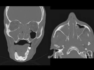
Burkitt lymphoma typically presents as a rapidly growing mass, often involving the jaw, facial bones, or abdominal organs. Symptoms may include swollen lymph nodes, fever, night sweats, weight loss, and fatigue. Diagnosis is made through a biopsy of the affected tissue, followed by immunohistochemical staining and genetic analysis to confirm the presence of characteristic chromosomal translocations involving the MYC oncogene.
Treatment for Burkitt lymphoma typically involves intensive chemotherapy regimens, often combined with targeted therapy or immunotherapy. The prognosis is generally good when treated aggressively and promptly, with a high cure rate in children and young adults. However, the prognosis may be poorer in older patients or those with advanced-stage disease at diagnosis.
1. Receptors: In the context of physiology and medicine, receptors are specialized proteins found on the surface of cells or inside cells that detect and respond to specific molecules, known as ligands. These interactions can trigger a variety of responses within the cell, such as starting a signaling cascade or changing the cell's metabolism. Receptors play crucial roles in various biological processes, including communication between cells, regulation of immune responses, and perception of senses.
2. Antigen: An antigen is any substance (usually a protein) that can be recognized by the adaptive immune system, specifically by B-cells and T-cells. Antigens can be derived from various sources, such as microorganisms (like bacteria, viruses, or fungi), pollen, dust mites, or even components of our own cells (for instance, in autoimmune diseases). An antigen's ability to stimulate an immune response is determined by its molecular structure and whether it can be recognized by the receptors on immune cells.
3. B-Cell: B-cells are a type of white blood cell that plays a critical role in the adaptive immune system, particularly in humoral immunity. They originate from hematopoietic stem cells in the bone marrow and are responsible for producing antibodies, which are proteins that recognize and bind to specific antigens. Each B-cell has receptors on its surface called B-cell receptors (BCRs) that can recognize a unique antigen. When a B-cell encounters its specific antigen, it becomes activated, undergoes proliferation, and differentiates into plasma cells that secrete large amounts of antibodies to neutralize or eliminate the antigen.
Leukemia, B-cell is a type of cancer that affects the blood and bone marrow, characterized by an overproduction of abnormal B-lymphocytes, a type of white blood cell. These abnormal cells accumulate in the bone marrow and interfere with the production of normal blood cells, leading to anemia, infection, and bleeding.
B-cells are a type of lymphocyte that plays a crucial role in the immune system by producing antibodies to help fight off infections. In B-cell leukemia, the cancerous B-cells do not mature properly and accumulate in the bone marrow, leading to a decrease in the number of healthy white blood cells, red blood cells, and platelets.
There are several types of B-cell leukemia, including acute lymphoblastic leukemia (ALL) and chronic lymphocytic leukemia (CLL). ALL is more common in children and young adults, while CLL is more common in older adults. Treatment options for B-cell leukemia depend on the type and stage of the disease and may include chemotherapy, radiation therapy, stem cell transplantation, or targeted therapies.
Immunoglobulin mu-chains (IgM) are a type of heavy chain found in immunoglobulins, also known as antibodies. IgM is the first antibody to be produced in response to an initial exposure to an antigen and plays a crucial role in the early stages of the immune response.
IgM antibodies are composed of four monomeric units, each consisting of two heavy chains and two light chains. The heavy chains in IgM are called mu-chains, which have a molecular weight of approximately 72 kDa. Each mu-chain contains five domains: one variable (V) domain at the N-terminus, four constant (C) domains (Cμ1-4), and a membrane-spanning region followed by a short cytoplasmic tail.
IgM antibodies are primarily found on the surface of B cells as part of the B cell receptor (BCR). When a B cell encounters an antigen, the BCR binds to it, triggering a series of intracellular signaling events that lead to B cell activation and differentiation into plasma cells. In response to activation, the B cell begins to secrete IgM antibodies into the bloodstream.
IgM antibodies have several unique features that make them effective in the early stages of an immune response. They are highly efficient at agglutination, or clumping together, of pathogens and antigens, which helps to neutralize them. IgM antibodies also activate the complement system, a group of proteins that work together to destroy pathogens.
Overall, Immunoglobulin mu-chains are an essential component of the immune system, providing early protection against pathogens and initiating the adaptive immune response.
CD19 is a type of protein found on the surface of B cells, which are a type of white blood cell that plays a key role in the body's immune response. CD19 is a marker that helps identify and distinguish B cells from other types of cells in the body. It is also a target for immunotherapy in certain diseases, such as B-cell malignancies.
An antigen is any substance that can stimulate an immune response, particularly the production of antibodies. In the context of CD19, antigens refer to substances that can bind to CD19 and trigger a response from the immune system. This can include proteins, carbohydrates, or other molecules found on the surface of bacteria, viruses, or cancer cells.
Therefore, 'antigens, CD19' refers to any substances that can bind to the CD19 protein on B cells and trigger an immune response. These antigens may be used in the development of immunotherapies for the treatment of B-cell malignancies or other diseases.
Acute myeloid leukemia (AML) is a type of cancer that originates in the bone marrow, the soft inner part of certain bones where new blood cells are made. In AML, the immature cells, called blasts, in the bone marrow fail to mature into normal blood cells. Instead, these blasts accumulate and interfere with the production of normal blood cells, leading to a shortage of red blood cells (anemia), platelets (thrombocytopenia), and normal white blood cells (leukopenia).
AML is called "acute" because it can progress quickly and become severe within days or weeks without treatment. It is a type of myeloid leukemia, which means that it affects the myeloid cells in the bone marrow. Myeloid cells are a type of white blood cell that includes monocytes and granulocytes, which help fight infection and defend the body against foreign invaders.
In AML, the blasts can build up in the bone marrow and spread to other parts of the body, including the blood, lymph nodes, liver, spleen, and brain. This can cause a variety of symptoms, such as fatigue, fever, frequent infections, easy bruising or bleeding, and weight loss.
AML is typically treated with a combination of chemotherapy, radiation therapy, and/or stem cell transplantation. The specific treatment plan will depend on several factors, including the patient's age, overall health, and the type and stage of the leukemia.
Immunoglobulin light chains are the smaller protein subunits of an immunoglobulin, also known as an antibody. They are composed of two polypeptide chains, called kappa (κ) and lambda (λ), which are produced by B cells during the immune response. Each immunoglobulin molecule contains either two kappa or two lambda light chains, in association with two heavy chains.
Light chains play a crucial role in the antigen-binding site of an antibody, where they contribute to the specificity and affinity of the interaction between the antibody and its target antigen. In addition to their role in immune function, abnormal production or accumulation of light chains can lead to various diseases, such as multiple myeloma and amyloidosis.
Leukemia, T-cell is a type of cancer that affects the T-cells or T-lymphocytes, which are a type of white blood cells responsible for cell-mediated immunity. It is characterized by an excessive and uncontrolled production of abnormal T-cells in the bone marrow, leading to the displacement of healthy cells and impairing the body's ability to fight infections and regulate immune responses.
T-cell leukemia can be acute or chronic, depending on the rate at which the disease progresses. Acute T-cell leukemia progresses rapidly, while chronic T-cell leukemia has a slower course of progression. Symptoms may include fatigue, fever, frequent infections, weight loss, easy bruising or bleeding, and swollen lymph nodes. Treatment typically involves chemotherapy, radiation therapy, stem cell transplantation, or targeted therapy, depending on the type and stage of the disease.
Hematopoietic stem cells (HSCs) are immature, self-renewing cells that give rise to all the mature blood and immune cells in the body. They are capable of both producing more hematopoietic stem cells (self-renewal) and differentiating into early progenitor cells that eventually develop into red blood cells, white blood cells, and platelets. HSCs are found in the bone marrow, umbilical cord blood, and peripheral blood. They have the ability to repair damaged tissues and offer significant therapeutic potential for treating various diseases, including hematological disorders, genetic diseases, and cancer.
Immunoglobulin heavy chains are proteins that make up the framework of antibodies, which are Y-shaped immune proteins. These heavy chains, along with light chains, form the antigen-binding sites of an antibody, which recognize and bind to specific foreign substances (antigens) in order to neutralize or remove them from the body.
The heavy chain is composed of a variable region, which contains the antigen-binding site, and constant regions that determine the class and function of the antibody. There are five classes of immunoglobulins (IgA, IgD, IgE, IgG, and IgM) that differ in their heavy chain constant regions and therefore have different functions in the immune response.
Immunoglobulin heavy chains are synthesized by B cells, a type of white blood cell involved in the adaptive immune response. The genetic rearrangement of immunoglobulin heavy chain genes during B cell development results in the production of a vast array of different antibodies with unique antigen-binding sites, allowing for the recognition and elimination of a wide variety of pathogens.
Asparaginase is a medication that is used in the treatment of certain types of cancer, such as acute lymphoblastic leukemia (ALL) and non-Hodgkin lymphoma (NHL). It is an enzyme that breaks down the amino acid asparagine, which is a building block of proteins. Some cancer cells are unable to produce their own asparagine and rely on obtaining it from the bloodstream. By reducing the amount of asparagine in the blood, asparaginase can help to slow or stop the growth of these cancer cells.
Asparaginase is usually given as an injection into a muscle (intramuscularly) or into a vein (intravenously). It may be given alone or in combination with other chemotherapy drugs. The specific dosage and duration of treatment will depend on the individual's medical history, the type and stage of cancer being treated, and how well the person tolerates the medication.
Like all medications, asparaginase can cause side effects. Common side effects include nausea, vomiting, loss of appetite, and changes in liver function tests. Less common but more serious side effects may include allergic reactions, pancreatitis, and blood clotting problems. It is important for patients to discuss the potential risks and benefits of asparaginase with their healthcare provider before starting treatment.
Immunoglobulins (Igs), also known as antibodies, are proteins produced by the immune system to recognize and neutralize foreign substances such as pathogens or toxins. They are composed of four polypeptide chains: two heavy chains and two light chains, which are held together by disulfide bonds. The variable regions of the heavy and light chains contain loops that form the antigen-binding site, allowing each Ig molecule to recognize a specific epitope (antigenic determinant) on an antigen.
Genes encoding immunoglobulins are located on chromosome 14 (light chain genes) and chromosomes 22 and 2 (heavy chain genes). The diversity of the immune system is generated through a process called V(D)J recombination, where variable (V), diversity (D), and joining (J) gene segments are randomly selected and assembled to form the variable regions of the heavy and light chains. This results in an enormous number of possible combinations, allowing the immune system to recognize and respond to a vast array of potential threats.
There are five classes of immunoglobulins: IgA, IgD, IgE, IgG, and IgM, each with distinct functions and structures. For example, IgG is the most abundant class in serum and provides long-term protection against pathogens, while IgA is found on mucosal surfaces and helps prevent the entry of pathogens into the body.
Interleukin-7 (IL-7) is a small signaling protein that is involved in the development and function of immune cells, particularly T cells and B cells. It is produced by stromal cells found in the bone marrow, thymus, and lymphoid organs. IL-7 binds to its receptor, IL-7R, which is expressed on the surface of immature T cells and B cells, as well as some mature immune cells.
IL-7 plays a critical role in the survival, proliferation, and differentiation of T cells and B cells during their development in the thymus and bone marrow, respectively. It also helps to maintain the homeostasis of these cell populations in peripheral tissues by promoting their survival and preventing apoptosis.
In addition to its role in immune cell development and homeostasis, IL-7 has been shown to have potential therapeutic applications in the treatment of various diseases, including cancer, infectious diseases, and autoimmune disorders. However, further research is needed to fully understand its mechanisms of action and potential side effects before it can be widely used in clinical settings.
Agammaglobulinemia is a medical condition characterized by a severe deficiency or complete absence of gamma globulins (a type of antibodies) in the blood. This deficiency results from a lack of functional B cells, which are a type of white blood cell that produces antibodies to help fight off infections.
There are two main types of agammaglobulinemia: X-linked agammaglobulinemia (XLA) and autosomal recessive agammaglobulinemia (ARA). XLA is caused by mutations in the BTK gene and primarily affects males, while ARA is caused by mutations in other genes and can affect both males and females.
People with agammaglobulinemia are at increased risk for recurrent bacterial infections, particularly respiratory tract infections such as pneumonia and sinusitis. They may also be more susceptible to certain viral and parasitic infections. Treatment typically involves replacement therapy with intravenous immunoglobulin (IVIG) to provide the patient with functional antibodies.
Large B-cell lymphoma, diffuse is a type of cancer that starts in cells called B-lymphocytes, which are part of the body's immune system. "Large B-cell" refers to the size and appearance of the abnormal cells when viewed under a microscope. "Diffuse" means that the abnormal cells are spread throughout the lymph node or tissue where the cancer has started, rather than being clustered in one area.
This type of lymphoma is typically aggressive, which means it grows and spreads quickly. It can occur almost anywhere in the body, but most commonly affects the lymph nodes, spleen, and bone marrow. Symptoms may include swollen lymph nodes, fever, night sweats, weight loss, and fatigue.
Treatment for large B-cell lymphoma, diffuse typically involves chemotherapy, radiation therapy, or a combination of both. In some cases, stem cell transplantation or targeted therapy may also be recommended. The prognosis varies depending on several factors, including the stage and location of the cancer, as well as the patient's age and overall health.
Immunoglobulin lambda-chains (Igλ) are one type of light chain found in the immunoglobulins, also known as antibodies. Antibodies are proteins that play a crucial role in the immune system's response to foreign substances, such as bacteria and viruses.
Immunoglobulins are composed of two heavy chains and two light chains, which are interconnected by disulfide bonds. There are two types of light chains: kappa (κ) and lambda (λ). Igλ chains are one type of light chain that can be found in association with heavy chains to form functional antibodies.
Igλ chains contain a variable region, which is responsible for recognizing and binding to specific antigens, and a constant region, which determines the class of the immunoglobulin (e.g., IgA, IgD, IgE, IgG, or IgM).
In humans, approximately 60% of all antibodies contain Igλ chains, while the remaining 40% contain Igκ chains. The ratio of Igλ to Igκ chains can vary depending on the type of immunoglobulin and its function in the immune response.
Succinimides are a group of anticonvulsant medications used to treat various types of seizures. They include drugs such as ethosuximide, methsuximide, and phensuximide. These medications work by reducing the abnormal electrical activity in the brain that leads to seizures.
The name "succinimides" comes from their chemical structure, which contains a five-membered ring containing two nitrogen atoms and a carbonyl group. This structure is similar to that of other anticonvulsant medications, such as barbiturates, but the succinimides have fewer side effects and are less likely to cause sedation or respiratory depression.
Succinimides are primarily used to treat absence seizures, which are characterized by brief periods of staring and lack of responsiveness. They may also be used as adjunctive therapy in the treatment of generalized tonic-clonic seizures and other types of seizures.
Like all medications, succinimides can cause side effects, including nausea, vomiting, dizziness, headache, and rash. More serious side effects, such as blood dyscrasias, liver toxicity, and Stevens-Johnson syndrome, are rare but have been reported. It is important for patients taking succinimides to be monitored regularly by their healthcare provider to ensure safe and effective use of the medication.
Flow cytometry is a medical and research technique used to measure physical and chemical characteristics of cells or particles, one cell at a time, as they flow in a fluid stream through a beam of light. The properties measured include:
* Cell size (light scatter)
* Cell internal complexity (granularity, also light scatter)
* Presence or absence of specific proteins or other molecules on the cell surface or inside the cell (using fluorescent antibodies or other fluorescent probes)
The technique is widely used in cell counting, cell sorting, protein engineering, biomarker discovery and monitoring disease progression, particularly in hematology, immunology, and cancer research.
Bone marrow cells are the types of cells found within the bone marrow, which is the spongy tissue inside certain bones in the body. The main function of bone marrow is to produce blood cells. There are two types of bone marrow: red and yellow. Red bone marrow is where most blood cell production takes place, while yellow bone marrow serves as a fat storage site.
The three main types of bone marrow cells are:
1. Hematopoietic stem cells (HSCs): These are immature cells that can differentiate into any type of blood cell, including red blood cells, white blood cells, and platelets. They have the ability to self-renew, meaning they can divide and create more hematopoietic stem cells.
2. Red blood cell progenitors: These are immature cells that will develop into mature red blood cells, also known as erythrocytes. Red blood cells carry oxygen from the lungs to the body's tissues and carbon dioxide back to the lungs.
3. Myeloid and lymphoid white blood cell progenitors: These are immature cells that will develop into various types of white blood cells, which play a crucial role in the body's immune system by fighting infections and diseases. Myeloid progenitors give rise to granulocytes (neutrophils, eosinophils, and basophils), monocytes, and megakaryocytes (which eventually become platelets). Lymphoid progenitors differentiate into B cells, T cells, and natural killer (NK) cells.
Bone marrow cells are essential for maintaining a healthy blood cell count and immune system function. Abnormalities in bone marrow cells can lead to various medical conditions, such as anemia, leukopenia, leukocytosis, thrombocytopenia, or thrombocytosis, depending on the specific type of blood cell affected. Additionally, bone marrow cells are often used in transplantation procedures to treat patients with certain types of cancer, such as leukemia and lymphoma, or other hematologic disorders.
Human T-lymphotropic virus 1 (HTLV-1) is a complex retrovirus that infects CD4+ T lymphocytes and can cause adult T-cell leukemia/lymphoma (ATLL) and HTLV-1-associated myelopathy/tropical spastic paraparesis (HAM/TSP). The virus is primarily transmitted through breastfeeding, sexual contact, or contaminated blood products. After infection, the virus integrates into the host's genome and can remain latent for years or even decades before leading to disease. HTLV-1 is endemic in certain regions of the world, including Japan, the Caribbean, Central and South America, and parts of Africa.
Gene expression regulation in leukemia refers to the processes that control the production or activation of specific proteins encoded by genes in leukemic cells. These regulatory mechanisms include various molecular interactions that can either promote or inhibit gene transcription and translation. In leukemia, abnormal gene expression regulation can lead to uncontrolled proliferation, differentiation arrest, and accumulation of malignant white blood cells (leukemia cells) in the bone marrow and peripheral blood.
Dysregulated gene expression in leukemia may involve genetic alterations such as mutations, chromosomal translocations, or epigenetic changes that affect DNA methylation patterns and histone modifications. These changes can result in the overexpression of oncogenes (genes with cancer-promoting functions) or underexpression of tumor suppressor genes (genes that prevent uncontrolled cell growth).
Understanding gene expression regulation in leukemia is crucial for developing targeted therapies and improving diagnostic, prognostic, and treatment strategies.
Follicular lymphoma is a specific type of low-grade or indolent non-Hodgkin lymphoma (NHL). It develops from the B-lymphocytes, a type of white blood cell found in the lymphatic system. This lymphoma is characterized by the presence of abnormal follicles or nodules in the lymph nodes and other organs. The neoplastic cells in this subtype exhibit a distinct growth pattern that resembles normal follicular centers, hence the name "follicular lymphoma."
The majority of cases involve a translocation between chromosomes 14 and 18 [t(14;18)], leading to an overexpression of the BCL-2 gene. This genetic alteration contributes to the cancer cells' resistance to programmed cell death, allowing them to accumulate in the body.
Follicular lymphoma is typically slow-growing and may not cause symptoms for a long time. Common manifestations include painless swelling of lymph nodes, fatigue, weight loss, and night sweats. Treatment options depend on various factors such as the stage of the disease, patient's age, and overall health. Watchful waiting, chemotherapy, immunotherapy, targeted therapy, radiation therapy, or a combination of these approaches may be used to manage follicular lymphoma.
Chronic lymphocytic leukemia (CLL) is a type of cancer that starts from cells that become certain white blood cells (called lymphocytes) in the bone marrow. The cancer (leukemia) cells start in the bone marrow but then go into the blood.
In CLL, the leukemia cells often build up slowly. Many people don't have any symptoms for at least a few years. But over time, the cells can spread to other parts of the body, including the lymph nodes, liver, and spleen.
The "B-cell" part of the name refers to the fact that the cancer starts in a type of white blood cell called a B lymphocyte or B cell. The "chronic" part means that this leukemia usually progresses more slowly than other types of leukemia.
It's important to note that chronic lymphocytic leukemia is different from chronic myelogenous leukemia (CML). Although both are cancers of the white blood cells, they start in different types of white blood cells and progress differently.
Cell differentiation is the process by which a less specialized cell, or stem cell, becomes a more specialized cell type with specific functions and structures. This process involves changes in gene expression, which are regulated by various intracellular signaling pathways and transcription factors. Differentiation results in the development of distinct cell types that make up tissues and organs in multicellular organisms. It is a crucial aspect of embryonic development, tissue repair, and maintenance of homeostasis in the body.
Bone marrow is the spongy tissue found inside certain bones in the body, such as the hips, thighs, and vertebrae. It is responsible for producing blood-forming cells, including red blood cells, white blood cells, and platelets. There are two types of bone marrow: red marrow, which is involved in blood cell production, and yellow marrow, which contains fatty tissue.
Red bone marrow contains hematopoietic stem cells, which can differentiate into various types of blood cells. These stem cells continuously divide and mature to produce new blood cells that are released into the circulation. Red blood cells carry oxygen throughout the body, white blood cells help fight infections, and platelets play a crucial role in blood clotting.
Bone marrow also serves as a site for immune cell development and maturation. It contains various types of immune cells, such as lymphocytes, macrophages, and dendritic cells, which help protect the body against infections and diseases.
Abnormalities in bone marrow function can lead to several medical conditions, including anemia, leukopenia, thrombocytopenia, and various types of cancer, such as leukemia and multiple myeloma. Bone marrow aspiration and biopsy are common diagnostic procedures used to evaluate bone marrow health and function.
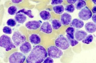
Immunophenotyping is a medical laboratory technique used to identify and classify cells, usually in the context of hematologic (blood) disorders and malignancies (cancers), based on their surface or intracellular expression of various proteins and antigens. This technique utilizes specific antibodies tagged with fluorochromes, which bind to the target antigens on the cell surface or within the cells. The labeled cells are then analyzed using flow cytometry, allowing for the detection and quantification of multiple antigenic markers simultaneously.
Immunophenotyping helps in understanding the distribution of different cell types, their subsets, and activation status, which can be crucial in diagnosing various hematological disorders, immunodeficiencies, and distinguishing between different types of leukemias, lymphomas, and other malignancies. Additionally, it can also be used to monitor the progression of diseases, evaluate the effectiveness of treatments, and detect minimal residual disease (MRD) during follow-up care.
Remission induction is a treatment approach in medicine, particularly in the field of oncology and hematology. It refers to the initial phase of therapy aimed at reducing or eliminating the signs and symptoms of active disease, such as cancer or autoimmune disorders. The primary goal of remission induction is to achieve a complete response (disappearance of all detectable signs of the disease) or a partial response (a decrease in the measurable extent of the disease). This phase of treatment is often intensive and may involve the use of multiple drugs or therapies, including chemotherapy, immunotherapy, or targeted therapy. After remission induction, patients may receive additional treatments to maintain the remission and prevent relapse, known as consolidation or maintenance therapy.
B-lymphocyte gene rearrangement is a fundamental biological process that occurs during the development of B-lymphocytes (also known as B cells), which are a type of white blood cell responsible for producing antibodies to help fight infections. This process involves the rearrangement of genetic material within the B-lymphocyte's immunoglobulin genes, specifically the heavy chain (IgH) and light chain (IgL) genes, to create a diverse repertoire of antibodies with unique specificities.
During B-lymphocyte gene rearrangement, large segments of DNA are cut, deleted, or inverted, and then rejoined to form a functional IgH or IgL gene that encodes an antigen-binding site on the antibody molecule. The process occurs in two main steps:
1. Variable (V), diversity (D), and joining (J) gene segments are rearranged to form the heavy chain gene, which is located on chromosome 14. This results in a vast array of possible combinations, allowing for the generation of a diverse set of antibody molecules.
2. A separate variable (V) and joining (J) gene segment rearrangement occurs to form the light chain gene, which can be either kappa or lambda type, located on chromosomes 2 and 22, respectively.
Once the heavy and light chain genes are successfully rearranged, they are transcribed into mRNA and translated into immunoglobulin proteins, forming a functional antibody molecule. If the initial gene rearrangement fails to produce a functional antibody, additional attempts at rearrangement can occur, involving different combinations of V, D, and J segments or the use of alternative reading frames.
Errors in B-lymphocyte gene rearrangement can lead to various genetic disorders, such as lymphomas and leukemias, due to the production of aberrant antibodies or uncontrolled cell growth.
Experimental leukemia refers to the stage of research or clinical trials where new therapies, treatments, or diagnostic methods are being studied for leukemia. Leukemia is a type of cancer that affects the blood and bone marrow, leading to an overproduction of abnormal white blood cells.
In the experimental stage, researchers investigate various aspects of leukemia, such as its causes, progression, and potential treatments. They may conduct laboratory studies using cell cultures or animal models to understand the disease better and test new therapeutic approaches. Additionally, clinical trials may be conducted to evaluate the safety and efficacy of novel treatments in human patients with leukemia.
Experimental research in leukemia is crucial for advancing our understanding of the disease and developing more effective treatment strategies. It involves a rigorous and systematic process that adheres to ethical guidelines and scientific standards to ensure the validity and reliability of the findings.
Translocation, genetic, refers to a type of chromosomal abnormality in which a segment of a chromosome is transferred from one chromosome to another, resulting in an altered genome. This can occur between two non-homologous chromosomes (non-reciprocal translocation) or between two homologous chromosomes (reciprocal translocation). Genetic translocations can lead to various clinical consequences, depending on the genes involved and the location of the translocation. Some translocations may result in no apparent effects, while others can cause developmental abnormalities, cancer, or other genetic disorders. In some cases, translocations can also increase the risk of having offspring with genetic conditions.
Immunoglobulin M (IgM) is a type of antibody that is primarily found in the blood and lymph fluid. It is the first antibody to be produced in response to an initial exposure to an antigen, making it an important part of the body's primary immune response. IgM antibodies are large molecules that are composed of five basic units, giving them a pentameric structure. They are primarily found on the surface of B cells as membrane-bound immunoglobulins (mlgM), where they function as receptors for antigens. Once an mlgM receptor binds to an antigen, it triggers the activation and differentiation of the B cell into a plasma cell that produces and secretes large amounts of soluble IgM antibodies.
IgM antibodies are particularly effective at agglutination (clumping) and complement activation, which makes them important in the early stages of an immune response to help clear pathogens from the bloodstream. However, they are not as stable or long-lived as other types of antibodies, such as IgG, and their levels tend to decline after the initial immune response has occurred.
In summary, Immunoglobulin M (IgM) is a type of antibody that plays a crucial role in the primary immune response to antigens by agglutination and complement activation. It is primarily found in the blood and lymph fluid, and it is produced by B cells after they are activated by an antigen.
B-cell marginal zone lymphoma (MZL) is a type of indolent (slow-growing) non-Hodgkin lymphoma (NHL). It arises from B-lymphocytes, a type of white blood cell found in the lymphatic system. MZLs typically involve the marginal zone of lymphoid follicles, which are structures found in lymph nodes and other lymphatic tissues.
There are three subtypes of MZL: extranodal MZL (also known as mucosa-associated lymphoid tissue or MALT lymphoma), nodal MZL, and splenic MZL. Extranodal MZL is the most common form and can occur at various extranodal sites, such as the stomach, lungs, skin, eyes, and salivary glands. Nodal MZL involves the lymph nodes without evidence of extranodal disease, while splenic MZL primarily affects the spleen.
MZLs are typically low-grade malignancies, but they can transform into more aggressive forms over time. Treatment options depend on the stage and location of the disease, as well as the patient's overall health. Common treatments include watchful waiting, radiation therapy, chemotherapy, immunotherapy, targeted therapy, or a combination of these approaches.
HTLV-I (Human T-lymphotropic virus type 1) infection is a viral infection that attacks the CD4+ T-cells (a type of white blood cell) and can lead to the development of various diseases, including Adult T-cell Leukemia/Lymphoma (ATLL) and HTLV-I Associated Myelopathy/Tropical Spastic Paraparesis (HAM/TSP). The virus is primarily transmitted through breastfeeding, sexual contact, or contaminated blood products. After infection, the virus becomes integrated into the host's DNA and can remain dormant for years, even decades, before leading to the development of disease. Most people infected with HTLV-I do not develop any symptoms, but a small percentage will go on to develop serious complications.
Chronic myelogenous leukemia (CML), BCR-ABL positive is a specific subtype of leukemia that originates in the bone marrow and involves the excessive production of mature granulocytes, a type of white blood cell. It is characterized by the presence of the Philadelphia chromosome, which is formed by a genetic translocation between chromosomes 9 and 22, resulting in the formation of the BCR-ABL fusion gene. This gene encodes for an abnormal protein with increased tyrosine kinase activity, leading to uncontrolled cell growth and division. The presence of this genetic abnormality is used to confirm the diagnosis and guide treatment decisions.
Fluorescein is not a medical condition, but rather a diagnostic dye that is used in various medical tests and procedures. It is a fluorescent compound that absorbs light at one wavelength and emits light at another wavelength, which makes it useful for imaging and detecting various conditions.
In ophthalmology, fluorescein is commonly used in eye examinations to evaluate the health of the cornea, conjunctiva, and anterior chamber of the eye. A fluorescein dye is applied to the surface of the eye, and then the eye is examined under a blue light. The dye highlights any damage or abnormalities on the surface of the eye, such as scratches, ulcers, or inflammation.
Fluorescein is also used in angiography, a medical imaging technique used to examine blood vessels in the body. A fluorescein dye is injected into a vein, and then a special camera takes pictures of the dye as it flows through the blood vessels. This can help doctors diagnose and monitor conditions such as cancer, diabetes, and macular degeneration.
Overall, fluorescein is a valuable diagnostic tool that helps medical professionals detect and monitor various conditions in the body.
Protein precursors, also known as proproteins or prohormones, are inactive forms of proteins that undergo post-translational modification to become active. These modifications typically include cleavage of the precursor protein by specific enzymes, resulting in the release of the active protein. This process allows for the regulation and control of protein activity within the body. Protein precursors can be found in various biological processes, including the endocrine system where they serve as inactive hormones that can be converted into their active forms when needed.
"Cells, cultured" is a medical term that refers to cells that have been removed from an organism and grown in controlled laboratory conditions outside of the body. This process is called cell culture and it allows scientists to study cells in a more controlled and accessible environment than they would have inside the body. Cultured cells can be derived from a variety of sources, including tissues, organs, or fluids from humans, animals, or cell lines that have been previously established in the laboratory.
Cell culture involves several steps, including isolation of the cells from the tissue, purification and characterization of the cells, and maintenance of the cells in appropriate growth conditions. The cells are typically grown in specialized media that contain nutrients, growth factors, and other components necessary for their survival and proliferation. Cultured cells can be used for a variety of purposes, including basic research, drug development and testing, and production of biological products such as vaccines and gene therapies.
It is important to note that cultured cells may behave differently than they do in the body, and results obtained from cell culture studies may not always translate directly to human physiology or disease. Therefore, it is essential to validate findings from cell culture experiments using additional models and ultimately in clinical trials involving human subjects.
A residual neoplasm is a term used in pathology and oncology to describe the remaining abnormal tissue or cancer cells after a surgical procedure or treatment aimed at completely removing a tumor. This means that some cancer cells have been left behind and continue to persist in the body. The presence of residual neoplasm can increase the risk of recurrence or progression of the disease, as these remaining cells may continue to grow and divide.
Residual neoplasm is often assessed during follow-up appointments and monitoring, using imaging techniques like CT scans, MRIs, or PET scans, and sometimes through biopsies. The extent of residual neoplasm can influence the choice of further treatment options, such as additional surgery, radiation therapy, chemotherapy, or targeted therapies, to eliminate the remaining cancer cells and reduce the risk of recurrence.
Vincristine is an antineoplastic agent, specifically a vinca alkaloid. It is derived from the Madagascar periwinkle plant (Catharanthus roseus). Vincristine binds to tubulin, a protein found in microtubules, and inhibits their polymerization, which results in disruption of mitotic spindles leading to cell cycle arrest and apoptosis (programmed cell death). It is used in the treatment of various types of cancer including leukemias, lymphomas, and solid tumors. Common side effects include peripheral neuropathy, constipation, and alopecia.
Karyotyping is a medical laboratory test used to study the chromosomes in a cell. It involves obtaining a sample of cells from a patient, usually from blood or bone marrow, and then staining the chromosomes so they can be easily seen under a microscope. The chromosomes are then arranged in pairs based on their size, shape, and other features to create a karyotype. This visual representation allows for the identification and analysis of any chromosomal abnormalities, such as extra or missing chromosomes, or structural changes like translocations or inversions. These abnormalities can provide important information about genetic disorders, diseases, and developmental problems.
'Gene rearrangement in B-lymphocytes, light chain' refers to the biological process that occurs during the development of B-lymphocytes (a type of white blood cell) in the bone marrow. Specifically, it relates to the rearrangement of genes that code for the light chains of immunoglobulins, which are antibodies that help the immune system recognize and fight off foreign substances.
During gene rearrangement, the variable region genes of the light chain locus (which consist of multiple gene segments, including V, D, and J segments) undergo a series of DNA recombination events to form a functional variable region exon. This process allows for the generation of a vast diversity of antibody molecules with different specificities, enabling the immune system to recognize and respond to a wide range of potential threats.
Abnormalities in this gene rearrangement process can lead to various immunodeficiency disorders or malignancies such as B-cell lymphomas.
"Gene rearrangement" is a process that involves the alteration of the order, orientation, or copy number of genes or gene segments within an organism's genome. This natural mechanism plays a crucial role in generating diversity and specificity in the immune system, particularly in vertebrates.
In the context of the immune system, gene rearrangement occurs during the development of B-cells and T-cells, which are responsible for adaptive immunity. The process involves breaking and rejoining DNA segments that encode antigen recognition sites, resulting in a unique combination of gene segments and creating a vast array of possible antigen receptors.
There are two main types of gene rearrangement:
1. V(D)J recombination: This process occurs in both B-cells and T-cells. It involves the recombination of variable (V), diversity (D), and joining (J) gene segments to form a functional antigen receptor gene. In humans, there are multiple copies of V, D, and J segments for each antigen receptor gene, allowing for a vast number of possible combinations.
2. Class switch recombination: This process occurs only in mature B-cells after antigen exposure. It involves the replacement of the constant (C) region of the immunoglobulin heavy chain gene with another C region, resulting in the production of different isotypes of antibodies (IgG, IgA, or IgE) that have distinct effector functions while maintaining the same antigen specificity.
These processes contribute to the generation of a diverse repertoire of antigen receptors, allowing the immune system to recognize and respond effectively to a wide range of pathogens.
Cutaneous T-cell lymphoma (CTCL) is a type of cancer that affects T-cells, a specific group of white blood cells called lymphocytes. These cells play a crucial role in the body's immune system and help protect against infection and disease. In CTCL, the T-cells become malignant and accumulate in the skin, leading to various skin symptoms and lesions.
CTCL is a subtype of non-Hodgkin lymphoma (NHL), which refers to a group of cancers that originate from lymphocytes. Within NHL, CTCL is categorized as a type of extranodal lymphoma since it primarily involves organs or tissues outside the lymphatic system, in this case, the skin.
The two most common subtypes of CTCL are mycosis fungoides and Sézary syndrome:
1. Mycosis fungoides (MF): This is the more prevalent form of CTCL, characterized by patches, plaques, or tumors on the skin. The lesions may be scaly, itchy, or change in size, shape, and color over time. MF usually progresses slowly, with early-stage disease often confined to the skin for several years before spreading to lymph nodes or other organs.
2. Sézary syndrome (SS): This is a more aggressive form of CTCL that involves not only the skin but also the blood and lymph nodes. SS is characterized by the presence of malignant T-cells, known as Sézary cells, in the peripheral blood. Patients with SS typically have generalized erythroderma (reddening and scaling of the entire body), pruritus (severe itching), lymphadenopathy (swollen lymph nodes), and alopecia (hair loss).
The diagnosis of CTCL usually involves a combination of clinical examination, skin biopsy, and immunophenotyping to identify the malignant T-cells. Treatment options depend on the stage and subtype of the disease and may include topical therapies, phototherapy, systemic medications, or targeted therapies.
Leukemic infiltration is the abnormal spread and accumulation of malignant white blood cells (leukemia cells) in various tissues and organs outside the bone marrow. The bone marrow is the spongy tissue inside bones where blood cells are normally produced. In leukemia, the bone marrow produces large numbers of abnormal white blood cells that do not function properly. These abnormal cells can sometimes spill into the bloodstream and infiltrate other organs, such as the lymph nodes, spleen, liver, and central nervous system (brain and spinal cord). Leukemic infiltration can cause damage to these organs and lead to various symptoms. The pattern of organ involvement and the severity of infiltration depend on the type and stage of leukemia.
Antineoplastic combined chemotherapy protocols refer to a treatment plan for cancer that involves the use of more than one antineoplastic (chemotherapy) drug given in a specific sequence and schedule. The combination of drugs is used because they may work better together to destroy cancer cells compared to using a single agent alone. This approach can also help to reduce the likelihood of cancer cells becoming resistant to the treatment.
The choice of drugs, dose, duration, and frequency are determined by various factors such as the type and stage of cancer, patient's overall health, and potential side effects. Combination chemotherapy protocols can be used in various settings, including as a primary treatment, adjuvant therapy (given after surgery or radiation to kill any remaining cancer cells), neoadjuvant therapy (given before surgery or radiation to shrink the tumor), or palliative care (to alleviate symptoms and prolong survival).
It is important to note that while combined chemotherapy protocols can be effective in treating certain types of cancer, they can also cause significant side effects, including nausea, vomiting, hair loss, fatigue, and an increased risk of infection. Therefore, patients undergoing such treatment should be closely monitored and managed by a healthcare team experienced in administering chemotherapy.
The Philadelphia chromosome is a specific genetic alteration in certain types of leukemia and lymphoma, including chronic myelogenous leukemia (CML) and acute lymphoblastic leukemia (ALL). It is the result of a translocation between chromosomes 9 and 22, which forms an abnormal fusion gene called BCR-ABL. This gene produces an abnormal protein that leads to unregulated cell growth and division, causing cancer. The Philadelphia chromosome was first discovered in Philadelphia, USA, hence the name.
T-cell peripheral lymphoma is a type of cancer that affects the T-cells, which are a type of white blood cell that plays a crucial role in the body's immune system. This type of lymphoma is called "peripheral" because it typically develops in T-cells that have matured and are found in various tissues and organs outside of the bone marrow, such as the lymph nodes, spleen, skin, and digestive tract.
Peripheral T-cell lymphomas (PTCL) are relatively rare and can be aggressive, with a tendency to spread quickly throughout the body. They can arise from different types of T-cells, leading to various subtypes of PTCL that may have different clinical features, treatment options, and prognoses.
Some common subtypes of peripheral T-cell lymphoma include:
1. PTCL, not otherwise specified (NOS): This is the most common subtype, accounting for about 25-30% of all PTCL cases. It includes cases that do not fit into any specific category or have features of more than one subtype.
2. Anaplastic large cell lymphoma (ALCL): ALCL can be further divided into two groups: systemic ALCL and cutaneous ALCL. Systemic ALCL is a more aggressive form, while cutaneous ALCL tends to be less aggressive and primarily affects the skin.
3. Angioimmunoblastic T-cell lymphoma (AITL): AITL is an aggressive subtype that often involves the lymph nodes and can affect other organs such as the spleen, liver, and bone marrow. It frequently presents with B symptoms (fever, night sweats, and weight loss) and abnormal blood tests.
4. Enteropathy-associated T-cell lymphoma (EATL): EATL is a rare but aggressive subtype that primarily affects the intestines, particularly in individuals with a history of celiac disease or gluten sensitivity.
5. Adult T-cell leukemia/lymphoma (ATLL): ATLL is caused by the human T-cell leukemia virus type 1 (HTLV-1) and primarily affects adults from regions where HTLV-1 is endemic, such as Japan, the Caribbean, and parts of Africa.
Treatment for PTCL depends on the specific subtype, stage, and individual patient factors. Common treatment options include chemotherapy, targeted therapy, immunotherapy, radiation therapy, stem cell transplantation, or a combination of these approaches. Clinical trials are also available for eligible patients to test new therapies and combinations.
Apoptosis is a programmed and controlled cell death process that occurs in multicellular organisms. It is a natural process that helps maintain tissue homeostasis by eliminating damaged, infected, or unwanted cells. During apoptosis, the cell undergoes a series of morphological changes, including cell shrinkage, chromatin condensation, and fragmentation into membrane-bound vesicles called apoptotic bodies. These bodies are then recognized and engulfed by neighboring cells or phagocytic cells, preventing an inflammatory response. Apoptosis is regulated by a complex network of intracellular signaling pathways that involve proteins such as caspases, Bcl-2 family members, and inhibitors of apoptosis (IAPs).
A base sequence in the context of molecular biology refers to the specific order of nucleotides in a DNA or RNA molecule. In DNA, these nucleotides are adenine (A), guanine (G), cytosine (C), and thymine (T). In RNA, uracil (U) takes the place of thymine. The base sequence contains genetic information that is transcribed into RNA and ultimately translated into proteins. It is the exact order of these bases that determines the genetic code and thus the function of the DNA or RNA molecule.
Molecular sequence data refers to the specific arrangement of molecules, most commonly nucleotides in DNA or RNA, or amino acids in proteins, that make up a biological macromolecule. This data is generated through laboratory techniques such as sequencing, and provides information about the exact order of the constituent molecules. This data is crucial in various fields of biology, including genetics, evolution, and molecular biology, allowing for comparisons between different organisms, identification of genetic variations, and studies of gene function and regulation.
Prognosis is a medical term that refers to the prediction of the likely outcome or course of a disease, including the chances of recovery or recurrence, based on the patient's symptoms, medical history, physical examination, and diagnostic tests. It is an important aspect of clinical decision-making and patient communication, as it helps doctors and patients make informed decisions about treatment options, set realistic expectations, and plan for future care.
Prognosis can be expressed in various ways, such as percentages, categories (e.g., good, fair, poor), or survival rates, depending on the nature of the disease and the available evidence. However, it is important to note that prognosis is not an exact science and may vary depending on individual factors, such as age, overall health status, and response to treatment. Therefore, it should be used as a guide rather than a definitive forecast.
A "knockout" mouse is a genetically engineered mouse in which one or more genes have been deleted or "knocked out" using molecular biology techniques. This allows researchers to study the function of specific genes and their role in various biological processes, as well as potential associations with human diseases. The mice are generated by introducing targeted DNA modifications into embryonic stem cells, which are then used to create a live animal. Knockout mice have been widely used in biomedical research to investigate gene function, disease mechanisms, and potential therapeutic targets.
Deltaretroviruses are a genus of retroviruses that can cause chronic infections in humans and animals. The two main deltaretroviruses that infect humans are the Human T-cell Leukemia Virus type 1 (HTLV-1) and Human T-cell Leukemia Virus type 2 (HTLV-2).
HTLV-1 is primarily transmitted through breastfeeding, sexual contact, and contaminated blood products. It can cause several diseases, including Adult T-cell Leukemia/Lymphoma (ATLL) and a neurological disorder called HTLV-1 Associated Myelopathy/Tropical Spastic Paraparesis (HAM/TSP).
HTLV-2 is primarily transmitted through intravenous drug use and sexual contact. While it has been associated with some diseases, such as neurological disorders and rare cases of leukemia, the link between HTLV-2 and disease is not as clear as it is for HTLV-1.
Deltaretrovirus infections can be diagnosed through blood tests that detect antibodies to the viruses or through genetic testing to detect the virus itself. There is currently no cure for deltaretrovirus infections, but antiretroviral therapy (ART) may help manage the infection and reduce the risk of transmission.
It's important to note that deltaretrovirus infections are relatively rare, and most people who are infected do not develop symptoms or disease. However, if you believe you may have been exposed to these viruses, it is important to speak with a healthcare provider for further evaluation and testing.
6-Mercaptopurine (6-MP) is a medication used primarily in the treatment of cancer, specifically acute lymphoblastic leukemia (ALL), and to prevent rejection in organ transplantation. It is an antimetabolite that works by interfering with the synthesis of DNA and RNA, thereby inhibiting cell division and growth.
6-MP is a prodrug, meaning it requires metabolic activation in the body to exert its therapeutic effects. Once absorbed, 6-MP is converted into several active metabolites, including thioguanine nucleotides (TGN), which are incorporated into DNA and RNA, leading to cytotoxicity and cell death.
Common side effects of 6-MP include nausea, vomiting, diarrhea, mouth sores, and increased susceptibility to infections. Long-term use of the medication can also lead to liver toxicity, pancreatitis, and anemia. Regular monitoring of blood counts, liver function tests, and TGN levels is necessary during treatment with 6-MP to minimize potential side effects and ensure safe and effective dosing.
Methotrexate is a medication used in the treatment of certain types of cancer and autoimmune diseases. It is an antimetabolite that inhibits the enzyme dihydrofolate reductase, which is necessary for the synthesis of purines and pyrimidines, essential components of DNA and RNA. By blocking this enzyme, methotrexate interferes with cell division and growth, making it effective in treating rapidly dividing cells such as cancer cells.
In addition to its use in cancer treatment, methotrexate is also used to manage autoimmune diseases such as rheumatoid arthritis, psoriasis, and inflammatory bowel disease. In these conditions, methotrexate modulates the immune system and reduces inflammation.
It's important to note that methotrexate can have significant side effects and should be used under the close supervision of a healthcare provider. Regular monitoring of blood counts, liver function, and kidney function is necessary during treatment with methotrexate.
The Myeloid-Lymphoid Leukemia (MLL) protein, also known as MLL1 or HRX, is a histone methyltransferase that plays a crucial role in the regulation of gene expression. It is involved in various cellular processes, including embryonic development and hematopoiesis (the formation of blood cells).
The MLL protein is encoded by the MLL gene, which is located on chromosome 11q23. This gene is frequently rearranged or mutated in certain types of leukemia, leading to the production of abnormal fusion proteins that contribute to tumor development and progression. These MLL-rearranged leukemias are aggressive and have a poor prognosis, making them an important area of research in the field of oncology.
Antineoplastic agents are a class of drugs used to treat malignant neoplasms or cancer. These agents work by inhibiting the growth and proliferation of cancer cells, either by killing them or preventing their division and replication. Antineoplastic agents can be classified based on their mechanism of action, such as alkylating agents, antimetabolites, topoisomerase inhibitors, mitotic inhibitors, and targeted therapy agents.
Alkylating agents work by adding alkyl groups to DNA, which can cause cross-linking of DNA strands and ultimately lead to cell death. Antimetabolites interfere with the metabolic processes necessary for DNA synthesis and replication, while topoisomerase inhibitors prevent the relaxation of supercoiled DNA during replication. Mitotic inhibitors disrupt the normal functioning of the mitotic spindle, which is essential for cell division. Targeted therapy agents are designed to target specific molecular abnormalities in cancer cells, such as mutated oncogenes or dysregulated signaling pathways.
It's important to note that antineoplastic agents can also affect normal cells and tissues, leading to various side effects such as nausea, vomiting, hair loss, and myelosuppression (suppression of bone marrow function). Therefore, the use of these drugs requires careful monitoring and management of their potential adverse effects.
Mantle cell lymphoma (MCL) is a type of non-Hodgkin lymphoma (NHL), which is a cancer of the lymphatic system. Specifically, MCL arises from abnormal B-lymphocytes (a type of white blood cell) that typically reside in the "mantle zone" of the lymph node. The malignant cells in MCL tend to have a characteristic genetic abnormality where the cyclin D1 gene is translocated to the immunoglobulin heavy chain gene locus, resulting in overexpression of cyclin D1 protein. This leads to uncontrolled cell division and proliferation.
Mantle cell lymphoma often presents with advanced-stage disease, involving multiple lymph nodes, bone marrow, and sometimes extranodal sites such as the gastrointestinal tract. Symptoms may include swollen lymph nodes, fatigue, weight loss, night sweats, and abdominal pain or discomfort.
Treatment for MCL typically involves a combination of chemotherapy, immunotherapy, and sometimes targeted therapy or stem cell transplantation. However, the prognosis for MCL is generally less favorable compared to other types of NHL, with a median overall survival of around 5-7 years.
'Tumor cells, cultured' refers to the process of removing cancerous cells from a tumor and growing them in controlled laboratory conditions. This is typically done by isolating the tumor cells from a patient's tissue sample, then placing them in a nutrient-rich environment that promotes their growth and multiplication.
The resulting cultured tumor cells can be used for various research purposes, including the study of cancer biology, drug development, and toxicity testing. They provide a valuable tool for researchers to better understand the behavior and characteristics of cancer cells outside of the human body, which can lead to the development of more effective cancer treatments.
It is important to note that cultured tumor cells may not always behave exactly the same way as they do in the human body, so findings from cell culture studies must be validated through further research, such as animal models or clinical trials.
Treatment outcome is a term used to describe the result or effect of medical treatment on a patient's health status. It can be measured in various ways, such as through symptoms improvement, disease remission, reduced disability, improved quality of life, or survival rates. The treatment outcome helps healthcare providers evaluate the effectiveness of a particular treatment plan and make informed decisions about future care. It is also used in clinical research to compare the efficacy of different treatments and improve patient care.
Recurrence, in a medical context, refers to the return of symptoms or signs of a disease after a period of improvement or remission. It indicates that the condition has not been fully eradicated and may require further treatment. Recurrence is often used to describe situations where a disease such as cancer comes back after initial treatment, but it can also apply to other medical conditions. The likelihood of recurrence varies depending on the type of disease and individual patient factors.
Core Binding Factor Alpha 2 Subunit, also known as CBF-A2 or CEBP-α, is a protein that forms a complex with other proteins to act as a transcription factor. Transcription factors are proteins that help regulate the expression of genes by binding to specific DNA sequences and controlling the rate of transcription of genetic information from DNA to RNA.
CBF-A2 is a member of the CCAAT/enhancer-binding protein (C/EBP) family of transcription factors, which are important in regulating various biological processes such as cell growth, development, and inflammation. CBF-A2 forms a heterodimer with Core Binding Factor Beta (CBF-β) to form the active transcription factor complex known as the core binding factor (CBF).
The CBF complex binds to the CCAAT box, a specific DNA sequence found in the promoter regions of many genes. By binding to this sequence, the CBF complex can either activate or repress the transcription of target genes, depending on the context and the presence of other regulatory factors.
Mutations in the gene encoding CBF-A2 have been associated with several human diseases, including acute myeloid leukemia (AML) and multiple myeloma. In AML, mutations in the CBF-A2 gene can lead to the formation of abnormal CBF complexes that disrupt normal gene expression patterns and contribute to the development of leukemia.
Cytarabine is a chemotherapeutic agent used in the treatment of various types of cancer, including leukemias and lymphomas. Its chemical name is cytosine arabinoside, and it works by interfering with the DNA synthesis of cancer cells, which ultimately leads to their death.
Cytarabine is often used in combination with other chemotherapy drugs and may be administered through various routes, such as intravenous (IV) or subcutaneous injection, or orally. The specific dosage and duration of treatment will depend on the type and stage of cancer being treated, as well as the patient's overall health status.
Like all chemotherapy drugs, cytarabine can cause a range of side effects, including nausea, vomiting, diarrhea, hair loss, and an increased risk of infection. It may also cause more serious side effects, such as damage to the liver, kidneys, or nervous system, and it is important for patients to be closely monitored during treatment to minimize these risks.
It's important to note that medical treatments should only be administered under the supervision of a qualified healthcare professional, and this information should not be used as a substitute for medical advice.
Notch 1 is a type of receptor that belongs to the family of single-transmembrane receptors known as Notch receptors. It is a heterodimeric transmembrane protein composed of an extracellular domain and an intracellular domain, which play crucial roles in cell fate determination, proliferation, differentiation, and apoptosis during embryonic development and adult tissue homeostasis.
The Notch 1 receptor is activated through a conserved mechanism of ligand-receptor interaction, where the extracellular domain of the receptor interacts with the membrane-bound ligands Jagged 1 or 2 and Delta-like 1, 3, or 4 expressed on adjacent cells. This interaction triggers a series of proteolytic cleavages that release the intracellular domain of Notch 1 (NICD) from the membrane. NICD then translocates to the nucleus and interacts with the DNA-binding protein CSL (CBF1/RBPJκ in mammals) and coactivators Mastermind-like proteins to regulate the expression of target genes, including members of the HES and HEY families.
Mutations in NOTCH1 have been associated with various human diseases, such as T-cell acute lymphoblastic leukemia (T-ALL), a type of cancer that affects the immune system's T cells, and vascular diseases, including arterial calcification, atherosclerosis, and aneurysms.
Polymerase Chain Reaction (PCR) is a laboratory technique used to amplify specific regions of DNA. It enables the production of thousands to millions of copies of a particular DNA sequence in a rapid and efficient manner, making it an essential tool in various fields such as molecular biology, medical diagnostics, forensic science, and research.
The PCR process involves repeated cycles of heating and cooling to separate the DNA strands, allow primers (short sequences of single-stranded DNA) to attach to the target regions, and extend these primers using an enzyme called Taq polymerase, resulting in the exponential amplification of the desired DNA segment.
In a medical context, PCR is often used for detecting and quantifying specific pathogens (viruses, bacteria, fungi, or parasites) in clinical samples, identifying genetic mutations or polymorphisms associated with diseases, monitoring disease progression, and evaluating treatment effectiveness.
Medical Definition:
Murine leukemia virus (MLV) is a type of retrovirus that primarily infects and causes various types of malignancies such as leukemias and lymphomas in mice. It is a complex genus of viruses, with many strains showing different pathogenic properties.
MLV contains two identical single-stranded RNA genomes and has the ability to reverse transcribe its RNA into DNA upon infection, integrating this proviral DNA into the host cell's genome. This is facilitated by an enzyme called reverse transcriptase, which MLV carries within its viral particle.
The virus can be horizontally transmitted between mice through close contact with infected saliva, urine, or milk. Vertical transmission from mother to offspring can also occur either in-utero or through the ingestion of infected breast milk.
MLV has been extensively studied as a model system for retroviral pathogenesis and tumorigenesis, contributing significantly to our understanding of oncogenes and their role in cancer development. It's important to note that Murine Leukemia Virus does not infect humans.
A cell line that is derived from tumor cells and has been adapted to grow in culture. These cell lines are often used in research to study the characteristics of cancer cells, including their growth patterns, genetic changes, and responses to various treatments. They can be established from many different types of tumors, such as carcinomas, sarcomas, and leukemias. Once established, these cell lines can be grown and maintained indefinitely in the laboratory, allowing researchers to conduct experiments and studies that would not be feasible using primary tumor cells. It is important to note that tumor cell lines may not always accurately represent the behavior of the original tumor, as they can undergo genetic changes during their time in culture.
Prednisone is a synthetic glucocorticoid, which is a type of corticosteroid hormone. It is primarily used to reduce inflammation in various conditions such as asthma, allergies, arthritis, and autoimmune disorders. Prednisone works by mimicking the effects of natural hormones produced by the adrenal glands, suppressing the immune system's response and reducing the release of substances that cause inflammation.
It is available in oral tablet form and is typically prescribed to be taken at specific times during the day, depending on the condition being treated. Common side effects of prednisone include increased appetite, weight gain, mood changes, insomnia, and easy bruising. Long-term use or high doses can lead to more serious side effects such as osteoporosis, diabetes, cataracts, and increased susceptibility to infections.
Healthcare providers closely monitor patients taking prednisone for extended periods to minimize the risk of adverse effects. It is essential to follow the prescribed dosage regimen and not discontinue the medication abruptly without medical supervision, as this can lead to withdrawal symptoms or a rebound of the underlying condition.
Chromosome aberrations refer to structural and numerical changes in the chromosomes that can occur spontaneously or as a result of exposure to mutagenic agents. These changes can affect the genetic material encoded in the chromosomes, leading to various consequences such as developmental abnormalities, cancer, or infertility.
Structural aberrations include deletions, duplications, inversions, translocations, and rings, which result from breaks and rearrangements of chromosome segments. Numerical aberrations involve changes in the number of chromosomes, such as aneuploidy (extra or missing chromosomes) or polyploidy (multiples of a complete set of chromosomes).
Chromosome aberrations can be detected and analyzed using various cytogenetic techniques, including karyotyping, fluorescence in situ hybridization (FISH), and comparative genomic hybridization (CGH). These methods allow for the identification and characterization of chromosomal changes at the molecular level, providing valuable information for genetic counseling, diagnosis, and research.
The Immunoglobulin (Ig) variable region is the antigen-binding part of an antibody, which is highly variable in its amino acid sequence and therefore specific to a particular epitope (the site on an antigen that is recognized by the antigen-binding site of an antibody). This variability is generated during the process of V(D)J recombination in the maturation of B cells, allowing for a diverse repertoire of antibodies to be produced and recognizing a wide range of potential pathogens.
The variable region is composed of several sub-regions including:
1. The heavy chain variable region (VH)
2. The light chain variable region (VL)
3. The heavy chain joining region (JH)
4. The light chain joining region (JL)
These regions are further divided into framework regions and complementarity-determining regions (CDRs). The CDRs, particularly CDR3, contain the most variability and are primarily responsible for antigen recognition.
Deltaretroviruses are a genus of retroviruses that include human T-lymphotropic virus (HTLV) types 1 and 2, bovine leukemia virus (BLV), and simian T-lymphotropic viruses. These viruses are characterized by their ability to cause persistent infections and can lead to the development of various diseases such as adult T-cell leukemia/lymphoma (ATLL) and tropical spastic paraparesis/HTLV-associated myelopathy (TSP/HAM).
The genome of deltaretroviruses contains two copies of single-stranded RNA, which are reverse transcribed into double-stranded DNA during the replication process. The viral DNA is then integrated into the host cell's genome, leading to a lifelong infection.
Deltaretroviruses primarily infect CD4+ T cells and other immune cells, and transmission typically occurs through bodily fluids such as breast milk, blood, and sexual contact. Prevention measures include avoiding high-risk behaviors, screening blood products, and implementing strict infection control practices in healthcare settings.
A gene product is the biochemical material, such as a protein or RNA, that is produced by the expression of a gene. Gene products are the result of the translation and transcription of genetic information encoded in DNA or RNA.
In the context of "tax," this term is not typically used in a medical definition of gene products. However, it may refer to the concept of taxing or regulating gene products in the context of genetic engineering or synthetic biology. This could involve imposing fees or restrictions on the production, use, or sale of certain gene products, particularly those that are genetically modified or engineered. The regulation of gene products is an important aspect of ensuring their safe and effective use in various applications, including medical treatments, agricultural production, and industrial processes.
T-lymphocytes, also known as T-cells, are a type of white blood cell that plays a key role in the adaptive immune system's response to infection. They are produced in the bone marrow and mature in the thymus gland. There are several different types of T-cells, including CD4+ helper T-cells, CD8+ cytotoxic T-cells, and regulatory T-cells (Tregs).
CD4+ helper T-cells assist in activating other immune cells, such as B-lymphocytes and macrophages. They also produce cytokines, which are signaling molecules that help coordinate the immune response. CD8+ cytotoxic T-cells directly kill infected cells by releasing toxic substances. Regulatory T-cells help maintain immune tolerance and prevent autoimmune diseases by suppressing the activity of other immune cells.
T-lymphocytes are important in the immune response to viral infections, cancer, and other diseases. Dysfunction or depletion of T-cells can lead to immunodeficiency and increased susceptibility to infections. On the other hand, an overactive T-cell response can contribute to autoimmune diseases and chronic inflammation.
Monoclonal antibodies are a type of antibody that are identical because they are produced by a single clone of cells. They are laboratory-produced molecules that act like human antibodies in the immune system. They can be designed to attach to specific proteins found on the surface of cancer cells, making them useful for targeting and treating cancer. Monoclonal antibodies can also be used as a therapy for other diseases, such as autoimmune disorders and inflammatory conditions.
Monoclonal antibodies are produced by fusing a single type of immune cell, called a B cell, with a tumor cell to create a hybrid cell, or hybridoma. This hybrid cell is then able to replicate indefinitely, producing a large number of identical copies of the original antibody. These antibodies can be further modified and engineered to enhance their ability to bind to specific targets, increase their stability, and improve their effectiveness as therapeutic agents.
Monoclonal antibodies have several mechanisms of action in cancer therapy. They can directly kill cancer cells by binding to them and triggering an immune response. They can also block the signals that promote cancer growth and survival. Additionally, monoclonal antibodies can be used to deliver drugs or radiation directly to cancer cells, increasing the effectiveness of these treatments while minimizing their side effects on healthy tissues.
Monoclonal antibodies have become an important tool in modern medicine, with several approved for use in cancer therapy and other diseases. They are continuing to be studied and developed as a promising approach to treating a wide range of medical conditions.
A cell line is a culture of cells that are grown in a laboratory for use in research. These cells are usually taken from a single cell or group of cells, and they are able to divide and grow continuously in the lab. Cell lines can come from many different sources, including animals, plants, and humans. They are often used in scientific research to study cellular processes, disease mechanisms, and to test new drugs or treatments. Some common types of human cell lines include HeLa cells (which come from a cancer patient named Henrietta Lacks), HEK293 cells (which come from embryonic kidney cells), and HUVEC cells (which come from umbilical vein endothelial cells). It is important to note that cell lines are not the same as primary cells, which are cells that are taken directly from a living organism and have not been grown in the lab.
A fusion protein known as "BCR-ABL" is formed due to a genetic abnormality called the Philadelphia chromosome (derived from a reciprocal translocation between chromosomes 9 and 22). This results in the formation of the oncogenic BCR-ABL tyrosine kinase, which contributes to unregulated cell growth and division, leading to chronic myeloid leukemia (CML) and some types of acute lymphoblastic leukemia (ALL). The BCR-ABL fusion protein has constitutively active tyrosine kinase activity, which results in the activation of various signaling pathways promoting cell proliferation, survival, and inhibition of apoptosis. This genetic alteration is crucial in the development and progression of CML and some types of ALL, making BCR-ABL an important therapeutic target for these malignancies.
AIDS-related lymphoma (ARL) is a type of cancer that affects the lymphatic system and is associated with acquired immunodeficiency syndrome (AIDS). It is caused by the infection of the lymphocytes, a type of white blood cell, with the human immunodeficiency virus (HIV), which weakens the immune system and makes individuals more susceptible to developing lymphoma.
There are two main types of AIDS-related lymphomas: diffuse large B-cell lymphoma (DLBCL) and Burkitt lymphoma (BL). DLBCL is the most common type and tends to grow rapidly, while BL is a more aggressive form that can also spread quickly.
Symptoms of AIDS-related lymphoma may include swollen lymph nodes, fever, night sweats, fatigue, weight loss, and decreased appetite. Diagnosis typically involves a biopsy of the affected lymph node or other tissue, followed by various imaging tests to determine the extent of the disease.
Treatment for AIDS-related lymphoma usually involves a combination of chemotherapy, radiation therapy, and/or immunotherapy, along with antiretroviral therapy (ART) to manage HIV infection. The prognosis for ARL varies depending on several factors, including the type and stage of the disease, the patient's overall health, and their response to treatment.
SCID mice is an acronym for Severe Combined Immunodeficiency mice. These are genetically modified mice that lack a functional immune system due to the mutation or knockout of several key genes required for immunity. This makes them ideal for studying the human immune system, infectious diseases, and cancer, as well as testing new therapies and treatments in a controlled environment without the risk of interference from the mouse's own immune system. SCID mice are often used in xenotransplantation studies, where human cells or tissues are transplanted into the mouse to study their behavior and interactions with the human immune system.
CD (cluster of differentiation) antigens are cell-surface proteins that are expressed on leukocytes (white blood cells) and can be used to identify and distinguish different subsets of these cells. They are important markers in the field of immunology and hematology, and are commonly used to diagnose and monitor various diseases, including cancer, autoimmune disorders, and infectious diseases.
CD antigens are designated by numbers, such as CD4, CD8, CD19, etc., which refer to specific proteins found on the surface of different types of leukocytes. For example, CD4 is a protein found on the surface of helper T cells, while CD8 is found on cytotoxic T cells.
CD antigens can be used as targets for immunotherapy, such as monoclonal antibody therapy, in which antibodies are designed to bind to specific CD antigens and trigger an immune response against cancer cells or infected cells. They can also be used as markers to monitor the effectiveness of treatments and to detect minimal residual disease (MRD) after treatment.
It's important to note that not all CD antigens are exclusive to leukocytes, some can be found on other cell types as well, and their expression can vary depending on the activation state or differentiation stage of the cells.
Large cell anaplastic lymphoma is a type of cancer that starts in white blood cells called lymphocytes, which are part of the body's immune system. It is classified as a type of non-Hodgkin lymphoma (NHL).
Anaplastic large cell lymphoma (ALCL) is a subtype of NHL characterized by the presence of large cancer cells that look abnormal under a microscope. These cells are called "anaplastic" because they lack many of the usual features of mature lymphocytes.
ALCL can occur in many different parts of the body, including the lymph nodes, skin, lungs, and soft tissues. It is typically an aggressive form of NHL that grows and spreads quickly.
ALCL is further divided into two main subtypes based on the presence or absence of a genetic abnormality involving a protein called ALK (anaplastic lymphoma kinase). ALK-positive ALCL tends to occur in younger patients and has a better prognosis than ALK-negative ALCL.
Treatment for large cell anaplastic lymphoma typically involves chemotherapy, radiation therapy, and/or immunotherapy, depending on the stage and location of the cancer. In some cases, stem cell transplantation may also be recommended.
Neprilysin (NEP), also known as membrane metallo-endopeptidase or CD10, is a type II transmembrane glycoprotein that functions as a zinc-dependent metalloprotease. It is widely expressed in various tissues, including the kidney, brain, heart, and vasculature. Neprilysin plays a crucial role in the breakdown and regulation of several endogenous bioactive peptides, such as natriuretic peptides, bradykinin, substance P, and angiotensin II. By degrading these peptides, neprilysin helps maintain cardiovascular homeostasis, modulate inflammation, and regulate neurotransmission. In the context of heart failure, neprilysin inhibitors have been developed to increase natriuretic peptide levels, promoting diuresis and vasodilation, ultimately improving cardiac function.
Gene expression profiling is a laboratory technique used to measure the activity (expression) of thousands of genes at once. This technique allows researchers and clinicians to identify which genes are turned on or off in a particular cell, tissue, or organism under specific conditions, such as during health, disease, development, or in response to various treatments.
The process typically involves isolating RNA from the cells or tissues of interest, converting it into complementary DNA (cDNA), and then using microarray or high-throughput sequencing technologies to determine which genes are expressed and at what levels. The resulting data can be used to identify patterns of gene expression that are associated with specific biological states or processes, providing valuable insights into the underlying molecular mechanisms of diseases and potential targets for therapeutic intervention.
In recent years, gene expression profiling has become an essential tool in various fields, including cancer research, drug discovery, and personalized medicine, where it is used to identify biomarkers of disease, predict patient outcomes, and guide treatment decisions.
Human chromosome pair 14 consists of two rod-shaped structures present in the nucleus of human cells, which contain genetic material in the form of DNA and proteins. Each member of the pair contains a single very long DNA molecule that carries an identical set of genes and other genetic elements, totaling approximately 105 million base pairs. These chromosomes play a crucial role in the development, functioning, and reproduction of human beings.
Chromosome 14 is one of the autosomal chromosomes, meaning it is not involved in determining the sex of an individual. It contains around 800-1,000 genes that provide instructions for producing various proteins responsible for numerous cellular functions and processes. Some notable genes located on chromosome 14 include those associated with neurodevelopmental disorders, cancer susceptibility, and immune system regulation.
Human cells typically have 23 pairs of chromosomes, including 22 autosomal pairs (numbered 1-22) and one pair of sex chromosomes (XX for females or XY for males). Chromosome pair 14 is the eighth largest autosomal pair in terms of its total length.
It's important to note that genetic information on chromosome 14, like all human chromosomes, can vary between individuals due to genetic variations and mutations. These differences contribute to the unique characteristics and traits found among humans.
The term "DNA, neoplasm" is not a standard medical term or concept. DNA refers to deoxyribonucleic acid, which is the genetic material present in the cells of living organisms. A neoplasm, on the other hand, is a tumor or growth of abnormal tissue that can be benign (non-cancerous) or malignant (cancerous).
In some contexts, "DNA, neoplasm" may refer to genetic alterations found in cancer cells. These genetic changes can include mutations, amplifications, deletions, or rearrangements of DNA sequences that contribute to the development and progression of cancer. Identifying these genetic abnormalities can help doctors diagnose and treat certain types of cancer more effectively.
However, it's important to note that "DNA, neoplasm" is not a term that would typically be used in medical reports or research papers without further clarification. If you have any specific questions about DNA changes in cancer cells or neoplasms, I would recommend consulting with a healthcare professional or conducting further research on the topic.
Disease-free survival (DFS) is a term used in medical research and clinical practice, particularly in the field of oncology. It refers to the length of time after primary treatment for a cancer during which no evidence of the disease can be found. This means that the patient shows no signs or symptoms of the cancer, and any imaging studies or other tests do not reveal any tumors or other indications of the disease.
DFS is often used as an important endpoint in clinical trials to evaluate the effectiveness of different treatments for cancer. By measuring the length of time until the cancer recurs or a new cancer develops, researchers can get a better sense of how well a particular treatment is working and whether it is improving patient outcomes.
It's important to note that DFS is not the same as overall survival (OS), which refers to the length of time from primary treatment until death from any cause. While DFS can provide valuable information about the effectiveness of cancer treatments, it does not necessarily reflect the impact of those treatments on patients' overall survival.
Hodgkin disease, also known as Hodgkin lymphoma, is a type of cancer that originates in the white blood cells called lymphocytes. It typically affects the lymphatic system, which is a network of vessels and glands spread throughout the body. The disease is characterized by the presence of a specific type of abnormal cell, known as a Reed-Sternberg cell, within the affected lymph nodes.
The symptoms of Hodgkin disease may include painless swelling of the lymph nodes in the neck, armpits, or groin; fever; night sweats; weight loss; and fatigue. The exact cause of Hodgkin disease is unknown, but it is thought to involve a combination of genetic, environmental, and infectious factors.
Hodgkin disease is typically treated with a combination of chemotherapy, radiation therapy, and/or immunotherapy, depending on the stage and extent of the disease. With appropriate treatment, the prognosis for Hodgkin disease is generally very good, with a high cure rate. However, long-term side effects of treatment may include an increased risk of secondary cancers and other health problems.
Bone marrow transplantation (BMT) is a medical procedure in which damaged or destroyed bone marrow is replaced with healthy bone marrow from a donor. Bone marrow is the spongy tissue inside bones that produces blood cells. The main types of BMT are autologous, allogeneic, and umbilical cord blood transplantation.
In autologous BMT, the patient's own bone marrow is used for the transplant. This type of BMT is often used in patients with lymphoma or multiple myeloma who have undergone high-dose chemotherapy or radiation therapy to destroy their cancerous bone marrow.
In allogeneic BMT, bone marrow from a genetically matched donor is used for the transplant. This type of BMT is often used in patients with leukemia, lymphoma, or other blood disorders who have failed other treatments.
Umbilical cord blood transplantation involves using stem cells from umbilical cord blood as a source of healthy bone marrow. This type of BMT is often used in children and adults who do not have a matched donor for allogeneic BMT.
The process of BMT typically involves several steps, including harvesting the bone marrow or stem cells from the donor, conditioning the patient's body to receive the new bone marrow or stem cells, transplanting the new bone marrow or stem cells into the patient's body, and monitoring the patient for signs of engraftment and complications.
BMT is a complex and potentially risky procedure that requires careful planning, preparation, and follow-up care. However, it can be a life-saving treatment for many patients with blood disorders or cancer.
Daunorubicin is an anthracycline antibiotic used in the treatment of various types of cancer, including leukemia, Hodgkin's lymphoma, and breast cancer. It works by intercalating with DNA and inhibiting topoisomerase II, which results in DNA damage and ultimately cell death.
The drug is administered intravenously and may cause side effects such as nausea, vomiting, hair loss, mouth sores, and damage to the heart muscle (cardiotoxicity) with long-term use. Regular monitoring of cardiac function is recommended during treatment with daunorubicin.
It's important to note that this medication should only be used under the supervision of a qualified healthcare professional, as it can have serious and potentially life-threatening consequences if not used correctly.
Ikaros is a family of transcription factors that are primarily expressed in hematopoietic cells, which are the cells that give rise to all blood cells. Transcription factors are proteins that regulate gene expression by binding to specific DNA sequences and controlling the flow of genetic information from DNA to messenger RNA.
The Ikaros family includes several different proteins, including IKAROS, AIOLOS, and HELIOS, which share a similar structure and function. These proteins contain multiple C2H2-type zinc finger domains, which are regions of the protein that bind to DNA, as well as a helix-loop-helix domain, which is involved in protein-protein interactions.
Ikaros transcription factors play important roles in the development and function of the immune system. They are involved in the differentiation and activation of various types of immune cells, including T cells, B cells, and natural killer (NK) cells. Ikaros proteins can also act as transcriptional repressors, inhibiting the expression of certain genes that are not needed at a given time or in a particular cell type.
Mutations in the genes encoding Ikaros transcription factors have been associated with various immune disorders, including immunodeficiency and autoimmunity. Further research is needed to fully understand the functions of these proteins and their role in human health and disease.
Neoplasm antigens, also known as tumor antigens, are substances that are produced by cancer cells (neoplasms) and can stimulate an immune response. These antigens can be proteins, carbohydrates, or other molecules that are either unique to the cancer cells or are overexpressed or mutated versions of normal cellular proteins.
Neoplasm antigens can be classified into two main categories: tumor-specific antigens (TSAs) and tumor-associated antigens (TAAs). TSAs are unique to cancer cells and are not expressed by normal cells, while TAAs are present at low levels in normal cells but are overexpressed or altered in cancer cells.
TSAs can be further divided into viral antigens and mutated antigens. Viral antigens are produced when cancer is caused by a virus, such as human papillomavirus (HPV) in cervical cancer. Mutated antigens are the result of genetic mutations that occur during cancer development and are unique to each patient's tumor.
Neoplasm antigens play an important role in the immune response against cancer. They can be recognized by the immune system, leading to the activation of immune cells such as T cells and natural killer (NK) cells, which can then attack and destroy cancer cells. However, cancer cells often develop mechanisms to evade the immune response, allowing them to continue growing and spreading.
Understanding neoplasm antigens is important for the development of cancer immunotherapies, which aim to enhance the body's natural immune response against cancer. These therapies include checkpoint inhibitors, which block proteins that inhibit T cell activation, and therapeutic vaccines, which stimulate an immune response against specific tumor antigens.
An oncogene protein fusion is a result of a genetic alteration in which parts of two different genes combine to create a hybrid gene that can contribute to the development of cancer. This fusion can lead to the production of an abnormal protein that promotes uncontrolled cell growth and division, ultimately resulting in a malignant tumor. Oncogene protein fusions are often caused by chromosomal rearrangements such as translocations, inversions, or deletions and are commonly found in various types of cancer, including leukemia and sarcoma. These genetic alterations can serve as potential targets for cancer diagnosis and therapy.
CD7 is a type of protein found on the surface of certain cells in the human body, including some immune cells like T-cells and natural killer cells. It is a type of antigen that can be recognized by other immune cells and their receptors, and it plays a role in the regulation of the immune response.
CD7 antigens are often used as targets for immunotherapy in certain types of cancer, as they are overexpressed on the surface of some cancer cells. For example, anti-CD7 monoclonal antibodies have been developed to target and kill CD7-positive cancer cells, or to deliver drugs or radiation directly to those cells.
It's important to note that while CD7 is a well-established target for immunotherapy in certain types of cancer, it is not a specific disease or condition itself. Rather, it is a molecular marker that can be used to identify and target certain types of cells in the body.
A fatal outcome is a term used in medical context to describe a situation where a disease, injury, or illness results in the death of an individual. It is the most severe and unfortunate possible outcome of any medical condition, and is often used as a measure of the severity and prognosis of various diseases and injuries. In clinical trials and research, fatal outcome may be used as an endpoint to evaluate the effectiveness and safety of different treatments or interventions.
Proto-oncogene proteins are normal cellular proteins that play crucial roles in various cellular processes, such as signal transduction, cell cycle regulation, and apoptosis (programmed cell death). They are involved in the regulation of cell growth, differentiation, and survival under physiological conditions.
When proto-oncogene proteins undergo mutations or aberrations in their expression levels, they can transform into oncogenic forms, leading to uncontrolled cell growth and division. These altered proteins are then referred to as oncogene products or oncoproteins. Oncogenic mutations can occur due to various factors, including genetic predisposition, environmental exposures, and aging.
Examples of proto-oncogene proteins include:
1. Ras proteins: Involved in signal transduction pathways that regulate cell growth and differentiation. Activating mutations in Ras genes are found in various human cancers.
2. Myc proteins: Regulate gene expression related to cell cycle progression, apoptosis, and metabolism. Overexpression of Myc proteins is associated with several types of cancer.
3. EGFR (Epidermal Growth Factor Receptor): A transmembrane receptor tyrosine kinase that regulates cell proliferation, survival, and differentiation. Mutations or overexpression of EGFR are linked to various malignancies, such as lung cancer and glioblastoma.
4. Src family kinases: Intracellular tyrosine kinases that regulate signal transduction pathways involved in cell proliferation, survival, and migration. Dysregulation of Src family kinases is implicated in several types of cancer.
5. Abl kinases: Cytoplasmic tyrosine kinases that regulate various cellular processes, including cell growth, differentiation, and stress responses. Aberrant activation of Abl kinases, as seen in chronic myelogenous leukemia (CML), leads to uncontrolled cell proliferation.
Understanding the roles of proto-oncogene proteins and their dysregulation in cancer development is essential for developing targeted cancer therapies that aim to inhibit or modulate these aberrant signaling pathways.
A mutation is a permanent change in the DNA sequence of an organism's genome. Mutations can occur spontaneously or be caused by environmental factors such as exposure to radiation, chemicals, or viruses. They may have various effects on the organism, ranging from benign to harmful, depending on where they occur and whether they alter the function of essential proteins. In some cases, mutations can increase an individual's susceptibility to certain diseases or disorders, while in others, they may confer a survival advantage. Mutations are the driving force behind evolution, as they introduce new genetic variability into populations, which can then be acted upon by natural selection.
Messenger RNA (mRNA) is a type of RNA (ribonucleic acid) that carries genetic information copied from DNA in the form of a series of three-base code "words," each of which specifies a particular amino acid. This information is used by the cell's machinery to construct proteins, a process known as translation. After being transcribed from DNA, mRNA travels out of the nucleus to the ribosomes in the cytoplasm where protein synthesis occurs. Once the protein has been synthesized, the mRNA may be degraded and recycled. Post-transcriptional modifications can also occur to mRNA, such as alternative splicing and addition of a 5' cap and a poly(A) tail, which can affect its stability, localization, and translation efficiency.
Biphenotypic acute leukemia (BAL) is a rare subtype of acute leukemia that possesses the features of both myeloid and lymphoid lineages. It is characterized by the presence of blasts that express antigens associated with both cell lines, which can make it challenging to diagnose and treat. BAL is considered an aggressive form of leukemia and requires prompt medical attention and treatment. The exact cause of BAL is not well understood, but like other forms of leukemia, it is thought to result from genetic mutations that lead to uncontrolled cell growth and division.
Transgenic mice are genetically modified rodents that have incorporated foreign DNA (exogenous DNA) into their own genome. This is typically done through the use of recombinant DNA technology, where a specific gene or genetic sequence of interest is isolated and then introduced into the mouse embryo. The resulting transgenic mice can then express the protein encoded by the foreign gene, allowing researchers to study its function in a living organism.
The process of creating transgenic mice usually involves microinjecting the exogenous DNA into the pronucleus of a fertilized egg, which is then implanted into a surrogate mother. The offspring that result from this procedure are screened for the presence of the foreign DNA, and those that carry the desired genetic modification are used to establish a transgenic mouse line.
Transgenic mice have been widely used in biomedical research to model human diseases, study gene function, and test new therapies. They provide a valuable tool for understanding complex biological processes and developing new treatments for a variety of medical conditions.
 Precursor B-cell lymphoblastic leukemia
Acute leukemia
Brexucabtagene autoleucel
Acute lymphoblastic leukemia
Biphenotypic acute leukaemia
List of cancer types
Lymphoid leukemia
B-cell leukemia
B cell
Follicular lymphoma
CAR T cell
John Theurer Cancer Center
Tisagenlecleucel
ETV6
Myeloproliferative neoplasm
Mir-17 microRNA precursor family
Inotuzumab ozogamicin
Mir-223
Clonal hypereosinophilia
Mir-19 microRNA precursor family
CD79
Antibody-drug conjugate
Adoptive cell transfer
Eosinophilia
FIP1L1
Mir-16 microRNA precursor family
TRD (gene)
List of gene therapies
Patrick A. Baeuerle
Sialyl-Lewis X
Lymphoid7
- Precursor B-cell lymphoblastic leukemia is a form of lymphoid leukemia in which too many B-cell lymphoblasts (immature white blood cells) are found in the blood and bone marrow. (wikipedia.org)
- A malignancy in the lymphoid lineage that includes white blood cells such as T lymphocytes and B lymphocytes. (medgadget.com)
- Acute lymphoblastic leukemia (acute lymphocytic leukemia, ALL) is a malignant (clonal) disease of the bone marrow in which early lymphoid precursors proliferate and replace the normal hematopoietic cells of the marrow. (medscape.com)
- The malignant cells of ALL are lymphoid precursor cells (ie, lymphoblasts) that are arrested in an early stage of development. (medscape.com)
- Virtually absent from normal pediatric and adult tissues, with the exception of low-level expression in a subset of immature b cell precursors known as hematogones and adipocytes, ROR1 is notably overexpressed, and considered a survival factor, in a number of B lymphoid and epithelial malignancies: including chronic lymphocytic leukemia (CLL), mantle cell lymphoma (MCL), acute lymphoblastic leukemia (ALL), marginal zone lymphoma, lung adenocarcinoma. (peprotech.com)
- A neoplasm characterized by abnormalities of the lymphoid cell precursors leading to excessive lymphoblasts in the marrow and other organs. (lookformedical.com)
- The 2016 World Health Organization (WHO) classification of lymphoid neoplasms incorporates genetic data, clinical features, cell morphology, and immunophenotype, all of which have important implications for disease prognosis and management. (msdmanuals.com)
Chemotherapy6
- While more than half of patients with AML go into remission after treatment with chemotherapy, many relapse due to residual leukemia cells evading both chemotherapy and the immune system. (pharmacytimes.com)
- Scholars@Duke publication: Sex-Specific Associations Between Chemotherapy, Chronic Conditions, and Neurocognitive Impairment in Acute Lymphoblastic Leukemia Survivors: A Report From the Childhood Cancer Survivor Study. (duke.edu)
- BACKGROUND: The purpose was to examine associations between treatment and chronic health conditions with neurocognitive impairment survivors of acute lymphoblastic leukemia (ALL) treated with chemotherapy only. (duke.edu)
- We will establish two prospective groups of patients with Acute Lymphoclastic Leukemia (ALL): "Cohort A" will be enrolled on the study at the time of diagnosis while "Cohort B" will be enrolled during maintenance chemotherapy. (rochester.edu)
- The genomic MLL-MLLT3 (MLL-AF9) fusion site was amplified by a multiplex, nested long-range PCR and used as a clonal marker for quantification of the MLL-MLLT3-positive cells during chemotherapy. (uni-luebeck.de)
- Treatment typically includes combination chemotherapy to achieve remission, intrathecal and systemic chemotherapy and/or corticosteroids for CNS prophylaxis, and sometimes cerebral irradiation for intracerebral leukemic infiltration, consolidation chemotherapy with or without stem cell transplantation, and maintenance chemotherapy for up to 3 years to avoid relapse. (msdmanuals.com)
Acute lymphobla7
- Acute lymphoblastic leukaemia in infancy: experience in MRC UKALL trials. (nih.gov)
- Acute lymphoblastic leukaemia (ALL) is rare under 1 year and has a poor prognosis. (nih.gov)
- This retrospective study was carried out in 66 children ranging from 1 « years to 12 « years who were referred to Cancer Institute, Maharagama, Sri Lanka after being diagnosed as having Acute Lymphoblastic Leukaemia. (who.int)
- The treatment given had been the standard treatment for Acute lymphoblastic leukaemia during the period concerned. (who.int)
- SIRIWARDENA, PAV, Acute lymphoblastic leukaemia in Sri Lankan children: a 10 year experience at National Cancer Institute, Sri Lanka, Post Graduate Institute of Medicine, Colombo PGIM , 1995: 31p. (who.int)
- TPMT polymorphisms and minimal residual disease after 6-mercaptopurine post-remission consolidation therapy of childhood acute lymphoblastic leukaemia. (cancercentrum.se)
- The spectrum of acute central nervous system symptoms during the treatment of childhood acute lymphoblastic leukaemia. (cancercentrum.se)
Relapsed or refractory7
- KTE-X19 for relapsed or refractory adult B-cell acute lymphoblastic leukemia: phase 2 results of the single-arm, open-label, multicentre ZUMA-3 study. (rochester.edu)
- Brexucabtagene autoleucel (Tecartus - Kite) has been approved by the FDA for treatment of adults with relapsed or refractory B-cell precursor acute lymphoblastic leukemia (ALL). (medletter.com)
- It was previously approved for treatment of relapsed or refractory mantle cell lymphoma. (medletter.com)
- The CAR T-cell immunotherapy tisagenlecleucel (Kymriah) was approved in 2017 for treatment of relapsed or refractory B-cell precursor ALL in patients ≤25 years old. (medletter.com)
- Tisagenlecleucel is approved for patients up to 25 years of age with relapsed or refractory B-cell precursor acute lymphoblastic leukemia (ALL) and for adult patients with relapsed or refractory large B-cell lymphoma after two or more lines of systemic therapy including diffuse large B-cell lymphoma (DLBCL) not otherwise specified, high grade B-cell lymphoma, and DLBCL arising from follicular lymphoma. (ascopost.com)
- A. Cytokine-release syndrome was observed in 40% to 50% of pediatric and young adult patients with relapsed or refractory ALL and adult patients with relapsed or refractory large B-cell lymphomas. (ascopost.com)
- Gilead's Yescarta (axicabtagene ciloleucel), at $373,000, is approved for adult patients with relapsed or refractory large B-cell lymphoma after two or more lines of systemic therapy. (managedhealthcareexecutive.com)
Bone marrow7
- Acute lymphocytic leukemia (ALL) in children is a clonal disease of bone marrow hematopoietic stem cells. (medscimonit.com)
- A progressive, malignant disease of the blood-forming organs, characterized by distorted proliferation and development of leukocytes and their precursors in the blood and bone marrow. (curehunter.com)
- It is arise from hematopoietic stem and progenitor cells in the bone marrow. (medgadget.com)
- According to the case study, an acute myeloid leukemia patient has remained cancer free for 9 months following treatment with the chimeric antigen receptor (CAR) T-cell treatment, CYAD-01, and a bone marrow transplant. (pharmacytimes.com)
- Acute lymphoblastic leukemia (ALL): Bone marrow shows proliferation of large and heterogeneous lymphoblasts consistent with pre-B-cell ALL (French-American-British L2 morphology). (medscape.com)
- In the present study, we backtracked bone marrow samples from three children during treatment for acute lymphoblastic leukemia (ALL). (uni-luebeck.de)
- 20% bone marrow blasts) or as a lymphoma Overview of Lymphoma when blasts infiltrate mainly extramedullary tissue. (msdmanuals.com)
Hematopoietic4
- Continuous efforts have been made in Japan to investigate the role of hematopoietic stem cell transplantation (HSCT) for infants with KMT2A-r ALL, but improvement in outcome was modest. (nih.gov)
- Hematopoietic Stem-Cell Transplantation Does Not Improve the Poor Outcome of Children With Hypodiploid Acute Lymphoblastic Leukemia: A Report From Children's Oncology Group. (ucdenver.edu)
- ZUMA-3 is an ongoing international multicenter (US, Canada, Europe), single arm, open label, registrational Phase 1/2 study of Tecartus in adult patients (≥18 years old) with ALL whose disease is refractory to or has relapsed following standard systemic therapy or hematopoietic stem cell transplantation. (gilead.com)
- Malignant transformation and uncontrolled proliferation of an abnormally differentiated, long-lived hematopoietic progenitor cell results in a high circulating number of blasts, replacement of normal marrow by malignant cells, and the potential for leukemic infiltration of the central nervous system (CNS) and testes. (msdmanuals.com)
Neoplasms1
- Within the B-cell and T-cell categories, two subdivisions are recognized: precursor neoplasms, which correspond to the earliest stages of differentiation, and more mature differentiated neoplasms. (medscape.com)
Large B-cell lym4
- T-cell/histiocyte-rich large B-cell lymphoma - a distinct clinicopathologic entity or a variant of diffuse large B cell lymphoma? (llu.edu)
- SERT was readily detected in derived B cell lines with origins as diverse as B cell precursor acute lymphoblastic leukemia, mantle cell lymphoma, diffuse large B cell lymphoma, and multiple myeloma. (erowid.org)
- The FDA has approved lisocabtagene maraleucel (Breyanzi - BMS) for treatment of adults with large B-cell lymphoma (LBCL), including diffuse large B-cell lymphoma (DLBCL) not otherwise. (medletter.com)
- The CAR T-cell products axicabtagene ciloleucel (Yescarta) and tisagenlecleucel (Kymriah) are also FDA-approved for treatment of large B-cell lymphoma. (medletter.com)
Follicular1
- These phrases were contained in old descriptors which now are subsumed under NON-HODGKINS LYMPHOMA or FOLLICULAR LYMPHOMA. (bvsalud.org)
Descriptors1
- In some cases the distinctions between leukemias and lymphomas are now considered artificial, and so the nomenclature contains new descriptors such as PRECURSOR CELL LYMPHOBLASTIC LEUKEMIA-LYMPHOMA. (bvsalud.org)
Adults7
- BOSULIF (bosutinib) has been granted a positive opinion for the treatment of adults with newly diagnosed chronic phase Philadelphia chromosome-positive chronic myelogenous leukemia (Ph+ CML). (medgadget.com)
- Additionally,Leukemia is diagnosed 10 times more often in adults than children.New cases of leukemia, lymphoma and myeloma are expected to account for 10 percent of the estimated 1,762,450 new cancer cases diagnosed in the US in 2019. (medgadget.com)
- The study was performed in 423 younger adults with Philadelphia chromosome-negative ALL in first remission (265 B-cell precursor [BCP] and 158 T-cell ALL), with cumulative incidence of relapse (CIR) as the primary end point. (unige.ch)
- American Society of Hematology 2020 guidelines for treating newly diagnosed acute myeloid leukemia in older adults. (rochester.edu)
- Results from the analysis showed a median overall survival (OS) of 26 months and demonstrated that responses remained durable in adults with relapsed/refractory B-cell acute lymphoblastic leukemia (R/R B-ALL) with a consistent safety profile observed since the two-year analysis. (gilead.com)
- In adults, B-cell precursor ALL is the most common form, accounting for 75% of cases. (gilead.com)
- Management of adults and children receiving CAR T-cell therapy: 2021 best practice recommendations of the European Society for Blood and Marrow Transplantation (EBMT) and the Joint Accreditation Committee of ISCT and EBMT (JACIE) and the European Haematology Association (EHA). (nih.gov)
Type of acute lymphobl2
- It is the most common type of acute lymphoblastic leukemia (ALL). (wikipedia.org)
- The most frequent type of acute lymphoblastic leukemia. (nih.gov)
Infants with acute lymphobl1
- The prognosis for infants with acute lymphoblastic leukemia (ALL), particularly those with KMT2A gene rearrangement (KMT2A-r), is dismal. (nih.gov)
Newly diagnosed3
- ALL is the most prevalent childhood malignancy, with precursor B-cell ALL (B-ALL) accounting for approximately 75-80% of newly diagnosed pediatric ALL cases. (wikipedia.org)
- OBJECTIVES: I. Assess the feasibility and outcome of intensified induction/consolidation followed by intensified re-induction/re-intensification in infants less than 1 year of age with newly diagnosed acute lymphocytic leukemia (ALL). (knowcancer.com)
- Impact of corticosteroid pretreatment in pediatric patients with newly diagnosed B-lymphoblastic leukemia: a report from the Children's Oncology Group. (ucdenver.edu)
Subtypes3
- In the past, the rarity of non-B-cell malignancies and their similar morphologic findings, coupled with the unavailability of cell markers, made it impossible to establish definitive classifications of subtypes of non-B-cell NHL. (medscape.com)
- It consists of the following subtypes: t(9;22)-BCR/ ABL t(v;11q23)-MLL rearrangement t(1;19)-E2A/PBX1 t(12;21)-ETV/ CBFα t(17;19)-E2A-HLF One interesting model of precursor B ALL shows aberrant function of a single gene, namely Pax5, as capable to change phenotype of B cells toward precursor cells. (wikipedia.org)
- PAX5-driven subtypes of B-progenitor acute lymphoblastic leukemia. (ucdenver.edu)
Lineage3
- The WHO modification of the REAL classification of NHL is based on morphology and cell lineage. (medscape.com)
- In the absence of unequivocal proof of the exact lineage of NK-cell lymphoma, many investigators prefer to use the term NK/T-cell lymphoma (NKTCL) when classifying this condition. (medscape.com)
- And, malignancy in the myeloid lineage that includes precursor cells to red blood cells, platelets and white blood cells such as granulocytes. (medgadget.com)
Lymphocytic4
- This study aimed to explore the associations between MTHFR or TS genetic polymorphisms and susceptibility to acute lymphocytic leukemia (ALL) in children. (medscimonit.com)
- For examples include acute lymphoblastic leukemia, chronic lymphocytic leukemia, lymphomas and multiple myeloma. (medgadget.com)
- The most commonly diagnosed blood cancers are non-Hodgkin lymphoma, chronic lymphocytic leukemia, acute myeloid leukemia, acute lymphoblastic leukemia and multiple myeloma. (medgadget.com)
- Hematologic Malignancie market report is segmented on the basis of type, therapy and by regional & country level.Based upontype, Hematologic Malignancie market is classified intoLeukemia, Acute Lymphocytic Leukemia, Chronic Lymphocytic Leukemia, Acute Myeloid Leukemia, Chronic Myeloid Leukemia, Lymphoma,Multiple Myeloma and Others. (medgadget.com)
Diagnosis6
- The diagnosis and differential diagnosis of 'small' B cell lymphomas. (llu.edu)
- Acute Leukemia Diagnosis - A Practical Morphologic, Immunophenotypic, and Cytogenetic Approach. (llu.edu)
- See the Childhood Acute Lymphoblastic Leukemia: Diagnosis, Management, and Complications slideshow to help recognize and treat this disease and its associated complications. (medscape.com)
- Which statement about the diagnosis and management of cytokine-release syndrome after CAR T-cell infusion is correct? (ascopost.com)
- Many of the leukemia terms have undergone name changes as immunophenotypic and molecular biological techniques have made diagnosis more precise. (bvsalud.org)
- Symptoms and signs of acute lymphoblastic leukemia may be present for only days to weeks before diagnosis. (msdmanuals.com)
Myeloid6
- researchers are investigating Celyad's new CAR T therapy CYAD-01 in the treatment of acute myeloid leukemia (AML). (pharmacytimes.com)
- Secondary Objectives: To evaluate the effect of the Fludarabine-(etoposide, doxorubicin, vincristine, prednisone, cyclophosphamide) EPOCH regimen on host T cell depletion and myeloid depletion prior to allogeneic SCT. (drugpatentwatch.com)
- See also Pediatric Acute Lymphoblastic Leukemia and Acute Myeloid Leukemia (AML) . (medscape.com)
- Introduction: Myeloid/Natural killer (NK) cell precursor acute leukemia (MNKPL) is a rare hematologic malignancy prevalent in East Asia. (confex.com)
- Therapy-related acute myeloid leukemia (t-AML) characterized by the t(9;11)(p22;q23) translocation is one of the most frequent secondary malignancies. (uni-luebeck.de)
- It has been approved in Europe to treat myelodysplastic syndromes, myeloid leukemia acute, and myelomonocytic leukemia chronic. (pharmakb.com)
Malignancy2
- Acute lymphoblastic leukemia (ALL) is the commonest childhood malignancy and is characterized by recurring structural genetic alterations. (lu.se)
- Following our previous description of the serotonin transporter (SERT) acting as a conduit to 5-hydroxytryptamine (5-HT)-mediated apoptosis, specifically in Burkitt's lymphoma, we now detail its expression among a broad spectrum of B cell malignancy, while exploring additional SERT substrates for potential therapeutic activity. (erowid.org)
Transplantation2
- Allogeneic stem cell transplantation (SCT) results in a high percentage of complete remissions, but it can be associated with significant treatment-related mortality, which has been primarily attributed to conventional myeloablative transplant regimens. (drugpatentwatch.com)
- To determine the treatment-related morbidity and mortality of allogeneic stem cell transplantation using a non-myeloablative conditioning regimen in multiple myeloma. (drugpatentwatch.com)
Classification6
- This classification divides NHL into two groups: those of B-cell origin and those of T-cell/natural killer (NK)-cell origin. (medscape.com)
- The unavailability of this information is demonstrated in previous classification systems, including the Lukes-Collins, Kiel, and Working Formulation systems, which did not identify subclasses of NK/T-cell malignancies. (medscape.com)
- Advances in tumor cell biology have led to the ability to subclassify NHL via the World Health Organization (WHO) classification of lymphomas (see below). (medscape.com)
- MNKPL is classified as mixed phenotype acute leukemia, and not otherwise specified rare types (MPAL NOS rare types) in WHO classification. (confex.com)
- The Working Formulation, originally proposed in 1982, classified and grouped lymphomas by morphology and clinical behavior (ie, low, intermediate, or high grade) with 10 subgroups labeled A to J.{Ref 1} In 1994, the Revised European-American Lymphoma (REAL) classification attempted to apply immunophenotypic and genetic features in identifying distinct clinicopathologic NHL entities. (medscape.com)
- The old classification system for lymphomas was discarded for a simpler model. (bvsalud.org)
Relapse4
- A higher specific hazard of relapse was independently associated with postinduction MRD level ≥10(-4) and unfavorable genetic characteristics (ie, MLL gene rearrangement or focal IKZF1 gene deletion in BCP-ALL and no NOTCH1/FBXW7 mutation and/or N/K-RAS mutation and/or PTEN gene alteration in T-cell ALL). (unige.ch)
- At $475,000, Novartis' Kymriah (tisagenlecleucel) is approved by FDA for patients up to 25 years of age with B-cell precursor acute lymphoblastic leukemia that's refractory or in second or later relapse. (managedhealthcareexecutive.com)
- Relapse risk following truncation of PEG-asparaginase in childhood acute lymphoblastic leukemia. (cancercentrum.se)
- ÓG, Vaitkeviciene G, Lepik K, Forslund A, Heyman M, Harila-Saari A. Impact of body mass index on relapse in children with acute lymphoblastic leukemia treated according to Nordic treatment protocols. (cancercentrum.se)
Lymphoblasts1
- These aberrant lymphoblasts proliferate, reducing the number of the normal marrow elements that produce other blood cell lines (red blood cells, platelets, and neutrophils). (medscape.com)
Survivors1
- Vitamin D and bone minerals status in the long term survivors of childhood acute lymphoblastic leuke. (ac.ir)
Remission1
- It interferes with nucleic acid synthesis by inhibiting purine metabolism and is used, usually in combination with other drugs, in the treatment of or in remission maintenance programs for leukemia. (lookformedical.com)
Hematology1
- Methods: The Leukemia and Lymphoma Committee of the Japanese Society of Pediatric Hematology and Oncology (JSPHO) sent out questionnaires to 110 JSPHO affiliated hospitals and collected cases of MNPKL diagnosed during the period 2000-2013. (confex.com)
Morphology1
- The National Cancer Institute's Working Formulation, originally proposed in 1982, classified and grouped lymphomas by morphology and clinical behavior (ie, low, intermediate, or high grade) with 10 subgroups labeled A to J. (medscape.com)
Malignant4
- [ 3 ] Previous terms for NK-cell malignancies and other forms of non-B-cell NHL included lethal midline granuloma, angiocentric lymphoma, malignant granuloma, malignant midline reticulosis, and polymorphic reticulosis. (medscape.com)
- These data indicate a potential for SERT as a novel anti-tumor target for amphetamine analogs, while evidence is presented that the seemingly more promising antidepressants are likely impacting malignant B cells independently of the transporter itself. (erowid.org)
- Malignant transformation usually occurs at the pluripotent stem cell level, although it sometimes involves a committed stem cell with more limited capacity for self-renewal. (msdmanuals.com)
- Abnormal proliferation, clonal expansion, aberrant differentiation, and diminished apoptosis (programmed cell death) lead to replacement of normal blood elements with malignant cells. (msdmanuals.com)
Abstract1
- abstract = "IKAROS, encoded by the IKZF1 gene, is a DNA-binding protein that functions as a tumor suppressor in T cell acute lymphoblastic leukemia (T-ALL). (psu.edu)
Hodgkin2
- Non-Hodgkin lymphoma (NHL) represents a heterogeneous group of malignancies of different biology and prognosis. (medscape.com)
- Natural killer (NK)-cell lymphoma is a type of non-Hodgkin lymphoma (NHL). (medscape.com)
Mercaptopurine4
- Mercaptopurine is a prescription medication used to treat a type of cancer called acute lymphatic/lymphoblastic leukemia , or ALL. (rxwiki.com)
- Mercaptopurine belongs to a group of drugs called purine antagonists, which work by stopping the growth of cancer cells. (rxwiki.com)
- It has been used similarly to MERCAPTOPURINE in the treatment of leukemia. (lookformedical.com)
- Acute lymphoblastic leukemia and down syndrome: 6-mercaptopurine and methotrexate metabolites during maintenance therapy. (cancercentrum.se)
Prognosis1
- This is a good prognosis leukemia. (nih.gov)
Lymphatic1
- check the tag ADOLESCENCE HN - 2008 BX - Nutrition in Adolescence FX - Adolescent Nutrition Physiology MH - Peritoneal Stomata UI - D054048 MN - A01.047.025.600.700 MN - A10.810 MS - Natural openings in the subdiaphragmatic lymphatic plexus in the PERITONEUM, delimited by adjacent mesothelial cells. (bvsalud.org)
Refractory B-cell2
- C. Severe neurologic toxicities were observed in 30% to 40% of pediatric and young adult patients with relapsed/refractory B-cell ALL and adult patients with relapsed/refractory large B-cell lymphomas. (ascopost.com)
- Phase 1 clinical trial of CRISPR-engineered CAR19 universal T cells for treatment of children with refractory B cell leukemia. (nih.gov)
Therapy14
- Arm I (closed to accrual as of 4/2006): Patients receive same induction, consolidation, and interim maintenance therapy schedule as localized lymphoblastic lymphoma patients. (knowcancer.com)
- Maintenance (84 day course): Patients receive same therapy as localized lymphoblastic lymphoma patients, except methotrexate IT is administered on day 0 and 28 (for first 4 courses). (knowcancer.com)
- With intensified pediatric-like therapy and genetic disease dissection, the field of adult acute lymphoblastic leukemia (ALL) has evolved recently. (unige.ch)
- In this new context, we aimed to reassess the value of conventional risk factors with regard to new genetic alterations and early response to therapy, as assessed by immunoglobulin/T-cell receptor minimal residual disease (MRD) levels. (unige.ch)
- SANTA MONICA, Calif.--(BUSINESS WIRE)-- Kite, a Gilead Company (Nasdaq: GILD), today announced the three-year follow-up results from the pivotal ZUMA-3 study of the CAR T-cell therapy Tecartus ® (brexucabtagene autoleucel). (gilead.com)
- In this installment, Drs. Abutalib, Hashmi, and Porter explore the chimeric antigen receptor (CAR) T-cell gene therapy, focusing on managing the well-recognized adverse events specific to the U.S. Food and Drug Administration (FDA)-approved anti-CD19 CAR T-cell therapies (Table 1). (ascopost.com)
- Recently, the FDA and European Medical Association approved two anti-CD19 CAR T-cell therapy products: tisagenlecleucel and axicabtagene ciloleucel. (ascopost.com)
- Founder and Co-Editor, Advances in Cell and Gene Therapy. (ascopost.com)
- Not all CAR T-cell therapy products are created equal (tisagenlecleucel ≠ axicabtagene ciloleucel)! (ascopost.com)
- Approximately what proportion of patients develop infection(s) after the first month of CAR T-cell therapy platform (lymphodepleting therapy plus anti-CD 19 CAR T-cell infusion) despite being on appropriate antimicrobial prophylaxis? (ascopost.com)
- An alternative approach to engage T cells for cancer therapy are antibodies, which are bispecific for a surface target antigen on cancer cells, and for CD3 on T cells. (aacrjournals.org)
- DNA polymerase gamma variants and hepatotoxicity during maintenance therapy of childhood acute lymphoblastic leukemia: is there a causal relationship? (regionh.dk)
- CAR T-cell therapy and other immunotherapies may cure patients who have endured multiple rounds of treatment. (managedhealthcareexecutive.com)
- The drug is used in the therapy of acute leukemia. (lookformedical.com)
Adult patients3
- however, Kymriah holds an additional indication to treat B-cell precursor acute lymphoblastic leukemia in certain pediatric and young adult patients. (pharmacytimes.com)
- The continued durable response and significant improvement in survival indicated by these new data can potentially establish a new standard of care for adult patients living with this aggressive form of leukemia. (gilead.com)
- B. Neurotoxicity was observed in 50% of adult patients with large B-cell lymphomas. (ascopost.com)
Cancer18
- 1 Division of Leukemia and Lymphoma, Children's Cancer Center, National Center for Child Health and Development, Tokyo, Japan. (nih.gov)
- 2016. Sulforaphane modulates telomerase activity via epigenetic regulation in prostate cancer cell lines. . (oregonstate.edu)
- The phytochemical 3,3'-diindolylmethane decreases expression of AR-controlled DNA damage repair genes through repressive chromatin modifications and is associated with DNA damage in prostate cancer cells. (oregonstate.edu)
- Effects of sulforaphane and 3,3'-diindolylmethane on genome-wide promoter methylation in normal prostate epithelial cells and prostate cancer cells. (oregonstate.edu)
- According to Leukemia Research Foundation, in2019, every three minutes, someone is diagnosed with blood cancer more than 175,000 new cases are expected in the United States. (medgadget.com)
- Impact of Initial CSF Findings on Outcome Among Patients With National Cancer Institute Standard- and High-Risk B-Cell Acute Lymphoblastic Leukemia: A Report From the Children's Oncology Group. (ucdenver.edu)
- ALL is the most common type of cancer and leukemia in children in the United States. (medscape.com)
- Jodi Fisher Horowitz Professor in Leukemia Care Excellence, Abramson Cancer Center and Perelman School of Medicine, University of Pennsylvania, Philadelphia. (ascopost.com)
- There is increasing evidence that T cells are able to control tumor growth and survival in cancer patients, both in early and late stages of the disease. (aacrjournals.org)
- However, tumor-specific T-cell responses are difficult to mount and sustain in cancer patients, and are limited by numerous immune escape mechanisms of tumor cells selected during immunoediting. (aacrjournals.org)
- These are capable of connecting any kind of cytotoxic T cell to a cancer cell, independently of T-cell receptor specificity, costimulation, or peptide antigen presentation. (aacrjournals.org)
- The concept of using such bispecific antibodies to engage cytotoxic T cells for cancer cell lysis was shown by Staerz and colleagues ( 1 ). (aacrjournals.org)
- Pioneering work by Kufer and colleagues ( 6 ) showed that CD3/target antigen-bispecific antibodies of this particular design had an exceptionally high potency, and could likewise engage CD8 + and CD4 + T cells for redirected lysis of cancer cells at very low effector to target (E:T) ratios. (aacrjournals.org)
- Wnt-5a has since been suggested as a candidate ligand for ROR1, and ROR1 has been implicated to function as a pseudokinase, promoting proliferation and resistance to apoptosis in cancer cells through interaction with Wnt-5a, and TCL1-co-activation of AKT. (peprotech.com)
- Acute lymphoblastic leukemia (ALL) is a highly aggressive pediatric cancer that can affect both B cells and T cells. (lu.se)
- We observed that all the venetoclax-resistant T-ALL cell lines displayed non-universal changes in the expression of BCL2 family members and cancer stem cell markers, along with specific enrichment of cytokine signaling pathways. (lu.se)
- It is the most common cancer in children and accounts for the vast majority of all childhood leukemias. (lookformedical.com)
- The American Cancer Society estimates that in the United States in 2023 there will be over 6500 new cases of acute lymphoblastic leukemia (ALL) and almost 1400 deaths will have occurred. (msdmanuals.com)