Catheters, Indwelling
Catheter-Related Infections
Catheterization, Central Venous
Prostheses and Implants
Joint Prosthesis
Prosthesis Failure
Penile Prosthesis
Heart Valve Prosthesis
Visual Prosthesis
Prosthesis Fitting
Neural Prostheses
Ossicular Prosthesis
Dental Prosthesis
Dental Prosthesis, Implant-Supported
Heart Valve Prosthesis Implantation
Amputees
Blood Vessel Prosthesis
Maxillofacial Prosthesis
Larynx, Artificial
Eye, Artificial
Penile Implantation
Prosthesis-Related Infections
Reoperation
Dental Prosthesis Design
Bioprosthesis
Bone Cements
Silicone Elastomers
Dental Prosthesis Retention
Aortic Valve
Polyethylene Terephthalates
Cementation
Amputation Stumps
Denture, Partial, Fixed
Maxillofacial Prosthesis Implantation
Range of Motion, Articular
Chromium Alloys
Treatment Outcome
Hip Joint
Postoperative Complications
Osseointegration
Polyethylenes
Femoral Neoplasms
Stapes Surgery
Aortic Valve Stenosis
Follow-Up Studies
Blood Vessel Prosthesis Implantation
Total Disc Replacement
Reconstructive Surgical Procedures
Dental Abutments
Alloys
Denture Design
Polyethylene
Denture, Partial, Removable
Equipment Failure Analysis
Mandibular Prosthesis
Orbital Implants
Titanium
Durapatite
Coated Materials, Biocompatible
Biomechanical Phenomena
Shoulder Joint
Metal-on-Metal Joint Prostheses
Polytetrafluoroethylene
Electrodes, Implanted
Aortic Valve Insufficiency
Acetabulum
Speech, Esophageal
Jaw, Edentulous
Heart Valve Diseases
Dental Implants
Tibia
Surgical Mesh
Retrospective Studies
Humeral Head
Materials Testing
Osteoarthritis
Dental Implantation, Endosseous
Biocompatible Materials
Osteoarthritis, Hip
Denture, Complete, Lower
Denture, Complete, Upper
Debridement
Esthetics
Denture, Complete
Tooth, Artificial
Voice Quality
Polypropylenes
Neurofeedback
Methylmethacrylates
Phosphenes
Dental Impression Technique
Computer-Aided Design
Otosclerosis
Femoral Neck Fractures
Intervertebral Disc
Zirconium
Crowns
Speech, Alaryngeal
Dental Restoration Failure
Recovery of Function
Polyesters
Dental Veneers
Penis
Shoulder Fractures
Thromboembolism
Prospective Studies
Joint Instability
Cardiac Catheterization
Corrosion
Eye Evisceration
Ceramics
Silicones
Tooth Preparation, Prosthodontic
Femur Head
Periprosthetic Fractures
Breast Implants
Foreign-Body Migration
Denture Precision Attachment
Ankle Joint
Erectile Dysfunction
Dental Porcelain
Weight-Bearing
Surgical Flaps
Denture, Partial, Temporary
Eye Enucleation
Penile Induration
Longitudinal Ligaments
Surface Properties
Osteoarthritis, Knee
Aluminum Oxide
Echocardiography, Transesophageal
Orthopedic Equipment
Electronics, Medical
Aorta, Abdominal
Tympanoplasty
Stress, Mechanical
Microelectrodes
Amplifiers, Electronic
Vestibular Diseases
Dental Implants, Single-Tooth
Patient Satisfaction
Methylmethacrylate
Blindness
Infections associated with dental procedures in total hip arthroplasty. (1/1281)
Dental procedures may lead to a transient bacteraemia lasting for up to 30 minutes. Of the numerous cases of total hip arthroplasty (THA) reported which have been infected from haematogenous sources, dental procedures have been involved only infrequently. We reviewed the records of 2973 patients after THA. Of the late infections identified in 52 patients, three (6%) were strongly associated with a dental procedure. Infection was diagnosed by culture from the affected joint; Streptococcus viridans was identified in two cases and Peptostreptococcus in one. One patient had diabetes mellitus and another rheumatoid arthritis, both conditions predisposing to infection. The dental operations all lasted for more than 45 minutes and no patient received perioperative antibiotics. Infection of a THA after dental procedures is more common than has been previously suspected. Patients with systemic disease, or who are undergoing extensive procedures, should be considered for prophylactic antibiotic treatment. (+info)Superficial femoral eversion endarterectomy combined with a vein segment as a composite artery-vein bypass graft for infrainguinal arterial reconstruction. (2/1281)
OBJECTIVE: The purpose of this study was to determine the results of composite artery-vein bypass grafting for infrainguinal arterial reconstruction. METHODS: This study was designed as a retrospective case series in two tertiary referral centers. Forty-eight of 51 patients underwent the procedure of interest for the treatment of ischemic skin lesions (n = 42), rest pain (n = 3), disabling claudication (n = 1), and infected prosthesis (n = 2). The intervention used was infrainguinal composite artery-vein bypass grafting to popliteal (n = 18) and infrapopliteal (n = 30) arteries, with an occluded segment of the superficial femoral artery prepared with eversion endarterectomy and an autogenous vein conduit harvested from greater saphenous veins (n = 43), arm veins (n = 3), and lesser saphenous veins (n = 2). The main outcome measures, primary graft patency rates, foot salvage rates, and patient survival rates, were described by means of the life-table method for a mean follow-up time of 15.5 months. RESULTS: The cumulative loss during the follow-up period was 6% and 24% at 6 and 12 months, respectively. The primary graft patency rates, the foot salvage rates, and the patient survival rates for patients with popliteal grafts were 60.0% +/- 9.07%, 75.7% +/- 9.18%, and 93.5% +/- 6.03%, respectively, at 1 month; 53.7% +/- 11.85%, 68.9% +/- 12.47%, and 85. 0% +/- 9.92% at 1 year; and 46.7% +/- 18.19%, 68.9% +/- 20.54%, and 53.1% +/- 17.15% at 5 years. For infrapopliteal grafts, the corresponding estimates were 72.4% +/- 7.06%, 72.9% +/- 6.99%, and 92.7% +/- 4.79% at 1 month; 55.6% +/- 10.70%, 55.4% +/- 10.07%, and 77.9% +/- 9.02% at 1 year; and 33.6% +/- 22.36%, 55.4% +/- 30.20%, and 20.8% +/- 9.89% at 5 years. CONCLUSION: The composite artery-vein bypass graft is a useful autogenous alternative for infrainguinal arterial reconstruction when a vein of the required quality is not available or when the procedure needs to be confined to the affected limb. (+info)Infrarenal endoluminal bifurcated stent graft infected with Listeria monocytogenes. (3/1281)
Prosthetic graft infection as a result of Listeria monocytogenes is an extremely rare event that recently occurred in a 77-year-old man who underwent endoluminal stent grafting for infrarenal abdominal aortic aneurysm. The infected aortic endoluminal prosthesis was removed by means of en bloc resection of the aneurysm and contained endograft with in situ aortoiliac reconstruction. At the 10-month follow-up examination, the patient was well and had no signs of infection. (+info)Infected total hip arthroplasty--the value of intraoperative histology. (4/1281)
Intraoperative histology showed a sensitivity of 100% and a specificity of 98%. These results were better than those observed for the other tests evaluated. Our data provide evidence that intraoperative histology is useful tool in the diagnosis of infected total hip arthroplasty. (+info)Three ventriculoplasty techniques applied to three left-ventricular pseudoaneurysms in the same patient. (5/1281)
A 59-year-old male patient underwent surgery for triple-vessel coronary artery disease and left-ventricular aneurysm in 1994. Four months after coronary artery bypass grafting and classical left-ventricular aneurysmectomy (with Teflon felt strips), a left-ventricular pseudoaneurysm developed due to infection, and this was treated surgically with an autologous glutaraldehyde-treated pericardium patch over which an omental pedicle graft was placed. Two months later, under emergent conditions, re-repair was performed with a diaphragmatic pericardial pedicle graft due to pseudoaneurysm reformation and rupture. A 3rd repair was required in a 3rd episode 8 months later. Sternocostal resection enabled implantation of the left pectoralis major muscle into the ventricular defect. Six months after the last surgical intervention, the patient died of cerebral malignancy. Pseudoaneurysm reformation, however, had not been observed. To our knowledge, our case is the 1st reported in the literature in which there have been 3 or more different operative techniques applied to 3 or more distinct episodes of pseudoaneurysm formation secondary to post-aneurysmectomy infection. We propose that pectoral muscle flaps be strongly considered as a material for re-repair of left-ventricular aneurysms. (+info)Ultrasonic enhancement of antibiotic action on Escherichia coli biofilms: an in vivo model. (6/1281)
Biofilm infections are a common complication of prosthetic devices in humans. Previous in vitro research has determined that low-frequency ultrasound combined with aminoglycoside antibiotics is an effective method of killing biofilms. We report the development of an in vivo model to determine if ultrasound enhances antibiotic action. Two 24-h-old Escherichia coli (ATCC 10798) biofilms grown on polyethylene disks were implanted subcutaneously on the backs of New Zealand White female rabbits, one on each side of the spine. Low-frequency (28.48-kHz) and low-power-density (100- and 300-mW/cm2) continuous ultrasound treatment was applied for 24 h with and without systemic administration of gentamicin. The disks were then removed, and the number of viable bacteria on each disk was determined. At the low ultrasonic power used in this study, exposure to ultrasound only (no gentamicin) caused no significant difference in bacterial viability. In the presence of antibiotic, there was a significant reduction due to 300-mW/cm2 ultrasound (P = 0.0485) but no significant reduction due to 100-mW/cm2 ultrasound. Tissue damage to the skin was noted at the 300-mW/cm2 treatment level. Further development of this technique has promise in treatment of clinical implant infections. (+info)Pacemaker lead extraction with the laser sheath: results of the pacing lead extraction with the excimer sheath (PLEXES) trial. (7/1281)
OBJECTIVES: The purpose of this study was to evaluate the safety and effectiveness of pacemaker lead extraction with the excimer sheath in comparison to nonlaser lead extraction. BACKGROUND: Fibrotic attachments that develop between chronically implanted pacemaker leads and to the venous, valvular and cardiac structures are the major obstacles to safe and consistent lead extraction. Locking stylets and telescoping sheaths produce a technically demanding but effective technique of mechanically disrupting the fibrosis. However, ultraviolet excimer laser light dissolves instead of tearing the tissue attachments. METHODS: A randomized trial of lead extraction was conducted in 301 patients with 465 chronically implanted pacemaker leads. The laser group patients had the leads removed with identical tools as the nonlaser group with the exception that the inner telescoping sheath was replaced with the 12-F excimer laser sheath. Success for both groups was defined as complete lead removal with the randomized therapy without complications. RESULTS: Complete lead removal rate was 94% in the laser group and 64% in the nonlaser group (p = 0.001). Failed nonlaser extraction was completed with the laser tools 88% of the time. The mean time to achieve a successful lead extraction was significantly reduced for patients randomized to the laser tools, 10.1 +/- 11.5 min compared with 12.9 +/- 19.2 min for patients randomized to nonlaser techniques (p < 0.04). Potentially life-threatening complications occurred in none of the nonlaser and three of the laser patients, including one death (p = NS). CONCLUSIONS: Laser-assisted pacemaker lead extraction has significant clinical advantages over extraction without laser tools and is associated with significant risks. (+info)Emergence of related nontoxigenic Corynebacterium diphtheriae biotype mitis strains in Western Europe. (8/1281)
We report on 17 isolates of Corynebacterium diphtheriae biotype mitis with related ribotypes from Switzerland, Germany, and France. Isolates came from skin and subcutaneous infections of injecting drug users, homeless persons, prisoners, and elderly orthopedic patients with joint prostheses or primary joint infections. Such isolates had only been observed in Switzerland. (+info)Indwelling catheters, also known as Foley catheters, are medical devices that are inserted into the bladder to drain urine. They have a small balloon at the tip that is inflated with water once the catheter is in the correct position in the bladder, allowing it to remain in place and continuously drain urine. Indwelling catheters are typically used for patients who are unable to empty their bladders on their own, such as those who are bedridden or have nerve damage that affects bladder function. They are also used during and after certain surgical procedures. Prolonged use of indwelling catheters can increase the risk of urinary tract infections and other complications.
Catheter-related infections are infections that occur due to the presence of a catheter, a flexible tube that is inserted into the body to perform various medical functions such as draining urine or administering medication. These infections can affect any part of the body where a catheter is inserted, including the bladder, bloodstream, heart, and lungs.
The most common type of catheter-related infection is a catheter-associated urinary tract infection (CAUTI), which occurs when bacteria enter the urinary tract through the catheter and cause an infection. Symptoms of CAUTI may include fever, chills, pain or burning during urination, and cloudy or foul-smelling urine.
Other types of catheter-related infections include catheter-associated bloodstream infections (CLABSI), which can occur when bacteria enter the bloodstream through the catheter, and catheter-related pulmonary infections, which can occur when secretions from the respiratory tract enter the lungs through a catheter.
Catheter-related infections are a significant concern in healthcare settings, as they can lead to serious complications such as sepsis, organ failure, and even death. Proper catheter insertion and maintenance techniques, as well as regular monitoring for signs of infection, can help prevent these types of infections.
Central venous catheterization is a medical procedure in which a flexible tube called a catheter is inserted into a large vein in the body, usually in the neck (internal jugular vein), chest (subclavian vein), or groin (femoral vein). The catheter is threaded through the vein until it reaches a central location, such as the superior vena cava or the right atrium of the heart.
Central venous catheterization may be performed for several reasons, including:
1. To administer medications, fluids, or nutritional support directly into the bloodstream.
2. To monitor central venous pressure (CVP), which can help assess a patient's volume status and cardiac function.
3. To draw blood samples for laboratory tests.
4. To deliver chemotherapy drugs or other medications that may be harmful to peripheral veins.
5. To provide access for hemodialysis or other long-term therapies.
The procedure requires careful attention to sterile technique to minimize the risk of infection, and it is usually performed under local anesthesia with sedation or general anesthesia. Complications of central venous catheterization may include bleeding, infection, pneumothorax (collapsed lung), arterial puncture, and catheter-related bloodstream infections (CRBSI).
Prosthesis design is a specialized field in medical device technology that involves creating and developing artificial substitutes to replace a missing body part, such as a limb, tooth, eye, or internal organ. The design process typically includes several stages: assessment of the patient's needs, selection of appropriate materials, creation of a prototype, testing and refinement, and final fabrication and fitting of the prosthesis.
The goal of prosthesis design is to create a device that functions as closely as possible to the natural body part it replaces, while also being comfortable, durable, and aesthetically pleasing for the patient. The design process may involve collaboration between medical professionals, engineers, and designers, and may take into account factors such as the patient's age, lifestyle, occupation, and overall health.
Prosthesis design can be highly complex, particularly for advanced devices such as robotic limbs or implantable organs. These devices often require sophisticated sensors, actuators, and control systems to mimic the natural functions of the body part they replace. As a result, prosthesis design is an active area of research and development in the medical field, with ongoing efforts to improve the functionality, comfort, and affordability of these devices for patients.
Staphylococcal infections are a type of infection caused by Staphylococcus bacteria, which are commonly found on the skin and nose of healthy people. However, if they enter the body through a cut, scratch, or other wound, they can cause an infection.
There are several types of Staphylococcus bacteria, but the most common one that causes infections is Staphylococcus aureus. These infections can range from minor skin infections such as pimples, boils, and impetigo to serious conditions such as pneumonia, bloodstream infections, and toxic shock syndrome.
Symptoms of staphylococcal infections depend on the type and severity of the infection. Treatment typically involves antibiotics, either topical or oral, depending on the severity and location of the infection. In some cases, hospitalization may be necessary for more severe infections. It is important to note that some strains of Staphylococcus aureus have developed resistance to certain antibiotics, making them more difficult to treat.
Anti-bacterial agents, also known as antibiotics, are a type of medication used to treat infections caused by bacteria. These agents work by either killing the bacteria or inhibiting their growth and reproduction. There are several different classes of anti-bacterial agents, including penicillins, cephalosporins, fluoroquinolones, macrolides, and tetracyclines, among others. Each class of antibiotic has a specific mechanism of action and is used to treat certain types of bacterial infections. It's important to note that anti-bacterial agents are not effective against viral infections, such as the common cold or flu. Misuse and overuse of antibiotics can lead to antibiotic resistance, which is a significant global health concern.
Prostheses: Artificial substitutes or replacements for missing body parts, such as limbs, eyes, or teeth. They are designed to restore the function, appearance, or mobility of the lost part. Prosthetic devices can be categorized into several types, including:
1. External prostheses: Devices that are attached to the outside of the body, like artificial arms, legs, hands, and feet. These may be further classified into:
a. Cosmetic or aesthetic prostheses: Primarily designed to improve the appearance of the affected area.
b. Functional prostheses: Designed to help restore the functionality and mobility of the lost limb.
2. Internal prostheses: Implanted artificial parts that replace missing internal organs, bones, or tissues, such as heart valves, hip joints, or intraocular lenses.
Implants: Medical devices or substances that are intentionally placed inside the body to replace or support a missing or damaged biological structure, deliver medication, monitor physiological functions, or enhance bodily functions. Examples of implants include:
1. Orthopedic implants: Devices used to replace or reinforce damaged bones, joints, or cartilage, such as knee or hip replacements.
2. Cardiovascular implants: Devices that help support or regulate heart function, like pacemakers, defibrillators, and artificial heart valves.
3. Dental implants: Artificial tooth roots that are placed into the jawbone to support dental prostheses, such as crowns, bridges, or dentures.
4. Neurological implants: Devices used to stimulate nerves, brain structures, or spinal cord tissues to treat various neurological conditions, like deep brain stimulators for Parkinson's disease or cochlear implants for hearing loss.
5. Ophthalmic implants: Artificial lenses that are placed inside the eye to replace a damaged or removed natural lens, such as intraocular lenses used in cataract surgery.
A joint prosthesis, also known as an artificial joint or a replacement joint, is a surgical implant used to replace all or part of a damaged or diseased joint. The most common types of joint prostheses are total hip replacements and total knee replacements. These prostheses typically consist of a combination of metal, plastic, and ceramic components that are designed to replicate the movement and function of a natural joint.
Joint prostheses are usually recommended for patients who have severe joint pain or mobility issues that cannot be adequately managed with other treatments such as physical therapy, medication, or lifestyle changes. The goal of joint replacement surgery is to relieve pain, improve joint function, and enhance the patient's quality of life.
Joint prostheses are typically made from materials such as titanium, cobalt-chrome alloys, stainless steel, polyethylene plastic, and ceramics. The choice of material depends on a variety of factors, including the patient's age, activity level, weight, and overall health.
While joint replacement surgery is generally safe and effective, there are risks associated with any surgical procedure, including infection, blood clots, implant loosening or failure, and nerve damage. Patients who undergo joint replacement surgery typically require several weeks of rehabilitation and physical therapy to regain strength and mobility in the affected joint.
Prosthesis failure is a term used to describe a situation where a prosthetic device, such as an artificial joint or limb, has stopped functioning or failed to meet its intended purpose. This can be due to various reasons, including mechanical failure, infection, loosening of the device, or a reaction to the materials used in the prosthesis.
Mechanical failure can occur due to wear and tear, manufacturing defects, or improper use of the prosthetic device. Infection can also lead to prosthesis failure, particularly in cases where the prosthesis is implanted inside the body. The immune system may react to the presence of the foreign material, leading to inflammation and infection.
Loosening of the prosthesis can also cause it to fail over time, as the device becomes less stable and eventually stops working properly. Additionally, some people may have a reaction to the materials used in the prosthesis, leading to tissue damage or other complications that can result in prosthesis failure.
In general, prosthesis failure can lead to decreased mobility, pain, and the need for additional surgeries or treatments to correct the problem. It is important for individuals with prosthetic devices to follow their healthcare provider's instructions carefully to minimize the risk of prosthesis failure and ensure that the device continues to function properly over time.
A knee prosthesis, also known as a knee replacement or artificial knee joint, is a medical device used to replace the damaged or diseased weight-bearing surfaces of the knee joint. It typically consists of three components: the femoral component (made of metal) that fits over the end of the thighbone (femur), the tibial component (often made of metal and plastic) that fits into the top of the shinbone (tibia), and a patellar component (usually made of plastic) that replaces the damaged surface of the kneecap.
The primary goal of knee prosthesis is to relieve pain, restore function, and improve quality of life for individuals with advanced knee joint damage due to conditions such as osteoarthritis, rheumatoid arthritis, or traumatic injuries. The procedure to implant a knee prosthesis is called knee replacement surgery or total knee arthroplasty (TKA).
A hip prosthesis, also known as a total hip replacement, is a surgical implant designed to replace the damaged or diseased components of the human hip joint. The procedure involves replacing the femoral head (the ball at the top of the thigh bone) and the acetabulum (the socket in the pelvis) with artificial parts, typically made from materials such as metal, ceramic, or plastic.
The goal of a hip prosthesis is to relieve pain, improve joint mobility, and restore function, allowing patients to return to their normal activities and enjoy an improved quality of life. The procedure is most commonly performed in individuals with advanced osteoarthritis, rheumatoid arthritis, or other degenerative conditions that have caused significant damage to the hip joint.
There are several different types of hip prostheses available, each with its own unique design and set of benefits and risks. The choice of prosthesis will depend on a variety of factors, including the patient's age, activity level, overall health, and specific medical needs. In general, however, all hip prostheses are designed to provide a durable, long-lasting solution for patients suffering from debilitating joint pain and stiffness.
A penile prosthesis is a medical device that is implanted inside the penis to treat erectile dysfunction. It consists of a pair of inflatable or semi-rigid rods, which are surgically placed into the corpora cavernosa (the two sponge-like areas inside the penis that fill with blood to create an erection). The implant allows the person with ED to have a controlled and manual erection suitable for sexual intercourse. This is usually considered as a last resort when other treatments, such as medications or vacuum devices, have failed.
A heart valve prosthesis is a medical device that is implanted in the heart to replace a damaged or malfunctioning heart valve. The prosthetic valve can be made of biological tissue (such as from a pig or cow) or artificial materials (such as carbon or polyester). Its function is to allow for the proper directional flow of blood through the heart, opening and closing with each heartbeat to prevent backflow of blood.
There are several types of heart valve prostheses, including:
1. Mechanical valves: These are made entirely of artificial materials and have a longer lifespan than biological valves. However, they require the patient to take blood-thinning medication for the rest of their life to prevent blood clots from forming on the valve.
2. Bioprosthetic valves: These are made of biological tissue and typically last 10-15 years before needing replacement. They do not require the patient to take blood-thinning medication, but there is a higher risk of reoperation due to degeneration of the tissue over time.
3. Homografts or allografts: These are human heart valves that have been donated and preserved for transplantation. They have similar longevity to bioprosthetic valves and do not require blood-thinning medication.
4. Autografts: In this case, the patient's own pulmonary valve is removed and used to replace the damaged aortic valve. This procedure is called the Ross procedure and has excellent long-term results, but it requires advanced surgical skills and is not widely available.
The choice of heart valve prosthesis depends on various factors, including the patient's age, overall health, lifestyle, and personal preferences.
Artificial limbs, also known as prosthetics, are artificial substitutes that replace a part or all of an absent extremity or limb. They are designed to restore the function, mobility, and appearance of the lost limb as much as possible. Artificial limbs can be made from various materials such as wood, plastic, metal, or carbon fiber, and they can be custom-made to fit the individual's specific needs and measurements.
Prosthetic limbs can be categorized into two main types: cosmetic and functional. Cosmetic prosthetics are designed to look like natural limbs and are primarily used to improve the appearance of the person. Functional prosthetics, on the other hand, are designed to help the individual perform specific tasks and activities. They may include features such as hooks, hands, or specialized feet that can be used for different purposes.
Advances in technology have led to the development of more sophisticated artificial limbs, including those that can be controlled by the user's nervous system, known as bionic prosthetics. These advanced prosthetic devices can provide a greater degree of mobility and control for the user, allowing them to perform complex movements and tasks with ease.
A visual prosthesis, also known as a retinal implant or bionic eye, is a medical device that aims to restore some functional vision in individuals who have severe visual impairment or blindness due to certain eye conditions such as retinitis pigmentosa or age-related macular degeneration.
The prosthesis works by electrically stimulating the remaining viable nerve cells in the retina, which then transmit the signals to the brain via the optic nerve. The device typically consists of a camera that captures visual information, a processor that converts the images into electrical signals, and an electrode array that is implanted onto the surface of the retina.
The electrical stimulation of the retinal cells creates patterns of light in the individual's visual field, allowing them to perceive shapes, edges, and movements. While the level of visual acuity achieved with current visual prostheses is still limited, they can significantly improve the quality of life for some individuals by enabling them to perform tasks such as recognizing objects, navigating their environment, and identifying facial expressions.
Prosthesis implantation is a surgical procedure where an artificial device or component, known as a prosthesis, is placed inside the body to replace a missing or damaged body part. The prosthesis can be made from various materials such as metal, plastic, or ceramic and is designed to perform the same function as the original body part.
The implantation procedure involves making an incision in the skin to create a pocket where the prosthesis will be placed. The prosthesis is then carefully positioned and secured in place using screws, cement, or other fixation methods. In some cases, tissue from the patient's own body may be used to help anchor the prosthesis.
Once the prosthesis is in place, the incision is closed with sutures or staples, and the area is bandaged. The patient will typically need to undergo rehabilitation and physical therapy to learn how to use the new prosthesis and regain mobility and strength.
Prosthesis implantation is commonly performed for a variety of reasons, including joint replacement due to arthritis or injury, dental implants to replace missing teeth, and breast reconstruction after mastectomy. The specific procedure and recovery time will depend on the type and location of the prosthesis being implanted.
Prosthesis fitting is the process of selecting, designing, fabricating, and fitting a prosthetic device to replace a part of an individual's body that is missing due to congenital absence, illness, injury, or amputation. The primary goal of prosthesis fitting is to restore the person's physical function, mobility, and independence, as well as improve their overall quality of life.
The process typically involves several steps:
1. Assessment: A thorough evaluation of the patient's medical history, physical condition, and functional needs is conducted to determine the most appropriate type of prosthesis. This may include measurements, castings, or digital scans of the residual limb.
2. Design: Based on the assessment, a customized design plan is created for the prosthetic device, taking into account factors such as the patient's lifestyle, occupation, and personal preferences.
3. Fabrication: The prosthesis is manufactured using various materials, components, and techniques to meet the specific requirements of the patient. This may involve the use of 3D printing, computer-aided design (CAD), or traditional handcrafting methods.
4. Fitting: Once the prosthesis is fabricated, it is carefully fitted to the patient's residual limb, ensuring optimal comfort, alignment, and stability. Adjustments may be made as needed to achieve the best fit and function.
5. Training: The patient receives training on how to use and care for their new prosthetic device, including exercises to strengthen the residual limb and improve overall mobility. Follow-up appointments are scheduled to monitor progress, make any necessary adjustments, and provide ongoing support.
A neural prosthesis is a type of medical device that is designed to assist or replace the function of impaired nervous system structures. These devices can be used to stimulate nerves and restore sensation, movement, or other functions that have been lost due to injury or disease. They may also be used to monitor neural activity and provide feedback to the user or to a external device.
Neural prostheses can take many forms, depending on the specific function they are intended to restore. For example, a cochlear implant is a type of neural prosthesis that is used to restore hearing in people with severe to profound hearing loss. The device consists of a microphone, a processor, and a array of electrodes that are implanted in the inner ear. Sound is converted into electrical signals by the microphone and processor, and these signals are then used to stimulate the remaining nerve cells in the inner ear, allowing the user to hear sounds.
Other examples of neural prostheses include deep brain stimulation devices, which are used to treat movement disorders such as Parkinson's disease; retinal implants, which are used to restore vision in people with certain types of blindness; and sacral nerve stimulators, which are used to treat urinary incontinence.
It is important to note that neural prostheses are not intended to cure or fully reverse the underlying condition that caused the impairment, but rather to help restore some level of function and improve the user's quality of life.
An ossicular prosthesis is a medical device used to replace one or more of the small bones (ossicles) in the middle ear that are involved in hearing. These bones, known as the malleus, incus, and stapes, form a chain responsible for transmitting sound vibrations from the eardrum to the inner ear.
An ossicular prosthesis is typically made of biocompatible materials such as ceramic, plastic, or metal. The prosthesis is designed to bypass damaged or missing ossicles and reestablish the connection between the eardrum and the inner ear, thereby improving hearing function. Ossicular prostheses are often used in surgeries aimed at reconstructing the middle ear, such as tympanoplasty or stapedectomy, to treat various types of conductive hearing loss.
A dental prosthesis is a device that replaces one or more missing teeth or parts of teeth to correct deficiencies in chewing ability, speech, and aesthetics. It can be removable or fixed (permanent) and can be made from various materials such as acrylic resin, porcelain, metal alloys, or a combination of these. Examples of dental prostheses include dentures, bridges, crowns, and implants.
A dental prosthesis that is supported by dental implants is an artificial replacement for one or more missing teeth. It is a type of dental restoration that is anchored to the jawbone using one or more titanium implant posts, which are surgically placed into the bone. The prosthesis is then attached to the implants, providing a stable and secure fit that closely mimics the function and appearance of natural teeth.
There are several types of implant-supported dental prostheses, including crowns, bridges, and dentures. A single crown may be used to replace a single missing tooth, while a bridge or denture can be used to replace multiple missing teeth. The specific type of prosthesis used will depend on the number and location of the missing teeth, as well as the patient's individual needs and preferences.
Implant-supported dental prostheses offer several advantages over traditional removable dentures, including improved stability, comfort, and functionality. They also help to preserve jawbone density and prevent facial sagging that can occur when teeth are missing. However, they do require a surgical procedure to place the implants, and may not be suitable for all patients due to factors such as bone density or overall health status.
Heart valve prosthesis implantation is a surgical procedure where an artificial heart valve is inserted to replace a damaged or malfunctioning native heart valve. This can be necessary for patients with valvular heart disease, including stenosis (narrowing) or regurgitation (leaking), who do not respond to medical management and are at risk of heart failure or other complications.
There are two main types of artificial heart valves used in prosthesis implantation: mechanical valves and biological valves. Mechanical valves are made of synthetic materials, such as carbon and metal, and can last a long time but require lifelong anticoagulation therapy to prevent blood clots from forming. Biological valves, on the other hand, are made from animal or human tissue and typically do not require anticoagulation therapy but may have a limited lifespan and may need to be replaced in the future.
The decision to undergo heart valve prosthesis implantation is based on several factors, including the patient's age, overall health, type and severity of valvular disease, and personal preferences. The procedure can be performed through traditional open-heart surgery or minimally invasive techniques, such as robotic-assisted surgery or transcatheter aortic valve replacement (TAVR). Recovery time varies depending on the approach used and individual patient factors.
An amputee is a person who has had a limb or extremity removed by trauma, medical illness, or surgical intervention. Amputation may affect any part of the body, including fingers, toes, hands, feet, arms, and legs. The level of amputation can vary from partial loss to complete removal of the affected limb.
There are several reasons why a person might become an amputee:
- Trauma: Accidents, injuries, or violence can result in amputations due to severe tissue damage or irreparable vascular injury.
- Medical illness: Certain medical conditions such as diabetes, peripheral arterial disease, and cancer may require amputation if the affected limb cannot be saved through other treatments.
- Infection: Severe infections that do not respond to antibiotics or other treatments may necessitate amputation to prevent the spread of infection.
- Congenital defects: Some individuals are born with missing or malformed limbs, making them congenital amputees.
Amputees face various challenges, including physical limitations, emotional distress, and social adjustment. However, advancements in prosthetics and rehabilitation have significantly improved the quality of life for many amputees, enabling them to lead active and fulfilling lives.
A blood vessel prosthesis is a medical device that is used as a substitute for a damaged or diseased natural blood vessel. It is typically made of synthetic materials such as polyester, Dacron, or ePTFE (expanded polytetrafluoroethylene) and is designed to mimic the function of a native blood vessel by allowing the flow of blood through it.
Blood vessel prostheses are used in various surgical procedures, including coronary artery bypass grafting, peripheral arterial reconstruction, and the creation of arteriovenous fistulas for dialysis access. The choice of material and size of the prosthesis depends on several factors, such as the location and diameter of the vessel being replaced, the patient's age and overall health status, and the surgeon's preference.
It is important to note that while blood vessel prostheses can be effective in restoring blood flow, they may also carry risks such as infection, thrombosis (blood clot formation), and graft failure over time. Therefore, careful patient selection, surgical technique, and postoperative management are crucial for the success of these procedures.
A maxillofacial prosthesis is a custom-made device used to replace all or part of a facial feature, such as an eye, ear, nose, or lip, that has been lost due to trauma, cancer surgery, or other causes. It is typically made from materials like silicone, acrylic, or nylon and is designed to mimic the appearance and texture of natural skin and tissues.
Maxillofacial prostheses are created by trained professionals called maxillofacial prosthodontists, who have specialized training in the diagnosis, treatment planning, and rehabilitation of patients with facial defects. The process of creating a maxillofacial prosthesis typically involves taking an impression of the affected area, creating a custom-made mold, and then fabricating the prosthesis to fit precisely over the defect.
Maxillofacial prostheses can help improve patients' appearance, self-confidence, and quality of life by restoring their facial symmetry and functionality. They may also help protect the underlying tissues and structures from injury or infection, and can be used in conjunction with other treatments, such as radiation therapy or chemotherapy, to enhance their effectiveness.
An artificial larynx, also known as a voice prosthesis or speech aid, is a device used to help individuals who have undergone a laryngectomy (surgical removal of the larynx) or have other conditions that prevent them from speaking normally. The device generates sound mechanically, which can then be shaped into speech by the user.
There are two main types of artificial larynx devices:
1. External: This type of device consists of a small electronic unit that produces sound when the user presses a button or activates it with a breath. The sound is then directed through a tube or hose into a face mask or a mouthpiece, where the user can shape it into speech.
2. Internal: An internal artificial larynx, also known as a voice prosthesis, is implanted in the body during surgery. It works by allowing air to flow from the trachea into the esophagus and then through the voice prosthesis, which creates sound that can be used for speech.
Both types of artificial larynx devices require practice and training to use effectively, but they can significantly improve communication and quality of life for individuals who have lost their natural voice due to laryngeal cancer or other conditions.
Arthroplasty, replacement, is a surgical procedure where a damaged or diseased joint surface is removed and replaced with an artificial implant or device. The goal of this surgery is to relieve pain, restore function, and improve the quality of life for patients who have severe joint damage due to arthritis or other conditions.
During the procedure, the surgeon removes the damaged cartilage and bone from the joint and replaces them with a metal, plastic, or ceramic component that replicates the shape and function of the natural joint surface. The most common types of joint replacement surgery are hip replacement, knee replacement, and shoulder replacement.
The success rate of joint replacement surgery is generally high, with many patients experiencing significant pain relief and improved mobility. However, as with any surgical procedure, there are risks involved, including infection, blood clots, implant loosening or failure, and nerve damage. Therefore, it's essential to discuss the potential benefits and risks of joint replacement surgery with a healthcare provider before making a decision.
An artificial eye, also known as a prosthetic eye, is a type of medical device that is used to replace a natural eye that has been removed or is not functional due to injury, disease, or congenital abnormalities. It is typically made of acrylic or glass and is custom-made to match the size, shape, and color of the patient's other eye as closely as possible.
The artificial eye is designed to fit over the eye socket and rest on the eyelids, allowing the person to have a more natural appearance and improve their ability to blink and close their eye. It does not restore vision, but it can help protect the eye socket and improve the patient's self-esteem and quality of life.
The process of fitting an artificial eye typically involves several appointments with an ocularist, who is a healthcare professional trained in the measurement, design, and fabrication of prosthetic eyes. The ocularist will take impressions of the eye socket, create a model, and then use that model to make the artificial eye. Once the artificial eye is made, the ocularist will fit it and make any necessary adjustments to ensure that it is comfortable and looks natural.
Penile implantation, also known as a prosthetic penis or penile prosthesis, is a surgical procedure to place devices into the penis to help a person with erectile dysfunction (ED) achieve an erection. The two main types of penile implants are inflatable and semi-rigid rods.
The inflatable implant consists of a fluid-filled reservoir, a pump, and two or three inflatable cylinders in the penis. The semi-rigid rod implant is a pair of flexible rods that are bent into an erect position for sexual intercourse and can be straightened when not in use.
Penile implantation is typically considered as a last resort treatment option for ED, when other treatments such as medications, vacuum constriction devices, or penile injections have failed or are not suitable. The procedure is typically performed by a urologist under general or spinal anesthesia and requires a hospital stay of one to two days.
It's important to note that like any surgical procedure, penile implantation also has risks such as infection, bleeding, mechanical failure, and device malfunction. It is essential for patients to discuss the potential benefits and risks with their healthcare provider before making a decision about this treatment option.
Prosthesis-related infections, also known as prosthetic joint infections (PJIs), are infections that occur around or within a prosthetic device, such as an artificial joint. These infections can be caused by bacteria, fungi, or other microorganisms and can lead to serious complications if not treated promptly and effectively.
Prosthesis-related infections can occur soon after the implantation of the prosthetic device (early infection) or months or even years later (late infection). Early infections are often caused by bacteria that enter the surgical site during the procedure, while late infections may be caused by hematogenous seeding (i.e., when bacteria from another source spread through the bloodstream and settle in the prosthetic device) or by contamination during a subsequent medical procedure.
Symptoms of prosthesis-related infections can include pain, swelling, redness, warmth, and drainage around the affected area. In some cases, patients may also experience fever, chills, or fatigue. Diagnosis typically involves a combination of clinical evaluation, laboratory tests (such as blood cultures, joint fluid analysis, and tissue biopsy), and imaging studies (such as X-rays, CT scans, or MRI).
Treatment of prosthesis-related infections usually involves a combination of antibiotics and surgical intervention. The specific treatment approach will depend on the type and severity of the infection, as well as the patient's overall health status. In some cases, it may be necessary to remove or replace the affected prosthetic device.
"Prosthesis coloring" is not a recognized medical term or concept in the field of prosthetics. However, I can provide you with some context that might help clarify what you are looking for.
In the context of artificial limbs (prostheses), patients may want their devices to match their skin tone as closely as possible to make them less noticeable and more aesthetically appealing. This process is called "prosthetic covering" or "cosmesis," which involves applying custom-made covers, sleeves, or skins over the prosthesis to mimic the appearance of natural skin color and texture.
Prosthetic covering materials can be painted, printed, or dyed to achieve the desired color match. This process is often referred to as "coloring" or "painting the prosthesis." The coloring technique may involve using various shades, tones, and textures to create a natural-looking appearance that blends well with the user's remaining limb or body.
In summary, while there is no formal medical definition for "prosthesis coloring," it likely refers to the process of applying custom colors, shading, or patterns to an artificial limb (prosthesis) to create a more natural and aesthetically pleasing appearance that matches the user's skin tone.
A reoperation is a surgical procedure that is performed again on a patient who has already undergone a previous operation for the same or related condition. Reoperations may be required due to various reasons, such as inadequate initial treatment, disease recurrence, infection, or complications from the first surgery. The nature and complexity of a reoperation can vary widely depending on the specific circumstances, but it often carries higher risks and potential complications compared to the original operation.
A dental prosthesis is a device that replaces missing teeth or parts of teeth and restores their function and appearance. The design of a dental prosthesis refers to the plan and specifications used to create it, including the materials, shape, size, and arrangement of the artificial teeth and any supporting structures.
The design of a dental prosthesis is typically based on a variety of factors, including:
* The number and location of missing teeth
* The condition of the remaining teeth and gums
* The patient's bite and jaw alignment
* The patient's aesthetic preferences
* The patient's ability to chew and speak properly
There are several types of dental prostheses, including:
* Dentures: A removable appliance that replaces all or most of the upper or lower teeth.
* Fixed partial denture (FPD): Also known as a bridge, this is a fixed (non-removable) appliance that replaces one or more missing teeth by attaching artificial teeth to the remaining natural teeth on either side of the gap.
* Removable partial denture (RPD): A removable appliance that replaces some but not all of the upper or lower teeth.
* Implant-supported prosthesis: An artificial tooth or set of teeth that is supported by dental implants, which are surgically placed in the jawbone.
The design of a dental prosthesis must be carefully planned and executed to ensure a good fit, proper function, and natural appearance. It may involve several appointments with a dentist or dental specialist, such as a prosthodontist, to take impressions, make measurements, and try in the finished prosthesis.
A bioprosthesis is a type of medical implant that is made from biological materials, such as heart valves or tendons taken from animals (xenografts) or humans (allografts). These materials are processed and sterilized to be used in surgical procedures to replace damaged or diseased tissues in the body.
Bioprosthetic implants are often used in cardiac surgery, such as heart valve replacement, because they are less likely to cause an immune response than synthetic materials. However, they may have a limited lifespan due to calcification and degeneration of the biological tissue over time. Therefore, bioprosthetic implants may need to be replaced after several years.
Bioprostheses can also be used in other types of surgical procedures, such as ligament or tendon repair, where natural tissue is needed to restore function and mobility. These prostheses are designed to mimic the properties of native tissues and provide a more physiological solution than synthetic materials.
Bone cements are medical-grade materials used in orthopedic and trauma surgery to fill gaps between bone surfaces and implants, such as artificial joints or screws. They serve to mechanically stabilize the implant and provide a smooth, load-bearing surface. The two most common types of bone cement are:
1. Polymethylmethacrylate (PMMA) cement: This is a two-component system consisting of powdered PMMA and liquid methyl methacrylate monomer. When mixed together, they form a dough-like consistency that hardens upon exposure to air. PMMA cement has been widely used for decades in joint replacement surgeries, such as hip or knee replacements.
2. Calcium phosphate (CP) cement: This is a two-component system consisting of a powdered CP compound and an aqueous solution. When mixed together, they form a paste that hardens through a chemical reaction at body temperature. CP cement has lower mechanical strength compared to PMMA but demonstrates better biocompatibility, bioactivity, and the ability to resorb over time.
Both types of bone cements have advantages and disadvantages, and their use depends on the specific surgical indication and patient factors.
Silicone elastomers are a type of synthetic rubber made from silicone, which is a polymer composed primarily of silicon-oxygen bonds. They are known for their durability, flexibility, and resistance to heat, cold, and moisture. Silicone elastomers can be manufactured in various forms, including liquids, gels, and solids, and they are used in a wide range of medical applications such as:
1. Breast implants: Silicone elastomer shells filled with silicone gel are commonly used for breast augmentation and reconstruction.
2. Contact lenses: Some contact lenses are made from silicone elastomers due to their high oxygen permeability, which allows for better eye health.
3. Catheters: Silicone elastomer catheters are flexible and resistant to kinking, making them suitable for long-term use in various medical procedures.
4. Implantable drug delivery systems: Silicone elastomers can be used as a matrix for controlled release of drugs, allowing for sustained and targeted medication administration.
5. Medical adhesives: Silicone elastomer adhesives are biocompatible and can be used to attach medical devices to the skin or other tissues.
6. Sealants and coatings: Silicone elastomers can be used as sealants and coatings in medical devices to prevent leakage, improve durability, and reduce infection risk.
It is important to note that while silicone elastomers are generally considered safe for medical use, there have been concerns about the potential health risks associated with breast implants, such as capsular contracture, breast pain, and immune system reactions. However, these risks vary depending on the individual's health status and the specific type of silicone elastomer used.
Hip arthroplasty, also known as hip replacement surgery, is a medical procedure where the damaged or diseased joint surfaces of the hip are removed and replaced with artificial components. These components typically include a metal or ceramic ball that replaces the head of the femur (thigh bone), and a polyethylene or ceramic socket that replaces the acetabulum (hip socket) in the pelvis.
The goal of hip arthroplasty is to relieve pain, improve joint mobility, and restore function to the hip joint. This procedure is commonly performed in patients with advanced osteoarthritis, rheumatoid arthritis, hip fractures, or other conditions that cause significant damage to the hip joint.
There are several types of hip replacement surgeries, including traditional total hip arthroplasty, partial (hemi) hip arthroplasty, and resurfacing hip arthroplasty. The choice of procedure depends on various factors, such as the patient's age, activity level, overall health, and the extent of joint damage.
After surgery, patients typically require rehabilitation to regain strength, mobility, and function in the affected hip. With proper care and follow-up, most patients can expect significant pain relief and improved quality of life following hip arthroplasty.
Prosthesis retention, in the context of medical prosthetics, refers to the secure and stable attachment or fixation of a prosthetic device to the body or the remaining limb (stump) of an amputee. The primary goal of prosthesis retention is to ensure that the artificial limb remains in place during various activities, providing optimal functionality, comfort, and safety for the user.
There are several methods for achieving prosthesis retention, including:
1. Suction sockets: A custom-made socket that creates a seal around the residual limb using a special liner and air pressure to keep the prosthesis in place.
2. Mechanical locks: Devices such as pin locks, lanyard locks, or magnetic couplings that secure the prosthetic limb to the residual limb by engaging with specific components within the socket.
3. Vacuum-assisted suspension: A system that uses vacuum pressure to create a seal between the residual limb and the socket, providing retention and stability.
4. Belt or harness systems: Straps or bands that attach to the prosthesis and wrap around the user's body or sound limb to keep the device in place.
5. Osseointegration: A surgical procedure that involves implanting a metal rod directly into the bone, allowing for a direct connection between the residual limb and the prosthetic device.
Prosthesis retention is crucial for ensuring the successful use of an artificial limb, as it enables users to perform their daily activities with confidence and ease.
Dental prosthesis retention refers to the means by which a dental prosthesis, such as a denture, is held in place in the mouth. The retention can be achieved through several methods, including:
1. Suction: This is the most common method of retention for lower dentures, where the shape and fit of the denture base create suction against the gums to hold it in place.
2. Mechanical retention: This involves the use of mechanical components such as clasps or attachments that hook onto remaining natural teeth or dental implants to hold the prosthesis in place.
3. Adhesive retention: Dental adhesives can be used to help secure the denture to the gums, providing additional retention and stability.
4. Implant retention: Dental implants can be used to provide a more secure and stable retention of the dental prosthesis. The implant is surgically placed in the jawbone and acts as an anchor for the prosthesis.
Proper retention of a dental prosthesis is essential for optimal function, comfort, and speech. A well-retained prosthesis can help prevent sore spots, improve chewing efficiency, and enhance overall quality of life.
The aortic valve is the valve located between the left ventricle (the lower left chamber of the heart) and the aorta (the largest artery in the body, which carries oxygenated blood from the heart to the rest of the body). It is made up of three thin flaps or leaflets that open and close to regulate blood flow. During a heartbeat, the aortic valve opens to allow blood to be pumped out of the left ventricle into the aorta, and then closes to prevent blood from flowing back into the ventricle when it relaxes. Any abnormality or damage to this valve can lead to various cardiovascular conditions such as aortic stenosis, aortic regurgitation, or infective endocarditis.
Arthroplasty, replacement, knee is a surgical procedure where the damaged or diseased joint surface of the knee is removed and replaced with an artificial joint or prosthesis. The procedure involves resurfacing the worn-out ends of the femur (thigh bone) and tibia (shin bone) with metal components, and the back of the kneecap with a plastic button. This surgery is usually performed to relieve pain and restore function in patients with severe knee osteoarthritis, rheumatoid arthritis, or traumatic injuries that have damaged the joint beyond repair. The goal of knee replacement surgery is to improve mobility, reduce pain, and enhance the quality of life for the patient.
I'm sorry for any confusion, but "Polyethylene Terephthalates" is not a medical term. It is a type of polymer used in the manufacturing of various products, such as plastic bottles and textile fibers. Medically, you might encounter the abbreviation "PET" or "PET scan," which stands for "Positron Emission Tomography." A PET scan is a type of medical imaging that provides detailed pictures of the body's interior. If you have any medical terms you would like defined, I'd be happy to help!
In the medical field, cementation refers to the process of using a type of dental cement or bonding agent to attach a dental restoration (such as a crown, bridge, or false tooth) to a natural tooth or implant. The cement helps to create a strong and secure attachment, while also helping to seal the restoration and prevent the entry of bacteria and saliva.
Dental cement can be made from various materials, including glass ionomers, resin-modified glass ionomers, zinc phosphate, and polycarboxylate cements. The choice of cement depends on several factors, such as the type of restoration being attached, the location in the mouth, and the patient's individual needs and preferences.
Cementation is an important step in many dental procedures, as it helps to ensure the longevity and success of the restoration. Proper technique and material selection are crucial for achieving a successful cementation that will last for years to come.
Amputation stumps, also known as residual limbs, refer to the remaining part of a limb after it has been amputated. The stump includes the soft tissue and bone that were once part of the amputated limb. Proper care and management of the amputation stump are essential for optimal healing, reducing the risk of complications such as infection or delayed wound healing, and promoting successful prosthetic fitting and use. This may involve various treatments such as wound care, pain management, physical therapy, and the use of specialized medical devices.
A partial denture that is fixed, also known as a fixed partial denture or a dental bridge, is a type of prosthetic device used to replace one or more missing teeth. Unlike removable partial dentures, which can be taken out of the mouth for cleaning and maintenance, fixed partial dentures are permanently attached to the remaining natural teeth or implants surrounding the gap left by the missing tooth or teeth.
A typical fixed partial denture consists of an artificial tooth (or pontic) that is fused to one or two crowns on either side. The crowns are cemented onto the prepared surfaces of the adjacent teeth, providing a stable and secure attachment for the pontic. This creates a natural-looking and functional replacement for the missing tooth or teeth.
Fixed partial dentures offer several advantages over removable options, including improved stability, comfort, and aesthetics. However, they typically require more extensive preparation of the adjacent teeth, which may involve removing some healthy tooth structure to accommodate the crowns. Proper oral hygiene is essential to maintain the health of the supporting teeth and gums, as well as the longevity of the fixed partial denture. Regular dental check-ups and professional cleanings are also necessary to ensure the continued success of this type of restoration.
Maxillofacial prosthesis implantation is a medical procedure that involves the surgical placement of osseointegrated implants (fixtures that are integrated into the bone) to support and retain a custom-made maxillofacial prosthesis. This type of prosthesis is designed to replace all or part of the facial structures, such as the eyes, nose, ears, or jaw, which may be missing due to congenital defects, trauma, or cancer resection.
The implantation procedure typically involves several steps:
1. Pre-surgical planning: This includes taking detailed measurements and creating a custom-made surgical guide based on the patient's anatomy.
2. Surgical placement of implants: The surgeon uses the surgical guide to place the implants in the bone at precise locations and angles.
3. Healing period: After the surgery, the implants are allowed to heal and integrate with the bone for several months.
4. Prosthesis fabrication: Once the implants have integrated, an impression is taken of the implant abutments (the parts that protrude through the gums) and a custom-made prosthesis is created.
5. Delivery of the prosthesis: The prosthesis is attached to the implant abutments using screws or other attachments.
Maxillofacial prosthesis implantation can significantly improve the patient's quality of life by restoring facial function, appearance, and speech. However, it requires careful planning, surgical skill, and close collaboration between the surgeon, prosthodontist, and patient.
Articular Range of Motion (AROM) is a term used in physiotherapy and orthopedics to describe the amount of movement available in a joint, measured in degrees of a circle. It refers to the range through which synovial joints can actively move without causing pain or injury. AROM is assessed by measuring the degree of motion achieved by active muscle contraction, as opposed to passive range of motion (PROM), where the movement is generated by an external force.
Assessment of AROM is important in evaluating a patient's functional ability and progress, planning treatment interventions, and determining return to normal activities or sports participation. It is also used to identify any restrictions in joint mobility that may be due to injury, disease, or surgery, and to monitor the effectiveness of rehabilitation programs.
A palatal obturator is a type of dental prosthesis that is used to close or block a hole or opening in the roof of the mouth, also known as the hard palate. This condition can occur due to various reasons such as cleft palate, cancer, trauma, or surgery. The obturator is designed to fit securely in the patient's mouth and restore normal speech, swallowing, and chewing functions.
The palatal obturator typically consists of a custom-made plate made of acrylic resin or other materials that are compatible with the oral tissues. The plate has an extension that fills the opening in the palate and creates a barrier between the oral and nasal cavities. This helps to prevent food and liquids from entering the nasal cavity during eating and speaking, which can cause discomfort, irritation, and infection.
Palatal obturators may be temporary or permanent, depending on the patient's needs and condition. They are usually fabricated based on an impression of the patient's mouth and fitted by a dental professional to ensure proper function and comfort. Proper care and maintenance of the obturator, including regular cleaning and adjustments, are essential to maintain its effectiveness and prevent complications.
Chromium alloys are materials made by combining chromium with other metals, such as nickel, cobalt, or iron. The addition of chromium to these alloys enhances their properties, making them resistant to corrosion and high temperatures. These alloys have a wide range of applications in various industries, including automotive, aerospace, and medical devices.
Chromium alloys can be classified into two main categories: stainless steels and superalloys. Stainless steels are alloys that contain at least 10.5% chromium by weight, which forms a passive oxide layer on the surface of the material, protecting it from corrosion. Superalloys, on the other hand, are high-performance alloys designed to operate in extreme environments, such as jet engines and gas turbines. They contain significant amounts of chromium, along with other elements like nickel, cobalt, and molybdenum.
Chromium alloys have several medical applications due to their excellent properties. For instance, they are used in surgical instruments, dental implants, and orthopedic devices because of their resistance to corrosion and biocompatibility. Additionally, some chromium alloys exhibit superelasticity, a property that allows them to return to their original shape after being deformed, making them suitable for use in stents and other medical devices that require flexibility and durability.
Treatment outcome is a term used to describe the result or effect of medical treatment on a patient's health status. It can be measured in various ways, such as through symptoms improvement, disease remission, reduced disability, improved quality of life, or survival rates. The treatment outcome helps healthcare providers evaluate the effectiveness of a particular treatment plan and make informed decisions about future care. It is also used in clinical research to compare the efficacy of different treatments and improve patient care.
The hip joint, also known as the coxal joint, is a ball-and-socket type synovial joint that connects the femur (thigh bone) to the pelvis. The "ball" is the head of the femur, while the "socket" is the acetabulum, a concave surface on the pelvic bone.
The hip joint is surrounded by a strong fibrous capsule and is reinforced by several ligaments, including the iliofemoral, ischiofemoral, and pubofemoral ligaments. The joint allows for flexion, extension, abduction, adduction, medial and lateral rotation, and circumduction movements, making it one of the most mobile joints in the body.
The hip joint is also supported by various muscles, including the gluteus maximus, gluteus medius, gluteus minimus, iliopsoas, and other hip flexors and extensors. These muscles provide stability and strength to the joint, allowing for weight-bearing activities such as walking, running, and jumping.
Amputation is defined as the surgical removal of all or part of a limb or extremity such as an arm, leg, foot, hand, toe, or finger. This procedure is typically performed to remove damaged or dead tissue due to various reasons like severe injury, infection, tumors, or chronic conditions that impair circulation, such as diabetes or peripheral arterial disease. The goal of amputation is to alleviate pain, prevent further complications, and improve the patient's quality of life. Following the surgery, patients may require rehabilitation and prosthetic devices to help them adapt to their new physical condition.
Postoperative complications refer to any unfavorable condition or event that occurs during the recovery period after a surgical procedure. These complications can vary in severity and may include, but are not limited to:
1. Infection: This can occur at the site of the incision or inside the body, such as pneumonia or urinary tract infection.
2. Bleeding: Excessive bleeding (hemorrhage) can lead to a drop in blood pressure and may require further surgical intervention.
3. Blood clots: These can form in the deep veins of the legs (deep vein thrombosis) and can potentially travel to the lungs (pulmonary embolism).
4. Wound dehiscence: This is when the surgical wound opens up, which can lead to infection and further complications.
5. Pulmonary issues: These include atelectasis (collapsed lung), pneumonia, or respiratory failure.
6. Cardiovascular problems: These include abnormal heart rhythms (arrhythmias), heart attack, or stroke.
7. Renal failure: This can occur due to various reasons such as dehydration, blood loss, or the use of certain medications.
8. Pain management issues: Inadequate pain control can lead to increased stress, anxiety, and decreased mobility.
9. Nausea and vomiting: These can be caused by anesthesia, opioid pain medication, or other factors.
10. Delirium: This is a state of confusion and disorientation that can occur in the elderly or those with certain medical conditions.
Prompt identification and management of these complications are crucial to ensure the best possible outcome for the patient.
Osseointegration is a direct structural and functional connection between living bone and the surface of an implant. It's a process where the bone grows in and around the implant, which is typically made of titanium or another biocompatible material. This process provides a solid foundation for dental prosthetics, such as crowns, bridges, or dentures, or for orthopedic devices like artificial limbs. The success of osseointegration depends on various factors, including the patient's overall health, the quality and quantity of available bone, and the surgical technique used for implant placement.
I believe there may be some confusion in your question as Polyethylenes are not a medical term, but rather a category of synthetic polymers commonly used in various industrial and medical applications. Here's a brief overview:
Polyethylene (PE) is a type of thermoplastic polymer made from the monomer ethylene. It is a versatile material with numerous applications due to its chemical resistance, durability, and flexibility. There are several types of polyethylenes, including:
1. Low-density polyethylene (LDPE): This type has a lower density and more branching in its molecular structure, which results in less crystallinity. LDPE is known for its flexibility and is often used in packaging films, bags, and containers.
2. High-density polyethylene (HDPE): HDPE has a higher density and less branching, resulting in greater crystallinity. It is more rigid than LDPE and is commonly used in applications such as bottles, pipes, and containers.
3. Linear low-density polyethylene (LLDPE): This type combines the flexibility of LDPE with some of the strength and rigidity of HDPE. LLDPE has fewer branches than LDPE but more than HDPE. It is often used in film applications, such as stretch wrap and agricultural films.
4. Ultra-high molecular weight polyethylene (UHMWPE): UHMWPE has an extremely high molecular weight, resulting in exceptional wear resistance, impact strength, and chemical resistance. It is commonly used in medical applications, such as orthopedic implants and joint replacements, due to its biocompatibility and low friction coefficient.
While polyethylenes are not a medical term per se, they do have significant medical applications, particularly UHMWPE in orthopedic devices.
Arthroplasty is a surgical procedure to restore the integrity and function of a joint. The term is derived from two Greek words: "arthro" meaning joint, and "plasty" meaning to mold or form. There are several types of arthroplasty, but most involve resurfacing the damaged joint cartilage with artificial materials such as metal, plastic, or ceramic.
The goal of arthroplasty is to relieve pain, improve mobility, and restore function in a joint that has been damaged by arthritis, injury, or other conditions. The most common types of arthroplasty are total joint replacement (TJR) and partial joint replacement (PJR).
In TJR, the surgeon removes the damaged ends of the bones in the joint and replaces them with artificial components called prostheses. These prostheses can be made of metal, plastic, or ceramic materials, and are designed to mimic the natural movement and function of the joint.
In PJR, only one side of the joint is resurfaced, typically because the damage is less extensive. This procedure is less invasive than TJR and may be recommended for younger patients who are still active or have a higher risk of complications from a full joint replacement.
Other types of arthroplasty include osteotomy, in which the surgeon cuts and reshapes the bone to realign the joint; arthrodesis, in which the surgeon fuses two bones together to create a stable joint; and resurfacing, in which the damaged cartilage is removed and replaced with a smooth, artificial surface.
Arthroplasty is typically recommended for patients who have tried other treatments, such as physical therapy, medication, or injections, but have not found relief from their symptoms. While arthroplasty can be highly effective in relieving pain and improving mobility, it is not without risks, including infection, blood clots, and implant failure. Patients should discuss the benefits and risks of arthroplasty with their healthcare provider to determine if it is the right treatment option for them.
Femoral neoplasms refer to abnormal growths or tumors that develop in the femur, which is the long thigh bone in the human body. These neoplasms can be benign (non-cancerous) or malignant (cancerous). Benign femoral neoplasms are slow-growing and rarely spread to other parts of the body, while malignant neoplasms are aggressive and can invade nearby tissues and organs, as well as metastasize (spread) to distant sites.
There are various types of femoral neoplasms, including osteochondromas, enchondromas, chondrosarcomas, osteosarcomas, and Ewing sarcomas, among others. The specific type of neoplasm is determined by the cell type from which it arises and its behavior.
Symptoms of femoral neoplasms may include pain, swelling, stiffness, or weakness in the thigh, as well as a palpable mass or limited mobility. Diagnosis typically involves imaging studies such as X-rays, CT scans, or MRI, as well as biopsy to determine the type and grade of the tumor. Treatment options may include surgery, radiation therapy, chemotherapy, or a combination of these approaches, depending on the type, size, location, and stage of the neoplasm.
Stapes surgery, also known as stapedectomy or stapedotomy, is a surgical procedure performed to correct hearing loss caused by otosclerosis. Otosclerosis is a condition in which the stapes bone in the middle ear becomes fixed and unable to vibrate properly, leading to conductive hearing loss.
During stapes surgery, the surgeon makes an incision behind the ear and creates a small opening in the eardrum. The fixed stapes bone is then removed or modified, and a prosthetic device is inserted in its place to allow sound vibrations to be transmitted to the inner ear. In some cases, a piece of tissue or artificial material may be used to fill the space left by the removed bone.
Stapedectomy involves complete removal of the stapes bone, while stapedotomy involves making a small hole in the stapes bone and inserting a prosthetic device through it. Both procedures are typically performed on an outpatient basis and have a high success rate in restoring hearing. However, as with any surgical procedure, there are risks involved, including infection, permanent hearing loss, and balance problems.
Aortic valve stenosis is a cardiac condition characterized by the narrowing or stiffening of the aortic valve, which separates the left ventricle (the heart's main pumping chamber) from the aorta (the large artery that carries oxygen-rich blood to the rest of the body). This narrowing or stiffening prevents the aortic valve from opening fully, resulting in reduced blood flow from the left ventricle to the aorta and the rest of the body.
The narrowing can be caused by several factors, including congenital heart defects, calcification (hardening) of the aortic valve due to aging, or scarring of the valve due to rheumatic fever or other inflammatory conditions. As a result, the left ventricle must work harder to pump blood through the narrowed valve, which can lead to thickening and enlargement of the left ventricular muscle (left ventricular hypertrophy).
Symptoms of aortic valve stenosis may include chest pain or tightness, shortness of breath, fatigue, dizziness or fainting, and heart palpitations. Severe aortic valve stenosis can lead to serious complications such as heart failure, arrhythmias, or even sudden cardiac death. Treatment options may include medications to manage symptoms, lifestyle changes, or surgical intervention such as aortic valve replacement.
The mitral valve, also known as the bicuspid valve, is a two-leaflet valve located between the left atrium and left ventricle in the heart. Its function is to ensure unidirectional flow of blood from the left atrium into the left ventricle during the cardiac cycle. The mitral valve consists of two leaflets (anterior and posterior), the chordae tendineae, papillary muscles, and the left atrial and ventricular myocardium. Dysfunction of the mitral valve can lead to various heart conditions such as mitral regurgitation or mitral stenosis.
Follow-up studies are a type of longitudinal research that involve repeated observations or measurements of the same variables over a period of time, in order to understand their long-term effects or outcomes. In medical context, follow-up studies are often used to evaluate the safety and efficacy of medical treatments, interventions, or procedures.
In a typical follow-up study, a group of individuals (called a cohort) who have received a particular treatment or intervention are identified and then followed over time through periodic assessments or data collection. The data collected may include information on clinical outcomes, adverse events, changes in symptoms or functional status, and other relevant measures.
The results of follow-up studies can provide important insights into the long-term benefits and risks of medical interventions, as well as help to identify factors that may influence treatment effectiveness or patient outcomes. However, it is important to note that follow-up studies can be subject to various biases and limitations, such as loss to follow-up, recall bias, and changes in clinical practice over time, which must be carefully considered when interpreting the results.
"Device Removal" in a medical context generally refers to the surgical or nonsurgical removal of a medical device that has been previously implanted in a patient's body. The purpose of removing the device may vary, depending on the individual case. Some common reasons for device removal include infection, malfunction, rejection, or when the device is no longer needed.
Examples of medical devices that may require removal include pacemakers, implantable cardioverter-defibrillators (ICDs), artificial joints, orthopedic hardware, breast implants, cochlear implants, and intrauterine devices (IUDs). The procedure for device removal will depend on the type of device, its location in the body, and the reason for its removal.
It is important to note that device removal carries certain risks, such as bleeding, infection, damage to surrounding tissues, or complications related to anesthesia. Therefore, the decision to remove a medical device should be made carefully, considering both the potential benefits and risks of the procedure.
Denture retention, in the field of dentistry, refers to the ability of a dental prosthesis (dentures) to maintain its position and stability within the mouth. It is achieved through various factors including the fit, shape, and design of the denture, as well as the use of dental implants or adhesives. Proper retention helps ensure comfortable and effective chewing, speaking, and smiling for individuals who have lost some or all of their natural teeth.
Blood vessel prosthesis implantation is a surgical procedure in which an artificial blood vessel, also known as a vascular graft or prosthetic graft, is inserted into the body to replace a damaged or diseased native blood vessel. The prosthetic graft can be made from various materials such as Dacron (polyester), PTFE (polytetrafluoroethylene), or bovine/human tissue.
The implantation of a blood vessel prosthesis is typically performed to treat conditions that cause narrowing or blockage of the blood vessels, such as atherosclerosis, aneurysms, or traumatic injuries. The procedure may be used to bypass blocked arteries in the legs (peripheral artery disease), heart (coronary artery bypass surgery), or neck (carotid endarterectomy). It can also be used to replace damaged veins for hemodialysis access in patients with kidney failure.
The success of blood vessel prosthesis implantation depends on various factors, including the patient's overall health, the location and extent of the vascular disease, and the type of graft material used. Possible complications include infection, bleeding, graft thrombosis (clotting), and graft failure, which may require further surgical intervention or endovascular treatments.
Total disc replacement (TDR), also known as total disc arthroplasty, is a surgical procedure in which the damaged or degenerated intervertebral disc in the spine is removed and replaced with an artificial device. The primary goal of this procedure is to maintain motion within the spinal segment while alleviating pain and other symptoms caused by the damaged disc.
The artificial disc, typically made from materials such as metal or polymer, is designed to replicate the natural movement and function of a healthy intervertebral disc. The surgery can be performed at various levels of the spine, including cervical (neck) and lumbar (lower back), depending on the location of the damaged disc.
TDR is generally considered for patients with degenerative disc disease who have not responded to non-surgical treatments such as physical therapy or pain management. The potential benefits of TDR over traditional spinal fusion surgery include preserving motion, reducing the risk of adjacent segment degeneration, and potentially faster recovery times. However, as with any surgical procedure, there are risks involved, including infection, implant wear, dislocation, or subsidence (sinking of the implant into the bone). It is essential to discuss these potential risks and benefits with a qualified medical professional before making a decision about undergoing TDR surgery.
The femur is the medical term for the thigh bone, which is the longest and strongest bone in the human body. It connects the hip bone to the knee joint and plays a crucial role in supporting the weight of the body and allowing movement during activities such as walking, running, and jumping. The femur is composed of a rounded head, a long shaft, and two condyles at the lower end that articulate with the tibia and patella to form the knee joint.
Reconstructive surgical procedures are a type of surgery aimed at restoring the form and function of body parts that are defective or damaged due to various reasons such as congenital abnormalities, trauma, infection, tumors, or disease. These procedures can involve the transfer of tissue from one part of the body to another, manipulation of bones, muscles, and tendons, or use of prosthetic materials to reconstruct the affected area. The goal is to improve both the physical appearance and functionality of the body part, thereby enhancing the patient's quality of life. Examples include breast reconstruction after mastectomy, cleft lip and palate repair, and treatment of severe burns.
A dental abutment is a component of a dental implant restoration that connects the implant to the replacement tooth or teeth. It serves as a support structure and is attached to the implant, which is surgically placed in the jawbone. The abutment provides a stable foundation for the placement of a crown, bridge, or denture, depending on the patient's individual needs.
Dental abutments can be made from various materials such as titanium, zirconia, or other biocompatible materials. They come in different shapes and sizes to accommodate the specific requirements of each implant case. The selection of an appropriate dental abutment is crucial for ensuring a successful and long-lasting dental implant restoration.
A dental prosthesis known as an "overlay denture" is a type of removable restoration that covers and restores only the occlusal (biting) surfaces of the natural teeth, while leaving the remaining tooth structure and surrounding soft tissues intact. This type of denture is typically used when there are still sufficient healthy tooth structures present to provide support and stability for the prosthesis.
Overlay dentures can be made from various materials such as acrylic resin or metal alloys, and they can be fabricated to fit over the natural teeth with precision, ensuring optimal comfort and functionality. These dentures are designed to improve the patient's ability to chew and speak properly, while also enhancing their smile and overall oral esthetics.
It is important to note that proper dental hygiene and regular check-ups with a dental professional are essential for maintaining good oral health and ensuring the longevity of an overlay denture.
'Alloys' is not a medical term. It is a term used in materials science and engineering to describe a mixture or solid solution composed of two or more elements, at least one of which is a metal. The components are typically present in significant amounts (>1% by weight). The properties of alloys, such as their strength, durability, and corrosion resistance, often differ from those of the constituent elements.
While not directly related to medicine, some alloys do have medical applications. For example, certain alloys are used in orthopedic implants, dental restorations, and other medical devices due to their desirable properties such as biocompatibility, strength, and resistance to corrosion.
The knee joint, also known as the tibiofemoral joint, is the largest and one of the most complex joints in the human body. It is a synovial joint that connects the thighbone (femur) to the shinbone (tibia). The patella (kneecap), which is a sesamoid bone, is located in front of the knee joint and helps in the extension of the leg.
The knee joint is made up of three articulations: the femorotibial joint between the femur and tibia, the femoropatellar joint between the femur and patella, and the tibiofibular joint between the tibia and fibula. These articulations are surrounded by a fibrous capsule that encloses the synovial membrane, which secretes synovial fluid to lubricate the joint.
The knee joint is stabilized by several ligaments, including the medial and lateral collateral ligaments, which provide stability to the sides of the joint, and the anterior and posterior cruciate ligaments, which prevent excessive forward and backward movement of the tibia relative to the femur. The menisci, which are C-shaped fibrocartilaginous structures located between the femoral condyles and tibial plateaus, also help to stabilize the joint by absorbing shock and distributing weight evenly across the articular surfaces.
The knee joint allows for flexion, extension, and a small amount of rotation, making it essential for activities such as walking, running, jumping, and sitting.
Denture design refers to the plan and configuration of a removable dental prosthesis, which is created to replace missing teeth and surrounding tissues in the mouth. The design process involves several factors such as:
1. The number and position of artificial teeth (pontics) used to restore the functional occlusion and aesthetics.
2. The type and arrangement of the denture base material that supports the artificial teeth and conforms to the oral tissues.
3. The selection and placement of various rests, clasps, or attachments to improve retention, stability, and support of the denture.
4. The choice of materials used for the construction of the denture, including the type of acrylic resin, metal alloys, or other components.
5. Consideration of the patient's individual needs, preferences, and oral conditions to ensure optimal fit, comfort, and functionality.
The design process is typically carried out by a dental professional, such as a prosthodontist or denturist, in close collaboration with the patient to achieve a custom-made solution that meets their specific requirements.
I'm sorry for any confusion, but "Polyethylene" is not a medical term. It is a type of synthetic plastic that is commonly used in various industrial and consumer products. Medical definitions typically refer to substances or conditions related to human health, treatment, or disease processes. If you have any questions related to medical terminology or concepts, I'd be happy to help!
A partial denture, removable is a type of dental prosthesis used when one or more natural teeth remain in the upper or lower jaw. It is designed to replace the missing teeth and rest on the remaining teeth and gums for support. This type of denture can be removed by the patient for cleaning and while sleeping. It is typically made of acrylic resin, metal, or a combination of both, and is custom-fabricated to fit the individual's mouth for comfort and functionality.
Equipment Failure Analysis is a process of identifying the cause of failure in medical equipment or devices. This involves a systematic examination and evaluation of the equipment, its components, and operational history to determine why it failed. The analysis may include physical inspection, chemical testing, and review of maintenance records, as well as assessment of design, manufacturing, and usage factors that may have contributed to the failure.
The goal of Equipment Failure Analysis is to identify the root cause of the failure, so that corrective actions can be taken to prevent similar failures in the future. This is important in medical settings to ensure patient safety and maintain the reliability and effectiveness of medical equipment.
A mandibular prosthesis is a type of dental prosthesis that replaces all or part of the lower jaw (mandible). It is typically used when teeth are missing in the lower jaw due to injury, decay, or other reasons. The prosthesis can be removable or fixed and is designed to restore function, such as chewing and speaking, as well as aesthetics.
Removable mandibular prostheses are often made of acrylic resin and may include artificial teeth attached to a pink-colored base that resembles gum tissue. The prosthesis is held in place with suction or with the help of dental adhesives.
Fixed mandibular prostheses, on the other hand, are typically used when there is enough bone structure remaining in the jaw to support them. These types of prostheses may be anchored to dental implants, which are small titanium screws that are surgically placed in the jawbone. The implants fuse with the bone over time, providing a stable foundation for the prosthesis.
In addition to restoring function and aesthetics, mandibular prostheses can also help prevent further dental problems, such as jaw misalignment and bite issues, that can occur when teeth are missing in the lower jaw.
Orbital implants are medical devices used in the field of ophthalmology, specifically for orbital fracture repair and enucleation or evisceration procedures. They serve as a replacement for the natural eye structure (the eyeball) when it is removed due to various reasons such as severe trauma, tumors, or painful blind eyes.
Orbital implants are typically made of biocompatible materials like porous polyethylene, hydroxyapatite, or glass. These materials allow for the growth of fibrovascular tissue into the pores, which helps to integrate the implant with the surrounding tissues and minimize movement. The size of the implant is chosen based on the individual patient's needs and may vary from 16mm to 24mm in diameter.
The primary function of orbital implants is to restore the volume and shape of the eye socket, maintain proper eyelid position and function, and provide a foundation for the attachment of an ocular prosthesis (artificial eye) that can be worn over the implant to give a more natural appearance.
Titanium is not a medical term, but rather a chemical element (symbol Ti, atomic number 22) that is widely used in the medical field due to its unique properties. Medically, it is often referred to as a biocompatible material used in various medical applications such as:
1. Orthopedic implants: Titanium and its alloys are used for making joint replacements (hips, knees, shoulders), bone plates, screws, and rods due to their high strength-to-weight ratio, excellent corrosion resistance, and biocompatibility.
2. Dental implants: Titanium is also commonly used in dental applications like implants, crowns, and bridges because of its ability to osseointegrate, or fuse directly with bone tissue, providing a stable foundation for replacement teeth.
3. Cardiovascular devices: Titanium alloys are used in the construction of heart valves, pacemakers, and other cardiovascular implants due to their non-magnetic properties, which prevent interference with magnetic resonance imaging (MRI) scans.
4. Medical instruments: Due to its resistance to corrosion and high strength, titanium is used in the manufacturing of various medical instruments such as surgical tools, needles, and catheters.
In summary, Titanium is a chemical element with unique properties that make it an ideal material for various medical applications, including orthopedic and dental implants, cardiovascular devices, and medical instruments.
Edentulous partially refers to a condition where some teeth are missing in the jaw but not all. In other words, it is a state of having fewer teeth than normal for that particular dental arch. A dental arch can be either the upper or lower jaw.
In medical terms, "edentulous" means lacking teeth. So, when we say "jaw, edentulous, partially," it indicates a jaw that has some missing teeth. This condition is different from being completely edentulous, which refers to having no teeth at all in the dental arch.
Being edentulous or partially edentulous can impact an individual's ability to eat, speak, and affect their overall quality of life. Dental professionals often recommend various treatment options, such as dentures, bridges, or implants, to restore functionality and aesthetics for those who are partially edentulous.
Dura Mater: The tough, outer membrane that covers the brain and spinal cord.
Hydroxyapatite: A naturally occurring mineral form of calcium apatite, also known as dahllite, with the formula Ca5(PO4)3(OH), is the primary mineral component of biological apatites found in bones and teeth.
Therefore, "Durapatite" isn't a recognized medical term, but it seems like it might be a combination of "dura mater" and "hydroxyapatite." If you meant to ask about a material used in medical or dental applications that combines properties of both dura mater and hydroxyapatite, please provide more context.
Biocompatible coated materials refer to surfaces or substances that are treated or engineered with a layer or film designed to interact safely and effectively with living tissues or biological systems, without causing harm or adverse reactions. The coating material is typically composed of biomaterials that can withstand the conditions of the specific application while promoting a positive response from the body.
The purpose of these coatings may vary depending on the medical device or application. For example, they might be used to enhance the lubricity and wear resistance of implantable devices, reduce the risk of infection, promote integration with surrounding tissues, control drug release, or prevent the formation of biofilms.
Biocompatible coated materials must undergo rigorous testing and evaluation to ensure their safety and efficacy in various clinical settings. This includes assessing potential cytotoxicity, genotoxicity, sensitization, hemocompatibility, carcinogenicity, and other factors that could impact the body's response to the material.
Examples of biocompatible coating materials include:
1. Hydrogels: Cross-linked networks of hydrophilic polymers that can be used for drug delivery, tissue engineering, or as lubricious coatings on medical devices.
2. Self-assembling monolayers (SAMs): Organosilane or thiol-based molecules that form a stable, well-ordered film on surfaces, which can be further functionalized to promote specific biological interactions.
3. Poly(ethylene glycol) (PEG): A biocompatible polymer often used as a coating material due to its ability to reduce protein adsorption and cell attachment, making it useful for preventing biofouling or thrombosis on medical devices.
4. Bioactive glass: A type of biomaterial composed of silica-based glasses that can stimulate bone growth and healing when used as a coating material in orthopedic or dental applications.
5. Drug-eluting coatings: Biocompatible polymers impregnated with therapeutic agents, designed to release the drug over time to promote healing, prevent infection, or inhibit restenosis in various medical devices.
Biomechanics is the application of mechanical laws to living structures and systems, particularly in the field of medicine and healthcare. A biomechanical phenomenon refers to a observable event or occurrence that involves the interaction of biological tissues or systems with mechanical forces. These phenomena can be studied at various levels, from the molecular and cellular level to the tissue, organ, and whole-body level.
Examples of biomechanical phenomena include:
1. The way that bones and muscles work together to produce movement (known as joint kinematics).
2. The mechanical behavior of biological tissues such as bone, cartilage, tendons, and ligaments under various loads and stresses.
3. The response of cells and tissues to mechanical stimuli, such as the way that bone tissue adapts to changes in loading conditions (known as Wolff's law).
4. The biomechanics of injury and disease processes, such as the mechanisms of joint injury or the development of osteoarthritis.
5. The use of mechanical devices and interventions to treat medical conditions, such as orthopedic implants or assistive devices for mobility impairments.
Understanding biomechanical phenomena is essential for developing effective treatments and prevention strategies for a wide range of medical conditions, from musculoskeletal injuries to neurological disorders.
A laryngectomy is a surgical procedure that involves the removal of the larynx, also known as the voice box. This is typically performed in cases of advanced laryngeal cancer or other severe diseases of the larynx. After the surgery, the patient will have a permanent stoma (opening) in the neck to allow for breathing. The ability to speak after a total laryngectomy can be restored through various methods such as esophageal speech, tracheoesophageal puncture with a voice prosthesis, or electronic devices.
The shoulder joint, also known as the glenohumeral joint, is the most mobile joint in the human body. It is a ball and socket synovial joint that connects the head of the humerus (upper arm bone) to the glenoid cavity of the scapula (shoulder blade). The shoulder joint allows for a wide range of movements including flexion, extension, abduction, adduction, internal rotation, and external rotation. It is surrounded by a group of muscles and tendons known as the rotator cuff that provide stability and enable smooth movement of the joint.
A Metal-on-Metal (MoM) joint prosthesis is a type of artificial joint replacement where both the ball and socket components are made of metal materials, typically cobalt-chromium alloys. This design was initially developed to offer increased durability, reduced wear, and improved range of motion compared to other types of joint prostheses. However, recent studies have raised concerns about potential adverse effects such as metallosis (metal debris accumulation in the tissue), local soft-tissue reactions, and elevated metal ion levels in the bloodstream, which may lead to systemic health issues. As a result, the use of MoM joint prostheses has become less common in recent years.
Polytetrafluoroethylene (PTFE) is not inherently a medical term, but it is a chemical compound with significant uses in the medical field. Medically, PTFE is often referred to by its brand name, Teflon. It is a synthetic fluoropolymer used in various medical applications due to its unique properties such as high resistance to heat, electrical and chemical interaction, and exceptional non-reactivity with body tissues.
PTFE can be found in medical devices like catheters, where it reduces friction, making insertion easier and minimizing trauma. It is also used in orthopedic and dental implants, drug delivery systems, and sutures due to its biocompatibility and non-adhesive nature.
Implanted electrodes are medical devices that are surgically placed inside the body to interface directly with nerves, neurons, or other electrically excitable tissue for various therapeutic purposes. These electrodes can be used to stimulate or record electrical activity from specific areas of the body, depending on their design and application.
There are several types of implanted electrodes, including:
1. Deep Brain Stimulation (DBS) electrodes: These are placed deep within the brain to treat movement disorders such as Parkinson's disease, essential tremor, and dystonia. DBS electrodes deliver electrical impulses that modulate abnormal neural activity in targeted brain regions.
2. Spinal Cord Stimulation (SCS) electrodes: These are implanted along the spinal cord to treat chronic pain syndromes. SCS electrodes emit low-level electrical pulses that interfere with pain signals traveling to the brain, providing relief for patients.
3. Cochlear Implant electrodes: These are surgically inserted into the cochlea of the inner ear to restore hearing in individuals with severe to profound hearing loss. The electrodes stimulate the auditory nerve directly, bypassing damaged hair cells within the cochlea.
4. Retinal Implant electrodes: These are implanted in the retina to treat certain forms of blindness caused by degenerative eye diseases like retinitis pigmentosa. The electrodes convert visual information from a camera into electrical signals, which stimulate remaining retinal cells and transmit the information to the brain via the optic nerve.
5. Sacral Nerve Stimulation (SNS) electrodes: These are placed near the sacral nerves in the lower back to treat urinary or fecal incontinence and overactive bladder syndrome. SNS electrodes deliver electrical impulses that regulate the function of the affected muscles and nerves.
6. Vagus Nerve Stimulation (VNS) electrodes: These are wrapped around the vagus nerve in the neck to treat epilepsy and depression. VNS electrodes provide intermittent electrical stimulation to the vagus nerve, which has connections to various regions of the brain involved in these conditions.
Overall, implanted electrodes serve as a crucial component in many neuromodulation therapies, offering an effective treatment option for numerous neurological and sensory disorders.
Aortic valve insufficiency, also known as aortic regurgitation or aortic incompetence, is a cardiac condition in which the aortic valve does not close properly during the contraction phase of the heart cycle. This allows blood to flow back into the left ventricle from the aorta, instead of being pumped out to the rest of the body. As a result, the left ventricle must work harder to maintain adequate cardiac output, which can lead to left ventricular enlargement and heart failure over time if left untreated.
The aortic valve is a trileaflet valve that lies between the left ventricle and the aorta. During systole (the contraction phase of the heart cycle), the aortic valve opens to allow blood to be pumped out of the left ventricle into the aorta and then distributed to the rest of the body. During diastole (the relaxation phase of the heart cycle), the aortic valve closes to prevent blood from flowing back into the left ventricle.
Aortic valve insufficiency can be caused by various conditions, including congenital heart defects, infective endocarditis, rheumatic heart disease, Marfan syndrome, and trauma. Symptoms of aortic valve insufficiency may include shortness of breath, fatigue, chest pain, palpitations, and edema (swelling). Diagnosis is typically made through physical examination, echocardiography, and other imaging studies. Treatment options depend on the severity of the condition and may include medication, surgery to repair or replace the aortic valve, or a combination of both.
The acetabulum is the cup-shaped cavity in the pelvic bone (specifically, the os coxa) where the head of the femur bone articulates to form the hip joint. It provides a stable and flexible connection between the lower limb and the trunk, allowing for a wide range of movements such as flexion, extension, abduction, adduction, rotation, and circumduction. The acetabulum is lined with articular cartilage, which facilitates smooth and frictionless movement of the hip joint. Its stability is further enhanced by various ligaments, muscles, and the labrum, a fibrocartilaginous rim that deepens the socket and increases its contact area with the femoral head.
Esophageal speech is not a type of "speech" in the traditional sense, but rather a method of producing sounds or words using the esophagus after a laryngectomy (surgical removal of the voice box). Here's a medical definition:
Esophageal Speech: A form of alaryngeal speech produced by swallowing air into the esophagus and releasing it through the upper esophageal sphincter, creating vibrations that are shaped into sounds and words. This method is used by individuals who have undergone a laryngectomy, where the vocal cords are removed, making traditional speech impossible. Mastering esophageal speech requires extensive practice and rehabilitation.
"Edentulous jaw" is a medical term used to describe a jaw that is missing all of its natural teeth. The term "edentulous" is derived from the Latin word "edentulus," which means "without teeth." This condition can affect either the upper jaw (maxilla) or the lower jaw (mandible), or both, resulting in a significant impact on an individual's ability to eat, speak, and maintain proper facial structure.
Edentulism is often associated with aging, as tooth loss becomes more common in older adults due to factors like gum disease, tooth decay, and injury. However, it can also affect younger individuals who have lost their teeth due to various reasons. Dental professionals typically recommend the use of dentures or dental implants to restore oral function and aesthetics for patients with edentulous jaws.
Heart valve diseases are a group of conditions that affect the function of one or more of the heart's four valves (tricuspid, pulmonic, mitral, and aortic). These valves are responsible for controlling the direction and flow of blood through the heart. Heart valve diseases can cause the valves to become narrowed (stenosis), leaky (regurgitation or insufficiency), or improperly closed (prolapse), leading to disrupted blood flow within the heart and potentially causing symptoms such as shortness of breath, fatigue, chest pain, and irregular heart rhythms. The causes of heart valve diseases can include congenital defects, age-related degenerative changes, infections, rheumatic heart disease, and high blood pressure. Treatment options may include medications, surgical repair or replacement of the affected valve(s), or transcatheter procedures.
Traumatic amputation is the accidental or spontaneous separation of a limb or body part due to trauma or severe injury. This can occur as a result of motor vehicle accidents, industrial incidents, agricultural mishaps, or military combat, among other causes. The severed portion may or may not be recoverable for reattachment depending on various factors such as the extent of damage, ischemia time, and conditions during transportation. Immediate medical attention is required to control bleeding, manage shock, prevent infection, and initiate appropriate wound care and potential reconstructive surgery.
Dental implants are artificial tooth roots that are surgically placed into the jawbone to replace missing or extracted teeth. They are typically made of titanium, a biocompatible material that can fuse with the bone over time in a process called osseointegration. Once the implant has integrated with the bone, a dental crown, bridge, or denture can be attached to it to restore function and aesthetics to the mouth.
Dental implants are a popular choice for tooth replacement because they offer several advantages over traditional options like dentures or bridges. They are more stable and comfortable, as they do not rely on adjacent teeth for support and do not slip or move around in the mouth. Additionally, dental implants can help to preserve jawbone density and prevent facial sagging that can occur when teeth are missing.
The process of getting dental implants typically involves several appointments with a dental specialist called a prosthodontist or an oral surgeon. During the first appointment, the implant is placed into the jawbone, and the gum tissue is stitched closed. Over the next few months, the implant will fuse with the bone. Once this process is complete, a second surgery may be necessary to expose the implant and attach an abutment, which connects the implant to the dental restoration. Finally, the crown, bridge, or denture is attached to the implant, providing a natural-looking and functional replacement for the missing tooth.
The tibia, also known as the shin bone, is the larger of the two bones in the lower leg and part of the knee joint. It supports most of the body's weight and is a major insertion point for muscles that flex the foot and bend the leg. The tibia articulates with the femur at the knee joint and with the fibula and talus bone at the ankle joint. Injuries to the tibia, such as fractures, are common in sports and other activities that put stress on the lower leg.
Surgical mesh is a medical device that is used in various surgical procedures, particularly in reconstructive surgery, to provide additional support to weakened or damaged tissues. It is typically made from synthetic materials such as polypropylene or polyester, or from biological materials such as animal tissue or human cadaveric tissue.
The mesh is designed to be implanted into the body, where it can help to reinforce and repair damaged tissues. For example, it may be used in hernia repairs to support the weakened abdominal wall, or in pelvic floor reconstruction surgery to treat conditions such as pelvic organ prolapse or stress urinary incontinence.
Surgical mesh can come in different forms, including sheets, plugs, and patches, and may be either absorbable or non-absorbable. The choice of mesh material and type will depend on the specific surgical indication and the patient's individual needs. It is important for patients to discuss the risks and benefits of surgical mesh with their healthcare provider before undergoing any surgical procedure that involves its use.
The elbow joint, also known as the cubitus joint, is a hinge joint that connects the humerus bone of the upper arm to the radius and ulna bones of the forearm. It allows for flexion and extension movements of the forearm, as well as some degree of rotation. The main articulation occurs between the trochlea of the humerus and the trochlear notch of the ulna, while the radial head of the radius also contributes to the joint's stability and motion. Ligaments, muscles, and tendons surround and support the elbow joint, providing strength and protection during movement.
Retrospective studies, also known as retrospective research or looking back studies, are a type of observational study that examines data from the past to draw conclusions about possible causal relationships between risk factors and outcomes. In these studies, researchers analyze existing records, medical charts, or previously collected data to test a hypothesis or answer a specific research question.
Retrospective studies can be useful for generating hypotheses and identifying trends, but they have limitations compared to prospective studies, which follow participants forward in time from exposure to outcome. Retrospective studies are subject to biases such as recall bias, selection bias, and information bias, which can affect the validity of the results. Therefore, retrospective studies should be interpreted with caution and used primarily to generate hypotheses for further testing in prospective studies.
The humeral head is the rounded, articular surface at the proximal end of the humerus bone in the human body. It forms the upper part of the shoulder joint and articulates with the glenoid fossa of the scapula to form the glenohumeral joint, allowing for a wide range of motion in the arm. The humeral head is covered with cartilage that helps to provide a smooth, lubricated surface for movement and shock absorption.
I'm happy to help! However, I must clarify that "materials testing" is not a medical term per se. It is a term used in various fields, including engineering and materials science, to describe the process of subjecting materials to different tests to evaluate their properties and performance under various conditions.
In the context of medical devices or implants, materials testing may refer to the evaluation of the physical and mechanical properties of materials used in their construction. These tests can include assessments of strength, durability, biocompatibility, and other factors that are critical to ensuring the safety and efficacy of medical devices.
Medical device manufacturers must comply with regulatory standards for materials testing to ensure that their products meet specific requirements for performance, safety, and quality. These standards may vary depending on the type of device, its intended use, and the country or region in which it will be marketed and sold.
Osteoarthritis (OA) is a type of joint disease that is characterized by the breakdown and eventual loss of cartilage - the tissue that cushions the ends of bones where they meet in the joints. This breakdown can cause the bones to rub against each other, causing pain, stiffness, and loss of mobility. OA can occur in any joint, but it most commonly affects the hands, knees, hips, and spine. It is often associated with aging and can be caused or worsened by obesity, injury, or overuse.
The medical definition of osteoarthritis is: "a degenerative, non-inflammatory joint disease characterized by the loss of articular cartilage, bone remodeling, and the formation of osteophytes (bone spurs). It is often associated with pain, stiffness, and decreased range of motion in the affected joint."
Disarticulation is a medical term that refers to the separation or dislocation of a joint. It can occur as a result of trauma, disease, or surgical intervention. In some cases, disarticulation may be necessary to relieve pain or improve mobility in a damaged joint. In forensic medicine, disarticulation is used to describe the postmortem separation of body parts at the joints, which can occur naturally in advanced decomposition or as a result of scavenging by animals.
Endosseous dental implantation is a medical procedure that involves the placement of an artificial tooth root (dental implant) directly into the jawbone. The term "endosseous" refers to the surgical placement of the implant within the bone (endo- meaning "within" and -osseous meaning "bony"). This type of dental implant is the most common and widely used method for replacing missing teeth.
During the procedure, a small incision is made in the gum tissue to expose the jawbone, and a hole is drilled into the bone to receive the implant. The implant is then carefully positioned and secured within the bone. Once the implant has integrated with the bone (a process that can take several months), a dental crown or bridge is attached to the implant to restore function and aesthetics to the mouth.
Endosseous dental implantation is a safe and effective procedure that has a high success rate, making it an excellent option for patients who are missing one or more teeth due to injury, decay, or other causes.
Biocompatible materials are non-toxic and non-reacting substances that can be used in medical devices, tissue engineering, and drug delivery systems without causing harm or adverse reactions to living tissues or organs. These materials are designed to mimic the properties of natural tissues and are able to integrate with biological systems without being rejected by the body's immune system.
Biocompatible materials can be made from a variety of substances, including metals, ceramics, polymers, and composites. The specific properties of these materials, such as their mechanical strength, flexibility, and biodegradability, are carefully selected to meet the requirements of their intended medical application.
Examples of biocompatible materials include titanium used in dental implants and joint replacements, polyethylene used in artificial hips, and hydrogels used in contact lenses and drug delivery systems. The use of biocompatible materials has revolutionized modern medicine by enabling the development of advanced medical technologies that can improve patient outcomes and quality of life.
Osteoarthritis (OA) of the hip is a degenerative joint disease that affects the articular cartilage and subchondral bone of the hip joint. It is characterized by the progressive loss of cartilage, remodeling of bone, osteophyte formation (bone spurs), cysts, and mild to moderate inflammation. The degenerative process can lead to pain, stiffness, limited range of motion, and crepitus (grating or crackling sound) during movement.
In the hip joint, OA typically affects the femoral head and acetabulum. As the articular cartilage wears away, the underlying bone becomes exposed and can lead to bone-on-bone contact, which is painful. The body responds by attempting to repair the damage through remodeling of the subchondral bone and formation of osteophytes. However, these changes can further limit joint mobility and exacerbate symptoms.
Risk factors for OA of the hip include age, obesity, genetics, previous joint injury or surgery, and repetitive stress on the joint. Treatment options may include pain management (such as NSAIDs, physical therapy, and injections), lifestyle modifications (such as weight loss and exercise), and, in severe cases, surgical intervention (such as hip replacement).
In the field of medicine, "time factors" refer to the duration of symptoms or time elapsed since the onset of a medical condition, which can have significant implications for diagnosis and treatment. Understanding time factors is crucial in determining the progression of a disease, evaluating the effectiveness of treatments, and making critical decisions regarding patient care.
For example, in stroke management, "time is brain," meaning that rapid intervention within a specific time frame (usually within 4.5 hours) is essential to administering tissue plasminogen activator (tPA), a clot-busting drug that can minimize brain damage and improve patient outcomes. Similarly, in trauma care, the "golden hour" concept emphasizes the importance of providing definitive care within the first 60 minutes after injury to increase survival rates and reduce morbidity.
Time factors also play a role in monitoring the progression of chronic conditions like diabetes or heart disease, where regular follow-ups and assessments help determine appropriate treatment adjustments and prevent complications. In infectious diseases, time factors are crucial for initiating antibiotic therapy and identifying potential outbreaks to control their spread.
Overall, "time factors" encompass the significance of recognizing and acting promptly in various medical scenarios to optimize patient outcomes and provide effective care.
A complete lower denture is a removable dental appliance that replaces all of the natural teeth in the lower jaw. It is typically made of plastic or a combination of plastic and metal, and it rests on the gums and bones of the lower jaw. The denture is designed to look and function like natural teeth, allowing the individual to speak, chew, and smile confidently. Complete lower dentures are custom-made for each patient to ensure a comfortable and secure fit.
A complete upper denture is a removable dental appliance that replaces all of the natural teeth in the upper jaw. It is typically made of acrylic resin and fits over the gums, creating a natural-looking smile and allowing the patient to chew and speak properly. The denture is custom-made to fit the unique contours of the patient's mouth, ensuring a comfortable and secure fit.
Complete upper dentures are designed to replace an entire arch of teeth, providing support for the lips and cheeks and helping to maintain the natural shape of the face. They can be held in place by suction or with the help of dental adhesives, and should be removed and cleaned regularly to ensure good oral hygiene and prevent damage to the gums and underlying bone.
Overall, complete upper dentures are an effective solution for patients who have lost all of their upper teeth due to injury, decay, or other factors. They can help restore function, aesthetics, and confidence, allowing individuals to lead a healthy and fulfilling life.
Denture repair is the process of fixing or mending broken, damaged, or ill-fitting dentures to restore their functionality, comfort, and appearance. This may involve repairing fractured denture bases or teeth, rebasing or relining dentures to ensure a better fit, or adding new teeth to replace those that have been lost due to decay or breakage. Denture repairs are typically performed by dental professionals, such as dentists or prosthodontists, who have the necessary training and expertise to provide high-quality and safe repairs. It is essential to have damaged dentures repaired promptly to prevent further damage and potential harm to the oral tissues.
Debridement is a medical procedure that involves the removal of dead, damaged, or infected tissue to improve the healing process or prevent further infection. This can be done through various methods such as surgical debridement (removal of tissue using scalpel or scissors), mechanical debridement (use of wound irrigation or high-pressure water jet), autolytic debridement (using the body's own enzymes to break down and reabsorb dead tissue), and enzymatic debridement (application of topical enzymes to dissolve necrotic tissue). The goal of debridement is to promote healthy tissue growth, reduce the risk of infection, and improve overall wound healing.
"Esthetics" is a term that refers to the branch of knowledge dealing with the principles of beauty and artistic taste, particularly as they relate to the appreciation of beauty in the visual arts. However, it is important to note that "esthetics" is not typically used as a medical term.
In the context of healthcare and medicine, the term that is more commonly used is "aesthetics," which refers to the study and theory of beauty and taste, but in relation to medical treatments or procedures that aim to improve or restore physical appearance. Aesthetic medicine includes procedures such as cosmetic surgery, dermatology, and other treatments aimed at enhancing or restoring physical appearance for reasons that are not related to medical necessity.
Therefore, the term "esthetics" is more appropriately used in the context of art, beauty, and culture rather than medicine.
The humerus is the long bone in the upper arm that extends from the shoulder joint (glenohumeral joint) to the elbow joint. It articulates with the glenoid cavity of the scapula to form the shoulder joint and with the radius and ulna bones at the elbow joint. The proximal end of the humerus has a rounded head that provides for movement in multiple planes, making it one of the most mobile joints in the body. The greater and lesser tubercles are bony prominences on the humeral head that serve as attachment sites for muscles that move the shoulder and arm. The narrow shaft of the humerus provides stability and strength for weight-bearing activities, while the distal end forms two articulations: one with the ulna (trochlea) and one with the radius (capitulum). Together, these structures allow for a wide range of motion in the shoulder and elbow joints.
A complete denture is a removable dental appliance that replaces all of the teeth in an upper or lower arch. It is also commonly referred to as a "full denture." A complete denture is created specifically to fit a patient's mouth and can be made of either acrylic resin (plastic) or metal and acrylic resin.
The upper complete denture covers the palate (roof of the mouth), while the lower complete denture is shaped like a horseshoe to leave room for the tongue. Dentures are held in place by forming a seal with the gums and remaining jawbone structure, and can be secured further with the use of dental adhesives.
Complete dentures not only restore the ability to eat and speak properly but also help support the facial structures, improving the patient's appearance and overall confidence. It is important to maintain regular dental check-ups even if all teeth are missing, as the dentist will monitor the fit and health of the oral tissues and make any necessary adjustments to the denture.
Ossicular replacement is a surgical procedure in which the damaged or diseased ossicles (the three smallest bones in the middle ear, namely the malleus, incus, and stapes) are replaced with artificial prostheses. This procedure is often performed to correct hearing loss caused by various conditions such as chronic otitis media, cholesteatoma, or ossicular chain discontinuity.
The artificial prostheses used in ossicular replacement can be made of various materials, including ceramic, plastic, or metal. The choice of material depends on several factors, including the patient's individual needs and preferences, the size and shape of the remaining ossicles, and the surgeon's experience and preference.
The goal of ossicular replacement is to improve hearing by restoring the normal function of the ossicular chain, which is responsible for transmitting sound vibrations from the eardrum to the inner ear. By replacing the damaged or missing ossicles with artificial prostheses, the surgeon can help to restore the patient's ability to hear and communicate effectively.
An artificial tooth, also known as a dental prosthesis or dental restoration, is a device made to replace a missing tooth or teeth. It can be removable, such as a denture, or fixed, such as a bridge or an implant-supported crown. The material used to make artificial teeth can vary and may include porcelain, resin, metal, or a combination of these materials. Its purpose is to restore function, aesthetics, and/or speech, and it is custom-made to fit the individual's mouth for comfort and effectiveness.
Osteolysis is a medical term that refers to the loss or resorption of bone tissue. It's a process where the body's normal bone remodeling cycle is disrupted, leading to an imbalance between bone formation and bone breakdown. This results in the progressive deterioration and destruction of bone.
Osteolysis can occur due to various reasons such as chronic inflammation, mechanical stress, or certain medical conditions like rheumatoid arthritis, Paget's disease, or bone tumors. It can also be a side effect of some medications, such as those used in cancer treatment or for managing osteoporosis.
In severe cases, osteolysis can lead to weakened bones, increased risk of fractures, and deformities. Treatment typically aims to address the underlying cause and may include medication, surgery, or lifestyle changes.
Voice quality, in the context of medicine and particularly in otolaryngology (ear, nose, and throat medicine), refers to the characteristic sound of an individual's voice that can be influenced by various factors. These factors include the vocal fold vibration, respiratory support, articulation, and any underlying medical conditions.
A change in voice quality might indicate a problem with the vocal folds or surrounding structures, neurological issues affecting the nerves that control vocal fold movement, or other medical conditions. Examples of terms used to describe voice quality include breathy, hoarse, rough, strained, or tense. A detailed analysis of voice quality is often part of a speech-language pathologist's assessment and can help in diagnosing and managing various voice disorders.
I'm sorry for any confusion, but "Polypropylenes" does not have a medical definition as it is not a medical term. Polypropylene is a type of plastic that is used in various medical and non-medical applications. It is a thermoplastic polymer resin that is produced by the polymerization of propylene.
In the medical field, polypropylene is sometimes used to make single-use surgical instruments, sutures, and medical devices due to its resistance to heat, chemicals, and electricity. It is also biocompatible, meaning it can be safely used in the body without causing adverse reactions. However, "Polypropylenes" as a medical term is not recognized or used in the medical community.
Neurofeedback, also known as neurobiofeedback or EEG biofeedback, is a type of biofeedback that involves measuring brain waves and providing that information to the individual in real-time so that they can learn to modify their brain wave activity. It typically involves the use of sensors placed on the scalp that measure electrical activity in the brain, which is displayed to the person in the form of visual or auditory feedback. Through this process, individuals can learn to voluntarily regulate their brain wave activity, with potential applications in the treatment of various neurological and psychiatric conditions such as attention deficit hyperactivity disorder (ADHD), epilepsy, migraines, anxiety disorders, and insomnia.
Methyl Methacrylates (MMA) are a family of synthetic materials that are commonly used in the medical field, particularly in orthopedic and dental applications. Medically, MMA is often used as a bone cement to fix prosthetic implants, such as artificial hips or knees, into place during surgeries.
Methyl methacrylates consist of a type of acrylic resin that hardens when mixed with a liquid catalyst. This property allows it to be easily molded and shaped before it sets, making it ideal for use in surgical procedures where precise positioning is required. Once hardened, MMA forms a strong, stable bond with the bone, helping to secure the implant in place.
It's important to note that while MMA is widely used in medical applications, there have been concerns about its safety in certain situations. For example, some studies have suggested that high levels of methyl methacrylate fumes released during the setting process may be harmful to both patients and surgical staff. Therefore, appropriate precautions should be taken when using MMA-based products in medical settings.
Phosphenes are described as the phenomenon of seeing light without light actually entering the eye. This can occur through various mechanisms such as applying pressure to the eyeball, due to rubbing or closing the eyes tightly, or after exposure to bright lights. Additionally, phosphenes can also be experienced during conditions like migraines or as a result of certain neurological disorders.
In simpler terms, phosphenes are the sensation of seeing flashes of light caused by internal stimuli rather than external light input.
A dental impression technique is a method used in dentistry to create a detailed and accurate replica of a patient's teeth and oral structures. This is typically accomplished by using an impression material, which is inserted into a tray and then placed in the patient's mouth. The material sets or hardens, capturing every detail of the teeth, gums, and other oral tissues.
There are several types of dental impression techniques, including:
1. Irreversible Hydrocolloid Impression Material: This is a common type of impression material that is made of alginate powder mixed with water. It is poured into a tray and inserted into the patient's mouth. Once set, it is removed and used to create a cast or model of the teeth.
2. Reversible Hydrocolloid Impression Material: This type of impression material is similar to irreversible hydrocolloid, but it can be reused. It is made of agar and water and is poured into a tray and inserted into the patient's mouth. Once set, it is removed and reheated to be used again.
3. Polyvinyl Siloxane (PVS) Impression Material: This is a two-part impression material that is made of a base and a catalyst. It is poured into a tray and inserted into the patient's mouth. Once set, it is removed and used to create a cast or model of the teeth. PVS is known for its high accuracy and detail.
4. Addition Silicone Impression Material: This is another two-part impression material that is made of a base and a catalyst. It is similar to PVS, but it has a longer working time and sets slower. It is often used for full-arch impressions or when there is a need for a very detailed impression.
5. Elastomeric Impression Material: This is a type of impression material that is made of a rubber-like substance. It is poured into a tray and inserted into the patient's mouth. Once set, it is removed and used to create a cast or model of the teeth. Elastomeric impression materials are known for their high accuracy and detail.
The dental impression technique is an essential part of many dental procedures, including creating crowns, bridges, dentures, and orthodontic appliances. The accuracy and detail of the impression can significantly impact the fit and function of the final restoration or appliance.
Computer-Aided Design (CAD) is the use of computer systems to aid in the creation, modification, analysis, or optimization of a design. CAD software is used to create and manage designs in a variety of fields, such as architecture, engineering, and manufacturing. It allows designers to visualize their ideas in 2D or 3D, simulate how the design will function, and make changes quickly and easily. This can help to improve the efficiency and accuracy of the design process, and can also facilitate collaboration and communication among team members.
The patella, also known as the kneecap, is a sesamoid bone located at the front of the knee joint. It is embedded in the tendon of the quadriceps muscle and serves to protect the knee joint and increase the leverage of the extensor mechanism, allowing for greater extension force of the lower leg. The patella moves within a groove on the femur called the trochlea during flexion and extension of the knee.
Otosclerosis is a medical condition that affects the bones in the middle ear. It is characterized by the abnormal growth and hardening (sclerosis) of the otosclerotic bone near the stapes footplate, one of the tiny bones in the middle ear (ossicles). This abnormal bone growth can cause stiffness or fixation of the stapes bone, preventing it from vibrating properly and leading to conductive hearing loss. In some cases, otosclerosis may also result in sensorineural hearing loss due to involvement of the inner ear structures. The exact cause of otosclerosis is not fully understood, but it is believed to have a genetic component and can sometimes be associated with pregnancy. Treatment options for otosclerosis include hearing aids or surgical procedures like stapedectomy or stapedotomy to bypass or remove the affected bone and improve hearing.
A femoral neck fracture is a type of hip fracture that occurs in the narrow, vertical section of bone just below the ball of the femur (thigh bone) that connects to the hip socket. This area is called the femoral neck. Femoral neck fractures can be categorized into different types based on their location and the direction of the fractured bone.
These fractures are typically caused by high-energy trauma, such as car accidents or falls from significant heights, in younger individuals. However, in older adults, particularly those with osteoporosis, femoral neck fractures can also result from low-energy trauma, like a simple fall from standing height.
Femoral neck fractures are often serious and require prompt medical attention. Treatment usually involves surgery to realign and stabilize the broken bone fragments, followed by rehabilitation to help regain mobility and strength. Potential complications of femoral neck fractures include avascular necrosis (loss of blood flow to the femoral head), nonunion or malunion (improper healing), and osteoarthritis in the hip joint.
An intervertebral disc is a fibrocartilaginous structure found between the vertebrae of the spinal column in humans and other animals. It functions as a shock absorber, distributes mechanical stress during weight-bearing activities, and allows for varying degrees of mobility between adjacent vertebrae.
The disc is composed of two parts: the annulus fibrosus, which forms the tough, outer layer; and the nucleus pulposus, which is a gel-like substance in the center that contains proteoglycans and water. The combination of these components provides the disc with its unique ability to distribute forces and allow for movement.
The intervertebral discs are essential for the normal functioning of the spine, providing stability, flexibility, and protection to the spinal cord and nerves. However, they can also be subject to degeneration and injury, which may result in conditions such as herniated discs or degenerative disc disease.
Ankylosis is a medical term that refers to the abnormal joining or fusion of bones, typically in a joint. This can occur as a result of various conditions such as injury, infection, or inflammatory diseases like rheumatoid arthritis. The fusion of bones can restrict movement and cause stiffness in the affected joint. In some cases, ankylosis can lead to deformity and disability if not treated promptly and effectively.
There are different types of ankylosis depending on the location and extent of bone fusion. For instance, when it affects the spine, it is called "ankylosing spondylitis," which is a chronic inflammatory disease that can cause stiffness and pain in the joints between the vertebrae.
Treatment for ankylosis depends on the underlying cause and severity of the condition. In some cases, physical therapy or surgery may be necessary to restore mobility and function to the affected joint.
Zirconium is not a medical term, but it is a chemical element with the symbol Zr and atomic number 40. It is a gray-white, strong, corrosion-resistant transition metal that is used primarily in nuclear reactors, as an opacifier in glazes for ceramic cookware, and in surgical implants such as artificial joints due to its biocompatibility.
In the context of medical devices or implants, zirconium alloys may be used for their mechanical properties and resistance to corrosion. For example, zirconia (a form of zirconium dioxide) is a popular material for dental crowns and implants due to its durability, strength, and natural appearance.
However, it's important to note that while zirconium itself is not considered a medical term, there are various medical applications and devices that utilize zirconium-based materials.
A dental crown is a type of dental restoration that completely caps or encircles a tooth or dental implant. Crowns are used to restore the strength, functionality, and appearance of teeth that have been damaged or weakened due to various reasons such as decay, fracture, or large fillings. They can be made from various materials including porcelain, ceramic, metal, or a combination of these. The crown is custom-made to fit over the prepared tooth and is cemented into place, becoming a permanent part of the tooth. Crowns are also used for cosmetic purposes to improve the appearance of discolored or misshapen teeth.
Alaryngeal speech refers to the various methods of communicating without the use of the vocal folds (cords) in the larynx, which are typically used for producing sounds during normal speech. This type of communication is necessary for individuals who have lost their larynx or have a non-functioning larynx due to conditions such as cancer, trauma, or surgery.
There are several types of alaryngeal speech, including:
1. Esophageal speech: In this method, air is swallowed into the esophagus and then released in short bursts to produce sounds. This technique requires significant practice and training to master.
2. Tracheoesophageal puncture (TEP) speech: A small opening is created between the trachea and the esophagus, allowing air from the lungs to pass directly into the esophagus. A one-way valve is placed in the opening to prevent food and liquids from entering the trachea. The air passing through the esophagus produces sound, which can be modified with articulation and resonance to produce speech.
3. Electrolarynx: This is a small electronic device that is held against the neck or jaw and produces vibrations that are used to create sound for speech. The user then shapes these sounds into words using their articulatory muscles (lips, tongue, teeth, etc.).
Alaryngeal speech can be challenging to learn and may require extensive therapy and practice to achieve proficiency. However, with proper training and support, many individuals are able to communicate effectively using these methods.
Dental restoration failure refers to the breakdown or loss of functionality of a dental restoration, which is a procedure performed to restore the function, integrity, and morphology of a tooth that has been damaged due to decay, trauma, or wear. The restoration can include fillings, crowns, veneers, bridges, and implants. Failure of dental restorations can occur due to various reasons such as recurrent decay, fracture, poor fit, or material failure, leading to further damage or loss of the tooth.
Bone transplantation, also known as bone grafting, is a surgical procedure in which bone or bone-like material is transferred from one part of the body to another or from one person to another. The graft may be composed of cortical (hard outer portion) bone, cancellous (spongy inner portion) bone, or a combination of both. It can be taken from different sites in the same individual (autograft), from another individual of the same species (allograft), or from an animal source (xenograft). The purpose of bone transplantation is to replace missing bone, provide structural support, and stimulate new bone growth. This procedure is commonly used in orthopedic, dental, and maxillofacial surgeries to repair bone defects caused by trauma, tumors, or congenital conditions.
Cineradiography is a medical imaging technique that combines fluoroscopy and cinematography to record moving images of the internal structures of a patient's body. It uses a special X-ray machine with a high-speed image intensifier and a movie camera or video recorder to capture real-time, dynamic visualizations of bodily functions such as swallowing, digestion, or muscle movements.
During cineradiography, a continuous X-ray beam is passed through the patient's body while the image intensifier converts the X-rays into visible light, which is then captured by the camera or video recorder. The resulting film or digital recordings can be played back in slow motion or frame by frame to analyze the movement and function of internal organs and structures.
Cineradiography has largely been replaced by newer imaging technologies such as CT and MRI, which offer higher resolution and more detailed images without the use of radiation. However, it is still used in some specialized applications where real-time, dynamic visualization is essential for diagnosis or treatment planning.
"Recovery of function" is a term used in medical rehabilitation to describe the process in which an individual regains the ability to perform activities or tasks that were previously difficult or impossible due to injury, illness, or disability. This can involve both physical and cognitive functions. The goal of recovery of function is to help the person return to their prior level of independence and participation in daily activities, work, and social roles as much as possible.
Recovery of function may be achieved through various interventions such as physical therapy, occupational therapy, speech-language therapy, and other rehabilitation strategies. The specific approach used will depend on the individual's needs and the nature of their impairment. Recovery of function can occur spontaneously as the body heals, or it may require targeted interventions to help facilitate the process.
It is important to note that recovery of function does not always mean a full return to pre-injury or pre-illness levels of ability. Instead, it often refers to the person's ability to adapt and compensate for any remaining impairments, allowing them to achieve their maximum level of functional independence and quality of life.
I'm sorry for any confusion, but "Polyesters" is not a medical term. It is a term used in materials science and textile industry to describe a type of synthetic fiber made from polymers characterized by the presence of ester groups in their main chain. If you have any questions related to medical terminology or concepts, I'd be happy to help with those instead!
Dental veneers, also known as dental porcelain laminates or just veneers, are thin custom-made shells of tooth-colored materials designed to cover the front surface of teeth to improve their appearance. These shells are bonded to the front of the teeth, changing their color, shape, size, or length.
Dental veneers can be made from porcelain or resin composite materials. Porcelain veneers are more stain-resistant and generally last longer than resin veneers. They also better mimic the light-reflecting properties of natural teeth. Resin veneers, on the other hand, are thinner and require less removal of the tooth's surface before placement.
Dental veneers are often used to treat dental conditions like discolored teeth, worn down teeth, chipped or broken teeth, misaligned teeth, irregularly shaped teeth, or gaps between teeth. The procedure usually requires three visits to the dentist: one for consultation and treatment planning, another to prepare the tooth and take an impression for the veneer, and a final visit to bond the veneer to the tooth.
It is important to note that while dental veneers can greatly improve the appearance of your teeth, they are not suitable for everyone. Your dentist will evaluate your oral health and discuss whether dental veneers are the right option for you.
The penis is a part of the male reproductive and urinary systems. It has three parts: the root, the body, and the glans. The root attaches to the pelvic bone and the body makes up the majority of the free-hanging portion. The glans is the cone-shaped end that protects the urethra, the tube inside the penis that carries urine from the bladder and semen from the testicles.
The penis has a dual function - it acts as a conduit for both urine and semen. During sexual arousal, the penis becomes erect when blood fills two chambers inside its shaft. This process is facilitated by the relaxation of the smooth muscles in the arterial walls and the trappping of blood in the corpora cavernosa. The stiffness of the penis enables sexual intercourse. After ejaculation, or when the sexual arousal passes, the muscles contract and the blood flows out of the penis back into the body, causing it to become flaccid again.
The foreskin, a layer of skin that covers the glans, is sometimes removed in a procedure called circumcision. Circumcision is often performed for religious or cultural reasons, or as a matter of family custom. In some countries, it's also done for medical reasons, such as to treat conditions like phimosis (an inability to retract the foreskin) or balanitis (inflammation of the glans).
It's important to note that any changes in appearance, size, or function of the penis should be evaluated by a healthcare professional, as they could indicate an underlying medical condition.
A shoulder fracture refers to a break in one or more bones that make up the shoulder joint, which includes the humerus (upper arm bone), scapula (shoulder blade), and clavicle (collarbone). These types of fractures can occur due to various reasons such as high-energy trauma, falls, or degenerative conditions. Symptoms may include severe pain, swelling, bruising, limited range of motion, deformity, and in some cases, numbness or tingling sensations. Treatment options depend on the severity and location of the fracture but can include immobilization with a sling or brace, surgery, or physical therapy.
Crutches are medical devices that provide support and assistance for mobility, typically used by individuals who have difficulty walking or standing due to injury, illness, or disability. They help to reduce weight-bearing stress on the affected limb, improve balance, and increase stability during ambulation. Crutches can be either manually operated or designed with special features such as springs or shock absorbers to enhance comfort and functionality. Proper fit, adjustment, and usage of crutches are crucial for ensuring safety, preventing further injury, and promoting rehabilitation.
Stomatitis, denture is a specific type of stomatitis (inflammation of the mouth) that is caused by ill-fitting or poorly cleaned dentures. It is also known as denture-induced stomatitis. The condition is often characterized by redness and soreness of the oral mucosa, particularly under the denture-bearing area.
The continuous irritation and friction from the denture, combined with the accumulation of microorganisms such as Candida albicans (yeast), can lead to this inflammatory response. Denture wearers, especially those who have been using their dentures for an extended period or those with poor oral hygiene, are at a higher risk of developing denture-induced stomatitis.
To manage this condition, it is essential to maintain good oral hygiene, clean the dentures thoroughly, and ensure a proper fit. In some cases, antifungal medications may be prescribed to treat any underlying Candida infection. Regular dental check-ups are also crucial for early detection and prevention of stomatitis, denture.
Thromboembolism is a medical condition that refers to the obstruction of a blood vessel by a thrombus (blood clot) that has formed elsewhere in the body and then been transported by the bloodstream to a narrower vessel, where it becomes lodged. This process can occur in various parts of the body, leading to different types of thromboembolisms:
1. Deep Vein Thrombosis (DVT): A thrombus forms in the deep veins, usually in the legs or pelvis, and then breaks off and travels to the lungs, causing a pulmonary embolism.
2. Pulmonary Embolism (PE): A thrombus formed elsewhere, often in the deep veins of the legs, dislodges and travels to the lungs, blocking one or more pulmonary arteries. This can lead to shortness of breath, chest pain, and potentially life-threatening complications if not treated promptly.
3. Cerebral Embolism: A thrombus formed in another part of the body, such as the heart or carotid artery, dislodges and travels to the brain, causing a stroke or transient ischemic attack (TIA).
4. Arterial Thromboembolism: A thrombus forms in an artery and breaks off, traveling to another part of the body and blocking blood flow to an organ or tissue, leading to potential damage or loss of function. Examples include mesenteric ischemia (intestinal damage due to blocked blood flow) and retinal artery occlusion (vision loss due to blocked blood flow in the eye).
Prevention, early detection, and appropriate treatment are crucial for managing thromboembolism and reducing the risk of severe complications.
A foreign-body reaction is an immune response that occurs when a non-native substance, or "foreign body," is introduced into the human body. This can include things like splinters, surgical implants, or even injected medications. The immune system recognizes these substances as foreign and mounts a response to try to eliminate them.
The initial response to a foreign body is often an acute inflammatory reaction, characterized by the release of chemical mediators that cause vasodilation, increased blood flow, and the migration of white blood cells to the site. This can result in symptoms such as redness, swelling, warmth, and pain.
If the foreign body is not eliminated, a chronic inflammatory response may develop, which can lead to the formation of granulation tissue, fibrosis, and encapsulation of the foreign body. In some cases, this reaction can cause significant tissue damage or impede proper healing.
It's worth noting that not all foreign bodies necessarily elicit a strong immune response. The nature and size of the foreign body, as well as its location in the body, can all influence the severity of the reaction.
Prospective studies, also known as longitudinal studies, are a type of cohort study in which data is collected forward in time, following a group of individuals who share a common characteristic or exposure over a period of time. The researchers clearly define the study population and exposure of interest at the beginning of the study and follow up with the participants to determine the outcomes that develop over time. This type of study design allows for the investigation of causal relationships between exposures and outcomes, as well as the identification of risk factors and the estimation of disease incidence rates. Prospective studies are particularly useful in epidemiology and medical research when studying diseases with long latency periods or rare outcomes.
Joint instability is a condition characterized by the loss of normal joint function and increased risk of joint injury due to impaired integrity of the supporting structures, such as ligaments, muscles, or cartilage. This can result in excessive movement or laxity within the joint, leading to decreased stability and increased susceptibility to dislocations or subluxations. Joint instability may cause pain, swelling, and limited range of motion, and it can significantly impact a person's mobility and quality of life. It is often caused by trauma, degenerative conditions, or congenital abnormalities and may require medical intervention, such as physical therapy, bracing, or surgery, to restore joint stability.
A cadaver is a deceased body that is used for medical research or education. In the field of medicine, cadavers are often used in anatomy lessons, surgical training, and other forms of medical research. The use of cadavers allows medical professionals to gain a deeper understanding of the human body and its various systems without causing harm to living subjects. Cadavers may be donated to medical schools or obtained through other means, such as through consent of the deceased or their next of kin. It is important to handle and treat cadavers with respect and dignity, as they were once living individuals who deserve to be treated with care even in death.
Silver compounds refer to chemical substances that combine silver (Ag) with one or more other elements. In the medical context, silver compounds are known for their antimicrobial properties and have been used in various medical applications such as wound dressings, creams, and coatings on medical devices.
Some examples of silver compounds include:
* Silver sulfadiazine (AgSD): a common topical antibiotic used to prevent and treat bacterial infections in burn wounds.
* Silver nitrate (AgNO3): a strong antiseptic used to treat wounds, skin infections, and eye conditions such as neonatal conjunctivitis.
* Silver chloride (AgCl): a compound used in some wound dressings for its antimicrobial properties.
* Silver proteinate: a silver compound that is often used in dietary supplements and claimed to have immune-boosting and anti-inflammatory effects, although its efficacy is not well established.
It's important to note that while silver compounds can be effective antimicrobial agents, they can also have potential side effects such as skin irritation, discoloration, and in some cases, argyria (a bluish-gray discoloration of the skin caused by excessive accumulation of silver). Therefore, they should be used under the guidance of a healthcare professional.
Cardiac catheterization is a medical procedure used to diagnose and treat cardiovascular conditions. In this procedure, a thin, flexible tube called a catheter is inserted into a blood vessel in the arm or leg and threaded up to the heart. The catheter can be used to perform various diagnostic tests, such as measuring the pressure inside the heart chambers and assessing the function of the heart valves.
Cardiac catheterization can also be used to treat certain cardiovascular conditions, such as narrowed or blocked arteries. In these cases, a balloon or stent may be inserted through the catheter to open up the blood vessel and improve blood flow. This procedure is known as angioplasty or percutaneous coronary intervention (PCI).
Cardiac catheterization is typically performed in a hospital cardiac catheterization laboratory by a team of healthcare professionals, including cardiologists, radiologists, and nurses. The procedure may be done under local anesthesia with sedation or general anesthesia, depending on the individual patient's needs and preferences.
Overall, cardiac catheterization is a valuable tool in the diagnosis and treatment of various heart conditions, and it can help improve symptoms, reduce complications, and prolong life for many patients.
Corrosion is a process of deterioration or damage to a material, usually a metal, caused by chemical reactions with its environment. In the medical context, corrosion may refer to the breakdown and destruction of living tissue due to exposure to harsh substances or environmental conditions. This can occur in various parts of the body, such as the skin, mouth, or gastrointestinal tract, and can be caused by factors like acid reflux, infection, or exposure to chemicals.
In the case of medical devices made of metal, corrosion can also refer to the degradation of the device due to chemical reactions with bodily fluids or tissues. This can compromise the function and safety of the device, potentially leading to complications or failure. Therefore, understanding and preventing corrosion is an important consideration in the design and use of medical devices made of metal.
Eye evisceration is a surgical procedure in which the contents of the eye are removed, leaving the sclera (the white part of the eye) and the eyelids intact. This procedure is typically performed to treat severe eye injuries or infections, as well as to alleviate pain in blind eyes. After the eye contents are removed, an orbital implant is placed in the eye socket to restore its shape and volume. The eyelids are then closed over the implant, creating a smooth appearance. It's important to note that although the eye appears to have some cosmetic normality after the procedure, vision cannot be restored.
In the field of medicine, ceramics are commonly referred to as inorganic, non-metallic materials that are made up of compounds such as oxides, carbides, and nitrides. These materials are often used in medical applications due to their biocompatibility, resistance to corrosion, and ability to withstand high temperatures. Some examples of medical ceramics include:
1. Bioceramics: These are ceramic materials that are used in medical devices and implants, such as hip replacements, dental implants, and bone grafts. They are designed to be biocompatible, which means they can be safely implanted into the body without causing an adverse reaction.
2. Ceramic coatings: These are thin layers of ceramic material that are applied to medical devices and implants to improve their performance and durability. For example, ceramic coatings may be used on orthopedic implants to reduce wear and tear, or on cardiovascular implants to prevent blood clots from forming.
3. Ceramic membranes: These are porous ceramic materials that are used in medical filtration systems, such as hemodialysis machines. They are designed to selectively filter out impurities while allowing essential molecules to pass through.
4. Ceramic scaffolds: These are three-dimensional structures made of ceramic material that are used in tissue engineering and regenerative medicine. They provide a framework for cells to grow and multiply, helping to repair or replace damaged tissues.
Overall, medical ceramics play an important role in modern healthcare, providing safe and effective solutions for a wide range of medical applications.
Silicones are not a medical term, but they are commonly used in the medical field, particularly in medical devices and healthcare products. Silicones are synthetic polymers made up of repeating units of siloxane, which is a chain of alternating silicon and oxygen atoms. They can exist in various forms such as oils, gels, rubbers, and resins.
In the medical context, silicones are often used for their unique properties, including:
1. Biocompatibility - Silicones have a low risk of causing an adverse reaction when they come into contact with living tissue.
2. Inertness - They do not react chemically with other substances, making them suitable for use in medical devices that need to remain stable over time.
3. Temperature resistance - Silicones can maintain their flexibility and elasticity even under extreme temperature conditions.
4. Gas permeability - Some silicone materials allow gases like oxygen and water vapor to pass through, which is useful in applications where maintaining a moist environment is essential.
5. Durability - Silicones have excellent resistance to aging, weathering, and environmental factors, ensuring long-lasting performance.
Examples of medical applications for silicones include:
1. Breast implants
2. Contact lenses
3. Catheters
4. Artificial joints and tendons
5. Bandages and wound dressings
6. Drug delivery systems
7. Medical adhesives
8. Infant care products (nipples, pacifiers)
Tooth preparation in prosthodontics refers to the process of altering the clinical crown of a tooth or teeth to receive a restoration, such as a crown, veneer, or bridge. This procedure involves removing a portion of the enamel and dentin to create a suitable foundation for the prosthetic device. The preparation aims to achieve proper retention, resistance form, and marginal fit, ensuring the successful integration and longevity of the restoration. The process may also include the management of tooth structure loss due to decay, trauma, or wear, and the establishment of harmonious occlusion with the opposing teeth.
The femoral head is the rounded, ball-like top portion of the femur (thigh bone) that fits into the hip socket (acetabulum) to form the hip joint. It has a smooth, articular cartilage surface that allows for smooth and stable articulation with the pelvis. The femoral head is connected to the femoral neck, which is a narrower section of bone that angles downward and leads into the shaft of the femur. Together, the femoral head and neck provide stability and range of motion to the hip joint.
Periprosthetic fractures are defined as fractures that occur in close proximity to a prosthetic joint, such as those found in total hip or knee replacements. These types of fractures typically occur as a result of low-energy trauma, and can be caused by a variety of factors including osteoporosis, bone weakness, or loosening of the prosthetic implant.
Periprosthetic fractures are classified based on the location of the fracture in relation to the prosthesis, as well as the stability of the implant. Treatment options for periprosthetic fractures may include non-surgical management, such as immobilization with a brace or cast, or surgical intervention, such as open reduction and internal fixation (ORIF) or revision arthroplasty.
The management of periprosthetic fractures can be complex and requires careful consideration of various factors, including the patient's age, overall health status, bone quality, and functional needs. As such, these types of fractures are typically managed by orthopedic surgeons with experience in joint replacement surgery and fracture care.
Breast implants are medical devices that are inserted into the breast to enhance their size, shape, or fullness. They can also be used for breast reconstruction after a mastectomy or other medical treatments. Breast implants typically consist of a silicone shell filled with either saline (sterile saltwater) or silicone gel.
There are two main types of breast implants:
1. Saline-filled implants: These implants have a silicone outer shell that is filled with sterile saline solution after the implant has been inserted into the breast. This allows for some adjustment in the size and shape of the implant after surgery.
2. Silicone gel-filled implants: These implants have a silicone outer shell that is pre-filled with a cohesive silicone gel. The gel is designed to feel more like natural breast tissue than saline implants.
Breast implants come in various sizes, shapes, and textures, and the choice of implant will depend on several factors, including the patient's body type, desired outcome, and personal preference. It is important for patients considering breast implants to discuss their options with a qualified plastic surgeon who can help them make an informed decision based on their individual needs and goals.
Foreign-body migration is a medical condition that occurs when a foreign object, such as a surgical implant, tissue graft, or trauma-induced fragment, moves from its original position within the body to a different location. This displacement can cause various complications and symptoms depending on the type of foreign body, the location it migrated to, and the individual's specific physiological response.
Foreign-body migration may result from insufficient fixation or anchoring of the object during implantation, inadequate wound healing, infection, or an inflammatory reaction. Symptoms can include pain, swelling, redness, or infection at the new location, as well as potential damage to surrounding tissues and organs. Diagnosis typically involves imaging techniques like X-rays, CT scans, or MRIs to locate the foreign body, followed by a surgical procedure to remove it and address any resulting complications.
A denture precision attachment is a type of dental prosthesis that uses a precise and secure connection between the denture and the remaining natural teeth or implants. This connection is made using a specially designed male and female attachment system, which allows for easy removal and cleaning of the denture while providing stability and support during use.
The male component of the attachment is typically incorporated into the denture, while the female component is attached to the natural tooth or implant. The two components are designed to fit together precisely, creating a strong and stable connection that helps to improve the function, comfort, and aesthetics of the denture.
Precision attachments are often used in cases where there are insufficient teeth or bone structure to support a traditional denture, or where patients desire a more secure and stable fit than can be achieved with a standard denture adhesive. They may also be used in conjunction with dental implants to provide additional support and retention for the denture.
Overall, precision attachments offer a reliable and effective solution for patients who require replacement of missing teeth, and can help to improve their quality of life by restoring their ability to eat, speak, and smile with confidence.
The ankle joint, also known as the talocrural joint, is the articulation between the bones of the lower leg (tibia and fibula) and the talus bone in the foot. It is a synovial hinge joint that allows for dorsiflexion and plantarflexion movements, which are essential for walking, running, and jumping. The ankle joint is reinforced by strong ligaments on both sides to provide stability during these movements.
A hip dislocation is a medical emergency that occurs when the head of the femur (thighbone) slips out of its socket in the pelvis. This can happen due to high-energy trauma, such as a car accident or a severe fall. Hip dislocations can also occur in people with certain health conditions that make their hips more prone to displacement, such as developmental dysplasia of the hip.
There are two main types of hip dislocations: posterior and anterior. In a posterior dislocation, the femur head moves out of the back of the socket, which is the most common type. In an anterior dislocation, the femur head moves out of the front of the socket. Both types of hip dislocations can cause severe pain, swelling, and difficulty moving the affected leg.
Immediate medical attention is necessary for a hip dislocation to realign the bones and prevent further damage. Treatment typically involves sedation or anesthesia to relax the muscles around the joint, followed by a closed reduction procedure to gently guide the femur head back into the socket. In some cases, surgery may be required to repair any associated injuries, such as fractures or damaged ligaments. After treatment, physical therapy and rehabilitation are usually necessary to restore strength, mobility, and function to the affected hip joint.
Erectile dysfunction (ED) is the inability to achieve or maintain an erection sufficient for satisfactory sexual performance. It can have physical and psychological causes, such as underlying health conditions like diabetes, heart disease, obesity, and mental health issues like stress, anxiety, and depression. ED can also be a side effect of certain medications. Treatment options include lifestyle changes, medication, counseling, and in some cases, surgery.
Dental porcelain is a type of biocompatible ceramic material that is commonly used in restorative and cosmetic dentistry to create tooth-colored restorations such as crowns, veneers, inlays, onlays, and bridges. It is made from a mixture of powdered porcelain and water, which is heated to high temperatures to form a hard, glass-like substance. Dental porcelain has several desirable properties for dental restorations, including:
1. High strength and durability: Dental porcelain is strong enough to withstand the forces of biting and chewing, making it suitable for use in load-bearing restorations such as crowns and bridges.
2. Natural appearance: Dental porcelain can be matched closely to the color, translucency, and texture of natural teeth, allowing for highly aesthetic restorations that blend seamlessly with the surrounding dentition.
3. Biocompatibility: Dental porcelain is biologically inert and does not cause adverse reactions or toxicity in the body, making it a safe choice for dental restorations.
4. Chemical resistance: Dental porcelain is resistant to staining and chemical attack from substances such as coffee, tea, red wine, and acidic foods and drinks.
5. Low thermal conductivity: Dental porcelain has low thermal conductivity, which means it does not transmit heat or cold readily, reducing the risk of temperature sensitivity in dental restorations.
Overall, dental porcelain is a versatile and reliable material for creating high-quality, natural-looking, and durable dental restorations.
"Weight-bearing" is a term used in the medical field to describe the ability of a body part or limb to support the weight or pressure exerted upon it, typically while standing, walking, or performing other physical activities. In a clinical setting, healthcare professionals often use the term "weight-bearing exercise" to refer to physical activities that involve supporting one's own body weight, such as walking, jogging, or climbing stairs. These exercises can help improve bone density, muscle strength, and overall physical function, particularly in individuals with conditions affecting the bones, joints, or muscles.
In addition, "weight-bearing" is also used to describe the positioning of a body part during medical imaging studies, such as X-rays or MRIs. For example, a weight-bearing X-ray of the foot or ankle involves taking an image while the patient stands on the affected limb, allowing healthcare providers to assess any alignment or stability issues that may not be apparent in a non-weight-bearing position.
A femoral fracture is a medical term that refers to a break in the thigh bone, which is the longest and strongest bone in the human body. The femur extends from the hip joint to the knee joint and is responsible for supporting the weight of the upper body and allowing movement of the lower extremity. Femoral fractures can occur due to various reasons such as high-energy trauma, low-energy trauma in individuals with weak bones (osteoporosis), or as a result of a direct blow to the thigh.
Femoral fractures can be classified into different types based on their location, pattern, and severity. Some common types of femoral fractures include:
1. Transverse fracture: A break that occurs straight across the bone.
2. Oblique fracture: A break that occurs at an angle across the bone.
3. Spiral fracture: A break that occurs in a helical pattern around the bone.
4. Comminuted fracture: A break that results in multiple fragments of the bone.
5. Open or compound fracture: A break in which the bone pierces through the skin.
6. Closed or simple fracture: A break in which the bone does not pierce through the skin.
Femoral fractures can cause severe pain, swelling, bruising, and difficulty walking or bearing weight on the affected leg. Diagnosis typically involves a physical examination, medical history, and imaging tests such as X-rays or CT scans. Treatment may involve surgical intervention, including the use of metal rods, plates, or screws to stabilize the bone, followed by rehabilitation and physical therapy to restore mobility and strength.
A surgical flap is a specialized type of surgical procedure where a section of living tissue (including skin, fat, muscle, and/or blood vessels) is lifted from its original site and moved to another location, while still maintaining a blood supply through its attached pedicle. This technique allows the surgeon to cover and reconstruct defects or wounds that cannot be closed easily with simple suturing or stapling.
Surgical flaps can be classified based on their vascularity, type of tissue involved, or method of transfer. The choice of using a specific type of surgical flap depends on the location and size of the defect, the patient's overall health, and the surgeon's expertise. Some common types of surgical flaps include:
1. Random-pattern flaps: These flaps are based on random blood vessels within the tissue and are typically used for smaller defects in areas with good vascularity, such as the face or scalp.
2. Axial pattern flaps: These flaps are designed based on a known major blood vessel and its branches, allowing them to cover larger defects or reach distant sites. Examples include the radial forearm flap and the anterolateral thigh flap.
3. Local flaps: These flaps involve tissue adjacent to the wound and can be further classified into advancement, rotation, transposition, and interpolation flaps based on their movement and orientation.
4. Distant flaps: These flaps are harvested from a distant site and then transferred to the defect after being tunneled beneath the skin or through a separate incision. Examples include the groin flap and the latissimus dorsi flap.
5. Free flaps: In these flaps, the tissue is completely detached from its original blood supply and then reattached at the new site using microvascular surgical techniques. This allows for greater flexibility in terms of reach and placement but requires specialized expertise and equipment.
Surgical flaps play a crucial role in reconstructive surgery, helping to restore form and function after trauma, tumor removal, or other conditions that result in tissue loss.
In medical terms, the leg refers to the lower portion of the human body that extends from the knee down to the foot. It includes the thigh (femur), lower leg (tibia and fibula), foot, and ankle. The leg is primarily responsible for supporting the body's weight and enabling movements such as standing, walking, running, and jumping.
The leg contains several important structures, including bones, muscles, tendons, ligaments, blood vessels, nerves, and joints. These structures work together to provide stability, support, and mobility to the lower extremity. Common medical conditions that can affect the leg include fractures, sprains, strains, infections, peripheral artery disease, and neurological disorders.
A partial, temporary denture is a removable dental appliance that is used to replace one or more missing teeth on a temporary basis. It is also known as an "interim" or "transitional" partial denture. This type of denture is typically made from acrylic resin and may be held in place with clasps that fit around remaining natural teeth or with the use of dental adhesives.
Partial, temporary dentures are used for a variety of reasons, such as to maintain the position of existing teeth while a patient waits for a permanent restoration, to allow gum tissue to heal after tooth extraction, or to provide an aesthetic solution for missing teeth during the healing process. They may also be used as a long-term solution for individuals who cannot tolerate a full denture or who are not candidates for other types of dental restorations.
It is important to note that while temporary partial dentures can help improve function and aesthetics, they are not meant to be a permanent replacement for missing teeth. A dental professional should be consulted for a comprehensive evaluation and treatment plan to address long-term oral health needs.
Eye enucleation is a surgical procedure that involves the removal of the entire eyeball, leaving the eye muscles, eyelids, and orbital structures intact. This procedure is typically performed to treat severe eye conditions or injuries, such as uncontrollable pain, blindness, cancer, or trauma. After the eyeball is removed, an implant may be placed in the socket to help maintain its shape and appearance. The optic nerve and other surrounding tissues are cut during the enucleation procedure, which means that vision cannot be restored in the affected eye. However, the remaining eye structures can still function normally, allowing for regular blinking, tear production, and eyelid movement.
Penile induration is a medical term used to describe the hardening or thickening of the tissues in the penis. This condition can be caused by various factors, including Peyronie's disease, which is a connective tissue disorder characterized by the formation of scar tissue (plaques) inside the penis. These plaques can cause the penis to curve or bend during an erection, resulting in painful intercourse and difficulty having or maintaining an erection. Other possible causes of penile induration include sexually transmitted infections, trauma, and certain skin conditions. Treatment for penile induration depends on the underlying cause and may include medication, surgery, or other therapies.
Longitudinal ligaments, in the context of anatomy, refer to the fibrous bands that run lengthwise along the spine. They are named as such because they extend in the same direction as the long axis of the body. The main function of these ligaments is to provide stability and limit excessive movement in the spinal column.
There are three layers of longitudinal ligaments in the spine:
1. Anterior Longitudinal Ligament (ALL): This ligament runs down the front of the vertebral bodies, attached to their anterior aspects. It helps to prevent hyperextension of the spine.
2. Posterior Longitudinal Ligament (PLL): The PLL is located on the posterior side of the vertebral bodies and extends from the axis (C2) to the sacrum. Its primary function is to limit hyperflexion of the spine.
3. Ligamentum Flavum: Although not strictly a 'longitudinal' ligament, it is often grouped with them due to its longitudinal orientation. The ligamentum flavum is a pair of elastic bands that connect adjacent laminae (posterior bony parts) of the vertebral arch in the spine. Its main function is to maintain tension and stability while allowing slight movement between the vertebrae.
These longitudinal ligaments play an essential role in maintaining spinal alignment, protecting the spinal cord, and facilitating controlled movements within the spine.
Surface properties in the context of medical science refer to the characteristics and features of the outermost layer or surface of a biological material or structure, such as cells, tissues, organs, or medical devices. These properties can include physical attributes like roughness, smoothness, hydrophobicity or hydrophilicity, and electrical conductivity, as well as chemical properties like charge, reactivity, and composition.
In the field of biomaterials science, understanding surface properties is crucial for designing medical implants, devices, and drug delivery systems that can interact safely and effectively with biological tissues and fluids. Surface modifications, such as coatings or chemical treatments, can be used to alter surface properties and enhance biocompatibility, improve lubricity, reduce fouling, or promote specific cellular responses like adhesion, proliferation, or differentiation.
Similarly, in the field of cell biology, understanding surface properties is essential for studying cell-cell interactions, cell signaling, and cell behavior. Cells can sense and respond to changes in their environment, including variations in surface properties, which can influence cell shape, motility, and function. Therefore, characterizing and manipulating surface properties can provide valuable insights into the mechanisms of cellular processes and offer new strategies for developing therapies and treatments for various diseases.
Osteoarthritis (OA) of the knee is a degenerative joint disease that affects the articular cartilage and subchondral bone in the knee joint. It is characterized by the breakdown and eventual loss of the smooth, cushioning cartilage that covers the ends of bones and allows for easy movement within joints. As the cartilage wears away, the bones rub against each other, causing pain, stiffness, and limited mobility. Osteoarthritis of the knee can also lead to the formation of bone spurs (osteophytes) and cysts in the joint. This condition is most commonly found in older adults, but it can also occur in younger people as a result of injury or overuse. Risk factors include obesity, family history, previous joint injuries, and repetitive stress on the knee joint. Treatment options typically include pain management, physical therapy, and in some cases, surgery.
Aluminum oxide is a chemical compound with the formula Al2O3. It is also known as alumina and it is a white solid that is widely used in various industries due to its unique properties. Aluminum oxide is highly resistant to corrosion, has a high melting point, and is an electrical insulator.
In the medical field, aluminum oxide is used in a variety of applications such as:
1. Dental crowns and implants: Aluminum oxide is used in the production of dental crowns and implants due to its strength and durability.
2. Orthopedic implants: Aluminum oxide is used in some types of orthopedic implants, such as knee and hip replacements, because of its biocompatibility and resistance to wear.
3. Medical ceramics: Aluminum oxide is used in the production of medical ceramics, which are used in various medical devices such as pacemakers and hearing aids.
4. Pharmaceuticals: Aluminum oxide is used as an excipient in some pharmaceutical products, such as tablets and capsules, to improve their stability and shelf life.
5. Medical research: Aluminum oxide is used in medical research, for example, as a substrate material for growing cells or as a coating material for medical devices.
It's important to note that while aluminum oxide has many useful applications in the medical field, exposure to high levels of aluminum can be harmful to human health. Therefore, it is important to use aluminum oxide and other aluminum-containing materials safely and according to established guidelines.
Transesophageal echocardiography (TEE) is a type of echocardiogram, which is a medical test that uses sound waves to create detailed images of the heart. In TEE, a special probe containing a transducer is passed down the esophagus (the tube that connects the mouth to the stomach) to obtain views of the heart from behind. This allows for more detailed images of the heart structures and function compared to a standard echocardiogram, which uses a probe placed on the chest. TEE is often used in patients with poor image quality from a standard echocardiogram or when more detailed images are needed to diagnose or monitor certain heart conditions. It is typically performed by a trained cardiologist or sonographer under the direction of a cardiologist.
Orthopedic equipment refers to devices or appliances used in the practice of orthopedics, which is a branch of medicine focused on the correction, support, and prevention of disorders, injuries, or deformities of the skeletal system, including bones, joints, ligaments, tendons, and muscles. These devices can be categorized into various types based on their function and application:
1. Mobility aids: Equipment that helps individuals with impaired mobility to move around more easily, such as walkers, crutches, canes, wheelchairs, and scooters.
2. Immobilization devices: Used to restrict movement of a specific body part to promote healing, prevent further injury, or provide support during rehabilitation, including casts, braces, splints, slings, and collars.
3. Prosthetics: Artificial limbs that replace missing body parts due to amputation, illness, or congenital defects, enabling individuals to perform daily activities and maintain independence.
4. Orthotics: Custom-made or off-the-shelf devices worn inside shoes or on the body to correct foot alignment issues, provide arch support, or alleviate pain in the lower extremities.
5. Rehabilitation equipment: Devices used during physical therapy sessions to improve strength, flexibility, balance, and coordination, such as resistance bands, exercise balls, balance boards, and weight training machines.
6. Surgical instruments: Specialized tools used by orthopedic surgeons during operations to repair fractures, replace joints, or correct deformities, including saws, drills, retractors, and screwdrivers.
7. Diagnostic equipment: Imaging devices that help healthcare professionals assess musculoskeletal conditions, such as X-ray machines, CT scanners, MRI machines, and ultrasound systems.
These various types of orthopedic equipment play a crucial role in the diagnosis, treatment, rehabilitation, and management of orthopedic disorders and injuries, enhancing patients' quality of life and functional abilities.
Equipment design, in the medical context, refers to the process of creating and developing medical equipment and devices, such as surgical instruments, diagnostic machines, or assistive technologies. This process involves several stages, including:
1. Identifying user needs and requirements
2. Concept development and brainstorming
3. Prototyping and testing
4. Design for manufacturing and assembly
5. Safety and regulatory compliance
6. Verification and validation
7. Training and support
The goal of equipment design is to create safe, effective, and efficient medical devices that meet the needs of healthcare providers and patients while complying with relevant regulations and standards. The design process typically involves a multidisciplinary team of engineers, clinicians, designers, and researchers who work together to develop innovative solutions that improve patient care and outcomes.
"Medical electronics" refers to the field of electronics that is specifically designed for medical applications. This can include a wide range of devices and systems, such as:
1. Medical imaging equipment, such as X-ray machines, CT scanners, MRI machines, and ultrasound machines.
2. Patient monitoring equipment, such as heart rate monitors, blood pressure monitors, and oxygen saturation monitors.
3. Therapeutic devices, such as pacemakers, defibrillators, and deep brain stimulators.
4. Laboratory equipment, such as DNA sequencers, mass spectrometers, and microarray scanners.
5. Wearable health technology, such as fitness trackers, smartwatches, and continuous glucose monitors.
6. Telemedicine systems that enable remote consultations and patient monitoring.
Medical electronics must meet strict regulatory requirements to ensure safety, effectiveness, and reliability. These devices often require specialized electronic components, such as sensors, signal processing circuits, and power management circuits, that are designed to operate in the challenging environments found in medical settings. Medical electronics engineers must have a deep understanding of both electronics and medical applications to design and develop these complex systems.
The abdominal aorta is the portion of the aorta, which is the largest artery in the body, that runs through the abdomen. It originates from the thoracic aorta at the level of the diaphragm and descends through the abdomen, where it branches off into several smaller arteries that supply blood to the pelvis, legs, and various abdominal organs. The abdominal aorta is typically divided into four segments: the suprarenal, infrarenal, visceral, and parietal portions. Disorders of the abdominal aorta can include aneurysms, atherosclerosis, and dissections, which can have serious consequences if left untreated.
Tympanoplasty is a surgical procedure performed to reconstruct or repair the tympanic membrane (eardrum) and/or the small bones of the middle ear (ossicles). The primary goal of this surgery is to restore hearing, but it can also help manage chronic middle ear infections, traumatic eardrum perforations, or cholesteatoma (a skin growth in the middle ear).
During the procedure, a surgeon may use various techniques such as grafting tissue from another part of the body to rebuild the eardrum or using prosthetic materials to reconstruct the ossicles. The choice of technique depends on the extent and location of the damage. Tympanoplasty is typically an outpatient procedure, meaning patients can return home on the same day of the surgery.
Gait is a medical term used to describe the pattern of movement of the limbs during walking or running. It includes the manner or style of walking, including factors such as rhythm, speed, and step length. A person's gait can provide important clues about their physical health and neurological function, and abnormalities in gait may indicate the presence of underlying medical conditions, such as neuromuscular disorders, orthopedic problems, or injuries.
A typical human gait cycle involves two main phases: the stance phase, during which the foot is in contact with the ground, and the swing phase, during which the foot is lifted and moved forward in preparation for the next step. The gait cycle can be further broken down into several sub-phases, including heel strike, foot flat, midstance, heel off, and toe off.
Gait analysis is a specialized field of study that involves observing and measuring a person's gait pattern using various techniques, such as video recordings, force plates, and motion capture systems. This information can be used to diagnose and treat gait abnormalities, improve mobility and function, and prevent injuries.
Mechanical stress, in the context of physiology and medicine, refers to any type of force that is applied to body tissues or organs, which can cause deformation or displacement of those structures. Mechanical stress can be either external, such as forces exerted on the body during physical activity or trauma, or internal, such as the pressure changes that occur within blood vessels or other hollow organs.
Mechanical stress can have a variety of effects on the body, depending on the type, duration, and magnitude of the force applied. For example, prolonged exposure to mechanical stress can lead to tissue damage, inflammation, and chronic pain. Additionally, abnormal or excessive mechanical stress can contribute to the development of various musculoskeletal disorders, such as tendinitis, osteoarthritis, and herniated discs.
In order to mitigate the negative effects of mechanical stress, the body has a number of adaptive responses that help to distribute forces more evenly across tissues and maintain structural integrity. These responses include changes in muscle tone, joint positioning, and connective tissue stiffness, as well as the remodeling of bone and other tissues over time. However, when these adaptive mechanisms are overwhelmed or impaired, mechanical stress can become a significant factor in the development of various pathological conditions.
A microelectrode is a small electrode with dimensions ranging from several micrometers to a few tens of micrometers in diameter. They are used in various biomedical applications, such as neurophysiological studies, neuromodulation, and brain-computer interfaces. In these applications, microelectrodes serve to record electrical activity from individual or small groups of neurons or deliver electrical stimuli to specific neural structures with high spatial resolution.
Microelectrodes can be fabricated using various materials, including metals (e.g., tungsten, stainless steel, platinum), metal alloys, carbon fibers, and semiconductor materials like silicon. The design of microelectrodes may vary depending on the specific application, with some common types being sharpened metal wires, glass-insulated metal microwires, and silicon-based probes with multiple recording sites.
The development and use of microelectrodes have significantly contributed to our understanding of neural function in health and disease, enabling researchers and clinicians to investigate the underlying mechanisms of neurological disorders and develop novel therapies for conditions such as Parkinson's disease, epilepsy, and hearing loss.
In medical terms, the arm refers to the upper limb of the human body, extending from the shoulder to the wrist. It is composed of three major bones: the humerus in the upper arm, and the radius and ulna in the lower arm. The arm contains several joints, including the shoulder joint, elbow joint, and wrist joint, which allow for a wide range of motion. The arm also contains muscles, blood vessels, nerves, and other soft tissues that are essential for normal function.
An electronic amplifier is a device that increases the power of an electrical signal. It does this by taking a small input signal and producing a larger output signal while maintaining the same or similar signal shape. Amplifiers are used in various applications, such as audio systems, radio communications, and medical equipment.
In medical terminology, electronic amplifiers can be found in different diagnostic and therapeutic devices. For example, they are used in electrocardiogram (ECG) machines to amplify the small electrical signals generated by the heart, making them strong enough to be recorded and analyzed. Similarly, in electromyography (EMG) tests, electronic amplifiers are used to amplify the weak electrical signals produced by muscles.
In addition, electronic amplifiers play a crucial role in neurostimulation devices such as cochlear implants, which require amplification of electrical signals to stimulate the auditory nerve and restore hearing in individuals with severe hearing loss. Overall, electronic amplifiers are essential components in many medical applications that involve the detection, measurement, or manipulation of weak electrical signals.
Vestibular diseases are a group of disorders that affect the vestibular system, which is responsible for maintaining balance and spatial orientation. The vestibular system includes the inner ear and parts of the brain that process sensory information related to movement and position.
These diseases can cause symptoms such as vertigo (a spinning sensation), dizziness, imbalance, nausea, and visual disturbances. Examples of vestibular diseases include:
1. Benign paroxysmal positional vertigo (BPPV): a condition in which small crystals in the inner ear become dislodged and cause brief episodes of vertigo triggered by changes in head position.
2. Labyrinthitis: an inner ear infection that can cause sudden onset of vertigo, hearing loss, and tinnitus (ringing in the ears).
3. Vestibular neuronitis: inflammation of the vestibular nerve that causes severe vertigo, nausea, and imbalance but typically spares hearing.
4. Meniere's disease: a disorder characterized by recurrent episodes of vertigo, tinnitus, hearing loss, and a feeling of fullness in the affected ear.
5. Vestibular migraine: a type of migraine that includes vestibular symptoms such as dizziness, imbalance, and disorientation.
6. Superior canal dehiscence syndrome: a condition in which there is a thinning or absence of bone over the superior semicircular canal in the inner ear, leading to vertigo, sound- or pressure-induced dizziness, and hearing loss.
7. Bilateral vestibular hypofunction: reduced function of both vestibular systems, causing chronic imbalance, unsteadiness, and visual disturbances.
Treatment for vestibular diseases varies depending on the specific diagnosis but may include medication, physical therapy, surgery, or a combination of these approaches.
A dental implant is a surgical component that interfaces with the bone of the jaw or skull to support a dental prosthesis such as a crown, bridge, denture, facial prosthesis or to act as an orthodontic anchor.
A single-tooth dental implant specifically refers to the replacement of a single missing tooth. The process typically involves three stages:
1. Placement: A titanium screw is placed into the jawbone where the missing tooth once was, acting as a root for the new tooth.
2. Osseointegration: Over several months, the jawbone grows around and fuses with the implant, creating a strong and stable foundation for the replacement tooth.
3. Restoration: A custom-made crown is attached to the implant, restoring the natural appearance and function of the missing tooth.
Single-tooth dental implants are a popular choice because they look, feel, and function like natural teeth, and they do not require the alteration of adjacent teeth, as is necessary with traditional bridgework.
Patient satisfaction is a concept in healthcare quality measurement that reflects the patient's perspective and evaluates their experience with the healthcare services they have received. It is a multidimensional construct that includes various aspects such as interpersonal mannerisms of healthcare providers, technical competence, accessibility, timeliness, comfort, and communication.
Patient satisfaction is typically measured through standardized surveys or questionnaires that ask patients to rate their experiences on various aspects of care. The results are often used to assess the quality of care provided by healthcare organizations, identify areas for improvement, and inform policy decisions. However, it's important to note that patient satisfaction is just one aspect of healthcare quality and should be considered alongside other measures such as clinical outcomes and patient safety.
Methyl Methacrylate (MMA) is not a medical term itself, but it is a chemical compound that is used in various medical applications. Therefore, I will provide you with a general definition and some of its medical uses.
Methyl methacrylate (C5H8O2) is an organic compound, specifically an ester of methacrylic acid and methanol. It is a colorless liquid at room temperature, with a characteristic sweet odor. MMA is primarily used in the production of polymethyl methacrylate (PMMA), a transparent thermoplastic often referred to as acrylic glass or plexiglass.
In the medical field, PMMA has several applications:
1. Intraocular lenses: PMMA is used to create artificial intraocular lenses (IOLs) that replace natural lenses during cataract surgery. These IOLs are biocompatible and provide excellent optical clarity.
2. Bone cement: MMA is mixed with a powdered polymer to form polymethyl methacrylate bone cement, which is used in orthopedic and trauma surgeries for fixation of prosthetic joint replacements, vertebroplasty, and kyphoplasty.
3. Dental applications: PMMA is used in the fabrication of dental crowns, bridges, and dentures due to its excellent mechanical properties and biocompatibility.
4. Surgical implants: PMMA is also used in various surgical implants, such as cranial plates and reconstructive surgery, because of its transparency and ability to be molded into specific shapes.
Blindness is a condition of complete or near-complete vision loss. It can be caused by various factors such as eye diseases, injuries, or birth defects. Total blindness means that a person cannot see anything at all, while near-complete blindness refers to having only light perception or the ability to perceive the direction of light, but not able to discern shapes or forms. Legal blindness is a term used to define a certain level of visual impairment that qualifies an individual for government assistance and benefits; it usually means best corrected visual acuity of 20/200 or worse in the better eye, or a visual field no greater than 20 degrees in diameter.
A surgical wound infection, also known as a surgical site infection (SSI), is defined by the Centers for Disease Control and Prevention (CDC) as an infection that occurs within 30 days after surgery (or within one year if an implant is left in place) and involves either:
1. Purulent drainage from the incision;
2. Organisms isolated from an aseptically obtained culture of fluid or tissue from the incision;
3. At least one of the following signs or symptoms of infection: pain or tenderness, localized swelling, redness, or heat; and
4. Diagnosis of surgical site infection by the surgeon or attending physician.
SSIs can be classified as superficial incisional, deep incisional, or organ/space infections, depending on the depth and extent of tissue involvement. They are a common healthcare-associated infection and can lead to increased morbidity, mortality, and healthcare costs.
 Inflammatory papillary hyperplasia
Hippocampal prosthesis
Partial androgen insensitivity syndrome
List of MeSH codes (C23)
Cutibacterium acnes
Penile implants in spinal cord injury
Aortic valve repair
Silver nanoparticle
Staphylococcus haemolyticus
Trapeziometacarpal osteoarthritis
Antimicrobial surface
Dietmar Wittmann
Medical uses of silver
Artificial ligament
Periodontal disease
Periodontal diagnosis and classification
Pericardial heart valves
Bone-anchored hearing aid
Alloplasty
Bad breath
Candida parapsilosis
Neurostimulation
Osteomyelitis
Biomaterial
Biofilm
Alan Oliveira
Giant cell
Laryngeal cancer
Lumbar anterior root stimulator
Neuroprosthetics
 ICD-10 Code for Infection and inflammatory reaction due to internal left hip prosthesis, subsequent encounter- T84.52XD- Codify...
Outbreak of Nontuberculous Mycobacteria Joint Prosthesis Infections, Oregon, USA, 2010-2016 - Volume 25, Number 5-May 2019 -...
 Use of healthcare claims to validate the Prevention of Arrhythmia Device Infection Trial cardiac implantable electronic device...
Streptococcus bovis infection of total hip arthroplasty in association with carcinoma of colon
 Advanced Search Results - Public Health Image Library(PHIL)
 Hip Replacement Imaging: Practice Essentials, Radiography, Computed Tomography
 Infection of orthopedic prostheses after Staphylococcus aureus bacteremia.
 View All Pages | Cancer.Net
Inflammatory papillary hyperplasia - Wikipedia
 Tracking adhesion factors in Stap... | Archive ouverte UNIGE
 Evaluation of Painful Prostheses | CADTH
 AMS 700 Designed for Satisfaction - Boston Scientific
 The TURis System for Transurethral Resection of the Prostate: A NICE Medical Technology Guidance | Applied Health Economics and...
 Next-Gen Contact Lenses: Beyond Vision Correction
 Bone Grafts Research Paper - 261 Words | Bartleby
 Orthopedic CME Activities | Orthopedic News
January 2016
 Strange Case Of Upper Obstructive Syndrome Described | ScienceDaily
 WTS database | WHO FCTC
 Podiatry | Orthopaedics | Euxton Hall Hospital | Wigan Road, Euxton, Chorley, Lancashire, PR7 6DY
 Indications, Safety, and Warnings - Valiant Thoracic Stent Graft | Medtronic
 Departament de Ciència i Enginyeria de Materials
 Revision Elbow Replacement | Clinical Keywords | Yale Medicine
 21079 Prepare face/oral prosthesis - ClearHealthCosts
 Common antifungal drug amphotericin offers hope for cystic fibrosis
Denture Stomatitis: Practice Essentials, Background, Pathophysiology
 Patient Story: Infection After Fracture - OrthoInfo - AAOS
Arthroplasty-Associated Infections Treatment & Management: Principles of Treatment, Exchange Arthroplasty, Antibiotic Therapy
 Prosthesis implantation. Medical search
 Benefits, Risks, And Recovery Of Penile Prostheses
Penile prostheses11
- You can be confident that the AMS 700™ Inflatable Penile Prostheses will address your ED patients' needs. (bostonscientific.com)
- Comparison of the patient and partner satisfaction with 700CX and Titan penile prostheses. (bostonscientific.com)
- A penile prosthesis is a quite simple device, but the genuine mechanism relies upon the type of penile prostheses. (frillnewz.com)
- Most guys don't like malleable penile prostheses as they locate the regular semi-inflexible state uncomfortable. (frillnewz.com)
- There are two main types of penile prostheses: inflatable and semi-rigid. (penileclinic.com)
- Inflatable penile prostheses are the most common type. (penileclinic.com)
- Semi-rigid penile prostheses are made of silicone or other flexible material. (penileclinic.com)
- Both types of penile prostheses are effective in treating ED. The type of prosthesis that is best for a man will depend on his individual needs and preferences. (penileclinic.com)
- Men with penile prostheses can usually resume sexual activity within 4-6 weeks after surgery. (penileclinic.com)
- Penile prostheses are a safe and effective treatment for ED. They can help men regain their sexual function and self-confidence. (penileclinic.com)
- Both types of penile prostheses allow men with ED to have an erection on demand, which can improve their ability to engage in sexual intercourse. (penileclinic.com)
Implantation6
- Infections were treated by implant removal and antibiotic spacer implantation. (nih.gov)
- Ten Staphylococcus caprae strains isolated from four patients and responsible for bone infections following implantation of orthopaedic material were compared to four S. caprae strains collected from milk samples of healthy goats. (unige.ch)
- Prosthesis implantation. (lookformedical.com)
- When only portions of the mandible are replaced, it is referred to as MANDIBULAR PROSTHESIS IMPLANTATION. (lookformedical.com)
- Implanted medical devices, such as intramedullary nails or total joint prostheses, encourage bacterial colonization at the site of implantation and are therefore highly susceptible to infection [ 7 ]. (biomedcentral.com)
- Penile prosthesis implantation is generally considered a safe and effective treatment for severe ED when other conservative options have failed. (penileclinic.com)
Humans4
- Staphylococcus aureus gene expression has been sparsely studied in deep-sited infections in humans. (biomedcentral.com)
- 6 ] is the only published investigation of the transcriptome of S. aureus in humans with cutaneous infections caused by the methicillin-resistant USA300 strain. (biomedcentral.com)
- These results emphasize that S. zooepidemicus transmitted from horses can lead to severe infections in humans. (cdc.gov)
- In humans, S. zooepidemicus may cause glomerulonephritis and rheumatic fever, which are known sequelae of Streptococcus pyogenes infections. (cdc.gov)
Bone14
- Labelled WBCs accumulate at sites of infection and in the bone. (cadth.ca)
- hematopoietically active marrow develops around prosthetic joints, which can reduce the diagnostic accuracy of labelled WBCs if bone marrow and infection cannot be distinguished. (cadth.ca)
- Titanium (Ti) has been used for decades for bone prostheses due to its mechanical performance and good biocompatibility. (tesisenred.net)
- A prosthesis that gains its support, stability, and retention from a substructure that is implanted under the soft tissues of the basal seat of the device and is in contact with bone. (lookformedical.com)
- A reproducible, preclinical model is essential to evaluate future methods used to mitigate blood-borne bacteria hardware and bone infections. (biomedcentral.com)
- A hematogenous infection, often from Staphylococcus aureus, originates from a secondary infection, most often from the skin, gums/teeth, or urinary tract, [ 1 ] and has traveled through the blood to the bone and implant. (biomedcentral.com)
- The plain ceramic bone substitute has been proven to regenerate bone but should also be useful in preventing bone infection. (boneandjoint.org.uk)
- The aim of this study was to investigate a new commercially available bone substitute that elutes antibiotics, combining an initial high local release with a sustainable antibiotic level for a sufficiently long time to eradicate or prevent infection effectively. (boneandjoint.org.uk)
- Periprosthetic joint infections (PJI) and fracture-related infections (FRI) of the distal femur (DF) may result in massive bone defects. (bvsalud.org)
- Articulated DF SC MP in a two-stage protocol is a safe and effective treatment for chronic knee infection with severe bone loss. (bvsalud.org)
- Patients undergoing craniectomy for neoplasms, infections, or traumas with involvement of the bone (including infectious complications of the previous neurosurgical operations) should be added to this list. (surgicalneurologyint.com)
- He felt a customized prosthesis was needed because of the small amount of bone left to work with. (clevelandclinic.org)
- He has pioneered a novel foot and ankle reconstruction surgery, using PMMA (Poly Methyl Methacrylate), also called Bone Cement, as foot bone replacement prosthesis for severely destroyed foot and ankle bones. (theseoultimes.com)
- Skin problems and difficulty fitting the prosthesis are more likely when the residual limb has certain features, including excessive distal tissue beyond bone termination, loose skin, thick scars, skin and tissue invaginations, skin and tissue adherences, and terminal bone exostoses. (msdmanuals.com)
Prosthetic joint10
- We investigated a cluster of Mycobacterium fortuitum and M. goodii prosthetic joint surgical site infections occurring during 2010-2014. (cdc.gov)
- NTM prosthetic joint surgical site infections are associated with severe disease and require debridement, prosthesis excision, and prolonged administration of intravenous and broad spectrum antimicrobial drugs before prosthesis reimplantation ( 3 , 4 ). (cdc.gov)
- A case was defined as a culture-positive NTM prosthetic joint surgical site infection within 1 year of joint replacement surgery performed in an Oregon patient on or after October 1, 2010. (cdc.gov)
- 4 Aseptic loosening of the prosthetic joint and infection are the two most frequently encountered delayed complications, and may be suspected when a patient complains of pain in a previously healed prosthetic joint. (cadth.ca)
- A 99m Tc-SC scan can be used in conjunction with white blood cells (WBCs) labelled with a radiopharmaceutical such as 99m Tc hexamethylpropyleneamine oxime ( 99m Tc-HMPAO) or Indium-111 ( 111 In) to diagnose infection as the underlying cause of pain in a prosthetic joint. (cadth.ca)
- Staphylococci are the most common pathogens involved in prosthetic joint infections, accounting for approximately 50% of infections overall, followed by streptococci, enterococci, Enterobacteriaceae species, Pseudomonas aeruginosa, and anaerobe species. (orthosupersite.com)
- Historically, a majority of prosthetic joint infections (PJIs) grew Gram-positive bacteria. (hindawi.com)
- The purpose of this study was to identify and compare the incidence of prosthetic joint infection (PJI) and characterize the microbiologic profile of these infections that occurred between two hospital settings: a general university hospital and a dedicated orthopaedic specialty hospital. (hindawi.com)
- Here, we characterized the staphylococcal transcriptome in vivo and the joint fluid metabolome in a prosthetic joint infection with an acute presentation using deep RNA sequencing and nuclear magnetic resonance spectroscopy, respectively. (biomedcentral.com)
- The aim of this study was to compare the in vivo expression of virulence and metabolic genes of S. aureus in a prosthetic joint infection in a human subject with growth in vitro as reference using RNA sequencing (RNA-seq). (biomedcentral.com)
Inflatable prostheses2
- That stated, the two-piece inflatable prostheses are not as inflexible as the three-piece inflatable penile prosthesis. (frillnewz.com)
- This type of prosthesis is not as popular as inflatable prostheses, but it may be a good option for men who do not want to deal with the hassle of inflating and deflating a pump. (penileclinic.com)
Dental prostheses2
- However, all Candida species produced a similar biofilm on acrylic resin, material used in dental prostheses. (biomedcentral.com)
- As a dental laboratory technician, the father may be at risk of exposure to beryllium (during casting and grinding of alloys used in dental prostheses), as well as to mercury (during mixing of dental amalgams). (cdc.gov)
Complications3
- Complications include delayed union (49%) and fracture (27%), although there were no cases of deep infection. (lww.com)
- A total of eight complications not related to infection were found after reimplantation, but only five of them required further surgery. (bvsalud.org)
- However, in several cases multiple complications arose, such as an infection or a shift in the prosthesis position, with subsequent revision operations needed. (bbc.co.uk)
Biofilm5
- The future will see technical advances for infections of tumor prostheses in areas such as microbiological diagnostics and biofilm-resistant prostheses. (orthosupersite.com)
- Otherwise, secondary ADF is an uncommon (0.3% - 2%) and life-threatening long-term complication of aortic reconstructive surgery, with only hypothetic and speculative pathogenesis (mechanical erosion, lack of interposed retroperitoneal tissue, excessive pulsation of redundantly placed grafts, septic procedures by Staphylococcus epidermidis ¡°biofilm¡± infection, inadequate prosthetic materials). (sciencedaily.com)
- We compared oral and systemic isolates for the capacity to form biofilm in an in vitro biofilm model and pathogenicity in the Galleria mellonella infection model. (biomedcentral.com)
- Importantly, for C. albicans the pathogenicity of oral Candida isolates was similar to systemic Candida isolates, suggesting that Candida isolates have similar biofilm-forming ability and virulence regardless of the infection site from which it was isolated. (biomedcentral.com)
- Besides acute infections, S. aureus can adapt to a biofilm mode of growth in response to certain environmental cues and thereby infections become persistent and recurrent, particularly in association with prosthetic implants [ 1 ]. (biomedcentral.com)
Staphylococcus2
- Scholars@Duke publication: Infection of orthopedic prostheses after Staphylococcus aureus bacteremia. (duke.edu)
- Staphylococcus aureus is one of the leading causes of community- and hospital-acquired infections worldwide. (biomedcentral.com)
Erectile dysfunction3
- Studies affirm penile prosthesis is a safe, effective, and durable remedy option for erectile dysfunction. (frillnewz.com)
- A penile prosthesis is a surgically implanted device that helps men with erectile dysfunction (ED) achieve and maintain an erection. (penileclinic.com)
- A penile prosthesis is a medical device surgically implanted into the penis to help men with erectile dysfunction (ED) achieve and maintain an erection for sexual intercourse. (penileclinic.com)
Hematogenous7
- Acute hip pain in patients with hip endoprostheses and simultaneous bowel malignancy should be evaluated promptly for hematogenous infection by standard radiograph, complete blood count, C-reactive protein, sedimentation rate, and hip aspiration. (nih.gov)
- After exclusion of patients with primary postoperative infections, the risk of a prosthesis becoming infected by means of hematogenous seeding after SAB was 34% (15 of 44 patients) for prosthetic joints and 7% (1 of 15 patients) for other orthopedic prostheses. (duke.edu)
- Tsukayama et al based their treatment of infections after total hip arthroplasty (THA) on the clinical presentation-that is, positive intraoperative cultures, early postoperative infection, acute hematogenous infection, or late chronic infection. (medscape.com)
- Patients who have an acute hematogenous infection are also managed with débridement, replacement of the PE insert, retention of the prosthesis if it is not loose, and IV administration of antibiotics for 6 weeks. (medscape.com)
- The scope of this project was to develop an animal model in which a consistent and measurable infection can be formed on an orthopedic implant when bacteria is introduced via a hematogenous source. (biomedcentral.com)
- Along with any bacterial contamination associated with surgery, there is a threat of infection throughout the life of the implant from hematogenous sources. (biomedcentral.com)
- Symptoms for implant-related osteomyelitis are similar to those experienced by children with hematogenous osteomyelitis, to include local pain and inflammation. (biomedcentral.com)
Artificial limb2
- Every amputation is different, but many people can use a prosthesis, which is an artificial limb. (cdc.gov)
- Overview of Limb Prosthetics A limb prosthesis is an artificial limb that replaces a missing body part. (msdmanuals.com)
Microbiological1
- Cutibacterium isolates were species determined using MALDI-TOF MS. Patients were classified as having true infection or contamination according to a definition considering both clinical and microbiological features and these groups were compared. (lu.se)
Limb5
- Morbidity (limb loss in 10% - 40%) and mortality related to treated ADF are also high (75%) and require preventive measures, including more particularly delicate surgery and antibiotic therapy in case of infection. (sciencedaily.com)
- In managing a septic prosthetic failure, the goals are to eradicate the infection and to provide a functional limb. (medscape.com)
- The infection and progression of these ulcers very often lead to loss of the limb in the immune compromised patients of diabetes. (theseoultimes.com)
- It takes time to get fitted for a permanent prosthesis once the affected limb has completely healed. (cdc.gov)
- Pain is the first indication of a problem, and the patient should remove the prosthesis and inspect residual-limb skin when an unpleasant sensation is initially felt. (msdmanuals.com)
Postoperative infections2
- With this increase in hip and knee arthroplasty, there will likely be a corresponding increase in postoperative infections. (hindawi.com)
- These postoperative infections can have debilitating consequences for patients, often necessitating extensive antibiotic use with potential side effects and repeat operations. (hindawi.com)
Malleable1
- The role can be modified, so if you want to have sex, you could set the malleable prosthesis into an upward role. (frillnewz.com)
Semi-rigid3
- Design: A semi-rigid penile prosthesis consists of two flexible rods that are surgically implanted within the penis. (penileclinic.com)
- Operation: Unlike the inflatable prosthesis, there is no need for a pump or fluid reservoir with a semi-rigid prosthesis. (penileclinic.com)
- The choice between an inflatable or semi-rigid prosthesis depends on a patient's preferences and medical considerations, as well as the advice of their healthcare provider. (penileclinic.com)
Patients27
- A frailty model allowing multiple procedures per patient was fit using Data Set A, with PADIT score as the only predictor, excluding patients with prior CIED infection. (nih.gov)
- A data set of 54 042 index procedures among 51 623 patients with 574 infections was extracted. (nih.gov)
- Among patients with no history of prior CIED infection, a 1 unit increase in the PADIT score was associated with a relative 28% increase in infection risk. (nih.gov)
- The PADIT risk score predicts increased CIED infection risk, identifying higher risk patients that could potentially benefit from targeted interventions to reduce the risk of CIED infection. (nih.gov)
- In general, IPH is associated with the use of removable upper dentures, although it also has been found in dentulous patients with no history of a dental prosthesis. (wikipedia.org)
- Candida -associated palatal papillary hyperplasia also has been reported in dentate patients with human immunodeficiency virus (HIV) infection. (wikipedia.org)
- Patients with joint prostheses and symptoms such as pain or fever. (cadth.ca)
- Leukocytes (WBCs) labelled with 99m Tc ( 99m Tc-WBC) are used to image infections in immunocompetent patients. (cadth.ca)
- Infection of tumor prostheses has been a major concern because of the extensive soft tissue dissection, long operating times, and patients' immunosuppression by cancer and adjuvant treatments. (orthosupersite.com)
- In Dacron prosthesis patients, fistula develops in the proximal graft tract opening in the third segment of duodenum. (sciencedaily.com)
- New research from the University of Illinois and the University of Iowa has showed that a common antifungal drug, called amphotericin, could help cystic fibrosis patients fight chronic lung infections. (pharmaceutical-technology.com)
- In these patients, a revision prosthesis has already been implanted for presumed aseptic loosening before the results of the intraoperative cultures become available. (medscape.com)
- Patients who had an early postoperative infection are managed with débridement, replacement of the polyethylene (PE) insert of the acetabular/tibial component, retention of the prosthesis, and IV administration of antibiotics for 6 weeks. (medscape.com)
- Sterilization in Orthodontics and particularly in the entire dental practice is an important topic that requires special attention be-cause both the patients and the practitioners have a substantial risk of spreading infections like hepatitis B, pneumonia, tuberculosis and HIV. (lbp.world)
- With this type of surgery, each prosthesis can be adapted to the individual case, allowing for better recovery and improved clinical results for patients. (clinicbarcelona.org)
- Eight (17.8%) patients had a recurrent infection. (bvsalud.org)
- Additionally, patients must share their medical history since certain chronic diseases, infections, and routine medications might prevent the success of a penile implant operation. (yasarbasaga.com)
- Each patient, especially patients with spine injury and diabetes, has an infection risk. (yasarbasaga.com)
- Patients receive a urethra to enable them to urinate, and an inflatable penile prosthesis to allow an erection to engage in sexual intercourse. (bbc.co.uk)
- The use of decompressive craniectomy for the emergency treatment of several pathologic conditions such as severe head injuries, ischemic, and hemorrhagic strokes, cerebral venous thrombosis, severe brain infections, and subarachnoid hemorrhages,[ 5 , 6 , 11 , 16 ] has led to an increase in patients with cranial defect. (surgicalneurologyint.com)
- Candida can cause mucocutaneous and/or systemic infections in hospitalized and immunosuppressed patients. (biomedcentral.com)
- We investigated the incidence and features of patients with true Cutibacterium infection. (lu.se)
- Patients with true Cutibacterium infection often had an unknown focus of infection (n = 21) or a focus in the respiratory tract (n = 18). (lu.se)
- Two patients, where Cutibacterium was isolated at the site of infection, had only one positive blood culture. (lu.se)
- and put patients at risk of amputation also increase the risk of skin breakdown and infection after amputation. (msdmanuals.com)
- other neurologic disorders) can delay diagnosis by preventing patients from feeling discomfort or pain from skin breakdown or infection. (msdmanuals.com)
- Patients with a sensory disorder should remove their prosthesis several times a day to check the skin for redness and other signs of breakdown or infection. (msdmanuals.com)
Denture stomatitis3
- Strong correlation between denture stomatitis and poor hygiene in the use of prostheses have been found in a few studies in Brazil. (wikipedia.org)
- Denture stomatitis is the most frequent denture related mucosal lesion and is always associated with Candida albicans. (wikipedia.org)
- However, Candida albicans infection is an opportunistic event and is not consequential for developing denture stomatitis and inflammatory papillary hyperplasia. (wikipedia.org)
Severe4
- But for some people living with diabetes, an LLA may be the only safe treatment for a severe infection. (cdc.gov)
- Cutibacterium is a genus often considered a contaminant when present in blood cultures, but it can also cause severe infections, especially related to implanted foreign materials. (lu.se)
- In horses, S. zooepidemicus is an opportunistic pathogen, but human infections associated with S. zooepidemicus are often severe. (cdc.gov)
- Within six months in 2011, three unrelated cases of severe, disseminated S. zooepidemicus infections occurred in men working with horses in eastern Finland. (cdc.gov)
Bacterial3
- Affected lungs are vulnerable to bacterial infections. (pharmaceutical-technology.com)
- Increased susceptibility to infection from S. aureus can be attributed to the mechanism of bacterial attachment to an implant and the deactivation of local granulocytes upon introduction of the implant itself [ 1 ]. (biomedcentral.com)
- The regulatory pattern of catabolic pathway genes indicated that the bacterial infection was sustained on amino acids, glycans and nucleosides. (biomedcentral.com)
Fracture2
- Revision elbow replacement is a surgical procedure performed to replace all or part of a previously implanted artificial elbow joint that has failed due to various reasons such as wear, loosening, infection, or fracture. (yalemedicine.org)
- The incidence of delayed union, but not infection or fracture, was increased by the use of chemotherapy. (lww.com)
Risk Factors3
- PADIT risk factors and major CIED infections (with system removal, invasive procedure without system removal, or infection-attributable death) were identified through diagnosis and procedure codes. (nih.gov)
- No statistically relevant difference was found according to the initial diagnosis (PJI vs. FRI). Among possible risk factors, only resection length was found to significantly worsen the outcomes in terms of infection control (p = 0.031). (bvsalud.org)
- Title : Surgical Site Infection Risk Factors and Risk Stratification Personal Author(s) : Florschutz, Anthony V.;Fagan, Ryan P.;Matar, Wadih Y.;Sawyer, Robert G.;Berrios-Torres, Sandra I. (cdc.gov)
Total hip arthropl1
- This report presents two cases of total hip arthroplasty with Streptococcus Bovis infection at 18 and 11 years after arthroplasty. (nih.gov)
Femur1
- Dr. Mesko worked with an English company to produce a one-of-a-kind knee and femur prosthesis specific to Rick's specifications. (clevelandclinic.org)
Stent graft infection2
Tumor2
- Deep infection and tumor progression were considered nonmechanical. (bvsalud.org)
- Nonmechanical failures included 8 (3.3%) peri-implant infections and 15 (6.3%) tumor progressions with implant contamination. (bvsalud.org)
Chronic1
- In addition, age-related chronic disease (eg, type 2 diabetes mellitus), iatrogenic drugs, and age-associated immunocompromise contribute to this risk level. (medscape.com)
Revision3
- In most cases, a 1- or 2-stage revision surgery is necessary for eradicating the megaprosthetic infection. (orthosupersite.com)
- Revision total hip replacement is hip surgery to repair your artificial hip joint, when your prosthesis has become damaged over time due to normal wear and tear or infection, so that your hip can function normally again. (ramsayhealth.co.uk)
- Infections of implanted orthopedic devices can lead to implant failure, resulting in additional antibiotic therapies and possible surgical intervention, removal of the implant and subsequent revision of the implant site. (biomedcentral.com)
Diagnosis1
- Streptococcus bovis infection of hip arthroplasty may provide an opportunity for diagnosis of colonic neoplasia. (nih.gov)
Prevention2
- The Prevention of Arrhythmia Device Infection Trial (PADIT) infection risk score, developed based on a large prospectively collected data set, identified five independent predictors of cardiac implantable electronic device (CIED) infection. (nih.gov)
- Prevention of infection is important. (orthosupersite.com)
Colonization1
- A less common source of implant-associated infections is transient NTM colonization of human hair and body sites ( 7 , 8 ). (cdc.gov)
Mortality1
- World statistics show that when walking even with the best of prosthesis, mortality at 5 years after unilateral below knee amputation is 50%, and there is 50% mortality after above knee amputation in 3 years, due to cardiac failure. (theseoultimes.com)
Streptococcus2
Procedures1
- This can lead to fewer procedures to avoid LLAs, and higher rates of diabetes-related amputations. (cdc.gov)
Systemic2
- The prosthesis was not changed because there were no local or systemic signs of infection. (sciencedaily.com)
- Among Candida species, C. albicans is the most commonly isolated and responsible for the majority of superficial and systemic infections. (biomedcentral.com)
Etiology1
- [ 13 ] The etiology is best considered multifactorial, with the prosthesis considered the prime etiologic factor. (medscape.com)
Joint8
- Cases were defined as culture-positive nontuberculous mycobacteria surgical site infections that had occurred within 1 year of joint replacement surgery performed on or after October 1, 2010. (cdc.gov)
- Nuclear imaging techniques are useful in assessing orthopedic joints because the image quality is not affected by the joint prostheses, as may be the case with some other imaging techniques, such as magnetic resonance imaging (MRI) and computed tomography (CT). (cadth.ca)
- The surgery involves removing the old prosthesis and implanting a new one, aiming to restore function and alleviate pain in the elbow joint. (yalemedicine.org)
- Prostheses used to partially or totally replace a human or animal joint. (lookformedical.com)
- The current infection rate for joint arthroplasty ranges from 0.6% to 2.4% [ 2 ]. (hindawi.com)
- Standard culture of joint fluid, tissue biopsies, and prosthesis components revealed a pure growth of S. aureus with a pansusceptible antibiogram (see case history in Methods ). (biomedcentral.com)
- Amplicon sequencing was used for detection of bacteria in fluid obtained by sonication of prosthesis components (all joint fluid was used for RNA-seq). (biomedcentral.com)
- The joint infection had an acute presentation although a previous indolent period cannot be precluded (see case history). (biomedcentral.com)
Antibiotic2
- Dr Loh currently serves as the Infection Control Chairman in Farrer Park hospital, and the Head of the antibiotic stewardship program in Farrer Park Hospital. (starmedspecialist.com)
- It is recognized that the success of S. aureus depends not only on its virulence genes and development of antibiotic resistance, but also on a coordinated and timely expression of genes upon infection of its host. (biomedcentral.com)
Metabolism1
- However, few studies have investigated nutrient acquisition and metabolism of S. aureus in vivo during infection, which is an important aspect of S. aureus pathophysiology. (biomedcentral.com)
Pathogenesis2
- They are typically low organism burden infections, the pathogenesis of which is related to bacteria growing in biofilms. (orthosupersite.com)
- This single case study highlights the capacity of combined transcriptome and metabolome analyses for elucidating the pathogenesis of prosthetic infections of major clinical importance. (biomedcentral.com)
Erection1
- A penile prosthesis is a device positioned within the penis to permit guys to achieve an erection. (frillnewz.com)
Failure1
- The use of prosthesis in a patient living with diabetes increases the cardiac strain to over 15%, leading to cardiac failure over a period of time. (theseoultimes.com)
Fungal infection1
- Here's how Tatum looked before the fungal infection attacked his sinuses. (cnn.com)
Knee arthroplasty1
- Furthermore, within two years of knee arthroplasty, the rate of infection is estimated at 1.55% [ 3 ]. (hindawi.com)
Surgical site infections2
- 90 days can miss surgical site infections caused by slow-growing organisms requiring specialized cultures, like mycobacteria. (cdc.gov)
- However, the routine use of vancomycin to potentially prevent surgical site infections is a controversial topic. (hindawi.com)
Hospital4
- In May 2014, the same 2 hospitals and a third hospital in the region reported 4 M. fortuitum infections after prosthetic hip and knee implantations performed during July-December 2013 (cases 3, 4, 6, and 7). (cdc.gov)
- While previous studies stratified PJI risk with specific organisms by patient comorbidities, we compared infection rates and microbiologic characteristics of PJIs by hospital setting: a dedicated orthopaedic hospital versus a general hospital serving multiple surgical specialties. (hindawi.com)
- The general university hospital PJI incidence rate was 1.43%, with 5.3% of infections from Gram-negative species. (hindawi.com)
- After entering private practice in 2021, Dr Loh continued his clinical duties in both private hospitals and COVID treatment facilities, seeing a wide range of infectious disease conditions such as community-acquired infections, hospital & device-related infection, and travel-related infections such as malaria and sexually transmitted infection. (starmedspecialist.com)
Implants1
- Although an attempt is generally made to salvage the prosthesis, removal may be warranted in cases where infection is recalcitrant or the implants are loose. (medscape.com)
Bacteria1
- It is also induced by low-grade infection by bacteria or Candida yeast. (wikipedia.org)
Skip1
- With daily exercise, Skip continued to gain strength and has adapted to using his prosthesis. (orthoinfo.org)


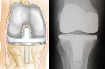


![HealthLine of Northern Colorado - September 2009 - [PDF Document]](https://static.fdocuments.in/doc/1200x630/568bf2481a28ab8933961a22/healthline-of-northern-colorado-september-2009.jpg?t=1696304570)