Pudendal Neuralgia
Trigeminal Neuralgia
Neuralgia, Postherpetic
Pudendal Nerve
Neuralgia
Facial Neuralgia
Trigeminal Nerve
Glossopharyngeal Nerve Diseases
Nerve Compression Syndromes
Urethra
Herpes Zoster Vaccine
Anal Canal
Microvascular Decompression Surgery
CT-guided percutaneous infiltration for the treatment of Alcock's neuralgia. (1/4)
The pudendal nerve may be strained either between the sacrospinous and sacrotuberous ligaments at the ischial spine level or within Alcock's canal. Alcock's neuralgia is a rare, painful condition caused by compression of the pudendal nerve within Alcock's canal (pudendal canal) which is an aponeurotic tunnel that cannot be stretched. Patients usually present with intense, unilateral pain involving anatomic areas along the pudendal nerve's root, genital, anal, and pelvic regions causing mobility impairment. A computed tomography (CT)--guided percutaneous infiltration of the pudendal nerve with a mixture of a local anesthetic and a long-acting corticosteroid is a safe and efficient method that reduces the pain caused by the neuralgia. Corticosteroids and local anesthetics interfere with the neurons, the encoding, and the processing of noxious stimuli; interrupt the pain-spasm cycle; and reduce inflammation. The injected glucocorticosteroid may take 3-5 days to reach its anti-inflammatory effect; therefore, the initial pain relief from the local anesthetic is followed by a baseline pain return and then secondary pain relief at 3-5 days. The procedure is performed under minimal or no anesthesia. In general, at discharge, a responsible person must accompany the patient and ensure a safe return home. Clinical evaluation is performed after 7-10 days. There are 2 types of potential complications that are associated with percutaneous steroid infiltrations: intra-operative (associated with needle placement) and post-operative (infection, bleeding and those associated with the injectate administration). In all cases that steroids were administered within therapeutic doses, no complications were noted. In conclusion, CT-guided percutaneous infiltration with a mixture of long-acting corticosteroid and local anesthetic seems to be a safe and efficient method for the treatment of Alcock's neuralgia. (+info)De novo pudendal neuropathy after TOT-O surgery for stress urinary incontinence. (2/4)
(+info)Chronic proctalgia and chronic pelvic pain syndromes: new etiologic insights and treatment options. (3/4)
(+info)Pudendal entrapment neuropathy: a rare complication of pelvic radiation therapy. (4/4)
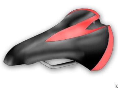
Pudendal nerve entrapment (PNE) is an uncommon cause of chronic pain. Pudendal nerve entrapment typically occurs when the pudendal nerve is fused to nearby anatomical structures or trapped between the sacrotuberous and sacrospinalis ligaments. Pudendal nerve entrapment can be caused by excessive bicycling, pregnancy, anatomic abnormalities, scarring due to surgery, or as a sequela of radiation therapy. Radiation-induced peripheral neuropathy is usually chronic, progressive, and often irreversible. Radiation-induced pudendal neuropathy is much less common than the more familiar brachial plexopathy secondary to radiation treatment for breast cancer. The prevalence of PNE, however, is increasing due to improved long-term cancer survival. Diagnosis of pudendal neuralgia is essentially clinical; no specific clinical signs or complementary tests are reliably confirmatory. A detailed pain history with correlative clinical examination is paramount for accurate diagnosis. Performance of a pudendal nerve block can serve as both a diagnostic and therapeutic tool. Utilization of various imaging studies, as well as the performance of an electrophysiological study with pudendal nerve motor latency testing, may yield valuable evidence in support of a pudendal neuralgia diagnosis. We present the case of a 59-year-old man with stage IV prostate cancer, referred to the pain clinic for chronic perineal and right sided pelvic pain. His pain began insidiously, approximately 2 months after undergoing radiation treatment and chemotherapy 3 years prior. He was ultimately diagnosed as having a right sided pudendal entrapment neuropathy. His pain was refractory to all conventional treatment modalities; therefore we decided to pursue neuromodulation via a dorsal column spinal cord stimulator implant. Below, we describe the decision making process for the diagnosis and treatment of his pudendal neuropathy. (+info)Pudendal Neuralgia is a chronic pain condition characterized by the irritation or damage to the pudendal nerve, which supplies sensation and innervation to the perineum, genital region, and lower rectum. The symptoms often include burning pain, numbness, tingling, or shooting pain in these areas, which can be worsened by sitting or certain movements. It is important to note that Pudendal Neuralgia is not the same as Pudendal Nerve Entrapment (PNE), although PNE can lead to Pudendal Neuralgia. The diagnosis of this condition typically involves a thorough physical examination, medical history, and sometimes specialized tests like nerve blocks or electromyography (EMG) studies.
Trigeminal neuralgia is a chronic pain condition that affects the trigeminal nerve, which is one of the largest nerves in the head. It carries sensations from the face to the brain.
Medically, trigeminal neuralgia is defined as a neuropathic disorder characterized by episodes of intense, stabbing, electric shock-like pain in the areas of the face supplied by the trigeminal nerve (the ophthalmic, maxillary, and mandibular divisions). The pain can be triggered by simple activities such as talking, eating, brushing teeth, or even touching the face lightly.
The condition is more common in women over 50, but it can occur at any age and in either gender. While the exact cause of trigeminal neuralgia is not always known, it can sometimes be related to pressure on the trigeminal nerve from a nearby blood vessel or other causes such as multiple sclerosis. Treatment typically involves medications, surgery, or a combination of both.
Postherpetic neuralgia (PHN) is a type of neuralgia, which is defined as pain in the distribution of a nerve or nerves. Specifically, PHN is a neuropathic pain condition that develops after an individual has had herpes zoster, also known as shingles. Shingles is caused by the reactivation of the varicella-zoster virus, which lies dormant in the nervous system following chickenpox infection.
PHN is characterized by persistent burning pain, often accompanied by sensory abnormalities such as numbness, tingling, or itching, in the area of the body where shingles occurred. The pain can be severe and debilitating, significantly impacting a person's quality of life. PHN primarily affects older adults and individuals with weakened immune systems.
The exact cause of PHN is not fully understood, but it is believed to result from damage to the affected nerves and their surrounding tissues during the shingles infection. This damage can lead to altered nerve function and increased sensitivity to stimuli, resulting in chronic pain. Treatment for PHN typically involves a combination of medications, such as antidepressants, anticonvulsants, or opioids, as well as topical treatments, physical therapy, and lifestyle modifications to help manage the pain and improve quality of life.
The Pudendal Nerve is a somatic nerve that carries sensory and motor fibers to the genital region in both males and females. It originates from the sacral plexus, specifically from nerves S2, S3, and S4. The pudendal nerve provides innervation to the skin of the perineum, labia majora/scrotum, and the lower portions of the vagina/penis. Additionally, it supplies motor function to the external anal and urethral sphincters, as well as to some muscles of the pelvic floor, such as the bulbospongiosus and ischiocavernosus muscles. The pudendal nerve plays a crucial role in sexual response and urinary and fecal continence.
Neuralgia is a type of pain that occurs along the pathway of a nerve, often caused by damage or irritation to the nerve. It is typically described as a sharp, stabbing, burning, or electric-shock like pain that can be severe and debilitating. Neuralgia can affect any nerve in the body, but it most commonly occurs in the facial area (trigeminal neuralgia) or in the nerves related to the spine (postherpetic neuralgia). The pain associated with neuralgia can be intermittent or constant and may be worsened by certain triggers such as touch, temperature changes, or movement. Treatment for neuralgia typically involves medications to manage pain, as well as other therapies such as nerve blocks, surgery, or lifestyle modifications.
Facial neuralgia is a general term that refers to painful conditions affecting the facial nerves. It is often used to describe two specific disorders: trigeminal neuralgia and glossopharyngeal neuralgia.
1. Trigeminal neuralgia (TN), also known as tic douloureux, is a chronic pain condition that affects the trigeminal nerve, one of the major nerves of the face. The trigeminal nerve is responsible for sensations in the face and motor functions such as biting and chewing. Trigeminal neuralgia causes intense, stabbing, electric shock-like pain in the areas of the face where the branches of the nerve are distributed: the lower jaw, upper jaw, and cheek. The pain usually affects one side of the face, is triggered by light touch or other stimuli, and can last from a few seconds to several minutes.
2. Glossopharyngeal neuralgia (GPN) is a similar but less common condition that involves the glossopharyngeal nerve, which is responsible for sensations in the throat, tongue, and ear on one side of the face. GPN causes sharp, stabbing pain in these areas, often triggered by swallowing, talking, or coughing.
Both trigeminal neuralgia and glossopharyngeal neuralgia can be debilitating and significantly impact a person's quality of life. The exact cause of these conditions is not always clear, but they are often associated with nerve compression by blood vessels or tumors, age-related changes in the nerves and blood vessels, multiple sclerosis, or other underlying medical conditions. Treatment options may include medications to manage pain, surgical procedures to decompress the affected nerves, or, in some cases, radiofrequency ablation or gamma knife radiosurgery to destroy a portion of the nerve and reduce pain signals.
The trigeminal nerve, also known as the fifth cranial nerve or CNV, is a paired nerve that carries both sensory and motor information. It has three major branches: ophthalmic (V1), maxillary (V2), and mandibular (V3). The ophthalmic branch provides sensation to the forehead, eyes, and upper portion of the nose; the maxillary branch supplies sensation to the lower eyelid, cheek, nasal cavity, and upper lip; and the mandibular branch is responsible for sensation in the lower lip, chin, and parts of the oral cavity, as well as motor function to the muscles involved in chewing. The trigeminal nerve plays a crucial role in sensations of touch, pain, temperature, and pressure in the face and mouth, and it also contributes to biting, chewing, and swallowing functions.
The glossopharyngeal nerve, also known as the ninth cranial nerve (CN IX), is primarily responsible for providing motor innervation to the stylopharyngeus muscle and sensory innervation to parts of the pharynx, middle ear, and posterior tongue. It also plays a role in the reflexive control of heart rate via the baroreceptors located in the carotid sinus.
Glossopharyngeal nerve diseases refer to conditions that affect the function of this nerve, leading to various symptoms. These diseases can be classified into two main categories: peripheral and central. Peripheral disorders are caused by damage or injury to the nerve itself, while central disorders result from problems in the brainstem where the glossopharyngeal nerve originates.
Some examples of glossopharyngeal nerve diseases include:
1. Glossopharyngeal neuralgia: A rare condition characterized by severe, stabbing pain in the throat, ear, or tongue, often triggered by swallowing or talking. This disorder may be caused by compression of the nerve by blood vessels or other structures.
2. Infections: Bacterial and viral infections can cause inflammation and damage to the glossopharyngeal nerve, leading to dysfunction. Examples include Lyme disease, herpes zoster (shingles), and meningitis.
3. Tumors: Benign or malignant growths in the head and neck region can compress and injure the glossopharyngeal nerve, resulting in symptoms related to its dysfunction.
4. Trauma: Direct trauma to the neck or skull base can damage the glossopharyngeal nerve, causing various deficits depending on the severity of the injury.
5. Neurological disorders: Conditions such as multiple sclerosis and stroke can affect the central connections of the glossopharyngeal nerve in the brainstem, leading to dysfunction.
6. Genetic conditions: Rare genetic disorders like Moersch-Woltman syndrome (also known as stiff person syndrome) can involve the glossopharyngeal nerve and cause symptoms related to its dysfunction.
Symptoms of glossopharyngeal nerve dysfunction may include difficulty swallowing, hoarseness, loss of taste on the back of the tongue, decreased sensation in the throat or ear, and pain in the neck, throat, or ear. Treatment for these conditions depends on the underlying cause and may involve medications, surgery, or other interventions to address the specific problem.
Nerve compression syndromes refer to a group of conditions characterized by the pressure or irritation of a peripheral nerve, causing various symptoms such as pain, numbness, tingling, and weakness in the affected area. This compression can occur due to several reasons, including injury, repetitive motion, bone spurs, tumors, or swelling. Common examples of nerve compression syndromes include carpal tunnel syndrome, cubital tunnel syndrome, radial nerve compression, and ulnar nerve entrapment at the wrist or elbow. Treatment options may include physical therapy, splinting, medications, injections, or surgery, depending on the severity and underlying cause of the condition.
Rhizotomy is a surgical procedure where the root(s) of a nerve are cut. It is often used to treat chronic pain, spasticity, or other neurological symptoms that have not responded to other treatments. In some cases, only a portion of the nerve root may be severed (selective rhizotomy), while in others the entire root may be cut (root transaction). The specific nerves targeted during a rhizotomy depend on the individual patient's condition and symptoms.
This procedure is typically performed by a neurosurgeon, and it can be done through an open surgical approach or using minimally invasive techniques such as endoscopic or percutaneous approaches. After the surgery, patients may require physical therapy to help regain strength and mobility in the affected area. Potential risks of rhizotomy include numbness, weakness, and loss of reflexes in the areas served by the severed nerves.
The urethra is the tube that carries urine from the bladder out of the body. In males, it also serves as the conduit for semen during ejaculation. The male urethra is longer than the female urethra and is divided into sections: the prostatic, membranous, and spongy (or penile) urethra. The female urethra extends from the bladder to the external urethral orifice, which is located just above the vaginal opening.
The Herpes Zoster vaccine, also known as the shingles vaccine, is a preventive measure against the reactivation of the varicella-zoster virus (VZV) in individuals who have previously had chickenpox. The vaccine contains a live but weakened form of VZV that boosts the immune system's ability to recognize and fight off the virus, thereby reducing the risk of developing shingles and its complications. It is typically administered as a single dose for people aged 50 and older, or as a two-dose series for those aged 19 and older who have weakened immune systems.
The anal canal is the terminal portion of the digestive tract, located between the rectum and the anus. It is a short tube-like structure that is about 1 to 1.5 inches long in adults. The main function of the anal canal is to provide a seal for the elimination of feces from the body while also preventing the leakage of intestinal contents.
The inner lining of the anal canal is called the mucosa, which is kept moist by the production of mucus. The walls of the anal canal contain specialized muscles that help control the passage of stool during bowel movements. These muscles include the internal and external sphincters, which work together to maintain continence and allow for the voluntary release of feces.
The anal canal is an important part of the digestive system and plays a critical role in maintaining bowel function and overall health.
Microvascular decompression surgery (MVD) is a surgical procedure used to alleviate the symptoms of certain neurological conditions, such as trigeminal neuralgia and hemifacial spasm. The primary goal of MVD is to relieve pressure on the affected cranial nerve by placing a small pad or sponge between the nerve and the blood vessel that is causing compression. This procedure is typically performed under a microscope, hence the term "microvascular."
During the surgery, the neurosurgeon makes an incision behind the ear and creates a small opening in the skull (a craniotomy) to access the brain. The surgeon then identifies the affected nerve and the blood vessel that is compressing it. Using specialized instruments under the microscope, the surgeon carefully separates the blood vessel from the nerve and places a tiny pad or sponge between them to prevent further compression.
The benefits of MVD include its high success rate in relieving symptoms, minimal impact on surrounding brain tissue, and lower risk of complications compared to other surgical options for treating these conditions. However, as with any surgery, there are potential risks and complications associated with MVD, including infection, bleeding, cerebrospinal fluid leakage, facial numbness, hearing loss, balance problems, and very rarely, stroke or death.
It is essential to consult a qualified neurosurgeon for a thorough evaluation and discussion of the risks and benefits of microvascular decompression surgery before making a treatment decision.
The lumbosacral plexus is a complex network of nerves that arises from the lower part of the spinal cord, specifically the lumbar (L1-L5) and sacral (S1-S4) roots. This plexus is responsible for providing innervation to the lower extremities, including the legs, feet, and some parts of the abdomen and pelvis.
The lumbosacral plexus can be divided into several major branches:
1. The femoral nerve: It arises from the L2-L4 roots and supplies motor innervation to the muscles in the anterior compartment of the thigh, as well as sensation to the anterior and medial aspects of the leg and thigh.
2. The obturator nerve: It originates from the L2-L4 roots and provides motor innervation to the adductor muscles of the thigh and sensation to the inner aspect of the thigh.
3. The sciatic nerve: This is the largest nerve in the body, formed by the union of the tibial and common fibular (peroneal) nerves. It arises from the L4-S3 roots and supplies motor innervation to the muscles of the lower leg and foot, as well as sensation to the posterior aspect of the leg and foot.
4. The pudendal nerve: It originates from the S2-S4 roots and is responsible for providing motor innervation to the pelvic floor muscles and sensory innervation to the genital region.
5. Other smaller nerves, such as the ilioinguinal, iliohypogastric, and genitofemoral nerves, also arise from the lumbosacral plexus and supply sensation to various regions in the lower abdomen and pelvis.
Damage or injury to the lumbosacral plexus can result in significant neurological deficits, including muscle weakness, numbness, and pain in the lower extremities.
 Pudendal nerve entrapment