Pulmonary Emphysema
Pancreatic Elastase
alpha 1-Antitrypsin Deficiency
alpha 1-Antitrypsin
Lung
Matrix Metalloproteinase 12
Pulmonary Alveoli
Mediastinal Emphysema
Respiratory Function Tests
Smoke
Helium
Pulmonary Disease, Chronic Obstructive
Vasculitis, Leukocytoclastic, Cutaneous
Elastin
Panniculitis
Tomography, X-Ray Computed
Bronchoalveolar Lavage Fluid
Forced Expiratory Volume
Pulmonary Fibrosis
Lung Compliance
Disease Models, Animal
Bronchi
Mice, Inbred C57BL
Fragile lung in the Marfan syndrome. (1/1021)
Two cases of the Marfan syndrome presented with spontaneous pneumothorax. Both had chest radiographs showing bilateral bullae in the upper lung zones and pulmonary function tests consistent with mild emphysema. There were dereases in forced expiratory flow rates at low lung volumes, carbon monoxide transfer factor, and lung elastic recoil. It is suggested that pneumothorax and bullous emphysema in this syndrome are caused by a weakness in the pulmonary connective tissue framework. (+info)Detection of anti-cytokeratin 8 antibody in the serum of patients with cryptogenic fibrosing alveolitis and pulmonary fibrosis associated with collagen vascular disorders. (2/1021)
BACKGROUND: It has been suggested that the humoral immune system plays a role in the pathogenesis of cryptogenic fibrosing alveolitis (CFA). Although circulating autoantibodies to lung protein(s) have been suggested, none of the lung proteins have been characterised. The purpose of this study was to determine the antigen to which the serum from patients with pulmonary fibrosis reacted. METHODS: The anti-A549 cell antibody was characterised in a patient with CFA using Western immunoblotting and immunohistochemical staining of A549 cells. As we identified that one of the antibodies against A549 cells was anti-cytokeratin 8, the expression of mRNA of cytokeratin 8 in A549 cells was evaluated. In addition, we attempted to establish an enzyme linked immunosorbent assay to measure the levels of anti-cytokeratin 8 antibody in the serum of patients with CFA and pulmonary fibrosis associated with collagen vascular disorders (PF-CVD). RESULTS: Initially two anti-A549 cell antibodies were detected in the serum of patients with pulmonary fibrosis, one of which was characterised as anticytokeratin 8 antibody by Western immunoblotting. We were able to establish an ELISA to measure anti-cytokeratin 8 antibody and found significantly higher levels in patients with CFA and PF-CVD than in normal volunteers, patients with sarcoidosis, pneumonia, and pulmonary emphysema. CONCLUSIONS: One of the anti-A549 cell antibodies in the serum of patients with CFA was against cytokeratin 8. The serum levels of anti-cytokeratin 8 antibody were increased in patients with CFA and PF-CVD. These results suggest that anticytokeratin 8 antibody may be involved in the process of lung injury in pulmonary fibrosis. (+info)Improvements in thoracic movement following lung volume reduction surgery in patients with severe emphysema. (3/1021)
In twelve patients with severe emphysema who underwent lung volume reduction surgery (LVRS), we assessed the results of dyspnea scale, pulmonary function, 6-minute walk distance (6MD), and thoracic movement prior to and 6 months following LVRS. Postoperatively, forced expiratory volume (FEV1), maximum inspiratory mouth pressures (MIP), maximum expiratory mouth pressures (MEP), maximum voluntary ventilation (MVV), diffusing capacity for carbon monoxide (DLCO), partial pressure of oxygen (PaO2) and 6MD were significantly increased with the decrease in dyspnea scale and lung hyperinflation. Thoracic movement, as assessed by the bilateral lung area ratio of the mid-sagittal dimension of dynamic magnetic resonance imaging (MRI) at full inspiration to that at full expiration, was significantly increased. The improvement in thoracic movement was significantly correlated with the increases in FEV1, MVV, and MIP, and with the decrease in residual volume (RV), and with the improvement in the dyspnea scale. These findings suggest that LVRS is an effective procedure for improving not only the airflow limitation and gas exchange but also the thoracic movement in severe emphysema, and these improvements may contribute to an increase in exercise performance and relief of dyspnea. (+info)Physiological basis of improvement after lung volume reduction surgery for severe emphysema: where are we? (4/1021)
Lung volume reduction surgery has become an accepted therapeutic option to relieve the symptoms of selected patients with severe emphysema. In a majority of these patients, it causes objective as well as subjective functional improvement. A proper understanding of the physiological determinants underlying these beneficial effects appears very important in order to better select patients for the procedure that is currently largely carried out on an empirical basis. Lung volume reduction surgery has two distinct effects. Firstly, it causes an increased elastic recoil, which at least partially explains the enhanced maximal expiratory flow. Secondly, it is associated with a reduction of hyperinflation which allows for an increase in global inspiratory muscle strength and in diaphragmatic contribution to tidal volume as well as a decrease in the inspiratory elastic load imposed by the chest wall. Taken together, these effects result in a reduced work of breathing and in an enhanced maximal ventilation which both contribute to the increased exercise capacity and reduced dyspnoea after surgery. The improved lung recoil and the reduced hyperinflation after volume reduction surgery were the primary postulates upon which the usual selection criteria for the procedure were based. It is now likely that these are correct. Nevertheless, some patients do not benefit from lung volume reduction surgery and the current literature does not allow for a refinement of the selection process from a physiological point of view. The exact mechanisms underlying the improvement in lung recoil, lung mechanics, and respiratory muscle function remain incompletely understood. Moreover, the effects of lung volume reduction surgery on gas exchange and pulmonary haemodynamics still need to be more fully investigated. An analysis of the characteristics of patients who do not benefit from the procedure and the development of an animal model for lung volume reduction surgery would probably help address these important issues. (+info)Subjective differentiation of normal and pathological bronchi on thin-section CT: impact of observer training. (5/1021)
The effect of observer training on sensitivity, specificity and interobserver agreement in the differentiation between normal and pathological bronchi on computed tomography (CT) was studied. The wall thickness of bronchi with normal walls and with pathologically thickened walls were subjectively scored by three independent observers before and after a training period of 2 weeks. Sensitivity, specificity and interobserver agreement were calculated for reading sessions before and after training. Increase and decrease in agreement after training were determined. There was a statistically significant difference (p=0.001) between objectively measured wall thickness of normal and pathological bronchi, both for reference bronchi and for bronchi used for reading sessions. While training increased interobserver agreement, it had no effect on sensitivity (0.46 versus 0.44 after training) and specificity (0.71 versus 0.72 after training) in detecting pathological bronchi. Increased agreement after training was significantly (p=0.001) more frequent than decreased agreement. There is a discrepancy between the effect of training on interobserver agreement and on sensitivity and specificity in the subjective differentiation between normal and pathological bronchi. Interobserver agreement alone is not a reliable indicator of a beneficial effect of training in the evaluation of this parameter. (+info)Outcome of asthma: longitudinal changes in lung function. (6/1021)
Current knowledge about factors determining outcome of asthma is limited, but observations over the last few decades suggest that active asthma has a negative impact on the longitudinal changes in lung function. This review aims to give an overview of the present knowledge concerning longitudinal changes in lung function, including clinical markers for distinctly poor outcome with regard to lung function, in children and adults suffering from asthma. The majority of patients with asthma have a good prognosis. However, some patients with asthma, especially those with more severe disease, are at risk of impaired growth of lung function during childhood, a lower maximally attained level of lung function and excessive decline in lung function in adulthood, which may lead to life-threatening lung function impairment. Clinical markers of poorly controlled airway inflammation appear to have a negative impact on the longitudinal changes in lung function, and disease progression to nonreversible airflow obstruction may be observed in a minority of patients with asthma. Early intervention with anti-inflammatory therapy may improve the short-term outcome of asthma, but long-term controlled studies are clearly needed in order to verify whether or not treatment, especially with inhaled corticosteroids, according to the current international guidelines alters the natural history of asthma, i.e. disease progression with regard to changes in lung function and possible development of nonreversible airflow obstruction. (+info)Surgical aspects and techniques of lung volume reduction surgery for severe emphysema. (7/1021)
Lung volume reduction surgery (LVRS) has become an accepted procedure for palliative treatment of diffuse, nonbullous emphysema. Single or multiple peripheral segmental wedge resections of the most destroyed areas of the lungs are performed with the use of stapling devices, in order to decrease hyperinflation and restore diaphragmatic function. Median sternotomy, videoendoscopy or anterior muscle sparing thoracotomies have been used as surgical approaches. The functional improvement after bilateral resections exceed those after a unilateral approach. LVRS has demonstrated its potential as an alternative to transplantation, and with growing experience, the indications for the procedure have been widened. In selected patients with peripheral lung cancer who have been considered unsuitable for a surgical resection, the combination of both tumour resection and LVRS has successfully been performed. In contrast to LVRS, laser surgery of the emphysematous lung has been abandoned in most institutions. (+info)Emphysematous lesions, inflammation, and fibrosis in the lungs of transgenic mice overexpressing platelet-derived growth factor. (8/1021)
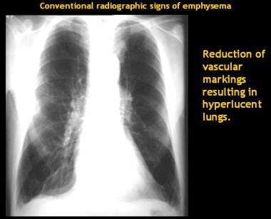
Because of its expression pattern and its potent effects on mesenchymal cells, platelet-derived growth factor (PDGF) has been implicated as an important factor in epithelial-mesenchymal cell interactions during normal lung development and in the pathogenesis of fibrotic lung disease. To further explore the role of PDGF in these processes, we have developed transgenic mice that express the PDGF-B gene from the lung-specific surfactant protein C (SPC) promoter. Adult SPC-PDGFB transgenic mice exhibited lung pathology characterized by enlarged airspaces, inflammation, and fibrosis. Emphysematous changes frequently occurred throughout the lung, but inflammation and fibrotic lesions were usually confined to focal areas. The severity of this phenotype varied significantly among individual mice within the same SPC-PDGFB transgenic lineage. A pathology similar to that observed in adult mice was noted in lungs from transgenic mice as young as 1 week of age. Neonatal transgenic mice exhibited enlarged saccules and thickened primary septa. Results of these studies indicated that overexpression of PDGF-B induced distinct abnormalities in the developing and adult lung and led to a complex phenotype that encompassed aspects of both emphysema and fibrotic lung disease. (+info)Pulmonary emphysema is a chronic respiratory disease characterized by abnormal, permanent enlargement of the airspaces distal to the terminal bronchioles, accompanied by destruction of their walls and without obvious fibrosis. This results in loss of elastic recoil, which leads to trappling of air within the lungs and difficulty exhaling. It is often caused by cigarette smoking or long-term exposure to harmful pollutants. The disease is part of a group of conditions known as chronic obstructive pulmonary disease (COPD), which also includes chronic bronchitis.
Emphysema is a chronic respiratory disease characterized by abnormal, permanent enlargement of the airspaces called alveoli in the lungs, accompanied by destruction of their walls. This results in loss of elasticity and decreased gas exchange efficiency, causing shortness of breath and coughing. It is often caused by smoking or exposure to harmful pollutants. The damage to the lungs is irreversible, but quitting smoking and using medications can help alleviate symptoms and slow disease progression.
Pancreatic elastase is a type of elastase that is specifically produced by the pancreas. It is an enzyme that helps in digesting proteins found in the food we eat. Pancreatic elastase breaks down elastin, a protein that provides elasticity to tissues and organs in the body.
In clinical practice, pancreatic elastase is often measured in stool samples as a diagnostic tool to assess exocrine pancreatic function. Low levels of pancreatic elastase in stool may indicate malabsorption or exocrine pancreatic insufficiency, which can be caused by various conditions such as chronic pancreatitis, cystic fibrosis, or pancreatic cancer.
Alpha 1-Antitrypsin (AAT) deficiency is a genetic disorder that results from insufficient levels of the protective protein AAT in the blood and lungs. This protein is produced by the liver and helps to protect the lungs from damage caused by inflammation and the action of enzymes, such as neutrophil elastase, that are released during the immune response.
In people with AAT deficiency, the lack of adequate AAT levels leads to an uncontrolled increase in neutrophil elastase activity, which can cause damage to lung tissue and result in emphysema, a condition characterized by shortness of breath, coughing, and wheezing. Additionally, some individuals with AAT deficiency may develop liver disease due to the accumulation of abnormal AAT proteins in liver cells.
There are different variants or genotypes associated with AAT deficiency, with the most common and severe form being the PiZZ genotype. This variant is caused by mutations in the SERPINA1 gene, which encodes for the AAT protein. Individuals who inherit two copies of this mutated gene (one from each parent) will have very low levels of AAT in their blood and are at increased risk of developing emphysema and liver disease.
Diagnosis of AAT deficiency typically involves measuring AAT levels in the blood and performing genetic testing to identify specific variants of the SERPINA1 gene. Treatment may include lifestyle modifications, such as smoking cessation, bronchodilators, and corticosteroids to manage lung symptoms, as well as augmentation therapy with intravenous infusions of AAT protein to help slow disease progression in individuals with severe deficiency. Liver transplantation may be considered for those with advanced liver disease.
Subcutaneous emphysema is a medical condition where air or gas collects in the subcutaneous tissue, which lies beneath the skin and above the muscle layer. This tissue covers the entire body, but the collection of air usually occurs in the chest wall, neck, or face. The accumulation of air can cause swelling, crepitus (a crackling or crunching sound when touched), and tightness in the affected area. Subcutaneous emphysema is often associated with underlying conditions such as trauma, pulmonary disease, or certain medical procedures that result in air leaks from the lungs or other structures into the subcutaneous tissue. It can be a serious condition if left untreated, as it may lead to complications like mediastinal emphysema or tension pneumothorax. Immediate medical attention is necessary for proper diagnosis and treatment.
Alpha 1-antitrypsin (AAT, or α1-antiproteinase, A1AP) is a protein that is primarily produced by the liver and released into the bloodstream. It belongs to a group of proteins called serine protease inhibitors, which help regulate inflammation and protect tissues from damage caused by enzymes involved in the immune response.
Alpha 1-antitrypsin is particularly important for protecting the lungs from damage caused by neutrophil elastase, an enzyme released by white blood cells called neutrophils during inflammation. In the lungs, AAT binds to and inhibits neutrophil elastase, preventing it from degrading the extracellular matrix and damaging lung tissue.
Deficiency in alpha 1-antitrypsin can lead to chronic obstructive pulmonary disease (COPD) and liver disease. The most common cause of AAT deficiency is a genetic mutation that results in abnormal folding and accumulation of the protein within liver cells, leading to reduced levels of functional AAT in the bloodstream. This condition is called alpha 1-antitrypsin deficiency (AATD) and can be inherited in an autosomal codominant manner. Individuals with severe AATD may require augmentation therapy with intravenous infusions of purified human AAT to help prevent lung damage.
A lung is a pair of spongy, elastic organs in the chest that work together to enable breathing. They are responsible for taking in oxygen and expelling carbon dioxide through the process of respiration. The left lung has two lobes, while the right lung has three lobes. The lungs are protected by the ribcage and are covered by a double-layered membrane called the pleura. The trachea divides into two bronchi, which further divide into smaller bronchioles, leading to millions of tiny air sacs called alveoli, where the exchange of gases occurs.
Matrix metalloproteinase 12 (MMP-12) is a type of enzyme that belongs to the matrix metalloproteinase (MMP) family. MMPs are involved in the breakdown and remodeling of extracellular matrices, which are the structures that provide support and organization to cells in tissues and organs.
MMP-12 is also known as macrophage elastase because it is primarily produced by macrophages, a type of white blood cell that plays a key role in the immune system. MMP-12 is capable of degrading various components of the extracellular matrix, including elastin, a protein that provides elasticity to tissues such as lungs, arteries, and skin.
MMP-12 has been implicated in several physiological and pathological processes, including tissue remodeling, wound healing, inflammation, and cancer. Dysregulation of MMP-12 activity has been associated with various diseases, such as chronic obstructive pulmonary disease (COPD), atherosclerosis, and tumor metastasis.
Pulmonary alveoli, also known as air sacs, are tiny clusters of air-filled pouches located at the end of the bronchioles in the lungs. They play a crucial role in the process of gas exchange during respiration. The thin walls of the alveoli, called alveolar membranes, allow oxygen from inhaled air to pass into the bloodstream and carbon dioxide from the bloodstream to pass into the alveoli to be exhaled out of the body. This vital function enables the lungs to supply oxygen-rich blood to the rest of the body and remove waste products like carbon dioxide.
Mediastinal emphysema is a medical condition characterized by the presence of air or gas within the mediastinum, which is the central compartment of the thorax that contains the heart, esophagus, trachea, bronchi, thymus gland, and other associated structures.
In mediastinal emphysema, the air accumulates in the mediastinal tissues and spaces, leading to their abnormal distention or swelling. This condition can result from various causes, including:
* Pulmonary trauma or barotrauma (e.g., mechanical ventilation, scuba diving)
* Infections that cause gas-forming organisms (e.g., pneumomediastinum)
* Air leakage from the lungs or airways (e.g., bronchial rupture, esophageal perforation)
* Certain medical procedures (e.g., mediastinoscopy, tracheostomy)
Mediastinal emphysema can cause symptoms such as chest pain, cough, difficulty breathing, and swallowing problems. In severe cases, it may lead to life-threatening complications, including tension pneumothorax or mediastinitis. Treatment depends on the underlying cause and severity of the condition.
Respiratory Function Tests (RFTs) are a group of medical tests that measure how well your lungs take in and exhale air, and how well they transfer oxygen and carbon dioxide into and out of your blood. They can help diagnose certain lung disorders, measure the severity of lung disease, and monitor response to treatment.
RFTs include several types of tests, such as:
1. Spirometry: This test measures how much air you can exhale and how quickly you can do it. It's often used to diagnose and monitor conditions like asthma, chronic obstructive pulmonary disease (COPD), and other lung diseases.
2. Lung volume testing: This test measures the total amount of air in your lungs. It can help diagnose restrictive lung diseases, such as pulmonary fibrosis or sarcoidosis.
3. Diffusion capacity testing: This test measures how well oxygen moves from your lungs into your bloodstream. It's often used to diagnose and monitor conditions like pulmonary fibrosis, interstitial lung disease, and other lung diseases that affect the ability of the lungs to transfer oxygen to the blood.
4. Bronchoprovocation testing: This test involves inhaling a substance that can cause your airways to narrow, such as methacholine or histamine. It's often used to diagnose and monitor asthma.
5. Exercise stress testing: This test measures how well your lungs and heart work together during exercise. It's often used to diagnose lung or heart disease.
Overall, Respiratory Function Tests are an important tool for diagnosing and managing a wide range of lung conditions.
'Smoke' is not typically defined in a medical context, but it can be described as a mixture of small particles and gases that are released when something burns. Smoke can be composed of various components including carbon monoxide, particulate matter, volatile organic compounds (VOCs), benzene, toluene, styrene, and polycyclic aromatic hydrocarbons (PAHs). Exposure to smoke can cause a range of health problems, including respiratory symptoms, cardiovascular disease, and cancer.
In the medical field, exposure to smoke is often referred to as "secondhand smoke" or "passive smoking" when someone breathes in smoke from another person's cigarette, cigar, or pipe. This type of exposure can be just as harmful as smoking itself and has been linked to a range of health problems, including respiratory infections, asthma, lung cancer, and heart disease.
Helium is not a medical term, but it's a chemical element with symbol He and atomic number 2. It's a colorless, odorless, tasteless, non-toxic, inert, monatomic gas that heads the noble gases section of the periodic table. In medicine, helium is sometimes used in medical settings for its unique properties, such as being less dense than air, which can help improve the delivery of oxygen to patients with respiratory conditions. For example, heliox, a mixture of helium and oxygen, may be used to reduce the work of breathing in patients with conditions like chronic obstructive pulmonary disease (COPD) or asthma. Additionally, helium is also used in cryogenic medical equipment and in magnetic resonance imaging (MRI) machines to cool the superconducting magnets.
Chronic obstructive pulmonary disease (COPD) is a progressive lung disease characterized by the persistent obstruction of airflow in and out of the lungs. This obstruction is usually caused by two primary conditions: chronic bronchitis and emphysema. Chronic bronchitis involves inflammation and narrowing of the airways, leading to excessive mucus production and coughing. Emphysema is a condition where the alveoli (air sacs) in the lungs are damaged, resulting in decreased gas exchange and shortness of breath.
The main symptoms of COPD include progressive shortness of breath, chronic cough, chest tightness, wheezing, and excessive mucus production. The disease is often associated with exposure to harmful particles or gases, such as cigarette smoke, air pollution, or occupational dusts and chemicals. While there is no cure for COPD, treatments can help alleviate symptoms, improve quality of life, and slow the progression of the disease. These treatments may include bronchodilators, corticosteroids, combination inhalers, pulmonary rehabilitation, and, in severe cases, oxygen therapy or lung transplantation.
A pneumonectomy is a surgical procedure in which an entire lung is removed. This type of surgery is typically performed as a treatment for certain types of lung cancer, although it may also be used to treat other conditions such as severe damage or infection in the lung that does not respond to other treatments. The surgery requires general anesthesia and can be quite complex, with potential risks including bleeding, infection, pneumonia, and air leaks. Recovery from a pneumonectomy can take several weeks, and patients may require ongoing rehabilitation to regain strength and mobility.
Leukocytoclastic vasculitis, cutaneous is a type of vasculitis that is limited to the skin. Vasculitis refers to inflammation of the blood vessels, which can cause damage to the vessel walls and impair blood flow to various tissues in the body. In leukocytoclastic vasculitis, the small blood vessels (capillaries and venules) in the skin become inflamed, leading to damage and destruction of the vessel walls.
The term "leukocytoclastic" refers to the presence of nuclear debris from white blood cells (leukocytes) that have been destroyed within the affected blood vessels. This type of vasculitis is often associated with the deposition of immune complexes (formed by the interaction between antibodies and antigens) in the walls of the blood vessels, which triggers an inflammatory response.
Cutaneous leukocytoclastic vasculitis typically presents as palpable purpura (small to large, raised, purple or red spots on the skin), usually located on the lower extremities, but can also affect other areas of the body. Other symptoms may include burning or itching sensations in the affected area, and in some cases, ulcers or necrosis (tissue death) may occur.
The diagnosis of cutaneous leukocytoclastic vasculitis is typically made based on clinical presentation, laboratory tests, and histopathological examination of a skin biopsy specimen. Treatment usually involves addressing any underlying causes or triggers, as well as managing symptoms with medications such as corticosteroids or immunosuppressive agents.
Elastin is a protein that provides elasticity to tissues and organs, allowing them to resume their shape after stretching or contracting. It is a major component of the extracellular matrix in many tissues, including the skin, lungs, blood vessels, and ligaments. Elastin fibers can stretch up to 1.5 times their original length and then return to their original shape due to the unique properties of this protein. The elastin molecule is made up of cross-linked chains of the protein tropoelastin, which are produced by cells called fibroblasts and then assembled into larger elastin fibers by enzymes called lysyl oxidases. Elastin has a very long half-life, with some estimates suggesting that it can remain in the body for up to 70 years or more.
Panniculitis is a medical term that refers to inflammation of the subcutaneous fat, or the layer of fat located just beneath the skin. This condition can affect people of all ages and genders, although it is more commonly seen in middle-aged women. The inflammation can be caused by a variety of factors, including infections, autoimmune disorders, trauma, and medications.
The symptoms of panniculitis may include:
* Red, painful lumps or nodules under the skin
* Skin lesions that may be tender, warm, or bruised
* Swelling and redness in the affected area
* Fever, fatigue, and malaise (a general feeling of illness)
The diagnosis of panniculitis typically involves a physical examination, medical history, and sometimes a biopsy of the affected tissue. Treatment depends on the underlying cause of the inflammation and may include antibiotics, anti-inflammatory medications, or other therapies. In severe cases, hospitalization may be necessary to manage symptoms and prevent complications.
Smoking is not a medical condition, but it's a significant health risk behavior. Here is the definition from a public health perspective:
Smoking is the act of inhaling and exhaling the smoke of burning tobacco that is commonly consumed through cigarettes, pipes, and cigars. The smoke contains over 7,000 chemicals, including nicotine, tar, carbon monoxide, and numerous toxic and carcinogenic substances. These toxins contribute to a wide range of diseases and health conditions, such as lung cancer, heart disease, stroke, chronic obstructive pulmonary disease (COPD), and various other cancers, as well as adverse reproductive outcomes and negative impacts on the developing fetus during pregnancy. Smoking is highly addictive due to the nicotine content, which makes quitting smoking a significant challenge for many individuals.
X-ray computed tomography (CT or CAT scan) is a medical imaging method that uses computer-processed combinations of many X-ray images taken from different angles to produce cross-sectional (tomographic) images (virtual "slices") of the body. These cross-sectional images can then be used to display detailed internal views of organs, bones, and soft tissues in the body.
The term "computed tomography" is used instead of "CT scan" or "CAT scan" because the machines take a series of X-ray measurements from different angles around the body and then use a computer to process these data to create detailed images of internal structures within the body.
CT scanning is a noninvasive, painless medical test that helps physicians diagnose and treat medical conditions. CT imaging provides detailed information about many types of tissue including lung, bone, soft tissue and blood vessels. CT examinations can be performed on every part of the body for a variety of reasons including diagnosis, surgical planning, and monitoring of therapeutic responses.
In computed tomography (CT), an X-ray source and detector rotate around the patient, measuring the X-ray attenuation at many different angles. A computer uses this data to construct a cross-sectional image by the process of reconstruction. This technique is called "tomography". The term "computed" refers to the use of a computer to reconstruct the images.
CT has become an important tool in medical imaging and diagnosis, allowing radiologists and other physicians to view detailed internal images of the body. It can help identify many different medical conditions including cancer, heart disease, lung nodules, liver tumors, and internal injuries from trauma. CT is also commonly used for guiding biopsies and other minimally invasive procedures.
In summary, X-ray computed tomography (CT or CAT scan) is a medical imaging technique that uses computer-processed combinations of many X-ray images taken from different angles to produce cross-sectional images of the body. It provides detailed internal views of organs, bones, and soft tissues in the body, allowing physicians to diagnose and treat medical conditions.
Bronchoalveolar lavage (BAL) fluid is a type of clinical specimen obtained through a procedure called bronchoalveolar lavage. This procedure involves inserting a bronchoscope into the lungs and instilling a small amount of saline solution into a specific area of the lung, then gently aspirating the fluid back out. The fluid that is recovered is called bronchoalveolar lavage fluid.
BAL fluid contains cells and other substances that are present in the lower respiratory tract, including the alveoli (the tiny air sacs where gas exchange occurs). By analyzing BAL fluid, doctors can diagnose various lung conditions, such as pneumonia, interstitial lung disease, and lung cancer. They can also monitor the effectiveness of treatments for these conditions by comparing the composition of BAL fluid before and after treatment.
BAL fluid is typically analyzed for its cellular content, including the number and type of white blood cells present, as well as for the presence of bacteria, viruses, or other microorganisms. The fluid may also be tested for various proteins, enzymes, and other biomarkers that can provide additional information about lung health and disease.
Forced Expiratory Volume (FEV) is a medical term used to describe the volume of air that can be forcefully exhaled from the lungs in one second. It is often measured during pulmonary function testing to assess lung function and diagnose conditions such as chronic obstructive pulmonary disease (COPD) or asthma.
FEV is typically expressed as a percentage of the Forced Vital Capacity (FVC), which is the total volume of air that can be exhaled from the lungs after taking a deep breath in. The ratio of FEV to FVC is used to determine whether there is obstruction in the airways, with a lower ratio indicating more severe obstruction.
There are different types of FEV measurements, including FEV1 (the volume of air exhaled in one second), FEV25-75 (the average volume of air exhaled during the middle 50% of the FVC maneuver), and FEV0.5 (the volume of air exhaled in half a second). These measurements can provide additional information about lung function and help guide treatment decisions.
Pulmonary fibrosis is a specific type of lung disease that results from the thickening and scarring of the lung tissues, particularly those in the alveoli (air sacs) and interstitium (the space around the air sacs). This scarring makes it harder for the lungs to properly expand and transfer oxygen into the bloodstream, leading to symptoms such as shortness of breath, coughing, fatigue, and eventually respiratory failure. The exact cause of pulmonary fibrosis can vary, with some cases being idiopathic (without a known cause) or related to environmental factors, medications, medical conditions, or genetic predisposition.
Lung compliance is a measure of the ease with which the lungs expand and is defined as the change in lung volume for a given change in transpulmonary pressure. It is often expressed in units of liters per centimeter of water (L/cm H2O). A higher compliance indicates that the lungs are more easily distensible, while a lower compliance suggests that the lungs are stiffer and require more force to expand. Lung compliance can be affected by various conditions such as pulmonary fibrosis, pneumonia, acute respiratory distress syndrome (ARDS), and chronic obstructive pulmonary disease (COPD).
Pneumonia is an infection or inflammation of the alveoli (tiny air sacs) in one or both lungs. It's often caused by bacteria, viruses, or fungi. Accumulated pus and fluid in these air sacs make it difficult to breathe, which can lead to coughing, chest pain, fever, and difficulty breathing. The severity of symptoms can vary from mild to life-threatening, depending on the underlying cause, the patient's overall health, and age. Pneumonia is typically diagnosed through a combination of physical examination, medical history, and diagnostic tests such as chest X-rays or blood tests. Treatment usually involves antibiotics for bacterial pneumonia, antivirals for viral pneumonia, and supportive care like oxygen therapy, hydration, and rest.
Animal disease models are specialized animals, typically rodents such as mice or rats, that have been genetically engineered or exposed to certain conditions to develop symptoms and physiological changes similar to those seen in human diseases. These models are used in medical research to study the pathophysiology of diseases, identify potential therapeutic targets, test drug efficacy and safety, and understand disease mechanisms.
The genetic modifications can include knockout or knock-in mutations, transgenic expression of specific genes, or RNA interference techniques. The animals may also be exposed to environmental factors such as chemicals, radiation, or infectious agents to induce the disease state.
Examples of animal disease models include:
1. Mouse models of cancer: Genetically engineered mice that develop various types of tumors, allowing researchers to study cancer initiation, progression, and metastasis.
2. Alzheimer's disease models: Transgenic mice expressing mutant human genes associated with Alzheimer's disease, which exhibit amyloid plaque formation and cognitive decline.
3. Diabetes models: Obese and diabetic mouse strains like the NOD (non-obese diabetic) or db/db mice, used to study the development of type 1 and type 2 diabetes, respectively.
4. Cardiovascular disease models: Atherosclerosis-prone mice, such as ApoE-deficient or LDLR-deficient mice, that develop plaque buildup in their arteries when fed a high-fat diet.
5. Inflammatory bowel disease models: Mice with genetic mutations affecting intestinal barrier function and immune response, such as IL-10 knockout or SAMP1/YitFc mice, which develop colitis.
Animal disease models are essential tools in preclinical research, but it is important to recognize their limitations. Differences between species can affect the translatability of results from animal studies to human patients. Therefore, researchers must carefully consider the choice of model and interpret findings cautiously when applying them to human diseases.
Lung diseases refer to a broad category of disorders that affect the lungs and other structures within the respiratory system. These diseases can impair lung function, leading to symptoms such as coughing, shortness of breath, chest pain, and wheezing. They can be categorized into several types based on the underlying cause and nature of the disease process. Some common examples include:
1. Obstructive lung diseases: These are characterized by narrowing or blockage of the airways, making it difficult to breathe out. Examples include chronic obstructive pulmonary disease (COPD), asthma, bronchiectasis, and cystic fibrosis.
2. Restrictive lung diseases: These involve stiffening or scarring of the lungs, which reduces their ability to expand and take in air. Examples include idiopathic pulmonary fibrosis, sarcoidosis, and asbestosis.
3. Infectious lung diseases: These are caused by bacteria, viruses, fungi, or parasites that infect the lungs. Examples include pneumonia, tuberculosis, and influenza.
4. Vascular lung diseases: These affect the blood vessels in the lungs, impairing oxygen exchange. Examples include pulmonary embolism, pulmonary hypertension, and chronic thromboembolic pulmonary hypertension (CTEPH).
5. Neoplastic lung diseases: These involve abnormal growth of cells within the lungs, leading to cancer. Examples include small cell lung cancer, non-small cell lung cancer, and mesothelioma.
6. Other lung diseases: These include interstitial lung diseases, pleural effusions, and rare disorders such as pulmonary alveolar proteinosis and lymphangioleiomyomatosis (LAM).
It is important to note that this list is not exhaustive, and there are many other conditions that can affect the lungs. Proper diagnosis and treatment of lung diseases require consultation with a healthcare professional, such as a pulmonologist or respiratory therapist.
"Bronchi" are a pair of airways in the respiratory system that branch off from the trachea (windpipe) and lead to the lungs. They are responsible for delivering oxygen-rich air to the lungs and removing carbon dioxide during exhalation. The right bronchus is slightly larger and more vertical than the left, and they further divide into smaller branches called bronchioles within the lungs. Any abnormalities or diseases affecting the bronchi can impact lung function and overall respiratory health.
C57BL/6 (C57 Black 6) is an inbred strain of laboratory mouse that is widely used in biomedical research. The term "inbred" refers to a strain of animals where matings have been carried out between siblings or other closely related individuals for many generations, resulting in a population that is highly homozygous at most genetic loci.
The C57BL/6 strain was established in 1920 by crossing a female mouse from the dilute brown (DBA) strain with a male mouse from the black strain. The resulting offspring were then interbred for many generations to create the inbred C57BL/6 strain.
C57BL/6 mice are known for their robust health, longevity, and ease of handling, making them a popular choice for researchers. They have been used in a wide range of biomedical research areas, including studies of cancer, immunology, neuroscience, cardiovascular disease, and metabolism.
One of the most notable features of the C57BL/6 strain is its sensitivity to certain genetic modifications, such as the introduction of mutations that lead to obesity or impaired glucose tolerance. This has made it a valuable tool for studying the genetic basis of complex diseases and traits.
Overall, the C57BL/6 inbred mouse strain is an important model organism in biomedical research, providing a valuable resource for understanding the genetic and molecular mechanisms underlying human health and disease.
Tobacco is not a medical term, but it refers to the leaves of the plant Nicotiana tabacum that are dried and fermented before being used in a variety of ways. Medically speaking, tobacco is often referred to in the context of its health effects. According to the World Health Organization (WHO), "tobacco" can also refer to any product prepared from the leaf of the tobacco plant for smoking, sucking, chewing or snuffing.
Tobacco use is a major risk factor for a number of diseases, including cancer, heart disease, stroke, lung disease, and various other medical conditions. The smoke produced by burning tobacco contains thousands of chemicals, many of which are toxic and can cause serious health problems. Nicotine, one of the primary active constituents in tobacco, is highly addictive and can lead to dependence.
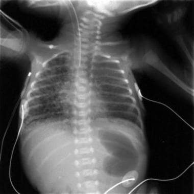
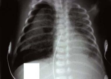
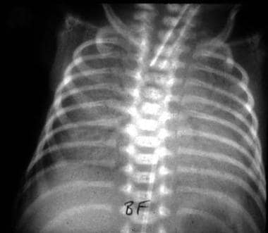
 Pulmonary interstitial emphysema
Combined pulmonary fibrosis and emphysema
Emphysema
2017 in Mexico
Deaths in December 1991
Papilledema
Swyer-James syndrome
Fog fever
Obstructive lung disease
Chronic obstructive pulmonary disease
Subcutaneous emphysema
Deaths in January 2001
Deaths in August 2001
Deaths in January 1994
Deaths in June 2012
Deaths in September 1992
Deaths in July 1993
Deaths in December 2000
Deaths in May 1996
Deaths in May 2011
Pulmonary sequestration
Deaths in January 2000
Tracheotomy
José Piñera Carvallo
Pulmonary artery
Avelino Muñoz Stevenson
Deaths in May 2001
Deaths in December 1995
Deaths in December 2014
Deaths in August 2011
 FastStats - Chronic Lower Respiratory Disease
Pulmonary interstitial emphysema - Wikipedia
 Pulmonary Interstitial Emphysema: Pearls, Background, Pathophysiology
 Combined pulmonary fibrosis and emphysema syndrome in connective tissue disease
The application of pressure, including exsufflation, in pulmonary emphysema
 Pulmonary Emphysema - Merck Veterinary Manual
 Symptoms and Signs of Pulmonary Edema vs. Emphysema: Treatment
JCI - Amelioration of emphysema in mice through lentiviral transduction of long-lived pulmonary alveolar macrophages
 Combined pulmonary fibrosis and emphysema: a distinct underrecognised entity | European Respiratory Society
 Anxiety and Panic Attacks In Emphysema/ Chronic Obstructive Pulmonary Diseases (COPD)
 Improved Diagnosis of Pulmonary Emphysema Using In Vivo Dark-Field Radiography. | Lund University Publications
 JCI Insight -
Heme scavenging reduces pulmonary endoplasmic reticulum stress, fibrosis, and emphysema
JCI Insight -
Heme scavenging reduces pulmonary endoplasmic reticulum stress, fibrosis, and emphysema
 Emphysema | Emphysema Symptoms | Emphysema Treatment | MedlinePlus
 Pulmonary Emphysema
 Heliox With Inhaled Nitric Oxide: A Novel Strategy for Severe Localized Interstitial Pulmonary Emphysema in Preterm Neonatal...
Skosmos: Thesaurus INRAE: pulmonary emphysema
Pulmonary Interstitial Emphysema: Background, Pathophysiology, Etiology
 Forschung - FB 11 - Medizin
 Emphysema | Conditions | UCSF Health
 Chronic obstructive Pulmonary Disease COPD /Emphysema | Spring Field Medical Clinic
 A simple method to save on costs in pulmonary emphysema operations
 What is COPD?
JCI Insight -
Heme scavenging reduces pulmonary endoplasmic reticulum stress, fibrosis, and emphysema
 Find NHLBI Clinical Trials | NHLBI, NIH
 DIGI Grant
 Told i have mild emphysema | Page 2 | Chronic Obstructive Pulmonary Disease | Forums | Patient
CDC - NIOSH Pocket Guide to Chemical Hazards -
Sulfuric acid
RXQ RX J
 38 CFR Appendix C to Part 4 - Appendix C to Part 4-Alphabetical Index of Disabilities | Electronic Code of Federal Regulations ...
COPD19
- however, its contribution to chronic obstructive pulmonary disease (COPD) pathogenesis is poorly understood. (asthmafoundation.org.nz)
- Necroptosis-related mRNA and proteins as well as cell death were examined in lungs and pulmonary macrophages of mice with cigarette smoke (CS)-induced experimental COPD. (asthmafoundation.org.nz)
- A subcategory of chronic obstructive pulmonary disease (COPD). (nih.gov)
- Most people with COPD have both emphysema and chronic bronchitis, but how severe each type is can be different from person to person. (medlineplus.gov)
- Also, smokers who get emphysema are more likely to get it if they have a family history of COPD. (medlineplus.gov)
- Chronic obstructive pulmonary (or lung) diseases (COPD or COLD) refers to a group of diseases in which airflow into or out of the lungs is insufficient. (ccohs.ca)
- Most people who have COPD have both chronic bronchitis and emphysema. (virtua.org)
- We found elevated plasma heme in chronic obstructive pulmonary disease (COPD) (GOLD stage 4) patients and also in a ferret model of COPD secondary to chronic cigarette smoke inhalation. (jci.org)
- In emphysema, a type of chronic obstructive pulmonary disease (COPD), tissue in the lungs becomes damaged and dies. (nih.gov)
- Many cases are likely to remain undiagnosed, particularly those with chronic obstructive pulmonary disease (COPD). (medindia.net)
- The diagnosis of chronic obstructive pulmonary disease (COPD) depends on thinking of it as a cause of breathlessness or cough. (nice.org.uk)
- 6 Only 30% had access to a dedicated chronic obstructive pulmonary disease (COPD) multidisciplinary meeting to review patients, and there was no consensus as to the correct strategy to adopt to identify appropriate patients. (bmj.com)
- Animal models and mechanisms of tobacco smoke-induced chronic obstructive pulmonary disease (COPD). (cdc.gov)
- Chronic obstructive pulmonary disease (COPD) is the third leading cause of death worldwide, and its global health burden is increasing. (cdc.gov)
- COPD is characterized by emphysema, mucus hypersecretion, and persistent lung inflammation, and clinically by chronic airflow obstruction and symptoms of dyspnea, cough, and fatigue in patients. (cdc.gov)
- A cluster of pathologies including chronic bronchitis, emphysema, asthma, and cardiovascular disease in the form of hypertension and atherosclerosis variably coexist in COPD patients. (cdc.gov)
- About 15% of smokers display the declining pulmonary function that leads to COPD and its associated disability. (testprepreview.com)
- Chronic obstructive pulmonary disease (COPD) is a group of diseases that lead to blockage in airflow and problems relating to breathing. (news-medical.net)
- Chronic obstructive pulmonary disease (COPD) is airflow limitation caused by an inflammatory response to inhaled toxins, often cigarette smoke. (msdmanuals.com)
Lobar emphysema5
- Some infants may develop chronic lobar emphysema, which may require surgical lobectomies. (wikipedia.org)
- Perea L, Blinman T, Piccione J, Laje P. Bilateral congenital lobar emphysema: staged management. (medscape.com)
- A preoperative diagnosis of congenital lobar emphysema was made on the basis of chest radiograph and computed tomography scan. (aku.edu)
- Acquired lobar emphysema (ALE) is an increasingly recognized complication of advanced BPD. (who.int)
- However, the infant deteriorated clinically and repeated radiography revealed lobar emphysema on the right lower lung. (who.int)
Developed pulmonary interstitial emphysema1
- In a 1987 study 3% of infants admitted to the neonatal intensive care unit (NICU) developed pulmonary interstitial emphysema. (wikipedia.org)
Obstructive4
- Chronic obstructive pulmonary diseases is a general term for a number of diseases that include chronic bronchitis and emphysema. (ccohs.ca)
- 2002. Chronic obstructive pulmonary disease 3: Experimental animal models of pulmonary emphysema. (nih.gov)
- In the context of poorer Asian countries, the main causes of chronic obstructive pulmonary disease are smoking, indoor air pollution from cooking and heating, and outdoor air pollution, Kaufman tells SciDev.Net . (news-medical.net)
- 2022) Burden of chronic obstructive pulmonary disease and its attributable risk factors in 204 countries and territories, 1990-2019: results from the Global Burden of Disease Study 2019. (news-medical.net)
Lungs9
- High-resolution computed tomography (HRCT) of the chest showed conspicuous centrilobular emphysema in the upper zones of the lungs, and diffuse, infiltrative lung disease in the lower zones ( figure 1 ). (bmj.com)
- High-resolution computed tomography of the chest of adult non-smoking patient demonstrating centrilobular emphysema in the upper zones of the lungs, and diffuse, infiltrative lung disease in the lower zones, including bilateral reticulation without subpleural predominance, traction bronchiectasis, architectural distortion and ground glass attenuation. (bmj.com)
- Emphysema affects the air sacs in your lungs. (medlineplus.gov)
- In emphysema, the walls between many of the air sacs in the lungs are damaged. (medlineplus.gov)
- The cause of emphysema is usually long-term exposure to irritants that damage your lungs and the airways. (medlineplus.gov)
- The lungs of a person with emphysema have weak, inelastic (not stretchy) air sacs that exchange less gas than healthy lungs. (virtua.org)
- It pumps blood into your lungs through the pulmonary arteries . (webmd.com)
- Affected individuals are likely to develop emphysema, a disease that affects the air sacs in the lungs. (medindia.net)
- The relation of inspiratory distention of the lungs to emphysema. (nih.gov)
Cigarette smoke-induced emphysema2
Interstitial pneumonia3
- In order to compare histomorphometrical, roentgenological and immunohistochemical aspects of usual interstitial pneumonia (UIP) with and without associated pulmonary emphysema, 17 patients with biopsy-proven UIP were evaluated. (nih.gov)
- Idiopathic pulmonary fibrosis (IPF) is characterised by chronic, progressive, fibrosing interstitial pneumonia of unknown aetiology. (bmj.com)
- Interstitial pneumonia represents a group of respiratory diseases characterized by an acute onset of severe respiratory distress and a combination of lung lesions that include pulmonary edema and congestion, interstitial emphysema, alveolar epithelialization, and hyaline membrane formation. (msdvetmanual.com)
Asthma1
- The specificity of HRCT for bronchial asthma is limited by the similarity of its changes to those of other diseases, such as bronchiectasis, chronic bronchitis, emphysema, and bronchopulmonary aspergillosis. (medscape.com)
Idiopathic6
- It has been suggested that the presence of emphysema modifies the outcome of patients with idiopathic pulmonary fibrosis (IPF). (nih.gov)
- Idiopathic pulmonary fibrosis has been associated with emphysema in cigarette smokers as a new clinical entity: combined pulmonary fibrosis and emphysema (CPFE). (nih.gov)
- Objectives To analyse the clinical characteristics and prognosis of acute exacerbation (AE) in patients with idiopathic pulmonary fibrosis (IPF) and pulmonary emphysema. (bmj.com)
- The studied population was patients with idiopathic pulmonary fibrosis (IPF) who developed acute exacerbations. (bmj.com)
- This is a group of lung conditions that includes sarcoidosis , idiopathic pulmonary fibrosis , and autoimmune disease . (webmd.com)
- If your doctor can't find a cause, they'll call it idiopathic pulmonary arterial hypertension . (webmd.com)
Fibrosis and emphysema2
Hypertension2
Inflammation6
- Mlkl deletion and qVD-OPh treatment reduced chronic CS-induced airway inflammation, but only Mlkl deletion prevented airway remodeling and emphysema. (asthmafoundation.org.nz)
- Inhibiting necroptosis attenuates CS-induced airway inflammation, airway remodeling, and emphysema. (asthmafoundation.org.nz)
- Sendai virus has been reported to cause emphysema in neonatal rats, presumably as a result of epithelial necrosis and inflammation, though infection by this agent is now rare due to modern animal husbandry practices. (nih.gov)
- 2011. A model of chronic inflammation and pulmonary emphysema after multiple ozone exposures in mice. (nih.gov)
- These data identify the important contributions of IL-23 to the development of elastase-induced pulmonary inflammation and emphysema, mediated through an IL-23/IL-17 pathway. (elsevierpure.com)
- Some doctors might recommend inhalers, oxygen, pulmonary rehabilitation therapy, or other medications to help reduce inflammation in the airways. (medicalnewstoday.com)
Cystic1
- B) Postnatal day56: emphysema on the right lower lobe of the lung, deviation of the heart and mediastinal structures to the left, diffuse cystic parenchymal changes on the right upper lobe and left lobe of the lung. (who.int)
Appearance of pulmonary1
- Although air-space, interstitial, or ground-glass opacities can dominate the CT appearance of pulmonary alveolar proteinosis, some combination of these findings is more common. (ajronline.org)
Mortality4
- In this article we compare clinical features, smoking history, pulmonary function, estimated systolic pulmonary artery pressure (eSPAP), and mortality in IPF with emphysema vs IPF without emphysematous changes. (nih.gov)
- IPF patients with emphysema exhibited higher mortality compared with those with IPF without emphysema. (nih.gov)
- The 90-day mortality rate was significantly lower in patients with emphysema than in those with IPF alone (23% vs 52%, p=0.03). (bmj.com)
- End-points will include exercise ability, pulmonary function, quality of life, morbidity and mortality. (nih.gov)
Edema5
- Acute bovine pulmonary emphysema and edema (ABPEE) is one of the more common causes of acute respiratory distress in adult cattle on pasture. (msdvetmanual.com)
- Acute bovine pulmonary emphysema and edema is a respiratory distress syndrome affecting groups of cattle after movement onto lush pasture. (msdvetmanual.com)
- Metabolites of the naturally occurring amino acid l -tryptophan probably are responsible for many outbreaks of acute bovine pulmonary emphysema and edema. (msdvetmanual.com)
- Acute bovine pulmonary emphysema and edema is most common in adult beef cows but may occur in either sex and in dairy or beef cattle under similar management conditions. (msdvetmanual.com)
- Pneumonia and pulmonary edema can also affect your interstitium. (webmd.com)
Systolic pulmonary1
- Echocardiography showed normal heart cavities, with estimated systolic pulmonary arterial pressure of 40 mm Hg. (bmj.com)
Alveolar proteinosis12
- This investigation describes the CT features of pulmonary alveolar proteinosis in a large group of patients. (ajronline.org)
- A retrospective review of 139 chest CT scans (79 thick-section scans and 60 thin-section scans) from 27 patients with pathologically proven pulmonary alveolar proteinosis was performed. (ajronline.org)
- Pulmonary alveolar proteinosis does not present only with alveolar disease. (ajronline.org)
- CT can play an important role in the diagnosis of pulmonary alveolar proteinosis. (ajronline.org)
- Several reports have described the CT aspects of pulmonary alveolar proteinosis [ 2 , 3 , 4 , 5 , 6 ], but these reports have analyzed only a small number of CT scans. (ajronline.org)
- Although CT findings can suggest the diagnosis of pulmonary alveolar proteinosis, the CT features are not pathognomonic [ 1 ]. (ajronline.org)
- For radiologists, the term "pulmonary alveolar proteinosis" is misleading because the CT appearance is not purely alveolar. (ajronline.org)
- This article systematically reviews the intraslice patterns and zonal distribution of disease on CT from the largest number of CT scans of patients with pulmonary alveolar proteinosis that has yet, to our knowledge, been described. (ajronline.org)
- The relative frequency of different types of pulmonary opacities in pulmonary alveolar proteinosis and the influence of section thickness are examined. (ajronline.org)
- Patients from our medical center with a confirmed diagnosis of pulmonary alveolar proteinosis from open-lung biopsy, transbronchial biopsy, or bronchoalveolar lavage fluid were identified by searching a pathology database. (ajronline.org)
- Patients with AIDS or a known infectious cause of pulmonary alveolar proteinosis were excluded. (ajronline.org)
- None of the patients had pulmonary alveolar proteinosis related to a known malignancy. (ajronline.org)
Diffuse3
- Diffuse persistent pulmonary interstitial emphysema secondary to mechanical ventilation in bronchiolitis. (medscape.com)
- Persistent pulmonary interstitial emphysema manifests as localized or diffuse forms, the latter carrying a poor prognosis. (asahq.org)
- A case of diffuse persistent pulmonary emphysema: When is difficult the diagnosis? (asahq.org)
Coronary1
- pulmonary emphysema, coronary heart disease and cancer of the bladder. (who.int)
People with emphysema2
- Some people with emphysema get frequent respiratory infections such as colds and the flu . (medlineplus.gov)
- 1.1.12 Consider primary care respiratory review and spirometry (see the recommendations on symptoms and spirometry ) for people with emphysema or signs of chronic airways disease on a chest X-ray or CT scan. (nice.org.uk)
Presence of emphysema1
- be aware that the presence of emphysema on a CT scan is an independent risk factor for lung cancer. (nice.org.uk)
Acute5
- The diagnosis of IPF, emphysema and acute exacerbation was based on a multidisciplinary discussion by pulmonary physicians and a radiologist. (bmj.com)
- The incidence of acute exacerbation among patients with IPF with or without emphysema could not be analysed because of the retrospective design of this study. (bmj.com)
- Some data such as pulmonary function tests prior to acute exacerbation were not available. (bmj.com)
- Acute thermal and chemical pulmonary damage, i.e., smoke inhalation with pulmonary insufficiency. (cms.gov)
- Mycoplasma pneumoniae: acute illness, antibiotics, and subsequent pulmonary function. (bmj.com)
Thoracic1
- Clinical centers must document a strongly integrated team with expertise in the following areas: thoracic surgery, the surgery being offered, the pre-, peri- and post-operative care of end-stage emphysema patients undergoing thoracic surgery, pulmonary medicine and the care of end-stage emphysema patients, the rehabilitation and education of end-stage emphysema patients, the conducting of clinical trials, pulmonary physiology, cardiology, radiology and evaluation of dyspnea and quality of life. (nih.gov)
Include pulmonary1
- Maximum medical therapy will include pulmonary rehabilitation and education. (nih.gov)
Alveoli3
- Pulmonary interstitial emphysema (PIE) is a collection of air outside of the normal air space of the pulmonary alveoli, found instead inside the connective tissue of the peribronchovascular sheaths, interlobular septa, and visceral pleura. (wikipedia.org)
- Pulmonary interstitial emphysema is a concern in any of the following: Prematurity Infant respiratory distress syndrome (IRDS) Meconium aspiration syndrome (MAS) Amniotic fluid aspiration Sepsis Infections Mechanical ventilation Pulmonary interstitial emphysema is created when air bursts or ruptures through tissue from the alveoli and bronchioles into the perivascular tissue of the lung. (wikipedia.org)
- Emphysema, often associated with chronic bronchitis, is a chronic lung disease in which the air sacs (alveoli) at the end of the small bronchioles are enlarged or over-inflated and are eventually destroyed. (ccohs.ca)
Tissue5
- This supportive tissue is called the pulmonary interstitium. (wikipedia.org)
- If the lesion is considered to be an artifact, emphysema should not be diagnosed, but a tissue note may be entered to that effect. (nih.gov)
- Higher outdoor concentrations of the pollutants ozone, fine particulate matter, and oxides of nitrogen (NOx) at a person's residence at the beginning of the study were associated with increases in emphysema-like lung tissue on CT scans over the following decade. (nih.gov)
- PERSISTENT pulmonary interstitial emphysema is collection of air in the pulmonary interstitial tissue outside normal air passages. (asahq.org)
- Alpha-1 Antitrypsin Deficiency Alpha-1 antitrypsin deficiency is congenital lack of a primary lung antiprotease, alpha-1 antitrypsin, which leads to increased protease-mediated tissue destruction and emphysema in adults. (msdmanuals.com)
Congenital1
- Congenital diaphragmatic hernia: association between pulmonary vascular resistance and plasma thromboxane concentrations. (bmj.com)
Pathogenesis1
- These features strongly implicate intravenous drug abuse in the pathogenesis of these patients' bullous pulmonary damage. (nih.gov)
Cause emphysema1
- Pipe, cigar, and other types of tobacco smoke can also cause emphysema, especially if you inhale them. (medlineplus.gov)
Patients with IPF3
Develop emphysema2
Disease6
- Pulmonary interstitial emphysema is more frequent in premature infants who require mechanical ventilation for severe lung disease. (wikipedia.org)
- Because air exchange is impaired, a person who develops emphysema experiences shortness of breath that occurs during strenuous exertion in the early stages of the disease and even at rest in later stages. (ccohs.ca)
- Additional risk factors include certain occupational exposures, cancer history and a disease history of pulmonary fibrosis, emphysema, chronic bronchitis. (virtua.org)
- The pulmonary air leak syndromes, including pneumomediastinum, pneumothorax, pulmonary interstitial emphysema and pneumopericardium, comprise a spectrum of disease with the same underlying pathophysiology. (who.int)
- Targeting IL-23 in emphysema is a potential therapeutic strategy for delaying disease progression. (elsevierpure.com)
- Although stopping smoking slows the progression of the disease, persons with COP do not recover lost pulmonary function. (testprepreview.com)
Refractory2
- Mahapatra S, Scottoline B. Steroid-induced resolution of refractory pulmonary interstitial emphysema. (medscape.com)
- Select patients with advanced emphysema refractory to optimized medical care may benefit from surgical or bronchoscopic interventional treatments. (medscape.com)
Symptoms4
- Most people who have emphysema are at least 40 years old when their symptoms begin. (medlineplus.gov)
- What are the symptoms of emphysema? (medlineplus.gov)
- Pulmonary rehabilitation improves symptoms, physical and emotional participation in everyday activities, and quality of life. (medscape.com)
- However it has now been recommended that this method of treatment be carried out after the emergence of emphysema symptoms. (medindia.net)
Airway1
- Emphysema is destruction of lung parenchyma leading to loss of elastic recoil and loss of alveolar septa and radial airway traction, which increases the tendency for airway collapse. (msdmanuals.com)
Persistent3
- Localized persistent pulmonary interstitial emphysema in a preterm infant in the absence of mechanical ventilation. (medscape.com)
- 1 Persistent pulmonary interstitial emphysema can occur in term infants and in those who have not been mechanically ventilated. (asahq.org)
- Lateral decubitus position as therapy for persistent focal pulmonary interstitial emphysema in neonates: A preliminary report. (asahq.org)
Oxygen4
- Nunez-Ramiro A, Aguar M, Cernada M, Parra-Llorca A, Vento M. Oxygen needs during resuscitation and surfactant to achieve stabilisation were independent risks factors for pulmonary interstitial emphysema in preterm infants. (medscape.com)
- There was no significant difference in the arterial partial oxygen pressure/fraction of inhaled oxygen (P/F) ratio or other laboratory and radiographic data between patients with and without emphysema. (bmj.com)
- Oxygen therapy , if you have severe emphysema and low levels of oxygen in your blood. (medlineplus.gov)
- The clot sticks in a pulmonary artery, often causing shortness of breath and low blood oxygen levels. (webmd.com)
Diseases1
- Chronic pulmonary occurs in persons with pre-existing lung diseases such as emphysema. (ccohs.ca)
Abstract2
- Abstract : Purpose : The purpose of this study was to evaluate the usefulness of thick slabminimum intensity projection (MinIP) as a follow-up method in patients with pulmonaryemphysema. (tokushima-u.ac.jp)
- abstract = "We recently reported that IL-17A plays a critical role in the development of porcine pancreatic elastase (PPE)-induced emphysema. (elsevierpure.com)
Lesions2
- Pulmonary emphysema can be classified by the location and distribution of the lesions. (nih.gov)
- At HRTC scan, emphysematous lesions were prevalent in the upper fields of both emphysema/UIP and emphysema groups and the distribution of fibrotic lesions was similar in emphysema/UIP compared to UIP alone. (nih.gov)
Bronchitis2
- After several tests, I was told I have Chronic Bronchitis and Emphysema that resulted in the blood in my sputum. (mindpub.com)
- It includes conditions like chronic bronchitis and emphysema. (news-medical.net)
Diagnosis2
- The preoperative diagnosis of pulmonary sequestration is not possible in most of the cases. (aku.edu)
- Diagnosis is based on history, physical examination, chest x-ray, and pulmonary function tests. (msdmanuals.com)
Artery1
- These arteries (except the thyroid artery) form a peribronchial plexus that follows the bronchial tree deep into the lung parenchyma to supply blood also to the visceral pleura and the walls of the pulmonary arteries and veins (vasa vasorum). (medscape.com)
Prevalence2
- The prevalence of pulmonary interstitial emphysema widely varies with the population studied. (wikipedia.org)
- The prevalence of emphysema in the IPF cohort was 28% (31 of 110 patients). (nih.gov)
Pneumomediastinum1
- Presence of subpleural pulmonary interstitial emphysema as an indication of single or multiple alveolar ruptures on CT in patients with spontaneous pneumomediastinum. (medscape.com)
Rehabilitation2
- Make sure you take advantage of any patient education and pulmonary rehabilitation that is available. (mindpub.com)
- A registry will be established to serve as a repository of severe end stage emphysematous patients, who have been referred for evaluation for LVRS, transplant or pulmonary rehabilitation to any of the participating clinical centers. (nih.gov)
Sequestration1
- At operation, an intralobar pulmonary sequestration was found. (aku.edu)
Destruction1
- In contrast, fibroblasts in areas of parenchymal destruction of emphysema/UIP expressed MMP-2, MMP-9, MMP-7 and MT1-MMP at variable but significantly higher levels when compared to emphysema subjects, in the presence of similar levels of TIMP-1, TIMP-2 and TNF-alpha. (nih.gov)
Severe1
- In severe cases, emphysema can cause weight loss, weakness in your lower muscles, and swelling in your ankles, feet, or legs. (medlineplus.gov)
Infants3
- Infants with pulmonary interstitial emphysema are typically recommended for admission to a neonatal intensive care unit. (wikipedia.org)
- Studies reflecting international frequency demonstrated that 2-3% of all infants in NICUs develop pulmonary interstitial emphysema. (wikipedia.org)
- Verma RP, Chandra S, Niwas R, Komaroff E. Risk factors and clinical outcomes of pulmonary interstitial emphysema in extremely low birth weight infants. (medscape.com)