Pulmonary Veins
Catheter Ablation
Atrial Fibrillation
Pulmonary Veno-Occlusive Disease
Electrophysiologic Techniques, Cardiac
Heart Conduction System
Body Surface Potential Mapping
Tachycardia, Ectopic Atrial
Femoral Vein
Cardiac Catheters
Jugular Veins
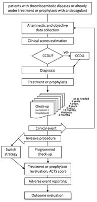
Treatment Outcome
Vena Cava, Superior
Phlebography
Mesenteric Veins
Scimitar Syndrome
Constriction, Pathologic
Umbilical Veins
Iliac Vein
Atrial Septum
Anti-Arrhythmia Agents
Surgery, Computer-Assisted
Electrocardiography
Autonomic Denervation
Atrial Premature Complexes
Dogs
Imaging, Three-Dimensional
Popliteal Vein
Follow-Up Studies
Breath Holding
Echocardiography, Transesophageal
Atrial Flutter
Electrocardiography, Ambulatory
Cardiac Pacing, Artificial
Azygos Vein
Electrophysiological Phenomena
Cardiac Complexes, Premature
Catheterization
Subclavian Vein
Pulmonary Artery
Splenic Vein
Tomography, X-Ray Computed
Sinoatrial Node
Tachycardia
Heart Septal Defects, Atrial
Coronary Sinus
Tomography, Spiral Computed
Refractory Period, Electrophysiological
Magnetic Resonance Angiography
Retinal Vein
Pulmonary Atresia
Brachiocephalic Veins
Respiratory-Gated Imaging Techniques
Ganglia, Autonomic
Prospective Studies
Multidetector Computed Tomography
Feasibility Studies
Pericardium
Axillary Vein
Vascular Malformations
Transcatheter occlusion of a post-Fontan residual hepatic vein to pulmonary venous atrium communication using the Amplatzer septal occluder. (1/1323)
A residual hepatic vein to left atrial communication may result in progressive cyanosis after the Fontan procedure. This problem has usually been treated surgically by ligation or re-inclusion of the residual hepatic vein in the Fontan circulation. Previous attempts at transcatheter closure of such veins have been unsuccessful. An Amplatzer septal occluder was successfully used for transcatheter closure of a post-Fontan hepatic vein to pulmonary venous atrium fistula in an 8 year old boy. (+info)Effects of respiratory cycle on pulmonary venous flow and cardiac cycle on pulmonary venous diameter of dogs: a transesophageal echocardiography study. (2/1323)
We investigated 12 anesthetized normal dogs using transesophageal echocardiography to understand the effects of respiration on the pulmonary venous flow. Additionally, we observed whether the diameter of the pulmonary vein changes with the heart beat. The pulsed Doppler wave form of pulmonary venous flow predominantly demonstrated two backward flows, with one peak occurring during ventricular systole and another during ventricular diastole. Sometimes a small forward flow occurred during left atrial contraction. In comparison with expiration, the peak velocity and velocity-time integral of the flow wave under inspiration occurred during both systole and diastole were significantly smaller. The diameter of the pulmonary vein decreased during left atrial contraction and increased during left ventricular systole and diastole. (+info)Site of functional bronchopulmonary anastomoses in sheep. (3/1323)
The location of bronchopulmonary anastomoses has long been a topic of discussion, and pre-, post-, and capillary sites have all been demonstrated in postmortem examinations. However, there have been few studies that have provided insight into the patency and function of these anastomoses in the intact lung. To identify these functional sites where the bronchial circulation anastomoses with the pulmonary circulation, we studied sheep lungs in situ serial sectioned with high-resolution computed tomography (CT). Differences in radiodensities of blood, air, and nonionic contrast medium were used to differentiate and localize airways and vessels and to identify the effluent from the bronchial circulation. After an initial series of scans to identify the pulmonary arteries and veins adjacent to airways 2-12 mm in diameter, contrast material was infused into the bronchial artery. In three sheep, the major accumulation of contrast medium was found in pulmonary veins. In one of the sheep, a comparable number of pulmonary arteries and veins contained contrast medium. Serial histologic sections were able to identify small bronchial venules lying within subepithelial bronchial folds that drain directly into pulmonary veins. These results using serial CT and histologic images suggest that drainage from the intraparenchymal bronchial vasculature is predominantly into postcapillary pulmonary vessels. (+info)Pulmonary venous flow in hypertrophic cardiomyopathy as assessed by the transoesophageal approach. The additive value of pulmonary venous flow and left atrial size variables in estimating the mitral inflow pattern in hypertrophic cardiomyopathy. (4/1323)
AIMS: This study was conducted to assess the characteristics of the pattern of pulmonary venous flow and to document the interaction of this flow and left atrial function with the pattern of mitral inflow in hypertrophic cardiomyopathy. METHODS AND RESULTS: Pulmonary venous and mitral flows were evaluated by the transoesophageal approach in 80 patients with hypertrophic cardiomyopathy. Left atrial size and function were measured by the transthoracic approach. Their values were compared with those obtained from 35 normal controls. Twelve patients showed significant (> 2+) mitral regurgitation. As a group, hypertrophic cardiomyopathy patients showed increased atrial reversal flow and longer deceleration time of the diastolic wave, but a wide variability of pulmonary venous flow patterns were observed. Thirty patients (37.5%) had pseudonormal mitral flow patterns. Stepwise multilinear regression analysis identified the ratio of systolic to diastolic pulmonary venous flow velocity, the ratio of velocity-time integrals of both flow waves at atrial contraction, the left atrial minimal volume and the systolic fraction as independent predictive variables of the mitral E/A wave velocity ratio (r = 0.82). By logistic regression, the former three variables were selected as independent predictive covariates of a pseudonormal mitral flow pattern (sensitivity: 83%, specificity: 90%). The ratio of velocity-time integrals of both atrial waves was the most important predictive variable in both analyses. CONCLUSIONS: The observed variability in the configuration of pulmonary venous flow velocity waveform is related to what occurs in transmitral flow in patients with hypertrophic cardiomyopathy. Significant mitral regurgitation is not an independent correlate of pseudonormal mitral inflow patterns in these patients. Our results further emphasize the complementary, additive value of the pulmonary venous flow velocity pattern and left atrial size in the interpretation of the mitral flow velocity pattern, and indirectly suggest the underlying increased left ventricular filling pressures of patients with hypertrophic cardiomyopathy and pseudonormal mitral flow patterns. (+info)The functional anatomy of the bronchial circulation of the domestic fowl. (5/1323)
The bronchial circulation was studied in 25 adult domestic fowls. The right and left bronchial arteries originated caudal to the syrinx from a bronchoesophageal artery which is a branch of the right common carotid artery. Each bronchial artery ramified on the wall of the extrapulmonary part of the corresponding primary bronchus and finally anastomosed directly with a branch of the pulmonary artery at the hilus of the lung. Thr bronchial artery did not accompany the intrapulmonary part of the primary bronchus. The branches of each bronchial artery formed an anastomosing network on the wall of the extrapulmonary part of the primary bronchus. The calibre of the bronchial artery at its anastomosis with the branch of the pulmonary artery was greater than at its origin from the bronchoesophageal artery. Intravenous injections of Lycopodium spores indicated that the blood flows from the pulmonary artery into the bronchial artery. Small bronchial veins drained the extrapulmonary part of the primary bronchus into the pulmonary vein and the oesophageal veins. The intrapulmonary part of the primary bronchus was supplied by branches of the pulmonary artery and drained by tributaries of the pulmonary vein. The blood supply to the primary bronchus could constitute a shunt capable of passing blood from the pulmonary artery into the pulmonary vein without going through the exchange tissue. The parabronchial (atrial) muscles received a blood supply directly from the exchange tissue via septal venules which formed a network underneath the muscle bundles, without actually penetrating between the muscle cells. These venules drained into atrial veins which were tributaries of the pulmonary vein. The atrial muscles probably also received oxygen by direct diffusion from the parabronchial lumen. The pleura was supplied by the oesophageal branches of the bronchoesophageal artery, and by small twigs from the internal thoracic and intercostal arteries. (+info)Defibrillation-guided radiofrequency ablation of atrial fibrillation secondary to an atrial focus. (6/1323)
OBJECTIVES: Our aim was to evaluate a potential focal source of atrial fibrillation (AF) by unmasking spontaneous early reinitiation of AF after transvenous atrial defibrillation (TADF), and to describe a method of using repeated TADF to map and ablate the focus. BACKGROUND: Atrial fibrillation may develop secondary to a rapidly discharging atrial focus that the atria cannot follow synchronously, with suppression of the focus once AF establishes. Focus mapping and radiofrequency (RF) ablation may be curative but is limited if the patient is in AF or if the focus is quiescent. Early reinitiation of AF has been observed following defibrillation, which might have a focal mechanism. METHODS: We performed TADF in patients with drug-refractory lone AF using electrodes in the right atrium (RA) and the coronary sinus. When reproducible early reinitiation of AF within 2 min after TADF was observed that exhibited a potential focal mechanism, both mapping and RF ablation were performed to suppress AF reinitiation. Clinical and ambulatory ECG monitoring was used to assess AF recurrence. RESULTS: A total of 44 lone AF patients (40 men, 4 women; 32 persistent, 12 paroxysmal AF) with a mean age of 58+/-13 years underwent TADF. Sixteen patients had early reinitiation of AF after TADF, nine (20%; 5 paroxysmal) exhibited a pattern of focal reinitiation. Earliest atrial activation was mapped to the right superior (n = 4) and the left superior (n = 3) pulmonary vein, just inside the orifice, in the seven patients who underwent further study. At the onset of AF reinitiation, the site of earliest activation was 86+/-38 ms ahead of the RA reference electrogram. The atrial activities from this site were fragmented and exhibited progressive cycle-length shortening with decremental conduction to the rest of the atrium until AF reinitiated. Radiofrequency ablation at the earliest activation site resulted in suppression of AF reinitiation despite pace-inducibility. Improved clinical outcome was observed over 8+/-4 months' follow-up. CONCLUSIONS: Transvenous atrial defibrillation can help to unmask, map, and ablate a potential atrial focus in patients with paroxysmal and persistent AF. A consistent atrial focus is the cause of early reinitiation of AF in 20% of patients with lone AF, and these patients may benefit from this technique. (+info)Prostanoid receptors involved in the relaxation of human pulmonary vessels. (7/1323)
1. To characterize the prostanoid receptors on human pulmonary smooth muscle involved in vasodilatations, isolated arteries and veins were contracted with norepinephrine (10 microM) and vessels were subsequently challenged with different prostanoid-receptor agonists in the absence or presence of selective antagonists. 2. Prostaglandin D2 (PGD2) and the selective DP-receptor agonist, BW245C, induced relaxations in the contracted human pulmonary venous preparations. The pD2 values were: 6.88+/-0.11 (n=17) and 7.31+/-0.12 (n=5), respectively. The relaxant responses induced by PGD2 were reduced by the selective DP-receptor antagonist, BWA868C, and the estimated pA2 value was 7.84+/-0.16 (n=4). PGD2 and BW245C did not relax contracted human pulmonary arteries. 3. The selective IP-receptor agonists, iloprost and cicaprost, both induced relaxations in the contracted human vascular preparations. The pD2 values for iloprost were: 7.84+/-0.08 (n=6) and 8.25+/-0.06 (n=4) and for cicaprost: 8.06+/-0.12 (n=5) and 8.11+/-0.09 (n=5) in arteries and veins respectively. 4. Prostaglandin E2 (PGE2) and the EP2/EP3-receptor agonist, misoprostol, partially relaxed the contracted venous preparations and the pD2 values were: 8.10+/-0.15 (n=15) and 6.24+/-0.33 (n=3), respectively. These relaxations suggest the presence of an EP receptor in the human pulmonary veins. The contracted human pulmonary arteries did not relax when challenged with PGE2. 5. In human pulmonary venous preparations, the PGE2-induced relaxations were neither modified by treatment with TP/EP4-receptor antagonist, AH23848B (10 and 30 microM, n=6), nor by the DP/EP1/EP2-receptor antagonist, AH6809 (3 microM, n=6). 6. These data suggest that the relaxation induced by prostanoids involved DP-, IP-receptors and to a lesser extent an EP-receptor on human pulmonary venous smooth muscle. In contrast, only the IP-receptor is involved in the prostanoid induced relaxations on human pulmonary arterial smooth muscle. (+info)Fetal pulmonary venous flow into the left atrium relative to diastolic and systolic cardiac time intervals. (8/1323)
OBJECTIVE: To establish the nature and gestational age dependency of the pulmonary venous flow velocity pattern into the left atrium relative to systolic and diastolic phases of the cardiac cycle. DESIGN: This was a cross-sectional study of Doppler measurements of fetal pulmonary venous inflow velocities, which were correlated with simultaneous recordings of transmitral and aortic flow velocity waveforms based on an equal cardiac cycle length (+/- 5%). RESULTS: Successful recordings were obtained in 28 out of 60 (47%) normal singleton pregnancies at 20-36 weeks of gestation. Reproducibility of waveform analysis of the various phases of the cardiac cycle was satisfactory, within-patient variance ranging between 1.7% and 6.5%. A statistically significant increase (p < 0.05) in pulmonary venous time average velocity and velocity integral with advancing gestational age was established. A statistically significant increase (p < 0.05) of the pulmonary flow velocity integral was also found when related to each of the systolic and diastolic segments of the cardiac cycle, with the exception of isovolemic relaxation time. The duration of each of the diastolic and systolic segments of the cardiac cycle, as well as the pulmonary venous velocity integral expressed as a percentage of the cardiac cycle, remained constant with advancing gestational age. CONCLUSIONS: The second half of pregnancy is characterized by pulmonary venous inflow into the left atrium throughout the cardiac cycle. Pulmonary venous inflow into the left atrium occurs predominantly during the filling and ejection phases of the cardiac cycle. Absolute cardiac diastolic and systolic time intervals as well as the percentage distribution of pulmonary venous flow velocity integrals between these cardiac time intervals remain unchanged with advancing gestational age. (+info)Pulmonary veins are blood vessels that carry oxygenated blood from the lungs to the left atrium of the heart. There are four pulmonary veins in total, two from each lung, and they are the only veins in the body that carry oxygen-rich blood. The oxygenated blood from the pulmonary veins is then pumped by the left ventricle to the rest of the body through the aorta. Any blockage or damage to the pulmonary veins can lead to various cardiopulmonary conditions, such as pulmonary hypertension and congestive heart failure.
Catheter ablation is a medical procedure in which specific areas of heart tissue that are causing arrhythmias (irregular heartbeats) are destroyed or ablated using heat energy (radiofrequency ablation), cold energy (cryoablation), or other methods. The procedure involves threading one or more catheters through the blood vessels to the heart, where the tip of the catheter can be used to selectively destroy the problematic tissue. Catheter ablation is often used to treat atrial fibrillation, atrial flutter, and other types of arrhythmias that originate in the heart's upper chambers (atria). It may also be used to treat certain types of arrhythmias that originate in the heart's lower chambers (ventricles), such as ventricular tachycardia.
The goal of catheter ablation is to eliminate or reduce the frequency and severity of arrhythmias, thereby improving symptoms and quality of life. In some cases, it may also help to reduce the risk of stroke and other complications associated with arrhythmias. Catheter ablation is typically performed by a specialist in heart rhythm disorders (electrophysiologist) in a hospital or outpatient setting under local anesthesia and sedation. The procedure can take several hours to complete, depending on the complexity of the arrhythmia being treated.
It's important to note that while catheter ablation is generally safe and effective, it does carry some risks, such as bleeding, infection, damage to nearby structures, and the possibility of recurrent arrhythmias. Patients should discuss the potential benefits and risks of the procedure with their healthcare provider before making a decision about treatment.
Atrial fibrillation (A-tre-al fi-bru-la'shun) is a type of abnormal heart rhythm characterized by rapid and irregular beating of the atria, the upper chambers of the heart. In this condition, the electrical signals that coordinate heartbeats don't function properly, causing the atria to quiver instead of contracting effectively. As a result, blood may not be pumped efficiently into the ventricles, which can lead to blood clots, stroke, and other complications. Atrial fibrillation is a common type of arrhythmia and can cause symptoms such as palpitations, shortness of breath, fatigue, and dizziness. It can be caused by various factors, including heart disease, high blood pressure, age, and genetics. Treatment options include medications, electrical cardioversion, and surgical procedures to restore normal heart rhythm.
Pulmonary Veno-Occlusive Disease (PVOD) is a rare form of pulmonary hypertension, characterized by the obstruction or blockage of the pulmonary veins. This obstruction can lead to increased pressure in the pulmonary circulation, ultimately causing right heart failure.
The medical definition of Pulmonary Veno-Occlusive Disease is: "A progressive and often fatal condition in which there is a selective occlusion or obliteration of the pulmonary venules and small veins, resulting in pulmonary hypertension, right ventricular failure, and death."
The obstruction of the pulmonary veins can be caused by various factors, including inflammation, fibrosis, or thrombosis. Symptoms of PVOD may include shortness of breath, fatigue, coughing up blood, and signs of right heart failure such as peripheral edema and ascites.
Diagnosis of PVOD can be challenging due to its rarity and nonspecific symptoms. Imaging studies, such as chest X-ray or CT scan, may show signs of pulmonary congestion and enlarged central pulmonary veins. A definitive diagnosis usually requires a lung biopsy.
Treatment options for PVOD are limited, and there is no cure for the disease. Currently, lung transplantation remains the only potentially curative treatment option for patients with PVOD.
Veins are blood vessels that carry deoxygenated blood from the tissues back to the heart. They have a lower pressure than arteries and contain valves to prevent the backflow of blood. Veins have a thin, flexible wall with a larger lumen compared to arteries, allowing them to accommodate more blood volume. The color of veins is often blue or green due to the absorption characteristics of light and the reduced oxygen content in the blood they carry.
The heart atria are the upper chambers of the heart that receive blood from the veins and deliver it to the lower chambers, or ventricles. There are two atria in the heart: the right atrium receives oxygen-poor blood from the body and pumps it into the right ventricle, which then sends it to the lungs to be oxygenated; and the left atrium receives oxygen-rich blood from the lungs and pumps it into the left ventricle, which then sends it out to the rest of the body. The atria contract before the ventricles during each heartbeat, helping to fill the ventricles with blood and prepare them for contraction.
Electrophysiologic techniques, cardiac, refer to medical procedures used to study the electrical activities and conduction systems of the heart. These techniques involve the insertion of electrode catheters into the heart through blood vessels under fluoroscopic guidance to record and stimulate electrical signals. The information obtained from these studies can help diagnose and evaluate various cardiac arrhythmias, determine the optimal treatment strategy, and assess the effectiveness of therapies such as ablation or implantable devices.
The electrophysiologic study (EPS) is a type of cardiac electrophysiologic technique that involves the measurement of electrical signals from different regions of the heart to evaluate its conduction system's function. The procedure can help identify the location of abnormal electrical pathways responsible for arrhythmias and determine the optimal treatment strategy, such as catheter ablation or medication therapy.
Cardiac electrophysiologic techniques are also used in device implantation procedures, such as pacemaker or defibrillator implantation, to ensure proper placement and function of the devices. These techniques can help program and test the devices to optimize their settings for each patient's needs.
In summary, cardiac electrophysiologic techniques are medical procedures used to study and manipulate the electrical activities of the heart, helping diagnose and treat various arrhythmias and other cardiac conditions.
The saphenous vein is a term used in anatomical description to refer to the great or small saphenous veins, which are superficial veins located in the lower extremities of the human body.
The great saphenous vein (GSV) is the longest vein in the body and originates from the medial aspect of the foot, ascending along the medial side of the leg and thigh, and drains into the femoral vein at the saphenofemoral junction, located in the upper third of the thigh.
The small saphenous vein (SSV) is a shorter vein that originates from the lateral aspect of the foot, ascends along the posterior calf, and drains into the popliteal vein at the saphenopopliteal junction, located in the popliteal fossa.
These veins are often used as conduits for coronary artery bypass grafting (CABG) surgery due to their consistent anatomy and length.
Cryosurgery is a medical procedure that uses extreme cold, such as liquid nitrogen or argon gas, to destroy abnormal or unwanted tissue. The intense cold causes the water inside the cells to freeze and form ice crystals, which can rupture the cell membrane and cause the cells to die. Cryosurgery is often used to treat a variety of conditions including skin growths such as warts and tumors, precancerous lesions, and some types of cancer. The procedure is typically performed in a doctor's office or outpatient setting and may require local anesthesia.
The portal vein is the large venous trunk that carries blood from the gastrointestinal tract, spleen, pancreas, and gallbladder to the liver. It is formed by the union of the superior mesenteric vein (draining the small intestine and a portion of the large intestine) and the splenic vein (draining the spleen and pancreas). The portal vein then divides into right and left branches within the liver, where the blood flows through the sinusoids and gets enriched with oxygen and nutrients before being drained by the hepatic veins into the inferior vena cava. This unique arrangement allows the liver to process and detoxify the absorbed nutrients, remove waste products, and regulate metabolic homeostasis.
Varicose veins are defined as enlarged, swollen, and twisting veins often appearing blue or dark purple, which usually occur in the legs. They are caused by weakened valves and vein walls that can't effectively push blood back toward the heart. This results in a buildup of blood, causing the veins to bulge and become varicose.
The condition is generally harmless but may cause symptoms like aching, burning, muscle cramp, or a feeling of heaviness in the legs. In some cases, varicose veins can lead to more serious problems, such as skin ulcers, blood clots, or chronic venous insufficiency. Treatment options include lifestyle changes, compression stockings, and medical procedures like sclerotherapy, laser surgery, or endovenous ablation.
The heart conduction system is a group of specialized cardiac muscle cells that generate and conduct electrical impulses to coordinate the contraction of the heart chambers. The main components of the heart conduction system include:
1. Sinoatrial (SA) node: Also known as the sinus node, it is located in the right atrium near the entrance of the superior vena cava and functions as the primary pacemaker of the heart. It sets the heart rate by generating electrical impulses at regular intervals.
2. Atrioventricular (AV) node: Located in the interatrial septum, near the opening of the coronary sinus, it serves as a relay station for electrical signals between the atria and ventricles. The AV node delays the transmission of impulses to allow the atria to contract before the ventricles.
3. Bundle of His: A bundle of specialized cardiac muscle fibers that conducts electrical impulses from the AV node to the ventricles. It divides into two main branches, the right and left bundle branches, which further divide into smaller Purkinje fibers.
4. Right and left bundle branches: These are extensions of the Bundle of His that transmit electrical impulses to the respective right and left ventricular myocardium. They consist of specialized conducting tissue with large diameters and minimal resistance, allowing for rapid conduction of electrical signals.
5. Purkinje fibers: Fine, branching fibers that arise from the bundle branches and spread throughout the ventricular myocardium. They are responsible for transmitting electrical impulses to the working cardiac muscle cells, triggering coordinated ventricular contraction.
In summary, the heart conduction system is a complex network of specialized muscle cells responsible for generating and conducting electrical signals that coordinate the contraction of the atria and ventricles, ensuring efficient blood flow throughout the body.
Body Surface Potential Mapping (BSPM) is a non-invasive medical technique used to record and analyze the electrical activity of the heart from the surface of the body. It involves placing multiple electrodes on the skin of the chest, back, and limbs to measure the potential differences between these points during each heartbeat. This information is then used to create a detailed, visual representation of the electrical activation pattern of the heart, which can help in the diagnosis and evaluation of various cardiac disorders such as arrhythmias, myocardial infarction, and ventricular hypertrophy.
The BSPM technique provides high-resolution spatial and temporal information about the cardiac electrical activity, making it a valuable tool for both clinical and research purposes. It can help identify the origin and spread of abnormal electrical signals in the heart, which is crucial for determining appropriate treatment strategies. Overall, Body Surface Potential Mapping is an important diagnostic modality that offers unique insights into the electrical functioning of the heart.
Tachycardia is a heart rate that is faster than normal when resting. In adults, a normal resting heart rate is typically between 60 and 100 beats per minute (bpm). Tachycardia is generally considered to be a heart rate of more than 100 bpm.
Ectopic atrial tachycardia (EAT) is a type of supraventricular tachycardia (SVT), which means that the abnormal rapid heartbeats originate in the atria, the upper chambers of the heart. EAT is caused by an ectopic focus, or an abnormal electrical focus outside of the sinoatrial node (the heart's natural pacemaker). This ectopic focus can be located in one of the pulmonary veins or in other atrial tissue.
EAT may present with symptoms such as palpitations, lightheadedness, shortness of breath, chest discomfort, or syncope (fainting). In some cases, EAT may not cause any symptoms and can be an incidental finding on an electrocardiogram (ECG) or Holter monitor.
The diagnosis of EAT is typically made based on the ECG findings, which show a regular narrow QRS complex tachycardia with P waves that are inverted in the inferior leads and often dissociated from the QRS complexes. Treatment options for EAT include observation, pharmacologic therapy, cardioversion, or catheter ablation.
The femoral vein is the large vein that runs through the thigh and carries oxygen-depleted blood from the lower limbs back to the heart. It is located in the femoral triangle, along with the femoral artery and nerve. The femoral vein begins at the knee as the popliteal vein, which then joins with the deep vein of the thigh to form the femoral vein. As it moves up the leg, it is joined by several other veins, including the great saphenous vein, before it becomes the external iliac vein at the inguinal ligament in the groin.
A cardiac catheter is a thin, flexible tube that is inserted into the heart or adjacent blood vessels during a cardiac catheterization procedure. This procedure is typically performed to diagnose and treat various cardiovascular conditions such as heart disease, heart defects, or abnormal heart rhythms.
Cardiac catheters can be used for several purposes:
1. To measure the pressure and oxygen levels in different chambers of the heart and blood vessels.
2. To inject dye into the coronary arteries to visualize blockages or narrowing through angiography.
3. To perform interventions such as balloon angioplasty, stent placement, or valvuloplasty to open up blocked or narrowed blood vessels or repair damaged heart valves.
4. To collect samples of heart muscle tissue for biopsy, which can help diagnose conditions like cardiomyopathy or myocarditis.
There are various types of cardiac catheters, including:
1. Diagnostic catheters - used to measure pressure and oxygen levels in the heart and blood vessels.
2. Guiding catheters - used to guide other interventional devices like balloons or stents into place.
3. Angioplasty balloon catheters - used to inflate a balloon at the tip of the catheter, which helps open up blocked or narrowed blood vessels.
4. Thermodilution catheters - used to measure cardiac output and other hemodynamic parameters.
5. Microcatheters - smaller, more flexible catheters used for complex interventions or accessing difficult-to-reach areas of the heart and blood vessels.
Cardiac catheterization is a minimally invasive procedure that usually requires only local anesthesia and mild sedation. The recovery time is typically short, with most patients returning home within 24 hours after the procedure.
The jugular veins are a pair of large, superficial veins that carry blood from the head and neck to the heart. They are located in the neck and are easily visible when looking at the side of a person's neck. The external jugular vein runs along the surface of the muscles in the neck, while the internal jugular vein runs within the carotid sheath along with the carotid artery and the vagus nerve.
The jugular veins are important in clinical examinations because they can provide information about a person's cardiovascular function and intracranial pressure. For example, distention of the jugular veins may indicate heart failure or increased intracranial pressure, while decreased venous pulsations may suggest a low blood pressure or shock.
It is important to note that medical conditions such as deep vein thrombosis (DVT) can also affect the jugular veins and can lead to serious complications if not treated promptly.
Treatment outcome is a term used to describe the result or effect of medical treatment on a patient's health status. It can be measured in various ways, such as through symptoms improvement, disease remission, reduced disability, improved quality of life, or survival rates. The treatment outcome helps healthcare providers evaluate the effectiveness of a particular treatment plan and make informed decisions about future care. It is also used in clinical research to compare the efficacy of different treatments and improve patient care.
The superior vena cava is a large vein that carries deoxygenated blood from the upper half of the body to the right atrium of the heart. It is formed by the union of the left and right brachiocephalic veins (also known as the internal jugular and subclavian veins) near the base of the neck. The superior vena cava runs posteriorly to the sternum and enters the upper right portion of the right atrium, just posterior to the opening of the inferior vena cava. It plays a crucial role in the circulatory system by allowing blood returning from the head, neck, upper limbs, and thorax to bypass the liver before entering the heart.
Recurrence, in a medical context, refers to the return of symptoms or signs of a disease after a period of improvement or remission. It indicates that the condition has not been fully eradicated and may require further treatment. Recurrence is often used to describe situations where a disease such as cancer comes back after initial treatment, but it can also apply to other medical conditions. The likelihood of recurrence varies depending on the type of disease and individual patient factors.
Phlebography is a medical imaging technique used to visualize and assess the veins, particularly in the legs. It involves the injection of a contrast agent into the veins, followed by X-ray imaging to capture the flow of the contrast material through the veins. This allows doctors to identify any abnormalities such as blood clots, blockages, or malformations in the venous system.
There are different types of phlebography, including ascending phlebography (where the contrast agent is injected into a foot vein and travels up the leg) and descending phlebography (where the contrast agent is injected into a vein in the groin or neck and travels down the leg).
Phlebography is an invasive procedure that requires careful preparation and monitoring, and it is typically performed by radiologists or vascular specialists. It has largely been replaced by non-invasive imaging techniques such as ultrasound and CT angiography in many clinical settings.
Paroxysmal Tachycardia is a type of arrhythmia (abnormal heart rhythm) characterized by rapid and abrupt onset and offset of episodes of tachycardia, which are faster than normal heart rates. The term "paroxysmal" refers to the sudden and recurring nature of these episodes.
Paroxysmal Tachycardia can occur in various parts of the heart, including the atria (small upper chambers) or ventricles (larger lower chambers). The two most common types are Atrial Paroxysmal Tachycardia (APT) and Ventricular Paroxysmal Tachycardia (VPT).
APT is more common and typically results in a rapid heart rate of 100-250 beats per minute. It usually begins and ends suddenly, lasting for seconds to hours. APT can cause symptoms such as palpitations, lightheadedness, shortness of breath, chest discomfort, or anxiety.
VPT is less common but more serious because it involves the ventricles, which are responsible for pumping blood to the rest of the body. VPT can lead to decreased cardiac output and potentially life-threatening conditions such as syncope (fainting) or even cardiac arrest.
Treatment options for Paroxysmal Tachycardia depend on the underlying cause, severity, and frequency of symptoms. These may include lifestyle modifications, medications, cardioversion (electrical shock to restore normal rhythm), catheter ablation (destroying problematic heart tissue), or implantable devices such as pacemakers or defibrillators.
Fluoroscopy is a type of medical imaging that uses X-rays to obtain real-time moving images of the internal structures of the body. A continuous X-ray beam is passed through the body part being examined, and the resulting fluoroscopic images are transmitted to a monitor, allowing the medical professional to view the structure and movement of the internal organs and bones in real time.
Fluoroscopy is often used to guide minimally invasive procedures such as catheterization, stent placement, or joint injections. It can also be used to diagnose and monitor a variety of medical conditions, including gastrointestinal disorders, musculoskeletal injuries, and cardiovascular diseases.
It is important to note that fluoroscopy involves exposure to ionizing radiation, and the risks associated with this exposure should be carefully weighed against the benefits of the procedure. Medical professionals are trained to use the lowest possible dose of radiation necessary to obtain the desired diagnostic information.
The mesenteric veins are a set of blood vessels that are responsible for draining deoxygenated blood from the small and large intestines. There are two main mesenteric veins: the superior mesenteric vein and the inferior mesenteric vein. The superior mesenteric vein drains blood from the majority of the small intestine, as well as the ascending colon and proximal two-thirds of the transverse colon. The inferior mesenteric vein drains blood from the distal third of the transverse colon, descending colon, sigmoid colon, and rectum. These veins ultimately drain into the portal vein, which carries the blood to the liver for further processing.
Scimitar Syndrome, also known as "congenital venolobar syndrome," is a rare congenital heart defect characterized by the following features:
1. An anomalous pulmonary vein (or veins) that drains into the inferior vena cava or right atrium instead of the left atrium. This vein often has a curved, scimitar-like appearance on imaging studies, hence the name of the syndrome.
2. Hypoplasia (underdevelopment) of the right lung or part of the right lung, which is often associated with abnormalities of the pulmonary artery and bronchial tree in that area.
3. Cardiac shunting, either from left to right (resulting in increased blood flow to the lungs) or right to left (resulting in cyanosis).
4. Other congenital heart defects may also be present, such as atrial septal defect, ventricular septal defect, or patent ductus arteriosus.
Symptoms of Scimitar Syndrome can vary widely depending on the severity of the anomaly and associated cardiac shunting. Mild cases may be asymptomatic, while severe cases can present with respiratory distress, heart failure, or cyanosis in infancy or early childhood. Treatment typically involves surgical correction of the anomalous pulmonary vein and any associated cardiac defects.
The renal veins are a pair of large veins that carry oxygen-depleted blood and waste products from the kidneys to the inferior vena cava, which is the largest vein in the body that returns blood to the heart. The renal veins are formed by the union of several smaller veins that drain blood from different parts of the kidney.
In humans, the right renal vein is shorter and passes directly into the inferior vena cava, while the left renal vein is longer and passes in front of the aorta before entering the inferior vena cava. The left renal vein also receives blood from the gonadal (testicular or ovarian) veins, suprarenal (adrenal) veins, and the lumbar veins.
It is important to note that the renal veins are vulnerable to compression by surrounding structures, such as the overlying artery or a tumor, which can lead to renal vein thrombosis, a serious condition that requires prompt medical attention.
Pathological constriction refers to an abnormal narrowing or tightening of a body passage or organ, which can interfere with the normal flow of blood, air, or other substances through the area. This constriction can occur due to various reasons such as inflammation, scarring, or abnormal growths, and can affect different parts of the body, including blood vessels, airways, intestines, and ureters. Pathological constriction can lead to a range of symptoms and complications depending on its location and severity, and may require medical intervention to correct.
The umbilical veins are blood vessels in the umbilical cord that carry oxygenated and nutrient-rich blood from the mother to the developing fetus during pregnancy. There are typically two umbilical veins, one of which usually degenerates and becomes obliterated, leaving a single functional vein. This remaining vein is known as the larger umbilical vein or the venous duct. It enters the fetal abdomen through the umbilicus and passes through the liver, where it branches off to form the portal sinus. Ultimately, the blood from the umbilical vein mixes with the blood from the inferior vena cava and is pumped to the heart through the right atrium.
It's important to note that after birth, the umbilical veins are no longer needed and undergo involution, becoming the ligamentum teres in the adult.
The iliac veins are a pair of large veins in the human body that carry deoxygenated blood from the lower extremities and the pelvic area back to the heart. They are formed by the union of the common iliac veins, which receive blood from the lower abdomen and legs, at the level of the fifth lumbar vertebra.
The combined iliac vein is called the inferior vena cava, which continues upward to the right atrium of the heart. The iliac veins are located deep within the pelvis, lateral to the corresponding iliac arteries, and are accompanied by the iliac lymphatic vessels.
The left common iliac vein is longer than the right because it must cross the left common iliac artery to join the right common iliac vein. The external and internal iliac veins are the two branches of the common iliac vein, with the external iliac vein carrying blood from the lower limbs and the internal iliac vein carrying blood from the pelvic organs.
It is essential to maintain proper blood flow in the iliac veins to prevent deep vein thrombosis (DVT), a condition that can lead to serious complications such as pulmonary embolism.
The atrial septum is the wall of tissue that divides the right and left atria, which are the upper chambers of the heart. This septum ensures that oxygen-rich blood in the left atrium is kept separate from oxygen-poor blood in the right atrium. Defects or abnormalities in the atrial septum, such as a hole or a gap, can result in various heart conditions, including septal defects and congenital heart diseases.
Anti-arrhythmia agents are a class of medications used to treat abnormal heart rhythms or arrhythmias. These drugs work by modifying the electrical activity of the heart to restore and maintain a normal heart rhythm. There are several types of anti-arrhythmia agents, including:
1. Sodium channel blockers: These drugs slow down the conduction of electrical signals in the heart, which helps to reduce rapid or irregular heartbeats. Examples include flecainide, propafenone, and quinidine.
2. Beta-blockers: These medications work by blocking the effects of adrenaline on the heart, which helps to slow down the heart rate and reduce the force of heart contractions. Examples include metoprolol, atenolol, and esmolol.
3. Calcium channel blockers: These drugs block the entry of calcium into heart muscle cells, which helps to slow down the heart rate and reduce the force of heart contractions. Examples include verapamil and diltiazem.
4. Potassium channel blockers: These medications work by prolonging the duration of the heart's electrical cycle, which helps to prevent abnormal rhythms. Examples include amiodarone and sotalol.
5. Digoxin: This drug increases the force of heart contractions and slows down the heart rate, which can help to restore a normal rhythm in certain types of arrhythmias.
It's important to note that anti-arrhythmia agents can have significant side effects and should only be prescribed by a healthcare professional who has experience in managing arrhythmias. Close monitoring is necessary to ensure the medication is working effectively and not causing any adverse effects.
Computer-assisted surgery (CAS) refers to the use of computer systems and technologies to assist and enhance surgical procedures. These systems can include a variety of tools such as imaging software, robotic systems, and navigation devices that help surgeons plan, guide, and perform surgeries with greater precision and accuracy.
In CAS, preoperative images such as CT scans or MRI images are used to create a three-dimensional model of the surgical site. This model can be used to plan the surgery, identify potential challenges, and determine the optimal approach. During the surgery, the surgeon can use the computer system to navigate and guide instruments with real-time feedback, allowing for more precise movements and reduced risk of complications.
Robotic systems can also be used in CAS to perform minimally invasive procedures with smaller incisions and faster recovery times. The surgeon controls the robotic arms from a console, allowing for greater range of motion and accuracy than traditional hand-held instruments.
Overall, computer-assisted surgery provides a number of benefits over traditional surgical techniques, including improved precision, reduced risk of complications, and faster recovery times for patients.
Electrocardiography (ECG or EKG) is a medical procedure that records the electrical activity of the heart. It provides a graphic representation of the electrical changes that occur during each heartbeat. The resulting tracing, called an electrocardiogram, can reveal information about the heart's rate and rhythm, as well as any damage to its cells or abnormalities in its conduction system.
During an ECG, small electrodes are placed on the skin of the chest, arms, and legs. These electrodes detect the electrical signals produced by the heart and transmit them to a machine that amplifies and records them. The procedure is non-invasive, painless, and quick, usually taking only a few minutes.
ECGs are commonly used to diagnose and monitor various heart conditions, including arrhythmias, coronary artery disease, heart attacks, and electrolyte imbalances. They can also be used to evaluate the effectiveness of certain medications or treatments.
Autonomic denervation is a medical term that refers to the interruption or loss of nerve supply to the autonomic nervous system. The autonomic nervous system is the part of the nervous system that controls involuntary actions, such as heart rate, blood pressure, digestion, and pupil dilation.
Autonomic denervation can occur due to various reasons, including surgical procedures, trauma, degenerative diseases, or medical conditions such as diabetes. The interruption of nerve supply can lead to a range of symptoms depending on the specific autonomic functions that are affected.
For example, autonomic denervation in the heart can lead to abnormal heart rhythms or low blood pressure. In the digestive system, it can cause problems with motility and secretion, leading to symptoms such as bloating, constipation, or diarrhea. Autonomic denervation in the eyes can result in pupil abnormalities, dry eyes, or light sensitivity.
Treatment for autonomic denervation depends on the underlying cause and the specific symptoms that are present. In some cases, medication may be used to manage symptoms, while in others, surgical intervention may be necessary to repair or restore nerve function.
Atrial premature complexes (APCs or APCTs) are extra heartbeats that originate in the atria, which are the upper chambers of the heart. These early beats disrupt the normal rhythm and cause a premature contraction before the next scheduled beat. APCs can sometimes be felt as a "skipped" beat or palpitation. They are usually benign and do not require treatment unless they occur frequently or are associated with underlying heart disease.
I believe there might be a misunderstanding in your question. "Dogs" is not a medical term or condition. It is the common name for a domesticated carnivore of the family Canidae, specifically the genus Canis, which includes wolves, foxes, and other extant and extinct species of mammals. Dogs are often kept as pets and companions, and they have been bred in a wide variety of forms and sizes for different purposes, such as hunting, herding, guarding, assisting police and military forces, and providing companionship and emotional support.
If you meant to ask about a specific medical condition or term related to dogs, please provide more context so I can give you an accurate answer.
The hepatic veins are blood vessels that carry oxygen-depleted blood from the liver back to the heart. There are typically three major hepatic veins - right, middle, and left - that originate from the posterior aspect of the liver and drain into the inferior vena cava just below the diaphragm. These veins are responsible for returning the majority of the blood flow from the gastrointestinal tract and spleen to the heart. It's important to note that the hepatic veins do not have valves, which can make them susceptible to a condition called Budd-Chiari syndrome, where blood clots form in the veins and obstruct the flow of blood from the liver.
The atrial appendage, also known as the left atrial appendage (LAA), is a small, ear-shaped structure that is located on the upper left chamber of the heart (left atrium). It has a unique muscular structure and plays a role in the normal functioning of the heart. However, it is best known for its association with atrial fibrillation, a common type of irregular heart rhythm. In people with atrial fibrillation, blood clots can form in the LAA, which can then travel to other parts of the body and cause strokes. For this reason, one treatment option for atrial fibrillation is to close off or remove the LAA to reduce the risk of stroke.
Three-dimensional (3D) imaging in medicine refers to the use of technologies and techniques that generate a 3D representation of internal body structures, organs, or tissues. This is achieved by acquiring and processing data from various imaging modalities such as X-ray computed tomography (CT), magnetic resonance imaging (MRI), ultrasound, or confocal microscopy. The resulting 3D images offer a more detailed visualization of the anatomy and pathology compared to traditional 2D imaging techniques, allowing for improved diagnostic accuracy, surgical planning, and minimally invasive interventions.
In 3D imaging, specialized software is used to reconstruct the acquired data into a volumetric model, which can be manipulated and viewed from different angles and perspectives. This enables healthcare professionals to better understand complex anatomical relationships, detect abnormalities, assess disease progression, and monitor treatment response. Common applications of 3D imaging include neuroimaging, orthopedic surgery planning, cancer staging, dental and maxillofacial reconstruction, and interventional radiology procedures.
The popliteal vein is the continuation of the tibial and fibular (or anterior and posterior tibial) veins, forming in the lower leg's back portion or popliteal fossa. It carries blood from the leg towards the heart. The popliteal vein is located deep within the body and is accompanied by the popliteal artery, which supplies oxygenated blood to the lower leg. This venous structure is a crucial part of the venous system in the lower extremities and is often assessed during physical examinations for signs of venous insufficiency or deep vein thrombosis (DVT).
Atrial function in a medical context refers to the role and performance of the two upper chambers of the heart, known as the atria. The main functions of the atria are to receive blood from the veins and help pump it into the ventricles, which are the lower pumping chambers of the heart.
The atria contract in response to electrical signals generated by the sinoatrial node, which is the heart's natural pacemaker. This contraction helps to fill the ventricles with blood before they contract and pump blood out to the rest of the body. Atrial function can be assessed through various diagnostic tests, such as echocardiograms or electrocardiograms (ECGs), which can help identify any abnormalities in atrial structure or electrical activity that may affect heart function.
Follow-up studies are a type of longitudinal research that involve repeated observations or measurements of the same variables over a period of time, in order to understand their long-term effects or outcomes. In medical context, follow-up studies are often used to evaluate the safety and efficacy of medical treatments, interventions, or procedures.
In a typical follow-up study, a group of individuals (called a cohort) who have received a particular treatment or intervention are identified and then followed over time through periodic assessments or data collection. The data collected may include information on clinical outcomes, adverse events, changes in symptoms or functional status, and other relevant measures.
The results of follow-up studies can provide important insights into the long-term benefits and risks of medical interventions, as well as help to identify factors that may influence treatment effectiveness or patient outcomes. However, it is important to note that follow-up studies can be subject to various biases and limitations, such as loss to follow-up, recall bias, and changes in clinical practice over time, which must be carefully considered when interpreting the results.
Breath holding is a physiological response where an individual holds their breath, intentionally or unintentionally, for a period of time. This can occur in various situations such as during swimming underwater, while lifting heavy weights, or in response to emotional stress or pain. In some cases, it can also be associated with certain medical conditions like seizures or syncope (fainting).
In the context of medical terminology, breath holding is often described as "voluntary" or "involuntary." Voluntary breath-holding is when an individual consciously chooses to hold their breath, while involuntary breath-holding occurs unconsciously, usually in response to a trigger such as a sudden increase in carbon dioxide levels or a decrease in oxygen levels.
It's important to note that prolonged breath-holding can be dangerous and may lead to hypoxia (lack of oxygen) and hypercapnia (excessive carbon dioxide), which can cause dizziness, loss of consciousness, or even more severe consequences such as brain damage or death. Therefore, it's essential not to hold one's breath for extended periods and seek medical attention if experiencing any symptoms related to breath-holding.
Left atrial function refers to the role and performance of the left atrium in the heart. The left atrium is the upper chamber on the left side of the heart that receives oxygenated blood from the lungs via the pulmonary veins and then contracts to help pump it into the left ventricle, which is the lower chamber that pumps blood out to the rest of the body.
The main functions of the left atrium include:
1. Receiving oxygen-rich blood from the lungs: The left atrium receives oxygenated blood from the pulmonary veins and acts as a reservoir for this blood before it is pumped into the left ventricle.
2. Contracting to help pump blood into the left ventricle: During atrial contraction, also known as atrial kick, the left atrium contracts and helps push blood into the left ventricle, increasing the amount of blood that can be ejected with each heartbeat.
3. Relaxing to receive more blood: Between heartbeats, the left atrium relaxes and fills up with more oxygenated blood from the lungs.
4. Contributing to heart rate regulation: The left atrium contains specialized cells called pacemaker cells that can help regulate the heart rate by initiating electrical impulses that trigger heart contractions.
Left atrial function is crucial for maintaining efficient cardiac output and overall cardiovascular health. Various conditions, such as heart failure, atrial fibrillation, and hypertension, can negatively impact left atrial function and contribute to the development of complications like stroke and reduced exercise tolerance.
Transesophageal echocardiography (TEE) is a type of echocardiogram, which is a medical test that uses sound waves to create detailed images of the heart. In TEE, a special probe containing a transducer is passed down the esophagus (the tube that connects the mouth to the stomach) to obtain views of the heart from behind. This allows for more detailed images of the heart structures and function compared to a standard echocardiogram, which uses a probe placed on the chest. TEE is often used in patients with poor image quality from a standard echocardiogram or when more detailed images are needed to diagnose or monitor certain heart conditions. It is typically performed by a trained cardiologist or sonographer under the direction of a cardiologist.
Atrial flutter is a type of abnormal heart rhythm or arrhythmia that originates in the atria - the upper chambers of the heart. In atrial flutter, the atria beat too quickly, usually between 250 and 350 beats per minute, which is much faster than the normal resting rate of 60 to 100 beats per minute.
This rapid beating causes the atria to quiver or "flutter" instead of contracting effectively. As a result, blood may not be pumped efficiently into the ventricles - the lower chambers of the heart - which can lead to reduced cardiac output and symptoms such as palpitations, shortness of breath, fatigue, dizziness, or chest discomfort.
Atrial flutter is often caused by underlying heart conditions, such as coronary artery disease, hypertension, valvular heart disease, or congenital heart defects. It can also be a complication of cardiac surgery or other medical procedures. In some cases, atrial flutter may occur without any apparent underlying cause, which is known as lone atrial flutter.
Treatment for atrial flutter typically involves medications to control the heart rate and rhythm, electrical cardioversion to restore a normal heart rhythm, or catheter ablation to destroy the abnormal electrical pathways in the heart that are causing the arrhythmia. In some cases, surgical intervention may be necessary to treat atrial flutter.
Ambulatory electrocardiography, also known as ambulatory ECG or Holter monitoring, is a non-invasive method of recording the electrical activity of the heart over an extended period of time (typically 24 hours or more) while the patient goes about their daily activities. The device used to record the ECG is called a Holter monitor, which consists of a small, portable recorder that is attached to the patient's chest with electrodes.
The recorded data provides information on any abnormalities in the heart's rhythm or electrical activity during different stages of activity and rest, allowing healthcare providers to diagnose and evaluate various cardiac conditions such as arrhythmias, ischemia, and infarction. The ability to monitor the heart's activity over an extended period while the patient performs their normal activities provides valuable information that may not be captured during a standard ECG, which only records the heart's electrical activity for a few seconds.
In summary, ambulatory electrocardiography is a diagnostic tool used to evaluate the electrical activity of the heart over an extended period, allowing healthcare providers to diagnose and manage various cardiac conditions.
Artificial cardiac pacing is a medical procedure that involves the use of an artificial device to regulate and stimulate the contraction of the heart muscle. This is often necessary when the heart's natural pacemaker, the sinoatrial node, is not functioning properly and the heart is beating too slowly or irregularly.
The artificial pacemaker consists of a small generator that produces electrical impulses and leads that are positioned in the heart to transmit the impulses. The generator is typically implanted just under the skin in the chest, while the leads are inserted into the heart through a vein.
There are different types of artificial cardiac pacing systems, including single-chamber pacemakers, which stimulate either the right atrium or right ventricle, and dual-chamber pacemakers, which stimulate both chambers of the heart. Some pacemakers also have additional features that allow them to respond to changes in the body's needs, such as during exercise or sleep.
Artificial cardiac pacing is a safe and effective treatment for many people with abnormal heart rhythms, and it can significantly improve their quality of life and longevity.
The azygos vein is a large, unpaired venous structure in the thoracic cavity of the human body. It begins as the ascending lumbar vein, which receives blood from the lower extremities and abdominal organs. As it enters the thorax through the diaphragm, it becomes the azygos vein and continues to ascend along the vertebral column.
The azygos vein receives blood from various tributaries, including the intercostal veins, esophageal veins, mediastinal veins, and bronchial veins. It then arches over the right mainstem bronchus and empties into the superior vena cava, which returns blood to the right atrium of the heart.
The azygos vein provides an important collateral pathway for venous return in cases where the inferior vena cava is obstructed or occluded. It also plays a role in the spread of certain thoracic diseases, such as tuberculosis and cancer.
Electrophysiological phenomena refer to the electrical properties and activities of biological tissues, cells, or organ systems, particularly in relation to nerve and muscle function. These phenomena can be studied using various techniques such as electrocardiography (ECG), electromyography (EMG), and electroencephalography (EEG).
In the context of cardiology, electrophysiological phenomena are often used to describe the electrical activity of the heart. The ECG is a non-invasive test that measures the electrical activity of the heart as it contracts and relaxes. By analyzing the patterns of electrical activity, doctors can diagnose various heart conditions such as arrhythmias, myocardial infarction, and electrolyte imbalances.
In neurology, electrophysiological phenomena are used to study the electrical activity of the brain. The EEG is a non-invasive test that measures the electrical activity of the brain through sensors placed on the scalp. By analyzing the patterns of electrical activity, doctors can diagnose various neurological conditions such as epilepsy, sleep disorders, and brain injuries.
Overall, electrophysiological phenomena are an important tool in medical diagnostics and research, providing valuable insights into the function of various organ systems.
Premature cardiac complexes, also known as premature heartbeats or premature ventricular contractions (PVCs), refer to extra or early heartbeats that originate in the lower chambers of the heart (the ventricles). These extra beats disrupt the normal rhythm and sequence of heartbeats, causing the heart to beat earlier than expected.
Premature cardiac complexes can occur in healthy individuals as well as those with heart disease. They are usually harmless and do not cause any symptoms, but in some cases, they may cause palpitations, skipped beats, or a fluttering sensation in the chest. In rare cases, frequent premature cardiac complexes can lead to more serious heart rhythm disorders or decreased heart function.
The diagnosis of premature cardiac complexes is usually made through an electrocardiogram (ECG) or Holter monitoring, which records the electrical activity of the heart over a period of time. Treatment is typically not necessary unless the premature complexes are frequent, symptomatic, or associated with underlying heart disease. In such cases, medications, cardioversion, or catheter ablation may be recommended.
Catheterization is a medical procedure in which a catheter (a flexible tube) is inserted into the body to treat various medical conditions or for diagnostic purposes. The specific definition can vary depending on the area of medicine and the particular procedure being discussed. Here are some common types of catheterization:
1. Urinary catheterization: This involves inserting a catheter through the urethra into the bladder to drain urine. It is often performed to manage urinary retention, monitor urine output in critically ill patients, or assist with surgical procedures.
2. Cardiac catheterization: A procedure where a catheter is inserted into a blood vessel, usually in the groin or arm, and guided to the heart. This allows for various diagnostic tests and treatments, such as measuring pressures within the heart chambers, assessing blood flow, or performing angioplasty and stenting of narrowed coronary arteries.
3. Central venous catheterization: A catheter is inserted into a large vein, typically in the neck, chest, or groin, to administer medications, fluids, or nutrition, or to monitor central venous pressure.
4. Peritoneal dialysis catheterization: A catheter is placed into the abdominal cavity for individuals undergoing peritoneal dialysis, a type of kidney replacement therapy.
5. Neurological catheterization: In some cases, a catheter may be inserted into the cerebrospinal fluid space (lumbar puncture) or the brain's ventricular system (ventriculostomy) to diagnose or treat various neurological conditions.
These are just a few examples of catheterization procedures in medicine. The specific definition and purpose will depend on the medical context and the particular organ or body system involved.
The subclavian vein is a large venous structure that carries deoxygenated blood from the upper limb and part of the thorax back to the heart. It forms when the axillary vein passes through the narrow space between the first rib and the clavicle (collarbone), becoming the subclavian vein.
On the left side, the subclavian vein joins with the internal jugular vein to form the brachiocephalic vein, while on the right side, the subclavian vein directly merges with the internal jugular vein to create the brachiocephalic vein. These brachiocephalic veins then unite to form the superior vena cava, which drains blood into the right atrium of the heart.
The subclavian vein is an essential structure for venous access in various medical procedures and interventions, such as placing central venous catheters or performing blood tests.
The pulmonary artery is a large blood vessel that carries deoxygenated blood from the right ventricle of the heart to the lungs for oxygenation. It divides into two main branches, the right and left pulmonary arteries, which further divide into smaller vessels called arterioles, and then into a vast network of capillaries in the lungs where gas exchange occurs. The thin walls of these capillaries allow oxygen to diffuse into the blood and carbon dioxide to diffuse out, making the blood oxygen-rich before it is pumped back to the left side of the heart through the pulmonary veins. This process is crucial for maintaining proper oxygenation of the body's tissues and organs.
Angiography is a medical procedure in which an x-ray image is taken to visualize the internal structure of blood vessels, arteries, or veins. This is done by injecting a radiopaque contrast agent (dye) into the blood vessel using a thin, flexible catheter. The dye makes the blood vessels visible on an x-ray image, allowing doctors to diagnose and treat various medical conditions such as blockages, narrowing, or malformations of the blood vessels.
There are several types of angiography, including:
* Cardiac angiography (also called coronary angiography) - used to examine the blood vessels of the heart
* Cerebral angiography - used to examine the blood vessels of the brain
* Peripheral angiography - used to examine the blood vessels in the limbs or other parts of the body.
Angiography is typically performed by a radiologist, cardiologist, or vascular surgeon in a hospital setting. It can help diagnose conditions such as coronary artery disease, aneurysms, and peripheral arterial disease, among others.
The splenic vein is a large, thin-walled vein that carries oxygenated blood from the spleen and pancreas to the liver. It is formed by the union of several smaller veins that drain the upper part of the stomach, the pancreas, and the left side of the colon (splenic flexure). The splenic vein runs along the top border of the pancreas and merges with the superior mesenteric vein to form the portal vein. This venous system allows for the filtration and detoxification of blood by the liver before it is distributed to the rest of the body.
X-ray computed tomography (CT or CAT scan) is a medical imaging method that uses computer-processed combinations of many X-ray images taken from different angles to produce cross-sectional (tomographic) images (virtual "slices") of the body. These cross-sectional images can then be used to display detailed internal views of organs, bones, and soft tissues in the body.
The term "computed tomography" is used instead of "CT scan" or "CAT scan" because the machines take a series of X-ray measurements from different angles around the body and then use a computer to process these data to create detailed images of internal structures within the body.
CT scanning is a noninvasive, painless medical test that helps physicians diagnose and treat medical conditions. CT imaging provides detailed information about many types of tissue including lung, bone, soft tissue and blood vessels. CT examinations can be performed on every part of the body for a variety of reasons including diagnosis, surgical planning, and monitoring of therapeutic responses.
In computed tomography (CT), an X-ray source and detector rotate around the patient, measuring the X-ray attenuation at many different angles. A computer uses this data to construct a cross-sectional image by the process of reconstruction. This technique is called "tomography". The term "computed" refers to the use of a computer to reconstruct the images.
CT has become an important tool in medical imaging and diagnosis, allowing radiologists and other physicians to view detailed internal images of the body. It can help identify many different medical conditions including cancer, heart disease, lung nodules, liver tumors, and internal injuries from trauma. CT is also commonly used for guiding biopsies and other minimally invasive procedures.
In summary, X-ray computed tomography (CT or CAT scan) is a medical imaging technique that uses computer-processed combinations of many X-ray images taken from different angles to produce cross-sectional images of the body. It provides detailed internal views of organs, bones, and soft tissues in the body, allowing physicians to diagnose and treat medical conditions.
Cerebral veins are the blood vessels that carry deoxygenated blood from the brain to the dural venous sinuses, which are located between the layers of tissue covering the brain. The largest cerebral vein is the superior sagittal sinus, which runs along the top of the brain. Other major cerebral veins include the straight sinus, transverse sinus, sigmoid sinus, and cavernous sinus. These veins receive blood from smaller veins called venules that drain the surface and deep structures of the brain. The cerebral veins play an important role in maintaining normal circulation and pressure within the brain.
The sinoatrial (SA) node, also known as the sinus node, is the primary pacemaker of the heart. It is a small bundle of specialized cardiac conduction tissue located in the upper part of the right atrium, near the entrance of the superior vena cava. The SA node generates electrical impulses that initiate each heartbeat, causing the atria to contract and pump blood into the ventricles. This process is called sinus rhythm.
The SA node's electrical activity is regulated by the autonomic nervous system, which can adjust the heart rate in response to changes in the body's needs, such as during exercise or rest. The SA node's rate of firing determines the heart rate, with a normal resting heart rate ranging from 60 to 100 beats per minute.
If the SA node fails to function properly or its electrical impulses are blocked, other secondary pacemakers in the heart may take over, resulting in abnormal heart rhythms called arrhythmias.
Pulmonary circulation refers to the process of blood flow through the lungs, where blood picks up oxygen and releases carbon dioxide. This is a vital part of the overall circulatory system, which delivers nutrients and oxygen to the body's cells while removing waste products like carbon dioxide.
In pulmonary circulation, deoxygenated blood from the systemic circulation returns to the right atrium of the heart via the superior and inferior vena cava. The blood then moves into the right ventricle through the tricuspid valve and gets pumped into the pulmonary artery when the right ventricle contracts.
The pulmonary artery divides into smaller vessels called arterioles, which further branch into a vast network of tiny capillaries in the lungs. Here, oxygen from the alveoli diffuses into the blood, binding to hemoglobin in red blood cells, while carbon dioxide leaves the blood and is exhaled through the nose or mouth.
The now oxygenated blood collects in venules, which merge to form pulmonary veins. These veins transport the oxygen-rich blood back to the left atrium of the heart, where it enters the systemic circulation once again. This continuous cycle enables the body's cells to receive the necessary oxygen and nutrients for proper functioning while disposing of waste products.
In the field of medicine, "time factors" refer to the duration of symptoms or time elapsed since the onset of a medical condition, which can have significant implications for diagnosis and treatment. Understanding time factors is crucial in determining the progression of a disease, evaluating the effectiveness of treatments, and making critical decisions regarding patient care.
For example, in stroke management, "time is brain," meaning that rapid intervention within a specific time frame (usually within 4.5 hours) is essential to administering tissue plasminogen activator (tPA), a clot-busting drug that can minimize brain damage and improve patient outcomes. Similarly, in trauma care, the "golden hour" concept emphasizes the importance of providing definitive care within the first 60 minutes after injury to increase survival rates and reduce morbidity.
Time factors also play a role in monitoring the progression of chronic conditions like diabetes or heart disease, where regular follow-ups and assessments help determine appropriate treatment adjustments and prevent complications. In infectious diseases, time factors are crucial for initiating antibiotic therapy and identifying potential outbreaks to control their spread.
Overall, "time factors" encompass the significance of recognizing and acting promptly in various medical scenarios to optimize patient outcomes and provide effective care.
Lymphangiectasis is a medical condition characterized by the dilation and abnormal expansion of lymphatic vessels, which are responsible for transporting lymph fluid throughout the body. These dilated lymphatic vessels can be found in various tissues and organs, including the intestines, lungs, or other parts of the body.
In the case of intestinal lymphangiectasis (also known as Waldmann's disease), the lymphatic vessels in the small intestine become enlarged, leading to impaired absorption of nutrients and lymph fluid. This can result in protein-losing enteropathy, malnutrition, diarrhea, and edema (swelling) due to the loss of proteins and lymphatic fluids into the gastrointestinal tract.
Pulmonary lymphangiectasis is a rare congenital disorder where the lymphatic vessels in the lungs are abnormally developed and dilated, causing respiratory distress, recurrent lung infections, and chylous effusions (accumulation of milky lymph fluid in the pleural space surrounding the lungs).
Treatment for lymphangiectasis depends on the underlying cause and severity of the condition. It may involve dietary modifications, medications to manage symptoms, or surgical interventions in some cases.
Tachycardia is a medical term that refers to an abnormally rapid heart rate, often defined as a heart rate greater than 100 beats per minute in adults. It can occur in either the atria (upper chambers) or ventricles (lower chambers) of the heart. Different types of tachycardia include supraventricular tachycardia (SVT), atrial fibrillation, atrial flutter, and ventricular tachycardia.
Tachycardia can cause various symptoms such as palpitations, shortness of breath, dizziness, lightheadedness, chest discomfort, or syncope (fainting). In some cases, tachycardia may not cause any symptoms and may only be detected during a routine physical examination or medical test.
The underlying causes of tachycardia can vary widely, including heart disease, electrolyte imbalances, medications, illicit drug use, alcohol abuse, smoking, stress, anxiety, and other medical conditions. In some cases, the cause may be unknown. Treatment for tachycardia depends on the underlying cause, type, severity, and duration of the arrhythmia.
Atrial septal defect (ASD) is a type of congenital heart defect that involves the septum, which is the wall that separates the two upper chambers of the heart (atria). An ASD is a hole or abnormal opening in the atrial septum, allowing oxygen-rich blood to leak into the oxygen-poor blood chambers in the heart. This leads to an overload of blood in the right side of the heart, which can cause enlargement of the heart and increased work for the right ventricle.
ASDs can vary in size, and small defects may not cause any symptoms or require treatment. Larger defects, however, can result in symptoms such as shortness of breath, fatigue, and heart rhythm abnormalities. Over time, if left untreated, ASDs can lead to complications like pulmonary hypertension, atrial fibrillation, and stroke.
Treatment for ASD typically involves surgical closure of the defect or catheter-based procedures using devices to close the hole. The choice of treatment depends on factors such as the size and location of the defect, the patient's age and overall health, and the presence of any coexisting conditions.
The coronary sinus is a large vein that receives blood from the heart's muscle tissue. It is located on the posterior side of the heart and is a part of the cardiovascular system. The coronary sinus collects oxygen-depleted blood from the myocardium (the heart muscle) and drains it into the right atrium, where it will then be pumped to the lungs for oxygenation.
The coronary sinus is an essential structure in medical procedures such as cardiac catheterization and electrophysiological studies. It is also a common site for the implantation of pacemakers and other cardiac devices.
Equipment design, in the medical context, refers to the process of creating and developing medical equipment and devices, such as surgical instruments, diagnostic machines, or assistive technologies. This process involves several stages, including:
1. Identifying user needs and requirements
2. Concept development and brainstorming
3. Prototyping and testing
4. Design for manufacturing and assembly
5. Safety and regulatory compliance
6. Verification and validation
7. Training and support
The goal of equipment design is to create safe, effective, and efficient medical devices that meet the needs of healthcare providers and patients while complying with relevant regulations and standards. The design process typically involves a multidisciplinary team of engineers, clinicians, designers, and researchers who work together to develop innovative solutions that improve patient care and outcomes.
Spiral Computed Tomography (CT), also known as Helical CT, is a type of computed tomography scan in which the X-ray tube and detector rotate around the patient in a spiral path, capturing data as the table moves the patient through the scanner. This continuous spiral motion allows for faster and more detailed volumetric imaging of internal organs and structures, reducing the need for multiple slices and providing improved image reconstruction. It is commonly used to diagnose and monitor various medical conditions, including cancer, heart disease, and trauma injuries.
The refractory period, electrophysiological, refers to the time interval during which a cardiac or neural cell is unable to respond to a new stimulus immediately after an action potential has been generated. This period is divided into two phases: the absolute refractory period and the relative refractory period.
During the absolute refractory period, the cell cannot be re-stimulated, regardless of the strength of the stimulus, due to the rapid inactivation of voltage-gated sodium channels that are responsible for the rapid depolarization during an action potential. This phase is crucial for maintaining the unidirectional conduction of electrical impulses and preventing the occurrence of re-entry circuits, which can lead to life-threatening arrhythmias in the heart or hyperexcitability in neural tissue.
The relative refractory period follows the absolute refractory period and is characterized by a reduced excitability of the cell. During this phase, a stronger than normal stimulus is required to elicit an action potential due to the slower recovery of voltage-gated sodium channels and the partial activation of potassium channels, which promote repolarization. The duration of both the absolute and relative refractory periods varies depending on the cell type, its physiological state, and other factors such as temperature and pH.
In summary, the electrophysiological refractory period is a fundamental property of excitable cells that ensures proper electrical signaling and prevents uncontrolled excitation or re-entry circuits.
Magnetic Resonance Angiography (MRA) is a non-invasive medical imaging technique that uses magnetic fields and radio waves to create detailed images of the blood vessels or arteries within the body. It is a type of Magnetic Resonance Imaging (MRI) that focuses specifically on the circulatory system.
MRA can be used to diagnose and evaluate various conditions related to the blood vessels, such as aneurysms, stenosis (narrowing of the vessel), or the presence of plaques or tumors. It can also be used to plan for surgeries or other treatments related to the vascular system. The procedure does not use radiation and is generally considered safe, although people with certain implants like pacemakers may not be able to have an MRA due to safety concerns.
A Retinal Vein is a vessel that carries oxygen-depleted blood away from the retina, a light-sensitive layer at the back of the eye. The retinal veins originate from a network of smaller vessels called venules and ultimately merge to form the central retinal vein, which exits the eye through the optic nerve.
Retinal veins are crucial for maintaining the health and function of the retina, as they facilitate the removal of waste products and help regulate the ocular environment. However, they can also be susceptible to various pathological conditions such as retinal vein occlusions, which can lead to vision loss or damage to the eye.
Pulmonary atresia is a congenital heart defect where the pulmonary valve, which controls blood flow from the right ventricle to the lungs, doesn't form properly and instead of being open, there is a membranous obstruction or atresia. This results in an absence of communication between the right ventricle and the pulmonary artery.
The right ventricle is often small and underdeveloped due to this condition, and blood flow to the lungs can be severely limited. In some cases, there may be additional heart defects present, such as a ventricular septal defect (a hole between the two lower chambers of the heart) or patent ductus arteriosus (an abnormal connection between the pulmonary artery and the aorta).
Pulmonary atresia can range from mild to severe, and treatment options depend on the specific anatomy and physiology of each individual case. Treatment may include medications, catheter-based procedures, or open-heart surgery, and in some cases, a heart transplant may be necessary.
The brachiocephalic veins, also known as the innominate veins, are large veins in the human body. They are formed by the union of the subclavian vein and the internal jugular vein on each side of the body. The resulting vein then carries blood from the upper limbs, head, and neck to the superior vena cava, which is the large vein that returns blood to the heart.
Here's a more detailed medical definition:
The brachiocephalic veins are paired venous structures that result from the union of the subclavian vein and the internal jugular vein on each side of the body. These veins are located in the superior mediastinum, near the base of the neck, and are typically about 2 to 3 centimeters in length. The brachiocephalic veins receive blood from several sources, including the upper extremities, head, neck, and thoracic wall. They then transport this blood to the superior vena cava, which is a large vein that returns blood to the right atrium of the heart.
It's worth noting that the brachiocephalic veins are subject to various pathological conditions, including thrombosis (blood clots), stenosis (narrowing), and compression by nearby structures such as the first rib or the scalene muscles. These conditions can lead to a variety of symptoms, including swelling, pain, and difficulty breathing.
Respiratory-gated imaging techniques are medical imaging procedures that synchronize the data acquisition with the patient's respiratory cycle, in order to reduce motion artifacts and improve image quality. These techniques are often used in CT (computed tomography) and MR (magnetic resonance) imaging for thoracic and abdominal examinations, where respiratory motion can degrade the images and compromise diagnostic accuracy.
In a respiratory-gated imaging technique, the patient's breathing pattern is monitored using sensors such as pressure belts or navigators, which detect the movement of the diaphragm or chest wall. The imaging data are then acquired only during specific phases of the respiratory cycle, typically during the end-expiration phase when motion is minimal. This allows for the creation of sharp and detailed images that accurately represent the anatomy and pathology of interest.
Respiratory gating can be particularly useful in imaging patients with lung cancer, liver tumors, or other conditions that involve moving structures in the chest and abdomen. By reducing motion artifacts, these techniques can help ensure more accurate diagnosis, staging, and treatment planning.
Autonomic ganglia are collections of neurons located outside the central nervous system (CNS) that are a part of the autonomic nervous system (ANS). The ANS is responsible for controlling various involuntary physiological functions such as heart rate, digestion, respiratory rate, pupillary response, urination, and sexual arousal.
Autonomic ganglia receive inputs from preganglionic neurons, whose cell bodies are located in the CNS, and send outputs to effector organs through postganglionic neurons. The autonomic ganglia can be divided into two main subsystems: the sympathetic and parasympathetic systems.
Sympathetic ganglia are typically located close to the spinal cord and receive inputs from preganglionic neurons whose cell bodies are located in the thoracic and lumbar regions of the spinal cord. The postganglionic neurons of the sympathetic system release noradrenaline (also known as norepinephrine) as their primary neurotransmitter, which acts on effector organs to produce a range of responses such as increasing heart rate and blood pressure, dilating pupils, and promoting glucose mobilization.
Parasympathetic ganglia are typically located closer to the target organs and receive inputs from preganglionic neurons whose cell bodies are located in the brainstem and sacral regions of the spinal cord. The postganglionic neurons of the parasympathetic system release acetylcholine as their primary neurotransmitter, which acts on effector organs to produce a range of responses such as decreasing heart rate and blood pressure, constricting pupils, and promoting digestion and urination.
Overall, autonomic ganglia play a critical role in regulating various physiological functions that are essential for maintaining homeostasis in the body.
Prospective studies, also known as longitudinal studies, are a type of cohort study in which data is collected forward in time, following a group of individuals who share a common characteristic or exposure over a period of time. The researchers clearly define the study population and exposure of interest at the beginning of the study and follow up with the participants to determine the outcomes that develop over time. This type of study design allows for the investigation of causal relationships between exposures and outcomes, as well as the identification of risk factors and the estimation of disease incidence rates. Prospective studies are particularly useful in epidemiology and medical research when studying diseases with long latency periods or rare outcomes.
"Venae Cavae" is a term that refers to the two large veins in the human body that return deoxygenated blood from the systemic circulation to the right atrium of the heart.
The "Superior Vena Cava" receives blood from the upper half of the body, including the head, neck, upper limbs, and chest, while the "Inferior Vena Cava" collects blood from the lower half of the body, including the abdomen and lower limbs.
Together, these veins play a crucial role in the circulatory system by ensuring that oxygen-depleted blood is efficiently returned to the heart for reoxygenation in the lungs.
Multidetector computed tomography (MDCT) is a type of computed tomography (CT) scan that uses multiple rows of detectors to acquire several slices of images simultaneously, thereby reducing the total time required for the scan and improving the spatial resolution. This technology allows for faster scanning of moving organs, such as the heart, and provides high-resolution images with detailed information about various body structures, including bones, soft tissues, and blood vessels. MDCT has numerous applications in diagnostic imaging, interventional procedures, and cancer staging and treatment follow-up.
A feasibility study is a preliminary investigation or analysis conducted to determine the viability of a proposed project, program, or product. In the medical field, feasibility studies are often conducted before implementing new treatments, procedures, equipment, or facilities. These studies help to assess the practicality and effectiveness of the proposed intervention, as well as its potential benefits and risks.
Feasibility studies in healthcare typically involve several steps:
1. Problem identification: Clearly define the problem that the proposed project, program, or product aims to address.
2. Objectives setting: Establish specific, measurable, achievable, relevant, and time-bound (SMART) objectives for the study.
3. Literature review: Conduct a thorough review of existing research and best practices related to the proposed intervention.
4. Methodology development: Design a methodology for data collection and analysis that will help answer the research questions and achieve the study's objectives.
5. Resource assessment: Evaluate the availability and adequacy of resources, including personnel, time, and finances, required to carry out the proposed intervention.
6. Risk assessment: Identify potential risks and challenges associated with the implementation of the proposed intervention and develop strategies to mitigate them.
7. Cost-benefit analysis: Estimate the costs and benefits of the proposed intervention, including direct and indirect costs, as well as short-term and long-term benefits.
8. Stakeholder engagement: Engage relevant stakeholders, such as patients, healthcare providers, administrators, and policymakers, to gather their input and support for the proposed intervention.
9. Decision-making: Based on the findings of the feasibility study, make an informed decision about whether or not to proceed with the proposed project, program, or product.
Feasibility studies are essential in healthcare as they help ensure that resources are allocated efficiently and effectively, and that interventions are evidence-based, safe, and beneficial for patients.
The pericardium is the double-walled sac that surrounds the heart. It has an outer fibrous layer and an inner serous layer, which further divides into two parts: the parietal layer lining the fibrous pericardium and the visceral layer (epicardium) closely adhering to the heart surface.
The space between these two layers is filled with a small amount of lubricating serous fluid, allowing for smooth movement of the heart within the pericardial cavity. The pericardium provides protection, support, and helps maintain the heart's normal position within the chest while reducing friction during heart contractions.
The axillary vein is a large vein that runs through the axilla or armpit region. It is formed by the union of the brachial vein and the basilic vein at the lower border of the teres major muscle. The axillary vein carries deoxygenated blood from the upper limb, chest wall, and breast towards the heart. As it moves proximally, it becomes continuous with the subclavian vein to form the brachiocephalic vein. It is accompanied by the axillary artery and forms part of the important neurovascular bundle in the axilla.
Vascular malformations are abnormalities in the development and growth of blood vessels and lymphatic vessels that can occur anywhere in the body. They can be present at birth or develop later in life, and they can affect both the form and function of the affected tissues and organs. Vascular malformations can involve arteries, veins, capillaries, and/or lymphatic vessels, and they can range from simple, localized lesions to complex, multifocal disorders.
Vascular malformations are typically classified based on their location, size, flow characteristics, and the type of blood or lymphatic vessels involved. Some common types of vascular malformations include:
1. Capillary malformations (CMs): These are characterized by abnormal dilated capillaries that can cause red or pink discoloration of the skin, typically on the face or neck.
2. Venous malformations (VMs): These involve abnormal veins that can cause swelling, pain, and disfigurement in the affected area.
3. Lymphatic malformations (LMs): These involve abnormal lymphatic vessels that can cause swelling, infection, and other complications.
4. Arteriovenous malformations (AVMs): These involve a tangled mass of arteries and veins that can cause high-flow lesions, bleeding, and other serious complications.
5. Combined vascular malformations: These involve a combination of different types of blood or lymphatic vessels, such as capillary-lymphatic-venous malformations (CLVMs) or arteriovenous-lymphatic malformations (AVLMs).
The exact cause of vascular malformations is not fully understood, but they are believed to result from genetic mutations that affect the development and growth of blood vessels and lymphatic vessels. Treatment options for vascular malformations depend on the type, size, location, and severity of the lesion, as well as the patient's age and overall health. Treatment may include medication, compression garments, sclerotherapy, surgery, or a combination of these approaches.
Echocardiography is a medical procedure that uses sound waves to produce detailed images of the heart's structure, function, and motion. It is a non-invasive test that can help diagnose various heart conditions, such as valve problems, heart muscle damage, blood clots, and congenital heart defects.
During an echocardiogram, a transducer (a device that sends and receives sound waves) is placed on the chest or passed through the esophagus to obtain images of the heart. The sound waves produced by the transducer bounce off the heart structures and return to the transducer, which then converts them into electrical signals that are processed to create images of the heart.
There are several types of echocardiograms, including:
* Transthoracic echocardiography (TTE): This is the most common type of echocardiogram and involves placing the transducer on the chest.
* Transesophageal echocardiography (TEE): This type of echocardiogram involves passing a specialized transducer through the esophagus to obtain images of the heart from a closer proximity.
* Stress echocardiography: This type of echocardiogram is performed during exercise or medication-induced stress to assess how the heart functions under stress.
* Doppler echocardiography: This type of echocardiogram uses sound waves to measure blood flow and velocity in the heart and blood vessels.
Echocardiography is a valuable tool for diagnosing and managing various heart conditions, as it provides detailed information about the structure and function of the heart. It is generally safe, non-invasive, and painless, making it a popular choice for doctors and patients alike.
 Pulmonary vein
Pulmonary vein stenosis
José L. Duomarco
Catheter ablation
Bronchial veins
Walter Lawrence Jr.
Pulmonary sequestration
Deep vein thrombosis
List of OMIM disorder codes
Pulmonary pleurae
Af-nest
Anticoagulant
Peabody's sign
Primitive atrium
Christian J. Lambertsen
Cardiac magnetic resonance imaging
Thromboxane A2
Bronchial artery
Low pressure receptors
Lotte Strauss
Thrombosis prevention
Atrial fibrillation
Michel Haïssaguerre
Pulmonary circulation
Prostaglandin EP4 receptor
Henri Bounameaux
Intermittent pneumatic compression
Ultrasonography of deep vein thrombosis
Atrioventricular septal defect
Alveolar capillary dysplasia
Deep Vein Thrombosis & Pulmonary Embolism | CDC Yellow Book 2024
Pulmonary vein - Wikipedia
 Pulmonary Embolism | Deep Vein Thrombosis | MedlinePlus
 Pulmonary embolism: the killer clot in your veins
 Primary tibial vein aneurysm with recurrent pulmonary emboli
 Evaluation of the QDOT MICRO Catheter for pulmonary vein isolation in subjects with Paroxysmal Atrial Fibrillation | CMS
Pulmonary vein ablation for idiopathic atrial fibrillation: six month outcome of first procedure in 100 consecutive patients |...
 Regulation of Coagulation in Orthopaedic Surgery to Prevent Deep Vein Thrombosis and Pulmonary Embolism 4 - American College of...
 Deep Vein Thrombosis and Pulmonary Embolism in the Operating Room: Practice Essentials, Problem, Management
 Risk of Stroke, Myocardial Infarction, Deep Vein Thrombosis, Pulmonary Embolism, and Death after Retinal Vein Occlusion. -...
Increased Risks of Deep Vein Thrombosis and Pulmonary Embolism in Sjögren Syndrome: A Nationwide Cohort Study | The Journal of...
 Deep vein thrombosis and pulmonary embolism in the apixaban era : from bench to bedside :: COBISS+
An unusual cause of pulmonary vein stenosis: a case report and review of the literature. | Profiles RNS
 Cutting Through Comorbidities: Clearing the Way to Appropriate Anticoagulant Therapy in Patients at Risk for Deep Vein...
Renal Vein Thrombosis Imaging: Practice Essentials, Diagnosis
 Nitroprusside modulates pulmonary vein arrhythmogenic activity | Journal of Biomedical Science | Full Text
 A4C LA zoomed pulmonary veins (Echo) - TECHmED
Acquired unilateral pulmonary vein atresia in a 3-year-old boy - McMaster Experts
NeoCardio Lab - Pulmonary vein stenosis
Blood in the pulmonary vein is
Pulmonary Vein Isolation - Young & Partners Forum
 ESC 365 - Acute safety and efficacy outcome of pulmonary vein isolation using pulsed field ablation technology
 Petitions and Responses - WTC Health Program
 Pulmonary Embolism
 Complications of Cryoballoon Pulmonary Vein Isolation | AER Journal
 Deep Vein Thrombosis & Pulmonary Embolism Diagnosis - Meds.wiki
 The safety and efficacy of multiple consecutive cryo lesions in canine pulmonary veins-left atrial junction<...
 Compression Ultra Sonography For Diagnosing Deep Vein Thrombosis
 Partners in Care | Pulmonary Vein Stenosis treatment at the Texas…
Quantification of pulmonary vein morphology using centerline tracking<...
Embolism49
- A pulmonary embolism (PE) occurs when a part of the DVT clot breaks off and travels to the lungs, which can be life-threatening. (cdc.gov)
- What is a pulmonary embolism (PE)? (medlineplus.gov)
- A pulmonary embolism (PE) is a sudden blockage in a lung artery. (medlineplus.gov)
- What causes a pulmonary embolism (PE)? (medlineplus.gov)
- Who is more likely to develop a pulmonary embolism (PE)? (medlineplus.gov)
- What are the symptoms of a pulmonary embolism (PE)? (medlineplus.gov)
- How is a pulmonary embolism (PE) diagnosed? (medlineplus.gov)
- What are the treatments for a pulmonary embolism (PE)? (medlineplus.gov)
- The filter catches blood clots before they travel to the lungs, which prevents pulmonary embolism. (medlineplus.gov)
- Can pulmonary embolism (PE) be prevented? (medlineplus.gov)
- Her increased weight, family history, decreased mobility, and hard fall that had left her patella broken down the middle, had placed my sister on the radar of one of the fastest killers known: a pulmonary embolism (PE). (kevinmd.com)
- anticoagulation may be ineffective in preventing pulmonary embolism, and the surgical repair is treatment of choice of this pathology because it is safe and effective. (nih.gov)
- Colloquially known as blood clots, deep vein thrombosis (DVT) and pulmonary embolism (PE) are forms of venous thromboembolism (VTE). (medscape.com)
- It can also be classified according to its severity, which is determined on the basis of the presence or absence of hemodynamic instability, the presence or absence of respiratory symptoms, and the anatomic location of the embolism in the pulmonary vasculature. (medscape.com)
- The primary efficacy outcome, any DVT, nonfatal pulmonary embolism (PE), or all-cause mortality at 17 days occurred in 6.9% of the rivaroxaban group versus 10.1% of the enoxaparin group (p = 0.012). (acc.org)
- To examine rates of stroke, myocardial infarction (MI), deep vein thrombosis (DVT), pulmonary embolism (PE), and death in patients after retinal vein occlusion (RVO) compared to controls. (physiciansweekly.com)
- Studies of the risks of deep vein thrombosis (DVT) and pulmonary embolism (PE) in patients with Sjögren syndrome (SS) in Asia are scant. (jrheum.org)
- Venous thromboembolism (VTE) includes deep vein thrombosis (DVT) and pulmonary embolism (PE). (jrheum.org)
- Early diagnosis and treatment of DVT can prevent vein damage and a potentially deadly pulmonary embolism. (meds.wiki)
- Pulmonary embolism is a blockage in one of the pulmonary arteries in your lungs. (vvcnj.com)
- In most cases, pulmonary embolism is caused by blood clots that travel to the lungs from the legs or, rarely, other parts of the body (deep vein thrombosis). (vvcnj.com)
- Pulmonary embolism is a medical emergency. (vvcnj.com)
- If you have had pulmonary embolism once, you are more likely to have it again. (vvcnj.com)
- Pulmonary embolism (PE) occurs when these clots break away and block the pulmonary artery. (medindia.net)
- What is the Connection Between Deep Vein Thrombosis and Pulmonary Embolism? (wisegeek.net)
- Deep vein thrombosis and pulmonary embolism are closely linked, because a clot in one of the deep veins of the body can break off and travel to the lungs, causing a pulmonary embolism . (wisegeek.net)
- A patient with deep vein thrombosis needs to receive a careful evaluation to determine the risk of an embolism so a doctor can come up with the most appropriate treatment option. (wisegeek.net)
- The connection between deep vein thrombosis and pulmonary embolism is a particular problem in the case of veins in the thigh and groin , while clots in other deep veins pose less of a risk. (wisegeek.net)
- The blood clot formed in a deep vein thrombosis can lead to a pulmonary embolism. (wisegeek.net)
- An awareness of the link between deep vein thrombosis and pulmonary embolism has led the medical community to take steps to prevent clotting in the deep veins of the body. (wisegeek.net)
- The risk with deep vein thrombosis and pulmonary embolism is that if a clot breaks off, it can travel into the lungs. (wisegeek.net)
- Chest pain and excessive sweating are common signs of a pulmonary embolism. (wisegeek.net)
- When deep vein thrombosis is a known problem for the patient, treating it is usually recommended to prevent a pulmonary embolism. (wisegeek.net)
- Being aware of the connection between deep vein thrombosis and pulmonary embolism is important for care providers working with severely injured or immobilized patients, as they may need to take action to address a blood clot. (wisegeek.net)
- A pulmonary embolism cannot cause a stroke because it occurs in the right side of the vascular system. (flhealthcare.com.au)
- How do I know if I have Deep Vein Thrombosis (DVT) or Pulmonary embolism (PE)? (flhealthcare.com.au)
- Pulmonary embolism sometimes causes a sharp pain in the chest that is worse when you breathe in. (flhealthcare.com.au)
- A chest x-ray will not reveal whether you have suffered a pulmonary embolism, instead a specialist scan is required. (flhealthcare.com.au)
- What can I do to reduce the chance of developing Deep Vein Thrombosis (DVT) or Pulmonary embolism (PE) when flying? (flhealthcare.com.au)
- Dr. Ansell went through a number of topics concerning deep vein thrombosis (DVT and pulmonary embolism (PE) including the differences between DVT and PE, risk factors, diagnosis and treatment and several others. (outbreaknewstoday.com)
- In other instances, it can be a consequence of other conditions such as heart disease, scleroderma or a pulmonary embolism. (healthline.com)
- This condition sometimes occurs with a blood clot in lungs ( pulmonary embolism , or PE). (rxlist.com)
- INNOHEP® (tinzaparin) is indicated for the treatment of acute symptomatic deep vein thrombosis with or without pulmonary embolism when administered in conjunction with warfarin sodium. (rxlist.com)
- In some cases, however, fatal pulmonary embolism may develop. (springeropen.com)
- We report a case of pulmonary embolism presenting with severe hypotension and loss of consciousness during cesarean section possibly caused by ovarian vein thrombosis. (springeropen.com)
- A diagnosis of pulmonary embolism caused by ovarian vein thrombosis was made, and heparin was administered. (springeropen.com)
- Fatal pulmonary embolism might be caused by ovarian vein thrombosis during cesarean section. (springeropen.com)
- Deep vein thrombosis (DVT) and pulmonary embolism (PE) are two manifestations of venous thromboembolism (VTE), an underdiagnosed, serious, and sometimes-preventable medical condition that occurs when a blood clot forms in a deep vein (DVT) and subsequently breaks up and travels to the lungs (PE). (cdc.gov)
- Sheep acquire strong resist- most frequent location of the cyst is and can cause pulmonary embolism, ance against new cysts developing but the myocardial region, particularly pulmonary hypertension and death. (who.int)
Stenosis22
- nor have any developed symptoms of late pulmonary vein stenosis. (bmj.com)
- An unusual cause of pulmonary vein stenosis: a case report and review of the literature. (umassmed.edu)
- Rosetti M, Tighe DA, Chandok D, Gammie JS, Griffith BP, Folland ED. An unusual cause of pulmonary vein stenosis: a case report and review of the literature. (umassmed.edu)
- There has been increasing recognition of acquired pulmonary vein stenosis (PVS) in extremely premature newborns. (neocardiolab.com)
- Pulmonary vein stenosis is often suspected by echcocardiography when there is a monophasic Doppler flow profile with a mean gradient of more than 4 mmHg. (neocardiolab.com)
- Here are clips that exemplify pulmonary venous stenosis in the context of prematurity. (neocardiolab.com)
- 1] Benjamin JT, Hamm CR, Zayek M, Eyal FG, Carlson S, Manci E. Acquired left-sided pulmonary vein stenosis in an extremely premature infant: a new entity? (neocardiolab.com)
- Pulmonary Vein Stenosis: Outcomes in Children With Congenital Heart Disease and Prematurity. (neocardiolab.com)
- Pulmonary vein stenosis: prematurity and associated conditions. (neocardiolab.com)
- Primary pulmonary vein stenosis during infancy: state of the art review. (neocardiolab.com)
- 5] Heching HJ, Turner M, Farkouh-Karoleski C, Krishnan U. Pulmonary vein stenosis and necrotising enterocolitis: is there a possible link with necrotising enterocolitis? (neocardiolab.com)
- 6] Hofbeck M, Singer H, Buheitel G, Ries M. Balloon valvuloplasty of critical pulmonary valve stenosis in a premature neonate. (neocardiolab.com)
- The use of radiofrequency to achieve electrical isolation of the pulmonary veins (PVs) has been associated with PV stenosis. (illinois.edu)
- Pulmonary vein stenosis is a congenital heart defect in which there is a narrowing or blockage in the veins that bring oxygenated blood from the lungs to the heart. (partnersincare.health)
- When pulmonary vein stenosis occurs, oxygenated blood leaving the lungs cannot easily return to the left side of the heart, causing a backup of blood in the lungs. (partnersincare.health)
- The exact cause of pulmonary vein stenosis is not known. (partnersincare.health)
- While pulmonary vein stenosis can be present at birth, it can also occur as a complication associated with another heart or lung problem. (partnersincare.health)
- Pulmonary vein stenosis is typically progressive (tends to get worse with time) and can be a very difficult problem to manage. (partnersincare.health)
- When a child has pulmonary vein stenosis, these abnormal heart and lung sounds are caused by the backing up of the blood in the lungs. (partnersincare.health)
- Treatment options for children with pulmonary vein stenosis are depending upon how severe the narrowing is as well as the number of veins affected. (partnersincare.health)
- Objectives: Primary pulmonary vein stenosis (PPVS) is increasingly diagnosed in children with no prior pulmonary vein intervention history, and management is challenging. (elsevierpure.com)
- Despite reports of remote pulmonary vein (PV) stenosis after visually guided laser balloon (VGLB) ablation, circumferential (360°) lesion sets are routinely performed. (bvsalud.org)
Isolation6
- Since the cryoballoon (CB) was introduced into clinical practice, more than 400,000 patients have undergone a pulmonary vein (PV) isolation with a CB throughout the world. (aerjournal.com)
- Pulmonary vein isolation (PVI) has become an accepted therapeutic strategy for AF. (aerjournal.com)
- Is creation of a fully circumferential lesion set necessary for laser balloon ablation-based pulmonary vein isolation? (bvsalud.org)
- Autonomic denervation added to pulmonary vein isolation for paroxysmal atrial fibrillation: a randomized clinical trial. (springer.com)
- MRI-guided pulmonary vein isolation for atrial fibrillation: What is good enough? (mdrresearch.nl)
- Next to anticoagulation, pulmonary vein isolation (PVI) is the most important interventional procedure in the treatment of atrial fibrillation (AF). (mdrresearch.nl)
Atrial fibrillation9
- To report six month outcome in patients undergoing their first pulmonary vein ablation procedure for idiopathic atrial fibrillation (AF) at a "non-pioneering" hospital. (bmj.com)
- Pulmonary veins (PVs) are the most important sources of ectopic beats with the initiation of paroxysmal atrial fibrillation, or the foci of ectopic atrial tachycardia and focal atrial fibrillation. (biomedcentral.com)
- The assessment of left atrial and pulmonary vein morphology has important applications in the context of atrial fibrillation, a condition in which the atria of the heart beat rapidly and irregularly. (elsevierpure.com)
- Following ablation therapy, a treatment strategy for atrial fibrillation, reverse structural remodeling has been demonstrated with decreases in both left atrial size and pulmonary vein diameters. (elsevierpure.com)
- Background - Most of the ectopic beats initiating paroxysmal atrial fibrillation (PAF) originate from the pulmonary vein (PV). (tmu.edu.tw)
- Understanding pulmonary vein (PV) function before and after catheter ablation can validate the benefit of the treatment and provide mechanistic insight into atrial fibrillation (AF). (nycu.edu.tw)
- Introduction: Understanding the structural remodeling and reverse remodeling of the left atrium (LA) and pulmonary vein (PV) after radiofrequency ablation of atrial fibrillation (AF) may provide important insights into the mechanism and management of AF. (tmu.edu.tw)
- Pulmonary vein region ablation in experimental vagal atrial fibrillation: role of pulmonary veins versus autonomic ganglia. (springer.com)
- Experimental model for paroxysmal atrial fibrillation arising at the pulmonary vein-atrial junctions. (springer.com)
Lungs12
- The pulmonary veins are the veins that transfer oxygenated blood from the lungs to the heart. (wikipedia.org)
- Pulmonary vessels, seen in a dorsal view of the heart and lungs. (wikipedia.org)
- Sometimes people with PE don't have any symptoms until they have serious complications, such as pulmonary hypertension (high blood pressure in the arteries to your lungs). (medlineplus.gov)
- A PE is a blood clot that has traveled from its origin, likely a vein in the leg, and lodges in one or more arteries in the lungs. (kevinmd.com)
- The pulmonary vein brings oxygenated blood back from the lungs to the heart. (github.io)
- Blood in the pulmonary veins is returning from the lungs toward the heart and is therefore high in oxygen and low in carbon dioxide. (github.io)
- The right side of the heart pumps blood to the lungs, where the blood becomes oxygenated before traveling back to the heart through the pulmonary veins and being pumped out to the rest of the body. (partnersincare.health)
- As the pulmonary veins become narrower, the blood has difficulty flowing from the lungs, which causes pulmonary congestion and can lead to congestive heart failure. (partnersincare.health)
- Medications may be prescribed to help relieve the congestion in the lungs, to lower the elevated pressure in the lungs, or to attempt to slow the thickening of the walls of the pulmonary veins that is causing the narrowing. (partnersincare.health)
- The main pulmonary artery is responsible for transporting oxygen-depleted blood away from the heart and back toward the lungs. (healthline.com)
- We also commonly use thin section scanning for fine detailed examination of the lungs and pulmonary vessels. (ohsu.edu)
- The 'right heart,' during contraction, pumps the blood to the lungs via the pulmonary artery. (cdc.gov)
Ablation4
- The first 100 consecutive patients undergoing their first pulmonary vein catheter ablation procedure for highly symptomatic, drug resistant AF in the period 1999-2002. (bmj.com)
- In selected patients with drug resistant AF, focal pulmonary vein catheter ablation offers a realistic prospect of achieving stable sinus rhythm compared with alternatives. (bmj.com)
- The overall method requires only minimal manual interaction during the data pre-processing steps and is demonstrated on each of the four pulmononary veins in five subjects scanned with CT imaging prior to ablation therapy. (elsevierpure.com)
- Transesophageal echocardiography detected an ovoid mass on the anterior aspect of the right superior pulmonary vein atrium, where the previous ablation was performed. (authorea.com)
Arteries2
- In fact, your pulmonary arteries are the only arteries in your body that carry oxygen-poor blood. (github.io)
- We perform detailed anatomic, functional, and physiologic imaging of the coronary arteries, myocardium, cardiac chambers, valves, aorta, pulmonary arteries, and pericardium using cardiac magnetic resonance imaging (MRI) and computed tomography (CT) in adults and pediatric patients suffering from a broad range of congenital and acquired cardiac diseases. (ohsu.edu)
Hypertension8
- VTE is often recurrent and can lead to long-term complications (e.g., post-thrombotic syndrome after a DVT, chronic thromboembolic pulmonary hypertension after a PE). (cdc.gov)
- This may lead to post-capillary pulmonary hypertension, although many of these infants may also have a contribution of pulmonary arterial hypertension (pre-capillary) superimposed on the phenotype. (neocardiolab.com)
- These infants may have significant pulmonary hypertension and right ventricular failure. (neocardiolab.com)
- These cases should be managed by an experienced pulmonary hypertension team. (neocardiolab.com)
- When blood pressure increases in the right or left pulmonary artery or the main pulmonary artery, this is referred to as pulmonary hypertension and can result in symptoms such as fainting, dizziness and shortness of breath. (healthline.com)
- When someone suffers from pulmonary hypertension, it can cause heart failure in the right ventricle of the heart. (healthline.com)
- Children are rarely symptomatic, but long-term complications after 20 years of age include pulmonary hypertension, heart failure, paradoxical emboli, and atrial arrhythmias. (msdmanuals.com)
- If unrepaired, these large shunts may lead to pulmonary artery hypertension, elevated pulmonary vascular resistance, and right ventricular hypertrophy by the time people are in their 30s or 40s. (msdmanuals.com)
Unilateral pulmonary vein1
- 91%) had unilateral pulmonary vein involvement and the median PPVS severity score was 3.5 (interquartile range, 3-5). (elsevierpure.com)
Inferior5
- There are four main pulmonary veins, two from each lung - an inferior and a superior main vein, emerging from each hilum. (wikipedia.org)
- Inferior cavography demonstrates thrombus extending from the left renal vein into the inferior vena cava. (medscape.com)
- CT reveals thrombus extending from the left renal vein into the inferior vena cava. (medscape.com)
- A catheter can also be used to position a filter in the body's main vein - called the inferior vena cava - that leads from your legs to the right side of your heart. (vvcnj.com)
- Postoperative computed tomography revealed no abnormality in the brain or in the pulmonary artery, but a dilated right ovarian vein with thrombi, extending up to the inferior vena cava, was found. (springeropen.com)
Vessels2
- Deep Vein Thrombosis, or DVT, is the formation of one or more blood clots in the body's large veins (vessels that carry blood back to the heart), usually in the lower leg or calf. (meds.wiki)
- Veins are the series of vessels which carry blood from various parts of the body back to the heart. (cdc.gov)
Diagnosis4
- Three months before, he had been treated for similar features with the diagnosis of pulmonary emboli. (nih.gov)
- Absence of the left pulmonary veins and the presence of collaterals draining to the innominate vein confirmed the diagnosis of acquired UPVA. (mcmaster.ca)
- In a study of more than 1,000 outpatients with clinically suspected deep vein thrombosis, diagnosis was performed by physician experts in vascular ultrasonography and GPs trained in the technique. (medindia.net)
- More rapid diagnosis, directly obtained by GPs in primary care, could improve appropriate management of deep vein thrombosis and help address the growing need for professionals trained in compression ultrasonography. (medindia.net)
Thrombosis28
- Deep vein thrombosis (DVT) is a condition in which a blood clot develops in the deep veins, usually in the lower extremities. (cdc.gov)
- The clot is usually a deep vein thrombosis (DVT), a clot in the leg. (medlineplus.gov)
- He had no clinical evidence of deep venous thrombosis, but an accurate venous duplex examination revealed a thrombosis of the posterior tibial vein aneurysm. (nih.gov)
- In 1840, Rayer first described the association between renal vein thrombosis (RVT) and nephrotic syndrome . (medscape.com)
- General practitioners trained in compression ultrasonography have excellent accuracy and agreement in diagnosing symptomatic proximal deep vein thrombosis. (medindia.net)
- A blood clot (thrombus) in the deep venous system of the leg leads to deep vein thrombosis (DVT). (medindia.net)
- GPs in primary care, could improve appropriate management of deep vein thrombosis. (medindia.net)
- Expert physicians diagnosed deep vein thrombosis in 200 patients, corresponding to an overall prevalence of 18 percent. (medindia.net)
- Compression ultrasonography performed by GPs had sensitivity of 90 percent and specificity of 97 percent with diagnostic accuracy for deep vein thrombosis of 96 percent. (medindia.net)
- Deep vein thrombosis (DVT) is a condition where blood clots develop usually in the deep veins of the legs. (medindia.net)
- We've compiled a list of symptoms, causes, and treatment for deep vein thrombosis and pulmonary embolisms with information from MedlinePlus and the National Heart, Lung, and Blood Institute. (medlineplus.gov)
- In deep vein thrombosis, a blood clot forms in one of the deep veins of the body. (wisegeek.net)
- Doctors can diagnose deep vein thrombosis with ultrasound to check for clots. (wisegeek.net)
- Varicose veins may be a symptom of deep vein thrombosis. (wisegeek.net)
- What is the chance of developing Deep Vein Thrombosis (DVT) when flying? (flhealthcare.com.au)
- If you are at risk of developing Deep Vein Thrombosis (one factor), wearing stockings (flight socks) can help prevent the pooling of blood. (flhealthcare.com.au)
- How are Pulmonary Embolisms (PEs) and Deep Vein Thrombosis (DVTs) treated? (flhealthcare.com.au)
- Innohep (tinzaparin) is a blood thinner ( anticoagulant ) used together with warfarin ( Coumadin ) to treat a type of blood clot called deep vein thrombosis , or DVT. (rxlist.com)
- Ovarian vein thrombosis is a rare complication of pregnancy. (springeropen.com)
- The representative complaints of patients with ovarian vein thrombosis are abdominal pain and fever. (springeropen.com)
- She did not experience any symptoms indicative of deep vein thrombosis. (springeropen.com)
- Ovarian vein thrombosis, a rare complication during pregnancy, is a type of deep vein thrombosis that can lead to PE. (springeropen.com)
- The typical clinical presentation of ovarian vein thrombosis is lower abdominal pain and fever, but it can cause massive PE that leads to severe hypotension, syncope, and cardiac arrest in the postpartum period [ 4 , 5 ]. (springeropen.com)
- However, there have been no reports of PE caused by ovarian vein thrombosis during cesarean section. (springeropen.com)
- We report a case of a parturient who developed PE with respiratory arrest and loss of consciousness due to ovarian vein thrombosis during cesarean section. (springeropen.com)
- There was no abnormal bleeding or lower abdominal or leg pain indicative of deep vein thrombosis. (springeropen.com)
- This is a somewhat rare type of thrombosis involving large veins inside the head such as those shown on this diagram. (cdc.gov)
- Effective anticoagulation is very ral thrombus, syphilis and pulmonary Teatment options include heparin, important for patients with mechanical vein thrombosis [7]. (who.int)
Symptoms2
- If you have any symptoms of vein disease (such as swelling, pain, tenderness, or warmth in one leg or along a leg vein), contact your doctor immediately so she or he can investigate whether you have blood clots in the veins of the legs. (meds.wiki)
- Some people aren't aware of a deep vein clot until they have signs and symptoms of a PE. (medlineplus.gov)
Vascular1
- The location of the cyst in myocardi- may radiate to the epigastrium and it passes through the pulmonary vascular al tissue is subepicardial, subendocardial may closely mimic acute abdomen. (who.int)
Occurs1
- Gas exchange occurs in the pulmonary capillaries (oxygen into the blood, carbon dioxide out), and blood high in oxygen and low in carbon dioxide is returned to the left atrium. (github.io)
Acute1
- In an acute setting, US may reveal an edematous and enlarged kidney with decreased echogenicity caused by diffuse edema, as well as focal or diffuse disruption of parenchymal architecture and/or thrombus in the renal veins. (medscape.com)
Thrombus1
- CT findings include decreased nephrographic attenuation, loss of corticomedullary differentiation, a low-attenuating thrombus in the renal vein, renal enlargement with persistent parenchymal opacification, and renal vein enlargement. (medscape.com)
Doppler2
- [ 19 ] Doppler US may reveal increased blood velocity and turbulence in narrowed sections of the renal vein or cessation of blood flow if the lumen is completely obstructed. (medscape.com)
- However, follow-up Doppler investigation revealed a pulmonary artery size difference with minimal forward flow and reverse flow during diastole. (mcmaster.ca)
Cardiac3
- During cardiac catheterization, a small catheter (thin tube) is inserted into a larger blood vessel, typically in the groin, and guided to the heart where blood pressure and oxygen measurements can be taken in the aorta and pulmonary artery as well as the four chambers of the heart. (partnersincare.health)
- Patients who had undergone prior cardiac surgery involving the pulmonary veins or atrial switch were excluded. (elsevierpure.com)
- Pulmonary veins (PV) contain receptors important in cardiac reflexes. (springer.com)
Aorta1
- A crucial difference is that your pulmonary artery carries oxygen-poor (deoxygenated) blood, while your aorta carries oxygen-rich (oxygenated) blood. (github.io)
Tomography2
- Computed tomography of a normal lung, with different levels of pulmonary veins. (wikipedia.org)
- Computed tomography scan showed multiple bilateral pulmonary emboli. (nih.gov)
Diameters1
- On chest X-ray, the diameters of pulmonary veins increases from upper to lower lobes, from 3 mm at the first intercoastal space, to 6 mm just above the diaphragm. (wikipedia.org)
Anomalous1
- A rare genetic defect of the pulmonary veins can cause them to drain into the pulmonary circulation in whole or in part, this is known as a total anomalous pulmonary venous connection (or drainage), or partial anomalous pulmonary connection, respectively. (wikipedia.org)
Vena cava2
- Your health care provider inserts a filter inside a large vein called the vena cava. (medlineplus.gov)
- The filter is inserted inside a large vein called the vena cava. (medlineplus.gov)
Structure of the veins1
- Most techniques for assessment of pulmonary vein morphology, however, consist of manual measurements made on cross-sectional images or volume renderings, thereby limiting the ability to fully quantify the 3D structure of the veins. (elsevierpure.com)
Atresia1
- While unilateral pulmonary venous atresia (UPVA) most commonly presents as an extremely rare late embryological defect resulting in complete occlusion or absence of the PV pathway, it may also be an acquired pathology. (mcmaster.ca)
Balloon4
- The management is controversial and many strategies have been described: pulmonary vasodilators to redistribute the blood flow to unaltered areas of pulmonary venous drainage, cath-based procedures (balloon, stent), surgical suture-less repair, immunomodulation, lung transplant. (neocardiolab.com)
- A balloon can be pushed through the tube and inflated to stretch the vein open and improve the narrowing. (partnersincare.health)
- A doctor puts the balloon-tipped catheter through a small cut in a vein in the groin and moves the tip of the catheter up to the heart. (fda.gov)
- The balloon is then inflated with cold gas at the opening of a vein that carries blood from the lung to the heart ( pulmonary vein ). (fda.gov)
Left12
- The largest pulmonary veins are the four main pulmonary veins, two from each lung that drain into the left atrium of the heart. (wikipedia.org)
- The main pulmonary veins receive blood from three or four feeding veins in each lung, and drain into the left atrium. (wikipedia.org)
- Occasionally the three lobar veins on the right side remain separate, and not infrequently the two left lobar veins end by a common opening into the left atrium. (wikipedia.org)
- Therefore, the number of pulmonary veins opening into the left atrium can vary between three and five in the healthy population. (wikipedia.org)
- The pulmonary veins play an essential role in respiration, by receiving blood that has been oxygenated in the alveoli and returning it to the left atrium. (wikipedia.org)
- Note that the left renal vein is retroaortic. (medscape.com)
- Especially when suspecting high pulmonary pressures, there should be an exhaustive evaluation of the pulmonary veins and the left sided structures (mitral valve, aortic valve, left ventricular performance and pulmonary venous drainage). (neocardiolab.com)
- The purpose of the study is to evaluate the safety and efficacy of multiple cryo lesions in canine pulmonary veins-left atrial junction. (illinois.edu)
- The main artery splits into the left pulmonary artery and the right pulmonary artery, each of which directs the blood to the corresponding lung. (healthline.com)
- Within the body, there are a total of four pulmonary veins, and all of them connect to the left atrium of the heart. (healthline.com)
- The blood then returns to the 'left heart' via the pulmonary vein. (cdc.gov)
- bed into the left ventricle, from where or intramyocardial, However, when a Most patients with calcification of it could reach any part of the body cyst is located in subendocardial en- the cyst wall remain asymptomatic for through systemic circulation [1-3]. (who.int)
Vasculature1
- Neonatal echocardiographic investigations revealed normal mediastinal anatomy and pulmonary vasculature with a bicuspid aortic valve. (mcmaster.ca)
Surgery2
- Some of the risk factors noted by the NBCA are hospitalizations, major surgery, injury to a vein from a broken bone, obesity, family history, and severe trauma. (kevinmd.com)
- Vein injuries during surgery or long periods of immobility in a hospital or nursing home can trigger blood clots in women who are already at risk. (meds.wiki)
Tenderness in the leg1
- During a physical examination for DVT, your doctor will examine your legs for varicose veins and swelling or tenderness in the leg veins, and ask you about any leg pain you have been experiencing. (meds.wiki)
Right ventricle2
- The pulmonary artery, or pulmonary trunk, originates from the bottom of the right ventricle of the heart. (healthline.com)
- The pulmonary trunk is a major vessel of the human heart that originates from the right ventricle. (healthline.com)
Lung1
- Cardiothoracic Imaging works closely with many clinical departments, such as the Knight Cardiovascular Institute, Pulmonary Medicine and the Knight Lung Cancer team. (ohsu.edu)
Transverse1
- Bronchial anatomy Transverse section of thorax, showing relations of pulmonary artery. (wikipedia.org)