Radicular Cyst
Odontogenic Cysts
Maxillary Diseases
Dentigerous Cyst
Mandibular Diseases
Ameloblastoma
Periapical Granuloma
Jaw Neoplasms
Cysts
Radiculopathy
Jaw Diseases
Tooth, Deciduous
Epidermal Cyst
Spinal Nerve Roots
Sciatica
Synovial Cyst
Injections, Epidural
Odontogenic Cyst, Calcifying
Differential diagnosis and therapeutic approach to periapical cysts in daily dental practice. (1/42)
The diagnosis and therapeutic approach to periapical cysts is an extremely controversial concern for dentists. Furthermore, as this complaint represents the most frequent cystic lesion of the maxilla, together with the fact that its differential diagnosis with chronic apical periodontitis presents special difficulty, the question takes on even greater importance. The purpose of this article is to assess the validity of the various diagnostic techniques used to differentiate between both pathologies and make a critical analysis of the controversy surrounding the therapeutic approach to suspected periapical cysts through non-surgical and follow-up treatment, or surgical enucleation and histopathological analysis. (+info)Immunochemical and biological characterization of outer membrane proteins of Porphyromonas endodontalis. (2/42)
Outer membrane proteins (OMP) of Porphyromonas endodontalis HG 370 (ATCC 35406) were prepared from the cell envelope fraction of the organisms. The cell envelope that had been obtained by sonication of the whole cells was extracted in 2% lithium dodecyl sulfate and then successively chromatographed with Sephacryl S-200 HR and DEAE-Sepharose Fast Flow. Two OMP fractions, OMP-I and OMP-II, were obtained, and their immunochemical properties and induction of specific antibodies were examined. The OMP-I preparation consisted of a major protein with an apparent molecular mass of 31 kDa and other moderate to minor proteins of 40.3, 51.4, 67, and 71.6 kDa, while the OMP-II preparation contained 14-, 15.5-, 27-, and 44-kDa proteins as revealed by sodium dodecyl sulfate-polyacrylamide gel electrophoretic analysis. OMP-I was found to form hydrophilic diffusion pores by incorporation into artificial liposomes composed of egg yolk phosphatidylcholine and dicetylphosphate, indicating that OMP-I exhibited significant porin activity. However, the liposomes containing heat-denatured OMP-I were scarcely active. Spontaneous and antigen-specific immunoglobulin M (IgM)-, IgG-, and IgA-secreting spot-forming cells (SFC) enzymatically dissociated into single-cell suspensions from chronically inflamed periapical tissues and were enumerated by enzyme-linked immunospot assay. In patients with radicular cysts or dental granulomas, the major isotype of spontaneous SFC was IgG. In radicular cysts, the OMP-II-specific IgG SFC represented 0.13% of the total IgG SFC, while the antigen-specific IgA or IgM SFC was not observed. It was also found that none of these mononuclear cells produced antibodies specific for OMP-I or lipopolysaccharide of P. endodontalis. (+info)Distribution of CD8 and CD20 lymphocytes in chronic periapical inflammatory lesions. (3/42)
The objective of this study was to investigate the distribution of CD8+ and CD20+ lymphocytes in chronic periapical inflammatory lesions. A total of 90 periapical inflammatory lesions (chronic abscesses, abscessed cysts, and inflammatory cysts) were evaluated. The biotin-streptavidin immunohistochemical technique was used to identify cytotoxic/suppressor T-lymphocytes (CD8) and B-lymphocytes (CD20). Age ranged from 10 to 67 years. Patients between 26 and 45 years old (54.4%), females (52.2%), and white patients (74.4%) were more frequently affected. CD8+ cell distribution was as follows: 1) fibrous capsule: diffuse in 58.8% of chronic abscesses and absent in 64.1% of abscessed cysts and in 70.6% of inflammatory cysts; 2) infiltration zone: diffuse in 100% of abscessed cysts and in 82.4% of inflammatory cysts; 3) sub-epithelial zone: absent in 53.0% of inflammatory cysts and diffuse in 56.4% of abscessed cysts; 4) suppurative zone: diffuse in 100% of chronic abscesses and in 97.5% of abscessed cysts. CD20+ cell distribution was as follows: 1) fibrous capsule: absent in 100% of inflammatory cysts, in 94.8% of abscessed cysts, and in 88.3% of chronic abscesses; 2) infiltration zone: diffuse in 100% of abscessed cysts and in 53% of inflammatory cysts; 3) sub-epithelial zone: absent in 58.8% of inflammatory cysts and focal in 46.2% of abscessed cysts; 4) suppurative zone: diffuse in 100% of abscessed cysts and in 100% of chronic abscesses. The distribution of the lymphocytic infiltrate in the lesions was usually diffuse for both types of lymphocytes. (+info)Mucous and ciliated cell metaplasia in epithelial linings of odontogenic inflammatory and developmental cysts. (4/42)
The incidence of mucous and ciliated cells in epithelial linings was examined among odontogenic inflammatory cysts (radicular cysts) and developmental cysts (dentigerous and primordial cysts). Mucous cells were found in 20.8% of all cysts examined, while ciliated cells were found in 11.4%; however, ciliated cells were always accompanied by mucous cells. The incidence of mucous cells in radicular cysts and dentigerous cysts and that of ciliated cells in radicular cysts was higher in the maxilla than in the mandible, while the incidence of mucous cells in primordial cysts and that of ciliated cells in dentigerous cysts and primordial cysts was higher in the mandible than in the maxilla. The present results regarding mucous cells and ciliated cells in the epithelial linings of intraosseous odontogenic cysts indicate a metaplasic origin, but the cause and biological significance of this phenomenon is not known. Mucous cells were present in the surface layer of epithelial linings, and intraepithelial gland-like structures lined with mucous cells were observed in the hyperplastic regions of epithelial linings of several radicular and dentigerous cysts. Such gland-like structures lined by mucous cells in the thickened epithelial lining, which have not been demonstrated previously, resembled the glandular structures of "glandular odontogenic cysts". (+info)Periapical cyst repair after nonsurgical endodontic therapy--case report. (5/42)
This article presents the procedures that must be considered for periapical cyst repair after nonsurgical endodontic treatment. The case of a periapical cyst associated to the left maxillary lateral incisor is reported. Nonsurgical root canal therapy was performed and lesion healing was confirmed radiographically after 24 months. Differential diagnosis, endodontic infection control, apical foramen enlargement and filling of the cystic cavity with a calcium hydroxide paste were important procedures for case resolution. (+info)Radicular cyst associated with a deciduous molar: A case report with unusual clinical presentation. (6/42)
This article presents case report of a patient with radicular cyst associated with a primary molar with an unusual clinical presentation. The management comprised of enucleation of the cystic sac under general anesthesia. (+info)Unicystic ameloblastoma of the maxilla: a case report. (7/42)
Unicystic ameloblastoma is believed to be less aggressive and responds more favorably to conservative surgery than the solid or multicystic ameloblastomas. This report is a rare case of unicystic ameloblastoma of the maxilla that was treated by enucleation under suspicion of a radicular cyst related to a dens in dente. The neoplastic nature of the lesion became evident only when the enucleated material was available for histologic examination. With this report, the authors illustrate the importance and complexity of a differential diagnosis of lesions with a cystic aspect in the anterior region of the maxilla, among them - inflammatory radicular cysts, odontogenic keratocysts, adenomatoid odontogenic and unicystic ameloblastoma. Relevant diagnostic problems and choice of treatment of unicystic ameloblastoma are presented along with a review of the literature. (+info)The inflammatory radicular cysts have higher concentration of tnf-alpha in comparison to odontogenic keratocysts (odontogenic tumour). (8/42)
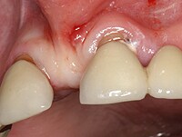
TNF-alpha is a pleiotropic cytokine that is considered as a primary modifier of inflammatory and immune reaction in response to various inflammatory diseases and tumour. We investigated levels of TNF-alpha in 43 radicular cysts and 15 odontogenic keratocysts, obtained from patients undergoing surgery, under local anaesthesia, and after aspiration of cystic fluid from non-ruptured cysts. TNF-alpha is elevated in both cysts' fluid, but higher values were found in radicular cysts in comparison to keratocysts. The significantly higher concentration of TNF-alpha was associated with smaller radicular cysts, higher protein concentration, higher presence of inflammatory cells in peri cystic tissues, and the degree of vascularisation and cysts wall thickness (Mann-Whitney U-test, p < 0.05). No correlation was found based on these parameters in odontogenic keratocyst, but all cysts have detectable concentrations of TNF-alpha. We here for the first time present that a difference in the concentration of TNF-alpha exists between these two cystic types. (+info)A radicular cyst is a type of dental cyst that forms around the root of a tooth, usually as a result of chronic infection or inflammation. It is also known as a periapical cyst. The cyst develops from the accumulation of fluid and cells in the periodontal ligament, which is the tissue that connects the tooth to the jawbone.
Radicular cysts are often caused by untreated dental caries or trauma to the tooth that allows bacteria to enter the pulp chamber of the tooth and cause an infection. Over time, the infection can spread to the surrounding tissues, leading to the formation of a cyst. Symptoms of a radicular cyst may include pain, swelling, and tenderness in the affected area. Treatment typically involves removing the affected tooth and the cyst through a surgical procedure.
Odontogenic cysts are a type of cyst that originates from the dental tissues or odontogenic apparatus. They are typically found in the jawbones, and can be classified as developmental or inflammatory in origin. Developmental odontogenic cysts arise from remnants of the tooth-forming structures, while inflammatory odontogenic cysts result from an infection or injury to a tooth.
The most common types of odontogenic cysts include:
1. Periapical cyst - an inflammatory cyst that forms at the tip of the root of a dead or non-vital tooth.
2. Dentigerous cyst - a developmental cyst that surrounds the crown of an unerupted or impacted tooth.
3. Follicular cyst - a type of dentigerous cyst that forms around the crown of an unerupted wisdom tooth.
4. Odontogenic keratocyst - a developmental cyst that arises from the dental lamina and has a high recurrence rate.
5. Lateral periodontal cyst - a rare, developmental cyst that forms in the periodontal ligament of a vital tooth.
Odontogenic cysts can cause various symptoms such as swelling, pain, or numbness in the affected area. They may also displace or resorb adjacent teeth. Diagnosis is typically made through radiographic imaging and histopathological examination of tissue samples obtained through biopsy. Treatment options include surgical excision, marsupialization (a procedure that creates an opening between the cyst and oral cavity), or enucleation (removal of the cyst lining).
Maxillary diseases refer to conditions that affect the maxilla, which is the upper bone of the jaw. This bone plays an essential role in functions such as biting, chewing, and speaking, and also forms the upper part of the oral cavity, houses the upper teeth, and supports the nose and the eyes.
Maxillary diseases can be caused by various factors, including infections, trauma, tumors, congenital abnormalities, or systemic conditions. Some common maxillary diseases include:
1. Maxillary sinusitis: Inflammation of the maxillary sinuses, which are air-filled cavities located within the maxilla, can cause symptoms such as nasal congestion, facial pain, and headaches.
2. Periodontal disease: Infection and inflammation of the tissues surrounding the teeth, including the gums and the alveolar bone (which is part of the maxilla), can lead to tooth loss and other complications.
3. Maxillary fractures: Trauma to the face can result in fractures of the maxilla, which can cause pain, swelling, and difficulty breathing or speaking.
4. Maxillary cysts and tumors: Abnormal growths in the maxilla can be benign or malignant and may require surgical intervention.
5. Oral cancer: Cancerous lesions in the oral cavity, including the maxilla, can cause pain, swelling, and difficulty swallowing or speaking.
Treatment for maxillary diseases depends on the specific condition and its severity. Treatment options may include antibiotics, surgery, radiation therapy, or chemotherapy. Regular dental check-ups and good oral hygiene practices can help prevent many maxillary diseases.
A dentigerous cyst is a type of odontogenic cyst that forms around the crown of an unerupted tooth. It is typically slow-growing and often asymptomatic, but it can cause displacement or resorption of adjacent teeth if it becomes large enough. Dentigerous cysts are more common in permanent teeth than primary teeth, and they are more likely to occur in the mandible (lower jaw) than the maxilla (upper jaw). They are usually diagnosed through radiographic examination and can be treated by surgical removal of the cyst along with the affected tooth. If left untreated, dentigerous cysts can continue to grow and may eventually develop into a tumor or cancer.
Mandibular diseases refer to conditions that affect the mandible, or lower jawbone. These diseases can be classified as congenital (present at birth) or acquired (developing after birth). They can also be categorized based on the tissues involved, such as bone, muscle, or cartilage. Some examples of mandibular diseases include:
1. Mandibular fractures: These are breaks in the lower jawbone that can result from trauma or injury.
2. Osteomyelitis: This is an infection of the bone and surrounding tissues, which can affect the mandible.
3. Temporomandibular joint (TMJ) disorders: These are conditions that affect the joint that connects the jawbone to the skull, causing pain and limited movement.
4. Mandibular tumors: These are abnormal growths that can be benign or malignant, and can develop in any of the tissues of the mandible.
5. Osteonecrosis: This is a condition where the bone tissue dies due to lack of blood supply, which can affect the mandible.
6. Cleft lip and palate: This is a congenital deformity that affects the development of the face and mouth, including the lower jawbone.
7. Mandibular hypoplasia: This is a condition where the lower jawbone does not develop properly, leading to a small or recessed chin.
8. Developmental disorders: These are conditions that affect the growth and development of the mandible, such as condylar hyperplasia or hemifacial microsomia.
Ameloblastoma is a slow-growing, non-cancerous tumor that develops in the jawbone, typically in the lower jaw. It originates from the cells that form the enamel (the hard, outer surface of the teeth). This tumor can cause swelling, pain, and displacement or loosening of teeth. In some cases, it may also lead to fractures of the jawbone.
There are different types of ameloblastomas, including solid or multicystic, unicystic, and peripheral ameloblastoma. Treatment usually involves surgical removal of the tumor, with careful monitoring to ensure that it does not recur. In rare cases, more aggressive treatment may be necessary if the tumor is large or has invaded surrounding tissues.
It's important to note that while ameloblastomas are generally benign, they can still cause significant morbidity and should be treated promptly by an oral and maxillofacial surgeon or other qualified healthcare professional.
Cyst fluid refers to the fluid accumulated within a cyst, which is a closed sac-like or capsular structure, typically filled with liquid or semi-solid material. Cysts can develop in various parts of the body for different reasons, and the composition of cyst fluid may vary depending on the type of cyst and its location.
In some cases, cyst fluid might contain proteins, sugars, hormones, or even cells from the surrounding tissue. Infected cysts may have pus-like fluid, while cancerous or precancerous cysts might contain abnormal cells or tumor markers. The analysis of cyst fluid can help medical professionals diagnose and manage various medical conditions, including infections, inflammatory diseases, genetic disorders, and cancers.
It is important to note that the term 'cyst fluid' generally refers to the liquid content within a cyst, but the specific composition and appearance of this fluid may vary significantly depending on the underlying cause and type of cyst.
A periapical granuloma is a type of dental lesion that occurs at the root tip of a tooth (the apical region) in response to an infection in the pulp tissue. It is a collection of inflammatory cells, mainly composed of lymphocytes, plasma cells, and histiocytes, within the periodontal ligament and alveolar bone. The granuloma forms as a result of the body's attempt to contain the spread of infection from the pulp into the surrounding tissues.
The primary cause of periapical granulomas is untreated dental caries or tooth trauma, which allows bacteria to invade the pulp chamber and eventually reach the apical region. The resulting inflammation can lead to bone resorption and the formation of a radiolucent area around the apex of the affected tooth, visible on a dental radiograph.
Periapical granulomas may not always cause noticeable symptoms, but some patients might experience pain, swelling, or sensitivity in the affected tooth. Treatment typically involves root canal therapy to remove the infected pulp tissue and medicate the canals, followed by a filling or crown to seal and protect the tooth. In some cases, extraction of the tooth may be necessary if the infection is severe or if the tooth cannot be restored.
Jaw neoplasms refer to abnormal growths or tumors in the jawbone (mandible) or maxilla (upper jaw). These growths can be benign (non-cancerous) or malignant (cancerous). Benign neoplasms are not considered life-threatening, but they can still cause problems by invading nearby tissues and causing damage. Malignant neoplasms, on the other hand, can spread to other parts of the body and can be life-threatening if not treated promptly and effectively.
Jaw neoplasms can present with various symptoms such as swelling, pain, loose teeth, numbness or tingling in the lips or tongue, difficulty chewing or swallowing, and jaw stiffness or limited movement. The diagnosis of jaw neoplasms typically involves a thorough clinical examination, imaging studies such as X-rays, CT scans, or MRI, and sometimes a biopsy to determine the type and extent of the tumor.
Treatment options for jaw neoplasms depend on several factors, including the type, size, location, and stage of the tumor, as well as the patient's overall health and medical history. Treatment may involve surgery, radiation therapy, chemotherapy, or a combination of these modalities. Regular follow-up care is essential to monitor for recurrence or metastasis (spread) of the neoplasm.
A cyst is a closed sac, having a distinct membrane and division between the sac and its surrounding tissue, that contains fluid, air, or semisolid material. Cysts can occur in various parts of the body, including the skin, internal organs, and bones. They can be caused by various factors, such as infection, genetic predisposition, or blockage of a duct or gland. Some cysts may cause symptoms, such as pain or discomfort, while others may not cause any symptoms at all. Treatment for cysts depends on the type and location of the cyst, as well as whether it is causing any problems. Some cysts may go away on their own, while others may need to be drained or removed through a surgical procedure.
Radiculopathy is a medical term that refers to the condition where there is damage or disturbance in the nerve roots as they exit the spinal column. These nerve roots, also known as radicles, can become damaged due to various reasons such as compression, inflammation, or injury, leading to a range of symptoms.
Radiculopathy may occur in any part of the spine, but it is most commonly found in the cervical (neck) and lumbar (lower back) regions. When the nerve roots in the cervical region are affected, it can result in symptoms such as neck pain, shoulder pain, arm pain, numbness, tingling, or weakness in the arms or fingers. On the other hand, when the nerve roots in the lumbar region are affected, it can cause lower back pain, leg pain, numbness, tingling, or weakness in the legs or feet.
The symptoms of radiculopathy can vary depending on the severity and location of the damage to the nerve roots. In some cases, the condition may resolve on its own with rest and conservative treatment. However, in more severe cases, medical intervention such as physical therapy, medication, or surgery may be necessary to alleviate the symptoms and prevent further damage.
Jaw diseases refer to a variety of conditions that affect the temporomandibular joint (TMJ) and the surrounding muscles, as well as dental disorders that can impact the jaw. Some common examples include:
1. Temporomandibular Joint Disorders (TMD): These are problems with the TMJ and the muscles that control jaw movement. Symptoms may include pain, clicking or popping sounds, and limited movement of the jaw.
2. Osteonecrosis of the Jaw: This is a condition where bone in the jaw dies due to lack of blood supply. It can be caused by radiation therapy, chemotherapy, or certain medications.
3. Dental Cavities: These are holes in the teeth caused by bacteria. If left untreated, they can cause pain, infection, and damage to the jawbone.
4. Periodontal Disease: This is an infection of the gums and bones that support the teeth. Advanced periodontal disease can lead to loss of teeth and damage to the jawbone.
5. Jaw Fractures: These are breaks in the jawbone, often caused by trauma.
6. Oral Cancer: This is a type of cancer that starts in the mouth or throat. If not treated early, it can spread to the jaw and other parts of the body.
7. Cysts and Tumors: These are abnormal growths in the jawbone or surrounding tissues. While some are benign (non-cancerous), others can be malignant (cancerous).
8. Osteomyelitis: This is an infection of the bone, often occurring in the lower jaw. It can cause pain, swelling, and fever.
9. Oral Thrush: This is a fungal infection that causes white patches on the inside of the mouth. If left untreated, it can spread to the jaw and other parts of the body.
10. Sinusitis: Inflammation of the sinuses can sometimes cause pain in the upper jaw.
A deciduous tooth, also known as a baby tooth or primary tooth, is a type of temporary tooth that humans and some other mammals develop during childhood. They are called "deciduous" because they are eventually shed and replaced by permanent teeth, much like how leaves on a deciduous tree fall off and are replaced by new growth.
Deciduous teeth begin to form in the womb and start to erupt through the gums when a child is around six months old. By the time a child reaches age three, they typically have a full set of 20 deciduous teeth, including incisors, canines, and molars. These teeth are smaller and less durable than permanent teeth, but they serve important functions such as helping children chew food properly, speak clearly, and maintain space in the jaw for the permanent teeth to grow into.
Deciduous teeth usually begin to fall out around age six or seven, starting with the lower central incisors. This process continues until all of the deciduous teeth have been shed, typically by age 12 or 13. At this point, the permanent teeth will have grown in and taken their place, with the exception of the wisdom teeth, which may not erupt until later in adolescence or early adulthood.
An ovarian cyst is a sac or pouch filled with fluid that forms on the ovary. Ovarian cysts are quite common in women during their childbearing years, and they often cause no symptoms. In most cases, ovarian cysts disappear without treatment over a few months. However, larger or persistent cysts may require medical intervention, including surgical removal.
There are various types of ovarian cysts, such as functional cysts (follicular and corpus luteum cysts), which develop during the menstrual cycle due to hormonal changes, and non-functional cysts (dermoid cysts, endometriomas, and cystadenomas), which can form due to different causes.
While many ovarian cysts are benign, some may have malignant potential or indicate an underlying medical condition like polycystic ovary syndrome (PCOS). Regular gynecological check-ups, including pelvic examinations and ultrasounds, can help detect and monitor ovarian cysts.
An epidermal cyst is a common benign skin condition characterized by the growth of a sac-like structure filled with keratin, a protein found in the outermost layer of the skin (epidermis). These cysts typically appear as round, firm bumps just under the surface of the skin, often on the face, neck, trunk, or scalp. They can vary in size from a few millimeters to several centimeters in diameter.
Epidermal cysts usually develop as a result of the accumulation of dead skin cells that become trapped within a hair follicle or a pilosebaceous unit (a structure that contains a hair follicle and an oil gland). The keratin produced by the skin cells then collects inside the sac, causing it to expand gradually.
These cysts are generally slow-growing, painless, and rarely cause any symptoms. However, they may become infected or inflamed, leading to redness, tenderness, pain, or pus formation. In such cases, medical attention might be necessary to drain the cyst or administer antibiotics to treat the infection.
Epidermal cysts can be removed surgically if they cause cosmetic concerns or become frequently infected. The procedure typically involves making an incision in the skin and removing the entire sac along with its contents to prevent recurrence.
Spinal nerve roots are the initial parts of spinal nerves that emerge from the spinal cord through the intervertebral foramen, which are small openings between each vertebra in the spine. These nerve roots carry motor, sensory, and autonomic fibers to and from specific regions of the body. There are 31 pairs of spinal nerve roots in total, with 8 cervical, 12 thoracic, 5 lumbar, 5 sacral, and 1 coccygeal pair. Each root has a dorsal (posterior) and ventral (anterior) ramus that branch off to form the peripheral nervous system. Irritation or compression of these nerve roots can result in pain, numbness, weakness, or loss of reflexes in the affected area.
Sciatica is not a medical condition itself but rather a symptom of an underlying medical problem. It's typically described as pain that radiates along the sciatic nerve, which runs from your lower back through your hips and buttocks and down each leg.
The pain can vary widely, from a mild ache to a sharp, burning sensation or excruciating discomfort. Sometimes, the pain is severe enough to make moving difficult. Sciatica most commonly occurs when a herniated disk, bone spur on the spine, or narrowing of the spine (spinal stenosis) compresses part of the nerve.
While sciatica can be quite painful, it's not typically a sign of permanent nerve damage and can often be relieved with non-surgical treatments. However, if the pain is severe or persists for a long period, it's essential to seek medical attention as it could indicate a more serious underlying condition.
A Synovial Cyst is a type of benign cyst that typically develops in the synovium, which is the membrane that lines and lubricates joint capsules. These cysts are filled with synovial fluid, which is the same lubricating fluid found inside joints. They usually form as a result of degenerative changes, trauma, or underlying joint diseases such as osteoarthritis.
Synovial cysts commonly occur in the spine (particularly in the facet joints), but they can also develop in other areas of the body, including the knees, hips, and hands. While synovial cysts are generally not harmful, they may cause discomfort or pain if they press on nearby nerves or restrict movement in the affected joint. Treatment options for synovial cysts range from conservative measures like physical therapy and pain management to surgical intervention in severe cases.
Epidural injection is a medical procedure where a medication is injected into the epidural space of the spine. The epidural space is the area between the outer covering of the spinal cord (dura mater) and the vertebral column. This procedure is typically used to provide analgesia (pain relief) or anesthesia for surgical procedures, labor and delivery, or chronic pain management.
The injection usually contains a local anesthetic and/or a steroid medication, which can help reduce inflammation and swelling in the affected area. The medication is delivered through a thin needle that is inserted into the epidural space using the guidance of fluoroscopy or computed tomography (CT) scans.
Epidural injections are commonly used to treat various types of pain, including lower back pain, leg pain (sciatica), and neck pain. They can also be used to diagnose the source of pain by injecting a local anesthetic to numb the area and determine if it is the cause of the pain.
While epidural injections are generally safe, they do carry some risks, such as infection, bleeding, nerve damage, or allergic reactions to the medication. It's important to discuss these risks with your healthcare provider before undergoing the procedure.
An Odontogenic Cyst, Calcifying is a specific type of cyst that originates from the dental tissues. It's also known as a calcifying odontogenic cyst or Gorlin cyst. This cyst is characterized by the presence of calcified structures within its lining.
The calcifications can appear as flecks or more complex structures, such as teeth-like formations. The lining of this cyst often contains ghost cells, which are the remains of epithelial cells that have undergone calcification.
These cysts are typically slow-growing and asymptomatic, although they can sometimes cause swelling or pain if they become large enough to compress adjacent tissues. They are most commonly found in the jaw bones, particularly the mandible.
While the exact cause of calcifying odontogenic cysts is not fully understood, they are thought to arise from developmental abnormalities in the tissues that form teeth. Treatment typically involves surgical removal of the cyst.
Odontogenic tumors are a group of neoplasms that originate from the dental tissues or their remnants, including the odontogenic epithelium, ectomesenchyme, and/or their derivatives. These tumors can be benign or malignant and may affect the jaw bones and surrounding structures. They can cause various symptoms, such as swelling, pain, loosening of teeth, and altered bite. The classification of odontogenic tumors includes a wide range of entities with different biological behaviors, clinical features, and treatment approaches. Accurate diagnosis is essential for proper management and prognosis.
Aneurysmal bone cyst (ABC) is a benign but locally aggressive tumor that typically involves the metaphysis of long bones in children and adolescents. It is characterized by blood-filled spaces or cysts separated by fibrous septa containing osteoclast-type giant cells, spindle cells, and capillary vessels.
ABCs can also arise in other locations such as the vertebral column, pelvis, and skull. They may cause bone pain, swelling, or pathologic fractures. The exact cause of ABC is unknown, but it is thought to be related to a reactive process to a primary bone lesion or trauma.
Treatment options for ABC include curettage and bone grafting, intralesional injection of corticosteroids or bone marrow aspirate, and adjuvant therapy with phenol or liquid nitrogen. In some cases, radiation therapy may be used, but it is generally avoided due to the risk of secondary malignancies. Recurrence rates after treatment range from 10-30%.
 Odontogenic cyst
Periapical cyst
Cysts of the jaws
Glandular odontogenic cyst
Cell counting
Dentigerous cyst
Periapical granuloma
Wisdom tooth
Pulp (tooth)
Periodontal fiber
Pulp necrosis
Odontogenic keratocyst
Toothache
Epithelial cell rests of Malassez
List of MeSH codes (C07)
Tarlov cyst
Dural ectasia
Periapical periodontitis
Lyme disease
Radiculopathy
Spinal stenosis
Dental follicle
List of diseases (S)
Abscess
Low back pain
Sciatica
Nerve compression syndrome
Biphasic calcium sulfate
Dentin dysplasia
List of MeSH codes (C05)
List of MeSH codes (C04)
Dental emergency
 Extensive Radicular Cyst: a Case Report
 Huge Radicular Cyst Leads to Blurred Vision | AVESİS
Radicular cyst removal under general anesthesia: a case report | International Journal of Current Research
Odontogenic cyst - Wikipedia
The use of grafting material biphasic calcium sulfate for the treatment of osseous defects resulting from radicular cysts....
 Volume 13(8) 2007 (17)
 Mandibular Cysts and Odontogenic Tumors: Overview, Odontogenic Mandibular Cysts, Nonodontogenic Mandibular Cysts
 Medical Abbreviations - R - GlobalRPH
 White Spot Lesions: A New Topographic Classification - Dental News
CCR3 | Cancer Genetics Web
 Troy Buck - Articles - Scientific Research Publishing
Troy Buck - Articles - Scientific Research Publishing
 Clinico pathological study of Odontogenic cysts and tumors in a Tertiary care Dental hospital of Nepal
| Journal of...
Clinico pathological study of Odontogenic cysts and tumors in a Tertiary care Dental hospital of Nepal
| Journal of...
 Pubblicazione | SILVIA TORTORICI | Università degli Studi di Palermo
 DT News - International - Non-surgical retreatment following failed apicoectomy with re-use of intra-radicular restoration
 Et tilfelle av sentralt ossifiserende fibrom | Den norske tannlegeforenings Tidende
 Amina Kozaric | IntechOpen
 Poster Gallery - The Morita Prize 2013 - IAPD
 Can wisdom teeth cause permanent damage? (Explained)
 Case Study - United Hospital
Indian Journal of Pathology and Microbiology (IJPM)
 Pesquisa | BVS Odontologia
 Search - MyZerodonto
 Search | Preprints.org
0 Comments
Human Genome Epidemiology Literature Finder|Home|PHGKB
 LCD - Epidural Steroid Injections for Pain Management (L38994)
Volume 13(1) 2007 Supl. (24)
Apoptotic index in periapical cysts with atrophic and hyperplastic epithelium
Frequency of radicular cysts2
- Increased frequency of radicular cysts and conventional ameloblastoma were appreciated with male predisposition in tumours and female predisposition in cysts. (nepjol.info)
- Unlike dentigerous cysts, the frequency of radicular cysts decreased from 10.4% in 1986-1995 to about 8% in 1996-2005 (P (unipa.it)
Granulomas1
- However, granulomas and cysts are not neoplastic. (medscape.com)
Tumors2
- Radicular cyst (associated with the roots of non-vital teeth, also known as Periapical cyst) Residual cyst Most cysts in the body are benign (dysfunctional) tumors, the result of plugged ducts or other natural body outlets for secretions. (wikipedia.org)
- instead, it confines itself to an overview of the major odontogenic cysts and tumors with a brief discussion of other mandibular lesions that are often called cysts but are not true cystic lesions. (medscape.com)
Apical1
- Radicular cyst, apical and lateral ii. (wikipedia.org)
Aneurysmal bone2
- ranging from anatomic variants such as Stafne static bone cyst, to the aggressive aneurysmal bone cyst. (wikipedia.org)
- Aneurysmal bone cyst II. (wikipedia.org)
Inflammatory4
- Background: The radicular cyst is considered to be an inflammatory odontogenic cyst. (journalcra.com)
- Inflammatory collateral cyst B. Non-epithelial-lined cysts 1. (wikipedia.org)
- Inflammatory radicular cysts were observed more in male gender, younger age at diagnosis and anterior maxilla as site of presentation. (unipa.it)
- Inflammatory radicular cysts are the most represented group among OCs in our area with a higher prevalence than that reported in other countries. (unipa.it)
Distribution of odontogenic1
- Prevalence and distribution of odontogenic cyst in Indian population: A 10-year retrospective study. (nepjol.info)
Odontogenic keratocyst3
- However, sometimes these masses are considered neoplasm: Keratocyst Calcifying odontogenic cyst According to the current (2005) classification of the World Health Organization, both (parakeratizied) odontogenic keratocyst and calcifying odontogenic cyst have neoplastic characteristics, thus renamed as Keratocystic odontogenic tumor and Calcifying odontogenic tumor, respectively. (wikipedia.org)
- Cystic ameloblastoma Long standing dentigerous cyst, odontogenic keratocyst, and residual cyst may have neoplastic potential converting into the locally aggressive ameloblastoma, or the malignant squamous cell carcinoma and mucoepidermoid carcinoma. (wikipedia.org)
- common ones include radicular, dentigerous, and odontogenic keratocyst. (precisionstg.com)
Dentigerous cysts2
- Two types of cysts commonly associated with wisdom teeth are dentigerous cysts that involve the crown and radicular cysts that form around the root tips. (cdhp.org)
- Dentigerous cysts usually manifest around emerging teeth trapped inside gums leading to crowding bone loss resulting from delayed/undetected development, besides other complications. (precisionstg.com)
Lesions5
- among the odontogenic lesions, radicular cysts are frequently found in the jaws and are associated with the apex of a tooth with necrotic pulp. (bvsalud.org)
- The removed cyst must be evaluated by pathologist to confirm the diagnosis, and to rule out other neoplastic lesions with similar clinical or radiographic features (e.g., cystic or solid ameloblastoma, central mucoepidermoid carcinoma). (wikipedia.org)
- Fourth edition WHO 2017, classification of Head and Neck lesions, reclassified odontogenic cysts and tumours. (nepjol.info)
- Cases with complete clinical details were included whereas non-odontogenic cysts, oral soft tissue, and salivary gland lesions were excluded. (nepjol.info)
- Odontogenic cysts are a group of common pathological lesions of the jaw. (intechopen.com)
Diagnosis4
- Diagnosis of odontogenic cysts and tumours requires detailed clinical, radiographical, and histopathological findings. (nepjol.info)
- Aspiration of the lesion showed cystic fluid and a clinical diagnosis of a radicular cyst of the jaw was made. (uhlbd.com)
- Also helpful is to make the radiologist aware that a synovial cyst is part of the differential diagnosis because this entity is often overlooked. (medscape.com)
- Extramedullary plasmacytoma of the maxilla simulating a maxillary radicular cyst: quick diagnosis and management. (jomos.org)
Nonsurgical2
- Nonsurgical treatment of radicular cysts]. (bvsalud.org)
- Epidural steroid injections (ESIs) have been endorsed by the North American Spine Society and the Agency for Healthcare Research and Quality (formerly, the Agency for Health Care Policy and Research) of the Department of Health and Human Services as an integral part of nonsurgical management of radicular pain from lumbar spine disorders. (medscape.com)
Surgically removed2
- The residual roots and cysts were surgically removed. (bvsalud.org)
- Cysts may need to be surgically removed along with the wisdom tooth. (cdhp.org)
Granuloma2
- Eventually, this epithelium undergoes necrosis caused by a lack of blood supply, and the granuloma becomes a cyst. (medscape.com)
- Radiographically, distinguishing between a granuloma and a cyst is impossible, although some say that if the lesion is quite large it is more likely to be a cyst. (medscape.com)
Mandibular2
- One of the cysts measured approximately 4 x 2 cm and was associated with a residual root in the right mandibular molar area. (bvsalud.org)
- The other cyst measured approximately 1.7 x 1.3 cm and was associated with a residual root in the left mandibular premolar area. (bvsalud.org)
Synovial Cyst2
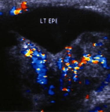
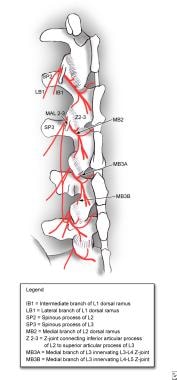
- MRI is particularly useful for the evaluation of a synovial cyst emanating from a Z-joint and for distinguishing a synovial cyst from other abnormalities. (medscape.com)
- Gadolinium enhancement is useful in the evaluation of a potential synovial cyst. (medscape.com)
Endodontic therapy1
- Many cysts resolve with endodontic therapy of the involved tooth. (medscape.com)
Pathological2
- To know relative frequency of odontogenic cysts and tumours according to WHO 2017 classification and to know their clinico-pathological characteristics in selected population of Nepal. (nepjol.info)
- Prevalence of odontogenic cysts and tumours: A retrospective clinico-pathological study of 204 cases. (nepjol.info)
Epithelial6
- Intra-bony cysts are most common in the jaws, because the mandible and maxilla are the only bones with epithelial components. (wikipedia.org)
- However, epithelial rests may be the origin for the cyst lining later. (wikipedia.org)
- I. Cysts of the jaws A. Epithelial-lined cysts 1. (wikipedia.org)
- Odontogenic cysts are defined as epithelial-lined structures derived from odontogenic epithelium. (medscape.com)
- MMPs) are expressed in epithelial lining of the cyst. (intechopen.com)
- Methodology: 15 samples of periapical cyst with most of the epithelial lining represented by the atrophic type and 15 samples with most of the epithelium lining represented by the hyperplastic type were selected, all originating in the Anatomical Pathology Laboratory of the Dental School, Pontificia Universidade Católica of Minas Gerais. (bvsalud.org)
Tooth4
- Odontogenic cyst are a group of jaw cysts that are formed from tissues involved in odontogenesis (tooth development). (wikipedia.org)
- The second most common odontogenic cyst is the dentigerous cyst, which develops within the normal dental follicle that surrounds an unerupted tooth. (medscape.com)
- These cysts develop in the jawbone tissue surrounding the impacted tooth and can damage neighboring teeth and bone. (cdhp.org)
- Radicular cysts emerge from a tooth experiencing pulp necrosis, often related to untreated cavities, tooth roots, or trauma. (precisionstg.com)
Maxilla1
- The objective of this paper is to report a classic case of radicular cyst located in the anterior region of maxilla. (journalcra.com)
Ameloblastoma1
- Radicular cyst (49/120, 40.83%) and conventional ameloblastoma (23/43 53.48%) were the commonest cysts and tumours. (nepjol.info)
Tissues3
- Cysts of the soft tissues of the mouth, face and neck 1. (wikipedia.org)
- Although these cysts arise from a mature resting epithelium and thus have a relatively low growth potential, a squamous cell carcinoma occasionally may arise de novo in a radicular cyst, thus the recommendation for histopathologic examination of all tissues removed. (medscape.com)
- Upon requiring surgery, the cysts removal procedure involves the removal of healthy tissue around the infected tissues promoting proper cavity restoration. (precisionstg.com)
Epithelium3
- Odontogenic cysts are closed sacs, and have a distinct membrane derived from rests of odontogenic epithelium. (wikipedia.org)
- Oral cysts with gastric or intestinal epithelium (oral alimentary tract cyst) 6. (wikipedia.org)
- Objectives: This study describes the occurrence of apoptosis in periapical cysts with atrophic and hyperplastic epithelium, making a morphologic study quantifying the apoptotic indices and verifying the quantitative differences between them. (bvsalud.org)
Jaws1
- trichinosis Buccal bifurcation cyst Calcifying odontogenic cyst Dentigerous cyst (associated with the crowns of non-erupted teeth) Glandular odontogenic cyst Keratocyst (in the jaws, these can appear solitary or associated with the Gorlin-Goltz or Nevoid basal cell carcinoma syndrome. (wikipedia.org)
Cystic1
- Cystic fibrosis is an example of a genetic disorder whereby cysts and fibrosis develop in the lungs. (wikidoc.org)
Sacs1
- Fluid-filled sacs called cysts sometimes form around impacted wisdom teeth . (cdhp.org)
Renal1
- and renal, pancreatic, and epididymal cysts. (medscape.com)
Benign1
- Among odontogenic cysts with benign pathology, up to 60% of all jaw cysts are radicular cysts, which originate from root canal infection. (intechopen.com)
Neoplastic1
- The dentigerous cyst is not thought to be neoplastic. (medscape.com)
Prevalence3
- Prevalence of odontogenic cysts and tumours on turkish sample according to latest classification of world health organisation: A 10-year retrospective study. (nepjol.info)
- Prevalence of odontogenic cysts and tumours among UAE population. (nepjol.info)
- The objective of this study was to assess the prevalence of odontogenic cysts (OCs) in Sicily and evaluate their distribution during a 20-year period. (unipa.it)
Bone2
- Solitary bone cyst 2. (wikipedia.org)
- In the above mentioned case, surgical inoculation of the cyst was done under GA with bone guttering. (uhlbd.com)
Residual1
- Residual cyst iii. (wikipedia.org)
Maxillary2
- Cysts associated with the maxillary antrum 1. (wikipedia.org)
- Postoperative maxillary cyst III. (wikipedia.org)
Roots2
- Inflammation within the epidural space and nerve roots, as can be provoked by a herniated disk, is a significant factor in causing radicular pain. (medscape.com)
- These cysts usually form around dental roots or in the areas around teeth and jawbones. (precisionstg.com)
Commonly1
- Unfortunately, one painful scenario is when fluid-filled sac forms from injuries from food particles, infections, etc., occur, known commonly as odontogenic cysts. (precisionstg.com)
Anterior1
- Anterior median lingual cyst (intralingual cyst of foregut origin) 5. (wikipedia.org)
Defects1
- The use of grafting material biphasic calcium sulfate for the treatment of osseous defects resulting from radicular cysts. (augmabio.com)
Lateral1
- Developmental lateral periodontal cyst vii. (wikipedia.org)
Vessels1
- Note the stretching of vessels around the cyst. (medscape.com)
Teeth1
- Radicular cysts are usually slow growing and asymptomatic, unless infected or has clinical signs like swelling and teeth displacement. (uhlbd.com)
Retrospective study1
- A cross-sectional retrospective study was carried out in 1,310 cysts of the jaw diagnosed in 12,197 individuals, who consecutively attended the Odontostomatologic Clinic of Palermo from 1986 to 2005. (unipa.it)
Treatment6
- Treatment ranges from simple enucleation of the cyst to curettage to resection. (wikipedia.org)
- For example, small radicular cyst may resolved after successful endodontic ("root-canal") treatment. (wikipedia.org)
- However, the conservative enucleation is the treatment of choice for most odontogenic cysts. (wikipedia.org)
- Several treatment options exist for such cysts. (medscape.com)
- Treatment options include marsupialization of the cyst or enucleation of the cyst along with its lining. (uhlbd.com)
- If delayed, these cysts may require immediate treatment, such as surgeries or treatment through oral medications. (precisionstg.com)
Distinct membrane1
- A cyst is a closed sac having a distinct membrane and division on the nearby tissue. (wikidoc.org)
Arise1
- Do not hesitate to consult our team immediately if any suspicions arise regarding odontogenic cyst formation so our experts can diagnose/treat accordingly. (precisionstg.com)
Resolve1
- Once formed, the cyst will remain in the tissue but can be removed by surgery or resolve by taking medications. (wikidoc.org)
Case2
- This case report presents two radicular cysts in a 55-year-old patient. (bvsalud.org)
- A cyst may also be a sack that encloses an organism during a dormant period, such as in the case of certain parasites. (wikidoc.org)
Pulp1
- Radicular dentin tubules extend from the pulp to the cemento-dentinal junction (CDJ) (1). (docshare.tips)
Common3
- Paradental cyst Periapical cyst (The periapical cyst, otherwise known as radicular cyst, is the most common odontogenic cyst. (wikipedia.org)
- A periapical (radicular) cyst is the most common odontogenic cyst. (medscape.com)
- Both cysts and tumours were common in second to third decade of life affecting middle and posterior region of mandible. (nepjol.info)
Occur1
- Similar mechanisms of radicular pain are postulated to occur in the thoracic and cervical spine as well. (medscape.com)
Medications1
- Complete oral hygiene is key to preventing odontogenic cysts from ever manifesting, reducing the need for surgery\medications as long as proper hygiene protocols are followed. (precisionstg.com)
Root5
- Radicular pain often is the result of nerve root inflammation with or without mechanical irritation. (medscape.com)
- Historical evidence of nerve root inflammation has been demonstrated during surgery in patients with radicular low back pain (LBP) from lumbar disk herniation. (medscape.com)
- The radicular LBP caused by spinal stenosis is probably related to the inhibition of normal nerve root vascular flow with resultant nerve root nutrition, nerve root edema, and nerve root dysfunction. (medscape.com)
- If dorsal root ganglia are chronically compressed and irritated, this theoretically can lead to their sensitization and resultant radicular pain. (medscape.com)
- In summary, clinical practice and animal research suggest that radicular pain is the result of inflammation of the nerve root in the epidural space provoked by leakage of disk material, compression of the nerve root vasculature, and/or irritation of dorsal root ganglia from spinal stenosis. (medscape.com)
Enhancement1
- Coronal vertebral angiogram of the same patient as in the previous 2 images shows a hypervascular intramural nodule that demonstrates a prolonged and intense enhancement with a surrounding avascular area, which represents the cyst surrounding the mural nodule. (medscape.com)
Oral4
- Not all oral cysts are odontogenic cysts. (wikipedia.org)
- For example, mucous cyst of the oral mucosa and nasolabial duct cyst are not of odontogenic origin. (wikipedia.org)
- Cysts of the oral and maxillofacial regions (4th ed. (wikipedia.org)
- En 51 år gammel kvinne ble henvist til spesialist i oral kirurgi for fjernelse av en oppklaring på ca. 2 cm i diameter rundt rotspissen av 36. (tannlegetidende.no)