Reverse Transcriptase Polymerase Chain Reaction
Polymerase Chain Reaction
RNA, Messenger
Base Sequence
Molecular Sequence Data
HIV Reverse Transcriptase
DNA Primers
RNA-Directed DNA Polymerase
Gene Expression
Reverse Transcriptase Inhibitors
Sensitivity and Specificity
Neoplastic Cells, Circulating
Immunohistochemistry
Amino Acid Sequence
Real-Time Polymerase Chain Reaction
DNA, Complementary
Transcription, Genetic
Cells, Cultured
Gene Expression Profiling
Cloning, Molecular
In Situ Hybridization
Rhabdomyosarcoma, Embryonal
Blotting, Southern
DNA-Directed DNA Polymerase
Neoplasm Proteins
Blotting, Western
Encephalomyelitis, Western Equine
Gene Expression Regulation, Neoplastic
Rhabdomyosarcoma, Alveolar
Monophenol Monooxygenase
Gene Expression Regulation
Tumor Markers, Biological
Up-Regulation
Tumor Cells, Cultured
Sequence Analysis, DNA
Mutation
Translocation, Genetic
RNA
Oncogene Proteins, Fusion
Exons
Oligonucleotide Array Sequence Analysis
DNA
Neoplasm, Residual
Blotting, Northern
RNA Polymerase II
Genotype
DNA-Binding Proteins
Prognosis
Tissue Distribution
Immunoenzyme Techniques
Sequence Homology, Amino Acid
Flow Cytometry
Enzyme-Linked Immunosorbent Assay
Rats, Sprague-Dawley
Melanoma
Sarcoma, Synovial
Gene Rearrangement
Biopsy
Transcription Factors
Bone Marrow
Templates, Genetic
Sarcoma, Ewing
Sequence Homology, Nucleic Acid
Sequence Alignment
Keratins
Promoter Regions, Genetic
Transfection
Down-Regulation
In Situ Hybridization, Fluorescence
Oligodeoxyribonucleotides
Gene Expression Regulation, Enzymologic
DNA-Directed RNA Polymerases
Cytokines
Phenotype
Telomerase
HIV-1
Alternative Splicing
Liver
Oligonucleotides, Antisense
Mice, Inbred C57BL
Cattle
DNA Polymerase I
Gene Expression Regulation, Developmental
Antigens, Neoplasm
Transforming Growth Factor beta
Case-Control Studies
Disease Outbreaks
Neoplasm Staging
Apoptosis
Drug Resistance, Multiple
Reproducibility of Results
Interleukin-1
Oligonucleotide Probes
Tumor Necrosis Factor-alpha
Pregnancy
Leukocytes, Mononuclear
Skin
Disease Models, Animal
Prospective Studies
Keratinocytes
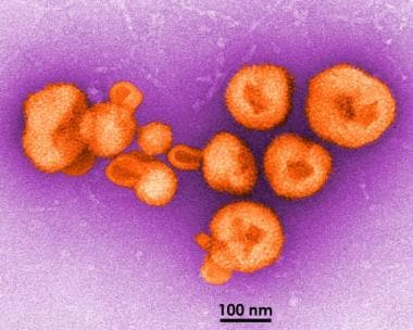
Influenza, Human
Swine
Dose-Response Relationship, Drug
Rats, Wistar
P-Glycoprotein
Cell Differentiation
DNA Polymerase II
Survival Analysis
Testis
Influenza A Virus, H1N1 Subtype
Precursor Cell Lymphoblastic Leukemia-Lymphoma
Interleukin-6
Predictive Value of Tests
Chickens
MicroRNAs
Membrane Proteins
Ribonuclease H
Protein Isoforms
Isoenzymes
Lymph Nodes
Organ Specificity
Fibroblasts
Epithelial Cells
Polymorphism, Restriction Fragment Length
Multiplex Polymerase Chain Reaction
DNA Polymerase III
Signal Transduction
Cell Division
Gene Amplification
Immunoblotting
Escherichia coli
RNA, Small Interfering
Proteins
Cluster Analysis
Electrophoresis, Agar Gel
Biological Markers
Mice, Inbred BALB C
Statistics, Nonparametric
Anti-HIV Agents
Interleukin-8
Polymorphism, Genetic
DNA Probes
Interleukin-10
Disease Progression
Alleles
Interferon-gamma
Zidovudine
Colorectal Neoplasms
Antigens, CD
Brain
Lymphatic Metastasis
Proto-Oncogene Proteins
Neoplasm Metastasis
Drug Resistance, Viral
Vascular Endothelial Growth Factor A
Cell Survival
Rabbits
Treatment Outcome
RNA Polymerase I
RNA Polymerase III
Homeodomain Proteins
T-Lymphocytes
Immunoglobulin Heavy Chains
Nucleic Acid Hybridization
DNA Polymerase beta
Neoplasm Recurrence, Local
Lung
Cell Movement
Retrospective Studies
Restriction Mapping
Thymine Nucleotides
Virus Replication
Lipopolysaccharides
Alternative sulfonylurea receptor expression defines metabolic sensitivity of K-ATP channels in dopaminergic midbrain neurons. (1/55562)
ATP-sensitive potassium (K-ATP) channels couple the metabolic state to cellular excitability in various tissues. Several isoforms of the K-ATP channel subunits, the sulfonylurea receptor (SUR) and inwardly rectifying K channel (Kir6.X), have been cloned, but the molecular composition and functional diversity of native neuronal K-ATP channels remain unresolved. We combined functional analysis of K-ATP channels with expression profiling of K-ATP subunits at the level of single substantia nigra (SN) neurons in mouse brain slices using an RT-multiplex PCR protocol. In contrast to GABAergic neurons, single dopaminergic SN neurons displayed alternative co-expression of either SUR1, SUR2B or both SUR isoforms with Kir6.2. Dopaminergic SN neurons expressed alternative K-ATP channel species distinguished by significant differences in sulfonylurea affinity and metabolic sensitivity. In single dopaminergic SN neurons, co-expression of SUR1 + Kir6.2, but not of SUR2B + Kir6.2, correlated with functional K-ATP channels highly sensitive to metabolic inhibition. In contrast to wild-type, surviving dopaminergic SN neurons of homozygous weaver mouse exclusively expressed SUR1 + Kir6.2 during the active period of dopaminergic neurodegeneration. Therefore, alternative expression of K-ATP channel subunits defines the differential response to metabolic stress and constitutes a novel candidate mechanism for the differential vulnerability of dopaminergic neurons in response to respiratory chain dysfunction in Parkinson's disease. (+info)Anopheles gambiae Ag-STAT, a new insect member of the STAT family, is activated in response to bacterial infection. (2/55562)
A new insect member of the STAT family of transcription factors (Ag-STAT) has been cloned from the human malaria vector Anopheles gambiae. The domain involved in DNA interaction and the SH2 domain are well conserved. Ag-STAT is most similar to Drosophila D-STAT and to vertebrate STATs 5 and 6, constituting a proposed ancient class A of the STAT family. The mRNA is expressed at all developmental stages, and the protein is present in hemocytes, pericardial cells, midgut, skeletal muscle and fat body cells. There is no evidence of transcriptional activation following bacterial challenge. However, bacterial challenge results in nuclear translocation of Ag-STAT protein in fat body cells and induction of DNA-binding activity that recognizes a STAT target site. In vitro treatment with pervanadate (vanadate and H2O2) translocates Ag-STAT to the nucleus in midgut epithelial cells. This is the first evidence of direct participation of the STAT pathway in immune responses in insects. (+info)Expression of novel alternatively spliced isoforms of the oct-1 transcription factor. (3/55562)
Analysis of the alternatively spliced isoforms of the human and mouse oct-1 genes, combined with their exon-intron structure, show a high level of evolutionary conservation between these two species. The differential expression of several oct-1 isoforms was examined by reverse transcription-polymerase chain reaction performed on the 3' region of the murine oct-1 cDNA. Variations in the relative levels and patterns of expression of the isoforms were found among different tissues. Three novel isoforms originating from the 3'-distal region of oct-1, were isolated and sequenced: Two were derived from testis, and one from myeloma cells. Splicing out of different exons as revealed in the structure of these isoforms results in reading frameshifts that presumably lead to the expression of shortened Oct-1 proteins, with distinct C-terminal tails. Altogether, six out of the eight known murine oct-1 isoforms may have distinct C-termini, implying that these multiple tails have different functional roles in cellular differentiation and physiology. (+info)Chemokine mRNA expression in gastric mucosa is associated with Helicobacter pylori cagA positivity and severity of gastritis. (4/55562)
AIM: To investigate the association between the quantity of gastric chemokine mRNA expression, severity of gastritis, and cagA positivity in Helicobacter pylori associated gastritis. METHODS: In 83 dyspeptic patients, antral and corpus biopsies were taken for semiquantitative reverse transcription polymerase chain reaction (RT-PCR) and histological grading of gastritis. Gastritis was evaluated by visual analogue scales. Quantities of chemokine (IL-8, GRO alpha, ENA-78, RANTES, MCP-1) RT-PCR products were compared with G3PDH products. Each sample was also evaluated for the presence of cagA and ureA mRNA by RT-PCR. RESULTS: mRNA expression of all five chemokines was significantly greater in H pylori positive than in H pylori negative mucosa. In H pylori positive patients, in the antrum C-X-C chemokine mRNA expression was significantly greater in cagA positive patients than in cagA negative patients, but there were no significant differences in C-C chemokine mRNA expression. In H pylori positive patients, chemokine mRNA expression in the corpus was less than in the antrum. In contrast to the antrum, only GRO alpha mRNA expression was significantly greater in cagA positive infection. Polymorphonuclear cell infiltration was correlated with C-X-C chemokine mRNA expression. Significant correlations were also found between bacterial density and C-X-C chemokine mRNA expression. CONCLUSIONS: In H pylori infection, C-X-C chemokines may play a primary role in active gastritis. Infection with cagA positive H pylori induces greater gastric chemokine mRNA expression in the antral mucosa, which may be relevant to the increased mucosal damage associated with cagA positive H pylori infection. (+info)The role of alternative splicing of the adhesion molecule, CD44, in lymphoid malignancy. (5/55562)
AIM: To investigate the expression of CD44 isoforms containing variant exon 6 (v6) in a well characterised cohort of patients with non-Hodgkin's lymphoma (NHL) and chronic lymphocytic leukaemia (CLL), and to correlate this with phenotype and disease course. METHODS: Cryostat sections of OCT embedded diagnostic nodal material from NHL patients and cryopreserved mononuclear preparations from CLL patients were used as sources of RNA. After reverse transcription, PCR was carried out with amplimers positioned at either side of the variant exon insertion site to amplify all possible CD44 isoforms. Those isoforms containing v6 were identified after Southern blotting and hybridisation with a radiolabelled oligonucleotide. RESULTS: Of 32 NHL samples analysed, 16 did not express CD44 isoforms containing v6, six expressed an isoform containing exon v6 alone, and 10 expressed v6 long isoforms which contained exon v6 in addition to other variant exons. These data did not correlate with lymphoma classification, disease staging, or the presence or absence of extranodal disease. However, those patients expressing v6 long CD44 isoforms had a worse overall survival than those that did not. The plateau of the survival curves was 50% compared with 82%. No v6 long isoforms were detected in the 21 CLL samples investigated. CONCLUSIONS: The expression of v6 long CD44 isoforms is associated with aggressive disease in NHL, independent of grade, stage, or presence of extranodal disease. (+info)Transcriptional regulation and induction of apoptosis: implications for the use of monomeric p53 variants in gene therapy. (6/55562)
The p53 tumour suppressor protein is a transcriptional activator, which can induce cell cycle arrest and apoptosis. p53 Gene mutations occur in more than 50% of all human tumours. Reintroduction of wild-type p53 but also of oligomerisation-independent p53 variants into tumour cells by gene transfer methods has been considered. We have investigated the biological properties of two carboxy-terminal deletion mutants of p53, p53 delta 300 (comprising amino acids 1-300) and p53 delta 326 (amino acids 1-326), to evaluate their potential deployment in gene therapy. Transactivation was measured in transiently transfected HeLa and SKBR3 cells. Both monomeric variants showed reduced activities compared with wild-type p53. Individual promoters were differently affected. In contrast to wild-type p53, monomeric variants were not able to induce apoptosis. We also provided wild-type p53 and p53 delta 326 with tetracycline-regulated promoters and stably introduced these constructs into Saos2 and SKBR3 cells. Upon induction, wild-type p53 expressing cells, but not p53 delta 326 expressing cells underwent apoptosis. Consistently, only wild-type p53 expressing cells accumulated p21/waf1/cip1 mRNA and protein and showed increased bax, Gadd45 and mdm2 mRNA. Neither wild-type p53 nor p53 delta 326 repressed the transcription of the IGF-1R gene in these cell lines. We conclude that the transactivation potential of monomeric, carboxy-terminally truncated p53 is not sufficient to cause induction of the endogenous target genes which trigger apoptosis. (+info)Astrocyte-specific expression of tyrosine hydroxylase after intracerebral gene transfer induces behavioral recovery in experimental parkinsonism. (7/55562)
Parkinson's disease is a neurodegenerative disorder characterized by the depletion of dopamine in the caudate putamen. Dopamine replacement with levodopa, a precursor of the neurotransmitter, is presently the most common treatment for this disease. However, in an effort to obtain better therapeutic results, tissue or cells that synthesize catecholamines have been grafted into experimental animals and human patients. In this paper, we present a novel technique to express tyrosine hydroxylase (TH) in the host's own astrocytes. This procedure uses a transgene in which the expression of a TH cDNA is under the control of a glial fibrillary acidic protein (GFAP) promoter, which confers astrocyte-specific expression and also increases its activity in response to brain injury. The method was tested in a rat model of Parkinson's disease produced by lesioning the striatum with 6-hydroxydopamine. Following microinjection of the transgene into the denervated striatum as a DNA-liposome complex, expression of the transgene was detected by RT-PCR and TH protein was observed specifically in astrocytes by using double-labeling immunofluorescence for GFAP and TH coupled with laser confocal microscopy. Efficacy was demonstrated by significant behavioral recovery, as assessed by a decrease in the pharmacologically induced turning behavior generated by the unilateral denervation of the rat striatum. These results suggest this is a valuable technique to express molecules of therapeutic interest in the brain. (+info)Increased expression of fibroblast growth factor 8 in human breast cancer. (8/55562)
Fibroblast growth factor 8 (FGF8) is an important developmental protein which is oncogenic and able to cooperate with wnt-1 to produce mouse mammary carcinoma. The level of expression of FGF8 mRNA was measured in 68 breast cancers and 24 non-malignant breast tissues. Elevated levels of FGF8 mRNA were found in malignant compared to non-malignant breast tissues with significantly more malignant tissues expressing FGF8 (P=0.019) at significantly higher levels (P=0.031). In situ hybridization of breast cancer tissues and analysis of purified populations of normal epithelial cells and breast cancer cell lines showed that malignant epithelial cells expressed FGF8 mRNA at high levels compared to non-malignant epithelial and myoepithelial cells and fibroblasts. Although two of the receptors which FGF8 binds to (FGFR2-IIIc, FGFR3-IIIc) are not expressed in breast cancer cells, an autocrine activation loop is possible since expression of fibroblast growth factor receptor (FGFR) 4 and FGFR1 are retained in malignant epithelial cells. This is the first member of the FGF family to have increased expression in breast cancer and a potential autocrine role in its progression. (+info)Reverse Transcriptase Polymerase Chain Reaction (RT-PCR) is a laboratory technique used in molecular biology to amplify and detect specific DNA sequences. This technique is particularly useful for the detection and quantification of RNA viruses, as well as for the analysis of gene expression.
The process involves two main steps: reverse transcription and polymerase chain reaction (PCR). In the first step, reverse transcriptase enzyme is used to convert RNA into complementary DNA (cDNA) by reading the template provided by the RNA molecule. This cDNA then serves as a template for the PCR amplification step.
In the second step, the PCR reaction uses two primers that flank the target DNA sequence and a thermostable polymerase enzyme to repeatedly copy the targeted cDNA sequence. The reaction mixture is heated and cooled in cycles, allowing the primers to anneal to the template, and the polymerase to extend the new strand. This results in exponential amplification of the target DNA sequence, making it possible to detect even small amounts of RNA or cDNA.
RT-PCR is a sensitive and specific technique that has many applications in medical research and diagnostics, including the detection of viruses such as HIV, hepatitis C virus, and SARS-CoV-2 (the virus that causes COVID-19). It can also be used to study gene expression, identify genetic mutations, and diagnose genetic disorders.
Polymerase Chain Reaction (PCR) is a laboratory technique used to amplify specific regions of DNA. It enables the production of thousands to millions of copies of a particular DNA sequence in a rapid and efficient manner, making it an essential tool in various fields such as molecular biology, medical diagnostics, forensic science, and research.
The PCR process involves repeated cycles of heating and cooling to separate the DNA strands, allow primers (short sequences of single-stranded DNA) to attach to the target regions, and extend these primers using an enzyme called Taq polymerase, resulting in the exponential amplification of the desired DNA segment.
In a medical context, PCR is often used for detecting and quantifying specific pathogens (viruses, bacteria, fungi, or parasites) in clinical samples, identifying genetic mutations or polymorphisms associated with diseases, monitoring disease progression, and evaluating treatment effectiveness.
Messenger RNA (mRNA) is a type of RNA (ribonucleic acid) that carries genetic information copied from DNA in the form of a series of three-base code "words," each of which specifies a particular amino acid. This information is used by the cell's machinery to construct proteins, a process known as translation. After being transcribed from DNA, mRNA travels out of the nucleus to the ribosomes in the cytoplasm where protein synthesis occurs. Once the protein has been synthesized, the mRNA may be degraded and recycled. Post-transcriptional modifications can also occur to mRNA, such as alternative splicing and addition of a 5' cap and a poly(A) tail, which can affect its stability, localization, and translation efficiency.
A base sequence in the context of molecular biology refers to the specific order of nucleotides in a DNA or RNA molecule. In DNA, these nucleotides are adenine (A), guanine (G), cytosine (C), and thymine (T). In RNA, uracil (U) takes the place of thymine. The base sequence contains genetic information that is transcribed into RNA and ultimately translated into proteins. It is the exact order of these bases that determines the genetic code and thus the function of the DNA or RNA molecule.
Molecular sequence data refers to the specific arrangement of molecules, most commonly nucleotides in DNA or RNA, or amino acids in proteins, that make up a biological macromolecule. This data is generated through laboratory techniques such as sequencing, and provides information about the exact order of the constituent molecules. This data is crucial in various fields of biology, including genetics, evolution, and molecular biology, allowing for comparisons between different organisms, identification of genetic variations, and studies of gene function and regulation.
HIV Reverse Transcriptase is an enzyme that is encoded by the HIV-1 and HIV-2 viruses. It plays a crucial role in the replication cycle of the human immunodeficiency virus (HIV), which causes AIDS.
Reverse transcriptase is responsible for transcribing the viral RNA genome into DNA, a process known as reverse transcription. This allows the viral genetic material to integrate into the host cell's DNA and replicate along with it, leading to the production of new virus particles.
The enzyme has three distinct activities: a polymerase activity that synthesizes DNA using RNA as a template, an RNase H activity that degrades the RNA template during reverse transcription, and a DNA-dependent DNA polymerase activity that synthesizes DNA using a DNA template.
Reverse transcriptase inhibitors are a class of antiretroviral drugs used to treat HIV infection. They work by binding to and inhibiting the activity of the reverse transcriptase enzyme, thereby preventing the virus from replicating.
DNA primers are short single-stranded DNA molecules that serve as a starting point for DNA synthesis. They are typically used in laboratory techniques such as the polymerase chain reaction (PCR) and DNA sequencing. The primer binds to a complementary sequence on the DNA template through base pairing, providing a free 3'-hydroxyl group for the DNA polymerase enzyme to add nucleotides and synthesize a new strand of DNA. This allows for specific and targeted amplification or analysis of a particular region of interest within a larger DNA molecule.
RNA-directed DNA polymerase is a type of enzyme that can synthesize DNA using an RNA molecule as a template. This process is called reverse transcription, and it is the mechanism by which retroviruses, such as HIV, replicate their genetic material. The enzyme responsible for this reaction in retroviruses is called reverse transcriptase.
Reverse transcriptase is an important target for antiretroviral therapy used to treat HIV infection and AIDS. In addition to its role in viral replication, RNA-directed DNA polymerase also has applications in molecular biology research, such as in the production of complementary DNA (cDNA) copies of RNA molecules for use in downstream applications like cloning and sequencing.
Gene expression is the process by which the information encoded in a gene is used to synthesize a functional gene product, such as a protein or RNA molecule. This process involves several steps: transcription, RNA processing, and translation. During transcription, the genetic information in DNA is copied into a complementary RNA molecule, known as messenger RNA (mRNA). The mRNA then undergoes RNA processing, which includes adding a cap and tail to the mRNA and splicing out non-coding regions called introns. The resulting mature mRNA is then translated into a protein on ribosomes in the cytoplasm through the process of translation.
The regulation of gene expression is a complex and highly controlled process that allows cells to respond to changes in their environment, such as growth factors, hormones, and stress signals. This regulation can occur at various stages of gene expression, including transcriptional activation or repression, RNA processing, mRNA stability, and translation. Dysregulation of gene expression has been implicated in many diseases, including cancer, genetic disorders, and neurological conditions.
Reverse Transcriptase Inhibitors (RTIs) are a class of antiretroviral drugs that are primarily used in the treatment and management of HIV (Human Immunodeficiency Virus) infection. They work by inhibiting the reverse transcriptase enzyme, which is essential for the replication of HIV.
HIV is a retrovirus, meaning it has an RNA genome and uses a unique enzyme called reverse transcriptase to convert its RNA into DNA. This process is necessary for the virus to integrate into the host cell's genome and replicate. Reverse Transcriptase Inhibitors interfere with this process by binding to the reverse transcriptase enzyme, preventing it from converting the viral RNA into DNA.
RTIs can be further divided into two categories: nucleoside/nucleotide reverse transcriptase inhibitors (NRTIs) and non-nucleoside reverse transcriptase inhibitors (NNRTIs). NRTIs are analogs of the building blocks of DNA, which get incorporated into the growing DNA chain during replication, causing termination of the chain. NNRTIs bind directly to the reverse transcriptase enzyme, causing a conformational change that prevents it from functioning.
By inhibiting the reverse transcriptase enzyme, RTIs can prevent the virus from replicating and reduce the viral load in an infected individual, thereby slowing down the progression of HIV infection and AIDS (Acquired Immunodeficiency Syndrome).
Sensitivity and specificity are statistical measures used to describe the performance of a diagnostic test or screening tool in identifying true positive and true negative results.
* Sensitivity refers to the proportion of people who have a particular condition (true positives) who are correctly identified by the test. It is also known as the "true positive rate" or "recall." A highly sensitive test will identify most or all of the people with the condition, but may also produce more false positives.
* Specificity refers to the proportion of people who do not have a particular condition (true negatives) who are correctly identified by the test. It is also known as the "true negative rate." A highly specific test will identify most or all of the people without the condition, but may also produce more false negatives.
In medical testing, both sensitivity and specificity are important considerations when evaluating a diagnostic test. High sensitivity is desirable for screening tests that aim to identify as many cases of a condition as possible, while high specificity is desirable for confirmatory tests that aim to rule out the condition in people who do not have it.
It's worth noting that sensitivity and specificity are often influenced by factors such as the prevalence of the condition in the population being tested, the threshold used to define a positive result, and the reliability and validity of the test itself. Therefore, it's important to consider these factors when interpreting the results of a diagnostic test.
Circulating neoplastic cells (CNCs) are defined as malignant cancer cells that have detached from the primary tumor site and are found circulating in the peripheral blood. These cells have undergone genetic and epigenetic changes, leading to uncontrolled cell growth and division, and can form new tumors at distant sites in the body, a process known as metastasis.
The presence of CNCs has been shown to be a prognostic factor for poor outcomes in various types of cancer, including breast, colon, and prostate cancer. The detection and characterization of CNCs can provide valuable information about the tumor's biology, aggressiveness, and response to therapy, allowing for more personalized treatment approaches.
However, the detection of CNCs is challenging due to their rarity in the bloodstream, with only a few cells present among billions of normal blood cells. Therefore, highly sensitive methods such as flow cytometry, polymerase chain reaction (PCR), and next-generation sequencing are used for their identification and quantification.
RNA (Ribonucleic acid) is a single-stranded molecule similar in structure to DNA, involved in the process of protein synthesis in the cell. It acts as a messenger carrying genetic information from DNA to the ribosomes, where proteins are produced.
A neoplasm, on the other hand, is an abnormal growth of cells, which can be benign or malignant. Benign neoplasms are not cancerous and do not invade nearby tissues or spread to other parts of the body. Malignant neoplasms, however, are cancerous and have the potential to invade surrounding tissues and spread to distant sites in the body through a process called metastasis.
Therefore, an 'RNA neoplasm' is not a recognized medical term as RNA is not a type of growth or tumor. However, there are certain types of cancer-causing viruses known as oncoviruses that contain RNA as their genetic material and can cause neoplasms. For example, human T-cell leukemia virus (HTLV-1) and hepatitis C virus (HCV) are RNA viruses that can cause certain types of cancer in humans.
Immunohistochemistry (IHC) is a technique used in pathology and laboratory medicine to identify specific proteins or antigens in tissue sections. It combines the principles of immunology and histology to detect the presence and location of these target molecules within cells and tissues. This technique utilizes antibodies that are specific to the protein or antigen of interest, which are then tagged with a detection system such as a chromogen or fluorophore. The stained tissue sections can be examined under a microscope, allowing for the visualization and analysis of the distribution and expression patterns of the target molecule in the context of the tissue architecture. Immunohistochemistry is widely used in diagnostic pathology to help identify various diseases, including cancer, infectious diseases, and immune-mediated disorders.
An amino acid sequence is the specific order of amino acids in a protein or peptide molecule, formed by the linking of the amino group (-NH2) of one amino acid to the carboxyl group (-COOH) of another amino acid through a peptide bond. The sequence is determined by the genetic code and is unique to each type of protein or peptide. It plays a crucial role in determining the three-dimensional structure and function of proteins.
Real-Time Polymerase Chain Reaction (RT-PCR) is a laboratory technique used in molecular biology to amplify and detect specific DNA sequences in real-time. It is a sensitive and specific method that allows for the quantification of target nucleic acids, such as DNA or RNA, through the use of fluorescent reporter molecules.
The RT-PCR process involves several steps: first, the template DNA is denatured to separate the double-stranded DNA into single strands. Then, primers (short sequences of DNA) specific to the target sequence are added and allowed to anneal to the template DNA. Next, a heat-stable enzyme called Taq polymerase adds nucleotides to the annealed primers, extending them along the template DNA until a new double-stranded DNA molecule is formed.
During each amplification cycle, fluorescent reporter molecules are added that bind specifically to the newly synthesized DNA. As more and more copies of the target sequence are generated, the amount of fluorescence increases in proportion to the number of copies present. This allows for real-time monitoring of the PCR reaction and quantification of the target nucleic acid.
RT-PCR is commonly used in medical diagnostics, research, and forensics to detect and quantify specific DNA or RNA sequences. It has been widely used in the diagnosis of infectious diseases, genetic disorders, and cancer, as well as in the identification of microbial pathogens and the detection of gene expression.
Complementary DNA (cDNA) is a type of DNA that is synthesized from a single-stranded RNA molecule through the process of reverse transcription. In this process, the enzyme reverse transcriptase uses an RNA molecule as a template to synthesize a complementary DNA strand. The resulting cDNA is therefore complementary to the original RNA molecule and is a copy of its coding sequence, but it does not contain non-coding regions such as introns that are present in genomic DNA.
Complementary DNA is often used in molecular biology research to study gene expression, protein function, and other genetic phenomena. For example, cDNA can be used to create cDNA libraries, which are collections of cloned cDNA fragments that represent the expressed genes in a particular cell type or tissue. These libraries can then be screened for specific genes or gene products of interest. Additionally, cDNA can be used to produce recombinant proteins in heterologous expression systems, allowing researchers to study the structure and function of proteins that may be difficult to express or purify from their native sources.
Genetic transcription is the process by which the information in a strand of DNA is used to create a complementary RNA molecule. This process is the first step in gene expression, where the genetic code in DNA is converted into a form that can be used to produce proteins or functional RNAs.
During transcription, an enzyme called RNA polymerase binds to the DNA template strand and reads the sequence of nucleotide bases. As it moves along the template, it adds complementary RNA nucleotides to the growing RNA chain, creating a single-stranded RNA molecule that is complementary to the DNA template strand. Once transcription is complete, the RNA molecule may undergo further processing before it can be translated into protein or perform its functional role in the cell.
Transcription can be either "constitutive" or "regulated." Constitutive transcription occurs at a relatively constant rate and produces essential proteins that are required for basic cellular functions. Regulated transcription, on the other hand, is subject to control by various intracellular and extracellular signals, allowing cells to respond to changing environmental conditions or developmental cues.
"Cells, cultured" is a medical term that refers to cells that have been removed from an organism and grown in controlled laboratory conditions outside of the body. This process is called cell culture and it allows scientists to study cells in a more controlled and accessible environment than they would have inside the body. Cultured cells can be derived from a variety of sources, including tissues, organs, or fluids from humans, animals, or cell lines that have been previously established in the laboratory.
Cell culture involves several steps, including isolation of the cells from the tissue, purification and characterization of the cells, and maintenance of the cells in appropriate growth conditions. The cells are typically grown in specialized media that contain nutrients, growth factors, and other components necessary for their survival and proliferation. Cultured cells can be used for a variety of purposes, including basic research, drug development and testing, and production of biological products such as vaccines and gene therapies.
It is important to note that cultured cells may behave differently than they do in the body, and results obtained from cell culture studies may not always translate directly to human physiology or disease. Therefore, it is essential to validate findings from cell culture experiments using additional models and ultimately in clinical trials involving human subjects.
Gene expression profiling is a laboratory technique used to measure the activity (expression) of thousands of genes at once. This technique allows researchers and clinicians to identify which genes are turned on or off in a particular cell, tissue, or organism under specific conditions, such as during health, disease, development, or in response to various treatments.
The process typically involves isolating RNA from the cells or tissues of interest, converting it into complementary DNA (cDNA), and then using microarray or high-throughput sequencing technologies to determine which genes are expressed and at what levels. The resulting data can be used to identify patterns of gene expression that are associated with specific biological states or processes, providing valuable insights into the underlying molecular mechanisms of diseases and potential targets for therapeutic intervention.
In recent years, gene expression profiling has become an essential tool in various fields, including cancer research, drug discovery, and personalized medicine, where it is used to identify biomarkers of disease, predict patient outcomes, and guide treatment decisions.
A viral RNA (ribonucleic acid) is the genetic material found in certain types of viruses, as opposed to viruses that contain DNA (deoxyribonucleic acid). These viruses are known as RNA viruses. The RNA can be single-stranded or double-stranded and can exist as several different forms, such as positive-sense, negative-sense, or ambisense RNA. Upon infecting a host cell, the viral RNA uses the host's cellular machinery to translate the genetic information into proteins, leading to the production of new virus particles and the continuation of the viral life cycle. Examples of human diseases caused by RNA viruses include influenza, COVID-19 (SARS-CoV-2), hepatitis C, and polio.
Molecular cloning is a laboratory technique used to create multiple copies of a specific DNA sequence. This process involves several steps:
1. Isolation: The first step in molecular cloning is to isolate the DNA sequence of interest from the rest of the genomic DNA. This can be done using various methods such as PCR (polymerase chain reaction), restriction enzymes, or hybridization.
2. Vector construction: Once the DNA sequence of interest has been isolated, it must be inserted into a vector, which is a small circular DNA molecule that can replicate independently in a host cell. Common vectors used in molecular cloning include plasmids and phages.
3. Transformation: The constructed vector is then introduced into a host cell, usually a bacterial or yeast cell, through a process called transformation. This can be done using various methods such as electroporation or chemical transformation.
4. Selection: After transformation, the host cells are grown in selective media that allow only those cells containing the vector to grow. This ensures that the DNA sequence of interest has been successfully cloned into the vector.
5. Amplification: Once the host cells have been selected, they can be grown in large quantities to amplify the number of copies of the cloned DNA sequence.
Molecular cloning is a powerful tool in molecular biology and has numerous applications, including the production of recombinant proteins, gene therapy, functional analysis of genes, and genetic engineering.
In situ hybridization (ISH) is a molecular biology technique used to detect and localize specific nucleic acid sequences, such as DNA or RNA, within cells or tissues. This technique involves the use of a labeled probe that is complementary to the target nucleic acid sequence. The probe can be labeled with various types of markers, including radioisotopes, fluorescent dyes, or enzymes.
During the ISH procedure, the labeled probe is hybridized to the target nucleic acid sequence in situ, meaning that the hybridization occurs within the intact cells or tissues. After washing away unbound probe, the location of the labeled probe can be visualized using various methods depending on the type of label used.
In situ hybridization has a wide range of applications in both research and diagnostic settings, including the detection of gene expression patterns, identification of viral infections, and diagnosis of genetic disorders.
Rhabdomyosarcoma, embryonal is a type of soft tissue sarcoma, which is a cancer that develops in the body's connective tissues, such as muscles, tendons, ligaments, and cartilage. Specifically, embryonal rhabdomyosarcoma is a subtype of rhabdomyosarcoma that arises from cells that are in the process of becoming muscle cells. This type of cancer typically affects children, with most cases diagnosed before the age of 10.
Embryonal rhabdomyosarcoma can develop in various parts of the body, including the head and neck, genitourinary tract (reproductive and urinary organs), and extremities. The tumors are often aggressive and fast-growing, but they can be treated with a combination of surgery, radiation therapy, and chemotherapy.
The medical definition of embryonal rhabdomyosarcoma is: "A malignant neoplasm composed of small, round to avoid cells with hyperchromatic nuclei and scant cytoplasm, often arranged in a loose, fascicular pattern. It arises from primitive muscle cells and typically affects children and adolescents. The tumor can develop in various parts of the body, including the head and neck, genitourinary tract, and extremities."
Southern blotting is a type of membrane-based blotting technique that is used in molecular biology to detect and locate specific DNA sequences within a DNA sample. This technique is named after its inventor, Edward M. Southern.
In Southern blotting, the DNA sample is first digested with one or more restriction enzymes, which cut the DNA at specific recognition sites. The resulting DNA fragments are then separated based on their size by gel electrophoresis. After separation, the DNA fragments are denatured to convert them into single-stranded DNA and transferred onto a nitrocellulose or nylon membrane.
Once the DNA has been transferred to the membrane, it is hybridized with a labeled probe that is complementary to the sequence of interest. The probe can be labeled with radioactive isotopes, fluorescent dyes, or chemiluminescent compounds. After hybridization, the membrane is washed to remove any unbound probe and then exposed to X-ray film (in the case of radioactive probes) or scanned (in the case of non-radioactive probes) to detect the location of the labeled probe on the membrane.
The position of the labeled probe on the membrane corresponds to the location of the specific DNA sequence within the original DNA sample. Southern blotting is a powerful tool for identifying and characterizing specific DNA sequences, such as those associated with genetic diseases or gene regulation.
DNA-directed DNA polymerase is a type of enzyme that synthesizes new strands of DNA by adding nucleotides to an existing DNA template in a 5' to 3' direction. These enzymes are essential for DNA replication, repair, and recombination. They require a single-stranded DNA template, a primer with a free 3' hydroxyl group, and the four deoxyribonucleoside triphosphates (dNTPs) as substrates to carry out the polymerization reaction.
DNA polymerases also have proofreading activity, which allows them to correct errors that occur during DNA replication by removing mismatched nucleotides and replacing them with the correct ones. This helps ensure the fidelity of the genetic information passed from one generation to the next.
There are several different types of DNA polymerases, each with specific functions and characteristics. For example, DNA polymerase I is involved in both DNA replication and repair, while DNA polymerase III is the primary enzyme responsible for DNA replication in bacteria. In eukaryotic cells, DNA polymerase alpha, beta, gamma, delta, and epsilon have distinct roles in DNA replication, repair, and maintenance.
A neoplasm is a tumor or growth that is formed by an abnormal and excessive proliferation of cells, which can be benign or malignant. Neoplasm proteins are therefore any proteins that are expressed or produced in these neoplastic cells. These proteins can play various roles in the development, progression, and maintenance of neoplasms.
Some neoplasm proteins may contribute to the uncontrolled cell growth and division seen in cancer, such as oncogenic proteins that promote cell cycle progression or inhibit apoptosis (programmed cell death). Others may help the neoplastic cells evade the immune system, allowing them to proliferate undetected. Still others may be involved in angiogenesis, the formation of new blood vessels that supply the tumor with nutrients and oxygen.
Neoplasm proteins can also serve as biomarkers for cancer diagnosis, prognosis, or treatment response. For example, the presence or level of certain neoplasm proteins in biological samples such as blood or tissue may indicate the presence of a specific type of cancer, help predict the likelihood of cancer recurrence, or suggest whether a particular therapy will be effective.
Overall, understanding the roles and behaviors of neoplasm proteins can provide valuable insights into the biology of cancer and inform the development of new diagnostic and therapeutic strategies.
Western blotting is a laboratory technique used in molecular biology to detect and quantify specific proteins in a mixture of many different proteins. This technique is commonly used to confirm the expression of a protein of interest, determine its size, and investigate its post-translational modifications. The name "Western" blotting distinguishes this technique from Southern blotting (for DNA) and Northern blotting (for RNA).
The Western blotting procedure involves several steps:
1. Protein extraction: The sample containing the proteins of interest is first extracted, often by breaking open cells or tissues and using a buffer to extract the proteins.
2. Separation of proteins by electrophoresis: The extracted proteins are then separated based on their size by loading them onto a polyacrylamide gel and running an electric current through the gel (a process called sodium dodecyl sulfate-polyacrylamide gel electrophoresis or SDS-PAGE). This separates the proteins according to their molecular weight, with smaller proteins migrating faster than larger ones.
3. Transfer of proteins to a membrane: After separation, the proteins are transferred from the gel onto a nitrocellulose or polyvinylidene fluoride (PVDF) membrane using an electric current in a process called blotting. This creates a replica of the protein pattern on the gel but now immobilized on the membrane for further analysis.
4. Blocking: The membrane is then blocked with a blocking agent, such as non-fat dry milk or bovine serum albumin (BSA), to prevent non-specific binding of antibodies in subsequent steps.
5. Primary antibody incubation: A primary antibody that specifically recognizes the protein of interest is added and allowed to bind to its target protein on the membrane. This step may be performed at room temperature or 4°C overnight, depending on the antibody's properties.
6. Washing: The membrane is washed with a buffer to remove unbound primary antibodies.
7. Secondary antibody incubation: A secondary antibody that recognizes the primary antibody (often coupled to an enzyme or fluorophore) is added and allowed to bind to the primary antibody. This step may involve using a horseradish peroxidase (HRP)-conjugated or alkaline phosphatase (AP)-conjugated secondary antibody, depending on the detection method used later.
8. Washing: The membrane is washed again to remove unbound secondary antibodies.
9. Detection: A detection reagent is added to visualize the protein of interest by detecting the signal generated from the enzyme-conjugated or fluorophore-conjugated secondary antibody. This can be done using chemiluminescent, colorimetric, or fluorescent methods.
10. Analysis: The resulting image is analyzed to determine the presence and quantity of the protein of interest in the sample.
Western blotting is a powerful technique for identifying and quantifying specific proteins within complex mixtures. It can be used to study protein expression, post-translational modifications, protein-protein interactions, and more. However, it requires careful optimization and validation to ensure accurate and reproducible results.
Western equine encephalomyelitis (WEE) is a viral disease that affects the central nervous system of horses and, less commonly, humans. The medical definition of WEE is as follows:
Western equine encephalomyelitis is an inflammation of the brain and spinal cord (encephalomyelitis) caused by the Western equine encephalitis virus (WEEV), a member of the family Togaviridae, genus Alphavirus. The virus is primarily transmitted to horses and other animals through the bite of infected mosquitoes, most commonly Culex tarsalis.
Horses are the primary amplifying host for WEEV, meaning that they can develop high levels of the virus in their bloodstream, which makes them attractive targets for mosquitoes. Humans and other animals can become incidentally infected when bitten by an infectious mosquito.
In humans, WEE is often asymptomatic or may cause mild flu-like symptoms such as fever, headache, and muscle aches. However, in severe cases, the virus can invade the central nervous system, causing encephalitis (inflammation of the brain) or meningitis (inflammation of the membranes surrounding the brain and spinal cord). These neurological manifestations can lead to symptoms such as seizures, coma, and permanent neurological damage or death.
There is no specific treatment for WEE, and management primarily focuses on supportive care, such as addressing fever, dehydration, and other complications. Prevention measures include avoiding mosquito bites through the use of insect repellent, wearing protective clothing, and reducing mosquito breeding sites around homes and communities. Vaccines are available for horses to protect them from WEEV infection, but no human vaccine is currently available.
Neoplastic gene expression regulation refers to the processes that control the production of proteins and other molecules from genes in neoplastic cells, or cells that are part of a tumor or cancer. In a normal cell, gene expression is tightly regulated to ensure that the right genes are turned on or off at the right time. However, in cancer cells, this regulation can be disrupted, leading to the overexpression or underexpression of certain genes.
Neoplastic gene expression regulation can be affected by a variety of factors, including genetic mutations, epigenetic changes, and signals from the tumor microenvironment. These changes can lead to the activation of oncogenes (genes that promote cancer growth and development) or the inactivation of tumor suppressor genes (genes that prevent cancer).
Understanding neoplastic gene expression regulation is important for developing new therapies for cancer, as targeting specific genes or pathways involved in this process can help to inhibit cancer growth and progression.
Alveolar Rhabdomyosarcoma (ARMS) is a type of soft tissue sarcoma, which is a rare cancer that affects the muscles and connective tissues. ARMS is characterized by the presence of specific genetic alterations involving the PAX3 or PAX7 genes, which are fused with the FOXO1 gene. These genetic changes lead to the formation of abnormal proteins that promote uncontrolled cell growth and division, resulting in the development of tumors.
ARMS typically affects children and adolescents, although it can occur in adults as well. The most common sites for ARMS include the extremities, trunk, head, and neck. The alveolar subtype is named for its histological resemblance to lung tissue, with tumors forming small, thin-walled cavities or spaces that look like the air sacs (alveoli) in the lungs.
ARMS tends to be more aggressive than other types of rhabdomyosarcoma and has a higher risk of metastasis (spreading to other parts of the body). Treatment usually involves a combination of surgery, radiation therapy, and chemotherapy. The prognosis for ARMS depends on several factors, including the patient's age, the size and location of the tumor, and the extent of spread at the time of diagnosis.
Tyrosinase, also known as monophenol monooxygenase, is an enzyme (EC 1.14.18.1) that catalyzes the ortho-hydroxylation of monophenols (like tyrosine) to o-diphenols (like L-DOPA) and the oxidation of o-diphenols to o-quinones. This enzyme plays a crucial role in melanin synthesis, which is responsible for the color of skin, hair, and eyes in humans and animals. Tyrosinase is found in various organisms, including plants, fungi, and animals. In humans, tyrosinase is primarily located in melanocytes, the cells that produce melanin. The enzyme's activity is regulated by several factors, such as pH, temperature, and metal ions like copper, which are essential for its catalytic function.
'Gene expression regulation' refers to the processes that control whether, when, and where a particular gene is expressed, meaning the production of a specific protein or functional RNA encoded by that gene. This complex mechanism can be influenced by various factors such as transcription factors, chromatin remodeling, DNA methylation, non-coding RNAs, and post-transcriptional modifications, among others. Proper regulation of gene expression is crucial for normal cellular function, development, and maintaining homeostasis in living organisms. Dysregulation of gene expression can lead to various diseases, including cancer and genetic disorders.
Tumor markers are substances that can be found in the body and their presence can indicate the presence of certain types of cancer or other conditions. Biological tumor markers refer to those substances that are produced by cancer cells or by other cells in response to cancer or certain benign (non-cancerous) conditions. These markers can be found in various bodily fluids such as blood, urine, or tissue samples.
Examples of biological tumor markers include:
1. Proteins: Some tumor markers are proteins that are produced by cancer cells or by other cells in response to the presence of cancer. For example, prostate-specific antigen (PSA) is a protein produced by normal prostate cells and in higher amounts by prostate cancer cells.
2. Genetic material: Tumor markers can also include genetic material such as DNA, RNA, or microRNA that are shed by cancer cells into bodily fluids. For example, circulating tumor DNA (ctDNA) is genetic material from cancer cells that can be found in the bloodstream.
3. Metabolites: Tumor markers can also include metabolic products produced by cancer cells or by other cells in response to cancer. For example, lactate dehydrogenase (LDH) is an enzyme that is released into the bloodstream when cancer cells break down glucose for energy.
It's important to note that tumor markers are not specific to cancer and can be elevated in non-cancerous conditions as well. Therefore, they should not be used alone to diagnose cancer but rather as a tool in conjunction with other diagnostic tests and clinical evaluations.
Up-regulation is a term used in molecular biology and medicine to describe an increase in the expression or activity of a gene, protein, or receptor in response to a stimulus. This can occur through various mechanisms such as increased transcription, translation, or reduced degradation of the molecule. Up-regulation can have important functional consequences, for example, enhancing the sensitivity or response of a cell to a hormone, neurotransmitter, or drug. It is a normal physiological process that can also be induced by disease or pharmacological interventions.
'Tumor cells, cultured' refers to the process of removing cancerous cells from a tumor and growing them in controlled laboratory conditions. This is typically done by isolating the tumor cells from a patient's tissue sample, then placing them in a nutrient-rich environment that promotes their growth and multiplication.
The resulting cultured tumor cells can be used for various research purposes, including the study of cancer biology, drug development, and toxicity testing. They provide a valuable tool for researchers to better understand the behavior and characteristics of cancer cells outside of the human body, which can lead to the development of more effective cancer treatments.
It is important to note that cultured tumor cells may not always behave exactly the same way as they do in the human body, so findings from cell culture studies must be validated through further research, such as animal models or clinical trials.
DNA Sequence Analysis is the systematic determination of the order of nucleotides in a DNA molecule. It is a critical component of modern molecular biology, genetics, and genetic engineering. The process involves determining the exact order of the four nucleotide bases - adenine (A), guanine (G), cytosine (C), and thymine (T) - in a DNA molecule or fragment. This information is used in various applications such as identifying gene mutations, studying evolutionary relationships, developing molecular markers for breeding, and diagnosing genetic diseases.
The process of DNA Sequence Analysis typically involves several steps, including DNA extraction, PCR amplification (if necessary), purification, sequencing reaction, and electrophoresis. The resulting data is then analyzed using specialized software to determine the exact sequence of nucleotides.
In recent years, high-throughput DNA sequencing technologies have revolutionized the field of genomics, enabling the rapid and cost-effective sequencing of entire genomes. This has led to an explosion of genomic data and new insights into the genetic basis of many diseases and traits.
A mutation is a permanent change in the DNA sequence of an organism's genome. Mutations can occur spontaneously or be caused by environmental factors such as exposure to radiation, chemicals, or viruses. They may have various effects on the organism, ranging from benign to harmful, depending on where they occur and whether they alter the function of essential proteins. In some cases, mutations can increase an individual's susceptibility to certain diseases or disorders, while in others, they may confer a survival advantage. Mutations are the driving force behind evolution, as they introduce new genetic variability into populations, which can then be acted upon by natural selection.
Translocation, genetic, refers to a type of chromosomal abnormality in which a segment of a chromosome is transferred from one chromosome to another, resulting in an altered genome. This can occur between two non-homologous chromosomes (non-reciprocal translocation) or between two homologous chromosomes (reciprocal translocation). Genetic translocations can lead to various clinical consequences, depending on the genes involved and the location of the translocation. Some translocations may result in no apparent effects, while others can cause developmental abnormalities, cancer, or other genetic disorders. In some cases, translocations can also increase the risk of having offspring with genetic conditions.
A cell line is a culture of cells that are grown in a laboratory for use in research. These cells are usually taken from a single cell or group of cells, and they are able to divide and grow continuously in the lab. Cell lines can come from many different sources, including animals, plants, and humans. They are often used in scientific research to study cellular processes, disease mechanisms, and to test new drugs or treatments. Some common types of human cell lines include HeLa cells (which come from a cancer patient named Henrietta Lacks), HEK293 cells (which come from embryonic kidney cells), and HUVEC cells (which come from umbilical vein endothelial cells). It is important to note that cell lines are not the same as primary cells, which are cells that are taken directly from a living organism and have not been grown in the lab.
RNA (Ribonucleic Acid) is a single-stranded, linear polymer of ribonucleotides. It is a nucleic acid present in the cells of all living organisms and some viruses. RNAs play crucial roles in various biological processes such as protein synthesis, gene regulation, and cellular signaling. There are several types of RNA including messenger RNA (mRNA), ribosomal RNA (rRNA), transfer RNA (tRNA), small nuclear RNA (snRNA), microRNA (miRNA), and long non-coding RNA (lncRNA). These RNAs differ in their structure, function, and location within the cell.
An oncogene protein fusion is a result of a genetic alteration in which parts of two different genes combine to create a hybrid gene that can contribute to the development of cancer. This fusion can lead to the production of an abnormal protein that promotes uncontrolled cell growth and division, ultimately resulting in a malignant tumor. Oncogene protein fusions are often caused by chromosomal rearrangements such as translocations, inversions, or deletions and are commonly found in various types of cancer, including leukemia and sarcoma. These genetic alterations can serve as potential targets for cancer diagnosis and therapy.
Exons are the coding regions of DNA that remain in the mature, processed mRNA after the removal of non-coding intronic sequences during RNA splicing. These exons contain the information necessary to encode proteins, as they specify the sequence of amino acids within a polypeptide chain. The arrangement and order of exons can vary between different genes and even between different versions of the same gene (alternative splicing), allowing for the generation of multiple protein isoforms from a single gene. This complexity in exon structure and usage significantly contributes to the diversity and functionality of the proteome.
Oligonucleotide Array Sequence Analysis is a type of microarray analysis that allows for the simultaneous measurement of the expression levels of thousands of genes in a single sample. In this technique, oligonucleotides (short DNA sequences) are attached to a solid support, such as a glass slide, in a specific pattern. These oligonucleotides are designed to be complementary to specific target mRNA sequences from the sample being analyzed.
During the analysis, labeled RNA or cDNA from the sample is hybridized to the oligonucleotide array. The level of hybridization is then measured and used to determine the relative abundance of each target sequence in the sample. This information can be used to identify differences in gene expression between samples, which can help researchers understand the underlying biological processes involved in various diseases or developmental stages.
It's important to note that this technique requires specialized equipment and bioinformatics tools for data analysis, as well as careful experimental design and validation to ensure accurate and reproducible results.
Viral DNA refers to the genetic material present in viruses that consist of DNA as their core component. Deoxyribonucleic acid (DNA) is one of the two types of nucleic acids that are responsible for storing and transmitting genetic information in living organisms. Viruses are infectious agents much smaller than bacteria that can only replicate inside the cells of other organisms, called hosts.
Viral DNA can be double-stranded (dsDNA) or single-stranded (ssDNA), depending on the type of virus. Double-stranded DNA viruses have a genome made up of two complementary strands of DNA, while single-stranded DNA viruses contain only one strand of DNA.
Examples of dsDNA viruses include Adenoviruses, Herpesviruses, and Poxviruses, while ssDNA viruses include Parvoviruses and Circoviruses. Viral DNA plays a crucial role in the replication cycle of the virus, encoding for various proteins necessary for its multiplication and survival within the host cell.
Deoxyribonucleic acid (DNA) is the genetic material present in the cells of organisms where it is responsible for the storage and transmission of hereditary information. DNA is a long molecule that consists of two strands coiled together to form a double helix. Each strand is made up of a series of four nucleotide bases - adenine (A), guanine (G), cytosine (C), and thymine (T) - that are linked together by phosphate and sugar groups. The sequence of these bases along the length of the molecule encodes genetic information, with A always pairing with T and C always pairing with G. This base-pairing allows for the replication and transcription of DNA, which are essential processes in the functioning and reproduction of all living organisms.
A residual neoplasm is a term used in pathology and oncology to describe the remaining abnormal tissue or cancer cells after a surgical procedure or treatment aimed at completely removing a tumor. This means that some cancer cells have been left behind and continue to persist in the body. The presence of residual neoplasm can increase the risk of recurrence or progression of the disease, as these remaining cells may continue to grow and divide.
Residual neoplasm is often assessed during follow-up appointments and monitoring, using imaging techniques like CT scans, MRIs, or PET scans, and sometimes through biopsies. The extent of residual neoplasm can influence the choice of further treatment options, such as additional surgery, radiation therapy, chemotherapy, or targeted therapies, to eliminate the remaining cancer cells and reduce the risk of recurrence.
Northern blotting is a laboratory technique used in molecular biology to detect and analyze specific RNA molecules (such as mRNA) in a mixture of total RNA extracted from cells or tissues. This technique is called "Northern" blotting because it is analogous to the Southern blotting method, which is used for DNA detection.
The Northern blotting procedure involves several steps:
1. Electrophoresis: The total RNA mixture is first separated based on size by running it through an agarose gel using electrical current. This separates the RNA molecules according to their length, with smaller RNA fragments migrating faster than larger ones.
2. Transfer: After electrophoresis, the RNA bands are denatured (made single-stranded) and transferred from the gel onto a nitrocellulose or nylon membrane using a technique called capillary transfer or vacuum blotting. This step ensures that the order and relative positions of the RNA fragments are preserved on the membrane, similar to how they appear in the gel.
3. Cross-linking: The RNA is then chemically cross-linked to the membrane using UV light or heat treatment, which helps to immobilize the RNA onto the membrane and prevent it from washing off during subsequent steps.
4. Prehybridization: Before adding the labeled probe, the membrane is prehybridized in a solution containing blocking agents (such as salmon sperm DNA or yeast tRNA) to minimize non-specific binding of the probe to the membrane.
5. Hybridization: A labeled nucleic acid probe, specific to the RNA of interest, is added to the prehybridization solution and allowed to hybridize (form base pairs) with its complementary RNA sequence on the membrane. The probe can be either a DNA or an RNA molecule, and it is typically labeled with a radioactive isotope (such as ³²P) or a non-radioactive label (such as digoxigenin).
6. Washing: After hybridization, the membrane is washed to remove unbound probe and reduce background noise. The washing conditions (temperature, salt concentration, and detergent concentration) are optimized based on the stringency required for specific hybridization.
7. Detection: The presence of the labeled probe is then detected using an appropriate method, depending on the type of label used. For radioactive probes, this typically involves exposing the membrane to X-ray film or a phosphorimager screen and analyzing the resulting image. For non-radioactive probes, detection can be performed using colorimetric, chemiluminescent, or fluorescent methods.
8. Data analysis: The intensity of the signal is quantified and compared to controls (such as housekeeping genes) to determine the relative expression level of the RNA of interest. This information can be used for various purposes, such as identifying differentially expressed genes in response to a specific treatment or comparing gene expression levels across different samples or conditions.
RNA Polymerase II is a type of enzyme responsible for transcribing DNA into RNA in eukaryotic cells. It plays a crucial role in the process of gene expression, where the information stored in DNA is used to create proteins. Specifically, RNA Polymerase II transcribes protein-coding genes to produce precursor messenger RNA (pre-mRNA), which is then processed into mature mRNA. This mature mRNA serves as a template for protein synthesis during translation.
RNA Polymerase II has a complex structure, consisting of multiple subunits, and it requires the assistance of various transcription factors and coactivators to initiate and regulate transcription. The enzyme recognizes specific promoter sequences in DNA, unwinds the double-stranded DNA, and synthesizes a complementary RNA strand using one of the unwound DNA strands as a template. This process results in the formation of a nascent RNA molecule that is further processed into mature mRNA for protein synthesis or other functional RNAs involved in gene regulation.
Genotype, in genetics, refers to the complete heritable genetic makeup of an individual organism, including all of its genes. It is the set of instructions contained in an organism's DNA for the development and function of that organism. The genotype is the basis for an individual's inherited traits, and it can be contrasted with an individual's phenotype, which refers to the observable physical or biochemical characteristics of an organism that result from the expression of its genes in combination with environmental influences.
It is important to note that an individual's genotype is not necessarily identical to their genetic sequence. Some genes have multiple forms called alleles, and an individual may inherit different alleles for a given gene from each parent. The combination of alleles that an individual inherits for a particular gene is known as their genotype for that gene.
Understanding an individual's genotype can provide important information about their susceptibility to certain diseases, their response to drugs and other treatments, and their risk of passing on inherited genetic disorders to their offspring.
In the field of medicine, "time factors" refer to the duration of symptoms or time elapsed since the onset of a medical condition, which can have significant implications for diagnosis and treatment. Understanding time factors is crucial in determining the progression of a disease, evaluating the effectiveness of treatments, and making critical decisions regarding patient care.
For example, in stroke management, "time is brain," meaning that rapid intervention within a specific time frame (usually within 4.5 hours) is essential to administering tissue plasminogen activator (tPA), a clot-busting drug that can minimize brain damage and improve patient outcomes. Similarly, in trauma care, the "golden hour" concept emphasizes the importance of providing definitive care within the first 60 minutes after injury to increase survival rates and reduce morbidity.
Time factors also play a role in monitoring the progression of chronic conditions like diabetes or heart disease, where regular follow-ups and assessments help determine appropriate treatment adjustments and prevent complications. In infectious diseases, time factors are crucial for initiating antibiotic therapy and identifying potential outbreaks to control their spread.
Overall, "time factors" encompass the significance of recognizing and acting promptly in various medical scenarios to optimize patient outcomes and provide effective care.
DNA-binding proteins are a type of protein that have the ability to bind to DNA (deoxyribonucleic acid), the genetic material of organisms. These proteins play crucial roles in various biological processes, such as regulation of gene expression, DNA replication, repair and recombination.
The binding of DNA-binding proteins to specific DNA sequences is mediated by non-covalent interactions, including electrostatic, hydrogen bonding, and van der Waals forces. The specificity of binding is determined by the recognition of particular nucleotide sequences or structural features of the DNA molecule.
DNA-binding proteins can be classified into several categories based on their structure and function, such as transcription factors, histones, and restriction enzymes. Transcription factors are a major class of DNA-binding proteins that regulate gene expression by binding to specific DNA sequences in the promoter region of genes and recruiting other proteins to modulate transcription. Histones are DNA-binding proteins that package DNA into nucleosomes, the basic unit of chromatin structure. Restriction enzymes are DNA-binding proteins that recognize and cleave specific DNA sequences, and are widely used in molecular biology research and biotechnology applications.
Prognosis is a medical term that refers to the prediction of the likely outcome or course of a disease, including the chances of recovery or recurrence, based on the patient's symptoms, medical history, physical examination, and diagnostic tests. It is an important aspect of clinical decision-making and patient communication, as it helps doctors and patients make informed decisions about treatment options, set realistic expectations, and plan for future care.
Prognosis can be expressed in various ways, such as percentages, categories (e.g., good, fair, poor), or survival rates, depending on the nature of the disease and the available evidence. However, it is important to note that prognosis is not an exact science and may vary depending on individual factors, such as age, overall health status, and response to treatment. Therefore, it should be used as a guide rather than a definitive forecast.
Tissue distribution, in the context of pharmacology and toxicology, refers to the way that a drug or xenobiotic (a chemical substance found within an organism that is not naturally produced by or expected to be present within that organism) is distributed throughout the body's tissues after administration. It describes how much of the drug or xenobiotic can be found in various tissues and organs, and is influenced by factors such as blood flow, lipid solubility, protein binding, and the permeability of cell membranes. Understanding tissue distribution is important for predicting the potential effects of a drug or toxin on different parts of the body, and for designing drugs with improved safety and efficacy profiles.
Immunoenzyme techniques are a group of laboratory methods used in immunology and clinical chemistry that combine the specificity of antibody-antigen reactions with the sensitivity and amplification capabilities of enzyme reactions. These techniques are primarily used for the detection, quantitation, or identification of various analytes (such as proteins, hormones, drugs, viruses, or bacteria) in biological samples.
In immunoenzyme techniques, an enzyme is linked to an antibody or antigen, creating a conjugate. This conjugate then interacts with the target analyte in the sample, forming an immune complex. The presence and amount of this immune complex can be visualized or measured by detecting the enzymatic activity associated with it.
There are several types of immunoenzyme techniques, including:
1. Enzyme-linked Immunosorbent Assay (ELISA): A widely used method for detecting and quantifying various analytes in a sample. In ELISA, an enzyme is attached to either the capture antibody or the detection antibody. After the immune complex formation, a substrate is added that reacts with the enzyme, producing a colored product that can be measured spectrophotometrically.
2. Immunoblotting (Western blot): A method used for detecting specific proteins in a complex mixture, such as a protein extract from cells or tissues. In this technique, proteins are separated by gel electrophoresis and transferred to a membrane, where they are probed with an enzyme-conjugated antibody directed against the target protein.
3. Immunohistochemistry (IHC): A method used for detecting specific antigens in tissue sections or cells. In IHC, an enzyme-conjugated primary or secondary antibody is applied to the sample, and the presence of the antigen is visualized using a chromogenic substrate that produces a colored product at the site of the antigen-antibody interaction.
4. Immunofluorescence (IF): A method used for detecting specific antigens in cells or tissues by employing fluorophore-conjugated antibodies. The presence of the antigen is visualized using a fluorescence microscope.
5. Enzyme-linked immunosorbent assay (ELISA): A method used for detecting and quantifying specific antigens or antibodies in liquid samples, such as serum or culture supernatants. In ELISA, an enzyme-conjugated detection antibody is added after the immune complex formation, and a substrate is added that reacts with the enzyme to produce a colored product that can be measured spectrophotometrically.
These techniques are widely used in research and diagnostic laboratories for various applications, including protein characterization, disease diagnosis, and monitoring treatment responses.
Sequence homology, amino acid, refers to the similarity in the order of amino acids in a protein or a portion of a protein between two or more species. This similarity can be used to infer evolutionary relationships and functional similarities between proteins. The higher the degree of sequence homology, the more likely it is that the proteins are related and have similar functions. Sequence homology can be determined through various methods such as pairwise alignment or multiple sequence alignment, which compare the sequences and calculate a score based on the number and type of matching amino acids.
Flow cytometry is a medical and research technique used to measure physical and chemical characteristics of cells or particles, one cell at a time, as they flow in a fluid stream through a beam of light. The properties measured include:
* Cell size (light scatter)
* Cell internal complexity (granularity, also light scatter)
* Presence or absence of specific proteins or other molecules on the cell surface or inside the cell (using fluorescent antibodies or other fluorescent probes)
The technique is widely used in cell counting, cell sorting, protein engineering, biomarker discovery and monitoring disease progression, particularly in hematology, immunology, and cancer research.
An Enzyme-Linked Immunosorbent Assay (ELISA) is a type of analytical biochemistry assay used to detect and quantify the presence of a substance, typically a protein or peptide, in a liquid sample. It takes its name from the enzyme-linked antibodies used in the assay.
In an ELISA, the sample is added to a well containing a surface that has been treated to capture the target substance. If the target substance is present in the sample, it will bind to the surface. Next, an enzyme-linked antibody specific to the target substance is added. This antibody will bind to the captured target substance if it is present. After washing away any unbound material, a substrate for the enzyme is added. If the enzyme is present due to its linkage to the antibody, it will catalyze a reaction that produces a detectable signal, such as a color change or fluorescence. The intensity of this signal is proportional to the amount of target substance present in the sample, allowing for quantification.
ELISAs are widely used in research and clinical settings to detect and measure various substances, including hormones, viruses, and bacteria. They offer high sensitivity, specificity, and reproducibility, making them a reliable choice for many applications.
Sprague-Dawley rats are a strain of albino laboratory rats that are widely used in scientific research. They were first developed by researchers H.H. Sprague and R.C. Dawley in the early 20th century, and have since become one of the most commonly used rat strains in biomedical research due to their relatively large size, ease of handling, and consistent genetic background.
Sprague-Dawley rats are outbred, which means that they are genetically diverse and do not suffer from the same limitations as inbred strains, which can have reduced fertility and increased susceptibility to certain diseases. They are also characterized by their docile nature and low levels of aggression, making them easier to handle and study than some other rat strains.
These rats are used in a wide variety of research areas, including toxicology, pharmacology, nutrition, cancer, and behavioral studies. Because they are genetically diverse, Sprague-Dawley rats can be used to model a range of human diseases and conditions, making them an important tool in the development of new drugs and therapies.
Phylogeny is the evolutionary history and relationship among biological entities, such as species or genes, based on their shared characteristics. In other words, it refers to the branching pattern of evolution that shows how various organisms have descended from a common ancestor over time. Phylogenetic analysis involves constructing a tree-like diagram called a phylogenetic tree, which depicts the inferred evolutionary relationships among organisms or genes based on molecular sequence data or other types of characters. This information is crucial for understanding the diversity and distribution of life on Earth, as well as for studying the emergence and spread of diseases.
A cell line that is derived from tumor cells and has been adapted to grow in culture. These cell lines are often used in research to study the characteristics of cancer cells, including their growth patterns, genetic changes, and responses to various treatments. They can be established from many different types of tumors, such as carcinomas, sarcomas, and leukemias. Once established, these cell lines can be grown and maintained indefinitely in the laboratory, allowing researchers to conduct experiments and studies that would not be feasible using primary tumor cells. It is important to note that tumor cell lines may not always accurately represent the behavior of the original tumor, as they can undergo genetic changes during their time in culture.
Melanoma is defined as a type of cancer that develops from the pigment-containing cells known as melanocytes. It typically occurs in the skin but can rarely occur in other parts of the body, including the eyes and internal organs. Melanoma is characterized by the uncontrolled growth and multiplication of melanocytes, which can form malignant tumors that invade and destroy surrounding tissue.
Melanoma is often caused by exposure to ultraviolet (UV) radiation from the sun or tanning beds, but it can also occur in areas of the body not exposed to the sun. It is more likely to develop in people with fair skin, light hair, and blue or green eyes, but it can affect anyone, regardless of their skin type.
Melanoma can be treated effectively if detected early, but if left untreated, it can spread to other parts of the body and become life-threatening. Treatment options for melanoma include surgery, radiation therapy, chemotherapy, immunotherapy, and targeted therapy, depending on the stage and location of the cancer. Regular skin examinations and self-checks are recommended to detect any changes or abnormalities in moles or other pigmented lesions that may indicate melanoma.
Synovial sarcoma is a rare type of cancer that typically develops in the soft tissues surrounding the joints, such as the synovial membrane, which lines the joint capsules. Despite its name, synovial sarcoma does not necessarily arise from the synovium. It is called so due to its resemblance to this tissue under a microscope.
This form of sarcoma primarily affects young adults and can be found in various parts of the body, but it most commonly occurs in the extremities, particularly near the knees. Synovial sarcoma is characterized by specific genetic changes that result in the formation of fusion proteins, which contribute to uncontrolled cell growth and tumor development.
There are two main subtypes of synovial sarcoma: monophasic and biphasic. Monophasic synovial sarcoma is composed of either spindle-shaped (spaghetti-like) cells or epithelioid (roundish) cells, while biphasic synovial sarcoma contains both types of cells. A third subtype, called poorly differentiated synovial sarcoma, has a more aggressive behavior and is composed of small round cells that do not resemble the typical spindle or epithelioid cells.
Treatment for synovial sarcoma usually involves surgical removal of the tumor, often followed by radiation therapy and/or chemotherapy to reduce the risk of recurrence and metastasis. The prognosis varies depending on factors such as the size and location of the tumor, the patient's age, and the presence of metastases at diagnosis.
"Gene rearrangement" is a process that involves the alteration of the order, orientation, or copy number of genes or gene segments within an organism's genome. This natural mechanism plays a crucial role in generating diversity and specificity in the immune system, particularly in vertebrates.
In the context of the immune system, gene rearrangement occurs during the development of B-cells and T-cells, which are responsible for adaptive immunity. The process involves breaking and rejoining DNA segments that encode antigen recognition sites, resulting in a unique combination of gene segments and creating a vast array of possible antigen receptors.
There are two main types of gene rearrangement:
1. V(D)J recombination: This process occurs in both B-cells and T-cells. It involves the recombination of variable (V), diversity (D), and joining (J) gene segments to form a functional antigen receptor gene. In humans, there are multiple copies of V, D, and J segments for each antigen receptor gene, allowing for a vast number of possible combinations.
2. Class switch recombination: This process occurs only in mature B-cells after antigen exposure. It involves the replacement of the constant (C) region of the immunoglobulin heavy chain gene with another C region, resulting in the production of different isotypes of antibodies (IgG, IgA, or IgE) that have distinct effector functions while maintaining the same antigen specificity.
These processes contribute to the generation of a diverse repertoire of antigen receptors, allowing the immune system to recognize and respond effectively to a wide range of pathogens.
A biopsy is a medical procedure in which a small sample of tissue is taken from the body to be examined under a microscope for the presence of disease. This can help doctors diagnose and monitor various medical conditions, such as cancer, infections, or autoimmune disorders. The type of biopsy performed will depend on the location and nature of the suspected condition. Some common types of biopsies include:
1. Incisional biopsy: In this procedure, a surgeon removes a piece of tissue from an abnormal area using a scalpel or other surgical instrument. This type of biopsy is often used when the lesion is too large to be removed entirely during the initial biopsy.
2. Excisional biopsy: An excisional biopsy involves removing the entire abnormal area, along with a margin of healthy tissue surrounding it. This technique is typically employed for smaller lesions or when cancer is suspected.
3. Needle biopsy: A needle biopsy uses a thin, hollow needle to extract cells or fluid from the body. There are two main types of needle biopsies: fine-needle aspiration (FNA) and core needle biopsy. FNA extracts loose cells, while a core needle biopsy removes a small piece of tissue.
4. Punch biopsy: In a punch biopsy, a round, sharp tool is used to remove a small cylindrical sample of skin tissue. This type of biopsy is often used for evaluating rashes or other skin abnormalities.
5. Shave biopsy: During a shave biopsy, a thin slice of tissue is removed from the surface of the skin using a sharp razor-like instrument. This technique is typically used for superficial lesions or growths on the skin.
After the biopsy sample has been collected, it is sent to a laboratory where a pathologist will examine the tissue under a microscope and provide a diagnosis based on their findings. The results of the biopsy can help guide further treatment decisions and determine the best course of action for managing the patient's condition.
Transcription factors are proteins that play a crucial role in regulating gene expression by controlling the transcription of DNA to messenger RNA (mRNA). They function by binding to specific DNA sequences, known as response elements, located in the promoter region or enhancer regions of target genes. This binding can either activate or repress the initiation of transcription, depending on the properties and interactions of the particular transcription factor. Transcription factors often act as part of a complex network of regulatory proteins that determine the precise spatiotemporal patterns of gene expression during development, differentiation, and homeostasis in an organism.
The term "DNA, neoplasm" is not a standard medical term or concept. DNA refers to deoxyribonucleic acid, which is the genetic material present in the cells of living organisms. A neoplasm, on the other hand, is a tumor or growth of abnormal tissue that can be benign (non-cancerous) or malignant (cancerous).
In some contexts, "DNA, neoplasm" may refer to genetic alterations found in cancer cells. These genetic changes can include mutations, amplifications, deletions, or rearrangements of DNA sequences that contribute to the development and progression of cancer. Identifying these genetic abnormalities can help doctors diagnose and treat certain types of cancer more effectively.
However, it's important to note that "DNA, neoplasm" is not a term that would typically be used in medical reports or research papers without further clarification. If you have any specific questions about DNA changes in cancer cells or neoplasms, I would recommend consulting with a healthcare professional or conducting further research on the topic.
Bone marrow is the spongy tissue found inside certain bones in the body, such as the hips, thighs, and vertebrae. It is responsible for producing blood-forming cells, including red blood cells, white blood cells, and platelets. There are two types of bone marrow: red marrow, which is involved in blood cell production, and yellow marrow, which contains fatty tissue.
Red bone marrow contains hematopoietic stem cells, which can differentiate into various types of blood cells. These stem cells continuously divide and mature to produce new blood cells that are released into the circulation. Red blood cells carry oxygen throughout the body, white blood cells help fight infections, and platelets play a crucial role in blood clotting.
Bone marrow also serves as a site for immune cell development and maturation. It contains various types of immune cells, such as lymphocytes, macrophages, and dendritic cells, which help protect the body against infections and diseases.
Abnormalities in bone marrow function can lead to several medical conditions, including anemia, leukopenia, thrombocytopenia, and various types of cancer, such as leukemia and multiple myeloma. Bone marrow aspiration and biopsy are common diagnostic procedures used to evaluate bone marrow health and function.
Bacterial DNA refers to the genetic material found in bacteria. It is composed of a double-stranded helix containing four nucleotide bases - adenine (A), thymine (T), guanine (G), and cytosine (C) - that are linked together by phosphodiester bonds. The sequence of these bases in the DNA molecule carries the genetic information necessary for the growth, development, and reproduction of bacteria.
Bacterial DNA is circular in most bacterial species, although some have linear chromosomes. In addition to the main chromosome, many bacteria also contain small circular pieces of DNA called plasmids that can carry additional genes and provide resistance to antibiotics or other environmental stressors.
Unlike eukaryotic cells, which have their DNA enclosed within a nucleus, bacterial DNA is present in the cytoplasm of the cell, where it is in direct contact with the cell's metabolic machinery. This allows for rapid gene expression and regulation in response to changing environmental conditions.
A genetic template refers to the sequence of DNA or RNA that contains the instructions for the development and function of an organism or any of its components. These templates provide the code for the synthesis of proteins and other functional molecules, and determine many of the inherited traits and characteristics of an individual. In this sense, genetic templates serve as the blueprint for life and are passed down from one generation to the next through the process of reproduction.
In molecular biology, the term "template" is used to describe the strand of DNA or RNA that serves as a guide or pattern for the synthesis of a complementary strand during processes such as transcription and replication. During transcription, the template strand of DNA is transcribed into a complementary RNA molecule, while during replication, each parental DNA strand serves as a template for the synthesis of a new complementary strand.
In genetic engineering and synthetic biology, genetic templates can be manipulated and modified to introduce new functions or alter existing ones in organisms. This is achieved through techniques such as gene editing, where specific sequences in the genetic template are targeted and altered using tools like CRISPR-Cas9. Overall, genetic templates play a crucial role in shaping the structure, function, and evolution of all living organisms.
Ewing sarcoma is a type of cancer that originates in bones or the soft tissues surrounding them, such as muscles and tendons. It primarily affects children and adolescents, although it can occur in adults as well. The disease is characterized by small, round tumor cells that typically grow quickly and are prone to metastasize (spread) to other parts of the body, most commonly the lungs, bones, and bone marrow.
Ewing sarcoma is caused by a genetic abnormality, specifically a chromosomal translocation that results in the fusion of two genes, EWSR1 and FLI1. This gene fusion leads to the formation of an abnormal protein that disrupts normal cell growth and division processes, ultimately resulting in cancer.
Symptoms of Ewing sarcoma can vary depending on the location and size of the tumor but may include pain or swelling in the affected area, fever, fatigue, and weight loss. Diagnosis typically involves imaging studies such as X-rays, CT scans, or MRI scans to locate the tumor, followed by a biopsy to confirm the presence of cancer cells. Treatment may involve surgery, radiation therapy, chemotherapy, or a combination of these approaches, depending on the stage and location of the disease.
Sequence homology in nucleic acids refers to the similarity or identity between the nucleotide sequences of two or more DNA or RNA molecules. It is often used as a measure of biological relationship between genes, organisms, or populations. High sequence homology suggests a recent common ancestry or functional constraint, while low sequence homology may indicate a more distant relationship or different functions.
Nucleic acid sequence homology can be determined by various methods such as pairwise alignment, multiple sequence alignment, and statistical analysis. The degree of homology is typically expressed as a percentage of identical or similar nucleotides in a given window of comparison.
It's important to note that the interpretation of sequence homology depends on the biological context and the evolutionary distance between the sequences compared. Therefore, functional and experimental validation is often necessary to confirm the significance of sequence homology.
In genetics, sequence alignment is the process of arranging two or more DNA, RNA, or protein sequences to identify regions of similarity or homology between them. This is often done using computational methods to compare the nucleotide or amino acid sequences and identify matching patterns, which can provide insight into evolutionary relationships, functional domains, or potential genetic disorders. The alignment process typically involves adjusting gaps and mismatches in the sequences to maximize the similarity between them, resulting in an aligned sequence that can be visually represented and analyzed.
Keratins are a type of fibrous structural proteins that constitute the main component of the integumentary system, which includes the hair, nails, and skin of vertebrates. They are also found in other tissues such as horns, hooves, feathers, and reptilian scales. Keratins are insoluble proteins that provide strength, rigidity, and protection to these structures.
Keratins are classified into two types: soft keratins (Type I) and hard keratins (Type II). Soft keratins are found in the skin and simple epithelial tissues, while hard keratins are present in structures like hair, nails, horns, and hooves.
Keratin proteins have a complex structure consisting of several domains, including an alpha-helical domain, beta-pleated sheet domain, and a non-repetitive domain. These domains provide keratin with its unique properties, such as resistance to heat, chemicals, and mechanical stress.
In summary, keratins are fibrous structural proteins that play a crucial role in providing strength, rigidity, and protection to various tissues in the body.
Recombinant proteins are artificially created proteins produced through the use of recombinant DNA technology. This process involves combining DNA molecules from different sources to create a new set of genes that encode for a specific protein. The resulting recombinant protein can then be expressed, purified, and used for various applications in research, medicine, and industry.
Recombinant proteins are widely used in biomedical research to study protein function, structure, and interactions. They are also used in the development of diagnostic tests, vaccines, and therapeutic drugs. For example, recombinant insulin is a common treatment for diabetes, while recombinant human growth hormone is used to treat growth disorders.
The production of recombinant proteins typically involves the use of host cells, such as bacteria, yeast, or mammalian cells, which are engineered to express the desired protein. The host cells are transformed with a plasmid vector containing the gene of interest, along with regulatory elements that control its expression. Once the host cells are cultured and the protein is expressed, it can be purified using various chromatography techniques.
Overall, recombinant proteins have revolutionized many areas of biology and medicine, enabling researchers to study and manipulate proteins in ways that were previously impossible.
Promoter regions in genetics refer to specific DNA sequences located near the transcription start site of a gene. They serve as binding sites for RNA polymerase and various transcription factors that regulate the initiation of gene transcription. These regulatory elements help control the rate of transcription and, therefore, the level of gene expression. Promoter regions can be composed of different types of sequences, such as the TATA box and CAAT box, and their organization and composition can vary between different genes and species.
Transfection is a term used in molecular biology that refers to the process of deliberately introducing foreign genetic material (DNA, RNA or artificial gene constructs) into cells. This is typically done using chemical or physical methods, such as lipofection or electroporation. Transfection is widely used in research and medical settings for various purposes, including studying gene function, producing proteins, developing gene therapies, and creating genetically modified organisms. It's important to note that transfection is different from transduction, which is the process of introducing genetic material into cells using viruses as vectors.
Down-regulation is a process that occurs in response to various stimuli, where the number or sensitivity of cell surface receptors or the expression of specific genes is decreased. This process helps maintain homeostasis within cells and tissues by reducing the ability of cells to respond to certain signals or molecules.
In the context of cell surface receptors, down-regulation can occur through several mechanisms:
1. Receptor internalization: After binding to their ligands, receptors can be internalized into the cell through endocytosis. Once inside the cell, these receptors may be degraded or recycled back to the cell surface in smaller numbers.
2. Reduced receptor synthesis: Down-regulation can also occur at the transcriptional level, where the expression of genes encoding for specific receptors is decreased, leading to fewer receptors being produced.
3. Receptor desensitization: Prolonged exposure to a ligand can lead to a decrease in receptor sensitivity or affinity, making it more difficult for the cell to respond to the signal.
In the context of gene expression, down-regulation refers to the decreased transcription and/or stability of specific mRNAs, leading to reduced protein levels. This process can be induced by various factors, including microRNA (miRNA)-mediated regulation, histone modification, or DNA methylation.
Down-regulation is an essential mechanism in many physiological processes and can also contribute to the development of several diseases, such as cancer and neurodegenerative disorders.
In situ hybridization, fluorescence (FISH) is a type of molecular cytogenetic technique used to detect and localize the presence or absence of specific DNA sequences on chromosomes through the use of fluorescent probes. This technique allows for the direct visualization of genetic material at a cellular level, making it possible to identify chromosomal abnormalities such as deletions, duplications, translocations, and other rearrangements.
The process involves denaturing the DNA in the sample to separate the double-stranded molecules into single strands, then adding fluorescently labeled probes that are complementary to the target DNA sequence. The probe hybridizes to the complementary sequence in the sample, and the location of the probe is detected by fluorescence microscopy.
FISH has a wide range of applications in both clinical and research settings, including prenatal diagnosis, cancer diagnosis and monitoring, and the study of gene expression and regulation. It is a powerful tool for identifying genetic abnormalities and understanding their role in human disease.
Oligodeoxyribonucleotides (ODNs) are relatively short, synthetic single-stranded DNA molecules. They typically contain 15 to 30 nucleotides, but can range from 2 to several hundred nucleotides in length. ODNs are often used as tools in molecular biology research for various applications such as:
1. Nucleic acid detection and quantification (e.g., real-time PCR)
2. Gene regulation (antisense, RNA interference)
3. Gene editing (CRISPR-Cas systems)
4. Vaccine development
5. Diagnostic purposes
Due to their specificity and affinity towards complementary DNA or RNA sequences, ODNs can be designed to target a particular gene or sequence of interest. This makes them valuable tools in understanding gene function, regulation, and interaction with other molecules within the cell.
Gene expression regulation, enzymologic refers to the biochemical processes and mechanisms that control the transcription and translation of specific genes into functional proteins or enzymes. This regulation is achieved through various enzymatic activities that can either activate or repress gene expression at different levels, such as chromatin remodeling, transcription factor activation, mRNA processing, and protein degradation.
Enzymologic regulation of gene expression involves the action of specific enzymes that catalyze chemical reactions involved in these processes. For example, histone-modifying enzymes can alter the structure of chromatin to make genes more or less accessible for transcription, while RNA polymerase and its associated factors are responsible for transcribing DNA into mRNA. Additionally, various enzymes are involved in post-transcriptional modifications of mRNA, such as splicing, capping, and tailing, which can affect the stability and translation of the transcript.
Overall, the enzymologic regulation of gene expression is a complex and dynamic process that allows cells to respond to changes in their environment and maintain proper physiological function.
DNA-directed RNA polymerases are enzymes that synthesize RNA molecules using a DNA template in a process called transcription. These enzymes read the sequence of nucleotides in a DNA molecule and use it as a blueprint to construct a complementary RNA strand.
The RNA polymerase moves along the DNA template, adding ribonucleotides one by one to the growing RNA chain. The synthesis is directional, starting at the promoter region of the DNA and moving towards the terminator region.
In bacteria, there is a single type of RNA polymerase that is responsible for transcribing all types of RNA (mRNA, tRNA, and rRNA). In eukaryotic cells, however, there are three different types of RNA polymerases: RNA polymerase I, II, and III. Each type is responsible for transcribing specific types of RNA.
RNA polymerases play a crucial role in gene expression, as they link the genetic information encoded in DNA to the production of functional proteins. Inhibition or mutation of these enzymes can have significant consequences for cellular function and survival.
Cytokines are a broad and diverse category of small signaling proteins that are secreted by various cells, including immune cells, in response to different stimuli. They play crucial roles in regulating the immune response, inflammation, hematopoiesis, and cellular communication.
Cytokines mediate their effects by binding to specific receptors on the surface of target cells, which triggers intracellular signaling pathways that ultimately result in changes in gene expression, cell behavior, and function. Some key functions of cytokines include:
1. Regulating the activation, differentiation, and proliferation of immune cells such as T cells, B cells, natural killer (NK) cells, and macrophages.
2. Coordinating the inflammatory response by recruiting immune cells to sites of infection or tissue damage and modulating their effector functions.
3. Regulating hematopoiesis, the process of blood cell formation in the bone marrow, by controlling the proliferation, differentiation, and survival of hematopoietic stem and progenitor cells.
4. Modulating the development and function of the nervous system, including neuroinflammation, neuroprotection, and neuroregeneration.
Cytokines can be classified into several categories based on their structure, function, or cellular origin. Some common types of cytokines include interleukins (ILs), interferons (IFNs), tumor necrosis factors (TNFs), chemokines, colony-stimulating factors (CSFs), and transforming growth factors (TGFs). Dysregulation of cytokine production and signaling has been implicated in various pathological conditions, such as autoimmune diseases, chronic inflammation, cancer, and neurodegenerative disorders.
A phenotype is the physical or biochemical expression of an organism's genes, or the observable traits and characteristics resulting from the interaction of its genetic constitution (genotype) with environmental factors. These characteristics can include appearance, development, behavior, and resistance to disease, among others. Phenotypes can vary widely, even among individuals with identical genotypes, due to differences in environmental influences, gene expression, and genetic interactions.
Telomerase is an enzyme that adds repetitive DNA sequences (telomeres) to the ends of chromosomes, which are lost during each cell division due to the incomplete replication of the ends of linear chromosomes. Telomerase is not actively present in most somatic cells, but it is highly expressed in germ cells and stem cells, allowing them to divide indefinitely. However, in many types of cancer cells, telomerase is abnormally activated, which leads to the maintenance or lengthening of telomeres, contributing to their unlimited replicative potential and tumorigenesis.
Cell proliferation is the process by which cells increase in number, typically through the process of cell division. In the context of biology and medicine, it refers to the reproduction of cells that makes up living tissue, allowing growth, maintenance, and repair. It involves several stages including the transition from a phase of quiescence (G0 phase) to an active phase (G1 phase), DNA replication in the S phase, and mitosis or M phase, where the cell divides into two daughter cells.
Abnormal or uncontrolled cell proliferation is a characteristic feature of many diseases, including cancer, where deregulated cell cycle control leads to excessive and unregulated growth of cells, forming tumors that can invade surrounding tissues and metastasize to distant sites in the body.
HIV-1 (Human Immunodeficiency Virus type 1) is a species of the retrovirus genus that causes acquired immunodeficiency syndrome (AIDS). It is primarily transmitted through sexual contact, exposure to infected blood or blood products, and from mother to child during pregnancy, childbirth, or breastfeeding. HIV-1 infects vital cells in the human immune system, such as CD4+ T cells, macrophages, and dendritic cells, leading to a decline in their numbers and weakening of the immune response over time. This results in the individual becoming susceptible to various opportunistic infections and cancers that ultimately cause death if left untreated. HIV-1 is the most prevalent form of HIV worldwide and has been identified as the causative agent of the global AIDS pandemic.
Alternative splicing is a process in molecular biology that occurs during the post-transcriptional modification of pre-messenger RNA (pre-mRNA) molecules. It involves the removal of non-coding sequences, known as introns, and the joining together of coding sequences, or exons, to form a mature messenger RNA (mRNA) molecule that can be translated into a protein.
In alternative splicing, different combinations of exons are selected and joined together to create multiple distinct mRNA transcripts from a single pre-mRNA template. This process increases the diversity of proteins that can be produced from a limited number of genes, allowing for greater functional complexity in organisms.
Alternative splicing is regulated by various cis-acting elements and trans-acting factors that bind to specific sequences in the pre-mRNA molecule and influence which exons are included or excluded during splicing. Abnormal alternative splicing has been implicated in several human diseases, including cancer, neurological disorders, and cardiovascular disease.
The liver is a large, solid organ located in the upper right portion of the abdomen, beneath the diaphragm and above the stomach. It plays a vital role in several bodily functions, including:
1. Metabolism: The liver helps to metabolize carbohydrates, fats, and proteins from the food we eat into energy and nutrients that our bodies can use.
2. Detoxification: The liver detoxifies harmful substances in the body by breaking them down into less toxic forms or excreting them through bile.
3. Synthesis: The liver synthesizes important proteins, such as albumin and clotting factors, that are necessary for proper bodily function.
4. Storage: The liver stores glucose, vitamins, and minerals that can be released when the body needs them.
5. Bile production: The liver produces bile, a digestive juice that helps to break down fats in the small intestine.
6. Immune function: The liver plays a role in the immune system by filtering out bacteria and other harmful substances from the blood.
Overall, the liver is an essential organ that plays a critical role in maintaining overall health and well-being.
Antisense oligonucleotides (ASOs) are short synthetic single stranded DNA-like molecules that are designed to complementarily bind to a specific RNA sequence through base-pairing, with the goal of preventing the translation of the target RNA into protein or promoting its degradation.
The antisense oligonucleotides work by hybridizing to the targeted messenger RNA (mRNA) molecule and inducing RNase H-mediated degradation, sterically blocking ribosomal translation, or modulating alternative splicing of the pre-mRNA.
ASOs have shown promise as therapeutic agents for various genetic diseases, viral infections, and cancers by specifically targeting disease-causing genes. However, their clinical application is still facing challenges such as off-target effects, stability, delivery, and potential immunogenicity.
C57BL/6 (C57 Black 6) is an inbred strain of laboratory mouse that is widely used in biomedical research. The term "inbred" refers to a strain of animals where matings have been carried out between siblings or other closely related individuals for many generations, resulting in a population that is highly homozygous at most genetic loci.
The C57BL/6 strain was established in 1920 by crossing a female mouse from the dilute brown (DBA) strain with a male mouse from the black strain. The resulting offspring were then interbred for many generations to create the inbred C57BL/6 strain.
C57BL/6 mice are known for their robust health, longevity, and ease of handling, making them a popular choice for researchers. They have been used in a wide range of biomedical research areas, including studies of cancer, immunology, neuroscience, cardiovascular disease, and metabolism.
One of the most notable features of the C57BL/6 strain is its sensitivity to certain genetic modifications, such as the introduction of mutations that lead to obesity or impaired glucose tolerance. This has made it a valuable tool for studying the genetic basis of complex diseases and traits.
Overall, the C57BL/6 inbred mouse strain is an important model organism in biomedical research, providing a valuable resource for understanding the genetic and molecular mechanisms underlying human health and disease.
"Cattle" is a term used in the agricultural and veterinary fields to refer to domesticated animals of the genus *Bos*, primarily *Bos taurus* (European cattle) and *Bos indicus* (Zebu). These animals are often raised for meat, milk, leather, and labor. They are also known as bovines or cows (for females), bulls (intact males), and steers/bullocks (castrated males). However, in a strict medical definition, "cattle" does not apply to humans or other animals.
DNA Polymerase I is a type of enzyme that plays a crucial role in DNA replication and repair in prokaryotic cells, such as bacteria. It is responsible for synthesizing new strands of DNA by adding nucleotides to the 3' end of an existing strand, using the complementary strand as a template.
DNA Polymerase I has several key functions during DNA replication:
1. **5' to 3' exonuclease activity:** It can remove nucleotides from the 5' end of a DNA strand in a process called excision repair, which helps to correct errors that may have occurred during DNA replication.
2. **3' to 5' exonuclease activity:** This enzyme can also proofread newly synthesized DNA by removing incorrect nucleotides from the 3' end of a strand, ensuring accurate replication.
3. **Polymerase activity:** DNA Polymerase I adds new nucleotides to the 3' end of an existing strand, extending the length of the DNA molecule during replication and repair processes.
4. **Pyrophosphorolysis:** It can reverse the polymerization reaction by removing a nucleotide from the 3' end of a DNA strand while releasing pyrophosphate, which is an important step in some DNA repair pathways.
In summary, DNA Polymerase I is a versatile enzyme involved in various aspects of DNA replication and repair, contributing to the maintenance of genetic information in prokaryotic cells.
Skin neoplasms refer to abnormal growths or tumors in the skin that can be benign (non-cancerous) or malignant (cancerous). They result from uncontrolled multiplication of skin cells, which can form various types of lesions. These growths may appear as lumps, bumps, sores, patches, or discolored areas on the skin.
Benign skin neoplasms include conditions such as moles, warts, and seborrheic keratoses, while malignant skin neoplasms are primarily classified into melanoma, squamous cell carcinoma, and basal cell carcinoma. These three types of cancerous skin growths are collectively known as non-melanoma skin cancers (NMSCs). Melanoma is the most aggressive and dangerous form of skin cancer, while NMSCs tend to be less invasive but more common.
It's essential to monitor any changes in existing skin lesions or the appearance of new growths and consult a healthcare professional for proper evaluation and treatment if needed.
Developmental gene expression regulation refers to the processes that control the activation or repression of specific genes during embryonic and fetal development. These regulatory mechanisms ensure that genes are expressed at the right time, in the right cells, and at appropriate levels to guide proper growth, differentiation, and morphogenesis of an organism.
Developmental gene expression regulation is a complex and dynamic process involving various molecular players, such as transcription factors, chromatin modifiers, non-coding RNAs, and signaling molecules. These regulators can interact with cis-regulatory elements, like enhancers and promoters, to fine-tune the spatiotemporal patterns of gene expression during development.
Dysregulation of developmental gene expression can lead to various congenital disorders and developmental abnormalities. Therefore, understanding the principles and mechanisms governing developmental gene expression regulation is crucial for uncovering the etiology of developmental diseases and devising potential therapeutic strategies.
Neoplasm antigens, also known as tumor antigens, are substances that are produced by cancer cells (neoplasms) and can stimulate an immune response. These antigens can be proteins, carbohydrates, or other molecules that are either unique to the cancer cells or are overexpressed or mutated versions of normal cellular proteins.
Neoplasm antigens can be classified into two main categories: tumor-specific antigens (TSAs) and tumor-associated antigens (TAAs). TSAs are unique to cancer cells and are not expressed by normal cells, while TAAs are present at low levels in normal cells but are overexpressed or altered in cancer cells.
TSAs can be further divided into viral antigens and mutated antigens. Viral antigens are produced when cancer is caused by a virus, such as human papillomavirus (HPV) in cervical cancer. Mutated antigens are the result of genetic mutations that occur during cancer development and are unique to each patient's tumor.
Neoplasm antigens play an important role in the immune response against cancer. They can be recognized by the immune system, leading to the activation of immune cells such as T cells and natural killer (NK) cells, which can then attack and destroy cancer cells. However, cancer cells often develop mechanisms to evade the immune response, allowing them to continue growing and spreading.
Understanding neoplasm antigens is important for the development of cancer immunotherapies, which aim to enhance the body's natural immune response against cancer. These therapies include checkpoint inhibitors, which block proteins that inhibit T cell activation, and therapeutic vaccines, which stimulate an immune response against specific tumor antigens.
Transforming Growth Factor-beta (TGF-β) is a type of cytokine, which is a cell signaling protein involved in the regulation of various cellular processes, including cell growth, differentiation, and apoptosis (programmed cell death). TGF-β plays a critical role in embryonic development, tissue homeostasis, and wound healing. It also has been implicated in several pathological conditions such as fibrosis, cancer, and autoimmune diseases.
TGF-β exists in multiple isoforms (TGF-β1, TGF-β2, and TGF-β3) that are produced by many different cell types, including immune cells, epithelial cells, and fibroblasts. The protein is synthesized as a precursor molecule, which is cleaved to release the active TGF-β peptide. Once activated, TGF-β binds to its receptors on the cell surface, leading to the activation of intracellular signaling pathways that regulate gene expression and cell behavior.
In summary, Transforming Growth Factor-beta (TGF-β) is a multifunctional cytokine involved in various cellular processes, including cell growth, differentiation, apoptosis, embryonic development, tissue homeostasis, and wound healing. It has been implicated in several pathological conditions such as fibrosis, cancer, and autoimmune diseases.
A case-control study is an observational research design used to identify risk factors or causes of a disease or health outcome. In this type of study, individuals with the disease or condition (cases) are compared with similar individuals who do not have the disease or condition (controls). The exposure history or other characteristics of interest are then compared between the two groups to determine if there is an association between the exposure and the disease.
Case-control studies are often used when it is not feasible or ethical to conduct a randomized controlled trial, as they can provide valuable insights into potential causes of diseases or health outcomes in a relatively short period of time and at a lower cost than other study designs. However, because case-control studies rely on retrospective data collection, they are subject to biases such as recall bias and selection bias, which can affect the validity of the results. Therefore, it is important to carefully design and conduct case-control studies to minimize these potential sources of bias.
A disease outbreak is defined as the occurrence of cases of a disease in excess of what would normally be expected in a given time and place. It may affect a small and localized group or a large number of people spread over a wide area, even internationally. An outbreak may be caused by a new agent, a change in the agent's virulence or host susceptibility, or an increase in the size or density of the host population.
Outbreaks can have significant public health and economic impacts, and require prompt investigation and control measures to prevent further spread of the disease. The investigation typically involves identifying the source of the outbreak, determining the mode of transmission, and implementing measures to interrupt the chain of infection. This may include vaccination, isolation or quarantine, and education of the public about the risks and prevention strategies.
Examples of disease outbreaks include foodborne illnesses linked to contaminated food or water, respiratory infections spread through coughing and sneezing, and mosquito-borne diseases such as Zika virus and West Nile virus. Outbreaks can also occur in healthcare settings, such as hospitals and nursing homes, where vulnerable populations may be at increased risk of infection.
Neoplasm staging is a systematic process used in medicine to describe the extent of spread of a cancer, including the size and location of the original (primary) tumor and whether it has metastasized (spread) to other parts of the body. The most widely accepted system for this purpose is the TNM classification system developed by the American Joint Committee on Cancer (AJCC) and the Union for International Cancer Control (UICC).
In this system, T stands for tumor, and it describes the size and extent of the primary tumor. N stands for nodes, and it indicates whether the cancer has spread to nearby lymph nodes. M stands for metastasis, and it shows whether the cancer has spread to distant parts of the body.
Each letter is followed by a number that provides more details about the extent of the disease. For example, a T1N0M0 cancer means that the primary tumor is small and has not spread to nearby lymph nodes or distant sites. The higher the numbers, the more advanced the cancer.
Staging helps doctors determine the most appropriate treatment for each patient and estimate the patient's prognosis. It is an essential tool for communication among members of the healthcare team and for comparing outcomes of treatments in clinical trials.
Apoptosis is a programmed and controlled cell death process that occurs in multicellular organisms. It is a natural process that helps maintain tissue homeostasis by eliminating damaged, infected, or unwanted cells. During apoptosis, the cell undergoes a series of morphological changes, including cell shrinkage, chromatin condensation, and fragmentation into membrane-bound vesicles called apoptotic bodies. These bodies are then recognized and engulfed by neighboring cells or phagocytic cells, preventing an inflammatory response. Apoptosis is regulated by a complex network of intracellular signaling pathways that involve proteins such as caspases, Bcl-2 family members, and inhibitors of apoptosis (IAPs).
"Multiple drug resistance" (MDR) is a term used in medicine to describe the condition where a patient's infection becomes resistant to multiple antimicrobial drugs. This means that the bacteria, virus, fungus or parasite that is causing the infection has developed the ability to survive and multiply despite being exposed to medications that were originally designed to kill or inhibit its growth.
In particular, MDR occurs when an organism becomes resistant to at least one drug in three or more antimicrobial categories. This can happen due to genetic changes in the microorganism that allow it to survive in the presence of these drugs. The development of MDR is a significant concern for public health because it limits treatment options and can make infections harder, if not impossible, to treat.
MDR can develop through several mechanisms, including mutations in the genes that encode drug targets or enzymes involved in drug metabolism, as well as the acquisition of genetic elements such as plasmids and transposons that carry resistance genes. The overuse and misuse of antimicrobial drugs are major drivers of MDR, as they create selective pressure for the emergence and spread of resistant strains.
MDR infections can occur in various settings, including hospitals, long-term care facilities, and communities. They can affect people of all ages and backgrounds, although certain populations may be at higher risk, such as those with weakened immune systems or chronic medical conditions. Preventing the spread of MDR requires a multifaceted approach that includes surveillance, infection control, antimicrobial stewardship, and research into new therapies and diagnostics.
Reproducibility of results in a medical context refers to the ability to obtain consistent and comparable findings when a particular experiment or study is repeated, either by the same researcher or by different researchers, following the same experimental protocol. It is an essential principle in scientific research that helps to ensure the validity and reliability of research findings.
In medical research, reproducibility of results is crucial for establishing the effectiveness and safety of new treatments, interventions, or diagnostic tools. It involves conducting well-designed studies with adequate sample sizes, appropriate statistical analyses, and transparent reporting of methods and findings to allow other researchers to replicate the study and confirm or refute the results.
The lack of reproducibility in medical research has become a significant concern in recent years, as several high-profile studies have failed to produce consistent findings when replicated by other researchers. This has led to increased scrutiny of research practices and a call for greater transparency, rigor, and standardization in the conduct and reporting of medical research.
Interleukin-1 (IL-1) is a type of cytokine, which are proteins that play a crucial role in cell signaling. Specifically, IL-1 is a pro-inflammatory cytokine that is involved in the regulation of immune and inflammatory responses in the body. It is produced by various cells, including monocytes, macrophages, and dendritic cells, in response to infection or injury.
IL-1 exists in two forms, IL-1α and IL-1β, which have similar biological activities but are encoded by different genes. Both forms of IL-1 bind to the same receptor, IL-1R, and activate intracellular signaling pathways that lead to the production of other cytokines, chemokines, and inflammatory mediators.
IL-1 has a wide range of biological effects, including fever induction, activation of immune cells, regulation of hematopoiesis (the formation of blood cells), and modulation of bone metabolism. Dysregulation of IL-1 production or activity has been implicated in various inflammatory diseases, such as rheumatoid arthritis, gout, and inflammatory bowel disease. Therefore, IL-1 is an important target for the development of therapies aimed at modulating the immune response and reducing inflammation.
An oligonucleotide probe is a short, single-stranded DNA or RNA molecule that contains a specific sequence of nucleotides designed to hybridize with a complementary sequence in a target nucleic acid (DNA or RNA). These probes are typically 15-50 nucleotides long and are used in various molecular biology techniques, such as polymerase chain reaction (PCR), DNA sequencing, microarray analysis, and blotting methods.
Oligonucleotide probes can be labeled with various reporter molecules, like fluorescent dyes or radioactive isotopes, to enable the detection of hybridized targets. The high specificity of oligonucleotide probes allows for the precise identification and quantification of target nucleic acids in complex biological samples, making them valuable tools in diagnostic, research, and forensic applications.
Tumor Necrosis Factor-alpha (TNF-α) is a cytokine, a type of small signaling protein involved in immune response and inflammation. It is primarily produced by activated macrophages, although other cell types such as T-cells, natural killer cells, and mast cells can also produce it.
TNF-α plays a crucial role in the body's defense against infection and tissue injury by mediating inflammatory responses, activating immune cells, and inducing apoptosis (programmed cell death) in certain types of cells. It does this by binding to its receptors, TNFR1 and TNFR2, which are found on the surface of many cell types.
In addition to its role in the immune response, TNF-α has been implicated in the pathogenesis of several diseases, including autoimmune disorders such as rheumatoid arthritis, inflammatory bowel disease, and psoriasis, as well as cancer, where it can promote tumor growth and metastasis.
Therapeutic agents that target TNF-α, such as infliximab, adalimumab, and etanercept, have been developed to treat these conditions. However, these drugs can also increase the risk of infections and other side effects, so their use must be carefully monitored.
Pregnancy is a physiological state or condition where a fertilized egg (zygote) successfully implants and grows in the uterus of a woman, leading to the development of an embryo and finally a fetus. This process typically spans approximately 40 weeks, divided into three trimesters, and culminates in childbirth. Throughout this period, numerous hormonal and physical changes occur to support the growing offspring, including uterine enlargement, breast development, and various maternal adaptations to ensure the fetus's optimal growth and well-being.
Mononuclear leukocytes are a type of white blood cells (leukocytes) that have a single, large nucleus. They include lymphocytes (B-cells, T-cells, and natural killer cells), monocytes, and dendritic cells. These cells play important roles in the body's immune system, including defending against infection and disease, and participating in immune responses and surveillance. Mononuclear leukocytes can be found in the bloodstream as well as in tissues throughout the body. They are involved in both innate and adaptive immunity, providing specific and nonspecific defense mechanisms to protect the body from harmful pathogens and other threats.
In medical terms, the skin is the largest organ of the human body. It consists of two main layers: the epidermis (outer layer) and dermis (inner layer), as well as accessory structures like hair follicles, sweat glands, and oil glands. The skin plays a crucial role in protecting us from external factors such as bacteria, viruses, and environmental hazards, while also regulating body temperature and enabling the sense of touch.
Animal disease models are specialized animals, typically rodents such as mice or rats, that have been genetically engineered or exposed to certain conditions to develop symptoms and physiological changes similar to those seen in human diseases. These models are used in medical research to study the pathophysiology of diseases, identify potential therapeutic targets, test drug efficacy and safety, and understand disease mechanisms.
The genetic modifications can include knockout or knock-in mutations, transgenic expression of specific genes, or RNA interference techniques. The animals may also be exposed to environmental factors such as chemicals, radiation, or infectious agents to induce the disease state.
Examples of animal disease models include:
1. Mouse models of cancer: Genetically engineered mice that develop various types of tumors, allowing researchers to study cancer initiation, progression, and metastasis.
2. Alzheimer's disease models: Transgenic mice expressing mutant human genes associated with Alzheimer's disease, which exhibit amyloid plaque formation and cognitive decline.
3. Diabetes models: Obese and diabetic mouse strains like the NOD (non-obese diabetic) or db/db mice, used to study the development of type 1 and type 2 diabetes, respectively.
4. Cardiovascular disease models: Atherosclerosis-prone mice, such as ApoE-deficient or LDLR-deficient mice, that develop plaque buildup in their arteries when fed a high-fat diet.
5. Inflammatory bowel disease models: Mice with genetic mutations affecting intestinal barrier function and immune response, such as IL-10 knockout or SAMP1/YitFc mice, which develop colitis.
Animal disease models are essential tools in preclinical research, but it is important to recognize their limitations. Differences between species can affect the translatability of results from animal studies to human patients. Therefore, researchers must carefully consider the choice of model and interpret findings cautiously when applying them to human diseases.
Prospective studies, also known as longitudinal studies, are a type of cohort study in which data is collected forward in time, following a group of individuals who share a common characteristic or exposure over a period of time. The researchers clearly define the study population and exposure of interest at the beginning of the study and follow up with the participants to determine the outcomes that develop over time. This type of study design allows for the investigation of causal relationships between exposures and outcomes, as well as the identification of risk factors and the estimation of disease incidence rates. Prospective studies are particularly useful in epidemiology and medical research when studying diseases with long latency periods or rare outcomes.
Breast neoplasms refer to abnormal growths in the breast tissue that can be benign or malignant. Benign breast neoplasms are non-cancerous tumors or growths, while malignant breast neoplasms are cancerous tumors that can invade surrounding tissues and spread to other parts of the body.
Breast neoplasms can arise from different types of cells in the breast, including milk ducts, milk sacs (lobules), or connective tissue. The most common type of breast cancer is ductal carcinoma, which starts in the milk ducts and can spread to other parts of the breast and nearby structures.
Breast neoplasms are usually detected through screening methods such as mammography, ultrasound, or MRI, or through self-examination or clinical examination. Treatment options for breast neoplasms depend on several factors, including the type and stage of the tumor, the patient's age and overall health, and personal preferences. Treatment may include surgery, radiation therapy, chemotherapy, hormone therapy, or targeted therapy.
Keratinocytes are the predominant type of cells found in the epidermis, which is the outermost layer of the skin. These cells are responsible for producing keratin, a tough protein that provides structural support and protection to the skin. Keratinocytes undergo constant turnover, with new cells produced in the basal layer of the epidermis and older cells moving upward and eventually becoming flattened and filled with keratin as they reach the surface of the skin, where they are then shed. They also play a role in the immune response and can release cytokines and other signaling molecules to help protect the body from infection and injury.
Influenza, also known as the flu, is a highly contagious viral infection that attacks the respiratory system of humans. It is caused by influenza viruses A, B, or C and is characterized by the sudden onset of fever, chills, headache, muscle pain, sore throat, cough, runny nose, and fatigue. Influenza can lead to complications such as pneumonia, bronchitis, and ear infections, and can be particularly dangerous for young children, older adults, pregnant women, and people with weakened immune systems or chronic medical conditions. The virus is spread through respiratory droplets produced when an infected person coughs, sneezes, or talks, and can also survive on surfaces for a period of time. Influenza viruses are constantly changing, which makes it necessary to get vaccinated annually to protect against the most recent and prevalent strains.
"Swine" is a common term used to refer to even-toed ungulates of the family Suidae, including domestic pigs and wild boars. However, in a medical context, "swine" often appears in the phrase "swine flu," which is a strain of influenza virus that typically infects pigs but can also cause illness in humans. The 2009 H1N1 pandemic was caused by a new strain of swine-origin influenza A virus, which was commonly referred to as "swine flu." It's important to note that this virus is not transmitted through eating cooked pork products; it spreads from person to person, mainly through respiratory droplets produced when an infected person coughs or sneezes.
A dose-response relationship in the context of drugs refers to the changes in the effects or symptoms that occur as the dose of a drug is increased or decreased. Generally, as the dose of a drug is increased, the severity or intensity of its effects also increases. Conversely, as the dose is decreased, the effects of the drug become less severe or may disappear altogether.
The dose-response relationship is an important concept in pharmacology and toxicology because it helps to establish the safe and effective dosage range for a drug. By understanding how changes in the dose of a drug affect its therapeutic and adverse effects, healthcare providers can optimize treatment plans for their patients while minimizing the risk of harm.
The dose-response relationship is typically depicted as a curve that shows the relationship between the dose of a drug and its effect. The shape of the curve may vary depending on the drug and the specific effect being measured. Some drugs may have a steep dose-response curve, meaning that small changes in the dose can result in large differences in the effect. Other drugs may have a more gradual dose-response curve, where larger changes in the dose are needed to produce significant effects.
In addition to helping establish safe and effective dosages, the dose-response relationship is also used to evaluate the potential therapeutic benefits and risks of new drugs during clinical trials. By systematically testing different doses of a drug in controlled studies, researchers can identify the optimal dosage range for the drug and assess its safety and efficacy.
"Wistar rats" are a strain of albino rats that are widely used in laboratory research. They were developed at the Wistar Institute in Philadelphia, USA, and were first introduced in 1906. Wistar rats are outbred, which means that they are genetically diverse and do not have a fixed set of genetic characteristics like inbred strains.
Wistar rats are commonly used as animal models in biomedical research because of their size, ease of handling, and relatively low cost. They are used in a wide range of research areas, including toxicology, pharmacology, nutrition, cancer, cardiovascular disease, and behavioral studies. Wistar rats are also used in safety testing of drugs, medical devices, and other products.
Wistar rats are typically larger than many other rat strains, with males weighing between 500-700 grams and females weighing between 250-350 grams. They have a lifespan of approximately 2-3 years. Wistar rats are also known for their docile and friendly nature, making them easy to handle and work with in the laboratory setting.
P-glycoprotein (P-gp) is a type of membrane transport protein that plays a crucial role in the efflux (extrusion) of various substrates, including drugs and toxins, out of cells. It is also known as multidrug resistance protein 1 (MDR1).
P-gp is encoded by the ABCB1 gene and is primarily located on the apical membrane of epithelial cells in several tissues, such as the intestine, liver, kidney, and blood-brain barrier. Its main function is to protect these organs from harmful substances by actively pumping them out of the cells and back into the lumen or bloodstream.
In the context of pharmacology, P-gp can contribute to multidrug resistance (MDR) in cancer cells. When overexpressed, P-gp can reduce the intracellular concentration of various anticancer drugs, making them less effective. This has led to extensive research on inhibitors of P-gp as potential adjuvants for cancer therapy.
In summary, P-glycoprotein is a vital efflux transporter that helps maintain homeostasis by removing potentially harmful substances from cells and can impact drug disposition and response in various tissues, including the intestine, liver, kidney, and blood-brain barrier.
Cell differentiation is the process by which a less specialized cell, or stem cell, becomes a more specialized cell type with specific functions and structures. This process involves changes in gene expression, which are regulated by various intracellular signaling pathways and transcription factors. Differentiation results in the development of distinct cell types that make up tissues and organs in multicellular organisms. It is a crucial aspect of embryonic development, tissue repair, and maintenance of homeostasis in the body.
DNA Polymerase II is a type of enzyme involved in DNA replication and repair in eukaryotic cells. It plays a crucial role in the process of proofreading and correcting errors that may occur during DNA synthesis.
During DNA replication, DNA polymerase II helps to fill in gaps or missing nucleotides behind the main replicative enzyme, DNA Polymerase epsilon. It also plays a significant role in repairing damaged DNA by removing and replacing incorrect or damaged nucleotides.
DNA Polymerase II is highly accurate and has a strong proofreading activity, which allows it to correct most of the errors that occur during DNA synthesis. This enzyme is also involved in the process of translesion synthesis, where it helps to bypass lesions or damage in the DNA template, allowing replication to continue.
Overall, DNA Polymerase II is an essential enzyme for maintaining genomic stability and preventing the accumulation of mutations in eukaryotic cells.
Survival analysis is a branch of statistics that deals with the analysis of time to event data. It is used to estimate the time it takes for a certain event of interest to occur, such as death, disease recurrence, or treatment failure. The event of interest is called the "failure" event, and survival analysis estimates the probability of not experiencing the failure event until a certain point in time, also known as the "survival" probability.
Survival analysis can provide important information about the effectiveness of treatments, the prognosis of patients, and the identification of risk factors associated with the event of interest. It can handle censored data, which is common in medical research where some participants may drop out or be lost to follow-up before the event of interest occurs.
Survival analysis typically involves estimating the survival function, which describes the probability of surviving beyond a certain time point, as well as hazard functions, which describe the instantaneous rate of failure at a given time point. Other important concepts in survival analysis include median survival times, restricted mean survival times, and various statistical tests to compare survival curves between groups.
The testis, also known as the testicle, is a male reproductive organ that is part of the endocrine system. It is located in the scrotum, outside of the abdominal cavity. The main function of the testis is to produce sperm and testosterone, the primary male sex hormone.
The testis is composed of many tiny tubules called seminiferous tubules, where sperm are produced. These tubules are surrounded by a network of blood vessels, nerves, and supportive tissues. The sperm then travel through a series of ducts to the epididymis, where they mature and become capable of fertilization.
Testosterone is produced in the Leydig cells, which are located in the interstitial tissue between the seminiferous tubules. Testosterone plays a crucial role in the development and maintenance of male secondary sexual characteristics, such as facial hair, deep voice, and muscle mass. It also supports sperm production and sexual function.
Abnormalities in testicular function can lead to infertility, hormonal imbalances, and other health problems. Regular self-examinations and medical check-ups are recommended for early detection and treatment of any potential issues.
'Influenza A Virus, H1N1 Subtype' is a specific subtype of the influenza A virus that causes flu in humans and animals. It contains certain proteins called hemagglutinin (H) and neuraminidase (N) on its surface, with this subtype specifically having H1 and N1 antigens. The H1N1 strain is well-known for causing the 2009 swine flu pandemic, which was a global outbreak of flu that resulted in significant morbidity and mortality. This subtype can also cause seasonal flu, although the severity and symptoms may vary. It is important to note that influenza viruses are constantly changing, and new strains or subtypes can emerge over time, requiring regular updates to vaccines to protect against them.
Precursor Cell Lymphoblastic Leukemia-Lymphoma (previously known as Precursor T-lymphoblastic Leukemia/Lymphoma) is a type of cancer that affects the early stages of T-cell development. It is a subtype of acute lymphoblastic leukemia (ALL), which is characterized by the overproduction of immature white blood cells called lymphoblasts in the bone marrow, blood, and other organs.
In Precursor Cell Lymphoblastic Leukemia-Lymphoma, these abnormal lymphoblasts accumulate primarily in the lymphoid tissues such as the thymus and lymph nodes, leading to the enlargement of these organs. This subtype is more aggressive than other forms of ALL and has a higher risk of spreading to the central nervous system (CNS).
The medical definition of Precursor Cell Lymphoblastic Leukemia-Lymphoma includes:
1. A malignant neoplasm of immature T-cell precursors, also known as lymphoblasts.
2. Characterized by the proliferation and accumulation of these abnormal cells in the bone marrow, blood, and lymphoid tissues such as the thymus and lymph nodes.
3. Often associated with chromosomal abnormalities, genetic mutations, or aberrant gene expression that contribute to its aggressive behavior and poor prognosis.
4. Typically presents with symptoms related to bone marrow failure (anemia, neutropenia, thrombocytopenia), lymphadenopathy (swollen lymph nodes), hepatosplenomegaly (enlarged liver and spleen), and potential CNS involvement.
5. Diagnosed through a combination of clinical evaluation, imaging studies, and laboratory tests, including bone marrow aspiration and biopsy, immunophenotyping, cytogenetic analysis, and molecular genetic testing.
6. Treated with intensive multi-agent chemotherapy regimens, often combined with radiation therapy and/or stem cell transplantation to achieve remission and improve survival outcomes.
Interleukin-6 (IL-6) is a cytokine, a type of protein that plays a crucial role in communication between cells, especially in the immune system. It is produced by various cells including T-cells, B-cells, fibroblasts, and endothelial cells in response to infection, injury, or inflammation.
IL-6 has diverse effects on different cell types. In the immune system, it stimulates the growth and differentiation of B-cells into plasma cells that produce antibodies. It also promotes the activation and survival of T-cells. Moreover, IL-6 plays a role in fever induction by acting on the hypothalamus to raise body temperature during an immune response.
In addition to its functions in the immune system, IL-6 has been implicated in various physiological processes such as hematopoiesis (the formation of blood cells), bone metabolism, and neural development. However, abnormal levels of IL-6 have also been associated with several diseases, including autoimmune disorders, chronic inflammation, and cancer.
The Predictive Value of Tests, specifically the Positive Predictive Value (PPV) and Negative Predictive Value (NPV), are measures used in diagnostic tests to determine the probability that a positive or negative test result is correct.
Positive Predictive Value (PPV) is the proportion of patients with a positive test result who actually have the disease. It is calculated as the number of true positives divided by the total number of positive results (true positives + false positives). A higher PPV indicates that a positive test result is more likely to be a true positive, and therefore the disease is more likely to be present.
Negative Predictive Value (NPV) is the proportion of patients with a negative test result who do not have the disease. It is calculated as the number of true negatives divided by the total number of negative results (true negatives + false negatives). A higher NPV indicates that a negative test result is more likely to be a true negative, and therefore the disease is less likely to be present.
The predictive value of tests depends on the prevalence of the disease in the population being tested, as well as the sensitivity and specificity of the test. A test with high sensitivity and specificity will generally have higher predictive values than a test with low sensitivity and specificity. However, even a highly sensitive and specific test can have low predictive values if the prevalence of the disease is low in the population being tested.
"Chickens" is a common term used to refer to the domesticated bird, Gallus gallus domesticus, which is widely raised for its eggs and meat. However, in medical terms, "chickens" is not a standard term with a specific definition. If you have any specific medical concern or question related to chickens, such as food safety or allergies, please provide more details so I can give a more accurate answer.
MicroRNAs (miRNAs) are a class of small non-coding RNAs, typically consisting of around 20-24 nucleotides, that play crucial roles in post-transcriptional regulation of gene expression. They primarily bind to the 3' untranslated region (3' UTR) of target messenger RNAs (mRNAs), leading to mRNA degradation or translational repression. MicroRNAs are involved in various biological processes, including development, differentiation, proliferation, and apoptosis, and have been implicated in numerous diseases, such as cancers and neurological disorders. They can be found in various organisms, from plants to animals, and are often conserved across species. MicroRNAs are usually transcribed from DNA sequences located in introns or exons of protein-coding genes or in intergenic regions. After transcription, they undergo a series of processing steps, including cleavage by ribonucleases Drosha and Dicer, to generate mature miRNA molecules capable of binding to their target mRNAs.
Membrane proteins are a type of protein that are embedded in the lipid bilayer of biological membranes, such as the plasma membrane of cells or the inner membrane of mitochondria. These proteins play crucial roles in various cellular processes, including:
1. Cell-cell recognition and signaling
2. Transport of molecules across the membrane (selective permeability)
3. Enzymatic reactions at the membrane surface
4. Energy transduction and conversion
5. Mechanosensation and signal transduction
Membrane proteins can be classified into two main categories: integral membrane proteins, which are permanently associated with the lipid bilayer, and peripheral membrane proteins, which are temporarily or loosely attached to the membrane surface. Integral membrane proteins can further be divided into three subcategories based on their topology:
1. Transmembrane proteins, which span the entire width of the lipid bilayer with one or more alpha-helices or beta-barrels.
2. Lipid-anchored proteins, which are covalently attached to lipids in the membrane via a glycosylphosphatidylinositol (GPI) anchor or other lipid modifications.
3. Monotopic proteins, which are partially embedded in the membrane and have one or more domains exposed to either side of the bilayer.
Membrane proteins are essential for maintaining cellular homeostasis and are targets for various therapeutic interventions, including drug development and gene therapy. However, their structural complexity and hydrophobicity make them challenging to study using traditional biochemical methods, requiring specialized techniques such as X-ray crystallography, nuclear magnetic resonance (NMR) spectroscopy, and single-particle cryo-electron microscopy (cryo-EM).
Ribonuclease H (RNase H) is an enzyme that specifically degrades the RNA portion of an RNA-DNA hybrid. It cleaves the phosphodiester bond between the ribose sugar and the phosphate group in the RNA strand, leaving the DNA strand intact. This enzyme plays a crucial role in several cellular processes, including DNA replication, repair, and transcription.
There are two main types of RNase H: type 1 and type 2. Type 1 RNase H is found in both prokaryotic and eukaryotic cells, while type 2 RNase H is primarily found in eukaryotes. The primary function of RNase H is to remove RNA primers that are synthesized during DNA replication. These RNA primers are replaced with DNA nucleotides by another enzyme called polymerase δ, leaving behind a gap in the DNA strand. RNase H then cleaves the RNA-DNA hybrid, allowing for the repair of the gap and the completion of DNA replication.
RNase H has also been implicated in the regulation of gene expression, as it can degrade RNA-DNA hybrids formed during transcription. This process, known as transcription-coupled RNA decay, helps to prevent the accumulation of aberrant RNA molecules and ensures proper gene expression.
In addition to its cellular functions, RNase H has been studied for its potential therapeutic applications. For example, inhibitors of RNase H have been shown to have antiviral activity against HIV-1, as they prevent the degradation of viral RNA during reverse transcription. On the other hand, activators of RNase H have been explored as a means to enhance the efficiency of RNA interference (RNAi) therapies by promoting the degradation of target RNA molecules.
Protein isoforms are different forms or variants of a protein that are produced from a single gene through the process of alternative splicing, where different exons (or parts of exons) are included in the mature mRNA molecule. This results in the production of multiple, slightly different proteins that share a common core structure but have distinct sequences and functions. Protein isoforms can also arise from genetic variations such as single nucleotide polymorphisms or mutations that alter the protein-coding sequence of a gene. These differences in protein sequence can affect the stability, localization, activity, or interaction partners of the protein isoform, leading to functional diversity and specialization within cells and organisms.
Isoenzymes, also known as isoforms, are multiple forms of an enzyme that catalyze the same chemical reaction but differ in their amino acid sequence, structure, and/or kinetic properties. They are encoded by different genes or alternative splicing of the same gene. Isoenzymes can be found in various tissues and organs, and they play a crucial role in biological processes such as metabolism, detoxification, and cell signaling. Measurement of isoenzyme levels in body fluids (such as blood) can provide valuable diagnostic information for certain medical conditions, including tissue damage, inflammation, and various diseases.
Lymph nodes are small, bean-shaped organs that are part of the immune system. They are found throughout the body, especially in the neck, armpits, groin, and abdomen. Lymph nodes filter lymph fluid, which carries waste and unwanted substances such as bacteria, viruses, and cancer cells. They contain white blood cells called lymphocytes that help fight infections and diseases by attacking and destroying the harmful substances found in the lymph fluid. When an infection or disease is present, lymph nodes may swell due to the increased number of immune cells and fluid accumulation as they work to fight off the invaders.
Organ specificity, in the context of immunology and toxicology, refers to the phenomenon where a substance (such as a drug or toxin) or an immune response primarily affects certain organs or tissues in the body. This can occur due to various reasons such as:
1. The presence of specific targets (like antigens in the case of an immune response or receptors in the case of drugs) that are more abundant in these organs.
2. The unique properties of certain cells or tissues that make them more susceptible to damage.
3. The way a substance is metabolized or cleared from the body, which can concentrate it in specific organs.
For example, in autoimmune diseases, organ specificity describes immune responses that are directed against antigens found only in certain organs, such as the thyroid gland in Hashimoto's disease. Similarly, some toxins or drugs may have a particular affinity for liver cells, leading to liver damage or specific drug interactions.
Fibroblasts are specialized cells that play a critical role in the body's immune response and wound healing process. They are responsible for producing and maintaining the extracellular matrix (ECM), which is the non-cellular component present within all tissues and organs, providing structural support and biochemical signals for surrounding cells.
Fibroblasts produce various ECM proteins such as collagens, elastin, fibronectin, and laminins, forming a complex network of fibers that give tissues their strength and flexibility. They also help in the regulation of tissue homeostasis by controlling the turnover of ECM components through the process of remodeling.
In response to injury or infection, fibroblasts become activated and start to proliferate rapidly, migrating towards the site of damage. Here, they participate in the inflammatory response, releasing cytokines and chemokines that attract immune cells to the area. Additionally, they deposit new ECM components to help repair the damaged tissue and restore its functionality.
Dysregulation of fibroblast activity has been implicated in several pathological conditions, including fibrosis (excessive scarring), cancer (where they can contribute to tumor growth and progression), and autoimmune diseases (such as rheumatoid arthritis).
Epithelial cells are types of cells that cover the outer surfaces of the body, line the inner surfaces of organs and glands, and form the lining of blood vessels and body cavities. They provide a protective barrier against the external environment, regulate the movement of materials between the internal and external environments, and are involved in the sense of touch, temperature, and pain. Epithelial cells can be squamous (flat and thin), cuboidal (square-shaped and of equal height), or columnar (tall and narrow) in shape and are classified based on their location and function.
Cell culture is a technique used in scientific research to grow and maintain cells from plants, animals, or humans in a controlled environment outside of their original organism. This environment typically consists of a sterile container called a cell culture flask or plate, and a nutrient-rich liquid medium that provides the necessary components for the cells' growth and survival, such as amino acids, vitamins, minerals, and hormones.
There are several different types of cell culture techniques used in research, including:
1. Adherent cell culture: In this technique, cells are grown on a flat surface, such as the bottom of a tissue culture dish or flask. The cells attach to the surface and spread out, forming a monolayer that can be observed and manipulated under a microscope.
2. Suspension cell culture: In suspension culture, cells are grown in liquid medium without any attachment to a solid surface. These cells remain suspended in the medium and can be agitated or mixed to ensure even distribution of nutrients.
3. Organoid culture: Organoids are three-dimensional structures that resemble miniature organs and are grown from stem cells or other progenitor cells. They can be used to study organ development, disease processes, and drug responses.
4. Co-culture: In co-culture, two or more different types of cells are grown together in the same culture dish or flask. This technique is used to study cell-cell interactions and communication.
5. Conditioned medium culture: In this technique, cells are grown in a medium that has been conditioned by previous cultures of other cells. The conditioned medium contains factors secreted by the previous cells that can influence the growth and behavior of the new cells.
Cell culture techniques are widely used in biomedical research to study cellular processes, develop drugs, test toxicity, and investigate disease mechanisms. However, it is important to note that cell cultures may not always accurately represent the behavior of cells in a living organism, and results from cell culture experiments should be validated using other methods.
Restriction Fragment Length Polymorphism (RFLP) is a term used in molecular biology and genetics. It refers to the presence of variations in DNA sequences among individuals, which can be detected by restriction enzymes. These enzymes cut DNA at specific sites, creating fragments of different lengths.
In RFLP analysis, DNA is isolated from an individual and treated with a specific restriction enzyme that cuts the DNA at particular recognition sites. The resulting fragments are then separated by size using gel electrophoresis, creating a pattern unique to that individual's DNA. If there are variations in the DNA sequence between individuals, the restriction enzyme may cut the DNA at different sites, leading to differences in the length of the fragments and thus, a different pattern on the gel.
These variations can be used for various purposes, such as identifying individuals, diagnosing genetic diseases, or studying evolutionary relationships between species. However, RFLP analysis has largely been replaced by more modern techniques like polymerase chain reaction (PCR)-based methods and DNA sequencing, which offer higher resolution and throughput.
Multiplex polymerase chain reaction (Multiplex PCR) is a laboratory technique that allows the simultaneous amplification and detection of multiple specific DNA sequences in a single reaction. This method utilizes multiple sets of primers, each specifically designed to recognize and bind to a unique target sequence within the DNA sample.
The process involves several steps:
1. Denaturation: The DNA sample is heated to separate the double-stranded DNA into single strands.
2. Annealing: Primers specific to the target sequences are added, and the mixture is cooled, allowing the primers to attach to their respective complementary sequences on the DNA strands.
3. Extension/Amplification: Polymerase enzymes extend the primers along the DNA template, synthesizing new strands of DNA that contain the target sequence. This step is repeated multiple times (usually 25-40 cycles) to exponentially amplify the targeted sequences.
In multiplex PCR, several primer sets are used in a single reaction, allowing for the simultaneous amplification of different target sequences. After amplification, various methods can be employed to distinguish and detect the specific products, such as gel electrophoresis, capillary electrophoresis, or microarray analysis.
Multiplex PCR is widely used in diagnostic tests, pathogen detection, genetic testing, and research applications where multiple DNA targets need to be analyzed simultaneously.
DNA Polymerase III is a critical enzyme in the process of DNA replication in bacteria. It is responsible for synthesizing new strands of DNA by adding nucleotides to the growing chain, based on the template provided by the existing DNA strand. This enzyme has multiple subunits and possesses both polymerase and exonuclease activities. The polymerase activity adds nucleotides to the growing DNA strand, while the exonuclease activity proofreads and corrects any errors that occur during replication. Overall, DNA Polymerase III plays a crucial role in maintaining the accuracy and integrity of genetic information during bacterial cell division.
Signal transduction is the process by which a cell converts an extracellular signal, such as a hormone or neurotransmitter, into an intracellular response. This involves a series of molecular events that transmit the signal from the cell surface to the interior of the cell, ultimately resulting in changes in gene expression, protein activity, or metabolism.
The process typically begins with the binding of the extracellular signal to a receptor located on the cell membrane. This binding event activates the receptor, which then triggers a cascade of intracellular signaling molecules, such as second messengers, protein kinases, and ion channels. These molecules amplify and propagate the signal, ultimately leading to the activation or inhibition of specific cellular responses.
Signal transduction pathways are highly regulated and can be modulated by various factors, including other signaling molecules, post-translational modifications, and feedback mechanisms. Dysregulation of these pathways has been implicated in a variety of diseases, including cancer, diabetes, and neurological disorders.
Cell division is the process by which a single eukaryotic cell (a cell with a true nucleus) divides into two identical daughter cells. This complex process involves several stages, including replication of DNA, separation of chromosomes, and division of the cytoplasm. There are two main types of cell division: mitosis and meiosis.
Mitosis is the type of cell division that results in two genetically identical daughter cells. It is a fundamental process for growth, development, and tissue repair in multicellular organisms. The stages of mitosis include prophase, prometaphase, metaphase, anaphase, and telophase, followed by cytokinesis, which divides the cytoplasm.
Meiosis, on the other hand, is a type of cell division that occurs in the gonads (ovaries and testes) during the production of gametes (sex cells). Meiosis results in four genetically unique daughter cells, each with half the number of chromosomes as the parent cell. This process is essential for sexual reproduction and genetic diversity. The stages of meiosis include meiosis I and meiosis II, which are further divided into prophase, prometaphase, metaphase, anaphase, and telophase.
In summary, cell division is the process by which a single cell divides into two daughter cells, either through mitosis or meiosis. This process is critical for growth, development, tissue repair, and sexual reproduction in multicellular organisms.
Liver neoplasms refer to abnormal growths in the liver that can be benign or malignant. Benign liver neoplasms are non-cancerous tumors that do not spread to other parts of the body, while malignant liver neoplasms are cancerous tumors that can invade and destroy surrounding tissue and spread to other organs.
Liver neoplasms can be primary, meaning they originate in the liver, or secondary, meaning they have metastasized (spread) to the liver from another part of the body. Primary liver neoplasms can be further classified into different types based on their cell of origin and behavior, including hepatocellular carcinoma, cholangiocarcinoma, and hepatic hemangioma.
The diagnosis of liver neoplasms typically involves a combination of imaging studies, such as ultrasound, CT scan, or MRI, and biopsy to confirm the type and stage of the tumor. Treatment options depend on the type and extent of the neoplasm and may include surgery, radiation therapy, chemotherapy, or liver transplantation.
Chondrocytes are the specialized cells that produce and maintain the extracellular matrix of cartilage tissue. They are responsible for synthesizing and secreting the collagen fibers, proteoglycans, and other components that give cartilage its unique properties, such as elasticity, resiliency, and resistance to compression. Chondrocytes are located within lacunae, or small cavities, in the cartilage matrix, and they receive nutrients and oxygen through diffusion from the surrounding tissue fluid. They are capable of adapting to changes in mechanical stress by modulating the production and organization of the extracellular matrix, which allows cartilage to withstand various loads and maintain its structural integrity. Chondrocytes play a crucial role in the development, maintenance, and repair of cartilaginous tissues throughout the body, including articular cartilage, costal cartilage, and growth plate cartilage.
Gene amplification is a process in molecular biology where a specific gene or set of genes are copied multiple times, leading to an increased number of copies of that gene within the genome. This can occur naturally in cells as a response to various stimuli, such as stress or exposure to certain chemicals, but it can also be induced artificially through laboratory techniques for research purposes.
In cancer biology, gene amplification is often associated with tumor development and progression, where the amplified genes can contribute to increased cell growth, survival, and drug resistance. For example, the overamplification of the HER2/neu gene in breast cancer has been linked to more aggressive tumors and poorer patient outcomes.
In diagnostic and research settings, gene amplification techniques like polymerase chain reaction (PCR) are commonly used to detect and analyze specific genes or genetic sequences of interest. These methods allow researchers to quickly and efficiently generate many copies of a particular DNA sequence, facilitating downstream analysis and detection of low-abundance targets.
Immunoblotting, also known as western blotting, is a laboratory technique used in molecular biology and immunogenetics to detect and quantify specific proteins in a complex mixture. This technique combines the electrophoretic separation of proteins by gel electrophoresis with their detection using antibodies that recognize specific epitopes (protein fragments) on the target protein.
The process involves several steps: first, the protein sample is separated based on size through sodium dodecyl sulfate-polyacrylamide gel electrophoresis (SDS-PAGE). Next, the separated proteins are transferred onto a nitrocellulose or polyvinylidene fluoride (PVDF) membrane using an electric field. The membrane is then blocked with a blocking agent to prevent non-specific binding of antibodies.
After blocking, the membrane is incubated with a primary antibody that specifically recognizes the target protein. Following this, the membrane is washed to remove unbound primary antibodies and then incubated with a secondary antibody conjugated to an enzyme such as horseradish peroxidase (HRP) or alkaline phosphatase (AP). The enzyme catalyzes a colorimetric or chemiluminescent reaction that allows for the detection of the target protein.
Immunoblotting is widely used in research and clinical settings to study protein expression, post-translational modifications, protein-protein interactions, and disease biomarkers. It provides high specificity and sensitivity, making it a valuable tool for identifying and quantifying proteins in various biological samples.
'Escherichia coli' (E. coli) is a type of gram-negative, facultatively anaerobic, rod-shaped bacterium that commonly inhabits the intestinal tract of humans and warm-blooded animals. It is a member of the family Enterobacteriaceae and one of the most well-studied prokaryotic model organisms in molecular biology.
While most E. coli strains are harmless and even beneficial to their hosts, some serotypes can cause various forms of gastrointestinal and extraintestinal illnesses in humans and animals. These pathogenic strains possess virulence factors that enable them to colonize and damage host tissues, leading to diseases such as diarrhea, urinary tract infections, pneumonia, and sepsis.
E. coli is a versatile organism with remarkable genetic diversity, which allows it to adapt to various environmental niches. It can be found in water, soil, food, and various man-made environments, making it an essential indicator of fecal contamination and a common cause of foodborne illnesses. The study of E. coli has contributed significantly to our understanding of fundamental biological processes, including DNA replication, gene regulation, and protein synthesis.
Small interfering RNA (siRNA) is a type of short, double-stranded RNA molecule that plays a role in the RNA interference (RNAi) pathway. The RNAi pathway is a natural cellular process that regulates gene expression by targeting and destroying specific messenger RNA (mRNA) molecules, thereby preventing the translation of those mRNAs into proteins.
SiRNAs are typically 20-25 base pairs in length and are generated from longer double-stranded RNA precursors called hairpin RNAs or dsRNAs by an enzyme called Dicer. Once generated, siRNAs associate with a protein complex called the RNA-induced silencing complex (RISC), which uses one strand of the siRNA (the guide strand) to recognize and bind to complementary sequences in the target mRNA. The RISC then cleaves the target mRNA, leading to its degradation and the inhibition of protein synthesis.
SiRNAs have emerged as a powerful tool for studying gene function and have shown promise as therapeutic agents for a variety of diseases, including viral infections, cancer, and genetic disorders. However, their use as therapeutics is still in the early stages of development, and there are challenges associated with delivering siRNAs to specific cells and tissues in the body.
Proteins are complex, large molecules that play critical roles in the body's functions. They are made up of amino acids, which are organic compounds that are the building blocks of proteins. Proteins are required for the structure, function, and regulation of the body's tissues and organs. They are essential for the growth, repair, and maintenance of body tissues, and they play a crucial role in many biological processes, including metabolism, immune response, and cellular signaling. Proteins can be classified into different types based on their structure and function, such as enzymes, hormones, antibodies, and structural proteins. They are found in various foods, especially animal-derived products like meat, dairy, and eggs, as well as plant-based sources like beans, nuts, and grains.
Cluster analysis is a statistical method used to group similar objects or data points together based on their characteristics or features. In medical and healthcare research, cluster analysis can be used to identify patterns or relationships within complex datasets, such as patient records or genetic information. This technique can help researchers to classify patients into distinct subgroups based on their symptoms, diagnoses, or other variables, which can inform more personalized treatment plans or public health interventions.
Cluster analysis involves several steps, including:
1. Data preparation: The researcher must first collect and clean the data, ensuring that it is complete and free from errors. This may involve removing outlier values or missing data points.
2. Distance measurement: Next, the researcher must determine how to measure the distance between each pair of data points. Common methods include Euclidean distance (the straight-line distance between two points) or Manhattan distance (the distance between two points along a grid).
3. Clustering algorithm: The researcher then applies a clustering algorithm, which groups similar data points together based on their distances from one another. Common algorithms include hierarchical clustering (which creates a tree-like structure of clusters) or k-means clustering (which assigns each data point to the nearest centroid).
4. Validation: Finally, the researcher must validate the results of the cluster analysis by evaluating the stability and robustness of the clusters. This may involve re-running the analysis with different distance measures or clustering algorithms, or comparing the results to external criteria.
Cluster analysis is a powerful tool for identifying patterns and relationships within complex datasets, but it requires careful consideration of the data preparation, distance measurement, and validation steps to ensure accurate and meaningful results.
There doesn't seem to be a specific medical definition for "DNA, protozoan" as it is simply a reference to the DNA found in protozoa. Protozoa are single-celled eukaryotic organisms that can be found in various environments such as soil, water, and the digestive tracts of animals.
Protozoan DNA refers to the genetic material present in these organisms. It is composed of nucleic acids, including deoxyribonucleic acid (DNA) and ribonucleic acid (RNA), which contain the instructions for the development, growth, and reproduction of the protozoan.
The DNA in protozoa, like in other organisms, is made up of two strands of nucleotides that coil together to form a double helix. The four nucleotide bases that make up protozoan DNA are adenine (A), thymine (T), guanine (G), and cytosine (C). These bases pair with each other to form the rungs of the DNA ladder, with A always pairing with T and G always pairing with C.
The genetic information stored in protozoan DNA is encoded in the sequence of these nucleotide bases. This information is used to synthesize proteins, which are essential for the structure and function of the organism's cells. Protozoan DNA also contains other types of genetic material, such as regulatory sequences that control gene expression and repetitive elements with no known function.
Understanding the DNA of protozoa is important for studying their biology, evolution, and pathogenicity. It can help researchers develop new treatments for protozoan diseases and gain insights into the fundamental principles of genetics and cellular function.
Electrophoresis, Agar Gel is a laboratory technique used to separate and analyze DNA, RNA, or proteins based on their size and electrical charge. In this method, the sample is mixed with agarose gel, a gelatinous substance derived from seaweed, and then solidified in a horizontal slab-like format. An electric field is applied to the gel, causing the negatively charged DNA or RNA molecules to migrate towards the positive electrode. The smaller molecules move faster through the gel than the larger ones, resulting in their separation based on size. This technique is widely used in molecular biology and genetics research, as well as in diagnostic testing for various genetic disorders.
Membrane glycoproteins are proteins that contain oligosaccharide chains (glycans) covalently attached to their polypeptide backbone. They are integral components of biological membranes, spanning the lipid bilayer and playing crucial roles in various cellular processes.
The glycosylation of these proteins occurs in the endoplasmic reticulum (ER) and Golgi apparatus during protein folding and trafficking. The attached glycans can vary in structure, length, and composition, which contributes to the diversity of membrane glycoproteins.
Membrane glycoproteins can be classified into two main types based on their orientation within the lipid bilayer:
1. Type I (N-linked): These glycoproteins have a single transmembrane domain and an extracellular N-terminus, where the oligosaccharides are predominantly attached via asparagine residues (Asn-X-Ser/Thr sequon).
2. Type II (C-linked): These glycoproteins possess two transmembrane domains and an intracellular C-terminus, with the oligosaccharides linked to tryptophan residues via a mannose moiety.
Membrane glycoproteins are involved in various cellular functions, such as:
* Cell adhesion and recognition
* Receptor-mediated signal transduction
* Enzymatic catalysis
* Transport of molecules across membranes
* Cell-cell communication
* Immunological responses
Some examples of membrane glycoproteins include cell surface receptors (e.g., growth factor receptors, cytokine receptors), adhesion molecules (e.g., integrins, cadherins), and transporters (e.g., ion channels, ABC transporters).
A biological marker, often referred to as a biomarker, is a measurable indicator that reflects the presence or severity of a disease state, or a response to a therapeutic intervention. Biomarkers can be found in various materials such as blood, tissues, or bodily fluids, and they can take many forms, including molecular, histologic, radiographic, or physiological measurements.
In the context of medical research and clinical practice, biomarkers are used for a variety of purposes, such as:
1. Diagnosis: Biomarkers can help diagnose a disease by indicating the presence or absence of a particular condition. For example, prostate-specific antigen (PSA) is a biomarker used to detect prostate cancer.
2. Monitoring: Biomarkers can be used to monitor the progression or regression of a disease over time. For instance, hemoglobin A1c (HbA1c) levels are monitored in diabetes patients to assess long-term blood glucose control.
3. Predicting: Biomarkers can help predict the likelihood of developing a particular disease or the risk of a negative outcome. For example, the presence of certain genetic mutations can indicate an increased risk for breast cancer.
4. Response to treatment: Biomarkers can be used to evaluate the effectiveness of a specific treatment by measuring changes in the biomarker levels before and after the intervention. This is particularly useful in personalized medicine, where treatments are tailored to individual patients based on their unique biomarker profiles.
It's important to note that for a biomarker to be considered clinically valid and useful, it must undergo rigorous validation through well-designed studies, including demonstrating sensitivity, specificity, reproducibility, and clinical relevance.
Antineoplastic agents are a class of drugs used to treat malignant neoplasms or cancer. These agents work by inhibiting the growth and proliferation of cancer cells, either by killing them or preventing their division and replication. Antineoplastic agents can be classified based on their mechanism of action, such as alkylating agents, antimetabolites, topoisomerase inhibitors, mitotic inhibitors, and targeted therapy agents.
Alkylating agents work by adding alkyl groups to DNA, which can cause cross-linking of DNA strands and ultimately lead to cell death. Antimetabolites interfere with the metabolic processes necessary for DNA synthesis and replication, while topoisomerase inhibitors prevent the relaxation of supercoiled DNA during replication. Mitotic inhibitors disrupt the normal functioning of the mitotic spindle, which is essential for cell division. Targeted therapy agents are designed to target specific molecular abnormalities in cancer cells, such as mutated oncogenes or dysregulated signaling pathways.
It's important to note that antineoplastic agents can also affect normal cells and tissues, leading to various side effects such as nausea, vomiting, hair loss, and myelosuppression (suppression of bone marrow function). Therefore, the use of these drugs requires careful monitoring and management of their potential adverse effects.
BALB/c is an inbred strain of laboratory mouse that is widely used in biomedical research. The strain was developed at the Institute of Cancer Research in London by Henry Baldwin and his colleagues in the 1920s, and it has since become one of the most commonly used inbred strains in the world.
BALB/c mice are characterized by their black coat color, which is determined by a recessive allele at the tyrosinase locus. They are also known for their docile and friendly temperament, making them easy to handle and work with in the laboratory.
One of the key features of BALB/c mice that makes them useful for research is their susceptibility to certain types of tumors and immune responses. For example, they are highly susceptible to developing mammary tumors, which can be induced by chemical carcinogens or viral infection. They also have a strong Th2-biased immune response, which makes them useful models for studying allergic diseases and asthma.
BALB/c mice are also commonly used in studies of genetics, neuroscience, behavior, and infectious diseases. Because they are an inbred strain, they have a uniform genetic background, which makes it easier to control for genetic factors in experiments. Additionally, because they have been bred in the laboratory for many generations, they are highly standardized and reproducible, making them ideal subjects for scientific research.
Nonparametric statistics is a branch of statistics that does not rely on assumptions about the distribution of variables in the population from which the sample is drawn. In contrast to parametric methods, nonparametric techniques make fewer assumptions about the data and are therefore more flexible in their application. Nonparametric tests are often used when the data do not meet the assumptions required for parametric tests, such as normality or equal variances.
Nonparametric statistical methods include tests such as the Wilcoxon rank-sum test (also known as the Mann-Whitney U test) for comparing two independent groups, the Wilcoxon signed-rank test for comparing two related groups, and the Kruskal-Wallis test for comparing more than two independent groups. These tests use the ranks of the data rather than the actual values to make comparisons, which allows them to be used with ordinal or continuous data that do not meet the assumptions of parametric tests.
Overall, nonparametric statistics provide a useful set of tools for analyzing data in situations where the assumptions of parametric methods are not met, and can help researchers draw valid conclusions from their data even when the data are not normally distributed or have other characteristics that violate the assumptions of parametric tests.
Anti-HIV agents are a class of medications specifically designed to treat HIV (Human Immunodeficiency Virus) infection. These drugs work by interfering with various stages of the HIV replication cycle, preventing the virus from infecting and killing CD4+ T cells, which are crucial for maintaining a healthy immune system.
There are several classes of anti-HIV agents, including:
1. Nucleoside/Nucleotide Reverse Transcriptase Inhibitors (NRTIs): These drugs act as faulty building blocks that the virus incorporates into its genetic material, causing the replication process to halt. Examples include zidovudine (AZT), lamivudine (3TC), and tenofovir.
2. Non-nucleoside Reverse Transcriptase Inhibitors (NNRTIs): These medications bind directly to the reverse transcriptase enzyme, altering its shape and preventing it from functioning properly. Examples include efavirenz, nevirapine, and rilpivirine.
3. Protease Inhibitors (PIs): These drugs target the protease enzyme, which is responsible for cleaving viral polyproteins into functional components. By inhibiting this enzyme, PIs prevent the formation of mature, infectious virus particles. Examples include atazanavir, darunavir, and lopinavir.
4. Integrase Strand Transfer Inhibitors (INSTIs): These medications block the integrase enzyme, which is responsible for inserting the viral genetic material into the host cell's DNA. By inhibiting this step, INSTIs prevent the virus from establishing a permanent infection within the host cell. Examples include raltegravir, dolutegravir, and bictegravir.
5. Fusion/Entry Inhibitors: These drugs target different steps of the viral entry process, preventing HIV from infecting CD4+ T cells. Examples include enfuvirtide (T-20), maraviroc, and ibalizumab.
6. Post-Attachment Inhibitors: This class of medications prevents the virus from attaching to the host cell's receptors, thereby inhibiting infection. Currently, there is only one approved post-attachment inhibitor, fostemsavir.
Combination therapy using multiple classes of antiretroviral drugs has been shown to effectively suppress viral replication and improve clinical outcomes in people living with HIV. Regular adherence to the prescribed treatment regimen is crucial for maintaining an undetectable viral load and reducing the risk of transmission.
Interleukin-8 (IL-8) is a type of cytokine, which is a small signaling protein involved in immune response and inflammation. IL-8 is also known as neutrophil chemotactic factor or NCF because it attracts neutrophils, a type of white blood cell, to the site of infection or injury.
IL-8 is produced by various cells including macrophages, epithelial cells, and endothelial cells in response to bacterial or inflammatory stimuli. It acts by binding to specific receptors called CXCR1 and CXCR2 on the surface of neutrophils, which triggers a series of intracellular signaling events leading to neutrophil activation, migration, and degranulation.
IL-8 plays an important role in the recruitment of neutrophils to the site of infection or tissue damage, where they can phagocytose and destroy invading microorganisms. However, excessive or prolonged production of IL-8 has been implicated in various inflammatory diseases such as chronic obstructive pulmonary disease (COPD), rheumatoid arthritis, and cancer.
Adenocarcinoma is a type of cancer that arises from glandular epithelial cells. These cells line the inside of many internal organs, including the breasts, prostate, colon, and lungs. Adenocarcinomas can occur in any of these organs, as well as in other locations where glands are present.
The term "adenocarcinoma" is used to describe a cancer that has features of glandular tissue, such as mucus-secreting cells or cells that produce hormones. These cancers often form glandular structures within the tumor mass and may produce mucus or other substances.
Adenocarcinomas are typically slow-growing and tend to spread (metastasize) to other parts of the body through the lymphatic system or bloodstream. They can be treated with surgery, radiation therapy, chemotherapy, targeted therapy, or a combination of these treatments. The prognosis for adenocarcinoma depends on several factors, including the location and stage of the cancer, as well as the patient's overall health and age.
Genetic polymorphism refers to the occurrence of multiple forms (called alleles) of a particular gene within a population. These variations in the DNA sequence do not generally affect the function or survival of the organism, but they can contribute to differences in traits among individuals. Genetic polymorphisms can be caused by single nucleotide changes (SNPs), insertions or deletions of DNA segments, or other types of genetic rearrangements. They are important for understanding genetic diversity and evolution, as well as for identifying genetic factors that may contribute to disease susceptibility in humans.
A DNA probe is a single-stranded DNA molecule that contains a specific sequence of nucleotides, and is labeled with a detectable marker such as a radioisotope or a fluorescent dye. It is used in molecular biology to identify and locate a complementary sequence within a sample of DNA. The probe hybridizes (forms a stable double-stranded structure) with its complementary sequence through base pairing, allowing for the detection and analysis of the target DNA. This technique is widely used in various applications such as genetic testing, diagnosis of infectious diseases, and forensic science.
Interleukin-10 (IL-10) is an anti-inflammatory cytokine that plays a crucial role in the modulation of immune responses. It is produced by various cell types, including T cells, macrophages, and dendritic cells. IL-10 inhibits the production of pro-inflammatory cytokines, such as TNF-α, IL-1, IL-6, IL-8, and IL-12, and downregulates the expression of costimulatory molecules on antigen-presenting cells. This results in the suppression of T cell activation and effector functions, which ultimately helps to limit tissue damage during inflammation and promote tissue repair. Dysregulation of IL-10 has been implicated in various pathological conditions, including chronic infections, autoimmune diseases, and cancer.
Disease progression is the worsening or advancement of a medical condition over time. It refers to the natural course of a disease, including its development, the severity of symptoms and complications, and the impact on the patient's overall health and quality of life. Understanding disease progression is important for developing appropriate treatment plans, monitoring response to therapy, and predicting outcomes.
The rate of disease progression can vary widely depending on the type of medical condition, individual patient factors, and the effectiveness of treatment. Some diseases may progress rapidly over a short period of time, while others may progress more slowly over many years. In some cases, disease progression may be slowed or even halted with appropriate medical interventions, while in other cases, the progression may be inevitable and irreversible.
In clinical practice, healthcare providers closely monitor disease progression through regular assessments, imaging studies, and laboratory tests. This information is used to guide treatment decisions and adjust care plans as needed to optimize patient outcomes and improve quality of life.
A newborn infant is a baby who is within the first 28 days of life. This period is also referred to as the neonatal period. Newborns require specialized care and attention due to their immature bodily systems and increased vulnerability to various health issues. They are closely monitored for signs of well-being, growth, and development during this critical time.
An allele is a variant form of a gene that is located at a specific position on a specific chromosome. Alleles are alternative forms of the same gene that arise by mutation and are found at the same locus or position on homologous chromosomes.
Each person typically inherits two copies of each gene, one from each parent. If the two alleles are identical, a person is said to be homozygous for that trait. If the alleles are different, the person is heterozygous.
For example, the ABO blood group system has three alleles, A, B, and O, which determine a person's blood type. If a person inherits two A alleles, they will have type A blood; if they inherit one A and one B allele, they will have type AB blood; if they inherit two B alleles, they will have type B blood; and if they inherit two O alleles, they will have type O blood.
Alleles can also influence traits such as eye color, hair color, height, and other physical characteristics. Some alleles are dominant, meaning that only one copy of the allele is needed to express the trait, while others are recessive, meaning that two copies of the allele are needed to express the trait.
Interferon-gamma (IFN-γ) is a soluble cytokine that is primarily produced by the activation of natural killer (NK) cells and T lymphocytes, especially CD4+ Th1 cells and CD8+ cytotoxic T cells. It plays a crucial role in the regulation of the immune response against viral and intracellular bacterial infections, as well as tumor cells. IFN-γ has several functions, including activating macrophages to enhance their microbicidal activity, increasing the presentation of major histocompatibility complex (MHC) class I and II molecules on antigen-presenting cells, stimulating the proliferation and differentiation of T cells and NK cells, and inducing the production of other cytokines and chemokines. Additionally, IFN-γ has direct antiproliferative effects on certain types of tumor cells and can enhance the cytotoxic activity of immune cells against infected or malignant cells.
Zidovudine is defined as an antiretroviral medication used to prevent and treat HIV/AIDS. It is a reverse transcriptase inhibitor (NRTI) that works by blocking the action of the reverse transcriptase enzyme, thereby preventing the virus from replicating in human cells.
Zidovudine is often used in combination with other antiretroviral drugs as part of highly active antiretroviral therapy (HAART) to manage HIV infection and reduce the risk of transmission. It is also used to prevent mother-to-child transmission of HIV during pregnancy, labor, delivery, and breastfeeding.
The most common side effects of zidovudine include headache, nausea, vomiting, and muscle pain. Prolonged use of zidovudine can lead to serious side effects such as anemia, neutropenia, and lactic acidosis. Therefore, regular monitoring of blood counts and liver function tests is necessary during treatment with this medication.
Colorectal neoplasms refer to abnormal growths in the colon or rectum, which can be benign or malignant. These growths can arise from the inner lining (mucosa) of the colon or rectum and can take various forms such as polyps, adenomas, or carcinomas.
Benign neoplasms, such as hyperplastic polyps and inflammatory polyps, are not cancerous but may need to be removed to prevent the development of malignant tumors. Adenomas, on the other hand, are precancerous lesions that can develop into colorectal cancer if left untreated.
Colorectal cancer is a malignant neoplasm that arises from the uncontrolled growth and division of cells in the colon or rectum. It is one of the most common types of cancer worldwide and can spread to other parts of the body through the bloodstream or lymphatic system.
Regular screening for colorectal neoplasms is recommended for individuals over the age of 50, as early detection and removal of precancerous lesions can significantly reduce the risk of developing colorectal cancer.
CD (cluster of differentiation) antigens are cell-surface proteins that are expressed on leukocytes (white blood cells) and can be used to identify and distinguish different subsets of these cells. They are important markers in the field of immunology and hematology, and are commonly used to diagnose and monitor various diseases, including cancer, autoimmune disorders, and infectious diseases.
CD antigens are designated by numbers, such as CD4, CD8, CD19, etc., which refer to specific proteins found on the surface of different types of leukocytes. For example, CD4 is a protein found on the surface of helper T cells, while CD8 is found on cytotoxic T cells.
CD antigens can be used as targets for immunotherapy, such as monoclonal antibody therapy, in which antibodies are designed to bind to specific CD antigens and trigger an immune response against cancer cells or infected cells. They can also be used as markers to monitor the effectiveness of treatments and to detect minimal residual disease (MRD) after treatment.
It's important to note that not all CD antigens are exclusive to leukocytes, some can be found on other cell types as well, and their expression can vary depending on the activation state or differentiation stage of the cells.
The brain is the central organ of the nervous system, responsible for receiving and processing sensory information, regulating vital functions, and controlling behavior, movement, and cognition. It is divided into several distinct regions, each with specific functions:
1. Cerebrum: The largest part of the brain, responsible for higher cognitive functions such as thinking, learning, memory, language, and perception. It is divided into two hemispheres, each controlling the opposite side of the body.
2. Cerebellum: Located at the back of the brain, it is responsible for coordinating muscle movements, maintaining balance, and fine-tuning motor skills.
3. Brainstem: Connects the cerebrum and cerebellum to the spinal cord, controlling vital functions such as breathing, heart rate, and blood pressure. It also serves as a relay center for sensory information and motor commands between the brain and the rest of the body.
4. Diencephalon: A region that includes the thalamus (a major sensory relay station) and hypothalamus (regulates hormones, temperature, hunger, thirst, and sleep).
5. Limbic system: A group of structures involved in emotional processing, memory formation, and motivation, including the hippocampus, amygdala, and cingulate gyrus.
The brain is composed of billions of interconnected neurons that communicate through electrical and chemical signals. It is protected by the skull and surrounded by three layers of membranes called meninges, as well as cerebrospinal fluid that provides cushioning and nutrients.
Lymphatic metastasis is the spread of cancer cells from a primary tumor to distant lymph nodes through the lymphatic system. It occurs when malignant cells break away from the original tumor, enter the lymphatic vessels, and travel to nearby or remote lymph nodes. Once there, these cancer cells can multiply and form new tumors, leading to further progression of the disease. Lymphatic metastasis is a common way for many types of cancer to spread and can have significant implications for prognosis and treatment strategies.
Proto-oncogene proteins are normal cellular proteins that play crucial roles in various cellular processes, such as signal transduction, cell cycle regulation, and apoptosis (programmed cell death). They are involved in the regulation of cell growth, differentiation, and survival under physiological conditions.
When proto-oncogene proteins undergo mutations or aberrations in their expression levels, they can transform into oncogenic forms, leading to uncontrolled cell growth and division. These altered proteins are then referred to as oncogene products or oncoproteins. Oncogenic mutations can occur due to various factors, including genetic predisposition, environmental exposures, and aging.
Examples of proto-oncogene proteins include:
1. Ras proteins: Involved in signal transduction pathways that regulate cell growth and differentiation. Activating mutations in Ras genes are found in various human cancers.
2. Myc proteins: Regulate gene expression related to cell cycle progression, apoptosis, and metabolism. Overexpression of Myc proteins is associated with several types of cancer.
3. EGFR (Epidermal Growth Factor Receptor): A transmembrane receptor tyrosine kinase that regulates cell proliferation, survival, and differentiation. Mutations or overexpression of EGFR are linked to various malignancies, such as lung cancer and glioblastoma.
4. Src family kinases: Intracellular tyrosine kinases that regulate signal transduction pathways involved in cell proliferation, survival, and migration. Dysregulation of Src family kinases is implicated in several types of cancer.
5. Abl kinases: Cytoplasmic tyrosine kinases that regulate various cellular processes, including cell growth, differentiation, and stress responses. Aberrant activation of Abl kinases, as seen in chronic myelogenous leukemia (CML), leads to uncontrolled cell proliferation.
Understanding the roles of proto-oncogene proteins and their dysregulation in cancer development is essential for developing targeted cancer therapies that aim to inhibit or modulate these aberrant signaling pathways.
Neoplasm metastasis is the spread of cancer cells from the primary site (where the original or primary tumor formed) to other places in the body. This happens when cancer cells break away from the original (primary) tumor and enter the bloodstream or lymphatic system. The cancer cells can then travel to other parts of the body and form new tumors, called secondary tumors or metastases.
Metastasis is a key feature of malignant neoplasms (cancers), and it is one of the main ways that cancer can cause harm in the body. The metastatic tumors may continue to grow and may cause damage to the organs and tissues where they are located. They can also release additional cancer cells into the bloodstream or lymphatic system, leading to further spread of the cancer.
The metastatic tumors are named based on the location where they are found, as well as the type of primary cancer. For example, if a patient has a primary lung cancer that has metastasized to the liver, the metastatic tumor would be called a liver metastasis from lung cancer.
It is important to note that the presence of metastases can significantly affect a person's prognosis and treatment options. In general, metastatic cancer is more difficult to treat than cancer that has not spread beyond its original site. However, there are many factors that can influence a person's prognosis and response to treatment, so it is important for each individual to discuss their specific situation with their healthcare team.
Drug resistance, viral, refers to the ability of a virus to continue replicating in the presence of antiviral drugs that are designed to inhibit or stop its growth. This occurs when the virus mutates and changes its genetic makeup in such a way that the drug can no longer effectively bind to and inhibit the function of its target protein, allowing the virus to continue infecting host cells and causing disease.
Viral drug resistance can develop due to several factors, including:
1. Mutations in the viral genome that alter the structure or function of the drug's target protein.
2. Changes in the expression levels or location of the drug's target protein within the virus-infected cell.
3. Activation of alternative pathways that allow the virus to replicate despite the presence of the drug.
4. Increased efflux of the drug from the virus-infected cell, reducing its intracellular concentration and effectiveness.
Viral drug resistance is a significant concern in the treatment of viral infections such as HIV, hepatitis B and C, herpes simplex virus, and influenza. It can lead to reduced treatment efficacy, increased risk of treatment failure, and the need for more toxic or expensive drugs. Therefore, it is essential to monitor viral drug resistance during treatment and adjust therapy accordingly to ensure optimal outcomes.
Vascular Endothelial Growth Factor A (VEGFA) is a specific isoform of the vascular endothelial growth factor (VEGF) family. It is a well-characterized signaling protein that plays a crucial role in angiogenesis, the process of new blood vessel formation from pre-existing vessels. VEGFA stimulates the proliferation and migration of endothelial cells, which line the interior surface of blood vessels, thereby contributing to the growth and development of new vasculature. This protein is essential for physiological processes such as embryonic development and wound healing, but it has also been implicated in various pathological conditions, including cancer, age-related macular degeneration, and diabetic retinopathy. The regulation of VEGFA expression and activity is critical to maintaining proper vascular function and homeostasis.
Cell survival refers to the ability of a cell to continue living and functioning normally, despite being exposed to potentially harmful conditions or treatments. This can include exposure to toxins, radiation, chemotherapeutic drugs, or other stressors that can damage cells or interfere with their normal processes.
In scientific research, measures of cell survival are often used to evaluate the effectiveness of various therapies or treatments. For example, researchers may expose cells to a particular drug or treatment and then measure the percentage of cells that survive to assess its potential therapeutic value. Similarly, in toxicology studies, measures of cell survival can help to determine the safety of various chemicals or substances.
It's important to note that cell survival is not the same as cell proliferation, which refers to the ability of cells to divide and multiply. While some treatments may promote cell survival, they may also inhibit cell proliferation, making them useful for treating diseases such as cancer. Conversely, other treatments may be designed to specifically target and kill cancer cells, even if it means sacrificing some healthy cells in the process.
I believe there may be some confusion in your question. "Rabbits" is a common name used to refer to the Lagomorpha species, particularly members of the family Leporidae. They are small mammals known for their long ears, strong legs, and quick reproduction.
However, if you're referring to "rabbits" in a medical context, there is a term called "rabbit syndrome," which is a rare movement disorder characterized by repetitive, involuntary movements of the fingers, resembling those of a rabbit chewing. It is also known as "finger-chewing chorea." This condition is usually associated with certain medications, particularly antipsychotics, and typically resolves when the medication is stopped or adjusted.
Treatment outcome is a term used to describe the result or effect of medical treatment on a patient's health status. It can be measured in various ways, such as through symptoms improvement, disease remission, reduced disability, improved quality of life, or survival rates. The treatment outcome helps healthcare providers evaluate the effectiveness of a particular treatment plan and make informed decisions about future care. It is also used in clinical research to compare the efficacy of different treatments and improve patient care.
In the context of medicine and pharmacology, "kinetics" refers to the study of how a drug moves throughout the body, including its absorption, distribution, metabolism, and excretion (often abbreviated as ADME). This field is called "pharmacokinetics."
1. Absorption: This is the process of a drug moving from its site of administration into the bloodstream. Factors such as the route of administration (e.g., oral, intravenous, etc.), formulation, and individual physiological differences can affect absorption.
2. Distribution: Once a drug is in the bloodstream, it gets distributed throughout the body to various tissues and organs. This process is influenced by factors like blood flow, protein binding, and lipid solubility of the drug.
3. Metabolism: Drugs are often chemically modified in the body, typically in the liver, through processes known as metabolism. These changes can lead to the formation of active or inactive metabolites, which may then be further distributed, excreted, or undergo additional metabolic transformations.
4. Excretion: This is the process by which drugs and their metabolites are eliminated from the body, primarily through the kidneys (urine) and the liver (bile).
Understanding the kinetics of a drug is crucial for determining its optimal dosing regimen, potential interactions with other medications or foods, and any necessary adjustments for special populations like pediatric or geriatric patients, or those with impaired renal or hepatic function.
Dideoxynucleotides are analogs of nucleotides, which are the building blocks of DNA and RNA. In a nucleotide, there is a sugar molecule (deoxyribose in DNA and ribose in RNA) attached to a phosphate group and one of four nitrogenous bases (adenine, guanine, cytosine, or thymine in DNA; adenine, guanine, cytosine, or uracil in RNA).
In a dideoxynucleotide, there are two fewer oxygen molecules on the sugar component. Specifically, instead of having a hydroxyl group (-OH) at both the 2' and 3' carbons of the sugar, a dideoxynucleotide has a hydrogen atom (-H) at the 3' carbon and a hydroxyl or another group at the 2' carbon.
Dideoxynucleotides are used in scientific research and medical diagnostics, most notably in the Sanger method of DNA sequencing. In this process, DNA polymerase adds nucleotides to a single-stranded DNA template during replication. When a dideoxynucleotide is incorporated into the growing DNA chain, it acts as a terminator because there is no 3' hydroxyl group for the next nucleotide to be added. By running multiple reactions with different dideoxynucleotides and comparing the lengths of the resulting DNA fragments, researchers can determine the sequence of the template DNA.
Dideoxynucleotides are also used as antiretroviral drugs in the treatment of HIV infection. They inhibit the reverse transcriptase enzyme that HIV uses to convert its RNA genome into DNA, thus preventing the virus from replicating. Examples of dideoxynucleoside analog reverse transcriptase inhibitors (ddNRTIs) include zidovudine (AZT), didanosine (ddI), stavudine (d4T), and lamivudine (3TC).
RNA Polymerase I is a type of enzyme that carries out the transcription of ribosomal RNA (rRNA) genes in eukaryotic cells. These enzymes are responsible for synthesizing the rRNA molecules, which are crucial components of ribosomes, the cellular structures where protein synthesis occurs. RNA Polymerase I is found in the nucleolus, a specialized region within the nucleus of eukaryotic cells, and it primarily transcribes the 5S, 18S, and 28S rRNA genes. The enzyme binds to the promoter regions of these genes and synthesizes the rRNA molecules by adding ribonucleotides in a template-directed manner, using DNA as a template. This process is essential for maintaining normal cellular function and for the production of proteins required for growth, development, and homeostasis.
RNA Polymerase III is a type of enzyme that carries out the transcription of DNA into RNA, specifically functioning in the synthesis of small, stable RNAs. These RNAs include 5S rRNA, transfer RNAs (tRNAs), and other small nuclear RNAs (snRNAs). The enzyme recognizes specific promoter sequences in DNA and catalyzes the formation of phosphodiester bonds between ribonucleotides to create a complementary RNA strand. RNA Polymerase III is essential for protein synthesis and cell survival, and its activity is tightly regulated within the cell.
Avian myeloblastosis virus (AMV) is a type of retrovirus that primarily infects birds, particularly chickens. It is named after the disease it causes, avian myeloblastosis, which is a malignant condition affecting the bone marrow and blood cells of infected birds.
AMV is classified as an alpharetrovirus and has a single-stranded RNA genome. When the virus infects a host cell, its RNA genome is reverse transcribed into DNA, which then integrates into the host's chromosomal DNA. This integrated viral DNA, known as a provirus, can then direct the production of new virus particles.
AMV has been extensively studied as a model system for retroviruses and has contributed significantly to our understanding of their replication and pathogenesis. The virus is also used in laboratory research as a tool for generating genetically modified animals and for studying the regulation of gene expression. However, it is not known to infect or cause disease in humans or other mammals.
Lung neoplasms refer to abnormal growths or tumors in the lung tissue. These tumors can be benign (non-cancerous) or malignant (cancerous). Malignant lung neoplasms are further classified into two main types: small cell lung carcinoma and non-small cell lung carcinoma. Lung neoplasms can cause symptoms such as cough, chest pain, shortness of breath, and weight loss. They are often caused by smoking or exposure to secondhand smoke, but can also occur due to genetic factors, radiation exposure, and other environmental carcinogens. Early detection and treatment of lung neoplasms is crucial for improving outcomes and survival rates.
Homeodomain proteins are a group of transcription factors that play crucial roles in the development and differentiation of cells in animals and plants. They are characterized by the presence of a highly conserved DNA-binding domain called the homeodomain, which is typically about 60 amino acids long. The homeodomain consists of three helices, with the third helix responsible for recognizing and binding to specific DNA sequences.
Homeodomain proteins are involved in regulating gene expression during embryonic development, tissue maintenance, and organismal growth. They can act as activators or repressors of transcription, depending on the context and the presence of cofactors. Mutations in homeodomain proteins have been associated with various human diseases, including cancer, congenital abnormalities, and neurological disorders.
Some examples of homeodomain proteins include PAX6, which is essential for eye development, HOX genes, which are involved in body patterning, and NANOG, which plays a role in maintaining pluripotency in stem cells.
Prostatic neoplasms refer to abnormal growths in the prostate gland, which can be benign or malignant. The term "neoplasm" simply means new or abnormal tissue growth. When it comes to the prostate, neoplasms are often referred to as tumors.
Benign prostatic neoplasms, such as prostate adenomas, are non-cancerous overgrowths of prostate tissue. They usually grow slowly and do not spread to other parts of the body. While they can cause uncomfortable symptoms like difficulty urinating, they are generally not life-threatening.
Malignant prostatic neoplasms, on the other hand, are cancerous growths. The most common type of prostate cancer is adenocarcinoma, which arises from the glandular cells in the prostate. Prostate cancer often grows slowly and may not cause any symptoms for many years. However, some types of prostate cancer can be aggressive and spread quickly to other parts of the body, such as the bones or lymph nodes.
It's important to note that while prostate neoplasms can be concerning, early detection and treatment can significantly improve outcomes for many men. Regular check-ups with a healthcare provider are key to monitoring prostate health and catching any potential issues early on.
T-lymphocytes, also known as T-cells, are a type of white blood cell that plays a key role in the adaptive immune system's response to infection. They are produced in the bone marrow and mature in the thymus gland. There are several different types of T-cells, including CD4+ helper T-cells, CD8+ cytotoxic T-cells, and regulatory T-cells (Tregs).
CD4+ helper T-cells assist in activating other immune cells, such as B-lymphocytes and macrophages. They also produce cytokines, which are signaling molecules that help coordinate the immune response. CD8+ cytotoxic T-cells directly kill infected cells by releasing toxic substances. Regulatory T-cells help maintain immune tolerance and prevent autoimmune diseases by suppressing the activity of other immune cells.
T-lymphocytes are important in the immune response to viral infections, cancer, and other diseases. Dysfunction or depletion of T-cells can lead to immunodeficiency and increased susceptibility to infections. On the other hand, an overactive T-cell response can contribute to autoimmune diseases and chronic inflammation.
Immunoglobulin heavy chains are proteins that make up the framework of antibodies, which are Y-shaped immune proteins. These heavy chains, along with light chains, form the antigen-binding sites of an antibody, which recognize and bind to specific foreign substances (antigens) in order to neutralize or remove them from the body.
The heavy chain is composed of a variable region, which contains the antigen-binding site, and constant regions that determine the class and function of the antibody. There are five classes of immunoglobulins (IgA, IgD, IgE, IgG, and IgM) that differ in their heavy chain constant regions and therefore have different functions in the immune response.
Immunoglobulin heavy chains are synthesized by B cells, a type of white blood cell involved in the adaptive immune response. The genetic rearrangement of immunoglobulin heavy chain genes during B cell development results in the production of a vast array of different antibodies with unique antigen-binding sites, allowing for the recognition and elimination of a wide variety of pathogens.
Nucleic acid hybridization is a process in molecular biology where two single-stranded nucleic acids (DNA, RNA) with complementary sequences pair together to form a double-stranded molecule through hydrogen bonding. The strands can be from the same type of nucleic acid or different types (i.e., DNA-RNA or DNA-cDNA). This process is commonly used in various laboratory techniques, such as Southern blotting, Northern blotting, polymerase chain reaction (PCR), and microarray analysis, to detect, isolate, and analyze specific nucleic acid sequences. The hybridization temperature and conditions are critical to ensure the specificity of the interaction between the two strands.
DNA polymerase beta is a type of enzyme that plays a crucial role in the repair and maintenance of DNA in cells. It is a member of the DNA polymerase family, which are enzymes responsible for synthesizing new strands of DNA during replication and repair processes.
More specifically, DNA polymerase beta is involved in the base excision repair (BER) pathway, which is a mechanism for correcting damaged or mismatched bases in DNA. This enzyme functions by removing the damaged or incorrect base and replacing it with a new, correct one, using the undamaged strand as a template.
DNA polymerase beta has several key features that make it well-suited to its role in BER. It is highly processive, meaning that it can add many nucleotides to the growing DNA chain before dissociating from the template. It also has a high catalytic rate and is able to efficiently incorporate new nucleotides into the DNA chain.
Overall, DNA polymerase beta is an essential enzyme for maintaining genomic stability and preventing the accumulation of mutations in cells. Defects in this enzyme have been linked to various human diseases, including cancer and neurodegenerative disorders.
Local neoplasm recurrence is the return or regrowth of a tumor in the same location where it was originally removed or treated. This means that cancer cells have survived the initial treatment and started to grow again in the same area. It's essential to monitor and detect any local recurrence as early as possible, as it can affect the prognosis and may require additional treatment.
A lung is a pair of spongy, elastic organs in the chest that work together to enable breathing. They are responsible for taking in oxygen and expelling carbon dioxide through the process of respiration. The left lung has two lobes, while the right lung has three lobes. The lungs are protected by the ribcage and are covered by a double-layered membrane called the pleura. The trachea divides into two bronchi, which further divide into smaller bronchioles, leading to millions of tiny air sacs called alveoli, where the exchange of gases occurs.
Cell movement, also known as cell motility, refers to the ability of cells to move independently and change their location within tissue or inside the body. This process is essential for various biological functions, including embryonic development, wound healing, immune responses, and cancer metastasis.
There are several types of cell movement, including:
1. **Crawling or mesenchymal migration:** Cells move by extending and retracting protrusions called pseudopodia or filopodia, which contain actin filaments. This type of movement is common in fibroblasts, immune cells, and cancer cells during tissue invasion and metastasis.
2. **Amoeboid migration:** Cells move by changing their shape and squeezing through tight spaces without forming protrusions. This type of movement is often observed in white blood cells (leukocytes) as they migrate through the body to fight infections.
3. **Pseudopodial extension:** Cells extend pseudopodia, which are temporary cytoplasmic projections containing actin filaments. These protrusions help the cell explore its environment and move forward.
4. **Bacterial flagellar motion:** Bacteria use a whip-like structure called a flagellum to propel themselves through their environment. The rotation of the flagellum is driven by a molecular motor in the bacterial cell membrane.
5. **Ciliary and ependymal movement:** Ciliated cells, such as those lining the respiratory tract and fallopian tubes, have hair-like structures called cilia that beat in coordinated waves to move fluids or mucus across the cell surface.
Cell movement is regulated by a complex interplay of signaling pathways, cytoskeletal rearrangements, and adhesion molecules, which enable cells to respond to environmental cues and navigate through tissues.
Retrospective studies, also known as retrospective research or looking back studies, are a type of observational study that examines data from the past to draw conclusions about possible causal relationships between risk factors and outcomes. In these studies, researchers analyze existing records, medical charts, or previously collected data to test a hypothesis or answer a specific research question.
Retrospective studies can be useful for generating hypotheses and identifying trends, but they have limitations compared to prospective studies, which follow participants forward in time from exposure to outcome. Retrospective studies are subject to biases such as recall bias, selection bias, and information bias, which can affect the validity of the results. Therefore, retrospective studies should be interpreted with caution and used primarily to generate hypotheses for further testing in prospective studies.
Restriction mapping is a technique used in molecular biology to identify the location and arrangement of specific restriction endonuclease recognition sites within a DNA molecule. Restriction endonucleases are enzymes that cut double-stranded DNA at specific sequences, producing fragments of various lengths. By digesting the DNA with different combinations of these enzymes and analyzing the resulting fragment sizes through techniques such as agarose gel electrophoresis, researchers can generate a restriction map - a visual representation of the locations and distances between recognition sites on the DNA molecule. This information is crucial for various applications, including cloning, genome analysis, and genetic engineering.
DNA Mutational Analysis is a laboratory test used to identify genetic variations or changes (mutations) in the DNA sequence of a gene. This type of analysis can be used to diagnose genetic disorders, predict the risk of developing certain diseases, determine the most effective treatment for cancer, or assess the likelihood of passing on an inherited condition to offspring.
The test involves extracting DNA from a patient's sample (such as blood, saliva, or tissue), amplifying specific regions of interest using polymerase chain reaction (PCR), and then sequencing those regions to determine the precise order of nucleotide bases in the DNA molecule. The resulting sequence is then compared to reference sequences to identify any variations or mutations that may be present.
DNA Mutational Analysis can detect a wide range of genetic changes, including single-nucleotide polymorphisms (SNPs), insertions, deletions, duplications, and rearrangements. The test is often used in conjunction with other diagnostic tests and clinical evaluations to provide a comprehensive assessment of a patient's genetic profile.
It is important to note that not all mutations are pathogenic or associated with disease, and the interpretation of DNA Mutational Analysis results requires careful consideration of the patient's medical history, family history, and other relevant factors.
Thymine nucleotides are biochemical components that play a crucial role in the structure and function of DNA (deoxyribonucleic acid), which is the genetic material present in living organisms. A thymine nucleotide consists of three parts: a sugar molecule called deoxyribose, a phosphate group, and a nitrogenous base called thymine.
Thymine is one of the four nucleobases in DNA, along with adenine, guanine, and cytosine. It specifically pairs with adenine through hydrogen bonding, forming a base pair that is essential for maintaining the structure and stability of the double helix. Thymine nucleotides are linked together by phosphodiester bonds between the sugar molecules of adjacent nucleotides, creating a long, linear polymer known as a DNA strand.
In summary, thymine nucleotides are building blocks of DNA that consist of deoxyribose, a phosphate group, and the nitrogenous base thymine, which pairs with adenine in the double helix structure.
Virus replication is the process by which a virus produces copies or reproduces itself inside a host cell. This involves several steps:
1. Attachment: The virus attaches to a specific receptor on the surface of the host cell.
2. Penetration: The viral genetic material enters the host cell, either by invagination of the cell membrane or endocytosis.
3. Uncoating: The viral genetic material is released from its protective coat (capsid) inside the host cell.
4. Replication: The viral genetic material uses the host cell's machinery to produce new viral components, such as proteins and nucleic acids.
5. Assembly: The newly synthesized viral components are assembled into new virus particles.
6. Release: The newly formed viruses are released from the host cell, often through lysis (breaking) of the cell membrane or by budding off the cell membrane.
The specific mechanisms and details of virus replication can vary depending on the type of virus. Some viruses, such as DNA viruses, use the host cell's DNA polymerase to replicate their genetic material, while others, such as RNA viruses, use their own RNA-dependent RNA polymerase or reverse transcriptase enzymes. Understanding the process of virus replication is important for developing antiviral therapies and vaccines.
Lipopolysaccharides (LPS) are large molecules found in the outer membrane of Gram-negative bacteria. They consist of a hydrophilic polysaccharide called the O-antigen, a core oligosaccharide, and a lipid portion known as Lipid A. The Lipid A component is responsible for the endotoxic activity of LPS, which can trigger a powerful immune response in animals, including humans. This response can lead to symptoms such as fever, inflammation, and septic shock, especially when large amounts of LPS are introduced into the bloodstream.
A point mutation is a type of genetic mutation where a single nucleotide base (A, T, C, or G) in DNA is altered, deleted, or substituted with another nucleotide. Point mutations can have various effects on the organism, depending on the location of the mutation and whether it affects the function of any genes. Some point mutations may not have any noticeable effect, while others might lead to changes in the amino acids that make up proteins, potentially causing diseases or altering traits. Point mutations can occur spontaneously due to errors during DNA replication or be inherited from parents.
Species specificity is a term used in the field of biology, including medicine, to refer to the characteristic of a biological entity (such as a virus, bacterium, or other microorganism) that allows it to interact exclusively or preferentially with a particular species. This means that the biological entity has a strong affinity for, or is only able to infect, a specific host species.
For example, HIV is specifically adapted to infect human cells and does not typically infect other animal species. Similarly, some bacterial toxins are species-specific and can only affect certain types of animals or humans. This concept is important in understanding the transmission dynamics and host range of various pathogens, as well as in developing targeted therapies and vaccines.
HIV (Human Immunodeficiency Virus) infection is a viral illness that progressively attacks and weakens the immune system, making individuals more susceptible to other infections and diseases. The virus primarily infects CD4+ T cells, a type of white blood cell essential for fighting off infections. Over time, as the number of these immune cells declines, the body becomes increasingly vulnerable to opportunistic infections and cancers.
HIV infection has three stages:
1. Acute HIV infection: This is the initial stage that occurs within 2-4 weeks after exposure to the virus. During this period, individuals may experience flu-like symptoms such as fever, fatigue, rash, swollen glands, and muscle aches. The virus replicates rapidly, and the viral load in the body is very high.
2. Chronic HIV infection (Clinical latency): This stage follows the acute infection and can last several years if left untreated. Although individuals may not show any symptoms during this phase, the virus continues to replicate at low levels, and the immune system gradually weakens. The viral load remains relatively stable, but the number of CD4+ T cells declines over time.
3. AIDS (Acquired Immunodeficiency Syndrome): This is the most advanced stage of HIV infection, characterized by a severely damaged immune system and numerous opportunistic infections or cancers. At this stage, the CD4+ T cell count drops below 200 cells/mm3 of blood.
It's important to note that with proper antiretroviral therapy (ART), individuals with HIV infection can effectively manage the virus, maintain a healthy immune system, and significantly reduce the risk of transmission to others. Early diagnosis and treatment are crucial for improving long-term health outcomes and reducing the spread of HIV.
Carcinoma is a type of cancer that develops from epithelial cells, which are the cells that line the inner and outer surfaces of the body. These cells cover organs, glands, and other structures within the body. Carcinomas can occur in various parts of the body, including the skin, lungs, breasts, prostate, colon, and pancreas. They are often characterized by the uncontrolled growth and division of abnormal cells that can invade surrounding tissues and spread to other parts of the body through a process called metastasis. Carcinomas can be further classified based on their appearance under a microscope, such as adenocarcinoma, squamous cell carcinoma, and basal cell carcinoma.
In the context of medical and biological sciences, a "binding site" refers to a specific location on a protein, molecule, or cell where another molecule can attach or bind. This binding interaction can lead to various functional changes in the original protein or molecule. The other molecule that binds to the binding site is often referred to as a ligand, which can be a small molecule, ion, or even another protein.
The binding between a ligand and its target binding site can be specific and selective, meaning that only certain ligands can bind to particular binding sites with high affinity. This specificity plays a crucial role in various biological processes, such as signal transduction, enzyme catalysis, or drug action.
In the case of drug development, understanding the location and properties of binding sites on target proteins is essential for designing drugs that can selectively bind to these sites and modulate protein function. This knowledge can help create more effective and safer therapeutic options for various diseases.
Disease-free survival (DFS) is a term used in medical research and clinical practice, particularly in the field of oncology. It refers to the length of time after primary treatment for a cancer during which no evidence of the disease can be found. This means that the patient shows no signs or symptoms of the cancer, and any imaging studies or other tests do not reveal any tumors or other indications of the disease.
DFS is often used as an important endpoint in clinical trials to evaluate the effectiveness of different treatments for cancer. By measuring the length of time until the cancer recurs or a new cancer develops, researchers can get a better sense of how well a particular treatment is working and whether it is improving patient outcomes.
It's important to note that DFS is not the same as overall survival (OS), which refers to the length of time from primary treatment until death from any cause. While DFS can provide valuable information about the effectiveness of cancer treatments, it does not necessarily reflect the impact of those treatments on patients' overall survival.
Medical survival rate is a statistical measure used to determine the percentage of patients who are still alive for a specific period of time after their diagnosis or treatment for a certain condition or disease. It is often expressed as a five-year survival rate, which refers to the proportion of people who are alive five years after their diagnosis. Survival rates can be affected by many factors, including the stage of the disease at diagnosis, the patient's age and overall health, the effectiveness of treatment, and other health conditions that the patient may have. It is important to note that survival rates are statistical estimates and do not necessarily predict an individual patient's prognosis.
A smooth muscle within the vascular system refers to the involuntary, innervated muscle that is found in the walls of blood vessels. These muscles are responsible for controlling the diameter of the blood vessels, which in turn regulates blood flow and blood pressure. They are called "smooth" muscles because their individual muscle cells do not have the striations, or cross-striped patterns, that are observed in skeletal and cardiac muscle cells. Smooth muscle in the vascular system is controlled by the autonomic nervous system and by hormones, and can contract or relax slowly over a period of time.
DNA replication is the biological process by which DNA makes an identical copy of itself during cell division. It is a fundamental mechanism that allows genetic information to be passed down from one generation of cells to the next. During DNA replication, each strand of the double helix serves as a template for the synthesis of a new complementary strand. This results in the creation of two identical DNA molecules. The enzymes responsible for DNA replication include helicase, which unwinds the double helix, and polymerase, which adds nucleotides to the growing strands.
Nucleic acid amplification techniques (NAATs) are medical laboratory methods used to increase the number of copies of a specific DNA or RNA sequence. These techniques are widely used in molecular biology and diagnostics, including the detection and diagnosis of infectious diseases, genetic disorders, and cancer.
The most commonly used NAAT is the polymerase chain reaction (PCR), which involves repeated cycles of heating and cooling to separate and replicate DNA strands. Other NAATs include loop-mediated isothermal amplification (LAMP), nucleic acid sequence-based amplification (NASBA), and transcription-mediated amplification (TMA).
NAATs offer several advantages over traditional culture methods for detecting pathogens, including faster turnaround times, increased sensitivity and specificity, and the ability to detect viable but non-culturable organisms. However, they also require specialized equipment and trained personnel, and there is a risk of contamination and false positive results if proper precautions are not taken.
Taq polymerase is not a medical term per se, but it is a biological term commonly used in the field of molecular biology and genetics. It's often mentioned in medical contexts related to DNA analysis and amplification. Here's a definition:
Taq polymerase is a thermostable enzyme originally isolated from the bacterium Thermus aquaticus, which lives in hot springs. This enzyme has the ability to synthesize new strands of DNA by adding nucleotides complementary to a given DNA template, a process known as DNA polymerization. It plays a crucial role in the polymerase chain reaction (PCR), a technique used to amplify specific DNA sequences exponentially. The thermostability of Taq polymerase allows it to withstand the high temperatures required during PCR cycling, making it an essential tool for various genetic analyses and diagnostic applications in medicine.
Viral proteins are the proteins that are encoded by the viral genome and are essential for the viral life cycle. These proteins can be structural or non-structural and play various roles in the virus's replication, infection, and assembly process. Structural proteins make up the physical structure of the virus, including the capsid (the protein shell that surrounds the viral genome) and any envelope proteins (that may be present on enveloped viruses). Non-structural proteins are involved in the replication of the viral genome and modulation of the host cell environment to favor viral replication. Overall, a thorough understanding of viral proteins is crucial for developing antiviral therapies and vaccines.
"Evaluation studies" is a broad term that refers to the systematic assessment or examination of a program, project, policy, intervention, or product. The goal of an evaluation study is to determine its merits, worth, and value by measuring its effects, efficiency, and impact. There are different types of evaluation studies, including formative evaluations (conducted during the development or implementation of a program to provide feedback for improvement), summative evaluations (conducted at the end of a program to determine its overall effectiveness), process evaluations (focusing on how a program is implemented and delivered), outcome evaluations (assessing the short-term and intermediate effects of a program), and impact evaluations (measuring the long-term and broad consequences of a program).
In medical contexts, evaluation studies are often used to assess the safety, efficacy, and cost-effectiveness of new treatments, interventions, or technologies. These studies can help healthcare providers make informed decisions about patient care, guide policymakers in developing evidence-based policies, and promote accountability and transparency in healthcare systems. Examples of evaluation studies in medicine include randomized controlled trials (RCTs) that compare the outcomes of a new treatment to those of a standard or placebo treatment, observational studies that examine the real-world effectiveness and safety of interventions, and economic evaluations that assess the costs and benefits of different healthcare options.
Gene frequency, also known as allele frequency, is a measure in population genetics that reflects the proportion of a particular gene or allele (variant of a gene) in a given population. It is calculated as the number of copies of a specific allele divided by the total number of all alleles at that genetic locus in the population.
For example, if we consider a gene with two possible alleles, A and a, the gene frequency of allele A (denoted as p) can be calculated as follows:
p = (number of copies of allele A) / (total number of all alleles at that locus)
Similarly, the gene frequency of allele a (denoted as q) would be:
q = (number of copies of allele a) / (total number of all alleles at that locus)
Since there are only two possible alleles for this gene in this example, p + q = 1. These frequencies can help researchers understand genetic diversity and evolutionary processes within populations.
A plasmid is a small, circular, double-stranded DNA molecule that is separate from the chromosomal DNA of a bacterium or other organism. Plasmids are typically not essential for the survival of the organism, but they can confer beneficial traits such as antibiotic resistance or the ability to degrade certain types of pollutants.
Plasmids are capable of replicating independently of the chromosomal DNA and can be transferred between bacteria through a process called conjugation. They often contain genes that provide resistance to antibiotics, heavy metals, and other environmental stressors. Plasmids have also been engineered for use in molecular biology as cloning vectors, allowing scientists to replicate and manipulate specific DNA sequences.
Plasmids are important tools in genetic engineering and biotechnology because they can be easily manipulated and transferred between organisms. They have been used to produce vaccines, diagnostic tests, and genetically modified organisms (GMOs) for various applications, including agriculture, medicine, and industry.
Feces are the solid or semisolid remains of food that could not be digested or absorbed in the small intestine, along with bacteria and other waste products. After being stored in the colon, feces are eliminated from the body through the rectum and anus during defecation. Feces can vary in color, consistency, and odor depending on a person's diet, health status, and other factors.
Antiviral agents are a class of medications that are designed to treat infections caused by viruses. Unlike antibiotics, which target bacteria, antiviral agents interfere with the replication and infection mechanisms of viruses, either by inhibiting their ability to replicate or by modulating the host's immune response to the virus.
Antiviral agents are used to treat a variety of viral infections, including influenza, herpes simplex virus (HSV) infections, human immunodeficiency virus (HIV) infection, hepatitis B and C, and respiratory syncytial virus (RSV) infections.
These medications can be administered orally, intravenously, or topically, depending on the type of viral infection being treated. Some antiviral agents are also used for prophylaxis, or prevention, of certain viral infections.
It is important to note that antiviral agents are not effective against all types of viruses and may have significant side effects. Therefore, it is essential to consult with a healthcare professional before starting any antiviral therapy.
Genetic variation refers to the differences in DNA sequences among individuals and populations. These variations can result from mutations, genetic recombination, or gene flow between populations. Genetic variation is essential for evolution by providing the raw material upon which natural selection acts. It can occur within a single gene, between different genes, or at larger scales, such as differences in the number of chromosomes or entire sets of chromosomes. The study of genetic variation is crucial in understanding the genetic basis of diseases and traits, as well as the evolutionary history and relationships among species.
Single-Stranded Conformational Polymorphism (SSCP) is not a medical condition but rather a laboratory technique used in molecular biology and genetics. It refers to the phenomenon where a single-stranded DNA or RNA molecule can adopt different conformations or shapes based on its nucleotide sequence, even if the difference in the sequence is as small as a single base pair change. This property is used in SSCP analysis to detect mutations or variations in DNA or RNA sequences.
In SSCP analysis, the denatured single-stranded DNA or RNA sample is subjected to electrophoresis on a non-denaturing polyacrylamide gel. The different conformations of the single-stranded molecules migrate at different rates in the gel, creating multiple bands that can be visualized by staining or other detection methods. The presence of additional bands or shifts in band patterns can indicate the presence of a sequence variant or mutation.
SSCP analysis is often used as a screening tool for genetic diseases, cancer, and infectious diseases to identify genetic variations associated with these conditions. However, it has largely been replaced by more sensitive and accurate methods such as next-generation sequencing.
Electrophoresis, polyacrylamide gel (EPG) is a laboratory technique used to separate and analyze complex mixtures of proteins or nucleic acids (DNA or RNA) based on their size and electrical charge. This technique utilizes a matrix made of cross-linked polyacrylamide, a type of gel, which provides a stable and uniform environment for the separation of molecules.
In this process:
1. The polyacrylamide gel is prepared by mixing acrylamide monomers with a cross-linking agent (bis-acrylamide) and a catalyst (ammonium persulfate) in the presence of a buffer solution.
2. The gel is then poured into a mold and allowed to polymerize, forming a solid matrix with uniform pore sizes that depend on the concentration of acrylamide used. Higher concentrations result in smaller pores, providing better resolution for separating smaller molecules.
3. Once the gel has set, it is placed in an electrophoresis apparatus containing a buffer solution. Samples containing the mixture of proteins or nucleic acids are loaded into wells on the top of the gel.
4. An electric field is applied across the gel, causing the negatively charged molecules to migrate towards the positive electrode (anode) while positively charged molecules move toward the negative electrode (cathode). The rate of migration depends on the size, charge, and shape of the molecules.
5. Smaller molecules move faster through the gel matrix and will migrate farther from the origin compared to larger molecules, resulting in separation based on size. Proteins and nucleic acids can be selectively stained after electrophoresis to visualize the separated bands.
EPG is widely used in various research fields, including molecular biology, genetics, proteomics, and forensic science, for applications such as protein characterization, DNA fragment analysis, cloning, mutation detection, and quality control of nucleic acid or protein samples.
Molecular models are three-dimensional representations of molecular structures that are used in the field of molecular biology and chemistry to visualize and understand the spatial arrangement of atoms and bonds within a molecule. These models can be physical or computer-generated and allow researchers to study the shape, size, and behavior of molecules, which is crucial for understanding their function and interactions with other molecules.
Physical molecular models are often made up of balls (representing atoms) connected by rods or sticks (representing bonds). These models can be constructed manually using materials such as plastic or wooden balls and rods, or they can be created using 3D printing technology.
Computer-generated molecular models, on the other hand, are created using specialized software that allows researchers to visualize and manipulate molecular structures in three dimensions. These models can be used to simulate molecular interactions, predict molecular behavior, and design new drugs or chemicals with specific properties. Overall, molecular models play a critical role in advancing our understanding of molecular structures and their functions.
Oligonucleotides are short sequences of nucleotides, the building blocks of DNA and RNA. They typically contain fewer than 100 nucleotides, and can be synthesized chemically to have specific sequences. Oligonucleotides are used in a variety of applications in molecular biology, including as probes for detecting specific DNA or RNA sequences, as inhibitors of gene expression, and as components of diagnostic tests and therapies. They can also be used in the study of protein-nucleic acid interactions and in the development of new drugs.
Nevirapine is defined as an antiretroviral medication used to treat and prevent HIV/AIDS. It is a non-nucleoside reverse transcriptase inhibitor (NNRTI) that works by binding to and disrupting the activity of the reverse transcriptase enzyme, which is necessary for HIV replication. By blocking this enzyme, Nevirapine prevents the virus from multiplying in the body, reducing the amount of virus in the bloodstream and slowing down the progression of the disease.
Nevirapine is often used in combination with other antiretroviral drugs as part of a highly active antiretroviral therapy (HAART) regimen. It is available in tablet form and is usually taken once or twice daily, depending on the dosage prescribed by a healthcare provider. Common side effects of Nevirapine include rash, nausea, headache, and fatigue. In rare cases, Nevirapine can cause severe liver toxicity, so patients should be closely monitored for signs of liver damage during treatment.
Deoxyribonucleotides are the building blocks of DNA (deoxyribonucleic acid). They consist of a deoxyribose sugar, a phosphate group, and one of four nitrogenous bases: adenine (A), guanine (G), cytosine (C), or thymine (T). A deoxyribonucleotide is formed when a nucleotide loses a hydroxyl group from its sugar molecule. In DNA, deoxyribonucleotides link together to form a long, double-helix structure through phosphodiester bonds between the sugar of one deoxyribonucleotide and the phosphate group of another. The sequence of these nucleotides carries genetic information that is essential for the development and function of all known living organisms and many viruses.
Protein binding, in the context of medical and biological sciences, refers to the interaction between a protein and another molecule (known as the ligand) that results in a stable complex. This process is often reversible and can be influenced by various factors such as pH, temperature, and concentration of the involved molecules.
In clinical chemistry, protein binding is particularly important when it comes to drugs, as many of them bind to proteins (especially albumin) in the bloodstream. The degree of protein binding can affect a drug's distribution, metabolism, and excretion, which in turn influence its therapeutic effectiveness and potential side effects.
Protein-bound drugs may be less available for interaction with their target tissues, as only the unbound or "free" fraction of the drug is active. Therefore, understanding protein binding can help optimize dosing regimens and minimize adverse reactions.
Immunoglobulin light chains are the smaller protein subunits of an immunoglobulin, also known as an antibody. They are composed of two polypeptide chains, called kappa (κ) and lambda (λ), which are produced by B cells during the immune response. Each immunoglobulin molecule contains either two kappa or two lambda light chains, in association with two heavy chains.
Light chains play a crucial role in the antigen-binding site of an antibody, where they contribute to the specificity and affinity of the interaction between the antibody and its target antigen. In addition to their role in immune function, abnormal production or accumulation of light chains can lead to various diseases, such as multiple myeloma and amyloidosis.
Paraffin embedding is a process in histology (the study of the microscopic structure of tissues) where tissue samples are impregnated with paraffin wax to create a solid, stable block. This allows for thin, uniform sections of the tissue to be cut and mounted on slides for further examination under a microscope.
The process involves fixing the tissue sample with a chemical fixative to preserve its structure, dehydrating it through a series of increasing concentrations of alcohol, clearing it in a solvent such as xylene to remove the alcohol, and then impregnating it with melted paraffin wax. The tissue is then cooled and hardened into a block, which can be stored, transported, and sectioned as needed.
Paraffin embedding is a commonly used technique in histology due to its relative simplicity, low cost, and ability to produce high-quality sections for microscopic examination.
RNA-dependent RNA polymerase, also known as RNA replicase, is an enzyme that catalyzes the production of RNA from an RNA template. It plays a crucial role in the replication of certain viruses, such as positive-strand RNA viruses and retroviruses, which use RNA as their genetic material. The enzyme uses the existing RNA strand as a template to create a new complementary RNA strand, effectively replicating the viral genome. This process is essential for the propagation of these viruses within host cells and is a target for antiviral therapies.
A genetic vector is a vehicle, often a plasmid or a virus, that is used to introduce foreign DNA into a host cell as part of genetic engineering or gene therapy techniques. The vector contains the desired gene or genes, along with regulatory elements such as promoters and enhancers, which are needed for the expression of the gene in the target cells.
The choice of vector depends on several factors, including the size of the DNA to be inserted, the type of cell to be targeted, and the efficiency of uptake and expression required. Commonly used vectors include plasmids, adenoviruses, retroviruses, and lentiviruses.
Plasmids are small circular DNA molecules that can replicate independently in bacteria. They are often used as cloning vectors to amplify and manipulate DNA fragments. Adenoviruses are double-stranded DNA viruses that infect a wide range of host cells, including human cells. They are commonly used as gene therapy vectors because they can efficiently transfer genes into both dividing and non-dividing cells.
Retroviruses and lentiviruses are RNA viruses that integrate their genetic material into the host cell's genome. This allows for stable expression of the transgene over time. Lentiviruses, a subclass of retroviruses, have the advantage of being able to infect non-dividing cells, making them useful for gene therapy applications in post-mitotic tissues such as neurons and muscle cells.
Overall, genetic vectors play a crucial role in modern molecular biology and medicine, enabling researchers to study gene function, develop new therapies, and modify organisms for various purposes.
A tumor virus infection is a condition in which a person's cells become cancerous or transformed due to the integration and disruption of normal cellular functions by a viral pathogen. These viruses are also known as oncoviruses, and they can cause tumors or cancer by altering the host cell's genetic material, promoting uncontrolled cell growth and division, evading immune surveillance, and inhibiting apoptosis (programmed cell death).
Examples of tumor viruses include:
1. DNA tumor viruses: These are double-stranded DNA viruses that can cause cancer in humans. Examples include human papillomavirus (HPV), hepatitis B virus (HBV), and Merkel cell polyomavirus (MCV).
2. RNA tumor viruses: Also known as retroviruses, these single-stranded RNA viruses can cause cancer in humans. Examples include human T-cell leukemia virus type 1 (HTLV-1) and human immunodeficiency virus (HIV).
Tumor virus infections are responsible for approximately 15-20% of all cancer cases worldwide, making them a significant public health concern. Prevention strategies, such as vaccination against HPV and HBV, have been shown to reduce the incidence of associated cancers.
Reverse transcription is the enzymatic process by which an RNA molecule is copied into a DNA sequence. This process is performed by the reverse transcriptase enzyme, which synthesizes a complementary DNA (cDNA) strand using the RNA as a template. Reverse transcription occurs naturally in retroviruses, such as HIV, where it allows the viral RNA genome to be integrated into the host cell's DNA. This mechanism is also used in molecular biology techniques like cDNA cloning and gene expression analysis.
A bacterial gene is a segment of DNA (or RNA in some viruses) that contains the genetic information necessary for the synthesis of a functional bacterial protein or RNA molecule. These genes are responsible for encoding various characteristics and functions of bacteria such as metabolism, reproduction, and resistance to antibiotics. They can be transmitted between bacteria through horizontal gene transfer mechanisms like conjugation, transformation, and transduction. Bacterial genes are often organized into operons, which are clusters of genes that are transcribed together as a single mRNA molecule.
It's important to note that the term "bacterial gene" is used to describe genetic elements found in bacteria, but not all genetic elements in bacteria are considered genes. For example, some DNA sequences may not encode functional products and are therefore not considered genes. Additionally, some bacterial genes may be plasmid-borne or phage-borne, rather than being located on the bacterial chromosome.
Bacterial proteins are a type of protein that are produced by bacteria as part of their structural or functional components. These proteins can be involved in various cellular processes, such as metabolism, DNA replication, transcription, and translation. They can also play a role in bacterial pathogenesis, helping the bacteria to evade the host's immune system, acquire nutrients, and multiply within the host.
Bacterial proteins can be classified into different categories based on their function, such as:
1. Enzymes: Proteins that catalyze chemical reactions in the bacterial cell.
2. Structural proteins: Proteins that provide structural support and maintain the shape of the bacterial cell.
3. Signaling proteins: Proteins that help bacteria to communicate with each other and coordinate their behavior.
4. Transport proteins: Proteins that facilitate the movement of molecules across the bacterial cell membrane.
5. Toxins: Proteins that are produced by pathogenic bacteria to damage host cells and promote infection.
6. Surface proteins: Proteins that are located on the surface of the bacterial cell and interact with the environment or host cells.
Understanding the structure and function of bacterial proteins is important for developing new antibiotics, vaccines, and other therapeutic strategies to combat bacterial infections.
HIV (Human Immunodeficiency Virus) is a species of lentivirus (a subgroup of retrovirus) that causes HIV infection and over time, HIV infection can lead to AIDS (Acquired Immunodeficiency Syndrome). This virus attacks the immune system, specifically the CD4 cells, also known as T cells, which are a type of white blood cell that helps coordinate the body's immune response. As HIV destroys these cells, the body becomes more vulnerable to other infections and diseases. It is primarily spread through bodily fluids like blood, semen, vaginal fluids, and breast milk.
It's important to note that while there is no cure for HIV, with proper medical care, HIV can be controlled. Treatment for HIV is called antiretroviral therapy (ART). If taken as prescribed, this medicine reduces the amount of HIV in the body to a very low level, which keeps the immune system working and prevents illness. This treatment also greatly reduces the risk of transmission.
A "gene" is a basic unit of heredity in living organisms. It is a segment of DNA (deoxyribonucleic acid) that contains the instructions for the development and function of an organism. Genes are responsible for inherited traits, such as eye color, hair color, and height, as well as susceptibility to certain diseases.
"Pol" is short for "polymerase," which is an enzyme that helps synthesize DNA or RNA (ribonucleic acid). In the context of genes, "pol" often refers to "DNA polymerase," an enzyme that plays a crucial role in DNA replication and repair.
Therefore, "genes, pol" may refer to the genes involved in the regulation or function of DNA polymerases. These genes are essential for maintaining the integrity and stability of an organism's genome. Mutations in these genes can lead to various genetic disorders and cancer.
There is no medical definition for "dog diseases" as it is too broad a term. However, dogs can suffer from various health conditions and illnesses that are specific to their species or similar to those found in humans. Some common categories of dog diseases include:
1. Infectious Diseases: These are caused by viruses, bacteria, fungi, or parasites. Examples include distemper, parvovirus, kennel cough, Lyme disease, and heartworms.
2. Hereditary/Genetic Disorders: Some dogs may inherit certain genetic disorders from their parents. Examples include hip dysplasia, elbow dysplasia, progressive retinal atrophy (PRA), and degenerative myelopathy.
3. Age-Related Diseases: As dogs age, they become more susceptible to various health issues. Common age-related diseases in dogs include arthritis, dental disease, cancer, and cognitive dysfunction syndrome (CDS).
4. Nutritional Disorders: Malnutrition or improper feeding can lead to various health problems in dogs. Examples include obesity, malnutrition, and vitamin deficiencies.
5. Environmental Diseases: These are caused by exposure to environmental factors such as toxins, allergens, or extreme temperatures. Examples include heatstroke, frostbite, and toxicities from ingesting harmful substances.
6. Neurological Disorders: Dogs can suffer from various neurological conditions that affect their nervous system. Examples include epilepsy, intervertebral disc disease (IVDD), and vestibular disease.
7. Behavioral Disorders: Some dogs may develop behavioral issues due to various factors such as anxiety, fear, or aggression. Examples include separation anxiety, noise phobias, and resource guarding.
It's important to note that regular veterinary care, proper nutrition, exercise, and preventative measures can help reduce the risk of many dog diseases.
A codon is a sequence of three adjacent nucleotides in DNA or RNA that specifies the insertion of a particular amino acid during protein synthesis, or signals the beginning or end of translation. In DNA, these triplets are read during transcription to produce a complementary mRNA molecule, which is then translated into a polypeptide chain during translation. There are 64 possible codons in the standard genetic code, with 61 encoding for specific amino acids and three serving as stop codons that signal the termination of protein synthesis.
Molecular diagnostic techniques are a group of laboratory methods used to analyze biological markers in DNA, RNA, and proteins to identify specific health conditions or diseases at the molecular level. These techniques include various methods such as polymerase chain reaction (PCR), DNA sequencing, gene expression analysis, fluorescence in situ hybridization (FISH), and mass spectrometry.
Molecular diagnostic techniques are used to detect genetic mutations, chromosomal abnormalities, viral and bacterial infections, and other molecular changes associated with various diseases, including cancer, genetic disorders, infectious diseases, and neurological disorders. These techniques provide valuable information for disease diagnosis, prognosis, treatment planning, and monitoring of treatment response.
Compared to traditional diagnostic methods, molecular diagnostic techniques offer several advantages, such as higher sensitivity, specificity, and speed. They can detect small amounts of genetic material or proteins, even in early stages of the disease, and provide accurate results with a lower risk of false positives or negatives. Additionally, molecular diagnostic techniques can be automated, standardized, and performed in high-throughput formats, making them suitable for large-scale screening and research applications.
Fungal DNA refers to the genetic material present in fungi, which are a group of eukaryotic organisms that include microorganisms such as yeasts and molds, as well as larger organisms like mushrooms. The DNA of fungi, like that of all living organisms, is made up of nucleotides that are arranged in a double helix structure.
Fungal DNA contains the genetic information necessary for the growth, development, and reproduction of fungi. This includes the instructions for making proteins, which are essential for the structure and function of cells, as well as other important molecules such as enzymes and nucleic acids.
Studying fungal DNA can provide valuable insights into the biology and evolution of fungi, as well as their potential uses in medicine, agriculture, and industry. For example, researchers have used genetic engineering techniques to modify the DNA of fungi to produce drugs, biofuels, and other useful products. Additionally, understanding the genetic makeup of pathogenic fungi can help scientists develop new strategies for preventing and treating fungal infections.
Papillomaviridae is a family of small, non-enveloped DNA viruses that primarily infect the epithelial cells of mammals, birds, and reptiles. The name "papillomavirus" comes from the Latin word "papilla," which means nipple or small projection, reflecting the characteristic wart-like growths (papillomas) that these viruses can cause in infected host tissues.
The family Papillomaviridae includes more than 200 distinct papillomavirus types, with each type being defined by its specific DNA sequence. Human papillomaviruses (HPVs), which are the most well-studied members of this family, are associated with a range of diseases, from benign warts and lesions to malignant cancers such as cervical, anal, penile, vulvar, and oropharyngeal cancers.
Papillomaviruses have a circular, double-stranded DNA genome that is approximately 8 kbp in size. The viral genome encodes several early (E) proteins involved in viral replication and oncogenesis, as well as late (L) proteins that form the viral capsid. The life cycle of papillomaviruses is tightly linked to the differentiation program of their host epithelial cells, with productive infection occurring primarily in the differentiated layers of the epithelium.
In summary, Papillomaviridae is a family of DNA viruses that infect epithelial cells and can cause a variety of benign and malignant diseases. Human papillomaviruses are a significant public health concern due to their association with several cancer types.
Viral load refers to the amount or quantity of virus (like HIV, Hepatitis C, SARS-CoV-2) present in an individual's blood or bodily fluids. It is often expressed as the number of virus copies per milliliter of blood or fluid. Monitoring viral load is important in managing and treating certain viral infections, as a higher viral load may indicate increased infectivity, disease progression, or response to treatment.
Substrate specificity in the context of medical biochemistry and enzymology refers to the ability of an enzyme to selectively bind and catalyze a chemical reaction with a particular substrate (or a group of similar substrates) while discriminating against other molecules that are not substrates. This specificity arises from the three-dimensional structure of the enzyme, which has evolved to match the shape, charge distribution, and functional groups of its physiological substrate(s).
Substrate specificity is a fundamental property of enzymes that enables them to carry out highly selective chemical transformations in the complex cellular environment. The active site of an enzyme, where the catalysis takes place, has a unique conformation that complements the shape and charge distribution of its substrate(s). This ensures efficient recognition, binding, and conversion of the substrate into the desired product while minimizing unwanted side reactions with other molecules.
Substrate specificity can be categorized as:
1. Absolute specificity: An enzyme that can only act on a single substrate or a very narrow group of structurally related substrates, showing no activity towards any other molecule.
2. Group specificity: An enzyme that prefers to act on a particular functional group or class of compounds but can still accommodate minor structural variations within the substrate.
3. Broad or promiscuous specificity: An enzyme that can act on a wide range of structurally diverse substrates, albeit with varying catalytic efficiencies.
Understanding substrate specificity is crucial for elucidating enzymatic mechanisms, designing drugs that target specific enzymes or pathways, and developing biotechnological applications that rely on the controlled manipulation of enzyme activities.
An amino acid substitution is a type of mutation in which one amino acid in a protein is replaced by another. This occurs when there is a change in the DNA sequence that codes for a particular amino acid in a protein. The genetic code is redundant, meaning that most amino acids are encoded by more than one codon (a sequence of three nucleotides). As a result, a single base pair change in the DNA sequence may not necessarily lead to an amino acid substitution. However, if a change does occur, it can have a variety of effects on the protein's structure and function, depending on the nature of the substituted amino acids. Some substitutions may be harmless, while others may alter the protein's activity or stability, leading to disease.
Nucleic acid conformation refers to the three-dimensional structure that nucleic acids (DNA and RNA) adopt as a result of the bonding patterns between the atoms within the molecule. The primary structure of nucleic acids is determined by the sequence of nucleotides, while the conformation is influenced by factors such as the sugar-phosphate backbone, base stacking, and hydrogen bonding.
Two common conformations of DNA are the B-form and the A-form. The B-form is a right-handed helix with a diameter of about 20 Å and a pitch of 34 Å, while the A-form has a smaller diameter (about 18 Å) and a shorter pitch (about 25 Å). RNA typically adopts an A-form conformation.
The conformation of nucleic acids can have significant implications for their function, as it can affect their ability to interact with other molecules such as proteins or drugs. Understanding the conformational properties of nucleic acids is therefore an important area of research in molecular biology and medicine.
Adenine is a purine nucleotide base that is a fundamental component of DNA and RNA, the genetic material of living organisms. In DNA, adenine pairs with thymine via double hydrogen bonds, while in RNA, it pairs with uracil. Adenine is essential for the structure and function of nucleic acids, as well as for energy transfer reactions in cells through its role in the formation of adenosine triphosphate (ATP), the primary energy currency of the cell.
Gene deletion is a type of mutation where a segment of DNA, containing one or more genes, is permanently lost or removed from a chromosome. This can occur due to various genetic mechanisms such as homologous recombination, non-homologous end joining, or other types of genomic rearrangements.
The deletion of a gene can have varying effects on the organism, depending on the function of the deleted gene and its importance for normal physiological processes. If the deleted gene is essential for survival, the deletion may result in embryonic lethality or developmental abnormalities. However, if the gene is non-essential or has redundant functions, the deletion may not have any noticeable effects on the organism's phenotype.
Gene deletions can also be used as a tool in genetic research to study the function of specific genes and their role in various biological processes. For example, researchers may use gene deletion techniques to create genetically modified animal models to investigate the impact of gene deletion on disease progression or development.
A viral genome is the genetic material (DNA or RNA) that is present in a virus. It contains all the genetic information that a virus needs to replicate itself and infect its host. The size and complexity of viral genomes can vary greatly, ranging from a few thousand bases to hundreds of thousands of bases. Some viruses have linear genomes, while others have circular genomes. The genome of a virus also contains the information necessary for the virus to hijack the host cell's machinery and use it to produce new copies of the virus. Understanding the genetic makeup of viruses is important for developing vaccines and antiviral treatments.
Nucleotides are the basic structural units of nucleic acids, such as DNA and RNA. They consist of a nitrogenous base (adenine, guanine, cytosine, thymine or uracil), a pentose sugar (ribose in RNA and deoxyribose in DNA) and one to three phosphate groups. Nucleotides are linked together by phosphodiester bonds between the sugar of one nucleotide and the phosphate group of another, forming long chains known as polynucleotides. The sequence of these nucleotides determines the genetic information carried in DNA and RNA, which is essential for the functioning, reproduction and survival of all living organisms.
Molecular weight, also known as molecular mass, is the mass of a molecule. It is expressed in units of atomic mass units (amu) or daltons (Da). Molecular weight is calculated by adding up the atomic weights of each atom in a molecule. It is a useful property in chemistry and biology, as it can be used to determine the concentration of a substance in a solution, or to calculate the amount of a substance that will react with another in a chemical reaction.
Introns are non-coding sequences of DNA that are present within the genes of eukaryotic organisms, including plants, animals, and humans. Introns are removed during the process of RNA splicing, in which the initial RNA transcript is cut and reconnected to form a mature, functional RNA molecule.
After the intron sequences are removed, the remaining coding sequences, known as exons, are joined together to create a continuous stretch of genetic information that can be translated into a protein or used to produce non-coding RNAs with specific functions. The removal of introns allows for greater flexibility in gene expression and regulation, enabling the generation of multiple proteins from a single gene through alternative splicing.
In summary, introns are non-coding DNA sequences within genes that are removed during RNA processing to create functional RNA molecules or proteins.
A missense mutation is a type of point mutation in which a single nucleotide change results in the substitution of a different amino acid in the protein that is encoded by the affected gene. This occurs when the altered codon (a sequence of three nucleotides that corresponds to a specific amino acid) specifies a different amino acid than the original one. The function and/or stability of the resulting protein may be affected, depending on the type and location of the missense mutation. Missense mutations can have various effects, ranging from benign to severe, depending on the importance of the changed amino acid for the protein's structure or function.
Benzoxazines are a class of heterocyclic organic compounds that contain a benzene fused to an oxazine ring. They are known for their diverse chemical and pharmacological properties, including anti-inflammatory, antimicrobial, and antitumor activities. Some benzoxazines also exhibit potential as building blocks in the synthesis of pharmaceuticals and materials. However, it is important to note that specific medical definitions for individual compounds within this class may vary depending on their unique structures and properties.
Virology is the study of viruses, their classification, and their effects on living organisms. It involves the examination of viral genetic material, viral replication, how viruses cause disease, and the development of antiviral drugs and vaccines to treat or prevent virus infections. Virologists study various types of viruses that can infect animals, plants, and microorganisms, as well as understand their evolution and transmission patterns.
Organophosphonates are a class of organic compounds characterized by the presence of a carbon-phosphorus bond. They contain a phosphonic acid group, which consists of a phosphorus atom bonded to four oxygen or nitrogen atoms, with one of those bonds being replaced by a carbon atom.
In a medical context, organophosphonates are commonly used as radiopharmaceuticals in diagnostic nuclear medicine procedures, such as bone scans. These compounds have the ability to bind to hydroxyapatite, the mineral component of bones, and can be labeled with radioactive isotopes for imaging purposes. They may also be used in therapeutic settings, including as treatments for conditions such as tumor-induced hypercalcemia and Paget's disease of bone.
It is important to note that organophosphonates are distinct from organophosphates, another class of compounds that contain a phosphorus atom bonded to three oxygen or sulfur atoms and one carbon atom. Organophosphates have been widely used as pesticides and chemical warfare agents, and can pose significant health risks due to their toxicity.
Delavirdine is an antiretroviral medication used to treat HIV infection. It belongs to a class of drugs called non-nucleoside reverse transcriptase inhibitors (NNRTIs), which work by blocking the action of reverse transcriptase, an enzyme that the virus needs to multiply. By inhibiting this enzyme, delavirdine helps prevent the spread of HIV throughout the body and reduces the amount of virus in the bloodstream.
The medical definition of 'Delavirdine' is:
A synthetic non-nucleoside reverse transcriptase inhibitor (NNRTI) used in combination with other antiretroviral agents for the treatment of HIV infection. Delavirdine binds to and inhibits the activity of reverse transcriptase, an enzyme necessary for HIV replication. It is available as a tablet or oral solution for administration three times daily.
Common side effects of delavirdine include rash, nausea, diarrhea, headache, and fatigue. Serious side effects may include severe skin reactions, liver toxicity, and interactions with other medications. Delavirdine should be used with caution in patients with a history of liver disease or who are taking other medications that may interact with it.
It is important to note that delavirdine does not cure HIV infection or AIDS, but it can help slow down the progression of the disease and improve quality of life for people living with HIV.
Viral genes refer to the genetic material present in viruses that contains the information necessary for their replication and the production of viral proteins. In DNA viruses, the genetic material is composed of double-stranded or single-stranded DNA, while in RNA viruses, it is composed of single-stranded or double-stranded RNA.
Viral genes can be classified into three categories: early, late, and structural. Early genes encode proteins involved in the replication of the viral genome, modulation of host cell processes, and regulation of viral gene expression. Late genes encode structural proteins that make up the viral capsid or envelope. Some viruses also have structural genes that are expressed throughout their replication cycle.
Understanding the genetic makeup of viruses is crucial for developing antiviral therapies and vaccines. By targeting specific viral genes, researchers can develop drugs that inhibit viral replication and reduce the severity of viral infections. Additionally, knowledge of viral gene sequences can inform the development of vaccines that stimulate an immune response to specific viral proteins.
Deoxyguanine nucleotides are chemical compounds that are the building blocks of DNA, one of the fundamental molecules of life. Specifically, deoxyguanine nucleotides contain a sugar molecule called deoxyribose, a phosphate group, and the nitrogenous base guanine.
Guanine is one of the four nitrogenous bases found in DNA, along with adenine, thymine, and cytosine. In DNA, guanine always pairs with cytosine through hydrogen bonding, forming a stable base pair that is crucial for maintaining the structure and integrity of the genetic code.
Deoxyguanine nucleotides are synthesized in cells during the process of DNA replication, which occurs prior to cell division. During replication, the double helix structure of DNA is unwound, and each strand serves as a template for the synthesis of a new complementary strand. Deoxyguanine nucleotides are added to the growing chain of nucleotides by an enzyme called DNA polymerase, which catalyzes the formation of a phosphodiester bond between the deoxyribose sugar of one nucleotide and the phosphate group of the next.
Abnormalities in the synthesis or metabolism of deoxyguanine nucleotides can lead to genetic disorders and cancer. For example, mutations in genes that encode enzymes involved in the synthesis of deoxyguanine nucleotides have been linked to inherited diseases such as xeroderma pigmentosum and Bloom syndrome, which are characterized by increased sensitivity to sunlight and a predisposition to cancer. Additionally, defects in the repair of damaged deoxyguanine nucleotides can lead to the accumulation of mutations and contribute to the development of cancer.
Retroviridae is a family of viruses that includes human immunodeficiency virus (HIV) and other viruses that primarily use RNA as their genetic material. The name "retrovirus" comes from the fact that these viruses reverse transcribe their RNA genome into DNA, which then becomes integrated into the host cell's genome. This is a unique characteristic of retroviruses, as most other viruses use DNA as their genetic material.
Retroviruses can cause a variety of diseases in animals and humans, including cancer, neurological disorders, and immunodeficiency syndromes like AIDS. They have a lipid membrane envelope that contains glycoprotein spikes, which allow them to attach to and enter host cells. Once inside the host cell, the viral RNA is reverse transcribed into DNA by the enzyme reverse transcriptase, which is then integrated into the host genome by the enzyme integrase.
Retroviruses can remain dormant in the host genome for extended periods of time, and may be reactivated under certain conditions to produce new viral particles. This ability to integrate into the host genome has also made retroviruses useful tools in molecular biology, where they are used as vectors for gene therapy and other genetic manipulations.
Genetic markers are specific segments of DNA that are used in genetic mapping and genotyping to identify specific genetic locations, diseases, or traits. They can be composed of short tandem repeats (STRs), single nucleotide polymorphisms (SNPs), restriction fragment length polymorphisms (RFLPs), or variable number tandem repeats (VNTRs). These markers are useful in various fields such as genetic research, medical diagnostics, forensic science, and breeding programs. They can help to track inheritance patterns, identify genetic predispositions to diseases, and solve crimes by linking biological evidence to suspects or victims.
A "gene library" is not a recognized term in medical genetics or molecular biology. However, the closest concept that might be referred to by this term is a "genomic library," which is a collection of DNA clones that represent the entire genetic material of an organism. These libraries are used for various research purposes, such as identifying and studying specific genes or gene functions.
HeLa cells are a type of immortalized cell line used in scientific research. They are derived from a cancer that developed in the cervical tissue of Henrietta Lacks, an African-American woman, in 1951. After her death, cells taken from her tumor were found to be capable of continuous division and growth in a laboratory setting, making them an invaluable resource for medical research.
HeLa cells have been used in a wide range of scientific studies, including research on cancer, viruses, genetics, and drug development. They were the first human cell line to be successfully cloned and are able to grow rapidly in culture, doubling their population every 20-24 hours. This has made them an essential tool for many areas of biomedical research.
It is important to note that while HeLa cells have been instrumental in numerous scientific breakthroughs, the story of their origin raises ethical questions about informed consent and the use of human tissue in research.
Macromolecular substances, also known as macromolecules, are large, complex molecules made up of repeating subunits called monomers. These substances are formed through polymerization, a process in which many small molecules combine to form a larger one. Macromolecular substances can be naturally occurring, such as proteins, DNA, and carbohydrates, or synthetic, such as plastics and synthetic fibers.
In the context of medicine, macromolecular substances are often used in the development of drugs and medical devices. For example, some drugs are designed to bind to specific macromolecules in the body, such as proteins or DNA, in order to alter their function and produce a therapeutic effect. Additionally, macromolecular substances may be used in the creation of medical implants, such as artificial joints and heart valves, due to their strength and durability.
It is important for healthcare professionals to have an understanding of macromolecular substances and how they function in the body, as this knowledge can inform the development and use of medical treatments.
Genetic predisposition to disease refers to an increased susceptibility or vulnerability to develop a particular illness or condition due to inheriting specific genetic variations or mutations from one's parents. These genetic factors can make it more likely for an individual to develop a certain disease, but it does not guarantee that the person will definitely get the disease. Environmental factors, lifestyle choices, and interactions between genes also play crucial roles in determining if a genetically predisposed person will actually develop the disease. It is essential to understand that having a genetic predisposition only implies a higher risk, not an inevitable outcome.
Repetitive sequences in nucleic acid refer to repeated stretches of DNA or RNA nucleotide bases that are present in a genome. These sequences can vary in length and can be arranged in different patterns such as direct repeats, inverted repeats, or tandem repeats. In some cases, these repetitive sequences do not code for proteins and are often found in non-coding regions of the genome. They can play a role in genetic instability, regulation of gene expression, and evolutionary processes. However, certain types of repeat expansions have been associated with various neurodegenerative disorders and other human diseases.
Retroelements are a type of mobile genetic element that can move within a host genome by reverse transcription of an RNA intermediate. They are called "retro" because they replicate through a retrotransposition process, which involves the reverse transcription of their RNA into DNA, and then integration of the resulting cDNA into a new location in the genome.
Retroelements are typically divided into two main categories: long terminal repeat (LTR) retrotransposons and non-LTR retrotransposons. LTR retrotransposons have direct repeats of several hundred base pairs at their ends, similar to retroviruses, while non-LTR retrotransposons lack these repeats.
Retroelements are widespread in eukaryotic genomes and can make up a significant fraction of the DNA content. They are thought to play important roles in genome evolution, including the creation of new genes and the regulation of gene expression. However, they can also cause genetic instability and disease when they insert into or near functional genes.
Cattle diseases are a range of health conditions that affect cattle, which include but are not limited to:
1. Bovine Respiratory Disease (BRD): Also known as "shipping fever," BRD is a common respiratory illness in feedlot cattle that can be caused by several viruses and bacteria.
2. Bovine Viral Diarrhea (BVD): A viral disease that can cause a variety of symptoms, including diarrhea, fever, and reproductive issues.
3. Johne's Disease: A chronic wasting disease caused by the bacterium Mycobacterium avium subspecies paratuberculosis. It primarily affects the intestines and can cause severe diarrhea and weight loss.
4. Digital Dermatitis: Also known as "hairy heel warts," this is a highly contagious skin disease that affects the feet of cattle, causing lameness and decreased productivity.
5. Infectious Bovine Keratoconjunctivitis (IBK): Also known as "pinkeye," IBK is a common and contagious eye infection in cattle that can cause blindness if left untreated.
6. Salmonella: A group of bacteria that can cause severe gastrointestinal illness in cattle, including diarrhea, dehydration, and septicemia.
7. Leptospirosis: A bacterial disease that can cause a wide range of symptoms in cattle, including abortion, stillbirths, and kidney damage.
8. Blackleg: A highly fatal bacterial disease that causes rapid death in young cattle. It is caused by Clostridium chauvoei and vaccination is recommended for prevention.
9. Anthrax: A serious infectious disease caused by the bacterium Bacillus anthracis. Cattle can become infected by ingesting spores found in contaminated soil, feed or water.
10. Foot-and-Mouth Disease (FMD): A highly contagious viral disease that affects cloven-hooved animals, including cattle. It is characterized by fever and blisters on the feet, mouth, and teats. FMD is not a threat to human health but can have serious economic consequences for the livestock industry.
It's important to note that many of these diseases can be prevented or controlled through good management practices, such as vaccination, biosecurity measures, and proper nutrition. Regular veterinary care and monitoring are also crucial for early detection and treatment of any potential health issues in your herd.
Helminth DNA refers to the genetic material found in parasitic worms that belong to the phylum Platyhelminthes (flatworms) and Nematoda (roundworms). These parasites can infect various organs and tissues of humans and animals, causing a range of diseases.
Helminths have complex life cycles involving multiple developmental stages and hosts. The study of their DNA has provided valuable insights into their evolutionary history, genetic diversity, and mechanisms of pathogenesis. It has also facilitated the development of molecular diagnostic tools for identifying and monitoring helminth infections.
Understanding the genetic makeup of these parasites is crucial for developing effective control strategies, including drug discovery, vaccine development, and disease management.
A gene is a specific sequence of nucleotides in DNA that carries genetic information. Genes are the fundamental units of heredity and are responsible for the development and function of all living organisms. They code for proteins or RNA molecules, which carry out various functions within cells and are essential for the structure, function, and regulation of the body's tissues and organs.
Each gene has a specific location on a chromosome, and each person inherits two copies of every gene, one from each parent. Variations in the sequence of nucleotides in a gene can lead to differences in traits between individuals, including physical characteristics, susceptibility to disease, and responses to environmental factors.
Medical genetics is the study of genes and their role in health and disease. It involves understanding how genes contribute to the development and progression of various medical conditions, as well as identifying genetic risk factors and developing strategies for prevention, diagnosis, and treatment.
Herpesviridae infections refer to diseases caused by the Herpesviridae family of double-stranded DNA viruses, which include herpes simplex virus type 1 (HSV-1), herpes simplex virus type 2 (HSV-2), varicella-zoster virus (VZV), cytomegalovirus (CMV), human herpesvirus 6 (HHV-6), human herpesvirus 7 (HHV-7), and human herpesvirus 8 (HHV-8). These viruses can cause a variety of clinical manifestations, ranging from mild skin lesions to severe systemic diseases.
After the initial infection, these viruses typically become latent in various tissues and may reactivate later in life, causing recurrent symptoms. The clinical presentation of Herpesviridae infections depends on the specific virus and the immune status of the host. Common manifestations include oral or genital ulcers (HSV-1 and HSV-2), chickenpox and shingles (VZV), mononucleosis (CMV), roseola (HHV-6), and Kaposi's sarcoma (HHV-8).
Preventive measures include avoiding close contact with infected individuals during the active phase of the infection, practicing safe sex, and avoiding sharing personal items that may come into contact with infectious lesions. Antiviral medications are available to treat Herpesviridae infections and reduce the severity and duration of symptoms.
Ribosomal RNA (rRNA) is a type of RNA that combines with proteins to form ribosomes, which are complex structures inside cells where protein synthesis occurs. The "16S" refers to the sedimentation coefficient of the rRNA molecule, which is a measure of its size and shape. In particular, 16S rRNA is a component of the smaller subunit of the prokaryotic ribosome (found in bacteria and archaea), and is often used as a molecular marker for identifying and classifying these organisms due to its relative stability and conservation among species. The sequence of 16S rRNA can be compared across different species to determine their evolutionary relationships and taxonomic positions.
Deoxycytosine nucleotides are chemical compounds that are the building blocks of DNA, one of the two nucleic acids found in cells. Specifically, deoxycytosine nucleotides consist of a deoxyribose sugar, a phosphate group, and the nitrogenous base cytosine.
In DNA, deoxycytosine nucleotides pair with deoxyguanosine nucleotides through hydrogen bonding between the bases to form a stable structure that stores genetic information. The synthesis of deoxycytosine nucleotides is tightly regulated in cells to ensure proper replication and repair of DNA.
Disruptions in the regulation of deoxycytosine nucleotide metabolism can lead to various genetic disorders, including mitochondrial DNA depletion syndromes and cancer. Therefore, understanding the biochemistry and regulation of deoxycytosine nucleotides is crucial for developing effective therapies for these conditions.
Prevalence, in medical terms, refers to the total number of people in a given population who have a particular disease or condition at a specific point in time, or over a specified period. It is typically expressed as a percentage or a ratio of the number of cases to the size of the population. Prevalence differs from incidence, which measures the number of new cases that develop during a certain period.
Lamivudine is an antiretroviral medication used in the treatment and management of HIV (Human Immunodeficiency Virus) infection and HBV (Hepatitis B Virus) infection. It is a nucleoside reverse transcriptase inhibitor (NRTI), which means it works by blocking the action of the reverse transcriptase enzyme that the viruses need to multiply. By doing this, Lamivudine helps to reduce the amount of the virus in the body, which in turn helps to slow down or prevent the damage that the virus can cause to the immune system and improve the patient's quality of life.
The medical definition of Lamivudine is: "A synthetic nucleoside analogue with activity against both HIV-1 and HBV. It is used in the treatment of HIV infection and AIDS, as well as chronic hepatitis B."
Mutagenesis is the process by which the genetic material (DNA or RNA) of an organism is changed in a way that can alter its phenotype, or observable traits. These changes, known as mutations, can be caused by various factors such as chemicals, radiation, or viruses. Some mutations may have no effect on the organism, while others can cause harm, including diseases and cancer. Mutagenesis is a crucial area of study in genetics and molecular biology, with implications for understanding evolution, genetic disorders, and the development of new medical treatments.
I am not aware of a specific medical definition for the term "China." Generally, it is used to refer to:
1. The People's Republic of China (PRC), which is a country in East Asia. It is the most populous country in the world and the fourth largest by geographical area. Its capital city is Beijing.
2. In a historical context, "China" was used to refer to various dynasties and empires that existed in East Asia over thousands of years. The term "Middle Kingdom" or "Zhongguo" (中国) has been used by the Chinese people to refer to their country for centuries.
3. In a more general sense, "China" can also be used to describe products or goods that originate from or are associated with the People's Republic of China.
If you have a specific context in which you encountered the term "China" related to medicine, please provide it so I can give a more accurate response.
Bacterial RNA refers to the genetic material present in bacteria that is composed of ribonucleic acid (RNA). Unlike higher organisms, bacteria contain a single circular chromosome made up of DNA, along with smaller circular pieces of DNA called plasmids. These bacterial genetic materials contain the information necessary for the growth and reproduction of the organism.
Bacterial RNA can be divided into three main categories: messenger RNA (mRNA), ribosomal RNA (rRNA), and transfer RNA (tRNA). mRNA carries genetic information copied from DNA, which is then translated into proteins by the rRNA and tRNA molecules. rRNA is a structural component of the ribosome, where protein synthesis occurs, while tRNA acts as an adapter that brings amino acids to the ribosome during protein synthesis.
Bacterial RNA plays a crucial role in various cellular processes, including gene expression, protein synthesis, and regulation of metabolic pathways. Understanding the structure and function of bacterial RNA is essential for developing new antibiotics and other therapeutic strategies to combat bacterial infections.
Tertiary protein structure refers to the three-dimensional arrangement of all the elements (polypeptide chains) of a single protein molecule. It is the highest level of structural organization and results from interactions between various side chains (R groups) of the amino acids that make up the protein. These interactions, which include hydrogen bonds, ionic bonds, van der Waals forces, and disulfide bridges, give the protein its unique shape and stability, which in turn determines its function. The tertiary structure of a protein can be stabilized by various factors such as temperature, pH, and the presence of certain ions. Any changes in these factors can lead to denaturation, where the protein loses its tertiary structure and thus its function.
A sequence deletion in a genetic context refers to the removal or absence of one or more nucleotides (the building blocks of DNA or RNA) from a specific region in a DNA or RNA molecule. This type of mutation can lead to the loss of genetic information, potentially resulting in changes in the function or expression of a gene. If the deletion involves a critical portion of the gene, it can cause diseases, depending on the role of that gene in the body. The size of the deleted sequence can vary, ranging from a single nucleotide to a large segment of DNA.
'Gene rearrangement in B-lymphocytes, heavy chain' refers to the biological process that occurs during the development of B-lymphocytes (a type of white blood cell) in the bone marrow. This process involves the rearrangement of genetic material on chromosome 14, specifically within the immunoglobulin heavy chain gene locus.
During B-cell maturation, the variable region of the heavy chain gene is assembled from several gene segments, including the variable (V), diversity (D), and joining (J) segments. Through a series of genetic recombination events, these segments are randomly selected and joined together to form a unique V(D)J exon that encodes the variable region of the immunoglobulin heavy chain protein.
This gene rearrangement process allows for the generation of a diverse repertoire of antibodies with different specificities, enabling B-lymphocytes to recognize and respond to a wide range of foreign antigens. However, if errors occur during this process, it can lead to the production of autoantibodies that target the body's own cells and tissues, contributing to the development of certain immune disorders such as autoimmune diseases.
Cytomegalovirus (CMV) is a type of herpesvirus that can cause infection in humans. It is characterized by the enlargement of infected cells (cytomegaly) and is typically transmitted through close contact with an infected person, such as through saliva, urine, breast milk, or sexual contact.
CMV infection can also be acquired through organ transplantation, blood transfusions, or during pregnancy from mother to fetus. While many people infected with CMV experience no symptoms, it can cause serious complications in individuals with weakened immune systems, such as those undergoing cancer treatment or those who have HIV/AIDS.
In newborns, congenital CMV infection can lead to hearing loss, vision problems, and developmental delays. Pregnant women who become infected with CMV for the first time during pregnancy are at higher risk of transmitting the virus to their unborn child. There is no cure for CMV, but antiviral medications can help manage symptoms and reduce the risk of complications in severe cases.
Zalcitabine (also known as ddC) is an antiretroviral medication used in the treatment of HIV infection. It belongs to a class of drugs called nucleoside reverse transcriptase inhibitors (NRTIs). Zalcitabine works by interfering with the replication of the virus, thus slowing down the progression of the disease.
The medical definition of Zalcitabine is: "A synthetic pyrimidine nucleoside analogue used as an antiretroviral agent in the treatment of HIV infection. It is converted to its active 5'-triphosphate form, which inhibits the activity of reverse transcriptase and results in chain termination."
It is important to note that Zalcitabine has been largely replaced by other antiretroviral drugs due to its significant side effects and the development of better treatment options.
Dideoxynucleosides are a type of modified nucleoside used in the treatment of certain viral infections, such as HIV and HBV. These compounds lack a hydroxyl group (-OH) at the 3'-carbon position of the sugar moiety, which prevents them from being further metabolized into DNA.
When incorporated into a growing DNA chain during reverse transcription, dideoxynucleosides act as chain terminators, inhibiting viral replication. Common examples of dideoxynucleosides include zidovudine (AZT), didanosine (ddI), stavudine (d4T), and lamivudine (3TC). These drugs are often used in combination with other antiretroviral agents to form highly active antiretroviral therapy (HAART) regimens for the treatment of HIV infection.
Ribosomal DNA (rDNA) refers to the specific regions of DNA in a cell that contain the genes for ribosomal RNA (rRNA). Ribosomes are complex structures composed of proteins and rRNA, which play a crucial role in protein synthesis by translating messenger RNA (mRNA) into proteins.
In humans, there are four types of rRNA molecules: 18S, 5.8S, 28S, and 5S. These rRNAs are encoded by multiple copies of rDNA genes that are organized in clusters on specific chromosomes. In humans, the majority of rDNA genes are located on the short arms of acrocentric chromosomes 13, 14, 15, 21, and 22.
Each cluster of rDNA genes contains both transcribed and non-transcribed spacer regions. The transcribed regions contain the genes for the four types of rRNA, while the non-transcribed spacers contain regulatory elements that control the transcription of the rRNA genes.
The number of rDNA copies varies between species and even within individuals of the same species. The copy number can also change during development and in response to environmental factors. Variations in rDNA copy number have been associated with various diseases, including cancer and neurological disorders.
I must clarify that the term "pedigree" is not typically used in medical definitions. Instead, it is often employed in genetics and breeding, where it refers to the recorded ancestry of an individual or a family, tracing the inheritance of specific traits or diseases. In human genetics, a pedigree can help illustrate the pattern of genetic inheritance in families over multiple generations. However, it is not a medical term with a specific clinical definition.
Protein conformation refers to the specific three-dimensional shape that a protein molecule assumes due to the spatial arrangement of its constituent amino acid residues and their associated chemical groups. This complex structure is determined by several factors, including covalent bonds (disulfide bridges), hydrogen bonds, van der Waals forces, and ionic bonds, which help stabilize the protein's unique conformation.
Protein conformations can be broadly classified into two categories: primary, secondary, tertiary, and quaternary structures. The primary structure represents the linear sequence of amino acids in a polypeptide chain. The secondary structure arises from local interactions between adjacent amino acid residues, leading to the formation of recurring motifs such as α-helices and β-sheets. Tertiary structure refers to the overall three-dimensional folding pattern of a single polypeptide chain, while quaternary structure describes the spatial arrangement of multiple folded polypeptide chains (subunits) that interact to form a functional protein complex.
Understanding protein conformation is crucial for elucidating protein function, as the specific three-dimensional shape of a protein directly influences its ability to interact with other molecules, such as ligands, nucleic acids, or other proteins. Any alterations in protein conformation due to genetic mutations, environmental factors, or chemical modifications can lead to loss of function, misfolding, aggregation, and disease states like neurodegenerative disorders and cancer.
Chromosome mapping, also known as physical mapping, is the process of determining the location and order of specific genes or genetic markers on a chromosome. This is typically done by using various laboratory techniques to identify landmarks along the chromosome, such as restriction enzyme cutting sites or patterns of DNA sequence repeats. The resulting map provides important information about the organization and structure of the genome, and can be used for a variety of purposes, including identifying the location of genes associated with genetic diseases, studying evolutionary relationships between organisms, and developing genetic markers for use in breeding or forensic applications.
Temperature, in a medical context, is a measure of the degree of hotness or coldness of a body or environment. It is usually measured using a thermometer and reported in degrees Celsius (°C), degrees Fahrenheit (°F), or kelvin (K). In the human body, normal core temperature ranges from about 36.5-37.5°C (97.7-99.5°F) when measured rectally, and can vary slightly depending on factors such as time of day, physical activity, and menstrual cycle. Elevated body temperature is a common sign of infection or inflammation, while abnormally low body temperature can indicate hypothermia or other medical conditions.
Microbial drug resistance is a significant medical issue that refers to the ability of microorganisms (such as bacteria, viruses, fungi, or parasites) to withstand or survive exposure to drugs or medications designed to kill them or limit their growth. This phenomenon has become a major global health concern, particularly in the context of bacterial infections, where it is also known as antibiotic resistance.
Drug resistance arises due to genetic changes in microorganisms that enable them to modify or bypass the effects of antimicrobial agents. These genetic alterations can be caused by mutations or the acquisition of resistance genes through horizontal gene transfer. The resistant microbes then replicate and multiply, forming populations that are increasingly difficult to eradicate with conventional treatments.
The consequences of drug-resistant infections include increased morbidity, mortality, healthcare costs, and the potential for widespread outbreaks. Factors contributing to the emergence and spread of microbial drug resistance include the overuse or misuse of antimicrobials, poor infection control practices, and inadequate surveillance systems.
To address this challenge, it is crucial to promote prudent antibiotic use, strengthen infection prevention and control measures, develop new antimicrobial agents, and invest in research to better understand the mechanisms underlying drug resistance.
Ligase Chain Reaction (LCR) is a highly specific and sensitive method used in molecular biology for the detection of point mutations or small deletions or insertions in DNA. It is an enzymatic reaction-based technique that relies on the repeated ligation of adjacent oligonucleotide probes to form a continuous strand, followed by thermal denaturation and reannealing of the strands.
The LCR process involves the use of a thermostable ligase enzyme, which catalyzes the formation of a phosphodiester bond between two adjacent oligonucleotide probes that are hybridized to complementary sequences in the target DNA. The oligonucleotides are designed to have a gap at the site of the mutation or deletion/insertion, such that ligation can only occur if the probe sequences match perfectly with the target DNA.
After each round of ligation and denaturation, the reaction mixture is subjected to PCR amplification using primers flanking the region of interest. The amplified products are then analyzed for the presence or absence of ligated probes, indicating the presence or absence of the mutation or deletion/insertion in the target DNA.
LCR has been widely used in diagnostic and research applications, including the detection of genetic diseases, infectious agents, and cancer-associated mutations. However, it has largely been replaced by other more sensitive and high-throughput methods such as real-time PCR and next-generation sequencing.
An open reading frame (ORF) is a continuous stretch of DNA or RNA sequence that has the potential to be translated into a protein. It begins with a start codon (usually "ATG" in DNA, which corresponds to "AUG" in RNA) and ends with a stop codon ("TAA", "TAG", or "TGA" in DNA; "UAA", "UAG", or "UGA" in RNA). The sequence between these two points is called a coding sequence (CDS), which, when transcribed into mRNA and translated into amino acids, forms a polypeptide chain.
In eukaryotic cells, ORFs can be located in either protein-coding genes or non-coding regions of the genome. In prokaryotic cells, multiple ORFs may be present on a single strand of DNA, often organized into operons that are transcribed together as a single mRNA molecule.
It's important to note that not all ORFs necessarily represent functional proteins; some may be pseudogenes or result from errors in genome annotation. Therefore, additional experimental evidence is typically required to confirm the expression and functionality of a given ORF.
I believe there might be a misunderstanding in your question. "Dogs" is not a medical term or condition. It is the common name for a domesticated carnivore of the family Canidae, specifically the genus Canis, which includes wolves, foxes, and other extant and extinct species of mammals. Dogs are often kept as pets and companions, and they have been bred in a wide variety of forms and sizes for different purposes, such as hunting, herding, guarding, assisting police and military forces, and providing companionship and emotional support.
If you meant to ask about a specific medical condition or term related to dogs, please provide more context so I can give you an accurate answer.
A heterozygote is an individual who has inherited two different alleles (versions) of a particular gene, one from each parent. This means that the individual's genotype for that gene contains both a dominant and a recessive allele. The dominant allele will be expressed phenotypically (outwardly visible), while the recessive allele may or may not have any effect on the individual's observable traits, depending on the specific gene and its function. Heterozygotes are often represented as 'Aa', where 'A' is the dominant allele and 'a' is the recessive allele.
"Saccharomyces cerevisiae" is not typically considered a medical term, but it is a scientific name used in the field of microbiology. It refers to a species of yeast that is commonly used in various industrial processes, such as baking and brewing. It's also widely used in scientific research due to its genetic tractability and eukaryotic cellular organization.
However, it does have some relevance to medical fields like medicine and nutrition. For example, certain strains of S. cerevisiae are used as probiotics, which can provide health benefits when consumed. They may help support gut health, enhance the immune system, and even assist in the digestion of certain nutrients.
In summary, "Saccharomyces cerevisiae" is a species of yeast with various industrial and potential medical applications.
Multiple drug resistance (MDR) in viruses refers to the ability of a virus to resist or inhibit the effects of multiple antiviral agents. This occurs when a virus mutates and develops mechanisms that prevent antiviral drugs from effectively binding to their target sites, rendering the drugs unable to suppress viral replication.
In the context of virology, "multiple" typically means resistance to at least three or more classes of antiviral drugs. This is a significant concern in the management of viral infections such as HIV, HCV, and influenza, where MDR can lead to reduced treatment options, increased risk of disease progression, and potential transmission of resistant strains. Regular monitoring and appropriate use of antiviral agents are crucial for preventing and managing multiple drug resistance in viruses.
Carrier proteins, also known as transport proteins, are a type of protein that facilitates the movement of molecules across cell membranes. They are responsible for the selective and active transport of ions, sugars, amino acids, and other molecules from one side of the membrane to the other, against their concentration gradient. This process requires energy, usually in the form of ATP (adenosine triphosphate).
Carrier proteins have a specific binding site for the molecule they transport, and undergo conformational changes upon binding, which allows them to move the molecule across the membrane. Once the molecule has been transported, the carrier protein returns to its original conformation, ready to bind and transport another molecule.
Carrier proteins play a crucial role in maintaining the balance of ions and other molecules inside and outside of cells, and are essential for many physiological processes, including nerve impulse transmission, muscle contraction, and nutrient uptake.
I understand you are asking for a medical definition of "Horse Diseases." However, it's important to note that there isn't a specific medical field dedicated to horse diseases as we typically categorize medical fields by human diseases and conditions. Veterinary medicine is the field responsible for studying, diagnosing, and treating diseases in animals, including horses.
Here's a general definition of 'Horse Diseases':
Horse diseases are health issues or medical conditions that affect equine species, particularly horses. These diseases can be caused by various factors such as bacterial, viral, fungal, or parasitic infections; genetic predispositions; environmental factors; and metabolic disorders. Examples of horse diseases include Strangles (Streptococcus equi), Equine Influenza, Equine Herpesvirus, West Nile Virus, Rabies, Potomac Horse Fever, Lyme Disease, and internal or external parasites like worms and ticks. Additionally, horses can suffer from musculoskeletal disorders such as arthritis, laminitis, and various injuries. Regular veterinary care, preventative measures, and proper management are crucial for maintaining horse health and preventing diseases.
Human chromosome pair 14 consists of two rod-shaped structures present in the nucleus of human cells, which contain genetic material in the form of DNA and proteins. Each member of the pair contains a single very long DNA molecule that carries an identical set of genes and other genetic elements, totaling approximately 105 million base pairs. These chromosomes play a crucial role in the development, functioning, and reproduction of human beings.
Chromosome 14 is one of the autosomal chromosomes, meaning it is not involved in determining the sex of an individual. It contains around 800-1,000 genes that provide instructions for producing various proteins responsible for numerous cellular functions and processes. Some notable genes located on chromosome 14 include those associated with neurodevelopmental disorders, cancer susceptibility, and immune system regulation.
Human cells typically have 23 pairs of chromosomes, including 22 autosomal pairs (numbered 1-22) and one pair of sex chromosomes (XX for females or XY for males). Chromosome pair 14 is the eighth largest autosomal pair in terms of its total length.
It's important to note that genetic information on chromosome 14, like all human chromosomes, can vary between individuals due to genetic variations and mutations. These differences contribute to the unique characteristics and traits found among humans.
Immunoglobulins (Igs), also known as antibodies, are proteins produced by the immune system to recognize and neutralize foreign substances such as pathogens or toxins. They are composed of four polypeptide chains: two heavy chains and two light chains, which are held together by disulfide bonds. The variable regions of the heavy and light chains contain loops that form the antigen-binding site, allowing each Ig molecule to recognize a specific epitope (antigenic determinant) on an antigen.
Genes encoding immunoglobulins are located on chromosome 14 (light chain genes) and chromosomes 22 and 2 (heavy chain genes). The diversity of the immune system is generated through a process called V(D)J recombination, where variable (V), diversity (D), and joining (J) gene segments are randomly selected and assembled to form the variable regions of the heavy and light chains. This results in an enormous number of possible combinations, allowing the immune system to recognize and respond to a vast array of potential threats.
There are five classes of immunoglobulins: IgA, IgD, IgE, IgG, and IgM, each with distinct functions and structures. For example, IgG is the most abundant class in serum and provides long-term protection against pathogens, while IgA is found on mucosal surfaces and helps prevent the entry of pathogens into the body.
A nucleoside is a biochemical molecule that consists of a pentose sugar (a type of simple sugar with five carbon atoms) covalently linked to a nitrogenous base. The nitrogenous base can be one of several types, including adenine, guanine, cytosine, thymine, or uracil. Nucleosides are important components of nucleic acids, such as DNA and RNA, which are the genetic materials found in cells. They play a crucial role in various biological processes, including cell division, protein synthesis, and gene expression.
A clone is a group of cells that are genetically identical to each other because they are derived from a common ancestor cell through processes such as mitosis or asexual reproduction. Therefore, the term "clone cells" refers to a population of cells that are genetic copies of a single parent cell.
In the context of laboratory research, cells can be cloned by isolating a single cell and allowing it to divide in culture, creating a population of genetically identical cells. This is useful for studying the behavior and characteristics of individual cell types, as well as for generating large quantities of cells for use in experiments.
It's important to note that while clone cells are genetically identical, they may still exhibit differences in their phenotype (physical traits) due to epigenetic factors or environmental influences.
A gene product is the biochemical material, such as a protein or RNA, that is produced by the expression of a gene. "pol" in gene products usually refers to "polymerase," which is an enzyme that synthesizes DNA or RNA molecules by adding nucleotides one by one to a growing chain. Therefore, "gene products, pol" typically refer to the proteins that make up various types of RNA and DNA polymerases, which are involved in the transcription and replication of genetic material. These enzymes play crucial roles in many cellular processes, including gene expression, DNA repair, and cell division.
Recombinant fusion proteins are artificially created biomolecules that combine the functional domains or properties of two or more different proteins into a single protein entity. They are generated through recombinant DNA technology, where the genes encoding the desired protein domains are linked together and expressed as a single, chimeric gene in a host organism, such as bacteria, yeast, or mammalian cells.
The resulting fusion protein retains the functional properties of its individual constituent proteins, allowing for novel applications in research, diagnostics, and therapeutics. For instance, recombinant fusion proteins can be designed to enhance protein stability, solubility, or immunogenicity, making them valuable tools for studying protein-protein interactions, developing targeted therapies, or generating vaccines against infectious diseases or cancer.
Examples of recombinant fusion proteins include:
1. Etaglunatide (ABT-523): A soluble Fc fusion protein that combines the heavy chain fragment crystallizable region (Fc) of an immunoglobulin with the extracellular domain of the human interleukin-6 receptor (IL-6R). This fusion protein functions as a decoy receptor, neutralizing IL-6 and its downstream signaling pathways in rheumatoid arthritis.
2. Etanercept (Enbrel): A soluble TNF receptor p75 Fc fusion protein that binds to tumor necrosis factor-alpha (TNF-α) and inhibits its proinflammatory activity, making it a valuable therapeutic option for treating autoimmune diseases like rheumatoid arthritis, ankylosing spondylitis, and psoriasis.
3. Abatacept (Orencia): A fusion protein consisting of the extracellular domain of cytotoxic T-lymphocyte antigen 4 (CTLA-4) linked to the Fc region of an immunoglobulin, which downregulates T-cell activation and proliferation in autoimmune diseases like rheumatoid arthritis.
4. Belimumab (Benlysta): A monoclonal antibody that targets B-lymphocyte stimulator (BLyS) protein, preventing its interaction with the B-cell surface receptor and inhibiting B-cell activation in systemic lupus erythematosus (SLE).
5. Romiplostim (Nplate): A fusion protein consisting of a thrombopoietin receptor agonist peptide linked to an immunoglobulin Fc region, which stimulates platelet production in patients with chronic immune thrombocytopenia (ITP).
6. Darbepoetin alfa (Aranesp): A hyperglycosylated erythropoiesis-stimulating protein that functions as a longer-acting form of recombinant human erythropoietin, used to treat anemia in patients with chronic kidney disease or cancer.
7. Palivizumab (Synagis): A monoclonal antibody directed against the F protein of respiratory syncytial virus (RSV), which prevents RSV infection and is administered prophylactically to high-risk infants during the RSV season.
8. Ranibizumab (Lucentis): A recombinant humanized monoclonal antibody fragment that binds and inhibits vascular endothelial growth factor A (VEGF-A), used in the treatment of age-related macular degeneration, diabetic retinopathy, and other ocular disorders.
9. Cetuximab (Erbitux): A chimeric monoclonal antibody that binds to epidermal growth factor receptor (EGFR), used in the treatment of colorectal cancer and head and neck squamous cell carcinoma.
10. Adalimumab (Humira): A fully humanized monoclonal antibody that targets tumor necrosis factor-alpha (TNF-α), used in the treatment of various inflammatory diseases, including rheumatoid arthritis, psoriasis, and Crohn's disease.
11. Bevacizumab (Avastin): A recombinant humanized monoclonal antibody that binds to VEGF-A, used in the treatment of various cancers, including colorectal, lung, breast, and kidney cancer.
12. Trastuzumab (Herceptin): A humanized monoclonal antibody that targets HER2/neu receptor, used in the treatment of breast cancer.
13. Rituximab (Rituxan): A chimeric monoclonal antibody that binds to CD20 antigen on B cells, used in the treatment of non-Hodgkin's lymphoma and rheumatoid arthritis.
14. Palivizumab (Synagis): A humanized monoclonal antibody that binds to the F protein of respiratory syncytial virus, used in the prevention of respiratory syncytial virus infection in high-risk infants.
15. Infliximab (Remicade): A chimeric monoclonal antibody that targets TNF-α, used in the treatment of various inflammatory diseases, including Crohn's disease, ulcerative colitis, rheumatoid arthritis, and ankylosing spondylitis.
16. Natalizumab (Tysabri): A humanized monoclonal antibody that binds to α4β1 integrin, used in the treatment of multiple sclerosis and Crohn's disease.
17. Adalimumab (Humira): A fully human monoclonal antibody that targets TNF-α, used in the treatment of various inflammatory diseases, including rheumatoid arthritis, psoriatic arthritis, ankylosing spondylitis, Crohn's disease, and ulcerative colitis.
18. Golimumab (Simponi): A fully human monoclonal antibody that targets TNF-α, used in the treatment of rheumatoid arthritis, psoriatic arthritis, ankylosing spondylitis, and ulcerative colitis.
19. Certolizumab pegol (Cimzia): A PEGylated Fab' fragment of a humanized monoclonal antibody that targets TNF-α, used in the treatment of rheumatoid arthritis, psoriatic arthritis, ankylosing spondylitis, and Crohn's disease.
20. Ustekinumab (Stelara): A fully human monoclonal antibody that targets IL-12 and IL-23, used in the treatment of psoriasis, psoriatic arthritis, and Crohn's disease.
21. Secukinumab (Cosentyx): A fully human monoclonal antibody that targets IL-17A, used in the treatment of psoriasis, psoriatic arthritis, and ankylosing spondylitis.
22. Ixekizumab (Taltz): A fully human monoclonal antibody that targets IL-17A, used in the treatment of psoriasis and psoriatic arthritis.
23. Brodalumab (Siliq): A fully human monoclonal antibody that targets IL-17 receptor A, used in the treatment of psoriasis.
24. Sarilumab (Kevzara): A fully human monoclonal antibody that targets the IL-6 receptor, used in the treatment of rheumatoid arthritis.
25. Tocilizumab (Actemra): A humanized monoclonal antibody that targets the IL-6 receptor, used in the treatment of rheumatoid arthritis, systemic juvenile idiopathic arthritis, polyarticular juvenile idiopathic arthritis, giant cell arteritis, and chimeric antigen receptor T-cell-induced cytokine release syndrome.
26. Siltuximab (Sylvant): A chimeric monoclonal antibody that targets IL-6, used in the treatment of multicentric Castleman disease.
27. Satralizumab (Enspryng): A humanized monoclonal antibody that targets IL-6 receptor alpha, used in the treatment of neuromyelitis optica spectrum disorder.
28. Sirukumab (Plivensia): A human monoclonal antibody that targets IL-6, used in the treatment
Swine diseases refer to a wide range of infectious and non-infectious conditions that affect pigs. These diseases can be caused by viruses, bacteria, fungi, parasites, or environmental factors. Some common swine diseases include:
1. Porcine Reproductive and Respiratory Syndrome (PRRS): a viral disease that causes reproductive failure in sows and respiratory problems in piglets and grower pigs.
2. Classical Swine Fever (CSF): also known as hog cholera, is a highly contagious viral disease that affects pigs of all ages.
3. Porcine Circovirus Disease (PCVD): a group of diseases caused by porcine circoviruses, including Porcine CircoVirus Associated Disease (PCVAD) and Postweaning Multisystemic Wasting Syndrome (PMWS).
4. Swine Influenza: a respiratory disease caused by type A influenza viruses that can infect pigs and humans.
5. Mycoplasma Hyopneumoniae: a bacterial disease that causes pneumonia in pigs.
6. Actinobacillus Pleuropneumoniae: a bacterial disease that causes severe pneumonia in pigs.
7. Salmonella: a group of bacteria that can cause food poisoning in humans and a variety of diseases in pigs, including septicemia, meningitis, and abortion.
8. Brachyspira Hyodysenteriae: a bacterial disease that causes dysentery in pigs.
9. Erysipelothrix Rhusiopathiae: a bacterial disease that causes erysipelas in pigs.
10. External and internal parasites, such as lice, mites, worms, and flukes, can also cause diseases in swine.
Prevention and control of swine diseases rely on good biosecurity practices, vaccination programs, proper nutrition, and management practices. Regular veterinary check-ups and monitoring are essential to detect and treat diseases early.
Single-stranded DNA (ssDNA) is a form of DNA that consists of a single polynucleotide chain. In contrast, double-stranded DNA (dsDNA) consists of two complementary polynucleotide chains that are held together by hydrogen bonds.
In the double-helix structure of dsDNA, each nucleotide base on one strand pairs with a specific base on the other strand through hydrogen bonding: adenine (A) with thymine (T), and guanine (G) with cytosine (C). This base pairing provides stability to the double-stranded structure.
Single-stranded DNA, on the other hand, lacks this complementary base pairing and is therefore less stable than dsDNA. However, ssDNA can still form secondary structures through intrastrand base pairing, such as hairpin loops or cruciform structures.
Single-stranded DNA is found in various biological contexts, including viral genomes, transcription bubbles during gene expression, and in certain types of genetic recombination. It also plays a critical role in some laboratory techniques, such as polymerase chain reaction (PCR) and DNA sequencing.
Mitochondrial DNA (mtDNA) is the genetic material present in the mitochondria, which are specialized structures within cells that generate energy. Unlike nuclear DNA, which is present in the cell nucleus and inherited from both parents, mtDNA is inherited solely from the mother.
MtDNA is a circular molecule that contains 37 genes, including 13 genes that encode for proteins involved in oxidative phosphorylation, a process that generates energy in the form of ATP. The remaining genes encode for rRNAs and tRNAs, which are necessary for protein synthesis within the mitochondria.
Mutations in mtDNA can lead to a variety of genetic disorders, including mitochondrial diseases, which can affect any organ system in the body. These mutations can also be used in forensic science to identify individuals and establish biological relationships.
Hepatitis B virus (HBV) is a DNA virus that belongs to the Hepadnaviridae family and causes the infectious disease known as hepatitis B. This virus primarily targets the liver, where it can lead to inflammation and damage of the liver tissue. The infection can range from acute to chronic, with chronic hepatitis B increasing the risk of developing serious liver complications such as cirrhosis and liver cancer.
The Hepatitis B virus has a complex life cycle, involving both nuclear and cytoplasmic phases. It enters hepatocytes (liver cells) via binding to specific receptors and is taken up by endocytosis. The viral DNA is released into the nucleus, where it is converted into a covalently closed circular DNA (cccDNA) form, which serves as the template for viral transcription.
HBV transcribes several RNAs, including pregenomic RNA (pgRNA), which is used as a template for reverse transcription during virion assembly. The pgRNA is encapsidated into core particles along with the viral polymerase and undergoes reverse transcription to generate new viral DNA. This process occurs within the cytoplasm of the hepatocyte, resulting in the formation of immature virions containing partially double-stranded DNA.
These immature virions are then enveloped by host cell membranes containing HBV envelope proteins (known as surface antigens) to form mature virions that can be secreted from the hepatocyte and infect other cells. The virus can also integrate into the host genome, which may contribute to the development of hepatocellular carcinoma in chronic cases.
Hepatitis B is primarily transmitted through exposure to infected blood or bodily fluids containing the virus, such as through sexual contact, sharing needles, or from mother to child during childbirth. Prevention strategies include vaccination, safe sex practices, and avoiding needle-sharing behaviors. Treatment for hepatitis B typically involves antiviral medications that can help suppress viral replication and reduce the risk of liver damage.
Myosin Heavy Chains are the large, essential components of myosin molecules, which are responsible for the molecular motility in muscle cells. These heavy chains have a molecular weight of approximately 200 kDa and form the motor domain of myosin, which binds to actin filaments and hydrolyzes ATP to generate force and movement during muscle contraction. There are several different types of myosin heavy chains, each with specific roles in various tissues and cellular functions. In skeletal and cardiac muscles, for example, myosin heavy chains have distinct isoforms that contribute to the contractile properties of these tissues.
The Fluorescent Antibody Technique (FAT), Direct is a type of immunofluorescence assay used in laboratory diagnostic tests. It is a method for identifying and locating specific antigens in cells or tissues by using fluorescent-labeled antibodies that directly bind to the target antigen.
In this technique, a sample (such as a tissue section or cell smear) is prepared and then treated with a fluorescently labeled primary antibody that specifically binds to the antigen of interest. After washing away unbound antibodies, the sample is examined under a fluorescence microscope. If the antigen is present in the sample, it will be visible as distinct areas of fluorescence, allowing for the direct visualization and localization of the antigen within the cells or tissues.
Direct FAT is commonly used in diagnostic laboratories to identify and diagnose various infectious diseases, including bacterial, viral, and fungal infections. It can also be used to detect specific proteins or antigens in research and clinical settings.
The Moloney murine leukemia virus (Mo-MLV) is a type of retrovirus, specifically a gammaretrovirus, that is commonly found in mice. It was first discovered and isolated by John Moloney in 1960. Mo-MLV is known to cause various types of cancerous conditions, particularly leukemia, in susceptible mouse strains.
Mo-MLV has a single-stranded RNA genome that is reverse transcribed into double-stranded DNA upon infection of the host cell. This viral DNA then integrates into the host's genome and utilizes the host's cellular machinery to produce new virus particles. The Mo-MLV genome encodes for several viral proteins, including gag (group-specific antigen), pol (polymerase), and env (envelope) proteins, which are essential for the replication cycle of the virus.
Mo-MLV is widely used in laboratory research as a model retrovirus to study various aspects of viral replication, gene therapy, and oncogenesis. It has also been engineered as a vector for gene delivery applications due to its ability to efficiently integrate into the host genome and deliver large DNA sequences. However, it is important to note that Mo-MLV and other retroviruses have the potential to cause insertional mutagenesis, which can lead to unintended genetic alterations and adverse effects in some cases.
A telomere is a region of repetitive DNA sequences found at the end of chromosomes, which protects the genetic data from damage and degradation during cell division. Telomeres naturally shorten as cells divide, and when they become too short, the cell can no longer divide and becomes senescent or dies. This natural process is associated with aging and various age-related diseases. The length of telomeres can also be influenced by various genetic and environmental factors, including stress, diet, and lifestyle.
Protozoan infections in animals refer to diseases caused by the invasion and colonization of one or more protozoan species in an animal host's body. Protozoa are single-celled eukaryotic organisms that can exist as parasites and can be transmitted through various modes, such as direct contact with infected animals, contaminated food or water, vectors like insects, and fecal-oral route.
Examples of protozoan infections in animals include:
1. Coccidiosis: It is a common intestinal disease caused by several species of the genus Eimeria that affects various animals, including poultry, cattle, sheep, goats, and pets like cats and dogs. The parasites infect the epithelial cells lining the intestines, causing diarrhea, weight loss, dehydration, and sometimes death in severe cases.
2. Toxoplasmosis: It is a zoonotic disease caused by the protozoan Toxoplasma gondii that can infect various warm-blooded animals, including humans, livestock, and pets like cats. The parasite forms cysts in various tissues, such as muscles, brain, and eyes, causing mild to severe symptoms depending on the host's immune status.
3. Babesiosis: It is a tick-borne disease caused by several species of Babesia protozoa that affect various animals, including cattle, horses, dogs, and humans. The parasites infect red blood cells, causing anemia, fever, weakness, and sometimes death in severe cases.
4. Leishmaniasis: It is a vector-borne disease caused by several species of Leishmania protozoa that affect various animals, including dogs, cats, and humans. The parasites are transmitted through the bite of infected sandflies and can cause skin lesions, anemia, fever, weight loss, and sometimes death in severe cases.
5. Cryptosporidiosis: It is a waterborne disease caused by the protozoan Cryptosporidium parvum that affects various animals, including humans, livestock, and pets like dogs and cats. The parasites infect the epithelial cells lining the intestines, causing diarrhea, abdominal pain, and dehydration.
Prevention and control of these diseases rely on various measures, such as vaccination, chemoprophylaxis, vector control, and environmental management. Public awareness and education are also essential to prevent the transmission and spread of these diseases.
Avian leukosis virus (ALV) is a type of retrovirus that primarily affects chickens and other birds. It is responsible for a group of diseases known as avian leukosis, which includes various types of tumors and immunosuppressive conditions. The virus is transmitted horizontally through the shedder's dander, feathers, and vertical transmission through infected eggs.
There are several subgroups of ALV (A, B, C, D, E, and J), each with different host ranges and pathogenicity. Some strains can cause rapid death in young chickens, while others may take years to develop clinical signs. The most common form of the disease is neoplastic, characterized by the development of various types of tumors such as lymphomas, myelomas, and sarcomas.
Avian leukosis virus infection can have significant economic impacts on the poultry industry due to decreased growth rates, increased mortality, and condemnation of infected birds at processing. Control measures include eradication programs, biosecurity practices, vaccination, and breeding for genetic resistance.
The term "Asian Continental Ancestry Group" is a medical/ethnic classification used to describe a person's genetic background and ancestry. According to this categorization, individuals with origins in the Asian continent are grouped together. This includes populations from regions such as East Asia (e.g., China, Japan, Korea), South Asia (e.g., India, Pakistan, Bangladesh), Southeast Asia (e.g., Philippines, Indonesia, Thailand), and Central Asia (e.g., Kazakhstan, Uzbekistan, Tajikistan). It is important to note that this broad categorization may not fully capture the genetic diversity within these regions or accurately reflect an individual's specific ancestral origins.
Specimen handling is a set of procedures and practices followed in the collection, storage, transportation, and processing of medical samples or specimens (e.g., blood, tissue, urine, etc.) for laboratory analysis. Proper specimen handling ensures accurate test results, patient safety, and data integrity. It includes:
1. Correct labeling of the specimen container with required patient information.
2. Using appropriate containers and materials to collect, store, and transport the specimen.
3. Following proper collection techniques to avoid contamination or damage to the specimen.
4. Adhering to specific storage conditions (temperature, time, etc.) before testing.
5. Ensuring secure and timely transportation of the specimen to the laboratory.
6. Properly documenting all steps in the handling process for traceability and quality assurance.
RNA splicing is a post-transcriptional modification process in which the non-coding sequences (introns) are removed and the coding sequences (exons) are joined together in a messenger RNA (mRNA) molecule. This results in a continuous mRNA sequence that can be translated into a single protein. Alternative splicing, where different combinations of exons are included or excluded, allows for the creation of multiple proteins from a single gene.
Medical Definition of "Herpesvirus 4, Human" (Epstein-Barr Virus)
"Herpesvirus 4, Human," also known as Epstein-Barr virus (EBV), is a member of the Herpesviridae family and is one of the most common human viruses. It is primarily transmitted through saliva and is often referred to as the "kissing disease."
EBV is the causative agent of infectious mononucleosis (IM), also known as glandular fever, which is characterized by symptoms such as fatigue, sore throat, fever, and swollen lymph nodes. The virus can also cause other diseases, including certain types of cancer, such as Burkitt's lymphoma, Hodgkin's lymphoma, and nasopharyngeal carcinoma.
Once a person becomes infected with EBV, the virus remains in the body for the rest of their life, residing in certain white blood cells called B lymphocytes. In most people, the virus remains dormant and does not cause any further symptoms. However, in some individuals, the virus may reactivate, leading to recurrent or persistent symptoms.
EBV infection is diagnosed through various tests, including blood tests that detect antibodies against the virus or direct detection of the virus itself through polymerase chain reaction (PCR) assays. There is no cure for EBV infection, and treatment is generally supportive, focusing on relieving symptoms and managing complications. Prevention measures include practicing good hygiene, avoiding close contact with infected individuals, and not sharing personal items such as toothbrushes or drinking glasses.
I apologize, but "Brazil" is not a medical term or condition. It is the largest country in both South America and Latin America by land area and population. If you have any questions related to medical terminology or health concerns, please provide more information and I will do my best to help.
HIV Protease is a crucial enzyme that plays a significant role in the replication cycle of the Human Immunodeficiency Virus (HIV). It is responsible for cleaving or cutting specific long protein chains, produced during the translation of viral RNA, into smaller functional proteins. These proteins are essential for the formation of new virus particles.
The HIV Protease enzyme functions like a pair of molecular scissors, recognizing and cutting particular amino acid sequences in these polyprotein chains. By inhibiting this enzyme's activity with antiretroviral drugs known as protease inhibitors, the production of mature, infectious viral particles can be effectively prevented, which is a crucial component of highly active antiretroviral therapy (HAART) for managing HIV infection and reducing the risk of transmitting the virus to others.
Human chromosome pair 18 consists of two rod-shaped structures present in the nucleus of each cell of the human body. Chromosomes are made up of DNA, protein, and RNA, and they carry genetic information that determines an individual's physical characteristics, biochemical processes, and susceptibility to disease.
Chromosome pair 18 is one of the 23 pairs of chromosomes that make up the human genome. Each member of chromosome pair 18 has a length of about 75 million base pairs and contains around 600 genes. Chromosome pair 18 is also known as the "smart chromosome" because it contains many genes involved in brain development, function, and cognition.
Abnormalities in chromosome pair 18 can lead to genetic disorders such as Edwards syndrome (trisomy 18), in which there is an extra copy of chromosome 18, or deletion of a portion of the chromosome, leading to various developmental and cognitive impairments.
Site-directed mutagenesis is a molecular biology technique used to introduce specific and targeted changes to a specific DNA sequence. This process involves creating a new variant of a gene or a specific region of interest within a DNA molecule by introducing a planned, deliberate change, or mutation, at a predetermined site within the DNA sequence.
The methodology typically involves the use of molecular tools such as PCR (polymerase chain reaction), restriction enzymes, and/or ligases to introduce the desired mutation(s) into a plasmid or other vector containing the target DNA sequence. The resulting modified DNA molecule can then be used to transform host cells, allowing for the production of large quantities of the mutated gene or protein for further study.
Site-directed mutagenesis is a valuable tool in basic research, drug discovery, and biotechnology applications where specific changes to a DNA sequence are required to understand gene function, investigate protein structure/function relationships, or engineer novel biological properties into existing genes or proteins.
Gamma-chain T-cell antigen receptor gene rearrangement refers to the genetic process that occurs during the development of T-cells in the thymus. The T-cell antigen receptor (TCR) is a protein complex found on the surface of T-cells, which plays a critical role in adaptive immunity by recognizing and binding to specific peptide antigens presented in the context of major histocompatibility complex (MHC) molecules.
The TCR is composed of two types of polypeptide chains: alpha and beta chains or gamma and delta chains, which are encoded by separate genes. The gene rearrangement process involves the somatic recombination of variable (V), diversity (D), joining (J), and constant (C) gene segments to generate a diverse repertoire of TCRs capable of recognizing a wide range of antigens.
Gamma-chain TCR gene rearrangement specifically refers to the genetic rearrangement that occurs in the genes encoding the gamma chain of the TCR. This process involves the recombination of V, J, and C gene segments to form a functional gamma chain gene. The resulting gamma chain protein pairs with the delta chain to form the gamma-delta TCR, which is expressed on a subset of T-cells that have distinct functions in immune surveillance and defense against infections and cancer.
Abnormalities in gamma-chain TCR gene rearrangement can lead to the development of various immunodeficiency disorders or malignancies, such as T-cell acute lymphoblastic leukemia (T-ALL) or gamma-delta T-cell lymphomas.
DNA fingerprinting, also known as DNA profiling or genetic fingerprinting, is a laboratory technique used to identify and compare the unique genetic makeup of individuals by analyzing specific regions of their DNA. This method is based on the variation in the length of repetitive sequences of DNA called variable number tandem repeats (VNTRs) or short tandem repeats (STRs), which are located at specific locations in the human genome and differ significantly among individuals, except in the case of identical twins.
The process of DNA fingerprinting involves extracting DNA from a sample, amplifying targeted regions using the polymerase chain reaction (PCR), and then separating and visualizing the resulting DNA fragments through electrophoresis. The fragment patterns are then compared to determine the likelihood of a match between two samples.
DNA fingerprinting has numerous applications in forensic science, paternity testing, identity verification, and genealogical research. It is considered an essential tool for providing strong evidence in criminal investigations and resolving disputes related to parentage and inheritance.
A provirus is a form of the genetic material of a retrovirus that is integrated into the DNA of the host cell it has infected. Once integrated, the provirus is replicated along with the host's own DNA every time the cell divides, and it becomes a permanent part of the host's genome.
The process of integration involves the reverse transcription of the retroviral RNA genome into DNA by the enzyme reverse transcriptase, followed by the integration of the resulting double-stranded proviral DNA into the host chromosome by the enzyme integrase.
Proviruses can remain dormant and inactive for long periods of time, or they can become active and produce new viral particles that can infect other cells. In some cases, proviruses can also disrupt the normal functioning of host genes, leading to various diseases such as cancer.
Human Herpesvirus 6 (HHV-6) is a species of the Roseolovirus genus in the Herpesviridae family. It is a double-stranded DNA virus and is one of the human herpesviruses, which are a group of viruses that includes eight different types that can infect humans.
There are two variants of HHV-6, known as HHV-6A and HHV-6B. Both variants are closely related but have distinct biological properties and clinical manifestations. HHV-6B is the cause of exanthem subitum (also known as roseola infantum or sixth disease), a common childhood illness characterized by fever and rash, while HHV-6A has been associated with various diseases in immunocompromised individuals, such as encephalitis, pneumonitis, and bone marrow suppression.
HHV-6 is highly prevalent in the human population, with most people getting infected during early childhood. After the initial infection, the virus remains latent in the body for the rest of a person's life, and it can reactivate under certain conditions, such as immune suppression or stress. Reactivation of HHV-6 has been associated with various diseases, including encephalitis, seizures, and fatigue.
It is important to note that while HHV-6 infection is common, most people do not develop any symptoms or long-term complications. However, in some cases, the virus can cause significant illness, especially in immunocompromised individuals.
Bacteriophage T7 is a type of virus that infects and replicates within the bacterium Escherichia coli (E. coli). It is a double-stranded DNA virus that specifically recognizes and binds to the outer membrane of E. coli bacteria through its tail fibers. After attachment, the viral genome is injected into the host cell, where it hijacks the bacterial machinery to produce new phage particles. The rapid reproduction of T7 phages within the host cell often results in lysis, or rupture, of the bacterial cell, leading to the release of newly formed phage virions. Bacteriophage T7 is widely studied as a model system for understanding virus-host interactions and molecular biology.
RNA nucleotidyltransferases are a class of enzymes that catalyze the template-independent addition of nucleotides to the 3' end of RNA molecules, using nucleoside triphosphates as substrates. These enzymes play crucial roles in various biological processes, including RNA maturation, quality control, and regulation.
The reaction catalyzed by RNA nucleotidyltransferases involves the formation of a phosphodiester bond between the 3'-hydroxyl group of the RNA substrate and the alpha-phosphate group of the incoming nucleoside triphosphate. This results in the elongation of the RNA molecule by one or more nucleotides, depending on the specific enzyme and context.
Examples of RNA nucleotidyltransferases include poly(A) polymerases, which add poly(A) tails to mRNAs during processing, and terminal transferases, which are involved in DNA repair and V(D)J recombination in the immune system. These enzymes have been implicated in various diseases, including cancer and neurological disorders, making them potential targets for therapeutic intervention.
Single Nucleotide Polymorphism (SNP) is a type of genetic variation that occurs when a single nucleotide (A, T, C, or G) in the DNA sequence is altered. This alteration must occur in at least 1% of the population to be considered a SNP. These variations can help explain why some people are more susceptible to certain diseases than others and can also influence how an individual responds to certain medications. SNPs can serve as biological markers, helping scientists locate genes that are associated with disease. They can also provide information about an individual's ancestry and ethnic background.
Tissue fixation is a process in histology (the study of the microscopic structure of tissues) where fixed tissue samples are prepared for further examination, typically through microscopy. The goal of tissue fixation is to preserve the original three-dimensional structure and biochemical composition of tissues and cells as much as possible, making them stable and suitable for various analyses.
The most common method for tissue fixation involves immersing the sample in a chemical fixative, such as formaldehyde or glutaraldehyde. These fixatives cross-link proteins within the tissue, creating a stable matrix that maintains the original structure and prevents decay. Other methods of tissue fixation may include freezing or embedding samples in various media to preserve their integrity.
Properly fixed tissue samples can be sectioned, stained, and examined under a microscope, allowing pathologists and researchers to study cellular structures, diagnose diseases, and understand biological processes at the molecular level.
DNA methylation is a process by which methyl groups (-CH3) are added to the cytosine ring of DNA molecules, often at the 5' position of cytospine phosphate-deoxyguanosine (CpG) dinucleotides. This modification is catalyzed by DNA methyltransferase enzymes and results in the formation of 5-methylcytosine.
DNA methylation plays a crucial role in the regulation of gene expression, genomic imprinting, X chromosome inactivation, and suppression of transposable elements. Abnormal DNA methylation patterns have been associated with various diseases, including cancer, where tumor suppressor genes are often silenced by promoter methylation.
In summary, DNA methylation is a fundamental epigenetic modification that influences gene expression and genome stability, and its dysregulation has important implications for human health and disease.
Parvoviridae infections refer to diseases caused by viruses belonging to the Parvoviridae family. These viruses are known to infect a wide range of hosts, including humans, animals, and insects. The most well-known member of this family is the human parvovirus B19, which is responsible for a variety of clinical manifestations such as:
1. Erythema infectiosum (Fifth disease): A common childhood exanthem characterized by a "slapped cheek" rash and a lace-like rash on the extremities.
2. Transient aplastic crisis: A sudden and temporary halt in red blood cell production, which can lead to severe anemia in individuals with underlying hematologic disorders.
3. Hydrops fetalis: Intrauterine death due to severe anemia caused by parvovirus B19 infection in pregnant women, leading to heart failure and widespread fluid accumulation in the fetus.
Parvoviruses are small, non-enveloped viruses with a single-stranded DNA genome. They primarily infect and replicate within actively dividing cells, making them particularly harmful to rapidly proliferating tissues such as bone marrow and fetal tissues. In addition to parvovirus B19, other Parvoviridae family members can cause significant diseases in animals, including cats, dogs, and livestock.
The Fluorescent Antibody Technique (FAT), Indirect is a type of immunofluorescence assay used to detect the presence of specific antigens in a sample. In this method, the sample is first incubated with a primary antibody that binds to the target antigen. After washing to remove unbound primary antibodies, a secondary fluorescently labeled antibody is added, which recognizes and binds to the primary antibody. This indirect labeling approach allows for amplification of the signal, making it more sensitive than direct methods. The sample is then examined under a fluorescence microscope to visualize the location and amount of antigen based on the emitted light from the fluorescent secondary antibody. It's commonly used in diagnostic laboratories for detection of various bacteria, viruses, and other antigens in clinical specimens.
I'm happy to help! Here are the definitions for each of those terms:
1. RNA (Ribonucleic Acid): RNA is a nucleic acid that plays a crucial role in the process of gene expression. It carries genetic information from DNA to the ribosomes, where proteins are synthesized. RNA is made up of a long chain of nucleotides, just like DNA, but with some key differences in its structure and composition.
2. Transfer: In the context of biochemistry, "transfer" refers to the movement or transport of molecules from one location to another within a cell or between cells. This process is often facilitated by specific proteins or other molecular carriers.
3. Lys (Lysine): Lysine is an essential amino acid that cannot be synthesized by the human body and must be obtained through diet. It plays important roles in various biological processes, including protein synthesis, enzyme function, hormone production, and energy metabolism. In molecular biology, lysine is often used as a marker for certain types of modifications to proteins or nucleic acids.
Therefore, "RNA, Transfer, Lys" could refer to the transfer RNA (tRNA) molecule that carries a specific amino acid, such as lysine, to the ribosome during protein synthesis. The tRNA molecule recognizes a specific codon on the messenger RNA (mRNA) and brings the corresponding amino acid to the growing polypeptide chain, allowing for the translation of genetic information into a functional protein.
A conserved sequence in the context of molecular biology refers to a pattern of nucleotides (in DNA or RNA) or amino acids (in proteins) that has remained relatively unchanged over evolutionary time. These sequences are often functionally important and are highly conserved across different species, indicating strong selection pressure against changes in these regions.
In the case of protein-coding genes, the corresponding amino acid sequence is deduced from the DNA sequence through the genetic code. Conserved sequences in proteins may indicate structurally or functionally important regions, such as active sites or binding sites, that are critical for the protein's activity. Similarly, conserved non-coding sequences in DNA may represent regulatory elements that control gene expression.
Identifying conserved sequences can be useful for inferring evolutionary relationships between species and for predicting the function of unknown genes or proteins.
Papillomavirus infections are a group of diseases caused by various types of human papillomaviruses (HPVs). These viruses infect the skin and mucous membranes, and can cause benign growths such as warts or papillomas, as well as malignant growths like cervical cancer.
There are more than 100 different types of HPVs, and they can be classified into low-risk and high-risk types based on their potential to cause cancer. Low-risk HPV types, such as HPV-6 and HPV-11, commonly cause benign genital warts and respiratory papillomas. High-risk HPV types, such as HPV-16 and HPV-18, are associated with an increased risk of developing cancer, including cervical, anal, penile, vulvar, and oropharyngeal cancers.
HPV infections are typically transmitted through sexual contact, and most sexually active individuals will acquire at least one HPV infection during their lifetime. In many cases, the immune system is able to clear the virus without any symptoms or long-term consequences. However, persistent high-risk HPV infections can lead to the development of cancer over time.
Prevention measures for HPV infections include vaccination against high-risk HPV types, safe sex practices, and regular screening for cervical cancer in women. The HPV vaccine is recommended for both boys and girls aged 11-12 years old, and can also be given to older individuals up to age 45 who have not previously been vaccinated or who have not completed the full series of shots.
Nuclear proteins are a category of proteins that are primarily found in the nucleus of a eukaryotic cell. They play crucial roles in various nuclear functions, such as DNA replication, transcription, repair, and RNA processing. This group includes structural proteins like lamins, which form the nuclear lamina, and regulatory proteins, such as histones and transcription factors, that are involved in gene expression. Nuclear localization signals (NLS) often help target these proteins to the nucleus by interacting with importin proteins during active transport across the nuclear membrane.
There are many diseases that can affect cats, and the specific medical definitions for these conditions can be quite detailed and complex. However, here are some common categories of feline diseases and examples of each:
1. Infectious diseases: These are caused by viruses, bacteria, fungi, or parasites. Examples include:
* Feline panleukopenia virus (FPV), also known as feline parvovirus, which can cause severe gastrointestinal symptoms and death in kittens.
* Feline calicivirus (FCV), which can cause upper respiratory symptoms such as sneezing and nasal discharge.
* Feline leukemia virus (FeLV), which can suppress the immune system and lead to a variety of secondary infections and diseases.
* Bacterial infections, such as those caused by Pasteurella multocida or Bartonella henselae, which can cause abscesses or other symptoms.
2. Neoplastic diseases: These are cancerous conditions that can affect various organs and tissues in cats. Examples include:
* Lymphoma, which is a common type of cancer in cats that can affect the lymph nodes, spleen, liver, and other organs.
* Fibrosarcoma, which is a type of soft tissue cancer that can arise from fibrous connective tissue.
* Squamous cell carcinoma, which is a type of skin cancer that can be caused by exposure to sunlight or tobacco smoke.
3. Degenerative diseases: These are conditions that result from the normal wear and tear of aging or other factors. Examples include:
* Osteoarthritis, which is a degenerative joint disease that can cause pain and stiffness in older cats.
* Dental disease, which is a common condition in cats that can lead to tooth loss, gum inflammation, and other problems.
* Heart disease, such as hypertrophic cardiomyopathy (HCM), which is a thickening of the heart muscle that can lead to congestive heart failure.
4. Hereditary diseases: These are conditions that are inherited from a cat's parents and are present at birth or develop early in life. Examples include:
* Polycystic kidney disease (PKD), which is a genetic disorder that causes cysts to form in the kidneys and can lead to kidney failure.
* Hypertrophic cardiomyopathy (HCM), which can be inherited as an autosomal dominant trait in some cats.
* Progressive retinal atrophy (PRA), which is a group of genetic disorders that cause degeneration of the retina and can lead to blindness.
Stavudine is an antiviral medication used to treat HIV (human immunodeficiency virus) infections. It works by blocking the action of reverse transcriptase, an enzyme that the virus needs to multiply. By preventing the multiplication of the virus, Stavudine helps reduce the amount of HIV in the body and slows down the progression of the disease.
Stavudine is often prescribed in combination with other antiretroviral drugs as part of a highly active antiretroviral therapy (HAART) regimen. It is available in oral form, typically taken twice daily, and is usually prescribed at a dose of 40 milligrams per dose for adults.
It's important to note that Stavudine can cause serious side effects, including peripheral neuropathy (nerve damage that causes pain, numbness, or tingling in the hands and feet), pancreatitis (inflammation of the pancreas), and lipoatrophy (loss of fat tissue under the skin). As a result, it is generally only prescribed when other antiretroviral drugs are not effective or tolerated.
If you have any questions about Stavudine or your HIV treatment regimen, be sure to talk with your healthcare provider.
A Structure-Activity Relationship (SAR) in the context of medicinal chemistry and pharmacology refers to the relationship between the chemical structure of a drug or molecule and its biological activity or effect on a target protein, cell, or organism. SAR studies aim to identify patterns and correlations between structural features of a compound and its ability to interact with a specific biological target, leading to a desired therapeutic response or undesired side effects.
By analyzing the SAR, researchers can optimize the chemical structure of lead compounds to enhance their potency, selectivity, safety, and pharmacokinetic properties, ultimately guiding the design and development of novel drugs with improved efficacy and reduced toxicity.
Cytomegalovirus (CMV) infections are caused by the human herpesvirus 5 (HHV-5), a type of herpesvirus. The infection can affect people of all ages, but it is more common in individuals with weakened immune systems, such as those with HIV/AIDS or who have undergone organ transplantation.
CMV can be spread through close contact with an infected person's saliva, urine, blood, tears, semen, or breast milk. It can also be spread through sexual contact or by sharing contaminated objects, such as toys, eating utensils, or drinking glasses. Once a person is infected with CMV, the virus remains in their body for life and can reactivate later, causing symptoms to recur.
Most people who are infected with CMV do not experience any symptoms, but some may develop a mononucleosis-like illness, characterized by fever, fatigue, swollen glands, and sore throat. In people with weakened immune systems, CMV infections can cause more severe symptoms, including pneumonia, gastrointestinal disease, retinitis, and encephalitis.
Congenital CMV infection occurs when a pregnant woman passes the virus to her fetus through the placenta. This can lead to serious complications, such as hearing loss, vision loss, developmental delays, and mental disability.
Diagnosis of CMV infections is typically made through blood tests or by detecting the virus in bodily fluids, such as urine or saliva. Treatment depends on the severity of the infection and the patient's overall health. Antiviral medications may be prescribed to help manage symptoms and prevent complications.
Chlamydia infections are caused by the bacterium Chlamydia trachomatis and can affect multiple body sites, including the genitals, eyes, and respiratory system. The most common type of chlamydia infection is a sexually transmitted infection (STI) that affects the genitals.
In women, chlamydia infections can cause symptoms such as abnormal vaginal discharge, burning during urination, and pain in the lower abdomen. In men, symptoms may include discharge from the penis, painful urination, and testicular pain or swelling. However, many people with chlamydia infections do not experience any symptoms at all.
If left untreated, chlamydia infections can lead to serious complications, such as pelvic inflammatory disease (PID) in women, which can cause infertility and ectopic pregnancy. In men, chlamydia infections can cause epididymitis, an inflammation of the tube that carries sperm from the testicles, which can also lead to infertility.
Chlamydia infections are diagnosed through a variety of tests, including urine tests and swabs taken from the affected area. Once diagnosed, chlamydia infections can be treated with antibiotics such as azithromycin or doxycycline. It is important to note that treatment only clears the infection and does not repair any damage caused by the infection.
Prevention measures include practicing safe sex, getting regular STI screenings, and avoiding sharing towels or other personal items that may come into contact with infected bodily fluids.
A homozygote is an individual who has inherited the same allele (version of a gene) from both parents and therefore possesses two identical copies of that allele at a specific genetic locus. This can result in either having two dominant alleles (homozygous dominant) or two recessive alleles (homozygous recessive). In contrast, a heterozygote has inherited different alleles from each parent for a particular gene.
The term "homozygote" is used in genetics to describe the genetic makeup of an individual at a specific locus on their chromosomes. Homozygosity can play a significant role in determining an individual's phenotype (observable traits), as having two identical alleles can strengthen the expression of certain characteristics compared to having just one dominant and one recessive allele.
A multigene family is a group of genetically related genes that share a common ancestry and have similar sequences or structures. These genes are arranged in clusters on a chromosome and often encode proteins with similar functions. They can arise through various mechanisms, including gene duplication, recombination, and transposition. Multigene families play crucial roles in many biological processes, such as development, immunity, and metabolism. Examples of multigene families include the globin genes involved in oxygen transport, the immune system's major histocompatibility complex (MHC) genes, and the cytochrome P450 genes associated with drug metabolism.
An aborted fetus refers to a developing human organism that is expelled or removed from the uterus before it is viable, typically as a result of an induced abortion. An abortion is a medical procedure that intentionally ends a pregnancy and can be performed through various methods, depending on the stage of the pregnancy.
It's important to note that the term "abortion" is often used in different contexts and may carry different connotations depending on one's perspective. In medical terminology, an abortion refers specifically to the intentional ending of a pregnancy before viability. However, in other contexts, the term may be used more broadly to refer to any spontaneous or induced loss of a pregnancy, including miscarriages and stillbirths.
The definition of "viable" can vary, but it generally refers to the point at which a fetus can survive outside the uterus with medical assistance, typically around 24 weeks of gestation. Fetal viability is a complex issue that depends on many factors, including the availability and accessibility of medical technology and resources.
In summary, an aborted fetus is a developing human organism that is intentionally expelled or removed from the uterus before it is viable, typically as a result of a medical procedure called an abortion.
18S rRNA (ribosomal RNA) is the smaller subunit of the eukaryotic ribosome, which is the cellular organelle responsible for protein synthesis. The "18S" refers to the sedimentation coefficient of this rRNA molecule, which is a measure of its rate of sedimentation in a centrifuge and is expressed in Svedberg units (S).
The 18S rRNA is a component of the 40S subunit of the ribosome, and it plays a crucial role in the decoding of messenger RNA (mRNA) during protein synthesis. Specifically, the 18S rRNA helps to form the structure of the ribosome and contains several conserved regions that are involved in binding to mRNA and guiding the movement of transfer RNAs (tRNAs) during translation.
The 18S rRNA is also a commonly used molecular marker for evolutionary studies, as its sequence is highly conserved across different species and can be used to infer phylogenetic relationships between organisms. Additionally, the analysis of 18S rRNA gene sequences has been widely used in various fields such as ecology, environmental science, and medicine to study biodiversity, biogeography, and infectious diseases.
Herpesviridae is a family of large, double-stranded DNA viruses that includes several important pathogens affecting humans and animals. The herpesviruses are characterized by their ability to establish latency in infected host cells, allowing them to persist for the lifetime of the host and leading to recurrent episodes of disease.
The family Herpesviridae is divided into three subfamilies: Alphaherpesvirinae, Betaherpesvirinae, and Gammaherpesvirinae. Each subfamily includes several genera and species that infect various hosts, including humans, primates, rodents, birds, and reptiles.
Human herpesviruses include:
* Alphaherpesvirinae: Herpes simplex virus type 1 (HSV-1), Herpes simplex virus type 2 (HSV-2), and Varicella-zoster virus (VZV)
* Betaherpesvirinae: Human cytomegalovirus (HCMV), Human herpesvirus 6A (HHV-6A), Human herpesvirus 6B (HHV-6B), and Human herpesvirus 7 (HHV-7)
* Gammaherpesvirinae: Epstein-Barr virus (EBV) and Kaposi's sarcoma-associated herpesvirus (KSHV, also known as HHV-8)
These viruses are responsible for a wide range of clinical manifestations, from mild skin lesions to life-threatening diseases. Primary infections usually occur during childhood or adolescence and can be followed by recurrent episodes due to virus reactivation from latency.
The kinetoplast is a unique structure found in the single, mitochondrion of certain protozoan parasites, including those of the genera Trypanosoma and Leishmania. It consists of a network of circular DNA molecules that are highly concentrated and tightly packed. These DNA molecules contain genetic information necessary for the functioning of the unique mitochondrion in these organisms.
The kinetoplast DNA (kDNA) is organized into thousands of maxicircles and minicircles, which vary in size and number depending on the species. Maxicircles are similar to mammalian mitochondrial DNA and encode proteins involved in oxidative phosphorylation, while minicircles contain sequences that code for guide RNAs involved in the editing of maxicircle transcripts.
The kDNA undergoes dynamic rearrangements during the life cycle of these parasites, which involves different morphological and metabolic forms. The study of kDNA has provided valuable insights into the biology and evolution of these important pathogens and has contributed to the development of novel therapeutic strategies.
Deoxyadenine nucleotides are the chemical components that make up DNA, one of the building blocks of life. Specifically, deoxyadenine nucleotides contain a sugar molecule called deoxyribose, a phosphate group, and the nitrogenous base adenine. Adenine always pairs with thymine in DNA through hydrogen bonding. Together, these components form the building blocks of the genetic code that determines many of an organism's traits and characteristics.
Ras genes are a group of genes that encode for proteins involved in cell signaling pathways that regulate cell growth, differentiation, and survival. Mutations in Ras genes have been associated with various types of cancer, as well as other diseases such as developmental disorders and autoimmune diseases. The Ras protein family includes H-Ras, K-Ras, and N-Ras, which are activated by growth factor receptors and other signals to activate downstream effectors involved in cell proliferation and survival. Abnormal activation of Ras signaling due to mutations or dysregulation can contribute to tumor development and progression.
'Chlamydia trachomatis' is a species of bacterium that is the causative agent of several infectious diseases in humans. It is an obligate intracellular pathogen, meaning it can only survive and reproduce inside host cells. The bacteria are transmitted through sexual contact, and can cause a range of genital tract infections, including urethritis, cervicitis, pelvic inflammatory disease, and epididymitis. In women, chlamydial infection can also lead to serious complications such as ectopic pregnancy and infertility.
In addition to genital infections, 'Chlamydia trachomatis' is also responsible for two other diseases: trachoma and lymphogranuloma venereum (LGV). Trachoma is a leading cause of preventable blindness worldwide, affecting mostly children in developing countries. It is spread through contact with contaminated hands, clothing, or eye secretions. LGV is a sexually transmitted infection that can cause inflammation of the lymph nodes, rectum, and genitals.
'Chlamydia trachomatis' infections are often asymptomatic, making them difficult to diagnose and treat. However, they can be detected through laboratory tests such as nucleic acid amplification tests (NAATs) or culture. Treatment typically involves antibiotics such as azithromycin or doxycycline. Prevention measures include safe sex practices, regular screening for STIs, and good hygiene.
The cell nucleus is a membrane-bound organelle found in the eukaryotic cells (cells with a true nucleus). It contains most of the cell's genetic material, organized as DNA molecules in complex with proteins, RNA molecules, and histones to form chromosomes.
The primary function of the cell nucleus is to regulate and control the activities of the cell, including growth, metabolism, protein synthesis, and reproduction. It also plays a crucial role in the process of mitosis (cell division) by separating and protecting the genetic material during this process. The nuclear membrane, or nuclear envelope, surrounding the nucleus is composed of two lipid bilayers with numerous pores that allow for the selective transport of molecules between the nucleoplasm (nucleus interior) and the cytoplasm (cell exterior).
The cell nucleus is a vital structure in eukaryotic cells, and its dysfunction can lead to various diseases, including cancer and genetic disorders.
Monoclonal antibodies are a type of antibody that are identical because they are produced by a single clone of cells. They are laboratory-produced molecules that act like human antibodies in the immune system. They can be designed to attach to specific proteins found on the surface of cancer cells, making them useful for targeting and treating cancer. Monoclonal antibodies can also be used as a therapy for other diseases, such as autoimmune disorders and inflammatory conditions.
Monoclonal antibodies are produced by fusing a single type of immune cell, called a B cell, with a tumor cell to create a hybrid cell, or hybridoma. This hybrid cell is then able to replicate indefinitely, producing a large number of identical copies of the original antibody. These antibodies can be further modified and engineered to enhance their ability to bind to specific targets, increase their stability, and improve their effectiveness as therapeutic agents.
Monoclonal antibodies have several mechanisms of action in cancer therapy. They can directly kill cancer cells by binding to them and triggering an immune response. They can also block the signals that promote cancer growth and survival. Additionally, monoclonal antibodies can be used to deliver drugs or radiation directly to cancer cells, increasing the effectiveness of these treatments while minimizing their side effects on healthy tissues.
Monoclonal antibodies have become an important tool in modern medicine, with several approved for use in cancer therapy and other diseases. They are continuing to be studied and developed as a promising approach to treating a wide range of medical conditions.
The Fluorescent Antibody Technique (FAT) is a type of immunofluorescence assay used in laboratory medicine and pathology for the detection and localization of specific antigens or antibodies in tissues, cells, or microorganisms. In this technique, a fluorescein-labeled antibody is used to selectively bind to the target antigen or antibody, forming an immune complex. When excited by light of a specific wavelength, the fluorescein label emits light at a longer wavelength, typically visualized as green fluorescence under a fluorescence microscope.
The FAT is widely used in diagnostic microbiology for the identification and characterization of various bacteria, viruses, fungi, and parasites. It has also been applied in the diagnosis of autoimmune diseases and certain cancers by detecting specific antibodies or antigens in patient samples. The main advantage of FAT is its high sensitivity and specificity, allowing for accurate detection and differentiation of various pathogens and disease markers. However, it requires specialized equipment and trained personnel to perform and interpret the results.
Mycoplasma infections refer to illnesses caused by bacteria belonging to the genus Mycoplasma. These are among the smallest free-living organisms, lacking a cell wall and possessing a unique molecular structure. They can cause various respiratory tract infections (like pneumonia, bronchitis), urogenital infections, and other systemic diseases in humans, animals, and birds.
The most common Mycoplasma species that infect humans include M. pneumoniae, M. genitalium, M. hominis, and Ureaplasma urealyticum. Transmission usually occurs through respiratory droplets or sexual contact. Symptoms can vary widely depending on the site of infection but may include cough, chest pain, difficulty breathing, fatigue, joint pain, rash, and genital discharge or pelvic pain in women. Diagnosis often requires specific laboratory tests due to their unique growth requirements and resistance to many common antibiotics. Treatment typically involves macrolide or fluoroquinolone antibiotics.
Viremia is a medical term that refers to the presence of viruses in the bloodstream. It occurs when a virus successfully infects a host and replicates within the body's cells, releasing new viral particles into the blood. This condition can lead to various clinical manifestations depending on the specific virus involved and the immune response of the infected individual. Some viral infections result in asymptomatic viremia, while others can cause severe illness or even life-threatening conditions. The detection of viremia is crucial for diagnosing certain viral infections and monitoring disease progression or treatment effectiveness.
Didanosine is a medication used to treat HIV (human immunodeficiency virus) infection. It is an antiretroviral drug, specifically a nucleoside reverse transcriptase inhibitor (NRTI), that works by interfering with the replication of the virus in the body. Didanosine is often used in combination with other antiretroviral drugs as part of highly active antiretroviral therapy (HAART) to help control HIV infection and reduce the risk of HIV-related illnesses.
The medical definition of 'Didanosine' is:
A synthetic nucleoside analogue that inhibits the reverse transcriptase activity of the human immunodeficiency virus (HIV). It is converted in vivo to the active metabolite dideoxyadenosine triphosphate, which competitively inhibits HIV DNA polymerase and has antiviral properties. The drug is used in the treatment of HIV infection and AIDS.
The Immunoglobulin (Ig) variable region is the antigen-binding part of an antibody, which is highly variable in its amino acid sequence and therefore specific to a particular epitope (the site on an antigen that is recognized by the antigen-binding site of an antibody). This variability is generated during the process of V(D)J recombination in the maturation of B cells, allowing for a diverse repertoire of antibodies to be produced and recognizing a wide range of potential pathogens.
The variable region is composed of several sub-regions including:
1. The heavy chain variable region (VH)
2. The light chain variable region (VL)
3. The heavy chain joining region (JH)
4. The light chain joining region (JL)
These regions are further divided into framework regions and complementarity-determining regions (CDRs). The CDRs, particularly CDR3, contain the most variability and are primarily responsible for antigen recognition.
Foscarnet is an antiviral medication used to treat infections caused by viruses, particularly herpes simplex virus (HSV) and varicella-zoster virus (VZV). It is a pyrophosphate analog that inhibits viral DNA polymerase, preventing the replication of viral DNA.
Foscarnet is indicated for the treatment of severe HSV infections, such as mucocutaneous HSV in immunocompromised patients, and acyclovir-resistant HSV infections. It is also used to treat VZV infections, including shingles and varicella zoster virus (VZV) infection in immunocompromised patients.
Foscarnet is administered intravenously and its use requires careful monitoring of renal function and electrolyte levels due to the potential for nephrotoxicity and electrolyte imbalances. Common side effects include nausea, vomiting, diarrhea, and headache.
Bacteriological techniques refer to the various methods and procedures used in the laboratory for the cultivation, identification, and study of bacteria. These techniques are essential in fields such as medicine, biotechnology, and research. Here are some common bacteriological techniques:
1. **Sterilization**: This is a process that eliminates or kills all forms of life, including bacteria, viruses, fungi, and spores. Common sterilization methods include autoclaving (using steam under pressure), dry heat (in an oven), chemical sterilants, and radiation.
2. **Aseptic Technique**: This refers to practices used to prevent contamination of sterile materials or environments with microorganisms. It includes the use of sterile equipment, gloves, and lab coats, as well as techniques such as flaming, alcohol swabbing, and using aseptic transfer devices.
3. **Media Preparation**: This involves the preparation of nutrient-rich substances that support bacterial growth. There are various types of media, including solid (agar), liquid (broth), and semi-solid (e.g., stab agar). The choice of medium depends on the type of bacteria being cultured and the purpose of the investigation.
4. **Inoculation**: This is the process of introducing a bacterial culture into a medium. It can be done using a loop, swab, or needle. The inoculum should be taken from a pure culture to avoid contamination.
5. **Incubation**: After inoculation, the bacteria are allowed to grow under controlled conditions of temperature, humidity, and atmospheric composition. This process is called incubation.
6. **Staining and Microscopy**: Bacteria are too small to be seen with the naked eye. Therefore, they need to be stained and observed under a microscope. Gram staining is a common method used to differentiate between two major groups of bacteria based on their cell wall composition.
7. **Biochemical Tests**: These are tests used to identify specific bacterial species based on their biochemical characteristics, such as their ability to ferment certain sugars, produce particular enzymes, or resist certain antibiotics.
8. **Molecular Techniques**: Advanced techniques like PCR and DNA sequencing can provide more precise identification of bacteria. They can also be used for genetic analysis and epidemiological studies.
Remember, handling microorganisms requires careful attention to biosafety procedures to prevent accidental infection or environmental contamination.
Ribosomal RNA (rRNA) is a type of RNA molecule that is a key component of ribosomes, which are the cellular structures where protein synthesis occurs in cells. In ribosomes, rRNA plays a crucial role in the process of translation, where genetic information from messenger RNA (mRNA) is translated into proteins.
Ribosomal RNA is synthesized in the nucleus and then transported to the cytoplasm, where it assembles with ribosomal proteins to form ribosomes. Within the ribosome, rRNA provides a structural framework for the assembly of the ribosome and also plays an active role in catalyzing the formation of peptide bonds between amino acids during protein synthesis.
There are several different types of rRNA molecules, including 5S, 5.8S, 18S, and 28S rRNA, which vary in size and function. These rRNA molecules are highly conserved across different species, indicating their essential role in protein synthesis and cellular function.
Digoxigenin is a steroidal glycoside compound that is derived from the digitalis plant, which includes foxglove species. This compound is known for its cardiotonic properties and has been used in the treatment of various heart conditions, such as congestive heart failure and atrial arrhythmias.
In a medical or scientific context, digoxigenin is often used in research and diagnostic applications due to its ability to bind to specific antibodies or other molecules. This binding property makes it useful for techniques like immunohistochemistry, where it can be used to label and visualize specific proteins or structures within cells or tissues.
It's important to note that digoxigenin itself is not a medication or treatment, but rather a component derived from a plant that has been used in the development of certain medications and research tools.
Antibodies, viral are proteins produced by the immune system in response to an infection with a virus. These antibodies are capable of recognizing and binding to specific antigens on the surface of the virus, which helps to neutralize or destroy the virus and prevent its replication. Once produced, these antibodies can provide immunity against future infections with the same virus.
Viral antibodies are typically composed of four polypeptide chains - two heavy chains and two light chains - that are held together by disulfide bonds. The binding site for the antigen is located at the tip of the Y-shaped structure, formed by the variable regions of the heavy and light chains.
There are five classes of antibodies in humans: IgA, IgD, IgE, IgG, and IgM. Each class has a different function and is distributed differently throughout the body. For example, IgG is the most common type of antibody found in the bloodstream and provides long-term immunity against viruses, while IgA is found primarily in mucous membranes and helps to protect against respiratory and gastrointestinal infections.
In addition to their role in the immune response, viral antibodies can also be used as diagnostic tools to detect the presence of a specific virus in a patient's blood or other bodily fluids.
"Fish diseases" is a broad term that refers to various health conditions and infections affecting fish populations in aquaculture, ornamental fish tanks, or wild aquatic environments. These diseases can be caused by bacteria, viruses, fungi, parasites, or environmental factors such as water quality, temperature, and stress.
Some common examples of fish diseases include:
1. Bacterial diseases: Examples include furunculosis (caused by Aeromonas salmonicida), columnaris disease (caused by Flavobacterium columnare), and enteric septicemia of catfish (caused by Edwardsiella ictaluri).
2. Viral diseases: Examples include infectious pancreatic necrosis virus (IPNV) in salmonids, viral hemorrhagic septicemia virus (VHSV), and koi herpesvirus (KHV).
3. Fungal diseases: Examples include saprolegniasis (caused by Saprolegnia spp.) and cotton wool disease (caused by Aphanomyces spp.).
4. Parasitic diseases: Examples include ichthyophthirius multifiliis (Ich), costia, trichodina, and various worm infestations such as anchor worms (Lernaea spp.) and tapeworms (Diphyllobothrium spp.).
5. Environmental diseases: These are caused by poor water quality, temperature stress, or other environmental factors that weaken the fish's immune system and make them more susceptible to infections. Examples include osmoregulatory disorders, ammonia toxicity, and low dissolved oxygen levels.
It is essential to diagnose and treat fish diseases promptly to prevent their spread among fish populations and maintain healthy aquatic ecosystems. Preventative measures such as proper sanitation, water quality management, biosecurity practices, and vaccination can help reduce the risk of fish diseases in both farmed and ornamental fish settings.
A fatal outcome is a term used in medical context to describe a situation where a disease, injury, or illness results in the death of an individual. It is the most severe and unfortunate possible outcome of any medical condition, and is often used as a measure of the severity and prognosis of various diseases and injuries. In clinical trials and research, fatal outcome may be used as an endpoint to evaluate the effectiveness and safety of different treatments or interventions.
Microsatellite repeats, also known as short tandem repeats (STRs), are repetitive DNA sequences made up of units of 1-6 base pairs that are repeated in a head-to-tail manner. These repeats are spread throughout the human genome and are highly polymorphic, meaning they can have different numbers of repeat units in different individuals.
Microsatellites are useful as genetic markers because of their high degree of variability. They are commonly used in forensic science to identify individuals, in genealogy to trace ancestry, and in medical research to study genetic diseases and disorders. Mutations in microsatellite repeats have been associated with various neurological conditions, including Huntington's disease and fragile X syndrome.
HIV Protease Inhibitors are a class of antiretroviral medications used in the treatment of HIV infection. They work by blocking the activity of the HIV protease enzyme, which is necessary for the virus to replicate and infect new cells. By inhibiting this enzyme, the medication prevents the virus from maturing and assembling into new infectious particles.
HIV protease inhibitors are often used in combination with other antiretroviral drugs as part of a highly active antiretroviral therapy (HAART) regimen. This approach has been shown to effectively suppress viral replication, reduce the amount of virus in the bloodstream (viral load), and improve the health and longevity of people living with HIV.
Examples of HIV protease inhibitors include saquinavir, ritonavir, indinavir, nelfinavir, amprenavir, fosamprenavir, atazanavir, darunavir, and tipranavir. These medications are usually taken orally in the form of tablets or capsules, and may be prescribed alone or in combination with other antiretroviral drugs.
It is important to note that HIV protease inhibitors can have significant side effects, including gastrointestinal symptoms such as nausea, diarrhea, and abdominal pain, as well as metabolic changes such as increased cholesterol and triglyceride levels. Therefore, regular monitoring of liver function, lipid levels, and other health parameters is necessary to ensure safe and effective use of these medications.
An enterovirus is a type of virus that primarily infects the gastrointestinal tract. There are over 100 different types of enteroviruses, including polioviruses, coxsackieviruses, echoviruses, and newer enteroviruses such as EV-D68 and EV-A71. These viruses are typically spread through close contact with an infected person, or by consuming food or water contaminated with the virus.
While many people infected with enteroviruses may not experience any symptoms, some may develop mild to severe illnesses such as hand, foot and mouth disease, herpangina, meningitis, encephalitis, myocarditis, and paralysis (in case of poliovirus). Infection can occur in people of all ages, but young children are more susceptible to infection and severe illness.
Prevention measures include practicing good hygiene, such as washing hands frequently with soap and water, avoiding close contact with sick individuals, and not sharing food or drinks with someone who is ill. There are also vaccines available to prevent poliovirus infection.
Polynucleotide adenylyltransferase is not a medical term per se, but rather a biological term used to describe an enzyme that catalyzes the addition of adenine residues to the 3'-hydroxyl end of polynucleotides. In other words, these enzymes transfer AMP (adenosine monophosphate) molecules to the ends of DNA or RNA strands, creating a chain of adenine nucleotides.
One of the most well-known examples of this class of enzyme is terminal transferase, which is often used in research settings for various molecular biology techniques such as adding homopolymeric tails to DNA molecules. It's worth noting that while these enzymes have important applications in scientific research, they are not typically associated with medical diagnoses or treatments.
Oxazines are heterocyclic organic compounds that contain a six-membered ring with one nitrogen atom, one oxygen atom, and four carbon atoms. The structure of oxazine is similar to benzene, but with one methine group (=CH−) replaced by a nitrogen atom and another methine group replaced by an oxygen atom.
Oxazines have important applications in the pharmaceutical industry as they are used in the synthesis of various drugs, including anti-inflammatory, antiviral, and anticancer agents. However, oxazines themselves do not have a specific medical definition, as they refer to a class of chemical compounds rather than a medical condition or treatment.
Catalysis is the process of increasing the rate of a chemical reaction by adding a substance known as a catalyst, which remains unchanged at the end of the reaction. A catalyst lowers the activation energy required for the reaction to occur, thereby allowing the reaction to proceed more quickly and efficiently. This can be particularly important in biological systems, where enzymes act as catalysts to speed up metabolic reactions that are essential for life.
I'm sorry for any confusion, but there seems to be no established medical definition or recognition of a "Rauscher Virus" in human or veterinary medicine. It is possible that you may have misspelled or misremembered the name of a specific virus or medical term. If you have more information or context about where this term was used, I'd be happy to help you further research the topic.
I'm sorry for any confusion, but "horses" is not a medical term. It is the common name given to the species *Equus ferus caballus*, which are large domesticated mammals used for transportation, work, and recreation. If you have any questions about horses or a related topic that you would like a medical perspective on, please let me know and I'd be happy to help!
A chromosome deletion is a type of genetic abnormality that occurs when a portion of a chromosome is missing or deleted. Chromosomes are thread-like structures located in the nucleus of cells that contain our genetic material, which is organized into genes.
Chromosome deletions can occur spontaneously during the formation of reproductive cells (eggs or sperm) or can be inherited from a parent. They can affect any chromosome and can vary in size, from a small segment to a large portion of the chromosome.
The severity of the symptoms associated with a chromosome deletion depends on the size and location of the deleted segment. In some cases, the deletion may be so small that it does not cause any noticeable symptoms. However, larger deletions can lead to developmental delays, intellectual disabilities, physical abnormalities, and various medical conditions.
Chromosome deletions are typically detected through a genetic test called karyotyping, which involves analyzing the number and structure of an individual's chromosomes. Other more precise tests, such as fluorescence in situ hybridization (FISH) or chromosomal microarray analysis (CMA), may also be used to confirm the diagnosis and identify the specific location and size of the deletion.
A catalytic domain is a portion or region within a protein that contains the active site, where the chemical reactions necessary for the protein's function are carried out. This domain is responsible for the catalysis of biological reactions, hence the name "catalytic domain." The catalytic domain is often composed of specific amino acid residues that come together to form the active site, creating a unique three-dimensional structure that enables the protein to perform its specific function.
In enzymes, for example, the catalytic domain contains the residues that bind and convert substrates into products through chemical reactions. In receptors, the catalytic domain may be involved in signal transduction or other regulatory functions. Understanding the structure and function of catalytic domains is crucial to understanding the mechanisms of protein function and can provide valuable insights for drug design and therapeutic interventions.
'Chlamydophila pneumoniae' is a type of bacteria that can cause respiratory infections in humans. It is the causative agent of a form of pneumonia known as "atypical pneumonia," which is characterized by milder symptoms and a slower onset than other types of pneumonia.
The bacteria are transmitted through respiratory droplets, such as those produced when an infected person coughs or sneezes. 'Chlamydophila pneumoniae' infections can occur throughout the year, but they are more common in the fall and winter months.
Symptoms of a 'Chlamydophila pneumoniae' infection may include cough, chest pain, fever, fatigue, and difficulty breathing. The infection can also cause other respiratory symptoms, such as sore throat, headache, and muscle aches. In some cases, the infection may spread to other parts of the body, causing complications such as ear infections or inflammation of the heart or brain.
Diagnosis of 'Chlamydophila pneumoniae' infection typically involves testing a sample of respiratory secretions, such as sputum or nasal swabs, for the presence of the bacteria. Treatment usually involves antibiotics, such as azithromycin or doxycycline, which are effective against 'Chlamydophila pneumoniae'.
It's important to note that while 'Chlamydophila pneumoniae' infections can cause serious respiratory illness, they are generally not as severe as other types of bacterial pneumonia. However, if left untreated, the infection can lead to complications and worsening symptoms.
A peptide fragment is a short chain of amino acids that is derived from a larger peptide or protein through various biological or chemical processes. These fragments can result from the natural breakdown of proteins in the body during regular physiological processes, such as digestion, or they can be produced experimentally in a laboratory setting for research or therapeutic purposes.
Peptide fragments are often used in research to map the structure and function of larger peptides and proteins, as well as to study their interactions with other molecules. In some cases, peptide fragments may also have biological activity of their own and can be developed into drugs or diagnostic tools. For example, certain peptide fragments derived from hormones or neurotransmitters may bind to receptors in the body and mimic or block the effects of the full-length molecule.
Genetic recombination is the process by which genetic material is exchanged between two similar or identical molecules of DNA during meiosis, resulting in new combinations of genes on each chromosome. This exchange occurs during crossover, where segments of DNA are swapped between non-sister homologous chromatids, creating genetic diversity among the offspring. It is a crucial mechanism for generating genetic variability and facilitating evolutionary change within populations. Additionally, recombination also plays an essential role in DNA repair processes through mechanisms such as homologous recombinational repair (HRR) and non-homologous end joining (NHEJ).
The cervix uteri, often simply referred to as the cervix, is the lower part of the uterus (womb) that connects to the vagina. It has an opening called the external os through which menstrual blood exits the uterus and sperm enters during sexual intercourse. During childbirth, the cervix dilates or opens to allow for the passage of the baby through the birth canal.
"Poly A" is an abbreviation for "poly(A) tail" or "polyadenylation." It refers to the addition of multiple adenine (A) nucleotides to the 3' end of eukaryotic mRNA molecules during the process of transcription. This poly(A) tail plays a crucial role in various aspects of mRNA metabolism, including stability, transport, and translation. The length of the poly(A) tail can vary from around 50 to 250 nucleotides depending on the cell type and developmental stage.
Molecular probes, also known as bioprobes or molecular tracers, are molecules that are used to detect and visualize specific biological targets or processes within cells, tissues, or organisms. These probes can be labeled with a variety of detection methods such as fluorescence, radioactivity, or enzymatic activity. They can bind to specific biomolecules such as DNA, RNA, proteins, or lipids and are used in various fields including molecular biology, cell biology, diagnostic medicine, and medical research.
For example, a fluorescent molecular probe may be designed to bind specifically to a certain protein in a living cell. When the probe binds to its target, it emits a detectable signal that can be observed under a microscope, allowing researchers to track the location and behavior of the protein within the cell.
Molecular probes are valuable tools for understanding biological systems at the molecular level, enabling researchers to study complex processes such as gene expression, signal transduction, and metabolism in real-time. They can also be used in clinical settings for diagnostic purposes, such as detecting specific biomarkers of disease or monitoring the effectiveness of therapies.
A nucleic acid heteroduplex is a double-stranded structure formed by the pairing of two complementary single strands of nucleic acids (DNA or RNA) that are derived from different sources. The term "hetero" refers to the fact that the two strands are not identical and come from different parents, genes, or organisms.
Heteroduplexes can form spontaneously during processes like genetic recombination, where DNA repair mechanisms may mistakenly pair complementary regions between two different double-stranded DNA molecules. They can also be generated intentionally in laboratory settings for various purposes, such as analyzing the similarity of DNA sequences or detecting mutations.
Heteroduplexes are often used in molecular biology techniques like polymerase chain reaction (PCR) and DNA sequencing, where they can help identify mismatches, insertions, deletions, or other sequence variations between the two parental strands. These variations can provide valuable information about genetic diversity, evolutionary relationships, and disease-causing mutations.
DNA virus infections refer to diseases or conditions caused by the invasion and replication of DNA viruses in a host organism. DNA viruses are a type of virus that uses DNA as their genetic material. They can cause a variety of diseases, ranging from relatively mild illnesses to severe or life-threatening conditions.
Some examples of DNA viruses include herpes simplex virus (HSV), varicella-zoster virus (VZV), human papillomavirus (HPV), hepatitis B virus (HBV), and adenoviruses. These viruses can cause a range of diseases, including cold sores, genital herpes, chickenpox, shingles, cervical cancer, liver cancer, and respiratory infections.
DNA virus infections typically occur when the virus enters the body through a break in the skin or mucous membranes, such as those found in the eyes, nose, mouth, or genitals. Once inside the body, the virus infects cells and uses their machinery to replicate itself, often causing damage to the host cells in the process.
The symptoms of DNA virus infections can vary widely depending on the specific virus and the severity of the infection. Treatment may include antiviral medications, which can help to reduce the severity and duration of symptoms, as well as prevent the spread of the virus to others. In some cases, vaccines may be available to prevent DNA virus infections.
DNA transposable elements, also known as transposons or jumping genes, are mobile genetic elements that can change their position within a genome. They are composed of DNA sequences that include genes encoding the enzymes required for their own movement (transposase) and regulatory elements. When activated, the transposase recognizes specific sequences at the ends of the element and catalyzes the excision and reintegration of the transposable element into a new location in the genome. This process can lead to genetic variation, as the insertion of a transposable element can disrupt the function of nearby genes or create new combinations of gene regulatory elements. Transposable elements are widespread in both prokaryotic and eukaryotic genomes and are thought to play a significant role in genome evolution.
Myosin light chains are regulatory proteins that bind to the myosin head region of myosin molecules, which are involved in muscle contraction. There are two types of myosin light chains, essential and regulatory, that have different functions. The essential light chains are necessary for the assembly and stability of the myosin filaments, while the regulatory light chains control the calcium-sensitive activation of the myosin ATPase activity during muscle contraction. Phosphorylation of the regulatory light chains plays a critical role in regulating muscle contraction and relaxation.
Pyranocoumarins are a type of chemical compound that are found in various plants. They are characterized by a furanocoumarin structure, which is a combination of a furan ring (a five-membered aromatic ring containing four carbon atoms and one oxygen atom) and a coumarin ring (a six-membered aromatic ring containing two carbonyl groups).
Pyranocoumarins are known to have various biological activities, including phototoxic, antimicrobial, and anti-inflammatory effects. They can also inhibit certain enzymes, such as cytochrome P450 enzymes, which are involved in drug metabolism. Some pyranocoumarins have been studied for their potential use in cancer therapy, although more research is needed to fully understand their mechanisms of action and safety profiles.
A cell-free system is a biochemical environment in which biological reactions can occur outside of an intact living cell. These systems are often used to study specific cellular processes or pathways, as they allow researchers to control and manipulate the conditions in which the reactions take place. In a cell-free system, the necessary enzymes, substrates, and cofactors for a particular reaction are provided in a test tube or other container, rather than within a whole cell.
Cell-free systems can be derived from various sources, including bacteria, yeast, and mammalian cells. They can be used to study a wide range of cellular processes, such as transcription, translation, protein folding, and metabolism. For example, a cell-free system might be used to express and purify a specific protein, or to investigate the regulation of a particular metabolic pathway.
One advantage of using cell-free systems is that they can provide valuable insights into the mechanisms of cellular processes without the need for time-consuming and resource-intensive cell culture or genetic manipulation. Additionally, because cell-free systems are not constrained by the limitations of a whole cell, they offer greater flexibility in terms of reaction conditions and the ability to study complex or transient interactions between biological molecules.
Overall, cell-free systems are an important tool in molecular biology and biochemistry, providing researchers with a versatile and powerful means of investigating the fundamental processes that underlie life at the cellular level.
Genetic testing is a type of medical test that identifies changes in chromosomes, genes, or proteins. The results of a genetic test can confirm or rule out a suspected genetic condition or help determine a person's chance of developing or passing on a genetic disorder. Genetic tests are performed on a sample of blood, hair, skin, amniotic fluid (the fluid that surrounds a fetus during pregnancy), or other tissue. For example, a physician may recommend genetic testing to help diagnose a genetic condition, confirm the presence of a gene mutation known to increase the risk of developing certain cancers, or determine the chance for a couple to have a child with a genetic disorder.
There are several types of genetic tests, including:
* Diagnostic testing: This type of test is used to identify or confirm a suspected genetic condition in an individual. It may be performed before birth (prenatal testing) or at any time during a person's life.
* Predictive testing: This type of test is used to determine the likelihood that a person will develop a genetic disorder. It is typically offered to individuals who have a family history of a genetic condition but do not show any symptoms themselves.
* Carrier testing: This type of test is used to determine whether a person carries a gene mutation for a genetic disorder. It is often offered to couples who are planning to have children and have a family history of a genetic condition or belong to a population that has an increased risk of certain genetic disorders.
* Preimplantation genetic testing: This type of test is used in conjunction with in vitro fertilization (IVF) to identify genetic changes in embryos before they are implanted in the uterus. It can help couples who have a family history of a genetic disorder or who are at risk of having a child with a genetic condition to conceive a child who is free of the genetic change in question.
* Pharmacogenetic testing: This type of test is used to determine how an individual's genes may affect their response to certain medications. It can help healthcare providers choose the most effective medication and dosage for a patient, reducing the risk of adverse drug reactions.
It is important to note that genetic testing should be performed under the guidance of a qualified healthcare professional who can interpret the results and provide appropriate counseling and support.
Hepacivirus is a genus of viruses in the family Flaviviridae. The most well-known member of this genus is Hepatitis C virus (HCV), which is a major cause of liver disease worldwide. HCV infection can lead to chronic hepatitis, cirrhosis, and liver cancer.
Hepaciviruses are enveloped viruses with a single-stranded, positive-sense RNA genome. They have a small icosahedral capsid and infect a variety of hosts, including humans, non-human primates, horses, and birds. The virus enters the host cell by binding to specific receptors on the cell surface and is then internalized through endocytosis.
HCV has a high degree of genetic diversity and is classified into seven major genotypes and numerous subtypes based on differences in its RNA sequence. This genetic variability can affect the virus's ability to evade the host immune response, making treatment more challenging.
In addition to HCV, other hepaciviruses have been identified in various animal species, including equine hepacivirus (EHCV), rodent hepacivirus (RHV), and bat hepacivirus (BtHepCV). These viruses are being studied to better understand the biology of hepaciviruses and their potential impact on human health.
Polynucleotides are long, chain-like molecules composed of repeating units called nucleotides. Each nucleotide contains a sugar molecule (deoxyribose in DNA or ribose in RNA), a phosphate group, and a nitrogenous base (adenine, guanine, cytosine, thymine in DNA or adenine, guanine, uracil, cytosine in RNA). In DNA, the nucleotides are joined together by phosphodiester bonds between the sugar of one nucleotide and the phosphate group of the next, creating a double helix structure. In RNA, the nucleotides are also joined by phosphodiester bonds but form a single strand. Polynucleotides play crucial roles in storing and transmitting genetic information within cells.
Stomach neoplasms refer to abnormal growths in the stomach that can be benign or malignant. They include a wide range of conditions such as:
1. Gastric adenomas: These are benign tumors that develop from glandular cells in the stomach lining.
2. Gastrointestinal stromal tumors (GISTs): These are rare tumors that can be found in the stomach and other parts of the digestive tract. They originate from the stem cells in the wall of the digestive tract.
3. Leiomyomas: These are benign tumors that develop from smooth muscle cells in the stomach wall.
4. Lipomas: These are benign tumors that develop from fat cells in the stomach wall.
5. Neuroendocrine tumors (NETs): These are tumors that develop from the neuroendocrine cells in the stomach lining. They can be benign or malignant.
6. Gastric carcinomas: These are malignant tumors that develop from the glandular cells in the stomach lining. They are the most common type of stomach neoplasm and include adenocarcinomas, signet ring cell carcinomas, and others.
7. Lymphomas: These are malignant tumors that develop from the immune cells in the stomach wall.
Stomach neoplasms can cause various symptoms such as abdominal pain, nausea, vomiting, weight loss, and difficulty swallowing. The diagnosis of stomach neoplasms usually involves a combination of imaging tests, endoscopy, and biopsy. Treatment options depend on the type and stage of the neoplasm and may include surgery, chemotherapy, radiation therapy, or targeted therapy.
Hepatocellular carcinoma (HCC) is the most common type of primary liver cancer in adults. It originates from the hepatocytes, which are the main functional cells of the liver. This type of cancer is often associated with chronic liver diseases such as cirrhosis caused by hepatitis B or C virus infection, alcohol abuse, non-alcoholic fatty liver disease (NAFLD), and aflatoxin exposure.
The symptoms of HCC can vary but may include unexplained weight loss, lack of appetite, abdominal pain or swelling, jaundice, and fatigue. The diagnosis of HCC typically involves imaging tests such as ultrasound, CT scan, or MRI, as well as blood tests to measure alpha-fetoprotein (AFP) levels. Treatment options for Hepatocellular carcinoma depend on the stage and extent of the cancer, as well as the patient's overall health and liver function. Treatment options may include surgery, radiation therapy, chemotherapy, targeted therapy, or liver transplantation.
I'm sorry for any confusion, but "Thailand" is not a medical term. It is a country located in Southeast Asia. If you have any questions about medical terms or concepts, I would be happy to help answer those for you!
Peptides are short chains of amino acid residues linked by covalent bonds, known as peptide bonds. They are formed when two or more amino acids are joined together through a condensation reaction, which results in the elimination of a water molecule and the formation of an amide bond between the carboxyl group of one amino acid and the amino group of another.
Peptides can vary in length from two to about fifty amino acids, and they are often classified based on their size. For example, dipeptides contain two amino acids, tripeptides contain three, and so on. Oligopeptides typically contain up to ten amino acids, while polypeptides can contain dozens or even hundreds of amino acids.
Peptides play many important roles in the body, including serving as hormones, neurotransmitters, enzymes, and antibiotics. They are also used in medical research and therapeutic applications, such as drug delivery and tissue engineering.
Lymphocytes are a type of white blood cell that is an essential part of the immune system. They are responsible for recognizing and responding to potentially harmful substances such as viruses, bacteria, and other foreign invaders. There are two main types of lymphocytes: B-lymphocytes (B-cells) and T-lymphocytes (T-cells).
B-lymphocytes produce antibodies, which are proteins that help to neutralize or destroy foreign substances. When a B-cell encounters a foreign substance, it becomes activated and begins to divide and differentiate into plasma cells, which produce and secrete large amounts of antibodies. These antibodies bind to the foreign substance, marking it for destruction by other immune cells.
T-lymphocytes, on the other hand, are involved in cell-mediated immunity. They directly attack and destroy infected cells or cancerous cells. T-cells can also help to regulate the immune response by producing chemical signals that activate or inhibit other immune cells.
Lymphocytes are produced in the bone marrow and mature in either the bone marrow (B-cells) or the thymus gland (T-cells). They circulate throughout the body in the blood and lymphatic system, where they can be found in high concentrations in lymph nodes, the spleen, and other lymphoid organs.
Abnormalities in the number or function of lymphocytes can lead to a variety of immune-related disorders, including immunodeficiency diseases, autoimmune disorders, and cancer.
Acquired Immunodeficiency Syndrome (AIDS) is a chronic, life-threatening condition caused by the Human Immunodeficiency Virus (HIV). AIDS is the most advanced stage of HIV infection, characterized by the significant weakening of the immune system, making the person more susceptible to various opportunistic infections and cancers.
The medical definition of AIDS includes specific criteria based on CD4+ T-cell count or the presence of certain opportunistic infections and diseases. According to the Centers for Disease Control and Prevention (CDC), a person with HIV is diagnosed with AIDS when:
1. The CD4+ T-cell count falls below 200 cells per cubic millimeter of blood (mm3) - a normal range is typically between 500 and 1,600 cells/mm3.
2. They develop one or more opportunistic infections or cancers that are indicative of advanced HIV disease, regardless of their CD4+ T-cell count.
Some examples of these opportunistic infections and cancers include:
* Pneumocystis pneumonia (PCP)
* Candidiasis (thrush) affecting the esophagus, trachea, or lungs
* Cryptococcal meningitis
* Toxoplasmosis of the brain
* Cytomegalovirus disease
* Kaposi's sarcoma
* Non-Hodgkin's lymphoma
* Invasive cervical cancer
It is important to note that with appropriate antiretroviral therapy (ART), people living with HIV can maintain their CD4+ T-cell counts, suppress viral replication, and prevent the progression to AIDS. Early diagnosis and consistent treatment are crucial for managing HIV and improving life expectancy and quality of life.
Medical Definition:
"Risk factors" are any attribute, characteristic or exposure of an individual that increases the likelihood of developing a disease or injury. They can be divided into modifiable and non-modifiable risk factors. Modifiable risk factors are those that can be changed through lifestyle choices or medical treatment, while non-modifiable risk factors are inherent traits such as age, gender, or genetic predisposition. Examples of modifiable risk factors include smoking, alcohol consumption, physical inactivity, and unhealthy diet, while non-modifiable risk factors include age, sex, and family history. It is important to note that having a risk factor does not guarantee that a person will develop the disease, but rather indicates an increased susceptibility.
A plant disease is a disorder that affects the normal growth and development of plants, caused by pathogenic organisms such as bacteria, viruses, fungi, parasites, or nematodes, as well as environmental factors like nutrient deficiencies, extreme temperatures, or physical damage. These diseases can cause various symptoms, including discoloration, wilting, stunted growth, necrosis, and reduced yield or productivity, which can have significant economic and ecological impacts.
A computer system is a collection of hardware and software components that work together to perform specific tasks. This includes the physical components such as the central processing unit (CPU), memory, storage devices, and input/output devices, as well as the operating system and application software that run on the hardware. Computer systems can range from small, embedded systems found in appliances and devices, to large, complex networks of interconnected computers used for enterprise-level operations.
In a medical context, computer systems are often used for tasks such as storing and retrieving electronic health records (EHRs), managing patient scheduling and billing, performing diagnostic imaging and analysis, and delivering telemedicine services. These systems must adhere to strict regulatory standards, such as the Health Insurance Portability and Accountability Act (HIPAA) in the United States, to ensure the privacy and security of sensitive medical information.
Protein biosynthesis is the process by which cells generate new proteins. It involves two major steps: transcription and translation. Transcription is the process of creating a complementary RNA copy of a sequence of DNA. This RNA copy, or messenger RNA (mRNA), carries the genetic information to the site of protein synthesis, the ribosome. During translation, the mRNA is read by transfer RNA (tRNA) molecules, which bring specific amino acids to the ribosome based on the sequence of nucleotides in the mRNA. The ribosome then links these amino acids together in the correct order to form a polypeptide chain, which may then fold into a functional protein. Protein biosynthesis is essential for the growth and maintenance of all living organisms.
B-lymphocytes, also known as B-cells, are a type of white blood cell that plays a key role in the immune system's response to infection. They are responsible for producing antibodies, which are proteins that help to neutralize or destroy pathogens such as bacteria and viruses.
When a B-lymphocyte encounters a pathogen, it becomes activated and begins to divide and differentiate into plasma cells, which produce and secrete large amounts of antibodies specific to the antigens on the surface of the pathogen. These antibodies bind to the pathogen, marking it for destruction by other immune cells such as neutrophils and macrophages.
B-lymphocytes also have a role in presenting antigens to T-lymphocytes, another type of white blood cell involved in the immune response. This helps to stimulate the activation and proliferation of T-lymphocytes, which can then go on to destroy infected cells or help to coordinate the overall immune response.
Overall, B-lymphocytes are an essential part of the adaptive immune system, providing long-lasting immunity to previously encountered pathogens and helping to protect against future infections.
I'm sorry for any confusion, but "Japan" is not a medical term. Japan is the name of a country, officially known as Nippon-koku or Nihon-koku in Japanese, and is located in East Asia. It is an island nation in the Pacific Ocean with a population of about 126 million people.
If you have any medical questions or terms that you would like me to define, please let me know!
Formaldehyde is a colorless, pungent, and volatile chemical compound with the formula CH2O. It is a naturally occurring substance that is found in certain fruits like apples and vegetables, as well as in animals. However, the majority of formaldehyde used in industry is synthetically produced.
In the medical field, formaldehyde is commonly used as a preservative for biological specimens such as organs, tissues, and cells. It works by killing bacteria and inhibiting the decaying process. Formaldehyde is also used in the production of various industrial products, including adhesives, resins, textiles, and paper products.
However, formaldehyde can be harmful to human health if inhaled or ingested in large quantities. It can cause irritation to the eyes, nose, throat, and skin, and prolonged exposure has been linked to respiratory problems and cancer. Therefore, it is essential to handle formaldehyde with care and use appropriate safety measures when working with this chemical compound.
The nasopharynx is the uppermost part of the pharynx (throat), which is located behind the nose. It is a muscular cavity that serves as a passageway for air and food. The nasopharynx extends from the base of the skull to the lower border of the soft palate, where it continues as the oropharynx. Its primary function is to allow air to flow into the respiratory system through the nostrils while also facilitating the drainage of mucus from the nose into the throat. The nasopharynx contains several important structures, including the adenoids and the opening of the Eustachian tubes, which connect the middle ear to the back of the nasopharynx.
A frameshift mutation is a type of genetic mutation that occurs when the addition or deletion of nucleotides in a DNA sequence is not divisible by three. Since DNA is read in groups of three nucleotides (codons), which each specify an amino acid, this can shift the "reading frame," leading to the insertion or deletion of one or more amino acids in the resulting protein. This can cause a protein to be significantly different from the normal protein, often resulting in a nonfunctional protein and potentially causing disease. Frameshift mutations are typically caused by insertions or deletions of nucleotides, but they can also result from more complex genetic rearrangements.
p53 is a tumor suppressor gene that encodes a protein responsible for controlling cell growth and division. The p53 protein plays a crucial role in preventing the development of cancer by regulating the cell cycle and activating DNA repair processes when genetic damage is detected. If the damage is too severe to be repaired, p53 can trigger apoptosis, or programmed cell death, to prevent the propagation of potentially cancerous cells. Mutations in the TP53 gene, which encodes the p53 protein, are among the most common genetic alterations found in human cancers and are often associated with a poor prognosis.
Squamous cell carcinoma is a type of skin cancer that begins in the squamous cells, which are flat, thin cells that form the outer layer of the skin (epidermis). It commonly occurs on sun-exposed areas such as the face, ears, lips, and backs of the hands. Squamous cell carcinoma can also develop in other areas of the body including the mouth, lungs, and cervix.
This type of cancer usually develops slowly and may appear as a rough or scaly patch of skin, a red, firm nodule, or a sore or ulcer that doesn't heal. While squamous cell carcinoma is not as aggressive as some other types of cancer, it can metastasize (spread) to other parts of the body if left untreated, making early detection and treatment important.
Risk factors for developing squamous cell carcinoma include prolonged exposure to ultraviolet (UV) radiation from the sun or tanning beds, fair skin, a history of sunburns, a weakened immune system, and older age. Prevention measures include protecting your skin from the sun by wearing protective clothing, using a broad-spectrum sunscreen with an SPF of at least 30, avoiding tanning beds, and getting regular skin examinations.
Genetic models are theoretical frameworks used in genetics to describe and explain the inheritance patterns and genetic architecture of traits, diseases, or phenomena. These models are based on mathematical equations and statistical methods that incorporate information about gene frequencies, modes of inheritance, and the effects of environmental factors. They can be used to predict the probability of certain genetic outcomes, to understand the genetic basis of complex traits, and to inform medical management and treatment decisions.
There are several types of genetic models, including:
1. Mendelian models: These models describe the inheritance patterns of simple genetic traits that follow Mendel's laws of segregation and independent assortment. Examples include autosomal dominant, autosomal recessive, and X-linked inheritance.
2. Complex trait models: These models describe the inheritance patterns of complex traits that are influenced by multiple genes and environmental factors. Examples include heart disease, diabetes, and cancer.
3. Population genetics models: These models describe the distribution and frequency of genetic variants within populations over time. They can be used to study evolutionary processes, such as natural selection and genetic drift.
4. Quantitative genetics models: These models describe the relationship between genetic variation and phenotypic variation in continuous traits, such as height or IQ. They can be used to estimate heritability and to identify quantitative trait loci (QTLs) that contribute to trait variation.
5. Statistical genetics models: These models use statistical methods to analyze genetic data and infer the presence of genetic associations or linkage. They can be used to identify genetic risk factors for diseases or traits.
Overall, genetic models are essential tools in genetics research and medical genetics, as they allow researchers to make predictions about genetic outcomes, test hypotheses about the genetic basis of traits and diseases, and develop strategies for prevention, diagnosis, and treatment.
An antigen is any substance that can stimulate an immune response, particularly the production of antibodies. Viral antigens are antigens that are found on or produced by viruses. They can be proteins, glycoproteins, or carbohydrates present on the surface or inside the viral particle.
Viral antigens play a crucial role in the immune system's recognition and response to viral infections. When a virus infects a host cell, it may display its antigens on the surface of the infected cell. This allows the immune system to recognize and target the infected cells for destruction, thereby limiting the spread of the virus.
Viral antigens are also important targets for vaccines. Vaccines typically work by introducing a harmless form of a viral antigen to the body, which then stimulates the production of antibodies and memory T-cells that can recognize and respond quickly and effectively to future infections with the actual virus.
It's worth noting that different types of viruses have different antigens, and these antigens can vary between strains of the same virus. This is why there are often different vaccines available for different viral diseases, and why flu vaccines need to be updated every year to account for changes in the circulating influenza virus strains.
Insertional mutagenesis is a process of introducing new genetic material into an organism's genome at a specific location, which can result in a change or disruption of the function of the gene at that site. This technique is often used in molecular biology research to study gene function and regulation. The introduction of the foreign DNA is typically accomplished through the use of mobile genetic elements, such as transposons or viruses, which are capable of inserting themselves into the genome.
The insertion of the new genetic material can lead to a loss or gain of function in the affected gene, resulting in a mutation. This type of mutagenesis is called "insertional" because the mutation is caused by the insertion of foreign DNA into the genome. The effects of insertional mutagenesis can range from subtle changes in gene expression to the complete inactivation of a gene.
This technique has been widely used in genetic research, including the study of developmental biology, cancer, and genetic diseases. It is also used in the development of genetically modified organisms (GMOs) for agricultural and industrial applications.
'Mycobacterium tuberculosis' is a species of slow-growing, aerobic, gram-positive bacteria that demonstrates acid-fastness. It is the primary causative agent of tuberculosis (TB) in humans. This bacterium has a complex cell wall rich in lipids, including mycolic acids, which provides a hydrophobic barrier and makes it resistant to many conventional antibiotics. The ability of M. tuberculosis to survive within host macrophages and resist the immune response contributes to its pathogenicity and the difficulty in treating TB infections.
M. tuberculosis is typically transmitted through inhalation of infectious droplets containing the bacteria, which primarily targets the lungs but can spread to other parts of the body (extrapulmonary TB). The infection may result in a spectrum of clinical manifestations, ranging from latent TB infection (LTBI) to active disease. LTBI represents a dormant state where individuals are infected with M. tuberculosis but do not show symptoms and cannot transmit the bacteria. However, they remain at risk of developing active TB throughout their lifetime, especially if their immune system becomes compromised.
Effective prevention and control strategies for TB rely on early detection, treatment, and public health interventions to limit transmission. The current first-line treatments for drug-susceptible TB include a combination of isoniazid, rifampin, ethambutol, and pyrazinamide for at least six months. Multidrug-resistant (MDR) and extensively drug-resistant (XDR) strains of M. tuberculosis present significant challenges in TB control and require more complex treatment regimens.
A kidney, in medical terms, is one of two bean-shaped organs located in the lower back region of the body. They are essential for maintaining homeostasis within the body by performing several crucial functions such as:
1. Regulation of water and electrolyte balance: Kidneys help regulate the amount of water and various electrolytes like sodium, potassium, and calcium in the bloodstream to maintain a stable internal environment.
2. Excretion of waste products: They filter waste products from the blood, including urea (a byproduct of protein metabolism), creatinine (a breakdown product of muscle tissue), and other harmful substances that result from normal cellular functions or external sources like medications and toxins.
3. Endocrine function: Kidneys produce several hormones with important roles in the body, such as erythropoietin (stimulates red blood cell production), renin (regulates blood pressure), and calcitriol (activated form of vitamin D that helps regulate calcium homeostasis).
4. pH balance regulation: Kidneys maintain the proper acid-base balance in the body by excreting either hydrogen ions or bicarbonate ions, depending on whether the blood is too acidic or too alkaline.
5. Blood pressure control: The kidneys play a significant role in regulating blood pressure through the renin-angiotensin-aldosterone system (RAAS), which constricts blood vessels and promotes sodium and water retention to increase blood volume and, consequently, blood pressure.
Anatomically, each kidney is approximately 10-12 cm long, 5-7 cm wide, and 3 cm thick, with a weight of about 120-170 grams. They are surrounded by a protective layer of fat and connected to the urinary system through the renal pelvis, ureters, bladder, and urethra.
In a medical context, paraffin is often referred to as "medical-grade paraffin," which is a type of mineral wax that is highly refined and purified for use in various medical applications. It is typically used in the form of paraffin baths for heat therapy, where a part of the body is dipped into a bath of melted paraffin to provide soothing warmth and pain relief. Medical-grade paraffin is colorless, odorless, tasteless, and chemically stable, making it safe for topical use on the skin. It has a high melting point and does not conduct electricity, which also makes it suitable for use in certain types of medical equipment and supplies.
A virion is the complete, infectious form of a virus outside its host cell. It consists of the viral genome (DNA or RNA) enclosed within a protein coat called the capsid, which is often surrounded by a lipid membrane called the envelope. The envelope may contain viral proteins and glycoproteins that aid in attachment to and entry into host cells during infection. The term "virion" emphasizes the infectious nature of the virus particle, as opposed to non-infectious components like individual capsid proteins or naked viral genome.
DNA repair is the process by which cells identify and correct damage to the DNA molecules that encode their genome. DNA can be damaged by a variety of internal and external factors, such as radiation, chemicals, and metabolic byproducts. If left unrepaired, this damage can lead to mutations, which may in turn lead to cancer and other diseases.
There are several different mechanisms for repairing DNA damage, including:
1. Base excision repair (BER): This process repairs damage to a single base in the DNA molecule. An enzyme called a glycosylase removes the damaged base, leaving a gap that is then filled in by other enzymes.
2. Nucleotide excision repair (NER): This process repairs more severe damage, such as bulky adducts or crosslinks between the two strands of the DNA molecule. An enzyme cuts out a section of the damaged DNA, and the gap is then filled in by other enzymes.
3. Mismatch repair (MMR): This process repairs errors that occur during DNA replication, such as mismatched bases or small insertions or deletions. Specialized enzymes recognize the error and remove a section of the newly synthesized strand, which is then replaced by new nucleotides.
4. Double-strand break repair (DSBR): This process repairs breaks in both strands of the DNA molecule. There are two main pathways for DSBR: non-homologous end joining (NHEJ) and homologous recombination (HR). NHEJ directly rejoins the broken ends, while HR uses a template from a sister chromatid to repair the break.
Overall, DNA repair is a crucial process that helps maintain genome stability and prevent the development of diseases caused by genetic mutations.
Drug resistance, also known as antimicrobial resistance, is the ability of a microorganism (such as bacteria, viruses, fungi, or parasites) to withstand the effects of a drug that was originally designed to inhibit or kill it. This occurs when the microorganism undergoes genetic changes that allow it to survive in the presence of the drug. As a result, the drug becomes less effective or even completely ineffective at treating infections caused by these resistant organisms.
Drug resistance can develop through various mechanisms, including mutations in the genes responsible for producing the target protein of the drug, alteration of the drug's target site, modification or destruction of the drug by enzymes produced by the microorganism, and active efflux of the drug from the cell.
The emergence and spread of drug-resistant microorganisms pose significant challenges in medical treatment, as they can lead to increased morbidity, mortality, and healthcare costs. The overuse and misuse of antimicrobial agents, as well as poor infection control practices, contribute to the development and dissemination of drug-resistant strains. To address this issue, it is crucial to promote prudent use of antimicrobials, enhance surveillance and monitoring of resistance patterns, invest in research and development of new antimicrobial agents, and strengthen infection prevention and control measures.
Molecular structure, in the context of biochemistry and molecular biology, refers to the arrangement and organization of atoms and chemical bonds within a molecule. It describes the three-dimensional layout of the constituent elements, including their spatial relationships, bond lengths, and angles. Understanding molecular structure is crucial for elucidating the functions and reactivities of biological macromolecules such as proteins, nucleic acids, lipids, and carbohydrates. Various experimental techniques, like X-ray crystallography, nuclear magnetic resonance (NMR) spectroscopy, and cryo-electron microscopy (cryo-EM), are employed to determine molecular structures at atomic resolution, providing valuable insights into their biological roles and potential therapeutic targets.
Insect vectors are insects that transmit disease-causing pathogens (such as viruses, bacteria, parasites) from one host to another. They do this while feeding on the host's blood or tissues. The insects themselves are not infected by the pathogen but act as mechanical carriers that pass it on during their bite. Examples of diseases spread by insect vectors include malaria (transmitted by mosquitoes), Lyme disease (transmitted by ticks), and plague (transmitted by fleas). Proper prevention measures, such as using insect repellent and reducing standing water where mosquitoes breed, can help reduce the risk of contracting these diseases.
DNA nucleotidyltransferases are a class of enzymes that catalyze the addition of one or more nucleotides to the 3'-hydroxyl end of a DNA molecule. These enzymes play important roles in various biological processes, including DNA repair, recombination, and replication.
The reaction catalyzed by DNA nucleotidyltransferases involves the transfer of a nucleotide triphosphate (NTP) to the 3'-hydroxyl end of a DNA molecule, resulting in the formation of a phosphodiester bond and the release of pyrophosphate. The enzymes can add a single nucleotide or multiple nucleotides, depending on the specific enzyme and its function.
DNA nucleotidyltransferases are classified into several subfamilies based on their sequence similarity and function, including polymerases, terminal transferases, and primases. These enzymes have been extensively studied for their potential applications in biotechnology and medicine, such as in DNA sequencing, diagnostics, and gene therapy.
High-performance liquid chromatography (HPLC) is a type of chromatography that separates and analyzes compounds based on their interactions with a stationary phase and a mobile phase under high pressure. The mobile phase, which can be a gas or liquid, carries the sample mixture through a column containing the stationary phase.
In HPLC, the mobile phase is a liquid, and it is pumped through the column at high pressures (up to several hundred atmospheres) to achieve faster separation times and better resolution than other types of liquid chromatography. The stationary phase can be a solid or a liquid supported on a solid, and it interacts differently with each component in the sample mixture, causing them to separate as they travel through the column.
HPLC is widely used in analytical chemistry, pharmaceuticals, biotechnology, and other fields to separate, identify, and quantify compounds present in complex mixtures. It can be used to analyze a wide range of substances, including drugs, hormones, vitamins, pigments, flavors, and pollutants. HPLC is also used in the preparation of pure samples for further study or use.
Hepatitis C is a liver infection caused by the hepatitis C virus (HCV). It's primarily spread through contact with contaminated blood, often through sharing needles or other equipment to inject drugs. For some people, hepatitis C is a short-term illness but for most — about 75-85% — it becomes a long-term, chronic infection that can lead to serious health problems like liver damage, liver failure, and even liver cancer. The virus can infect and inflame the liver, causing symptoms like jaundice (yellowing of the skin and eyes), abdominal pain, fatigue, and dark urine. Many people with hepatitis C don't have any symptoms, so they might not know they have the infection until they experience complications. There are effective treatments available for hepatitis C, including antiviral medications that can cure the infection in most people. Regular testing is important to diagnose and treat hepatitis C early, before it causes serious health problems.
Cricetinae is a subfamily of rodents that includes hamsters, gerbils, and relatives. These small mammals are characterized by having short limbs, compact bodies, and cheek pouches for storing food. They are native to various parts of the world, particularly in Europe, Asia, and Africa. Some species are popular pets due to their small size, easy care, and friendly nature. In a medical context, understanding the biology and behavior of Cricetinae species can be important for individuals who keep them as pets or for researchers studying their physiology.
Anti-bacterial agents, also known as antibiotics, are a type of medication used to treat infections caused by bacteria. These agents work by either killing the bacteria or inhibiting their growth and reproduction. There are several different classes of anti-bacterial agents, including penicillins, cephalosporins, fluoroquinolones, macrolides, and tetracyclines, among others. Each class of antibiotic has a specific mechanism of action and is used to treat certain types of bacterial infections. It's important to note that anti-bacterial agents are not effective against viral infections, such as the common cold or flu. Misuse and overuse of antibiotics can lead to antibiotic resistance, which is a significant global health concern.
Amanitins are a type of bicyclic octapeptide toxin found in several species of mushrooms belonging to the Amanita genus, including the death cap (Amanita phalloides) and the destroying angel (Amanita virosa). These toxins are part of the group of compounds known as amatoxins.
Amanitins are highly toxic to humans and other animals, affecting the liver and kidneys in particular. They work by inhibiting RNA polymerase II, an enzyme that plays a crucial role in gene expression by transcribing DNA into messenger RNA (mRNA). This interference with protein synthesis can lead to severe damage to cells and tissues, potentially resulting in organ failure and death if left untreated.
Symptoms of amanitin poisoning typically appear in two phases. The first phase, which occurs within 6-24 hours after ingestion, includes gastrointestinal distress such as vomiting, diarrhea, and abdominal pain. This initial phase may subside for a short period, giving a false sense of recovery. However, the second phase, which can occur 3-7 days later, is characterized by liver and kidney damage, with symptoms such as jaundice, disorientation, seizures, coma, and ultimately, multiple organ failure if not treated promptly and effectively.
Treatment for amanitin poisoning usually involves supportive care, such as fluid replacement and addressing any complications that arise. In some cases, medications like silibinin (from milk thistle) or activated charcoal may be used to help reduce the absorption and toxicity of the amanitins. Additionally, liver transplantation might be considered in severe cases where organ failure is imminent. Prevention is key when it comes to amanitin poisoning, as there is no antidote available. Being able to identify and avoid potentially deadly mushrooms is essential for foragers and those who enjoy gathering wild fungi.
I'm sorry for any confusion, but "India" is not a medical term or concept. It is a country in South Asia, the second-most populous country in the world, known for its rich history, diverse culture, and numerous contributions to various fields including medicine. If you have any questions related to medical topics, I would be happy to help answer them!
T-cell lymphoma is a type of cancer that affects the T-cells, which are a specific type of white blood cell responsible for immune function. These lymphomas develop from mature T-cells and can be classified into various subtypes based on their clinical and pathological features.
T-cell lymphomas can arise in many different organs, including the lymph nodes, skin, and other soft tissues. They often present with symptoms such as enlarged lymph nodes, fever, night sweats, and weight loss. The diagnosis of T-cell lymphoma typically involves a biopsy of the affected tissue, followed by immunophenotyping and genetic analysis to determine the specific subtype.
Treatment for T-cell lymphomas may include chemotherapy, radiation therapy, immunotherapy, or stem cell transplantation, depending on the stage and aggressiveness of the disease. The prognosis for T-cell lymphoma varies widely depending on the subtype and individual patient factors.
Vertical transmission of infectious diseases refers to the spread of an infection from an infected mother to her offspring during pregnancy, childbirth, or breastfeeding. This mode of transmission can occur through several pathways:
1. Transplacental transmission: The infection crosses the placenta and reaches the fetus while it is still in the womb. Examples include HIV, syphilis, and toxoplasmosis.
2. Intrauterine infection: The mother's infection causes direct damage to the developing fetus or its surrounding tissues, leading to complications such as congenital defects. Examples include rubella and cytomegalovirus (CMV).
3. Perinatal transmission: This occurs during childbirth when the infant comes into contact with the mother's infected genital tract or bodily fluids. Examples include group B streptococcus, herpes simplex virus (HSV), and hepatitis B.
4. Postnatal transmission: This occurs after birth, often through breastfeeding, when the infant ingests infected milk or comes into contact with the mother's contaminated bodily fluids. Examples include HIV and HTLV-I (human T-lymphotropic virus type I).
Vertical transmission is a significant concern in public health, as it can lead to severe complications, congenital disabilities, or even death in newborns. Preventive measures, such as prenatal screening, vaccination, and antimicrobial treatment, are crucial for reducing the risk of vertical transmission and ensuring better outcomes for both mothers and their offspring.
Fixatives are substances used in histology and pathology to preserve tissue specimens for microscopic examination. They work by stabilizing the structural components of cells and tissues, preventing decomposition and autolysis. This helps to maintain the original structure and composition of the specimen as closely as possible, allowing for accurate diagnosis and research. Commonly used fixatives include formalin, glutaraldehyde, methanol, and ethanol. The choice of fixative depends on the specific type of tissue being preserved and the intended use of the specimen.
"Mycoplasma pneumoniae" is a type of bacteria that lacks a cell wall and can cause respiratory infections, particularly bronchitis and atypical pneumonia. It is one of the most common causes of community-acquired pneumonia. Infection with "M. pneumoniae" typically results in mild symptoms, such as cough, fever, and fatigue, although more severe complications can occur in some cases. The bacteria can also cause various extrapulmonary manifestations, including skin rashes, joint pain, and neurological symptoms. Diagnosis of "M. pneumoniae" infection is typically made through serological tests or PCR assays. Treatment usually involves antibiotics such as macrolides or tetracyclines.
A protein subunit refers to a distinct and independently folding polypeptide chain that makes up a larger protein complex. Proteins are often composed of multiple subunits, which can be identical or different, that come together to form the functional unit of the protein. These subunits can interact with each other through non-covalent interactions such as hydrogen bonds, ionic bonds, and van der Waals forces, as well as covalent bonds like disulfide bridges. The arrangement and interaction of these subunits contribute to the overall structure and function of the protein.
Parvovirus B19, Human is a single-stranded DNA virus that primarily infects humans. It belongs to the Parvoviridae family and Erbovirus genus. This virus is the causative agent of erythema infectiosum, also known as fifth disease, a mild, self-limiting illness characterized by a facial rash and occasionally joint pain or inflammation.
Parvovirus B19 has a strong tropism for erythroid progenitor cells in the bone marrow, where it replicates and causes temporary suppression of red blood cell production (aplastic crisis) in individuals with underlying hemolytic disorders such as sickle cell disease or spherocytosis.
Additionally, Parvovirus B19 can cause more severe complications in immunocompromised individuals, pregnant women, and fetuses. Infection during pregnancy may lead to hydrops fetalis, anemia, or even fetal death, particularly in the first and second trimesters. Transmission of the virus occurs primarily through respiratory droplets and occasionally via blood transfusions or vertical transmission from mother to fetus.
Reagent kits, diagnostic are prepackaged sets of chemical reagents and other components designed for performing specific diagnostic tests or assays. These kits are often used in clinical laboratories to detect and measure the presence or absence of various biomarkers, such as proteins, antibodies, antigens, nucleic acids, or small molecules, in biological samples like blood, urine, or tissues.
Diagnostic reagent kits typically contain detailed instructions for their use, along with the necessary reagents, controls, and sometimes specialized equipment or supplies. They are designed to simplify the testing process, reduce human error, and increase standardization, ensuring accurate and reliable results. Examples of diagnostic reagent kits include those used for pregnancy tests, infectious disease screening, drug testing, genetic testing, and cancer biomarker detection.
Gene expression regulation in bacteria refers to the complex cellular processes that control the production of proteins from specific genes. This regulation allows bacteria to adapt to changing environmental conditions and ensure the appropriate amount of protein is produced at the right time.
Bacteria have a variety of mechanisms for regulating gene expression, including:
1. Operon structure: Many bacterial genes are organized into operons, which are clusters of genes that are transcribed together as a single mRNA molecule. The expression of these genes can be coordinately regulated by controlling the transcription of the entire operon.
2. Promoter regulation: Transcription is initiated at promoter regions upstream of the gene or operon. Bacteria have regulatory proteins called sigma factors that bind to the promoter and recruit RNA polymerase, the enzyme responsible for transcribing DNA into RNA. The binding of sigma factors can be influenced by environmental signals, allowing for regulation of transcription.
3. Attenuation: Some operons have regulatory regions called attenuators that control transcription termination. These regions contain hairpin structures that can form in the mRNA and cause transcription to stop prematurely. The formation of these hairpins is influenced by the concentration of specific metabolites, allowing for regulation of gene expression based on the availability of those metabolites.
4. Riboswitches: Some bacterial mRNAs contain regulatory elements called riboswitches that bind small molecules directly. When a small molecule binds to the riboswitch, it changes conformation and affects transcription or translation of the associated gene.
5. CRISPR-Cas systems: Bacteria use CRISPR-Cas systems for adaptive immunity against viruses and plasmids. These systems incorporate short sequences from foreign DNA into their own genome, which can then be used to recognize and cleave similar sequences in invading genetic elements.
Overall, gene expression regulation in bacteria is a complex process that allows them to respond quickly and efficiently to changing environmental conditions. Understanding these regulatory mechanisms can provide insights into bacterial physiology and help inform strategies for controlling bacterial growth and behavior.
Blood is the fluid that circulates in the body of living organisms, carrying oxygen and nutrients to the cells and removing carbon dioxide and other waste products. It is composed of red and white blood cells suspended in a liquid called plasma. The main function of blood is to transport oxygen from the lungs to the body's tissues and carbon dioxide from the tissues to the lungs. It also transports nutrients, hormones, and other substances to the cells and removes waste products from them. Additionally, blood plays a crucial role in the body's immune system by helping to fight infection and disease.
The placenta is an organ that develops in the uterus during pregnancy and provides oxygen and nutrients to the growing baby through the umbilical cord. It also removes waste products from the baby's blood. The placenta attaches to the wall of the uterus, and the baby's side of the placenta contains many tiny blood vessels that connect to the baby's circulatory system. This allows for the exchange of oxygen, nutrients, and waste between the mother's and baby's blood. After the baby is born, the placenta is usually expelled from the uterus in a process called afterbirth.
Phosphorylation is the process of adding a phosphate group (a molecule consisting of one phosphorus atom and four oxygen atoms) to a protein or other organic molecule, which is usually done by enzymes called kinases. This post-translational modification can change the function, localization, or activity of the target molecule, playing a crucial role in various cellular processes such as signal transduction, metabolism, and regulation of gene expression. Phosphorylation is reversible, and the removal of the phosphate group is facilitated by enzymes called phosphatases.
Influenza A virus is defined as a negative-sense, single-stranded, segmented RNA virus belonging to the family Orthomyxoviridae. It is responsible for causing epidemic and pandemic influenza in humans and is also known to infect various animal species, such as birds, pigs, horses, and seals. The viral surface proteins, hemagglutinin (HA) and neuraminidase (NA), are the primary targets for antiviral drugs and vaccines. There are 18 different HA subtypes and 11 known NA subtypes, which contribute to the diversity and antigenic drift of Influenza A viruses. The zoonotic nature of this virus allows for genetic reassortment between human and animal strains, leading to the emergence of novel variants with pandemic potential.
Reference values, also known as reference ranges or reference intervals, are the set of values that are considered normal or typical for a particular population or group of people. These values are often used in laboratory tests to help interpret test results and determine whether a patient's value falls within the expected range.
The process of establishing reference values typically involves measuring a particular biomarker or parameter in a large, healthy population and then calculating the mean and standard deviation of the measurements. Based on these statistics, a range is established that includes a certain percentage of the population (often 95%) and excludes extreme outliers.
It's important to note that reference values can vary depending on factors such as age, sex, race, and other demographic characteristics. Therefore, it's essential to use reference values that are specific to the relevant population when interpreting laboratory test results. Additionally, reference values may change over time due to advances in measurement technology or changes in the population being studied.
A blood donor is a person who voluntarily gives their own blood or blood components to be used for the benefit of another person in need. The blood donation process involves collecting the donor's blood, testing it for infectious diseases, and then storing it until it is needed by a patient. There are several types of blood donations, including:
1. Whole blood donation: This is the most common type of blood donation, where a donor gives one unit (about 450-500 milliliters) of whole blood. The blood is then separated into its components (red cells, plasma, and platelets) for transfusion to patients with different needs.
2. Double red cell donation: In this type of donation, the donor's blood is collected using a special machine that separates two units of red cells from the whole blood. The remaining plasma and platelets are returned to the donor during the donation process. This type of donation can be done every 112 days.
3. Platelet donation: A donor's blood is collected using a special machine that separates platelets from the whole blood. The red cells and plasma are then returned to the donor during the donation process. This type of donation can be done every seven days, up to 24 times a year.
4. Plasma donation: A donor's blood is collected using a special machine that separates plasma from the whole blood. The red cells and platelets are then returned to the donor during the donation process. This type of donation can be done every 28 days, up to 13 times a year.
Blood donors must meet certain eligibility criteria, such as being in good health, aged between 18 and 65 (in some countries, the upper age limit may vary), and weighing over 50 kg (110 lbs). Donors are also required to answer medical questionnaires and undergo a mini-physical examination before each donation. The frequency of blood donations varies depending on the type of donation and the donor's health status.
Uterine cervical neoplasms, also known as cervical cancer or cervical dysplasia, refer to abnormal growths or lesions on the lining of the cervix that have the potential to become cancerous. These growths are usually caused by human papillomavirus (HPV) infection and can be detected through routine Pap smears.
Cervical neoplasms are classified into different grades based on their level of severity, ranging from mild dysplasia (CIN I) to severe dysplasia or carcinoma in situ (CIN III). In some cases, cervical neoplasms may progress to invasive cancer if left untreated.
Risk factors for developing cervical neoplasms include early sexual activity, multiple sexual partners, smoking, and a weakened immune system. Regular Pap smears and HPV testing are recommended for early detection and prevention of cervical cancer.
Gene dosage, in genetic terms, refers to the number of copies of a particular gene present in an organism's genome. Each gene usually has two copies (alleles) in diploid organisms, one inherited from each parent. An increase or decrease in the number of copies of a specific gene can lead to changes in the amount of protein it encodes, which can subsequently affect various biological processes and phenotypic traits.
For example, gene dosage imbalances have been associated with several genetic disorders, such as Down syndrome (trisomy 21), where an individual has three copies of chromosome 21 instead of the typical two copies, leading to developmental delays and intellectual disabilities. Similarly, in certain cases of cancer, gene amplification (an increase in the number of copies of a particular gene) can result in overexpression of oncogenes, contributing to tumor growth and progression.
Glutathione transferases (GSTs) are a group of enzymes involved in the detoxification of xenobiotics and endogenous compounds. They facilitate the conjugation of these compounds with glutathione, a tripeptide consisting of cysteine, glutamic acid, and glycine, which results in more water-soluble products that can be easily excreted from the body.
GSTs play a crucial role in protecting cells against oxidative stress and chemical injury by neutralizing reactive electrophilic species and peroxides. They are found in various tissues, including the liver, kidneys, lungs, and intestines, and are classified into several families based on their structure and function.
Abnormalities in GST activity have been associated with increased susceptibility to certain diseases, such as cancer, neurological disorders, and respiratory diseases. Therefore, GSTs have become a subject of interest in toxicology, pharmacology, and clinical research.
DNA, or deoxyribonucleic acid, is the genetic material present in the cells of all living organisms, including plants. In plants, DNA is located in the nucleus of a cell, as well as in chloroplasts and mitochondria. Plant DNA contains the instructions for the development, growth, and function of the plant, and is passed down from one generation to the next through the process of reproduction.
The structure of DNA is a double helix, formed by two strands of nucleotides that are linked together by hydrogen bonds. Each nucleotide contains a sugar molecule (deoxyribose), a phosphate group, and a nitrogenous base. There are four types of nitrogenous bases in DNA: adenine (A), guanine (G), cytosine (C), and thymine (T). Adenine pairs with thymine, and guanine pairs with cytosine, forming the rungs of the ladder that make up the double helix.
The genetic information in DNA is encoded in the sequence of these nitrogenous bases. Large sequences of bases form genes, which provide the instructions for the production of proteins. The process of gene expression involves transcribing the DNA sequence into a complementary RNA molecule, which is then translated into a protein.
Plant DNA is similar to animal DNA in many ways, but there are also some differences. For example, plant DNA contains a higher proportion of repetitive sequences and transposable elements, which are mobile genetic elements that can move around the genome and cause mutations. Additionally, plant cells have cell walls and chloroplasts, which are not present in animal cells, and these structures contain their own DNA.
Sequence analysis in the context of molecular biology and genetics refers to the systematic examination and interpretation of DNA or protein sequences to understand their features, structures, functions, and evolutionary relationships. It involves using various computational methods and bioinformatics tools to compare, align, and analyze sequences to identify patterns, conserved regions, motifs, or mutations that can provide insights into molecular mechanisms, disease associations, or taxonomic classifications.
In a medical context, sequence analysis can be applied to diagnose genetic disorders, predict disease susceptibility, inform treatment decisions, and guide research in personalized medicine. For example, analyzing the sequence of a gene associated with a particular inherited condition can help identify the specific mutation responsible for the disorder, providing valuable information for genetic counseling and family planning. Similarly, comparing the sequences of pathogens from different patients can reveal drug resistance patterns or transmission dynamics, informing infection control strategies and therapeutic interventions.
B-cell lymphoma is a type of cancer that originates from the B-lymphocytes, which are a part of the immune system and play a crucial role in fighting infections. These cells can develop mutations in their DNA, leading to uncontrolled growth and division, resulting in the formation of a tumor.
B-cell lymphomas can be classified into two main categories: Hodgkin's lymphoma and non-Hodgkin's lymphoma. B-cell lymphomas are further divided into subtypes based on their specific characteristics, such as the appearance of the cells under a microscope, the genetic changes present in the cancer cells, and the aggressiveness of the disease.
Some common types of B-cell lymphomas include diffuse large B-cell lymphoma, follicular lymphoma, mantle cell lymphoma, and Burkitt lymphoma. Treatment options for B-cell lymphomas depend on the specific subtype, stage of the disease, and other individual factors. Treatment may include chemotherapy, radiation therapy, immunotherapy, targeted therapy, or stem cell transplantation.
Ribonucleases (RNases) are a group of enzymes that catalyze the degradation of ribonucleic acid (RNA) molecules by hydrolyzing the phosphodiester bonds. These enzymes play crucial roles in various biological processes, such as RNA processing, turnover, and quality control. They can be classified into several types based on their specificities, mechanisms, and cellular localizations.
Some common classes of ribonucleases include:
1. Endoribonucleases: These enzymes cleave RNA internally, at specific sequences or structural motifs. Examples include RNase A, which targets single-stranded RNA; RNase III, which cuts double-stranded RNA at specific stem-loop structures; and RNase T1, which recognizes and cuts unpaired guanosine residues in RNA molecules.
2. Exoribonucleases: These enzymes remove nucleotides from the ends of RNA molecules. They can be further divided into 5'-3' exoribonucleases, which degrade RNA starting from the 5' end, and 3'-5' exoribonucleases, which start at the 3' end. Examples include Xrn1, a 5'-3' exoribonuclease involved in mRNA decay; and Dis3/RRP6, a 3'-5' exoribonuclease that participates in ribosomal RNA processing and degradation.
3. Specific ribonucleases: These enzymes target specific RNA molecules or regions with high precision. For example, RNase P is responsible for cleaving the 5' leader sequence of precursor tRNAs (pre-tRNAs) during their maturation; and RNase MRP is involved in the processing of ribosomal RNA and mitochondrial RNA molecules.
Dysregulation or mutations in ribonucleases have been implicated in various human diseases, such as neurological disorders, cancer, and viral infections. Therefore, understanding their functions and mechanisms is crucial for developing novel therapeutic strategies.
Enzyme inhibitors are substances that bind to an enzyme and decrease its activity, preventing it from catalyzing a chemical reaction in the body. They can work by several mechanisms, including blocking the active site where the substrate binds, or binding to another site on the enzyme to change its shape and prevent substrate binding. Enzyme inhibitors are often used as drugs to treat various medical conditions, such as high blood pressure, abnormal heart rhythms, and bacterial infections. They can also be found naturally in some foods and plants, and can be used in research to understand enzyme function and regulation.
Cerebrospinal fluid (CSF) is a clear, colorless fluid that surrounds and protects the brain and spinal cord. It acts as a shock absorber for the central nervous system and provides nutrients to the brain while removing waste products. CSF is produced by specialized cells called ependymal cells in the choroid plexus of the ventricles (fluid-filled spaces) inside the brain. From there, it circulates through the ventricular system and around the outside of the brain and spinal cord before being absorbed back into the bloodstream. CSF analysis is an important diagnostic tool for various neurological conditions, including infections, inflammation, and cancer.
Follicular lymphoma is a specific type of low-grade or indolent non-Hodgkin lymphoma (NHL). It develops from the B-lymphocytes, a type of white blood cell found in the lymphatic system. This lymphoma is characterized by the presence of abnormal follicles or nodules in the lymph nodes and other organs. The neoplastic cells in this subtype exhibit a distinct growth pattern that resembles normal follicular centers, hence the name "follicular lymphoma."
The majority of cases involve a translocation between chromosomes 14 and 18 [t(14;18)], leading to an overexpression of the BCL-2 gene. This genetic alteration contributes to the cancer cells' resistance to programmed cell death, allowing them to accumulate in the body.
Follicular lymphoma is typically slow-growing and may not cause symptoms for a long time. Common manifestations include painless swelling of lymph nodes, fatigue, weight loss, and night sweats. Treatment options depend on various factors such as the stage of the disease, patient's age, and overall health. Watchful waiting, chemotherapy, immunotherapy, targeted therapy, radiation therapy, or a combination of these approaches may be used to manage follicular lymphoma.
Serotyping is a laboratory technique used to classify microorganisms, such as bacteria and viruses, based on the specific antigens or proteins present on their surface. It involves treating the microorganism with different types of antibodies and observing which ones bind to its surface. Each distinct set of antigens corresponds to a specific serotype, allowing for precise identification and characterization of the microorganism. This technique is particularly useful in epidemiology, vaccine development, and infection control.
Repressor proteins are a type of regulatory protein in molecular biology that suppress the transcription of specific genes into messenger RNA (mRNA) by binding to DNA. They function as part of gene regulation processes, often working in conjunction with an operator region and a promoter region within the DNA molecule. Repressor proteins can be activated or deactivated by various signals, allowing for precise control over gene expression in response to changing cellular conditions.
There are two main types of repressor proteins:
1. DNA-binding repressors: These directly bind to specific DNA sequences (operator regions) near the target gene and prevent RNA polymerase from transcribing the gene into mRNA.
2. Allosteric repressors: These bind to effector molecules, which then cause a conformational change in the repressor protein, enabling it to bind to DNA and inhibit transcription.
Repressor proteins play crucial roles in various biological processes, such as development, metabolism, and stress response, by controlling gene expression patterns in cells.
"Theileria" is a genus of intracellular parasitic protozoans belonging to the phylum Apicomplexa. These parasites are primarily transmitted by ticks and infect various species of mammals, including cattle, sheep, and humans. Theileria species are known to cause significant economic losses in the livestock industry due to the diseases they cause, which can result in severe anemia, fever, and even death in infected animals.
Theileria parasites have a complex life cycle that involves two hosts: the tick vector and the mammalian host. The parasites infect and multiply within the tick's salivary glands and are transmitted to the mammalian host during feeding. Once inside the host, the parasites invade and multiply within the host's white blood cells, causing a variety of clinical symptoms depending on the species of Theileria involved.
One of the most well-known species of Theileria is Theileria parva, which causes East Coast fever in cattle. This disease is highly fatal and can result in mortality rates of up to 90% in infected animals if left untreated. Other notable species include Theileria annulata, which causes Tropical Theileriosis in cattle, and Theileria lestoquardi, which infects sheep and goats.
The diagnosis of Theileria infections typically involves the examination of blood smears or other clinical samples using microscopy, as well as molecular techniques such as PCR to identify the specific species of parasite involved. Treatment options for Theileria infections include the use of antiprotozoal drugs such as buparvaquone and halofuginone, as well as supportive care such as fluid therapy and blood transfusions in severe cases. Preventive measures include the use of tick control strategies such as acaricides and vaccination.
I'm sorry for any confusion, but "Korea" is not a medical term. It refers to a region in East Asia that is divided into two distinct sovereign states: North Korea (officially the Democratic People's Republic of Korea) and South Korea (officially the Republic of Korea).
If you're looking for medical terms, I'd be happy to help. Could you please provide more context?
Microdissection is a surgical technique that involves the use of a microscope to allow for precise, minimalistic dissection of tissue. It is often used in research and clinical settings to isolate specific cells, tissues or structures while minimizing damage to surrounding areas. This technique can be performed using various methods such as laser capture microdissection (LCM) or manual microdissection with microsurgical tools. The size and scale of the dissection required will determine the specific method used. In general, microdissection allows for the examination and analysis of very small and delicate structures that would otherwise be difficult to access and study.
A haplotype is a group of genes or DNA sequences that are inherited together from a single parent. It refers to a combination of alleles (variant forms of a gene) that are located on the same chromosome and are usually transmitted as a unit. Haplotypes can be useful in tracing genetic ancestry, understanding the genetic basis of diseases, and developing personalized medical treatments.
In population genetics, haplotypes are often used to study patterns of genetic variation within and between populations. By comparing haplotype frequencies across populations, researchers can infer historical events such as migrations, population expansions, and bottlenecks. Additionally, haplotypes can provide information about the evolutionary history of genes and genomic regions.
In clinical genetics, haplotypes can be used to identify genetic risk factors for diseases or to predict an individual's response to certain medications. For example, specific haplotypes in the HLA gene region have been associated with increased susceptibility to certain autoimmune diseases, while other haplotypes in the CYP450 gene family can affect how individuals metabolize drugs.
Overall, haplotypes provide a powerful tool for understanding the genetic basis of complex traits and diseases, as well as for developing personalized medical treatments based on an individual's genetic makeup.
I'd be happy to help! However, I believe there might be a slight confusion in your question. RNA stands for Ribonucleic Acid, which is a type of nucleic acid involved in various biological roles in the coding, decoding, regulation, and expression of genes. On the other hand, protozoan refers to a unicellular organism from the kingdom Protista, which includes a wide variety of simple eukaryotic organisms such as amoebas, paramecia, and plasmodium (the malaria-causing parasite).
There isn't a specific medical definition for "RNA, protozoan" since RNA is a molecule present in all living cells, including human cells, and protozoans are a group of organisms. However, I can tell you that RNA plays crucial roles in protozoan biology, such as acting as a messenger between DNA and ribosomes during protein synthesis or regulating gene expression.
If you have any further questions or need more specific information about RNA in protozoans, please let me know!
Nucleic acid denaturation is the process of separating the two strands of a double-stranded DNA molecule, or unwinding the helical structure of an RNA molecule, by disrupting the hydrogen bonds that hold the strands together. This process is typically caused by exposure to high temperatures, changes in pH, or the presence of chemicals called denaturants.
Denaturation can also cause changes in the shape and function of nucleic acids. For example, it can disrupt the secondary and tertiary structures of RNA molecules, which can affect their ability to bind to other molecules and carry out their functions within the cell.
In molecular biology, nucleic acid denaturation is often used as a tool for studying the structure and function of nucleic acids. For example, it can be used to separate the two strands of a DNA molecule for sequencing or amplification, or to study the interactions between nucleic acids and other molecules.
It's important to note that denaturation is a reversible process, and under the right conditions, the double-stranded structure of DNA can be restored through a process called renaturation or annealing.
'Escherichia coli (E. coli) proteins' refer to the various types of proteins that are produced and expressed by the bacterium Escherichia coli. These proteins play a critical role in the growth, development, and survival of the organism. They are involved in various cellular processes such as metabolism, DNA replication, transcription, translation, repair, and regulation.
E. coli is a gram-negative, facultative anaerobe that is commonly found in the intestines of warm-blooded organisms. It is widely used as a model organism in scientific research due to its well-studied genetics, rapid growth, and ability to be easily manipulated in the laboratory. As a result, many E. coli proteins have been identified, characterized, and studied in great detail.
Some examples of E. coli proteins include enzymes involved in carbohydrate metabolism such as lactase, sucrase, and maltose; proteins involved in DNA replication such as the polymerases, single-stranded binding proteins, and helicases; proteins involved in transcription such as RNA polymerase and sigma factors; proteins involved in translation such as ribosomal proteins, tRNAs, and aminoacyl-tRNA synthetases; and regulatory proteins such as global regulators, two-component systems, and transcription factors.
Understanding the structure, function, and regulation of E. coli proteins is essential for understanding the basic biology of this important organism, as well as for developing new strategies for combating bacterial infections and improving industrial processes involving bacteria.
A consensus sequence in genetics refers to the most common nucleotide (DNA or RNA) or amino acid at each position in a multiple sequence alignment. It is derived by comparing and analyzing several sequences of the same gene or protein from different individuals or organisms. The consensus sequence provides a general pattern or motif that is shared among these sequences and can be useful in identifying functional regions, conserved domains, or evolutionary relationships. However, it's important to note that not every sequence will exactly match the consensus sequence, as variations can occur naturally due to mutations or genetic differences among individuals.
Exonucleases are a type of enzyme that cleaves nucleotides from the ends of a DNA or RNA molecule. They differ from endonucleases, which cut internal bonds within the nucleic acid chain. Exonucleases can be further classified based on whether they remove nucleotides from the 5' or 3' end of the molecule.
5' exonucleases remove nucleotides from the 5' end of the molecule, starting at the terminal phosphate group and working their way towards the interior of the molecule. This process releases nucleotide monophosphates (NMPs) as products.
3' exonucleases, on the other hand, remove nucleotides from the 3' end of the molecule, starting at the terminal hydroxyl group and working their way towards the interior of the molecule. This process releases nucleoside diphosphates (NDPs) as products.
Exonucleases play important roles in various biological processes, including DNA replication, repair, and degradation, as well as RNA processing and turnover. They are also used in molecular biology research for a variety of applications, such as DNA sequencing, cloning, and genome engineering.
The synovial membrane, also known as the synovium, is the soft tissue that lines the inner surface of the capsule of a synovial joint, which is a type of joint that allows for smooth movement between bones. This membrane secretes synovial fluid, a viscous substance that lubricates and nourishes the cartilage and helps to reduce friction within the joint during movement.
The synovial membrane has a highly specialized structure, consisting of two layers: the intima and the subintima. The intima is a thin layer of cells that are in direct contact with the synovial fluid, while the subintima is a more fibrous layer that contains blood vessels and nerves.
The main function of the synovial membrane is to produce and regulate the production of synovial fluid, as well as to provide nutrients to the articular cartilage. It also plays a role in the immune response within the joint, helping to protect against infection and inflammation. However, abnormalities in the synovial membrane can lead to conditions such as rheumatoid arthritis, where the membrane becomes inflamed and produces excess synovial fluid, leading to pain, swelling, and joint damage.
A "false negative" reaction in medical testing refers to a situation where a diagnostic test incorrectly indicates the absence of a specific condition or disease, when in fact it is present. This can occur due to various reasons such as issues with the sensitivity of the test, improper sample collection, or specimen handling and storage.
False negative results can have serious consequences, as they may lead to delayed treatment, misdiagnosis, or a false sense of security for the patient. Therefore, it is essential to interpret medical test results in conjunction with other clinical findings, patient history, and physical examination. In some cases, repeating the test or using a different diagnostic method may be necessary to confirm the initial result.
Globins are a group of proteins that contain a heme prosthetic group, which binds and transports oxygen in the blood. The most well-known globin is hemoglobin, which is found in red blood cells and is responsible for carrying oxygen from the lungs to the body's tissues. Other members of the globin family include myoglobin, which is found in muscle tissue and stores oxygen, and neuroglobin and cytoglobin, which are found in the brain and other organs and may have roles in protecting against oxidative stress and hypoxia (low oxygen levels). Globins share a similar structure, with a folded protein surrounding a central heme group. Mutations in globin genes can lead to various diseases, such as sickle cell anemia and thalassemia.
Saccharomyces cerevisiae proteins are the proteins that are produced by the budding yeast, Saccharomyces cerevisiae. This organism is a single-celled eukaryote that has been widely used as a model organism in scientific research for many years due to its relatively simple genetic makeup and its similarity to higher eukaryotic cells.
The genome of Saccharomyces cerevisiae has been fully sequenced, and it is estimated to contain approximately 6,000 genes that encode proteins. These proteins play a wide variety of roles in the cell, including catalyzing metabolic reactions, regulating gene expression, maintaining the structure of the cell, and responding to environmental stimuli.
Many Saccharomyces cerevisiae proteins have human homologs and are involved in similar biological processes, making this organism a valuable tool for studying human disease. For example, many of the proteins involved in DNA replication, repair, and recombination in yeast have human counterparts that are associated with cancer and other diseases. By studying these proteins in yeast, researchers can gain insights into their function and regulation in humans, which may lead to new treatments for disease.
Helicobacter pylori (H. pylori) is a gram-negative, microaerophilic bacterium that colonizes the stomach of approximately 50% of the global population. It is closely associated with gastritis and peptic ulcer disease, and is implicated in the pathogenesis of gastric adenocarcinoma and mucosa-associated lymphoid tissue (MALT) lymphoma. H. pylori infection is usually acquired in childhood and can persist for life if not treated. The bacterium's spiral shape and flagella allow it to penetrate the mucus layer and adhere to the gastric epithelium, where it releases virulence factors that cause inflammation and tissue damage. Diagnosis of H. pylori infection can be made through various tests, including urea breath test, stool antigen test, or histological examination of a gastric biopsy. Treatment typically involves a combination of antibiotics and proton pump inhibitors to eradicate the bacteria and promote healing of the stomach lining.
"Cat" is a common name that refers to various species of small carnivorous mammals that belong to the family Felidae. The domestic cat, also known as Felis catus or Felis silvestris catus, is a popular pet and companion animal. It is a subspecies of the wildcat, which is found in Europe, Africa, and Asia.
Domestic cats are often kept as pets because of their companionship, playful behavior, and ability to hunt vermin. They are also valued for their ability to provide emotional support and therapy to people. Cats are obligate carnivores, which means that they require a diet that consists mainly of meat to meet their nutritional needs.
Cats are known for their agility, sharp senses, and predatory instincts. They have retractable claws, which they use for hunting and self-defense. Cats also have a keen sense of smell, hearing, and vision, which allow them to detect prey and navigate their environment.
In medical terms, cats can be hosts to various parasites and diseases that can affect humans and other animals. Some common feline diseases include rabies, feline leukemia virus (FeLV), feline immunodeficiency virus (FIV), and toxoplasmosis. It is important for cat owners to keep their pets healthy and up-to-date on vaccinations and preventative treatments to protect both the cats and their human companions.
Tandem Repeat Sequences (TRS) in genetics refer to repeating DNA sequences that are arranged directly after each other, hence the term "tandem." These sequences consist of a core repeat unit that is typically 2-6 base pairs long and is repeated multiple times in a head-to-tail fashion. The number of repetitions can vary between individuals and even between different cells within an individual, leading to genetic heterogeneity.
TRS can be classified into several types based on the number of repeat units and their stability. Short Tandem Repeats (STRs), also known as microsatellites, have fewer than 10 repeats, while Minisatellites have 10-60 repeats. Variations in the number of these repeats can lead to genetic instability and are associated with various genetic disorders and diseases, including neurological disorders, cancer, and forensic identification.
It's worth noting that TRS can also occur in protein-coding regions of genes, leading to the production of repetitive amino acid sequences. These can affect protein structure and function, contributing to disease phenotypes.
'Bird diseases' is a broad term that refers to the various medical conditions and infections that can affect avian species. These diseases can be caused by bacteria, viruses, fungi, parasites, or toxic substances and can affect pet birds, wild birds, and poultry. Some common bird diseases include:
1. Avian influenza (bird flu) - a viral infection that can cause respiratory symptoms, decreased appetite, and sudden death in birds.
2. Psittacosis (parrot fever) - a bacterial infection that can cause respiratory symptoms, fever, and lethargy in birds and humans who come into contact with them.
3. Aspergillosis - a fungal infection that can cause respiratory symptoms and weight loss in birds.
4. Candidiasis (thrush) - a fungal infection that can affect the mouth, crop, and other parts of the digestive system in birds.
5. Newcastle disease - a viral infection that can cause respiratory symptoms, neurological signs, and decreased egg production in birds.
6. Salmonellosis - a bacterial infection that can cause diarrhea, lethargy, and decreased appetite in birds and humans who come into contact with them.
7. Trichomoniasis - a parasitic infection that can affect the mouth, crop, and digestive system in birds.
8. Chlamydiosis (psittacosis) - a bacterial infection that can cause respiratory symptoms, lethargy, and decreased appetite in birds and humans who come into contact with them.
9. Coccidiosis - a parasitic infection that can affect the digestive system in birds.
10. Mycobacteriosis (avian tuberculosis) - a bacterial infection that can cause chronic weight loss, respiratory symptoms, and skin lesions in birds.
It is important to note that some bird diseases can be transmitted to humans and other animals, so it is essential to practice good hygiene when handling birds or their droppings. If you suspect your bird may be sick, it is best to consult with a veterinarian who specializes in avian medicine.
HIV-2 (Human Immunodeficiency Virus type 2) is a retrovirus that infects humans and can lead to the development of AIDS (Acquired Immunodeficiency Syndrome). It is closely related to HIV-1, which is the virus more commonly associated with AIDS worldwide. However, HIV-2 is primarily found in West Africa and is less efficiently transmitted than HIV-1, meaning it generally takes longer for the infection to progress to AIDS.
Like HIV-1, HIV-2 infects CD4+ T cells, a type of white blood cell that plays a central role in the immune response. Over time, the progressive loss of these cells weakens the immune system and leaves the individual susceptible to opportunistic infections and cancers.
While there are similarities between HIV-1 and HIV-2, there are also differences. For example, HIV-2 is less pathogenic than HIV-1, meaning it generally progresses more slowly and causes less severe disease. Additionally, HIV-2 is less responsive to some antiretroviral drugs used to treat HIV-1 infection.
It's important to note that both HIV-1 and HIV-2 can be transmitted through sexual contact, sharing of needles, and from mother to child during pregnancy, childbirth, or breastfeeding. Accurate diagnosis and appropriate medical care are crucial for managing either type of HIV infection and preventing its transmission to others.
Antiretroviral Therapy, Highly Active (HAART) is a medical treatment regimen used to manage HIV infection. It involves the combination of three or more antiretroviral drugs from at least two different classes, aiming to maximally suppress viral replication and prevent the development of drug resistance. The goal of HAART is to reduce the amount of HIV in the body to undetectable levels, preserve immune function, and improve quality of life for people living with HIV. Commonly used antiretroviral classes include nucleoside/nucleotide reverse transcriptase inhibitors (NRTIs), non-nucleoside reverse transcriptase inhibitors (NNRTIs), protease inhibitors (PIs), integrase strand transfer inhibitors (INSTIs), and fusion inhibitors.
Virus shedding refers to the release of virus particles by an infected individual, who can then transmit the virus to others through various means such as respiratory droplets, fecal matter, or bodily fluids. This occurs when the virus replicates inside the host's cells and is released into the surrounding environment, where it can infect other individuals. The duration of virus shedding varies depending on the specific virus and the individual's immune response. It's important to note that some individuals may shed viruses even before they show symptoms, making infection control measures such as hand hygiene, mask-wearing, and social distancing crucial in preventing the spread of infectious diseases.
A cohort study is a type of observational study in which a group of individuals who share a common characteristic or exposure are followed up over time to determine the incidence of a specific outcome or outcomes. The cohort, or group, is defined based on the exposure status (e.g., exposed vs. unexposed) and then monitored prospectively to assess for the development of new health events or conditions.
Cohort studies can be either prospective or retrospective in design. In a prospective cohort study, participants are enrolled and followed forward in time from the beginning of the study. In contrast, in a retrospective cohort study, researchers identify a cohort that has already been assembled through medical records, insurance claims, or other sources and then look back in time to assess exposure status and health outcomes.
Cohort studies are useful for establishing causality between an exposure and an outcome because they allow researchers to observe the temporal relationship between the two. They can also provide information on the incidence of a disease or condition in different populations, which can be used to inform public health policy and interventions. However, cohort studies can be expensive and time-consuming to conduct, and they may be subject to bias if participants are not representative of the population or if there is loss to follow-up.
Heterozygote detection is a method used in genetics to identify individuals who carry one normal and one mutated copy of a gene. These individuals are known as heterozygotes and they do not typically show symptoms of the genetic disorder associated with the mutation, but they can pass the mutated gene on to their offspring, who may then be affected.
Heterozygote detection is often used in genetic counseling and screening programs for recessive disorders such as cystic fibrosis or sickle cell anemia. By identifying heterozygotes, individuals can be informed of their carrier status and the potential risks to their offspring. This information can help them make informed decisions about family planning and reproductive options.
Various methods can be used for heterozygote detection, including polymerase chain reaction (PCR) based tests, DNA sequencing, and genetic linkage analysis. The choice of method depends on the specific gene or mutation being tested, as well as the availability and cost of the testing technology.
Electron microscopy (EM) is a type of microscopy that uses a beam of electrons to create an image of the sample being examined, resulting in much higher magnification and resolution than light microscopy. There are several types of electron microscopy, including transmission electron microscopy (TEM), scanning electron microscopy (SEM), and reflection electron microscopy (REM).
In TEM, a beam of electrons is transmitted through a thin slice of the sample, and the electrons that pass through the sample are focused to form an image. This technique can provide detailed information about the internal structure of cells, viruses, and other biological specimens, as well as the composition and structure of materials at the atomic level.
In SEM, a beam of electrons is scanned across the surface of the sample, and the electrons that are scattered back from the surface are detected to create an image. This technique can provide information about the topography and composition of surfaces, as well as the structure of materials at the microscopic level.
REM is a variation of SEM in which the beam of electrons is reflected off the surface of the sample, rather than scattered back from it. This technique can provide information about the surface chemistry and composition of materials.
Electron microscopy has a wide range of applications in biology, medicine, and materials science, including the study of cellular structure and function, disease diagnosis, and the development of new materials and technologies.
Gene silencing is a process by which the expression of a gene is blocked or inhibited, preventing the production of its corresponding protein. This can occur naturally through various mechanisms such as RNA interference (RNAi), where small RNAs bind to and degrade specific mRNAs, or DNA methylation, where methyl groups are added to the DNA molecule, preventing transcription. Gene silencing can also be induced artificially using techniques such as RNAi-based therapies, antisense oligonucleotides, or CRISPR-Cas9 systems, which allow for targeted suppression of gene expression in research and therapeutic applications.
Exodeoxyribonucleases are a type of enzyme that cleave (break) nucleotides from the ends of DNA molecules. They are further classified into 5' exodeoxyribonucleases and 3' exodeoxyribonucleases based on the end of the DNA molecule they act upon.
5' Exodeoxyribonucleases remove nucleotides from the 5' end (phosphate group) of a DNA strand, while 3' exodeoxyribonucleases remove nucleotides from the 3' end (hydroxyl group) of a DNA strand.
These enzymes play important roles in various biological processes such as DNA replication, repair, and degradation. They are also used in molecular biology research for various applications such as DNA sequencing, cloning, and genetic engineering.
Respiratory tract infections (RTIs) are infections that affect the respiratory system, which includes the nose, throat (pharynx), voice box (larynx), windpipe (trachea), bronchi, and lungs. These infections can be caused by viruses, bacteria, or, less commonly, fungi.
RTIs are classified into two categories based on their location: upper respiratory tract infections (URTIs) and lower respiratory tract infections (LRTIs). URTIs include infections of the nose, sinuses, throat, and larynx, such as the common cold, flu, laryngitis, and sinusitis. LRTIs involve the lower airways, including the bronchi and lungs, and can be more severe. Examples of LRTIs are pneumonia, bronchitis, and bronchiolitis.
Symptoms of RTIs depend on the location and cause of the infection but may include cough, congestion, runny nose, sore throat, difficulty breathing, wheezing, fever, fatigue, and chest pain. Treatment for RTIs varies depending on the severity and underlying cause of the infection. For viral infections, treatment typically involves supportive care to manage symptoms, while antibiotics may be prescribed for bacterial infections.
Glycoproteins are complex proteins that contain oligosaccharide chains (glycans) covalently attached to their polypeptide backbone. These glycans are linked to the protein through asparagine residues (N-linked) or serine/threonine residues (O-linked). Glycoproteins play crucial roles in various biological processes, including cell recognition, cell-cell interactions, cell adhesion, and signal transduction. They are widely distributed in nature and can be found on the outer surface of cell membranes, in extracellular fluids, and as components of the extracellular matrix. The structure and composition of glycoproteins can vary significantly depending on their function and location within an organism.
Gene expression regulation, viral, refers to the processes that control the production of viral gene products, such as proteins and nucleic acids, during the viral life cycle. This can involve both viral and host cell factors that regulate transcription, RNA processing, translation, and post-translational modifications of viral genes.
Viral gene expression regulation is critical for the virus to replicate and produce progeny virions. Different types of viruses have evolved diverse mechanisms to regulate their gene expression, including the use of promoters, enhancers, transcription factors, RNA silencing, and epigenetic modifications. Understanding these regulatory processes can provide insights into viral pathogenesis and help in the development of antiviral therapies.
Biological models, also known as physiological models or organismal models, are simplified representations of biological systems, processes, or mechanisms that are used to understand and explain the underlying principles and relationships. These models can be theoretical (conceptual or mathematical) or physical (such as anatomical models, cell cultures, or animal models). They are widely used in biomedical research to study various phenomena, including disease pathophysiology, drug action, and therapeutic interventions.
Examples of biological models include:
1. Mathematical models: These use mathematical equations and formulas to describe complex biological systems or processes, such as population dynamics, metabolic pathways, or gene regulation networks. They can help predict the behavior of these systems under different conditions and test hypotheses about their underlying mechanisms.
2. Cell cultures: These are collections of cells grown in a controlled environment, typically in a laboratory dish or flask. They can be used to study cellular processes, such as signal transduction, gene expression, or metabolism, and to test the effects of drugs or other treatments on these processes.
3. Animal models: These are living organisms, usually vertebrates like mice, rats, or non-human primates, that are used to study various aspects of human biology and disease. They can provide valuable insights into the pathophysiology of diseases, the mechanisms of drug action, and the safety and efficacy of new therapies.
4. Anatomical models: These are physical representations of biological structures or systems, such as plastic models of organs or tissues, that can be used for educational purposes or to plan surgical procedures. They can also serve as a basis for developing more sophisticated models, such as computer simulations or 3D-printed replicas.
Overall, biological models play a crucial role in advancing our understanding of biology and medicine, helping to identify new targets for therapeutic intervention, develop novel drugs and treatments, and improve human health.
 Somatostatin receptor 5
Somatostatin receptor 1
Somatostatin receptor 4
Somatostatin receptor 3
Philippine government response to the COVID-19 pandemic
Transcriptomics technologies
Rabies
Mammaglobin
Feline infectious peritonitis
Taura syndrome
COVID-19 testing
CEACAM7
Deformed wing virus
COVID-19 rapid antigen test
Virus quantification
Webtag
KiSS1-derived peptide receptor
Infectious pancreatic necrosis virus
Template-switching polymerase chain reaction
Guanylate cyclase 2C
Glucuronic acid
Glucuronidation
Human metapneumovirus
Viral disease testing
11β-Hydroxysteroid dehydrogenase type 1
AMPA receptor
Cannabinoid
Potato virus Y
Morpholino
Paget's disease of bone
Viral1
- The diagnosis of many infectious diseases, both viral and bacterial, may include the use of reverse transcriptase-polymerase chain reaction (RT-PCR). (medscape.com)
Tissues2
- Next-generation sequencing analyses revealed the expression of TRPV1 in several native breast cancer tissues, which was subsequently validated via reverse transcriptase-polymerase chain reaction. (dovepress.com)
- At the end of 8 weeks, all rats were sacrificed to obtain the kidney tissues for histological examination and reverse transcription polymerase chain reaction (RT-PCR) was used to analyze the transcriptional levels of nephrin and podocin genes. (biomedcentral.com)
Transcription2
- Reverse transcription is the synthesis of a complementary DNA sequence from an RNA template using reverse transcriptase, which is an RNA-dependent DNA polymerase. (medscape.com)
- EXPERIMENTAL DESIGN: To identify the genes involved in pancreatic cancer metastasis, we analyzed the gene expression profiles between highly metastatic Colo357L3.6pl and parental Colo357FG pancreatic cancer cell lines using cDNA microarrays and confirmed differential gene expression by reverse transcription-PCR, Western blotting, and immunologic analysis of 54 samples from pancreatic cancer patients. (duke.edu)
Primers2
- Initially, DNA is taken from the clinical specimen, as well as certain sequence-specific oligonucleotide primers, thermostable DNA polymerase, nucleotides, and buffer. (medscape.com)
- The initial PCR reaction amplifies a region of the HCV genome (External primers). (cdc.gov)
Sequences1
- In some PCR reactions multiple sequences have identical nucleotide sequences, for example if five PCR products have identical sequences a 5 will be found in this column. (cdc.gov)
Chemistry1
- The discovery of this reaction earned Kary Mullis, a US scientist, the Nobel Prize for chemistry in 1993. (island.lk)
Analysis2
- A: Semiquantitative reverse transcriptase-polymerase chain reaction (RT-PCR) analysis of FGK gene expression. (xenbase.org)
- Pooled analysis of 16 studies (3818 patients) estimated a sensitivity of 87.8% (95% CI 81.5% to 92.2%) for an initial reverse-transcriptase PCR test. (bmj.com)
Applicable1
- Is the Subject Area "Reverse transcriptase-polymerase chain reaction" applicable to this article? (plos.org)
Tests1
- We report the correlation of results of reverse transcriptase polymerase chain reaction (RT-PCR) and immunofluorescent serologic testing for SARS-CoV in 1,048 cases investigated for SARS in the first 5 weeks after the first-generation diagnostic tests became available in Hong Kong. (cdc.gov)
Genome1
- The genome of the virus is a long chain of four 'letters' of the genetic alphabet called RNA (most genomes, such as ours, are DNA). (island.lk)
Samples3
- Vomitus and stool samples from affected passengers were tested for norovirus by reverse transcriptase-polymerase chain reaction. (cdc.gov)
- Hepatitis C RNA: Testing for HCV RNA by reverse-transcriptase ¬polymerase-chain reaction (RT-PCR) amplification of the 5' noncoding region was performed on anti-HCV positive samples. (cdc.gov)
- Samples found to be negative for HCV RNA were extracted a second time by the same procedure with an additional incubation at 50 degrees Celsius for 45 minutes with 25 units of reverse transcriptase (Boehringer Mannnheim, Inndianapolis) and 10 units of RNAsin (Boehringer Mannnheim). (cdc.gov)
Patients1
- HLA-G expression was analysed in thyroid tissue taken from six patients with GD and one with HT by reverse transcriptase polymerase chain reaction. (bmj.com)
Confirmed via reverse-transcri1
- COVID-19 cases were confirmed via reverse-transcriptase polymerase chain reaction assays performed on nasopharyngeal swab specimens. (medpagetoday.com)
Quantitative reverse3
- These results were validated by quantitative reverse transcriptase-polymerase chain reaction (RT-PCR) and Western blotting, confirming a concentration-dependent increase in HMOX1 and a decrease in THBS1 expression in ASC following iAs exposure. (nih.gov)
- 26. Monitoring of minimal residual disease (MRD) by real-time quantitative reverse transcription PCR (RQ-RT-PCR) in childhood acute myeloid leukemia with AML1/ETO rearrangement. (nih.gov)
- NP swabs were collected, RNA extracted, and SARS-CoV-2 testing performed using quantitative reverse-transcriptase polymerase chain reaction (qRT-PCR). (medrxiv.org)
Specimen1
- Initially, DNA is taken from the clinical specimen, as well as certain sequence-specific oligonucleotide primers, thermostable DNA polymerase, nucleotides, and buffer. (medscape.com)
Genetics1
- PCR or the Polymerase Chain Reaction is a technique, which is widely used in various scientific fields including molecular biology, diagnostics, genetics, forensic science, and paternity testing among others. (brighthub.com)
Hospital2
- On July 10, the hospital laboratory sent a blood culture isolate to Arizona State Public Health Laboratory (ASPHL) and Yersinia pestis was presumptively identified on July 12 using reverse transcriptase polymerase chain reaction testing. (cdc.gov)
- A single center study reported that 7.41% of COVID-19 patients re-tested positive for SARS-CoV-2 RNA by real-time reverse transcriptase polymerase chain reaction (RT-PCR) test after discharge 8 , and this finding has challenged the current hospital discharge criteria for containing the pandemic. (nature.com)
Detection3
- Objective: To ascertain whether reverse transcriptase polymerase chain reaction (RT-PCR) cycle amplifications until detection, the cycle threshold (Ct), could help inform return to work (RTW) strategies for health care worker s (HCWs) recovering from COVID-19 infection. (cdc.gov)
- 25. Rapid quantitative detection of BCR-ABL transcripts in chronic myeloid leukemia patients by real-time reverse transcriptase polymerase-chain reaction using fluorescently labeled probes. (nih.gov)
- The detection of epithelial cells in dried bloodstains by reverse transcriptase-polymerase chain reaction is based on cell- and tissuespecific gene expression. (astm.org)
Complementary3
- [ 1 ] The resultant complementary DNA is amplified by polymerase chain reaction (PCR). (medscape.com)
- Finally, nucleotides complementary to the target DNA are added extending each primer by the thermostable DNA polymerase. (medscape.com)
- In this technique, RNA molecule is transcribed into complementary DNA, or cDNA using an enzyme reverse transcriptase and the cDNA is further modified by the PCR technique. (brighthub.com)
Temperature2
- In PCR, a thermostable DNA polymerase is used to amplify target DNA 2-fold with each temperature cycle. (medscape.com)
- At this temperature, the DNA polymerase enzyme works well and extends the chain further. (brighthub.com)
Gene1
- Generation of the primary antibody repertoire requires V(D)J recombination of hundreds of gene segments in the immunoglobulin heavy chain (Igh) locus. (babraham.ac.uk)
Acute2
- Tests for severe acute respiratory syndrome coronavirus 2 (SARS-CoV-2) based on reverse transcriptase polymerase chain reaction (RT-PCR) are being used to rule out infection among high-risk persons, such as exposed inpatients and health care workers. (nih.gov)
- Anaphylaxis and anaphylactic shock (including fatal cases), and other severe acute hypersensitivity reactions have been reported. (nih.gov)
Present1
- The machine heats and cools the test tubes present in the reaction mixture. (brighthub.com)
Process2
- Our findings suggest that such peptides may be used in reversing or halting the neurodegenerative process observed in neurodegenerative diseases, such as the peripheral neuropathy associated with type 2 diabetes mellitus and Alzheimer's and Parkinson's diseases. (aspetjournals.org)
- Phosphorylation regulates, in a process called state transition, a profound reorganization of the electron transfer chain and remodeling of the thylakoid membranes. (unige.ch)
Specific1
- The study evaluated a combined antigen-antibody based test for rapid, sensitive and specific diagnostic methodology comparable to polymerase chain reaction. (uwi.edu)
Previous1
- Previous significant hypersensitivity reaction to Synagis. (nih.gov)