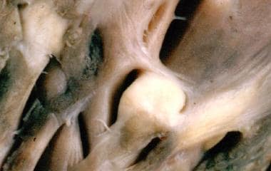
Rhabdomyoma
Heart Neoplasms
Tuberous Sclerosis
Neoplasm Regression, Spontaneous
Periodic Acid-Schiff Reaction
Fetal Diseases
Rhabdomyosarcoma
Dystrophic calcification of the fetal myocardium. (1/74)
Intramural cardiac masses were detected antenatally in three fetuses by echocardiography. The masses were initially thought to be rhabdomyomas. All three pregnancies were terminated and histology showed dystrophic calcification in all, with no evidence of tumour. Therefore, dystrophic calcification of the fetal myocardium may have a similar appearance to single or multiple rhabdomyomas. This should be considered when counselling parents after detection of masses in the fetal heart, particularly when considering the risk of associated tuberous sclerosis. (+info)Spectrum of cystic variants of Wilm's tumour: cystic nephroma (multilocular cyst) and cystic partially differentiated nephroma--a report of four cases. (2/74)
Two cases of cystic nephroma (multilocular cyst of the kidney), and one case each of cystic partially differentiated nephroblastoma (CPDN) and rhabdomyomatous Wilms' tumour are described. All were male and in the pediatric age group. Grossly tumours were unilateral, unicentric and multiloculated. The need for proper designation of these lesions is highlighted because of difference in the treatment and prognosis of these tumours. (+info)Ultrastructural changes in medullomyoblastoma. Similarities with foetal rhabdomyoma. (3/74)
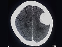
The light and electronmicroscopic changes are described in two cases of medullomyoblastoma, and compared with the changes seen in a case of foetal rhabdomyoma. The medullomyoblastomas in two children aged 8 and 5 years, consisted predominantly of classical type of medulloblastoma cells, along with few to many 'strap cells' or 'myoid cells' which, on closer examination, showed clear cross striations, consistent with muscle fibres or myofibrils. The primitive myoid cells were similar to those encountered in larger numbers in a post-auricular rhabdomyoma, possibly of foetal origin in a 40 day old infant. The four pathogenetic mechanisms i.e. (i) an embryonal stage of myofibrillar differentiation; (ii) a malformative factor; (iii) a teratoid factor on account of the presence of mesenchyme derived striated muscle tissue in the obviously predominant ectodermal medulloblastoma; and (iv) metaplasia of the vascular smooth muscle cells in the medullomyoblastoma, are discussed. (+info)Prenatal detection of cerebral lesions in a fetus with tuberous sclerosis. (4/74)
We report a newborn, diagnosed prenatally with both cardiac rhabdomyomas and a brain tumor. To the best of our knowledge, this is the first report of central nervous system (CNS) lesions detected prenatally in a child with tuberous sclerosis with term follow-up. At 36 months, the child has normal growth and is developing appropriately. Thus the finding of CNS tumors on fetal ultrasound examination can help in the prenatal diagnosis of tuberous sclerosis but does not necessarily indicate a poor prognosis. (+info)Oesophageal rhabdomyoma. (5/74)
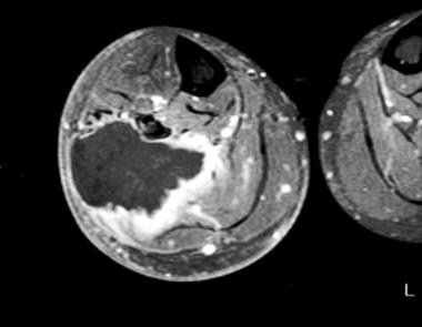
Extracardiac rhabdomyomas are rare benign tumours showing striated muscle differentiation. Seventy percent of these lesions occur in the head and neck region. The most common sites for these lesions are the larynx, pharynx, and the floor of the mouth. There has been only one previous report of a rhabdomyoma of the oesophagus; two further cases are described. (+info)Perinatal diagnosis of cardiac tumors. (6/74)
OBJECTIVE: As fetal cardiac tumors are a rare condition, we report the perinatal diagnosis and ultrasound findings of 12 cases. METHODS: In 10 cases the tumors were identified prenatally by fetal echocardiography; gestational age at detection ranged from 22 to 34 weeks. In two symptomatic infants cardiac tumors were diagnosed on the first day postpartum; prenatal ultrasound evaluation at 26 and 38 weeks of gestation did not reveal a cardiac lesion. RESULTS: Six fetuses had singular tumors, in six fetuses they were multifocal. The left ventricle was most often affected. Termination of pregnancy was chosen in three cases, one in association with trisomy 21 and tuberous sclerosis. One intrauterine and three neonatal deaths due to cardiac failure occurred. Histopathologic examination revealed cardiac rhabdomyoma in six fetuses and fibroma in one case. In the five surviving fetuses the size of the tumors spontaneously decreased postpartum. Rhabdomyomata were associated with tuberous sclerosis in four out of 11 cases. CONCLUSION: Cardiac tumors are detectable as early as 22 weeks of gestation. Presenting symptoms may be arrhythmia, dysfunction of the atrioventricular valves, pericardial effusion and fetal hydrops. The most common perinatal diagnosis is rhabdomyoma, which is often associated with tuberous sclerosis. Sequential examination in high risk patients should be considered as most tumors increase in size during pregnancy and may become evident in late second and third trimester of pregnancy. Postpartum, however, regression of tumor size is common. (+info)Surgical treatment of primary cardiac tumors: 28 years' experience in Kanazawa University Hospital. (7/74)
To examine the clinical features of primary cardiac tumors, 34 patients who underwent surgical treatment from 1973 to 2000 at the Kanazawa University Hospital were analyzed and the literature was reviewed. The 34 patients were divided into 3 categories: (i) myxomas; (ii) benign non-myxomas; and (iii) malignant tumors. Twenty-three patients (70%) were diagnosed with myxomas, including 22 left atrial myxomas and 1 right atrial myxoma. Seven patients (18%) were diagnosed with benign non-myxoma tumors, including 3 hemangiomas, 1 fibroma, 1 rhabdomyoma, 1 pheochromocytoma, and 1 lipoma. Four patients (12%) were diagnosed with malignant tumors, including 2 angiosarcomas, 1 rhabdomyosarcoma, and 1 malignant fibrous histiocytoma. Among the myxoma patients, in-hospital mortality was 9% (2/23), late mortality was 10% (2/21), and no recurrent myxomas have been identified. Among benign non-myxoma patients there were no perioperative deaths; however, 1 patient died 11 years after surgery, with no linked cause. No recurrent tumors have been identified. Among malignant tumor patients, 1 patient died the day following surgery and the rest died within 14 months. Early and late results of surgery were acceptable for those patients with benign tumors, while the prognosis for patients with malignant tumors was very poor. (+info)Serum creatine kinase levels parallel the clinical course for rhabdomyomatous Wilms tumor. (8/74)
A right-sided renal mass in an 11-month-old girl was diagnosed by percutaneous needle biopsy as Wilms tumor, which on histologic examination was found to be predominantly rhabdomyomatous. As part of the examination, serum creatine kinase (CK) and CK-MB levels were measured and were significantly elevated at 994 U/L (reference range, 42-180 U/L) and 40 U/L (reference range, 0-3 U/L), respectively. Subsequently, an 8-month-old girl was admitted to the hospital with septicemia and was found to have an abdominal mass. A diagnosis of bilateral Wilms tumor was made following percutaneous biopsy of both kidneys; histologic examination confirmed that the tumor was predominantly rhabdomyomatous. Serum CK and CK-MB levels also were measured and were significantly elevated at 685 U/L and 84.4 U/L, respectively. In both cases, the serum CK and CK-MB levels reflected the clinical course; elevation in serum levels was associated with tumor recurrence, infarction, or chemotherapy-related necrosis. We conclude that these enzymes have clinical usefulness as markers for Wilms tumor showing rhabdomyomatous morphologic features. (+info)Rhabdomyoma is a rare, benign tumor that arises from the striated muscle tissue, which is the type of muscle that enables movement and action in the body. These tumors most commonly occur in the heart (cardiac rhabdomyomas) or in the head and neck region (extracardiac rhabdomyomas). Cardiac rhabdomyomas are often associated with genetic disorders such as tuberous sclerosis complex, while extracardiac rhabdomyomas can be found in various locations like the skin, tongue, or skeletal muscles.
Cardiac rhabdomyomas typically appear in infancy or early childhood and may not cause any symptoms. However, they can potentially lead to complications such as heart rhythm abnormalities, obstruction of blood flow, or heart failure. Extracardiac rhabdomyomas are usually slow-growing and asymptomatic but can cause issues depending on their size and location. Surgical removal may be necessary if the tumor interferes with vital functions or causes discomfort.
It is essential to note that while rhabdomyomas are generally benign, they can undergo malignant transformation in rare cases, leading to a more aggressive form called rhabdomyosarcoma. Regular follow-ups and monitoring are crucial for early detection and management of any changes in the tumor's behavior.
Heart neoplasms are abnormal growths or tumors that develop within the heart tissue. They can be benign (noncancerous) or malignant (cancerous). Benign tumors, such as myxomas and rhabdomyomas, are typically slower growing and less likely to spread, but they can still cause serious complications if they obstruct blood flow or damage heart valves. Malignant tumors, such as angiosarcomas and rhabdomyosarcomas, are fast-growing and have a higher risk of spreading to other parts of the body. Symptoms of heart neoplasms can include shortness of breath, chest pain, fatigue, and irregular heart rhythms. Treatment options depend on the type, size, and location of the tumor, and may include surgery, radiation therapy, or chemotherapy.
Tuberous Sclerosis Complex (TSC) is a rare genetic disorder that causes non-cancerous (benign) tumors to grow in many parts of the body. These tumors can affect the brain, skin, heart, kidneys, eyes, and lungs. The signs and symptoms of TSC can vary widely, depending on where the tumors develop and how severely a person is affected.
The condition is caused by mutations in either the TSC1 or TSC2 gene, which regulate a protein that helps control cell growth and division. When these genes are mutated, the protein is not produced correctly, leading to excessive cell growth and the development of tumors.
TSC is typically diagnosed based on clinical symptoms, medical imaging, and genetic testing. Treatment for TSC often involves a multidisciplinary approach, with specialists in neurology, dermatology, cardiology, nephrology, pulmonology, and ophthalmology working together to manage the various symptoms of the condition. Medications, surgery, and other therapies may be used to help control seizures, developmental delays, skin abnormalities, and other complications of TSC.
A fibroma is a benign (non-cancerous) tumor that consists primarily of fibrous or connective tissue. It can occur in various parts of the body, including the skin, mouth, and internal organs. The term "fibroma" is often used to describe any benign fibrous growth, but there are specific types of fibromas such as dermatofibroma (found in the skin), oral fibroma (found in the mouth), and benign fibrous histiocytoma (found in soft tissues).
It's important to note that while fibromas are generally harmless, they can cause discomfort or problems depending on their size and location. If a fibroma is causing issues or there's concern about its growth or malignancy, it should be evaluated by a healthcare professional for potential removal or further assessment.
Spontaneous neoplasm regression is a rare and somewhat controversial phenomenon in which a tumor or malignancy appears to decrease in size or disappear without any treatment or with treatment that is typically not expected to produce such an effect. This can occur through various mechanisms, including immune-mediated processes, apoptosis (programmed cell death), differentiation of cancer cells into normal cells, and angiogenesis inhibition (preventing the growth of new blood vessels that feed the tumor).
Spontaneous regression of neoplasms is not well understood and is considered unpredictable. It has been reported in various types of cancers, including neuroblastoma, melanoma, renal cell carcinoma, and others. However, it should be noted that spontaneous regression does not imply a cure, as the tumor may still recur or metastasize later on.
In summary, spontaneous neoplasm regression refers to the partial or complete disappearance of a malignancy without any specific treatment or with treatment that is not typically associated with such an effect.
The Periodic Acid-Schiff (PAS) reaction is a histological staining method used to detect the presence of certain carbohydrates, such as glycogen and glycoproteins, in tissues or cells. This technique involves treating the tissue with periodic acid, which oxidizes the vicinal hydroxyl groups in the carbohydrates, creating aldehydes. The aldehydes then react with Schiff's reagent, forming a magenta-colored complex that is visible under a microscope.
The PAS reaction is commonly used to identify and analyze various tissue components, such as basement membranes, fungal cell walls, and mucins in the respiratory and gastrointestinal tracts. It can also be used to diagnose certain medical conditions, like kidney diseases, where abnormal accumulations of carbohydrates occur in the renal tubules or glomeruli.
In summary, the Periodic Acid-Schiff reaction is a staining method that detects specific carbohydrates in tissues or cells, which can aid in diagnostic and research applications.
Fetal diseases are medical conditions or abnormalities that affect a fetus during pregnancy. These diseases can be caused by genetic factors, environmental influences, or a combination of both. They can range from mild to severe and may impact various organ systems in the developing fetus. Examples of fetal diseases include congenital heart defects, neural tube defects, chromosomal abnormalities such as Down syndrome, and infectious diseases such as toxoplasmosis or rubella. Fetal diseases can be diagnosed through prenatal testing, including ultrasound, amniocentesis, and chorionic villus sampling. Treatment options may include medication, surgery, or delivery of the fetus, depending on the nature and severity of the disease.
Rhabdomyosarcoma is a type of cancer that develops in the body's soft tissues, specifically in the muscle cells. It is a rare and aggressive form of sarcoma, which is a broader category of cancers that affect the connective tissues such as muscles, tendons, cartilages, bones, blood vessels, and fatty tissues.
Rhabdomyosarcomas can occur in various parts of the body, including the head, neck, arms, legs, trunk, and genitourinary system. They are more common in children than adults, with most cases diagnosed before the age of 18. The exact cause of rhabdomyosarcoma is not known, but genetic factors and exposure to radiation or certain chemicals may increase the risk.
There are several subtypes of rhabdomyosarcoma, including embryonal, alveolar, pleomorphic, and spindle cell/sclerosing. The type and stage of the cancer determine the treatment options, which may include surgery, radiation therapy, chemotherapy, or a combination of these approaches. Early diagnosis and prompt treatment are crucial for improving the prognosis and long-term survival rates.
 Rhabdomyoma - Wikipedia
 Cardiac Rhabdomyoma Pathology: Definition, Epidemiology, Etiology
 Fetal cardiac rhabdomyoma due to paternal mosaicism of TSC2: A case report - PubMed
 Cardiac rhabdomyoma in an adult patient presenting with ventricular arrhythmia.
 The association of prenatally diagnosed cardiac rhabdomyoma and tuberosclerosis complex: a dismal combination? | AVESİS
CNS Tumors: A Single Center Pathology Review
 Tongue problems: MedlinePlus Medical Encyclopedia
 Search Strategy Used to Create the PubMed Cancer Filter
 Webpathology.com: A Collection of Surgical Pathology Images
Paratesticular rhabdomyoma in a young adult: case study and review of the literature. - Nuffield Department of Medicine
 Cancer in a 5 y/o - Oncology Nursing
 Letter to Editor Archives - CytoJournal
Biomarkers Search
 Tuberous sclerosis complex - About the Disease - Genetic and Rare Diseases Information Center
IndexCat
Fingernail, Toenail Changes and Flank Pain in a 20-Year-Old
 Tuberous Sclerosis - StatPearls - NCBI Bookshelf
 Atlas of dermatopathology: Collection of histological slides by prof. Werner Kempf
Phaeochromocytoma in a patient with a Birt-Hogg-Dubé syndrome phenotype | BMJ Case Reports
What Is Your Diagnosis? in: Journal of the American Veterinary Medical Association Volume 259 Issue 5 (2021)
 Different types of brain tumours | The Brain Tumour Charity
 Nemaline myopathy in a six-month-old Pomeranian dog
 Burke, Allen | University of Maryland School of Medicine
Specific PHGKB|Rare Diseases PHGKB|PHGKB
 Code Preferred Term Synonyms Definition Neoplastic Status
 Birmingham Medical Review - Google Boeken
 Overview of Heart Tumors - Heart and Blood Vessel Disorders - MSD Manual Consumer Version
Benign4
- A rhabdomyoma is a benign tumor of striated muscle. (wikipedia.org)
- Although the behavior of a cardiac rhabdomyoma is benign, the positioning within critical areas in the heart can lead to lethal arrhythmias and chamber obstruction. (medscape.com)
- Two cases of benign vaginal rhabdomyoma. (medscape.com)
- Benign heart tumors that may appear in children include the following: Rhabdomyoma: A tumor that forms in muscle made up of long fibers. (instituteofliving.org)
Associated with cardiac rhabdomyoma1
- The genetic disorder most commonly associated with cardiac rhabdomyoma is tuberous sclerosis. (medscape.com)
Tuberous3
- Routine surveillance of children with tuberous sclerosis for cardiac rhabdomyoma or other cardiovascular manifestations of their disease may include electrocardiogram (EKG) and echocardiography. (wikipedia.org)
- About 50% of patients with tuberous sclerosis develop a cardiac rhabdomyoma. (medscape.com)
- Atrial rhabdomyoma as seen on cardiac CT scan in a patient with tuberous sclerosis. (medscape.com)
Laryngeal2
- Canine laryngeal rhabdomyoma. (irjs.info)
- Laryngeal rhabdomyoma in a dog. (bestforpet.co.nz)
Fetal rhabdomyoma2
- Only rare cases of possible malignant change have been reported in fetal rhabdomyoma. (wikipedia.org)
- Fetal rhabdomyoma is identifiable by the presence of a mixture of spindle-shaped cells with indistinct cytoplasm and muscle fibers, which resemble striated muscle tissue seen in intrauterine development at 7-12 weeks. (medscape.com)
Extracardiac rhabdomyoma1
- Bjørndal Sørensen K, Godballe C, Ostergaard B, Krogdahl A. Adult extracardiac rhabdomyoma: light and immunohistochemical studies of two cases in the parapharyngeal space. (medscape.com)
Tumor4
- Cardiac rhabdomyoma is the most common primary pediatric tumor of the heart and is considered to be a hamartoma of developing cardiac myocytes. (medscape.com)
- In other cases, the first sign may be white patches on the skin (hypomelanotic macules) or the identification of cardiac tumor rhabdomyoma. (texaschildrens.org)
- Conclusion: Although it is a rare tumor, cardiac rhabdomyoma may lead to life-threatening symptoms. (ogu.edu.tr)
- In infants and children, the most common type of noncancerous primary heart tumor is a rhabdomyoma. (msdmanuals.com)
Genital2
- Extracardiac forms of rhabdomyoma are sub-classified into three distinct types: adult type, fetal type, and genital type. (wikipedia.org)
- Genital rhabdomyoma is made up of a mixture of fibroblastlike cells with clusters of mature cells containing distinct cross-striations and a matrix containing varying amounts of collagen and mucoid material. (medscape.com)
Lesions1
- A subsequent echocardiogram showed the presence of multiple endomyocardial lesions suggestive of rhabdomyoma. (elsevier.es)
Differential diagnosis1
- Hansen T, Katenkamp D. Rhabdomyoma of the head and neck: morphology and differential diagnosis. (medscape.com)
Skeletal1
- Rhabdomyoma is a rare non-cancerous tumour that starts in skeletal muscle. (cancer.ca)
Adult6
- Any masses, such as those found in the head and neck of patients with adult rhabdomyoma, should be biopsied to establish a diagnosis. (medscape.com)
- The histopathologic findings from patients with adult rhabdomyoma are characterized by the presence of well-differentiated large cells, which resemble striated muscle cells. (medscape.com)
- Histopathology of adult rhabdomyoma. (medscape.com)
- Microscopically, the adult rhabdomyoma contains deeply eosinophilic polygonal cells with peripherally placed nuclei. (medscape.com)
- Yadav SK, Sood N. Multinodular adult rhabdomyoma in female: A rare case report. (medscape.com)
- Unique manifestation of a multifocal adult rhabdomyoma involving the soft palate-case report and review of literature. (medscape.com)
Pediatric1
- This study evaluated the clinical and echocardiographic data of patients followed up in our Pediatric Cardiology Clinic for cardiac rhabdomyoma. (ogu.edu.tr)
Incidence1
- [ 7 ] Studies have demonstrated that the incidence of cardiac rhabdomyoma is 0.002-0.25% at autopsy, 0.02-0.08% in live-born infants, and 0.12% in prenatal reviews. (medscape.com)
Outcome1
- Bejiqi R, Retkoceri R, Bejiqi H. Prenatally Diagnosis and Outcome of Fetuses with Cardiac Rhabdomyoma - Single Centre Experience. (medscape.com)
Genetic1
- Contact a genetic specialist if your child is diagnosed with cardiac rhabdomyoma. (medlineplus.gov)
Heart2
- The specific clinical picture of a patient with a cardiac rhabdomyoma is determined by its location in the heart. (wikipedia.org)
- Cardiac rhabdomyoma is an abnormal growth of a fetal heart, often detected during pregnancy. (texaschildrens.org)
Patients1
- Methods: This study included patients with cardiac rhabdomyoma detected by echocardiography between 2008 and 2021. (ogu.edu.tr)
Numbers1
- The primitive myoid cells were similar to those encountered in larger numbers in a post-auricular rhabdomyoma, possibly of foetal origin in a 40 day old infant. (neurologyindia.com)
Tumors1
- Certain symptoms develop before to birth, such as heart tumors (rhabdomyoma). (nih.gov)
Benign tumor2
- A rhabdomyoma is a benign tumor of striated muscle. (wikipedia.org)
- Paratesticular rhabdomyoma is a rare benign tumor, which usually presents as a painless mass in the scrotum or groin. (ox.ac.uk)
Etiology1
- The pathological diagnosis of cardiac neoplasm tissue was cardiac rhabdomyoma, but the etiology was unknown. (nih.gov)
Ventricular1
- We describe a previously healthy man who presented with ventricular arrhythmias resulting from a right ventricular, cardiac rhabdomyoma. (duke.edu)
Prenatal1
- [ 7 ] Studies have demonstrated that the incidence of cardiac rhabdomyoma is 0.002-0.25% at autopsy, 0.02-0.08% in live-born infants, and 0.12% in prenatal reviews. (medscape.com)
Malignant1
- Only rare cases of possible malignant change have been reported in fetal rhabdomyoma. (wikipedia.org)
Hamartoma1
- Cardiac rhabdomyoma is the most common primary pediatric tumor of the heart and is considered to be a hamartoma of developing cardiac myocytes. (medscape.com)
Diagnosis1
- The etiological diagnosis of Rhabdomyoma is very important. (nih.gov)
Clinical1
- The specific clinical picture of a patient with a cardiac rhabdomyoma is determined by its location in the heart. (wikipedia.org)
Tissue1
- The tumour was excised en-bloc with a surrounding margin of healthy tissue (Figure 2 ). (biomedcentral.com)
Male1
- We report the first case of a locally invasive paratesticular rhabdomyoma in a 17-year-old male teenager who presented with chronic scrotal pain. (ox.ac.uk)
Association1
- The association of prenatally diagnosed cardiac rhabdomyoma and tuberosclerosis complex: a dismal combination? (istanbul.edu.tr)