Multiple Sclerosis
Sclerosis
Amyotrophic Lateral Sclerosis
Tuberous Sclerosis
Scleroderma, Systemic
Multiple Sclerosis, Chronic Progressive
Encephalomyelitis, Autoimmune, Experimental
Scleroderma, Diffuse
Demyelinating Diseases
Diffuse Cerebral Sclerosis of Schilder
Magnetic Resonance Imaging
Spinal Cord
Optic Neuritis
Scleroderma, Limited
Brain
Myelin Basic Protein
Motor Neuron Disease
Superoxide Dismutase
Oligoclonal Bands
Interferon-beta
Myelin Sheath
Disease Progression
Myelin-Oligodendrocyte Glycoprotein
Raynaud Disease
Angiomyolipoma
Peritoneal Fibrosis
Epilepsy, Temporal Lobe
Disability Evaluation
Scleroderma, Localized
Disease Models, Animal
Atrophy
Myelin Proteins
Oligodendroglia
Neuromyelitis Optica
Myelin-Associated Glycoprotein
Central Nervous System
Case-Control Studies
Autoantibodies
Myelin Proteolipid Protein
Rhabdomyoma
Severity of Illness Index
Immunologic Factors
RNA-Binding Protein FUS
Age of Onset
Lymphangioleiomyomatosis
Nerve Fibers, Myelinated
Mice, Transgenic
Nerve Degeneration
Skin
HLA-DR2 Antigen
Encephalomyelitis, Acute Disseminated
Autoimmune Diseases
Microglia
Nervous System Diseases
Tumor Suppressor Proteins
DNA Repeat Expansion
Propylene Glycols
Glomerulosclerosis, Focal Segmental
Mice, Inbred C57BL
Astrocytes
Neurodegenerative Diseases
Genetic Predisposition to Disease
Demyelinating Autoimmune Diseases, CNS
Cells, Cultured
Theilovirus
Immunosuppressive Agents
Biological Markers
T-Lymphocytes
HLA-DRB1 Chains
Skin Ulcer
Cerebrospinal Fluid Proteins
Follow-Up Studies
Neurofilament Proteins
TOR Serine-Threonine Kinases
Neurologic Examination
Frontotemporal Dementia
Monckeberg Medial Calcific Sclerosis
Cerebrospinal Fluid
Autoimmunity
Fibrosis
Immunoglobulin G
Mutation
Treatment Outcome
Inclusion Bodies
Psychosurgery
Guam
Riluzole
Frontotemporal Lobar Degeneration
Optic Nerve
Statistics, Nonparametric
Reproducibility of Results
Cohort Studies
Fatigue
Autoantigens
Brain Diseases
Pulmonary Fibrosis
Facial Neoplasms
Immunohistochemistry
Angiofibroma
Remission, Spontaneous
HLA-DR Antigens
Anterior Temporal Lobectomy
Blood-Brain Barrier
Epilepsy
Image Processing, Computer-Assisted
Phenotype
Retrospective Studies
Hippocampus
Corpus Callosum
Hamartoma
Inflammation
Enzyme-Linked Immunosorbent Assay
Cytokines
Cerebral Cortex
Evoked Potentials, Visual
Prospective Studies
Image Enhancement
Sensitivity and Specificity
Neuropsychological Tests
Central Nervous System Diseases
Kidney Glomerulus
Neurons
Temporal Lobe
Gadolinium
Pyramidal Tracts
Antibodies, Antinuclear
Signal Transduction
Hypertension, Pulmonary
Myelitis, Transverse
Cognition Disorders
CREST Syndrome
Disabled Persons
Longitudinal Studies
TDP-43 Proteinopathies
Neuritis
Neuroprotective Agents
Risk Factors
Lung Diseases, Interstitial
Peptides
Fasciculation
Alleles
Peritoneum
Malformations of Cortical Development
Glial Fibrillary Acidic Protein
Brain Stem
Spinal Cord Diseases
Dermis
Mice, Knockout
CD4-Positive T-Lymphocytes
Adjuvants, Immunologic
Fibroblasts
Rats, Inbred Lew
Muscle Spasticity
Genotype
Biopsy
Polymorphism, Single Nucleotide
Diffusion Tensor Imaging
Analysis of Variance
Reference Values
Lymphocyte Activation
Aspartic Acid
Encephalitis
Encephalomyelitis
RNA, Messenger
Gene Expression Regulation
Meninges
Creatine
Molecular Sequence Data
Systemic multifocal fibrosclerosis. (1/720)
We describe a case of hydronephrosis as a result of retroperitoneal fibrosis in a patient who had previous sclerosing lobulitis of the breast. To the best of our knowledge this is the first reported association between these two conditions in the english literature. We presume these conditions are linked and unify them under the general heading of systemic multifocal fibrosclerosis. (+info)Forebrain ischaemia with CA1 cell loss impairs epileptogenesis in the tetanus toxin limbic seizure model. (2/720)
There is a long-standing controversy as to whether Ammon's horn sclerosis is the result or the cause of severe limbic epilepsy. In the tetanus toxin model of limbic epilepsy, rats have intermittent spontaneous fits over a period of 3-6 weeks after injection of tetanus toxin into the hippocampus. The fits then usually remit and the EEG returns to normal. In a few rats, however, the fits recur some weeks to months later, and it was previously found that in these rats there was gross cell loss in area CA1 of the dorsal hippocampus (distant from the injection site in ventral hippocampus). Such cell loss might either promote recurrence of fits or be the result of the recurrence. In the present experiment, the effect of previous induction of CA1 cell loss by transient 4-vessel occlusion cerebral ischaemia on the subsequent development of the tetanus toxin-induced epilepsy was studied, using continuous time-lapse video monitoring to assess the number of fits. The hypothesis that the previous forebrain ischaemia would predispose rats to reoccurring fits was not supported: no rats in the ischaemia group had reoccurring fits and additionally fits were delayed and fewer occurred than in the control groups. (+info)Sclerosing peritonitis and systemic lupus erythematosus: a report of two cases. (3/720)
OBJECTIVE: To heighten the awareness of a possible association of sclerosing peritonitis in patients with systemic lupus erythematosus (SLE). METHODS AND RESULTS: Over the course of 17 years (from January 1981 to December 1997), 371 patients were treated with continuous ambulatory peritoneal dialysis (CAPD) at Rush-Presbyterian-St Lukes Medical Center. The patients were followed on CAPD for an average of 25 +/- 21 (SD) months with a median of 19 months (range 0.2-115 months). During this time only 2 (0.5%) patients were diagnosed with sclerosing peritonitis, and both had SLE with ongoing evidence of active disease while on CAPD. With a total of 26 SLE patients being treated with CAPD during the observation period, the prevalence of sclerosing peritonitis can be said to be as high as 8% in this patient population. CONCLUSION: These cases suggest that autoimmune diseases, such as SLE, that are well known to cause immune-mediated serositis may represent an additional factor predisposing to the development of sclerosing peritonitis in patients treated with CAPD. (+info)Sclerosing spinal pachymeningitis. A complication of intrathecal administration of Depo-Medrol for multiple sclerosis. (4/720)
Reported complications of intrathecal steroid therapy include aseptic meningitis, infectious meningitis, and arachnoiditis. We report a case of sclerosing spinal pachymeningitis complicating the attempted intrathecal administration of Depo-Medrol for multiple sclerosis. The lesion is characterised by concentric laminar proliferation of neomembranes within the subdural space of the entire spinal cord and cauda equina, resulting from repeated episodes of injury and repair to the spinal dura mater by Depo-Medrol. There is clinical and laboratory evidence that Depo-Medrol produces meningeal irritation and that the vehicle is the necrotising fraction. (+info)Mechanical strain of glomerular mesangial cells in the pathogenesis of glomerulosclerosis: clinical implications. (5/720)
Due to their elasticity, glomeruli will undergo excessive expansion and repetitive cycles of distension contraction under conditions of impaired glomerular pressure autoregulation and systemic arterial hypertension. These alterations in glomerular volume are associated with mesangial cell stretch which in turn stimulates the synthesis and deposition of ECM with eventual mesangial expansion and glomerulosclerosis. Hyperactivity of growth factors with prosclerotic activity is an important component in the translation of cellular mechanical strain into the abnormal metabolism of ECM components. Although mesangial cell mechanical strain is expected to occur in both remnant glomeruli and in glomeruli of diabetic kidneys, quantitatively different factors will determine the resultant metabolic consequences. In remnant glomeruli, the mechanical stretch is intense, being accounted for largely by the marked glomerular hypertrophy and increased glomerular compliance. In diabetic glomeruli, however, the mechanical stretch is less prominent but its effect on ECM synthesis is markedly aggravated by the presence of hyperglycaemia. There are presently no methods clinically available to diminish the prosclerotic action of growth factors at the glomerular level. In addition, there are no effective means to specifically improve glomerular pressure autoregulation. Therefore, current therapies must be aimed at decreasing systemic arterial pressure, blocking angiotensin II action and reducing glomerular hypertrophy. While there are effective drugs for the treatment of hypertension and for angiotensin II inhibition, protein restriction is the only measure available to diminish glomerular hypertrophy. Finally, in diabetes correction of systemic and glomerular hypertension should be coupled with strict glycaemic control to correct both glomerular autoregulation and increased ECM deposition. (+info)Changes in refractive error over a 5-year interval in the Beaver Dam Eye Study. (6/720)
PURPOSE: To examine changes in spherical equivalent over a 5-year period in persons 43 to 84 years of age. METHODS: All people 43 to 84 years of age and living in Beaver Dam, Wisconsin, in 1988 were invited for a baseline examination (1988-1990) and a 5-year follow-up examination (1993-1995). Refractions were determined according to the same protocol at both examinations. Aphakic and pseudophakic eyes were excluded as well as eyes with best corrected Snellen visual acuity of 20/40 and worse. After exclusions, refraction was obtained on 3007 right eyes and 3012 left eyes of the 3684 people participating in both examinations. RESULTS: Right and left eyes behaved similarly. Spherical equivalent became more positive in the youngest subjects and more negative in older subjects. After adjusting for other factors, the 5-year change in spherical equivalent of those 45, 55, 65, and 75 years of age was +0.15, +0.18, +0.10, and -0.07D, respectively. Severity of nuclear sclerosis was related to the amount of change. Those with mild nuclear sclerosis at baseline had a change of +0.2 D, whereas those with severe nuclear sclerosis had a change of -0.5 D. The amount of change was also related to gender, diabetes, and age at onset of myopia. It was unrelated to education and baseline spherical equivalent. CONCLUSIONS: Changes in spherical equivalent over a 5-year period were small. Before the age of 70, people became more hyperopic. After the age of 70, people became more myopic. Much of the myopic change may be related to increasing nuclear sclerosis. (+info)Phlebosclerosis of the colon with positive anti-centromere antibody. (7/720)
A 56-year-old woman with symptoms of chronic bowel disease presented a peculiar calcification of the mesenteric vein of the ascending to transverse colon on barium enema study. The resected colon was hard and black. Histo-pathologic examinations demonstrated fibrous change of the colon with a calcified and hyaline-deposited mesenteric vein. No cell infiltration was observed. These findings were compatible with phlebosclerosis and also with systemic sclerosis. Positive anti-centromere antibody and Raynaud's phenomenon, hallmarks of a variant systemic sclerosis, the CREST syndrome were observed. We therefore speculated that the pathogenesis of the phlebosclerosis of the colon is related to the CREST syndrome. (+info)Sclerosing Mucoepidermoid carcinoma with eosinophilia of the thyroid glands: a case report with clinical manifestation of recurrent neck mass. (8/720)
Sclerosing mucoepidermoid carcinoma with eosinophilia (SMECE) is a recently recognized malignant neoplasm of the thyroid gland. About 14 cases of SMECE have been reported and this is the first reported case in Korea. A 57-year-old woman presented with right neck mass for 20 years. Total thyroidectomy was performed under the impression of thyroid carcinoma. The resected thyroid gland showed a poorly circumscribed hard mass. Histologically, the tumor consisted of solid nests of large atypical cells with dense fibrous stroma. The tumor cells showed squamoid appearance with abundant eosinophilic cytoplasm. There were also rare mucin-containing cells within the nests. Within the hyalinized stroma, numerous eosinophils were found. The surrounding thyroid parenchyma displayed Hashimoto's thyroiditis. There was metastasis in a regional lymph node. Two years after initial surgery, she underwent a modified radical neck dissection due to recurrent neck mass. After the radiation therapy for eight weeks, laryngectomy and esophagectomy were performed due to a recurrent carcinoma in the esophageal wall. We report an additional case of SMECE, with metastasis to regional lymph nodes and esophagus. The tumor appears to be more aggressive than previously reported and a correct diagnosis can be rendered by just examining the metastatic lesions. (+info)Multiple Sclerosis (MS) is a chronic autoimmune disease that affects the central nervous system (CNS), which includes the brain, spinal cord, and optic nerves. In MS, the immune system mistakenly attacks the protective covering of nerve fibers, called myelin, leading to damage and scarring (sclerosis). This results in disrupted communication between the brain and the rest of the body, causing a variety of neurological symptoms that can vary widely from person to person.
The term "multiple" refers to the numerous areas of scarring that occur throughout the CNS in this condition. The progression, severity, and specific symptoms of MS are unpredictable and may include vision problems, muscle weakness, numbness or tingling, difficulty with balance and coordination, cognitive impairment, and mood changes. There is currently no cure for MS, but various treatments can help manage symptoms, modify the course of the disease, and improve quality of life for those affected.
Sclerosis is a medical term that refers to the abnormal hardening or scarring of body tissues, particularly in the context of various degenerative diseases affecting the nervous system. The term "sclerosis" comes from the Greek word "skleros," which means hard. In these conditions, the normally flexible and adaptable nerve cells or their protective coverings (myelin sheath) become rigid and inflexible due to the buildup of scar tissue or abnormal protein deposits.
There are several types of sclerosis, but one of the most well-known is multiple sclerosis (MS). In MS, the immune system mistakenly attacks the myelin sheath surrounding nerve fibers in the brain and spinal cord, leading to scarring and damage that disrupts communication between the brain and the rest of the body. This results in a wide range of symptoms, such as muscle weakness, numbness, vision problems, balance issues, and cognitive impairment.
Other conditions that involve sclerosis include:
1. Amyotrophic lateral sclerosis (ALS): Also known as Lou Gehrig's disease, ALS is a progressive neurodegenerative disorder affecting motor neurons in the brain and spinal cord, leading to muscle weakness, stiffness, and atrophy.
2. Systemic sclerosis: A rare autoimmune connective tissue disorder characterized by thickening and hardening of the skin and internal organs due to excessive collagen deposition.
3. Plaque psoriasis: A chronic inflammatory skin condition marked by red, scaly patches (plaques) resulting from rapid turnover and accumulation of skin cells.
4. Adhesive capsulitis: Also known as frozen shoulder, this condition involves stiffening and thickening of the shoulder joint's capsule due to scarring or inflammation, leading to limited mobility and pain.
Amyotrophic Lateral Sclerosis (ALS) is a progressive neurodegenerative disorder that affects nerve cells in the brain and spinal cord responsible for controlling voluntary muscle movements, such as speaking, walking, breathing, and swallowing. The condition is characterized by the degeneration of motor neurons in the brain (upper motor neurons) and spinal cord (lower motor neurons), leading to their death.
The term "amyotrophic" comes from the Greek words "a" meaning no or negative, "myo" referring to muscle, and "trophic" relating to nutrition. When a motor neuron degenerates and can no longer send impulses to the muscle, the muscle becomes weak and eventually atrophies due to lack of use.
The term "lateral sclerosis" refers to the hardening or scarring (sclerosis) of the lateral columns of the spinal cord, which are primarily composed of nerve fibers that carry information from the brain to the muscles.
ALS is often called Lou Gehrig's disease, named after the famous American baseball player who was diagnosed with the condition in 1939. The exact cause of ALS remains unknown, but it is believed to involve a combination of genetic and environmental factors. There is currently no cure for ALS, and treatment primarily focuses on managing symptoms and maintaining quality of life.
The progression of ALS varies from person to person, with some individuals experiencing rapid decline over just a few years, while others may have a more slow-progressing form of the disease that lasts several decades. The majority of people with ALS die from respiratory failure within 3 to 5 years after the onset of symptoms. However, approximately 10% of those affected live for 10 or more years following diagnosis.
Tuberous Sclerosis Complex (TSC) is a rare genetic disorder that causes non-cancerous (benign) tumors to grow in many parts of the body. These tumors can affect the brain, skin, heart, kidneys, eyes, and lungs. The signs and symptoms of TSC can vary widely, depending on where the tumors develop and how severely a person is affected.
The condition is caused by mutations in either the TSC1 or TSC2 gene, which regulate a protein that helps control cell growth and division. When these genes are mutated, the protein is not produced correctly, leading to excessive cell growth and the development of tumors.
TSC is typically diagnosed based on clinical symptoms, medical imaging, and genetic testing. Treatment for TSC often involves a multidisciplinary approach, with specialists in neurology, dermatology, cardiology, nephrology, pulmonology, and ophthalmology working together to manage the various symptoms of the condition. Medications, surgery, and other therapies may be used to help control seizures, developmental delays, skin abnormalities, and other complications of TSC.
Systemic Scleroderma, also known as Systemic Sclerosis (SSc), is a rare, chronic autoimmune disease that involves the abnormal growth and accumulation of collagen in various connective tissues, blood vessels, and organs throughout the body. This excessive collagen production leads to fibrosis or scarring, which can cause thickening, hardening, and tightening of the skin and damage to internal organs such as the heart, lungs, kidneys, and gastrointestinal tract.
Systemic Scleroderma is characterized by two main features: small blood vessel abnormalities (Raynaud's phenomenon) and fibrosis. The disease can be further classified into two subsets based on the extent of skin involvement: limited cutaneous systemic sclerosis (lcSSc) and diffuse cutaneous systemic sclerosis (dcSSc).
Limited cutaneous systemic sclerosis affects the skin distally, typically involving fingers, hands, forearms, feet, lower legs, and face. It is often associated with Raynaud's phenomenon, calcinosis, telangiectasias, and pulmonary arterial hypertension.
Diffuse cutaneous systemic sclerosis involves more extensive skin thickening and fibrosis that spreads proximally to affect the trunk, upper arms, thighs, and face. It is commonly associated with internal organ involvement, such as interstitial lung disease, heart disease, and kidney problems.
The exact cause of Systemic Scleroderma remains unknown; however, it is believed that genetic, environmental, and immunological factors contribute to its development. There is currently no cure for Systemic Scleroderma, but various treatments can help manage symptoms, slow disease progression, and improve quality of life.
Multiple Sclerosis (MS), Chronic Progressive is a form of Multiple Sclerosis, a chronic autoimmune disease that affects the central nervous system (CNS). In this form, the disease follows a steady progression with no distinct relapses or remissions. The symptoms worsen over time, leading to a decline in physical functioning and increased disability.
The term "chronic progressive" is used to describe the course of the disease, which is characterized by a continuous worsening of neurological functions from the onset, or after an initial relapsing-remitting phase. There are two types of chronic progressive MS: primary and secondary.
1. Primary Chronic Progressive MS (PCP): This form of MS shows a steady progression of symptoms from the beginning, with no distinct remissions or relapses. The disability accumulates gradually over time, and the person may experience varying degrees of physical and cognitive impairment.
2. Secondary Chronic Progressive MS (SCP): In this form, an individual initially has a relapsing-remitting course of MS (RRMS), characterized by unpredictable relapses followed by periods of partial or complete recovery (remissions). However, after some time, the disease transitions to a steady progression of symptoms and disability, even without distinct relapses. This is known as secondary chronic progressive MS.
The exact cause of Multiple Sclerosis remains unknown; however, it is believed to be influenced by genetic, environmental, and immunological factors. The disease involves the immune system attacking the myelin sheath, a protective covering surrounding nerve fibers in the CNS. This results in lesions or scars (scleroses) that disrupt communication between the brain, spinal cord, and other parts of the body, leading to various physical, cognitive, and sensory symptoms.
Management of Chronic Progressive MS typically involves a multidisciplinary approach, focusing on symptom management, rehabilitation, and maintaining quality of life. Currently, there are no approved disease-modifying therapies specifically for chronic progressive MS; however, some medications used to treat relapsing-remitting MS may help slow the progression of disability in certain individuals with secondary chronic progressive MS.
Autoimmune encephalomyelitis (EAE) is a model of inflammatory demyelinating disease used in medical research to study the mechanisms of multiple sclerosis (MS) and develop new therapies. It is experimentally induced in laboratory animals, typically mice or rats, through immunization with myelin antigens or T-cell transfer. The resulting immune response leads to inflammation, demyelination, and neurological dysfunction in the central nervous system (CNS), mimicking certain aspects of MS.
EAE is a valuable tool for understanding the pathogenesis of MS and testing potential treatments. However, it is essential to recognize that EAE is an experimental model and may not fully recapitulate all features of human autoimmune encephalomyelitis.
Diffuse scleroderma is a medical condition that falls under the systemic sclerosis category of autoimmune rheumatic diseases. It is characterized by thickening and hardening (sclerosis) of the skin and involvement of internal organs. In diffuse scleroderma, the process affects extensive areas of the skin and at least one internal organ.
The disease process involves an overproduction of collagen, a protein that makes up connective tissues in the body. This excessive collagen deposition leads to fibrosis (scarring) of the skin and various organs, including the esophagus, gastrointestinal tract, heart, lungs, and kidneys.
Diffuse scleroderma can present with a rapid progression of skin thickening within the first few years after onset. The skin involvement may extend to areas beyond the hands, feet, and face, which are commonly affected in limited scleroderma (another form of systemic sclerosis). Additionally, patients with diffuse scleroderma have a higher risk for severe internal organ complications compared to those with limited scleroderma.
Early diagnosis and appropriate management of diffuse scleroderma are crucial to prevent or slow down the progression of organ damage. Treatment typically involves a multidisciplinary approach, focusing on symptom management, immunosuppressive therapy, and addressing specific organ involvement.
Demyelinating diseases are a group of disorders that are characterized by damage to the myelin sheath, which is the protective covering surrounding nerve fibers in the brain, optic nerves, and spinal cord. Myelin is essential for the rapid transmission of nerve impulses, and its damage results in disrupted communication between the brain and other parts of the body.
The most common demyelinating disease is multiple sclerosis (MS), where the immune system mistakenly attacks the myelin sheath. Other demyelinating diseases include:
1. Acute Disseminated Encephalomyelitis (ADEM): An autoimmune disorder that typically follows a viral infection or vaccination, causing widespread inflammation and demyelination in the brain and spinal cord.
2. Neuromyelitis Optica (NMO) or Devic's Disease: A rare autoimmune disorder that primarily affects the optic nerves and spinal cord, leading to severe vision loss and motor disability.
3. Transverse Myelitis: Inflammation of the spinal cord causing damage to both sides of one level (segment) of the spinal cord, resulting in various neurological symptoms such as muscle weakness, numbness, or pain, depending on which part of the spinal cord is affected.
4. Guillain-Barré Syndrome: An autoimmune disorder that causes rapid-onset muscle weakness, often beginning in the legs and spreading to the upper body, including the face and breathing muscles. It occurs when the immune system attacks the peripheral nerves' myelin sheath.
5. Central Pontine Myelinolysis (CPM): A rare neurological disorder caused by rapid shifts in sodium levels in the blood, leading to damage to the myelin sheath in a specific area of the brainstem called the pons.
These diseases can result in various symptoms, such as muscle weakness, numbness, vision loss, difficulty with balance and coordination, and cognitive impairment, depending on the location and extent of the demyelination. Treatment typically focuses on managing symptoms, modifying the immune system's response, and promoting nerve regeneration and remyelination when possible.
Diffuse cerebral sclerosis of Schilder, also known as Schilder's disease, is a rare inflammatory demyelinating disorder of the central nervous system. It primarily affects children and young adults, but can occur at any age. The condition is characterized by widespread destruction of the myelin sheath, which surrounds and protects nerve fibers in the brain.
The hallmark feature of Schilder's disease is the presence of multiple, large, symmetrical lesions in the white matter of both cerebral hemispheres. These lesions are typically located in the parieto-occipital regions of the brain and can extend to involve other areas as well.
The symptoms of Schilder's disease vary depending on the location and extent of the lesions, but may include:
* Progressive intellectual decline
* Seizures
* Visual disturbances
* Weakness or paralysis on one side of the body (hemiparesis)
* Loss of sensation in various parts of the body
* Speech difficulties
* Behavioral changes, such as irritability, mood swings, and depression
The exact cause of Schilder's disease is not known, but it is believed to be an autoimmune disorder, in which the body's own immune system mistakenly attacks the myelin sheath. There is no cure for Schilder's disease, and treatment typically involves corticosteroids or other immunosuppressive therapies to reduce inflammation and slow the progression of the disease. Despite treatment, many patients with Schilder's disease experience significant disability and may require long-term care.
Medical Definition:
Magnetic Resonance Imaging (MRI) is a non-invasive diagnostic imaging technique that uses a strong magnetic field and radio waves to create detailed cross-sectional or three-dimensional images of the internal structures of the body. The patient lies within a large, cylindrical magnet, and the scanner detects changes in the direction of the magnetic field caused by protons in the body. These changes are then converted into detailed images that help medical professionals to diagnose and monitor various medical conditions, such as tumors, injuries, or diseases affecting the brain, spinal cord, heart, blood vessels, joints, and other internal organs. MRI does not use radiation like computed tomography (CT) scans.
The spinal cord is a major part of the nervous system, extending from the brainstem and continuing down to the lower back. It is a slender, tubular bundle of nerve fibers (axons) and support cells (glial cells) that carries signals between the brain and the rest of the body. The spinal cord primarily serves as a conduit for motor information, which travels from the brain to the muscles, and sensory information, which travels from the body to the brain. It also contains neurons that can independently process and respond to information within the spinal cord without direct input from the brain.
The spinal cord is protected by the bony vertebral column (spine) and is divided into 31 segments: 8 cervical, 12 thoracic, 5 lumbar, 5 sacral, and 1 coccygeal. Each segment corresponds to a specific region of the body and gives rise to pairs of spinal nerves that exit through the intervertebral foramina at each level.
The spinal cord is responsible for several vital functions, including:
1. Reflexes: Simple reflex actions, such as the withdrawal reflex when touching a hot surface, are mediated by the spinal cord without involving the brain.
2. Muscle control: The spinal cord carries motor signals from the brain to the muscles, enabling voluntary movement and muscle tone regulation.
3. Sensory perception: The spinal cord transmits sensory information, such as touch, temperature, pain, and vibration, from the body to the brain for processing and awareness.
4. Autonomic functions: The sympathetic and parasympathetic divisions of the autonomic nervous system originate in the thoracolumbar and sacral regions of the spinal cord, respectively, controlling involuntary physiological responses like heart rate, blood pressure, digestion, and respiration.
Damage to the spinal cord can result in various degrees of paralysis or loss of sensation below the level of injury, depending on the severity and location of the damage.
Optic neuritis is a medical condition characterized by inflammation and damage to the optic nerve, which transmits visual information from the eye to the brain. This condition can result in various symptoms such as vision loss, pain with eye movement, color vision disturbances, and pupillary abnormalities. Optic neuritis may occur in isolation or be associated with other underlying medical conditions, including multiple sclerosis, neuromyelitis optica, and autoimmune disorders. The diagnosis typically involves a comprehensive eye examination, including visual acuity testing, dilated funduscopic examination, and possibly imaging studies like MRI to evaluate the optic nerve and brain. Treatment options may include corticosteroids or other immunomodulatory therapies to reduce inflammation and prevent further damage to the optic nerve.
Limited scleroderma, also known as limited cutaneous systemic sclerosis (lcSSc), is a subtype of scleroderma, a chronic autoimmune connective tissue disease. In this form, the fibrosis or hardening and thickening of the skin is generally limited to areas below the elbows and knees, as well as the face and neck.
The limited cutaneous form often involves the hands, causing a tightening of the skin on the fingers, known as "sclerodactyly." It can also affect the internal organs, but this is usually less severe and occurs later in the disease course compared to diffuse scleroderma.
A common characteristic of limited scleroderma is the presence of CREST syndrome, an acronym for Calcinosis, Raynaud's phenomenon, Esophageal dysmotility, Sclerodactyly, and Telangiectasia. These are specific symptoms associated with this subtype.
However, it is important to note that the manifestations of scleroderma can vary significantly from person to person, and not everyone with limited scleroderma will develop all the features of CREST syndrome.
The brain is the central organ of the nervous system, responsible for receiving and processing sensory information, regulating vital functions, and controlling behavior, movement, and cognition. It is divided into several distinct regions, each with specific functions:
1. Cerebrum: The largest part of the brain, responsible for higher cognitive functions such as thinking, learning, memory, language, and perception. It is divided into two hemispheres, each controlling the opposite side of the body.
2. Cerebellum: Located at the back of the brain, it is responsible for coordinating muscle movements, maintaining balance, and fine-tuning motor skills.
3. Brainstem: Connects the cerebrum and cerebellum to the spinal cord, controlling vital functions such as breathing, heart rate, and blood pressure. It also serves as a relay center for sensory information and motor commands between the brain and the rest of the body.
4. Diencephalon: A region that includes the thalamus (a major sensory relay station) and hypothalamus (regulates hormones, temperature, hunger, thirst, and sleep).
5. Limbic system: A group of structures involved in emotional processing, memory formation, and motivation, including the hippocampus, amygdala, and cingulate gyrus.
The brain is composed of billions of interconnected neurons that communicate through electrical and chemical signals. It is protected by the skull and surrounded by three layers of membranes called meninges, as well as cerebrospinal fluid that provides cushioning and nutrients.
Myelin Basic Protein (MBP) is a key structural protein found in the myelin sheath, which is a multilayered membrane that surrounds and insulates nerve fibers (axons) in the nervous system. The myelin sheath enables efficient and rapid transmission of electrical signals (nerve impulses) along the axons, allowing for proper communication between different neurons.
MBP is one of several proteins responsible for maintaining the structural integrity and organization of the myelin sheath. It is a basic protein, meaning it has a high isoelectric point due to its abundance of positively charged amino acids. MBP is primarily located in the intraperiod line of the compact myelin, which is a region where the extracellular leaflets of the apposing membranes come into close contact without fusing.
MBP plays crucial roles in the formation, maintenance, and repair of the myelin sheath:
1. During development, MBP helps mediate the compaction of the myelin sheath by interacting with other proteins and lipids in the membrane.
2. MBP contributes to the stability and resilience of the myelin sheath by forming strong ionic bonds with negatively charged phospholipids in the membrane.
3. In response to injury or disease, MBP can be cleaved into smaller peptides that act as chemoattractants for immune cells, initiating the process of remyelination and repair.
Dysregulation or damage to MBP has been implicated in several demyelinating diseases, such as multiple sclerosis (MS), where the immune system mistakenly attacks the myelin sheath, leading to its degradation and loss. The presence of autoantibodies against MBP is a common feature in MS patients, suggesting that an abnormal immune response to this protein may contribute to the pathogenesis of the disease.
Motor Neuron Disease (MND) is a progressive neurodegenerative disorder that affects the motor neurons, which are nerve cells in the brain and spinal cord responsible for controlling voluntary muscles involved in movement, speaking, breathing, and swallowing. As the motor neurons degenerate and die, they stop sending signals to the muscles, causing them to weaken, waste away (atrophy), and eventually lead to paralysis.
There are several types of MND, including:
1. Amyotrophic Lateral Sclerosis (ALS): Also known as Lou Gehrig's disease, this is the most common form of MND. It affects both upper and lower motor neurons, causing muscle weakness, stiffness, twitching, and atrophy throughout the body.
2. Progressive Bulbar Palsy (PBP): This type primarily affects the bulbar muscles in the brainstem, which control speech, swallowing, and chewing. Patients with PBP experience difficulties with speaking, slurred speech, and problems swallowing and may also have weak facial muscles and limb weakness.
3. Primary Lateral Sclerosis (PLS): This form of MND affects only the upper motor neurons, causing muscle stiffness, spasticity, and weakness, primarily in the legs. PLS progresses more slowly than ALS, and patients usually maintain their ability to speak and swallow for a longer period.
4. Progressive Muscular Atrophy (PMA): This type of MND affects only the lower motor neurons, causing muscle wasting, weakness, and fasciculations (muscle twitches). PMA progresses more slowly than ALS but can still be severely disabling over time.
5. Spinal Muscular Atrophy (SMA): This is a genetic form of MND that typically presents in infancy or childhood, although adult-onset forms exist. SMA affects the lower motor neurons in the spinal cord, causing muscle weakness and atrophy, primarily in the legs and trunk.
The exact cause of Motor Neuron Disease is not fully understood, but it is believed to involve a combination of genetic, environmental, and lifestyle factors. There is currently no cure for MND, and treatment focuses on managing symptoms, maintaining quality of life, and slowing disease progression through various therapies and medications.
Medical Definition:
Superoxide dismutase (SOD) is an enzyme that catalyzes the dismutation of superoxide radicals (O2-) into oxygen (O2) and hydrogen peroxide (H2O2). This essential antioxidant defense mechanism helps protect the body's cells from damage caused by reactive oxygen species (ROS), which are produced during normal metabolic processes and can lead to oxidative stress when their levels become too high.
There are three main types of superoxide dismutase found in different cellular locations:
1. Copper-zinc superoxide dismutase (CuZnSOD or SOD1) - Present mainly in the cytoplasm of cells.
2. Manganese superoxide dismutase (MnSOD or SOD2) - Located within the mitochondrial matrix.
3. Extracellular superoxide dismutase (EcSOD or SOD3) - Found in the extracellular spaces, such as blood vessels and connective tissues.
Imbalances in SOD levels or activity have been linked to various pathological conditions, including neurodegenerative diseases, cancer, and aging-related disorders.
Oligoclonal bands (OB) are a pattern of immunoglobulin (antibody) proteins found in the cerebrospinal fluid (CSF) when it is analyzed using a technique called electrophoresis. This pattern shows a limited number (oligo) of distinct protein bands, which are clonally expanded (clonal), indicating the presence of an intr Theatreaterathecal immunoglobulin synthesis, typically in response to some sort of central nervous system (CNS) antigenic stimulation or immune response.
The detection of oligoclonal bands is often associated with inflammatory conditions affecting the CNS, such as multiple sclerosis (MS), neuromyelitis optica spectrum disorder (NMOSD), and other infectious or autoimmune diseases. However, it's important to note that their presence alone does not confirm a specific diagnosis, but rather serves as a supportive finding in conjunction with other clinical and diagnostic data.
Interferon-beta (IFN-β) is a type of cytokine - specifically, it's a protein that is produced and released by cells in response to stimulation by a virus or other foreign substance. It belongs to the interferon family of cytokines, which play important roles in the body's immune response to infection.
IFN-β has antiviral properties and helps to regulate the immune system. It works by binding to specific receptors on the surface of cells, which triggers a signaling cascade that leads to the activation of genes involved in the antiviral response. This results in the production of proteins that inhibit viral replication and promote the death of infected cells.
IFN-β is used as a medication for the treatment of certain autoimmune diseases, such as multiple sclerosis (MS). In MS, the immune system mistakenly attacks the protective coating around nerve fibers in the brain and spinal cord, causing inflammation and damage to the nerves. IFN-β has been shown to reduce the frequency and severity of relapses in people with MS, possibly by modulating the immune response and reducing inflammation.
It's important to note that while IFN-β is an important component of the body's natural defense system, it can also have side effects when used as a medication. Common side effects of IFN-β therapy include flu-like symptoms such as fever, chills, and muscle aches, as well as injection site reactions. More serious side effects are rare but can occur, so it's important to discuss the risks and benefits of this treatment with a healthcare provider.
The myelin sheath is a multilayered, fatty substance that surrounds and insulates many nerve fibers in the nervous system. It is essential for the rapid transmission of electrical signals, or nerve impulses, along these nerve fibers, allowing for efficient communication between different parts of the body. The myelin sheath is produced by specialized cells called oligodendrocytes in the central nervous system (CNS) and Schwann cells in the peripheral nervous system (PNS). Damage to the myelin sheath, as seen in conditions like multiple sclerosis, can significantly impair nerve function and result in various neurological symptoms.
Disease progression is the worsening or advancement of a medical condition over time. It refers to the natural course of a disease, including its development, the severity of symptoms and complications, and the impact on the patient's overall health and quality of life. Understanding disease progression is important for developing appropriate treatment plans, monitoring response to therapy, and predicting outcomes.
The rate of disease progression can vary widely depending on the type of medical condition, individual patient factors, and the effectiveness of treatment. Some diseases may progress rapidly over a short period of time, while others may progress more slowly over many years. In some cases, disease progression may be slowed or even halted with appropriate medical interventions, while in other cases, the progression may be inevitable and irreversible.
In clinical practice, healthcare providers closely monitor disease progression through regular assessments, imaging studies, and laboratory tests. This information is used to guide treatment decisions and adjust care plans as needed to optimize patient outcomes and improve quality of life.
Myelin-Oligodendrocyte Glycoprotein (MOG) is a protein found exclusively on the outermost layer of myelin sheath in the central nervous system (CNS). The myelin sheath is a fatty substance that surrounds and insulates nerve fibers, allowing for efficient and rapid transmission of electrical signals. MOG plays a crucial role in maintaining the integrity and structure of the myelin sheath. It is involved in the adhesion of oligodendrocytes to the surface of neuronal axons and contributes to the stability of the compact myelin structure. Autoimmune reactions against MOG have been implicated in certain inflammatory demyelinating diseases, such as optic neuritis, transverse myelitis, and acute disseminated encephalomyelitis (ADEM).
Raynaud's disease, also known as Raynaud's phenomenon or syndrome, is a condition that affects the blood vessels, particularly in the fingers and toes. It is characterized by episodes of vasospasm (constriction) of the small digital arteries and arterioles, which can be triggered by cold temperatures or emotional stress. This results in reduced blood flow to the affected areas, causing them to become pale or white and then cyanotic (blue) due to the accumulation of deoxygenated blood. As the episode resolves, the affected areas may turn red as blood flow returns, sometimes accompanied by pain, numbness, or tingling sensations.
Raynaud's disease can be primary, meaning it occurs without an underlying medical condition, or secondary, which is associated with connective tissue disorders, autoimmune diseases, or other health issues such as carpal tunnel syndrome, vibration tool usage, or smoking. Primary Raynaud's is more common and tends to be less severe than secondary Raynaud's.
Treatment for Raynaud's disease typically involves avoiding triggers, keeping the body warm, and using medications to help dilate blood vessels and improve circulation. In some cases, lifestyle modifications and smoking cessation may also be recommended to manage symptoms and prevent progression of the condition.
Angiomyolipoma is a type of benign tumor that occurs most commonly in the kidney. It is composed of blood vessels (angio-), smooth muscle cells (myo-), and fat cells (lipo-). Angiomyolipomas are usually associated with the genetic disorder tuberous sclerosis complex, but they can also occur spontaneously or as a result of other genetic conditions.
These tumors can vary in size and may cause symptoms such as pain, blood in the urine, or a palpable mass in the abdomen if they grow large enough. In some cases, angiomyolipomas may also be at risk for rupture and bleeding, particularly if they are larger than 4 cm in size.
Treatment options for angiomyolipomas include surveillance with imaging tests, medication to reduce the risk of bleeding, or surgical removal of the tumor. The choice of treatment depends on factors such as the size and location of the tumor, the presence of symptoms, and the patient's overall health.
Peritoneal fibrosis is a chronic condition characterized by the formation of excessive scar tissue (fibrosis) in the peritoneum, which is the thin, transparent membrane that lines the inner wall of the abdomen and covers the abdominal organs. This fibrotic process can lead to thickening and shrinkage of the peritoneum, resulting in impaired function of the affected organs and various complications.
Peritoneal fibrosis often occurs as a result of long-term exposure to dialysis solutions used during peritoneal dialysis, a type of renal replacement therapy for patients with end-stage kidney disease. The inflammatory response triggered by the dialysate can cause progressive fibrosis and thickening of the peritoneum, ultimately leading to reduced efficiency of the dialysis process and potential complications such as fluid leakage, hernias, or bowel obstruction.
In addition to peritoneal dialysis-induced fibrosis, other causes of peritoneal fibrosis include previous abdominal surgeries, intra-abdominal infections, autoimmune diseases, and certain medications. The diagnosis of peritoneal fibrosis typically involves a combination of clinical evaluation, imaging studies (such as ultrasound or CT scan), and direct examination of the peritoneum during diagnostic laparoscopy. Treatment options may include modifying the underlying cause, optimizing dialysis techniques, using anti-fibrotic medications, or considering alternative renal replacement therapies such as hemodialysis or transplantation.
Temporal lobe epilepsy (TLE) is a type of focal (localized) epilepsy that originates from the temporal lobes of the brain. The temporal lobes are located on each side of the brain and are involved in processing sensory information, memory, and emotion. TLE is characterized by recurrent seizures that originate from one or both temporal lobes.
The symptoms of TLE can vary depending on the specific area of the temporal lobe that is affected. However, common symptoms include auras (sensory or emotional experiences that occur before a seizure), strange smells or tastes, lip-smacking or chewing movements, and memory problems. Some people with TLE may also experience automatisms (involuntary movements such as picking at clothes or fumbling with objects) during their seizures.
Treatment for TLE typically involves medication to control seizures, although surgery may be recommended in some cases. The goal of treatment is to reduce the frequency and severity of seizures and improve quality of life.
Disability Evaluation is the process of determining the nature and extent of a person's functional limitations or impairments, and assessing their ability to perform various tasks and activities in order to determine eligibility for disability benefits or accommodations. This process typically involves a medical examination and assessment by a licensed healthcare professional, such as a physician or psychologist, who evaluates the individual's symptoms, medical history, laboratory test results, and functional abilities. The evaluation may also involve input from other professionals, such as vocational experts, occupational therapists, or speech-language pathologists, who can provide additional information about the person's ability to perform specific tasks and activities in a work or daily living context. Based on this information, a determination is made about whether the individual meets the criteria for disability as defined by the relevant governing authority, such as the Social Security Administration or the Americans with Disabilities Act.
Localized scleroderma, also known as morphea, is a rare autoimmune disorder that affects the skin and connective tissues. It is characterized by thickening and hardening (sclerosis) of the skin in patches or bands, usually on the trunk, limbs, or face. Unlike systemic scleroderma, localized scleroderma does not affect internal organs, although it can cause significant disfigurement and disability in some cases.
There are two main types of localized scleroderma: plaque morphea and generalized morphea. Plaque morphea typically presents as oval or circular patches of thickened, hard skin that are often white or pale in the center and surrounded by a purple or darker border. Generalized morphea, on the other hand, is characterized by larger areas of sclerosis that can cover much of the body surface.
The exact cause of localized scleroderma is not fully understood, but it is thought to involve an overactive immune system response that leads to inflammation and scarring of the skin and underlying tissues. Treatment typically involves a combination of topical therapies (such as corticosteroids or calcineurin inhibitors), phototherapy, and systemic medications (such as methotrexate or mycophenolate mofetil) in more severe cases.
Animal disease models are specialized animals, typically rodents such as mice or rats, that have been genetically engineered or exposed to certain conditions to develop symptoms and physiological changes similar to those seen in human diseases. These models are used in medical research to study the pathophysiology of diseases, identify potential therapeutic targets, test drug efficacy and safety, and understand disease mechanisms.
The genetic modifications can include knockout or knock-in mutations, transgenic expression of specific genes, or RNA interference techniques. The animals may also be exposed to environmental factors such as chemicals, radiation, or infectious agents to induce the disease state.
Examples of animal disease models include:
1. Mouse models of cancer: Genetically engineered mice that develop various types of tumors, allowing researchers to study cancer initiation, progression, and metastasis.
2. Alzheimer's disease models: Transgenic mice expressing mutant human genes associated with Alzheimer's disease, which exhibit amyloid plaque formation and cognitive decline.
3. Diabetes models: Obese and diabetic mouse strains like the NOD (non-obese diabetic) or db/db mice, used to study the development of type 1 and type 2 diabetes, respectively.
4. Cardiovascular disease models: Atherosclerosis-prone mice, such as ApoE-deficient or LDLR-deficient mice, that develop plaque buildup in their arteries when fed a high-fat diet.
5. Inflammatory bowel disease models: Mice with genetic mutations affecting intestinal barrier function and immune response, such as IL-10 knockout or SAMP1/YitFc mice, which develop colitis.
Animal disease models are essential tools in preclinical research, but it is important to recognize their limitations. Differences between species can affect the translatability of results from animal studies to human patients. Therefore, researchers must carefully consider the choice of model and interpret findings cautiously when applying them to human diseases.
Motor neurons are specialized nerve cells in the brain and spinal cord that play a crucial role in controlling voluntary muscle movements. They transmit electrical signals from the brain to the muscles, enabling us to perform actions such as walking, talking, and swallowing. There are two types of motor neurons: upper motor neurons, which originate in the brain's motor cortex and travel down to the brainstem and spinal cord; and lower motor neurons, which extend from the brainstem and spinal cord to the muscles. Damage or degeneration of these motor neurons can lead to various neurological disorders, such as amyotrophic lateral sclerosis (ALS) and spinal muscular atrophy (SMA).
Atrophy is a medical term that refers to the decrease in size and wasting of an organ or tissue due to the disappearance of cells, shrinkage of cells, or decreased number of cells. This process can be caused by various factors such as disuse, aging, degeneration, injury, or disease.
For example, if a muscle is immobilized for an extended period, it may undergo atrophy due to lack of use. Similarly, certain medical conditions like diabetes, cancer, and heart failure can lead to the wasting away of various tissues and organs in the body.
Atrophy can also occur as a result of natural aging processes, leading to decreased muscle mass and strength in older adults. In general, atrophy is characterized by a decrease in the volume or weight of an organ or tissue, which can have significant impacts on its function and overall health.
Myelin proteins are proteins that are found in the myelin sheath, which is a fatty (lipid-rich) substance that surrounds and insulates nerve fibers (axons) in the nervous system. The myelin sheath enables the rapid transmission of electrical signals (nerve impulses) along the axons, allowing for efficient communication between different parts of the nervous system.
There are several types of myelin proteins, including:
1. Proteolipid protein (PLP): This is the most abundant protein in the myelin sheath and plays a crucial role in maintaining the structure and function of the myelin sheath.
2. Myelin basic protein (MBP): This protein is also found in the myelin sheath and helps to stabilize the compact structure of the myelin sheath.
3. Myelin-associated glycoprotein (MAG): This protein is involved in the adhesion of the myelin sheath to the axon and helps to maintain the integrity of the myelin sheath.
4. 2'3'-cyclic nucleotide 3' phosphodiesterase (CNP): This protein is found in oligodendrocytes, which are the cells that produce the myelin sheath in the central nervous system. CNP plays a role in maintaining the structure and function of the oligodendrocytes.
Damage to myelin proteins can lead to demyelination, which is a characteristic feature of several neurological disorders, including multiple sclerosis (MS), Guillain-Barré syndrome, and Charcot-Marie-Tooth disease.
Oligodendroglia are a type of neuroglial cell found in the central nervous system (CNS) of vertebrates, including humans. These cells play a crucial role in providing support and insulation to nerve fibers (axons) in the CNS, which includes the brain and spinal cord.
More specifically, oligodendroglia produce a fatty substance called myelin that wraps around axons, forming myelin sheaths. This myelination process helps to increase the speed of electrical impulse transmission (nerve impulses) along the axons, allowing for efficient communication between different neurons.
In addition to their role in myelination, oligodendroglia also contribute to the overall health and maintenance of the CNS by providing essential nutrients and supporting factors to neurons. Dysfunction or damage to oligodendroglia has been implicated in various neurological disorders, such as multiple sclerosis (MS), where demyelination of axons leads to impaired nerve function and neurodegeneration.
Neuromyelitis optica (NMO), also known as Devic's disease, is an autoimmune disorder that affects the central nervous system (CNS). It primarily causes inflammation and damage to the optic nerves (which transmit visual signals from the eye to the brain) and the spinal cord. This results in optic neuritis (inflammation of the optic nerve, causing vision loss) and myelitis (inflammation of the spinal cord, leading to motor, sensory, and autonomic dysfunction).
A key feature of NMO is the presence of autoantibodies against aquaporin-4 (AQP4-IgG), a water channel protein found in astrocytes (a type of glial cell) in the CNS. These antibodies play a crucial role in the development of the disease, as they target and damage the AQP4 proteins, leading to inflammation, demyelination (loss of the protective myelin sheath around nerve fibers), and subsequent neurological dysfunction.
NMO is distinct from multiple sclerosis (MS), another autoimmune disorder affecting the CNS, as it has different clinical features, radiological findings, and treatment responses. However, NMO can sometimes be misdiagnosed as MS due to overlapping symptoms in some cases. Accurate diagnosis of NMO is essential for appropriate management and treatment, which often includes immunosuppressive therapies to control the autoimmune response and prevent further damage to the nervous system.
Myelin-Associated Glycoprotein (MAG) is a glycoprotein found on the surface of myelin sheaths, which are the protective insulating layers around nerve fibers in the nervous system. MAG plays a role in the adhesion and interaction between the myelin sheath and the axon it surrounds. It's particularly important during the development and maintenance of the nervous system. Additionally, MAG has been implicated in the regulation of neuronal growth and signal transmission. In certain autoimmune diseases like Guillain-Barré syndrome, the immune system may mistakenly attack MAG, leading to damage of the myelin sheath and associated neurological symptoms.
The Central Nervous System (CNS) is the part of the nervous system that consists of the brain and spinal cord. It is called the "central" system because it receives information from, and sends information to, the rest of the body through peripheral nerves, which make up the Peripheral Nervous System (PNS).
The CNS is responsible for processing sensory information, controlling motor functions, and regulating various autonomic processes like heart rate, respiration, and digestion. The brain, as the command center of the CNS, interprets sensory stimuli, formulates thoughts, and initiates actions. The spinal cord serves as a conduit for nerve impulses traveling to and from the brain and the rest of the body.
The CNS is protected by several structures, including the skull (which houses the brain) and the vertebral column (which surrounds and protects the spinal cord). Despite these protective measures, the CNS remains vulnerable to injury and disease, which can have severe consequences due to its crucial role in controlling essential bodily functions.
A case-control study is an observational research design used to identify risk factors or causes of a disease or health outcome. In this type of study, individuals with the disease or condition (cases) are compared with similar individuals who do not have the disease or condition (controls). The exposure history or other characteristics of interest are then compared between the two groups to determine if there is an association between the exposure and the disease.
Case-control studies are often used when it is not feasible or ethical to conduct a randomized controlled trial, as they can provide valuable insights into potential causes of diseases or health outcomes in a relatively short period of time and at a lower cost than other study designs. However, because case-control studies rely on retrospective data collection, they are subject to biases such as recall bias and selection bias, which can affect the validity of the results. Therefore, it is important to carefully design and conduct case-control studies to minimize these potential sources of bias.
Autoantibodies are defined as antibodies that are produced by the immune system and target the body's own cells, tissues, or organs. These antibodies mistakenly identify certain proteins or molecules in the body as foreign invaders and attack them, leading to an autoimmune response. Autoantibodies can be found in various autoimmune diseases such as rheumatoid arthritis, lupus, and thyroiditis. The presence of autoantibodies can also be used as a diagnostic marker for certain conditions.
Myelin Proteolipid Protein (PLP) is a major component of the myelin sheath, which is a fatty insulating substance that covers and protects nerve fibers in the central nervous system (CNS). PLP makes up about 50% of the proteins found in the myelin sheath. It plays a crucial role in the structure and function of the myelin sheath, including maintaining its compactness and stability. Defects or mutations in the gene that encodes for PLP can lead to various demyelinating diseases, such as X-linked adrenoleukodystrophy (X-ALD) and Pelizaeus-Merzbacher disease (PMD), which are characterized by the degeneration of the myelin sheath and subsequent neurological impairments.
Rhabdomyoma is a rare, benign tumor that arises from the striated muscle tissue, which is the type of muscle that enables movement and action in the body. These tumors most commonly occur in the heart (cardiac rhabdomyomas) or in the head and neck region (extracardiac rhabdomyomas). Cardiac rhabdomyomas are often associated with genetic disorders such as tuberous sclerosis complex, while extracardiac rhabdomyomas can be found in various locations like the skin, tongue, or skeletal muscles.
Cardiac rhabdomyomas typically appear in infancy or early childhood and may not cause any symptoms. However, they can potentially lead to complications such as heart rhythm abnormalities, obstruction of blood flow, or heart failure. Extracardiac rhabdomyomas are usually slow-growing and asymptomatic but can cause issues depending on their size and location. Surgical removal may be necessary if the tumor interferes with vital functions or causes discomfort.
It is essential to note that while rhabdomyomas are generally benign, they can undergo malignant transformation in rare cases, leading to a more aggressive form called rhabdomyosarcoma. Regular follow-ups and monitoring are crucial for early detection and management of any changes in the tumor's behavior.
A Severity of Illness Index is a measurement tool used in healthcare to assess the severity of a patient's condition and the risk of mortality or other adverse outcomes. These indices typically take into account various physiological and clinical variables, such as vital signs, laboratory values, and co-morbidities, to generate a score that reflects the patient's overall illness severity.
Examples of Severity of Illness Indices include the Acute Physiology and Chronic Health Evaluation (APACHE) system, the Simplified Acute Physiology Score (SAPS), and the Mortality Probability Model (MPM). These indices are often used in critical care settings to guide clinical decision-making, inform prognosis, and compare outcomes across different patient populations.
It is important to note that while these indices can provide valuable information about a patient's condition, they should not be used as the sole basis for clinical decision-making. Rather, they should be considered in conjunction with other factors, such as the patient's overall clinical presentation, treatment preferences, and goals of care.
Immunologic factors refer to the elements of the immune system that contribute to the body's defense against foreign substances, infectious agents, and cancerous cells. These factors include various types of white blood cells (such as lymphocytes, neutrophils, monocytes, and eosinophils), antibodies, complement proteins, cytokines, and other molecules involved in the immune response.
Immunologic factors can be categorized into two main types: innate immunity and adaptive immunity. Innate immunity is the non-specific defense mechanism that provides immediate protection against pathogens through physical barriers (e.g., skin, mucous membranes), chemical barriers (e.g., stomach acid, enzymes), and inflammatory responses. Adaptive immunity, on the other hand, is a specific defense mechanism that develops over time as the immune system learns to recognize and respond to particular pathogens or antigens.
Abnormalities in immunologic factors can lead to various medical conditions, such as autoimmune disorders, immunodeficiency diseases, and allergies. Therefore, understanding immunologic factors is crucial for diagnosing and treating these conditions.
FUS (Fused in Sarcoma) is a protein that in humans is encoded by the FUS gene. It is primarily located in the nucleus of the cell, but can also be found in the cytoplasm. FUS belongs to the family of RNA-binding proteins, which means it has the ability to bind to RNA molecules and play a role in post-transcriptional regulation of gene expression.
FUS has several functions, including:
1. Transcriptional regulation: FUS can interact with transcription factors and modulate the transcription of genes.
2. mRNA processing: FUS is involved in various aspects of mRNA processing, such as splicing, transport, localization, and stability.
3. DNA repair: FUS plays a role in DNA damage response and repair mechanisms.
4. Translational regulation: FUS can also regulate translation by interacting with ribosomes and other translational factors.
Mutations in the FUS gene have been associated with several neurodegenerative disorders, such as amyotrophic lateral sclerosis (ALS) and frontotemporal lobar degeneration (FTLD). These mutations often lead to an abnormal cytoplasmic accumulation of FUS protein, which can form aggregates and contribute to the pathogenesis of these diseases.
The "age of onset" is a medical term that refers to the age at which an individual first develops or displays symptoms of a particular disease, disorder, or condition. It can be used to describe various medical conditions, including both physical and mental health disorders. The age of onset can have implications for prognosis, treatment approaches, and potential causes of the condition. In some cases, early onset may indicate a more severe or progressive course of the disease, while late-onset symptoms might be associated with different underlying factors or etiologies. It is essential to provide accurate and precise information regarding the age of onset when discussing a patient's medical history and treatment plan.
Lymphangioleiomyomatosis (LAM) is a rare, progressive lung disease that primarily affects women of childbearing age. It is characterized by the abnormal growth of smooth muscle cells in the airways, blood vessels, and lymphatic system of the lungs. These cells can form cysts and lesions that can obstruct the airways and cause lung function to decline over time.
LAM can also affect other organs, such as the kidneys, where it can cause angiomyolipomas (benign tumors composed of blood vessels, muscle cells, and fat). In some cases, LAM may be associated with tuberous sclerosis complex (TSC), a genetic disorder that causes benign tumors to grow in various organs of the body.
The exact cause of LAM is not fully understood, but it is believed to be related to mutations in the TSC1 or TSC2 genes, which regulate cell growth and division. There is currently no cure for LAM, but treatments such as lung transplantation and medications that suppress the growth of smooth muscle cells may help manage symptoms and slow disease progression.
Myelinated nerve fibers are neuronal processes that are surrounded by a myelin sheath, a fatty insulating substance that is produced by Schwann cells in the peripheral nervous system and oligodendrocytes in the central nervous system. This myelin sheath helps to increase the speed of electrical impulse transmission, also known as action potentials, along the nerve fiber. The myelin sheath has gaps called nodes of Ranvier where the electrical impulses can jump from one node to the next, which also contributes to the rapid conduction of signals. Myelinated nerve fibers are typically found in the peripheral nerves and the optic nerve, but not in the central nervous system (CNS) tracts that are located within the brain and spinal cord.
Transgenic mice are genetically modified rodents that have incorporated foreign DNA (exogenous DNA) into their own genome. This is typically done through the use of recombinant DNA technology, where a specific gene or genetic sequence of interest is isolated and then introduced into the mouse embryo. The resulting transgenic mice can then express the protein encoded by the foreign gene, allowing researchers to study its function in a living organism.
The process of creating transgenic mice usually involves microinjecting the exogenous DNA into the pronucleus of a fertilized egg, which is then implanted into a surrogate mother. The offspring that result from this procedure are screened for the presence of the foreign DNA, and those that carry the desired genetic modification are used to establish a transgenic mouse line.
Transgenic mice have been widely used in biomedical research to model human diseases, study gene function, and test new therapies. They provide a valuable tool for understanding complex biological processes and developing new treatments for a variety of medical conditions.
Nerve degeneration, also known as neurodegeneration, is the progressive loss of structure and function of neurons, which can lead to cognitive decline, motor impairment, and various other symptoms. This process occurs due to a variety of factors, including genetics, environmental influences, and aging. It is a key feature in several neurological disorders such as Alzheimer's disease, Parkinson's disease, Huntington's disease, and multiple sclerosis. The degeneration can affect any part of the nervous system, leading to different symptoms depending on the location and extent of the damage.
In medical terms, the skin is the largest organ of the human body. It consists of two main layers: the epidermis (outer layer) and dermis (inner layer), as well as accessory structures like hair follicles, sweat glands, and oil glands. The skin plays a crucial role in protecting us from external factors such as bacteria, viruses, and environmental hazards, while also regulating body temperature and enabling the sense of touch.
An axon is a long, slender extension of a neuron (a type of nerve cell) that conducts electrical impulses (nerve impulses) away from the cell body to target cells, such as other neurons or muscle cells. Axons can vary in length from a few micrometers to over a meter long and are typically surrounded by a myelin sheath, which helps to insulate and protect the axon and allows for faster transmission of nerve impulses.
Axons play a critical role in the functioning of the nervous system, as they provide the means by which neurons communicate with one another and with other cells in the body. Damage to axons can result in serious neurological problems, such as those seen in spinal cord injuries or neurodegenerative diseases like multiple sclerosis.
HLA-DR2 antigen is a type of human leukocyte antigen (HLA) class II histocompatibility antigen. HLAs are proteins that play an important role in the body's immune system. They help the immune system distinguish between the body's own cells and foreign substances, such as viruses and bacteria.
The HLA-DR2 antigen is found on the surface of certain white blood cells called B lymphocytes and activated T lymphocytes. It is encoded by genes located on chromosome 6 in a region known as the major histocompatibility complex (MHC). The HLA-DR2 antigen is further divided into two subtypes, DRB1*1501 and DRB1*1502.
The HLA-DR2 antigen is associated with an increased risk of developing certain autoimmune diseases, such as multiple sclerosis, rheumatoid arthritis, and type 1 diabetes. It is also associated with an increased susceptibility to certain infectious diseases, such as leprosy and tuberculosis.
It's important to note that having the HLA-DR2 antigen does not guarantee that a person will develop an autoimmune or infectious disease, but it may increase their risk. Other genetic and environmental factors also play a role in the development of these conditions.
Acute disseminated encephalomyelitis (ADEM) is a rare inflammatory demyelinating disease of the central nervous system, characterized by a sudden onset of widespread inflammation and damage to the brain and spinal cord. It typically follows a viral infection or, less commonly, vaccination and is more prevalent in children than adults.
The condition involves the rapid development of multiple inflammatory lesions throughout the white matter of the brain and spinal cord. These lesions lead to demyelination, which means the loss of the protective myelin sheath surrounding nerve fibers, disrupting communication between neurons. This results in various neurological symptoms such as:
1. Encephalopathy (changes in consciousness, behavior, or mental status)
2. Weakness or paralysis of limbs
3. Visual disturbances
4. Speech and language problems
5. Seizures
6. Ataxia (loss of coordination and balance)
7. Sensory changes
8. Autonomic nervous system dysfunction (e.g., temperature regulation, blood pressure, heart rate)
The diagnosis of ADEM is based on clinical presentation, radiological findings, and laboratory tests to exclude other possible causes. Magnetic resonance imaging (MRI) typically shows multiple, large, bilateral lesions in the white matter of the brain and spinal cord. Cerebrospinal fluid analysis may reveal an elevated white blood cell count and increased protein levels.
Treatment for ADEM generally includes high-dose corticosteroids to reduce inflammation and improve outcomes. Intravenous immunoglobulin (IVIG) or plasma exchange (plasmapheresis) may be used if there is no response to steroid therapy. Most patients with ADEM recover completely or have significant improvement within several months, although some may experience residual neurological deficits.
Autoimmune diseases are a group of disorders in which the immune system, which normally protects the body from foreign invaders like bacteria and viruses, mistakenly attacks the body's own cells and tissues. This results in inflammation and damage to various organs and tissues in the body.
In autoimmune diseases, the body produces autoantibodies that target its own proteins or cell receptors, leading to their destruction or malfunction. The exact cause of autoimmune diseases is not fully understood, but it is believed that a combination of genetic and environmental factors contribute to their development.
There are over 80 different types of autoimmune diseases, including rheumatoid arthritis, lupus, multiple sclerosis, type 1 diabetes, Hashimoto's thyroiditis, Graves' disease, psoriasis, and inflammatory bowel disease. Symptoms can vary widely depending on the specific autoimmune disease and the organs or tissues affected. Treatment typically involves managing symptoms and suppressing the immune system to prevent further damage.
Microglia are a type of specialized immune cell found in the brain and spinal cord. They are part of the glial family, which provide support and protection to the neurons in the central nervous system (CNS). Microglia account for about 10-15% of all cells found in the CNS.
The primary role of microglia is to constantly survey their environment and eliminate any potentially harmful agents, such as pathogens, dead cells, or protein aggregates. They do this through a process called phagocytosis, where they engulf and digest foreign particles or cellular debris. In addition to their phagocytic function, microglia also release various cytokines, chemokines, and growth factors that help regulate the immune response in the CNS, promote neuronal survival, and contribute to synaptic plasticity.
Microglia can exist in different activation states depending on the nature of the stimuli they encounter. In a resting state, microglia have a small cell body with numerous branches that are constantly monitoring their surroundings. When activated by an injury, infection, or neurodegenerative process, microglia change their morphology and phenotype, retracting their processes and adopting an amoeboid shape to migrate towards the site of damage or inflammation. Based on the type of activation, microglia can release both pro-inflammatory and anti-inflammatory factors that contribute to either neuroprotection or neurotoxicity.
Dysregulation of microglial function has been implicated in several neurological disorders, including Alzheimer's disease, Parkinson's disease, multiple sclerosis, and Amyotrophic Lateral Sclerosis (ALS). Therefore, understanding the role of microglia in health and disease is crucial for developing novel therapeutic strategies to treat these conditions.
Nervous system diseases, also known as neurological disorders, refer to a group of conditions that affect the nervous system, which includes the brain, spinal cord, nerves, and muscles. These diseases can affect various functions of the body, such as movement, sensation, cognition, and behavior. They can be caused by genetics, infections, injuries, degeneration, or tumors. Examples of nervous system diseases include Alzheimer's disease, Parkinson's disease, multiple sclerosis, epilepsy, migraine, stroke, and neuroinfections like meningitis and encephalitis. The symptoms and severity of these disorders can vary widely, ranging from mild to severe and debilitating.
Tumor suppressor proteins are a type of regulatory protein that helps control the cell cycle and prevent cells from dividing and growing in an uncontrolled manner. They work to inhibit tumor growth by preventing the formation of tumors or slowing down their progression. These proteins can repair damaged DNA, regulate gene expression, and initiate programmed cell death (apoptosis) if the damage is too severe for repair.
Mutations in tumor suppressor genes, which provide the code for these proteins, can lead to a decrease or loss of function in the resulting protein. This can result in uncontrolled cell growth and division, leading to the formation of tumors and cancer. Examples of tumor suppressor proteins include p53, Rb (retinoblastoma), and BRCA1/2.
DNA repeat expansion is a genetic alteration in which a particular sequence of DNA base pairs is repeated multiple times. In normal genes, these repeats are relatively short and stable, but in certain diseases, the number of repeats can expand beyond a threshold, leading to changes in the structure or function of the gene. This type of mutation is often associated with neurological and neuromuscular disorders, such as Huntington's disease, myotonic dystrophy, and fragile X syndrome. The expanded repeats can also be unstable and may increase in size over generations, leading to more severe symptoms or earlier age of onset.
Cuprizone is not a medical condition or disease, but rather a chemical compound that is used in laboratory settings for research purposes. Cuprizone, also known as bis-cyclohexanone oxaldihydrazone, is a copper chelator, which means it can bind to and remove copper ions from various substances.
In research, cuprizone is often used to induce demyelination in animal models of multiple sclerosis (MS) and other neurological disorders. Demyelination refers to the loss or damage of the myelin sheath, which is a fatty substance that surrounds and protects nerve fibers in the brain and spinal cord. When cuprizone is added to the diet of laboratory animals such as mice, it can cause demyelination in specific areas of the brain, making it a useful tool for studying the mechanisms underlying MS and other demyelinating diseases.
It's important to note that while cuprizone is a valuable research tool, it is not used as a medical treatment or therapy for any human conditions.
Propylene glycol is not a medical term, but rather a chemical compound. Medically, it is classified as a humectant, which means it helps retain moisture. It is used in various pharmaceutical and cosmetic products as a solvent, preservative, and moisturizer. In medical settings, it can be found in topical creams, oral and injectable medications, and intravenous (IV) fluids.
The chemical definition of propylene glycol is:
Propylene glycol (IUPAC name: propan-1,2-diol) is a synthetic organic compound with the formula CH3CH(OH)CH2OH. It is a viscous, colorless, and nearly odorless liquid that is miscible with water, acetone, and chloroform. Propylene glycol is used as an antifreeze when mixed with water, as a solvent in the production of polymers, and as a moisturizer in various pharmaceutical and cosmetic products. It has a sweet taste and is considered generally recognized as safe (GRAS) by the U.S. Food and Drug Administration (FDA) for use as a food additive.
Focal segmental glomerulosclerosis (FSGS) is a pattern of kidney injury that involves scarring or sclerosis in some (segmental) areas of some (focal) glomeruli. Glomeruli are the tiny blood vessel clusters within the kidneys that filter waste and excess fluids from the blood.
In FSGS, the scarring occurs due to damage to the glomerular basement membrane, which can be caused by various factors such as genetic mutations, viral infections, or immune system disorders. The damage leads to the accumulation of extracellular matrix proteins and the formation of scar tissue, impairing the kidney's ability to filter blood effectively.
FSGS is characterized by proteinuria (protein in the urine), hematuria (blood in the urine), hypertension (high blood pressure), and declining kidney function, which can lead to end-stage renal disease if left untreated. The focal and segmental nature of the scarring means that not all glomeruli are affected, and only some areas of each affected glomerulus are damaged, making FSGS a highly variable condition with different clinical presentations and outcomes.
C57BL/6 (C57 Black 6) is an inbred strain of laboratory mouse that is widely used in biomedical research. The term "inbred" refers to a strain of animals where matings have been carried out between siblings or other closely related individuals for many generations, resulting in a population that is highly homozygous at most genetic loci.
The C57BL/6 strain was established in 1920 by crossing a female mouse from the dilute brown (DBA) strain with a male mouse from the black strain. The resulting offspring were then interbred for many generations to create the inbred C57BL/6 strain.
C57BL/6 mice are known for their robust health, longevity, and ease of handling, making them a popular choice for researchers. They have been used in a wide range of biomedical research areas, including studies of cancer, immunology, neuroscience, cardiovascular disease, and metabolism.
One of the most notable features of the C57BL/6 strain is its sensitivity to certain genetic modifications, such as the introduction of mutations that lead to obesity or impaired glucose tolerance. This has made it a valuable tool for studying the genetic basis of complex diseases and traits.
Overall, the C57BL/6 inbred mouse strain is an important model organism in biomedical research, providing a valuable resource for understanding the genetic and molecular mechanisms underlying human health and disease.
Astrocytes are a type of star-shaped glial cell found in the central nervous system (CNS), including the brain and spinal cord. They play crucial roles in supporting and maintaining the health and function of neurons, which are the primary cells responsible for transmitting information in the CNS.
Some of the essential functions of astrocytes include:
1. Supporting neuronal structure and function: Astrocytes provide structural support to neurons by ensheathing them and maintaining the integrity of the blood-brain barrier, which helps regulate the entry and exit of substances into the CNS.
2. Regulating neurotransmitter levels: Astrocytes help control the levels of neurotransmitters in the synaptic cleft (the space between two neurons) by taking up excess neurotransmitters and breaking them down, thus preventing excessive or prolonged activation of neuronal receptors.
3. Providing nutrients to neurons: Astrocytes help supply energy metabolites, such as lactate, to neurons, which are essential for their survival and function.
4. Modulating synaptic activity: Through the release of various signaling molecules, astrocytes can modulate synaptic strength and plasticity, contributing to learning and memory processes.
5. Participating in immune responses: Astrocytes can respond to CNS injuries or infections by releasing pro-inflammatory cytokines and chemokines, which help recruit immune cells to the site of injury or infection.
6. Promoting neuronal survival and repair: In response to injury or disease, astrocytes can become reactive and undergo morphological changes that aid in forming a glial scar, which helps contain damage and promote tissue repair. Additionally, they release growth factors and other molecules that support the survival and regeneration of injured neurons.
Dysfunction or damage to astrocytes has been implicated in several neurological disorders, including Alzheimer's disease, Parkinson's disease, amyotrophic lateral sclerosis (ALS), and multiple sclerosis (MS).
Neurodegenerative diseases are a group of disorders characterized by progressive and persistent loss of neuronal structure and function, often leading to cognitive decline, functional impairment, and ultimately death. These conditions are associated with the accumulation of abnormal protein aggregates, mitochondrial dysfunction, oxidative stress, chronic inflammation, and genetic mutations in the brain. Examples of neurodegenerative diseases include Alzheimer's disease, Parkinson's disease, Huntington's disease, Amyotrophic Lateral Sclerosis (ALS), and Spinal Muscular Atrophy (SMA). The underlying causes and mechanisms of these diseases are not fully understood, and there is currently no cure for most neurodegenerative disorders. Treatment typically focuses on managing symptoms and slowing disease progression.
Genetic predisposition to disease refers to an increased susceptibility or vulnerability to develop a particular illness or condition due to inheriting specific genetic variations or mutations from one's parents. These genetic factors can make it more likely for an individual to develop a certain disease, but it does not guarantee that the person will definitely get the disease. Environmental factors, lifestyle choices, and interactions between genes also play crucial roles in determining if a genetically predisposed person will actually develop the disease. It is essential to understand that having a genetic predisposition only implies a higher risk, not an inevitable outcome.
Demyelinating autoimmune diseases of the central nervous system (CNS) are a group of disorders characterized by inflammation and damage to the myelin sheath, which is the protective covering that surrounds nerve fibers in the brain and spinal cord. This damage can result in various neurological symptoms, including muscle weakness, sensory loss, vision problems, and cognitive impairment.
The most common demyelinating autoimmune disease of the CNS is multiple sclerosis (MS), which affects approximately 2.3 million people worldwide. Other examples include neuromyelitis optica spectrum disorder (NMOSD), acute disseminated encephalomyelitis (ADEM), and transverse myelitis.
These conditions are thought to arise when the immune system mistakenly attacks the myelin sheath, leading to inflammation, damage, and scarring (sclerosis) in the CNS. The exact cause of this autoimmune response is not fully understood, but it is believed to involve a complex interplay between genetic, environmental, and immunological factors.
Treatment for demyelinating autoimmune diseases of the CNS typically involves a combination of medications to manage symptoms, reduce inflammation, and modify the course of the disease. These may include corticosteroids, immunosuppressive drugs, and disease-modifying therapies (DMTs) that target specific components of the immune system.
"Cells, cultured" is a medical term that refers to cells that have been removed from an organism and grown in controlled laboratory conditions outside of the body. This process is called cell culture and it allows scientists to study cells in a more controlled and accessible environment than they would have inside the body. Cultured cells can be derived from a variety of sources, including tissues, organs, or fluids from humans, animals, or cell lines that have been previously established in the laboratory.
Cell culture involves several steps, including isolation of the cells from the tissue, purification and characterization of the cells, and maintenance of the cells in appropriate growth conditions. The cells are typically grown in specialized media that contain nutrients, growth factors, and other components necessary for their survival and proliferation. Cultured cells can be used for a variety of purposes, including basic research, drug development and testing, and production of biological products such as vaccines and gene therapies.
It is important to note that cultured cells may behave differently than they do in the body, and results obtained from cell culture studies may not always translate directly to human physiology or disease. Therefore, it is essential to validate findings from cell culture experiments using additional models and ultimately in clinical trials involving human subjects.
In the field of medicine, "time factors" refer to the duration of symptoms or time elapsed since the onset of a medical condition, which can have significant implications for diagnosis and treatment. Understanding time factors is crucial in determining the progression of a disease, evaluating the effectiveness of treatments, and making critical decisions regarding patient care.
For example, in stroke management, "time is brain," meaning that rapid intervention within a specific time frame (usually within 4.5 hours) is essential to administering tissue plasminogen activator (tPA), a clot-busting drug that can minimize brain damage and improve patient outcomes. Similarly, in trauma care, the "golden hour" concept emphasizes the importance of providing definitive care within the first 60 minutes after injury to increase survival rates and reduce morbidity.
Time factors also play a role in monitoring the progression of chronic conditions like diabetes or heart disease, where regular follow-ups and assessments help determine appropriate treatment adjustments and prevent complications. In infectious diseases, time factors are crucial for initiating antibiotic therapy and identifying potential outbreaks to control their spread.
Overall, "time factors" encompass the significance of recognizing and acting promptly in various medical scenarios to optimize patient outcomes and provide effective care.
Theilovirus is not typically considered a separate virus in modern virology. Instead, it is now classified as a genotype (genotype 3) of the human parechovirus (HPeV), which belongs to the family Picornaviridae. HPeVs are small, non-enveloped, single-stranded RNA viruses that can cause various clinical manifestations, ranging from mild respiratory or gastrointestinal symptoms to severe neurological diseases in infants and young children.
Historically, Theilovirus was first identified as a separate virus in 1958 by H. Theil and K. Maassab, isolated from the feces of healthy children. It was initially classified as a member of the Enterovirus genus but was later reclassified as a distinct genus, Theilovirus, in 1999. However, subsequent genetic analysis revealed that Theilovirus is closely related to HPeVs, and it is now considered a genotype within the HPeV species.
In summary, Theilovirus is not a separate medical term or virus but rather a historical name for what is now classified as human parechovirus genotype 3 (HPeV3).
Immunosuppressive agents are medications that decrease the activity of the immune system. They are often used to prevent the rejection of transplanted organs and to treat autoimmune diseases, where the immune system mistakenly attacks the body's own tissues. These drugs work by interfering with the immune system's normal responses, which helps to reduce inflammation and damage to tissues. However, because they suppress the immune system, people who take immunosuppressive agents are at increased risk for infections and other complications. Examples of immunosuppressive agents include corticosteroids, azathioprine, cyclophosphamide, mycophenolate mofetil, tacrolimus, and sirolimus.
A biological marker, often referred to as a biomarker, is a measurable indicator that reflects the presence or severity of a disease state, or a response to a therapeutic intervention. Biomarkers can be found in various materials such as blood, tissues, or bodily fluids, and they can take many forms, including molecular, histologic, radiographic, or physiological measurements.
In the context of medical research and clinical practice, biomarkers are used for a variety of purposes, such as:
1. Diagnosis: Biomarkers can help diagnose a disease by indicating the presence or absence of a particular condition. For example, prostate-specific antigen (PSA) is a biomarker used to detect prostate cancer.
2. Monitoring: Biomarkers can be used to monitor the progression or regression of a disease over time. For instance, hemoglobin A1c (HbA1c) levels are monitored in diabetes patients to assess long-term blood glucose control.
3. Predicting: Biomarkers can help predict the likelihood of developing a particular disease or the risk of a negative outcome. For example, the presence of certain genetic mutations can indicate an increased risk for breast cancer.
4. Response to treatment: Biomarkers can be used to evaluate the effectiveness of a specific treatment by measuring changes in the biomarker levels before and after the intervention. This is particularly useful in personalized medicine, where treatments are tailored to individual patients based on their unique biomarker profiles.
It's important to note that for a biomarker to be considered clinically valid and useful, it must undergo rigorous validation through well-designed studies, including demonstrating sensitivity, specificity, reproducibility, and clinical relevance.
Recurrence, in a medical context, refers to the return of symptoms or signs of a disease after a period of improvement or remission. It indicates that the condition has not been fully eradicated and may require further treatment. Recurrence is often used to describe situations where a disease such as cancer comes back after initial treatment, but it can also apply to other medical conditions. The likelihood of recurrence varies depending on the type of disease and individual patient factors.
T-lymphocytes, also known as T-cells, are a type of white blood cell that plays a key role in the adaptive immune system's response to infection. They are produced in the bone marrow and mature in the thymus gland. There are several different types of T-cells, including CD4+ helper T-cells, CD8+ cytotoxic T-cells, and regulatory T-cells (Tregs).
CD4+ helper T-cells assist in activating other immune cells, such as B-lymphocytes and macrophages. They also produce cytokines, which are signaling molecules that help coordinate the immune response. CD8+ cytotoxic T-cells directly kill infected cells by releasing toxic substances. Regulatory T-cells help maintain immune tolerance and prevent autoimmune diseases by suppressing the activity of other immune cells.
T-lymphocytes are important in the immune response to viral infections, cancer, and other diseases. Dysfunction or depletion of T-cells can lead to immunodeficiency and increased susceptibility to infections. On the other hand, an overactive T-cell response can contribute to autoimmune diseases and chronic inflammation.
HLA-DRB1 chains are part of the major histocompatibility complex (MHC) class II molecules in the human body. The MHC class II molecules play a crucial role in the immune system by presenting pieces of foreign proteins to CD4+ T cells, which then stimulate an immune response.
HLA-DRB1 chains are one of the two polypeptide chains that make up the HLA-DR heterodimer, the other chain being the HLA-DRA chain. The HLA-DRB1 chain contains specific regions called antigen-binding sites, which bind to and present foreign peptides to CD4+ T cells.
The HLA-DRB1 gene is highly polymorphic, meaning that there are many different variations or alleles of this gene in the human population. These variations can affect an individual's susceptibility or resistance to certain diseases, including autoimmune disorders and infectious diseases. Therefore, the identification and characterization of HLA-DRB1 alleles have important implications for disease diagnosis, treatment, and prevention.
A skin ulcer is a defined as a loss of continuity or disruption of the skin surface, often accompanied by inflammation and/or infection. These lesions can result from various causes including pressure, venous or arterial insufficiency, diabetes, and chronic dermatological conditions. Skin ulcers are typically characterized by their appearance, depth, location, and underlying cause. Common types of skin ulcers include pressure ulcers (also known as bedsores), venous leg ulcers, arterial ulcers, and diabetic foot ulcers. Proper evaluation, wound care, management of underlying conditions, and prevention strategies are crucial in the treatment of skin ulcers to promote healing and prevent complications.
Cerebrospinal fluid (CSF) proteins refer to the proteins present in the cerebrospinal fluid, which is a clear, colorless fluid that surrounds and protects the brain and spinal cord. The protein concentration in the CSF is much lower than that in the blood, and it contains a specific set of proteins that are produced by the brain, spinal cord, and associated tissues.
The normal range for CSF protein levels is typically between 15-45 mg/dL, although this can vary slightly depending on the laboratory's reference range. An elevation in CSF protein levels may indicate the presence of neurological disorders such as meningitis, encephalitis, multiple sclerosis, or Guillain-Barre syndrome. Additionally, certain conditions such as spinal cord injury, brain tumors, or neurodegenerative diseases can also cause an increase in CSF protein levels.
Therefore, measuring CSF protein levels is an important diagnostic tool for neurologists to evaluate various neurological disorders and monitor disease progression. However, it's essential to interpret the results of CSF protein tests in conjunction with other clinical findings and laboratory test results to make an accurate diagnosis.
Follow-up studies are a type of longitudinal research that involve repeated observations or measurements of the same variables over a period of time, in order to understand their long-term effects or outcomes. In medical context, follow-up studies are often used to evaluate the safety and efficacy of medical treatments, interventions, or procedures.
In a typical follow-up study, a group of individuals (called a cohort) who have received a particular treatment or intervention are identified and then followed over time through periodic assessments or data collection. The data collected may include information on clinical outcomes, adverse events, changes in symptoms or functional status, and other relevant measures.
The results of follow-up studies can provide important insights into the long-term benefits and risks of medical interventions, as well as help to identify factors that may influence treatment effectiveness or patient outcomes. However, it is important to note that follow-up studies can be subject to various biases and limitations, such as loss to follow-up, recall bias, and changes in clinical practice over time, which must be carefully considered when interpreting the results.
Neurofilament proteins (NFs) are type IV intermediate filament proteins that are specific to neurons. They are the major structural components of the neuronal cytoskeleton and play crucial roles in maintaining the structural integrity, stability, and diameter of axons. Neurofilaments are composed of three subunits: light (NFL), medium (NFM), and heavy (NFH) neurofilament proteins, which differ in their molecular weights. These subunits assemble into heteropolymers to form the neurofilament core, while the C-terminal tails of NFH and NFM extend outward from the core, interacting with other cellular components and participating in various neuronal functions. Increased levels of neurofilament proteins, particularly NFL, in cerebrospinal fluid (CSF) and blood are considered biomarkers for axonal damage and neurodegeneration in several neurological disorders, such as Alzheimer's disease, Parkinson's disease, amyotrophic lateral sclerosis (ALS), and multiple sclerosis (MS).
TOR (Target Of Rapamycin) Serine-Threonine Kinases are a family of conserved protein kinases that play crucial roles in the regulation of cell growth, proliferation, and metabolism in response to various environmental cues such as nutrients, growth factors, and energy status. They are named after their ability to phosphorylate serine and threonine residues on target proteins.
Mammalian cells express two distinct TOR kinases, mTORC1 and mTORC2, which have different protein compositions and functions. mTORC1 is rapamycin-sensitive and regulates cell growth, proliferation, and metabolism by phosphorylating downstream targets such as p70S6 kinase and 4E-BP1, thereby controlling protein synthesis, autophagy, and lysosome biogenesis. mTORC2 is rapamycin-insensitive and regulates cell survival, cytoskeleton organization, and metabolism by phosphorylating AGC kinases such as AKT and PKCα.
Dysregulation of TOR Serine-Threonine Kinases has been implicated in various human diseases, including cancer, diabetes, and neurological disorders. Therefore, targeting TOR kinases has emerged as a promising therapeutic strategy for the treatment of these diseases.
A neurological examination is a series of tests used to evaluate the functioning of the nervous system, including both the central nervous system (the brain and spinal cord) and peripheral nervous system (the nerves that extend from the brain and spinal cord to the rest of the body). It is typically performed by a healthcare professional such as a neurologist or a primary care physician with specialized training in neurology.
During a neurological examination, the healthcare provider will assess various aspects of neurological function, including:
1. Mental status: This involves evaluating a person's level of consciousness, orientation, memory, and cognitive abilities.
2. Cranial nerves: There are 12 cranial nerves that control functions such as vision, hearing, smell, taste, and movement of the face and neck. The healthcare provider will test each of these nerves to ensure they are functioning properly.
3. Motor function: This involves assessing muscle strength, tone, coordination, and reflexes. The healthcare provider may ask the person to perform certain movements or tasks to evaluate these functions.
4. Sensory function: The healthcare provider will test a person's ability to feel different types of sensations, such as touch, pain, temperature, vibration, and proprioception (the sense of where your body is in space).
5. Coordination and balance: The healthcare provider may assess a person's ability to perform coordinated movements, such as touching their finger to their nose or walking heel-to-toe.
6. Reflexes: The healthcare provider will test various reflexes throughout the body using a reflex hammer.
The results of a neurological examination can help healthcare providers diagnose and monitor conditions that affect the nervous system, such as stroke, multiple sclerosis, Parkinson's disease, or peripheral neuropathy.
Frontotemporal dementia (FTD) is a group of disorders caused by progressive degeneration of the frontal and temporal lobes of the brain. These areas of the brain are associated with personality, behavior, and language.
There are three main types of FTD:
1. Behavioral variant FTD (bvFTD): This type is characterized by changes in personality, behavior, and judgment. Individuals may become socially inappropriate, emotionally indifferent, or impulsive. They may lose interest in things they used to enjoy and have difficulty with tasks that require planning and organization.
2. Primary progressive aphasia (PPA): This type affects language abilities. There are two main subtypes of PPA: semantic dementia and progressive nonfluent aphasia. Semantic dementia is characterized by difficulty understanding words and objects, while progressive nonfluent aphasia is characterized by problems with speech production and articulation.
3. Motor neuron disease (MND) associated FTD: Some individuals with FTD may also develop motor neuron disease, which affects the nerves that control muscle movement. This can lead to weakness, stiffness, and wasting of muscles, as well as difficulty swallowing and speaking.
FTD is a degenerative disorder, meaning that symptoms get worse over time. There is no cure for FTD, but there are treatments available to help manage symptoms and improve quality of life. The exact cause of FTD is not known, but it is believed to be related to abnormalities in certain proteins in the brain. In some cases, FTD may run in families and be caused by genetic mutations.
Monckeberg medial calcific sclerosis is a medical condition that affects the muscular layer (media) of medium-sized and large arteries, most commonly the muscular arteries of the legs. It is characterized by calcium deposits in the media of the artery wall, resulting in stiffening and hardening of the arteries, which can lead to decreased blood flow and oxygen supply to the affected tissues.
The condition was first described by a German pathologist named Johann Georg Monckeberg in 1903. It is often asymptomatic and found incidentally during imaging studies or at autopsy. However, in some cases, it can lead to complications such as peripheral artery disease, claudication (pain in the legs with exercise), or even tissue necrosis (tissue death) due to insufficient blood supply.
It is important to note that Monckeberg medial calcific sclerosis is different from atherosclerosis, which is a more common condition characterized by plaque buildup in the inner lining of the arteries. While both conditions can lead to hardening and narrowing of the arteries, they have different underlying causes and risk factors.
Cerebrospinal fluid (CSF) is a clear, colorless fluid that surrounds and protects the brain and spinal cord. It acts as a shock absorber for the central nervous system and provides nutrients to the brain while removing waste products. CSF is produced by specialized cells called ependymal cells in the choroid plexus of the ventricles (fluid-filled spaces) inside the brain. From there, it circulates through the ventricular system and around the outside of the brain and spinal cord before being absorbed back into the bloodstream. CSF analysis is an important diagnostic tool for various neurological conditions, including infections, inflammation, and cancer.
Autoimmunity is a medical condition in which the body's immune system mistakenly attacks and destroys healthy tissues within the body. In normal function, the immune system recognizes and fights off foreign substances such as bacteria, viruses, and toxins. However, when autoimmunity occurs, the immune system identifies self-molecules or tissues as foreign and produces an immune response against them.
This misguided response can lead to chronic inflammation, tissue damage, and impaired organ function. Autoimmune diseases can affect various parts of the body, including the joints, skin, glands, muscles, and blood vessels. Some common examples of autoimmune diseases are rheumatoid arthritis, lupus, multiple sclerosis, type 1 diabetes, Hashimoto's thyroiditis, and Graves' disease.
The exact cause of autoimmunity is not fully understood, but it is believed to involve a combination of genetic, environmental, and lifestyle factors that trigger an abnormal immune response in susceptible individuals. Treatment for autoimmune diseases typically involves managing symptoms, reducing inflammation, and suppressing the immune system's overactive response using medications such as corticosteroids, immunosuppressants, and biologics.
Fibrosis is a pathological process characterized by the excessive accumulation and/or altered deposition of extracellular matrix components, particularly collagen, in various tissues and organs. This results in the formation of fibrous scar tissue that can impair organ function and structure. Fibrosis can occur as a result of chronic inflammation, tissue injury, or abnormal repair mechanisms, and it is a common feature of many diseases, including liver cirrhosis, lung fibrosis, heart failure, and kidney disease.
In medical terms, fibrosis is defined as:
"The process of producing scar tissue (consisting of collagen) in response to injury or chronic inflammation in normal connective tissue. This can lead to the thickening and stiffening of affected tissues and organs, impairing their function."
Immunoglobulin G (IgG) is a type of antibody, which is a protective protein produced by the immune system in response to foreign substances like bacteria or viruses. IgG is the most abundant type of antibody in human blood, making up about 75-80% of all antibodies. It is found in all body fluids and plays a crucial role in fighting infections caused by bacteria, viruses, and toxins.
IgG has several important functions:
1. Neutralization: IgG can bind to the surface of bacteria or viruses, preventing them from attaching to and infecting human cells.
2. Opsonization: IgG coats the surface of pathogens, making them more recognizable and easier for immune cells like neutrophils and macrophages to phagocytose (engulf and destroy) them.
3. Complement activation: IgG can activate the complement system, a group of proteins that work together to help eliminate pathogens from the body. Activation of the complement system leads to the formation of the membrane attack complex, which creates holes in the cell membranes of bacteria, leading to their lysis (destruction).
4. Antibody-dependent cellular cytotoxicity (ADCC): IgG can bind to immune cells like natural killer (NK) cells and trigger them to release substances that cause target cells (such as virus-infected or cancerous cells) to undergo apoptosis (programmed cell death).
5. Immune complex formation: IgG can form immune complexes with antigens, which can then be removed from the body through various mechanisms, such as phagocytosis by immune cells or excretion in urine.
IgG is a critical component of adaptive immunity and provides long-lasting protection against reinfection with many pathogens. It has four subclasses (IgG1, IgG2, IgG3, and IgG4) that differ in their structure, function, and distribution in the body.
A mutation is a permanent change in the DNA sequence of an organism's genome. Mutations can occur spontaneously or be caused by environmental factors such as exposure to radiation, chemicals, or viruses. They may have various effects on the organism, ranging from benign to harmful, depending on where they occur and whether they alter the function of essential proteins. In some cases, mutations can increase an individual's susceptibility to certain diseases or disorders, while in others, they may confer a survival advantage. Mutations are the driving force behind evolution, as they introduce new genetic variability into populations, which can then be acted upon by natural selection.
Treatment outcome is a term used to describe the result or effect of medical treatment on a patient's health status. It can be measured in various ways, such as through symptoms improvement, disease remission, reduced disability, improved quality of life, or survival rates. The treatment outcome helps healthcare providers evaluate the effectiveness of a particular treatment plan and make informed decisions about future care. It is also used in clinical research to compare the efficacy of different treatments and improve patient care.
Inclusion bodies are abnormal, intracellular accumulations or aggregations of various misfolded proteins, protein complexes, or other materials within the cells of an organism. They can be found in various tissues and cell types and are often associated with several pathological conditions, including infectious diseases, neurodegenerative disorders, and genetic diseases.
Inclusion bodies can vary in size, shape, and location depending on the specific disease or condition. Some inclusion bodies have a characteristic appearance under the microscope, such as eosinophilic (pink) staining with hematoxylin and eosin (H&E) histological stain, while others may require specialized stains or immunohistochemical techniques to identify the specific misfolded proteins involved.
Examples of diseases associated with inclusion bodies include:
1. Infectious diseases: Some viral infections, such as HIV, hepatitis B and C, and herpes simplex virus, can lead to the formation of inclusion bodies within infected cells.
2. Neurodegenerative disorders: Several neurodegenerative diseases are characterized by the presence of inclusion bodies, including Alzheimer's disease (amyloid-beta plaques and tau tangles), Parkinson's disease (Lewy bodies), Huntington's disease (Huntingtin aggregates), and amyotrophic lateral sclerosis (TDP-43 and SOD1 inclusions).
3. Genetic diseases: Certain genetic disorders, such as Danon disease, neuronal intranuclear inclusion disease, and some lysosomal storage disorders, can also present with inclusion bodies due to the accumulation of abnormal proteins or metabolic products within cells.
The exact role of inclusion bodies in disease pathogenesis remains unclear; however, they are often associated with cellular dysfunction, oxidative stress, and increased inflammation, which can contribute to disease progression and neurodegeneration.
Psychosurgery is a surgical intervention aimed at modifying or altering brain functions to treat severe and disabling mental disorders. It involves the deliberate destruction or disconnection of specific areas of the brain, typically through procedures such as lobotomy or stereotactic neurosurgery. These interventions are usually considered a last resort when other treatments have failed, and they are reserved for individuals with extreme cases of mental illness, such as intractable depression, obsessive-compulsive disorder, or severe anxiety disorders.
It's important to note that psychosurgery is a highly controversial and stigmatized field, and its use has declined significantly since the mid-20th century due to concerns about its effectiveness, ethics, and potential for harm. Today, psychosurgery is tightly regulated and subject to strict ethical guidelines in most countries.
I'm sorry for any confusion, but "Guam" is not a medical term. It is a territorial island in the Pacific Ocean and is an organized, unincorporated territory of the United States. Guam is known for its unique Chamorro culture, beautiful beaches, and historical sites. If you have any questions about medical terms or topics, I'd be happy to help with those instead!
Riluzole is a prescription medication that is primarily used in the treatment of amyotrophic lateral sclerosis (ALS), also known as Lou Gehrig's disease. It is a benzothiazole derivative that acts as a glutamate antagonist, reducing the release of the neurotransmitter glutamate in the brain and spinal cord.
Glutamate is an important excitatory neurotransmitter in the central nervous system, but excessive levels of glutamate can lead to neuronal damage and death, which is believed to contribute to the progression of ALS. By reducing glutamate levels, Riluzole may help slow down the degeneration of motor neurons and prolong survival in people with ALS.
Riluzole is available as a tablet or liquid formulation and is typically taken twice daily. Common side effects include dizziness, gastrointestinal symptoms such as nausea and vomiting, and liver enzyme elevations. Riluzole should be used with caution in patients with liver impairment and should not be used in those with a history of hypersensitivity to the drug or its components.
Frontotemporal lobar degeneration (FTLD) is a group of disorders characterized by the progressive degeneration of the frontal and temporal lobes of the brain. These areas of the brain are involved in decision-making, behavior, emotion, and language. FTLD can be divided into several subtypes based on the specific clinical features and the underlying protein abnormalities.
The three main subtypes of FTLD are:
1. Behavioral variant frontotemporal dementia (bvFTD): This subtype is characterized by changes in personality, behavior, and judgment. People with bvFTD may lose their social inhibitions, become impulsive, or develop compulsive behaviors. They may also have difficulty with emotional processing and empathy.
2. Primary progressive aphasia (PPA): This subtype is characterized by the gradual deterioration of language skills. People with PPA may have difficulty speaking, understanding spoken or written language, or both. There are three subtypes of PPA: nonfluent/agrammatic variant, semantic variant, and logopenic variant.
3. Motor neuron disease (MND) with FTLD: This subtype is characterized by the degeneration of motor neurons, which are the nerve cells responsible for controlling voluntary muscle movements. People with MND with FTLD may develop symptoms of amyotrophic lateral sclerosis (ALS), such as muscle weakness, stiffness, and twitching, as well as cognitive and behavioral changes associated with FTLD.
The underlying protein abnormalities in FTLD include:
1. Tau protein: In some forms of FTLD, the tau protein accumulates and forms clumps called tangles inside nerve cells. This is also seen in Alzheimer's disease.
2. TDP-43 protein: In other forms of FTLD, the TDP-43 protein accumulates and forms clumps inside nerve cells.
3. Fused in sarcoma (FUS) protein: In a small number of cases, the FUS protein accumulates and forms clumps inside nerve cells.
FTLD is typically a progressive disorder, meaning that symptoms worsen over time. There is currently no cure for FTLD, but there are treatments available to help manage symptoms and improve quality of life.
The optic nerve, also known as the second cranial nerve, is the nerve that transmits visual information from the retina to the brain. It is composed of approximately one million nerve fibers that carry signals related to vision, such as light intensity and color, from the eye's photoreceptor cells (rods and cones) to the visual cortex in the brain. The optic nerve is responsible for carrying this visual information so that it can be processed and interpreted by the brain, allowing us to see and perceive our surroundings. Damage to the optic nerve can result in vision loss or impairment.
Nonparametric statistics is a branch of statistics that does not rely on assumptions about the distribution of variables in the population from which the sample is drawn. In contrast to parametric methods, nonparametric techniques make fewer assumptions about the data and are therefore more flexible in their application. Nonparametric tests are often used when the data do not meet the assumptions required for parametric tests, such as normality or equal variances.
Nonparametric statistical methods include tests such as the Wilcoxon rank-sum test (also known as the Mann-Whitney U test) for comparing two independent groups, the Wilcoxon signed-rank test for comparing two related groups, and the Kruskal-Wallis test for comparing more than two independent groups. These tests use the ranks of the data rather than the actual values to make comparisons, which allows them to be used with ordinal or continuous data that do not meet the assumptions of parametric tests.
Overall, nonparametric statistics provide a useful set of tools for analyzing data in situations where the assumptions of parametric methods are not met, and can help researchers draw valid conclusions from their data even when the data are not normally distributed or have other characteristics that violate the assumptions of parametric tests.
Reproducibility of results in a medical context refers to the ability to obtain consistent and comparable findings when a particular experiment or study is repeated, either by the same researcher or by different researchers, following the same experimental protocol. It is an essential principle in scientific research that helps to ensure the validity and reliability of research findings.
In medical research, reproducibility of results is crucial for establishing the effectiveness and safety of new treatments, interventions, or diagnostic tools. It involves conducting well-designed studies with adequate sample sizes, appropriate statistical analyses, and transparent reporting of methods and findings to allow other researchers to replicate the study and confirm or refute the results.
The lack of reproducibility in medical research has become a significant concern in recent years, as several high-profile studies have failed to produce consistent findings when replicated by other researchers. This has led to increased scrutiny of research practices and a call for greater transparency, rigor, and standardization in the conduct and reporting of medical research.
A cohort study is a type of observational study in which a group of individuals who share a common characteristic or exposure are followed up over time to determine the incidence of a specific outcome or outcomes. The cohort, or group, is defined based on the exposure status (e.g., exposed vs. unexposed) and then monitored prospectively to assess for the development of new health events or conditions.
Cohort studies can be either prospective or retrospective in design. In a prospective cohort study, participants are enrolled and followed forward in time from the beginning of the study. In contrast, in a retrospective cohort study, researchers identify a cohort that has already been assembled through medical records, insurance claims, or other sources and then look back in time to assess exposure status and health outcomes.
Cohort studies are useful for establishing causality between an exposure and an outcome because they allow researchers to observe the temporal relationship between the two. They can also provide information on the incidence of a disease or condition in different populations, which can be used to inform public health policy and interventions. However, cohort studies can be expensive and time-consuming to conduct, and they may be subject to bias if participants are not representative of the population or if there is loss to follow-up.
Fatigue is a state of feeling very tired, weary, or exhausted, which can be physical, mental, or both. It is a common symptom that can be caused by various factors, including lack of sleep, poor nutrition, stress, medical conditions (such as anemia, diabetes, heart disease, or cancer), medications, and substance abuse. Fatigue can also be a symptom of depression or other mental health disorders. In medical terms, fatigue is often described as a subjective feeling of tiredness that is not proportional to recent activity levels and interferes with usual functioning. It is important to consult a healthcare professional if experiencing persistent or severe fatigue to determine the underlying cause and develop an appropriate treatment plan.
Autoantigens are substances that are typically found in an individual's own body, but can stimulate an immune response because they are recognized as foreign by the body's own immune system. In autoimmune diseases, the immune system mistakenly attacks and damages healthy tissues and organs because it recognizes some of their components as autoantigens. These autoantigens can be proteins, DNA, or other molecules that are normally present in the body but have become altered or exposed due to various factors such as infection, genetics, or environmental triggers. The immune system then produces antibodies and activates immune cells to attack these autoantigens, leading to tissue damage and inflammation.
Brain diseases, also known as neurological disorders, refer to a wide range of conditions that affect the brain and nervous system. These diseases can be caused by various factors such as genetics, infections, injuries, degeneration, or structural abnormalities. They can affect different parts of the brain, leading to a variety of symptoms and complications.
Some examples of brain diseases include:
1. Alzheimer's disease - a progressive degenerative disorder that affects memory and cognitive function.
2. Parkinson's disease - a movement disorder characterized by tremors, stiffness, and difficulty with coordination and balance.
3. Multiple sclerosis - a chronic autoimmune disease that affects the nervous system and can cause a range of symptoms such as vision loss, muscle weakness, and cognitive impairment.
4. Epilepsy - a neurological disorder characterized by recurrent seizures.
5. Brain tumors - abnormal growths in the brain that can be benign or malignant.
6. Stroke - a sudden interruption of blood flow to the brain, which can cause paralysis, speech difficulties, and other neurological symptoms.
7. Meningitis - an infection of the membranes surrounding the brain and spinal cord.
8. Encephalitis - an inflammation of the brain that can be caused by viruses, bacteria, or autoimmune disorders.
9. Huntington's disease - a genetic disorder that affects muscle coordination, cognitive function, and mental health.
10. Migraine - a neurological condition characterized by severe headaches, often accompanied by nausea, vomiting, and sensitivity to light and sound.
Brain diseases can range from mild to severe and may be treatable or incurable. They can affect people of all ages and backgrounds, and early diagnosis and treatment are essential for improving outcomes and quality of life.
Pulmonary fibrosis is a specific type of lung disease that results from the thickening and scarring of the lung tissues, particularly those in the alveoli (air sacs) and interstitium (the space around the air sacs). This scarring makes it harder for the lungs to properly expand and transfer oxygen into the bloodstream, leading to symptoms such as shortness of breath, coughing, fatigue, and eventually respiratory failure. The exact cause of pulmonary fibrosis can vary, with some cases being idiopathic (without a known cause) or related to environmental factors, medications, medical conditions, or genetic predisposition.
Facial neoplasms refer to abnormal growths or tumors that develop in the tissues of the face. These growths can be benign (non-cancerous) or malignant (cancerous). Facial neoplasms can occur in any of the facial structures, including the skin, muscles, bones, nerves, and glands.
Benign facial neoplasms are typically slow-growing and do not spread to other parts of the body. Examples include papillomas, hemangiomas, and neurofibromas. While these tumors are usually harmless, they can cause cosmetic concerns or interfere with normal facial function.
Malignant facial neoplasms, on the other hand, can be aggressive and invasive. They can spread to other parts of the face, as well as to distant sites in the body. Common types of malignant facial neoplasms include basal cell carcinoma, squamous cell carcinoma, and melanoma.
Treatment for facial neoplasms depends on several factors, including the type, size, location, and stage of the tumor. Treatment options may include surgery, radiation therapy, chemotherapy, or a combination of these approaches. It is important to seek medical attention promptly if you notice any unusual growths or changes in the skin or tissues of your face.
Methylprednisolone is a synthetic glucocorticoid drug, which is a class of hormones that naturally occur in the body and are produced by the adrenal gland. It is often used to treat various medical conditions such as inflammation, allergies, and autoimmune disorders. Methylprednisolone works by reducing the activity of the immune system, which helps to reduce symptoms such as swelling, pain, and redness.
Methylprednisolone is available in several forms, including tablets, oral suspension, and injectable solutions. It may be used for short-term or long-term treatment, depending on the condition being treated. Common side effects of methylprednisolone include increased appetite, weight gain, insomnia, mood changes, and increased susceptibility to infections. Long-term use of methylprednisolone can lead to more serious side effects such as osteoporosis, cataracts, and adrenal suppression.
It is important to note that methylprednisolone should be used under the close supervision of a healthcare provider, as it can cause serious side effects if not used properly. The dosage and duration of treatment will depend on various factors such as the patient's age, weight, medical history, and the condition being treated.
Immunohistochemistry (IHC) is a technique used in pathology and laboratory medicine to identify specific proteins or antigens in tissue sections. It combines the principles of immunology and histology to detect the presence and location of these target molecules within cells and tissues. This technique utilizes antibodies that are specific to the protein or antigen of interest, which are then tagged with a detection system such as a chromogen or fluorophore. The stained tissue sections can be examined under a microscope, allowing for the visualization and analysis of the distribution and expression patterns of the target molecule in the context of the tissue architecture. Immunohistochemistry is widely used in diagnostic pathology to help identify various diseases, including cancer, infectious diseases, and immune-mediated disorders.
Angiofibroma is a benign tumor that most commonly occurs in the nasopharynx (the upper part of the throat behind the nose) in adolescents and young adults, particularly males. It is composed of blood vessels and fibrous tissue. Angiofibromas are also known as juvenile nasopharyngeal angiofibromas because they often occur in young people and originate in the nasopharynx.
These tumors can cause symptoms such as nosebleeds, nasal congestion, and difficulty breathing through the nose. In some cases, they may also cause hearing problems or double vision. Angiofibromas are typically treated with surgery to remove the tumor. Radiation therapy may also be used in some cases.
It is important to note that angiofibroma is a specific type of tumor that has distinct characteristics and is treated differently from other types of tumors. If you have any concerns about this condition or if you are experiencing symptoms that you think may be related to an angiofibroma, it is important to consult with a healthcare professional for proper diagnosis and treatment.
Cardiovirus infections refer to diseases caused by viruses belonging to the Cardiovirus genus of the Picornaviridae family. These viruses are small, single-stranded, positive-sense RNA viruses that infect a wide range of hosts, including humans, animals, and birds.
In humans, the most common cardiovirus is the human enterovirus 71 (HEV71), which primarily causes hand, foot, and mouth disease (HFMD). HFMD is a mild, self-limiting illness characterized by fever, sore throat, and rash on the hands, feet, and mouth. However, in some cases, HEV71 infection can lead to severe neurological complications such as encephalitis, meningitis, and acute flaccid paralysis.
Another important cardiovirus is the Theiler's murine encephalomyelitis virus (TMEV), which primarily infects mice and causes a biphasic disease characterized by an initial phase of flaccid paralysis followed by a second phase of chronic demyelination. TMEV has been used as a model to study the mechanisms of viral-induced demyelination and has provided valuable insights into the pathogenesis of multiple sclerosis.
Cardiovirus infections are typically diagnosed through the detection of viral RNA or antigens in clinical specimens such as stool, throat swabs, or cerebrospinal fluid. Treatment is generally supportive and aimed at managing symptoms, as there are no specific antiviral therapies available for cardiovirus infections. Prevention measures include good hygiene practices, such as handwashing and avoiding close contact with infected individuals.
Spontaneous remission in a medical context refers to the disappearance or significant improvement of symptoms of a disease or condition without any specific treatment being administered. In other words, it's a situation where the disease resolves on its own, without any apparent cause. While spontaneous remission can occur in various conditions, it is relatively rare and not well understood. It's important to note that just because a remission occurs without treatment doesn't mean that medical care should be avoided, as many conditions can worsen or lead to complications if left untreated.
HLA-DR antigens are a type of human leukocyte antigen (HLA) class II molecule that plays a crucial role in the immune system. They are found on the surface of antigen-presenting cells, such as dendritic cells, macrophages, and B lymphocytes. HLA-DR molecules present peptide antigens to CD4+ T cells, also known as helper T cells, thereby initiating an immune response.
HLA-DR antigens are highly polymorphic, meaning that there are many different variants of these molecules in the human population. This diversity allows for a wide range of potential peptide antigens to be presented and recognized by the immune system. HLA-DR antigens are encoded by genes located on chromosome 6 in the major histocompatibility complex (MHC) region.
In transplantation, HLA-DR compatibility between donor and recipient is an important factor in determining the success of the transplant. Incompatibility can lead to a heightened immune response against the transplanted organ or tissue, resulting in rejection. Additionally, certain HLA-DR types have been associated with increased susceptibility to autoimmune diseases, such as rheumatoid arthritis and multiple sclerosis.
Peritoneal diseases refer to a group of conditions that affect the peritoneum, which is the thin, transparent membrane that lines the inner wall of the abdomen and covers the organs within it. The peritoneum has several functions, including providing protection and support to the abdominal organs, producing and absorbing fluids, and serving as a site for the immune system's response to infections and other foreign substances.
Peritoneal diseases can be broadly classified into two categories: infectious and non-infectious. Infectious peritoneal diseases are caused by bacterial, viral, fungal, or parasitic infections that spread to the peritoneum from other parts of the body or through contaminated food, water, or medical devices. Non-infectious peritoneal diseases, on the other hand, are not caused by infections but rather by other factors such as autoimmune disorders, cancer, or chemical irritants.
Some examples of peritoneal diseases include:
1. Peritonitis: Inflammation of the peritoneum due to bacterial or fungal infections, often caused by a ruptured appendix, perforated ulcer, or other abdominal injuries or conditions.
2. Tuberculous peritonitis: A form of peritonitis caused by Mycobacterium tuberculosis, the bacterium that causes tuberculosis (TB).
3. Peritoneal dialysis-associated peritonitis: Infection of the peritoneum in patients undergoing peritoneal dialysis, a type of kidney replacement therapy for patients with end-stage renal disease.
4. Malignant peritoneal mesothelioma: A rare and aggressive form of cancer that affects the mesothelial cells lining the peritoneum, often caused by exposure to asbestos.
5. Systemic lupus erythematosus (SLE): An autoimmune disorder that can cause inflammation and scarring of the peritoneum.
6. Peritoneal carcinomatosis: The spread of cancer cells from other parts of the body to the peritoneum, often seen in patients with advanced ovarian or colorectal cancer.
7. Cirrhotic ascites: Fluid accumulation in the peritoneal cavity due to liver cirrhosis and portal hypertension.
8. Meigs' syndrome: A rare condition characterized by the presence of a benign ovarian tumor, ascites, and pleural effusion.
Anterior Temporal Lobectomy is a surgical procedure that involves the removal of a portion of the anterior (front) part of the temporal lobe of the brain. This procedure is often performed to treat certain types of epilepsy that are resistant to medication, as well as other conditions such as tumors or degenerative diseases that affect this area of the brain.
The temporal lobe is located on each side of the brain and is involved in several important functions, including hearing, memory, emotion, and language comprehension. The anterior portion of the temporal lobe contains structures such as the amygdala and hippocampus, which are critical for the formation and retrieval of memories.
During an anterior temporal lobectomy, a neurosurgeon will make an incision in the skull and remove a portion of the brain tissue that is causing seizures or other symptoms. The size and location of the resection will depend on the specific condition being treated and the individual patient's needs. After the surgery, patients may require rehabilitation to help them recover from any cognitive or physical deficits caused by the procedure.
The Blood-Brain Barrier (BBB) is a highly specialized, selective interface between the central nervous system (CNS) and the circulating blood. It is formed by unique endothelial cells that line the brain's capillaries, along with tight junctions, astrocytic foot processes, and pericytes, which together restrict the passage of substances from the bloodstream into the CNS. This barrier serves to protect the brain from harmful agents and maintain a stable environment for proper neural function. However, it also poses a challenge in delivering therapeutics to the CNS, as most large and hydrophilic molecules cannot cross the BBB.
Epilepsy is a chronic neurological disorder characterized by recurrent, unprovoked seizures. These seizures are caused by abnormal electrical activity in the brain, which can result in a wide range of symptoms, including convulsions, loss of consciousness, and altered sensations or behaviors. Epilepsy can have many different causes, including genetic factors, brain injury, infection, or stroke. In some cases, the cause may be unknown.
There are many different types of seizures that can occur in people with epilepsy, and the specific type of seizure will depend on the location and extent of the abnormal electrical activity in the brain. Some people may experience only one type of seizure, while others may have several different types. Seizures can vary in frequency, from a few per year to dozens or even hundreds per day.
Epilepsy is typically diagnosed based on the patient's history of recurrent seizures and the results of an electroencephalogram (EEG), which measures the electrical activity in the brain. Imaging tests such as MRI or CT scans may also be used to help identify any structural abnormalities in the brain that may be contributing to the seizures.
While there is no cure for epilepsy, it can often be effectively managed with medication. In some cases, surgery may be recommended to remove the area of the brain responsible for the seizures. With proper treatment and management, many people with epilepsy are able to lead normal, productive lives.
Computer-assisted image processing is a medical term that refers to the use of computer systems and specialized software to improve, analyze, and interpret medical images obtained through various imaging techniques such as X-ray, CT (computed tomography), MRI (magnetic resonance imaging), ultrasound, and others.
The process typically involves several steps, including image acquisition, enhancement, segmentation, restoration, and analysis. Image processing algorithms can be used to enhance the quality of medical images by adjusting contrast, brightness, and sharpness, as well as removing noise and artifacts that may interfere with accurate diagnosis. Segmentation techniques can be used to isolate specific regions or structures of interest within an image, allowing for more detailed analysis.
Computer-assisted image processing has numerous applications in medical imaging, including detection and characterization of lesions, tumors, and other abnormalities; assessment of organ function and morphology; and guidance of interventional procedures such as biopsies and surgeries. By automating and standardizing image analysis tasks, computer-assisted image processing can help to improve diagnostic accuracy, efficiency, and consistency, while reducing the potential for human error.
A phenotype is the physical or biochemical expression of an organism's genes, or the observable traits and characteristics resulting from the interaction of its genetic constitution (genotype) with environmental factors. These characteristics can include appearance, development, behavior, and resistance to disease, among others. Phenotypes can vary widely, even among individuals with identical genotypes, due to differences in environmental influences, gene expression, and genetic interactions.
Retrospective studies, also known as retrospective research or looking back studies, are a type of observational study that examines data from the past to draw conclusions about possible causal relationships between risk factors and outcomes. In these studies, researchers analyze existing records, medical charts, or previously collected data to test a hypothesis or answer a specific research question.
Retrospective studies can be useful for generating hypotheses and identifying trends, but they have limitations compared to prospective studies, which follow participants forward in time from exposure to outcome. Retrospective studies are subject to biases such as recall bias, selection bias, and information bias, which can affect the validity of the results. Therefore, retrospective studies should be interpreted with caution and used primarily to generate hypotheses for further testing in prospective studies.
The hippocampus is a complex, curved formation in the brain that resembles a seahorse (hence its name, from the Greek word "hippos" meaning horse and "kampos" meaning sea monster). It's part of the limbic system and plays crucial roles in the formation of memories, particularly long-term ones.
This region is involved in spatial navigation and cognitive maps, allowing us to recognize locations and remember how to get to them. Additionally, it's one of the first areas affected by Alzheimer's disease, which often results in memory loss as an early symptom.
Anatomically, it consists of two main parts: the Ammon's horn (or cornu ammonis) and the dentate gyrus. These structures are made up of distinct types of neurons that contribute to different aspects of learning and memory.
The corpus callosum is the largest collection of white matter in the brain, consisting of approximately 200 million nerve fibers. It is a broad, flat band of tissue that connects the two hemispheres of the brain, allowing them to communicate and coordinate information processing. The corpus callosum plays a crucial role in integrating sensory, motor, and cognitive functions between the two sides of the brain. Damage to the corpus callosum can result in various neurological symptoms, including difficulties with movement, speech, memory, and social behavior.
A hamartoma is a benign tumor-like growth that is composed of an unusual mixture of cells and tissues that are normally found in the affected area. These growths can occur anywhere in the body, but they are most commonly found in the skin, lungs, and brain. Hamartomas are typically slow growing and do not spread to other parts of the body (metastasize). They are usually harmless, but in some cases, they may cause symptoms or complications depending on their size and location. In general, hamartomas do not require treatment unless they are causing problems.
Inflammation is a complex biological response of tissues to harmful stimuli, such as pathogens, damaged cells, or irritants. It is characterized by the following signs: rubor (redness), tumor (swelling), calor (heat), dolor (pain), and functio laesa (loss of function). The process involves the activation of the immune system, recruitment of white blood cells, and release of inflammatory mediators, which contribute to the elimination of the injurious stimuli and initiation of the healing process. However, uncontrolled or chronic inflammation can also lead to tissue damage and diseases.
An Enzyme-Linked Immunosorbent Assay (ELISA) is a type of analytical biochemistry assay used to detect and quantify the presence of a substance, typically a protein or peptide, in a liquid sample. It takes its name from the enzyme-linked antibodies used in the assay.
In an ELISA, the sample is added to a well containing a surface that has been treated to capture the target substance. If the target substance is present in the sample, it will bind to the surface. Next, an enzyme-linked antibody specific to the target substance is added. This antibody will bind to the captured target substance if it is present. After washing away any unbound material, a substrate for the enzyme is added. If the enzyme is present due to its linkage to the antibody, it will catalyze a reaction that produces a detectable signal, such as a color change or fluorescence. The intensity of this signal is proportional to the amount of target substance present in the sample, allowing for quantification.
ELISAs are widely used in research and clinical settings to detect and measure various substances, including hormones, viruses, and bacteria. They offer high sensitivity, specificity, and reproducibility, making them a reliable choice for many applications.
Cytokines are a broad and diverse category of small signaling proteins that are secreted by various cells, including immune cells, in response to different stimuli. They play crucial roles in regulating the immune response, inflammation, hematopoiesis, and cellular communication.
Cytokines mediate their effects by binding to specific receptors on the surface of target cells, which triggers intracellular signaling pathways that ultimately result in changes in gene expression, cell behavior, and function. Some key functions of cytokines include:
1. Regulating the activation, differentiation, and proliferation of immune cells such as T cells, B cells, natural killer (NK) cells, and macrophages.
2. Coordinating the inflammatory response by recruiting immune cells to sites of infection or tissue damage and modulating their effector functions.
3. Regulating hematopoiesis, the process of blood cell formation in the bone marrow, by controlling the proliferation, differentiation, and survival of hematopoietic stem and progenitor cells.
4. Modulating the development and function of the nervous system, including neuroinflammation, neuroprotection, and neuroregeneration.
Cytokines can be classified into several categories based on their structure, function, or cellular origin. Some common types of cytokines include interleukins (ILs), interferons (IFNs), tumor necrosis factors (TNFs), chemokines, colony-stimulating factors (CSFs), and transforming growth factors (TGFs). Dysregulation of cytokine production and signaling has been implicated in various pathological conditions, such as autoimmune diseases, chronic inflammation, cancer, and neurodegenerative disorders.
The cerebral cortex is the outermost layer of the brain, characterized by its intricate folded structure and wrinkled appearance. It is a region of great importance as it plays a key role in higher cognitive functions such as perception, consciousness, thought, memory, language, and attention. The cerebral cortex is divided into two hemispheres, each containing four lobes: the frontal, parietal, temporal, and occipital lobes. These areas are responsible for different functions, with some regions specializing in sensory processing while others are involved in motor control or associative functions. The cerebral cortex is composed of gray matter, which contains neuronal cell bodies, and is covered by a layer of white matter that consists mainly of myelinated nerve fibers.
Evoked potentials, visual, also known as visually evoked potentials (VEPs), are electrical responses recorded from the brain following the presentation of a visual stimulus. These responses are typically measured using electroencephalography (EEG) and can provide information about the functioning of the visual pathways in the brain.
There are several types of VEPs, including pattern-reversal VEPs and flash VEPs. Pattern-reversal VEPs are elicited by presenting alternating checkerboard patterns, while flash VEPs are elicited by flashing a light. The responses are typically analyzed in terms of their latency (the time it takes for the response to occur) and amplitude (the size of the response).
VEPs are often used in clinical settings to help diagnose and monitor conditions that affect the visual system, such as multiple sclerosis, optic neuritis, and brainstem tumors. They can also be used in research to study the neural mechanisms underlying visual perception.
An autopsy, also known as a post-mortem examination or obduction, is a medical procedure in which a qualified professional (usually a pathologist) examines a deceased person's body to determine the cause and manner of death. This process may involve various investigative techniques, such as incisions to study internal organs, tissue sampling, microscopic examination, toxicology testing, and other laboratory analyses. The primary purpose of an autopsy is to gather objective evidence about the medical conditions and factors contributing to the individual's demise, which can be essential for legal, insurance, or public health purposes. Additionally, autopsies can provide valuable insights into disease processes and aid in advancing medical knowledge.
Prospective studies, also known as longitudinal studies, are a type of cohort study in which data is collected forward in time, following a group of individuals who share a common characteristic or exposure over a period of time. The researchers clearly define the study population and exposure of interest at the beginning of the study and follow up with the participants to determine the outcomes that develop over time. This type of study design allows for the investigation of causal relationships between exposures and outcomes, as well as the identification of risk factors and the estimation of disease incidence rates. Prospective studies are particularly useful in epidemiology and medical research when studying diseases with long latency periods or rare outcomes.
Image enhancement in the medical context refers to the process of improving the quality and clarity of medical images, such as X-rays, CT scans, MRI scans, or ultrasound images, to aid in the diagnosis and treatment of medical conditions. Image enhancement techniques may include adjusting contrast, brightness, or sharpness; removing noise or artifacts; or applying specialized algorithms to highlight specific features or structures within the image.
The goal of image enhancement is to provide clinicians with more accurate and detailed information about a patient's anatomy or physiology, which can help inform medical decision-making and improve patient outcomes.
Sensitivity and specificity are statistical measures used to describe the performance of a diagnostic test or screening tool in identifying true positive and true negative results.
* Sensitivity refers to the proportion of people who have a particular condition (true positives) who are correctly identified by the test. It is also known as the "true positive rate" or "recall." A highly sensitive test will identify most or all of the people with the condition, but may also produce more false positives.
* Specificity refers to the proportion of people who do not have a particular condition (true negatives) who are correctly identified by the test. It is also known as the "true negative rate." A highly specific test will identify most or all of the people without the condition, but may also produce more false negatives.
In medical testing, both sensitivity and specificity are important considerations when evaluating a diagnostic test. High sensitivity is desirable for screening tests that aim to identify as many cases of a condition as possible, while high specificity is desirable for confirmatory tests that aim to rule out the condition in people who do not have it.
It's worth noting that sensitivity and specificity are often influenced by factors such as the prevalence of the condition in the population being tested, the threshold used to define a positive result, and the reliability and validity of the test itself. Therefore, it's important to consider these factors when interpreting the results of a diagnostic test.
Neuropsychological tests are a type of psychological assessment that measures cognitive functions, such as attention, memory, language, problem-solving, and perception. These tests are used to help diagnose and understand the cognitive impact of neurological conditions, including dementia, traumatic brain injury, stroke, Parkinson's disease, and other disorders that affect the brain.
The tests are typically administered by a trained neuropsychologist and can take several hours to complete. They may involve paper-and-pencil tasks, computerized tasks, or interactive activities. The results of the tests are compared to normative data to help identify any areas of cognitive weakness or strength.
Neuropsychological testing can provide valuable information for treatment planning, rehabilitation, and assessing response to treatment. It can also be used in research to better understand the neural basis of cognition and the impact of neurological conditions on cognitive function.
Central nervous system (CNS) diseases refer to medical conditions that primarily affect the brain and spinal cord. The CNS is responsible for controlling various functions in the body, including movement, sensation, cognition, and behavior. Therefore, diseases of the CNS can have significant impacts on a person's quality of life and overall health.
There are many different types of CNS diseases, including:
1. Infectious diseases: These are caused by viruses, bacteria, fungi, or parasites that infect the brain or spinal cord. Examples include meningitis, encephalitis, and polio.
2. Neurodegenerative diseases: These are characterized by progressive loss of nerve cells in the brain or spinal cord. Examples include Alzheimer's disease, Parkinson's disease, and Huntington's disease.
3. Structural diseases: These involve damage to the physical structure of the brain or spinal cord, such as from trauma, tumors, or stroke.
4. Functional diseases: These affect the function of the nervous system without obvious structural damage, such as multiple sclerosis and epilepsy.
5. Genetic disorders: Some CNS diseases are caused by genetic mutations, such as spinal muscular atrophy and Friedreich's ataxia.
Symptoms of CNS diseases can vary widely depending on the specific condition and the area of the brain or spinal cord that is affected. They may include muscle weakness, paralysis, seizures, loss of sensation, difficulty with coordination and balance, confusion, memory loss, changes in behavior or mood, and pain. Treatment for CNS diseases depends on the specific condition and may involve medications, surgery, rehabilitation therapy, or a combination of these approaches.
A kidney glomerulus is a functional unit in the nephron of the kidney. It is a tuft of capillaries enclosed within a structure called Bowman's capsule, which filters waste and excess fluids from the blood. The glomerulus receives blood from an afferent arteriole and drains into an efferent arteriole.
The process of filtration in the glomerulus is called ultrafiltration, where the pressure within the glomerular capillaries drives plasma fluid and small molecules (such as ions, glucose, amino acids, and waste products) through the filtration membrane into the Bowman's space. Larger molecules, like proteins and blood cells, are retained in the blood due to their larger size. The filtrate then continues down the nephron for further processing, eventually forming urine.
Neurons, also known as nerve cells or neurocytes, are specialized cells that constitute the basic unit of the nervous system. They are responsible for receiving, processing, and transmitting information and signals within the body. Neurons have three main parts: the dendrites, the cell body (soma), and the axon. The dendrites receive signals from other neurons or sensory receptors, while the axon transmits these signals to other neurons, muscles, or glands. The junction between two neurons is called a synapse, where neurotransmitters are released to transmit the signal across the gap (synaptic cleft) to the next neuron. Neurons vary in size, shape, and structure depending on their function and location within the nervous system.
Osteosclerosis is a medical term that refers to an abnormal thickening and increased density of bone tissue. This condition can occur as a result of various diseases or conditions, such as certain types of bone cancer, Paget's disease of bone, fluoride poisoning, or chronic infection of the bone. Osteosclerosis can also be seen in some benign conditions, such as osteopetrosis, which is a rare genetic disorder characterized by an excessively hard and dense skeleton.
In some cases, osteosclerosis may not cause any symptoms and may only be discovered on X-rays or other imaging studies. However, in other cases, it can lead to complications such as bone pain, fractures, or deformities. Treatment for osteosclerosis depends on the underlying cause of the condition and may include medications, surgery, or other therapies.
The temporal lobe is one of the four main lobes of the cerebral cortex in the brain, located on each side of the head roughly level with the ears. It plays a major role in auditory processing, memory, and emotion. The temporal lobe contains several key structures including the primary auditory cortex, which is responsible for analyzing sounds, and the hippocampus, which is crucial for forming new memories. Damage to the temporal lobe can result in various neurological symptoms such as hearing loss, memory impairment, and changes in emotional behavior.
Gadolinium is a rare earth metal that is used as a contrast agent in medical imaging techniques such as Magnetic Resonance Imaging (MRI) and Magnetic Resonance Angiography (MRA). It works by shortening the relaxation time of protons in tissues, which enhances the visibility of internal body structures on the images. Gadolinium-based contrast agents are injected into the patient's bloodstream during the imaging procedure.
It is important to note that in some individuals, gadolinium-based contrast agents can cause a condition called nephrogenic systemic fibrosis (NSF), which is a rare but serious disorder that affects people with severe kidney disease. NSF causes thickening and hardening of the skin, joints, eyes, and internal organs. Therefore, it is essential to evaluate a patient's renal function before administering gadolinium-based contrast agents.
The pyramidal tracts, also known as the corticospinal tracts, are bundles of nerve fibers that run through the brainstem and spinal cord, originating from the cerebral cortex. These tracts are responsible for transmitting motor signals from the brain to the muscles, enabling voluntary movement and control of the body.
The pyramidal tracts originate from the primary motor cortex in the frontal lobe of the brain and decussate (cross over) in the lower medulla oblongata before continuing down the spinal cord. The left pyramidal tract controls muscles on the right side of the body, while the right pyramidal tract controls muscles on the left side of the body.
Damage to the pyramidal tracts can result in various motor impairments, such as weakness or paralysis, spasticity, and loss of fine motor control, depending on the location and extent of the damage.
Antinuclear antibodies (ANA) are a type of autoantibody that target structures found in the nucleus of a cell. These antibodies are produced by the immune system and attack the body's own cells and tissues, leading to inflammation and damage. The presence of ANA is often used as a marker for certain autoimmune diseases, such as systemic lupus erythematosus (SLE), Sjogren's syndrome, rheumatoid arthritis, scleroderma, and polymyositis.
ANA can be detected through a blood test called the antinuclear antibody test. A positive result indicates the presence of ANA in the blood, but it does not necessarily mean that a person has an autoimmune disease. Further testing is usually needed to confirm a diagnosis and determine the specific type of autoantibodies present.
It's important to note that ANA can also be found in healthy individuals, particularly as they age. Therefore, the test results should be interpreted in conjunction with other clinical findings and symptoms.
Signal transduction is the process by which a cell converts an extracellular signal, such as a hormone or neurotransmitter, into an intracellular response. This involves a series of molecular events that transmit the signal from the cell surface to the interior of the cell, ultimately resulting in changes in gene expression, protein activity, or metabolism.
The process typically begins with the binding of the extracellular signal to a receptor located on the cell membrane. This binding event activates the receptor, which then triggers a cascade of intracellular signaling molecules, such as second messengers, protein kinases, and ion channels. These molecules amplify and propagate the signal, ultimately leading to the activation or inhibition of specific cellular responses.
Signal transduction pathways are highly regulated and can be modulated by various factors, including other signaling molecules, post-translational modifications, and feedback mechanisms. Dysregulation of these pathways has been implicated in a variety of diseases, including cancer, diabetes, and neurological disorders.
Pulmonary hypertension is a medical condition characterized by increased blood pressure in the pulmonary arteries, which are the blood vessels that carry blood from the right side of the heart to the lungs. This results in higher than normal pressures in the pulmonary circulation and can lead to various symptoms and complications.
Pulmonary hypertension is typically defined as a mean pulmonary artery pressure (mPAP) greater than or equal to 25 mmHg at rest, as measured by right heart catheterization. The World Health Organization (WHO) classifies pulmonary hypertension into five groups based on the underlying cause:
1. Pulmonary arterial hypertension (PAH): This group includes idiopathic PAH, heritable PAH, drug-induced PAH, and associated PAH due to conditions such as connective tissue diseases, HIV infection, portal hypertension, congenital heart disease, and schistosomiasis.
2. Pulmonary hypertension due to left heart disease: This group includes conditions that cause elevated left atrial pressure, such as left ventricular systolic or diastolic dysfunction, valvular heart disease, and congenital cardiovascular shunts.
3. Pulmonary hypertension due to lung diseases and/or hypoxia: This group includes chronic obstructive pulmonary disease (COPD), interstitial lung disease, sleep-disordered breathing, alveolar hypoventilation disorders, and high altitude exposure.
4. Chronic thromboembolic pulmonary hypertension (CTEPH): This group includes persistent obstruction of the pulmonary arteries due to organized thrombi or emboli.
5. Pulmonary hypertension with unclear and/or multifactorial mechanisms: This group includes hematologic disorders, systemic disorders, metabolic disorders, and other conditions that can cause pulmonary hypertension but do not fit into the previous groups.
Symptoms of pulmonary hypertension may include shortness of breath, fatigue, chest pain, lightheadedness, and syncope (fainting). Diagnosis typically involves a combination of medical history, physical examination, imaging studies, and invasive testing such as right heart catheterization. Treatment depends on the underlying cause but may include medications, oxygen therapy, pulmonary rehabilitation, and, in some cases, surgical intervention.
Transverse Myelitis is a neurological disorder that involves inflammation of the spinal cord, leading to damage in both sides of the cord. This results in varying degrees of motor, sensory, and autonomic dysfunction, typically defined by the level of the spine that's affected. Symptoms may include a sudden onset of lower back pain, muscle weakness, paraesthesia or loss of sensation, and bowel/bladder dysfunction. The exact cause is often unknown but can be associated with infections, autoimmune disorders, or other underlying conditions.
Cognitive disorders are a category of mental health disorders that primarily affect cognitive abilities including learning, memory, perception, and problem-solving. These disorders can be caused by various factors such as brain injury, degenerative diseases, infection, substance abuse, or developmental disabilities. Examples of cognitive disorders include dementia, amnesia, delirium, and intellectual disability. It's important to note that the specific definition and diagnostic criteria for cognitive disorders may vary depending on the medical source or classification system being used.
CREST syndrome is a subtype of a autoimmune connective tissue disorder called scleroderma (systemic sclerosis). The name "CREST" is an acronym that stands for the following five features:
* Calcinosis: The formation of calcium deposits in the skin and underlying tissues, which can cause painful ulcers.
* Raynaud's phenomenon: A condition in which the blood vessels in the fingers and toes constrict in response to cold or stress, causing the digits to turn white or blue and become numb or painful.
* Esophageal dysmotility: Difficulty swallowing due to weakened muscles in the esophagus.
* Sclerodactyly: Thickening and tightening of the skin on the fingers.
* Telangiectasias: Dilated blood vessels near the surface of the skin, causing red spots or lines.
It's important to note that not everyone with CREST syndrome will have all five of these features, and some people may have additional symptoms not included in the acronym. Additionally, CREST syndrome is a chronic condition that can cause a range of complications, including lung fibrosis, kidney problems, and digital ulcers. Treatment typically focuses on managing specific symptoms and slowing the progression of the disease.
According to the World Health Organization (WHO), "disabled persons" are those who have long-term physical, mental, intellectual or sensory impairments which may hinder their participation in society on an equal basis with others. The term "disability" is not meant to be understood as a 'personal tragedy' but rather as a complex interaction between the features of a person's body and mind, the activities they wish to perform and the physical and social barriers they encounter in their environment.
It's important to note that the term 'disabled persons' has been largely replaced by 'people with disabilities' or 'persons with disabilities' in many contexts, as it is considered more respectful and empowering to put the person first, rather than focusing on their disability. The United Nations Convention on the Rights of Persons with Disabilities (CRPD) uses the term "persons with disabilities" throughout its text.
Longitudinal studies are a type of research design where data is collected from the same subjects repeatedly over a period of time, often years or even decades. These studies are used to establish patterns of changes and events over time, and can help researchers identify causal relationships between variables. They are particularly useful in fields such as epidemiology, psychology, and sociology, where the focus is on understanding developmental trends and the long-term effects of various factors on health and behavior.
In medical research, longitudinal studies can be used to track the progression of diseases over time, identify risk factors for certain conditions, and evaluate the effectiveness of treatments or interventions. For example, a longitudinal study might follow a group of individuals over several decades to assess their exposure to certain environmental factors and their subsequent development of chronic diseases such as cancer or heart disease. By comparing data collected at multiple time points, researchers can identify trends and correlations that may not be apparent in shorter-term studies.
Longitudinal studies have several advantages over other research designs, including their ability to establish temporal relationships between variables, track changes over time, and reduce the impact of confounding factors. However, they also have some limitations, such as the potential for attrition (loss of participants over time), which can introduce bias and affect the validity of the results. Additionally, longitudinal studies can be expensive and time-consuming to conduct, requiring significant resources and a long-term commitment from both researchers and study participants.
TDP-43 proteinopathies refer to a group of neurodegenerative disorders characterized by the abnormal accumulation and aggregation of the TAR DNA-binding protein 43 (TDP-43) in neuronal and glial cells. The accumulated TDP-43 forms inclusions that are rich in ubiquitin and are a hallmark of these disorders.
TDP-43 is a nuclear protein involved in various cellular processes, including transcription, splicing, and transport of RNA. In TDP-43 proteinopathies, the protein undergoes post-translational modifications that lead to its mislocalization from the nucleus to the cytoplasm, where it forms aggregates.
TDP-43 proteinopathies include several neurodegenerative diseases such as amyotrophic lateral sclerosis (ALS), frontotemporal lobar degeneration (FTLD), and Alzheimer's disease (AD). In ALS, TDP-43 pathology is present in almost all cases, while in FTLD, it is found in about half of the cases. In AD, TDP-43 pathology is less common but still significant, particularly in patients with coexisting dementia.
TDP-43 proteinopathies are associated with various clinical manifestations depending on the specific disorder and the extent and location of TDP-43 aggregation. These manifestations include motor neuron degeneration, cognitive decline, behavioral changes, and language impairments. The underlying mechanisms leading to TDP-43 mislocalization and aggregation are not fully understood but are thought to involve genetic, environmental, and aging factors.
Neuritis is a general term that refers to inflammation of a nerve or nerves, often causing pain, loss of function, and/or sensory changes. It can affect any part of the nervous system, including the peripheral nerves (those outside the brain and spinal cord) or the cranial nerves (those that serve the head and neck). Neuritis may result from various causes, such as infections, autoimmune disorders, trauma, toxins, or metabolic conditions. The specific symptoms and treatment depend on the underlying cause and the affected nerve(s).
Neuroprotective agents are substances that protect neurons or nerve cells from damage, degeneration, or death caused by various factors such as trauma, inflammation, oxidative stress, or excitotoxicity. These agents work through different mechanisms, including reducing the production of free radicals, inhibiting the release of glutamate (a neurotransmitter that can cause cell damage in high concentrations), promoting the growth and survival of neurons, and preventing apoptosis (programmed cell death). Neuroprotective agents have been studied for their potential to treat various neurological disorders, including stroke, traumatic brain injury, Parkinson's disease, Alzheimer's disease, and multiple sclerosis. However, more research is needed to fully understand their mechanisms of action and to develop effective therapies.
Medical Definition:
"Risk factors" are any attribute, characteristic or exposure of an individual that increases the likelihood of developing a disease or injury. They can be divided into modifiable and non-modifiable risk factors. Modifiable risk factors are those that can be changed through lifestyle choices or medical treatment, while non-modifiable risk factors are inherent traits such as age, gender, or genetic predisposition. Examples of modifiable risk factors include smoking, alcohol consumption, physical inactivity, and unhealthy diet, while non-modifiable risk factors include age, sex, and family history. It is important to note that having a risk factor does not guarantee that a person will develop the disease, but rather indicates an increased susceptibility.
Interstitial lung diseases (ILDs) are a group of disorders characterized by inflammation and scarring (fibrosis) in the interstitium, the tissue and space around the air sacs (alveoli) of the lungs. The interstitium is where the blood vessels that deliver oxygen to the lungs are located. ILDs can be caused by a variety of factors, including environmental exposures, medications, connective tissue diseases, and autoimmune disorders.
The scarring and inflammation in ILDs can make it difficult for the lungs to expand and contract normally, leading to symptoms such as shortness of breath, cough, and fatigue. The scarring can also make it harder for oxygen to move from the air sacs into the bloodstream.
There are many different types of ILDs, including:
* Idiopathic pulmonary fibrosis (IPF): a type of ILD that is caused by unknown factors and tends to progress rapidly
* Hypersensitivity pneumonitis: an ILD that is caused by an allergic reaction to inhaled substances, such as mold or bird droppings
* Connective tissue diseases: ILDs can be a complication of conditions such as rheumatoid arthritis and scleroderma
* Sarcoidosis: an inflammatory disorder that can affect multiple organs, including the lungs
* Asbestosis: an ILD caused by exposure to asbestos fibers
Treatment for ILDs depends on the specific type of disease and its underlying cause. Some treatments may include corticosteroids, immunosuppressive medications, and oxygen therapy. In some cases, a lung transplant may be necessary.
Peptides are short chains of amino acid residues linked by covalent bonds, known as peptide bonds. They are formed when two or more amino acids are joined together through a condensation reaction, which results in the elimination of a water molecule and the formation of an amide bond between the carboxyl group of one amino acid and the amino group of another.
Peptides can vary in length from two to about fifty amino acids, and they are often classified based on their size. For example, dipeptides contain two amino acids, tripeptides contain three, and so on. Oligopeptides typically contain up to ten amino acids, while polypeptides can contain dozens or even hundreds of amino acids.
Peptides play many important roles in the body, including serving as hormones, neurotransmitters, enzymes, and antibiotics. They are also used in medical research and therapeutic applications, such as drug delivery and tissue engineering.
A fasciculation is an involuntary muscle contraction and relaxation that occurs randomly and spontaneously, causing a visible twitching of the muscle. Fasciculations can occur in any skeletal muscle of the body and are often described as feeling like a "mini-charley horse." They are generally harmless and can occur in people without any underlying neurological conditions. However, they can also be a symptom of certain neuromuscular disorders, such as amyotrophic lateral sclerosis (ALS) or motor neuron disease. In these cases, fasciculations are often accompanied by other symptoms, such as muscle weakness, atrophy, and cramping. If you are experiencing persistent or frequent fasciculations, it is important to consult with a healthcare professional for further evaluation and diagnosis.
An allele is a variant form of a gene that is located at a specific position on a specific chromosome. Alleles are alternative forms of the same gene that arise by mutation and are found at the same locus or position on homologous chromosomes.
Each person typically inherits two copies of each gene, one from each parent. If the two alleles are identical, a person is said to be homozygous for that trait. If the alleles are different, the person is heterozygous.
For example, the ABO blood group system has three alleles, A, B, and O, which determine a person's blood type. If a person inherits two A alleles, they will have type A blood; if they inherit one A and one B allele, they will have type AB blood; if they inherit two B alleles, they will have type B blood; and if they inherit two O alleles, they will have type O blood.
Alleles can also influence traits such as eye color, hair color, height, and other physical characteristics. Some alleles are dominant, meaning that only one copy of the allele is needed to express the trait, while others are recessive, meaning that two copies of the allele are needed to express the trait.
The peritoneum is the serous membrane that lines the abdominal cavity and covers the abdominal organs. It is composed of a mesothelial cell monolayer supported by a thin, loose connective tissue. The peritoneum has two layers: the parietal peritoneum, which lines the abdominal wall, and the visceral peritoneum, which covers the organs.
The potential space between these two layers is called the peritoneal cavity, which contains a small amount of serous fluid that allows for the smooth movement of the organs within the cavity. The peritoneum plays an important role in the absorption and secretion of fluids and electrolytes, as well as providing a surface for the circulation of immune cells.
In addition, it also provides a route for the spread of infection or malignant cells throughout the abdominal cavity, known as peritonitis. The peritoneum is highly vascularized and innervated, making it sensitive to pain and distention.
Malformations of Cortical Development (MCDs) are a group of congenital brain abnormalities that occur during the development and organization of the cerebral cortex, which is the brain region responsible for higher cognitive functions. These malformations result from disruptions in neuronal migration, proliferation, or organization, leading to varying degrees of cortical thickness, folding, and structural integrity.
MCDs can be classified into several subtypes based on their distinct neuroimaging and histopathological features. Some common MCD subtypes include:
1. Lissencephaly (smooth brain): A severe malformation characterized by the absence of normal gyral and sulcal patterns, resulting in a smooth cortical surface. This is caused by defects in neuronal migration during early development.
2. Polymicrogyria (many small folds): A condition where the cortex has an excessive number of small, irregular gyri, leading to thickened and disorganized cortical layers. This can be focal or diffuse and is caused by abnormal neuronal migration or organization during mid to late development.
3. Schizencephaly (cleft brain): A malformation characterized by a linear cleft or gap in the cerebral cortex, extending from the pial surface to the ventricular system. This can be unilateral or bilateral and is caused by disruptions in neuronal migration and/or cortical organization during early development.
4. Heterotopias (misplaced cells): A condition where groups of neurons are abnormally located within the white matter or at the gray-white matter junction, instead of their normal position in the cerebral cortex. This can be focal or diffuse and is caused by defects in neuronal migration during early development.
5. Focal cortical dysplasia (abnormal localized tissue): A condition characterized by abnormal cortical architecture, including disorganized lamination, enlarged neurons, and heterotopic neurons. This can be focal or multifocal and is caused by defects in cortical organization during late development.
MCDs are often associated with neurological symptoms such as epilepsy, intellectual disability, motor deficits, and behavioral abnormalities. The severity of these symptoms depends on the type, location, and extent of the malformation.
Glial Fibrillary Acidic Protein (GFAP) is a type of intermediate filament protein that is primarily found in astrocytes, which are a type of star-shaped glial cells in the central nervous system (CNS). These proteins play an essential role in maintaining the structural integrity and stability of astrocytes. They also participate in various cellular processes such as responding to injury, providing support to neurons, and regulating the extracellular environment.
GFAP is often used as a marker for astrocytic activation or reactivity, which can occur in response to CNS injuries, neuroinflammation, or neurodegenerative diseases. Elevated GFAP levels in cerebrospinal fluid (CSF) or blood can indicate astrocyte damage or dysfunction and are associated with several neurological conditions, including traumatic brain injury, stroke, multiple sclerosis, Alzheimer's disease, and Alexander's disease.
The brainstem is the lower part of the brain that connects to the spinal cord. It consists of the midbrain, pons, and medulla oblongata. The brainstem controls many vital functions such as heart rate, breathing, and blood pressure. It also serves as a relay center for sensory and motor information between the cerebral cortex and the rest of the body. Additionally, several cranial nerves originate from the brainstem, including those that control eye movements, facial movements, and hearing.
Spinal cord diseases refer to a group of conditions that affect the spinal cord, which is a part of the central nervous system responsible for transmitting messages between the brain and the rest of the body. These diseases can cause damage to the spinal cord, leading to various symptoms such as muscle weakness, numbness, pain, bladder and bowel dysfunction, and difficulty with movement and coordination.
Spinal cord diseases can be congenital or acquired, and they can result from a variety of causes, including infections, injuries, tumors, degenerative conditions, autoimmune disorders, and genetic factors. Some examples of spinal cord diseases include multiple sclerosis, spina bifida, spinal cord injury, herniated discs, spinal stenosis, and motor neuron diseases such as amyotrophic lateral sclerosis (ALS).
The treatment for spinal cord diseases varies depending on the underlying cause and severity of the condition. Treatment options may include medication, physical therapy, surgery, and rehabilitation. In some cases, the damage to the spinal cord may be irreversible, leading to permanent disability or paralysis.
The dermis is the layer of skin located beneath the epidermis, which is the outermost layer of the skin. It is composed of connective tissue and provides structure and support to the skin. The dermis contains blood vessels, nerves, hair follicles, sweat glands, and oil glands. It is also responsible for the production of collagen and elastin, which give the skin its strength and flexibility. The dermis can be further divided into two layers: the papillary dermis, which is the upper layer and contains finger-like projections called papillae that extend upwards into the epidermis, and the reticular dermis, which is the lower layer and contains thicker collagen bundles. Together, the epidermis and dermis make up the true skin.
A "knockout" mouse is a genetically engineered mouse in which one or more genes have been deleted or "knocked out" using molecular biology techniques. This allows researchers to study the function of specific genes and their role in various biological processes, as well as potential associations with human diseases. The mice are generated by introducing targeted DNA modifications into embryonic stem cells, which are then used to create a live animal. Knockout mice have been widely used in biomedical research to investigate gene function, disease mechanisms, and potential therapeutic targets.
CD4-positive T-lymphocytes, also known as CD4+ T cells or helper T cells, are a type of white blood cell that plays a crucial role in the immune response. They express the CD4 receptor on their surface and help coordinate the immune system's response to infectious agents such as viruses and bacteria.
CD4+ T cells recognize and bind to specific antigens presented by antigen-presenting cells, such as dendritic cells or macrophages. Once activated, they can differentiate into various subsets of effector cells, including Th1, Th2, Th17, and Treg cells, each with distinct functions in the immune response.
CD4+ T cells are particularly important in the immune response to HIV (human immunodeficiency virus), which targets and destroys these cells, leading to a weakened immune system and increased susceptibility to opportunistic infections. The number of CD4+ T cells is often used as a marker of disease progression in HIV infection, with lower counts indicating more advanced disease.
Immunologic adjuvants are substances that are added to a vaccine to enhance the body's immune response to the antigens contained in the vaccine. They work by stimulating the immune system and promoting the production of antibodies and activating immune cells, such as T-cells and macrophages, which help to provide a stronger and more sustained immune response to the vaccine.
Immunologic adjuvants can be derived from various sources, including bacteria, viruses, and chemicals. Some common examples include aluminum salts (alum), oil-in-water emulsions (such as MF59), and bacterial components (such as lipopolysaccharide or LPS).
The use of immunologic adjuvants in vaccines can help to improve the efficacy of the vaccine, particularly for vaccines that contain weak or poorly immunogenic antigens. They can also help to reduce the amount of antigen needed in a vaccine, which can be beneficial for vaccines that are difficult or expensive to produce.
It's important to note that while adjuvants can enhance the immune response to a vaccine, they can also increase the risk of adverse reactions, such as inflammation and pain at the injection site. Therefore, the use of immunologic adjuvants must be carefully balanced against their potential benefits and risks.
Fibroblasts are specialized cells that play a critical role in the body's immune response and wound healing process. They are responsible for producing and maintaining the extracellular matrix (ECM), which is the non-cellular component present within all tissues and organs, providing structural support and biochemical signals for surrounding cells.
Fibroblasts produce various ECM proteins such as collagens, elastin, fibronectin, and laminins, forming a complex network of fibers that give tissues their strength and flexibility. They also help in the regulation of tissue homeostasis by controlling the turnover of ECM components through the process of remodeling.
In response to injury or infection, fibroblasts become activated and start to proliferate rapidly, migrating towards the site of damage. Here, they participate in the inflammatory response, releasing cytokines and chemokines that attract immune cells to the area. Additionally, they deposit new ECM components to help repair the damaged tissue and restore its functionality.
Dysregulation of fibroblast activity has been implicated in several pathological conditions, including fibrosis (excessive scarring), cancer (where they can contribute to tumor growth and progression), and autoimmune diseases (such as rheumatoid arthritis).
I'm happy to help! "Rats, Inbred Lew" is a specific strain of laboratory rats that have been inbred for research purposes. The "Lew" part of the name refers to the location where they were first developed, the Lewis Institute in Lake Bluff, Illinois, USA.
Inbreeding is a process of mating closely related individuals over many generations to create a genetically homogeneous population. This results in a high degree of genetic similarity among members of the strain, making them ideal for use as experimental models because any differences observed between individuals are more likely to be due to the experimental manipulation rather than genetic variation.
Inbred Lew rats have been widely used in biomedical research, particularly in studies related to hypertension and cardiovascular disease. They exhibit a number of unique characteristics that make them useful for these types of studies, including their susceptibility to developing high blood pressure when fed a high-salt diet or given certain drugs.
It's important to note that while inbred strains like Lew rats can be very useful tools for researchers, they are not perfect models for human disease. Because they have been bred in a controlled environment and selected for specific traits, they may not respond to experimental manipulations in the same way that humans or other animals would. Therefore, it's important to interpret findings from these studies with caution and consider multiple lines of evidence before drawing any firm conclusions.
Muscle spasticity is a motor disorder characterized by an involuntary increase in muscle tone, leading to stiffness and difficulty in moving muscles. It is often seen in people with damage to the brain or spinal cord, such as those with cerebral palsy, multiple sclerosis, or spinal cord injuries.
In muscle spasticity, the muscles may contract excessively, causing rigid limbs, awkward movements, and abnormal postures. The severity of muscle spasticity can vary from mild stiffness to severe contractures that limit mobility and function.
Muscle spasticity is caused by an imbalance between excitatory and inhibitory signals in the central nervous system, leading to overactivity of the alpha motor neurons that control muscle contraction. This can result in hyperreflexia (overactive reflexes), clonus (rapid, rhythmic muscle contractions), and flexor or extensor spasms.
Effective management of muscle spasticity may involve a combination of physical therapy, medication, surgery, or other interventions to improve function, reduce pain, and prevent complications such as contractures and pressure sores.
Genotype, in genetics, refers to the complete heritable genetic makeup of an individual organism, including all of its genes. It is the set of instructions contained in an organism's DNA for the development and function of that organism. The genotype is the basis for an individual's inherited traits, and it can be contrasted with an individual's phenotype, which refers to the observable physical or biochemical characteristics of an organism that result from the expression of its genes in combination with environmental influences.
It is important to note that an individual's genotype is not necessarily identical to their genetic sequence. Some genes have multiple forms called alleles, and an individual may inherit different alleles for a given gene from each parent. The combination of alleles that an individual inherits for a particular gene is known as their genotype for that gene.
Understanding an individual's genotype can provide important information about their susceptibility to certain diseases, their response to drugs and other treatments, and their risk of passing on inherited genetic disorders to their offspring.
A biopsy is a medical procedure in which a small sample of tissue is taken from the body to be examined under a microscope for the presence of disease. This can help doctors diagnose and monitor various medical conditions, such as cancer, infections, or autoimmune disorders. The type of biopsy performed will depend on the location and nature of the suspected condition. Some common types of biopsies include:
1. Incisional biopsy: In this procedure, a surgeon removes a piece of tissue from an abnormal area using a scalpel or other surgical instrument. This type of biopsy is often used when the lesion is too large to be removed entirely during the initial biopsy.
2. Excisional biopsy: An excisional biopsy involves removing the entire abnormal area, along with a margin of healthy tissue surrounding it. This technique is typically employed for smaller lesions or when cancer is suspected.
3. Needle biopsy: A needle biopsy uses a thin, hollow needle to extract cells or fluid from the body. There are two main types of needle biopsies: fine-needle aspiration (FNA) and core needle biopsy. FNA extracts loose cells, while a core needle biopsy removes a small piece of tissue.
4. Punch biopsy: In a punch biopsy, a round, sharp tool is used to remove a small cylindrical sample of skin tissue. This type of biopsy is often used for evaluating rashes or other skin abnormalities.
5. Shave biopsy: During a shave biopsy, a thin slice of tissue is removed from the surface of the skin using a sharp razor-like instrument. This technique is typically used for superficial lesions or growths on the skin.
After the biopsy sample has been collected, it is sent to a laboratory where a pathologist will examine the tissue under a microscope and provide a diagnosis based on their findings. The results of the biopsy can help guide further treatment decisions and determine the best course of action for managing the patient's condition.
Single Nucleotide Polymorphism (SNP) is a type of genetic variation that occurs when a single nucleotide (A, T, C, or G) in the DNA sequence is altered. This alteration must occur in at least 1% of the population to be considered a SNP. These variations can help explain why some people are more susceptible to certain diseases than others and can also influence how an individual responds to certain medications. SNPs can serve as biological markers, helping scientists locate genes that are associated with disease. They can also provide information about an individual's ancestry and ethnic background.
Diffusion Tensor Imaging (DTI) is a type of magnetic resonance imaging (MRI) technique that allows for the measurement and visualization of water diffusion in biological tissues, particularly in the brain. DTI provides information about the microstructural organization and integrity of nerve fibers within the brain by measuring the directionality of water diffusion in the brain's white matter tracts.
In DTI, a tensor is used to describe the three-dimensional diffusion properties of water molecules in each voxel (three-dimensional pixel) of an MRI image. The tensor provides information about the magnitude and direction of water diffusion, which can be used to calculate various diffusion metrics such as fractional anisotropy (FA), mean diffusivity (MD), axial diffusivity (AD), and radial diffusivity (RD). These metrics provide insights into the structural properties of nerve fibers, including their orientation, density, and integrity.
DTI has numerous clinical applications, such as in the diagnosis and monitoring of neurological disorders like multiple sclerosis, traumatic brain injury, and neurodegenerative diseases. It can also be used for presurgical planning to identify critical white matter tracts that need to be preserved during surgery.
Analysis of Variance (ANOVA) is a statistical technique used to compare the means of two or more groups and determine whether there are any significant differences between them. It is a way to analyze the variance in a dataset to determine whether the variability between groups is greater than the variability within groups, which can indicate that the groups are significantly different from one another.
ANOVA is based on the concept of partitioning the total variance in a dataset into two components: variance due to differences between group means (also known as "between-group variance") and variance due to differences within each group (also known as "within-group variance"). By comparing these two sources of variance, ANOVA can help researchers determine whether any observed differences between groups are statistically significant, or whether they could have occurred by chance.
ANOVA is a widely used technique in many areas of research, including biology, psychology, engineering, and business. It is often used to compare the means of two or more experimental groups, such as a treatment group and a control group, to determine whether the treatment had a significant effect. ANOVA can also be used to compare the means of different populations or subgroups within a population, to identify any differences that may exist between them.
Reference values, also known as reference ranges or reference intervals, are the set of values that are considered normal or typical for a particular population or group of people. These values are often used in laboratory tests to help interpret test results and determine whether a patient's value falls within the expected range.
The process of establishing reference values typically involves measuring a particular biomarker or parameter in a large, healthy population and then calculating the mean and standard deviation of the measurements. Based on these statistics, a range is established that includes a certain percentage of the population (often 95%) and excludes extreme outliers.
It's important to note that reference values can vary depending on factors such as age, sex, race, and other demographic characteristics. Therefore, it's essential to use reference values that are specific to the relevant population when interpreting laboratory test results. Additionally, reference values may change over time due to advances in measurement technology or changes in the population being studied.
Lymphocyte activation is the process by which B-cells and T-cells (types of lymphocytes) become activated to perform effector functions in an immune response. This process involves the recognition of specific antigens presented on the surface of antigen-presenting cells, such as dendritic cells or macrophages.
The activation of B-cells leads to their differentiation into plasma cells that produce antibodies, while the activation of T-cells results in the production of cytotoxic T-cells (CD8+ T-cells) that can directly kill infected cells or helper T-cells (CD4+ T-cells) that assist other immune cells.
Lymphocyte activation involves a series of intracellular signaling events, including the binding of co-stimulatory molecules and the release of cytokines, which ultimately result in the expression of genes involved in cell proliferation, differentiation, and effector functions. The activation process is tightly regulated to prevent excessive or inappropriate immune responses that can lead to autoimmunity or chronic inflammation.
Aspartic acid is an α-amino acid with the chemical formula HO2CCH(NH2)CO2H. It is one of the twenty standard amino acids, and it is a polar, negatively charged, and hydrophilic amino acid. In proteins, aspartic acid usually occurs in its ionized form, aspartate, which has a single negative charge.
Aspartic acid plays important roles in various biological processes, including metabolism, neurotransmitter synthesis, and energy production. It is also a key component of many enzymes and proteins, where it often contributes to the formation of ionic bonds and helps stabilize protein structure.
In addition to its role as a building block of proteins, aspartic acid is also used in the synthesis of other important biological molecules, such as nucleotides, which are the building blocks of DNA and RNA. It is also a component of the dipeptide aspartame, an artificial sweetener that is widely used in food and beverages.
Like other amino acids, aspartic acid is essential for human health, but it cannot be synthesized by the body and must be obtained through the diet. Foods that are rich in aspartic acid include meat, poultry, fish, dairy products, eggs, legumes, and some fruits and vegetables.
Encephalitis is defined as inflammation of the brain parenchyma, which is often caused by viral infections but can also be due to bacterial, fungal, or parasitic infections, autoimmune disorders, or exposure to toxins. The infection or inflammation can cause various symptoms such as headache, fever, confusion, seizures, and altered consciousness, ranging from mild symptoms to severe cases that can lead to brain damage, long-term disabilities, or even death.
The diagnosis of encephalitis typically involves a combination of clinical evaluation, imaging studies (such as MRI or CT scans), and laboratory tests (such as cerebrospinal fluid analysis). Treatment may include antiviral medications, corticosteroids, immunoglobulins, and supportive care to manage symptoms and prevent complications.
Encephalomyelitis is a medical term that refers to inflammation of both the brain (encephalitis) and spinal cord (myelitis). This condition can be caused by various infectious agents, such as viruses, bacteria, fungi, or parasites, or it can be due to an autoimmune response where the body's own immune system attacks the nervous tissue.
The symptoms of encephalomyelitis can vary widely depending on the extent and location of the inflammation, but they may include fever, headache, stiff neck, seizures, muscle weakness, sensory changes, and difficulty with coordination or walking. In severe cases, encephalomyelitis can lead to permanent neurological damage or even death.
Treatment for encephalomyelitis typically involves addressing the underlying cause, such as administering antiviral medications for viral infections or immunosuppressive drugs for autoimmune reactions. Supportive care, such as pain management, physical therapy, and rehabilitation, may also be necessary to help manage symptoms and promote recovery.
Messenger RNA (mRNA) is a type of RNA (ribonucleic acid) that carries genetic information copied from DNA in the form of a series of three-base code "words," each of which specifies a particular amino acid. This information is used by the cell's machinery to construct proteins, a process known as translation. After being transcribed from DNA, mRNA travels out of the nucleus to the ribosomes in the cytoplasm where protein synthesis occurs. Once the protein has been synthesized, the mRNA may be degraded and recycled. Post-transcriptional modifications can also occur to mRNA, such as alternative splicing and addition of a 5' cap and a poly(A) tail, which can affect its stability, localization, and translation efficiency.
'Gene expression regulation' refers to the processes that control whether, when, and where a particular gene is expressed, meaning the production of a specific protein or functional RNA encoded by that gene. This complex mechanism can be influenced by various factors such as transcription factors, chromatin remodeling, DNA methylation, non-coding RNAs, and post-transcriptional modifications, among others. Proper regulation of gene expression is crucial for normal cellular function, development, and maintaining homeostasis in living organisms. Dysregulation of gene expression can lead to various diseases, including cancer and genetic disorders.
The meninges are the protective membranes that cover the brain and spinal cord. They consist of three layers: the dura mater (the outermost, toughest layer), the arachnoid mater (middle layer), and the pia mater (the innermost, delicate layer). These membranes provide protection and support to the central nervous system, and contain blood vessels that supply nutrients and remove waste products. Inflammation or infection of the meninges is called meningitis, which can be a serious medical condition requiring prompt treatment.
Creatine is a organic acid that is produced naturally in the liver, kidneys and pancreas. It is also found in small amounts in certain foods such as meat and fish. The chemical formula for creatine is C4H9N3O2. In the body, creatine is converted into creatine phosphate, which is used to help produce energy during high-intensity exercise, such as weightlifting or sprinting.
Creatine can also be taken as a dietary supplement, in the form of creatine monohydrate, with the goal of increasing muscle creatine and phosphocreatine levels, which may improve athletic performance and help with muscle growth. However, it is important to note that while some studies have found that creatine supplementation can improve exercise performance and muscle mass in certain populations, others have not found significant benefits.
Creatine supplements are generally considered safe when used as directed, but they can cause side effects such as weight gain, stomach discomfort, and muscle cramps in some people. It is always recommended to consult a healthcare professional before starting any new supplement regimen.
Molecular sequence data refers to the specific arrangement of molecules, most commonly nucleotides in DNA or RNA, or amino acids in proteins, that make up a biological macromolecule. This data is generated through laboratory techniques such as sequencing, and provides information about the exact order of the constituent molecules. This data is crucial in various fields of biology, including genetics, evolution, and molecular biology, allowing for comparisons between different organisms, identification of genetic variations, and studies of gene function and regulation.
A peptide fragment is a short chain of amino acids that is derived from a larger peptide or protein through various biological or chemical processes. These fragments can result from the natural breakdown of proteins in the body during regular physiological processes, such as digestion, or they can be produced experimentally in a laboratory setting for research or therapeutic purposes.
Peptide fragments are often used in research to map the structure and function of larger peptides and proteins, as well as to study their interactions with other molecules. In some cases, peptide fragments may also have biological activity of their own and can be developed into drugs or diagnostic tools. For example, certain peptide fragments derived from hormones or neurotransmitters may bind to receptors in the body and mimic or block the effects of the full-length molecule.
 Sclerosis


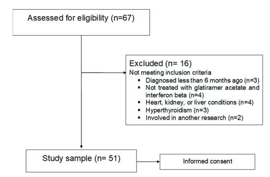


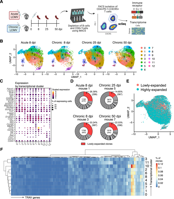


/cloudfront-us-east-1.images.arcpublishing.com/gray/SU55EX6TOBH5NNP27XOQDM7LFY.PNG)