Sertoli Cells
Sertoli Cell Tumor
Testis
Seminiferous Tubules
Spermatogenesis
Seminiferous Epithelium
Blood-Testis Barrier
Spermatids
Spermatogonia
Spermatocytes
Leydig Cells
Follicle Stimulating Hormone
Spermatozoa
Receptors, FSH
Androgen-Binding Protein
Testosterone
Sertoli Cell-Only Syndrome
Inhibins
Clusterin
Cryptorchidism
Cells, Cultured
Antispermatogenic Agents
Anti-Mullerian Hormone
Rats, Sprague-Dawley
Transferrin
Sex-Determining Region Y Protein
SOX9 Transcription Factor
Immunohistochemistry
Infertility, Male
Tight Junctions
Cell Count
GATA4 Transcription Factor
RNA, Messenger
Epididymis
Diethylhexyl Phthalate
Sex Differentiation
Rete Testis
Microscopy, Electron
Seminal Vesicles
Bucladesine
Oligospermia
Receptors, Androgen
Intercellular Junctions
Reverse Transcriptase Polymerase Chain Reaction
Orchitis
Rats, Inbred Strains
Inhibin-beta Subunits
Testicular Neoplasms
Effects of spinal cord injury on spermatogenesis and the expression of messenger ribonucleic acid for Sertoli cell proteins in rat Sertoli cell-enriched testes. (1/1665)
The study was an examination of the effects of spinal cord injury (SCI) on spermatogenesis and Sertoli cell functions in adult rats with Sertoli cell-enriched (SCE) testes. The effects of SCI on the seminiferous epithelium were characterized by abnormalities in the remaining spermatogenic cells during the first month after SCI. Three days after SCI, serum testosterone levels were 80% lower, while serum FSH and LH levels were 25% and 50% higher, respectively, than those of sham control SCE rats. At this time, the levels of mRNA for androgen receptor (AR), FSH receptor (FSH-R), and androgen-binding protein (ABP) were normal whereas those for transferrin (Trf) had decreased by 40%. Thereafter, serum testosterone levels increased, but they remained lower than those of the sham control rats 28 days after SCI; and serum FSH and LH levels returned to normal. The levels of mRNA for AR, ABP, and Trf exhibited a biphasic increase 7 days after SCI and remained elevated 28 days after SCI. FSH-R mRNA levels were also elevated 90 days after SCI. Unexpectedly, active spermatogenesis, including qualitatively complete spermatogenesis, persisted in > 40% of the tubules 90 days after SCI. These results suggest that the stem cells and/or undifferentiated spermatogonia in SCE testes are less susceptible to the deleterious effects of SCI than the normal testes and that they were able to proliferate and differentiate after SCI. The presence of elevated levels of mRNA for Sertoli cell FSH-R and AR, as well as of that for the Sertoli cell proteins, in the SCE testes during the chronic stage of SCI suggests a modification of Sertoli cell physiology. Such changes in Sertoli cell functions may provide a beneficial environment for the proliferation of the stem cells and differentiation of postmeiotic cells, thus resulting in the persistence of spermatogenesis in these testes. (+info)Role of class B scavenger receptor type I in phagocytosis of apoptotic rat spermatogenic cells by Sertoli cells. (2/1665)
Rat Sertoli cells phagocytose apoptotic spermatogenic cells, which consist mostly of spermatocytes, in primary culture by recognizing phosphatidylserine (PS) exposed on the surface of degenerating spermatogenic cells. We compared the mode of phagocytosis using spermatogenic cells at different stages of spermatogenesis. Spermatogenic cells were separated into several groups based on their ploidy, with purities of 60-90%. When the fractionated spermatogenic cell populations were subjected to a phagocytosis assay, cells with ploidies of 1n, 2n, and 4n were almost equally phagocytosed by Sertoli cells. All the cell populations exposed PS on the cell surface, and phagocytosis of all cell populations was similarly inhibited by the addition of PS-containing liposomes. Class B scavenger receptor type I (SR-BI), a candidate for the PS receptor, was detected in Sertoli cells. Overexpression of the rat SR-BI cDNA increased the PS-mediated phagocytic activity of Sertoli cell-derived cell lines. Moreover, phagocytosis of spermatogenic cells by Sertoli cells was inhibited in the presence of an anti-SR-BI antibody. Finally, the addition of high density lipoprotein, a ligand specific for SR-BI, decreased both phagocytosis of spermatogenic cells and incorporation of PS-containing liposomes by Sertoli cells. In conclusion, SR-BI functions at least partly as a PS receptor, enabling Sertoli cells to recognize and phagocytose apoptotic spermatogenic cells at all stages of differentiation. (+info)Constitutive Fas ligand gene transcription in Sertoli cells is regulated by Sp1. (3/1665)
The transcriptional regulation of the Fas ligand (FasL) gene in Sertoli cells was investigated, as these cells are known to have constitutive expression of FasL and hence maintain an "immune privileged" environment within the testicle. Using the Sertoli cell line TM4, it was demonstrated that a gene segment of the 5'-untranslated region located between -318 and -237 relative to the translation start site is required for constitutive FasL transcription. Deletion and mutation analysis demonstrate that an Sp1 rather than an NFAT or NFKB-like DNA binding motif present within this region is necessary and sufficient for constitutive FasL gene transcription. Nuclear extracts of Sertoli cells contain Sp1 and Sp3 that specifically binds to the Sp1 motif present in the FasL gene, and overexpression of Sp1 but not Sp3 leads to a further increase of transcription from the FasL promoter-enhancer region. The data presented demonstrates that constitutive FasL gene transcription in Sertoli cells is regulated by Sp1. In addition, it is shown that basal FasL expression in Jurkat T cells is also controlled by Sp1 and this is in contrast to induced FasL expression, which is NFAT-dependent. (+info)Spermatid translocation in the rat seminiferous epithelium: coupling membrane trafficking machinery to a junction plaque. (4/1665)
In this study, we demonstrate that specialized junction plaques that occur between Sertoli cells and spermatids in the rat testis support microtubule translocation in vitro. During spermatogenesis, Sertoli cells are attached to spermatids by specialized adhesion junctions termed ectoplasmic specializations (ESs). These structures consist of regions of the plasma membrane adherent to the spermatid head, a submembrane layer of tightly packed actin filaments, and an attached cistern of endoplasmic reticulum. It has been proposed that motor proteins on the endoplasmic reticulum interact with adjacent microtubules to translocate the junction plaques, and hence the attached spermatids, within the epithelium. If this hypothesis is true, then isolated junctions should support microtubule transport. To verify this prediction, we have mechanically isolated rat spermatids, together with their attached ESs, and tested them for their ability to transport microtubules in vitro. Most assays were done in the presence of 2 mg/ml testicular cytosol and at room temperature. ESs attached to spermatids supported microtubule translocation. In some cases in which motility events were detected, microtubules moved smoothly over the junction site. In others, the movement was slow but progressive, saltatory and "inch-worm-like." No motility was detected in the absence of exogenous ATP or in the presence of apyrase (an enzyme that catalyses the breakdown of ATP). Our results are consistent with the microtubule-based motility hypothesis of spermatid translocation. (+info)Rat testis motor proteins associated with spermatid translocation (dynein) and spermatid flagella (kinesin-II). (5/1665)
In this study, we report sites in the seminiferous epithelium of the rat testis that are immunoreactive with antibodies to the intermediate chain of cytoplasmic dynein and kinesin II. The study was done to determine whether or not microtubule-dependent motor proteins are present in Sertoli cell regions involved with spermatid translocation. Sections and epithelial fragments of perfusion-fixed rat testis were probed with an antibody (clone 74.1) to the intermediate chain of cytoplasmic dynein (IC74) and to kinesin-II. Labeling with the antibody to cytoplasmic dynein was dramatically evident in Sertoli cell regions surrounding apical crypts containing attached spermatids and known to contain unique intercellular attachment plaques. The antibody to kinesin II reacted only with spermatid tails. The levels of cytoplasmic dynein visible on immunoblots of supernatants collected from spermatid/junction complexes treated with an actin-severing enzyme (gelsolin) were greater than those of controls, indicating that at least some of the dynein may have been associated with Sertoli cell junction plaques attached to spermatids. Results are consistent with the conclusion that an isoform of cytoplasmic dynein may be responsible for the apical translocation of elongate spermatids that occurs before sperm release. Also, this is the first report of kinesin-II in mammalian spermatid tails. (+info)Distribution of keratins, vimentin, and actin in the testis of two South American camelids: vicuna (Vicugna vicugna) and llama (Lama glama). An immunohistochemical study. (6/1665)
The purpose of the present study was to investigate the pattern of distribution of cytokeratins, vimentin and muscular actin in the testis of vicuna (Vicugna vicugna) and llama (Lama glama) two species of camelids native of the Andean high plateau of South America. Testicular biopsies of four vicunas and five llamas were used. Animals were healthy breeders. The tissues were processed by standard immunohistochemistry with antipancytokeratinAE1/AE3, antikeratin 18 (K 18), CAM 5.2 (antikeratin 5, 18, and 19), antivimentin, and smooth-muscle-specific antiactin antibodies to track the cytoskeletal pattern of testicular cells. Using AE1/AE3 antibody the immunostaining was found in the epithelial lining of tubuli recti and rete testis. The reaction was relatively stronger in the apical cytoplasm of epithelial cells. The testicular cells of the two species showed no reaction to K 18 and CAM 5.2 antibodies. Antivimentin antibody stained the basal cytoplasm of the Sertoli cells, the Leydig cells, and the epithelial lining of tubuli recti and rete testis. In the last two structures the immunostain was relatively more intense in the basal cytoplasm of epithelial cells. Antiactin antibody stained the peritubular cells and the muscle cells of the lamina propria oftubuli recti and rete testis. The presence in these species of only some keratins found in man, its coexpression with vimentin in epithelial lining of tubuli recti and rete testis and the peritubule organization, so different from other ungulates may reflect a differential adaptation of the cytoskeleton to particular reproductive strategies. (+info)Plectin is concentrated at intercellular junctions and at the nuclear surface in morphologically differentiated rat Sertoli cells. (7/1665)
Intermediate filaments in Sertoli cells have a well-defined pattern of distribution. They form a basally situated perinuclear network from which filaments extend peripherally to adhesion plaques at the plasma membrane and to sites of codistribution with other major elements of the cytoskeleton, particularly with microtubules. Although the general pattern of intermediate filament distribution is known, the molecular components involved with linking the filaments to organelles and attachment plaques in these cells have not been identified. One candidate for such a linking element is plectin. In this study we test for the presence of, and determine the distribution of, plectin in Sertoli cells of the rat testis. Fixed frozen sections and fixed epithelial fragments of rat testis were probed for plectin and vimentin using antibodies. Tissue was evaluated using standard fluorescence microscopy and confocal microscopy. Plectin in Sertoli cells was concentrated in a narrow zone surrounding the nucleus, and at focal sites, presumably desmosome-like plaques, at interfaces with adjacent cells. Plectin was also concentrated at sites where intermediate filament bundles project into specialized actin-filament containing plaques at sites of attachment to elongate spermatids. Plectin in Sertoli cells is concentrated at the nuclear surface and in junction plaques associated with the plasma membrane. The pattern of distribution is consistent with plectin being involved with linking intermediate filaments centrally (basally) to the nucleus and peripherally to intercellular attachment sites. (+info)mRNAs encoding a von Ebner's-like protein and the Huntington disease protein are induced in rat male germ cells by Sertoli cells. (8/1665)
The success of spermatogenesis is dependent upon closely coordinated interactions between Sertoli cells and germ cells. To identify specific molecules that mediate interactions between somatic cells and germ cells in the rat testis, Sertoli cell-germ cell co-cultures and mRNA differential display were used. Two cDNAs, clone 1 (660 nucleotides) and clone 2 (390 nucleotides) were up-regulated when Sertoli cells were co-cultured with pachytene spermatocytes or round spermatids. Northern blot analyses confirmed the differential display expression patterns. Sequence analyses indicated that clone 1 was similar to a von Ebner's gland protein (87% at the nucleotide level and 80% at the amino acid level) and clone 2 was identical to a region of the Huntington disease protein. The von Ebner's-like protein mRNA was induced after 4 h of co-culture, while the Huntington disease protein required 18 h of co-culture for expression. The von Ebner's-like protein was induced in germ cells by a secreted Sertoli cell factor(s) smaller than 10 kDa that is sensitive to freezing and thawing or boiling. The Huntington disease protein was induced in germ cells by a Sertoli cell secreted factor(s) larger than 10 kDa which survives freezing and thawing, but is inactivated by boiling. (+info)Sertoli cells, also known as sustentacular cells or nurse cells, are specialized cells in the seminiferous tubules of the testis in mammals. They play a crucial role in supporting and nurturing the development of sperm cells (spermatogenesis). Sertoli cells create a microenvironment within the seminiferous tubules that facilitates the differentiation, maturation, and survival of germ cells.
These cells have several essential functions:
1. Blood-testis barrier formation: Sertoli cells form tight junctions with each other, creating a physical barrier called the blood-testis barrier, which separates the seminiferous tubules into basal and adluminal compartments. This barrier protects the developing sperm cells from the immune system and provides an isolated environment for their maturation.
2. Nutrition and support: Sertoli cells provide essential nutrients and growth factors to germ cells, ensuring their proper development and survival. They also engulf and digest residual bodies, which are byproducts of spermatid differentiation.
3. Phagocytosis: Sertoli cells have phagocytic properties, allowing them to remove debris and dead cells within the seminiferous tubules.
4. Hormone metabolism: Sertoli cells express receptors for various hormones, such as follicle-stimulating hormone (FSH), testosterone, and estradiol. They play a role in regulating hormonal signaling within the testis by metabolizing these hormones or producing inhibins, which modulate FSH secretion from the pituitary gland.
5. Regulation of spermatogenesis: Sertoli cells produce and secrete various proteins and growth factors that influence germ cell development and proliferation. They also control the release of mature sperm cells into the epididymis through a process called spermiation.
A Sertoli cell tumor is a rare type of sex-cord stromal tumor that develops in the testicles or, more rarely, in the ovaries. These tumors arise from the Sertoli cells, which are specialized cells within the testicle that help to nurture and protect the developing sperm cells. In the ovary, Sertoli cell tumors are thought to arise from similar cells that are part of the supporting tissue in the ovary.
Sertoli cell tumors can occur in people of any age but are most commonly found in middle-aged adults. They are usually slow-growing and may not cause any symptoms, especially if they are small. However, larger tumors or those that have spread (metastasized) may cause various symptoms depending on their location and size.
Symptoms of a Sertoli cell tumor can include:
* A painless lump or swelling in the testicle or ovary
* Abdominal pain or discomfort
* Bloating or a feeling of fullness in the abdomen
* Changes in bowel habits or urinary frequency
* Pain during sexual intercourse (in women)
* Hormonal imbalances, such as gynecomastia (breast development) in men or menstrual irregularities in women.
Diagnosis of a Sertoli cell tumor typically involves a combination of imaging tests, such as ultrasound, CT scan, or MRI, and blood tests to check for elevated levels of certain hormones that may be produced by the tumor. A biopsy may also be performed to confirm the diagnosis and determine the tumor's grade and stage.
Treatment for Sertoli cell tumors typically involves surgical removal of the tumor, along with any affected lymph nodes or other tissues. Additional treatments, such as radiation therapy or chemotherapy, may be recommended in cases where the tumor has spread or is at a higher risk of recurrence. Regular follow-up care is also important to monitor for any signs of recurrence or new tumors.
The testis, also known as the testicle, is a male reproductive organ that is part of the endocrine system. It is located in the scrotum, outside of the abdominal cavity. The main function of the testis is to produce sperm and testosterone, the primary male sex hormone.
The testis is composed of many tiny tubules called seminiferous tubules, where sperm are produced. These tubules are surrounded by a network of blood vessels, nerves, and supportive tissues. The sperm then travel through a series of ducts to the epididymis, where they mature and become capable of fertilization.
Testosterone is produced in the Leydig cells, which are located in the interstitial tissue between the seminiferous tubules. Testosterone plays a crucial role in the development and maintenance of male secondary sexual characteristics, such as facial hair, deep voice, and muscle mass. It also supports sperm production and sexual function.
Abnormalities in testicular function can lead to infertility, hormonal imbalances, and other health problems. Regular self-examinations and medical check-ups are recommended for early detection and treatment of any potential issues.
Seminiferous tubules are the long, convoluted tubes within the testicles that are responsible for producing sperm in males. They are lined with specialized epithelial cells called Sertoli cells, which provide structural support and nourishment to developing sperm cells. The seminiferous tubules also contain germ cells, which divide and differentiate into spermatozoa (sperm) through the process of spermatogenesis.
The seminiferous tubules are surrounded by a thin layer of smooth muscle called the tunica albuginea, which helps to maintain the structure and integrity of the testicle. The tubules are connected to the rete testis, a network of channels that transport sperm to the epididymis for further maturation and storage before ejaculation.
Damage or dysfunction of the seminiferous tubules can lead to male infertility, as well as other reproductive health issues.
Spermatogenesis is the process by which sperm cells, or spermatozoa, are produced in male organisms. It occurs in the seminiferous tubules of the testes and involves several stages:
1. Spermatocytogenesis: This is the initial stage where diploid spermatogonial stem cells divide mitotically to produce more spermatogonia, some of which will differentiate into primary spermatocytes.
2. Meiosis: The primary spermatocytes undergo meiotic division to form haploid secondary spermatocytes, which then divide again to form haploid spermatids. This process results in the reduction of chromosome number from 46 (diploid) to 23 (haploid).
3. Spermiogenesis: The spermatids differentiate into spermatozoa, undergoing morphological changes such as the formation of a head and tail. During this stage, most of the cytoplasm is discarded, resulting in highly compacted and streamlined sperm cells.
4. Spermation: The final stage where mature sperm are released from the seminiferous tubules into the epididymis for further maturation and storage.
The entire process takes approximately 72-74 days in humans, with continuous production throughout adulthood.
The seminiferous epithelium is a specialized type of epithelial tissue that lines the seminiferous tubules within the testes. It is composed of various cell types, including germ cells in different stages of development (spermatogonia, primary and secondary spermatocytes, spermatids) and supportive cells called Sertoli cells.
The primary function of the seminiferous epithelium is to support sperm production (spermatogenesis). The Sertoli cells provide structural support and nourishment to the developing germ cells, helping them to differentiate into mature spermatozoa (sperm). This process involves a series of complex cellular events, including mitosis, meiosis, and spermiogenesis.
In addition to its role in sperm production, the seminiferous epithelium also plays a crucial part in maintaining the blood-testis barrier, which separates the testicular environment from the systemic circulation. This barrier helps protect developing germ cells from potential immune attacks and maintains an optimal microenvironment for spermatogenesis.
The Blood-Testis Barrier (BTB) is a unique structural and functional feature of the seminiferous epithelium in the testes, which forms a tight junction between adjacent Sertoli cells in the semi-niferous tubules. This barrier selectively restricts the passage of molecules, including potentially harmful substances and immune cells, from the systemic circulation into the adluminal compartment of the seminiferous epithelium where spermatogenesis occurs. This helps to maintain a immunologically privileged microenvironment that is essential for the survival and maturation of developing sperm cells, preventing an immune response against them. The BTB also regulates the movement of molecules required for spermatogenesis, such as nutrients, hormones, and signaling molecules, from the basal compartment to the adluminal compartment.
Spermatids are immature sperm cells that are produced during the process of spermatogenesis in the male testes. They are the product of the final stage of meiosis, where a diploid spermatocyte divides into four haploid spermatids. Each spermatid then undergoes a series of changes, including the development of a tail for motility and the condensation of its nucleus to form a head containing the genetic material. Once this process is complete, the spermatids are considered mature spermatozoa and are capable of fertilizing an egg.
Spermatogonia are a type of diploid germ cells found in the seminiferous tubules of the testis. They are the stem cells responsible for sperm production (spermatogenesis) in males. There are two types of spermatogonia: A-dark (Ad) and A-pale (Ap). The Ad spermatogonia function as reserve stem cells, while the Ap spermatogonia serve as the progenitor cells that divide to produce type B spermatogonia. Type B spermatogonia then differentiate into primary spermatocytes, which undergo meiosis to form haploid spermatozoa.
Spermatocytes are a type of cell that is involved in the process of spermatogenesis, which is the formation of sperm in the testes. Specifically, spermatocytes are the cells that undergo meiosis, a special type of cell division that results in the production of four haploid daughter cells, each containing half the number of chromosomes as the parent cell.
There are two types of spermatocytes: primary and secondary. Primary spermatocytes are diploid cells that contain 46 chromosomes (23 pairs). During meiosis I, these cells undergo a process called crossing over, in which genetic material is exchanged between homologous chromosomes. After crossing over, the primary spermatocytes divide into two secondary spermatocytes, each containing 23 chromosomes (but still with 23 pairs).
Secondary spermatocytes then undergo meiosis II, which results in the formation of four haploid spermatids. Each spermatid contains 23 single chromosomes and will eventually develop into a mature sperm cell through a process called spermiogenesis.
It's worth noting that spermatocytes are only found in males, as they are specific to the male reproductive system.
Leydig cells, also known as interstitial cells of Leydig or interstitial cell-stroma, are cells in the testes that produce and release testosterone and other androgens into the bloodstream. They are located in the seminiferous tubules of the testis, near the blood vessels, and are named after Franz Leydig, the German physiologist who discovered them in 1850.
Leydig cells contain cholesterol esters, which serve as precursors for the synthesis of testosterone. They respond to luteinizing hormone (LH) released by the anterior pituitary gland, which stimulates the production and release of testosterone. Testosterone is essential for the development and maintenance of male secondary sexual characteristics, such as facial hair, deep voice, and muscle mass. It also plays a role in sperm production and bone density.
In addition to their endocrine function, Leydig cells have been shown to have non-hormonal functions, including phagocytosis, antigen presentation, and immune regulation. However, these functions are not as well understood as their hormonal roles.
Follicle-Stimulating Hormone (FSH) is a glycoprotein hormone secreted and released by the anterior pituitary gland. In females, it promotes the growth and development of ovarian follicles in the ovary, which ultimately leads to the maturation and release of an egg (ovulation). In males, FSH stimulates the testes to produce sperm. It works in conjunction with luteinizing hormone (LH) to regulate reproductive processes. The secretion of FSH is controlled by the hypothalamic-pituitary-gonadal axis and its release is influenced by the levels of gonadotropin-releasing hormone (GnRH), estrogen, inhibin, and androgens.
Spermatozoa are the male reproductive cells, or gametes, that are produced in the testes. They are microscopic, flagellated (tail-equipped) cells that are highly specialized for fertilization. A spermatozoon consists of a head, neck, and tail. The head contains the genetic material within the nucleus, covered by a cap-like structure called the acrosome which contains enzymes to help the sperm penetrate the female's egg (ovum). The long, thin tail propels the sperm forward through fluid, such as semen, enabling its journey towards the egg for fertilization.
Follicle-stimulating hormone (FSH) receptors are specialized protein structures found on the surface of specific cells in the body. They play a crucial role in the endocrine system, particularly in the regulation of reproduction and development.
FSH receptors are primarily located on the granulosa cells that surround and support the developing eggs (oocytes) within the ovarian follicles in females. In males, these receptors can be found on the Sertoli cells in the seminiferous tubules of the testes.
When FSH, a glycoprotein hormone secreted by the anterior pituitary gland, binds to its specific receptor, it triggers a series of intracellular signaling events that ultimately lead to various physiological responses. In females, FSH receptor activation stimulates follicle growth, estrogen production, and oocyte maturation. In males, FSH receptor signaling supports spermatogenesis, the process of sperm cell development within the testes.
In summary, FSH receptors are essential components in the hormonal regulation of reproduction and development, mediating the actions of follicle-stimulating hormone on target cells in both females and males.
Androgen-binding protein (ABP) is a protein that binds specifically to androgens, which are hormones such as testosterone that play a role in male sexual development and masculine characteristics. ABP is produced in the Sertoli cells of the testes and helps to regulate the levels of androgens within the testes by storing them and slowly releasing them over time. This is important for maintaining normal sperm production and male reproductive function.
ABP is also found in other tissues, including the prostate gland, where it may play a role in regulating the growth and development of this tissue. Abnormal levels of ABP have been associated with certain medical conditions, such as prostate cancer and infertility.
Testosterone is a steroid hormone that belongs to androsten class of hormones. It is primarily secreted by the Leydig cells in the testes of males and, to a lesser extent, by the ovaries and adrenal glands in females. Testosterone is the main male sex hormone and anabolic steroid. It plays a key role in the development of masculine characteristics, such as body hair and muscle mass, and contributes to bone density, fat distribution, red cell production, and sex drive. In females, testosterone contributes to sexual desire and bone health. Testosterone is synthesized from cholesterol and its production is regulated by luteinizing hormone (LH) and follicle-stimulating hormone (FSH).
Testicular hormones, also known as androgens, are a type of sex hormone primarily produced in the testes of males. The most important and well-known androgen is testosterone, which plays a crucial role in the development of male reproductive system and secondary sexual characteristics. Testosterone is responsible for the growth and maintenance of male sex organs, such as the testes and prostate, and it also promotes the development of secondary sexual characteristics like facial hair, deep voice, and muscle mass.
Testicular hormones are produced and regulated by a feedback system involving the hypothalamus and pituitary gland in the brain. The hypothalamus produces gonadotropin-releasing hormone (GnRH), which stimulates the pituitary gland to release follicle-stimulating hormone (FSH) and luteinizing hormone (LH). LH stimulates the testes to produce testosterone, while FSH works together with testosterone to promote sperm production.
In addition to their role in male sexual development and function, testicular hormones also have important effects on other bodily functions, such as bone density, muscle mass, red blood cell production, mood, and cognitive function.
Sertoli Cell-Only Syndrome, also known as Del Castillo Syndrome, is a rare condition characterized by the presence of only Sertoli cells in the seminiferous tubules of the testes. These are specialized cells that normally provide support and nourishment to the developing sperm cells. However, in this syndrome, there is an absence of germ cells, which are necessary for sperm production.
The condition can be unilateral or bilateral, meaning it can affect one or both testes. It's important to note that while men with Sertoli Cell-Only Syndrome do not produce sperm, they still produce testosterone, so their secondary sexual characteristics such as facial hair, deep voice, and muscle mass develop normally.
The syndrome is often detected during infertility investigations. While it's associated with infertility, it doesn't necessarily indicate a problem with the person's overall health. However, some studies suggest that men with this condition may have an increased risk of developing testicular cancer, so regular self-examinations and medical check-ups are recommended.
Inhibins are a group of protein hormones that play a crucial role in regulating the function of the reproductive system, specifically by inhibiting the production of follicle-stimulating hormone (FSH) in the pituitary gland. They are produced and secreted primarily by the granulosa cells in the ovaries of females and Sertoli cells in the testes of males.
Inhibins consist of two subunits, an alpha subunit, and a beta subunit, which can be further divided into two types: inhibin A and inhibin B. Inhibin A is primarily produced by the granulosa cells of developing follicles in the ovary, while inhibin B is mainly produced by the Sertoli cells in the testes.
By regulating FSH production, inhibins help control the development and maturation of ovarian follicles in females and spermatogenesis in males. Abnormal levels of inhibins have been associated with various reproductive disorders, including polycystic ovary syndrome (PCOS) and certain types of cancer.
Germ cells are the reproductive cells, also known as sex cells, that combine to form offspring in sexual reproduction. In females, germ cells are called ova or egg cells, and in males, they are called spermatozoa or sperm cells. These cells are unique because they carry half the genetic material necessary for creating new life. They are produced through a process called meiosis, which reduces their chromosome number by half, ensuring that when two germ cells combine during fertilization, the normal diploid number of chromosomes is restored.
Sexual maturation is the process of physical development during puberty that leads to the ability to reproduce. This process involves the development of primary and secondary sexual characteristics, changes in hormone levels, and the acquisition of reproductive capabilities. In females, this includes the onset of menstruation and the development of breasts and hips. In males, this includes the deepening of the voice, growth of facial hair, and the production of sperm. Achieving sexual maturation is an important milestone in human development and typically occurs during adolescence.
Clusterin is a protein that is widely expressed in many tissues and body fluids, including the tears, blood plasma, seminal fluid, milk, and cerebrospinal fluid. It is also known as apolipoprotein J or sulfated glycoprotein 2. Clusterin has diverse functions, including cell-cell communication, lipid transport, and protection against oxidative stress.
In the context of medicine and disease, clusterin has been studied for its potential role in several pathological processes, such as neurodegeneration, inflammation, cancer, and aging. In particular, clusterin has been implicated in the development and progression of various types of cancer, including prostate, breast, ovarian, and lung cancer. It is thought to contribute to tumor growth, invasion, and metastasis by promoting cell survival, angiogenesis, and resistance to chemotherapy.
Therefore, clusterin has been considered as a potential therapeutic target for cancer treatment, and several strategies have been developed to inhibit its expression or activity. However, more research is needed to fully understand the molecular mechanisms of clusterin in health and disease, and to translate these findings into effective clinical interventions.
Testicular diseases refer to a range of conditions that affect the testicles, the male reproductive organs located in the scrotum. These diseases can affect either one or both testicles and may cause pain, swelling, or impact fertility. Here are some examples of testicular diseases:
1. Testicular cancer: A malignant tumor that develops in the testicle. It is a relatively rare cancer but is highly treatable if detected early.
2. Testicular torsion: A surgical emergency that occurs when the spermatic cord, which supplies blood to the testicle, becomes twisted, cutting off the blood flow.
3. Epididymitis: An infection or inflammation of the epididymis, a coiled tube that stores and carries sperm from the testicle.
4. Orchitis: An infection or inflammation of the testicle itself. It can occur on its own or as a complication of mumps.
5. Hydrocele: A fluid-filled sac that forms around the testicle, causing swelling.
6. Varicocele: Enlarged veins in the scrotum that can cause pain and affect fertility.
7. Inguinal hernia: A condition where a portion of the intestine or fat protrudes through a weakened area in the abdominal wall, often appearing as a bulge in the groin or scrotum.
8. Testicular trauma: Injury to the testicle, which can result from accidents, sports injuries, or other causes.
9. Undescended testicles: A condition where one or both testicles fail to descend from the abdomen into the scrotum before birth.
It is essential for men to perform regular self-examinations to check for any unusual lumps, swelling, or pain in the testicles and seek medical attention if they notice any changes.
Cryptorchidism is a medical condition in which one or both of a male infant's testicles fail to descend from the abdomen into the scrotum before birth or within the first year of life. Normally, the testicles descend from the abdomen into the scrotum during fetal development in the second trimester. If the testicles do not descend on their own, medical intervention may be necessary to correct the condition.
Cryptorchidism is a common birth defect, affecting about 3-5% of full-term and 30% of preterm male infants. In most cases, the testicle will descend on its own within the first six months of life. If it does not, treatment may be necessary to prevent complications such as infertility, testicular cancer, and inguinal hernia.
Treatment for cryptorchidism typically involves surgery to bring the testicle down into the scrotum. This procedure is called orchiopexy and is usually performed before the age of 2. In some cases, hormonal therapy may be used as an alternative to surgery. However, this approach has limited success and is generally only recommended in certain situations.
Overall, cryptorchidism is a treatable condition that can help prevent future health problems if addressed early on. Regular check-ups with a pediatrician or healthcare provider can help ensure timely diagnosis and treatment of this condition.
"Cells, cultured" is a medical term that refers to cells that have been removed from an organism and grown in controlled laboratory conditions outside of the body. This process is called cell culture and it allows scientists to study cells in a more controlled and accessible environment than they would have inside the body. Cultured cells can be derived from a variety of sources, including tissues, organs, or fluids from humans, animals, or cell lines that have been previously established in the laboratory.
Cell culture involves several steps, including isolation of the cells from the tissue, purification and characterization of the cells, and maintenance of the cells in appropriate growth conditions. The cells are typically grown in specialized media that contain nutrients, growth factors, and other components necessary for their survival and proliferation. Cultured cells can be used for a variety of purposes, including basic research, drug development and testing, and production of biological products such as vaccines and gene therapies.
It is important to note that cultured cells may behave differently than they do in the body, and results obtained from cell culture studies may not always translate directly to human physiology or disease. Therefore, it is essential to validate findings from cell culture experiments using additional models and ultimately in clinical trials involving human subjects.
Antispermatogenic agents are substances or drugs that inhibit or prevent the production of sperm in the testes. These agents can work by various mechanisms, such as interfering with the formation and maturation of sperm cells, damaging sperm DNA, or suppressing the hormones responsible for sperm production.
Examples of antispermatogenic agents include chemotherapy drugs, radiation therapy, and certain medications used to treat prostate cancer or other conditions. Prolonged use of these agents can lead to infertility, so they are often used with caution and only when necessary. It is important to note that the use of antispermatogenic agents should be under the guidance and supervision of a medical professional.
Anti-Mullerian Hormone (AMH) is a glycoprotein hormone that belongs to the transforming growth factor-beta (TGF-β) family. It is primarily produced by the granulosa cells of developing follicles in the ovaries of females. AMH plays an essential role in female reproductive physiology, as it inhibits the recruitment and further development of primordial follicles, thereby regulating the size of the primordial follicle pool and the onset of puberty.
AMH levels are often used as a biomarker for ovarian reserve assessment in women. High AMH levels indicate a larger ovarian reserve, while low levels suggest a decreased reserve, which may be associated with reduced fertility or an earlier onset of menopause. Additionally, measuring AMH levels can help predict the response to ovarian stimulation during assisted reproductive technologies (ART) such as in vitro fertilization (IVF).
Sprague-Dawley rats are a strain of albino laboratory rats that are widely used in scientific research. They were first developed by researchers H.H. Sprague and R.C. Dawley in the early 20th century, and have since become one of the most commonly used rat strains in biomedical research due to their relatively large size, ease of handling, and consistent genetic background.
Sprague-Dawley rats are outbred, which means that they are genetically diverse and do not suffer from the same limitations as inbred strains, which can have reduced fertility and increased susceptibility to certain diseases. They are also characterized by their docile nature and low levels of aggression, making them easier to handle and study than some other rat strains.
These rats are used in a wide variety of research areas, including toxicology, pharmacology, nutrition, cancer, and behavioral studies. Because they are genetically diverse, Sprague-Dawley rats can be used to model a range of human diseases and conditions, making them an important tool in the development of new drugs and therapies.
Transferrin is a glycoprotein that plays a crucial role in the transport and homeostasis of iron in the body. It's produced mainly in the liver and has the ability to bind two ferric (Fe3+) ions in its N-lobe and C-lobe, thus creating transferrin saturation.
This protein is essential for delivering iron to cells while preventing the harmful effects of free iron, which can catalyze the formation of reactive oxygen species through Fenton reactions. Transferrin interacts with specific transferrin receptors on the surface of cells, particularly in erythroid precursors and brain endothelial cells, to facilitate iron uptake via receptor-mediated endocytosis.
In addition to its role in iron transport, transferrin also has antimicrobial properties due to its ability to sequester free iron, making it less available for bacterial growth and survival. Transferrin levels can be used as a clinical marker of iron status, with decreased levels indicating iron deficiency anemia and increased levels potentially signaling inflammation or liver disease.
Sperm count, also known as sperm concentration, is the number of sperm present in a given volume of semen. The World Health Organization (WHO) previously defined a normal sperm count as at least 20 million sperm per milliliter of semen. However, more recent studies suggest that fertility may be affected even when sperm counts are slightly lower than this threshold. It's important to note that sperm count is just one factor among many that can influence male fertility. Other factors, such as sperm motility (the ability of sperm to move properly) and morphology (the shape of the sperm), also play crucial roles in successful conception.
The Sex-Determining Region Y (SRY) protein is a transcription factor that plays a critical role in male sex determination. It is encoded by the SRY gene, which is located on the Y chromosome in humans and many other mammal species. The primary function of the SRY protein is to initiate the development of the testes during embryonic development.
In the absence of a functional SRY protein, the gonads will develop into ovaries. With a functional SRY protein, the gonads will develop into testes, which then produce androgens, including testosterone, that are necessary for the development of male secondary sexual characteristics. Mutations in the SRY gene can lead to sex reversal, where an individual with a Y chromosome develops as a female due to non-functional or absent SRY protein.
Organ size refers to the volume or physical measurement of an organ in the body of an individual. It can be described in terms of length, width, and height or by using specialized techniques such as imaging studies (like CT scans or MRIs) to determine the volume. The size of an organ can vary depending on factors such as age, sex, body size, and overall health status. Changes in organ size may indicate various medical conditions, including growths, inflammation, or atrophy.
SOX9 (SRY-related HMG-box gene 9) is a transcription factor that belongs to the SOX family of proteins, which are characterized by a high mobility group (HMG) box DNA-binding domain. SOX9 plays crucial roles in various developmental processes, including sex determination, chondrogenesis, and neurogenesis.
As a transcription factor, SOX9 binds to specific DNA sequences in the promoter or enhancer regions of its target genes and regulates their expression. In the context of sex determination, SOX9 is essential for the development of Sertoli cells in the male gonad, which are responsible for supporting sperm production. SOX9 also plays a role in maintaining the undifferentiated state of stem cells and promoting cell differentiation in various tissues.
Mutations in the SOX9 gene have been associated with several human genetic disorders, including campomelic dysplasia, a severe skeletal disorder characterized by bowed legs, and sex reversal in individuals with XY chromosomes.
Immunohistochemistry (IHC) is a technique used in pathology and laboratory medicine to identify specific proteins or antigens in tissue sections. It combines the principles of immunology and histology to detect the presence and location of these target molecules within cells and tissues. This technique utilizes antibodies that are specific to the protein or antigen of interest, which are then tagged with a detection system such as a chromogen or fluorophore. The stained tissue sections can be examined under a microscope, allowing for the visualization and analysis of the distribution and expression patterns of the target molecule in the context of the tissue architecture. Immunohistochemistry is widely used in diagnostic pathology to help identify various diseases, including cancer, infectious diseases, and immune-mediated disorders.
Male infertility is a condition characterized by the inability to cause pregnancy in a fertile female. It is typically defined as the failure to achieve a pregnancy after 12 months or more of regular unprotected sexual intercourse.
The causes of male infertility can be varied and include issues with sperm production, such as low sperm count or poor sperm quality, problems with sperm delivery, such as obstructions in the reproductive tract, or hormonal imbalances that affect sperm production. Other factors that may contribute to male infertility include genetic disorders, environmental exposures, lifestyle choices, and certain medical conditions or treatments.
It is important to note that male infertility can often be treated or managed with medical interventions, such as medication, surgery, or assisted reproductive technologies (ART). A healthcare provider can help diagnose the underlying cause of male infertility and recommend appropriate treatment options.
Tight junctions, also known as zonula occludens, are specialized types of intercellular junctions that occur in epithelial and endothelial cells. They are located near the apical side of the lateral membranes of adjacent cells, where they form a continuous belt-like structure that seals off the space between the cells.
Tight junctions are composed of several proteins, including occludin, claudins, and junctional adhesion molecules (JAMs), which interact to form a network of strands that create a tight barrier. This barrier regulates the paracellular permeability of ions, solutes, and water, preventing their uncontrolled movement across the epithelial or endothelial layer.
Tight junctions also play an important role in maintaining cell polarity by preventing the mixing of apical and basolateral membrane components. Additionally, they are involved in various signaling pathways that regulate cell proliferation, differentiation, and survival.
"Cell count" is a medical term that refers to the process of determining the number of cells present in a given volume or sample of fluid or tissue. This can be done through various laboratory methods, such as counting individual cells under a microscope using a specialized grid called a hemocytometer, or using automated cell counters that use light scattering and electrical impedance techniques to count and classify different types of cells.
Cell counts are used in a variety of medical contexts, including hematology (the study of blood and blood-forming tissues), microbiology (the study of microscopic organisms), and pathology (the study of diseases and their causes). For example, a complete blood count (CBC) is a routine laboratory test that includes a white blood cell (WBC) count, red blood cell (RBC) count, hemoglobin level, hematocrit value, and platelet count. Abnormal cell counts can indicate the presence of various medical conditions, such as infections, anemia, or leukemia.
GATA4 is a transcription factor that belongs to the GATA family of zinc finger proteins, which are characterized by their ability to bind to DNA sequences containing the core motif (A/T)GATA(A/G). GATA4 specifically recognizes and binds to GATA motifs in the promoter and enhancer regions of target genes, where it can modulate their transcription.
GATA4 is widely expressed in various tissues, including the heart, gut, lungs, and gonads. In the heart, GATA4 plays critical roles during cardiac development, such as promoting cardiomyocyte differentiation and regulating heart tube formation. It also continues to be expressed in adult hearts, where it helps maintain cardiac function and can contribute to heart repair after injury.
Mutations in the GATA4 gene have been associated with congenital heart defects, suggesting its essential role in heart development. Additionally, GATA4 has been implicated in cancer progression, particularly in gastrointestinal and lung cancers, where it can act as an oncogene by promoting cell proliferation and survival.
Messenger RNA (mRNA) is a type of RNA (ribonucleic acid) that carries genetic information copied from DNA in the form of a series of three-base code "words," each of which specifies a particular amino acid. This information is used by the cell's machinery to construct proteins, a process known as translation. After being transcribed from DNA, mRNA travels out of the nucleus to the ribosomes in the cytoplasm where protein synthesis occurs. Once the protein has been synthesized, the mRNA may be degraded and recycled. Post-transcriptional modifications can also occur to mRNA, such as alternative splicing and addition of a 5' cap and a poly(A) tail, which can affect its stability, localization, and translation efficiency.
The epididymis is a tightly coiled tube located on the upper and posterior portion of the testicle that serves as the site for sperm maturation and storage. It is an essential component of the male reproductive system. The epididymis can be divided into three parts: the head (where newly produced sperm enter from the testicle), the body, and the tail (where mature sperm exit and are stored). Any abnormalities or inflammation in the epididymis may lead to discomfort, pain, or infertility.
Diethylhexyl Phthalate (DEHP) is a type of phthalate compound that is commonly used as a plasticizer, a substance added to plastics to make them more flexible and durable. DEHP is a colorless, oily liquid with an odor similar to oil or benzene. It is soluble in organic solvents but not in water.
DEHP is used primarily in the production of polyvinyl chloride (PVC) plastics, such as flexible tubing, hoses, and medical devices like blood bags and intravenous (IV) lines. DEHP can leach out of these products over time, particularly when they are subjected to heat or other stressors, leading to potential human exposure.
Exposure to DEHP has been linked to a variety of health effects, including reproductive toxicity, developmental and neurological problems, and an increased risk of cancer. As a result, the use of DEHP in certain applications has been restricted or banned in some countries. The medical community is also moving towards using alternative plasticizers that are considered safer for human health.
"Sex differentiation" is a term used in the field of medicine, specifically in reproductive endocrinology and genetics. It refers to the biological development of sexual characteristics that distinguish males from females. This process is regulated by hormones and genetic factors.
There are two main stages of sex differentiation: genetic sex determination and gonadal sex differentiation. Genetic sex determination occurs at fertilization, where the combination of X and Y chromosomes determines the sex of the individual (typically, XX = female and XY = male). Gonadal sex differentiation then takes place during fetal development, where the genetic sex signals the development of either ovaries or testes.
Once the gonads are formed, they produce hormones that drive further sexual differentiation, leading to the development of internal reproductive structures (such as the uterus and fallopian tubes in females, and the vas deferens and seminal vesicles in males) and external genitalia.
It's important to note that while sex differentiation is typically categorized as male or female, there are individuals who may have variations in their sexual development, leading to intersex conditions. These variations can occur at any stage of the sex differentiation process and can result in a range of physical characteristics that do not fit neatly into male or female categories.
The rete testis is a network of tubules in the male reproductive system that serves as a passageway for sperm to travel from the seminiferous tubules, where sperm are produced, to the epididymis, where they mature. It is located in the mediastinum testis, which is the central part of the testicle.
The rete testis is made up of a series of interconnected tubules that are lined with simple cuboidal epithelial cells. These tubules merge to form larger ducts called efferent ductules, which then connect to the epididymis. The rete testis plays an important role in the transport and maturation of sperm, as well as in the regulation of fluid balance in the male reproductive system.
Electron microscopy (EM) is a type of microscopy that uses a beam of electrons to create an image of the sample being examined, resulting in much higher magnification and resolution than light microscopy. There are several types of electron microscopy, including transmission electron microscopy (TEM), scanning electron microscopy (SEM), and reflection electron microscopy (REM).
In TEM, a beam of electrons is transmitted through a thin slice of the sample, and the electrons that pass through the sample are focused to form an image. This technique can provide detailed information about the internal structure of cells, viruses, and other biological specimens, as well as the composition and structure of materials at the atomic level.
In SEM, a beam of electrons is scanned across the surface of the sample, and the electrons that are scattered back from the surface are detected to create an image. This technique can provide information about the topography and composition of surfaces, as well as the structure of materials at the microscopic level.
REM is a variation of SEM in which the beam of electrons is reflected off the surface of the sample, rather than scattered back from it. This technique can provide information about the surface chemistry and composition of materials.
Electron microscopy has a wide range of applications in biology, medicine, and materials science, including the study of cellular structure and function, disease diagnosis, and the development of new materials and technologies.
The seminal vesicles are a pair of glands located in the male reproductive system, posterior to the urinary bladder and superior to the prostate gland. They are approximately 5 cm long and have a convoluted structure with many finger-like projections called infoldings. The primary function of seminal vesicles is to produce and secrete a significant portion of the seminal fluid, which makes up the bulk of semen along with spermatozoa from the testes and fluids from the prostate gland and bulbourethral glands.
The secretion of the seminal vesicles is rich in fructose, which serves as an energy source for sperm, as well as various proteins, enzymes, vitamins, and minerals that contribute to maintaining the optimal environment for sperm survival, nourishment, and transport. During sexual arousal and ejaculation, the smooth muscles in the walls of the seminal vesicles contract, forcing the stored secretion into the urethra, where it mixes with other fluids before being expelled from the body as semen.
I apologize for the confusion, but "Bucladesine" is not a recognized medical term or a medication in current use in medicine. It's possible that there may be some mistake or typo in the spelling. If you have any more context about where you encountered this term, I might be able to provide a more accurate and helpful response.
Oligospermia is a medical term used to describe a condition in which the semen contains a lower than normal number of sperm. Generally, a sperm count of less than 15 million sperm per milliliter (ml) of semen is considered to be below the normal range.
Oligospermia can make it more difficult for a couple to conceive naturally and may require medical intervention such as intracytoplasmic sperm injection (ICSI) or in vitro fertilization (IVF). The condition can result from various factors, including hormonal imbalances, genetic abnormalities, varicocele, environmental factors, and certain medications.
It's important to note that oligospermia is not the same as azoospermia, which is a condition where there is no sperm present in the semen at all.
Androgen receptors (ARs) are a type of nuclear receptor protein that are expressed in various tissues throughout the body. They play a critical role in the development and maintenance of male sexual characteristics and reproductive function. ARs are activated by binding to androgens, which are steroid hormones such as testosterone and dihydrotestosterone (DHT). Once activated, ARs function as transcription factors that regulate gene expression, ultimately leading to various cellular responses.
In the context of medical definitions, androgen receptors can be defined as follows:
Androgen receptors are a type of nuclear receptor protein that bind to androgens, such as testosterone and dihydrotestosterone, and mediate their effects on gene expression in various tissues. They play critical roles in the development and maintenance of male sexual characteristics and reproductive function, and are involved in the pathogenesis of several medical conditions, including prostate cancer, benign prostatic hyperplasia, and androgen deficiency syndromes.
Intercellular junctions are specialized areas of contact between two or more adjacent cells in multicellular organisms. They play crucial roles in maintaining tissue structure and function by regulating the movement of ions, molecules, and even larger cellular structures from one cell to another. There are several types of intercellular junctions, including:
1. Tight Junctions (Zonulae Occludentes): These are the most apical structures in epithelial and endothelial cells, forming a virtually impermeable barrier to prevent the paracellular passage of solutes and water between the cells. They create a tight seal by connecting the transmembrane proteins of adjacent cells, such as occludin and claudins.
2. Adherens Junctions: These are located just below the tight junctions and help maintain cell-to-cell adhesion and tissue integrity. Adherens junctions consist of cadherin proteins that form homophilic interactions with cadherins on adjacent cells, as well as intracellular adaptor proteins like catenins, which connect to the actin cytoskeleton.
3. Desmosomes: These are another type of cell-to-cell adhesion structure, primarily found in tissues that experience mechanical stress, such as the skin and heart. Desmosomes consist of cadherin proteins (desmocadherins) that interact with each other and connect to intermediate filaments (keratin in epithelial cells) via plakoglobin and desmoplakin.
4. Gap Junctions: These are specialized channels that directly connect the cytoplasm of adjacent cells, allowing for the exchange of small molecules, ions, and second messengers. Gap junctions consist of connexin proteins that form hexameric structures called connexons in the plasma membrane of each cell. When two connexons align, they create a continuous pore or channel between the cells.
In summary, intercellular junctions are essential for maintaining tissue structure and function by regulating paracellular transport, cell-to-cell adhesion, and intercellular communication.
"Sex determination processes" refer to the series of genetic and biological events that occur during embryonic and fetal development which lead to the development of male or female physical characteristics. In humans, this process is typically determined by the presence or absence of a Y chromosome in the fertilized egg. If the egg has a Y chromosome, it will develop into a male (genetically XY) and if it does not have a Y chromosome, it will develop into a female (genetically XX).
The sex determination process involves the activation and repression of specific genes on the sex chromosomes, which direct the development of the gonads (ovaries or testes) and the production of hormones that influence the development of secondary sexual characteristics. This includes the development of internal and external genitalia, as well as other sex-specific physical traits.
It is important to note that while sex is typically determined by genetics and biology, gender identity is a separate construct that can be self-identified and may not align with an individual's biological sex.
Gonads are the reproductive organs that produce gametes (sex cells) and sex hormones. In males, the gonads are the testes, which produce sperm and testosterone. In females, the gonads are the ovaries, which produce eggs and estrogen and progesterone. The development, function, and regulation of the gonads are crucial for reproductive health and fertility.
Reverse Transcriptase Polymerase Chain Reaction (RT-PCR) is a laboratory technique used in molecular biology to amplify and detect specific DNA sequences. This technique is particularly useful for the detection and quantification of RNA viruses, as well as for the analysis of gene expression.
The process involves two main steps: reverse transcription and polymerase chain reaction (PCR). In the first step, reverse transcriptase enzyme is used to convert RNA into complementary DNA (cDNA) by reading the template provided by the RNA molecule. This cDNA then serves as a template for the PCR amplification step.
In the second step, the PCR reaction uses two primers that flank the target DNA sequence and a thermostable polymerase enzyme to repeatedly copy the targeted cDNA sequence. The reaction mixture is heated and cooled in cycles, allowing the primers to anneal to the template, and the polymerase to extend the new strand. This results in exponential amplification of the target DNA sequence, making it possible to detect even small amounts of RNA or cDNA.
RT-PCR is a sensitive and specific technique that has many applications in medical research and diagnostics, including the detection of viruses such as HIV, hepatitis C virus, and SARS-CoV-2 (the virus that causes COVID-19). It can also be used to study gene expression, identify genetic mutations, and diagnose genetic disorders.
Orchitis is a medical condition characterized by inflammation of one or both testicles, usually caused by an infection. The most common cause of orchitis is a bacterial infection that spreads from the epididymis, resulting in a condition known as epididymo-orchitis. However, viral infections such as mumps can also lead to orchitis. Symptoms may include sudden and severe pain in the testicle(s), swelling, warmth, redness of the overlying skin, nausea, vomiting, and fever. Treatment typically involves antibiotics for bacterial infections and supportive care for symptom relief. If left untreated, orchitis can lead to complications such as infertility or testicular atrophy.
"Inbred strains of rats" are genetically identical rodents that have been produced through many generations of brother-sister mating. This results in a high degree of homozygosity, where the genes at any particular locus in the genome are identical in all members of the strain.
Inbred strains of rats are widely used in biomedical research because they provide a consistent and reproducible genetic background for studying various biological phenomena, including the effects of drugs, environmental factors, and genetic mutations on health and disease. Additionally, inbred strains can be used to create genetically modified models of human diseases by introducing specific mutations into their genomes.
Some commonly used inbred strains of rats include the Wistar Kyoto (WKY), Sprague-Dawley (SD), and Fischer 344 (F344) rat strains. Each strain has its own unique genetic characteristics, making them suitable for different types of research.
Inhibin-β subunits are proteins that combine to form inhibins, which are hormones that play a role in regulating the function of the reproductive system. Specifically, inhibins help to regulate the production of follicle-stimulating hormone (FSH) by the pituitary gland.
There are two main types of Inhibin-β subunits, Inhibin-β A and Inhibin-β B, which combine with a common α subunit to form the inhibins. Inhibin-β A is produced primarily in the granulosa cells of the ovaries, while Inhibin-beta B is produced primarily in the testicular Sertoli cells.
Abnormal levels of Inhibin-β subunits have been associated with various reproductive disorders, such as polycystic ovary syndrome (PCOS) and certain types of cancer. Measurement of Inhibin-β subunits can be used as a biomarker for ovarian function, ovarian reserve and ovarian cancer detection.
Testicular neoplasms are abnormal growths or tumors in the testicle that can be benign (non-cancerous) or malignant (cancerous). They are a type of genitourinary cancer, which affects the reproductive and urinary systems. Testicular neoplasms can occur in men of any age but are most commonly found in young adults between the ages of 15 and 40.
Testicular neoplasms can be classified into two main categories: germ cell tumors and non-germ cell tumors. Germ cell tumors, which arise from the cells that give rise to sperm, are further divided into seminomas and non-seminomas. Seminomas are typically slow-growing and have a good prognosis, while non-seminomas tend to grow more quickly and can spread to other parts of the body.
Non-germ cell tumors are less common than germ cell tumors and include Leydig cell tumors, Sertoli cell tumors, and lymphomas. These tumors can have a variety of clinical behaviors, ranging from benign to malignant.
Testicular neoplasms often present as a painless mass or swelling in the testicle. Other symptoms may include a feeling of heaviness or discomfort in the scrotum, a dull ache in the lower abdomen or groin, and breast enlargement (gynecomastia).
Diagnosis typically involves a physical examination, imaging studies such as ultrasound or CT scan, and blood tests to detect tumor markers. Treatment options depend on the type and stage of the neoplasm but may include surgery, radiation therapy, chemotherapy, or a combination of these modalities. Regular self-examinations of the testicles are recommended for early detection and improved outcomes.
Fertility is the natural ability to conceive or to cause conception of offspring. In humans, it is the capacity of a woman and a man to reproduce through sexual reproduction. For women, fertility usually takes place during their reproductive years, which is from adolescence until menopause. A woman's fertility depends on various factors including her age, overall health, and the health of her reproductive system.
For men, fertility can be affected by a variety of factors such as age, genetics, general health, sexual function, and environmental factors that may affect sperm production or quality. Factors that can negatively impact male fertility include exposure to certain chemicals, radiation, smoking, alcohol consumption, drug use, and sexually transmitted infections (STIs).
Infertility is a common medical condition affecting about 10-15% of couples trying to conceive. Infertility can be primary or secondary. Primary infertility refers to the inability to conceive after one year of unprotected sexual intercourse, while secondary infertility refers to the inability to conceive following a previous pregnancy.
Infertility can be treated with various medical and surgical interventions depending on the underlying cause. These may include medications to stimulate ovulation, intrauterine insemination (IUI), in vitro fertilization (IVF), or surgery to correct anatomical abnormalities.
 Sertoli cell
Sertoli cell nodule
Sertoli cell tumour
Sertoli cell-only syndrome
Sertoli-Leydig cell tumour
Enrico Sertoli
FBXO38
Leydig cell tumour
Sex cord-gonadal stromal tumour
Catfish
McCune-Albright syndrome
C12orf24
Testicular immunology
Anteater
Human chorionic gonadotropin
Gynecomastia
A431 cells
Nurse cell
Immune privilege
Rete testis
Activin and inhibin
Paracrine signaling
Alice Embleton
Elevated alpha-fetoprotein
Daniel Djakiew
Sexual anomalies
Seminal vesicles
LRRC7
Hypogonadotropic hypogonadism
Gonadal dysgenesis
Blood-testis barrier
Carney complex
Testosterone
Arthur Böttcher
SPEF2
Sustentacular cell
Sertoli cell - Wikipedia
 Sertoli-Cell-Only Syndrome: Practice Essentials, Pathophysiology, Etiology
 Ovarian Sertoli-Leydig cell tumours: A systematic review of relapsed cases
 Sertoli-Leydig Cell Tumor | Androblastoma |Arrhenoblastoma | SLCTs
![Fluoride Action Network | [Effects. of N-acetylcysteine on fluoride-induced endoplasmic reticulum stress in sertoli cells].](data:image/png;base64,iVBORw0KGgoAAAANSUhEUgAAABAAAAAQCAMAAAAoLQ9TAAAAYFBMVEU3REw3REtFQEztFVaVK1A3Q0vh4uI8SFDw8PCepKdyen/t7e08QkyAMU9dOk2WnKBaZGpSXWN9hYpDT1a8v8GjqKtham/MztBNWF+LkZXDxsdud3zZ2tu0uLuNk5err7J/8kVkAAAAhElEQVQYlWXO0Q6DIAwF0HZbtYgKFdTh2Pj/vxwyZ0i8uU8nbVOAS/pblT7DcK8yZBgfVcbrBQAySi2TAlTWqSKNn5eVhTg9uS3QIWjdETdyQBDR3hDrcIDeXtG8if12rnxiisQT/wFSXHXLEs+JNTi2LOYHymLp7GTeAZFoL+YiXh//AjE9BtYEmsAwAAAAAElFTkSuQmCC) Fluoride Action Network | [Effects. of N-acetylcysteine on fluoride-induced endoplasmic reticulum stress in sertoli cells].
Fluoride Action Network | [Effects. of N-acetylcysteine on fluoride-induced endoplasmic reticulum stress in sertoli cells].
 Pathology Outlines - Intratubular large cell hyalinizing Sertoli cell neoplasia
 The actin filament network associated to Sertoli cell ectoplasmic specializations
 CIL:10898, Cavia porcellus, Sertoli cell. CIL. Dataset
Malignant Sertoli cell tumor in the retained abdominal testis of a unilaterally cryptorchid horse in: Journal of the American...
 Diagnosing Sertoli Cell Tumours in Dogs Using a Novel Biomarker: Anti-Müllerian Hormone - WSAVA 2015 Congress - VIN
 Giant Sertoli cell nodule of the testis: distinction from other Sertoli cell lesions | Journal of Clinical Pathology
 Endocrine-disrupting chemicals affect Sertoli TM4 cell functionality through dysregulation of gap junctional intercellular...
 Complete Androgen Insensitivity Syndrome (CAIS) and Karyotype 47, XXY, with Sertoli-Leydig Cell Tumor: Description of a Rare...
 Sclerosing Sertoli cell tumor without expression of typical sex cord stromal tumor markers: case report and literature review. ...
Sertoli Cell Tumor - Practical PetVet®
Sertoli-Cell-Only Syndrome: Background, Pathophysiology, Epidemiology
Effects of GH and IGF1 on Basal and FSH-Modulated Porcine Sertoli Cells In-Vitro
 Single-nucleotide polymorphisms in ETV5: a risk factor for Sertoli cell-only syndrome in Japanese men?
 UPF2, a nonsense-mediated mRNA decay factor, is required for prepubertal Sertoli cell development and male fertility by...
 High fat diet-induced obesity prolongs critical stages of the spermatogenic cycle in a Ldlr−/−.Leiden mouse model | Scientific...
Mechanism of humoral and cellular immune modulation provided by porcine Sertoli cells<...
 Cortactin and phagocytosis in isolated Sertoli cells | Journal of Negative Results in BioMedicine | Full Text
 Single-cell transcriptomic and antibody-based proteomic analysis of Sertoli cell-specific markers in human testis
 New Scientific Study Finds Vaping Can 'Shrink Male Testicles'? | Snopes.com
 Wnt/β-catenin signaling enhances transcription of the CX43 gene in murine Sertoli cells<...
Inhibitory effect of Sertoli cell-cultured media on LH binding to mouse Leydig cells in culture<...
Expression of cyclin-dependent kinase 5 and associated cyclins in Leydig and sertoli cells of the testis<...
Antisense oligonucleotide of clusterin mRNA induces apoptotic cell death and prevents adhesion of rat ASC-17D sertoli cells<...
 Ovarian Hilus Cell Hyperplasia and Sertoli-Leydig Cell Tumor in a Patient with Postmenopausal Virilization: a Rare Case Report
...
Ovarian Hilus Cell Hyperplasia and Sertoli-Leydig Cell Tumor in a Patient with Postmenopausal Virilization: a Rare Case Report
...
Testis15
- The occluding junctions of Sertoli cells form the blood-testis barrier, a structure that partitions the interstitial blood compartment of the testis from the adluminal compartment of the seminiferous tubules. (wikipedia.org)
- Sertoli cells control the entry and exit of nutrients, hormones, and other chemicals into the tubules of the testis as well as make the adluminal compartment an immune-privileged site. (wikipedia.org)
- [ 1 ] Sertoli cells help to make up the blood-testis barrier and are responsible assisting with sperm production. (medscape.com)
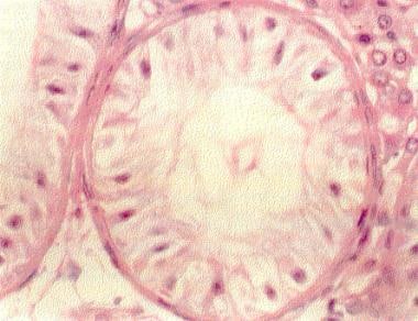
- This hematoxylin and eosin section of a testis biopsy (400X) demonstrates an individual tubule lined only with Sertoli cells (Sertoli-cell-only [SCO] syndrome). (medscape.com)
- Junctional devices in Sertoli cells conform the blood-testis barrier and play a key role in maturation and differentiation of germ cells. (techscience.com)
- Freeze-fracture preparation of the plasma membrane from an unspecialized area composed of Sertoli cells of the guinea pig testis. (ucsd.edu)
- The term "giant Sertoli cell nodule" is used for this unique, hitherto undescribed lesion and its distinction from other Sertoli cell lesions of the testis is considered here. (bmj.com)
- Proliferation and functional maturation of Sertoli cells, and their relevance to disorders of testis function in adulthood. (medscape.com)
- These provide structural and nutritional support for germ cells, regulate spermatogenesis, and maintain the blood-testis barrier. (diva-portal.org)
- 101 proteins were successfully stained and localized within testis tissue and subcellular locations in Sertoli cells were annotated. (diva-portal.org)
- In this study, we examined the expression and subcellular localization of cyclin-dependent kinase 5 (Cdk5), cyclin D1, and cyclin E in Leydig and Sertoli cell lines that were cultured with 7.5, 1.0, 0.5, or 0% serum (mixture of a 2:1 ratio of horse serum and fetal bovine serum) and in the developing rat testis to verify the possible functions of Cdk5, cyclin D1, and cyclin E in the testis. (elsevierpure.com)
- Our results suggest that Cdk5 in TM3 and Leydig cells of the testis might play a role in cell cycle regulation, whereas Cdk5 in TM4 and Sertoli cells of the adult testis might have some additional functions besides control of proliferation. (elsevierpure.com)
- In seminiferous epithelium, tight junctions (TJs) between adjacent Sertoli cells constitute the blood-testis barrier and must change synchronically for germ cells to translocate from the basal to the adluminal compartment during the spermatogenic cycle. (tmu.edu.tw)
- particularly the Sertoli cells, which play a major role in spermatogenesis and are the main cell type in the testis which expresses T 3 receptors [ 16 ]. (hindawi.com)
- It is well established that the formation of normal numbers of Sertoli cells is a key factor in determining testis size, germ cell numbers per testis, and sperm production rate in adulthood in a range of mammals, including humans [ 17 ]. (hindawi.com)
Tumor17
- Sertoli-Lydig cell tumors (SLCTs), also known as androblastoma or arrhenoblastoma , are a type of sex cord-stromal tumor that originates in the ovaries. (knowcancer.com)
- If the tumor is accompanied by high levels of testosterone, the tumor could be a Sertoli-Lydic cell tumor. (knowcancer.com)
- As a result, cells from the tumor must be biopsied (excised) and studied under a microscope for an accurate diagnosis. (knowcancer.com)
- Objective: To report the rare case of a patient with Complete Androgen Insensitivity Syndrome (CAIS) with Klinefelter Syndrome (KS) (47, XXY) presenting a Sertoli/Leydig cell tumor discovered during the gonadectomy. (remedypublications.com)
- Sclerosing Sertoli cell tumor without expression of typical sex cord stromal tumor markers: case report and literature review. (qxmd.com)
- Sertoli cell tumor is a potential histologic mimic of other tumors, such as seminoma due to similar histology and overlapping clinical presentation. (qxmd.com)
- Sclerosing Sertoli cell tumor is a rare sex cord stromal tumor variant, with 16 cases reported in the English literature. (qxmd.com)
- We present an unusual case of sclerosing Sertoli cell tumor in a 33-year-old Caucasian male, which was negative or weakly reactive using immunohistochemical markers typically positive in Sertoli cell tumors. (qxmd.com)
- This is the second reported case of a Sertoli cell tumor with reactivity limited to neuroendocrine markers and the first such case of the sclerosing variant. (qxmd.com)
- A literature review of sclerosing Sertoli cell tumor, including English and non-English literature, is described. (qxmd.com)
- Our case highlights that expected immunohistochemical markers may be negative, and awareness of antigenically unreactive tumors is needed to avoid confusion between Sertoli cell tumor and other entities. (qxmd.com)
- We present a case report regarding a 71-year-old woman with postmenopausal virilization caused by ovarian hilus cell hyperplasia and Sertoli-Leydig cell tumor who was suffered from hair loss, clitoromegaly and hirsutism. (aejog.com)
- The final pathology showed a poorly differentiated Sertoli Leydig cell tumor at the right ovary and hilus cell hyperplasia at the left ovary. (aejog.com)
- What distinguishes this case from others is that Sertoli-Leydig cell tumor and hilus cell hyperplasia may cause virilization symptoms together, in addition to its prevalence in advanced age. (aejog.com)
- Ertan B, Ozgozen E, Ilgen O, Bulbul G, Saatli B, Ulukus C. Ovarian Hilus Cell Hyperplasia and Sertoli-Leydig Cell Tumor in a Patient with Postmenopausal Virilization: a Rare Case Report. (aejog.com)
- Without appropriate regulation by miRNA, genes are likely expressed abnormally, which could cause cells to grow and divide uncontrollably and lead to tumor formation. (medlineplus.gov)
- This gene, often called a tumor suppressor gene, cannot protect cells from becoming cancerous when it isn't working properly. (choa.org)
Testes10
- Sertoli cells are a type of sustentacular "nurse" cell found in human testes which contribute to the process of spermatogenesis (the production of sperm) as a structural component of the seminiferous tubules. (wikipedia.org)
- Sertoli cells are specifically located in the convolutions of the seminiferous tubules, since this is the only place in the testes where spermatozoa are produced. (wikipedia.org)
- FSH is responsible for controlling the proliferation of Sertoli cells shortly after birth and stimulates the production of factors derived from Sertoli cells that control the development of the testes and germ cells. (wikipedia.org)
- Sertoli-cell-only (SCO) syndrome, also called germ cell aplasia or Del Castillo syndrome, describes a condition of the testes in which only Sertoli cells line the seminiferous tubules. (medscape.com)
- Taniguchi and colleagues reported that while hOvol1 and hOvol2 were detected by reverse transcription-polymerase chain reaction (RT-PCR) in the testes of patients capable of spermatogenesis, they were not detected in those with Sertoli cell-only syndrome. (medscape.com)
- Except for the large size, the findings were identical to those of a Sertoli cell nodule-a typically microscopic, unencapsulated lesion commonly detected in cryptorchid testes. (bmj.com)
- We induced high fat diet (HFD) obesity in low-density lipoprotein receptor knock-out Leiden (Ldlr −/− .Leiden) mice, and studied the morphological structure of the testes and histologically examined the proportion of Sertoli cells, spermatocytes and spermatids in the seminiferous tubules. (nature.com)
- Likewise, Sertoli cells isolated from pre-pubertal rat testes have been marked for later detection by the phagocytosis of latex beads [ 10 ]. (biomedcentral.com)
- Leydig cells and Sertoli cells were obtained from the testes of immature C57BL/6Ncrj mice and were cultured in serum-free medium for 7 days. (elsevierpure.com)
- Immunohistochemical localization of Cdk5, cyclin D1, and cyclin E in 1-week- old and adult rat testes revealed expression in both Leydig and Sertoli cells. (elsevierpure.com)
Proliferation4
- Sertoli cell proliferation and differentiation is mainly activated by FGF9, with which they also form a feedforward loop. (wikipedia.org)
- Several lines of evidence suggest that insulin-like growth factor 1 (IGF1) is involved in Sertoli cell (SC) proliferation and that its receptor (IGF1R) could mediate follicle-stimulating hormone (FSH) eects. (unict.it)
- Through this role in regulating the activity (expression) of genes, the Dicer protein is involved in many processes, including cell growth and division (proliferation) and the maturation of cells to take on specialized functions (differentiation). (medlineplus.gov)
- Sertoli cell proliferation reaches its maximum level just before birth and ceases by the age of 3 weeks [ 24 ]. (hindawi.com)
Fetal3
- It has been suggested that Sertoli cells may derive from the fetal mesonephros. (wikipedia.org)
- Immunohistochemical studies verified the fetal phenotype of the Sertoli cells and the non-neoplastic nature of the germ cell component. (bmj.com)
- Fetal type Leydig cells disappear soon after birth (during first 2 weeks after birth) and are replaced by adult type Leydig cells [ 22 , 23 ]. (hindawi.com)
Ovary1
- Stromal tumors of the ovary include germ-cell tumors, sex-cord stromal tumors, and other more rare types. (medscape.com)
Ectoplasmic specializations3
- The spacial distribution of ectoplasmic specializations of Sertoli cells was studied by β-actin immunolabelling, using laser confocal and transmission electron microscopy. (techscience.com)
- For confocal microscopy, β-actin immunolabelling of ectoplasmic specializations was studied over the background of either prosaposin or glutaredoxin immunolabelling of the Sertoli cytoplasm. (techscience.com)
- Cortactin, an actin binding protein, has been associated with Sertoli cell ectoplasmic specializations in vivo , based on its immunolocalization around the heads of elongated spermatids, but not previously identified in isolated Sertoli cells. (biomedcentral.com)
Immature4
- Microscopical examination showed the encapsulated nodule to be composed of tubules lined by immature Sertoli cells with interspersed spermatogonia and an interwoven network of hyalinised basement membrane having foci of calcification. (bmj.com)
- Using immunohistochemistry, AMH has been shown to be a useful marker of immature and neoplastic Sertoli cells in dogs. (vin.com)
- The aim of this study is to examine the influence of Sertoli cells on LH binding to Leydig cells in culture in immature mice. (elsevierpure.com)
- The objective of the present study is to investigate the modulation of antioxidant defence status in neonatal persistent hypothyroid rats before their sexual maturation and also to identify the specific testicular cell populations vulnerable to degeneration during neonatal hypothyroidism in immature rats. (hindawi.com)
Cytoplasm5
- their cytoplasm often completely surrounds the germline cells which they are responsible for nursing. (wikipedia.org)
- Sertoli cells also act as phagocytes, consuming the residual cytoplasm during spermatogenesis. (wikipedia.org)
- Labelling was found near the basal lamina, surrounding early spermatocytes (presumably in leptotene-zygotene) or at one of two levels in the seminiferous epithelium: (1) around deep infoldings of the Sertoli cell cytoplasm, in tubular stages before spermiation, and (2) in the superficial part of the seminiferous epithelium, in tubular stages after or during spermiation. (techscience.com)
- Cells exposed to no hormone and no beads appeared to have a ubiquitous distribution of cortactin throughout the cytoplasm. (biomedcentral.com)
- Cortactin immunostaining was diffuse throughout the Sertoli cell cytoplasm when incubated without beads (Fig. 1A ). (biomedcentral.com)
Spermatogenesis6
- Therefore, once spermatogenesis has begun, no more Sertoli cells are created, and their population within the seminiferous tubules is finite. (wikipedia.org)
- Because its main function is to nourish developing sperm cells through the stages of spermatogenesis, the Sertoli cell has also been called the "mother" or "nurse" cell. (wikipedia.org)
- Sertoli cells are also responsible for establishing and maintaining the spermatogonial stem cell niche, which ensures the renewal of stem cells and the differentiation of spermatogonia into mature germ cells that progress stepwise through the long process of spermatogenesis, ending in the release of spermatozoa in a process known as spermiation. (wikipedia.org)
- These cells respond to follicle-stimulating hormone (FSH) released by the hypothalamus, which helps to promote spermatogenesis. (medscape.com)
- Spermatogenesis, the process of sperm formation, occurs in the seminiferous tubules and involves the coordinated development of germ cells with the support of Sertoli cells. (diva-portal.org)
- This compromised testicular antioxidant status might have contributed to poor growth and development by affecting the spermatogenesis and steroidogenesis in rats before puberty as indicated by reduced germ cell number, complete absence of round spermatids, decreased seminiferous tubule diameter, and decreased testosterone level. (hindawi.com)
Testicular5
- This gives rise to the possibility of repairing some defects of testicular niche cells which may cause male infertility. (wikipedia.org)
- This study addresses effects on EDCs on adjacent prepubertal Sertoli TM4 cells, specifically on 1) testicular gap junctional intercellular communication (GJIC), one of the hallmarks of non-genotoxic carcinogenicity, 2) GJIC building blocks connexins (Cx), and 3) mitogen-activated protein kinases MAPKs. (muni.cz)
- Our results indicate that EDCs might disrupt testicular homeostasis and development via testicular GJIC, junctional and non-junctional functions of Cx43 and MAPK-signaling pathways in Sertoli cells. (muni.cz)
- Despite extensive research on Sertoli cells, their protein expression patterns and spatial localization in relation to other testicular cell types remain poorly characterized. (diva-portal.org)
- Recent studies have demonstrated that serum levels Inhibin-B, a Sertoli cell secretory protein, are suppressed in the presence of testicular injury. (cdc.gov)
Phenotype2
- Recently, however, scientists have found a way to induce Sertoli cells to a juvenile proliferative phenotype outside of the body. (wikipedia.org)
- Genetic mechanisms are implicated in aspects of male infertility and male mice lacking the gene Etv5 exhibited azoospermia with loss of germ cells: a phenotype identical to human Sertoli cell-only syndrome (SCOS). (imrpress.com)
Ovarian1
- Sertoli-Leydig cell tumors are rare ovarian neoplasms. (manipal.edu)
Secrete2
- For example, Sertoli cells secrete anti-müllerian hormone (AMH), which helps to ensure regression of müllerian ducts as a fetus develops into a male. (medscape.com)
- stimulates interstitial cells to secrete testosterone. (flashcardmachine.com)
Syndrome6
- As an example, Klinefelter syndrome is characterized by SCO and Leydig cell hyperplasia. (medscape.com)
- In SCO syndrome mouse model, PRPS2 overexpression significantly inhibited cell apoptosis and promoted cell cycle transition in TM4 Sertoli cells. (medscape.com)
- Yao and colleagues found 174 microRNAs (miRNAs) were differentially expressed in human Sertoli cells in men with SCO syndrome compared with men with obstructive azoospermia, suggesting that these miRNAs may be associated with the pathogenesis of SCO syndrome. (medscape.com)
- Single-nucleotide polymorphisms in the LRWD1 gene may be a genetic risk factor for Japanese patients with Sertoli cell-only syndrome. (medscape.com)
- Single-nucleotide polymorphisms in ETV5: a risk factor for Sertoli cell-only syndrome in Japanese men? (imrpress.com)
- DICER1 syndrome is also associated with tumors in the ovaries known as Sertoli-Leydig cell tumors, which typically develop in affected women in their teens or twenties. (medlineplus.gov)
Tumour1
- To synthesize the evidence on Sertoli-Leydig cell tumour (SLCT) relapses, and identify the clinicopathological characteristics and prognosis of patients with recurrent SLCT. (nih.gov)
Support for germ cells1
- Sertoli cells provide the nutritional and metabolic support for germ cells. (unab.cl)
Azoospermia1
- Massive deletion in AZFb/b+c and azoospermia with Sertoli cell only and/or maturation arrest. (medscape.com)
Seminiferous tubule1
- The Sertoli cells line the seminiferous tubule. (medscape.com)
Induce1
- These findings suggested that interruption of cell to plate substratum interaction might lead to over-expression of clusterin from Sertoli cells to induce cell to cell aggregation or, perhaps, to re-establish attachment with the substratum. (korea.ac.kr)
Puberty1
- After puberty, Sertoli cells begin to elongate. (wikipedia.org)
Tubules1
- Translocation of cells from the basal lamina to the lumen of the seminiferous tubules occurs by conformational changes in the lateral margins of the Sertoli cells. (wikipedia.org)
Abstract1
- abstract = "The understanding of main mechanisms that determine the ability of immune privilege related to Sertoli cells (SCs) will provide clues for promoting a local tolerogenic environment. (johnshopkins.edu)
Leydig Cells6
- The LH binding to Leydig cells and the FSH binding to Sertoli cells were dependent on incubation time, the number of cells, and the amount of labelled hormone added. (elsevierpure.com)
- The dissociation constant for LH binding to Leydig cells was 7.3 x 10 -10 M. Co-culture of Leydig cells with Sertoli cells for 7 days decreased LH binding to Leydig cells. (elsevierpure.com)
- The binding was 34.9% of that to Leydig cells cultured alone. (elsevierpure.com)
- After cultivation of Leydig cells with spent Sertoli cell-cultured medium (SM) for the last 4 days of the 7-day culture period, LH binding to Leydig cells decreased to as low as 17.4% of that of the controls. (elsevierpure.com)
- For the controls, LH binding was measured in Leydig cells cultured in spent Leydig cell-cultured medium (LM). (elsevierpure.com)
- These data suggest that some factor or factors are secreted from the cultured Sertoli cells and inhibit the binding of LH to Leydig cells in culture. (elsevierpure.com)
Cx434
- EDCs rapidly dysregulated GJIC in Sertoli TM4 cells mainly via MAPK p38 and/or Erk1/2 pathways by the intermediate hyper- or de-phosphorylation of Cx43 (Ser368, Ser282) and translocation of Cx43 from the plasma membrane, suggesting disturbed intracellular trafficking of Cx43 protein. (muni.cz)
- The Cx43 promoter contains TCF/β-catenin binding elements (TBEs) that contribute CX43 expression in different cell types and which may also be regulating the expression of this gene in Sertoli cells. (unab.cl)
- In this study, we demonstrate that 42GPA9 Sertoli cells respond to treatments that result in accumulation of β-catenin within the nucleus and in upregulation of CX43 gene transcription. (unab.cl)
- Our results also indicate that the Wnt/β-catenin-dependent upregulation of the Cx43 gene in Sertoli cells is accompanied by changes in epigenetic parameters that may be directly contributing to generating a chromatin environment that facilitates the establishment of the transcriptional machinery at this promoter. (unab.cl)
Maturation1
- Hadjkacem-Loukil L, Hadj-Kacem H, Hadj Salem I, Bahloul A, Fakhfakh F, Ayadi H. Genotyping of Tunisian azoospermic men with Sertoli cell-only and maturation arrest. (medscape.com)
Gonadal1
- In humans, AMH has been shown to be a specific marker of Sertoli cell origin in gonadal tumours. (vin.com)
Reproductive1
- thyroid-stimulating hormone, and hCG are all known to affect Sertoli cell development and male reproductive health. (wikipedia.org)
Tubular1
- Our results show a consistent labelling for β-actin before, during and after the release of spermatozoa in the tubular lumen (spermiation) suggesting a significant role of the actin network in spermatic cell differentiation. (techscience.com)
Localization3
- Double-labeling experiments in HeLa cells invaded with S. flexneri showed a near perfect co-localization of cortactin with actin in the entry structure and at the periphery of the phagosome [ 17 ]. (biomedcentral.com)
- The presence or absence of FSH had no apparent effect on the distribution or localization of cortactin within Sertoli cells. (biomedcentral.com)
- This project aimed to investigate the expression patterns and spatial localization of proteins in Sertoli cells. (diva-portal.org)
Proteins5
- Single-cell RNA sequencing data and immunohistochemistry stainings from the Human Protein Atlas were used to choose candidate proteins to be visualized in a multiplex Immunofluorescence panel. (diva-portal.org)
- Additionally, a list of uncharacterized proteins in Sertoli cells were identified. (diva-portal.org)
- The employed workflow demonstrated its potential for identifying Sertoli cell-specific proteins and can be applied to future research. (diva-portal.org)
- Additional validation is required to verify the presence and identify function of these proteins in Sertoli cells. (diva-portal.org)
- Together, we conclude that the activation of L-selectin induces Ca +2 influx through the L-type VOCC, which up-regulates Rho and Rac1 proteins, in ASC-17D cells. (tmu.edu.tw)
Serum4
- The abundance of Cdk5 and cyclin E in the Leydig cell line, TM3, was significantly reduced at low serum concentrations. (elsevierpure.com)
- In contrast, serum concentration had no effect on Cdk5 and cyclin E levels in the Sertoli cell line, TM4. (elsevierpure.com)
- Cyclin D1 was detected by western blot analysis in TM4 cells only, and its abundance was serum dose dependent. (elsevierpure.com)
- The kinase activity of Cdk5 in TM3 and TM4 cells that were cultured at various serum concentrations coincided with the levels of Cdk5 expression. (elsevierpure.com)
Immunohistochemical1
- Immunohistochemical staining for Cdk5 and cyclin E revealed nuclear and cytoplasmic distribution, both in TM3 and TM4 cells. (elsevierpure.com)
Pathways1
- CONCLUSION: Endoplasmic reticulum stress signaling pathways were activated by ROS, and NAC attenuate endoplasmic reticulum stress through inhibiting the levels of ROS in NaF-treated sertoli cells. (fluoridealert.org)
Markers1
- FSH is required for Sertoli cell mitogen, which stimulates the expression of various cell markers. (wikipedia.org)
Sperm production1
- Sertoli cells - one of the "most important cells for sperm production - were also shown to be altered. (snopes.com)
Tissue1
- Together, the results suggest that smoking traditional and e-cigarettes can increase oxidative stress , or cell death, which has been linked to tissue and DNA damage. (snopes.com)
Actin3
- Also, these different approaches allowed us to study the complex arrangement of the actin cytoskeleton of Sertoli cells branches, which surround germ cells in different stages of the spermatogenic cycle. (techscience.com)
- In the presence of cytochalasin D, cortactin immunostaining was punctate and distributed in a pattern similar to that reported for actin in cells exposed to cytochalasin D. Sertoli cells not exposed to FSH, but activated with beads, did not show cortactin immunostaining around the phagocytized beads at any of the time periods. (biomedcentral.com)
- Cortactin is recruited to the actin-rich membrane ruffles of the entry structure of Shigella flexneri when invading HeLa cells and is also found in the periphery of the phagosome shortly after internalization [ 17 ]. (biomedcentral.com)
Regulation3
- In addition, up-regulation of clusterin mRNA was so much greater when the Sertoli cells were plated on the petri dish for the bacterial culture instead of in a animal cell culture flask that therefore, the cells failed to attach to it. (korea.ac.kr)
- From these results, we speculate that down-regulation of clusterin expression in the anchorage-dependent Sertoli cells prevents them from attaching to the plate, and therefore induces cell death. (korea.ac.kr)
- We then demonstrated the up-regulation of Rho and Rac1 in ASC-17D cells following the administration of L-selectin ligand, and the pre-treatment with nifedipine, but not mibefradil, prior to L-selectin ligand-binding abolished the activation of both Rho and Rac1. (tmu.edu.tw)
Objective1
- OBJECTIVE: Investigated the effects of N-acetylcysteine (NAC) on endoplasmic reticulum stress of sertoli cells induced by sodium fluoride (NaF). (fluoridealert.org)
Microscopic1
- Further, microscopic observation showed that more noticeable cell death was induced by treating the antisense prior to plating the cells than by treating after cell attachment to the plate. (korea.ac.kr)
Expression2
- Recently we reported the transient expression of clusterin in pancreatic endocrine cells during the early developmental stages and suggested a role in aggregating the endocrine cells for islet formation. (korea.ac.kr)
- The expression of clusterin was transiently increased as early as 2-8 h after plating the ASC-17D Sertoli cells to the culture flask, which was the period of cell attachment. (korea.ac.kr)