Sinoatrial Node
Sinoatrial Block
Tachycardia, Sinoatrial Nodal Reentry
Lymph Nodes
Atrioventricular Node
Biological Clocks
Bradycardia
Rabbits
Action Potentials
Heart Conduction System
Hyperpolarization-Activated Cyclic Nucleotide-Gated Channels
Arrhythmia, Sinus
Models, Cardiovascular
Cyclic Nucleotide-Gated Cation Channels
Vagus Nerve
Sick Sinus Syndrome
Calcium Channels, L-Type
Heart Block
Electrophysiology
Membrane Potentials
Calcium Channels, T-Type
Connexins
Tachycardia, Sinus
Potassium Channels
Dogs
Electrocardiography
Cranial Nerve Injuries
Ion Channels
Connexin 43
Cardiac Pacing, Artificial
Lymph Node Excision
Patch-Clamp Techniques
Myocytes, Cardiac
Myoblasts, Cardiac
Microelectrodes
Aminophylline
Delayed Rectifier Potassium Channels
Electrophysiological Phenomena
Guinea Pigs
Sentinel Lymph Node Biopsy
Pacemaker, Artificial
Ryanodine
Arrhythmias, Cardiac
Sodium Channels
Cardiotonic Agents
Autonomic Nervous System
Aging, Premature
Anti-Arrhythmia Agents
Computer Simulation
Tachycardia, Supraventricular
Sarcoplasmic Reticulum
Acetylcholine
Penicillin Amidase
Natriuretic Peptide, C-Type
Parasympathetic Nervous System
Calcium
Calcium Signaling
Atropine
Ion Channel Gating
Electrophysiologic Techniques, Cardiac
Ryanodine Receptor Calcium Release Channel
Lymphatic Metastasis
Dual allosteric modulation of pacemaker (f) channels by cAMP and voltage in rabbit SA node. (1/896)
1. A Monod-Whyman-Changeux (MWC) allosteric reaction model was used in the attempt to describe the dual activation of 'pacemaker' f-channel gating subunits by voltage hyperpolarization and cyclic nucleotides. Whole-channel kinetics were described by assuming that channels are composed of two identical subunits gated independently according to the Hodgkin-Huxley (HH) equations. 2. The simple assumption that cAMP binding favours open channels was found to readily explain induction of depolarizing voltage shifts of open probability with a sigmoidal dependence on agonist concentration. 3. Voltage shifts of open probability were measured against cAMP concentration in macropatches of sino-atrial (SA) node cells; model fitting of dose-response relations yielded dissociation constants of 0.0732 and 0.4192 microM for cAMP binding to open and closed channels, respectively. The allosteric model correctly predicted the modification of the pacemaker current (If) time constant curve induced by 10 microM cAMP (13.7 mV depolarizing shift). 4. cAMP shifted deactivation more than activation rate constant curves, according to sigmoidal dose-response relations (maximal shifts of +22.3 and +13.4 mV at 10 microM cAMP, respectively); this feature was fully accounted for by allosteric interactions, and indicated that cAMP acts primarily by 'locking' f-channels in the open configuration. 5. These results provide an interpretation of the dual voltage- and cyclic nucleotide- dependence of f-channel activation. (+info)Regional differences in effects of E-4031 within the sinoatrial node. (2/896)
Effects of block of the rapid delayed rectifier K+ current (IK,r) by E-4031 on the electrical activity of small ball-like tissue preparations from different regions of the rabbit sinoatrial node were measured. The effects of partial block of IK,r by 0.1 microM E-4031 varied in different regions of the node. In tissue from the center of the node spontaneous activity was generally abolished, whereas in tissue from the periphery spontaneous activity persisted, although the action potential was prolonged, the maximum diastolic potential was decreased, and the spontaneous activity slowed. After partial block of IK,r, the electrical activity of peripheral tissue was more like that of central tissue under normal conditions. One possible explanation of these findings is that the density of IK,r is greater in the periphery of the node; this would explain the greater resistance of peripheral tissue to IK,r block and help explain why, under normal conditions, the maximum diastolic potential is more negative, the action potential is shorter, and pacemaking is faster in the periphery. (+info)Contribution of L-type Ca2+ current to electrical activity in sinoatrial nodal myocytes of rabbits. (3/896)
The role of L-type calcium current (ICa,L) in impulse generation was studied in single sinoatrial nodal myocytes of the rabbit, with the use of the amphotericin-perforated patch-clamp technique. Nifedipine, at a concentration of 5 microM, was used to block ICa,L. At this concentration, nifedipine selectively blocked ICa,L for 81% without affecting the T-type calcium current (ICa,T), the fast sodium current, the delayed rectifier current (IK), and the hyperpolarization-activated inward current. Furthermore, we did not observe the sustained inward current. The selective action of nifedipine on ICa,L enabled us to determine the activation threshold of ICa,L, which was around -60 mV. As nifedipine (5 microM) abolished spontaneous activity, we used a combined voltage- and current-clamp protocol to study the effects of ICa,L blockade on repolarization and diastolic depolarization. This protocol mimics the action potential such that the repolarization and subsequent diastolic depolarization are studied in current-clamp conditions. Nifedipine significantly decreased action potential duration at 50% repolarization and reduced diastolic depolarization rate over the entire diastole. Evidence was found that recovery from inactivation of ICa,L occurs during repolarization, which makes ICa,L available already early in diastole. We conclude that ICa,L contributes significantly to the net inward current during diastole and can modulate the entire diastolic depolarization. (+info)Electrophysiological effects of mexiletine in man. (4/896)
The electrophysiological effects of intravenous mexiletine in a dose of 200 to 250 mg given over 5 minutes, followed by continuous infusion of 60 to 90 mg per hour, were studied in 5 patients with normal conduction and in 20 patients with a variety of disturbances of impulse formation and conduction, by means of His bundle electrography, atrial pacing, and the extrastimulus method. In all but 2 patients the plasma level was above the lower therapeutic limit. Mexiletine had no consistent effects on sinus frequency and atrial refractoriness. The sinoatrial recovery time changed inconsistently in both directions; however, of the 5 patients in whom an increase was evident, 3 had sinus node dysfunction. In most patients mexiletine increased the AV nodal conduction time at paced atrial rates and shifted the Wenckebach point to a lower atrial rate. The effective refractory period of the AV node was not consistently influenced, while the functional refractory period increased in 12 out of 14 patients. The HV intervals increased by a mean of 11 ms in 8 patients and were unchanged in 17. Both the relative and effective refractory period of the His-Purkinje system increased after mexiletine. Non-cardiac side effects occurred in 7 out of 25 patients, and cardiac side effects, including one serious, in 2. The results indicate that mexiletine shares some electrophysiological properties with procainamide and quinidine, when given to patients with conduction defects, and that the drug should not be used in patients with pre-existing impairment of impulse formation or conduction. It has additional effects on AV nodal conduction which may be of value in the treatment of re-entrant tachycardias involving the AV node. (+info)The nerve supply and conducting system of the human heart at the end of the embryonic period proper. (5/896)
The nerve supply and conducting system were studied in a stage 23 human embryo of exceptional histological quality. The nerves on the right side arose from cervical sympathetic and from cervical and thoracic vagal filaments. Out of their interconnexions vagoxympathetic nerves emerged, which (1) sent a branch in front of the trachea to the aorticopulmonary ganglion, thereby supplying arterial and venous structures, and (2) formed the right sinal nerve, which supplied the sinu-atrial node, and gave filaments to the interatrial septum which could be traced to the atrioventricular node and pulmonary veins. The nerves on the left side arose similarly from cervical sympathetic and from cervical and thoracic vagal filaments. These formed several descending, ganglionated, vagosympathetic filaments that descended to the right of the arch of the aorta and entered the aorticopulmonary ganglion. Filaments leaving the ganglion supplied the pulmonary trunk, ascending aorta, interatrial septum, pulmonary veins, and, as the left sinal nerve, the fold of the left vena cava. The thoracic vagal filaments descended to the left of the arch of the aorta and supplied chiefly the arterial end of the heart. No thoracic sympathetic cardiac filaments were found. The sinu-atrial node began as a crescentic mass in front of the lower part of the superior vena cava. It gradually extended on each side of the superior vena cava and came to form its posterior wall at a more caudal level. The atrial myocardium that formed the septum spurium, venous valves, and interatrial septum could be traced from the sinu-atrial to the atrioventricular node. Myocardium also encircled the atrial aspects of the atrioventricular orifices, and could be traced caudally to the atrioventricular nde. The atrioventricular node was a conspicuous mass in the anterior and lower part of the interatrial septum, from which a clearly defined bundle left to enter the interventricular septum. Right and left limbs were observed, the former being a rounded bundle that passed immediately in front of the root of the aorta. (+info)Defibrillation-guided radiofrequency ablation of atrial fibrillation secondary to an atrial focus. (6/896)
OBJECTIVES: Our aim was to evaluate a potential focal source of atrial fibrillation (AF) by unmasking spontaneous early reinitiation of AF after transvenous atrial defibrillation (TADF), and to describe a method of using repeated TADF to map and ablate the focus. BACKGROUND: Atrial fibrillation may develop secondary to a rapidly discharging atrial focus that the atria cannot follow synchronously, with suppression of the focus once AF establishes. Focus mapping and radiofrequency (RF) ablation may be curative but is limited if the patient is in AF or if the focus is quiescent. Early reinitiation of AF has been observed following defibrillation, which might have a focal mechanism. METHODS: We performed TADF in patients with drug-refractory lone AF using electrodes in the right atrium (RA) and the coronary sinus. When reproducible early reinitiation of AF within 2 min after TADF was observed that exhibited a potential focal mechanism, both mapping and RF ablation were performed to suppress AF reinitiation. Clinical and ambulatory ECG monitoring was used to assess AF recurrence. RESULTS: A total of 44 lone AF patients (40 men, 4 women; 32 persistent, 12 paroxysmal AF) with a mean age of 58+/-13 years underwent TADF. Sixteen patients had early reinitiation of AF after TADF, nine (20%; 5 paroxysmal) exhibited a pattern of focal reinitiation. Earliest atrial activation was mapped to the right superior (n = 4) and the left superior (n = 3) pulmonary vein, just inside the orifice, in the seven patients who underwent further study. At the onset of AF reinitiation, the site of earliest activation was 86+/-38 ms ahead of the RA reference electrogram. The atrial activities from this site were fragmented and exhibited progressive cycle-length shortening with decremental conduction to the rest of the atrium until AF reinitiated. Radiofrequency ablation at the earliest activation site resulted in suppression of AF reinitiation despite pace-inducibility. Improved clinical outcome was observed over 8+/-4 months' follow-up. CONCLUSIONS: Transvenous atrial defibrillation can help to unmask, map, and ablate a potential atrial focus in patients with paroxysmal and persistent AF. A consistent atrial focus is the cause of early reinitiation of AF in 20% of patients with lone AF, and these patients may benefit from this technique. (+info)Heterogeneity of 4-aminopyridine-sensitive current in rabbit sinoatrial node cells. (7/896)
The electrophysiological properties of sinoatrial (SA) node pacemaker cells vary in different regions of the node. In this study, we have investigated variation of the 4-aminopyridine (4-AP)-sensitive current as a function of the size (as measured by the cell capacitance) of SA node cells to elucidate the ionic mechanisms. The 10 mM 4-AP-sensitive current recorded from rabbit SA node cells was composed of transient and sustained components (Itrans and Isus, respectively). The activation and inactivation properties [activation: membrane potential at which conductance is half-maximally activated (Vh) = 19.3 mV, slope factor (k) = 15.0 mV; inactivation: Vh = -31.5 mV, k = 7.2 mV] as well as the density of Itrans (9.0 pA/pF on average at +50 mV) were independent of cell capacitance. In contrast, the density of Isus (0.97 pA/pF on average at +50 mV) was greater in larger cells, giving rise to a significant correlation with cell capacitance. The greater density of Isus in larger cells (presumably from the periphery) can explain the shorter action potential in the periphery of the SA node compared with that in the center. Thus variation of the 4-AP-sensitive current may be involved in regional differences in repolarization within the SA node. (+info)Contribution of baroreceptors and chemoreceptors to ventricular hypertrophy produced by sino-aortic denervation in rats. (8/896)
1. To test whether sino-aortic denervation (SAD)-induced right ventricular hypertrophy (RVH) is a consequence of baroreceptor or chemoreceptor denervation, we compared the effects of aortic denervation (AD), carotid denervation (CD), SAD and a SAD procedure modified to spare the carotid chemoreceptors (mSAD), 6 weeks after denervation surgery in rats. A sham surgery group served as the control. 2. The blood pressure (BP) level was unaffected by AD, CD or SAD, but increased (9 %) following mSAD. The mean heart rate level was not affected. Short-term BP variability was elevated following AD (81 %), SAD (144 %) and mSAD (146 %), but not after CD. Baroreflex heart rate responses to phenylephrine were attenuated in all denervation groups. 3. Significant RVH occurred only following CD and SAD. These procedures also produced high mortality (CD and SAD) and significant increases in right ventricular pressures and haematocrit (CD). 4. Significant left ventricular hypertrophy occurred following CD, SAD and mSAD. Normalized left ventricular weight was significantly correlated with indices of BP variability. 5. These results suggest that SAD-induced RVH is a consequence of chemoreceptor, not baroreceptor, denervation. Our results also demonstrate that a mSAD procedure designed to spare the carotid chemoreceptors produced profound baroreflex dysfunction and significant left, but not right, ventricular hypertrophy. (+info)The sinoatrial (SA) node, also known as the sinus node, is the primary pacemaker of the heart. It is a small bundle of specialized cardiac conduction tissue located in the upper part of the right atrium, near the entrance of the superior vena cava. The SA node generates electrical impulses that initiate each heartbeat, causing the atria to contract and pump blood into the ventricles. This process is called sinus rhythm.
The SA node's electrical activity is regulated by the autonomic nervous system, which can adjust the heart rate in response to changes in the body's needs, such as during exercise or rest. The SA node's rate of firing determines the heart rate, with a normal resting heart rate ranging from 60 to 100 beats per minute.
If the SA node fails to function properly or its electrical impulses are blocked, other secondary pacemakers in the heart may take over, resulting in abnormal heart rhythms called arrhythmias.
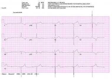
Sinoatrial block is a type of heart conduction disorder that affects the sinoatrial node, which is the natural pacemaker of the heart. In a sinoatrial block, the electrical impulses that originate in the sinoatrial node are delayed or blocked, resulting in a slower than normal heart rate or pauses between heartbeats.
A sinoatrial block can be classified as first-, second-, or third-degree, depending on the severity of the block. In a first-degree sinoatrial block, the electrical impulses are slowed but still conducted through to the atria. In a second-degree sinoatrial block, some of the electrical impulses are blocked, resulting in dropped beats or an irregular heart rhythm. In a third-degree sinoatrial block, also known as sinus node arrest, there is a complete failure of the sinoatrial node to generate impulses, resulting in a prolonged pause followed by a ventricular escape rhythm.
Sinoatrial blocks can be caused by various factors, including aging, heart disease, medication side effects, and electrolyte imbalances. In some cases, a sinoatrial block may not cause any symptoms and may only be detected during a routine electrocardiogram (ECG). However, in more severe cases, a sinoatrial block can lead to symptoms such as palpitations, dizziness, syncope (fainting), or shortness of breath. Treatment for a sinoatrial block depends on the underlying cause and may include medication adjustments, pacemaker implantation, or other interventions.
Tachycardia is a heart rate that is faster than normal. In sinoatrial nodal reentry tachycardia (SANRT), the abnormally fast heart rhythm originates in the sinoatrial node, which is the natural pacemaker of the heart. This type of tachycardia occurs due to a reentry circuit within the sinoatrial node, where an electrical impulse travels in a circular pattern and repeatedly stimulates the node to fire off abnormal rapid heartbeats. SANRT is typically characterized by a heart rate of over 100 beats per minute, palpitations, lightheadedness, or occasionally chest discomfort. It is usually a benign condition but can cause symptoms that affect quality of life. In some cases, treatment may be required to prevent recurrences and manage symptoms.
Lymph nodes are small, bean-shaped organs that are part of the immune system. They are found throughout the body, especially in the neck, armpits, groin, and abdomen. Lymph nodes filter lymph fluid, which carries waste and unwanted substances such as bacteria, viruses, and cancer cells. They contain white blood cells called lymphocytes that help fight infections and diseases by attacking and destroying the harmful substances found in the lymph fluid. When an infection or disease is present, lymph nodes may swell due to the increased number of immune cells and fluid accumulation as they work to fight off the invaders.
The atrioventricular (AV) node is a critical part of the electrical conduction system of the heart. It is a small cluster of specialized cardiac muscle cells located in the lower interatrial septum, near the opening of the coronary sinus. The AV node receives electrical impulses from the sinoatrial node (the heart's natural pacemaker) via the internodal pathways and delays their transmission for a brief period before transmitting them to the bundle of His and then to the ventricles. This delay allows the atria to contract and empty their contents into the ventricles before the ventricles themselves contract, ensuring efficient pumping of blood throughout the body.
The AV node plays an essential role in maintaining a normal heart rhythm, as it can also function as a backup pacemaker if the sinoatrial node fails to generate impulses. However, certain heart conditions or medications can affect the AV node's function and lead to abnormal heart rhythms, such as atrioventricular block or atrial tachycardia.
"Biological clocks" refer to the internal time-keeping systems in living organisms that regulate the timing of various physiological processes and behaviors according to a daily (circadian) rhythm. These rhythms are driven by genetic mechanisms and can be influenced by environmental factors such as light and temperature.
In humans, biological clocks help regulate functions such as sleep-wake cycles, hormone release, body temperature, and metabolism. Disruptions to these internal timekeeping systems have been linked to various health problems, including sleep disorders, mood disorders, and cognitive impairment.
Bradycardia is a medical term that refers to an abnormally slow heart rate, typically defined as a resting heart rate of less than 60 beats per minute in adults. While some people, particularly well-trained athletes, may have a naturally low resting heart rate, bradycardia can also be a sign of an underlying health problem.
There are several potential causes of bradycardia, including:
* Damage to the heart's electrical conduction system, such as from heart disease or aging
* Certain medications, including beta blockers, calcium channel blockers, and digoxin
* Hypothyroidism (underactive thyroid gland)
* Sleep apnea
* Infection of the heart (endocarditis or myocarditis)
* Infiltrative diseases such as amyloidosis or sarcoidosis
Symptoms of bradycardia can vary depending on the severity and underlying cause. Some people with bradycardia may not experience any symptoms, while others may feel weak, fatigued, dizzy, or short of breath. In severe cases, bradycardia can lead to fainting, confusion, or even cardiac arrest.
Treatment for bradycardia depends on the underlying cause. If a medication is causing the slow heart rate, adjusting the dosage or switching to a different medication may help. In other cases, a pacemaker may be necessary to regulate the heart's rhythm. It is important to seek medical attention if you experience symptoms of bradycardia, as it can be a sign of a serious underlying condition.
The heart atria are the upper chambers of the heart that receive blood from the veins and deliver it to the lower chambers, or ventricles. There are two atria in the heart: the right atrium receives oxygen-poor blood from the body and pumps it into the right ventricle, which then sends it to the lungs to be oxygenated; and the left atrium receives oxygen-rich blood from the lungs and pumps it into the left ventricle, which then sends it out to the rest of the body. The atria contract before the ventricles during each heartbeat, helping to fill the ventricles with blood and prepare them for contraction.
Heart rate is the number of heartbeats per unit of time, often expressed as beats per minute (bpm). It can vary significantly depending on factors such as age, physical fitness, emotions, and overall health status. A resting heart rate between 60-100 bpm is generally considered normal for adults, but athletes and individuals with high levels of physical fitness may have a resting heart rate below 60 bpm due to their enhanced cardiovascular efficiency. Monitoring heart rate can provide valuable insights into an individual's health status, exercise intensity, and response to various treatments or interventions.
I believe there may be some confusion in your question. "Rabbits" is a common name used to refer to the Lagomorpha species, particularly members of the family Leporidae. They are small mammals known for their long ears, strong legs, and quick reproduction.
However, if you're referring to "rabbits" in a medical context, there is a term called "rabbit syndrome," which is a rare movement disorder characterized by repetitive, involuntary movements of the fingers, resembling those of a rabbit chewing. It is also known as "finger-chewing chorea." This condition is usually associated with certain medications, particularly antipsychotics, and typically resolves when the medication is stopped or adjusted.
An action potential is a brief electrical signal that travels along the membrane of a nerve cell (neuron) or muscle cell. It is initiated by a rapid, localized change in the permeability of the cell membrane to specific ions, such as sodium and potassium, resulting in a rapid influx of sodium ions and a subsequent efflux of potassium ions. This ion movement causes a brief reversal of the electrical potential across the membrane, which is known as depolarization. The action potential then propagates along the cell membrane as a wave, allowing the electrical signal to be transmitted over long distances within the body. Action potentials play a crucial role in the communication and functioning of the nervous system and muscle tissue.
Atrial function in a medical context refers to the role and performance of the two upper chambers of the heart, known as the atria. The main functions of the atria are to receive blood from the veins and help pump it into the ventricles, which are the lower pumping chambers of the heart.
The atria contract in response to electrical signals generated by the sinoatrial node, which is the heart's natural pacemaker. This contraction helps to fill the ventricles with blood before they contract and pump blood out to the rest of the body. Atrial function can be assessed through various diagnostic tests, such as echocardiograms or electrocardiograms (ECGs), which can help identify any abnormalities in atrial structure or electrical activity that may affect heart function.
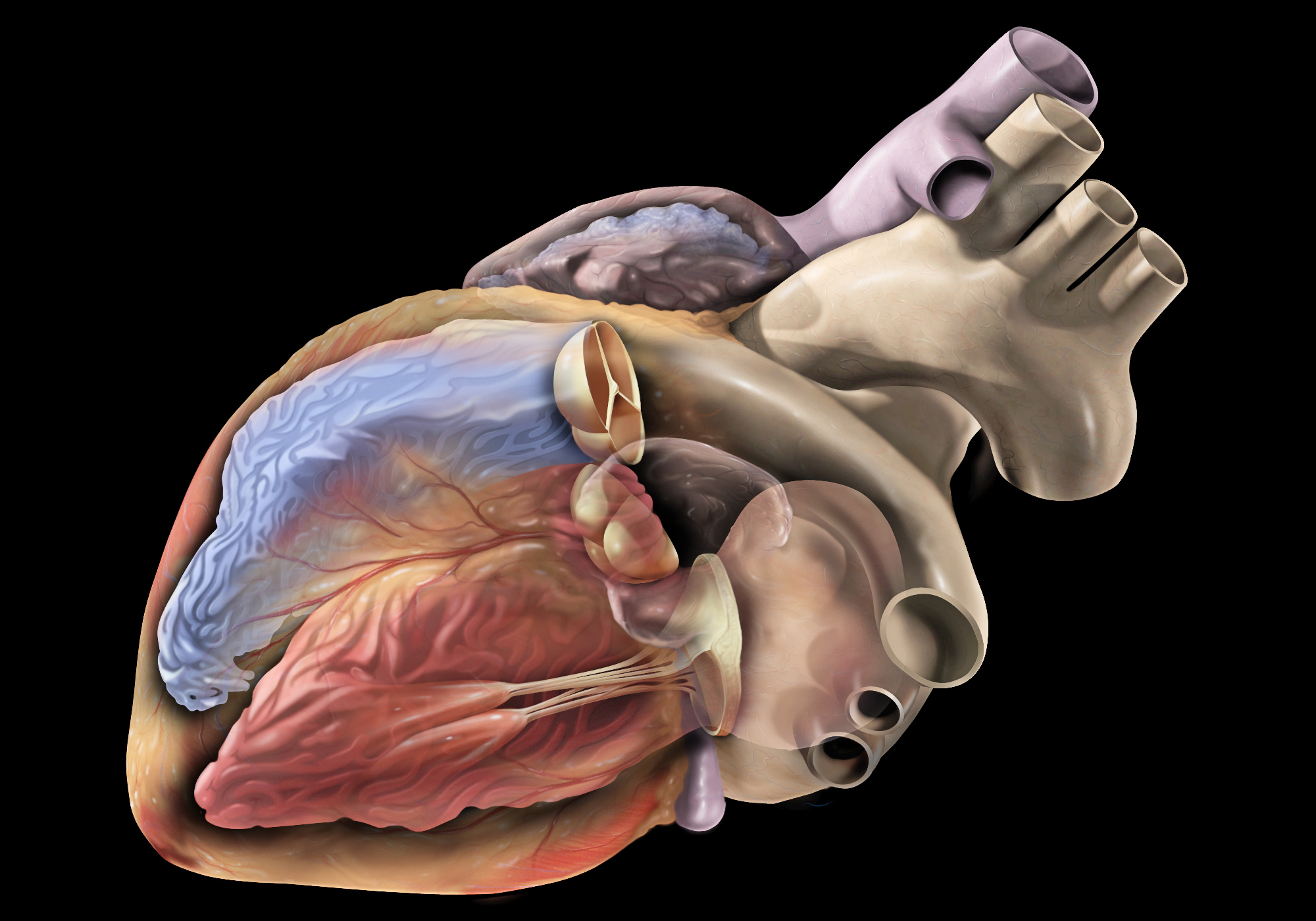
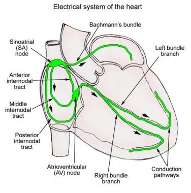
The heart conduction system is a group of specialized cardiac muscle cells that generate and conduct electrical impulses to coordinate the contraction of the heart chambers. The main components of the heart conduction system include:
1. Sinoatrial (SA) node: Also known as the sinus node, it is located in the right atrium near the entrance of the superior vena cava and functions as the primary pacemaker of the heart. It sets the heart rate by generating electrical impulses at regular intervals.
2. Atrioventricular (AV) node: Located in the interatrial septum, near the opening of the coronary sinus, it serves as a relay station for electrical signals between the atria and ventricles. The AV node delays the transmission of impulses to allow the atria to contract before the ventricles.
3. Bundle of His: A bundle of specialized cardiac muscle fibers that conducts electrical impulses from the AV node to the ventricles. It divides into two main branches, the right and left bundle branches, which further divide into smaller Purkinje fibers.
4. Right and left bundle branches: These are extensions of the Bundle of His that transmit electrical impulses to the respective right and left ventricular myocardium. They consist of specialized conducting tissue with large diameters and minimal resistance, allowing for rapid conduction of electrical signals.
5. Purkinje fibers: Fine, branching fibers that arise from the bundle branches and spread throughout the ventricular myocardium. They are responsible for transmitting electrical impulses to the working cardiac muscle cells, triggering coordinated ventricular contraction.
In summary, the heart conduction system is a complex network of specialized muscle cells responsible for generating and conducting electrical signals that coordinate the contraction of the atria and ventricles, ensuring efficient blood flow throughout the body.
Hyperpolarization-activated cyclic nucleotide-gated (HCN) channels are a type of ion channel found in the membranes of excitable cells, such as neurons and cardiac myocytes. These channels are unique because they open in response to membrane hyperpolarization, meaning that they allow the flow of ions into the cell when the voltage becomes more negative.
HCN channels are permeable to both sodium (Na+) and potassium (K+) ions, but they have a stronger preference for Na+ ions. When open, HCN channels conduct a current known as the "funny" or "Ih" current, which plays important roles in regulating the electrical excitability of cells.
HCN channels are also modulated by cyclic nucleotides, such as cyclic adenosine monophosphate (cAMP) and cyclic guanosine monophosphate (cGMP). Binding of these molecules to the intracellular domain of the channel can increase its open probability, leading to an enhancement of the funny current.
Dysfunction of HCN channels has been implicated in a variety of neurological and cardiac disorders, including epilepsy, sleep disorders, and heart rhythm abnormalities.
Sinus arrhythmia is a type of heart rhythm disorder (arrhythmia) where the normal rhythm generated by the sinus node in the heart varies in rate or pattern. The sinus node is the natural pacemaker of the heart and usually sets a steady pace for heartbeats. However, in sinus arrhythmia, the heart rate may speed up or slow down abnormally during breathing in (inspiration) or breathing out (expiration).
When the heart rate increases during inspiration, it is called "inspiratory sinus arrhythmia," and when the heart rate decreases during expiration, it is called "expiratory sinus arrhythmia." Most people experience a mild form of inspiratory sinus arrhythmia, which is considered normal, especially in children and young adults.
However, if the variation in heart rate is significant or accompanied by symptoms such as palpitations, dizziness, shortness of breath, or chest discomfort, it may require medical evaluation and treatment. Sinus arrhythmia can be caused by various factors, including lung disease, heart disease, electrolyte imbalances, or the use of certain medications.
Cardiovascular models are simplified representations or simulations of the human cardiovascular system used in medical research, education, and training. These models can be physical, computational, or mathematical and are designed to replicate various aspects of the heart, blood vessels, and blood flow. They can help researchers study the structure and function of the cardiovascular system, test new treatments and interventions, and train healthcare professionals in diagnostic and therapeutic techniques.
Physical cardiovascular models may include artificial hearts, blood vessels, or circulation systems made from materials such as plastic, rubber, or silicone. These models can be used to study the mechanics of heart valves, the effects of different surgical procedures, or the impact of various medical devices on blood flow.
Computational and mathematical cardiovascular models use algorithms and equations to simulate the behavior of the cardiovascular system. These models may range from simple representations of a single heart chamber to complex simulations of the entire circulatory system. They can be used to study the electrical activity of the heart, the biomechanics of blood flow, or the distribution of drugs in the body.
Overall, cardiovascular models play an essential role in advancing our understanding of the human body and improving patient care.
Cyclic nucleotide-gated (CNG) channels are a type of ion channel found in the membranes of certain cells, particularly in the sensory neurons of the visual and olfactory systems. They are called cyclic nucleotide-gated because they can be activated or regulated by the binding of cyclic nucleotides, such as cyclic adenosine monophosphate (cAMP) or cyclic guanosine monophosphate (cGMP), to the intracellular domain of the channel.
CNG channels are permeable to cations, including sodium (Na+) and calcium (Ca2+) ions, and their activation allows these ions to flow into the cell. This influx of cations can trigger a variety of cellular responses, such as the initiation of visual or olfactory signaling pathways.
CNG channels are composed of four subunits that form a functional channel. Each subunit has a cyclic nucleotide-binding domain (CNBD) in its intracellular region, which can bind to cyclic nucleotides and regulate the opening and closing of the channel. The CNBD is connected to the pore-forming region of the channel by a flexible linker, allowing for conformational changes in the CNBD to be transmitted to the pore and modulate ion conductance.
CNG channels play important roles in various physiological processes, including sensory perception, neurotransmission, and cellular signaling. Dysfunction of CNG channels has been implicated in several human diseases, such as retinitis pigmentosa, congenital stationary night blindness, and cystic fibrosis.
The vagus nerve, also known as the 10th cranial nerve (CN X), is the longest of the cranial nerves and extends from the brainstem to the abdomen. It has both sensory and motor functions and plays a crucial role in regulating various bodily functions such as heart rate, digestion, respiratory rate, speech, and sweating, among others.
The vagus nerve is responsible for carrying sensory information from the internal organs to the brain, and it also sends motor signals from the brain to the muscles of the throat and voice box, as well as to the heart, lungs, and digestive tract. The vagus nerve helps regulate the body's involuntary responses, such as controlling heart rate and blood pressure, promoting relaxation, and reducing inflammation.
Dysfunction in the vagus nerve can lead to various medical conditions, including gastroparesis, chronic pain, and autonomic nervous system disorders. Vagus nerve stimulation (VNS) is a therapeutic intervention that involves delivering electrical impulses to the vagus nerve to treat conditions such as epilepsy, depression, and migraine headaches.
Sick Sinus Syndrome (SSS) is a term used to describe a group of abnormal heart rhythm disturbances that originates in the sinoatrial node (the natural pacemaker of the heart). This syndrome is characterized by impaired functioning of the sinoatrial node, resulting in various abnormalities such as sinus bradycardia (abnormally slow heart rate), sinus arrest (complete cessation of sinus node activity), and/or sinoatrial exit block (failure of the electrical impulse to leave the sinus node and spread to the atria).
People with SSS may experience symptoms such as palpitations, dizziness, fatigue, shortness of breath, or syncope (fainting) due to inadequate blood supply to the brain caused by slow heart rate. The diagnosis of SSS is typically made based on the patient's symptoms and the results of an electrocardiogram (ECG), Holter monitoring, or event recorder that shows evidence of abnormal sinus node function. Treatment options for SSS may include lifestyle modifications, medications, or implantation of a pacemaker to regulate the heart rate.
Right atrial function refers to the role and performance of the right atrium in the heart. The right atrium is one of the four chambers of the heart and is responsible for receiving deoxygenated blood from the body via the superior and inferior vena cava. It then contracts to help pump the blood into the right ventricle, which subsequently sends it to the lungs for oxygenation.
Right atrial function can be assessed through various methods, including echocardiography, cardiac magnetic resonance imaging (MRI), and electrocardiogram (ECG). Abnormalities in right atrial function may indicate underlying heart conditions such as right-sided heart failure, atrial fibrillation, or other cardiovascular diseases. Proper evaluation and monitoring of right atrial function are essential for effective diagnosis, treatment, and management of these conditions.
Calcium channels, L-type, are a type of voltage-gated calcium channel that are widely expressed in many excitable cells, including cardiac and skeletal muscle cells, as well as certain neurons. These channels play a crucial role in the regulation of various cellular functions, such as excitation-contraction coupling, hormone secretion, and gene expression.
L-type calcium channels are composed of five subunits: alpha-1, alpha-2, beta, gamma, and delta. The alpha-1 subunit is the pore-forming subunit that contains the voltage sensor and the selectivity filter for calcium ions. It has four repeated domains (I-IV), each containing six transmembrane segments (S1-S6). The S4 segment in each domain functions as a voltage sensor, moving outward upon membrane depolarization to open the channel and allow calcium ions to flow into the cell.
L-type calcium channels are activated by membrane depolarization and have a relatively slow activation and inactivation time course. They are also modulated by various intracellular signaling molecules, such as protein kinases and G proteins. L-type calcium channel blockers, such as nifedipine and verapamil, are commonly used in the treatment of hypertension, angina, and certain cardiac arrhythmias.
Heart block is a cardiac condition characterized by the interruption of electrical impulse transmission from the atria (the upper chambers of the heart) to the ventricles (the lower chambers of the heart). This disruption can lead to abnormal heart rhythms, including bradycardia (a slower-than-normal heart rate), and in severe cases, can cause the heart to stop beating altogether. Heart block is typically caused by damage to the heart's electrical conduction system due to various factors such as aging, heart disease, or certain medications.
There are three types of heart block: first-degree, second-degree, and third-degree (also known as complete heart block). Each type has distinct electrocardiogram (ECG) findings and symptoms. Treatment for heart block depends on the severity of the condition and may include monitoring, medication, or implantation of a pacemaker to regulate the heart's electrical activity.
Electrophysiology is a branch of medicine that deals with the electrical activities of the body, particularly the heart. In a medical context, electrophysiology studies (EPS) are performed to assess abnormal heart rhythms (arrhythmias) and to evaluate the effectiveness of certain treatments, such as medication or pacemakers.
During an EPS, electrode catheters are inserted into the heart through blood vessels in the groin or neck. These catheters can record the electrical activity of the heart and stimulate it to help identify the source of the arrhythmia. The information gathered during the study can help doctors determine the best course of treatment for each patient.
In addition to cardiac electrophysiology, there are also other subspecialties within electrophysiology, such as neuromuscular electrophysiology, which deals with the electrical activity of the nervous system and muscles.
Membrane potential is the electrical potential difference across a cell membrane, typically for excitable cells such as nerve and muscle cells. It is the difference in electric charge between the inside and outside of a cell, created by the selective permeability of the cell membrane to different ions. The resting membrane potential of a typical animal cell is around -70 mV, with the interior being negative relative to the exterior. This potential is generated and maintained by the active transport of ions across the membrane, primarily through the action of the sodium-potassium pump. Membrane potentials play a crucial role in many physiological processes, including the transmission of nerve impulses and the contraction of muscle cells.
T-type calcium channels are a type of voltage-gated calcium channel that play a role in the regulation of excitable cells, such as neurons and cardiac myocytes. These channels are characterized by their low voltage activation threshold and rapid activation and inactivation kinetics. They are involved in various physiological processes, including neuronal excitability, gene expression, hormone secretion, and heart rhythm. Abnormal functioning of T-type calcium channels has been implicated in several diseases, such as epilepsy, chronic pain, and cardiac arrhythmias.
Connexins are a family of proteins that form the structural units of gap junctions, which are specialized channels that allow for the direct exchange of small molecules and ions between adjacent cells. These channels play crucial roles in maintaining tissue homeostasis, coordinating cellular activities, and enabling communication between cells. In humans, there are 21 different connexin genes that encode for these proteins, with each isoform having unique properties and distributions within the body. Mutations in connexin genes have been linked to a variety of human diseases, including hearing loss, skin disorders, and heart conditions.
Sinus tachycardia is a type of rapid heart rate, characterized by an abnormally fast sinus rhythm, with a rate greater than 100 beats per minute in adults. The sinoatrial node (SA node), which is the natural pacemaker of the heart, generates these impulses regularly and at an increased rate.
Sinus tachycardia is usually a physiological response to various stimuli or conditions, such as physical exertion, strong emotions, fever, anxiety, pain, or certain medications. It can also be caused by hormonal imbalances, anemia, hyperthyroidism, or other medical disorders.
In most cases, sinus tachycardia is not harmful and resolves once the underlying cause is addressed. However, if it occurs persistently or is associated with symptoms like palpitations, shortness of breath, dizziness, or chest discomfort, further evaluation by a healthcare professional is recommended to rule out any underlying heart conditions or other medical issues.
Potassium channels are membrane proteins that play a crucial role in regulating the electrical excitability of cells, including cardiac, neuronal, and muscle cells. These channels facilitate the selective passage of potassium ions (K+) across the cell membrane, maintaining the resting membrane potential and shaping action potentials. They are composed of four or six subunits that assemble to form a central pore through which potassium ions move down their electrochemical gradient. Potassium channels can be modulated by various factors such as voltage, ligands, mechanical stimuli, or temperature, allowing cells to fine-tune their electrical properties and respond to different physiological demands. Dysfunction of potassium channels has been implicated in several diseases, including cardiac arrhythmias, epilepsy, and neurodegenerative disorders.
I believe there might be a misunderstanding in your question. "Dogs" is not a medical term or condition. It is the common name for a domesticated carnivore of the family Canidae, specifically the genus Canis, which includes wolves, foxes, and other extant and extinct species of mammals. Dogs are often kept as pets and companions, and they have been bred in a wide variety of forms and sizes for different purposes, such as hunting, herding, guarding, assisting police and military forces, and providing companionship and emotional support.
If you meant to ask about a specific medical condition or term related to dogs, please provide more context so I can give you an accurate answer.
In medical terms, the heart is a muscular organ located in the thoracic cavity that functions as a pump to circulate blood throughout the body. It's responsible for delivering oxygen and nutrients to the tissues and removing carbon dioxide and other wastes. The human heart is divided into four chambers: two atria on the top and two ventricles on the bottom. The right side of the heart receives deoxygenated blood from the body and pumps it to the lungs, while the left side receives oxygenated blood from the lungs and pumps it out to the rest of the body. The heart's rhythmic contractions and relaxations are regulated by a complex electrical conduction system.
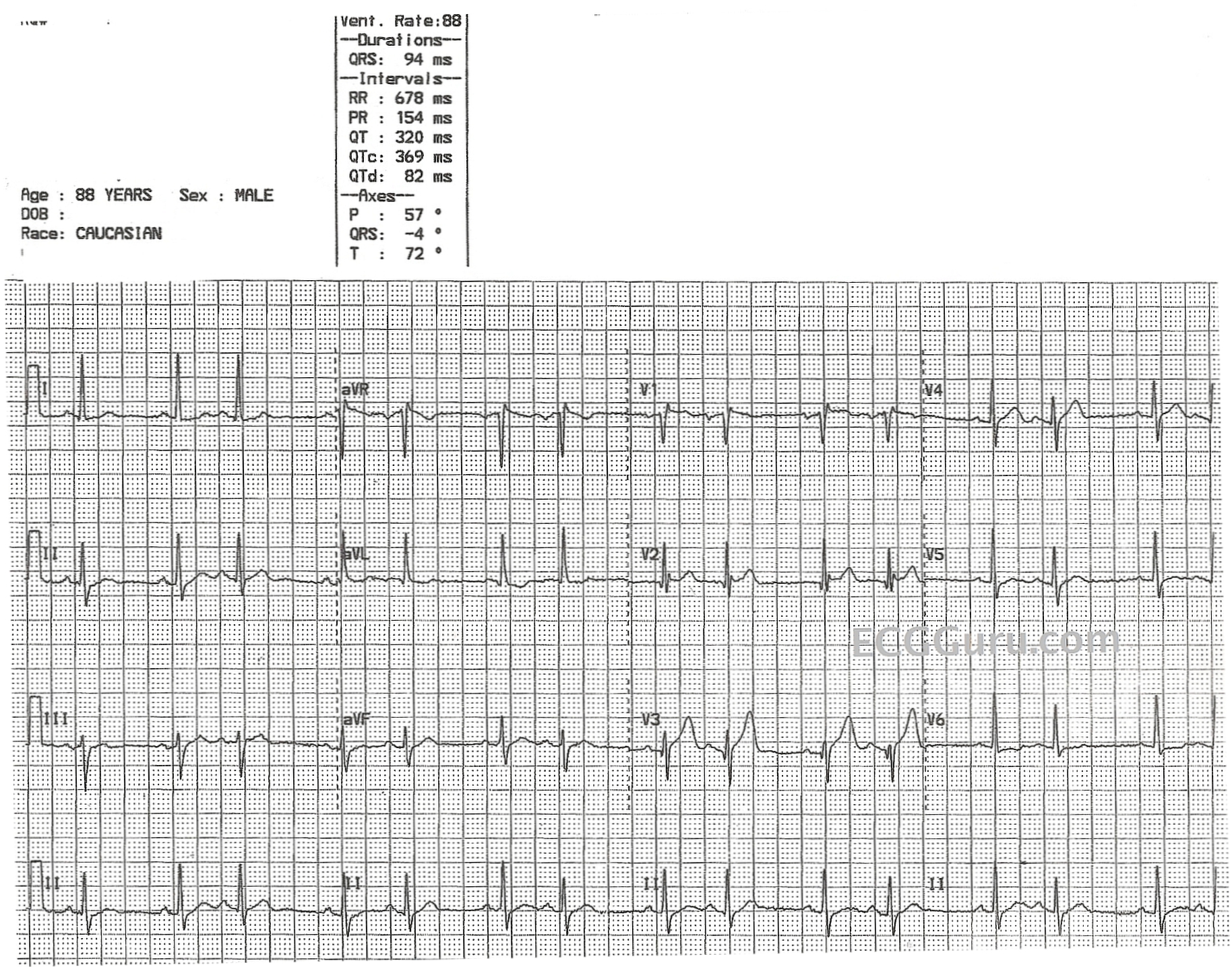
Electrocardiography (ECG or EKG) is a medical procedure that records the electrical activity of the heart. It provides a graphic representation of the electrical changes that occur during each heartbeat. The resulting tracing, called an electrocardiogram, can reveal information about the heart's rate and rhythm, as well as any damage to its cells or abnormalities in its conduction system.
During an ECG, small electrodes are placed on the skin of the chest, arms, and legs. These electrodes detect the electrical signals produced by the heart and transmit them to a machine that amplifies and records them. The procedure is non-invasive, painless, and quick, usually taking only a few minutes.
ECGs are commonly used to diagnose and monitor various heart conditions, including arrhythmias, coronary artery disease, heart attacks, and electrolyte imbalances. They can also be used to evaluate the effectiveness of certain medications or treatments.
Cranial nerve injuries refer to damages or trauma to one or more of the twelve cranial nerves (CN I through CN XII). These nerves originate from the brainstem and are responsible for transmitting sensory information (such as vision, hearing, smell, taste, and balance) and controlling various motor functions (like eye movement, facial expressions, swallowing, and speaking).
Cranial nerve injuries can result from various causes, including head trauma, tumors, infections, or neurological conditions. The severity of the injury may range from mild dysfunction to complete loss of function, depending on the extent of damage to the nerve. Treatment options vary based on the type and location of the injury but often involve a combination of medical management, physical therapy, surgical intervention, or rehabilitation.
Ion channels are specialized transmembrane proteins that form hydrophilic pores or gaps in the lipid bilayer of cell membranes. They regulate the movement of ions (such as sodium, potassium, calcium, and chloride) across the cell membrane by allowing these charged particles to pass through selectively in response to various stimuli, including voltage changes, ligand binding, mechanical stress, or temperature changes. This ion movement is essential for many physiological processes, including electrical signaling, neurotransmission, muscle contraction, and maintenance of resting membrane potential. Ion channels can be categorized based on their activation mechanisms, ion selectivity, and structural features. Dysfunction of ion channels can lead to various diseases, making them important targets for drug development.
Connexin 43 is a protein that forms gap junctions, which are specialized channels that allow for the direct communication and transport of small molecules between adjacent cells. Connexin 43 is widely expressed in many tissues, including the heart, brain, and various types of epithelial and connective tissues. In the heart, connexin 43 plays a crucial role in electrical conduction and coordination of contraction between cardiac muscle cells. Mutations in the gene that encodes connexin 43 have been associated with several human diseases, including certain types of cardiac arrhythmias and skin disorders.
Artificial cardiac pacing is a medical procedure that involves the use of an artificial device to regulate and stimulate the contraction of the heart muscle. This is often necessary when the heart's natural pacemaker, the sinoatrial node, is not functioning properly and the heart is beating too slowly or irregularly.
The artificial pacemaker consists of a small generator that produces electrical impulses and leads that are positioned in the heart to transmit the impulses. The generator is typically implanted just under the skin in the chest, while the leads are inserted into the heart through a vein.
There are different types of artificial cardiac pacing systems, including single-chamber pacemakers, which stimulate either the right atrium or right ventricle, and dual-chamber pacemakers, which stimulate both chambers of the heart. Some pacemakers also have additional features that allow them to respond to changes in the body's needs, such as during exercise or sleep.
Artificial cardiac pacing is a safe and effective treatment for many people with abnormal heart rhythms, and it can significantly improve their quality of life and longevity.
Lymph node excision is a surgical procedure in which one or more lymph nodes are removed from the body for the purpose of examination. This procedure is often conducted to help diagnose or stage various types of cancer, as malignant cells may spread to the lymphatic system and eventually accumulate within nearby lymph nodes.
During a lymph node excision, an incision is made in the skin overlying the affected lymph node(s). The surgeon carefully dissects the tissue surrounding the lymph node(s) to isolate them from adjacent structures before removing them. In some cases, a sentinel lymph node biopsy may be performed instead, where only the sentinel lymph node (the first lymph node to which cancer cells are likely to spread) is removed and examined.
The excised lymph nodes are then sent to a laboratory for histopathological examination, which involves staining and microscopic evaluation of the tissue to determine whether it contains any malignant cells. The results of this examination can help guide further treatment decisions and provide valuable prognostic information.
Patch-clamp techniques are a group of electrophysiological methods used to study ion channels and other electrical properties of cells. These techniques were developed by Erwin Neher and Bert Sakmann, who were awarded the Nobel Prize in Physiology or Medicine in 1991 for their work. The basic principle of patch-clamp techniques involves creating a high resistance seal between a glass micropipette and the cell membrane, allowing for the measurement of current flowing through individual ion channels or groups of channels.
There are several different configurations of patch-clamp techniques, including:
1. Cell-attached configuration: In this configuration, the micropipette is attached to the outer surface of the cell membrane, and the current flowing across a single ion channel can be measured. This configuration allows for the study of the properties of individual channels in their native environment.
2. Whole-cell configuration: Here, the micropipette breaks through the cell membrane, creating a low resistance electrical connection between the pipette and the inside of the cell. This configuration allows for the measurement of the total current flowing across all ion channels in the cell membrane.
3. Inside-out configuration: In this configuration, the micropipette is pulled away from the cell after establishing a seal, resulting in the exposure of the inner surface of the cell membrane to the solution in the pipette. This configuration allows for the study of the properties of ion channels in isolation from other cellular components.
4. Outside-out configuration: Here, the micropipette is pulled away from the cell after establishing a seal, resulting in the exposure of the outer surface of the cell membrane to the solution in the pipette. This configuration allows for the study of the properties of ion channels in their native environment, but with the ability to control the composition of the extracellular solution.
Patch-clamp techniques have been instrumental in advancing our understanding of ion channel function and have contributed to numerous breakthroughs in neuroscience, pharmacology, and physiology.
Cardiac myocytes are the muscle cells that make up the heart muscle, also known as the myocardium. These specialized cells are responsible for contracting and relaxing in a coordinated manner to pump blood throughout the body. They differ from skeletal muscle cells in several ways, including their ability to generate their own electrical impulses, which allows the heart to function as an independent rhythmical pump. Cardiac myocytes contain sarcomeres, the contractile units of the muscle, and are connected to each other by intercalated discs that help coordinate contraction and ensure the synchronous beating of the heart.
Myoblasts are immature cells that later develop into muscle cells (also known as myocytes). Cardiac myoblasts, therefore, are the immature cells that will specialize and develop into cardiac muscle cells. These cells play a crucial role in the growth, repair, and regeneration of heart muscles. In adults, however, the ability of these cells to regenerate damaged heart muscle tissue is limited. Recent research has focused on the potential use of cardiac myoblasts in cell-based therapies for various heart conditions, such as heart failure and myocardial infarction (heart attack).
A microelectrode is a small electrode with dimensions ranging from several micrometers to a few tens of micrometers in diameter. They are used in various biomedical applications, such as neurophysiological studies, neuromodulation, and brain-computer interfaces. In these applications, microelectrodes serve to record electrical activity from individual or small groups of neurons or deliver electrical stimuli to specific neural structures with high spatial resolution.
Microelectrodes can be fabricated using various materials, including metals (e.g., tungsten, stainless steel, platinum), metal alloys, carbon fibers, and semiconductor materials like silicon. The design of microelectrodes may vary depending on the specific application, with some common types being sharpened metal wires, glass-insulated metal microwires, and silicon-based probes with multiple recording sites.
The development and use of microelectrodes have significantly contributed to our understanding of neural function in health and disease, enabling researchers and clinicians to investigate the underlying mechanisms of neurological disorders and develop novel therapies for conditions such as Parkinson's disease, epilepsy, and hearing loss.
Aminophylline is a medication that is used to treat and prevent respiratory symptoms such as bronchospasm, wheezing, and shortness of breath. It is a combination of theophylline and ethylenediamine, and it works by relaxing muscles in the airways and increasing the efficiency of the diaphragm, which makes breathing easier.
Aminophylline is classified as a xanthine derivative and a methylxanthine bronchodilator. It is available in various forms, including tablets, capsules, and liquid solutions, and it is typically taken by mouth two to three times a day. The medication may also be given intravenously in hospital settings for the treatment of acute respiratory distress.
Common side effects of aminophylline include nausea, vomiting, headache, and insomnia. More serious side effects can occur at higher doses and may include irregular heartbeat, seizures, and potentially life-threatening allergic reactions. It is important to follow the dosage instructions carefully and to monitor for any signs of adverse reactions while taking this medication.
Delayed rectifier potassium channels are a type of ion channel found in the membrane of excitable cells, such as nerve and muscle cells. They are called "delayed rectifiers" because they activate and allow the flow of potassium ions (K+) out of the cell after a short delay following an action potential, or electrical signal.
These channels play a crucial role in regulating the duration and frequency of action potentials, helping to restore the resting membrane potential of the cell after it has fired. By allowing K+ to flow out of the cell, delayed rectifier potassium channels help to repolarize the membrane and bring it back to its resting state.
There are several different types of delayed rectifier potassium channels, which are classified based on their biophysical and pharmacological properties. These channels are important targets for drugs used to treat a variety of conditions, including cardiac arrhythmias, epilepsy, and psychiatric disorders.
Adrenergic beta-agonists are a class of medications that bind to and activate beta-adrenergic receptors, which are found in various tissues throughout the body. These receptors are part of the sympathetic nervous system and mediate the effects of the neurotransmitter norepinephrine (also called noradrenaline) and the hormone epinephrine (also called adrenaline).
When beta-agonists bind to these receptors, they stimulate a range of physiological responses, including relaxation of smooth muscle in the airways, increased heart rate and contractility, and increased metabolic rate. As a result, adrenergic beta-agonists are often used to treat conditions such as asthma, chronic obstructive pulmonary disease (COPD), and bronchitis, as they can help to dilate the airways and improve breathing.
There are several different types of beta-agonists, including short-acting and long-acting formulations. Short-acting beta-agonists (SABAs) are typically used for quick relief of symptoms, while long-acting beta-agonists (LABAs) are used for more sustained symptom control. Examples of adrenergic beta-agonists include albuterol (also known as salbutamol), terbutaline, formoterol, and salmeterol.
It's worth noting that while adrenergic beta-agonists can be very effective in treating respiratory conditions, they can also have side effects, particularly if used in high doses or for prolonged periods of time. These may include tremors, anxiety, palpitations, and increased blood pressure. As with any medication, it's important to use adrenergic beta-agonists only as directed by a healthcare professional.
Electrophysiological phenomena refer to the electrical properties and activities of biological tissues, cells, or organ systems, particularly in relation to nerve and muscle function. These phenomena can be studied using various techniques such as electrocardiography (ECG), electromyography (EMG), and electroencephalography (EEG).
In the context of cardiology, electrophysiological phenomena are often used to describe the electrical activity of the heart. The ECG is a non-invasive test that measures the electrical activity of the heart as it contracts and relaxes. By analyzing the patterns of electrical activity, doctors can diagnose various heart conditions such as arrhythmias, myocardial infarction, and electrolyte imbalances.
In neurology, electrophysiological phenomena are used to study the electrical activity of the brain. The EEG is a non-invasive test that measures the electrical activity of the brain through sensors placed on the scalp. By analyzing the patterns of electrical activity, doctors can diagnose various neurological conditions such as epilepsy, sleep disorders, and brain injuries.
Overall, electrophysiological phenomena are an important tool in medical diagnostics and research, providing valuable insights into the function of various organ systems.
I must clarify that the term "Guinea Pigs" is not typically used in medical definitions. However, in colloquial or informal language, it may refer to people who are used as the first to try out a new medical treatment or drug. This is known as being a "test subject" or "in a clinical trial."
In the field of scientific research, particularly in studies involving animals, guinea pigs are small rodents that are often used as experimental subjects due to their size, cost-effectiveness, and ease of handling. They are not actually pigs from Guinea, despite their name's origins being unclear. However, they do not exactly fit the description of being used in human medical experiments.
Benzazepines are a class of heterocyclic compounds that contain a benzene fused to a diazepine ring. In the context of pharmaceuticals, benzazepines refer to a group of drugs with various therapeutic uses, such as antipsychotics and antidepressants. Some examples of benzazepine-derived drugs include clozapine, olanzapine, and loxoprofen. These drugs have complex mechanisms of action, often involving multiple receptor systems in the brain.
A sentinel lymph node biopsy is a surgical procedure used in cancer staging to determine if the cancer has spread beyond the primary tumor to the lymphatic system. This procedure involves identifying and removing the sentinel lymph node(s), which are the first few lymph nodes to which cancer cells are most likely to spread from the primary tumor site.
The sentinel lymph node(s) are identified by injecting a tracer substance (usually a radioactive material and/or a blue dye) near the tumor site. The tracer substance is taken up by the lymphatic vessels and transported to the sentinel lymph node(s), allowing the surgeon to locate and remove them.
The removed sentinel lymph node(s) are then examined under a microscope for the presence of cancer cells. If no cancer cells are found, it is unlikely that the cancer has spread to other lymph nodes or distant sites in the body. However, if cancer cells are present, further lymph node dissection and/or additional treatment may be necessary.
Sentinel lymph node biopsy is commonly used in the staging of melanoma, breast cancer, and some types of head and neck cancer.
Cardiac myosins are a type of myosin protein that are specifically expressed in the cardiac muscle cells (or cardiomyocytes) of the heart. These proteins play a crucial role in the contraction and relaxation of heart muscles, which is essential for proper heart function and blood circulation.
Myosins are molecular motors that use chemical energy from ATP to generate force and movement. In the context of cardiac muscle cells, cardiac myosins interact with another protein called actin to form sarcomeres, which are the basic contractile units of muscle fibers. During contraction, the heads of cardiac myosin molecules bind to actin filaments and pull them together, causing the muscle fiber to shorten and generate force.
There are different isoforms of cardiac myosins that can vary in their structure and function. Mutations in the genes encoding these proteins have been linked to various forms of cardiomyopathy, which are diseases of the heart muscle that can lead to heart failure and other complications. Therefore, understanding the structure and function of cardiac myosins is an important area of research for developing therapies and treatments for heart disease.
An artificial pacemaker is a medical device that uses electrical impulses to regulate the beating of the heart. It is typically used when the heart's natural pacemaker, the sinoatrial node, is not functioning properly and the heart rate is too slow or irregular. The pacemaker consists of a small generator that contains a battery and electronic circuits, which are connected to one or more electrodes that are placed in the heart.
The generator sends electrical signals through the electrodes to stimulate the heart muscle and cause it to contract, thereby maintaining a regular heart rhythm. Artificial pacemakers can be programmed to deliver electrical impulses at a specific rate or in response to the body's needs. They are typically implanted in the chest during a surgical procedure and can last for many years before needing to be replaced.
Artificial pacemakers are an effective treatment for various types of bradycardia, which is a heart rhythm disorder characterized by a slow heart rate. Pacemakers can significantly improve symptoms associated with bradycardia, such as fatigue, dizziness, shortness of breath, and fainting spells.
Ryanodine is not a medical condition or term, but it is a chemical compound that interacts with ryanodine receptors (RyRs), which are calcium release channels found in the sarcoplasmic reticulum of muscle cells. Ryanodine receptors play a crucial role in excitation-contraction coupling, which is the process by which electrical signals trigger muscle contractions.
Ryanodine itself is a plant alkaloid that was initially isolated from the South American shrub Ryania speciosa. It can bind to and inhibit ryanodine receptors, altering calcium signaling in muscle cells. This ability of ryanodine to modulate calcium release has made it a valuable tool in researching excitation-contraction coupling and related processes.
In some cases, the term "ryanodine" may be used in a medical context to refer to the effects of ryanodine or ryanodine receptor modulation on muscle function, particularly in relation to diseases associated with calcium handling abnormalities. However, it is not a medical condition per se.
Cardiac arrhythmias are abnormal heart rhythms that result from disturbances in the electrical conduction system of the heart. The heart's normal rhythm is controlled by an electrical signal that originates in the sinoatrial (SA) node, located in the right atrium. This signal travels through the atrioventricular (AV) node and into the ventricles, causing them to contract and pump blood throughout the body.
An arrhythmia occurs when there is a disruption in this electrical pathway or when the heart's natural pacemaker produces an abnormal rhythm. This can cause the heart to beat too fast (tachycardia), too slow (bradycardia), or irregularly.
There are several types of cardiac arrhythmias, including:
1. Atrial fibrillation: A rapid and irregular heartbeat that starts in the atria (the upper chambers of the heart).
2. Atrial flutter: A rapid but regular heartbeat that starts in the atria.
3. Supraventricular tachycardia (SVT): A rapid heartbeat that starts above the ventricles, usually in the atria or AV node.
4. Ventricular tachycardia: A rapid and potentially life-threatening heart rhythm that originates in the ventricles.
5. Ventricular fibrillation: A chaotic and disorganized electrical activity in the ventricles, which can be fatal if not treated immediately.
6. Heart block: A delay or interruption in the conduction of electrical signals from the atria to the ventricles.
Cardiac arrhythmias can cause various symptoms, such as palpitations, dizziness, shortness of breath, chest pain, and fatigue. In some cases, they may not cause any symptoms and go unnoticed. However, if left untreated, certain types of arrhythmias can lead to serious complications, including stroke, heart failure, or even sudden cardiac death.
Treatment for cardiac arrhythmias depends on the type, severity, and underlying causes. Options may include lifestyle changes, medications, cardioversion (electrical shock therapy), catheter ablation, implantable devices such as pacemakers or defibrillators, and surgery. It is essential to consult a healthcare professional for proper evaluation and management of cardiac arrhythmias.
Sodium channels are specialized protein structures that are embedded in the membranes of excitable cells, such as nerve and muscle cells. They play a crucial role in the generation and transmission of electrical signals in these cells. Sodium channels are responsible for the rapid influx of sodium ions into the cell during the initial phase of an action potential, which is the electrical signal that travels along the membrane of a neuron or muscle fiber. This sudden influx of sodium ions causes the membrane potential to rapidly reverse, leading to the depolarization of the cell. After the action potential, the sodium channels close and become inactivated, preventing further entry of sodium ions and helping to restore the resting membrane potential.
Sodium channels are composed of a large alpha subunit and one or two smaller beta subunits. The alpha subunit forms the ion-conducting pore, while the beta subunits play a role in modulating the function and stability of the channel. Mutations in sodium channel genes have been associated with various inherited diseases, including certain forms of epilepsy, cardiac arrhythmias, and muscle disorders.
Cardiotonic agents are a type of medication that have a positive inotropic effect on the heart, meaning they help to improve the contractility and strength of heart muscle contractions. These medications are often used to treat heart failure, as they can help to improve the efficiency of the heart's pumping ability and increase cardiac output.
Cardiotonic agents work by increasing the levels of calcium ions inside heart muscle cells during each heartbeat, which in turn enhances the force of contraction. Some common examples of cardiotonic agents include digitalis glycosides (such as digoxin), which are derived from the foxglove plant, and synthetic medications such as dobutamine and milrinone.
While cardiotonic agents can be effective in improving heart function, they can also have potentially serious side effects, including arrhythmias, electrolyte imbalances, and digestive symptoms. As a result, they are typically used under close medical supervision and their dosages may need to be carefully monitored to minimize the risk of adverse effects.
The Autonomic Nervous System (ANS) is a part of the peripheral nervous system that operates largely below the level of consciousness and controls visceral functions. It is divided into two main subdivisions: the sympathetic and parasympathetic nervous systems, which generally have opposing effects and maintain homeostasis in the body.
The Sympathetic Nervous System (SNS) prepares the body for stressful or emergency situations, often referred to as the "fight or flight" response. It increases heart rate, blood pressure, respiratory rate, and metabolic rate, while also decreasing digestive activity. This response helps the body respond quickly to perceived threats.
The Parasympathetic Nervous System (PNS), on the other hand, promotes the "rest and digest" state, allowing the body to conserve energy and restore itself after the stress response has subsided. It decreases heart rate, blood pressure, and respiratory rate, while increasing digestive activity and promoting relaxation.
These two systems work together to maintain balance in the body by adjusting various functions based on internal and external demands. Disorders of the Autonomic Nervous System can lead to a variety of symptoms, such as orthostatic hypotension, gastroparesis, and cardiac arrhythmias, among others.
Premature aging, also known as "accelerated aging" or "early aging," refers to the physiological process in which the body shows signs of aging at an earlier age than typically expected. This can include various symptoms such as wrinkles, graying hair, decreased energy and mobility, cognitive decline, and increased risk of chronic diseases.
The medical definition of premature aging is not well-established, as aging is a complex process influenced by a variety of genetic and environmental factors. However, certain conditions and syndromes are associated with premature aging, such as Hutchinson-Gilford progeria syndrome, Werner syndrome, and Down syndrome.
In general, the signs of premature aging may be caused by a combination of genetic predisposition, lifestyle factors (such as smoking, alcohol consumption, and poor diet), exposure to environmental toxins, and chronic stress. While some aspects of aging are inevitable, maintaining a healthy lifestyle and reducing exposure to harmful factors can help slow down the aging process and improve overall quality of life.
Anti-arrhythmia agents are a class of medications used to treat abnormal heart rhythms or arrhythmias. These drugs work by modifying the electrical activity of the heart to restore and maintain a normal heart rhythm. There are several types of anti-arrhythmia agents, including:
1. Sodium channel blockers: These drugs slow down the conduction of electrical signals in the heart, which helps to reduce rapid or irregular heartbeats. Examples include flecainide, propafenone, and quinidine.
2. Beta-blockers: These medications work by blocking the effects of adrenaline on the heart, which helps to slow down the heart rate and reduce the force of heart contractions. Examples include metoprolol, atenolol, and esmolol.
3. Calcium channel blockers: These drugs block the entry of calcium into heart muscle cells, which helps to slow down the heart rate and reduce the force of heart contractions. Examples include verapamil and diltiazem.
4. Potassium channel blockers: These medications work by prolonging the duration of the heart's electrical cycle, which helps to prevent abnormal rhythms. Examples include amiodarone and sotalol.
5. Digoxin: This drug increases the force of heart contractions and slows down the heart rate, which can help to restore a normal rhythm in certain types of arrhythmias.
It's important to note that anti-arrhythmia agents can have significant side effects and should only be prescribed by a healthcare professional who has experience in managing arrhythmias. Close monitoring is necessary to ensure the medication is working effectively and not causing any adverse effects.
A computer simulation is a process that involves creating a model of a real-world system or phenomenon on a computer and then using that model to run experiments and make predictions about how the system will behave under different conditions. In the medical field, computer simulations are used for a variety of purposes, including:
1. Training and education: Computer simulations can be used to create realistic virtual environments where medical students and professionals can practice their skills and learn new procedures without risk to actual patients. For example, surgeons may use simulation software to practice complex surgical techniques before performing them on real patients.
2. Research and development: Computer simulations can help medical researchers study the behavior of biological systems at a level of detail that would be difficult or impossible to achieve through experimental methods alone. By creating detailed models of cells, tissues, organs, or even entire organisms, researchers can use simulation software to explore how these systems function and how they respond to different stimuli.
3. Drug discovery and development: Computer simulations are an essential tool in modern drug discovery and development. By modeling the behavior of drugs at a molecular level, researchers can predict how they will interact with their targets in the body and identify potential side effects or toxicities. This information can help guide the design of new drugs and reduce the need for expensive and time-consuming clinical trials.
4. Personalized medicine: Computer simulations can be used to create personalized models of individual patients based on their unique genetic, physiological, and environmental characteristics. These models can then be used to predict how a patient will respond to different treatments and identify the most effective therapy for their specific condition.
Overall, computer simulations are a powerful tool in modern medicine, enabling researchers and clinicians to study complex systems and make predictions about how they will behave under a wide range of conditions. By providing insights into the behavior of biological systems at a level of detail that would be difficult or impossible to achieve through experimental methods alone, computer simulations are helping to advance our understanding of human health and disease.
Supraventricular tachycardia (SVT) is a rapid heart rhythm that originates above the ventricles (the lower chambers of the heart). This type of tachycardia includes atrial tachycardia, atrioventricular nodal reentrant tachycardia (AVNRT), and atrioventricular reentrant tachycardia (AVRT). SVT usually causes a rapid heartbeat that starts and stops suddenly, and may not cause any other symptoms. However, some people may experience palpitations, shortness of breath, chest discomfort, dizziness, or fainting. SVT is typically diagnosed through an electrocardiogram (ECG) or Holter monitor, and can be treated with medications, cardioversion, or catheter ablation.
The sarcoplasmic reticulum (SR) is a specialized type of smooth endoplasmic reticulum found in muscle cells, particularly in striated muscles such as skeletal and cardiac muscles. It is a complex network of tubules that surrounds the myofibrils, the contractile elements of the muscle fiber.
The primary function of the sarcoplasmic reticulum is to store calcium ions (Ca2+) and regulate their release during muscle contraction and uptake during muscle relaxation. The SR contains a high concentration of calcium-binding proteins, such as calsequestrin, which help to maintain this storage.
The release of calcium ions from the sarcoplasmic reticulum is triggered by an action potential that travels along the muscle fiber's sarcolemma and into the muscle fiber's interior (the sarcoplasm). This action potential causes the voltage-gated calcium channels in the SR membrane, known as ryanodine receptors, to open, releasing Ca2+ ions into the sarcoplasm.
The increased concentration of Ca2+ ions in the sarcoplasm triggers muscle contraction by binding to troponin, a protein associated with actin filaments, causing a conformational change that exposes the active sites on actin for myosin heads to bind and generate force.
After muscle contraction, the calcium ions must be actively transported back into the sarcoplasmic reticulum by Ca2+ ATPase pumps, also known as sarco(endo)plasmic reticulum calcium ATPases (SERCAs). This process helps to lower the concentration of Ca2+ in the sarcoplasm and allows the muscle fiber to relax.
Overall, the sarcoplasmic reticulum plays a crucial role in excitation-contraction coupling, the process by which action potentials trigger muscle contraction.
Acetylcholine is a neurotransmitter, a type of chemical messenger that transmits signals across a chemical synapse from one neuron (nerve cell) to another "target" neuron, muscle cell, or gland cell. It is involved in both peripheral and central nervous system functions.
In the peripheral nervous system, acetylcholine acts as a neurotransmitter at the neuromuscular junction, where it transmits signals from motor neurons to activate muscles. Acetylcholine also acts as a neurotransmitter in the autonomic nervous system, where it is involved in both the sympathetic and parasympathetic systems.
In the central nervous system, acetylcholine plays a role in learning, memory, attention, and arousal. Disruptions in cholinergic neurotransmission have been implicated in several neurological disorders, including Alzheimer's disease, Parkinson's disease, and myasthenia gravis.
Acetylcholine is synthesized from choline and acetyl-CoA by the enzyme choline acetyltransferase and is stored in vesicles at the presynaptic terminal of the neuron. When a nerve impulse arrives, the vesicles fuse with the presynaptic membrane, releasing acetylcholine into the synapse. The acetylcholine then binds to receptors on the postsynaptic membrane, triggering a response in the target cell. Acetylcholine is subsequently degraded by the enzyme acetylcholinesterase, which terminates its action and allows for signal transduction to be repeated.
Penicillin amidase is not a medical term per se, but rather a biochemical term. It's also known as penicillin acylase or simply penicillinase. It refers to an enzyme that can break down certain types of penicillin antibiotics by cleaving the amide bond in the beta-lactam ring, which is the core structure of these antibiotics. This makes the antibiotic ineffective.
Beta-lactam antibiotics include penicillins and cephalosporins, among others. Some bacteria produce penicillin amidases as a form of resistance to these antibiotics. The enzyme can be used in biotechnology to produce semi-synthetic penicillins by cleaving the side chain of a parent penicillin and then attaching a different side chain, creating a new antibiotic with potentially different properties.
C-type Natriuretic Peptide (CNP) is a member of the natriuretic peptide family, which are hormones that play crucial roles in cardiovascular homeostasis and renal function. The natriuretic peptides include atrial natriuretic peptide (ANP), brain natriuretic peptide (BNP), and C-type natriuretic peptide (CNP).
C-type Natriuretic Peptide is primarily produced and secreted by the endothelial cells, and to a lesser extent by the central nervous system, chondrocytes, and vascular smooth muscle cells. CNP has a relatively short half-life of approximately 2 minutes due to its rapid clearance by the natriuretic peptide receptor-C (NPR-C) and neutral endopeptidase (NEP).
The primary physiological function of C-type Natriuretic Peptide is to regulate vascular tone, endothelial cell growth, differentiation, and survival. It also plays a role in bone development and maintenance by promoting chondrocyte proliferation and differentiation. In the kidney, CNP influences renal function through its effects on natriuresis (sodium excretion), diuresis (water excretion), and vasodilation of the afferent arteriole.
CNP binds to the NPR-B receptor, which is widely expressed in various tissues, including vascular endothelial cells, cardiomyocytes, osteoblasts, chondrocytes, and neurons. The activation of NPR-B leads to increased intracellular cyclic guanosine monophosphate (cGMP) levels, which in turn activates protein kinase G (PKG), resulting in vasodilation, anti-proliferative, and natriuretic effects.
Dysregulation of C-type Natriuretic Peptide has been implicated in several pathological conditions, such as cardiovascular diseases, bone disorders, and cancer. Therefore, understanding the role of CNP in these processes may provide novel therapeutic targets for treating these diseases.
The Parasympathetic Nervous System (PNS) is the part of the autonomic nervous system that primarily controls vegetative functions during rest, relaxation, and digestion. It is responsible for the body's "rest and digest" activities including decreasing heart rate, lowering blood pressure, increasing digestive activity, and stimulating sexual arousal. The PNS utilizes acetylcholine as its primary neurotransmitter and acts in opposition to the Sympathetic Nervous System (SNS), which is responsible for the "fight or flight" response.
Calcium is an essential mineral that is vital for various physiological processes in the human body. The medical definition of calcium is as follows:
Calcium (Ca2+) is a crucial cation and the most abundant mineral in the human body, with approximately 99% of it found in bones and teeth. It plays a vital role in maintaining structural integrity, nerve impulse transmission, muscle contraction, hormonal secretion, blood coagulation, and enzyme activation.
Calcium homeostasis is tightly regulated through the interplay of several hormones, including parathyroid hormone (PTH), calcitonin, and vitamin D. Dietary calcium intake, absorption, and excretion are also critical factors in maintaining optimal calcium levels in the body.
Hypocalcemia refers to low serum calcium levels, while hypercalcemia indicates high serum calcium levels. Both conditions can have detrimental effects on various organ systems and require medical intervention to correct.
Calcium signaling is the process by which cells regulate various functions through changes in intracellular calcium ion concentrations. Calcium ions (Ca^2+^) are crucial second messengers that play a critical role in many cellular processes, including muscle contraction, neurotransmitter release, gene expression, and programmed cell death (apoptosis).
Intracellular calcium levels are tightly regulated by a complex network of channels, pumps, and exchangers located on the plasma membrane and intracellular organelles such as the endoplasmic reticulum (ER) and mitochondria. These proteins control the influx, efflux, and storage of calcium ions within the cell.
Calcium signaling is initiated when an external signal, such as a hormone or neurotransmitter, binds to a specific receptor on the plasma membrane. This interaction triggers the opening of ion channels, allowing extracellular Ca^2+^ to flow into the cytoplasm. In some cases, this influx of calcium ions is sufficient to activate downstream targets directly. However, in most instances, the increase in intracellular Ca^2+^ serves as a trigger for the release of additional calcium from internal stores, such as the ER.
The release of calcium from the ER is mediated by ryanodine receptors (RyRs) and inositol trisphosphate receptors (IP3Rs), which are activated by specific second messengers generated in response to the initial external signal. The activation of these channels leads to a rapid increase in cytoplasmic Ca^2+^, creating a transient intracellular calcium signal known as a "calcium spark" or "calcium puff."
These localized increases in calcium concentration can then propagate throughout the cell as waves of elevated calcium, allowing for the spatial and temporal coordination of various cellular responses. The duration and amplitude of these calcium signals are finely tuned by the interplay between calcium-binding proteins, pumps, and exchangers, ensuring that appropriate responses are elicited in a controlled manner.
Dysregulation of intracellular calcium signaling has been implicated in numerous pathological conditions, including neurodegenerative diseases, cardiovascular disorders, and cancer. Therefore, understanding the molecular mechanisms governing calcium homeostasis and signaling is crucial for the development of novel therapeutic strategies targeting these diseases.
Atropine is an anticholinergic drug that blocks the action of the neurotransmitter acetylcholine in the central and peripheral nervous system. It is derived from the belladonna alkaloids, which are found in plants such as deadly nightshade (Atropa belladonna), Jimson weed (Datura stramonium), and Duboisia spp.
In clinical medicine, atropine is used to reduce secretions, increase heart rate, and dilate the pupils. It is often used before surgery to dry up secretions in the mouth, throat, and lungs, and to reduce salivation during the procedure. Atropine is also used to treat certain types of nerve agent and pesticide poisoning, as well as to manage bradycardia (slow heart rate) and hypotension (low blood pressure) caused by beta-blockers or calcium channel blockers.
Atropine can have several side effects, including dry mouth, blurred vision, dizziness, confusion, and difficulty urinating. In high doses, it can cause delirium, hallucinations, and seizures. Atropine should be used with caution in patients with glaucoma, prostatic hypertrophy, or other conditions that may be exacerbated by its anticholinergic effects.
Ion channel gating refers to the process by which ion channels in cell membranes open and close in response to various stimuli, allowing ions such as sodium, potassium, and calcium to flow into or out of the cell. This movement of ions is crucial for many physiological processes, including the generation and transmission of electrical signals in nerve cells, muscle contraction, and the regulation of hormone secretion.
Ion channel gating can be regulated by various factors, including voltage changes across the membrane (voltage-gated channels), ligand binding (ligand-gated channels), mechanical stress (mechanosensitive channels), or other intracellular signals (second messenger-gated channels). The opening and closing of ion channels are highly regulated and coordinated processes that play a critical role in maintaining the proper functioning of cells and organ systems.
Electrophysiologic techniques, cardiac, refer to medical procedures used to study the electrical activities and conduction systems of the heart. These techniques involve the insertion of electrode catheters into the heart through blood vessels under fluoroscopic guidance to record and stimulate electrical signals. The information obtained from these studies can help diagnose and evaluate various cardiac arrhythmias, determine the optimal treatment strategy, and assess the effectiveness of therapies such as ablation or implantable devices.
The electrophysiologic study (EPS) is a type of cardiac electrophysiologic technique that involves the measurement of electrical signals from different regions of the heart to evaluate its conduction system's function. The procedure can help identify the location of abnormal electrical pathways responsible for arrhythmias and determine the optimal treatment strategy, such as catheter ablation or medication therapy.
Cardiac electrophysiologic techniques are also used in device implantation procedures, such as pacemaker or defibrillator implantation, to ensure proper placement and function of the devices. These techniques can help program and test the devices to optimize their settings for each patient's needs.
In summary, cardiac electrophysiologic techniques are medical procedures used to study and manipulate the electrical activities of the heart, helping diagnose and treat various arrhythmias and other cardiac conditions.
The Ryanodine Receptor (RyR) is a calcium release channel located on the sarcoplasmic reticulum (SR), a type of endoplasmic reticulum found in muscle cells. It plays a crucial role in excitation-contraction coupling, which is the process by which electrical signals are converted into mechanical responses in muscle fibers.
In more detail, when an action potential reaches the muscle fiber's surface membrane, it triggers the opening of voltage-gated L-type calcium channels (Dihydropyridine Receptors or DHPRs) in the sarcolemma (the cell membrane of muscle fibers). This influx of calcium ions into the cytoplasm causes a conformational change in the RyR, leading to its own opening and the release of stored calcium from the SR into the cytoplasm. The increased cytoplasmic calcium concentration then initiates muscle contraction through interaction with contractile proteins like actin and myosin.
There are three isoforms of RyR: RyR1, RyR2, and RyR3. RyR1 is primarily found in skeletal muscle, while RyR2 is predominantly expressed in cardiac muscle. Both RyR1 and RyR2 are large homotetrameric proteins with a molecular weight of approximately 2.2 million Daltons. They contain multiple domains including an ion channel pore, regulatory domains, and a foot structure that interacts with DHPRs. RyR3 is more widely distributed, being found in various tissues such as the brain, smooth muscle, and some types of neurons.
Dysfunction of these channels has been implicated in several diseases including malignant hyperthermia, central core disease, catecholaminergic polymorphic ventricular tachycardia (CPVT), and certain forms of heart failure.
Myocardial contraction refers to the rhythmic and forceful shortening of heart muscle cells (myocytes) in the myocardium, which is the muscular wall of the heart. This process is initiated by electrical signals generated by the sinoatrial node, causing a wave of depolarization that spreads throughout the heart.
During myocardial contraction, calcium ions flow into the myocytes, triggering the interaction between actin and myosin filaments, which are the contractile proteins in the muscle cells. This interaction causes the myofilaments to slide past each other, resulting in the shortening of the sarcomeres (the functional units of muscle contraction) and ultimately leading to the contraction of the heart muscle.
Myocardial contraction is essential for pumping blood throughout the body and maintaining adequate circulation to vital organs. Any impairment in myocardial contractility can lead to various cardiac disorders, such as heart failure, cardiomyopathy, and arrhythmias.
Lymphatic metastasis is the spread of cancer cells from a primary tumor to distant lymph nodes through the lymphatic system. It occurs when malignant cells break away from the original tumor, enter the lymphatic vessels, and travel to nearby or remote lymph nodes. Once there, these cancer cells can multiply and form new tumors, leading to further progression of the disease. Lymphatic metastasis is a common way for many types of cancer to spread and can have significant implications for prognosis and treatment strategies.
Phosphodiesterase 3 (PDE3) inhibitors are a class of medications that work by blocking the enzyme phosphodiesterase 3, which is responsible for breaking down cyclic adenosine monophosphate (cAMP) in the body. cAMP is a secondary messenger involved in various cellular processes such as regulation of heart function, vascular smooth muscle relaxation, and metabolism.
By inhibiting PDE3, these medications increase the levels of cAMP in the body, leading to vasodilation (relaxation of blood vessels), positive inotropic effects (improvement of heart contractility), and increased lipolysis (breakdown of fats). As a result, PDE3 inhibitors are used in the treatment of conditions such as heart failure, pulmonary hypertension, and peripheral vascular disease.
Examples of PDE3 inhibitors include cilostazol, milrinone, and enoximone.
 Sinoatrial node
Crista terminalis
Cardiac muscle
Muscarinic antagonist
Sinoatrial nodal artery
Vagus nerve
HCN1
Ankyrin-2
Pacemaker potential
Right axis deviation
Cardiac action potential
HCN4
Heart rate
Beta-2 adrenergic receptor
Cyclic nucleotide-gated ion channel
HCN channel
Purkinje fibers
Threshold potential
Anatomy of the human heart
Dario DiFrancesco
Cardiac pacemaker
SCN5A
Terminal sulcus (heart)
Heart development
Ticagrelor
Cardiac physiology
AH-1058
Peter Kohl (scientist)
Muscle cell
Systole
Isolated sinoatrial node cells2
- These immunocytochemical data, together with morphological and electrophysiological data, obtained from the intact sinoatrial node and isolated sinoatrial node cells support the gradient model of the cellular organisation of the SA node. (ox.ac.uk)
- Selected contribution: axial stretch increases spontaneous pacemaker activity in rabbit isolated sinoatrial node cells. (ox.ac.uk)
Heart's pacemaker2
- The rhythmic contraction of cardiac muscle is regulated by the sinoatrial node, the heart's pacemaker. (britannica.com)
- Each heartbeat begins with an impulse from the heart's pacemaker (sinus or sinoatrial node). (msdmanuals.com)
Dysfunction2
- These findings strongly implicate rs6817105 minor allele in sinus node dysfunction and left atrial enlargement. (nature.com)
- 2000) Congenital deafness and sinoatrial node dysfunction in mice lacking class D L-type Ca2+ channels. (oeaw.ac.at)
Atrial21
- The SA node is located in the wall (epicardium) of the right atrium, laterally to the entrance of the superior vena cava in a region called the sinus venarum (hence sino- + atrial). (wikipedia.org)
- The connective tissue, along with the paranodal cells, insulate the SA node from the rest of the atrium, preventing the electrical activity of the atrial cells from affecting the SA node cells. (wikipedia.org)
- The SA node cells are smaller and paler than the surrounding atrial cells, with the average cell being around 8 micrometers in diameter and 20-30 micrometers in length (1 micrometer= 0.000001 meter). (wikipedia.org)
- Unlike the atrial cells, SA node cells contain fewer mitochondria and myofibers, as well as a smaller sarcoplasmic reticulum. (wikipedia.org)
- This means that the SA node cells are less equipped to contract compared to the atrial and ventricular cells. (wikipedia.org)
- This is again important in insulating the SA node from the surrounding atrial cells. (wikipedia.org)
- Sino-atrial node is called the pacemaker of our heart. (biologydiscussion.com)
- In the mosaic model, there is a variable mix of atrial and sinoatrial node cells from the centre to the periphery. (ox.ac.uk)
- Calcium transients in guinea-pig sino-atrial node cells imaged by confocal microscopy. (ox.ac.uk)
- Electrophysiological features of murine sino-atrial node in relation to the role of i(Na). (ox.ac.uk)
- Raised extracellular potassium attenuates the sympathetic modulation of sino-atrial node pacemaking in the isolated guinea-pig atria. (ox.ac.uk)
- Since changes in [K(+)](o) modulate membrane currents involved in sino-atrial node pacemaking, in particular the voltage-sensitive hyperpolarization-activated current (I(f)), we investigated whether raised [K(+)](o) (from 4 mM to 8 or 12 mM) could directly affect the heart rate response to cardiac sympathetic nerve stimulation (SNS). (ox.ac.uk)
- SAN optical action potentials had diastolic depolarization and multiple upstroke components that corresponded to the separate excitations of the node and surface atrial layers. (wustl.edu)
- After a 49±22 ms conduction delay within the SAN, excitation reached the atrial myocardium via superior and/or inferior sinoatrial exit pathways 8.8±3.2 mm from the leading pacemaker site. (wustl.edu)
- During normal sinus rhythm, the canine SAN is functionally insulated from the surrounding atrial myocardium except for 2 (or more) narrow superior and inferior sinoatrial exit pathways separated by 12.8±4.1 mm. (wustl.edu)
- Inhibition by Compound II, a sotalol analogue, of delayed rectifier current (iK) in rabbit isolated sino-atrial node cells. (ox.ac.uk)
- The effects of Compound II, a sotalol analogue, on spontaneous electrical activity and on three membrane currents (the delayed rectifier current, iK, the long-lasting inward calcium current, i(Ca,L) and hyperpolarization activated inward current, i(f)) were investigated in rabbit isolated sino-atrial node cells by whole cell clamp with amphotericin-permeabilised patches. (ox.ac.uk)
- Sinoatrial node (SAN) and pulmonary veins (PVs) are closely related atrial dysrhythmia. (tmu.edu.tw)
- Following intra-atrial conduction to the area of the lower intra-atrial septum, this wavefront reaches the inputs to the atrioventricular node (AVN). (medscape.com)
- The SA node is located less than 1 mm from the epicardial surface, laterally in the right atrial sulcus terminalis at the junction of the anteromedial aspect of the superior vena cava (SVC) and the right atrium (RA). (medscape.com)
- The human SA node contains a more than 3-fold greater density of beta-adrenergic and muscarinic cholinergic receptors than the adjacent atrial tissue. (medscape.com)
Atrioventricular node2
- Third-degree atrioventricular (AV) block, also referred to as third-degree heart block or complete heart block (CHB), is an abnormal heart rhythm resulting from a defect in the cardiac conduction system in which there is no conduction through the atrioventricular node (AVN), leading to complete dissociation of the atria and ventricles. (medscape.com)
- The stippled area adjacent to the central fibrous body is the approximate site of the compact atrioventricular node. (medscape.com)
Tissue7
- The cells of the SA node are spread out within a mesh of connective tissue, containing nerves, blood vessels, collagen and fat. (wikipedia.org)
- The sinoatrial (SA) node is a complex and inhomogeneous tissue in terms of cell morphology and electrical activity. (ox.ac.uk)
- This insulation coincided with connexin43-negative regions at the borders of the node, connective tissue, and coronary arteries. (wustl.edu)
- Node of specialized tissue lying near the bottom of the right atrium that fires an electrical impulse across the ventricles, causing them to contract. (encyclopedia.com)
- For example, researchers need to better understand the mechanisms controlling the development and maintenance of pacemaker cells in the sinoatrial node, just as they must develop ways to compare experimental biological pacemaker tissue with bona fide sinoatrial node tissue. (sciencedaily.com)
- The sinus (or sinoatrial) node is a small area of tissue in the wall of the right atrium. (kidshealth.org)
- The sinoatrial (SA) node is a spindle-shaped structure composed of a fibrous tissue matrix with closely packed cells. (medscape.com)
Murine1
- B ) A representative current-clamp recording of spontaneous action potentials from an acutely isolated murine sinoatrial node (SAN) cell. (elifesciences.org)
Arrhythmia1
- The death certificate, completed by the County Coroner, listed "probable arrhythmia due to hypertrophic cardiomyopathy, fibrosis of sinoatrial node" as the immediate cause of death. (cdc.gov)
Arrhythmias1
- iii) it helps protect the sinoatrial node from reentrant arrhythmias. (ox.ac.uk)
Nodal3
- The sinoatrial node receives its blood supply from the sinoatrial nodal artery. (wikipedia.org)
- Despite these many differences, there doesn't appear to be any advantage to how many sinoatrial nodal arteries an individual has, or where they originate. (wikipedia.org)
- Three-dimensional mapping and intracardiac echocardiography in the treatment of sinoatrial nodal tachycardias. (nih.gov)
Heartbeat3
- The human sinoatrial node (SAN) regulates our heartbeat. (wisc-online.com)
- This node is called the pacemaker of the heart because it sets the rate of the heartbeat and causes the rest of the heart to contract in its rhythm. (kidshealth.org)
- Under normal conditions, a group of cells called the "sinoatrial node" innervates the heart and controls the heartbeat. (medicalnewstoday.com)
Involving the sinus node1
- Supraventricular tachyarrhythmias involving the sinus node: clinical and electrophysiologic characteristics. (nih.gov)
Pathways1
- Conduction failure in these sinoatrial exit pathways leads to SAN exit block and is a modulator of heart rate. (wustl.edu)
Activates1
- Upon stimulation, acetylcholine (ACh) released from the vagus nerve binds to and activates M2Rs in sinoatrial node (SAN) pacemaker cells, promoting the engagement of the GDP-bound G protein trimer (Gα i (GDP)βγ). (elifesciences.org)
Cardiac muscle2
- The sinoatrial node (also known as the sinuatrial node, SA node or sinus node) is an oval shaped region of special cardiac muscle in the upper back wall of the right atrium made up of cells known as pacemaker cells. (wikipedia.org)
- The main role of a sinoatrial node cell is to initiate action potentials of the heart that can pass through cardiac muscle cells and cause contraction. (wikipedia.org)
Pacemaker of the heart1
- THe SA node acts as the pacemaker of the heart. (nih.gov)
Pacemakers1
- To create biological pacemakers, one approach is to coax stem cells to become specialized cardiac pacemaker cells that are normally found within the sinoatrial node of the heart. (sciencedaily.com)
Cells8
- Immediately surrounding the SA node cells are paranodal cells. (wikipedia.org)
- These cells have structures intermediate between that of the SA node cells and the rest of the atrium. (wikipedia.org)
- Although iNa has been recorded from sinoatrial (SA) node pacemaker cells, its precise role in SA node pacemaker function remains uncertain. (ox.ac.uk)
- According to the gradient model there is a gradual transition in morphology and electrical properties of SA node cells from the centre to the periphery of the SA node. (ox.ac.uk)
- Isolated, spontaneously beating rabbit sinoatrial node cells were subjected to longitudinal stretch, using carbon fibers attached to both ends of the cell. (ox.ac.uk)
- Which of the following statements describes phase 4 of the action potential of cells in the sinoatrial (SA) node? (nursingessay24x7.com)
- It is concluded that Compound II, a sotalol analogue, slows spontaneous activity of isolated rabbit SA node cells through a selective inhibition of iK. (ox.ac.uk)
- The normal cardiac impulse of the vertebrate heart originates in the pacemaker cells of the sinoatrial node, located in the right atrium. (medscape.com)
Right corona1
- The artery supplying the sinus node branches from the right coronary artery in 55-60% of hearts or the left circumflex artery in 40-45% of hearts. (medscape.com)
Impulse1
- In the heart, normal impulse initiation begins in the sinoatrial node (SAN). (medscape.com)
Artery2
- For example, in most humans, this is a single artery, although in some cases there have been either 2 or 3 sinoatrial node arteries supplying the SA node. (wikipedia.org)
- The artery approaches the node from a clockwise or counterclockwise direction around the SVC-RA junction. (medscape.com)
Nerve1
- The SA node is densely innervated with postganglionic adrenergic and cholinergic nerve terminals. (medscape.com)
Heart2
- In a healthy heart, the SA node continuously produces action potentials, setting the rhythm of the heart (sinus rhythm), and so is known as the heart's natural pacemaker. (wikipedia.org)
- Some cases of PVC occur when a group of fibers called the Purkinje fibers supply nerves to the heart instead of the sinoatrial node. (medicalnewstoday.com)
Modulate1
- Neurotransmitters modulate the SA node discharge rate by stimulation of beta-adrenergic and muscarinic receptors. (medscape.com)
Superior3
- The sinus node is approximately 15 mm long, 3 mm wide, and 1 mm thick, located directly below and to the side of the superior vena cava. (wikipedia.org)
- This band continues to the left atrium (LA), with the anterior internodal pathway entering the superior margin of the AV node. (medscape.com)
- The middle internodal tract begins at the superior and posterior margins of the sinus node, travels behind the SVC to the crest of the interatrial septum, and descends in the interatrial septum to the superior margin of the AV node. (medscape.com)
Veins1
- There are no large veins that drain blood away from the SA node. (wikipedia.org)
Cardiovascular1
- Old age and a variety of cardiovascular disorders may disrupt normal sinus node function. (nih.gov)
Travels1
- The posterior internodal tract starts at the posterior margin of the sinus node and travels posteriorly around the SVC and along the crista terminalis to the eustachian ridge and then into the interatrial septum above the coronary sinus, where it joins the posterior portion of the AV node. (medscape.com)
Human1
- Surface electrode recordings cannot delineate the activation within the human or canine sinoatrial node (SAN) because they are intramural structures. (wustl.edu)
Action1
- however, they do so at a slower rate and therefore, if the SA node is functioning properly, its action potentials usually override those that would be produced by other tissues. (wikipedia.org)
Atrium1
- An abnormality of the sinoatrial (SA) node in the right atrium. (nih.gov)
Rate1
- If the sinoatrial (SA) node fails, then at what rate (depolarizations per minute) can the atrioventricular (AV) node depolarize? (assignmentblock.com)
Techniques1
- Cardiotoxicity studies evaluating conduction node exposure might define dose constraints and criteria for additional cardiac-sparing techniques, such as respiratory techniques or proton therapy, which could benefit patients with underlying rhythmic or conduction disorders. (allenpress.com)
Function3
- SCN5A and sinoatrial node pacemaker function. (ox.ac.uk)
- iii) Experimental and computational evaluations of the functional roles of iNa in SA node pacemaker function. (ox.ac.uk)
- Taken together, these new observations suggest strong correlations between SCN5A-encoded Na+ channel and SA node pacemaker function. (ox.ac.uk)