Sinus Pericranii
Superior Sagittal Sinus
Papilledema
Cerebral Angiography
Sinus pericranii in the frontal region: a case report. (1/4)
Sinus pericranii is a rare vascular anomaly. A case of sinus pericranii at the nasion with an orbital extension is presented. The drainage was into the superior sagittal sinus. The pathogenesis is discussed and the literature is reviewed. (+info)Sinus pericranii: clinical and imaging findings in two cases of spontaneous partial thrombosis. (2/4)
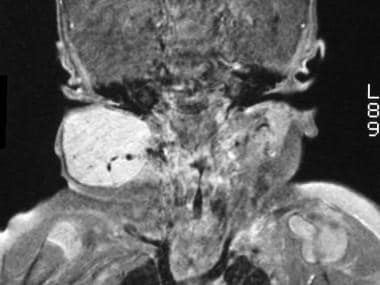
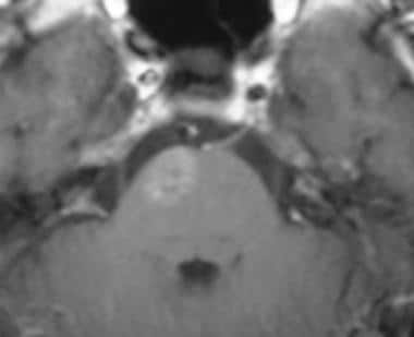
Sinus pericranii is an unusual venous anomaly characterized by communication of pericranial varicosities with an underlying dural sinus. We report two cases of spontaneous partial thrombosis of sinus pericranii presenting as focally tender, nonreducible mass lesions different in character from the baseline venous abnormality. CT, CT angiography, and CT venography (CTV) were performed in both cases. CTV was essential in depicting thrombi within the varicosities. MR (in one case) demonstrated the anomaly well, but the thrombus was not evident. Conservative therapy was instituted in both cases. (+info)Cerebrofacial venous anomalies, sinus pericranii, ocular abnormalities and developmental delay. (3/4)
The clinical implications of venous cerebrovascular maldevelopment remain poorly understood. We report on the association of cerebrofacial venous anomalies (including sinus pericranii), ocular abnormalities and mild developmental delay in two children. In addition, one child had a seizure disorder. Complex cerebrofacial slow-flow vascular anomalies may herald an underlying developmental aberration affecting the cerebrofacial and orbital regions. (+info)Surgical treatment of scaphocephaly with sinus pericranii. (4/4)
A 1-year-old female was admitted with a subcutaneous, pulsatile soft mass in the midline parietal region, and abnormal head shape. Fundus examination showed papilledema, suggesting elevated intracranial pressure. Radiological findings showed sagittal suture craniosynostosis with sinus pericranii. Magnetic resonance venography showed that the drainage through the sinus pericranii was not essential for the venous outflow from the brain. The patient underwent surgical resection of the sinus pericranii and total cranial remodeling. Ligation of the stalk-like orifice attached to the superior sagittal sinus with resection of the sinus pericranii and total cranial reconstruction were performed concurrently. The postoperative course was uneventful, and her papilledema resolved. No recurrence of the sinus pericranii has occurred for 3 years. This case describes a unique one-staged operation to treat sinus pericranii with sagittal suture craniosynostosis. (+info)Sinus Pericranii is a medical condition where there is an abnormal accumulation or collection of veins located between the periosteum (the outer membrane covering the bones) and the cranial bone. These venous collections are typically congenital but can also be acquired due to trauma, surgery, or increased venous pressure.
The sinus pericranii usually communicates with the intracranial venous system through emissary veins that pass through the skull. While it is generally asymptomatic, there is a risk of complications such as thrombosis, rupture, or infection, which can lead to serious neurological symptoms. Treatment options include observation, compression, or surgical excision, depending on the size, location, and associated symptoms.
The Superior Sagittal Sinus is a medical term that refers to a venous sinus (a channel for blood flow) located in the superior part (highest portion) of the sagittal suture, which is the line along the top of the skull where the two parietal bones join in the middle. It runs from front to back, starting at the frontal bone and ending at the occipital bone, and it receives blood from veins that drain the cerebral hemispheres (the right and left halves of the brain).
The Superior Sagittal Sinus is an important structure in the circulatory system of the brain as it plays a critical role in draining venous blood from the cranial cavity. It also contains valveless venous channels that allow for the flow of cerebrospinal fluid (CSF) between the intracranial and extracranial compartments.
It is worth noting that any damage to this structure, such as through trauma or infection, can lead to serious neurological complications, including increased intracranial pressure, seizures, and even death.
Cerebral veins are the blood vessels that carry deoxygenated blood from the brain to the dural venous sinuses, which are located between the layers of tissue covering the brain. The largest cerebral vein is the superior sagittal sinus, which runs along the top of the brain. Other major cerebral veins include the straight sinus, transverse sinus, sigmoid sinus, and cavernous sinus. These veins receive blood from smaller veins called venules that drain the surface and deep structures of the brain. The cerebral veins play an important role in maintaining normal circulation and pressure within the brain.
Papilledema is a medical term that refers to swelling of the optic nerve head, also known as the disc, which is the point where the optic nerve enters the back of the eye (the retina). This swelling can be caused by increased pressure within the skull, such as from brain tumors, meningitis, or idiopathic intracranial hypertension. Papilledema is usually detected through a routine eye examination and may be accompanied by symptoms such as headaches, visual disturbances, and nausea. If left untreated, papilledema can lead to permanent vision loss.
Cerebral angiography is a medical procedure that involves taking X-ray images of the blood vessels in the brain after injecting a contrast dye into them. This procedure helps doctors to diagnose and treat various conditions affecting the blood vessels in the brain, such as aneurysms, arteriovenous malformations, and stenosis (narrowing of the blood vessels).
During the procedure, a catheter is inserted into an artery in the leg and threaded through the body to the blood vessels in the neck or brain. The contrast dye is then injected through the catheter, and X-ray images are taken to visualize the blood flow through the brain's blood vessels.
Cerebral angiography provides detailed images of the blood vessels in the brain, allowing doctors to identify any abnormalities or blockages that may be causing symptoms or increasing the risk of stroke. Based on the results of the cerebral angiography, doctors can develop a treatment plan to address these issues and prevent further complications.
X-ray computed tomography (CT or CAT scan) is a medical imaging method that uses computer-processed combinations of many X-ray images taken from different angles to produce cross-sectional (tomographic) images (virtual "slices") of the body. These cross-sectional images can then be used to display detailed internal views of organs, bones, and soft tissues in the body.
The term "computed tomography" is used instead of "CT scan" or "CAT scan" because the machines take a series of X-ray measurements from different angles around the body and then use a computer to process these data to create detailed images of internal structures within the body.
CT scanning is a noninvasive, painless medical test that helps physicians diagnose and treat medical conditions. CT imaging provides detailed information about many types of tissue including lung, bone, soft tissue and blood vessels. CT examinations can be performed on every part of the body for a variety of reasons including diagnosis, surgical planning, and monitoring of therapeutic responses.
In computed tomography (CT), an X-ray source and detector rotate around the patient, measuring the X-ray attenuation at many different angles. A computer uses this data to construct a cross-sectional image by the process of reconstruction. This technique is called "tomography". The term "computed" refers to the use of a computer to reconstruct the images.
CT has become an important tool in medical imaging and diagnosis, allowing radiologists and other physicians to view detailed internal images of the body. It can help identify many different medical conditions including cancer, heart disease, lung nodules, liver tumors, and internal injuries from trauma. CT is also commonly used for guiding biopsies and other minimally invasive procedures.
In summary, X-ray computed tomography (CT or CAT scan) is a medical imaging technique that uses computer-processed combinations of many X-ray images taken from different angles to produce cross-sectional images of the body. It provides detailed internal views of organs, bones, and soft tissues in the body, allowing physicians to diagnose and treat medical conditions.
 Sinus pericranii
Sinusoidal hemangioma
Skin dimple
Stasis dermatitis
List of skin conditions
List of MeSH codes (C10)
List of MeSH codes (C16)
Sinus pericranii - Wikipedia
 Combined treatment of surgery and sclerotherapy for sinus pericranii
 Vascular Anomaly Imaging: Practice Essentials, Imaging, Hemangioma
 Interventional Neuroradiology | Johns Hopkins Medicine
 IndexCat
Aplasia Cutis Congenita Treatment & Management: Medical Care, Surgical Care, Consultations
DeCS - Términos Nuevos
Imaging in Vascular Anomalies: Overview, Imaging, Hemangioma
DeCS - Términos Nuevos
DeCS - Términos Nuevos
DeCS - New terms
DeCS - New terms
DeCS - Termos Novos
DeCS - Términos Nuevos
DeCS - New terms
DeCS - Termos Novos
DeCS - New terms
DeCS - New terms
 DeCS - Termos Novos
Diagnostic Imaging: Brain
 PDF) Pediatric Vascular Neurosurgery - Principles and Practice of Neurovascular Disorders (Part 1) By Abhishek Agrawal |...
MUSACCHIA, FRANCESCO
 Epilepsy Research Publications | Epilepsy In Children | Muir Maxwell Centre
 Búsqueda | Portal Regional de la BVS
 Kindler syndrome
 ReP USP - Resultado da busca
 Playlist 'A new cases' by Dr Abdullah Hajar
Intracranial8
- Sinus pericranii is an abnormal communication between the intracranial and extracranial venous drainage pathways. (wikipedia.org)
- Sinus pericranii is a venous anomaly where a communication between the intracranial dural sinuses and dilated epicranial venous structures exists. (wikipedia.org)
- That venous anomaly is a collection of nonmuscular venous blood vessels adhering tightly to the outer surface of the skull and directly communicating with intracranial venous sinuses through diploic veins. (wikipedia.org)
- The venous collections receive blood from and drain into the intracranial venous sinuses. (wikipedia.org)
- Radiologic findings showed venous malformations in the right parietal region communicating with the superior sagittal sinus in the intracranial region. (e-acfs.org)
- Sinus pericranii (SP) is a rare venous anomaly characterized by an abnormal communication between the intracranial and extracranial veins. (e-acfs.org)
- Pathologically, it exhibits a direct communication between the nonmuscular veins adhering to the outer surface of the skull and the intracranial venous sinuses [ 1 ]. (e-acfs.org)
- We suspected the presence of cranial holes facilitating the communication between the extracranial venous malformations and intracranial superior sagittal sinus, as observed on MRI. (e-acfs.org)
Scalp4
- Sinus pericranii (SP) is a rare disorder characterized by a congenital (or occasionally, acquired) epicranial venous malformation of the scalp. (wikipedia.org)
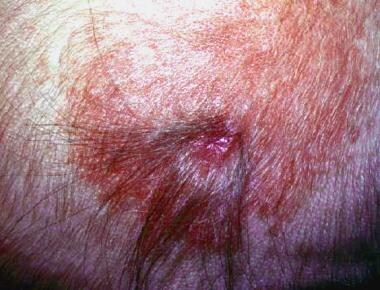
- Presence of a bluish and pulsating mass on the scalp, which showed bruit on auscultation, may indicate sinus pericranii, which should be included in the differential diagnosis. (e-acfs.org)
- Therefore, we confirmed that subcutaneous venous malformations in the scalp communicated with the superior sagittal sinus and diagnosed the patient with SP. (e-acfs.org)
- On the other hand, aplasia cutis congenita of the scalp may be complicated by sagittal sinus hemorrhage or thrombosis, and primary closure with scalp flaps may prevent a potentially fatal outcome. (medscape.com)
Abnormal1
- Sinus pericranii is a rare vascular anomaly characterized by abnormal venous communication between the inner and outer regions of the cranial cavity. (e-acfs.org)
Skull1
- Sinus pericranii typically present as soft palpable masses along midline skull, which may fluctuate in size depending on body positioning. (wikipedia.org)
Congenital2
- Sinus pericranii (SP) is a rare disorder characterized by a congenital (or occasionally, acquired) epicranial venous malformation of the scalp. (wikipedia.org)
- Sinus pericranii can be congenital or traumatic in origin. (nih.gov)
Venous malformation1
- CONCLUSION: Sinus pericranii is a rare cranial and venous malformation sometimes accompanied by brain malformations or craniosynostosis that may become more apparent as the brain and skull develop. (bvsalud.org)
Date2
- To date, to the best of our knowledge, there are no reports of sinus pericranii associated with syntelencephaly, a subtype of lobar holoprosencephaly. (bvsalud.org)
- At age 2 years and 7 months, the patient underwent a transection of the sinus pericranii and the mass resolved without any complications or recurrences for more than 2.5 years to date. (bvsalud.org)