Sleep Apnea Syndromes
Sleep
Sleep Apnea, Obstructive
Polysomnography
Sleep Apnea, Central
Sleep, REM
Sleep Disorders
Continuous Positive Airway Pressure
Pulmonary Disease, Chronic Obstructive
Lung Diseases, Obstructive
Palate, Soft
Wakefulness
Mandibular Advancement
Pharynx
Disorders of Excessive Somnolence
Sleep Initiation and Maintenance Disorders
Positive-Pressure Respiration
Severity of Illness Index
Jaundice, Obstructive
Arousal
Respiration
Sleep Stages
Pharyngeal Muscles
Oximetry
Tongue
Occlusal Splints
Respiratory Mechanics
Cheyne-Stokes Respiration
Oxygen
Circadian Rhythm
Pulmonary Ventilation
Electroencephalography
Body Mass Index
Orthodontic Appliances, Removable
Airway Resistance
Oropharynx
Risk Factors
Nasal Obstruction
Prospective Studies
Respiratory Function Tests
Treatment Outcome
Questionnaires
Obesity
Sleep Disorders, Intrinsic
Nocturnal Myoclonus Syndrome
Hypercapnia
Hyoid Bone
Electrooculography
Case-Control Studies
Prevalence
Monitoring, Ambulatory
Comorbidity
Narcolepsy
Forced Expiratory Volume
Sleep Arousal Disorders
Oxygen Inhalation Therapy
Fluid Shifts
Electromyography
Otorhinolaryngologic Surgical Procedures
Follow-Up Studies
Nose
Respiratory System
Respiratory Physiological Phenomena
Carbon Dioxide
Mouth Breathing
Hypopharynx
Monitoring, Physiologic
Cross-Sectional Studies
Sleep Medicine Specialty
Obesity Hypoventilation Syndrome
Vital Capacity
Quality of Life
Analysis of Variance
Statistics, Nonparametric
Cross-Over Studies
Retrospective Studies
Palatine Tonsil
Orthodontic Appliances
Obesity, Morbid
Hypoglossal Nerve
Respiration Disorders
Hypertension
Bronchodilator Agents
Fatigue
Pressure
Masks
Automobile Driving
Hypnotics and Sedatives
Restless Legs Syndrome
Rhinomanometry
Bradycardia
Sleep Bruxism
Lung
Dreams
Age Factors
Cohort Studies
Ventilators, Negative-Pressure
Heart Failure
Reference Values
Sleep Deprivation
Velopharyngeal Insufficiency
Palate
Sensitivity and Specificity
Cardiovascular Diseases
Double-Blind Method
Predictive Value of Tests
Laryngopharyngeal Reflux
Chronic Disease
Cholestasis
Sex Factors
Air Pressure
Partial Pressure
Risk Assessment
Reproducibility of Results
REM Sleep Behavior Disorder
Orthodontic Appliance Design
Anthropometry
Mandible
Chemoreceptor Cells
Autonomic Nervous System
Pulmonary Medicine
Facial Bones
Bariatric Surgery
Linear Models
Oxyhemoglobins
Respiratory Insufficiency
Signal Processing, Computer-Assisted
Diaphragm
Sympathetic Nervous System
Cardiomyopathy, Hypertrophic
Larynx
Electrocardiography
Hypertrophy
Regression Analysis
Delta Rhythm
Respiratory Therapy
Epiglottis
Azabicyclo Compounds
Nasal Cavity
Sleep Paralysis
Causality
Logistic Models
Tracheostomy
Pilot Projects
Sleep Disorders, Circadian Rhythm
Exercise Tolerance
Pulmonary Emphysema
Biological Markers
Work of Breathing
Tidal Volume
Anesthesia
Respiration, Artificial
Diagnosis, Computer-Assisted
Nocturia
Asthma
Lung Volume Measurements
Administration, Inhalation
ROC Curve
Incidence
Nasal Decongestants
Motor Vehicles
Implantable Neurostimulators
Sudden Infant Death
Ureteral Obstruction
Cataplexy
Maximal Expiratory Flow-Volume Curves
Gagging
Magnetic Resonance Imaging
Multivariate Analysis
Odds Ratio
High prevalence and persistence of sleep apnoea in patients referred for acute left ventricular failure and medically treated over 2 months. (1/2181)
AIMS: Cardiac failure patients were studied systematically using polysomnography 1 month after recovering from acute pulmonary oedema, and again after 2 months of optimal medical treatment for cardiac failure. METHODS AND RESULTS: This prospective study of consecutive patients was conducted in a cardiac care unit of a university hospital. V o(2)measurements and left ventricular ejection fraction were recorded. Thirty-four patients, initially recruited with pulmonary oedema, improved after 1 month of medical treatment to NYHA II or III. They were aged less than 75 years and had a left ventricular ejection fraction less than 45% at the time of inclusion. Age was 62 (9) years, body mass index= 27 (5) kg x m(-2)and an ejection fraction= 30 (10)%. Eighteen of the 34 patients (53%) had coronary artery disease. Twenty-eight of the 34 had sleep apnoea syndrome with an apnoea+hypopnoea index >15 x h(-1)of sleep. Thus, the prevalence of sleep apnoea in this population was 82%. Twenty-one of 28 (75%) patients had central sleep apnoea and seven of 28 (25%) had obstructive sleep apnoea. Patients with central sleep apnoea had a lower Pa co(2)than those with obstructive sleep apnoea (33 (5) vs 37 (5) mmHg, P<0.005). Significant correlations were found between apnoea+hypopnoea index and peak exercise oxygen consumption (r= -0.73, P<0.01), and apnoea+hypopnoea index and Pa co(2)(r= -0.42, P = 0.03). When only central sleep apnoea patients were considered, a correlation between apnoea+hypopnoea index and left ventricular ejection fraction was also demonstrated (r= -0.46, P<0.04). After 2 months of optimal medical treatment only two patients (both with central sleep apnoea) showed improvement (apnoea+hypopnoea index <15 x h(-1)). CONCLUSIONS: We have demonstrated a high prevalence of sleep apnoea, which persisted after 2 months of medical treatment, in patients referred for acute left ventricular failure. Central sleep apnoea can be considered a marker of the severity of congestive heart failure. (+info)An investigation into the changes in airway dimension and the efficacy of mandibular advancement appliances in subjects with obstructive sleep apnoea. (2/2181)
This prospective clinical study evaluates a group of 37 male Caucasians with obstructive sleep apnoea for changes in airway dimension and the efficacy associated with the use of mandibular advancement splints. Lateral skull radiographs were obtained with the subjects--upright in occlusion, supine in occlusion, and supine in protrusion. Each radiograph was traced and digitized, and changes in mandibular position, airway dimensions, and hyoid were examined. Subjects were invited to complete pre- and post-treatment questionnaires, and interviewed following fitting of a removable Herbst mandibular advancement splint. Significant changes were recorded in the airway dimensions in response to both a change in position, from upright to supine, and in response to mandibular advancement. A compliance rate of 76 per cent was achieved with no reported serious complications associated with the use of mandibular advancement devices. (+info)Nocturnal ischemic events in patients with obstructive sleep apnea syndrome and ischemic heart disease: effects of continuous positive air pressure treatment. (3/2181)
OBJECTIVES: To investigate the occurrence of nocturnal ischemic events in patients with obstructive sleep apnea syndrome (OSAS) and ischemic heart disease (IHD). BACKGROUND: Although previous reports documented nocturnal cardiac ischemic events among OSAS patients, the exact association between obstructive apneas and ischemia is not yet clear. It is also not known what differentiates between patients showing nocturnal ischemia and those that do not. METHODS: Fifty-one sleep apnea patients (age 61.3+/-8.3) with IHD participated in the study (after withdrawal of beta-adrenergic blocking agents and anti-anginotic treatment). All patients underwent whole-night polysomnography including ambulatory blood pressure recordings (30 min interval) and continuous Holter monitoring during sleep. A control group of 17 OSAS patients free from IHD were also similarly studied. Fifteen of the 51 patients were also recorded under continuous positive airway pressure (CPAP). RESULTS: Nocturnal ST segment depression occurred in 10 patients (a total of 15 events, 182 min), of whom six also had morning ischemia (06-08 am). Five additional patients had only morning ischemia. No ischemic events occurred in the control group. Age, sleep efficiency, oxygen desaturation, IHD severity and nocturnal-double product (DP) values were the main variables that significantly differentiated between patients who had ischemic events during sleep and those who did not. Nocturnal ischemia predominantly occurred during the rebreathing phase of the obstructive apneas, and it is characterized by increased heart rate (HR) and DP values. Treatment with continuous positive airway pressure significantly ameliorated the nocturnal ST depression time from 78 min to 33 min (p<0.001) as well as the maximal DP values (14,137+/-2,827 vs. 12,083+/-2,933, p<0.001). CONCLUSIONS: Exacerbation of ischemic events during sleep in OSAS may be explained by the combination of increased myocardial oxygen consumption as indicated by increased DP values and decreased oxygen supply due to oxygen desaturation with peak hemodynamic changes during the rebreathing phase of the obstructive apnea. Treatment with CPAP ameliorated the nocturnal ischemia. (+info)Obstructive sleep apnea. (4/2181)
Obstructive sleep apnea is a significant medical problem affecting up to 4 percent of middle-aged adults. The most common complaints are loud snoring, disrupted sleep and excessive daytime sleepiness. Patients with apnea suffer from fragmented sleep and may develop cardiovascular abnormalities because of the repetitive cycles of snoring, airway collapse and arousal. Although most patients are overweight and have a short, thick neck, some are of normal weight but have a small, receding jaw. Because many patients are not aware of their heavy snoring and nocturnal arousals, obstructive sleep apnea may remain undiagnosed; therefore, it is helpful to question the bedroom partner of a patient with chronic sleepiness and fatigue. Polysomnography in a sleep laboratory is the gold standard for confirming the diagnosis of obstructive sleep apnea; however, the test is expensive and not widely available. Home sleep studies are less costly but not as diagnostically accurate. Treatments include weight loss, nasal continuous positive airway pressure and dental devices that modify the position of the tongue or jaw. Upper airway and jaw surgical procedures may also be appropriate in selected patients, but invasiveness and expense restrict their use. (+info)Transtracheal air in the treatment of obstructive sleep apnoea hypopnoea syndrome. (5/2181)
A 49 year old woman with typical obstructive sleep apnoea hypopnoea syndrome underwent an unsuccessful trial with continuous positive airway pressure (CPAP) followed by uvulopalatopharyngoplasty with septorhinoplasty, treatment with protriptyline, and a second CPAP trial that was abandoned. Transtracheal air was then given and normalised sleep and breathing at a flow rate of 5 l/min. A sustained clinical improvement was observed at follow up visits. Transtracheal air could represent a simple and effective alternative to tracheotomy in patients with obstructive sleep apnoea hypopnoea syndrome in whom conventional treatments fail. (+info)Protruding the tongue improves posterior rhinomanometry in obstructive sleep apnoea syndrome. (6/2181)
In posterior rhinomanometry (PRM), oropharyngeal pressure is measured using a tube placed between the tongue and the hard palate. For valid results the patient must position the tongue and soft palate so that both the oropharynx and nasopharynx remain open. A high rate of failure of conventional PRM has been reported in normal individuals. In patients with obstructive sleep apnoea syndrome (OSAS), upper airway abnormalities may further increase the failure rate. This study proposes a modification of the technique in which protrusion of the tongue enhances pressure transmission between the nasopharynx and the mouth. In eight normal subjects, resistance was similar when measured by both methods. Of 24 OSAS patients, conventional PRM was unsuccessful in 11. In the remaining 13 patients, a significant correlation between the two methods was found, but resistance was lower by "tongue-out" than by conventional PRM, consistent with a decrease, during tongue protrusion, in retropalatal resistance, which is a component of the "nasal" resistance measured by PRM. In 26 OSAS patients, unilateral nasal resistance values measured by "tongue-out" PRM were similar to those measured by anterior rhinomanometry. When the "tongue-out" method was used routinely in 541 snorers, failure rates were 1.1% in the 272 non-OSAS patients and 3.7% in the 269 OSAS patients. These results indicate that posterior rhinomanometry with tongue protrusion is a highly effective tool for measuring nasal resistance in snorers. (+info)Effect of continuous positive airway pressure on blood pressure : a placebo trial. (7/2181)
This study examined the effect of continuous positive airway pressure (CPAP) treatment on blood pressure in patients with obstructive sleep apnea. Thirty-nine patients with sleep apnea were studied. Ambulatory blood pressure monitoring was obtained before and after patients were randomized to receive either 1 week of CPAP or placebo CPAP (CPAP administered at ineffective pressure). Blood pressure was examined over daytime hours (6 AM to 10 PM) and during nighttime hours (10 PM to 6 AM). Daytime mean arterial blood pressure decreased significantly but equally in both the active treatment group and the placebo treatment group (P=0.001). Nighttime mean arterial pressure levels decreased to a much greater extent over time in the patients who received active CPAP treatment (P=0. 032). CPAP does appear to decrease nighttime blood pressure. However, the decrease in daytime blood pressure may reflect a nonspecific response (ie, placebo), since both the active treatment group and the placebo treatment group developed comparable decreases in blood pressure. (+info)Ventilatory decline after hypoxia and hypercapnia is not different between healthy young men and women. (8/2181)
The gradual decay in ventilation after removal of a respiratory stimulus has been proposed to protect against cyclic breathing disorders such as obstructive sleep apnea (OSA). The male predominance of OSA, and the increased incidence of OSA in women after menopause, indicates that the respiratory-stimulating effect of progesterone may provide protection against OSA by altering the rate of poststimulus ventilatory decline (PSVD). It was therefore hypothesized that PSVD is longer in premenopausal women than in men and is longer in the luteal menstrual phase compared with the follicular phase. PSVD was measured in 12 men and in 11 women at both their luteal and follicular phases, after cessation of isocapnic hypoxia and normoxic hypercapnia. PSVD was compared between genders and between women in the luteal and follicular phases by repeated-measures ANOVA. There were no significant differences in PSVD between any of the groups after either respiratory stimulus. This suggests that the higher occurrence of OSA in men does not reflect an underlying gender difference in PSVD and implies the increased prevalence of OSA in women after menopause is not representative of an effect of progesterone on PSVD. (+info)Sleep apnea syndromes refer to a group of disorders characterized by abnormal breathing patterns during sleep. These patterns can result in repeated pauses in breathing (apneas) or shallow breaths (hypopneas), causing interruptions in sleep and decreased oxygen supply to the body. There are three main types of sleep apnea syndromes:
1. Obstructive Sleep Apnea (OSA): This is the most common form, caused by the collapse or obstruction of the upper airway during sleep, often due to relaxation of the muscles in the throat and tongue.
2. Central Sleep Apnea (CSA): This type is less common and results from the brain's failure to send proper signals to the breathing muscles. It can be associated with conditions such as heart failure, stroke, or certain medications.
3. Complex/Mixed Sleep Apnea: In some cases, a person may experience both obstructive and central sleep apnea symptoms, known as complex or mixed sleep apnea.
Symptoms of sleep apnea syndromes can include loud snoring, excessive daytime sleepiness, fatigue, morning headaches, difficulty concentrating, and mood changes. Diagnosis typically involves a sleep study (polysomnography) to monitor breathing patterns, heart rate, brain activity, and other physiological factors during sleep. Treatment options may include lifestyle modifications, oral appliances, positive airway pressure therapy, or even surgery in severe cases.
Sleep is a complex physiological process characterized by altered consciousness, relatively inhibited sensory activity, reduced voluntary muscle activity, and decreased interaction with the environment. It's typically associated with specific stages that can be identified through electroencephalography (EEG) patterns. These stages include rapid eye movement (REM) sleep, associated with dreaming, and non-rapid eye movement (NREM) sleep, which is further divided into three stages.
Sleep serves a variety of functions, including restoration and strengthening of the immune system, support for growth and development in children and adolescents, consolidation of memory, learning, and emotional regulation. The lack of sufficient sleep or poor quality sleep can lead to significant health problems, such as obesity, diabetes, cardiovascular disease, and even cognitive decline.
The American Academy of Sleep Medicine (AASM) defines sleep as "a period of daily recurring natural rest during which consciousness is suspended and metabolic processes are reduced." However, it's important to note that the exact mechanisms and purposes of sleep are still being researched and debated among scientists.
Obstructive Sleep Apnea (OSA) is a sleep-related breathing disorder that occurs when the upper airway becomes partially or completely blocked during sleep, leading to pauses in breathing or shallow breaths. These episodes, known as apneas or hypopneas, can last for 10 seconds or longer and may occur multiple times throughout the night, disrupting normal sleep patterns and causing oxygen levels in the blood to drop.
The obstruction in OSA is typically caused by the relaxation of the muscles in the back of the throat during sleep, which allows the soft tissues to collapse and block the airway. This can result in snoring, choking, gasping for air, or awakening from sleep with a start.
Contributing factors to OSA may include obesity, large neck circumference, enlarged tonsils or adenoids, alcohol consumption, smoking, and use of sedatives or muscle relaxants. Untreated OSA can lead to serious health consequences such as high blood pressure, heart disease, stroke, diabetes, and cognitive impairment. Treatment options for OSA include lifestyle changes, oral appliances, positive airway pressure therapy, and surgery.
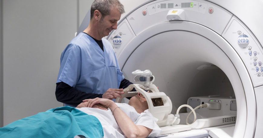
Polysomnography (PSG) is a comprehensive sleep study that monitors various body functions during sleep, including brain activity, eye movement, muscle tone, heart rate, respirations, and oxygen levels. It is typically conducted in a sleep laboratory under the supervision of a trained technologist. The data collected during PSG is used to diagnose and manage various sleep disorders such as sleep-related breathing disorders (e.g., sleep apnea), movement disorders (e.g., periodic limb movement disorder), parasomnias, and narcolepsy.
The study usually involves the attachment of electrodes to different parts of the body, such as the scalp, face, chest, and legs, to record electrical signals from the brain, eye movements, muscle activity, and heartbeats. Additionally, sensors may be placed on or near the nose and mouth to measure airflow, and a belt may be worn around the chest and abdomen to monitor breathing efforts. Oxygen levels are also monitored through a sensor attached to the finger or ear.
Polysomnography is often recommended when a sleep disorder is suspected based on symptoms or medical history, and other diagnostic tests have been inconclusive. The results of the study can help guide treatment decisions and improve overall sleep health.
Central sleep apnea (CSA) is a type of sleep-disordered breathing characterized by repeated cessations in breathing during sleep due to the brain's failure to transmit signals to the respiratory muscles that control breathing. Unlike obstructive sleep apnea (OSA), which results from airway obstruction, CSA occurs when the brain fails to send the necessary signals to the diaphragm and intercostal muscles to initiate or maintain respiratory efforts during sleep.
Central sleep apneas are usually associated with decreased oxygen saturation levels and can lead to frequent arousals from sleep, causing excessive daytime sleepiness, fatigue, and impaired cognitive function. CSA is often related to underlying medical conditions such as heart failure, stroke, or brainstem injury, and it may also be caused by the use of certain medications, including opioids.
There are several types of central sleep apnea, including:
1. Primary Central Sleep Apnea: This type occurs without any underlying medical condition or medication use.
2. Cheyne-Stokes Breathing: A pattern of central sleep apnea commonly seen in individuals with heart failure or stroke. It is characterized by a crescendo-decrescendo pattern of breathing, with periods of hyperventilation followed by hypoventilation and apnea.
3. High-Altitude Periodic Breathing: This type occurs at high altitudes due to the reduced oxygen levels and is usually reversible upon returning to lower altitudes.
4. Complex or Mixed Sleep Apnea: A combination of both central and obstructive sleep apneas, often observed in patients with OSA who are treated with continuous positive airway pressure (CPAP) therapy. In some cases, the central component may resolve over time with continued CPAP use.
Diagnosis of CSA typically involves a sleep study (polysomnography), which monitors various physiological parameters during sleep, such as brain waves, eye movements, muscle activity, heart rate, and breathing patterns. Treatment options for central sleep apnea depend on the underlying cause and may include medications, adjustments in medication dosages, or the use of devices that assist with breathing, such as adaptive servo-ventilation (ASV) or bilevel positive airway pressure (BiPAP) therapy.
REM sleep, or Rapid Eye Movement sleep, is a stage of sleep characterized by rapid eye movements, low muscle tone, and active brain activity. It is one of the two main types of sleep along with non-REM sleep and is marked by vivid dreaming, increased brain metabolism, and altered brain wave patterns. REM sleep is often referred to as "paradoxical sleep" because of the seemingly contradictory nature of its characteristics - an active brain in a state of relaxation. It is thought to play a role in memory consolidation, learning, and mood regulation. A typical night's sleep cycle includes several episodes of REM sleep, with each episode becoming longer as the night progresses.
Sleep disorders are a group of conditions that affect the ability to sleep well on a regular basis. They can include problems with falling asleep, staying asleep, or waking up too early in the morning. These disorders can be caused by various factors such as stress, anxiety, depression, medical conditions, or substance abuse.
The American Academy of Sleep Medicine (AASM) recognizes over 80 distinct sleep disorders, which are categorized into the following major groups:
1. Insomnia - difficulty falling asleep or staying asleep.
2. Sleep-related breathing disorders - abnormal breathing during sleep such as obstructive sleep apnea.
3. Central disorders of hypersomnolence - excessive daytime sleepiness, including narcolepsy.
4. Circadian rhythm sleep-wake disorders - disruption of the internal body clock that regulates the sleep-wake cycle.
5. Parasomnias - abnormal behaviors during sleep such as sleepwalking or night terrors.
6. Sleep-related movement disorders - repetitive movements during sleep such as restless legs syndrome.
7. Isolated symptoms and normal variants - brief and occasional symptoms that do not warrant a specific diagnosis.
Sleep disorders can have significant impacts on an individual's quality of life, productivity, and overall health. If you suspect that you may have a sleep disorder, it is recommended to consult with a healthcare professional or a sleep specialist for proper evaluation and treatment.
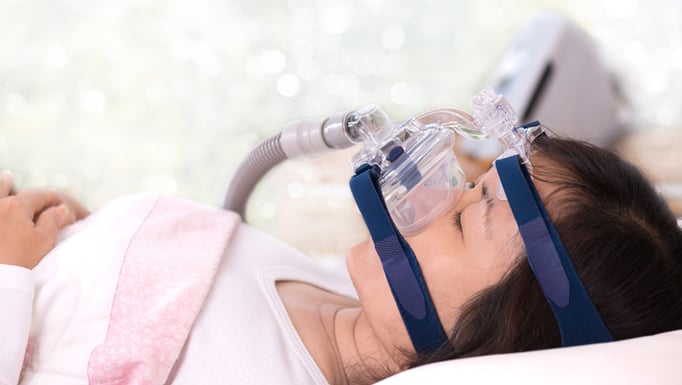
Continuous Positive Airway Pressure (CPAP) is a mode of non-invasive ventilation that delivers pressurized room air or oxygen to maintain airway patency and increase functional residual capacity in patients with respiratory disorders. A CPAP device, which typically includes a flow generator, tubing, and a mask, provides a constant positive pressure throughout the entire respiratory cycle, preventing the collapse of the upper airway during inspiration and expiration.
CPAP is commonly used to treat obstructive sleep apnea (OSA), a condition characterized by repetitive narrowing or closure of the upper airway during sleep, leading to intermittent hypoxia, hypercapnia, and sleep fragmentation. By delivering positive pressure, CPAP helps to stent open the airway, ensuring unobstructed breathing and reducing the frequency and severity of apneic events.
Additionally, CPAP can be used in other clinical scenarios, such as managing acute respiratory distress syndrome (ARDS), chronic obstructive pulmonary disease (COPD) exacerbations, or postoperative respiratory insufficiency, to improve oxygenation and reduce the work of breathing. The specific pressure settings and device configurations are tailored to each patient's needs based on their underlying condition, severity of symptoms, and response to therapy.
Snoring is defined as the vibration of respiratory structures and the resulting sound, due to obstructed air movement during breathing while sleeping. It occurs when the tissues at the back of the throat relax and narrow during sleep, partially blocking the airway. The airflow causes these tissues to vibrate, leading to the snoring sound. Snoring can be a sign of various conditions such as obstructive sleep apnea or other respiratory disorders. It can also be influenced by factors such as alcohol consumption, obesity, and sleeping position.
Chronic obstructive pulmonary disease (COPD) is a progressive lung disease characterized by the persistent obstruction of airflow in and out of the lungs. This obstruction is usually caused by two primary conditions: chronic bronchitis and emphysema. Chronic bronchitis involves inflammation and narrowing of the airways, leading to excessive mucus production and coughing. Emphysema is a condition where the alveoli (air sacs) in the lungs are damaged, resulting in decreased gas exchange and shortness of breath.
The main symptoms of COPD include progressive shortness of breath, chronic cough, chest tightness, wheezing, and excessive mucus production. The disease is often associated with exposure to harmful particles or gases, such as cigarette smoke, air pollution, or occupational dusts and chemicals. While there is no cure for COPD, treatments can help alleviate symptoms, improve quality of life, and slow the progression of the disease. These treatments may include bronchodilators, corticosteroids, combination inhalers, pulmonary rehabilitation, and, in severe cases, oxygen therapy or lung transplantation.
Obstructive lung disease is a category of respiratory diseases characterized by airflow limitation that causes difficulty in completely emptying the alveoli (tiny air sacs) of the lungs during exhaling. This results in the trapping of stale air and prevents fresh air from entering the alveoli, leading to various symptoms such as coughing, wheezing, shortness of breath, and decreased exercise tolerance.
The most common obstructive lung diseases include:
1. Chronic Obstructive Pulmonary Disease (COPD): A progressive disease that includes chronic bronchitis and emphysema, often caused by smoking or exposure to harmful pollutants.
2. Asthma: A chronic inflammatory disorder of the airways characterized by variable airflow obstruction, bronchial hyperresponsiveness, and an underlying inflammation. Symptoms can be triggered by various factors such as allergens, irritants, or physical activity.
3. Bronchiectasis: A condition in which the airways become abnormally widened, scarred, and thickened due to chronic inflammation or infection, leading to mucus buildup and impaired clearance.
4. Cystic Fibrosis: An inherited genetic disorder that affects the exocrine glands, resulting in thick and sticky mucus production in various organs, including the lungs. This can lead to chronic lung infections, inflammation, and airway obstruction.
5. Alpha-1 Antitrypsin Deficiency: A genetic condition characterized by low levels of alpha-1 antitrypsin protein, which leads to uncontrolled protease enzyme activity that damages the lung tissue, causing emphysema-like symptoms.
Treatment for obstructive lung diseases typically involves bronchodilators (to relax and widen the airways), corticosteroids (to reduce inflammation), and lifestyle modifications such as smoking cessation and pulmonary rehabilitation programs. In severe cases, oxygen therapy or even lung transplantation may be considered.
The soft palate, also known as the velum, is the rear portion of the roof of the mouth that is made up of muscle and mucous membrane. It extends from the hard palate (the bony front part of the roof of the mouth) to the uvula, which is the small piece of tissue that hangs down at the back of the throat.
The soft palate plays a crucial role in speech, swallowing, and breathing. During swallowing, it moves upward and backward to block off the nasal cavity, preventing food and liquids from entering the nose. In speech, it helps to direct the flow of air from the mouth into the nose, which is necessary for producing certain sounds.
Anatomically, the soft palate consists of several muscles that allow it to change shape and move. These muscles include the tensor veli palatini, levator veli palatini, musculus uvulae, palatopharyngeus, and palatoglossus. The soft palate also contains a rich supply of blood vessels and nerves that provide sensation and help regulate its function.
Wakefulness is a state of consciousness in which an individual is alert and aware of their surroundings. It is characterized by the ability to perceive, process, and respond to stimuli in a purposeful manner. In a medical context, wakefulness is often assessed using measures such as the electroencephalogram (EEG) to evaluate brain activity patterns associated with consciousness.
Wakefulness is regulated by several interconnected neural networks that promote arousal and attention. These networks include the ascending reticular activating system (ARAS), which consists of a group of neurons located in the brainstem that project to the thalamus and cerebral cortex, as well as other regions involved in regulating arousal and attention, such as the basal forebrain and hypothalamus.
Disorders of wakefulness can result from various underlying conditions, including neurological disorders, sleep disorders, medication side effects, or other medical conditions that affect brain function. Examples of such disorders include narcolepsy, insomnia, hypersomnia, and various forms of encephalopathy or brain injury.
Mandibular advancement is a treatment approach used in dentistry and sleep medicine, which involves the surgical or non-surgical forward movement of the mandible (lower jaw) to address certain medical conditions. The most common use of mandibular advancement is in the treatment of obstructive sleep apnea (OSA), where the tongue and soft tissues at the back of the throat can collapse into the airway during sleep, causing obstruction and breathing difficulties.
Mandibular advancement devices (MADs) are often used in non-surgical treatments. These custom-made oral appliances look similar to mouthguards or sports guards and are worn during sleep. They work by holding the lower jaw in a slightly forward position, which helps to keep the airway open and prevents the tongue and soft tissues from collapsing into it.
Surgical mandibular advancement is another option for patients with severe OSA who cannot tolerate or do not respond well to MADs or other treatments like continuous positive airway pressure (CPAP). In this procedure, the jaw is surgically moved forward and stabilized in that position using plates, screws, or wires. This creates more space in the airway and reduces the risk of obstruction during sleep.
In summary, mandibular advancement refers to the movement of the lower jaw forward, either through non-surgical means like MADs or surgical interventions, with the primary goal of treating obstructive sleep apnea by maintaining a patent airway during sleep.
The uvula is a small, conical piece of soft tissue that hangs down from the middle part of the back of the soft palate (the rear-most portion of the roof of the mouth). It contains muscle fibers and mucous glands, and its function is associated with swallowing, speaking, and protecting the airway. During swallowing, the uvula helps to prevent food and liquids from entering the nasal cavity by blocking the opening between the oral and nasal cavities (the nasopharynx). In speech, it plays a role in shaping certain sounds like "a" and "u."
The pharynx is a part of the digestive and respiratory systems that serves as a conduit for food and air. It is a musculo-membranous tube extending from the base of the skull to the level of the sixth cervical vertebra where it becomes continuous with the esophagus.
The pharynx has three regions: the nasopharynx, oropharynx, and laryngopharynx. The nasopharynx is the uppermost region, which lies above the soft palate and is connected to the nasal cavity. The oropharynx is the middle region, which includes the area between the soft palate and the hyoid bone, including the tonsils and base of the tongue. The laryngopharynx is the lowest region, which lies below the hyoid bone and connects to the larynx.
The primary function of the pharynx is to convey food from the oral cavity to the esophagus during swallowing and to allow air to pass from the nasal cavity to the larynx during breathing. It also plays a role in speech, taste, and immune defense.
Disorders of excessive somnolence (DES) are a group of medical conditions characterized by an increased tendency to fall asleep or experience excessive daytime sleepiness (EDS), despite having adequate opportunity and circumstances for sleep. These disorders are typically classified as central disorders of hypersomnolence according to the International Classification of Sleep Disorders (ICSD-3).
The most common DES is narcolepsy, a chronic neurological disorder caused by the brain's inability to regulate sleep-wake cycles normally. Other DES include idiopathic hypersomnia, Kleine-Levin syndrome, and recurrent hypersomnia. These disorders can significantly impact an individual's daily functioning, quality of life, and overall health.
Narcolepsy is further divided into two types: narcolepsy type 1 (NT1) and narcolepsy type 2 (NT2). NT1 is characterized by the presence of cataplexy, a sudden loss of muscle tone triggered by strong emotions, while NT2 does not include cataplexy. Both types of narcolepsy involve excessive daytime sleepiness, sleep paralysis, hypnagogic/hypnopompic hallucinations, and fragmented nighttime sleep.
Idiopathic hypersomnia is a DES without the presence of REM-related symptoms like cataplexy or sleep paralysis. Individuals with idiopathic hypersomnia experience excessive daytime sleepiness and prolonged nighttime sleep, often lasting 10 to 14 hours, but do not feel refreshed upon waking.
Kleine-Levin syndrome is a rare DES characterized by recurrent episodes of excessive sleepiness, often accompanied by cognitive impairment, altered perception, hyperphagia (excessive eating), and hypersexuality during the episodes. These episodes can last days to weeks and typically occur multiple times per year.
Recurrent hypersomnia is another rare DES with recurring episodes of excessive sleepiness lasting for several days, followed by a period of normal or reduced sleepiness. The episodes are not as predictable or consistent as those seen in Kleine-Levin syndrome.
Treatment for DES typically involves pharmacological interventions to manage symptoms and improve daytime alertness. Modafinil, armodafinil, and traditional stimulants like amphetamine salts are commonly used to treat excessive daytime sleepiness. Additionally, antidepressants may be prescribed to manage REM-related symptoms like cataplexy or sleep paralysis. Non-pharmacological interventions, such as scheduled napping and good sleep hygiene practices, can also help improve symptoms.
Sleep initiation and maintenance disorders are a category of sleep disorders that involve difficulty falling asleep and staying asleep throughout the night. This category includes:
1. Insomnia disorder: A persistent difficulty in initiating or maintaining sleep, or early morning awakening, despite adequate opportunity and circumstances for sleep, which causes clinically significant distress or impairment.
2. Narcolepsy: A chronic neurological disorder characterized by excessive daytime sleepiness, cataplexy (sudden loss of muscle tone triggered by strong emotions), hypnagogic hallucinations (vivid, dream-like experiences that occur while falling asleep) and sleep paralysis (temporary inability to move or speak while falling asleep or waking up).
3. Breathing-related sleep disorders: A group of disorders that involve abnormal breathing patterns during sleep, such as obstructive sleep apnea and central sleep apnea, which can lead to difficulty initiating and maintaining sleep.
4. Circadian rhythm sleep-wake disorders: A group of disorders that involve a misalignment between the individual's internal circadian rhythm and the external environment, leading to difficulty falling asleep and staying asleep at desired times.
5. Parasomnias: A group of disorders that involve abnormal behaviors or experiences during sleep, such as sleepwalking, night terrors, and REM sleep behavior disorder, which can disrupt sleep initiation and maintenance.
These disorders can have significant impacts on an individual's quality of life, daytime functioning, and overall health, and should be evaluated and managed by a healthcare professional with expertise in sleep medicine.
Positive-pressure respiration is a type of mechanical ventilation where positive pressure is applied to the airway and lungs, causing them to expand and inflate. This can be used to support or replace spontaneous breathing in patients who are unable to breathe effectively on their own due to conditions such as respiratory failure, neuromuscular disorders, or sedation for surgery.
During positive-pressure ventilation, a mechanical ventilator delivers breaths to the patient through an endotracheal tube or a tracheostomy tube. The ventilator is set to deliver a specific volume or pressure of air with each breath, and the patient's breathing is synchronized with the ventilator to ensure proper delivery of the breaths.
Positive-pressure ventilation can help improve oxygenation and remove carbon dioxide from the lungs, but it can also have potential complications such as barotrauma (injury to lung tissue due to excessive pressure), volutrauma (injury due to overdistention of the lungs), hemodynamic compromise (decreased blood pressure and cardiac output), and ventilator-associated pneumonia. Therefore, careful monitoring and adjustment of ventilator settings are essential to minimize these risks and provide safe and effective respiratory support.
Adenoidectomy is a surgical procedure in which the adenoids are removed. The adenoids are a patch of tissue located behind the nasal cavity, near the roof of the mouth. They help to filter out germs that are breathed in through the nose. However, sometimes the adenoids can become enlarged or infected, leading to problems such as difficulty breathing through the nose, recurrent ear infections, and sleep apnea. In these cases, an adenoidectomy may be recommended to remove the adenoids and alleviate these symptoms.
The procedure is typically performed on an outpatient basis, which means that the patient can go home the same day as the surgery. The surgeon will use a special instrument to remove the adenoids through the mouth, without making any external incisions. After the surgery, the patient may experience some discomfort, sore throat, and difficulty swallowing for a few days. However, these symptoms usually resolve within a week or two.
It is important to note that an adenoidectomy is not the same as a tonsillectomy, which is the surgical removal of the tonsils. While the tonsils and adenoids are both part of the immune system and located in the same area of the mouth, they serve different functions and may be removed separately or together depending on the individual's medical needs.
A tonsillectomy is a surgical procedure in which the tonsils, two masses of lymphoid tissue located on both sides of the back of the throat, are removed. This procedure is typically performed to treat recurrent or severe cases of tonsillitis (inflammation of the tonsils), sleep-disordered breathing such as obstructive sleep apnea, and other conditions where the tonsils are causing problems or complications. The surgery can be done under general anesthesia, and there are various methods for removing the tonsils, including traditional scalpel excision, electrocautery, and laser surgery. After a tonsillectomy, patients may experience pain, swelling, and difficulty swallowing, but these symptoms typically improve within 1-2 weeks post-surgery.
A Severity of Illness Index is a measurement tool used in healthcare to assess the severity of a patient's condition and the risk of mortality or other adverse outcomes. These indices typically take into account various physiological and clinical variables, such as vital signs, laboratory values, and co-morbidities, to generate a score that reflects the patient's overall illness severity.
Examples of Severity of Illness Indices include the Acute Physiology and Chronic Health Evaluation (APACHE) system, the Simplified Acute Physiology Score (SAPS), and the Mortality Probability Model (MPM). These indices are often used in critical care settings to guide clinical decision-making, inform prognosis, and compare outcomes across different patient populations.
It is important to note that while these indices can provide valuable information about a patient's condition, they should not be used as the sole basis for clinical decision-making. Rather, they should be considered in conjunction with other factors, such as the patient's overall clinical presentation, treatment preferences, and goals of care.
Obstructive Jaundice is a medical condition characterized by the yellowing of the skin, sclera (whites of the eyes), and mucous membranes due to the accumulation of bilirubin in the bloodstream. This occurs when there is an obstruction or blockage in the bile ducts that transport bile from the liver to the small intestine.
Bile, which contains bilirubin, aids in digestion and is usually released from the liver into the small intestine. When the flow of bile is obstructed, bilirubin builds up in the blood, causing jaundice. The obstruction can be caused by various factors, such as gallstones, tumors, or strictures in the bile ducts.
Obstructive jaundice may present with additional symptoms like dark urine, light-colored stools, itching, abdominal pain, and weight loss, depending on the cause and severity of the obstruction. It is essential to seek medical attention if jaundice is observed, as timely diagnosis and management can prevent potential complications, such as liver damage or infection.
In a medical or physiological context, "arousal" refers to the state of being awake and responsive to stimuli. It involves the activation of the nervous system, particularly the autonomic nervous system, which prepares the body for action. Arousal levels can vary from low (such as during sleep) to high (such as during states of excitement or stress). In clinical settings, changes in arousal may be assessed to help diagnose conditions such as coma, brain injury, or sleep disorders. It is also used in the context of sexual response, where it refers to the level of physical and mental awareness and readiness for sexual activity.
Medical Definition of Respiration:
Respiration, in physiology, is the process by which an organism takes in oxygen and gives out carbon dioxide. It's also known as breathing. This process is essential for most forms of life because it provides the necessary oxygen for cellular respiration, where the cells convert biochemical energy from nutrients into adenosine triphosphate (ATP), and releases waste products, primarily carbon dioxide.
In humans and other mammals, respiration is a two-stage process:
1. Breathing (or external respiration): This involves the exchange of gases with the environment. Air enters the lungs through the mouth or nose, then passes through the pharynx, larynx, trachea, and bronchi, finally reaching the alveoli where the actual gas exchange occurs. Oxygen from the inhaled air diffuses into the blood, while carbon dioxide, a waste product of metabolism, diffuses from the blood into the alveoli to be exhaled.
2. Cellular respiration (or internal respiration): This is the process by which cells convert glucose and other nutrients into ATP, water, and carbon dioxide in the presence of oxygen. The carbon dioxide produced during this process then diffuses out of the cells and into the bloodstream to be exhaled during breathing.
In summary, respiration is a vital physiological function that enables organisms to obtain the necessary oxygen for cellular metabolism while eliminating waste products like carbon dioxide.
Sleep stages are distinct patterns of brain activity that occur during sleep, as measured by an electroencephalogram (EEG). They are part of the sleep cycle and are used to describe the different types of sleep that humans go through during a normal night's rest. The sleep cycle includes several repeating stages:
1. Stage 1 (N1): This is the lightest stage of sleep, where you transition from wakefulness to sleep. During this stage, muscle activity and brain waves begin to slow down.
2. Stage 2 (N2): In this stage, your heart rate slows, body temperature decreases, and eye movements stop. Brain wave activity becomes slower, with occasional bursts of electrical activity called sleep spindles.
3. Stage 3 (N3): Also known as deep non-REM sleep, this stage is characterized by slow delta waves. It is during this stage that the body undergoes restorative processes such as tissue repair, growth, and immune function enhancement.
4. REM (Rapid Eye Movement) sleep: This is the stage where dreaming typically occurs. Your eyes move rapidly beneath closed eyelids, heart rate and respiration become irregular, and brain wave activity increases to levels similar to wakefulness. REM sleep is important for memory consolidation and learning.
The sleep cycle progresses through these stages multiple times during the night, with REM sleep periods becoming longer towards morning. Understanding sleep stages is crucial in diagnosing and treating various sleep disorders.
Airway obstruction is a medical condition that occurs when the normal flow of air into and out of the lungs is partially or completely blocked. This blockage can be caused by a variety of factors, including swelling of the tissues in the airway, the presence of foreign objects or substances, or abnormal growths such as tumors.
When the airway becomes obstructed, it can make it difficult for a person to breathe normally. They may experience symptoms such as shortness of breath, wheezing, coughing, and chest tightness. In severe cases, airway obstruction can lead to respiratory failure and other life-threatening complications.
There are several types of airway obstruction, including:
1. Upper airway obstruction: This occurs when the blockage is located in the upper part of the airway, such as the nose, throat, or voice box.
2. Lower airway obstruction: This occurs when the blockage is located in the lower part of the airway, such as the trachea or bronchi.
3. Partial airway obstruction: This occurs when the airway is partially blocked, allowing some air to flow in and out of the lungs.
4. Complete airway obstruction: This occurs when the airway is completely blocked, preventing any air from flowing into or out of the lungs.
Treatment for airway obstruction depends on the underlying cause of the condition. In some cases, removing the obstruction may be as simple as clearing the airway of foreign objects or mucus. In other cases, more invasive treatments such as surgery may be necessary.
Anoxia is a medical condition that refers to the absence or complete lack of oxygen supply in the body or a specific organ, tissue, or cell. This can lead to serious health consequences, including damage or death of cells and tissues, due to the vital role that oxygen plays in supporting cellular metabolism and energy production.
Anoxia can occur due to various reasons, such as respiratory failure, cardiac arrest, severe blood loss, carbon monoxide poisoning, or high altitude exposure. Prolonged anoxia can result in hypoxic-ischemic encephalopathy, a serious condition that can cause brain damage and long-term neurological impairments.
Medical professionals use various diagnostic tests, such as blood gas analysis, pulse oximetry, and electroencephalography (EEG), to assess oxygen levels in the body and diagnose anoxia. Treatment for anoxia typically involves addressing the underlying cause, providing supplemental oxygen, and supporting vital functions, such as breathing and circulation, to prevent further damage.
The pharyngeal muscles, also known as the musculature of the pharynx, are a group of skeletal muscles that make up the walls of the pharynx, which is the part of the throat located just above the esophagus and behind the nasal and oral cavities. These muscles play a crucial role in several vital functions, including:
1. Swallowing (deglutition): The pharyngeal muscles contract in a coordinated sequence to propel food or liquids from the mouth through the pharynx and into the esophagus during swallowing.
2. Speech: The contraction and relaxation of these muscles help shape the sounds produced by the vocal cords, contributing to the production of speech.
3. Respiration: The pharyngeal muscles assist in maintaining an open airway during breathing, especially during sleep and when the upper airways are obstructed.
The pharyngeal muscles consist of three layers: the outer circular muscle layer, the middle longitudinal muscle layer, and the inner inferior constrictor muscle layer. The specific muscles that make up these layers include:
1. Superior constrictor muscle (outer circular layer)
2. Middle constrictor muscle (middle longitudinal layer)
3. Inferior constrictor muscle (inner inferior constrictor layer)
4. Stylopharyngeus muscle
5. Salpingopharyngeus muscle
6. Palatopharyngeus muscle
7. Buccinator muscle (partially contributes to the middle longitudinal layer)
These muscles work together to perform their various functions, and any dysfunction in these muscles can lead to problems like swallowing difficulties (dysphagia), speech impairments, or respiratory issues.
Pulse oximetry is a noninvasive method for monitoring a person's oxygen saturation (SO2) and pulse rate. It uses a device called a pulse oximeter, which measures the amount of oxygen-carrying hemoglobin in the blood compared to the amount of hemoglobin that is not carrying oxygen. This measurement is expressed as a percentage, known as oxygen saturation (SpO2). Normal oxygen saturation levels are generally 95% or above at sea level. Lower levels may indicate hypoxemia, a condition where there is not enough oxygen in the blood to meet the body's needs. Pulse oximetry is commonly used in hospitals and other healthcare settings to monitor patients during surgery, in intensive care units, and in sleep studies to detect conditions such as sleep apnea. It can also be used by individuals with certain medical conditions, such as chronic obstructive pulmonary disease (COPD), to monitor their oxygen levels at home.
In medical terms, the tongue is a muscular organ in the oral cavity that plays a crucial role in various functions such as taste, swallowing, and speech. It's covered with a mucous membrane and contains papillae, which are tiny projections that contain taste buds to help us perceive different tastes - sweet, salty, sour, and bitter. The tongue also assists in the initial process of digestion by moving food around in the mouth for chewing and mixing with saliva. Additionally, it helps in forming words and speaking clearly by shaping the sounds produced in the mouth.
Occlusal splints, also known as bite guards or night guards, are removable dental appliances that are used to provide protection and stabilization for the teeth and jaw joint (temporomandibular joint or TMJ). They are typically made of hard acrylic or soft materials and are custom-fit to a patient's mouth.
Occlusal splints work by covering and separating the upper and lower teeth, preventing them from coming into contact with each other. This can help to reduce tooth grinding and clenching (bruxism), which can cause tooth wear, sensitivity, and TMJ disorders. They may also be used to help stabilize the jaw joint and muscles in patients with TMJ disorders or to provide protection for teeth that have undergone restorative dental work.
It is important to note that occlusal splints should only be worn under the guidance of a dentist, as improper use can lead to further dental problems.
Respiratory mechanics refers to the biomechanical properties and processes that involve the movement of air through the respiratory system during breathing. It encompasses the mechanical behavior of the lungs, chest wall, and the muscles of respiration, including the diaphragm and intercostal muscles.
Respiratory mechanics includes several key components:
1. **Compliance**: The ability of the lungs and chest wall to expand and recoil during breathing. High compliance means that the structures can easily expand and recoil, while low compliance indicates greater resistance to expansion and recoil.
2. **Resistance**: The opposition to airflow within the respiratory system, primarily due to the friction between the air and the airway walls. Airway resistance is influenced by factors such as airway diameter, length, and the viscosity of the air.
3. **Lung volumes and capacities**: These are the amounts of air present in the lungs during different phases of the breathing cycle. They include tidal volume (the amount of air inspired or expired during normal breathing), inspiratory reserve volume (additional air that can be inspired beyond the tidal volume), expiratory reserve volume (additional air that can be exhaled beyond the tidal volume), and residual volume (the air remaining in the lungs after a forced maximum exhalation).
4. **Work of breathing**: The energy required to overcome the resistance and elastic forces during breathing. This work is primarily performed by the respiratory muscles, which contract to generate negative intrathoracic pressure and expand the chest wall, allowing air to flow into the lungs.
5. **Pressure-volume relationships**: These describe how changes in lung volume are associated with changes in pressure within the respiratory system. Important pressure components include alveolar pressure (the pressure inside the alveoli), pleural pressure (the pressure between the lungs and the chest wall), and transpulmonary pressure (the difference between alveolar and pleural pressures).
Understanding respiratory mechanics is crucial for diagnosing and managing various respiratory disorders, such as chronic obstructive pulmonary disease (COPD), asthma, and restrictive lung diseases.
Cheyne-Stokes respiration is a pattern of breathing characterized by cyclical changes in the depth and rate of respirations. It is often associated with various medical conditions that affect the brainstem, such as stroke, brain injury, or certain neurological disorders.
In Cheyne-Stokes respiration, the individual's breathing starts with a series of deeper and faster breaths (hyperventilation), which gradually become shallower and slower (hypoventilation). This cycle repeats every few minutes, resulting in a pattern of waxing and waning of the depth and rate of respirations.
The underlying mechanism for Cheyne-Stokes respiration is related to the regulation of breathing by the brainstem. When there are abnormalities in this area, it can lead to instability in the control of breathing, resulting in the cyclical pattern of hyperventilation and hypoventilation.
Cheyne-Stokes respiration can be a sign of serious underlying medical conditions, and it is important to seek medical attention if you or someone else experiences this type of breathing pattern. Treatment may involve addressing the underlying cause, such as managing heart failure or reducing intracranial pressure in patients with brain injury or stroke.
Adenoids are a pair of masses of lymphoid tissue located in the nasopharynx, which is the upper part of the throat behind the nose. They are part of the immune system and help to protect against infection. Adenoids are largest in children and tend to shrink in size as people get older. In some cases, adenoids can become enlarged or infected, leading to problems such as breathing difficulties, ear infections, and sleep disorders. Treatment for enlarged or infected adenoids may include antibiotics, medications to reduce swelling, or surgical removal of the adenoids (adenoidectomy).
Oxygen is a colorless, odorless, tasteless gas that constitutes about 21% of the earth's atmosphere. It is a crucial element for human and most living organisms as it is vital for respiration. Inhaled oxygen enters the lungs and binds to hemoglobin in red blood cells, which carries it to tissues throughout the body where it is used to convert nutrients into energy and carbon dioxide, a waste product that is exhaled.
Medically, supplemental oxygen therapy may be provided to patients with conditions such as chronic obstructive pulmonary disease (COPD), pneumonia, heart failure, or other medical conditions that impair the body's ability to extract sufficient oxygen from the air. Oxygen can be administered through various devices, including nasal cannulas, face masks, and ventilators.
A circadian rhythm is a roughly 24-hour biological cycle that regulates various physiological and behavioral processes in living organisms. It is driven by the body's internal clock, which is primarily located in the suprachiasmatic nucleus (SCN) of the hypothalamus in the brain.
The circadian rhythm controls many aspects of human physiology, including sleep-wake cycles, hormone secretion, body temperature, and metabolism. It helps to synchronize these processes with the external environment, particularly the day-night cycle caused by the rotation of the Earth.
Disruptions to the circadian rhythm can have negative effects on health, leading to conditions such as insomnia, sleep disorders, depression, bipolar disorder, and even increased risk of chronic diseases like cancer, diabetes, and cardiovascular disease. Factors that can disrupt the circadian rhythm include shift work, jet lag, irregular sleep schedules, and exposure to artificial light at night.
Pulmonary ventilation, also known as pulmonary respiration or simply ventilation, is the process of moving air into and out of the lungs to facilitate gas exchange. It involves two main phases: inhalation (or inspiration) and exhalation (or expiration). During inhalation, the diaphragm and external intercostal muscles contract, causing the chest volume to increase and the pressure inside the chest to decrease, which then draws air into the lungs. Conversely, during exhalation, these muscles relax, causing the chest volume to decrease and the pressure inside the chest to increase, which pushes air out of the lungs. This process ensures that oxygen-rich air from the atmosphere enters the alveoli (air sacs in the lungs), where it can diffuse into the bloodstream, while carbon dioxide-rich air from the bloodstream in the capillaries surrounding the alveoli is expelled out of the body.
Electroencephalography (EEG) is a medical procedure that records electrical activity in the brain. It uses small, metal discs called electrodes, which are attached to the scalp with paste or a specialized cap. These electrodes detect tiny electrical charges that result from the activity of brain cells, and the EEG machine then amplifies and records these signals.
EEG is used to diagnose various conditions related to the brain, such as seizures, sleep disorders, head injuries, infections, and degenerative diseases like Alzheimer's or Parkinson's. It can also be used during surgery to monitor brain activity and ensure that surgical procedures do not interfere with vital functions.
EEG is a safe and non-invasive procedure that typically takes about 30 minutes to an hour to complete, although longer recordings may be necessary in some cases. Patients are usually asked to relax and remain still during the test, as movement can affect the quality of the recording.
Body Mass Index (BMI) is a measure used to assess whether a person has a healthy weight for their height. It's calculated by dividing a person's weight in kilograms by the square of their height in meters. Here is the medical definition:
Body Mass Index (BMI) = weight(kg) / [height(m)]^2
According to the World Health Organization, BMI categories are defined as follows:
* Less than 18.5: Underweight
* 18.5-24.9: Normal or healthy weight
* 25.0-29.9: Overweight
* 30.0 and above: Obese
It is important to note that while BMI can be a useful tool for identifying weight issues in populations, it does have limitations when applied to individuals. For example, it may not accurately reflect body fat distribution or muscle mass, which can affect health risks associated with excess weight. Therefore, BMI should be used as one of several factors when evaluating an individual's health status and risk for chronic diseases.
Orthodontic appliances, removable, are dental devices that can be removed and inserted by the patient as needed or directed. These appliances are designed to align and straighten teeth, correct bite issues, and improve the function and appearance of the teeth and jaws. They are typically made from materials such as plastic, metal, or acrylic and may include components like wires, springs, or screws. Examples of removable orthodontic appliances include aligners, retainers, and space maintainers. The specific type and design of the appliance will depend on the individual patient's orthodontic needs and treatment goals.
Airway resistance is a measure of the opposition to airflow during breathing, which is caused by the friction between the air and the walls of the respiratory tract. It is an important parameter in respiratory physiology because it can affect the work of breathing and gas exchange.
Airway resistance is usually expressed in units of cm H2O/L/s or Pa·s/m, and it can be measured during spontaneous breathing or during forced expiratory maneuvers, such as those used in pulmonary function testing. Increased airway resistance can result from a variety of conditions, including asthma, chronic obstructive pulmonary disease (COPD), bronchitis, and bronchiectasis. Decreased airway resistance can be seen in conditions such as emphysema or after a successful bronchodilator treatment.
The oropharynx is the part of the throat (pharynx) that is located immediately behind the mouth and includes the back one-third of the tongue, the soft palate, the side and back walls of the throat, and the tonsils. It serves as a passageway for both food and air, and is also an important area for the immune system due to the presence of tonsils.
Medical Definition:
"Risk factors" are any attribute, characteristic or exposure of an individual that increases the likelihood of developing a disease or injury. They can be divided into modifiable and non-modifiable risk factors. Modifiable risk factors are those that can be changed through lifestyle choices or medical treatment, while non-modifiable risk factors are inherent traits such as age, gender, or genetic predisposition. Examples of modifiable risk factors include smoking, alcohol consumption, physical inactivity, and unhealthy diet, while non-modifiable risk factors include age, sex, and family history. It is important to note that having a risk factor does not guarantee that a person will develop the disease, but rather indicates an increased susceptibility.
Nasal obstruction is a medical condition that refers to any blockage or restriction in the normal flow of air through the nasal passages. This can be caused by various factors such as inflammation, swelling, or physical abnormalities in the nasal cavity. Common causes of nasal obstruction include allergies, sinusitis, deviated septum, enlarged turbinates, and nasal polyps. Symptoms may include difficulty breathing through the nose, nasal congestion, and nasal discharge. Treatment options depend on the underlying cause and may include medications, surgery, or lifestyle changes.
Prospective studies, also known as longitudinal studies, are a type of cohort study in which data is collected forward in time, following a group of individuals who share a common characteristic or exposure over a period of time. The researchers clearly define the study population and exposure of interest at the beginning of the study and follow up with the participants to determine the outcomes that develop over time. This type of study design allows for the investigation of causal relationships between exposures and outcomes, as well as the identification of risk factors and the estimation of disease incidence rates. Prospective studies are particularly useful in epidemiology and medical research when studying diseases with long latency periods or rare outcomes.
Respiratory Function Tests (RFTs) are a group of medical tests that measure how well your lungs take in and exhale air, and how well they transfer oxygen and carbon dioxide into and out of your blood. They can help diagnose certain lung disorders, measure the severity of lung disease, and monitor response to treatment.
RFTs include several types of tests, such as:
1. Spirometry: This test measures how much air you can exhale and how quickly you can do it. It's often used to diagnose and monitor conditions like asthma, chronic obstructive pulmonary disease (COPD), and other lung diseases.
2. Lung volume testing: This test measures the total amount of air in your lungs. It can help diagnose restrictive lung diseases, such as pulmonary fibrosis or sarcoidosis.
3. Diffusion capacity testing: This test measures how well oxygen moves from your lungs into your bloodstream. It's often used to diagnose and monitor conditions like pulmonary fibrosis, interstitial lung disease, and other lung diseases that affect the ability of the lungs to transfer oxygen to the blood.
4. Bronchoprovocation testing: This test involves inhaling a substance that can cause your airways to narrow, such as methacholine or histamine. It's often used to diagnose and monitor asthma.
5. Exercise stress testing: This test measures how well your lungs and heart work together during exercise. It's often used to diagnose lung or heart disease.
Overall, Respiratory Function Tests are an important tool for diagnosing and managing a wide range of lung conditions.
Treatment outcome is a term used to describe the result or effect of medical treatment on a patient's health status. It can be measured in various ways, such as through symptoms improvement, disease remission, reduced disability, improved quality of life, or survival rates. The treatment outcome helps healthcare providers evaluate the effectiveness of a particular treatment plan and make informed decisions about future care. It is also used in clinical research to compare the efficacy of different treatments and improve patient care.
A questionnaire in the medical context is a standardized, systematic, and structured tool used to gather information from individuals regarding their symptoms, medical history, lifestyle, or other health-related factors. It typically consists of a series of written questions that can be either self-administered or administered by an interviewer. Questionnaires are widely used in various areas of healthcare, including clinical research, epidemiological studies, patient care, and health services evaluation to collect data that can inform diagnosis, treatment planning, and population health management. They provide a consistent and organized method for obtaining information from large groups or individual patients, helping to ensure accurate and comprehensive data collection while minimizing bias and variability in the information gathered.
Obesity is a complex disease characterized by an excess accumulation of body fat to the extent that it negatively impacts health. It's typically defined using Body Mass Index (BMI), a measure calculated from a person's weight and height. A BMI of 30 or higher is indicative of obesity. However, it's important to note that while BMI can be a useful tool for identifying obesity in populations, it does not directly measure body fat and may not accurately reflect health status in individuals. Other factors such as waist circumference, blood pressure, cholesterol levels, and blood sugar levels should also be considered when assessing health risks associated with weight.
Sleep disorders, intrinsic, refer to a group of sleep disorders that are caused by underlying medical conditions within an individual's body. These disorders originate from internal physiological or psychological factors and can significantly impact the quality, duration, and timing of sleep. The most common types of intrinsic sleep disorders include insomnia, sleep-related breathing disorders (such as sleep apnea), central hypersomnias (like narcolepsy), circadian rhythm sleep-wake disorders, and parasomnias (including nightmares and sleepwalking).
Intrinsic sleep disorders can lead to various negative consequences, such as excessive daytime sleepiness, impaired cognitive function, reduced quality of life, and increased risk of accidents or injuries. Proper diagnosis and management of these disorders typically involve addressing the underlying medical condition and implementing appropriate treatment strategies, which may include lifestyle modifications, pharmacological interventions, or medical devices.
Nocturnal Myoclonus Syndrome, also known as Periodic Limb Movement Disorder (PLMD), is a condition characterized by recurring involuntary jerking movements of the limbs during sleep, particularly the legs. These movements typically occur every 20-40 seconds and can last for an hour or more throughout the night. They often disrupt normal sleep patterns, causing insomnia or excessive daytime sleepiness.
The movements are usually jerky, rapid, and rhythmic, involving extension of the big toe and flexion of the ankle, knee, or hip. In some cases, these movements can be so forceful that they cause the person to wake up, although often individuals with this condition may not be aware of their nighttime leg movements.
Nocturnal Myoclonus Syndrome is different from another common sleep disorder called Restless Legs Syndrome (RLS), as RLS primarily causes discomfort or an irresistible urge to move the legs while awake and still, whereas Nocturnal Myoclonus Syndrome involves involuntary movements during sleep. However, up to 80% of people with RLS also have PLMD.
The exact cause of Nocturnal Myoclonus Syndrome is not fully understood, but it may be associated with abnormalities in the brain's regulation of muscle activity during sleep. Certain medications, neurological conditions, and iron deficiency anemia have been linked to an increased risk of developing this disorder. Treatment options include medication, lifestyle changes, and addressing any underlying medical conditions that may contribute to the development or worsening of symptoms.
Actigraphy is a non-invasive method used to estimate sleep-wake patterns and physical activity levels over extended periods, typically ranging from several days to weeks. It involves the use of a small device called an actigraph, which is usually worn on the wrist like a watch.
The actigraph contains an accelerometer that detects movement and records the intensity and duration of motion. This data is then analyzed using specialized software to provide information about sleep and wake times, as well as patterns of physical activity.
Actigraphy can be useful in assessing various sleep disorders, such as insomnia, circadian rhythm disorders, and sleep-related breathing disorders. It can also help evaluate the effectiveness of treatments for these conditions. However, it is important to note that actigraphy is not a substitute for a formal sleep study (polysomnography) and should be used in conjunction with other assessment tools and clinical evaluations.
Hypercapnia is a state of increased carbon dioxide (CO2) concentration in the blood, typically defined as an arterial CO2 tension (PaCO2) above 45 mmHg. It is often associated with conditions that impair gas exchange or eliminate CO2 from the body, such as chronic obstructive pulmonary disease (COPD), severe asthma, respiratory failure, or certain neuromuscular disorders. Hypercapnia can cause symptoms such as headache, confusion, shortness of breath, and in severe cases, it can lead to life-threatening complications such as respiratory acidosis, coma, and even death if not promptly treated.
The hyoid bone is a U-shaped bone located in the anterior neck, superior to the thyroid cartilage. It does not articulate with any other bones and serves as an attachment point for various muscles, including those involved in swallowing, breathing, and speaking. The unique structure of the hyoid bone allows it to support the tongue and contribute to the stability of the airway.
Electrooculography (EOG) is a technique for measuring the resting potential of the eye and the changes in this potential that occur with eye movements. It involves placing electrodes near the eyes to detect the small electric fields generated by the movement of the eyeball within the surrounding socket. This technique is used in research and clinical settings to study eye movements and their control, as well as in certain diagnostic applications such as assessing the function of the oculomotor system in patients with neurological disorders.
In the field of medicine, "time factors" refer to the duration of symptoms or time elapsed since the onset of a medical condition, which can have significant implications for diagnosis and treatment. Understanding time factors is crucial in determining the progression of a disease, evaluating the effectiveness of treatments, and making critical decisions regarding patient care.
For example, in stroke management, "time is brain," meaning that rapid intervention within a specific time frame (usually within 4.5 hours) is essential to administering tissue plasminogen activator (tPA), a clot-busting drug that can minimize brain damage and improve patient outcomes. Similarly, in trauma care, the "golden hour" concept emphasizes the importance of providing definitive care within the first 60 minutes after injury to increase survival rates and reduce morbidity.
Time factors also play a role in monitoring the progression of chronic conditions like diabetes or heart disease, where regular follow-ups and assessments help determine appropriate treatment adjustments and prevent complications. In infectious diseases, time factors are crucial for initiating antibiotic therapy and identifying potential outbreaks to control their spread.
Overall, "time factors" encompass the significance of recognizing and acting promptly in various medical scenarios to optimize patient outcomes and provide effective care.
A case-control study is an observational research design used to identify risk factors or causes of a disease or health outcome. In this type of study, individuals with the disease or condition (cases) are compared with similar individuals who do not have the disease or condition (controls). The exposure history or other characteristics of interest are then compared between the two groups to determine if there is an association between the exposure and the disease.
Case-control studies are often used when it is not feasible or ethical to conduct a randomized controlled trial, as they can provide valuable insights into potential causes of diseases or health outcomes in a relatively short period of time and at a lower cost than other study designs. However, because case-control studies rely on retrospective data collection, they are subject to biases such as recall bias and selection bias, which can affect the validity of the results. Therefore, it is important to carefully design and conduct case-control studies to minimize these potential sources of bias.
Prevalence, in medical terms, refers to the total number of people in a given population who have a particular disease or condition at a specific point in time, or over a specified period. It is typically expressed as a percentage or a ratio of the number of cases to the size of the population. Prevalence differs from incidence, which measures the number of new cases that develop during a certain period.
Cephalometry is a medical term that refers to the measurement and analysis of the skull, particularly the head face relations. It is commonly used in orthodontics and maxillofacial surgery to assess and plan treatment for abnormalities related to the teeth, jaws, and facial structures. The process typically involves taking X-ray images called cephalograms, which provide a lateral view of the head, and then using various landmarks and reference lines to make measurements and evaluate skeletal and dental relationships. This information can help clinicians diagnose problems, plan treatment, and assess treatment outcomes.
Spirometry is a common type of pulmonary function test (PFT) that measures how well your lungs work. This is done by measuring how much air you can exhale from your lungs after taking a deep breath, and how quickly you can exhale it. The results are compared to normal values for your age, height, sex, and ethnicity.
Spirometry is used to diagnose and monitor certain lung conditions, such as asthma, chronic obstructive pulmonary disease (COPD), and other respiratory diseases that cause narrowing of the airways. It can also be used to assess the effectiveness of treatment for these conditions. The test is non-invasive, safe, and easy to perform.
Ambulatory monitoring is a medical practice that involves the continuous or intermittent recording of physiological parameters in a patient who is mobile and able to perform their usual activities while outside of a hospital or clinical setting. This type of monitoring allows healthcare professionals to evaluate a patient's condition over an extended period, typically 24 hours or more, in their natural environment.
Ambulatory monitoring can be used to diagnose and manage various medical conditions such as hypertension, cardiac arrhythmias, sleep disorders, and mobility issues. Common methods of ambulatory monitoring include:
1. Holter monitoring: A small, portable device that records the electrical activity of the heart for 24-48 hours or more.
2. Ambulatory blood pressure monitoring (ABPM): A device that measures blood pressure at regular intervals throughout the day and night.
3. Event monitors: Devices that record heart rhythms only when symptoms occur or when activated by the patient.
4. Actigraphy: A non-invasive method of monitoring sleep-wake patterns, physical activity, and circadian rhythms using a wristwatch-like device.
5. Continuous glucose monitoring (CGM): A device that measures blood sugar levels continuously throughout the day and night.
Overall, ambulatory monitoring provides valuable information about a patient's physiological status in their natural environment, allowing healthcare professionals to make informed decisions regarding diagnosis, treatment, and management of medical conditions.
Comorbidity is the presence of one or more additional health conditions or diseases alongside a primary illness or condition. These co-occurring health issues can have an impact on the treatment plan, prognosis, and overall healthcare management of an individual. Comorbidities often interact with each other and the primary condition, leading to more complex clinical situations and increased healthcare needs. It is essential for healthcare professionals to consider and address comorbidities to provide comprehensive care and improve patient outcomes.
Narcolepsy is a chronic neurological disorder that affects the control of sleep and wakefulness. It's characterized by excessive daytime sleepiness (EDS), where people experience sudden, uncontrollable episodes of falling asleep during the day. These "sleep attacks" can occur at any time - while working, talking, eating, or even driving.
In addition to EDS, narcolepsy often includes cataplexy, a condition that causes loss of muscle tone, leading to weakness and sometimes collapse, often triggered by strong emotions like laughter or surprise. Other common symptoms are sleep paralysis (a temporary inability to move or speak while falling asleep or waking up), vivid hallucinations during the transitions between sleep and wakefulness, and fragmented nighttime sleep.
The exact cause of narcolepsy is not fully understood, but it's believed to involve genetic and environmental factors, as well as problems with certain neurotransmitters in the brain, such as hypocretin/orexin, which regulate sleep-wake cycles. Narcolepsy can significantly impact a person's quality of life, making it essential to seek medical attention for proper diagnosis and management.
The supine position is a term used in medicine to describe a body posture where an individual is lying down on their back, with their face and torso facing upwards. This position is often adopted during various medical procedures, examinations, or when resting, as it allows for easy access to the front of the body. It is also the position automatically assumed by most people who are falling asleep.
It's important to note that in the supine position, the head can be flat on the surface or raised with the use of pillows or specialized medical equipment like a hospital bed. This can help to alleviate potential issues such as breathing difficulties or swelling in the face and head.
Forced Expiratory Volume (FEV) is a medical term used to describe the volume of air that can be forcefully exhaled from the lungs in one second. It is often measured during pulmonary function testing to assess lung function and diagnose conditions such as chronic obstructive pulmonary disease (COPD) or asthma.
FEV is typically expressed as a percentage of the Forced Vital Capacity (FVC), which is the total volume of air that can be exhaled from the lungs after taking a deep breath in. The ratio of FEV to FVC is used to determine whether there is obstruction in the airways, with a lower ratio indicating more severe obstruction.
There are different types of FEV measurements, including FEV1 (the volume of air exhaled in one second), FEV25-75 (the average volume of air exhaled during the middle 50% of the FVC maneuver), and FEV0.5 (the volume of air exhaled in half a second). These measurements can provide additional information about lung function and help guide treatment decisions.
Sleep arousal disorders are a category of sleep disorders that involve the partial or complete awakening from sleep, often accompanied by confusion and disorientation. These disorders are characterized by an abnormal arousal process during sleep, which can result in brief periods of wakefulness or full awakenings. The most common types of sleep arousal disorders include sleepwalking (somnambulism), sleep talking (somniloquy), and night terrors (pavor nocturnus).
In sleepwalking, the individual may get out of bed and walk around while still asleep, often with a blank stare and without any memory of the event. Sleep talking can occur in various levels of sleep and may range from simple sounds to complex conversations. Night terrors are episodes of intense fear and agitation during sleep, often accompanied by screams or cries for help, rapid heart rate, and sweating.
These disorders can be caused by a variety of factors, including stress, anxiety, fever, certain medications, alcohol consumption, and underlying medical conditions such as sleep apnea or restless leg syndrome. They can also occur as a result of genetic predisposition. Sleep arousal disorders can have significant impacts on an individual's quality of life, leading to fatigue, daytime sleepiness, impaired cognitive function, and decreased overall well-being. Treatment options may include behavioral therapy, medication, or addressing any underlying medical conditions.
Patient compliance, also known as medication adherence or patient adherence, refers to the degree to which a patient's behavior matches the agreed-upon recommendations from their healthcare provider. This includes taking medications as prescribed (including the correct dosage, frequency, and duration), following dietary restrictions, making lifestyle changes, and attending follow-up appointments. Poor patient compliance can negatively impact treatment outcomes and lead to worsening of symptoms, increased healthcare costs, and development of drug-resistant strains in the case of antibiotics. It is a significant challenge in healthcare and efforts are being made to improve patient education, communication, and support to enhance compliance.
Oxygen inhalation therapy is a medical treatment that involves the administration of oxygen to a patient through a nasal tube or mask, with the purpose of increasing oxygen concentration in the body. This therapy is used to treat various medical conditions such as chronic obstructive pulmonary disease (COPD), pneumonia, heart failure, and other conditions that cause low levels of oxygen in the blood. The additional oxygen helps to improve tissue oxygenation, reduce work of breathing, and promote overall patient comfort and well-being. Oxygen therapy may be delivered continuously or intermittently, depending on the patient's needs and medical condition.
Fluid shifts, in a medical context, refer to the movement or redistribution of fluids between different compartments within the body. The human body is composed of two main fluid compartments: the intracellular fluid (ICF), which is present inside the cells, and the extracellular fluid (ECF), which is outside the cells. The ECF is further divided into interstitial fluid (present in the spaces between cells) and intravascular fluid (present within the blood vessels).
Fluid shifts can occur due to various physiological and pathological conditions, such as changes in hydrostatic pressure, oncotic pressure, or permeability of the capillary membranes. These shifts can have significant impacts on various body systems, particularly the cardiovascular, respiratory, and renal systems. For example, fluid shifting from the intravascular space to the interstitial space can lead to edema (swelling), while fluid shifts into the alveoli in the lungs can cause pulmonary edema and impair breathing.
In a clinical setting, healthcare professionals monitor and manage fluid shifts through various strategies, such as administering intravenous fluids, using diuretics, or implementing mechanical ventilation, depending on the underlying cause and the specific effects of the fluid shift on the patient's condition.
Electromyography (EMG) is a medical diagnostic procedure that measures the electrical activity of skeletal muscles during contraction and at rest. It involves inserting a thin needle electrode into the muscle to record the electrical signals generated by the muscle fibers. These signals are then displayed on an oscilloscope and may be heard through a speaker.
EMG can help diagnose various neuromuscular disorders, such as muscle weakness, numbness, or pain, and can distinguish between muscle and nerve disorders. It is often used in conjunction with other diagnostic tests, such as nerve conduction studies, to provide a comprehensive evaluation of the nervous system.
EMG is typically performed by a neurologist or a physiatrist, and the procedure may cause some discomfort or pain, although this is usually minimal. The results of an EMG can help guide treatment decisions and monitor the progression of neuromuscular conditions over time.
Otorhinolaryngologic surgical procedures are surgeries that are performed on the head and neck region, specifically involving the ear, nose, and throat (ENT) regions. This field is also known as otolaryngology-head and neck surgery. The procedures can range from relatively minor ones, such as removing a small nasal polyp or inserting ear tubes, to more complex surgeries like cochlear implantation, endoscopic sinus surgery, or removal of tumors in the head and neck region. These surgical procedures are typically performed by specialized physicians called otorhinolaryngologists (also known as ENT surgeons) who have completed extensive training in this area.
Follow-up studies are a type of longitudinal research that involve repeated observations or measurements of the same variables over a period of time, in order to understand their long-term effects or outcomes. In medical context, follow-up studies are often used to evaluate the safety and efficacy of medical treatments, interventions, or procedures.
In a typical follow-up study, a group of individuals (called a cohort) who have received a particular treatment or intervention are identified and then followed over time through periodic assessments or data collection. The data collected may include information on clinical outcomes, adverse events, changes in symptoms or functional status, and other relevant measures.
The results of follow-up studies can provide important insights into the long-term benefits and risks of medical interventions, as well as help to identify factors that may influence treatment effectiveness or patient outcomes. However, it is important to note that follow-up studies can be subject to various biases and limitations, such as loss to follow-up, recall bias, and changes in clinical practice over time, which must be carefully considered when interpreting the results.
In medical terms, the "neck" is defined as the portion of the body that extends from the skull/head to the thorax or chest region. It contains 7 cervical vertebrae, muscles, nerves, blood vessels, lymphatic vessels, and glands (such as the thyroid gland). The neck is responsible for supporting the head, allowing its movement in various directions, and housing vital structures that enable functions like respiration and circulation.
A nose, in a medical context, refers to the external part of the human body that is located on the face and serves as the primary organ for the sense of smell. It is composed of bone and cartilage, with a thin layer of skin covering it. The nose also contains nasal passages that are lined with mucous membranes and tiny hairs known as cilia. These structures help to filter, warm, and moisturize the air we breathe in before it reaches our lungs. Additionally, the nose plays an essential role in the process of verbal communication by shaping the sounds we make when we speak.
Respiratory muscles are a group of muscles involved in the process of breathing. They include the diaphragm, intercostal muscles (located between the ribs), scalene muscles (located in the neck), and abdominal muscles. These muscles work together to allow the chest cavity to expand or contract, which draws air into or pushes it out of the lungs. The diaphragm is the primary muscle responsible for breathing, contracting to increase the volume of the chest cavity and draw air into the lungs during inhalation. The intercostal muscles help to further expand the ribcage, while the abdominal muscles assist in exhaling by compressing the abdomen and pushing up on the diaphragm.
The Respiratory System is a complex network of organs and tissues that work together to facilitate the process of breathing, which involves the intake of oxygen and the elimination of carbon dioxide. This system primarily includes the nose, throat (pharynx), voice box (larynx), windpipe (trachea), bronchi, bronchioles, lungs, and diaphragm.
The nostrils or mouth take in air that travels through the pharynx, larynx, and trachea into the lungs. Within the lungs, the trachea divides into two bronchi, one for each lung, which further divide into smaller tubes called bronchioles. At the end of these bronchioles are tiny air sacs known as alveoli where the exchange of gases occurs. Oxygen from the inhaled air diffuses through the walls of the alveoli into the bloodstream, while carbon dioxide, a waste product, moves from the blood to the alveoli and is exhaled out of the body.
The diaphragm, a large muscle that separates the chest from the abdomen, plays a crucial role in breathing by contracting and relaxing to change the volume of the chest cavity, thereby allowing air to flow in and out of the lungs. Overall, the Respiratory System is essential for maintaining life by providing the body's cells with the oxygen needed for metabolism and removing waste products like carbon dioxide.
Respiratory physiological phenomena refer to the various mechanical, chemical, and biological processes and functions that occur in the respiratory system during breathing and gas exchange. These phenomena include:
1. Ventilation: The movement of air into and out of the lungs, which is achieved through the contraction and relaxation of the diaphragm and intercostal muscles.
2. Gas Exchange: The diffusion of oxygen (O2) from the alveoli into the bloodstream and carbon dioxide (CO2) from the bloodstream into the alveoli.
3. Respiratory Mechanics: The physical properties and forces that affect the movement of air in and out of the lungs, such as lung compliance, airway resistance, and chest wall elasticity.
4. Control of Breathing: The regulation of ventilation by the central nervous system through the integration of sensory information from chemoreceptors and mechanoreceptors in the respiratory system.
5. Acid-Base Balance: The maintenance of a stable pH level in the blood through the regulation of CO2 elimination and bicarbonate balance by the respiratory and renal systems.
6. Oxygen Transport: The binding of O2 to hemoglobin in the red blood cells and its delivery to the tissues for metabolic processes.
7. Defense Mechanisms: The various protective mechanisms that prevent the entry and colonization of pathogens and foreign particles into the respiratory system, such as mucociliary clearance, cough reflex, and immune responses.
Carbon dioxide (CO2) is a colorless, odorless gas that is naturally present in the Earth's atmosphere. It is a normal byproduct of cellular respiration in humans, animals, and plants, and is also produced through the combustion of fossil fuels such as coal, oil, and natural gas.
In medical terms, carbon dioxide is often used as a respiratory stimulant and to maintain the pH balance of blood. It is also used during certain medical procedures, such as laparoscopic surgery, to insufflate (inflate) the abdominal cavity and create a working space for the surgeon.
Elevated levels of carbon dioxide in the body can lead to respiratory acidosis, a condition characterized by an increased concentration of carbon dioxide in the blood and a decrease in pH. This can occur in conditions such as chronic obstructive pulmonary disease (COPD), asthma, or other lung diseases that impair breathing and gas exchange. Symptoms of respiratory acidosis may include shortness of breath, confusion, headache, and in severe cases, coma or death.
Inhalation is the act or process of breathing in where air or other gases are drawn into the lungs. It's also known as inspiration. This process involves several muscles, including the diaphragm and intercostal muscles between the ribs, working together to expand the chest cavity and decrease the pressure within the thorax, which then causes air to flow into the lungs.
In a medical context, inhalation can also refer to the administration of medications or therapeutic gases through the respiratory tract, typically using an inhaler or nebulizer. This route of administration allows for direct delivery of the medication to the lungs, where it can be quickly absorbed into the bloodstream and exert its effects.
Mouth breathing is a condition characterized by the regular habit of breathing through the mouth instead of the nose during awake states and sometimes during sleep. This can occur due to various reasons such as nasal congestion, deviated septum, enlarged tonsils or adenoids, or structural abnormalities in the jaw or airway. Prolonged mouth breathing can lead to several oral and general health issues, including dry mouth, bad breath, gum disease, and orthodontic problems. It can also affect sleep quality and cognitive function.
The hypopharynx is the lower part of the pharynx, which is the muscular tube that extends from the back of the nasal cavity and mouth to the esophagus and trachea. The hypopharynx lies posterior to the larynx and is divided into three regions: the pyriform (or piriform) sinuses, the postcricoid area, and the posterior pharyngeal wall. It serves as a passageway for both food and air, and any abnormalities or diseases in this region can lead to swallowing difficulties, aspiration, and other serious medical conditions.
Physiological monitoring is the continuous or intermittent observation and measurement of various body functions or parameters in a patient, with the aim of evaluating their health status, identifying any abnormalities or changes, and guiding clinical decision-making and treatment. This may involve the use of specialized medical equipment, such as cardiac monitors, pulse oximeters, blood pressure monitors, and capnographs, among others. The data collected through physiological monitoring can help healthcare professionals assess the effectiveness of treatments, detect complications early, and make timely adjustments to patient care plans.
A cross-sectional study is a type of observational research design that examines the relationship between variables at one point in time. It provides a snapshot or a "cross-section" of the population at a particular moment, allowing researchers to estimate the prevalence of a disease or condition and identify potential risk factors or associations.
In a cross-sectional study, data is collected from a sample of participants at a single time point, and the variables of interest are measured simultaneously. This design can be used to investigate the association between exposure and outcome, but it cannot establish causality because it does not follow changes over time.
Cross-sectional studies can be conducted using various data collection methods, such as surveys, interviews, or medical examinations. They are often used in epidemiology to estimate the prevalence of a disease or condition in a population and to identify potential risk factors that may contribute to its development. However, because cross-sectional studies only provide a snapshot of the population at one point in time, they cannot account for changes over time or determine whether exposure preceded the outcome.
Therefore, while cross-sectional studies can be useful for generating hypotheses and identifying potential associations between variables, further research using other study designs, such as cohort or case-control studies, is necessary to establish causality and confirm any findings.
Sleep medicine is a medical specialty or subspecialty devoted to the diagnosis and therapy of sleep disturbances and disorders. Sleep-related problems such as snoring, sleep apnea, insomnia, narcolepsy, restless legs syndrome, parasomnias, circadian rhythm disorders, and unusual behaviors during sleep are among the conditions that sleep medicine physicians diagnose and treat.
Sleep medicine specialists often work in multidisciplinary teams that include other healthcare professionals such as neurologists, psychiatrists, psychologists, pulmonologists, otolaryngologists, and dentists to provide comprehensive care for patients with sleep disorders. They use various diagnostic tools, including polysomnography (sleep studies), actigraphy, and multiple sleep latency tests, to evaluate patients' sleep patterns and diagnose their conditions accurately. Based on the diagnosis, they develop individualized treatment plans that may include lifestyle modifications, pharmacological interventions, medical devices, or surgery.
To become a sleep medicine specialist, physicians typically complete a residency in a related field such as neurology, pulmonology, psychiatry, or internal medicine and then pursue additional training and certification in sleep medicine. The American Board of Medical Specialties recognizes sleep medicine as a subspecialty, and the American Board of Sleep Medicine offers certification to qualified physicians who pass a rigorous examination.
Obesity Hypoventilation Syndrome (OHS) is a medical condition characterized by the presence of obesity (generally defined as a body mass index of 30 or higher) and chronic hypoventilation, which means that the person is not breathing adequately, resulting in low levels of oxygen and high levels of carbon dioxide in the blood.
In OHS, the excess weight of the chest walls makes it difficult for the respiratory muscles to work effectively, leading to reduced lung volumes and impaired gas exchange. This results in chronic hypoxemia (low oxygen levels) and hypercapnia (high carbon dioxide levels) during wakefulness and sleep.
OHS is often associated with obstructive sleep apnea (OSA), a condition characterized by repeated episodes of upper airway obstruction during sleep, which can further exacerbate hypoventilation. However, not all patients with OHS have OSA, and vice versa.
The diagnosis of OHS is typically made based on the presence of obesity, chronic hypoventilation (as evidenced by elevated arterial carbon dioxide levels), and the absence of other causes of hypoventilation. Treatment usually involves the use of non-invasive ventilation to support breathing and improve gas exchange, as well as weight loss interventions to address the underlying obesity.
Blood pressure is the force exerted by circulating blood on the walls of the blood vessels. It is measured in millimeters of mercury (mmHg) and is given as two figures:
1. Systolic pressure: This is the pressure when the heart pushes blood out into the arteries.
2. Diastolic pressure: This is the pressure when the heart rests between beats, allowing it to fill with blood.
Normal blood pressure for adults is typically around 120/80 mmHg, although this can vary slightly depending on age, sex, and other factors. High blood pressure (hypertension) is generally considered to be a reading of 130/80 mmHg or higher, while low blood pressure (hypotension) is usually defined as a reading below 90/60 mmHg. It's important to note that blood pressure can fluctuate throughout the day and may be affected by factors such as stress, physical activity, and medication use.
Vital capacity (VC) is a term used in pulmonary function tests to describe the maximum volume of air that can be exhaled after taking a deep breath. It is the sum of inspiratory reserve volume, tidal volume, and expiratory reserve volume. In other words, it's the total amount of air you can forcibly exhale after inhaling as deeply as possible. Vital capacity is an important measurement in assessing lung function and can be reduced in conditions such as chronic obstructive pulmonary disease (COPD), asthma, and other respiratory disorders.
Quality of Life (QOL) is a broad, multidimensional concept that usually includes an individual's physical health, psychological state, level of independence, social relationships, personal beliefs, and their relationship to salient features of their environment. It reflects the impact of disease and treatment on a patient's overall well-being and ability to function in daily life.
The World Health Organization (WHO) defines QOL as "an individual's perception of their position in life in the context of the culture and value systems in which they live and in relation to their goals, expectations, standards and concerns." It is a subjective concept, meaning it can vary greatly from person to person.
In healthcare, QOL is often used as an outcome measure in clinical trials and other research studies to assess the impact of interventions or treatments on overall patient well-being.
Analysis of Variance (ANOVA) is a statistical technique used to compare the means of two or more groups and determine whether there are any significant differences between them. It is a way to analyze the variance in a dataset to determine whether the variability between groups is greater than the variability within groups, which can indicate that the groups are significantly different from one another.
ANOVA is based on the concept of partitioning the total variance in a dataset into two components: variance due to differences between group means (also known as "between-group variance") and variance due to differences within each group (also known as "within-group variance"). By comparing these two sources of variance, ANOVA can help researchers determine whether any observed differences between groups are statistically significant, or whether they could have occurred by chance.
ANOVA is a widely used technique in many areas of research, including biology, psychology, engineering, and business. It is often used to compare the means of two or more experimental groups, such as a treatment group and a control group, to determine whether the treatment had a significant effect. ANOVA can also be used to compare the means of different populations or subgroups within a population, to identify any differences that may exist between them.
Nonparametric statistics is a branch of statistics that does not rely on assumptions about the distribution of variables in the population from which the sample is drawn. In contrast to parametric methods, nonparametric techniques make fewer assumptions about the data and are therefore more flexible in their application. Nonparametric tests are often used when the data do not meet the assumptions required for parametric tests, such as normality or equal variances.
Nonparametric statistical methods include tests such as the Wilcoxon rank-sum test (also known as the Mann-Whitney U test) for comparing two independent groups, the Wilcoxon signed-rank test for comparing two related groups, and the Kruskal-Wallis test for comparing more than two independent groups. These tests use the ranks of the data rather than the actual values to make comparisons, which allows them to be used with ordinal or continuous data that do not meet the assumptions of parametric tests.
Overall, nonparametric statistics provide a useful set of tools for analyzing data in situations where the assumptions of parametric methods are not met, and can help researchers draw valid conclusions from their data even when the data are not normally distributed or have other characteristics that violate the assumptions of parametric tests.
A cross-over study is a type of experimental design in which participants receive two or more interventions in a specific order. After a washout period, each participant receives the opposite intervention(s). The primary advantage of this design is that it controls for individual variability by allowing each participant to act as their own control.
In medical research, cross-over studies are often used to compare the efficacy or safety of two treatments. For example, a researcher might conduct a cross-over study to compare the effectiveness of two different medications for treating high blood pressure. Half of the participants would be randomly assigned to receive one medication first and then switch to the other medication after a washout period. The other half of the participants would receive the opposite order of treatments.
Cross-over studies can provide valuable insights into the relative merits of different interventions, but they also have some limitations. For example, they may not be suitable for studying conditions that are chronic or irreversible, as it may not be possible to completely reverse the effects of the first intervention before administering the second one. Additionally, carryover effects from the first intervention can confound the results if they persist into the second treatment period.
Overall, cross-over studies are a useful tool in medical research when used appropriately and with careful consideration of their limitations.
Hypoventilation is a medical condition that refers to the decreased rate and depth of breathing, which leads to an inadequate exchange of oxygen and carbon dioxide in the lungs. As a result, there is an increase in the levels of carbon dioxide (hypercapnia) and a decrease in the levels of oxygen (hypoxemia) in the blood. Hypoventilation can occur due to various reasons such as respiratory muscle weakness, sedative or narcotic overdose, chest wall deformities, neuromuscular disorders, obesity hypoventilation syndrome, and sleep-disordered breathing. Prolonged hypoventilation can lead to serious complications such as respiratory failure, cardiac arrhythmias, and even death.
Heart rate is the number of heartbeats per unit of time, often expressed as beats per minute (bpm). It can vary significantly depending on factors such as age, physical fitness, emotions, and overall health status. A resting heart rate between 60-100 bpm is generally considered normal for adults, but athletes and individuals with high levels of physical fitness may have a resting heart rate below 60 bpm due to their enhanced cardiovascular efficiency. Monitoring heart rate can provide valuable insights into an individual's health status, exercise intensity, and response to various treatments or interventions.
Retrospective studies, also known as retrospective research or looking back studies, are a type of observational study that examines data from the past to draw conclusions about possible causal relationships between risk factors and outcomes. In these studies, researchers analyze existing records, medical charts, or previously collected data to test a hypothesis or answer a specific research question.
Retrospective studies can be useful for generating hypotheses and identifying trends, but they have limitations compared to prospective studies, which follow participants forward in time from exposure to outcome. Retrospective studies are subject to biases such as recall bias, selection bias, and information bias, which can affect the validity of the results. Therefore, retrospective studies should be interpreted with caution and used primarily to generate hypotheses for further testing in prospective studies.
Posture is the position or alignment of body parts supported by the muscles, especially the spine and head in relation to the vertebral column. It can be described as static (related to a stationary position) or dynamic (related to movement). Good posture involves training your body to stand, walk, sit, and lie in positions where the least strain is placed on supporting muscles and ligaments during movement or weight-bearing activities. Poor posture can lead to various health issues such as back pain, neck pain, headaches, and respiratory problems.
The palatine tonsils, also known as the "tonsils," are two masses of lymphoid tissue located on either side of the oropharynx, at the back of the throat. They are part of the immune system and play a role in protecting the body from inhaled or ingested pathogens. Each tonsil has a surface covered with crypts and follicles that contain lymphocytes, which help to filter out bacteria and viruses that enter the mouth and nose.
The palatine tonsils are visible through the mouth and can be seen during a routine physical examination. They vary in size, but typically are about the size of a large olive or almond. Swelling or inflammation of the tonsils is called tonsillitis, which can cause symptoms such as sore throat, difficulty swallowing, fever, and swollen lymph nodes in the neck. In some cases, enlarged tonsils may need to be removed through a surgical procedure called a tonsillectomy.
Orthodontic appliances are devices used in orthodontics, a branch of dentistry focused on the diagnosis, prevention, and treatment of dental and facial irregularities. These appliances can be fixed or removable and are used to align teeth, correct jaw relationships, or modify dental forces. They can include braces, aligners, palatal expanders, space maintainers, and headgear, among others. The specific type of appliance used depends on the individual patient's needs and the treatment plan developed by the orthodontist.
Morbid obesity is a severe form of obesity, defined by a body mass index (BMI) of 40 or higher or a BMI of 35 or higher in the presence of at least one serious obesity-related health condition, such as diabetes, high blood pressure, or sleep apnea. It is called "morbid" because it significantly increases the risk of various life-threatening health problems and reduces life expectancy.
Morbid obesity is typically associated with significant excess body weight, often characterized by a large amount of abdominal fat, that can strain the body's organs and lead to serious medical complications, such as:
* Type 2 diabetes
* High blood pressure (hypertension)
* Heart disease
* Stroke
* Sleep apnea and other respiratory problems
* Nonalcoholic fatty liver disease (NAFLD)
* Osteoarthritis
* Certain types of cancer, such as breast, colon, and endometrial cancer
Morbid obesity can also have significant negative impacts on a person's quality of life, including mobility issues, difficulty with daily activities, and increased risk of mental health problems, such as depression and anxiety. Treatment for morbid obesity typically involves a combination of lifestyle changes, medication, and in some cases, surgery.
The hypoglossal nerve, also known as the 12th cranial nerve (CN XII), is primarily responsible for innervating the muscles of the tongue, allowing for its movement and function. These muscles include the intrinsic muscles that alter the shape of the tongue and the extrinsic muscles that position it in the oral cavity. The hypoglossal nerve also has some minor contributions to the innervation of two muscles in the neck: the sternocleidomastoid and the trapezius. These functions are related to head turning and maintaining head position. Any damage to this nerve can lead to weakness or paralysis of the tongue, causing difficulty with speech, swallowing, and tongue movements.
Blood gas analysis is a medical test that measures the levels of oxygen and carbon dioxide in the blood, as well as the pH level, which indicates the acidity or alkalinity of the blood. This test is often used to evaluate lung function, respiratory disorders, and acid-base balance in the body. It can also be used to monitor the effectiveness of treatments for conditions such as chronic obstructive pulmonary disease (COPD), asthma, and other respiratory illnesses. The analysis is typically performed on a sample of arterial blood, although venous blood may also be used in some cases.
Respiratory disorders are a group of conditions that affect the respiratory system, including the nose, throat (pharynx), windpipe (trachea), bronchi, lungs, and diaphragm. These disorders can make it difficult for a person to breathe normally and may cause symptoms such as coughing, wheezing, shortness of breath, and chest pain.
There are many different types of respiratory disorders, including:
1. Asthma: A chronic inflammatory disease that causes the airways to become narrow and swollen, leading to difficulty breathing.
2. Chronic obstructive pulmonary disease (COPD): A group of lung diseases, including emphysema and chronic bronchitis, that make it hard to breathe.
3. Pneumonia: An infection of the lungs that can cause coughing, chest pain, and difficulty breathing.
4. Lung cancer: A type of cancer that forms in the tissues of the lungs and can cause symptoms such as coughing, chest pain, and shortness of breath.
5. Tuberculosis (TB): A bacterial infection that mainly affects the lungs but can also affect other parts of the body.
6. Sleep apnea: A disorder that causes a person to stop breathing for short periods during sleep.
7. Interstitial lung disease: A group of disorders that cause scarring of the lung tissue, leading to difficulty breathing.
8. Pulmonary fibrosis: A type of interstitial lung disease that causes scarring of the lung tissue and makes it hard to breathe.
9. Pleural effusion: An abnormal accumulation of fluid in the space between the lungs and chest wall.
10. Lung transplantation: A surgical procedure to replace a diseased or failing lung with a healthy one from a donor.
Respiratory disorders can be caused by a variety of factors, including genetics, exposure to environmental pollutants, smoking, and infections. Treatment for respiratory disorders may include medications, oxygen therapy, breathing exercises, and lifestyle changes. In some cases, surgery may be necessary to treat the disorder.
Hypertension is a medical term used to describe abnormally high blood pressure in the arteries, often defined as consistently having systolic blood pressure (the top number in a blood pressure reading) over 130 mmHg and/or diastolic blood pressure (the bottom number) over 80 mmHg. It is also commonly referred to as high blood pressure.
Hypertension can be classified into two types: primary or essential hypertension, which has no identifiable cause and accounts for about 95% of cases, and secondary hypertension, which is caused by underlying medical conditions such as kidney disease, hormonal disorders, or use of certain medications.
If left untreated, hypertension can lead to serious health complications such as heart attack, stroke, heart failure, and chronic kidney disease. Therefore, it is important for individuals with hypertension to manage their condition through lifestyle modifications (such as healthy diet, regular exercise, stress management) and medication if necessary, under the guidance of a healthcare professional.
Bronchodilators are medications that relax and widen the airways (bronchioles) in the lungs, making it easier to breathe. They work by relaxing the smooth muscle around the airways, which allows them to dilate or open up. This results in improved airflow and reduced symptoms of bronchoconstriction, such as wheezing, coughing, and shortness of breath.
Bronchodilators can be classified into two main types: short-acting and long-acting. Short-acting bronchodilators are used for quick relief of symptoms and last for 4 to 6 hours, while long-acting bronchodilators are used for maintenance therapy and provide symptom relief for 12 hours or more.
Examples of bronchodilator agents include:
* Short-acting beta-agonists (SABAs) such as albuterol, levalbuterol, and pirbuterol
* Long-acting beta-agonists (LABAs) such as salmeterol, formoterol, and indacaterol
* Anticholinergics such as ipratropium, tiotropium, and aclidinium
* Combination bronchodilators that contain both a LABA and an anticholinergic, such as umeclidinium/vilanterol and glycopyrrolate/formoterol.
Fatigue is a state of feeling very tired, weary, or exhausted, which can be physical, mental, or both. It is a common symptom that can be caused by various factors, including lack of sleep, poor nutrition, stress, medical conditions (such as anemia, diabetes, heart disease, or cancer), medications, and substance abuse. Fatigue can also be a symptom of depression or other mental health disorders. In medical terms, fatigue is often described as a subjective feeling of tiredness that is not proportional to recent activity levels and interferes with usual functioning. It is important to consult a healthcare professional if experiencing persistent or severe fatigue to determine the underlying cause and develop an appropriate treatment plan.
In medical terms, pressure is defined as the force applied per unit area on an object or body surface. It is often measured in millimeters of mercury (mmHg) in clinical settings. For example, blood pressure is the force exerted by circulating blood on the walls of the arteries and is recorded as two numbers: systolic pressure (when the heart beats and pushes blood out) and diastolic pressure (when the heart rests between beats).
Pressure can also refer to the pressure exerted on a wound or incision to help control bleeding, or the pressure inside the skull or spinal canal. High or low pressure in different body systems can indicate various medical conditions and require appropriate treatment.
In a medical context, masks are typically used as personal protective equipment (PPE) to protect the wearer from inhaling airborne particles and contaminants. They can also help prevent the spread of respiratory droplets from the wearer to others, which is particularly important in clinical settings where patients may have infectious diseases.
There are several types of masks used in medical settings, including:
1. Medical Masks: These are loose-fitting, disposable masks that create a physical barrier between the mouth and nose of the wearer and potential contaminants in the immediate environment. They are commonly used by healthcare professionals during medical procedures to protect themselves and patients from respiratory droplets and aerosols.
2. N95 Respirators: These are tight-fitting masks that can filter out both large droplets and small aerosol particles, including those containing viruses. They offer a higher level of protection than medical masks and are recommended for use in healthcare settings where there is a risk of exposure to airborne contaminants, such as during certain medical procedures or when caring for patients with infectious diseases like tuberculosis or COVID-19.
3. Surgical N95 Respirators: These are a specialized type of N95 respirator designed for use in surgical settings. They have a clear plastic window that allows the wearer's mouth and nose to be visible, which is useful during surgery where clear communication and identification of the wearer's facial features are important.
4. Powered Air-Purifying Respirators (PAPRs): These are motorized masks that use a fan to draw air through a filter, providing a continuous supply of clean air to the wearer. They offer a high level of protection and are often used in healthcare settings where there is a risk of exposure to highly infectious diseases or hazardous substances.
It's important to note that masks should be used in conjunction with other infection prevention measures, such as hand hygiene and social distancing, to provide the best possible protection against respiratory illnesses.
The medical definition of 'Automobile Driving' is the act of operating a motor vehicle, typically a car, on public roads or highways. This requires a set of cognitive, physical, and sensory skills to safely control the vehicle, navigate through traffic, and respond to various situations that may arise while driving.
Cognitive skills include attention, memory, decision-making, problem-solving, and judgment. Physical abilities encompass fine motor coordination, reaction time, strength, and flexibility. Sensory functions such as vision, hearing, and touch are also essential for safe driving.
Various medical conditions or medications can impair these skills and affect a person's ability to drive safely. Therefore, it is crucial for individuals to consult with their healthcare providers about any potential risks associated with driving and follow any recommended restrictions or guidelines.
Hypnotics and sedatives are classes of medications that have depressant effects on the central nervous system, leading to sedation (calming or inducing sleep), reduction in anxiety, and in some cases, decreased awareness or memory. These agents work by affecting the neurotransmitter GABA (gamma-aminobutyric acid) in the brain, which results in inhibitory effects on neuronal activity.
Hypnotics are primarily used for the treatment of insomnia and other sleep disorders, while sedatives are often prescribed to manage anxiety or to produce a calming effect before medical procedures. Some medications can function as both hypnotics and sedatives, depending on the dosage and specific formulation. Common examples of these medications include benzodiazepines (such as diazepam and lorazepam), non-benzodiazepine hypnotics (such as zolpidem and eszopiclone), barbiturates, and certain antihistamines.
It is essential to use these medications under the guidance of a healthcare professional, as they can have potential side effects, such as drowsiness, dizziness, confusion, and impaired coordination. Additionally, long-term use or high doses may lead to tolerance, dependence, and withdrawal symptoms upon discontinuation.
Restless Legs Syndrome (RLS) is a neurological disorder characterized by an irresistible urge to move one's body to stop uncomfortable or odd sensations. It most commonly affects the legs. The condition worsens during periods of rest, particularly when lying or sitting.
The symptoms typically include:
1. An uncontrollable need or urge to move the legs to relieve uncomfortable sensations such as crawling, creeping, tingling, pulling, or painful feelings.
2. Symptoms begin or intensify during rest or inactivity.
3. Symptoms are partially or totally relieved by movement, such as walking or stretching, at least as long as the activity continues.
4. Symptoms are worse in the evening or night, often leading to disturbed sleep.
The exact cause of RLS is unknown, but it may be related to abnormalities in the brain's dopamine pathways that control muscle movements. It can also be associated with certain medical conditions like iron deficiency, kidney disease, diabetes, and pregnancy. Treatment often involves addressing any underlying conditions and using medications to manage symptoms.
Rhinomanometry is a medical diagnostic procedure that measures the pressure and flow of air through the nasal passages. It is used to assess the nasal airway resistance and function, and can help diagnose and monitor conditions such as nasal congestion, deviated septum, sinusitis, and other disorders that affect nasal breathing.
During the procedure, a small catheter or mask is placed over the nose, and the patient is asked to breathe normally while the pressure and airflow are measured. The data is then analyzed to determine any abnormalities in nasal function, such as increased resistance or asymmetry between the two sides of the nose.
Rhinomanometry can be performed using either anterior or posterior methods, depending on whether the measurement is taken at the entrance or exit of the nasal passages. The results of the test can help guide treatment decisions and assess the effectiveness of therapies such as medications or surgery.
Bradycardia is a medical term that refers to an abnormally slow heart rate, typically defined as a resting heart rate of less than 60 beats per minute in adults. While some people, particularly well-trained athletes, may have a naturally low resting heart rate, bradycardia can also be a sign of an underlying health problem.
There are several potential causes of bradycardia, including:
* Damage to the heart's electrical conduction system, such as from heart disease or aging
* Certain medications, including beta blockers, calcium channel blockers, and digoxin
* Hypothyroidism (underactive thyroid gland)
* Sleep apnea
* Infection of the heart (endocarditis or myocarditis)
* Infiltrative diseases such as amyloidosis or sarcoidosis
Symptoms of bradycardia can vary depending on the severity and underlying cause. Some people with bradycardia may not experience any symptoms, while others may feel weak, fatigued, dizzy, or short of breath. In severe cases, bradycardia can lead to fainting, confusion, or even cardiac arrest.
Treatment for bradycardia depends on the underlying cause. If a medication is causing the slow heart rate, adjusting the dosage or switching to a different medication may help. In other cases, a pacemaker may be necessary to regulate the heart's rhythm. It is important to seek medical attention if you experience symptoms of bradycardia, as it can be a sign of a serious underlying condition.
Sleep bruxism is a sleep-related movement disorder characterized by the involuntary clenching or grinding of teeth and jaw muscle activity during sleep, which can lead to tooth wear, jaw pain, headaches, and other oral health issues. It is typically considered a parasomnia, which is a type of abnormal behavior that occurs during sleep. The exact causes of sleep bruxism are not fully understood, but it may be associated with stress, certain medications, alcohol and drug use, and other factors. Treatment options can include stress management techniques, dental guards to protect the teeth, and in some cases, medication.
A lung is a pair of spongy, elastic organs in the chest that work together to enable breathing. They are responsible for taking in oxygen and expelling carbon dioxide through the process of respiration. The left lung has two lobes, while the right lung has three lobes. The lungs are protected by the ribcage and are covered by a double-layered membrane called the pleura. The trachea divides into two bronchi, which further divide into smaller bronchioles, leading to millions of tiny air sacs called alveoli, where the exchange of gases occurs.
Dreams are a series of thoughts, images, and sensations occurring in a person's mind during sleep. They can be vivid or vague, positive or negative, and may involve memories, emotions, and fears. The scientific study of dreams is called oneirology. While the exact purpose and function of dreams remain a topic of debate among researchers, some theories suggest that dreaming may help with memory consolidation, problem-solving, emotional processing, and learning.
Dreams usually occur during the rapid eye movement (REM) stage of sleep, although they can also happen in non-REM stages. They are typically associated with complex brain activities, involving areas such as the amygdala, hippocampus, and the neocortex. The content of dreams can be influenced by various factors, including a person's thoughts, experiences, emotions, physical state, and environmental conditions.
It is important to note that dreaming is a natural and universal human experience, and understanding dreams can provide insights into our cognitive processes, emotional well-being, and mental health.
"Age factors" refer to the effects, changes, or differences that age can have on various aspects of health, disease, and medical care. These factors can encompass a wide range of issues, including:
1. Physiological changes: As people age, their bodies undergo numerous physical changes that can affect how they respond to medications, illnesses, and medical procedures. For example, older adults may be more sensitive to certain drugs or have weaker immune systems, making them more susceptible to infections.
2. Chronic conditions: Age is a significant risk factor for many chronic diseases, such as heart disease, diabetes, cancer, and arthritis. As a result, age-related medical issues are common and can impact treatment decisions and outcomes.
3. Cognitive decline: Aging can also lead to cognitive changes, including memory loss and decreased decision-making abilities. These changes can affect a person's ability to understand and comply with medical instructions, leading to potential complications in their care.
4. Functional limitations: Older adults may experience physical limitations that impact their mobility, strength, and balance, increasing the risk of falls and other injuries. These limitations can also make it more challenging for them to perform daily activities, such as bathing, dressing, or cooking.
5. Social determinants: Age-related factors, such as social isolation, poverty, and lack of access to transportation, can impact a person's ability to obtain necessary medical care and affect their overall health outcomes.
Understanding age factors is critical for healthcare providers to deliver high-quality, patient-centered care that addresses the unique needs and challenges of older adults. By taking these factors into account, healthcare providers can develop personalized treatment plans that consider a person's age, physical condition, cognitive abilities, and social circumstances.
A cohort study is a type of observational study in which a group of individuals who share a common characteristic or exposure are followed up over time to determine the incidence of a specific outcome or outcomes. The cohort, or group, is defined based on the exposure status (e.g., exposed vs. unexposed) and then monitored prospectively to assess for the development of new health events or conditions.
Cohort studies can be either prospective or retrospective in design. In a prospective cohort study, participants are enrolled and followed forward in time from the beginning of the study. In contrast, in a retrospective cohort study, researchers identify a cohort that has already been assembled through medical records, insurance claims, or other sources and then look back in time to assess exposure status and health outcomes.
Cohort studies are useful for establishing causality between an exposure and an outcome because they allow researchers to observe the temporal relationship between the two. They can also provide information on the incidence of a disease or condition in different populations, which can be used to inform public health policy and interventions. However, cohort studies can be expensive and time-consuming to conduct, and they may be subject to bias if participants are not representative of the population or if there is loss to follow-up.
A negative pressure ventilator, also known as an iron lung, is a type of mechanical ventilator that creates a negative pressure environment around the patient's chest and abdomen to assist with breathing. This technology was widely used during the polio epidemic in the mid-20th century to help patients with respiratory paralysis caused by the disease.
In a negative pressure ventilator, the patient is placed inside an airtight chamber that is connected to a pump. The pump changes the air pressure within the chamber, creating a vacuum effect that causes the chest and abdomen to expand and contract, which in turn facilitates breathing. As the pressure around the chest decreases, the chest wall expands, allowing the lungs to fill with air. When the pressure increases, the chest wall contracts, pushing air out of the lungs.
Negative pressure ventilators have largely been replaced by positive pressure ventilators, which are more commonly used today. Positive pressure ventilators work by actively pushing air into the lungs, rather than relying on negative pressure to create a vacuum effect. However, negative pressure ventilators may still be used in certain situations where positive pressure ventilation is not appropriate or feasible.
Exhalation is the act of breathing out or exhaling, which is the reverse process of inhalation. During exhalation, the diaphragm relaxes and moves upwards, while the chest muscles also relax, causing the chest cavity to decrease in size. This decrease in size puts pressure on the lungs, causing them to deflate and expel air.
Exhalation is a passive process that occurs naturally after inhalation, but it can also be actively controlled during activities such as speaking, singing, or playing a wind instrument. In medical terms, exhalation may also be referred to as expiration.
Heart failure is a pathophysiological state in which the heart is unable to pump sufficient blood to meet the metabolic demands of the body or do so only at the expense of elevated filling pressures. It can be caused by various cardiac disorders, including coronary artery disease, hypertension, valvular heart disease, cardiomyopathy, and arrhythmias. Symptoms may include shortness of breath, fatigue, and fluid retention. Heart failure is often classified based on the ejection fraction (EF), which is the percentage of blood that is pumped out of the left ventricle during each contraction. A reduced EF (less than 40%) is indicative of heart failure with reduced ejection fraction (HFrEF), while a preserved EF (greater than or equal to 50%) is indicative of heart failure with preserved ejection fraction (HFpEF). There is also a category of heart failure with mid-range ejection fraction (HFmrEF) for those with an EF between 40-49%.
Reference values, also known as reference ranges or reference intervals, are the set of values that are considered normal or typical for a particular population or group of people. These values are often used in laboratory tests to help interpret test results and determine whether a patient's value falls within the expected range.
The process of establishing reference values typically involves measuring a particular biomarker or parameter in a large, healthy population and then calculating the mean and standard deviation of the measurements. Based on these statistics, a range is established that includes a certain percentage of the population (often 95%) and excludes extreme outliers.
It's important to note that reference values can vary depending on factors such as age, sex, race, and other demographic characteristics. Therefore, it's essential to use reference values that are specific to the relevant population when interpreting laboratory test results. Additionally, reference values may change over time due to advances in measurement technology or changes in the population being studied.
Sleep deprivation is a condition that occurs when an individual fails to get sufficient quality sleep or the recommended amount of sleep, typically 7-9 hours for adults. This can lead to various physical and mental health issues. It can be acute, lasting for one night or a few days, or chronic, persisting over a longer period.
The consequences of sleep deprivation include:
1. Fatigue and lack of energy
2. Difficulty concentrating or remembering things
3. Mood changes, such as irritability or depression
4. Weakened immune system
5. Increased appetite and potential weight gain
6. Higher risk of accidents due to decreased reaction time
7. Health problems like high blood pressure, diabetes, and heart disease over time
Sleep deprivation can be caused by various factors, including stress, shift work, sleep disorders like insomnia or sleep apnea, poor sleep hygiene, and certain medications. It's essential to address the underlying causes of sleep deprivation to ensure proper rest and overall well-being.
Apnea is a medical condition defined as the cessation of breathing for 10 seconds or more. It can occur during sleep (sleep apnea) or while awake (wakeful apnea). There are different types of sleep apnea, including obstructive sleep apnea, central sleep apnea, and complex sleep apnea syndrome. Obstructive sleep apnea occurs when the airway becomes blocked during sleep, while central sleep apnea occurs when the brain fails to signal the muscles to breathe. Complex sleep apnea syndrome, also known as treatment-emergent central sleep apnea, is a combination of obstructive and central sleep apneas. Sleep apnea can lead to various complications, such as fatigue, difficulty concentrating, high blood pressure, heart disease, and stroke.
Velopharyngeal Insufficiency (VPI) is a medical condition that affects the proper functioning of the velopharyngeal valve, which is responsible for closing off the nasal cavity from the mouth during speech. This valve is made up of the soft palate (the back part of the roof of the mouth), the pharynx (the back of the throat), and the muscles that control their movement.
In VPI, the velopharyngeal valve does not close completely or properly during speech, causing air to escape through the nose and resulting in hypernasality, nasal emission, and/or articulation errors. This can lead to difficulties with speech clarity and understanding, as well as social and emotional challenges.
VPI can be present from birth (congenital) or acquired later in life due to factors such as cleft palate, neurological disorders, trauma, or surgery. Treatment for VPI may include speech therapy, surgical intervention, or a combination of both.
The palate is the roof of the mouth in humans and other mammals, separating the oral cavity from the nasal cavity. It consists of two portions: the anterior hard palate, which is composed of bone, and the posterior soft palate, which is composed of muscle and connective tissue. The palate plays a crucial role in speech, swallowing, and breathing, as it helps to direct food and air to their appropriate locations during these activities.
Sensitivity and specificity are statistical measures used to describe the performance of a diagnostic test or screening tool in identifying true positive and true negative results.
* Sensitivity refers to the proportion of people who have a particular condition (true positives) who are correctly identified by the test. It is also known as the "true positive rate" or "recall." A highly sensitive test will identify most or all of the people with the condition, but may also produce more false positives.
* Specificity refers to the proportion of people who do not have a particular condition (true negatives) who are correctly identified by the test. It is also known as the "true negative rate." A highly specific test will identify most or all of the people without the condition, but may also produce more false negatives.
In medical testing, both sensitivity and specificity are important considerations when evaluating a diagnostic test. High sensitivity is desirable for screening tests that aim to identify as many cases of a condition as possible, while high specificity is desirable for confirmatory tests that aim to rule out the condition in people who do not have it.
It's worth noting that sensitivity and specificity are often influenced by factors such as the prevalence of the condition in the population being tested, the threshold used to define a positive result, and the reliability and validity of the test itself. Therefore, it's important to consider these factors when interpreting the results of a diagnostic test.
Cardiovascular diseases (CVDs) are a class of diseases that affect the heart and blood vessels. They are the leading cause of death globally, according to the World Health Organization (WHO). The term "cardiovascular disease" refers to a group of conditions that include:
1. Coronary artery disease (CAD): This is the most common type of heart disease and occurs when the arteries that supply blood to the heart become narrowed or blocked due to the buildup of cholesterol, fat, and other substances in the walls of the arteries. This can lead to chest pain, shortness of breath, or a heart attack.
2. Heart failure: This occurs when the heart is unable to pump blood efficiently to meet the body's needs. It can be caused by various conditions, including coronary artery disease, high blood pressure, and cardiomyopathy.
3. Stroke: A stroke occurs when the blood supply to a part of the brain is interrupted or reduced, often due to a clot or a ruptured blood vessel. This can cause brain damage or death.
4. Peripheral artery disease (PAD): This occurs when the arteries that supply blood to the limbs become narrowed or blocked, leading to pain, numbness, or weakness in the legs or arms.
5. Rheumatic heart disease: This is a complication of untreated strep throat and can cause damage to the heart valves, leading to heart failure or other complications.
6. Congenital heart defects: These are structural problems with the heart that are present at birth. They can range from mild to severe and may require medical intervention.
7. Cardiomyopathy: This is a disease of the heart muscle that makes it harder for the heart to pump blood efficiently. It can be caused by various factors, including genetics, infections, and certain medications.
8. Heart arrhythmias: These are abnormal heart rhythms that can cause the heart to beat too fast, too slow, or irregularly. They can lead to symptoms such as palpitations, dizziness, or fainting.
9. Valvular heart disease: This occurs when one or more of the heart valves become damaged or diseased, leading to problems with blood flow through the heart.
10. Aortic aneurysm and dissection: These are conditions that affect the aorta, the largest artery in the body. An aneurysm is a bulge in the aorta, while a dissection is a tear in the inner layer of the aorta. Both can be life-threatening if not treated promptly.
It's important to note that many of these conditions can be managed or treated with medical interventions such as medications, surgery, or lifestyle changes. If you have any concerns about your heart health, it's important to speak with a healthcare provider.
The double-blind method is a study design commonly used in research, including clinical trials, to minimize bias and ensure the objectivity of results. In this approach, both the participants and the researchers are unaware of which group the participants are assigned to, whether it be the experimental group or the control group. This means that neither the participants nor the researchers know who is receiving a particular treatment or placebo, thus reducing the potential for bias in the evaluation of outcomes. The assignment of participants to groups is typically done by a third party not involved in the study, and the codes are only revealed after all data have been collected and analyzed.
The Predictive Value of Tests, specifically the Positive Predictive Value (PPV) and Negative Predictive Value (NPV), are measures used in diagnostic tests to determine the probability that a positive or negative test result is correct.
Positive Predictive Value (PPV) is the proportion of patients with a positive test result who actually have the disease. It is calculated as the number of true positives divided by the total number of positive results (true positives + false positives). A higher PPV indicates that a positive test result is more likely to be a true positive, and therefore the disease is more likely to be present.
Negative Predictive Value (NPV) is the proportion of patients with a negative test result who do not have the disease. It is calculated as the number of true negatives divided by the total number of negative results (true negatives + false negatives). A higher NPV indicates that a negative test result is more likely to be a true negative, and therefore the disease is less likely to be present.
The predictive value of tests depends on the prevalence of the disease in the population being tested, as well as the sensitivity and specificity of the test. A test with high sensitivity and specificity will generally have higher predictive values than a test with low sensitivity and specificity. However, even a highly sensitive and specific test can have low predictive values if the prevalence of the disease is low in the population being tested.
Dyspnea is defined as difficulty or discomfort in breathing, often described as shortness of breath. It can range from mild to severe, and may occur during rest, exercise, or at any time. Dyspnea can be caused by various medical conditions, including heart and lung diseases, anemia, and neuromuscular disorders. It is important to seek medical attention if experiencing dyspnea, as it can be a sign of a serious underlying condition.
Laryngopharyngeal reflux (LPR) is a condition in which the stomach contents, particularly acid, flow backward from the stomach into the larynx (voice box) and pharynx (throat). This is also known as extraesophageal reflux disease (EERD) or supraesophageal reflux disease (SERD). Unlike gastroesophageal reflux disease (GERD), where acid reflux causes symptoms such as heartburn and regurgitation, LPR may not cause classic reflux symptoms, but rather symptoms related to the upper aerodigestive tract. These can include hoarseness, throat clearing, cough, difficulty swallowing, and a sensation of a lump in the throat.
A chronic disease is a long-term medical condition that often progresses slowly over a period of years and requires ongoing management and care. These diseases are typically not fully curable, but symptoms can be managed to improve quality of life. Common chronic diseases include heart disease, stroke, cancer, diabetes, arthritis, and COPD (chronic obstructive pulmonary disease). They are often associated with advanced age, although they can also affect children and younger adults. Chronic diseases can have significant impacts on individuals' physical, emotional, and social well-being, as well as on healthcare systems and society at large.
Cholestasis is a medical condition characterized by the interruption or reduction of bile flow from the liver to the small intestine. Bile is a digestive fluid produced by the liver that helps in the breakdown and absorption of fats. When the flow of bile is blocked or reduced, it can lead to an accumulation of bile components, such as bilirubin, in the blood, which can cause jaundice, itching, and other symptoms.
Cholestasis can be caused by various factors, including liver diseases (such as hepatitis, cirrhosis, or cancer), gallstones, alcohol abuse, certain medications, pregnancy, and genetic disorders. Depending on the underlying cause, cholestasis may be acute or chronic, and it can range from mild to severe in its symptoms and consequences. Treatment for cholestasis typically involves addressing the underlying cause and managing the symptoms with supportive care.
"Sex factors" is a term used in medicine and epidemiology to refer to the differences in disease incidence, prevalence, or response to treatment that are observed between males and females. These differences can be attributed to biological differences such as genetics, hormones, and anatomy, as well as social and cultural factors related to gender.
For example, some conditions such as autoimmune diseases, depression, and osteoporosis are more common in women, while others such as cardiovascular disease and certain types of cancer are more prevalent in men. Additionally, sex differences have been observed in the effectiveness and side effects of various medications and treatments.
It is important to consider sex factors in medical research and clinical practice to ensure that patients receive appropriate and effective care.
Air pressure, also known as atmospheric pressure, is the force exerted by the weight of air in the atmosphere on a surface. It is measured in units such as pounds per square inch (psi), hectopascals (hPa), or inches of mercury (inHg). The standard atmospheric pressure at sea level is defined as 101,325 Pa (14.7 psi/1013 hPa/29.92 inHg). Changes in air pressure can be used to predict weather patterns and are an important factor in the study of aerodynamics and respiratory physiology.
In the context of medicine, and specifically in physiology and respiratory therapy, partial pressure (P or p) is a measure of the pressure exerted by an individual gas in a mixture of gases. It's commonly used to describe the concentrations of gases in the body, such as oxygen (PO2), carbon dioxide (PCO2), and nitrogen (PN2).
The partial pressure of a specific gas is calculated as the fraction of that gas in the total mixture multiplied by the total pressure of the mixture. This concept is based on Dalton's law, which states that the total pressure exerted by a mixture of gases is equal to the sum of the pressures exerted by each individual gas.
For example, in room air at sea level, the partial pressure of oxygen (PO2) is approximately 160 mmHg (mm of mercury), which represents about 21% of the total barometric pressure (760 mmHg). This concept is crucial for understanding gas exchange in the lungs and how gases move across membranes, such as from alveoli to blood and vice versa.
Risk assessment in the medical context refers to the process of identifying, evaluating, and prioritizing risks to patients, healthcare workers, or the community related to healthcare delivery. It involves determining the likelihood and potential impact of adverse events or hazards, such as infectious diseases, medication errors, or medical devices failures, and implementing measures to mitigate or manage those risks. The goal of risk assessment is to promote safe and high-quality care by identifying areas for improvement and taking action to minimize harm.
Reproducibility of results in a medical context refers to the ability to obtain consistent and comparable findings when a particular experiment or study is repeated, either by the same researcher or by different researchers, following the same experimental protocol. It is an essential principle in scientific research that helps to ensure the validity and reliability of research findings.
In medical research, reproducibility of results is crucial for establishing the effectiveness and safety of new treatments, interventions, or diagnostic tools. It involves conducting well-designed studies with adequate sample sizes, appropriate statistical analyses, and transparent reporting of methods and findings to allow other researchers to replicate the study and confirm or refute the results.
The lack of reproducibility in medical research has become a significant concern in recent years, as several high-profile studies have failed to produce consistent findings when replicated by other researchers. This has led to increased scrutiny of research practices and a call for greater transparency, rigor, and standardization in the conduct and reporting of medical research.
Glossectomy is a surgical procedure that involves the partial or total removal of the tongue. This type of surgery may be performed for various reasons, such as treating certain types of cancer (like oral or tongue cancer) that have not responded to other forms of treatment, or removing a portion of the tongue that's severely damaged or injured due to trauma.
The extent of the glossectomy depends on the size and location of the tumor or lesion. A partial glossectomy refers to the removal of a part of the tongue, while a total glossectomy involves the complete excision of the tongue. In some cases, reconstructive surgery may be performed to help restore speech and swallowing functions after the procedure.
It is essential to note that a glossectomy can significantly impact a patient's quality of life, as the tongue plays crucial roles in speaking, swallowing, and taste sensation. Therefore, multidisciplinary care involving speech therapists, dietitians, and other healthcare professionals is often necessary to help patients adapt to their new conditions and optimize their recovery process.
Hypocapnia is a medical term that refers to a condition where there is an abnormally low level of carbon dioxide (CO2) in the blood. Carbon dioxide is a gas that is produced by the body's cells as they carry out their normal metabolic processes, and it is transported in the bloodstream to the lungs, where it is exhaled out of the body during breathing.
Hypocapnia can occur when a person breathes too quickly or too deeply, which can cause too much CO2 to be exhaled from the body. This condition can also result from certain medical conditions that affect breathing, such as chronic obstructive pulmonary disease (COPD), asthma, and sleep apnea.
Mild hypocapnia may not cause any noticeable symptoms, but more severe cases can lead to symptoms such as dizziness, lightheadedness, headache, confusion, and rapid breathing. In extreme cases, it can lead to life-threatening conditions such as respiratory failure or cardiac arrest.
Hypocapnia is typically diagnosed through blood tests that measure the level of CO2 in the blood. Treatment for hypocapnia may involve addressing any underlying medical conditions that are causing it, as well as providing supportive care to help the person breathe more effectively.
REM Sleep Behavior Disorder (RBD) is a parasomnia, which is a disorder that involves undesirable experiences or abnormal behaviors during sleep. Specifically, RBD is a type of rapid eye movement (REM) sleep parasomnia where the muscle atonia (lack of muscle tone) that normally occurs during REM sleep is absent or incomplete, allowing for the emergence of motor behaviors and vivid dreaming. These dreams can be quite intense and may result in the individual physically acting out their dreams, leading to potential harm for themselves or their bed partner. RBD can occur in isolation or as a symptom of another neurological condition.
Orthodontic appliance design refers to the creation and development of medical devices used in orthodontics, which is a branch of dentistry focused on the diagnosis, prevention, and correction of dental and facial irregularities. The design process involves creating a customized treatment plan for each patient, based on their specific needs and goals.
Orthodontic appliances can be removable or fixed and are used to move teeth into proper alignment, improve jaw function, and enhance the overall appearance of the smile. Some common types of orthodontic appliances include braces, aligners, palatal expanders, and retainers.
The design of an orthodontic appliance typically involves several factors, including:
1. The specific dental or facial problem being addressed
2. The patient's age, overall health, and oral hygiene habits
3. The patient's lifestyle and personal preferences
4. The estimated treatment time and cost
5. The potential risks and benefits of the appliance
Orthodontic appliance design is a complex process that requires a thorough understanding of dental anatomy, biomechanics, and materials science. It is typically performed by an orthodontist or a dental technician with specialized training in this area. The goal of orthodontic appliance design is to create a device that is both effective and comfortable for the patient, while also ensuring that it is safe and easy to use.
Equipment design, in the medical context, refers to the process of creating and developing medical equipment and devices, such as surgical instruments, diagnostic machines, or assistive technologies. This process involves several stages, including:
1. Identifying user needs and requirements
2. Concept development and brainstorming
3. Prototyping and testing
4. Design for manufacturing and assembly
5. Safety and regulatory compliance
6. Verification and validation
7. Training and support
The goal of equipment design is to create safe, effective, and efficient medical devices that meet the needs of healthcare providers and patients while complying with relevant regulations and standards. The design process typically involves a multidisciplinary team of engineers, clinicians, designers, and researchers who work together to develop innovative solutions that improve patient care and outcomes.
Anthropometry is the scientific study of measurements and proportions of the human body. It involves the systematic measurement and analysis of various physical characteristics, such as height, weight, blood pressure, waist circumference, and other body measurements. These measurements are used in a variety of fields, including medicine, ergonomics, forensics, and fashion design, to assess health status, fitness level, or to design products and environments that fit the human body. In a medical context, anthropometry is often used to assess growth and development, health status, and disease risk factors in individuals and populations.
Smoking is not a medical condition, but it's a significant health risk behavior. Here is the definition from a public health perspective:
Smoking is the act of inhaling and exhaling the smoke of burning tobacco that is commonly consumed through cigarettes, pipes, and cigars. The smoke contains over 7,000 chemicals, including nicotine, tar, carbon monoxide, and numerous toxic and carcinogenic substances. These toxins contribute to a wide range of diseases and health conditions, such as lung cancer, heart disease, stroke, chronic obstructive pulmonary disease (COPD), and various other cancers, as well as adverse reproductive outcomes and negative impacts on the developing fetus during pregnancy. Smoking is highly addictive due to the nicotine content, which makes quitting smoking a significant challenge for many individuals.
The mandible, also known as the lower jaw, is the largest and strongest bone in the human face. It forms the lower portion of the oral cavity and plays a crucial role in various functions such as mastication (chewing), speaking, and swallowing. The mandible is a U-shaped bone that consists of a horizontal part called the body and two vertical parts called rami.
The mandible articulates with the skull at the temporomandibular joints (TMJs) located in front of each ear, allowing for movements like opening and closing the mouth, protrusion, retraction, and side-to-side movement. The mandible contains the lower teeth sockets called alveolar processes, which hold the lower teeth in place.
In medical terminology, the term "mandible" refers specifically to this bone and its associated structures.
Chemoreceptor cells are specialized sensory neurons that detect and respond to chemical changes in the internal or external environment. They play a crucial role in maintaining homeostasis within the body by converting chemical signals into electrical impulses, which are then transmitted to the central nervous system for further processing and response.
There are two main types of chemoreceptor cells:
1. Oxygen Chemoreceptors: These cells are located in the carotid bodies near the bifurcation of the common carotid artery and in the aortic bodies close to the aortic arch. They monitor the levels of oxygen, carbon dioxide, and pH in the blood and respond to decreases in oxygen concentration or increases in carbon dioxide and hydrogen ions (indicating acidity) by increasing their firing rate. This signals the brain to increase respiratory rate and depth, thereby restoring normal oxygen levels.
2. Taste Cells: These chemoreceptor cells are found within the taste buds of the tongue and other areas of the oral cavity. They detect specific tastes (salty, sour, sweet, bitter, and umami) by interacting with molecules from food. When a tastant binds to receptors on the surface of a taste cell, it triggers a series of intracellular signaling events that ultimately lead to the generation of an action potential. This information is then relayed to the brain, where it is interpreted as taste sensation.
In summary, chemoreceptor cells are essential for maintaining physiological balance by detecting and responding to chemical stimuli in the body. They play a critical role in regulating vital functions such as respiration and digestion.
A tracheotomy is a surgical procedure that involves creating an opening in the neck and through the front (anterior) wall of the trachea (windpipe). This is performed to provide a new airway for the patient, bypassing any obstruction or damage in the upper airways. A tube is then inserted into this opening to maintain it and allow breathing.
This procedure is often conducted in emergency situations when there is an upper airway obstruction that cannot be easily removed or in critically ill patients who require long-term ventilation support. Complications can include infection, bleeding, damage to surrounding structures, and difficulties with speaking, swallowing, or coughing.
The Autonomic Nervous System (ANS) is a part of the peripheral nervous system that operates largely below the level of consciousness and controls visceral functions. It is divided into two main subdivisions: the sympathetic and parasympathetic nervous systems, which generally have opposing effects and maintain homeostasis in the body.
The Sympathetic Nervous System (SNS) prepares the body for stressful or emergency situations, often referred to as the "fight or flight" response. It increases heart rate, blood pressure, respiratory rate, and metabolic rate, while also decreasing digestive activity. This response helps the body respond quickly to perceived threats.
The Parasympathetic Nervous System (PNS), on the other hand, promotes the "rest and digest" state, allowing the body to conserve energy and restore itself after the stress response has subsided. It decreases heart rate, blood pressure, and respiratory rate, while increasing digestive activity and promoting relaxation.
These two systems work together to maintain balance in the body by adjusting various functions based on internal and external demands. Disorders of the Autonomic Nervous System can lead to a variety of symptoms, such as orthostatic hypotension, gastroparesis, and cardiac arrhythmias, among others.
Pulmonary medicine is a medical specialty that deals with the diagnosis, treatment, and prevention of diseases and conditions affecting the respiratory system, including the lungs, trachea, bronchi, bronchioles, and alveoli. Pulmonologists are specialists who treat a wide range of respiratory disorders such as chronic obstructive pulmonary disease (COPD), asthma, bronchitis, pneumonia, lung cancer, sleep-disordered breathing, tuberculosis, and interstitial lung diseases. They use various diagnostic techniques including chest X-rays, CT scans, pulmonary function tests, bronchoscopy, and sleep studies to evaluate and manage respiratory disorders. Pulmonologists also provide care for patients who require long-term mechanical ventilation or oxygen therapy.
The facial bones, also known as the facial skeleton, are a series of bones that make up the framework of the face. They include:
1. Frontal bone: This bone forms the forehead and the upper part of the eye sockets.
2. Nasal bones: These two thin bones form the bridge of the nose.
3. Maxilla bones: These are the largest bones in the facial skeleton, forming the upper jaw, the bottom of the eye sockets, and the sides of the nose. They also contain the upper teeth.
4. Zygomatic bones (cheekbones): These bones form the cheekbones and the outer part of the eye sockets.
5. Palatine bones: These bones form the back part of the roof of the mouth, the side walls of the nasal cavity, and contribute to the formation of the eye socket.
6. Inferior nasal conchae: These are thin, curved bones that form the lateral walls of the nasal cavity and help to filter and humidify air as it passes through the nose.
7. Lacrimal bones: These are the smallest bones in the skull, located at the inner corner of the eye socket, and help to form the tear duct.
8. Mandible (lower jaw): This is the only bone in the facial skeleton that can move. It holds the lower teeth and forms the chin.
These bones work together to protect vital structures such as the eyes, brain, and nasal passages, while also providing attachment points for muscles that control chewing, expression, and other facial movements.
Bariatric surgery is a branch of medicine that involves the surgical alteration of the stomach, intestines, or both to induce weight loss in individuals with severe obesity. The primary goal of bariatric surgery is to reduce the size of the stomach, leading to decreased food intake and absorption, which ultimately results in significant weight loss.
There are several types of bariatric surgeries, including:
1. Roux-en-Y gastric bypass (RYGB): This procedure involves creating a small pouch at the top of the stomach and connecting it directly to the middle portion of the small intestine, bypassing the rest of the stomach and the upper part of the small intestine.
2. Sleeve gastrectomy: In this procedure, a large portion of the stomach is removed, leaving behind a narrow sleeve-shaped pouch that restricts food intake.
3. Adjustable gastric banding (AGB): This surgery involves placing an adjustable band around the upper part of the stomach to create a small pouch and limit food intake.
4. Biliopancreatic diversion with duodenal switch (BPD/DS): This is a more complex procedure that involves both restricting the size of the stomach and rerouting the small intestine to reduce nutrient absorption.
Bariatric surgery can lead to significant weight loss, improvement in obesity-related health conditions such as diabetes, high blood pressure, sleep apnea, and reduced risk of mortality. However, it is not without risks and complications, including infection, bleeding, nutrient deficiencies, and dumping syndrome. Therefore, careful consideration and evaluation by a multidisciplinary team are necessary before undergoing bariatric surgery.
I'm sorry for any confusion, but "Linear Models" is actually a term from the field of statistics and machine learning, rather than medicine. A linear model is a type of statistical model that is used to analyze the relationship between two or more variables. In a linear model, the relationship between the dependent variable (the outcome or result) and the independent variable(s) (the factors being studied) is assumed to be linear, meaning that it can be described by a straight line on a graph.
The equation for a simple linear model with one independent variable (x) and one dependent variable (y) looks like this:
y = β0 + β1*x + ε
In this equation, β0 is the y-intercept or the value of y when x equals zero, β1 is the slope or the change in y for each unit increase in x, and ε is the error term or the difference between the actual values of y and the predicted values of y based on the linear model.
Linear models are widely used in medical research to study the relationship between various factors (such as exposure to a risk factor or treatment) and health outcomes (such as disease incidence or mortality). They can also be used to adjust for confounding variables, which are factors that may influence both the independent variable and the dependent variable, and thus affect the observed relationship between them.
Oxyhemoglobin is the form of hemoglobin that is combined with oxygen in red blood cells. It's created when oxygen molecules bind to the iron-containing heme groups of the hemoglobin protein inside the lungs, allowing for the transportation of oxygen from the lungs to body tissues. The affinity of hemoglobin for oxygen is influenced by factors such as pH, carbon dioxide concentration, and temperature, which can affect the release of oxygen from oxyhemoglobin in different parts of the body based on their specific needs.
Respiratory insufficiency is a condition characterized by the inability of the respiratory system to maintain adequate gas exchange, resulting in an inadequate supply of oxygen and/or removal of carbon dioxide from the body. This can occur due to various causes, such as lung diseases (e.g., chronic obstructive pulmonary disease, pneumonia), neuromuscular disorders (e.g., muscular dystrophy, spinal cord injury), or other medical conditions that affect breathing mechanics and/or gas exchange.
Respiratory insufficiency can manifest as hypoxemia (low oxygen levels in the blood) and/or hypercapnia (high carbon dioxide levels in the blood). Symptoms of respiratory insufficiency may include shortness of breath, rapid breathing, fatigue, confusion, and in severe cases, loss of consciousness or even death. Treatment depends on the underlying cause and severity of the condition and may include oxygen therapy, mechanical ventilation, medications, and/or other supportive measures.
Computer-assisted signal processing is a medical term that refers to the use of computer algorithms and software to analyze, interpret, and extract meaningful information from biological signals. These signals can include physiological data such as electrocardiogram (ECG) waves, electromyography (EMG) signals, electroencephalography (EEG) readings, or medical images.
The goal of computer-assisted signal processing is to automate the analysis of these complex signals and extract relevant features that can be used for diagnostic, monitoring, or therapeutic purposes. This process typically involves several steps, including:
1. Signal acquisition: Collecting raw data from sensors or medical devices.
2. Preprocessing: Cleaning and filtering the data to remove noise and artifacts.
3. Feature extraction: Identifying and quantifying relevant features in the signal, such as peaks, troughs, or patterns.
4. Analysis: Applying statistical or machine learning algorithms to interpret the extracted features and make predictions about the underlying physiological state.
5. Visualization: Presenting the results in a clear and intuitive way for clinicians to review and use.
Computer-assisted signal processing has numerous applications in healthcare, including:
* Diagnosing and monitoring cardiac arrhythmias or other heart conditions using ECG signals.
* Assessing muscle activity and function using EMG signals.
* Monitoring brain activity and diagnosing neurological disorders using EEG readings.
* Analyzing medical images to detect abnormalities, such as tumors or fractures.
Overall, computer-assisted signal processing is a powerful tool for improving the accuracy and efficiency of medical diagnosis and monitoring, enabling clinicians to make more informed decisions about patient care.
A diaphragm is a thin, dome-shaped muscle that separates the chest cavity from the abdominal cavity. It plays a vital role in the process of breathing as it contracts and flattens to draw air into the lungs (inhalation) and relaxes and returns to its domed shape to expel air out of the lungs (exhalation).
In addition, a diaphragm is also a type of barrier method of birth control. It is a flexible dome-shaped device made of silicone that fits over the cervix inside the vagina. When used correctly and consistently, it prevents sperm from entering the uterus and fertilizing an egg, thereby preventing pregnancy.
The sympathetic nervous system (SNS) is a part of the autonomic nervous system that operates largely below the level of consciousness, and it functions to produce appropriate physiological responses to perceived danger. It's often associated with the "fight or flight" response. The SNS uses nerve impulses to stimulate target organs, causing them to speed up (e.g., increased heart rate), prepare for action, or otherwise respond to stressful situations.
The sympathetic nervous system is activated due to stressful emotional or physical situations and it prepares the body for immediate actions. It dilates the pupils, increases heart rate and blood pressure, accelerates breathing, and slows down digestion. The primary neurotransmitter involved in this system is norepinephrine (also known as noradrenaline).
Hypertrophic cardiomyopathy (HCM) is a genetic disorder characterized by the thickening of the heart muscle, specifically the ventricles (the lower chambers of the heart that pump blood out to the body). This thickening can make it harder for the heart to pump blood effectively, which can lead to symptoms such as shortness of breath, chest pain, and fatigue. In some cases, HCM can also cause abnormal heart rhythms (arrhythmias) and may increase the risk of sudden cardiac death.
The thickening of the heart muscle in HCM is caused by an overgrowth of the cells that make up the heart muscle, known as cardiomyocytes. This overgrowth can be caused by mutations in any one of several genes that encode proteins involved in the structure and function of the heart muscle. These genetic mutations are usually inherited from a parent, but they can also occur spontaneously in an individual with no family history of the disorder.
HCM is typically diagnosed using echocardiography (a type of ultrasound that uses sound waves to create images of the heart) and other diagnostic tests such as electrocardiogram (ECG) and cardiac magnetic resonance imaging (MRI). Treatment for HCM may include medications to help manage symptoms, lifestyle modifications, and in some cases, surgical procedures or implantable devices to help prevent or treat arrhythmias.
The larynx, also known as the voice box, is a complex structure in the neck that plays a crucial role in protection of the lower respiratory tract and in phonation. It is composed of cartilaginous, muscular, and soft tissue structures. The primary functions of the larynx include:
1. Airway protection: During swallowing, the larynx moves upward and forward to close the opening of the trachea (the glottis) and prevent food or liquids from entering the lungs. This action is known as the swallowing reflex.
2. Phonation: The vocal cords within the larynx vibrate when air passes through them, producing sound that forms the basis of human speech and voice production.
3. Respiration: The larynx serves as a conduit for airflow between the upper and lower respiratory tracts during breathing.
The larynx is located at the level of the C3-C6 vertebrae in the neck, just above the trachea. It consists of several important structures:
1. Cartilages: The laryngeal cartilages include the thyroid, cricoid, and arytenoid cartilages, as well as the corniculate and cuneiform cartilages. These form a framework for the larynx and provide attachment points for various muscles.
2. Vocal cords: The vocal cords are thin bands of mucous membrane that stretch across the glottis (the opening between the arytenoid cartilages). They vibrate when air passes through them, producing sound.
3. Muscles: There are several intrinsic and extrinsic muscles associated with the larynx. The intrinsic muscles control the tension and position of the vocal cords, while the extrinsic muscles adjust the position and movement of the larynx within the neck.
4. Nerves: The larynx is innervated by both sensory and motor nerves. The recurrent laryngeal nerve provides motor innervation to all intrinsic laryngeal muscles, except for one muscle called the cricothyroid, which is innervated by the external branch of the superior laryngeal nerve. Sensory innervation is provided by the internal branch of the superior laryngeal nerve and the recurrent laryngeal nerve.
The larynx plays a crucial role in several essential functions, including breathing, speaking, and protecting the airway during swallowing. Dysfunction or damage to the larynx can result in various symptoms, such as hoarseness, difficulty swallowing, shortness of breath, or stridor (a high-pitched sound heard during inspiration).
Electrocardiography (ECG or EKG) is a medical procedure that records the electrical activity of the heart. It provides a graphic representation of the electrical changes that occur during each heartbeat. The resulting tracing, called an electrocardiogram, can reveal information about the heart's rate and rhythm, as well as any damage to its cells or abnormalities in its conduction system.
During an ECG, small electrodes are placed on the skin of the chest, arms, and legs. These electrodes detect the electrical signals produced by the heart and transmit them to a machine that amplifies and records them. The procedure is non-invasive, painless, and quick, usually taking only a few minutes.
ECGs are commonly used to diagnose and monitor various heart conditions, including arrhythmias, coronary artery disease, heart attacks, and electrolyte imbalances. They can also be used to evaluate the effectiveness of certain medications or treatments.
Hypertrophy, in the context of physiology and pathology, refers to an increase in the size of an organ or tissue due to an enlargement of its constituent cells. It is often used to describe the growth of muscle cells (myocytes) in response to increased workload or hormonal stimulation, resulting in an increase in muscle mass. However, hypertrophy can also occur in other organs such as the heart (cardiac hypertrophy) in response to high blood pressure or valvular heart disease.
It is important to note that while hypertrophy involves an increase in cell size, hyperplasia refers to an increase in cell number. In some cases, both hypertrophy and hyperplasia can occur together, leading to a significant increase in the overall size and function of the organ or tissue.
Regression analysis is a statistical technique used in medicine, as well as in other fields, to examine the relationship between one or more independent variables (predictors) and a dependent variable (outcome). It allows for the estimation of the average change in the outcome variable associated with a one-unit change in an independent variable, while controlling for the effects of other independent variables. This technique is often used to identify risk factors for diseases or to evaluate the effectiveness of medical interventions. In medical research, regression analysis can be used to adjust for potential confounding variables and to quantify the relationship between exposures and health outcomes. It can also be used in predictive modeling to estimate the probability of a particular outcome based on multiple predictors.
A "delta rhythm" is a term used in electroencephalography (EEG) to describe a pattern of brain waves that are typically seen in the delta frequency range (0.5-4 Hz) and are maximal over the posterior regions of the head. This rhythm is often observed during deep sleep stages, specifically stage 3 and stage 4 of non-rapid eye movement (NREM) sleep, also known as slow-wave sleep.
Delta waves are characterized by their high amplitude and slow frequency, making them easily distinguishable from other brain wave patterns. The presence of a robust delta rhythm during sleep is thought to reflect the restorative processes that occur during this stage of sleep, including memory consolidation and physical restoration.
However, it's important to note that abnormal delta rhythms can also be observed in certain neurological conditions, such as epilepsy or encephalopathy, where they may indicate underlying brain dysfunction or injury. In these cases, the presence of delta rhythm may have different clinical implications and require further evaluation by a medical professional.
Respiratory therapy is a healthcare profession that specializes in the diagnosis, treatment, and management of respiratory disorders and diseases. Respiratory therapists (RTs) work under the direction of physicians to provide care for patients with conditions such as chronic obstructive pulmonary disease (COPD), asthma, cystic fibrosis, sleep apnea, and neuromuscular diseases that affect breathing.
RTs use a variety of techniques and treatments to help patients breathe more easily, including oxygen therapy, aerosol medication delivery, chest physiotherapy, mechanical ventilation, and patient education. They also perform diagnostic tests such as pulmonary function studies to assess lung function and help diagnose respiratory conditions.
RTs work in a variety of healthcare settings, including hospitals, clinics, long-term care facilities, and home health agencies. They may provide care for patients of all ages, from premature infants to the elderly. The overall goal of respiratory therapy is to help patients achieve and maintain optimal lung function and quality of life.
The epiglottis is a flap-like structure located at the base of the tongue, near the back of the throat (pharynx). It is made of elastic cartilage and covered with mucous membrane. The primary function of the epiglottis is to protect the trachea (windpipe) from food or liquids entering it during swallowing.
During normal swallowing, the epiglottis closes over the opening of the larynx (voice box), redirecting the food or liquid bolus into the esophagus. In this way, the epiglottis prevents aspiration, which is the entry of foreign materials into the trachea and lungs.
Inflammation or infection of the epiglottis can lead to a serious medical condition called epiglottitis, characterized by swelling, redness, and pain in the epiglottis and surrounding tissues. Epiglottitis can cause difficulty breathing, speaking, and swallowing, and requires immediate medical attention.
Azabicyclo compounds are a type of organic compound that contain at least one nitrogen atom (azacycle) and two rings fused together (bicyclic). The nitrogen atom can be part of either a saturated or unsaturated ring, and the rings themselves can be composed of carbon atoms only or contain other heteroatoms such as oxygen or sulfur.
The term "azabicyclo" is often followed by a set of three numbers that specify the number of atoms in each of the three rings involved in the fusion. For example, azabicyclo[3.2.1]octane is a compound with two fused rings containing 3 and 2 carbon atoms, respectively, and one nitrogen atom forming the third ring of 1 carbon atom.
These compounds have a wide range of applications in pharmaceuticals, agrochemicals, and materials science due to their unique structures and properties. In particular, azabicyclo compounds are often used as building blocks for the synthesis of complex natural products and bioactive molecules.
The nasal cavity is the air-filled space located behind the nose, which is divided into two halves by the nasal septum. It is lined with mucous membrane and is responsible for several functions including respiration, filtration, humidification, and olfaction (smell). The nasal cavity serves as an important part of the upper respiratory tract, extending from the nares (nostrils) to the choanae (posterior openings of the nasal cavity that lead into the pharynx). It contains specialized structures such as turbinate bones, which help to warm, humidify and filter incoming air.
Sleep paralysis is a temporary inability to move or speak while falling asleep or waking up, often accompanied by frightening hallucinations. These episodes typically last a few seconds to several minutes. During sleep paralysis, a person's body is immobile and cannot perform voluntary muscle movements even though they are fully conscious and awake. This condition can be quite alarming, but it is generally harmless and does not pose any serious threat to one's health. Sleep paralysis is often associated with certain sleep disorders, such as narcolepsy, or other medical conditions, as well as stress, lack of sleep, and changes in sleep patterns.
Causality is the relationship between a cause and a result, where the cause directly or indirectly brings about the result. In the medical context, causality refers to determining whether an exposure (such as a drug, infection, or environmental factor) is the cause of a specific outcome (such as a disease or adverse event). Establishing causality often involves evaluating epidemiological data, laboratory studies, and clinical evidence using established criteria, such as those proposed by Bradford Hill. It's important to note that determining causality can be complex and challenging, particularly when there are multiple potential causes or confounding factors involved.
Logistic models, specifically logistic regression models, are a type of statistical analysis used in medical and epidemiological research to identify the relationship between the risk of a certain health outcome or disease (dependent variable) and one or more independent variables, such as demographic factors, exposure variables, or other clinical measurements.
In contrast to linear regression models, logistic regression models are used when the dependent variable is binary or dichotomous in nature, meaning it can only take on two values, such as "disease present" or "disease absent." The model uses a logistic function to estimate the probability of the outcome based on the independent variables.
Logistic regression models are useful for identifying risk factors and estimating the strength of associations between exposures and health outcomes, adjusting for potential confounders, and predicting the probability of an outcome given certain values of the independent variables. They can also be used to develop clinical prediction rules or scores that can aid in decision-making and patient care.
A tracheostomy is a surgically created opening through the neck into the trachea (windpipe). It is performed to provide an airway in cases where the upper airway is obstructed or access to the lower airway is required, such as in prolonged intubation, severe trauma, or chronic lung diseases. The procedure involves making an incision in the front of the neck and creating a direct opening into the trachea, through which a tracheostomy tube is inserted to maintain the patency of the airway. This allows for direct ventilation of the lungs, suctioning of secretions, and prevention of complications associated with upper airway obstruction.
I'm sorry for any confusion, but "pilot projects" is not a medical term per se. Instead, it is a general term used in various fields, including healthcare and medicine, to describe a small-scale initiative that is implemented on a temporary basis to evaluate its feasibility, effectiveness, or impact before deciding whether to expand or continue it.
In the context of healthcare, pilot projects might involve testing new treatment protocols, implementing innovative care models, or introducing technology solutions in a limited setting to assess their potential benefits and drawbacks. The results of these projects can help inform decisions about broader implementation and provide valuable insights for improving the quality and efficiency of healthcare services.
A Circadian Rhythm Sleep Disorder (CRSD) is a condition in which a person's sleep-wake cycle is out of sync with the typical 24-hour day. This means that their internal "body clock" that regulates sleep and wakefulness does not align with the external environment, leading to difficulties sleeping, staying awake, or functioning at appropriate times.
CRSDs can be caused by a variety of factors, including genetic predisposition, environmental influences, and medical conditions. Some common types of CRSDs include Delayed Sleep Phase Syndrome (DSPS), Advanced Sleep Phase Syndrome (ASPS), Non-24-Hour Sleep-Wake Rhythm Disorder, and Shift Work Disorder.
Symptoms of CRSDs may include difficulty falling asleep or staying asleep at the desired time, excessive sleepiness during the day, difficulty concentrating or functioning at work or school, and mood disturbances. Treatment for CRSDs may involve lifestyle changes, such as adjusting sleep schedules or exposure to light at certain times of day, as well as medications or other therapies.
'Night care' in a medical context typically refers to healthcare or support services provided to individuals during nighttime hours, usually between evening and early morning. This can include a range of services such as:
1. Monitoring vital signs and overall health status.
2. Administering medications.
3. Assisting with personal care needs like bathing, dressing, and using the bathroom.
4. Providing safety supervision to prevent falls or other accidents.
5. Offering comfort and companionship.
These services can be provided in various settings including hospitals, nursing homes, assisted living facilities, and private homes. They are often essential for individuals who require around-the-clock care but do not need hospital-level services during the night.
Exercise tolerance is a term used to describe the ability of an individual to perform physical activity or exercise without experiencing symptoms such as shortness of breath, chest pain, or undue fatigue. It is often used as a measure of cardiovascular fitness and can be assessed through various tests, such as a stress test or a six-minute walk test. Exercise intolerance may indicate the presence of underlying medical conditions, such as heart disease, lung disease, or deconditioning.
Pulmonary emphysema is a chronic respiratory disease characterized by abnormal, permanent enlargement of the airspaces distal to the terminal bronchioles, accompanied by destruction of their walls and without obvious fibrosis. This results in loss of elastic recoil, which leads to trappling of air within the lungs and difficulty exhaling. It is often caused by cigarette smoking or long-term exposure to harmful pollutants. The disease is part of a group of conditions known as chronic obstructive pulmonary disease (COPD), which also includes chronic bronchitis.
A biological marker, often referred to as a biomarker, is a measurable indicator that reflects the presence or severity of a disease state, or a response to a therapeutic intervention. Biomarkers can be found in various materials such as blood, tissues, or bodily fluids, and they can take many forms, including molecular, histologic, radiographic, or physiological measurements.
In the context of medical research and clinical practice, biomarkers are used for a variety of purposes, such as:
1. Diagnosis: Biomarkers can help diagnose a disease by indicating the presence or absence of a particular condition. For example, prostate-specific antigen (PSA) is a biomarker used to detect prostate cancer.
2. Monitoring: Biomarkers can be used to monitor the progression or regression of a disease over time. For instance, hemoglobin A1c (HbA1c) levels are monitored in diabetes patients to assess long-term blood glucose control.
3. Predicting: Biomarkers can help predict the likelihood of developing a particular disease or the risk of a negative outcome. For example, the presence of certain genetic mutations can indicate an increased risk for breast cancer.
4. Response to treatment: Biomarkers can be used to evaluate the effectiveness of a specific treatment by measuring changes in the biomarker levels before and after the intervention. This is particularly useful in personalized medicine, where treatments are tailored to individual patients based on their unique biomarker profiles.
It's important to note that for a biomarker to be considered clinically valid and useful, it must undergo rigorous validation through well-designed studies, including demonstrating sensitivity, specificity, reproducibility, and clinical relevance.
Work of breathing (WOB) is a term used in respiratory physiology to describe the amount of energy expended by the respiratory muscles to overcome the elastic and resistive forces in the lungs and chest wall during breathing. It is usually measured in joules per liter (J/L) or in breaths per minute (BPM).
WOB can be increased in various lung diseases, such as chronic obstructive pulmonary disease (COPD), asthma, and interstitial lung disease, due to increased airway resistance or decreased lung compliance. Increased WOB can lead to respiratory muscle fatigue, decreased exercise tolerance, and reduced quality of life.
WOB can be measured noninvasively using techniques such as esophageal pressure monitoring or transdiaphragmatic pressure measurement, or invasively through the use of indwelling catheters in the pleural space or within the airways. These measurements are often used in research settings to evaluate the effectiveness of various treatments for respiratory disorders.
Tidal volume (Vt) is the amount of air that moves into or out of the lungs during normal, resting breathing. It is the difference between the volume of air in the lungs at the end of a normal expiration and the volume at the end of a normal inspiration. In other words, it's the volume of each breath you take when you are not making any effort to breathe more deeply.
The average tidal volume for an adult human is around 500 milliliters (ml) per breath, but this can vary depending on factors such as age, sex, size, and fitness level. During exercise or other activities that require increased oxygen intake, tidal volume may increase to meet the body's demands for more oxygen.
Tidal volume is an important concept in respiratory physiology and clinical medicine, as it can be used to assess lung function and diagnose respiratory disorders such as chronic obstructive pulmonary disease (COPD) or asthma.
Weight loss is a reduction in body weight attributed to loss of fluid, fat, muscle, or bone mass. It can be intentional through dieting and exercise or unintentional due to illness or disease. Unintentional weight loss is often a cause for concern and should be evaluated by a healthcare professional to determine the underlying cause and develop an appropriate treatment plan. Rapid or significant weight loss can also have serious health consequences, so it's important to approach any weight loss plan in a healthy and sustainable way.
Anesthesia is a medical term that refers to the loss of sensation or awareness, usually induced by the administration of various drugs. It is commonly used during surgical procedures to prevent pain and discomfort. There are several types of anesthesia, including:
1. General anesthesia: This type of anesthesia causes a complete loss of consciousness and is typically used for major surgeries.
2. Regional anesthesia: This type of anesthesia numbs a specific area of the body, such as an arm or leg, while the patient remains conscious.
3. Local anesthesia: This type of anesthesia numbs a small area of the body, such as a cut or wound, and is typically used for minor procedures.
Anesthesia can be administered through various routes, including injection, inhalation, or topical application. The choice of anesthesia depends on several factors, including the type and duration of the procedure, the patient's medical history, and their overall health. Anesthesiologists are medical professionals who specialize in administering anesthesia and monitoring patients during surgical procedures to ensure their safety and comfort.
Artificial respiration is an emergency procedure that can be used to provide oxygen to a person who is not breathing or is breathing inadequately. It involves manually forcing air into the lungs, either by compressing the chest or using a device to deliver breaths. The goal of artificial respiration is to maintain adequate oxygenation of the body's tissues and organs until the person can breathe on their own or until advanced medical care arrives. Artificial respiration may be used in conjunction with cardiopulmonary resuscitation (CPR) in cases of cardiac arrest.
Computer-assisted diagnosis (CAD) is the use of computer systems to aid in the diagnostic process. It involves the use of advanced algorithms and data analysis techniques to analyze medical images, laboratory results, and other patient data to help healthcare professionals make more accurate and timely diagnoses. CAD systems can help identify patterns and anomalies that may be difficult for humans to detect, and they can provide second opinions and flag potential errors or uncertainties in the diagnostic process.
CAD systems are often used in conjunction with traditional diagnostic methods, such as physical examinations and patient interviews, to provide a more comprehensive assessment of a patient's health. They are commonly used in radiology, pathology, cardiology, and other medical specialties where imaging or laboratory tests play a key role in the diagnostic process.
While CAD systems can be very helpful in the diagnostic process, they are not infallible and should always be used as a tool to support, rather than replace, the expertise of trained healthcare professionals. It's important for medical professionals to use their clinical judgment and experience when interpreting CAD results and making final diagnoses.
Nocturia is a common symptom characterized by the need to wake up during the night one or more times to urinate. While it's normal to urinate a few times during the night, nocturia is defined as having to urinate more than twice per night, which can disrupt sleep and lead to daytime fatigue and sleepiness.
Nocturia can be caused by various factors, including underlying medical conditions such as diabetes, bladder infections, enlarged prostate, or sleep disorders like sleep apnea. It can also be a side effect of certain medications. In some cases, nocturia may be a symptom of more serious conditions, so it's important to speak with a healthcare provider if you experience frequent nighttime urination.
Asthma is a chronic respiratory disease characterized by inflammation and narrowing of the airways, leading to symptoms such as wheezing, coughing, shortness of breath, and chest tightness. The airway obstruction in asthma is usually reversible, either spontaneously or with treatment.
The underlying cause of asthma involves a combination of genetic and environmental factors that result in hypersensitivity of the airways to certain triggers, such as allergens, irritants, viruses, exercise, and emotional stress. When these triggers are encountered, the airways constrict due to smooth muscle spasm, swell due to inflammation, and produce excess mucus, leading to the characteristic symptoms of asthma.
Asthma is typically managed with a combination of medications that include bronchodilators to relax the airway muscles, corticosteroids to reduce inflammation, and leukotriene modifiers or mast cell stabilizers to prevent allergic reactions. Avoiding triggers and monitoring symptoms are also important components of asthma management.
There are several types of asthma, including allergic asthma, non-allergic asthma, exercise-induced asthma, occupational asthma, and nocturnal asthma, each with its own set of triggers and treatment approaches. Proper diagnosis and management of asthma can help prevent exacerbations, improve quality of life, and reduce the risk of long-term complications.
Lung volume measurements are clinical tests that determine the amount of air inhaled, exhaled, and present in the lungs at different times during the breathing cycle. These measurements include:
1. Tidal Volume (TV): The amount of air inhaled or exhaled during normal breathing, usually around 500 mL in resting adults.
2. Inspiratory Reserve Volume (IRV): The additional air that can be inhaled after a normal inspiration, approximately 3,000 mL in adults.
3. Expiratory Reserve Volume (ERV): The extra air that can be exhaled after a normal expiration, about 1,000-1,200 mL in adults.
4. Residual Volume (RV): The air remaining in the lungs after a maximal exhalation, approximately 1,100-1,500 mL in adults.
5. Total Lung Capacity (TLC): The total amount of air the lungs can hold at full inflation, calculated as TV + IRV + ERV + RV, around 6,000 mL in adults.
6. Functional Residual Capacity (FRC): The volume of air remaining in the lungs after a normal expiration, equal to ERV + RV, about 2,100-2,700 mL in adults.
7. Inspiratory Capacity (IC): The maximum amount of air that can be inhaled after a normal expiration, equal to TV + IRV, around 3,500 mL in adults.
8. Vital Capacity (VC): The total volume of air that can be exhaled after a maximal inspiration, calculated as IC + ERV, approximately 4,200-5,600 mL in adults.
These measurements help assess lung function and identify various respiratory disorders such as chronic obstructive pulmonary disease (COPD), asthma, and restrictive lung diseases.
"Inhalation administration" is a medical term that refers to the method of delivering medications or therapeutic agents directly into the lungs by inhaling them through the airways. This route of administration is commonly used for treating respiratory conditions such as asthma, COPD (chronic obstructive pulmonary disease), and cystic fibrosis.
Inhalation administration can be achieved using various devices, including metered-dose inhalers (MDIs), dry powder inhalers (DPIs), nebulizers, and soft-mist inhalers. Each device has its unique mechanism of delivering the medication into the lungs, but they all aim to provide a high concentration of the drug directly to the site of action while minimizing systemic exposure and side effects.
The advantages of inhalation administration include rapid onset of action, increased local drug concentration, reduced systemic side effects, and improved patient compliance due to the ease of use and non-invasive nature of the delivery method. However, proper technique and device usage are crucial for effective therapy, as incorrect usage may result in suboptimal drug deposition and therapeutic outcomes.
A Receiver Operating Characteristic (ROC) curve is a graphical representation used in medical decision-making and statistical analysis to illustrate the performance of a binary classifier system, such as a diagnostic test or a machine learning algorithm. It's a plot that shows the tradeoff between the true positive rate (sensitivity) and the false positive rate (1 - specificity) for different threshold settings.
The x-axis of an ROC curve represents the false positive rate (the proportion of negative cases incorrectly classified as positive), while the y-axis represents the true positive rate (the proportion of positive cases correctly classified as positive). Each point on the curve corresponds to a specific decision threshold, with higher points indicating better performance.
The area under the ROC curve (AUC) is a commonly used summary measure that reflects the overall performance of the classifier. An AUC value of 1 indicates perfect discrimination between positive and negative cases, while an AUC value of 0.5 suggests that the classifier performs no better than chance.
ROC curves are widely used in healthcare to evaluate diagnostic tests, predictive models, and screening tools for various medical conditions, helping clinicians make informed decisions about patient care based on the balance between sensitivity and specificity.
In epidemiology, the incidence of a disease is defined as the number of new cases of that disease within a specific population over a certain period of time. It is typically expressed as a rate, with the number of new cases in the numerator and the size of the population at risk in the denominator. Incidence provides information about the risk of developing a disease during a given time period and can be used to compare disease rates between different populations or to monitor trends in disease occurrence over time.
Nasal decongestants are medications that are used to relieve nasal congestion, or a "stuffy nose," by narrowing the blood vessels in the lining of the nose, which helps to reduce swelling and inflammation. This can help to make breathing easier and can also help to alleviate other symptoms associated with nasal congestion, such as sinus pressure and headache.
There are several different types of nasal decongestants available, including over-the-counter (OTC) and prescription options. Some common OTC nasal decongestants include pseudoephedrine (Sudafed) and phenylephrine (Neo-Synephrine), which are available in the form of tablets, capsules, liquids, and nasal sprays. Prescription nasal decongestants may be stronger than OTC options and may be prescribed for longer periods of time.
It is important to follow the instructions on the label when using nasal decongestants, as they can have side effects if not used properly. Some potential side effects of nasal decongestants include increased heart rate, blood pressure, and anxiety. It is also important to note that nasal decongestants should not be used for longer than a few days at a time, as prolonged use can actually make nasal congestion worse (this is known as "rebound congestion"). If you have any questions about using nasal decongestants or if your symptoms persist, it is best to speak with a healthcare provider.
I'm sorry for any confusion, but "Motor Vehicles" is not a term that typically has a medical definition. It is a general term used to describe vehicles that are powered by a motor or engine and are designed for land transportation. This would include cars, trucks, motorcycles, buses, and other similar types of vehicles.
However, in a legal context, a "motor vehicle" may have a specific definition that varies by jurisdiction. For example, in some places, the definition might only include vehicles that are intended for use on public roads, excluding things like golf carts or construction equipment.
If you're looking for a medical term related to motor vehicles, there are many that could apply, such as "motor vehicle accident," "whiplash injury," or "traumatic brain injury due to motor vehicle collision." But the term "motor vehicles" itself does not have a specific medical definition.
An implantable neurostimulator is a type of medical device that is surgically placed inside the body to deliver electrical stimulation to specific nerves or areas of the brain. This can be used to treat a variety of neurological conditions, such as chronic pain, epilepsy, Parkinson's disease, and essential tremor.
The device typically consists of one or more electrodes that are implanted near the target nerve or brain region, connected to a pulse generator that is implanted under the skin, usually in the chest or abdomen. The patient or healthcare provider can program the pulse generator to deliver electrical stimulation at specific times and intensities, which can help to modulate the activity of the targeted nerves and alleviate symptoms.
Implantable neurostimulators are typically considered when other treatments have failed or are not well-tolerated. While they are generally safe and effective, there are some risks associated with surgery and long-term use, such as infection, device malfunction, and discomfort at the implantation site. Therefore, careful consideration and discussion with a healthcare provider is necessary before deciding to proceed with this type of treatment.
Sudden Infant Death Syndrome (SIDS) is defined by the American Academy of Pediatrics as "the sudden unexpected death of an infant
Ureteral obstruction is a medical condition characterized by the partial or complete blockage of the ureter, which is the tube that carries urine from the kidney to the bladder. This blockage can be caused by various factors such as kidney stones, tumors, blood clots, or scar tissue, leading to a backup of urine in the kidney (hydronephrosis). Ureteral obstruction can cause pain, infection, and potential kidney damage if not treated promptly.
Cataplexy is a medical condition characterized by sudden and temporary loss of muscle tone or strength, typically triggered by strong emotions such as laughter, anger, or surprise. This can result in symptoms ranging from a slight slackening of the muscles to complete collapse. Cataplexy is often associated with narcolepsy, which is a neurological disorder that affects sleep-wake cycles. It's important to note that cataplexy is different from syncope (fainting), as it specifically involves muscle weakness rather than loss of consciousness.
Maximal expiratory flow-volume (MEFV) curves are a graphical representation of the maximum volume of air that can be exhaled during a forced breath, measured at different lung volumes. It is a pulmonary function test used to assess obstructive lung diseases such as asthma or chronic obstructive pulmonary disease (COPD).
The MEFV curve is created by having the patient take a deep breath in and then exhale as forcefully and quickly as possible into a spirometer, which measures the volume and flow of air. The test is repeated multiple times to ensure accurate results.
The MEFV curve provides information on the degree of obstruction in the airways, the location of the obstruction (central or peripheral), and the severity of the disease. It can also be used to monitor the effectiveness of treatment and disease progression over time.
"Gagging" is a reflexive response to an irritation or stimulation of the back of the throat, which involves involuntary contraction of the muscles at the back of the throat and sometimes accompanied by vomiting. It is a protective mechanism to prevent foreign objects from entering the lungs during swallowing. In a medical context, gagging may also refer to the use of a device or maneuver to temporarily block the upper airway as part of certain medical procedures.
Medical Definition:
Magnetic Resonance Imaging (MRI) is a non-invasive diagnostic imaging technique that uses a strong magnetic field and radio waves to create detailed cross-sectional or three-dimensional images of the internal structures of the body. The patient lies within a large, cylindrical magnet, and the scanner detects changes in the direction of the magnetic field caused by protons in the body. These changes are then converted into detailed images that help medical professionals to diagnose and monitor various medical conditions, such as tumors, injuries, or diseases affecting the brain, spinal cord, heart, blood vessels, joints, and other internal organs. MRI does not use radiation like computed tomography (CT) scans.
Multivariate analysis is a statistical method used to examine the relationship between multiple independent variables and a dependent variable. It allows for the simultaneous examination of the effects of two or more independent variables on an outcome, while controlling for the effects of other variables in the model. This technique can be used to identify patterns, associations, and interactions among multiple variables, and is commonly used in medical research to understand complex health outcomes and disease processes. Examples of multivariate analysis methods include multiple regression, factor analysis, cluster analysis, and discriminant analysis.
The odds ratio (OR) is a statistical measure used in epidemiology and research to estimate the association between an exposure and an outcome. It represents the odds that an event will occur in one group versus the odds that it will occur in another group, assuming that all other factors are held constant.
In medical research, the odds ratio is often used to quantify the strength of the relationship between a risk factor (exposure) and a disease outcome. An OR of 1 indicates no association between the exposure and the outcome, while an OR greater than 1 suggests that there is a positive association between the two. Conversely, an OR less than 1 implies a negative association.
It's important to note that the odds ratio is not the same as the relative risk (RR), which compares the incidence rates of an outcome in two groups. While the OR can approximate the RR when the outcome is rare, they are not interchangeable and can lead to different conclusions about the association between an exposure and an outcome.
Scopolamine derivatives are a class of compounds that are chemically related to scopolamine, a natural alkaloid found in certain plants such as nightshade. These derivatives share similar structural and pharmacological properties with scopolamine, which is a muscarinic antagonist. They block the action of acetylcholine, a neurotransmitter, at muscarinic receptors in the nervous system.
Scopolamine derivatives are commonly used in medical settings as anticholinergics, which have various therapeutic applications. They can be used to treat conditions such as motion sickness, nausea and vomiting, Parkinson's disease, and certain types of nerve agent poisoning. Some examples of scopolamine derivatives include hyoscine, pirenzepine, and telenzepine.
It is important to note that scopolamine derivatives can have significant side effects, including dry mouth, blurred vision, dizziness, and cognitive impairment. Therefore, they should be used with caution and under the close supervision of a healthcare provider.
Nose diseases, also known as rhinologic disorders, refer to a wide range of conditions that affect the nose and its surrounding structures. These may include:
1. Nasal Allergies (Allergic Rhinitis): An inflammation of the inner lining of the nose caused by an allergic reaction to substances such as pollen, dust mites, or mold.
2. Sinusitis: Inflammation or infection of the sinuses, which are air-filled cavities in the skull that surround the nasal cavity.
3. Nasal Polyps: Soft, fleshy growths that develop on the lining of the nasal passages or sinuses.
4. Deviated Septum: A condition where the thin wall (septum) between the two nostrils is displaced to one side, causing difficulty breathing through the nose.
5. Rhinitis Medicamentosa: Nasal congestion caused by overuse of decongestant nasal sprays.
6. Nosebleeds (Epistaxis): Bleeding from the nostrils, which can be caused by a variety of factors including dryness, trauma, or underlying medical conditions.
7. Nasal Fractures: Breaks in the bone structure of the nose, often caused by trauma.
8. Tumors: Abnormal growths that can occur in the nasal passages or sinuses. These can be benign or malignant.
9. Choanal Atresia: A congenital condition where the back of the nasal passage is blocked, often by a thin membrane or bony partition.
10. Nasal Valve Collapse: A condition where the side walls of the nose collapse inward during breathing, causing difficulty breathing through the nose.
These are just a few examples of the many diseases that can affect the nose.
A single-blind method in medical research is a study design where the participants are unaware of the group or intervention they have been assigned to, but the researchers conducting the study know which participant belongs to which group. This is done to prevent bias from the participants' expectations or knowledge of their assignment, while still allowing the researchers to control the study conditions and collect data.
In a single-blind trial, the participants do not know whether they are receiving the active treatment or a placebo (a sham treatment that looks like the real thing but has no therapeutic effect), whereas the researcher knows which participant is receiving which intervention. This design helps to ensure that the participants' responses and outcomes are not influenced by their knowledge of the treatment assignment, while still allowing the researchers to assess the effectiveness or safety of the intervention being studied.
Single-blind methods are commonly used in clinical trials and other medical research studies where it is important to minimize bias and control for confounding variables that could affect the study results.
Endoscopy is a medical procedure that involves the use of an endoscope, which is a flexible tube with a light and camera at the end, to examine the interior of a body cavity or organ. The endoscope is inserted through a natural opening in the body, such as the mouth or anus, or through a small incision. The images captured by the camera are transmitted to a monitor, allowing the physician to visualize the internal structures and detect any abnormalities, such as inflammation, ulcers, or tumors. Endoscopy can also be used for diagnostic purposes, such as taking tissue samples for biopsy, or for therapeutic purposes, such as removing polyps or performing minimally invasive surgeries.
Inspiratory Capacity (IC) is the maximum volume of air that can be breathed in after a normal expiration. It is the sum of the tidal volume (the amount of air displaced between normal inspiration and expiration during quiet breathing) and the inspiratory reserve volume (the additional amount of air that can be inspired over and above the tidal volume). IC is an important parameter used in pulmonary function testing to assess lung volumes and capacities in patients with respiratory disorders.
Noninvasive ventilation (NIV) refers to the delivery of mechanical ventilation without using an invasive airway, such as an endotracheal tube or tracheostomy. It is a technique used to support patients with respiratory insufficiency or failure, while avoiding the potential complications associated with intubation and invasive ventilation.
NIV can be provided through various interfaces, including nasal masks, full-face masks, or mouthpieces. The most common modes of NIV are continuous positive airway pressure (CPAP) and bilevel positive airway pressure (BiPAP), which provide a constant flow of pressurized air to maintain airway patency and support breathing efforts.
NIV is commonly used in the management of chronic respiratory conditions such as obstructive sleep apnea, COPD, and neuromuscular disorders, as well as acute respiratory failure due to causes such as pneumonia or exacerbation of chronic lung disease. However, it is not appropriate for all patients and should be used under the close supervision of a healthcare provider.
Humidity, in a medical context, is not typically defined on its own but is related to environmental conditions that can affect health. Humidity refers to the amount of water vapor present in the air. It is often discussed in terms of absolute humidity (the mass of water per unit volume of air) or relative humidity (the ratio of the current absolute humidity to the maximum possible absolute humidity, expressed as a percentage). High humidity can contribute to feelings of discomfort, difficulty sleeping, and exacerbation of respiratory conditions such as asthma.
Traffic accidents are incidents that occur when a vehicle collides with another vehicle, a pedestrian, an animal, or a stationary object, resulting in damage or injury. These accidents can be caused by various factors such as driver error, distracted driving, drunk driving, speeding, reckless driving, poor road conditions, and adverse weather conditions. Traffic accidents can range from minor fender benders to severe crashes that result in serious injuries or fatalities. They are a significant public health concern and cause a substantial burden on healthcare systems, emergency services, and society as a whole.
Depression is a mood disorder that is characterized by persistent feelings of sadness, hopelessness, and loss of interest in activities. It can also cause significant changes in sleep, appetite, energy level, concentration, and behavior. Depression can interfere with daily life and normal functioning, and it can increase the risk of suicide and other mental health disorders. The exact cause of depression is not known, but it is believed to be related to a combination of genetic, biological, environmental, and psychological factors. There are several types of depression, including major depressive disorder, persistent depressive disorder, postpartum depression, and seasonal affective disorder. Treatment for depression typically involves a combination of medication and psychotherapy.
The Chi-square distribution is a continuous probability distribution that is often used in statistical hypothesis testing. It is the distribution of a sum of squares of k independent standard normal random variables. The resulting quantity follows a chi-square distribution with k degrees of freedom, denoted as χ²(k).
The probability density function (pdf) of the Chi-square distribution with k degrees of freedom is given by:
f(x; k) = (1/ (2^(k/2) * Γ(k/2))) \* x^((k/2)-1) \* e^(-x/2), for x > 0 and 0, otherwise.
Where Γ(k/2) is the gamma function evaluated at k/2. The mean and variance of a Chi-square distribution with k degrees of freedom are k and 2k, respectively.
The Chi-square distribution has various applications in statistical inference, including testing goodness-of-fit, homogeneity of variances, and independence in contingency tables.
"Sex distribution" is a term used to describe the number of males and females in a study population or sample. It can be presented as a simple count, a percentage, or a ratio. This information is often used in research to identify any differences in health outcomes, disease prevalence, or response to treatment between males and females. Additionally, understanding sex distribution can help researchers ensure that their studies are representative of the general population and can inform the design of future studies.
Asphyxia is a medical condition that occurs when there is insufficient oxygen supply or excessive carbon dioxide buildup in the body, leading to impaired respiration and oxygenation of organs. This can result in unconsciousness, damage to internal organs, and potentially death if not treated promptly.
Asphyxia can be caused by various factors such as strangulation, choking, smoke inhalation, chemical exposure, or drowning. Symptoms of asphyxia may include shortness of breath, coughing, wheezing, cyanosis (bluish discoloration of the skin and mucous membranes), rapid heartbeat, confusion, and eventually loss of consciousness.
Immediate medical attention is required for individuals experiencing symptoms of asphyxia. Treatment may involve providing supplemental oxygen, removing the source of obstruction or exposure to harmful substances, and supporting respiratory function with mechanical ventilation if necessary. Prevention measures include avoiding hazardous environments, using proper safety equipment, and seeking prompt medical attention in case of suspected asphyxiation.
Neuropsychological tests are a type of psychological assessment that measures cognitive functions, such as attention, memory, language, problem-solving, and perception. These tests are used to help diagnose and understand the cognitive impact of neurological conditions, including dementia, traumatic brain injury, stroke, Parkinson's disease, and other disorders that affect the brain.
The tests are typically administered by a trained neuropsychologist and can take several hours to complete. They may involve paper-and-pencil tasks, computerized tasks, or interactive activities. The results of the tests are compared to normative data to help identify any areas of cognitive weakness or strength.
Neuropsychological testing can provide valuable information for treatment planning, rehabilitation, and assessing response to treatment. It can also be used in research to better understand the neural basis of cognition and the impact of neurological conditions on cognitive function.
An exercise test, also known as a stress test or an exercise stress test, is a medical procedure used to evaluate the heart's function and response to physical exertion. It typically involves walking on a treadmill or pedaling a stationary bike while being monitored for changes in heart rate, blood pressure, electrocardiogram (ECG), and sometimes other variables such as oxygen consumption or gas exchange.
During the test, the patient's symptoms, such as chest pain or shortness of breath, are also closely monitored. The exercise test can help diagnose coronary artery disease, assess the severity of heart-related symptoms, and evaluate the effectiveness of treatments for heart conditions. It may also be used to determine a person's safe level of physical activity and fitness.
There are different types of exercise tests, including treadmill stress testing, stationary bike stress testing, nuclear stress testing, and stress echocardiography. The specific type of test used depends on the patient's medical history, symptoms, and overall health status.
The "chin" is the lower, prominent part of the front portion of the jaw in humans and other animals. In medical terms, it is often referred to as the mentum or the symphysis of the mandible. The chin helps in protecting the soft tissues of the mouth and throat during activities such as eating, speaking, and swallowing. It also plays a role in shaping the overall appearance of the face. Anatomically, the chin is formed by the fusion of the two halves of the mandible (lower jawbone) at the symphysis menti.
Hemodynamics is the study of how blood flows through the cardiovascular system, including the heart and the vascular network. It examines various factors that affect blood flow, such as blood volume, viscosity, vessel length and diameter, and pressure differences between different parts of the circulatory system. Hemodynamics also considers the impact of various physiological and pathological conditions on these variables, and how they in turn influence the function of vital organs and systems in the body. It is a critical area of study in fields such as cardiology, anesthesiology, and critical care medicine.
Prognosis is a medical term that refers to the prediction of the likely outcome or course of a disease, including the chances of recovery or recurrence, based on the patient's symptoms, medical history, physical examination, and diagnostic tests. It is an important aspect of clinical decision-making and patient communication, as it helps doctors and patients make informed decisions about treatment options, set realistic expectations, and plan for future care.
Prognosis can be expressed in various ways, such as percentages, categories (e.g., good, fair, poor), or survival rates, depending on the nature of the disease and the available evidence. However, it is important to note that prognosis is not an exact science and may vary depending on individual factors, such as age, overall health status, and response to treatment. Therefore, it should be used as a guide rather than a definitive forecast.
C-reactive protein (CRP) is a protein produced by the liver in response to inflammation or infection in the body. It is named after its ability to bind to the C-polysaccharide of pneumococcus, a type of bacteria. CRP levels can be measured with a simple blood test and are often used as a marker of inflammation or infection. Elevated CRP levels may indicate a variety of conditions, including infections, tissue damage, and chronic diseases such as rheumatoid arthritis and cancer. However, it is important to note that CRP is not specific to any particular condition, so additional tests are usually needed to make a definitive diagnosis.
Cognitive disorders are a category of mental health disorders that primarily affect cognitive abilities including learning, memory, perception, and problem-solving. These disorders can be caused by various factors such as brain injury, degenerative diseases, infection, substance abuse, or developmental disabilities. Examples of cognitive disorders include dementia, amnesia, delirium, and intellectual disability. It's important to note that the specific definition and diagnostic criteria for cognitive disorders may vary depending on the medical source or classification system being used.
The adrenal cortex hormones are a group of steroid hormones produced and released by the outer portion (cortex) of the adrenal glands, which are located on top of each kidney. These hormones play crucial roles in regulating various physiological processes, including:
1. Glucose metabolism: Cortisol helps control blood sugar levels by increasing glucose production in the liver and reducing its uptake in peripheral tissues.
2. Protein and fat metabolism: Cortisol promotes protein breakdown and fatty acid mobilization, providing essential building blocks for energy production during stressful situations.
3. Immune response regulation: Cortisol suppresses immune function to prevent overactivation and potential damage to the body during stress.
4. Cardiovascular function: Aldosterone regulates electrolyte balance and blood pressure by promoting sodium reabsorption and potassium excretion in the kidneys.
5. Sex hormone production: The adrenal cortex produces small amounts of sex hormones, such as androgens and estrogens, which contribute to sexual development and function.
6. Growth and development: Cortisol plays a role in normal growth and development by influencing the activity of growth-promoting hormones like insulin-like growth factor 1 (IGF-1).
The main adrenal cortex hormones include:
1. Glucocorticoids: Cortisol is the primary glucocorticoid, responsible for regulating metabolism and stress response.
2. Mineralocorticoids: Aldosterone is the primary mineralocorticoid, involved in electrolyte balance and blood pressure regulation.
3. Androgens: Dehydroepiandrosterone (DHEA) and its sulfate derivative (DHEAS) are the most abundant adrenal androgens, contributing to sexual development and function.
4. Estrogens: Small amounts of estrogens are produced by the adrenal cortex, mainly in women.
Disorders related to impaired adrenal cortex hormone production or regulation can lead to various clinical manifestations, such as Addison's disease (adrenal insufficiency), Cushing's syndrome (hypercortisolism), and congenital adrenal hyperplasia (CAH).
The glottis is a medical term that refers to the opening between the vocal cords (the ligaments in the larynx that produce sound when air passes through them during speech) in the human throat or larynx. It is an important structure for breathing, swallowing, and producing sounds or speech. The glottis opens during inhalation to allow air into the lungs and closes during swallowing to prevent food or liquids from entering the trachea (windpipe) and lungs.
Oxidative stress is defined as an imbalance between the production of reactive oxygen species (free radicals) and the body's ability to detoxify them or repair the damage they cause. This imbalance can lead to cellular damage, oxidation of proteins, lipids, and DNA, disruption of cellular functions, and activation of inflammatory responses. Prolonged or excessive oxidative stress has been linked to various health conditions, including cancer, cardiovascular diseases, neurodegenerative disorders, and aging-related diseases.
Animal disease models are specialized animals, typically rodents such as mice or rats, that have been genetically engineered or exposed to certain conditions to develop symptoms and physiological changes similar to those seen in human diseases. These models are used in medical research to study the pathophysiology of diseases, identify potential therapeutic targets, test drug efficacy and safety, and understand disease mechanisms.
The genetic modifications can include knockout or knock-in mutations, transgenic expression of specific genes, or RNA interference techniques. The animals may also be exposed to environmental factors such as chemicals, radiation, or infectious agents to induce the disease state.
Examples of animal disease models include:
1. Mouse models of cancer: Genetically engineered mice that develop various types of tumors, allowing researchers to study cancer initiation, progression, and metastasis.
2. Alzheimer's disease models: Transgenic mice expressing mutant human genes associated with Alzheimer's disease, which exhibit amyloid plaque formation and cognitive decline.
3. Diabetes models: Obese and diabetic mouse strains like the NOD (non-obese diabetic) or db/db mice, used to study the development of type 1 and type 2 diabetes, respectively.
4. Cardiovascular disease models: Atherosclerosis-prone mice, such as ApoE-deficient or LDLR-deficient mice, that develop plaque buildup in their arteries when fed a high-fat diet.
5. Inflammatory bowel disease models: Mice with genetic mutations affecting intestinal barrier function and immune response, such as IL-10 knockout or SAMP1/YitFc mice, which develop colitis.
Animal disease models are essential tools in preclinical research, but it is important to recognize their limitations. Differences between species can affect the translatability of results from animal studies to human patients. Therefore, researchers must carefully consider the choice of model and interpret findings cautiously when applying them to human diseases.
Cardiac arrhythmias are abnormal heart rhythms that result from disturbances in the electrical conduction system of the heart. The heart's normal rhythm is controlled by an electrical signal that originates in the sinoatrial (SA) node, located in the right atrium. This signal travels through the atrioventricular (AV) node and into the ventricles, causing them to contract and pump blood throughout the body.
An arrhythmia occurs when there is a disruption in this electrical pathway or when the heart's natural pacemaker produces an abnormal rhythm. This can cause the heart to beat too fast (tachycardia), too slow (bradycardia), or irregularly.
There are several types of cardiac arrhythmias, including:
1. Atrial fibrillation: A rapid and irregular heartbeat that starts in the atria (the upper chambers of the heart).
2. Atrial flutter: A rapid but regular heartbeat that starts in the atria.
3. Supraventricular tachycardia (SVT): A rapid heartbeat that starts above the ventricles, usually in the atria or AV node.
4. Ventricular tachycardia: A rapid and potentially life-threatening heart rhythm that originates in the ventricles.
5. Ventricular fibrillation: A chaotic and disorganized electrical activity in the ventricles, which can be fatal if not treated immediately.
6. Heart block: A delay or interruption in the conduction of electrical signals from the atria to the ventricles.
Cardiac arrhythmias can cause various symptoms, such as palpitations, dizziness, shortness of breath, chest pain, and fatigue. In some cases, they may not cause any symptoms and go unnoticed. However, if left untreated, certain types of arrhythmias can lead to serious complications, including stroke, heart failure, or even sudden cardiac death.
Treatment for cardiac arrhythmias depends on the type, severity, and underlying causes. Options may include lifestyle changes, medications, cardioversion (electrical shock therapy), catheter ablation, implantable devices such as pacemakers or defibrillators, and surgery. It is essential to consult a healthcare professional for proper evaluation and management of cardiac arrhythmias.
The prone position is a body posture in which an individual lies on their stomach, with their face down and chest facing the floor or bed. This position is often used in medical settings for various purposes, such as during certain surgical procedures, respiratory support, or to alleviate pressure ulcers. It's also important to note that the prone position can have implications for patient safety, particularly in critically ill patients, and should be carefully monitored.
Home care services, also known as home health care, refer to a wide range of health and social services delivered at an individual's residence. These services are designed to help people who have special needs or disabilities, those recovering from illness or surgery, and the elderly or frail who require assistance with activities of daily living (ADLs) or skilled nursing care.
Home care services can include:
1. Skilled Nursing Care: Provided by registered nurses (RNs), licensed practical nurses (LPNs), or licensed vocational nurses (LVNs) to administer medications, wound care, injections, and other medical treatments. They also monitor the patient's health status, provide education on disease management, and coordinate with other healthcare professionals.
2. Therapy Services: Occupational therapists, physical therapists, and speech-language pathologists help patients regain strength, mobility, coordination, balance, and communication skills after an illness or injury. They develop personalized treatment plans to improve the patient's ability to perform daily activities independently.
3. Personal Care/Assistance with Activities of Daily Living (ADLs): Home health aides and personal care assistants provide assistance with bathing, dressing, grooming, toileting, and other personal care tasks. They may also help with light housekeeping, meal preparation, and shopping.
4. Social Work Services: Provided by licensed social workers who assess the patient's psychosocial needs, connect them to community resources, and provide counseling and support for patients and their families.
5. Nutritional Support: Registered dietitians evaluate the patient's nutritional status, develop meal plans, and provide education on special diets or feeding techniques as needed.
6. Telehealth Monitoring: Remote monitoring of a patient's health status using technology such as video conferencing, wearable devices, or mobile apps to track vital signs, medication adherence, and symptoms. This allows healthcare providers to monitor patients closely and adjust treatment plans as necessary without requiring in-person visits.
7. Hospice Care: End-of-life care provided in the patient's home to manage pain, provide emotional support, and address spiritual needs. The goal is to help the patient maintain dignity and quality of life during their final days.
8. Respite Care: Temporary relief for family caregivers who need a break from caring for their loved ones. This can include short-term stays in assisted living facilities or hiring professional caregivers to provide in-home support.
Craniofacial abnormalities refer to a group of birth defects that affect the development of the skull and face. These abnormalities can range from mild to severe and may involve differences in the shape and structure of the head, face, and jaws, as well as issues with the formation of facial features such as the eyes, nose, and mouth.
Craniofacial abnormalities can be caused by genetic factors, environmental influences, or a combination of both. Some common examples of craniofacial abnormalities include cleft lip and palate, craniosynostosis (premature fusion of the skull bones), and hemifacial microsomia (underdevelopment of one side of the face).
Treatment for craniofacial abnormalities may involve a team of healthcare professionals, including plastic surgeons, neurosurgeons, orthodontists, speech therapists, and other specialists. Treatment options may include surgery, bracing, therapy, and other interventions to help improve function and appearance.
Plethysmography is a non-invasive medical technique used to measure changes in volume or blood flow within an organ or body part, typically in the lungs or extremities. There are several types of plethysmography, including:
1. **Whole Body Plethysmography (WBP):** This type of plethysmography is used to assess lung function and volumes by measuring changes in pressure within a sealed chamber that contains the patient's entire body except for their head. The patient breathes normally while wearing a nose clip, allowing technicians to analyze respiratory patterns, airflow, and lung volume changes.
2. **Segmental or Local Plethysmography:** This technique measures volume or blood flow changes in specific body parts, such as the limbs or digits. It can help diagnose and monitor conditions affecting peripheral circulation, like deep vein thrombosis, arterial occlusive disease, or Raynaud's phenomenon.
3. **Impedance Plethysmography (IPG):** This non-invasive method uses electrical impedance to estimate changes in blood volume within an organ or body part. By applying a small electrical current and measuring the opposition to flow (impedance), technicians can determine variations in blood volume, which can help diagnose conditions like deep vein thrombosis or heart failure.
4. **Optical Plethysmography:** This technique uses light to measure changes in blood volume, typically in the skin or mucous membranes. By shining a light on the area and analyzing the reflected or transmitted light, technicians can detect variations in blood volume related to cardiac output, respiration, or other physiological factors.
Overall, plethysmography is an essential tool for diagnosing and monitoring various medical conditions affecting circulation, respiratory function, and organ volumes.
Ambulatory electrocardiography, also known as ambulatory ECG or Holter monitoring, is a non-invasive method of recording the electrical activity of the heart over an extended period of time (typically 24 hours or more) while the patient goes about their daily activities. The device used to record the ECG is called a Holter monitor, which consists of a small, portable recorder that is attached to the patient's chest with electrodes.
The recorded data provides information on any abnormalities in the heart's rhythm or electrical activity during different stages of activity and rest, allowing healthcare providers to diagnose and evaluate various cardiac conditions such as arrhythmias, ischemia, and infarction. The ability to monitor the heart's activity over an extended period while the patient performs their normal activities provides valuable information that may not be captured during a standard ECG, which only records the heart's electrical activity for a few seconds.
In summary, ambulatory electrocardiography is a diagnostic tool used to evaluate the electrical activity of the heart over an extended period, allowing healthcare providers to diagnose and manage various cardiac conditions.
The phrenic nerve is a motor nerve that originates from the cervical spine (C3-C5) and descends through the neck to reach the diaphragm, which is the primary muscle used for breathing. The main function of the phrenic nerve is to innervate the diaphragm and control its contraction and relaxation, thereby enabling respiration.
Damage or injury to the phrenic nerve can result in paralysis of the diaphragm, leading to difficulty breathing and potentially causing respiratory failure. Certain medical conditions, such as neuromuscular disorders, spinal cord injuries, and tumors, can affect the phrenic nerve and impair its function.
Intermittent Positive-Pressure Ventilation (IPPV) is a type of mechanical ventilation in which positive pressure is intermittently applied to the airway and lungs, allowing for inflation and deflation of the lungs. This mode of ventilation is often used in critical care settings such as intensive care units (ICUs) to support patients who are unable to breathe effectively on their own due to respiratory failure or other conditions that affect breathing.
During IPPV, a mechanical ventilator delivers breaths to the patient at set intervals, with each breath consisting of a set volume or pressure. The patient may also be allowed to take spontaneous breaths between the mechanically delivered breaths. The settings for IPPV can be adjusted based on the patient's needs and condition, including factors such as their respiratory rate, tidal volume (the amount of air moved with each breath), and positive end-expiratory pressure (PEEP), which helps to keep the alveoli open and prevent atelectasis.
IPPV can be used to provide short-term or long-term ventilatory support, depending on the patient's needs. It is an effective way to ensure that patients receive adequate oxygenation and ventilation while minimizing the risk of lung injury associated with high pressures or volumes. However, it is important to closely monitor patients receiving IPPV and adjust the settings as needed to avoid complications such as ventilator-associated pneumonia or barotrauma.
The palatal muscles, also known as the musculus uvulae, levator veli palatini, tensor veli palatini, and palatoglossus, are a group of muscles in the back of the roof of the mouth (the soft palate). These muscles work together to help with swallowing, speaking, and breathing.
* The musculus uvulae helps to elevate the uvula during swallowing.
* The levator veli palatini elevates and retracts the soft palate, helping to close off the nasal cavity from the mouth during swallowing and speaking.
* The tensor veli palatini tenses the soft palate and helps to keep the Eustachian tubes open, which connect the middle ear to the back of the throat and help to regulate air pressure in the ears.
* The palatoglossus helps to form the anterior pillars of the fauces (the tonsillar fossae) and elevates the back of the tongue during swallowing.
A syndrome, in medical terms, is a set of symptoms that collectively indicate or characterize a disease, disorder, or underlying pathological process. It's essentially a collection of signs and/or symptoms that frequently occur together and can suggest a particular cause or condition, even though the exact physiological mechanisms might not be fully understood.
For example, Down syndrome is characterized by specific physical features, cognitive delays, and other developmental issues resulting from an extra copy of chromosome 21. Similarly, metabolic syndromes like diabetes mellitus type 2 involve a group of risk factors such as obesity, high blood pressure, high blood sugar, and abnormal cholesterol or triglyceride levels that collectively increase the risk of heart disease, stroke, and diabetes.
It's important to note that a syndrome is not a specific diagnosis; rather, it's a pattern of symptoms that can help guide further diagnostic evaluation and management.
A reflex is an automatic, involuntary and rapid response to a stimulus that occurs without conscious intention. In the context of physiology and neurology, it's a basic mechanism that involves the transmission of nerve impulses between neurons, resulting in a muscle contraction or glandular secretion.
Reflexes are important for maintaining homeostasis, protecting the body from harm, and coordinating movements. They can be tested clinically to assess the integrity of the nervous system, such as the knee-j jerk reflex, which tests the function of the L3-L4 spinal nerve roots and the sensitivity of the stretch reflex arc.
Acromegaly is a rare hormonal disorder that typically occurs in middle-aged adults. It results from the pituitary gland producing too much growth hormone (GH) during adulthood. The excessive production of GH leads to abnormal growth of body tissues, particularly in the hands, feet, and face.
The term "acromegaly" is derived from two Greek words: "akros," meaning extremities, and "megaly," meaning enlargement. In most cases, acromegaly is caused by a benign tumor (adenoma) of the pituitary gland, which results in overproduction of GH.
Common symptoms include enlarged hands and feet, coarse facial features, deepened voice, joint pain, and sweating. If left untreated, acromegaly can lead to serious complications such as diabetes, hypertension, heart disease, and arthritis. Treatment usually involves surgical removal of the tumor, radiation therapy, or medication to control GH production.
Ambulatory Blood Pressure Monitoring (ABPM) is a non-invasive method of measuring blood pressure at regular intervals over a 24-hour period or more. This is typically done using a portable device that is worn on a belt around the waist and connected to a cuff wrapped around the upper arm. The device automatically inflates the cuff and records blood pressure readings at preset intervals, usually every 15 to 30 minutes during the day and every 30 to 60 minutes during the night.
ABPM provides valuable information about blood pressure patterns over an extended period, including how it varies throughout the day and in response to daily activities, posture changes, and sleep. This can help healthcare providers diagnose and manage hypertension more effectively, as well as assess the effectiveness of antihypertensive medications. ABPM is also useful for identifying white coat hypertension, a condition where blood pressure readings are higher in a medical setting than in daily life.
Overall, ambulatory blood pressure monitoring is an important tool in the diagnosis and management of hypertension and related cardiovascular diseases.
Wakefulness-promoting agents are a class of medications that are used to promote and maintain alertness and wakefulness. They work by stimulating the brain's arousal centers, increasing the release of neurotransmitters such as dopamine, norepinephrine, and histamine, which help to counteract the effects of sleep-promoting substances in the brain.
Wakefulness-promoting agents are typically used to treat excessive daytime sleepiness associated with conditions such as narcolepsy, obstructive sleep apnea, shift work sleep disorder, and other disorders that cause disrupted sleep patterns. Some examples of wakefulness-promoting agents include modafinil, armodafinil, pitolisant, and solriamfetol.
It is important to note that while these medications can help to promote alertness and reduce excessive daytime sleepiness, they are not a substitute for getting adequate amounts of quality sleep. It is also important to use them under the guidance of a healthcare provider, as they may have potential side effects and interactions with other medications.
Longitudinal studies are a type of research design where data is collected from the same subjects repeatedly over a period of time, often years or even decades. These studies are used to establish patterns of changes and events over time, and can help researchers identify causal relationships between variables. They are particularly useful in fields such as epidemiology, psychology, and sociology, where the focus is on understanding developmental trends and the long-term effects of various factors on health and behavior.
In medical research, longitudinal studies can be used to track the progression of diseases over time, identify risk factors for certain conditions, and evaluate the effectiveness of treatments or interventions. For example, a longitudinal study might follow a group of individuals over several decades to assess their exposure to certain environmental factors and their subsequent development of chronic diseases such as cancer or heart disease. By comparing data collected at multiple time points, researchers can identify trends and correlations that may not be apparent in shorter-term studies.
Longitudinal studies have several advantages over other research designs, including their ability to establish temporal relationships between variables, track changes over time, and reduce the impact of confounding factors. However, they also have some limitations, such as the potential for attrition (loss of participants over time), which can introduce bias and affect the validity of the results. Additionally, longitudinal studies can be expensive and time-consuming to conduct, requiring significant resources and a long-term commitment from both researchers and study participants.
In the context of medicine and healthcare, 'probability' does not have a specific medical definition. However, in general terms, probability is a branch of mathematics that deals with the study of numerical quantities called probabilities, which are assigned to events or sets of events. Probability is a measure of the likelihood that an event will occur. It is usually expressed as a number between 0 and 1, where 0 indicates that the event is impossible and 1 indicates that the event is certain to occur.
In medical research and statistics, probability is often used to quantify the uncertainty associated with statistical estimates or hypotheses. For example, a p-value is a probability that measures the strength of evidence against a hypothesis. A small p-value (typically less than 0.05) suggests that the observed data are unlikely under the assumption of the null hypothesis, and therefore provides evidence in favor of an alternative hypothesis.
Probability theory is also used to model complex systems and processes in medicine, such as disease transmission dynamics or the effectiveness of medical interventions. By quantifying the uncertainty associated with these models, researchers can make more informed decisions about healthcare policies and practices.
Bronchitis is a medical condition characterized by inflammation of the bronchi, which are the large airways that lead to the lungs. This inflammation can cause a variety of symptoms, including coughing, wheezing, chest tightness, and shortness of breath. Bronchitis can be either acute or chronic.
Acute bronchitis is usually caused by a viral infection, such as a cold or the flu, and typically lasts for a few days to a week. Symptoms may include a productive cough (coughing up mucus or phlegm), chest discomfort, and fatigue. Acute bronchitis often resolves on its own without specific medical treatment, although rest, hydration, and over-the-counter medications to manage symptoms may be helpful.
Chronic bronchitis, on the other hand, is a long-term condition that is characterized by a persistent cough with mucus production that lasts for at least three months out of the year for two consecutive years. Chronic bronchitis is typically caused by exposure to irritants such as cigarette smoke, air pollution, or occupational dusts and chemicals. It is often associated with chronic obstructive pulmonary disease (COPD), which includes both chronic bronchitis and emphysema.
Treatment for chronic bronchitis may include medications to help open the airways, such as bronchodilators and corticosteroids, as well as lifestyle changes such as smoking cessation and avoiding irritants. In severe cases, oxygen therapy or lung transplantation may be necessary.
Patient satisfaction is a concept in healthcare quality measurement that reflects the patient's perspective and evaluates their experience with the healthcare services they have received. It is a multidimensional construct that includes various aspects such as interpersonal mannerisms of healthcare providers, technical competence, accessibility, timeliness, comfort, and communication.
Patient satisfaction is typically measured through standardized surveys or questionnaires that ask patients to rate their experiences on various aspects of care. The results are often used to assess the quality of care provided by healthcare organizations, identify areas for improvement, and inform policy decisions. However, it's important to note that patient satisfaction is just one aspect of healthcare quality and should be considered alongside other measures such as clinical outcomes and patient safety.
Health status is a term used to describe the overall condition of an individual's health, including physical, mental, and social well-being. It is often assessed through various measures such as medical history, physical examination, laboratory tests, and self-reported health assessments. Health status can be used to identify health disparities, track changes in population health over time, and evaluate the effectiveness of healthcare interventions.
In the context of medical terminology, a "habit" refers to a regular, repeated behavior or practice that is often performed automatically or subconsciously. Habits can be physical (such as biting nails) or mental (such as worrying). They can be harmless, beneficial (like regularly brushing your teeth), or harmful (like smoking cigarettes).
Habits are different from instincts or reflexes because they involve a learned behavior that has been repeated and reinforced over time. Breaking a habit can often be challenging due to the deeply ingrained nature of the behavior.
Melatonin is a hormone that is produced by the pineal gland in the brain. It helps regulate sleep-wake cycles and is often referred to as the "hormone of darkness" because its production is stimulated by darkness and inhibited by light. Melatonin plays a key role in synchronizing the circadian rhythm, the body's internal clock that regulates various biological processes over a 24-hour period.
Melatonin is primarily released at night, and its levels in the blood can rise and fall in response to changes in light and darkness in an individual's environment. Supplementing with melatonin has been found to be helpful in treating sleep disorders such as insomnia, jet lag, and delayed sleep phase syndrome. It may also have other benefits, including antioxidant properties and potential uses in the treatment of certain neurological conditions.
It is important to note that while melatonin supplements are available over-the-counter in many countries, they should still be used under the guidance of a healthcare professional, as their use can have potential side effects and interactions with other medications.
Protriptyline is a tricyclic antidepressant (TCA) medication. It is primarily used to treat symptoms of depression, but it can also be used for other conditions such as anxiety disorders or to help manage chronic pain. Protriptyline works by increasing the levels of certain neurotransmitters in the brain, such as norepinephrine and serotonin, which can help to improve mood and reduce symptoms of depression.
Protriptyline has a sedating effect, so it may also be used to treat insomnia or agitation associated with depression. It is available in immediate-release tablet form and is typically taken two to four times per day. As with all medications, protriptyline can have side effects, including dry mouth, blurred vision, constipation, and dizziness. It may also cause cardiac arrhythmias and should be used with caution in patients with a history of heart disease.
It's important to note that the use of Protriptyline and other tricyclic antidepressants has declined over the years due to the development of newer classes of antidepressants, such as selective serotonin reuptake inhibitors (SSRIs), which have fewer side effects and are safer in overdose. However, protriptyline may still be prescribed in certain cases where other treatments have not been effective.
Inflammation is a complex biological response of tissues to harmful stimuli, such as pathogens, damaged cells, or irritants. It is characterized by the following signs: rubor (redness), tumor (swelling), calor (heat), dolor (pain), and functio laesa (loss of function). The process involves the activation of the immune system, recruitment of white blood cells, and release of inflammatory mediators, which contribute to the elimination of the injurious stimuli and initiation of the healing process. However, uncontrolled or chronic inflammation can also lead to tissue damage and diseases.
X-ray computed tomography (CT or CAT scan) is a medical imaging method that uses computer-processed combinations of many X-ray images taken from different angles to produce cross-sectional (tomographic) images (virtual "slices") of the body. These cross-sectional images can then be used to display detailed internal views of organs, bones, and soft tissues in the body.
The term "computed tomography" is used instead of "CT scan" or "CAT scan" because the machines take a series of X-ray measurements from different angles around the body and then use a computer to process these data to create detailed images of internal structures within the body.
CT scanning is a noninvasive, painless medical test that helps physicians diagnose and treat medical conditions. CT imaging provides detailed information about many types of tissue including lung, bone, soft tissue and blood vessels. CT examinations can be performed on every part of the body for a variety of reasons including diagnosis, surgical planning, and monitoring of therapeutic responses.
In computed tomography (CT), an X-ray source and detector rotate around the patient, measuring the X-ray attenuation at many different angles. A computer uses this data to construct a cross-sectional image by the process of reconstruction. This technique is called "tomography". The term "computed" refers to the use of a computer to reconstruct the images.
CT has become an important tool in medical imaging and diagnosis, allowing radiologists and other physicians to view detailed internal images of the body. It can help identify many different medical conditions including cancer, heart disease, lung nodules, liver tumors, and internal injuries from trauma. CT is also commonly used for guiding biopsies and other minimally invasive procedures.
In summary, X-ray computed tomography (CT or CAT scan) is a medical imaging technique that uses computer-processed combinations of many X-ray images taken from different angles to produce cross-sectional images of the body. It provides detailed internal views of organs, bones, and soft tissues in the body, allowing physicians to diagnose and treat medical conditions.
Expiratory Reserve Volume (ERV) is the maximum amount of air that can be exhaled forcefully after a normal tidal exhalation. It is the difference between the functional residual capacity (FRC) and the residual volume (RV). In other words, ERV is the extra volume of air that can be exhaled from the lungs after a normal breath out, when one tries to empty the lungs as much as possible. This volume is an important parameter in pulmonary function tests and helps assess lung health and disease. A decreased ERV may indicate restrictive lung diseases such as pulmonary fibrosis or neuromuscular disorders affecting respiratory muscles.
Cell-derived microparticles (CDMs), also known as microvesicles or microparticles, are small membrane-bound particles that are released from the cell surface upon activation or apoptosis of various cell types, including platelets, leukocytes, endothelial cells, and red blood cells. CDMs range in size from 0.1 to 1.0 micrometers in diameter and contain a variety of bioactive molecules, such as lipids, proteins, and nucleic acids, which can be transferred to neighboring or distant cells, thereby modulating their function.
CDMs have been implicated in various physiological and pathological processes, including coagulation, inflammation, immune response, angiogenesis, and cancer progression. They have also emerged as potential biomarkers for various diseases, such as cardiovascular disease, sepsis, and cancer, due to their distinct molecular signature and abundance in body fluids, such as blood, urine, and cerebrospinal fluid.
The mechanisms of CDM formation and release are complex and involve several cellular processes, including cytoskeletal rearrangement, membrane budding, and vesicle shedding. The molecular composition of CDMs reflects their cellular origin and activation state, and can be analyzed by various techniques, such as flow cytometry, proteomics, and transcriptomics, to gain insights into their biological functions and clinical relevance.
In medical terms, turbinates refer to the curled bone shelves that are present inside the nasal passages. They are covered by a mucous membrane and are responsible for warming, humidifying, and filtering the air that we breathe in through our nose. There are three pairs of turbinates in each nasal passage: inferior, middle, and superior turbinates. The inferior turbinate is the largest and most significant contributor to nasal airflow resistance. Inflammation or enlargement of the turbinates can lead to nasal congestion and difficulty breathing through the nose.
The Respiratory Center is a group of neurons located in the medulla oblongata and pons within the brainstem that are responsible for controlling and regulating breathing. It receives inputs from various sources, including chemoreceptors that detect changes in oxygen and carbon dioxide levels in the blood, as well as mechanoreceptors that provide information about the status of the lungs and airways. Based on these inputs, the respiratory center generates signals that are sent to the diaphragm and intercostal muscles to control the rate and depth of breathing, ensuring adequate gas exchange in the lungs.
Damage to the respiratory center can result in abnormal breathing patterns or even respiratory failure, highlighting its critical role in maintaining proper respiratory function.
Metabolic syndrome, also known as Syndrome X, is a cluster of conditions that increase the risk of heart disease, stroke, and diabetes. It is not a single disease but a group of risk factors that often co-occur. According to the American Heart Association and the National Heart, Lung, and Blood Institute, a person has metabolic syndrome if they have any three of the following five conditions:
1. Abdominal obesity (waist circumference of 40 inches or more in men, and 35 inches or more in women)
2. Triglyceride level of 150 milligrams per deciliter of blood (mg/dL) or greater
3. HDL cholesterol level of less than 40 mg/dL in men or less than 50 mg/dL in women
4. Systolic blood pressure of 130 millimeters of mercury (mmHg) or greater, or diastolic blood pressure of 85 mmHg or greater
5. Fasting glucose level of 100 mg/dL or greater
Metabolic syndrome is thought to be caused by a combination of genetic and lifestyle factors, such as physical inactivity and a diet high in refined carbohydrates and unhealthy fats. Treatment typically involves making lifestyle changes, such as eating a healthy diet, getting regular exercise, and losing weight if necessary. In some cases, medication may also be needed to manage individual components of the syndrome, such as high blood pressure or high cholesterol.
The baroreflex is a physiological mechanism that helps regulate blood pressure and heart rate in response to changes in stretch of the arterial walls. It is mediated by baroreceptors, which are specialized sensory nerve endings located in the carotid sinus and aortic arch. These receptors detect changes in blood pressure and send signals to the brainstem via the glossopharyngeal (cranial nerve IX) and vagus nerves (cranial nerve X), respectively.
In response to an increase in arterial pressure, the baroreceptors are stimulated, leading to increased firing of afferent neurons that signal the brainstem. This results in a reflexive decrease in heart rate and cardiac output, as well as vasodilation of peripheral blood vessels, which collectively work to reduce blood pressure back towards its normal level. Conversely, if arterial pressure decreases, the baroreceptors are less stimulated, leading to an increase in heart rate and cardiac output, as well as vasoconstriction of peripheral blood vessels, which helps restore blood pressure.
Overall, the baroreflex is a crucial homeostatic mechanism that helps maintain stable blood pressure and ensure adequate perfusion of vital organs.
I'm sorry for any confusion, but "United States" is a geopolitical entity, specifically the name of the country consisting of 50 states, and it is not a medical term or concept. Therefore, I can't provide a medical definition for it. If you have any questions related to health, medicine, or biology, I would be happy to try to help answer those!
Postoperative complications refer to any unfavorable condition or event that occurs during the recovery period after a surgical procedure. These complications can vary in severity and may include, but are not limited to:
1. Infection: This can occur at the site of the incision or inside the body, such as pneumonia or urinary tract infection.
2. Bleeding: Excessive bleeding (hemorrhage) can lead to a drop in blood pressure and may require further surgical intervention.
3. Blood clots: These can form in the deep veins of the legs (deep vein thrombosis) and can potentially travel to the lungs (pulmonary embolism).
4. Wound dehiscence: This is when the surgical wound opens up, which can lead to infection and further complications.
5. Pulmonary issues: These include atelectasis (collapsed lung), pneumonia, or respiratory failure.
6. Cardiovascular problems: These include abnormal heart rhythms (arrhythmias), heart attack, or stroke.
7. Renal failure: This can occur due to various reasons such as dehydration, blood loss, or the use of certain medications.
8. Pain management issues: Inadequate pain control can lead to increased stress, anxiety, and decreased mobility.
9. Nausea and vomiting: These can be caused by anesthesia, opioid pain medication, or other factors.
10. Delirium: This is a state of confusion and disorientation that can occur in the elderly or those with certain medical conditions.
Prompt identification and management of these complications are crucial to ensure the best possible outcome for the patient.
Sputum is defined as a mixture of saliva and phlegm that is expelled from the respiratory tract during coughing, sneezing or deep breathing. It can be clear, mucoid, or purulent (containing pus) depending on the underlying cause of the respiratory issue. Examination of sputum can help diagnose various respiratory conditions such as infections, inflammation, or other lung diseases.
Oxygen consumption, also known as oxygen uptake, is the amount of oxygen that is consumed or utilized by the body during a specific period of time, usually measured in liters per minute (L/min). It is a common measurement used in exercise physiology and critical care medicine to assess an individual's aerobic metabolism and overall health status.
In clinical settings, oxygen consumption is often measured during cardiopulmonary exercise testing (CPET) to evaluate cardiovascular function, pulmonary function, and exercise capacity in patients with various medical conditions such as heart failure, chronic obstructive pulmonary disease (COPD), and other respiratory or cardiac disorders.
During exercise, oxygen is consumed by the muscles to generate energy through a process called oxidative phosphorylation. The amount of oxygen consumed during exercise can provide important information about an individual's fitness level, exercise capacity, and overall health status. Additionally, measuring oxygen consumption can help healthcare providers assess the effectiveness of treatments and rehabilitation programs in patients with various medical conditions.
Gastroesophageal reflux (GER) is the retrograde movement of stomach contents into the esophagus, which can cause discomfort and symptoms. It occurs when the lower esophageal sphincter (a ring of muscle between the esophagus and stomach) relaxes inappropriately, allowing the acidic or non-acidic gastric contents to flow back into the esophagus.
Gastroesophageal reflux becomes gastroesophageal reflux disease (GERD) when it is more severe, persistent, and/or results in complications such as esophagitis, strictures, or Barrett's esophagus. Common symptoms of GERD include heartburn, regurgitation, chest pain, difficulty swallowing, and chronic cough or hoarseness.
Parasomnias are a category of sleep disorders that involve unwanted physical events or experiences that occur while falling asleep, sleeping, or waking up. These behaviors can include abnormal movements, talk, emotions, perceptions, or dreams. Parasomnias can be caused by various factors such as stress, alcohol, certain medications, or underlying medical conditions. Some examples of parasomnias are sleepwalking, night terrors, sleep talking, and REM sleep behavior disorder. These disorders can disrupt sleep and cause distress to the individual and their bed partner.
The heart septum is the thick, muscular wall that divides the right and left sides of the heart. It consists of two main parts: the atrial septum, which separates the right and left atria (the upper chambers of the heart), and the ventricular septum, which separates the right and left ventricles (the lower chambers of the heart). A normal heart septum ensures that oxygen-rich blood from the lungs does not mix with oxygen-poor blood from the body. Any defect or abnormality in the heart septum is called a septal defect, which can lead to various congenital heart diseases.
The brain is the central organ of the nervous system, responsible for receiving and processing sensory information, regulating vital functions, and controlling behavior, movement, and cognition. It is divided into several distinct regions, each with specific functions:
1. Cerebrum: The largest part of the brain, responsible for higher cognitive functions such as thinking, learning, memory, language, and perception. It is divided into two hemispheres, each controlling the opposite side of the body.
2. Cerebellum: Located at the back of the brain, it is responsible for coordinating muscle movements, maintaining balance, and fine-tuning motor skills.
3. Brainstem: Connects the cerebrum and cerebellum to the spinal cord, controlling vital functions such as breathing, heart rate, and blood pressure. It also serves as a relay center for sensory information and motor commands between the brain and the rest of the body.
4. Diencephalon: A region that includes the thalamus (a major sensory relay station) and hypothalamus (regulates hormones, temperature, hunger, thirst, and sleep).
5. Limbic system: A group of structures involved in emotional processing, memory formation, and motivation, including the hippocampus, amygdala, and cingulate gyrus.
The brain is composed of billions of interconnected neurons that communicate through electrical and chemical signals. It is protected by the skull and surrounded by three layers of membranes called meninges, as well as cerebrospinal fluid that provides cushioning and nutrients.
Pseudoephedrine is a decongestant medication that works by narrowing the blood vessels in the lining of the nose, which can help to relieve nasal congestion. It is commonly used to treat symptoms of allergies, colds, and sinusitis. Pseudoephedrine is available over-the-counter in various forms, including tablets, capsules, and liquids.
It is important to note that pseudoephedrine has been misused in the production of methamphetamine, a highly addictive and illegal drug. As a result, some countries have implemented regulations on the sale of products containing pseudoephedrine, requiring them to be sold behind the counter or kept in locked cases.
Diagnostic techniques for the respiratory system are methods used to identify and diagnose various diseases and conditions affecting the lungs and breathing. Here are some commonly used diagnostic techniques:
1. Physical Examination: A healthcare provider will listen to your chest with a stethoscope to check for abnormal breath sounds, such as wheezing or crackles. They may also observe your respiratory rate and effort.
2. Chest X-ray: This imaging test can help identify abnormalities in the lungs, such as tumors, fluid accumulation, or collapsed lung sections.
3. Computed Tomography (CT) Scan: A CT scan uses X-rays to create detailed cross-sectional images of the lungs and surrounding structures. It can help detect nodules, cysts, or other abnormalities that may not be visible on a chest X-ray.
4. Pulmonary Function Tests (PFTs): These tests measure how well your lungs are working by assessing your ability to inhale and exhale air. Common PFTs include spirometry, lung volume measurement, and diffusing capacity testing.
5. Bronchoscopy: A thin, flexible tube with a camera and light is inserted through the nose or mouth into the airways to examine the lungs' interior and obtain tissue samples for biopsy.
6. Bronchoalveolar Lavage (BAL): During a bronchoscopy, fluid is introduced into a specific area of the lung and then suctioned out to collect cells and other materials for analysis.
7. Sleep Studies: These tests monitor your breathing patterns during sleep to diagnose conditions like sleep apnea or other sleep-related breathing disorders.
8. Sputum Analysis: A sample of coughed-up mucus is examined under a microscope to identify any abnormal cells, bacteria, or other organisms that may be causing respiratory issues.
9. Blood Tests: Blood tests can help diagnose various respiratory conditions by measuring oxygen and carbon dioxide levels, identifying specific antibodies or antigens, or detecting genetic markers associated with certain diseases.
10. Positron Emission Tomography (PET) Scan: A PET scan uses a small amount of radioactive material to create detailed images of the body's internal structures and functions, helping identify areas of abnormal cell growth or metabolic activity in the lungs.
Cholinergic antagonists, also known as anticholinergics or parasympatholytics, are a class of drugs that block the action of the neurotransmitter acetylcholine in the nervous system. They achieve this by binding to and blocking the activation of muscarinic acetylcholine receptors, which are found in various organs throughout the body, including the eyes, lungs, heart, gastrointestinal tract, and urinary bladder.
The blockade of these receptors results in a range of effects depending on the specific organ system involved. For example, cholinergic antagonists can cause mydriasis (dilation of the pupils), cycloplegia (paralysis of the ciliary muscle of the eye), tachycardia (rapid heart rate), reduced gastrointestinal motility and secretion, urinary retention, and respiratory tract smooth muscle relaxation.
Cholinergic antagonists are used in a variety of clinical settings, including the treatment of conditions such as Parkinson's disease, chronic obstructive pulmonary disease (COPD), asthma, gastrointestinal disorders, and urinary incontinence. Some common examples of cholinergic antagonists include atropine, scopolamine, ipratropium, and oxybutynin.
It's important to note that cholinergic antagonists can have significant side effects, particularly when used in high doses or in combination with other medications that affect the nervous system. These side effects can include confusion, memory impairment, hallucinations, delirium, and blurred vision. Therefore, it's essential to use these drugs under the close supervision of a healthcare provider and to follow their instructions carefully.
A newborn infant is a baby who is within the first 28 days of life. This period is also referred to as the neonatal period. Newborns require specialized care and attention due to their immature bodily systems and increased vulnerability to various health issues. They are closely monitored for signs of well-being, growth, and development during this critical time.
Retrognathia is a dental and maxillofacial term that refers to a condition where the mandible (lower jaw) is positioned further back than normal, relative to the maxilla (upper jaw). This results in the chin appearing recessed or set back, and can lead to various functional and aesthetic problems. In severe cases, retrognathia can interfere with speaking, chewing, and breathing, and may require orthodontic or surgical intervention for correction.
Echocardiography is a medical procedure that uses sound waves to produce detailed images of the heart's structure, function, and motion. It is a non-invasive test that can help diagnose various heart conditions, such as valve problems, heart muscle damage, blood clots, and congenital heart defects.
During an echocardiogram, a transducer (a device that sends and receives sound waves) is placed on the chest or passed through the esophagus to obtain images of the heart. The sound waves produced by the transducer bounce off the heart structures and return to the transducer, which then converts them into electrical signals that are processed to create images of the heart.
There are several types of echocardiograms, including:
* Transthoracic echocardiography (TTE): This is the most common type of echocardiogram and involves placing the transducer on the chest.
* Transesophageal echocardiography (TEE): This type of echocardiogram involves passing a specialized transducer through the esophagus to obtain images of the heart from a closer proximity.
* Stress echocardiography: This type of echocardiogram is performed during exercise or medication-induced stress to assess how the heart functions under stress.
* Doppler echocardiography: This type of echocardiogram uses sound waves to measure blood flow and velocity in the heart and blood vessels.
Echocardiography is a valuable tool for diagnosing and managing various heart conditions, as it provides detailed information about the structure and function of the heart. It is generally safe, non-invasive, and painless, making it a popular choice for doctors and patients alike.
Body temperature is the measure of heat produced by the body. In humans, the normal body temperature range is typically between 97.8°F (36.5°C) and 99°F (37.2°C), with an average oral temperature of 98.6°F (37°C). Body temperature can be measured in various ways, including orally, rectally, axillary (under the arm), and temporally (on the forehead).
Maintaining a stable body temperature is crucial for proper bodily functions, as enzymes and other biological processes depend on specific temperature ranges. The hypothalamus region of the brain regulates body temperature through feedback mechanisms that involve shivering to produce heat and sweating to release heat. Fever is a common medical sign characterized by an elevated body temperature above the normal range, often as a response to infection or inflammation.
The brainstem is the lower part of the brain that connects to the spinal cord. It consists of the midbrain, pons, and medulla oblongata. The brainstem controls many vital functions such as heart rate, breathing, and blood pressure. It also serves as a relay center for sensory and motor information between the cerebral cortex and the rest of the body. Additionally, several cranial nerves originate from the brainstem, including those that control eye movements, facial movements, and hearing.
Long-term care (LTC) is a term used to describe various medical and support services that are required by individuals who need assistance with activities of daily living (such as bathing, dressing, using the toilet) or who have chronic health conditions that require ongoing supervision and care. LTC can be provided in a variety of settings, including nursing homes, assisted living facilities, adult day care centers, and private homes.
The goal of LTC is to help individuals maintain their independence and quality of life for as long as possible, while also ensuring that they receive the necessary medical and support services to meet their needs. LTC can be provided on a short-term or long-term basis, depending on the individual's needs and circumstances.
LTC is often required by older adults who have physical or cognitive limitations, but it can also be needed by people of any age who have disabilities or chronic illnesses that require ongoing care. LTC services may include nursing care, therapy (such as occupational, physical, or speech therapy), personal care (such as help with bathing and dressing), and social activities.
LTC is typically not covered by traditional health insurance plans, but it may be covered by long-term care insurance policies, Medicaid, or other government programs. It's important to plan for LTC needs well in advance, as the cost of care can be significant and can have a major impact on an individual's financial resources.
Oral surgical procedures refer to various types of surgeries performed in the oral cavity and maxillofacial region, which includes the mouth, jaws, face, and skull. These procedures are typically performed by oral and maxillofacial surgeons, who are dental specialists with extensive training in surgical procedures involving the mouth, jaws, and face.
Some common examples of oral surgical procedures include:
1. Tooth extractions: This involves removing a tooth that is damaged beyond repair or causing problems for the surrounding teeth. Wisdom tooth removal is a common type of tooth extraction.
2. Dental implant placement: This procedure involves placing a small titanium post in the jawbone to serve as a replacement root for a missing tooth. A dental crown is then attached to the implant, creating a natural-looking and functional replacement tooth.
3. Jaw surgery: Also known as orthognathic surgery, this procedure involves repositioning the jaws to correct bite problems or facial asymmetry.
4. Biopsy: This procedure involves removing a small sample of tissue from the oral cavity for laboratory analysis, often to diagnose suspicious lesions or growths.
5. Lesion removal: This procedure involves removing benign or malignant growths from the oral cavity, such as tumors or cysts.
6. Temporomandibular joint (TMJ) surgery: This procedure involves treating disorders of the TMJ, which connects the jawbone to the skull and allows for movement when eating, speaking, and yawning.
7. Facial reconstruction: This procedure involves rebuilding or reshaping the facial bones after trauma, cancer surgery, or other conditions that affect the face.
Overall, oral surgical procedures are an important part of dental and medical care, helping to diagnose and treat a wide range of conditions affecting the mouth, jaws, and face.
Ipratropium is an anticholinergic bronchodilator medication that is often used to treat respiratory conditions such as chronic obstructive pulmonary disease (COPD) and asthma. It works by blocking the action of acetylcholine, a chemical messenger in the body that causes muscles around the airways to tighten and narrow. By preventing this effect, ipratropium helps to relax the muscles around the airways, making it easier to breathe.
Ipratropium is available in several forms, including an aerosol spray, nebulizer solution, and dry powder inhaler. It is typically used in combination with other respiratory medications, such as beta-agonists or corticosteroids, to provide more effective relief of symptoms. Common side effects of ipratropium include dry mouth, throat irritation, and headache.
In the context of medicine, "periodicity" refers to the occurrence of events or phenomena at regular intervals or cycles. This term is often used in reference to recurring symptoms or diseases that have a pattern of appearing and disappearing over time. For example, some medical conditions like menstrual cycles, sleep-wake disorders, and certain infectious diseases exhibit periodicity. It's important to note that the duration and frequency of these cycles can vary depending on the specific condition or individual.
Insulin resistance is a condition in which the body's cells become less responsive to insulin, a hormone produced by the pancreas that regulates blood sugar levels. In response to this decreased sensitivity, the pancreas produces more insulin to help glucose enter the cells. However, over time, the pancreas may not be able to keep up with the increased demand for insulin, leading to high levels of glucose in the blood and potentially resulting in type 2 diabetes, prediabetes, or other health issues such as metabolic syndrome, cardiovascular disease, and non-alcoholic fatty liver disease. Insulin resistance is often associated with obesity, physical inactivity, and genetic factors.
Benzhydryl compounds are organic chemical compounds that contain the benzhydryl group, which is a functional group consisting of a diphenylmethane moiety. The benzhydryl group can be represented by the formula Ph2CH, where Ph represents the phenyl group (C6H5).
Benzhydryl compounds are characterized by their unique structure, which consists of two aromatic rings attached to a central carbon atom. This structure gives benzhydryl compounds unique chemical and physical properties, such as stability, rigidity, and high lipophilicity.
Benzhydryl compounds have various applications in organic synthesis, pharmaceuticals, and materials science. For example, they are used as building blocks in the synthesis of complex natural products, drugs, and functional materials. They also serve as useful intermediates in the preparation of other chemical compounds.
Some examples of benzhydryl compounds include diphenylmethane, benzphetamine, and diphenhydramine. These compounds have been widely used in medicine as stimulants, appetite suppressants, and antihistamines. However, some benzhydryl compounds have also been associated with potential health risks, such as liver toxicity and carcinogenicity, and their use should be carefully monitored and regulated.
Statistics, as a topic in the context of medicine and healthcare, refers to the scientific discipline that involves the collection, analysis, interpretation, and presentation of numerical data or quantifiable data in a meaningful and organized manner. It employs mathematical theories and models to draw conclusions, make predictions, and support evidence-based decision-making in various areas of medical research and practice.
Some key concepts and methods in medical statistics include:
1. Descriptive Statistics: Summarizing and visualizing data through measures of central tendency (mean, median, mode) and dispersion (range, variance, standard deviation).
2. Inferential Statistics: Drawing conclusions about a population based on a sample using hypothesis testing, confidence intervals, and statistical modeling.
3. Probability Theory: Quantifying the likelihood of events or outcomes in medical scenarios, such as diagnostic tests' sensitivity and specificity.
4. Study Designs: Planning and implementing various research study designs, including randomized controlled trials (RCTs), cohort studies, case-control studies, and cross-sectional surveys.
5. Sampling Methods: Selecting a representative sample from a population to ensure the validity and generalizability of research findings.
6. Multivariate Analysis: Examining the relationships between multiple variables simultaneously using techniques like regression analysis, factor analysis, or cluster analysis.
7. Survival Analysis: Analyzing time-to-event data, such as survival rates in clinical trials or disease progression.
8. Meta-Analysis: Systematically synthesizing and summarizing the results of multiple studies to provide a comprehensive understanding of a research question.
9. Biostatistics: A subfield of statistics that focuses on applying statistical methods to biological data, including medical research.
10. Epidemiology: The study of disease patterns in populations, which often relies on statistical methods for data analysis and interpretation.
Medical statistics is essential for evidence-based medicine, clinical decision-making, public health policy, and healthcare management. It helps researchers and practitioners evaluate the effectiveness and safety of medical interventions, assess risk factors and outcomes associated with diseases or treatments, and monitor trends in population health.
Respiratory system agents are substances that affect the respiratory system, which includes the nose, throat (pharynx), voice box (larynx), windpipe (trachea), bronchi, and lungs. These agents can be classified into different categories based on their effects:
1. Respiratory Stimulants: Agents that increase respiratory rate or depth by acting on the respiratory center in the brainstem.
2. Respiratory Depressants: Agents that decrease respiratory rate or depth, often as a side effect of their sedative or analgesic effects. Examples include opioids, benzodiazepines, and barbiturates.
3. Bronchodilators: Agents that widen the airways (bronchioles) in the lungs by relaxing the smooth muscle around them. They are used to treat asthma, chronic obstructive pulmonary disease (COPD), and other respiratory conditions. Examples include albuterol, ipratropium, and theophylline.
4. Anti-inflammatory Agents: Agents that reduce inflammation in the airways, which can help relieve symptoms of asthma, COPD, and other respiratory conditions. Examples include corticosteroids, leukotriene modifiers, and mast cell stabilizers.
5. Antitussives: Agents that suppress coughing, often by numbing the throat or acting on the cough center in the brainstem. Examples include dextromethorphan and codeine.
6. Expectorants: Agents that help thin and loosen mucus in the airways, making it easier to cough up and clear. Examples include guaifenesin and iodinated glycerol.
7. Decongestants: Agents that narrow blood vessels in the nose and throat, which can help relieve nasal congestion and sinus pressure. Examples include pseudoephedrine and phenylephrine.
8. Antimicrobial Agents: Agents that kill or inhibit the growth of microorganisms such as bacteria, viruses, and fungi that can cause respiratory infections. Examples include antibiotics, antiviral drugs, and antifungal agents.
Insufflation is a medical term that refers to the act of introducing a gas or vapor into a body cavity or passage, typically through a tube or surgical instrument. This procedure is often used in medical and surgical settings for various purposes, such as:
* To administer anesthesia during surgery (e.g., introducing nitrous oxide or other gases into the lungs)
* To introduce medication or other substances into the body (e.g., insufflating steroids into a joint)
* To perform diagnostic procedures (e.g., insufflating air or a contrast agent into the gastrointestinal tract to visualize it with X-rays)
* To clean out a body cavity (e.g., irrigating and insufflating the bladder during urological procedures).
It's important to note that insufflation should be performed under controlled conditions, as there are potential risks associated with introducing gases or vapors into the body, such as barotrauma (damage caused by changes in pressure) and infection.
Emphysema is a chronic respiratory disease characterized by abnormal, permanent enlargement of the airspaces called alveoli in the lungs, accompanied by destruction of their walls. This results in loss of elasticity and decreased gas exchange efficiency, causing shortness of breath and coughing. It is often caused by smoking or exposure to harmful pollutants. The damage to the lungs is irreversible, but quitting smoking and using medications can help alleviate symptoms and slow disease progression.
Nocturnal enuresis, also known as bedwetting, is a medical condition where an individual, usually a child, urinates involuntarily during sleep. It is considered to be a disorder when it occurs in children over the age of 5 years old, and is more common in boys than girls. Nocturnal enuresis can have various causes, including delayed development of bladder control, small bladder capacity, sleep disorders, urinary tract infections, structural or neurological abnormalities, and family history. Treatment options may include behavioral interventions, such as bladder training and fluid restriction, medications, or a combination of both.
Albuterol is a medication that is used to treat bronchospasm, or narrowing of the airways in the lungs, in conditions such as asthma and chronic obstructive pulmonary disease (COPD). It is a short-acting beta-2 agonist, which means it works by relaxing the muscles around the airways, making it easier to breathe. Albuterol is available in several forms, including an inhaler, nebulizer solution, and syrup, and it is typically used as needed to relieve symptoms of bronchospasm. It may also be used before exercise to prevent bronchospasm caused by physical activity.
The medical definition of Albuterol is: "A short-acting beta-2 adrenergic agonist used to treat bronchospasm in conditions such as asthma and COPD. It works by relaxing the muscles around the airways, making it easier to breathe."
The Stroop Test is a neuropsychological test that measures the ability to inhibit cognitive interference, or the ability to selectively focus on one task while suppressing irrelevant information. It was developed by John Ridley Stroop in 1935.
In this test, individuals are presented with three cards in each trial:
1. The first card displays a list of color names (e.g., "red," "green," "blue") printed in black ink.
2. The second card shows rectangles filled with different colors (e.g., red rectangle, green rectangle, blue rectangle).
3. The third card has words from the first card, but each word is written in a color that does not match its name (e.g., "red" may be printed in green ink, "green" in blue ink, and "blue" in red ink).
Participants are asked to name the color of the ink for each word on the third card as quickly and accurately as possible while ignoring the written word itself. The time it takes to complete this task is compared to the time taken to perform a control task (e.g., reading the words on the first card or naming the colors on the second card).
The difference in reaction times between these tasks reflects cognitive interference, which occurs when there is a conflict between two simultaneously competing mental processes. The Stroop Test has been widely used in both clinical and research settings to assess various aspects of cognition, including attention, executive function, and processing speed.
Nitrazepam is a benzodiazepine drug primarily used for the treatment of severe insomnia and sometimes for managing certain types of epilepsy. It works by increasing the activity of gamma-aminobutyric acid (GABA), a neurotransmitter that inhibits central nervous system activity, thereby producing calming effects.
According to the World Health Organization's (WHO) Anatomical Therapeutic Chemical (ATC) classification system, Nitrazepam falls under the category of "N05CD - Benzodiazepine derivatives" and has the ATC code "N05CD02".
It is essential to note that Nitrazepam should only be used under medical supervision due to its potential for dependence, addiction, and other side effects. It is also not recommended for long-term use or in pregnant or breastfeeding women without consulting a healthcare professional first.
Lung diseases refer to a broad category of disorders that affect the lungs and other structures within the respiratory system. These diseases can impair lung function, leading to symptoms such as coughing, shortness of breath, chest pain, and wheezing. They can be categorized into several types based on the underlying cause and nature of the disease process. Some common examples include:
1. Obstructive lung diseases: These are characterized by narrowing or blockage of the airways, making it difficult to breathe out. Examples include chronic obstructive pulmonary disease (COPD), asthma, bronchiectasis, and cystic fibrosis.
2. Restrictive lung diseases: These involve stiffening or scarring of the lungs, which reduces their ability to expand and take in air. Examples include idiopathic pulmonary fibrosis, sarcoidosis, and asbestosis.
3. Infectious lung diseases: These are caused by bacteria, viruses, fungi, or parasites that infect the lungs. Examples include pneumonia, tuberculosis, and influenza.
4. Vascular lung diseases: These affect the blood vessels in the lungs, impairing oxygen exchange. Examples include pulmonary embolism, pulmonary hypertension, and chronic thromboembolic pulmonary hypertension (CTEPH).
5. Neoplastic lung diseases: These involve abnormal growth of cells within the lungs, leading to cancer. Examples include small cell lung cancer, non-small cell lung cancer, and mesothelioma.
6. Other lung diseases: These include interstitial lung diseases, pleural effusions, and rare disorders such as pulmonary alveolar proteinosis and lymphangioleiomyomatosis (LAM).
It is important to note that this list is not exhaustive, and there are many other conditions that can affect the lungs. Proper diagnosis and treatment of lung diseases require consultation with a healthcare professional, such as a pulmonologist or respiratory therapist.
"Age distribution" is a term used to describe the number of individuals within a population or sample that fall into different age categories. It is often presented in the form of a graph, table, or chart, and can provide important information about the demographic structure of a population.
The age distribution of a population can be influenced by a variety of factors, including birth rates, mortality rates, migration patterns, and aging. Public health officials and researchers use age distribution data to inform policies and programs related to healthcare, social services, and other areas that affect the well-being of populations.
For example, an age distribution graph might show a larger number of individuals in the younger age categories, indicating a population with a high birth rate. Alternatively, it might show a larger number of individuals in the older age categories, indicating a population with a high life expectancy or an aging population. Understanding the age distribution of a population can help policymakers plan for future needs and allocate resources more effectively.
Disease progression is the worsening or advancement of a medical condition over time. It refers to the natural course of a disease, including its development, the severity of symptoms and complications, and the impact on the patient's overall health and quality of life. Understanding disease progression is important for developing appropriate treatment plans, monitoring response to therapy, and predicting outcomes.
The rate of disease progression can vary widely depending on the type of medical condition, individual patient factors, and the effectiveness of treatment. Some diseases may progress rapidly over a short period of time, while others may progress more slowly over many years. In some cases, disease progression may be slowed or even halted with appropriate medical interventions, while in other cases, the progression may be inevitable and irreversible.
In clinical practice, healthcare providers closely monitor disease progression through regular assessments, imaging studies, and laboratory tests. This information is used to guide treatment decisions and adjust care plans as needed to optimize patient outcomes and improve quality of life.
Ventricular dysfunction is a term that refers to the impaired ability of the ventricles, which are the lower chambers of the heart, to fill with blood or pump it efficiently to the rest of the body. This condition can lead to reduced cardiac output and may cause symptoms such as shortness of breath, fatigue, and fluid retention.
There are two types of ventricular dysfunction:
1. Systolic dysfunction: This occurs when the ventricles cannot contract forcefully enough to eject an adequate amount of blood out of the heart during each beat. This is often due to damage to the heart muscle, such as that caused by a heart attack or cardiomyopathy.
2. Diastolic dysfunction: This happens when the ventricles are unable to relax and fill properly with blood between beats. This can be caused by stiffening of the heart muscle, often due to aging, high blood pressure, or diabetes.
Both types of ventricular dysfunction can lead to heart failure, a serious condition in which the heart is unable to pump blood effectively to meet the body's needs. Treatment for ventricular dysfunction may include medications, lifestyle changes, and in some cases, medical procedures or surgery.
Sprague-Dawley rats are a strain of albino laboratory rats that are widely used in scientific research. They were first developed by researchers H.H. Sprague and R.C. Dawley in the early 20th century, and have since become one of the most commonly used rat strains in biomedical research due to their relatively large size, ease of handling, and consistent genetic background.
Sprague-Dawley rats are outbred, which means that they are genetically diverse and do not suffer from the same limitations as inbred strains, which can have reduced fertility and increased susceptibility to certain diseases. They are also characterized by their docile nature and low levels of aggression, making them easier to handle and study than some other rat strains.
These rats are used in a wide variety of research areas, including toxicology, pharmacology, nutrition, cancer, and behavioral studies. Because they are genetically diverse, Sprague-Dawley rats can be used to model a range of human diseases and conditions, making them an important tool in the development of new drugs and therapies.
A pressure transducer is a device that converts a mechanical force or pressure exerted upon it into an electrical signal which can be measured and standardized. In medical terms, pressure transducers are often used to measure various bodily pressures such as blood pressure, intracranial pressure, or intraocular pressure. These transducers typically consist of a diaphragm that is deflected by the pressure being measured, which then generates an electrical signal proportional to the amount of deflection. This signal can be processed and displayed in various ways, such as on a monitor or within an electronic medical record system.
Psychomotor performance refers to the integration and coordination of mental processes (cognitive functions) with physical movements. It involves the ability to perform complex tasks that require both cognitive skills, such as thinking, remembering, and perceiving, and motor skills, such as gross and fine motor movements. Examples of psychomotor performances include driving a car, playing a musical instrument, or performing surgical procedures.
In a medical context, psychomotor performance is often used to assess an individual's ability to perform activities of daily living (ADLs) and instrumental activities of daily living (IADLs), such as bathing, dressing, cooking, cleaning, and managing medications. Deficits in psychomotor performance can be a sign of neurological or psychiatric disorders, such as dementia, Parkinson's disease, or depression.
Assessment of psychomotor performance may involve tests that measure reaction time, coordination, speed, precision, and accuracy of movements, as well as cognitive functions such as attention, memory, and problem-solving skills. These assessments can help healthcare professionals develop appropriate treatment plans and monitor the progression of diseases or the effectiveness of interventions.
Pharyngostomy is not a commonly used medical term, but it generally refers to the surgical creation of an opening or stoma into the pharynx. This procedure may be performed for various reasons, such as to provide an alternative route for feeding and drainage in patients with swallowing difficulties or obstructions in the upper digestive tract. However, the specific medical definition and usage of this term can vary, so it's always best to consult a reliable medical source or healthcare professional for accurate information.
Perioperative care is a multidisciplinary approach to the management of patients before, during, and after surgery with the goal of optimizing outcomes and minimizing complications. It encompasses various aspects such as preoperative evaluation and preparation, intraoperative monitoring and management, and postoperative recovery and rehabilitation. The perioperative period begins when a decision is made to pursue surgical intervention and ends when the patient has fully recovered from the procedure. This care is typically provided by a team of healthcare professionals including anesthesiologists, surgeons, nurses, physical therapists, and other specialists as needed.
I'm sorry for any confusion, but "Japan" is not a medical term. Japan is the name of a country, officially known as Nippon-koku or Nihon-koku in Japanese, and is located in East Asia. It is an island nation in the Pacific Ocean with a population of about 126 million people.
If you have any medical questions or terms that you would like me to define, please let me know!
A "self-report" in a medical context refers to the information or data provided by an individual about their own symptoms, experiences, behaviors, or health status. This can be collected through various methods such as questionnaires, surveys, interviews, or diaries. Self-reports are commonly used in research and clinical settings to assess various aspects of health, including physical and mental health symptoms, quality of life, treatment adherence, and substance use.
While self-reports can be a valuable source of information, they may also be subject to biases such as recall bias, social desirability bias, or response distortion. Therefore, it is important to consider the potential limitations and validity of self-reported data in interpreting the results. In some cases, self-reports may be supplemented with other sources of information, such as medical records, physiological measures, or observer ratings.
Patient positioning in a medical context refers to the arrangement and placement of a patient's body in a specific posture or alignment on a hospital bed, examination table, or other medical device during medical procedures, surgeries, or diagnostic imaging examinations. The purpose of patient positioning is to optimize the patient's comfort, ensure their safety, facilitate access to the surgical site or area being examined, enhance the effectiveness of medical interventions, and improve the quality of medical images in diagnostic tests.
Proper patient positioning can help prevent complications such as pressure ulcers, nerve injuries, and respiratory difficulties. It may involve adjusting the height and angle of the bed, using pillows, blankets, or straps to support various parts of the body, and communicating with the patient to ensure they are comfortable and aware of what to expect during the procedure.
In surgical settings, patient positioning is carefully planned and executed by a team of healthcare professionals, including surgeons, anesthesiologists, nurses, and surgical technicians, to optimize surgical outcomes and minimize risks. In diagnostic imaging examinations, such as X-rays, CT scans, or MRIs, patient positioning is critical for obtaining high-quality images that can aid in accurate diagnosis and treatment planning.
Pulmonary hypertension is a medical condition characterized by increased blood pressure in the pulmonary arteries, which are the blood vessels that carry blood from the right side of the heart to the lungs. This results in higher than normal pressures in the pulmonary circulation and can lead to various symptoms and complications.
Pulmonary hypertension is typically defined as a mean pulmonary artery pressure (mPAP) greater than or equal to 25 mmHg at rest, as measured by right heart catheterization. The World Health Organization (WHO) classifies pulmonary hypertension into five groups based on the underlying cause:
1. Pulmonary arterial hypertension (PAH): This group includes idiopathic PAH, heritable PAH, drug-induced PAH, and associated PAH due to conditions such as connective tissue diseases, HIV infection, portal hypertension, congenital heart disease, and schistosomiasis.
2. Pulmonary hypertension due to left heart disease: This group includes conditions that cause elevated left atrial pressure, such as left ventricular systolic or diastolic dysfunction, valvular heart disease, and congenital cardiovascular shunts.
3. Pulmonary hypertension due to lung diseases and/or hypoxia: This group includes chronic obstructive pulmonary disease (COPD), interstitial lung disease, sleep-disordered breathing, alveolar hypoventilation disorders, and high altitude exposure.
4. Chronic thromboembolic pulmonary hypertension (CTEPH): This group includes persistent obstruction of the pulmonary arteries due to organized thrombi or emboli.
5. Pulmonary hypertension with unclear and/or multifactorial mechanisms: This group includes hematologic disorders, systemic disorders, metabolic disorders, and other conditions that can cause pulmonary hypertension but do not fit into the previous groups.
Symptoms of pulmonary hypertension may include shortness of breath, fatigue, chest pain, lightheadedness, and syncope (fainting). Diagnosis typically involves a combination of medical history, physical examination, imaging studies, and invasive testing such as right heart catheterization. Treatment depends on the underlying cause but may include medications, oxygen therapy, pulmonary rehabilitation, and, in some cases, surgical intervention.
Metabolic diseases are a group of disorders caused by abnormal chemical reactions in your body's cells. These reactions are part of a complex process called metabolism, where your body converts the food you eat into energy.
There are several types of metabolic diseases, but they most commonly result from:
1. Your body not producing enough of certain enzymes that are needed to convert food into energy.
2. Your body producing too much of certain substances or toxins, often due to a genetic disorder.
Examples of metabolic diseases include phenylketonuria (PKU), diabetes, and gout. PKU is a rare condition where the body cannot break down an amino acid called phenylalanine, which can lead to serious health problems if left untreated. Diabetes is a common disorder that occurs when your body doesn't produce enough insulin or can't properly use the insulin it produces, leading to high blood sugar levels. Gout is a type of arthritis that results from too much uric acid in the body, which can form crystals in the joints and cause pain and inflammation.
Metabolic diseases can be inherited or acquired through environmental factors such as diet or lifestyle choices. Many metabolic diseases can be managed with proper medical care, including medication, dietary changes, and lifestyle modifications.
Reaction time, in the context of medicine and physiology, refers to the time period between the presentation of a stimulus and the subsequent initiation of a response. This complex process involves the central nervous system, particularly the brain, which perceives the stimulus, processes it, and then sends signals to the appropriate muscles or glands to react.
There are different types of reaction times, including simple reaction time (responding to a single, expected stimulus) and choice reaction time (choosing an appropriate response from multiple possibilities). These measures can be used in clinical settings to assess various aspects of neurological function, such as cognitive processing speed, motor control, and alertness.
However, it is important to note that reaction times can be influenced by several factors, including age, fatigue, attention, and the use of certain medications or substances.
The laryngeal nerves are a pair of nerves that originate from the vagus nerve (cranial nerve X) and provide motor and sensory innervation to the larynx. There are two branches of the laryngeal nerves: the superior laryngeal nerve and the recurrent laryngeal nerve.
The superior laryngeal nerve has two branches: the external branch, which provides motor innervation to the cricothyroid muscle and sensation to the mucous membrane of the laryngeal vestibule; and the internal branch, which provides sensory innervation to the mucous membrane of the laryngeal vestibule.
The recurrent laryngeal nerve provides motor innervation to all the intrinsic muscles of the larynx, except for the cricothyroid muscle, and sensation to the mucous membrane below the vocal folds. The right recurrent laryngeal nerve has a longer course than the left one, as it hooks around the subclavian artery before ascending to the larynx.
Damage to the laryngeal nerves can result in voice changes, difficulty swallowing, and respiratory distress.
The cardiovascular system, also known as the circulatory system, is a biological system responsible for pumping and transporting blood throughout the body in animals and humans. It consists of the heart, blood vessels (comprising arteries, veins, and capillaries), and blood. The main function of this system is to transport oxygen, nutrients, hormones, and cellular waste products throughout the body to maintain homeostasis and support organ function.
The heart acts as a muscular pump that contracts and relaxes to circulate blood. It has four chambers: two atria on the top and two ventricles on the bottom. The right side of the heart receives deoxygenated blood from the body, pumps it through the lungs for oxygenation, and then sends it back to the left side of the heart. The left side of the heart then pumps the oxygenated blood through the aorta and into the systemic circulation, reaching all parts of the body via a network of arteries and capillaries. Deoxygenated blood is collected by veins and returned to the right atrium, completing the cycle.
The cardiovascular system plays a crucial role in regulating temperature, pH balance, and fluid balance throughout the body. It also contributes to the immune response and wound healing processes. Dysfunctions or diseases of the cardiovascular system can lead to severe health complications, such as hypertension, coronary artery disease, heart failure, stroke, and peripheral artery disease.
Chronic bronchitis is a long-term inflammation of the airways (bronchi) in the lungs. It is characterized by a persistent cough that produces excessive mucus or sputum. The cough and mucus production must be present for at least three months in two consecutive years to meet the diagnostic criteria for chronic bronchitis.
The inflammation of the airways can lead to narrowing, obstructing the flow of air into and out of the lungs, resulting in shortness of breath and wheezing. Chronic bronchitis is often associated with exposure to irritants such as tobacco smoke, dust, or chemical fumes over an extended period.
It is a significant component of chronic obstructive pulmonary disease (COPD), which also includes emphysema. While there is no cure for chronic bronchitis, treatments can help alleviate symptoms and slow the progression of the disease. These may include bronchodilators, corticosteroids, and pulmonary rehabilitation. Quitting smoking is crucial in managing this condition.
Pulmonary wedge pressure, also known as pulmonary capillary wedge pressure (PCWP) or left heart filling pressure, is a measurement obtained during right heart catheterization. It reflects the pressure in the left atrium, which is an estimate of the diastolic pressure in the left ventricle. Normal PCWP ranges from 4 to 12 mmHg. Increased pulmonary wedge pressure can indicate heart failure or other cardiac disorders that affect the left side of the heart.
REM sleep parasomnias are a category of disorders that involve abnormal behaviors, experiences, or physiological events occurring during REM (rapid eye movement) sleep, a stage of sleep characterized by rapid eye movements, low muscle tone, and vivid dreaming. These parasomnias include:
1. REM sleep behavior disorder (RBD): A condition where individuals act out their dreams during REM sleep, often resulting in complex motor behaviors, vocalizations, or even injuries to themselves or their bed partners.
2. Nightmare disorder: Recurrent episodes of extended, extremely vivid, and frightening dreams that cause significant distress and impairment upon awakening.
3. Sleep paralysis: A temporary inability to move or speak while falling asleep or waking up, often accompanied by hallucinations or a feeling of suffocation.
4. Catathrenia (nocturnal groaning): A rare parasomnia characterized by involuntary groaning or moaning during expiration (breathing out) in REM sleep.
5. Impaired sleep-related penile erections: The inability to achieve or maintain an erection during REM sleep, which can be a symptom of various medical conditions or medications.
6. Sleep-related painful erections: Spontaneous, often severe penile pain during REM sleep that can disrupt sleep and cause significant distress.
REM sleep parasomnias are thought to result from dysregulation in the brain mechanisms controlling REM sleep, leading to the intrusion of REM sleep phenomena into wakefulness or the intensification of REM-related physiological processes.
Craniology is a branch of physical anthropology that focuses on the study of the skull, including its shape, size, and characteristics. Historically, craniologists have used these features to make inferences about human evolution, racial classification, and individual identity. However, many of the conclusions drawn from craniological studies have been criticized for being overly simplistic and culturally biased. Today, craniology is still a valuable tool for studying human variation and evolution, but it must be approached with caution and an awareness of its limitations.
Brain hypoxia is a medical condition characterized by a reduced supply of oxygen to the brain. The brain requires a continuous supply of oxygen to function properly, and even a brief period of hypoxia can cause significant damage to brain cells.
Hypoxia can result from various conditions, such as cardiac arrest, respiratory failure, carbon monoxide poisoning, or high altitude exposure. When the brain is deprived of oxygen, it can lead to a range of symptoms, including confusion, disorientation, seizures, loss of consciousness, and ultimately, brain death.
Brain hypoxia is a medical emergency that requires immediate treatment to prevent long-term neurological damage or death. Treatment typically involves addressing the underlying cause of hypoxia, such as administering oxygen therapy, resuscitating the heart, or treating respiratory failure. In some cases, more invasive treatments, such as therapeutic hypothermia or mechanical ventilation, may be necessary to prevent further brain damage.
Central nervous system (CNS) stimulants are a class of drugs that increase alertness, attention, energy, and/or mood by directly acting on the brain. They can be prescribed to treat medical conditions such as narcolepsy, attention deficit hyperactivity disorder (ADHD), and depression that has not responded to other treatments.
Examples of CNS stimulants include amphetamine (Adderall), methylphenidate (Ritalin, Concerta), and modafinil (Provigil). These medications work by increasing the levels of certain neurotransmitters, such as dopamine and norepinephrine, in the brain.
In addition to their therapeutic uses, CNS stimulants are also sometimes misused for non-medical reasons, such as to enhance cognitive performance or to get high. However, it's important to note that misusing these drugs can lead to serious health consequences, including addiction, cardiovascular problems, and mental health issues.
In a medical or psychological context, attention is the cognitive process of selectively concentrating on certain aspects of the environment while ignoring other things. It involves focusing mental resources on specific stimuli, sensory inputs, or internal thoughts while blocking out irrelevant distractions. Attention can be divided into different types, including:
1. Sustained attention: The ability to maintain focus on a task or stimulus over time.
2. Selective attention: The ability to concentrate on relevant stimuli while ignoring irrelevant ones.
3. Divided attention: The capacity to pay attention to multiple tasks or stimuli simultaneously.
4. Alternating attention: The skill of shifting focus between different tasks or stimuli as needed.
Deficits in attention are common symptoms of various neurological and psychiatric conditions, such as ADHD, dementia, depression, and anxiety disorders. Assessment of attention is an essential part of neuropsychological evaluations and can be measured using various tests and tasks.
Muscle hypotonia, also known as decreased muscle tone, refers to a condition where the muscles appear to be flaccid or lacking in tension and stiffness. This results in reduced resistance to passive movements, making the limbs feel "floppy" or "like a rag doll." It can affect any muscle group in the body and can be caused by various medical conditions, including neurological disorders, genetic diseases, and injuries to the nervous system. Hypotonia should not be confused with muscle weakness, which refers to the inability to generate normal muscle strength.
Homeostasis is a fundamental concept in the field of medicine and physiology, referring to the body's ability to maintain a stable internal environment, despite changes in external conditions. It is the process by which biological systems regulate their internal environment to remain in a state of dynamic equilibrium. This is achieved through various feedback mechanisms that involve sensors, control centers, and effectors, working together to detect, interpret, and respond to disturbances in the system.
For example, the body maintains homeostasis through mechanisms such as temperature regulation (through sweating or shivering), fluid balance (through kidney function and thirst), and blood glucose levels (through insulin and glucagon secretion). When homeostasis is disrupted, it can lead to disease or dysfunction in the body.
In summary, homeostasis is the maintenance of a stable internal environment within biological systems, through various regulatory mechanisms that respond to changes in external conditions.
Electric stimulation therapy, also known as neuromuscular electrical stimulation (NMES) or electromyostimulation, is a therapeutic treatment that uses electrical impulses to stimulate muscles and nerves. The electrical signals are delivered through electrodes placed on the skin near the target muscle group or nerve.
The therapy can be used for various purposes, including:
1. Pain management: Electric stimulation can help reduce pain by stimulating the release of endorphins, which are natural painkillers produced by the body. It can also help block the transmission of pain signals to the brain.
2. Muscle rehabilitation: NMES can be used to prevent muscle atrophy and maintain muscle tone in individuals who are unable to move their muscles due to injury or illness, such as spinal cord injuries or stroke.
3. Improving circulation: Electric stimulation can help improve blood flow and reduce swelling by contracting the muscles and promoting the movement of fluids in the body.
4. Wound healing: NMES can be used to promote wound healing by increasing blood flow, reducing swelling, and improving muscle function around the wound site.
5. Muscle strengthening: Electric stimulation can be used to strengthen muscles by causing them to contract and relax repeatedly, which can help improve muscle strength and endurance.
It is important to note that electric stimulation therapy should only be administered under the guidance of a trained healthcare professional, as improper use can cause harm or discomfort.
Extrahepatic cholestasis is a medical condition characterized by the impaired flow of bile outside of the liver. Bile is a digestive fluid produced by the liver that helps in the absorption and digestion of fats. When the flow of bile is obstructed or blocked, it can lead to an accumulation of bile components, such as bilirubin, in the bloodstream, resulting in jaundice, dark urine, light-colored stools, and itching.
Extrahepatic cholestasis can be caused by various factors, including gallstones, tumors, strictures, or inflammation of the bile ducts. It is essential to diagnose and treat extrahepatic cholestasis promptly to prevent further complications, such as liver damage or infection. Treatment options may include medications, endoscopic procedures, or surgery, depending on the underlying cause of the condition.
The term "diving" is generally not used in the context of medical definitions. However, when referring to diving in relation to a medical or physiological context, it usually refers to the act of submerging the body underwater, typically for activities such as swimming, snorkeling, or scuba diving.
In a medical or physiological sense, diving can have specific effects on the human body due to changes in pressure, temperature, and exposure to water. Some of these effects include:
* Changes in lung volume and gas exchange due to increased ambient pressure at depth.
* Decompression sickness (DCS) or nitrogen narcosis, which can occur when dissolved gases form bubbles in the body during ascent from a dive.
* Hypothermia, which can occur if the water is cold and the diver is not adequately insulated.
* Barotrauma, which can occur due to pressure differences between the middle ear or sinuses and the surrounding environment.
* Other medical conditions such as seizures or heart problems can also be exacerbated by diving.
It's important for divers to undergo proper training and certification, follow safe diving practices, and monitor their health before and after dives to minimize the risks associated with diving.
Equipment safety in a medical context refers to the measures taken to ensure that medical equipment is free from potential harm or risks to patients, healthcare providers, and others who may come into contact with the equipment. This includes:
1. Designing and manufacturing the equipment to meet safety standards and regulations.
2. Properly maintaining and inspecting the equipment to ensure it remains safe over time.
3. Providing proper training for healthcare providers on how to use the equipment safely.
4. Implementing safeguards, such as alarms and warnings, to alert users of potential hazards.
5. Conducting regular risk assessments to identify and address any potential safety concerns.
6. Reporting and investigating any incidents or accidents involving the equipment to determine their cause and prevent future occurrences.
Sodium oxybate is a central nervous system depressant, which is a sodium salt of gamma-hydroxybutyric acid (GHB). It is also known as gamma-hydroxybutyrate monosodium salt or sodium GHB. Sodium oxybate is used in the medical field for the treatment of narcolepsy, a sleep disorder characterized by excessive daytime sleepiness and cataplexy (sudden loss of muscle tone). It is sold under the brand name Xyrem.
Sodium oxybate works by affecting the neurotransmitters in the brain, specifically increasing the levels of gamma-aminobutyric acid (GABA), an inhibitory neurotransmitter that helps regulate sleep and wakefulness. The medication is available only through a restricted distribution program due to its potential for abuse and dependence. It is usually taken at night in two doses, one at bedtime and the other about 2.5 to 4 hours later.
It's important to note that sodium oxybate has a high potential for misuse and addiction, and it should only be used under the close supervision of a healthcare provider.
Stroke volume is a term used in cardiovascular physiology and medicine. It refers to the amount of blood that is pumped out of the left ventricle of the heart during each contraction (systole). Specifically, it is the difference between the volume of blood in the left ventricle at the end of diastole (when the ventricle is filled with blood) and the volume at the end of systole (when the ventricle has contracted and ejected its contents into the aorta).
Stroke volume is an important measure of heart function, as it reflects the ability of the heart to pump blood effectively to the rest of the body. A low stroke volume may indicate that the heart is not pumping efficiently, while a high stroke volume may suggest that the heart is working too hard. Stroke volume can be affected by various factors, including heart disease, high blood pressure, and physical fitness level.
The formula for calculating stroke volume is:
Stroke Volume = End-Diastolic Volume - End-Systolic Volume
Where end-diastolic volume (EDV) is the volume of blood in the left ventricle at the end of diastole, and end-systolic volume (ESV) is the volume of blood in the left ventricle at the end of systole.
Multiple Symmetrical Lipomatosis is a rare condition characterized by the growth of multiple, symmetrical fatty tumors (lipomas) beneath the skin. These lipomas are typically slow-growing and benign, but their large number and symmetric distribution can lead to significant cosmetic concerns and, in some cases, functional impairment.
The tumors usually develop on the neck, shoulders, back, and abdomen, and they may also occur on the arms and legs. While the exact cause of Multiple Symmetrical Lipomatosis is not known, it has been associated with alcohol abuse and metabolic disorders. Treatment typically involves surgical removal of the lipomas, although this can be challenging due to their number and location. Recurrence of the tumors is also common.
An acute disease is a medical condition that has a rapid onset, develops quickly, and tends to be short in duration. Acute diseases can range from minor illnesses such as a common cold or flu, to more severe conditions such as pneumonia, meningitis, or a heart attack. These types of diseases often have clear symptoms that are easy to identify, and they may require immediate medical attention or treatment.
Acute diseases are typically caused by an external agent or factor, such as a bacterial or viral infection, a toxin, or an injury. They can also be the result of a sudden worsening of an existing chronic condition. In general, acute diseases are distinct from chronic diseases, which are long-term medical conditions that develop slowly over time and may require ongoing management and treatment.
Examples of acute diseases include:
* Acute bronchitis: a sudden inflammation of the airways in the lungs, often caused by a viral infection.
* Appendicitis: an inflammation of the appendix that can cause severe pain and requires surgical removal.
* Gastroenteritis: an inflammation of the stomach and intestines, often caused by a viral or bacterial infection.
* Migraine headaches: intense headaches that can last for hours or days, and are often accompanied by nausea, vomiting, and sensitivity to light and sound.
* Myocardial infarction (heart attack): a sudden blockage of blood flow to the heart muscle, often caused by a buildup of plaque in the coronary arteries.
* Pneumonia: an infection of the lungs that can cause coughing, chest pain, and difficulty breathing.
* Sinusitis: an inflammation of the sinuses, often caused by a viral or bacterial infection.
It's important to note that while some acute diseases may resolve on their own with rest and supportive care, others may require medical intervention or treatment to prevent complications and promote recovery. If you are experiencing symptoms of an acute disease, it is always best to seek medical attention to ensure proper diagnosis and treatment.
Cognition refers to the mental processes involved in acquiring, processing, and utilizing information. These processes include perception, attention, memory, language, problem-solving, and decision-making. Cognitive functions allow us to interact with our environment, understand and respond to stimuli, learn new skills, and remember experiences.
In a medical context, cognitive function is often assessed as part of a neurological or psychiatric evaluation. Impairments in cognition can be caused by various factors, such as brain injury, neurodegenerative diseases (e.g., Alzheimer's disease), infections, toxins, and mental health conditions. Assessing cognitive function helps healthcare professionals diagnose conditions, monitor disease progression, and develop treatment plans.
"Sampling studies" is not a specific medical term, but rather a general term that refers to research studies in which a sample of individuals or data is collected and analyzed to make inferences about a larger population. In medical research, sampling studies can be used to estimate the prevalence of diseases or risk factors within a certain population, to evaluate the effectiveness of treatments or interventions, or to study the relationships between various health-related variables.
The sample for a sampling study may be selected using various methods, such as random sampling, stratified sampling, cluster sampling, or convenience sampling. The choice of sampling method depends on the research question, the characteristics of the population of interest, and practical considerations related to cost, time, and feasibility.
It is important to note that sampling studies have limitations and potential sources of bias, just like any other research design. Therefore, it is essential to carefully consider the study methods and limitations when interpreting the results of sampling studies in medical research.
A physical examination is a methodical and systematic process of evaluating a patient's overall health status. It involves inspecting, palpating, percussing, and auscultating different parts of the body to detect any abnormalities or medical conditions. The primary purpose of a physical examination is to gather information about the patient's health, identify potential health risks, diagnose medical conditions, and develop an appropriate plan for prevention, treatment, or further evaluation.
During a physical examination, a healthcare provider may assess various aspects of a patient's health, including their vital signs (such as blood pressure, heart rate, temperature, and respiratory rate), height, weight, body mass index (BMI), and overall appearance. They may also examine different organ systems, such as the cardiovascular, respiratory, gastrointestinal, neurological, musculoskeletal, and genitourinary systems, to identify any signs of disease or abnormalities.
Physical examinations are an essential part of preventive healthcare and are typically performed during routine check-ups, annual physicals, and when patients present with symptoms or concerns about their health. The specific components of a physical examination may vary depending on the patient's age, sex, medical history, and presenting symptoms.
Activator appliances are a type of removable orthodontic device used to expand the arch of the teeth and make other adjustments to the bite. They are typically made of acrylic material and may include metal components such as screws or wires that can be adjusted to apply pressure to specific teeth or areas of the jaw.
The activator appliance works by using gentle forces to gradually move the teeth into their desired positions over time. It is often used in conjunction with other orthodontic treatments, such as braces or aligners, to help achieve optimal results. The appliance may be worn for several hours each day or overnight, depending on the specific treatment plan.
Activator appliances are typically custom-made for each patient based on a detailed evaluation of their oral structure and bite pattern. They can be used to treat a variety of orthodontic issues, including overbites, underbites, crossbites, and crowded teeth. Regular adjustments and follow-up appointments with an orthodontist are necessary to ensure that the appliance is working effectively and to make any necessary modifications to the treatment plan.
Arnold-Chiari malformation is a structural abnormality of the brain and skull base, specifically the cerebellum and brainstem. It is characterized by the descent of the cerebellar tonsils and sometimes parts of the brainstem through the foramen magnum (the opening at the base of the skull) into the upper spinal canal. This can cause pressure on the brainstem and cerebellum, potentially leading to a range of symptoms such as headaches, neck pain, unsteady gait, swallowing difficulties, hearing or balance problems, and in severe cases, neurological deficits. There are four types of Arnold-Chiari malformations, with type I being the most common and least severe form. Types II, III, and IV are progressively more severe and involve varying degrees of hindbrain herniation and associated neural tissue damage. Surgical intervention is often required to alleviate symptoms and prevent further neurological deterioration.
Hyperoxia is a medical term that refers to an abnormally high concentration of oxygen in the body or in a specific organ or tissue. It is often defined as the partial pressure of oxygen (PaO2) in arterial blood being greater than 100 mmHg.
This condition can occur due to various reasons such as exposure to high concentrations of oxygen during medical treatments, like mechanical ventilation, or due to certain diseases and conditions that cause the body to produce too much oxygen.
While oxygen is essential for human life, excessive levels can be harmful and lead to oxidative stress, which can damage cells and tissues. Hyperoxia has been linked to various complications, including lung injury, retinopathy of prematurity, and impaired wound healing.
The Autonomic Nervous System (ANS) is a part of the nervous system that controls involuntary actions, such as heart rate, digestion, respiratory rate, pupillary response, urination, and sexual arousal. It consists of two subdivisions: the sympathetic and parasympathetic nervous systems, which generally have opposing effects and maintain homeostasis in the body.
Autonomic Nervous System Diseases (also known as Autonomic Disorders or Autonomic Neuropathies) refer to a group of conditions that affect the functioning of the autonomic nervous system. These diseases can cause damage to the nerves that control automatic functions, leading to various symptoms and complications.
Autonomic Nervous System Diseases can be classified into two main categories:
1. Primary Autonomic Nervous System Disorders: These are conditions that primarily affect the autonomic nervous system without any underlying cause. Examples include:
* Pure Autonomic Failure (PAF): A rare disorder characterized by progressive loss of autonomic nerve function, leading to symptoms such as orthostatic hypotension, urinary retention, and constipation.
* Multiple System Atrophy (MSA): A degenerative neurological disorder that affects both the autonomic nervous system and movement coordination. Symptoms may include orthostatic hypotension, urinary incontinence, sexual dysfunction, and Parkinsonian features like stiffness and slowness of movements.
* Autonomic Neuropathy associated with Parkinson's Disease: Some individuals with Parkinson's disease develop autonomic symptoms such as orthostatic hypotension, constipation, and urinary dysfunction due to the degeneration of autonomic nerves.
2. Secondary Autonomic Nervous System Disorders: These are conditions that affect the autonomic nervous system as a result of an underlying cause or disease. Examples include:
* Diabetic Autonomic Neuropathy: A complication of diabetes mellitus that affects the autonomic nerves, leading to symptoms such as orthostatic hypotension, gastroparesis (delayed gastric emptying), and sexual dysfunction.
* Autoimmune-mediated Autonomic Neuropathies: Conditions like Guillain-Barré syndrome or autoimmune autonomic ganglionopathy can cause autonomic symptoms due to the immune system attacking the autonomic nerves.
* Infectious Autonomic Neuropathies: Certain infections, such as HIV or Lyme disease, can lead to autonomic dysfunction as a result of nerve damage.
* Toxin-induced Autonomic Neuropathy: Exposure to certain toxins, like heavy metals or organophosphate pesticides, can cause autonomic neuropathy.
Autonomic nervous system disorders can significantly impact a person's quality of life and daily functioning. Proper diagnosis and management are crucial for improving symptoms and preventing complications. Treatment options may include lifestyle modifications, medications, and in some cases, devices or surgical interventions.
"Body weights and measures" is a general term that refers to the various methods used to quantify an individual's physical characteristics, particularly those related to health and fitness. This can include:
1. Body weight: The total amount of weight that a person's body possesses, typically measured in pounds or kilograms.
2. Height: The vertical distance from the bottom of the feet to the top of the head, usually measured in inches or centimeters.
3. Blood pressure: The force exerted by the blood on the walls of the arteries as it circulates through the body, typically measured in millimeters of mercury (mmHg).
4. Body mass index (BMI): A measure of body fat based on an individual's weight and height, calculated by dividing a person's weight in kilograms by their height in meters squared.
5. Waist circumference: The distance around the narrowest part of the waist, typically measured at the level of the belly button.
6. Hip circumference: The distance around the widest part of the hips, usually measured at the level of the greatest protrusion of the buttocks.
7. Blood glucose levels: The concentration of glucose in the blood, typically measured in milligrams per deciliter (mg/dL) or millimoles per liter (mmol/L).
8. Cholesterol levels: The amount of cholesterol present in the blood, usually measured in milligrams per deciliter (mg/dL) or millimoles per liter (mmol/L).
These and other body weights and measures are commonly used by healthcare professionals to assess an individual's health status, identify potential health risks, and guide treatment decisions.
Medical history taking is the process of obtaining and documenting a patient's health information through a series of questions and observations. It is a critical component of the medical assessment and helps healthcare providers understand the patient's current health status, past medical conditions, medications, allergies, lifestyle habits, and family medical history.
The information gathered during medical history taking is used to make informed decisions about diagnosis, treatment, and management plans for the patient's care. The process typically includes asking open-ended questions, actively listening to the patient's responses, clarifying any uncertainties, and documenting the findings in a clear and concise manner.
Medical history taking can be conducted in various settings, including hospitals, clinics, or virtual consultations, and may be performed by physicians, nurses, or other healthcare professionals. It is essential to ensure that medical history taking is conducted in a private and confidential setting to protect the patient's privacy and maintain trust in the provider-patient relationship.
Otolaryngology is a specialized branch of medicine that deals with the diagnosis, management, and treatment of disorders related to the ear, nose, throat (ENT), and head and neck region. It's also known as ENT (Ear, Nose, Throat) specialty. Otolaryngologists are physicians trained in the medical and surgical management of conditions such as hearing and balance disorders, nasal congestion, sinusitis, allergies, sleep apnea, snoring, swallowing difficulties, voice and speech problems, and head and neck tumors.
An algorithm is not a medical term, but rather a concept from computer science and mathematics. In the context of medicine, algorithms are often used to describe step-by-step procedures for diagnosing or managing medical conditions. These procedures typically involve a series of rules or decision points that help healthcare professionals make informed decisions about patient care.
For example, an algorithm for diagnosing a particular type of heart disease might involve taking a patient's medical history, performing a physical exam, ordering certain diagnostic tests, and interpreting the results in a specific way. By following this algorithm, healthcare professionals can ensure that they are using a consistent and evidence-based approach to making a diagnosis.
Algorithms can also be used to guide treatment decisions. For instance, an algorithm for managing diabetes might involve setting target blood sugar levels, recommending certain medications or lifestyle changes based on the patient's individual needs, and monitoring the patient's response to treatment over time.
Overall, algorithms are valuable tools in medicine because they help standardize clinical decision-making and ensure that patients receive high-quality care based on the latest scientific evidence.
Laser therapy, also known as phototherapy or laser photobiomodulation, is a medical treatment that uses low-intensity lasers or light-emitting diodes (LEDs) to stimulate healing, reduce pain, and decrease inflammation. It works by promoting the increase of cellular metabolism, blood flow, and tissue regeneration through the process of photobiomodulation.
The therapy can be used on patients suffering from a variety of acute and chronic conditions, including musculoskeletal injuries, arthritis, neuropathic pain, and wound healing complications. The wavelength and intensity of the laser light are precisely controlled to ensure a safe and effective treatment.
During the procedure, the laser or LED device is placed directly on the skin over the area of injury or discomfort. The non-ionizing light penetrates the tissue without causing heat or damage, interacting with chromophores in the cells to initiate a series of photochemical reactions. This results in increased ATP production, modulation of reactive oxygen species, and activation of transcription factors that lead to improved cellular function and reduced pain.
In summary, laser therapy is a non-invasive, drug-free treatment option for various medical conditions, providing patients with an alternative or complementary approach to traditional therapies.
Proportional hazards models are a type of statistical analysis used in medical research to investigate the relationship between covariates (predictor variables) and survival times. The most common application of proportional hazards models is in the Cox regression model, which is named after its developer, Sir David Cox.
In a proportional hazards model, the hazard rate or risk of an event occurring at a given time is assumed to be proportional to the hazard rate of a reference group, after adjusting for the covariates. This means that the ratio of the hazard rates between any two individuals remains constant over time, regardless of their survival times.
Mathematically, the hazard function h(t) at time t for an individual with a set of covariates X can be expressed as:
h(t|X) = h0(t) \* exp(β1X1 + β2X2 + ... + βpXp)
where h0(t) is the baseline hazard function, X1, X2, ..., Xp are the covariates, and β1, β2, ..., βp are the regression coefficients that represent the effect of each covariate on the hazard rate.
The assumption of proportionality is crucial in the interpretation of the results from a Cox regression model. If the assumption is violated, then the estimated regression coefficients may be biased and misleading. Therefore, it is important to test for the proportional hazards assumption before interpreting the results of a Cox regression analysis.
Respiratory tract diseases refer to a broad range of medical conditions that affect the respiratory system, which includes the nose, throat (pharynx), windpipe (trachea), bronchi, bronchioles, and lungs. These diseases can be categorized into upper and lower respiratory tract infections based on the location of the infection.
Upper respiratory tract infections affect the nose, sinuses, pharynx, and larynx, and include conditions such as the common cold, flu, sinusitis, and laryngitis. Symptoms often include nasal congestion, sore throat, cough, and fever.
Lower respiratory tract infections affect the trachea, bronchi, bronchioles, and lungs, and can be more severe. They include conditions such as pneumonia, bronchitis, and tuberculosis. Symptoms may include cough, chest congestion, shortness of breath, and fever.
Respiratory tract diseases can also be caused by allergies, irritants, or genetic factors. Treatment varies depending on the specific condition and severity but may include medications, breathing treatments, or surgery in severe cases.
Urethral obstruction is a medical condition that refers to a blockage in the urethra, which is the tube that carries urine from the bladder out of the body. This blockage can be partial or complete and can be caused by various factors such as scar tissue, stones, tumors, or enlarged prostate gland in men. Symptoms may include difficulty in urinating, painful urination, frequent urination, and urinary retention. If left untreated, urethral obstruction can lead to serious complications such as kidney damage or infection.
Coronary artery disease (CAD) is a medical condition in which the coronary arteries, which supply oxygen-rich blood to the heart muscle, become narrowed or blocked due to the buildup of cholesterol, fatty deposits, and other substances, known as plaque. Over time, this buildup can cause the arteries to harden and narrow (a process called atherosclerosis), reducing blood flow to the heart muscle.
The reduction in blood flow can lead to various symptoms and complications, including:
1. Angina (chest pain or discomfort) - This occurs when the heart muscle doesn't receive enough oxygen-rich blood, causing pain, pressure, or discomfort in the chest, arms, neck, jaw, or back.
2. Shortness of breath - When the heart isn't receiving adequate blood flow, it can't pump blood efficiently to meet the body's demands, leading to shortness of breath during physical activities or at rest.
3. Heart attack - If a piece of plaque ruptures or breaks off in a coronary artery, a blood clot can form and block the artery, causing a heart attack (myocardial infarction). This can damage or destroy part of the heart muscle.
4. Heart failure - Chronic reduced blood flow to the heart muscle can weaken it over time, leading to heart failure, a condition in which the heart can't pump blood efficiently to meet the body's needs.
5. Arrhythmias - Reduced blood flow and damage to the heart muscle can lead to abnormal heart rhythms (arrhythmias), which can be life-threatening if not treated promptly.
Coronary artery disease is typically diagnosed through a combination of medical history, physical examination, and diagnostic tests such as electrocardiograms (ECGs), stress testing, cardiac catheterization, and imaging studies like coronary computed tomography angiography (CCTA). Treatment options for CAD include lifestyle modifications, medications, medical procedures, and surgery.
Epidemiologic methods are systematic approaches used to investigate and understand the distribution, determinants, and outcomes of health-related events or diseases in a population. These methods are applied to study the patterns of disease occurrence and transmission, identify risk factors and causes, and evaluate interventions for prevention and control. The core components of epidemiologic methods include:
1. Descriptive Epidemiology: This involves the systematic collection and analysis of data on the who, what, when, and where of health events to describe their distribution in a population. It includes measures such as incidence, prevalence, mortality, and morbidity rates, as well as geographic and temporal patterns.
2. Analytical Epidemiology: This involves the use of statistical methods to examine associations between potential risk factors and health outcomes. It includes observational studies (cohort, case-control, cross-sectional) and experimental studies (randomized controlled trials). The goal is to identify causal relationships and quantify the strength of associations.
3. Experimental Epidemiology: This involves the design and implementation of interventions or experiments to test hypotheses about disease prevention and control. It includes randomized controlled trials, community trials, and other experimental study designs.
4. Surveillance and Monitoring: This involves ongoing systematic collection, analysis, and interpretation of health-related data for early detection, tracking, and response to health events or diseases.
5. Ethical Considerations: Epidemiologic studies must adhere to ethical principles such as respect for autonomy, beneficence, non-maleficence, and justice. This includes obtaining informed consent, ensuring confidentiality, and minimizing harm to study participants.
Overall, epidemiologic methods provide a framework for investigating and understanding the complex interplay between host, agent, and environmental factors that contribute to the occurrence of health-related events or diseases in populations.
The medical term for nail biting is "Onychophagia." It's classified as a type of body-focused repetitive behavior, which is a category of mental health disorders characterized by the repeated compulsion to engage in certain self-grooming behaviors that cause physical damage. In the case of onychophagia, the individual repeatedly bites their nails, often until they bleed or become severely damaged. This can lead to various complications, such as infection and dental issues. It's important to note that while nail biting is a common habit, when it becomes repetitive, compulsive, and causes significant distress or impairment, it may be indicative of a broader mental health condition.
"Bronchi" are a pair of airways in the respiratory system that branch off from the trachea (windpipe) and lead to the lungs. They are responsible for delivering oxygen-rich air to the lungs and removing carbon dioxide during exhalation. The right bronchus is slightly larger and more vertical than the left, and they further divide into smaller branches called bronchioles within the lungs. Any abnormalities or diseases affecting the bronchi can impact lung function and overall respiratory health.
I'm sorry for any confusion, but "Tokyo" is not a medical term. It is the capital city of Japan and the country's largest metropolitan area. If you have any questions about medical terms or topics, I would be happy to help with those!
Body weight is the measure of the force exerted on a scale or balance by an object's mass, most commonly expressed in units such as pounds (lb) or kilograms (kg). In the context of medical definitions, body weight typically refers to an individual's total weight, which includes their skeletal muscle, fat, organs, and bodily fluids.
Healthcare professionals often use body weight as a basic indicator of overall health status, as it can provide insights into various aspects of a person's health, such as nutritional status, metabolic function, and risk factors for certain diseases. For example, being significantly underweight or overweight can increase the risk of developing conditions like malnutrition, diabetes, heart disease, and certain types of cancer.
It is important to note that body weight alone may not provide a complete picture of an individual's health, as it does not account for factors such as muscle mass, bone density, or body composition. Therefore, healthcare professionals often use additional measures, such as body mass index (BMI), waist circumference, and blood tests, to assess overall health status more comprehensively.
Medical mass screening, also known as population screening, is a public health service that aims to identify and detect asymptomatic individuals in a given population who have or are at risk of a specific disease. The goal is to provide early treatment, reduce morbidity and mortality, and prevent the spread of diseases within the community.
A mass screening program typically involves offering a simple, quick, and non-invasive test to a large number of people in a defined population, regardless of their risk factors or symptoms. Those who test positive are then referred for further diagnostic tests and appropriate medical interventions. Examples of mass screening programs include mammography for breast cancer detection, PSA (prostate-specific antigen) testing for prostate cancer, and fecal occult blood testing for colorectal cancer.
It is important to note that mass screening programs should be evidence-based, cost-effective, and ethically sound, with clear benefits outweighing potential harms. They should also consider factors such as the prevalence of the disease in the population, the accuracy and reliability of the screening test, and the availability and effectiveness of treatment options.
A stroke, also known as cerebrovascular accident (CVA), is a serious medical condition that occurs when the blood supply to part of the brain is interrupted or reduced, leading to deprivation of oxygen and nutrients to brain cells. This can result in the death of brain tissue and cause permanent damage or temporary impairment to cognitive functions, speech, memory, movement, and other body functions controlled by the affected area of the brain.
Strokes can be caused by either a blockage in an artery that supplies blood to the brain (ischemic stroke) or the rupture of a blood vessel in the brain (hemorrhagic stroke). A transient ischemic attack (TIA), also known as a "mini-stroke," is a temporary disruption of blood flow to the brain that lasts only a few minutes and does not cause permanent damage.
Symptoms of a stroke may include sudden weakness or numbness in the face, arm, or leg; difficulty speaking or understanding speech; vision problems; loss of balance or coordination; severe headache with no known cause; and confusion or disorientation. Immediate medical attention is crucial for stroke patients to receive appropriate treatment and prevent long-term complications.
The cerebral cortex is the outermost layer of the brain, characterized by its intricate folded structure and wrinkled appearance. It is a region of great importance as it plays a key role in higher cognitive functions such as perception, consciousness, thought, memory, language, and attention. The cerebral cortex is divided into two hemispheres, each containing four lobes: the frontal, parietal, temporal, and occipital lobes. These areas are responsible for different functions, with some regions specializing in sensory processing while others are involved in motor control or associative functions. The cerebral cortex is composed of gray matter, which contains neuronal cell bodies, and is covered by a layer of white matter that consists mainly of myelinated nerve fibers.
Medically, 'overweight' is a term used to describe a person whose body weight is greater than what is considered healthy for their height. This excess weight often comes from fat, muscle, bone, or water accumulation. The most commonly used measure to define overweight is the Body Mass Index (BMI), which is calculated by dividing a person's weight in kilograms by the square of their height in meters. A BMI of 25.0 to 29.9 is considered overweight, while a BMI of 30.0 or higher is considered obese. However, it's important to note that BMI doesn't directly measure body fat and may not accurately reflect health status for all individuals, such as athletes with high muscle mass.
Laryngoscopy is a medical procedure that involves the examination of the larynx, which is the upper part of the windpipe (trachea), and the vocal cords using a specialized instrument called a laryngoscope. The laryngoscope is inserted through the mouth or nose to provide a clear view of the larynx and surrounding structures. This procedure can be performed for diagnostic purposes, such as identifying abnormalities like growths, inflammation, or injuries, or for therapeutic reasons, such as removing foreign objects or taking tissue samples for biopsy. There are different types of laryngoscopes and techniques used depending on the reason for the examination and the patient's specific needs.
In medical terms, the mouth is officially referred to as the oral cavity. It is the first part of the digestive tract and includes several structures: the lips, vestibule (the space enclosed by the lips and teeth), teeth, gingiva (gums), hard and soft palate, tongue, floor of the mouth, and salivary glands. The mouth is responsible for several functions including speaking, swallowing, breathing, and eating, as it is the initial point of ingestion where food is broken down through mechanical and chemical processes, beginning the digestive process.
Ventricular outflow obstruction is a term used in cardiology to describe a condition where there is an obstruction or narrowing in the flow of blood as it exits the heart's ventricles (the lower chambers of the heart). This obstruction can occur due to various reasons such as congenital heart defects, hypertrophic cardiomyopathy, or calcification of the aortic valve.
In a normal heart, the left ventricle pumps oxygenated blood into the aorta through the aortic valve, and the right ventricle pumps deoxygenated blood into the pulmonary artery through the pulmonic valve. Any obstruction in these outflow tracts can lead to increased pressure within the ventricles, which can result in various symptoms such as shortness of breath, chest pain, dizziness, or fatigue.
The severity of the obstruction and the resulting symptoms can vary depending on the location and extent of the narrowing. Treatment options may include medications, surgical procedures, or catheter-based interventions to alleviate the obstruction and improve blood flow.
Mechanical Ventilators are medical devices that assist with breathing by providing mechanical ventilation to patients who are unable to breathe sufficiently on their own. These machines deliver breaths to the patient through an endotracheal tube or a tracheostomy tube, which is placed in the windpipe (trachea). Mechanical Ventilators can be set to deliver breaths at specific rates and volumes, and they can also be adjusted to provide varying levels of positive end-expiratory pressure (PEEP) to help keep the alveoli open and improve oxygenation.
Mechanical ventilation is typically used in critical care settings such as intensive care units (ICUs), and it may be employed for a variety of reasons, including respiratory failure, sedation, neuromuscular disorders, or surgery. Prolonged use of mechanical ventilation can lead to complications such as ventilator-associated pneumonia, muscle weakness, and decreased cardiac function, so the goal is usually to wean patients off the ventilator as soon as possible.
Somnambulism is defined as a parasomnia, which is a type of sleep disorder, that involves walking or performing other complex behaviors while asleep. It's more commonly known as sleepwalking. During a sleepwalking episode, a person will have their eyes open and may appear to be awake and aware of their surroundings, but they are actually in a state of low consciousness.
Sleepwalking can range from simply sitting up in bed and looking around, to walking around the house, dressing or undressing, or even leaving the house. Episodes usually occur during deep non-REM sleep early in the night and can last from several minutes to an hour.
Although it is more common in children, especially those between the ages of 3 and 7, somnambulism can also affect adults. Factors that may contribute to sleepwalking include stress, fatigue, fever, certain medications, alcohol consumption, and underlying medical or psychiatric conditions such as sleep apnea, restless leg syndrome, gastroesophageal reflux disease (GERD), post-traumatic stress disorder (PTSD), or dissociative states.
Most of the time, somnambulism is not a cause for concern and does not require treatment. However, if sleepwalking leads to potential harm or injury, or if it frequently disrupts sleep, medical advice should be sought to address any underlying conditions and ensure safety measures are in place during sleep.
Acetazolamide is a medication that belongs to a class of drugs called carbonic anhydrase inhibitors. It works by decreasing the production of bicarbonate in the body, which helps to reduce the amount of fluid in the eye and brain, making it useful for treating conditions such as glaucoma and epilepsy.
In medical terms, acetazolamide can be defined as: "A carbonic anhydrase inhibitor that is used to treat glaucoma, epilepsy, altitude sickness, and other conditions. It works by decreasing the production of bicarbonate in the body, which helps to reduce the amount of fluid in the eye and brain."
Acetazolamide may also be used for other purposes not listed here, so it is important to consult with a healthcare provider for specific medical advice.
"Motor activity" is a general term used in the field of medicine and neuroscience to refer to any kind of physical movement or action that is generated by the body's motor system. The motor system includes the brain, spinal cord, nerves, and muscles that work together to produce movements such as walking, talking, reaching for an object, or even subtle actions like moving your eyes.
Motor activity can be voluntary, meaning it is initiated intentionally by the individual, or involuntary, meaning it is triggered automatically by the nervous system without conscious control. Examples of voluntary motor activity include deliberately lifting your arm or kicking a ball, while examples of involuntary motor activity include heartbeat, digestion, and reflex actions like jerking your hand away from a hot stove.
Abnormalities in motor activity can be a sign of neurological or muscular disorders, such as Parkinson's disease, cerebral palsy, or multiple sclerosis. Assessment of motor activity is often used in the diagnosis and treatment of these conditions.
Anxiety: A feeling of worry, nervousness, or unease, typically about an imminent event or something with an uncertain outcome. In a medical context, anxiety refers to a mental health disorder characterized by feelings of excessive and persistent worry, fear, or panic that interfere with daily activities. It can also be a symptom of other medical conditions, such as heart disease, diabetes, or substance abuse disorders. Anxiety disorders include generalized anxiety disorder, panic disorder, social anxiety disorder, and phobias.
A mandibular prosthesis is a type of dental prosthesis that replaces all or part of the lower jaw (mandible). It is typically used when teeth are missing in the lower jaw due to injury, decay, or other reasons. The prosthesis can be removable or fixed and is designed to restore function, such as chewing and speaking, as well as aesthetics.
Removable mandibular prostheses are often made of acrylic resin and may include artificial teeth attached to a pink-colored base that resembles gum tissue. The prosthesis is held in place with suction or with the help of dental adhesives.
Fixed mandibular prostheses, on the other hand, are typically used when there is enough bone structure remaining in the jaw to support them. These types of prostheses may be anchored to dental implants, which are small titanium screws that are surgically placed in the jawbone. The implants fuse with the bone over time, providing a stable foundation for the prosthesis.
In addition to restoring function and aesthetics, mandibular prostheses can also help prevent further dental problems, such as jaw misalignment and bite issues, that can occur when teeth are missing in the lower jaw.
Respiratory system abnormalities refer to any conditions or structures that do not function properly or are outside the normal range in the respiratory system. The respiratory system is responsible for taking in oxygen and expelling carbon dioxide through the process of breathing. It includes the nose, throat (pharynx), voice box (larynx), windpipe (trachea), bronchi, bronchioles, alveoli, and muscles and nerves that support breathing.
Respiratory system abnormalities can be congenital or acquired. Congenital abnormalities are present at birth and may include conditions such as cystic fibrosis, pulmonary hypoplasia, and congenital diaphragmatic hernia. Acquired abnormalities can develop at any time throughout a person's life due to various factors such as infections, injuries, environmental exposures, or aging. Examples of acquired respiratory system abnormalities include chronic obstructive pulmonary disease (COPD), asthma, pneumonia, lung cancer, and sleep apnea.
Respiratory system abnormalities can cause a range of symptoms, including coughing, wheezing, shortness of breath, chest pain, and fatigue. Treatment for respiratory system abnormalities depends on the specific condition and severity and may include medications, breathing treatments, surgery, or lifestyle changes.
Azoospermia is a medical condition where there is no measurable level of sperm in the semen. This means that during ejaculation, the seminal fluid does not contain any sperm cells. Azoospermia can be caused by various factors including problems with testicular function, obstruction of the genital tract, or hormonal imbalances. It is an important cause of male infertility and may require further medical evaluation and treatment to determine the underlying cause and explore potential options for fertility.
There are two types of azoospermia: obstructive azoospermia and non-obstructive azoospermia. Obstructive azoospermia is caused by blockages or obstructions in the genital tract that prevent sperm from being released into the semen, while non-obstructive azoospermia is due to problems with sperm production in the testicles.
In some cases, men with azoospermia may still be able to father children through assisted reproductive technologies such as intracytoplasmic sperm injection (ICSI), where a single sperm is injected directly into an egg for fertilization. However, this will depend on the underlying cause of the azoospermia and whether or not there are viable sperm available for extraction.
Sleep phase chronotherapy is a specialized treatment approach in sleep medicine that involves systematically adjusting a person's sleep and wake times to realign their circadian rhythm (internal biological clock) with their desired sleep-wake schedule. This technique is often used to treat circadian rhythm sleep disorders, such as delayed sleep phase disorder (DSPD), advanced sleep phase disorder (ASPD), and irregular sleep-wake rhythm disorder.
The process of sleep phase chronotherapy typically involves gradually shifting the sleep onset time earlier or later in small increments (usually 15-60 minutes) every one to three days until the desired sleep schedule is achieved. This adjustment can be done over a period of several weeks, depending on the severity and nature of the sleep disorder.
It's important to note that sleep phase chronotherapy should be conducted under the supervision of a qualified healthcare professional or sleep specialist, as improper implementation may lead to further disruption of the circadian rhythm and exacerbate existing sleep problems.
Macroglossia is a medical term that refers to an abnormally large tongue in relation to the size of the oral cavity. It can result from various conditions, including certain genetic disorders (such as Down syndrome and Beckwith-Wiedemann syndrome), hormonal disorders (such as acromegaly), inflammatory diseases (such as amyloidosis), tumors or growths on the tongue, or neurological conditions. Macroglossia can cause difficulties with speaking, swallowing, and breathing, particularly during sleep. Treatment depends on the underlying cause but may include corticosteroids, radiation therapy, surgery, or a combination of these approaches.
Catheter ablation is a medical procedure in which specific areas of heart tissue that are causing arrhythmias (irregular heartbeats) are destroyed or ablated using heat energy (radiofrequency ablation), cold energy (cryoablation), or other methods. The procedure involves threading one or more catheters through the blood vessels to the heart, where the tip of the catheter can be used to selectively destroy the problematic tissue. Catheter ablation is often used to treat atrial fibrillation, atrial flutter, and other types of arrhythmias that originate in the heart's upper chambers (atria). It may also be used to treat certain types of arrhythmias that originate in the heart's lower chambers (ventricles), such as ventricular tachycardia.
The goal of catheter ablation is to eliminate or reduce the frequency and severity of arrhythmias, thereby improving symptoms and quality of life. In some cases, it may also help to reduce the risk of stroke and other complications associated with arrhythmias. Catheter ablation is typically performed by a specialist in heart rhythm disorders (electrophysiologist) in a hospital or outpatient setting under local anesthesia and sedation. The procedure can take several hours to complete, depending on the complexity of the arrhythmia being treated.
It's important to note that while catheter ablation is generally safe and effective, it does carry some risks, such as bleeding, infection, damage to nearby structures, and the possibility of recurrent arrhythmias. Patients should discuss the potential benefits and risks of the procedure with their healthcare provider before making a decision about treatment.
Activities of Daily Living (ADL) are routine self-care activities that individuals usually do every day without assistance. These activities are widely used as a measure to determine the functional status and independence of a person, particularly in the elderly or those with disabilities or chronic illnesses. The basic ADLs include:
1. Personal hygiene: Bathing, washing hands and face, brushing teeth, grooming, and using the toilet.
2. Dressing: Selecting appropriate clothes and dressing oneself.
3. Eating: Preparing and consuming food, either independently or with assistive devices.
4. Mobility: Moving in and out of bed, chairs, or wheelchairs, walking independently or using mobility aids.
5. Transferring: Moving from one place to another, such as getting in and out of a car, bath, or bed.
There are also more complex Instrumental Activities of Daily Living (IADLs) that assess an individual's ability to manage their own life and live independently. These include managing finances, shopping for groceries, using the telephone, taking medications as prescribed, preparing meals, and housekeeping tasks.
The maxilla is a paired bone that forms the upper jaw in vertebrates. In humans, it is a major bone in the face and plays several important roles in the craniofacial complex. Each maxilla consists of a body and four processes: frontal process, zygomatic process, alveolar process, and palatine process.
The maxillae contribute to the formation of the eye sockets (orbits), nasal cavity, and the hard palate of the mouth. They also contain the upper teeth sockets (alveoli) and help form the lower part of the orbit and the cheekbones (zygomatic arches).
Here's a quick rundown of its key functions:
1. Supports the upper teeth and forms the upper jaw.
2. Contributes to the formation of the eye sockets, nasal cavity, and hard palate.
3. Helps shape the lower part of the orbit and cheekbones.
4. Partakes in the creation of important sinuses, such as the maxillary sinus, which is located within the body of the maxilla.
In the context of medical terminology, "heating" generally refers to the application of heat to an area of the body for therapeutic purposes. This can be done using various methods such as hot packs, heating pads, warm compresses, or even heated wax. The goal of applying heat is to increase blood flow, reduce pain and muscle spasms, and promote healing in the affected area. It's important to note that excessive heating or application of heat to sensitive areas should be avoided, as it can lead to burns or other injuries.
I'm sorry for any confusion, but "Norway" is not a medical term or concept. It is a country in Northern Europe, known officially as the Kingdom of Norway. If you have any questions about medical topics or definitions, I would be happy to help!
Atrial premature complexes (APCs or APCTs) are extra heartbeats that originate in the atria, which are the upper chambers of the heart. These early beats disrupt the normal rhythm and cause a premature contraction before the next scheduled beat. APCs can sometimes be felt as a "skipped" beat or palpitation. They are usually benign and do not require treatment unless they occur frequently or are associated with underlying heart disease.
The nasopharynx is the uppermost part of the pharynx (throat), which is located behind the nose. It is a muscular cavity that serves as a passageway for air and food. The nasopharynx extends from the base of the skull to the lower border of the soft palate, where it continues as the oropharynx. Its primary function is to allow air to flow into the respiratory system through the nostrils while also facilitating the drainage of mucus from the nose into the throat. The nasopharynx contains several important structures, including the adenoids and the opening of the Eustachian tubes, which connect the middle ear to the back of the nasopharynx.
Respiratory rate is the number of breaths a person takes per minute. It is typically measured by counting the number of times the chest rises and falls in one minute. Normal respiratory rate at rest for an adult ranges from 12 to 20 breaths per minute. An increased respiratory rate (tachypnea) or decreased respiratory rate (bradypnea) can be a sign of various medical conditions, such as lung disease, heart failure, or neurological disorders. It is an important vital sign that should be regularly monitored in clinical settings.
The pons is a part of the brainstem that lies between the medulla oblongata and the midbrain. Its name comes from the Latin word "ponte" which means "bridge," as it serves to connect these two regions of the brainstem. The pons contains several important structures, including nerve fibers that carry signals between the cerebellum (the part of the brain responsible for coordinating muscle movements) and the rest of the nervous system. It also contains nuclei (clusters of neurons) that help regulate various functions such as respiration, sleep, and facial movements.
Intubation, intratracheal is a medical procedure in which a flexible plastic or rubber tube called an endotracheal tube (ETT) is inserted through the mouth or nose, passing through the vocal cords and into the trachea (windpipe). This procedure is performed to establish and maintain a patent airway, allowing for the delivery of oxygen and the removal of carbon dioxide during mechanical ventilation in various clinical scenarios, such as:
1. Respiratory failure or arrest
2. Procedural sedation
3. Surgery under general anesthesia
4. Neuromuscular disorders
5. Ingestion of toxic substances
6. Head and neck trauma
7. Critical illness or injury affecting the airway
The process of intubation is typically performed by trained medical professionals, such as anesthesiologists, emergency medicine physicians, or critical care specialists, using direct laryngoscopy or video laryngoscopy to visualize the vocal cords and guide the ETT into the correct position. Once placed, the ETT is secured to prevent dislodgement, and the patient's respiratory status is continuously monitored to ensure proper ventilation and oxygenation.
Health surveys are research studies that collect data from a sample population to describe the current health status, health behaviors, and healthcare utilization of a particular group or community. These surveys may include questions about various aspects of health such as physical health, mental health, chronic conditions, lifestyle habits, access to healthcare services, and demographic information. The data collected from health surveys can be used to monitor trends in health over time, identify disparities in health outcomes, develop and evaluate public health programs and policies, and inform resource allocation decisions. Examples of national health surveys include the National Health Interview Survey (NHIS) and the Behavioral Risk Factor Surveillance System (BRFSS).
Cross-sectional anatomy refers to the study and visualization of the internal structures of the body as if they were cut along a plane, creating a two-dimensional image. This method allows for a detailed examination of the relationships between various organs, tissues, and structures that may not be as easily appreciated through traditional observation or examination.
In cross-sectional anatomy, different imaging techniques such as computed tomography (CT) scans, magnetic resonance imaging (MRI), and ultrasound are used to create detailed images of the body's internal structures at various depths and planes. These images can help medical professionals diagnose conditions, plan treatments, and assess the effectiveness of interventions.
Cross-sectional anatomy is an important tool in modern medicine, as it provides a more comprehensive understanding of the human body than traditional gross anatomy alone. By allowing for a detailed examination of the internal structures of the body, cross-sectional anatomy can help medical professionals make more informed decisions about patient care.
Functional Orthodontic Appliances are removable or fixed devices used in orthodontics to correct the alignment and/or positioning of jaw bones and/or teeth. They work by harnessing the power of muscle function and growth to achieve desired changes in the dental arches and jaws. These appliances are typically used in growing children and adolescents, but can also be used in adults in certain cases. Examples of functional orthodontic appliances include activators, bionators, twin blocks, and Herbst appliances. The specific type of appliance used will depend on the individual patient's needs and treatment goals.
Preoperative care refers to the series of procedures, interventions, and preparations that are conducted before a surgical operation. The primary goal of preoperative care is to ensure the patient's well-being, optimize their physical condition, reduce potential risks, and prepare them mentally and emotionally for the upcoming surgery.
Preoperative care typically includes:
1. Preoperative assessment: A thorough evaluation of the patient's overall health status, including medical history, physical examination, laboratory tests, and diagnostic imaging, to identify any potential risk factors or comorbidities that may impact the surgical procedure and postoperative recovery.
2. Informed consent: The process of ensuring the patient understands the nature of the surgery, its purpose, associated risks, benefits, and alternative treatment options. The patient signs a consent form indicating they have been informed and voluntarily agree to undergo the surgery.
3. Preoperative instructions: Guidelines provided to the patient regarding their diet, medication use, and other activities in the days leading up to the surgery. These instructions may include fasting guidelines, discontinuing certain medications, or arranging for transportation after the procedure.
4. Anesthesia consultation: A meeting with the anesthesiologist to discuss the type of anesthesia that will be used during the surgery and address any concerns related to anesthesia risks, side effects, or postoperative pain management.
5. Preparation of the surgical site: Cleaning and shaving the area where the incision will be made, as well as administering appropriate antimicrobial agents to minimize the risk of infection.
6. Medical optimization: Addressing any underlying medical conditions or correcting abnormalities that may negatively impact the surgical outcome. This may involve adjusting medications, treating infections, or managing chronic diseases such as diabetes.
7. Emotional and psychological support: Providing counseling, reassurance, and education to help alleviate anxiety, fear, or emotional distress related to the surgery.
8. Preoperative holding area: The patient is transferred to a designated area near the operating room where they are prepared for surgery by changing into a gown, having intravenous (IV) lines inserted, and receiving monitoring equipment.
By following these preoperative care guidelines, healthcare professionals aim to ensure that patients undergo safe and successful surgical procedures with optimal outcomes.
Abdominal fat, also known as visceral fat, is the fat that is stored in the abdominal cavity and surrounds the internal organs such as the liver, pancreas, and intestines. It is different from subcutaneous fat, which is the fat located just under the skin, and is often measured using techniques such as CT scans or MRI to assess health risks. Excess abdominal fat has been linked to an increased risk of various health conditions, including type 2 diabetes, heart disease, and stroke.
In the context of medicine, risk is the probability or likelihood of an adverse health effect or the occurrence of a negative event related to treatment or exposure to certain hazards. It is usually expressed as a ratio or percentage and can be influenced by various factors such as age, gender, lifestyle, genetics, and environmental conditions. Risk assessment involves identifying, quantifying, and prioritizing risks to make informed decisions about prevention, mitigation, or treatment strategies.
'Activity cycles' is a term that can have different meanings in different contexts, and I could not find a specific medical definition for it. However, in the context of physiology or chronobiology, activity cycles often refer to the natural rhythms of behavior and physiological processes that occur over a 24-hour period, also known as circadian rhythms.
Circadian rhythms are biological processes that follow an approximate 24-hour cycle and regulate various functions in living organisms, including sleep-wake cycles, body temperature, hormone secretion, and metabolism. These rhythms help the body adapt to the changing environment and coordinate various physiological processes to optimize function and maintain homeostasis.
Therefore, activity cycles in a medical or physiological context may refer to the natural fluctuations in physical activity, alertness, and other behaviors that follow a circadian rhythm. Factors such as sleep deprivation, jet lag, and shift work can disrupt these rhythms and lead to various health problems, including sleep disorders, mood disturbances, and impaired cognitive function.
Diabetes Mellitus, Type 2 is a metabolic disorder characterized by high blood glucose (or sugar) levels resulting from the body's inability to produce sufficient amounts of insulin or effectively use the insulin it produces. This form of diabetes usually develops gradually over several years and is often associated with older age, obesity, physical inactivity, family history of diabetes, and certain ethnicities.
In Type 2 diabetes, the body's cells become resistant to insulin, meaning they don't respond properly to the hormone. As a result, the pancreas produces more insulin to help glucose enter the cells. Over time, the pancreas can't keep up with the increased demand, leading to high blood glucose levels and diabetes.
Type 2 diabetes is managed through lifestyle modifications such as weight loss, regular exercise, and a healthy diet. Medications, including insulin therapy, may also be necessary to control blood glucose levels and prevent long-term complications associated with the disease, such as heart disease, nerve damage, kidney damage, and vision loss.
Hyperventilation is a medical condition characterized by an increased respiratory rate and depth, resulting in excessive elimination of carbon dioxide (CO2) from the body. This leads to hypocapnia (low CO2 levels in the blood), which can cause symptoms such as lightheadedness, dizziness, confusion, tingling sensations in the extremities, and muscle spasms. Hyperventilation may occur due to various underlying causes, including anxiety disorders, lung diseases, neurological conditions, or certain medications. It is essential to identify and address the underlying cause of hyperventilation for proper treatment.
'Smoke' is not typically defined in a medical context, but it can be described as a mixture of small particles and gases that are released when something burns. Smoke can be composed of various components including carbon monoxide, particulate matter, volatile organic compounds (VOCs), benzene, toluene, styrene, and polycyclic aromatic hydrocarbons (PAHs). Exposure to smoke can cause a range of health problems, including respiratory symptoms, cardiovascular disease, and cancer.
In the medical field, exposure to smoke is often referred to as "secondhand smoke" or "passive smoking" when someone breathes in smoke from another person's cigarette, cigar, or pipe. This type of exposure can be just as harmful as smoking itself and has been linked to a range of health problems, including respiratory infections, asthma, lung cancer, and heart disease.
Propofol is a short-acting medication that is primarily used for the induction and maintenance of general anesthesia during procedures such as surgery. It belongs to a class of drugs called hypnotics or sedatives, which work by depressing the central nervous system to produce a calming effect. Propofol can also be used for sedation in mechanically ventilated patients in intensive care units and for procedural sedation in various diagnostic and therapeutic procedures outside the operating room.
The medical definition of Propofol is:
A rapid-onset, short-duration intravenous anesthetic agent that produces a hypnotic effect and is used for induction and maintenance of general anesthesia, sedation in mechanically ventilated patients, and procedural sedation. It acts by enhancing the inhibitory effects of gamma-aminobutyric acid (GABA) in the brain, leading to a decrease in neuronal activity and a reduction in consciousness. Propofol has a rapid clearance and distribution, allowing for quick recovery after discontinuation of its administration.
Adrenergic beta-2 receptor agonists are a class of medications that bind to and stimulate beta-2 adrenergic receptors, which are found in various tissues throughout the body, including the lungs, blood vessels, and skeletal muscles. These receptors are part of the sympathetic nervous system and play a role in regulating various physiological processes such as heart rate, blood pressure, and airway diameter.
When beta-2 receptor agonists bind to these receptors, they cause bronchodilation (opening of the airways), relaxation of smooth muscle, and increased heart rate and force of contraction. These effects make them useful in the treatment of conditions such as asthma, chronic obstructive pulmonary disease (COPD), and premature labor.
Examples of adrenergic beta-2 receptor agonists include albuterol, terbutaline, salmeterol, and formoterol. These medications can be administered by inhalation, oral administration, or injection, depending on the specific drug and the condition being treated.
It's important to note that while adrenergic beta-2 receptor agonists are generally safe and effective when used as directed, they can have side effects such as tremors, anxiety, palpitations, and headaches. In addition, long-term use of some beta-2 agonists has been associated with increased risk of severe asthma exacerbations and even death in some cases. Therefore, it's important to use these medications only as directed by a healthcare provider and to report any concerning symptoms promptly.
Hospitalization is the process of admitting a patient to a hospital for the purpose of receiving medical treatment, surgery, or other health care services. It involves staying in the hospital as an inpatient, typically under the care of doctors, nurses, and other healthcare professionals. The length of stay can vary depending on the individual's medical condition and the type of treatment required. Hospitalization may be necessary for a variety of reasons, such as to receive intensive care, to undergo diagnostic tests or procedures, to recover from surgery, or to manage chronic illnesses or injuries.
Patient education, as defined by the US National Library of Medicine's Medical Subject Headings (MeSH), is "the teaching or training of patients concerning their own health needs. It includes the patient's understanding of his or her condition and the necessary procedures for self, assisted, or professional care." This encompasses a wide range of activities and interventions aimed at helping patients and their families understand their medical conditions, treatment options, self-care skills, and overall health management. Effective patient education can lead to improved health outcomes, increased patient satisfaction, and better use of healthcare resources.
Hydronephrosis is a medical condition characterized by the swelling of one or both kidneys due to the accumulation of urine. This occurs when the flow of urine from the kidney to the bladder is obstructed, causing urine to back up into the kidney. The obstruction can be caused by various factors such as kidney stones, tumors, or congenital abnormalities. If left untreated, hydronephrosis can lead to serious complications including kidney damage and infection. It is typically diagnosed through imaging tests such as ultrasound, CT scan, or MRI.
General anesthesia is a state of controlled unconsciousness, induced by administering various medications, that eliminates awareness, movement, and pain sensation during medical procedures. It involves the use of a combination of intravenous and inhaled drugs to produce a reversible loss of consciousness, allowing patients to undergo surgical or diagnostic interventions safely and comfortably. The depth and duration of anesthesia are carefully monitored and adjusted throughout the procedure by an anesthesiologist or certified registered nurse anesthetist (CRNA) to ensure patient safety and optimize recovery. General anesthesia is typically used for more extensive surgical procedures, such as open-heart surgery, major orthopedic surgeries, and neurosurgery.
The thorax is the central part of the human body, located between the neck and the abdomen. In medical terms, it refers to the portion of the body that contains the heart, lungs, and associated structures within a protective cage made up of the sternum (breastbone), ribs, and thoracic vertebrae. The thorax is enclosed by muscles and protected by the ribcage, which helps to maintain its structural integrity and protect the vital organs contained within it.
The thorax plays a crucial role in respiration, as it allows for the expansion and contraction of the lungs during breathing. This movement is facilitated by the flexible nature of the ribcage, which expands and contracts with each breath, allowing air to enter and exit the lungs. Additionally, the thorax serves as a conduit for major blood vessels, such as the aorta and vena cava, which carry blood to and from the heart and the rest of the body.
Understanding the anatomy and function of the thorax is essential for medical professionals, as many conditions and diseases can affect this region of the body. These may include respiratory disorders such as pneumonia or chronic obstructive pulmonary disease (COPD), cardiovascular conditions like heart attacks or aortic aneurysms, and musculoskeletal issues involving the ribs, spine, or surrounding muscles.
Neuropeptides are small protein-like molecules that are used by neurons to communicate with each other and with other cells in the body. They are produced in the cell body of a neuron, processed from larger precursor proteins, and then transported to the nerve terminal where they are stored in secretory vesicles. When the neuron is stimulated, the vesicles fuse with the cell membrane and release their contents into the extracellular space.
Neuropeptides can act as neurotransmitters or neuromodulators, depending on their target receptors and the duration of their effects. They play important roles in a variety of physiological processes, including pain perception, appetite regulation, stress response, and social behavior. Some neuropeptides also have hormonal functions, such as oxytocin and vasopressin, which are produced in the hypothalamus and released into the bloodstream to regulate reproductive and cardiovascular function, respectively.
There are hundreds of different neuropeptides that have been identified in the nervous system, and many of them have multiple functions and interact with other signaling molecules to modulate neural activity. Dysregulation of neuropeptide systems has been implicated in various neurological and psychiatric disorders, such as chronic pain, addiction, depression, and anxiety.
A confidence interval (CI) is a range of values that is likely to contain the true value of a population parameter with a certain level of confidence. It is commonly used in statistical analysis to express the uncertainty associated with estimates derived from sample data.
For example, if we calculate a 95% confidence interval for the mean height of a population based on a sample of individuals, we can say that we are 95% confident that the true population mean height falls within the calculated range. The width of the confidence interval gives us an idea of how precise our estimate is - narrower intervals indicate more precise estimates, while wider intervals suggest greater uncertainty.
Confidence intervals are typically calculated using statistical formulas that take into account the sample size, standard deviation, and level of confidence desired. They can be used to compare different groups or to evaluate the effectiveness of interventions in medical research.
Blood glucose, also known as blood sugar, is the concentration of glucose in the blood. Glucose is a simple sugar that serves as the main source of energy for the body's cells. It is carried to each cell through the bloodstream and is absorbed into the cells with the help of insulin, a hormone produced by the pancreas.
The normal range for blood glucose levels in humans is typically between 70 and 130 milligrams per deciliter (mg/dL) when fasting, and less than 180 mg/dL after meals. Levels that are consistently higher than this may indicate diabetes or other metabolic disorders.
Blood glucose levels can be measured through a variety of methods, including fingerstick blood tests, continuous glucose monitoring systems, and laboratory tests. Regular monitoring of blood glucose levels is important for people with diabetes to help manage their condition and prevent complications.
The esophagus is the muscular tube that connects the throat (pharynx) to the stomach. It is located in the midline of the neck and chest, passing through the diaphragm to enter the abdomen and join the stomach. The main function of the esophagus is to transport food and liquids from the mouth to the stomach for digestion.
The esophagus has a few distinct parts: the upper esophageal sphincter (a ring of muscle that separates the esophagus from the throat), the middle esophagus, and the lower esophageal sphincter (another ring of muscle that separates the esophagus from the stomach). The lower esophageal sphincter relaxes to allow food and liquids to enter the stomach and then contracts to prevent stomach contents from flowing back into the esophagus.
The walls of the esophagus are made up of several layers, including mucosa (a moist tissue that lines the inside of the tube), submucosa (a layer of connective tissue), muscle (both voluntary and involuntary types), and adventitia (an outer layer of connective tissue).
Common conditions affecting the esophagus include gastroesophageal reflux disease (GERD), Barrett's esophagus, esophageal cancer, esophageal strictures, and eosinophilic esophagitis.
Pseudotumor cerebri, also known as idiopathic intracranial hypertension, is a condition characterized by increased pressure around the brain without any identifiable cause such as a tumor or other space-occupying lesion. The symptoms mimic those of a brain mass, hence the term "pseudotumor."
The primary manifestation of this condition is headaches, often accompanied by vision changes like blurry vision, double vision, or temporary loss of vision, and pulsatile tinnitus (a rhythmic whooshing sound in the ears). Other symptoms can include neck pain, nausea, vomiting, and papilledema (swelling of the optic nerve disc). If left untreated, pseudotumor cerebri can lead to permanent vision loss.
The exact cause of pseudotumor cerebri remains unknown, but it has been associated with certain factors such as obesity, rapid weight gain, female gender (particularly during reproductive years), sleep apnea, and the use of certain medications like tetracyclines, vitamin A derivatives, and steroid withdrawal. Diagnosis typically involves a series of tests including neurological examination, imaging studies (such as MRI or CT scan), and lumbar puncture to measure cerebrospinal fluid pressure. Treatment usually focuses on lowering intracranial pressure through medications, weight loss, and sometimes surgical interventions like optic nerve sheath fenestration or shunting procedures.
Observer variation, also known as inter-observer variability or measurement agreement, refers to the difference in observations or measurements made by different observers or raters when evaluating the same subject or phenomenon. It is a common issue in various fields such as medicine, research, and quality control, where subjective assessments are involved.
In medical terms, observer variation can occur in various contexts, including:
1. Diagnostic tests: Different radiologists may interpret the same X-ray or MRI scan differently, leading to variations in diagnosis.
2. Clinical trials: Different researchers may have different interpretations of clinical outcomes or adverse events, affecting the consistency and reliability of trial results.
3. Medical records: Different healthcare providers may document medical histories, physical examinations, or treatment plans differently, leading to inconsistencies in patient care.
4. Pathology: Different pathologists may have varying interpretations of tissue samples or laboratory tests, affecting diagnostic accuracy.
Observer variation can be minimized through various methods, such as standardized assessment tools, training and calibration of observers, and statistical analysis of inter-rater reliability.
In medical terms, "beds" generally refers to:
1. A piece of furniture for sleeping, typically consisting of a mattress on a frame with rails at the sides to prevent falling out. Hospital beds are specially designed to meet the needs of patients and may include features such as adjustable height, backrest, and side rails.
2. Inpatient capacity or number of available hospital beds for patient care. For example, "the hospital has 500 beds" means that there are 500 inpatient beds available for admitting patients.
3. Sometimes, it can also refer to the number of ICU (Intensive Care Unit) or CCU (Critical Care Unit) beds, which indicate the capacity of a hospital's critical care services.
Nasopharyngitis is the medical term for inflammation of the nasopharynx, which is the upper part of the throat behind the nose. It is often caused by viral infections such as the common cold, but can also be due to bacterial or allergic causes. Symptoms may include a runny or stuffy nose, sore throat, sneezing, and cough.
Left ventricular function refers to the ability of the left ventricle (the heart's lower-left chamber) to contract and relax, thereby filling with and ejecting blood. The left ventricle is responsible for pumping oxygenated blood to the rest of the body. Its function is evaluated by measuring several parameters, including:
1. Ejection fraction (EF): This is the percentage of blood that is pumped out of the left ventricle with each heartbeat. A normal ejection fraction ranges from 55% to 70%.
2. Stroke volume (SV): The amount of blood pumped by the left ventricle in one contraction. A typical SV is about 70 mL/beat.
3. Cardiac output (CO): The total volume of blood that the left ventricle pumps per minute, calculated as the product of stroke volume and heart rate. Normal CO ranges from 4 to 8 L/minute.
Assessment of left ventricular function is crucial in diagnosing and monitoring various cardiovascular conditions such as heart failure, coronary artery disease, valvular heart diseases, and cardiomyopathies.
'Equipment and Supplies' is a term used in the medical field to refer to the physical items and materials needed for medical care, treatment, and procedures. These can include a wide range of items, such as:
* Medical equipment: This includes devices and machines used for diagnostic, monitoring, or therapeutic purposes, such as stethoscopes, blood pressure monitors, EKG machines, ventilators, and infusion pumps.
* Medical supplies: These are consumable items that are used once and then discarded, such as syringes, needles, bandages, gowns, gloves, and face masks.
* Furniture and fixtures: This includes items such as hospital beds, examination tables, chairs, and cabinets that are used to create a functional medical space.
Having the right equipment and supplies is essential for providing safe and effective medical care. The specific items needed will depend on the type of medical practice or facility, as well as the needs of individual patients.
Exercise is defined in the medical context as a physical activity that is planned, structured, and repetitive, with the primary aim of improving or maintaining one or more components of physical fitness. Components of physical fitness include cardiorespiratory endurance, muscular strength, muscular endurance, flexibility, and body composition. Exercise can be classified based on its intensity (light, moderate, or vigorous), duration (length of time), and frequency (number of times per week). Common types of exercise include aerobic exercises, such as walking, jogging, cycling, and swimming; resistance exercises, such as weightlifting; flexibility exercises, such as stretching; and balance exercises. Exercise has numerous health benefits, including reducing the risk of chronic diseases, improving mental health, and enhancing overall quality of life.
Diabetes Mellitus is a chronic metabolic disorder characterized by elevated levels of glucose in the blood (hyperglycemia) due to absolute or relative deficiency in insulin secretion and/or insulin action. There are two main types: Type 1 diabetes, which results from the autoimmune destruction of pancreatic beta cells leading to insulin deficiency, and Type 2 diabetes, which is associated with insulin resistance and relative insulin deficiency.
Type 1 diabetes typically presents in childhood or young adulthood, while Type 2 diabetes tends to occur later in life, often in association with obesity and physical inactivity. Both types of diabetes can lead to long-term complications such as damage to the eyes, kidneys, nerves, and cardiovascular system if left untreated or not well controlled.
The diagnosis of diabetes is usually made based on fasting plasma glucose levels, oral glucose tolerance tests, or hemoglobin A1c (HbA1c) levels. Treatment typically involves lifestyle modifications such as diet and exercise, along with medications to lower blood glucose levels and manage associated conditions.
Respiratory sounds are the noises produced by the airflow through the respiratory tract during breathing. These sounds can provide valuable information about the health and function of the lungs and airways. They are typically categorized into two main types: normal breath sounds and adventitious (or abnormal) breath sounds.
Normal breath sounds include:
1. Vesicular breath sounds: These are soft, low-pitched sounds heard over most of the lung fields during quiet breathing. They are produced by the movement of air through the alveoli and smaller bronchioles.
2. Bronchovesicular breath sounds: These are medium-pitched, hollow sounds heard over the mainstem bronchi and near the upper sternal border during both inspiration and expiration. They are a combination of vesicular and bronchial breath sounds.
Abnormal or adventitious breath sounds include:
1. Crackles (or rales): These are discontinuous, non-musical sounds that resemble the crackling of paper or bubbling in a fluid-filled container. They can be heard during inspiration and are caused by the sudden opening of collapsed airways or the movement of fluid within the airways.
2. Wheezes: These are continuous, musical sounds resembling a whistle. They are produced by the narrowing or obstruction of the airways, causing turbulent airflow.
3. Rhonchi: These are low-pitched, rumbling, continuous sounds that can be heard during both inspiration and expiration. They are caused by the vibration of secretions or fluids in the larger airways.
4. Stridor: This is a high-pitched, inspiratory sound that resembles a harsh crowing or barking noise. It is usually indicative of upper airway narrowing or obstruction.
The character, location, and duration of respiratory sounds can help healthcare professionals diagnose various respiratory conditions, such as pneumonia, chronic obstructive pulmonary disease (COPD), asthma, and bronchitis.
The endothelium is a thin layer of simple squamous epithelial cells that lines the interior surface of blood vessels, lymphatic vessels, and heart chambers. The vascular endothelium, specifically, refers to the endothelial cells that line the blood vessels. These cells play a crucial role in maintaining vascular homeostasis by regulating vasomotor tone, coagulation, platelet activation, inflammation, and permeability of the vessel wall. They also contribute to the growth and repair of the vascular system and are involved in various pathological processes such as atherosclerosis, hypertension, and diabetes.
In medical terms, the jaw is referred to as the mandible (in humans and some other animals), which is the lower part of the face that holds the lower teeth in place. It's a large, horseshoe-shaped bone that forms the lower jaw and serves as a attachment point for several muscles that are involved in chewing and moving the lower jaw.
In addition to the mandible, the upper jaw is composed of two bones known as the maxillae, which fuse together at the midline of the face to form the upper jaw. The upper jaw holds the upper teeth in place and forms the roof of the mouth, as well as a portion of the eye sockets and nasal cavity.
Together, the mandible and maxillae allow for various functions such as speaking, eating, and breathing.
I apologize, but "video recording" is not a term that has a specific medical definition. It generally refers to the process of capturing and storing moving visual images on electronic media, which is used in various medical fields for different purposes such as surgical training, telemedicine consultations, or monitoring patient conditions. However, there is no unique medical meaning associated with this term.
Pulmonary gas exchange is the process by which oxygen (O2) from inhaled air is transferred to the blood, and carbon dioxide (CO2), a waste product of metabolism, is removed from the blood and exhaled. This process occurs in the lungs, primarily in the alveoli, where the thin walls of the alveoli and capillaries allow for the rapid diffusion of gases between them. The partial pressure gradient between the alveolar air and the blood in the pulmonary capillaries drives this diffusion process. Oxygen-rich blood is then transported to the body's tissues, while CO2-rich blood returns to the lungs to be exhaled.
Paranasal sinuses are air-filled cavities in the skull that surround the nasal cavity. There are four pairs of paranasal sinuses, including the maxillary, frontal, ethmoid, and sphenoid sinuses. These sinuses help to warm, humidify, and filter the air we breathe. They also contribute to our voice resonance and provide a slight cushioning effect for the skull. The openings of the paranasal sinuses lead directly into the nasal cavity, allowing mucus produced in the sinuses to drain into the nose. Infections or inflammation of the paranasal sinuses can result in conditions such as sinusitis.
Left ventricular dysfunction (LVD) is a condition characterized by the impaired ability of the left ventricle of the heart to pump blood efficiently during contraction. The left ventricle is one of the four chambers of the heart and is responsible for pumping oxygenated blood to the rest of the body.
LVD can be caused by various underlying conditions, such as coronary artery disease, cardiomyopathy, valvular heart disease, or hypertension. These conditions can lead to structural changes in the left ventricle, including remodeling, hypertrophy, and dilation, which ultimately impair its contractile function.
The severity of LVD is often assessed by measuring the ejection fraction (EF), which is the percentage of blood that is pumped out of the left ventricle during each contraction. A normal EF ranges from 55% to 70%, while an EF below 40% is indicative of LVD.
LVD can lead to various symptoms, such as shortness of breath, fatigue, fluid retention, and decreased exercise tolerance. It can also increase the risk of complications, such as heart failure, arrhythmias, and cardiac arrest. Treatment for LVD typically involves managing the underlying cause, along with medications to improve contractility, reduce fluid buildup, and control heart rate. In severe cases, devices such as implantable cardioverter-defibrillators (ICDs) or left ventricular assist devices (LVADs) may be required.
The term "Asian Continental Ancestry Group" is a medical/ethnic classification used to describe a person's genetic background and ancestry. According to this categorization, individuals with origins in the Asian continent are grouped together. This includes populations from regions such as East Asia (e.g., China, Japan, Korea), South Asia (e.g., India, Pakistan, Bangladesh), Southeast Asia (e.g., Philippines, Indonesia, Thailand), and Central Asia (e.g., Kazakhstan, Uzbekistan, Tajikistan). It is important to note that this broad categorization may not fully capture the genetic diversity within these regions or accurately reflect an individual's specific ancestral origins.
A placebo is a substance or treatment that has no inherent therapeutic effect. It is often used in clinical trials as a control against which the effects of a new drug or therapy can be compared. Placebos are typically made to resemble the active treatment, such as a sugar pill for a medication trial, so that participants cannot tell the difference between what they are receiving and the actual treatment.
The placebo effect refers to the phenomenon where patients experience real improvements in their symptoms or conditions even when given a placebo. This may be due to psychological factors such as belief in the effectiveness of the treatment, suggestion, or conditioning. The placebo effect is often used as a comparison group in clinical trials to help determine if the active treatment has a greater effect than no treatment at all.
Tonsillitis is a medical condition characterized by inflammation and infection of the tonsils, which are two masses of lymphoid tissue located on either side of the back of the throat. The tonsils serve as a defense mechanism against inhaled or ingested pathogens; however, they can become infected themselves, leading to tonsillitis.
The inflammation of the tonsils is often accompanied by symptoms such as sore throat, difficulty swallowing, fever, swollen and tender lymph nodes in the neck, cough, headache, and fatigue. In severe or recurrent cases, a tonsillectomy (surgical removal of the tonsils) may be recommended to alleviate symptoms and prevent complications.
Tonsillitis can be caused by both viral and bacterial infections, with group A streptococcus being one of the most common bacterial causes. It is typically diagnosed based on a physical examination and medical history, and sometimes further confirmed through laboratory tests such as a throat swab or rapid strep test. Treatment may include antibiotics for bacterial tonsillitis, pain relievers, and rest to aid in recovery.
Recurrence, in a medical context, refers to the return of symptoms or signs of a disease after a period of improvement or remission. It indicates that the condition has not been fully eradicated and may require further treatment. Recurrence is often used to describe situations where a disease such as cancer comes back after initial treatment, but it can also apply to other medical conditions. The likelihood of recurrence varies depending on the type of disease and individual patient factors.
Hydrocephalus is a medical condition characterized by an abnormal accumulation of cerebrospinal fluid (CSF) within the brain, leading to an increase in intracranial pressure and potentially causing damage to the brain tissues. This excessive buildup of CSF can result from either overproduction or impaired absorption of the fluid, which typically causes the ventricles (fluid-filled spaces) inside the brain to expand and put pressure on surrounding brain structures.
The condition can be congenital, present at birth due to genetic factors or abnormalities during fetal development, or acquired later in life as a result of injuries, infections, tumors, or other disorders affecting the brain's ability to regulate CSF flow and absorption. Symptoms may vary depending on age, severity, and duration but often include headaches, vomiting, balance problems, vision issues, cognitive impairment, and changes in behavior or personality.
Treatment for hydrocephalus typically involves surgically implanting a shunt system that diverts the excess CSF from the brain to another part of the body where it can be absorbed, such as the abdominal cavity. In some cases, endoscopic third ventriculostomy (ETV) might be an alternative treatment option, creating a new pathway for CSF flow within the brain. Regular follow-ups with neurosurgeons and other healthcare professionals are essential to monitor the condition and make any necessary adjustments to the treatment plan.
Leptin is a hormone primarily produced and released by adipocytes, which are the fat cells in our body. It plays a crucial role in regulating energy balance and appetite by sending signals to the brain when the body has had enough food. This helps control body weight by suppressing hunger and increasing energy expenditure. Leptin also influences various metabolic processes, including glucose homeostasis, neuroendocrine function, and immune response. Defects in leptin signaling can lead to obesity and other metabolic disorders.
I'm sorry for any confusion, but "Sweden" is not a medical term. It is a country located in northern Europe. If you have any questions about medical terminology or concepts, I would be happy to help answer those!
Interleukin-6 (IL-6) is a cytokine, a type of protein that plays a crucial role in communication between cells, especially in the immune system. It is produced by various cells including T-cells, B-cells, fibroblasts, and endothelial cells in response to infection, injury, or inflammation.
IL-6 has diverse effects on different cell types. In the immune system, it stimulates the growth and differentiation of B-cells into plasma cells that produce antibodies. It also promotes the activation and survival of T-cells. Moreover, IL-6 plays a role in fever induction by acting on the hypothalamus to raise body temperature during an immune response.
In addition to its functions in the immune system, IL-6 has been implicated in various physiological processes such as hematopoiesis (the formation of blood cells), bone metabolism, and neural development. However, abnormal levels of IL-6 have also been associated with several diseases, including autoimmune disorders, chronic inflammation, and cancer.
A medical definition of the wrist is the complex joint that connects the forearm to the hand, composed of eight carpal bones arranged in two rows. The wrist allows for movement and flexibility in the hand, enabling us to perform various activities such as grasping, writing, and typing. It also provides stability and support for the hand during these movements. Additionally, numerous ligaments, tendons, and nerves pass through or near the wrist, making it susceptible to injuries and conditions like carpal tunnel syndrome.
Psychometrics is a branch of psychology that deals with the theory and technique of psychological measurement, such as the development and standardization of tests used to measure intelligence, aptitude, personality, attitudes, and other mental abilities or traits. It involves the construction and validation of measurement instruments, including the determination of their reliability and validity, and the application of statistical methods to analyze test data and interpret results. The ultimate goal of psychometrics is to provide accurate, objective, and meaningful measurements that can be used to understand individual differences and make informed decisions in educational, clinical, and organizational settings.
I apologize, but "Brazil" is not a medical term or condition. It is the largest country in both South America and Latin America by land area and population. If you have any questions related to medical terminology or health concerns, please provide more information and I will do my best to help.
The trachea, also known as the windpipe, is a tube-like structure in the respiratory system that connects the larynx (voice box) to the bronchi (the two branches leading to each lung). It is composed of several incomplete rings of cartilage and smooth muscle, which provide support and flexibility. The trachea plays a crucial role in directing incoming air to the lungs during inspiration and outgoing air to the larynx during expiration.
Aging is a complex, progressive and inevitable process of bodily changes over time, characterized by the accumulation of cellular damage and degenerative changes that eventually lead to increased vulnerability to disease and death. It involves various biological, genetic, environmental, and lifestyle factors that contribute to the decline in physical and mental functions. The medical field studies aging through the discipline of gerontology, which aims to understand the underlying mechanisms of aging and develop interventions to promote healthy aging and extend the human healthspan.
Oligospermia is a medical term used to describe a condition in which the semen contains a lower than normal number of sperm. Generally, a sperm count of less than 15 million sperm per milliliter (ml) of semen is considered to be below the normal range.
Oligospermia can make it more difficult for a couple to conceive naturally and may require medical intervention such as intracytoplasmic sperm injection (ICSI) or in vitro fertilization (IVF). The condition can result from various factors, including hormonal imbalances, genetic abnormalities, varicocele, environmental factors, and certain medications.
It's important to note that oligospermia is not the same as azoospermia, which is a condition where there is no sperm present in the semen at all.
The term "European Continental Ancestry Group" is a medical/ethnic classification that refers to individuals who trace their genetic ancestry to the continent of Europe. This group includes people from various ethnic backgrounds and nationalities, such as Northern, Southern, Eastern, and Western European descent. It is often used in research and medical settings for population studies or to identify genetic patterns and predispositions to certain diseases that may be more common in specific ancestral groups. However, it's important to note that this classification can oversimplify the complex genetic diversity within and between populations, and should be used with caution.
Skeletal muscle, also known as striated or voluntary muscle, is a type of muscle that is attached to bones by tendons or aponeuroses and functions to produce movements and support the posture of the body. It is composed of long, multinucleated fibers that are arranged in parallel bundles and are characterized by alternating light and dark bands, giving them a striped appearance under a microscope. Skeletal muscle is under voluntary control, meaning that it is consciously activated through signals from the nervous system. It is responsible for activities such as walking, running, jumping, and lifting objects.
I'm sorry for any confusion, but "Spain" is not a medical term. It is a country located in southwestern Europe. If you have any questions about medical terms or topics, I would be happy to help answer them.
Chronic kidney failure, also known as chronic kidney disease (CKD) stage 5 or end-stage renal disease (ESRD), is a permanent loss of kidney function that occurs gradually over a period of months to years. It is defined as a glomerular filtration rate (GFR) of less than 15 ml/min, which means the kidneys are filtering waste and excess fluids at less than 15% of their normal capacity.
CKD can be caused by various underlying conditions such as diabetes, hypertension, glomerulonephritis, polycystic kidney disease, and recurrent kidney infections. Over time, the damage to the kidneys can lead to a buildup of waste products and fluids in the body, which can cause a range of symptoms including fatigue, weakness, shortness of breath, nausea, vomiting, and confusion.
Treatment for chronic kidney failure typically involves managing the underlying condition, making lifestyle changes such as following a healthy diet, and receiving supportive care such as dialysis or a kidney transplant to replace lost kidney function.
A "premature infant" is a newborn delivered before 37 weeks of gestation. They are at greater risk for various health complications and medical conditions compared to full-term infants, due to their immature organ systems and lower birth weight. Some common diseases and health issues that premature infants may face include:
1. Respiratory Distress Syndrome (RDS): A lung disorder caused by the lack of surfactant, a substance that helps keep the lungs inflated. Premature infants, especially those born before 34 weeks, are at higher risk for RDS.
2. Intraventricular Hemorrhage (IVH): Bleeding in the brain's ventricles, which can lead to developmental delays or neurological issues. The risk of IVH is inversely proportional to gestational age, meaning that the earlier the infant is born, the higher the risk.
3. Necrotizing Enterocolitis (NEC): A gastrointestinal disease where the intestinal tissue becomes inflamed and can die. Premature infants are at greater risk for NEC due to their immature digestive systems.
4. Jaundice: A yellowing of the skin and eyes caused by an accumulation of bilirubin, a waste product from broken-down red blood cells. Premature infants may have higher rates of jaundice due to their liver's immaturity.
5. Infections: Premature infants are more susceptible to infections because of their underdeveloped immune systems. Common sources of infection include the mother's genital tract, bloodstream, or hospital environment.
6. Anemia: A condition characterized by a low red blood cell count or insufficient hemoglobin. Premature infants may develop anemia due to frequent blood sampling, rapid growth, or inadequate erythropoietin production.
7. Retinopathy of Prematurity (ROP): An eye disorder affecting premature infants, where abnormal blood vessel growth occurs in the retina. Severe ROP can lead to vision loss or blindness if not treated promptly.
8. Developmental Delays: Premature infants are at risk for developmental delays due to their immature nervous systems and environmental factors such as sensory deprivation or separation from parents.
9. Patent Ductus Arteriosus (PDA): A congenital heart defect where the ductus arteriosus, a blood vessel that connects two major arteries in the fetal heart, fails to close after birth. Premature infants are at higher risk for PDA due to their immature cardiovascular systems.
10. Hypothermia: Premature infants have difficulty maintaining body temperature and are at risk for hypothermia, which can lead to increased metabolic demands, poor feeding, and infection.
Altitude is the height above a given level, especially mean sea level. In medical terms, altitude often refers to high altitude, which is generally considered to be 1500 meters (about 5000 feet) or more above sea level. At high altitudes, the air pressure is lower and there is less oxygen available, which can lead to altitude sickness in some people. Symptoms of altitude sickness can include headache, dizziness, shortness of breath, and fatigue. It's important for people who are traveling to high altitudes to allow themselves time to adjust to the lower oxygen levels and to watch for signs of altitude sickness.
 Obstructive sleep apnea
Temporomandibular joint dysfunction
Craniosynostosis
Sleep apnea
Uvula
Negative pressure ventilator
Magdy Younes
Polycythemia
Sleeping positions
ROHHAD
Attention deficit hyperactivity disorder
Holly Broadbent Jr.
Food labeling in Mexico
MORM syndrome
Non-alcoholic fatty liver disease
Adam J. Oppenheimer
Pete George
Neural dust
Sleep disorder
Maprotiline
2022 in Asian music
David Atienza
Karen Lam (researcher)
Tonsillectomy
Obesity-associated morbidity
Surgery in ancient Rome
Central sleep apnea
Atul Malhotra
Michael Albert Thomas
Lullaby
Obstructive sleep apnea - Wikipedia
 Obstructive sleep apnea: MedlinePlus Genetics
 Obstructive Sleep Apnea (OSA) Differential Diagnoses
 The epidemiology of adult obstructive sleep apnea
Obstructive Sleep Apnea: An Expert Interview With Christopher Lettieri, MD
 TA - Obstructive Sleep Apnea-Hypopnea Syndrome: modeling different diagnostic strategies
 Obstructive Sleep Apnea in Children | AAFP
Epidemiology of obstructive sleep apnea: a population health perspective
 Obstructive sleep apnea linked with later risk of heart disease | ScienceDaily
 Obstructive Sleep Apnea During Pregnancy: Causes, Signs, Treatment
Obstructive Sleep Apnea in WTC Responders; Role of Nasal Pathology - WTC Health Program Research Gateway
 Obstructive Sleep Apnea (OSA) | Boston Children's Hospital
Oral Appliances in Snoring and Obstructive Sleep Apnea: Sleep-Disordered Breathing, Products, Design Features
NCA - Continuous Positive Airway Pressure (CPAP) Therapy for Obstructive Sleep Apnea (OSA) (CAG-00093R)
 Vigor8.com - Resource For People With Obstructive Sleep Apnea | Visit vigor8.com
 Snoring and Obstructive Sleep Apnea
Obstructive Sleep Apnea | Diabetes Standards of Care and Resources for Clinicians and Educators
 Obstructive sleep apnea may affect heart health and increase blood pressure in children
 Frontiers | Proof of Concept Trial of Dronabinol in Obstructive Sleep Apnea
 Obstructive Sleep Apnea: Symptoms, Causes, and Treatments | Sleep Foundation
 Cardiac Arrhythmia and Conduction Disturbances on Patients with Obstructive Sleep Apnea: Clinical Implications and Technical...
 Obstructive Sleep Apnea | RGA
 Pediatric obstructive sleep apnea - Pediatric obstructive sleep apnea care at Mayo Clinic - Mayo Clinic
 High Risk of Obstructive Sleep Apnea in Commercial Drivers » CBIA
 PDF) Acoustic analysis of snoring sound in patients with simple snoring and obstructive sleep apnea
 Chapter - Obstructive Sleep Apnea and Safety
| Bentham Science
Chapter - Obstructive Sleep Apnea and Safety
| Bentham Science
 obstructive sleep apnea News Research Articles
 Snoring and Obstructive Sleep Apnea - Sydney Holistic Dental Centre
 Study finds obstructive sleep apnea causes complications in dental implants
 Early treatment of obstructive sleep apnea may speed recovery from stroke | DrBicuspid.com
Sleepiness18
- The terms obstructive sleep apnea syndrome (OSAS) or obstructive sleep apnea-hypopnea syndrome (OSAHS) may be used to refer to OSA when it is associated with symptoms during the daytime (e.g. excessive daytime sleepiness, decreased cognitive function). (wikipedia.org)
- Symptoms may be present for years or even decades without identification, during which time the person may become conditioned to the daytime sleepiness, headaches and fatigue associated with significant levels of sleep disturbance. (wikipedia.org)
- When hypopneas are present alongside apneas, the term obstructive sleep apnea-hypopnea is used and when it is associated with daytime sleepiness and other daytime symptoms, it is called obstructive sleep apnea-hypopnea syndrome. (wikipedia.org)
- Common symptoms of OSA syndrome include unexplained daytime sleepiness, restless sleep, and loud snoring (with periods of silence followed by gasps). (wikipedia.org)
- Interrupted and poor quality sleep can lead to daytime sleepiness and fatigue, impaired attention and memory, headaches, depression , and sexual dysfunction. (medlineplus.gov)
- Daytime sleepiness leads to a higher risk of motor vehicle accidents in individuals with obstructive sleep apnea. (medlineplus.gov)
- Its effect on nocturnal sleep quality and ensuing daytime fatigue and sleepiness are widely acknowledged. (nih.gov)
- Simple queries of the patient or bed-partner for the symptoms and signs of the disorder, namely, loud snoring, observed apneas, and daytime sleepiness, would help identify those in need of further diagnostic evaluation. (nih.gov)
- Evidence from methodologically strong cohort studies indicates that undiagnosed obstructive sleep apnea, with or without symptoms, is independently associated with increased likelihood of hypertension, cardiovascular disease, stroke, daytime sleepiness, motor vehicle accidents, and diminished quality of life. (nih.gov)
- Many patients don't experience symptoms of obstructive sleep apnea, such as daytime sleepiness, or if they do, don't mention it during routine medical exams. (sciencedaily.com)
- Epworth Sleepiness Scale (ESS) and Functional Outcome of Sleep Questionnaire (FOSQ) measured subjective sleepiness. (cdc.gov)
- Sleep apnea has emerged as one of the leading causes of excessive daytime sleepiness and is consequently being increasingly recognized as a strong risk factor for drowsy driving and related motor vehicle accidents. (benthamscience.com)
- It is identified as one of the major sleep problems that causes daytime sleepiness. (shdc.com.au)
- Sleep disturbances and hypoxemia contribute to excessive daytime sleepiness-a common symptom of the syndrome. (hindawi.com)
- Most people who suffer from sleep apnea apparently snore loudly and daytime sleepiness is commonly experienced by them. (healthjockey.com)
- MADs have been shown to reduce the severity of OSA (i.e. fewer apnoea/hypopnea events per hour during sleep) and improve symptoms such as daytime sleepiness. (racgp.org.au)
- Sleep apnea is underdiagnosed because the most common symptoms, like excessive daytime sleepiness or snoring, are undervalued by patients. (medscape.com)
- Occupation plays a significant role in this prioritization, as sleep fragmentation and daytime sleepiness can lead to workplace and vehicular accidents. (medscape.com)
Prevalence10
- The first landmark study regarding the prevalence of OSA was the Wisconsin Sleep Cohort study, which identified 24% of men and 9% of women who had an apnea-hypopnea index (AHI) of at least 5 events per hour. (medscape.com)
- Population-based epidemiologic studies have uncovered the high prevalence and wide severity spectrum of undiagnosed obstructive sleep apnea, and have consistently found that even mild obstructive sleep apnea is associated with significant morbidity. (nih.gov)
- Strategies to decrease the high prevalence and associated morbidity of obstructive sleep apnea are critically needed. (nih.gov)
- In WTC responders we hypothesized that chronic rhinosinusitis (CRS) would be associated with increased prevalence and severity of obstructive sleep apnea (OSA), mediated by elevated total nasal resistance (TNR). (cdc.gov)
- The Federal Motor Carrier Safety Administration and the Federal Railroad Administration on Aug. 4 withdrew a March 10, 2016, advance notice of proposed rulemaking concerning the prevalence of moderate-to-severe obstructive sleep apnea among individuals occupying safety sensitive positions in highway and rail transportation, and its potential consequences for the safety of highway and rail transportation. (cbia.com)
- Patients with OSA were more likely to be male, and had a significantly greater prevalence of habitual snoring, sleeping supine, wakening with heartburn, and dozing whilst driving. (ersjournals.com)
- The prevalence of adult obstructive sleep apnea (OSA) can vary significantly based on factors such as apnea-hyponea index (AHI), scoring criteria and test type ( 1 ). (frontiersin.org)
- Identifying the Prevalence of underdiagnosed Obstructive Sleep Apnea (" by Risa Goldberg, Alyssa Givens et al. (jefferson.edu)
- A chronic condition, obstructive sleep apnea (OSA) has a high prevalence in adults, [ 1 ] and the propensity for developing it depends on the complex interaction between anatomical and physiologic factors. (medscape.com)
- Obstructive sleep apnea (OSA) remains a significantly underdiagnosed condition, despite its high prevalence. (medscape.com)
Snoring24
- A bed partner or family member may observe a person snoring or appear to stop breathing, gasp, or choke while sleeping. (wikipedia.org)
- Obstructive sleep apnea has been associated with neurocognitive morbidity and there is a link between snoring and neurocognitive disorders. (wikipedia.org)
- To be categorized as obstructive, the hypopnea must meet one or more of the following symptoms: (1) snoring during the event, (2) increased oronasal flow flattening, or (3) thoraco-abdominal paradoxical respiration during the event. (wikipedia.org)
- Individuals with obstructive sleep apnea may experience interrupted sleep with frequent awakenings and loud snoring. (medlineplus.gov)
- The patient's bed partner reports loud snoring, breathing interruptions, or both during the patient's sleep. (medscape.com)
- Characteristics of OSA include snoring and sleep arousal, mood disorders, and increased body mass index (BMI). (medscape.com)
- Snoring, mouth breathing, and obstructive sleep apnea (OSA) often prompt parents to seek medical attention for their children. (aafp.org)
- Sleep-disordered breathing refers to a pathophysiologic continuum that includes snoring, upper airway resistance syndrome, obstructive hypopnea syndrome, and OSA. (aafp.org)
- Apnea is often accompanied by a pattern of heavy snoring, which gets louder just before it quiets completely while the airflow stops. (whattoexpect.com)
- See also Sleep-Disordered Breathing and CPAP and Surgical Approach to Snoring and Sleep Apnea . (medscape.com)
- Snoring, a common symptom of SDB, is a repetitive sound caused by vibration of upper airway structures during sleep. (medscape.com)
- See also Upper Airway Evaluation in Snoring and Obstructive Sleep Apnea and Physiologic Approach in Snoring and Obstructive Sleep Apnea . (medscape.com)
- Snoring is a major symptom of obstructive sleep apnea . (stlukesonline.org)
- Your snoring disturbs your sleep, such as pauses in breathing or gasping while sleeping. (stlukesonline.org)
- Your snoring doesn't disturb your sleep. (stlukesonline.org)
- Sleep-disordered breathing is when someone experiences abnormal episodes of labored breathing, snoring, and snoring sounds during sleep. (news-medical.net)
- It includes a spectrum of conditions from snoring to obstructive sleep apnea (OSA). (news-medical.net)
- Symptoms include tossing and turning during sleep, snoring, daytime fatigue, and headaches upon waking. (sleepfoundation.org)
- Starting with snoring and ending with obstructive sleep apnea, it has a considerable effect on many people's lives, and is a significant cause of increased mortality and morbidity. (rgare.com)
- In: Sleep Apnea and Snoring: Surgical and Non-Surgical Therapy. (mayoclinic.org)
- Snoring, a symptom which may indicate the presence of the obstructive sleep apnoea syndrome (OSA), is also common in the general population. (researchgate.net)
- In conclusion, this study demonstrates significant differences in the sound power spectrum of snoring sound between subjects with simple snoring and obstructive sleep apnoea patients. (researchgate.net)
- At Sydney Holistic Dental Centre , there are a number of ways we can treat sleep problems and snoring. (shdc.com.au)
- For example, dentists can screen for symptoms such as snoring, gasping arousals, and observed apnea. (dentistrytoday.com)
Daytime3
- Obstructive sleep apnea syndrome is OSA with more than 5 events per hour that has also some effect on either daytime function or symptoms. (medscape.com)
- Still, the toll that restless sleep and decreased oxygen takes may become more noticeable to you during the daytime. (whattoexpect.com)
- Obstructive sleep apnea syndrome (OSA) is characterized by the upper airway instability during sleep, reduction or elimination of airflow (hence oxygen desaturation), periodic arousals (hence sleep disruption), and daytime hypersomnolence. (hindawi.com)
Disorders25
- In the third edition of the International Classification of Sleep Disorders (ICSD-3), obstructive sleep apnea is classified amongst the sleep-related breathing disorders and is divided in two categories, namely adult OSA and pediatric OSA. (wikipedia.org)
- Another contributing factor is that there is a narrowed focus on sleep disorders during medical school and residency, which has led to a limited understanding of these conditions among many primary care clinicians. (medscape.com)
- OSA is common, affecting 24 percent of adult men and 9 percent of adult women, said Gottlieb, who is also director of the Sleep Disorders Center at VA Boston Healthcare System. (sciencedaily.com)
- Additionally, you can find accurate information about continuous positive airway pressure , a machine also known as CPAP that helps those suffering from sleep disorders such as obstructive sleep apnea. (killerstartups.com)
- Then, feel free to stop by Vigor8.com to buy CPAP masks and CPAP devices online as well as other products to treat obstructive sleep apnea and other sleep disorders. (killerstartups.com)
- They recommend a sleep study before a tonsillectomy in children with sleep-disordered breathing who have conditions that increase their risk for complications during surgery, such as obesity, Down syndrome, craniofacial abnormalities (e.g., cleft palate), neuromuscular disorders (e.g., muscular dystrophy), or sickle cell disease. (news-medical.net)
- His research and clinical practice focuses on the entire myriad of sleep disorders. (sleepfoundation.org)
- OSA is the most common type of sleep-related breathing disorder , which is a group of sleep disorders marked by abnormal breathing during sleep. (sleepfoundation.org)
- There are three other types of sleep-related breathing disorders in addition to obstructive sleep apnea, which differ from OSA in their causes and features. (sleepfoundation.org)
- Obstructive sleep apnea (OSA) or sleep-disordered breathing (SDB) were commonly seen among patients with cardiac rhythm disorders, and OSA is then presumed to operate as triggers cardiac arrhythmias and conduction disorders (ACDs). (easychair.org)
- The center is fully accredited by the American Academy of Sleep Medicine and staffed by specialists, nurses and technologists trained in management of sleep disorders. (mayoclinic.org)
- https://www.merckmanuals.com/professional/pulmonary-disorders/sleep-apnea/obstructive-sleep-apnea-osa-in-children?query=pediatric%20sleep%20apnea. (mayoclinic.org)
- Dr. Blanc and colleagues conclude, "Pending more definitive data, clinicians should take into account occupational factors in considering sleep disorders and OSA, which carry significant associated costs from comorbidities and occupational disability. (cbia.com)
- The technology could help with diagnosing sleep disorders, such as obstructive sleep apnea. (neurosciencenews.com)
- Fatigue associated with MS could be the result of widely undiagnosed sleep disorders, a new study reports. (neurosciencenews.com)
- Researchers stress more research needs to be conducted to determine a link between sleep disorders and cognitive impairment in the elderly. (neurosciencenews.com)
- Overnight polysomnography (PSG) is the gold standard for the evaluation of sleep-related breathing disorders. (hindawi.com)
- In the clinical assessment and diagnosis of OSA, patients should be reviewed by medical practitioners with an interest in sleep disorders to better navigate the complexities of assessment, as well as the identification of co-morbid conditions. (frontiersin.org)
- Sleep disorders can overlap, with OSA often co-existing with chronic insomnia, circadian misalignment, and other disorders. (frontiersin.org)
- We still have a lot to learn about the long-term effects of this virus, but this study could inform clinical care by identifying patients who may benefit from closer monitoring," said Marishka K. Brown, Ph.D., director of the National Center on Sleep Disorders Research at the National Heart, Lung, and Blood Institute (NHLBI). (news-line.com)
- The science behind sleep-related breathing disorders has advanced. (medscape.com)
- Rather than assuming that all OSA is related to an abnormal critical closing pressure (Pcrit), we now know that muscle tone, loop gain, and the RAT contribute to sleep-related breathing disorders. (medscape.com)
- Among sleep disorders , obstructive sleep apnea (OSA) appears to be a potentially interesting comorbidity , as it is highly prevalent in the middle- aged and elderly population , often associated with some cognitive impairment , associated with an increased risk of developing cognitive decline and dementia including AD, and indeed treatable. (bvsalud.org)
- Sleep disorders including sleep apnea are assessed by a single question located in the health history portion of the CDME form which is filled-out by the examinee. (cdc.gov)
- For example, sleep-related breathing disorders, such as obstructive sleep apnea, are becoming increasingly relevant and novel risk factors for metabolic syndrome. (medscape.com)
CPAP8
- Obstructive sleep apnea (OSA) is a highly prevalent disorder with significant morbidity and impact on quality of life that can be improved by treatment with continuous positive airway pressure (CPAP). (cdc.gov)
- The present study contributes to understanding the relationship of nasal /upper airway mechanisms to the development of sleep apnea in this population and explores the possibility of improving comfort and adherence to CPAP treatment by modifying how CPAP is delivered. (cdc.gov)
- Studies demonstrate that continuous positive airway pressure (CPAP) therapy is effective at predictably and consistently mitigating the risk of sleep apnea-related crashes and is also proven to be a very cost-effective strategy in reducing the financial costs. (benthamscience.com)
- According to a new study, depressive symptoms are common in people with obstructive sleep apnea, and the symptoms can improve when the sleep apnea is treated with CPAP therapy. (neurosciencenews.com)
- MADs are an alternative to continuous positive airway pressure (CPAP) for obstructive sleep apnoea (OSA). (racgp.org.au)
- If you have already been diagnosed with obstructive sleep apnea, the chances are high that you are required to use a continuous positive airway pressure (CPAP) machine. (sleepquest.com)
- Pépin JL, Tamisier R, Baguet JP, Lévy P. Arterial health is related to obstructive sleep apnea severity and improves with CPAP treatment. (acc.org)
- Gaining access to sleep study services and subsequent therapy, such as continuous positive airway pressure (CPAP), can be challenging. (medscape.com)
Polysomnography8
- Polysomnography (PSG) shows more than 5 scoreable respiratory events (eg, apneas, hypopneas, RERAs) per hour of sleep and/or evidence of respiratory effort during all or a portion of each respiratory event. (medscape.com)
- The statement reiterates the recommendation of the American Academy of Otolaryngology and Head and Neck Surgery that a sleep study, called polysomnography, is the best test for diagnosing sleep-disordered breathing. (news-medical.net)
- All studies used sleep laboratory testing (polysomnography, or PSG) to diagnose OSA. (cbia.com)
- A sleep study (polysomnography) is performed by a qualified sleep physician and measures oxygen saturation, brain waves, depth of sleep, number of times you actually stop breathing (for more than 10 seconds or have a restricted airway) and much more. (shdc.com.au)
- When positive symptoms are found, dentists can refer these children to a pediatric sleep center, where they can be evaluated and diagnosed with an overnight polysomnography, or sleep study. (dentistrytoday.com)
- The advantage of being a National Referral Centre for patients with suspected obstructive sleep apnoea (OSA) was used to seek clinical factors predictive of OSA, and thus determine if the number of polysomnography tests required could be reduced. (ersjournals.com)
- The remaining two thirds of patients would still require more detailed sleep studies, such as full polysomnography, to reach a confident diagnosis. (ersjournals.com)
- Diagnosis is based on sleep history and polysomnography. (msdmanuals.com)
Symptoms10
- Obstructive sleep apnea often occurs on its own, without signs and symptoms affecting other parts of the body. (medlineplus.gov)
- Learn how to spot the symptoms of this worrying sleep disorder so you can get the treatment (and rest! (whattoexpect.com)
- What are the symptoms of obstructive sleep apnea? (childrenshospital.org)
- The symptoms of obstructive sleep apnea (OSA) are sometimes difficult to detect in children. (childrenshospital.org)
- A short video of your child sleeping taken on your cell phone can also be very helpful to document your child's symptoms. (childrenshospital.org)
- Refer patients who screen positive for OSA symptoms for further evaluation such as a sleep study. (ihs.gov)
- If you share a bed with someone, your partner may likely identify the symptoms of obstructive sleep apnea before you do. (uth.edu)
- A home sleep test might also be prescribed depending on they symptoms. (uth.edu)
- Among people who have had COVID-19, adults with obstructive sleep apnea were more likely to experience long-term symptoms suggestive of long COVID than those without the sleep disorder, according to a large study supported by the National Institutes of Health (NIH). (news-line.com)
- Moreover, physicians' busy schedules and limited appointment times often lead to a focus on the symptoms reported by patients, and insufficient attention is paid to the quality of sleep. (medscape.com)
Severity7
- Another experimental objective was to test the effect of repeated doses of dronabinol on OSA severity measured by Apnea Hypopnea Index (AHI). (frontiersin.org)
- The severity of OSA is measured using the apnoea-hypopnoea index (AHI), which is the number of apnoea and hypopnoea events per hour (counted during an overnight sleep study). (racgp.org.au)
- We evaluated the effect of sleep medicine practitioner (SMP) and ear, nose, and throat surgeon (ENTS) review on patient diagnoses, disease severity, and management options in OSA. (frontiersin.org)
- Sleep medicine practitioner/ENTS review resulted in discovery of a non-OSA related sleep disorder or change in OSA severity in 47.8% (55/115). (frontiersin.org)
- After controlling for similarities among patients, including COVID-19 severity, age, sex, race and ethnicity, and underlying medical conditions, researchers found adults with obstructive sleep apnea in N3C, the largest study, were 75% more likely to experience long COVID. (news-line.com)
- Severity of obstructive sleep apnea was not controlled for, but sleep apnea is more likely to be undiagnosed in women - which could create a sample with women who have more severe cases. (news-line.com)
- The level of respiratory effort required to initiate arousal from sleep differs by many factors, including age, weight, and severity of OSA. (medscape.com)
People With Obstructive Sleep1
- People with obstructive sleep apnea should also keep up with their vaccinations to minimize the risk of infection," said Lorna E. Thorpe, Ph.D., M.P.H., the study's senior author and director of the Division of Epidemiology in the Department of Population Health at New York University's Grossman School of Medicine, New York City. (news-line.com)
Diagnosis of obstructive sleep1
- Recognition and diagnosis of obstructive sleep apnea in older Americans. (msdmanuals.com)
Individuals with obstructive sleep1
- Most individuals with obstructive sleep apnea are unaware of disturbances in breathing while sleeping, even after awakening. (wikipedia.org)
Severe sleep apnea3
- Given the evidence that men 40 to 70 years old with obstructive sleep apnea face a higher risk of coronary heart disease, "it's really time for us to perform clinical trials to assess whether coronary heart disease risk can be reduced in patients with severe sleep apnea by treating the apnea," he said. (sciencedaily.com)
- People with mild sleep apnea typically have five to 14 pauses in breathing an hour, those with moderate apnea have 15 to 29 and those with severe sleep apnea have more than 30 episodes an hour. (whattoexpect.com)
- Median AHI of 16 was seen in the controls that had less severe sleep apnea. (healthjockey.com)
Morbidity1
- This review focuses on obstructive sleep apnea, its epidemiology, treatment and impact on mortality and morbidity. (rgare.com)
Patients21
- There has been a progressive increase in the number of patients diagnosed with sleep apnea over the last decade. (medscape.com)
- Previous work has focused more narrowly on patients receiving care at sleep clinics. (sciencedaily.com)
- In obstructive sleep apnea, the airway collapses during sleep, leaving patients struggling to breathe. (sciencedaily.com)
- Patients who have obstructive sleep apnea (OSA) generally have smaller upper airways than normal individuals. (medscape.com)
- On this basis, we examined the safety, tolerability, and efficacy of dronabinol (Δ 9 THC), an exogenous Cannabinoid type 1 and type 2 (CB1 and CB2) receptor agonist in patients with Obstructive Sleep Apnea (OSA). (frontiersin.org)
- Seventeen male patients, 10 with OSA (apnoea/hypopnoea index (AHI) 26.2 events x h(-1)) and seven simple snorers (AHI 3.8 events x h(-1)), were studied. (researchgate.net)
- Using 3D imaging and artificial intelligence, researchers discovered the shortest distance between two points on the curved surface of the face predicted, with 89% accuracy, which patients had sleep apnea. (neurosciencenews.com)
- More than half these patients (140/235) had an apnea-hypopnea index (AHI) of at least 20 and were randomized to an nCPAP group or a control group. (drbicuspid.com)
- Although one concern is choosing the moment for starting nCPAP treatment given neurological deficits, including facial palsy, and impaired psychological status of stroke patients immediately after stroke," the authors conclude, "early use of nCPAP may prevent recurrent hypoxemia and cerebral flow fluctuations due to apnea that could damage the area of ischemic penumbra and, therefore, affect prognosis. (drbicuspid.com)
- Doctors might recommend that patients with obstructive sleep apnea adopt some lifestyle changes, such as losing weight or regular exercise. (uth.edu)
- The researchers looked at the medical records of 150 severe OSA patients seen in the CNHS Pediatric Sleep Center as well as their demographic variables, including where they live, their race and ethnicity, and their socioeconomic status. (dentistrytoday.com)
- In the John Hopkins University adult Sleep Center, the polysomnographic results were corresponded and judged against the retrospective control sample of 48 adult patients who went through the regular diagnostic nocturnal polysmnography. (healthjockey.com)
- MADs are not suitable for patients with central sleep apnoea or with no teeth or very poor dental structure. (racgp.org.au)
- However, combining clinical features and oximetry data, where appropriate, approximately one third of patients could be confidently designated as having obstructive sleep apnoea or not. (ersjournals.com)
- Retrospective consecutive cohort study of 505 patients referred to a single center between 15th September 2015 to 14th September 2016, multidisciplinary specialist sleep clinic presenting with a home sleep apnea test prior to referral. (frontiersin.org)
- The Role of Inflammation, Hypoxia, and Opioid Receptor Expression in Pain Modulation in Patients Suffering from Obstructive Sleep Apnea. (iasp-pain.org)
- The treatment of obstructive sleep apnea (OSA) in patients with acute ischemic stroke may represent one such approach. (lu.se)
- However, all of the patients who qualified for and completed sleep study testing using our screening algorithm were effectively diagnosed with OSA. (jefferson.edu)
- A follow-up analysis with additional patients confirmed these associations - showing a link between obstructive sleep apnea and increased odds for long COVID in adults. (news-line.com)
- In theory, then, these medications could be administered to patients with OSA and a low RAT to improve sleep continuity. (medscape.com)
- Patients with other conditions, such as coronary artery disease or cerebrovascular disease, should also be referred to a sleep center if OSA is suspected on the basis of comprehensive sleep assessment. (medscape.com)
Snore4
- From 3 percent to 12 percent of children snore, while obstructive sleep apnea syndrome affects 1 percent to 10 percent of children. (aafp.org)
- But even though most people who have sleep apnea snore, not all people who snore have sleep apnea. (stlukesonline.org)
- People who snore and those with obstructive sleep apnea tend to be less active during the day. (neurosciencenews.com)
- You may snore, gasp for breath, or snort while you are sleeping. (uth.edu)
Introduction Obstructive1
- Introduction Obstructive sleep apnea (OSA) among public-transportation drivers becomes a social concern in a certain number of traffic accidents. (bmj.com)
Hypopneas5
- These episodes are termed "apneas" with complete or near-complete cessation of breathing, or "hypopneas" when the reduction in breathing is partial. (wikipedia.org)
- A high frequency of apneas or hypopneas during sleep may interfere with the quality of sleep, which - in combination with disturbances in blood oxygenation - is thought to contribute to negative consequences to health and quality of life. (wikipedia.org)
- The RDI is defined as the average number of respiratory disturbances (obstructive apneas, hypopneas, and respiratory event-related arousals [RERAs]) per hour. (medscape.com)
- Apnea+Hypopneas with 4% desaturation (AHI4) and 1% desaturation/arousal surrogate (RDI) were obtained and OSA defined as AHI4 =5/hr or RDI =15/hr. (cdc.gov)
- Due to this, partial reduction in breathing (hypopneas) and absolute pauses (apneas) happen in breathing and sudden reductions in blood saturation can be generated. (healthjockey.com)
Adults with Sleep Apnea2
- In fact, multiple analyses of electronic health records (EHR) uncovered adults with sleep apnea may have up to a 75% higher risk of developing long COVID. (news-line.com)
- The research, which came from EHR data of more than 2.2 million Americans with COVID-19, suggests close monitoring after a COVID-19 infection may help adults with sleep apnea. (news-line.com)
Pauses6
- Obstructive sleep apnea is a condition in which individuals experience pauses in breathing (apnea) during sleep, which are associated with partial or complete closure of the throat (pper airway). (medlineplus.gov)
- Temporary pauses in breathing, called apneas, can last anywhere from 10 seconds to a minute or more. (whattoexpect.com)
- Sleep apnea involves brief (10- to 20-second) breathing pauses (apneas) that occur often throughout the night. (childrenshospital.org)
- Obstructive Sleep Apnea (OSA) occurs when the upper airway becomes blocked, leading to brief pauses in breathing during sleep. (sleepfoundation.org)
- Sulthiame, a CA inhibitor drug, reduced the number of breathing pauses and promoted oxygenation during the night in sufferers of obstructive sleep apnea. (neurosciencenews.com)
- It was also seen that moderate OSA with an apnea-hypopnea index (AHI) where they take more than 15 breathing pauses per hour of sleep were apparently seen in about 88 percent of the subjects. (healthjockey.com)
Etiology1
- The most common etiology of obstructive sleep apnea is adenotonsillar hypertrophy. (aafp.org)
Continuous3
- If the AHI or RDI is calculated based on less than 2 hours of continuous recorded sleep, the total number of recorded events to calculate the AHI or RDI during sleep testing is at least the number of events that would have been required in a 2-hour period. (medscape.com)
- To sum up, if you are looking for resources and information about obstructive sleep apnea and continuous positive airway pressure, this website might be worth a try. (killerstartups.com)
- NEW YORK (Reuters Health) - Early treatment of obstructive apnea (OSA) with nasal continuous positive airway pressure (nCPAP) may speed recovery from ischemic stroke, researchers from Spain say. (drbicuspid.com)
Disturbances1
- These nocturnal respiratory disturbances result in brief arousals from sleep (i.e., sleep fragmentation) that considerably disturb sleep architecture and may lead to a significant deprivation of rapid eye movement (REM) sleep and stages 3 and 4 of nonrapid eye movement (NREM) sleep. (hindawi.com)
Mild sleep apnea1
- Based on several large population-based studies, it is currently estimated that at least 1 in 5 American adults has at least mild sleep apnea and 1 in 15 have moderate or severe disease. (medscape.com)
Pediatric6
- Mayo Clinic experts who treat children for pediatric obstructive sleep apnea are pediatricians and pediatric subspecialists with extensive expertise in sleep medicine. (mayoclinic.org)
- Pediatric sleep services are offered through the Center for Sleep Medicine at Mayo Clinic's campus in Minnesota. (mayoclinic.org)
- Depending on the cause of your child's pediatric obstructive sleep apnea, the treatment team might include pediatric specialists in ear, nose and throat conditions ( otolaryngologists ), brain and nervous system conditions ( neurologists ), heart and blood vessel conditions ( cardiologists ), and lung and breathing conditions ( pulmonologists ). (mayoclinic.org)
- Evaluation and management of pediatric obstructive sleep apnea. (mayoclinic.org)
- Pediatric obstructive sleep apnea: Update for the primary care provider. (mayoclinic.org)
- Pediatric severe obstructive sleep apnea (OSA) is most common among inner-city African-American children from low-income families in the Washington, DC, metropolitan area, according to the Children's National Health System ( CNHS ). (dentistrytoday.com)
Fatigue1
- Other consequences of OSA that would then qualify for sleep apnea syndrome include refractory hypertension , depression, and fatigue. (medscape.com)
Arousals2
- The simple definition of obstructive sleep apnea is collapse of the upper airways during sleep, resulting in airflow obstruction and repetitive arousals. (medscape.com)
- Thus, OSA is defined as repetitive arousals from sleep from collapse of the upper airway. (medscape.com)
Risk factors3
- Identification of obstructive sleep apnea (OSA) in this population that is at high risk for OSA due to traditional risk factors and in addition due to upper airway inflammation. (cdc.gov)
- The researchers suggest that commercial drivers may have other OSA risk factors, including stress, high rates of obesity and high blood pressure, and abnormal sleep/wake schedules. (cbia.com)
- Part of the challenge is that many of the risk factors for sleep apnea are also risk factors for COVID-19 outcomes," said Thorpe. (news-line.com)
Episodes6
- Obstructive sleep apnea (OSA) is the most common sleep-related breathing disorder and is characterized by recurrent episodes of complete or partial obstruction of the upper airway leading to reduced or absent breathing during sleep. (wikipedia.org)
- Obstructive sleep apnea is differentiated from central sleep apnea (CSA), which is characterized by episodes of reduction or cessation in breathing attributable to decreased effort, rather than upper airway obstruction. (wikipedia.org)
- The AHI is defined as the average number of episodes of apnea and hypopnea per hour. (medscape.com)
- Obstructive sleep apnea is a chronic condition characterized by frequent episodes of upper airway collapse during sleep. (nih.gov)
- Typically, people with apnea don't wake up fully and don't remember these episodes, though their quality of sleep is greatly affected. (whattoexpect.com)
- It produces episodes of brief awakening from sleep as the body seeks to restore normal breathing. (ohe.org)
Disorder8
- Another current sleep disorder, medical or neurologic disorder, medication use, or substance use does not better account for the patient's condition. (medscape.com)
- Consequences of untreated obstructive sleep apnea include failure to thrive, enuresis, attention-deficit disorder, behavior problems, poor academic performance, and cardiopulmonary disease. (aafp.org)
- Obstructive sleep apnea (OSA) is a sleep disorder characterized by repeated obstruction to the airway during sleep. (sleepfoundation.org)
- Central sleep apnea (CSA) is a disorder in which a person repeatedly stops breathing at night. (sleepfoundation.org)
- Transplanting gut bacteria from mice with OSA to those without the disorder caused sleep disruptions. (neurosciencenews.com)
- Obstructive sleep apnea (OSA) is a common sleep disorder found in 24% of adult men and 9% of adult women. (embs.org)
- OSA therefore is a major intrinsic sleep disorder. (hindawi.com)
- American Academy of Sleep Medicine shares that OSA is a breathing disorder which is sleep related. (healthjockey.com)
Treatment10
- These children would be cared for more effectively with appropriate recognition and treatment of sleep-disordered breathing than with the use of stimulant medications. (aafp.org)
- Primary care providers will be central to clinical approaches for addressing the burden and the development of cost-effective case-finding strategies and feasible treatment for mild obstructive sleep apnea warrants high priority. (nih.gov)
- The take-away from our study is that obstructive sleep apnea is a serious condition that warrants medical treatment," said Gottlieb. (sciencedaily.com)
- And if you've already been diagnosed with OSA, treatment can help you get more of the sleep both you and your developing baby need. (whattoexpect.com)
- Persistent obstructive sleep apnoea in children: Treatment options and management considerations. (mayoclinic.org)
- UTHealth Neurosciences specialists have the experience and expertise to diagnose and create a comprehensive treatment plan for sleep apnea. (uth.edu)
- Also, studies have been in progress about the use of dental appliances in the treatment of sleep apnea in children with dental or facial abnormalities contributing to the obstruction. (dentistrytoday.com)
- To assess the validity of home sleep apnea test directed diagnosis and treatment of obstructive sleep apnea (OSA) in a real-life clinical setting and establish the extent to which clinical evaluation alters diagnosis and therapeutic intervention, in the context of the evolving realm of precision medicine. (frontiersin.org)
- This adds further complexity to diagnosis, meaning that when OSA is suspected, a comprehensive sleep assessment is important to personalize subsequent diagnostic investigations and treatment. (frontiersin.org)
- Treatment of associated obstructive sleep apnea may also play a significant role in the management of metabolic syndrome. (medscape.com)
Nasal1
- All pregnant women have an increased risk of obstructive sleep apnea because higher levels of estrogen during pregnancy can cause the mucus membranes in your nose to swell, leading to nasal congestion . (whattoexpect.com)
Obesity4
- Obesity is a major risk factor for obstructive sleep apnea as 60 to 70 percent of individuals with this condition have obesity. (medlineplus.gov)
- Sleep-disordered breathing in children is a timely public health concern, given the increasing rates of obesity and hyperactivity in this population. (aafp.org)
- About 30-60% of adolescents who meet the criteria for obesity (BMI≥95th percentile) also have obstructive sleep apnea. (news-medical.net)
- No significant links between sleep apnea and long COVID in children were found after researchers controlled for other medical conditions, including obesity. (news-line.com)
Airflow5
- The respiratory effort must then be assessed in order to correctly classify the apnea as obstructive given the specificity of the diaphragmatic activity in this condition: the inspiratory effort is continued or increased through the entire episode of absent airflow. (wikipedia.org)
- Continued narrowing results in an obstruction of the upper airways and a cessation of airflow, which is an obstructive apneic event. (medscape.com)
- This obstruction leads to an arousal from sleep which increases muscle tone, reestablishes airway patency, and allows normal airflow. (medscape.com)
- Sleep apnea is a chronic condition that occurs when the airways collapse during sleep, temporarily blocking airflow and causing involuntary moments of breathlessness or shallow breathing. (whattoexpect.com)
- [ 3 ] The respiratory centers in the brainstem track mechanical constraints (low lung volumes, resistance to airflow) and gas-exchange abnormalities (oxygen, pH, and carbon dioxide changes) during sleep. (medscape.com)
Common10
- Obstructive sleep apnea is a common condition. (medlineplus.gov)
- Obstructive sleep-disordered breathing is common in children. (aafp.org)
- Poor growth and failure to thrive are more common in children with sleep-disordered breathing. (aafp.org)
- This chain of events can make sleep apnea somewhat common while you're expecting. (whattoexpect.com)
- The most common form of sleep-disordered breathing is obstructive sleep apnea (OSA). (ihs.gov)
- Obstructive sleep apnea, a form of sleep-disordered breathing, is common in children and adolescents and may be associated with elevated blood pressure and changes in heart structure, according to a new scientific statement from the American Heart Association, published today in the Journal of the American Heart Association. (news-medical.net)
- Sleep-related disordered breathing is probably more common than any one of us realizes. (rgare.com)
- Obstructive sleep apnea is a common condition, but little is known about possible occupational factors associated with it. (cbia.com)
- With obstructive sleep apnea - the most common form of sleep apnea - throat muscles intermittently relax, blocking the airway. (uth.edu)
- Obstructive sleep apnea (OSA) is a relatively common disease in the general population. (iasp-pain.org)
Pathophysiology1
- A growing number of studies, in animal models and humans , have highlighted the link between sleep and Alzheimer's disease (AD) pathophysiology. (bvsalud.org)
Cardiovascular disease3
- Increasingly, obstructive sleep apnea is also being recognized as an independent risk factor for several clinical consequences, including systemic hypertension, cardiovascular disease, stroke, and abnormal glucose metabolism. (nih.gov)
- Background: Obstructive Sleep Apnoea (OSA) is associated with cardiovascular disease. (bl.uk)
- Obstructive sleep apnea (OSA) has become an important risk factor for cardiovascular disease (CVD). (acc.org)
Upper7
- Muscle tone maintains upper airway tone, so when we go to sleep our muscles relax and the caliper of our airways will narrow. (medscape.com)
- Sleep-disordered breathing (SDB) is characterized by repetitive upper airway obstruction and consequent oxyhemoglobin desaturation during the deeper stages of sleep. (medscape.com)
- During normal awake respiration, the obstructive tendency of the negative inspiratory pressure within the upper airway is balanced by the outward force of pharyngeal dilator muscle activity under central nervous system (CNS) control. (medscape.com)
- Children who were born premature (before 37 weeks gestation) may have an increased risk for sleep-disordered breathing, partly due to delayed development of respiratory control and the smaller size of the upper airway. (news-medical.net)
- It happens when during our sleep, muscles loosen up and due to which the soft tissues in the back of the throat give way and obstruct the upper airway. (healthjockey.com)
- Obstructive sleep apnea involves a compromise in upper airway anatomy during sleep. (msdmanuals.com)
- Sleep destabilizes patency of the upper airway, leading to partial or complete obstruction of the nasopharynx, oropharynx, or both. (msdmanuals.com)
Child's2
- The presenting problem in children with sleep-disordered breathing depends on the child's age. (aafp.org)
- Getting care for obstructive sleep apnea can have far-reaching positive effects on your child's overall health. (childrenshospital.org)
Moderate2
- In fact, in the Wisconsin Sleep Cohort Study, it was estimated that 93% of women and 82% of men with moderate to severe OSA are undiagnosed. (medscape.com)
- adult population has a moderate to severe case of obstructive sleep apnea. (cdc.gov)
Adult1
- An estimated 6% of the US adult population has chronic obstructive pulmonary disease (COPD). (cdc.gov)
Disruptions1
- Sleep disruptions due to sleep apnea have the potential to raise blood pressure and are linked with insulin resistance and abnormal lipids, all of which may adversely impact overall cardiovascular health later in life. (news-medical.net)
Clinical1
- This study was published in the Journal of Clinical Sleep Medicine (JCSM). (healthjockey.com)
Cessation2
Apneic1
- Apnea-hypopnea index (AHI) is the number of apneic and hypopneic events per hour of sleep. (hindawi.com)
Alzheimer's1
- Amyloid plaques form in the same location and spread in the same way in the brains of people with both obstructive sleep apnea and Alzheimer's disease. (neurosciencenews.com)
Significantly2
- Obstructive sleep apnea significantly predicts the risk of coronary heart disease in men up to age 70. (sciencedaily.com)
- Severe obstructive sleep apnea (OSA) raised the risk of heart failure for middle-aged and older men -- and significantly raised the risk of coronary heart disease in men up to age 70, according to research reported in Circulation: Journal of the American Heart Association . (sciencedaily.com)
Assess1
- The indices commonly used to assess sleep disordered breathing (SDB) are the apnea-hypopnea index (AHI) and the respiratory disturbance index (RDI). (medscape.com)