Epiphyses, Slipped
Slipped Capital Femoral Epiphyses
Epiphyses
Femur Head
Femur Head Necrosis
Hip Joint
Legg-Calve-Perthes Disease
Femoracetabular Impingement
Exostoses
Age Determination by Skeleton
Bone Nails
Range of Motion, Articular
HLA-DR5 Antigen
Orthopedic Procedures
Endocrine System Diseases
Acetabulum
Turner Syndrome
Capital Financing
Treatment Outcome
Manipulation, Orthopedic
Retrospective Studies
Craniopharyngioma
Traction
Nonoperative treatment of slipped capital femoral epiphysis: a scientific study. (1/25)
(+info)Current concepts in the diagnosis and management of femoroacetabular impingement. (2/25)
(+info)Total hip arthroplasty in young adults, with focus on Perthes' disease and slipped capital femoral epiphysis: follow-up of 540 subjects reported to the Norwegian Arthroplasty Register during 1987-2007. (3/25)
(+info)The gait function of slipped capital femoral epiphysis in patients after growth arrest and its correlation with the clinical outcome. (4/25)
(+info)Clinical stability of slipped capital femoral epiphysis does not correlate with intraoperative stability. (5/25)
(+info)Slipped capital femoral epiphysis in a patient with Turner syndrome receiving growth hormone therapy. (6/25)
(+info)The Locking Compression Paediatric Hip Plate: technical guide and critical analysis. (7/25)
(+info)Low revision rate after total hip arthroplasty in patients with pediatric hip diseases. (8/25)
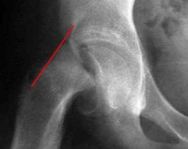
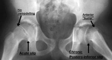
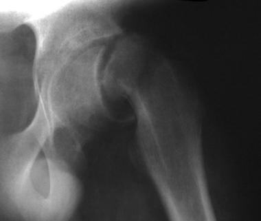
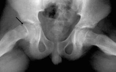
(+info)Slipped epiphyses refer to a medical condition where the growth plate (epiphysis) at the end of a bone slips away from the rest of the bone. This condition most commonly affects the hip joint in adolescents and is also known as slipped capital femoral epiphysis (SCFE).
The epiphysis is a layer of cartilage that is present at the ends of long bones in children and adolescents. It is responsible for the growth and development of the bone. In SCFE, the epiphysis on the upper end of the thighbone (femur) slips away from the shaft of the bone due to weakness or injury to the growth plate.
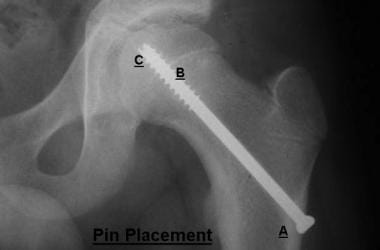
Slipped epiphyses can cause pain, stiffness, and limited mobility in the affected joint. If left untreated, it can lead to complications such as avascular necrosis (death of bone tissue due to lack of blood supply) and early arthritis. Treatment for slipped epiphyses typically involves surgery to realign and stabilize the growth plate with pins or screws.
Slipped Capital Femoral Epiphyses (SCFE) is a pediatric orthopedic condition that affects the growth plate (epiphysis) at the top of the thigh bone (femur). In SCFE, the epiphysis slips or shifts off the end of the femur, leading to abnormal hip function and potentially causing pain, stiffness, and limping. This condition typically occurs during periods of rapid growth, particularly in early adolescence, and is more common in overweight children. If left untreated, SCFE can result in significant long-term complications such as osteoarthritis or avascular necrosis (death of bone tissue due to lack of blood supply). Early diagnosis and appropriate medical intervention are crucial for optimal outcomes.
The epiphyses are the rounded ends of long bones in the body, which articulate with other bones to form joints. They are separated from the main shaft of the bone (diaphysis) by a growth plate called the physis or epiphyseal plate. The epiphyses are made up of spongy bone and covered with articular cartilage, which allows for smooth movement between bones. During growth, the epiphyseal plates produce new bone cells that cause the bone to lengthen until they eventually fuse during adulthood, at which point growth stops.
The femoral head is the rounded, ball-like top portion of the femur (thigh bone) that fits into the hip socket (acetabulum) to form the hip joint. It has a smooth, articular cartilage surface that allows for smooth and stable articulation with the pelvis. The femoral head is connected to the femoral neck, which is a narrower section of bone that angles downward and leads into the shaft of the femur. Together, the femoral head and neck provide stability and range of motion to the hip joint.
Femoral head necrosis, also known as avascular necrosis of the femoral head, is a medical condition that results from the interruption of blood flow to the femoral head, which is the rounded end of the thigh bone that fits into the hip joint. This lack of blood supply can cause the bone tissue to die, leading to the collapse of the femoral head and eventually resulting in hip joint damage or arthritis.
The condition can be caused by a variety of factors, including trauma, alcohol abuse, corticosteroid use, radiation therapy, and certain medical conditions such as sickle cell disease and lupus. Symptoms may include pain in the hip or groin, limited range of motion, and difficulty walking. Treatment options depend on the severity and progression of the necrosis and may include medication, physical therapy, or surgical intervention.
The hip joint, also known as the coxal joint, is a ball-and-socket type synovial joint that connects the femur (thigh bone) to the pelvis. The "ball" is the head of the femur, while the "socket" is the acetabulum, a concave surface on the pelvic bone.
The hip joint is surrounded by a strong fibrous capsule and is reinforced by several ligaments, including the iliofemoral, ischiofemoral, and pubofemoral ligaments. The joint allows for flexion, extension, abduction, adduction, medial and lateral rotation, and circumduction movements, making it one of the most mobile joints in the body.
The hip joint is also supported by various muscles, including the gluteus maximus, gluteus medius, gluteus minimus, iliopsoas, and other hip flexors and extensors. These muscles provide stability and strength to the joint, allowing for weight-bearing activities such as walking, running, and jumping.
Legg-Calve-Perthes disease is a childhood hip disorder that occurs when the blood supply to the ball part of the thigh bone (femoral head) is disrupted. This causes the bone tissue to die, leading to its collapse and deformity. The femoral head then regenerates itself, but often not as round and smooth as it should be, which can lead to hip problems in later life.
The disease is named after three doctors who independently described it: Arthur Legg, Jacques Calve, and Georg Perthes. It typically affects children between the ages of 4 and 10, more commonly boys than girls. Symptoms may include limping, pain in the hip or knee, reduced range of motion in the hip, and muscle wasting. Treatment often involves rest, physical therapy, and sometimes surgery to realign or reshape the femoral head.
The femur is the medical term for the thigh bone, which is the longest and strongest bone in the human body. It connects the hip bone to the knee joint and plays a crucial role in supporting the weight of the body and allowing movement during activities such as walking, running, and jumping. The femur is composed of a rounded head, a long shaft, and two condyles at the lower end that articulate with the tibia and patella to form the knee joint.
Femoroacetabular impingement (FAI) is a medical condition that affects the hip joint. It occurs when there is abnormal contact between the femoral head (the ball at the top of the thigh bone) and the acetabulum (the socket in the pelvis) during normal movement of the hip. This abnormal contact can cause damage to the cartilage and labrum (a ring of cartilage that helps to stabilize the hip joint) leading to pain, stiffness and decreased range of motion.
FAI is classified into two types: cam impingement and pincer impingement. Cam impingement occurs when there is an abnormal shape of the femoral head or neck, which leads to abnormal contact with the acetabulum during hip flexion and internal rotation. Pincer impingement occurs when there is overcoverage of the acetabulum, leading to abnormal contact with the femoral head or neck.
In some cases, both cam and pincer impingement can be present, which is referred to as mixed impingement. Symptoms of FAI may include hip pain, stiffness, limping, and reduced range of motion. Treatment options for FAI may include physical therapy, activity modification, medications, and in some cases, surgery.
Osteotomy is a surgical procedure in which a bone is cut to shorten, lengthen, or change its alignment. It is often performed to correct deformities or to realign bones that have been damaged by trauma or disease. The bone may be cut straight across (transverse osteotomy) or at an angle (oblique osteotomy). After the bone is cut, it can be realigned and held in place with pins, plates, or screws until it heals. This procedure is commonly performed on bones in the leg, such as the femur or tibia, but can also be done on other bones in the body.
The "femur neck" is the narrow, upper part of the femur (thigh bone) where it connects to the pelvis. It is the region through which the femoral head articulates with the acetabulum to form the hip joint. The femur neck is a common site for fractures, especially in older adults with osteoporosis.
Exostoses are benign (noncancerous) bone growths that develop on the surface of a bone, usually in response to repeated stress or friction. They are often small and smooth, but can become larger and more irregular over time. In some cases, they may cause pain or discomfort, especially if they continue to grow and put pressure on nearby nerves, muscles, or other bones.
Exostoses can occur in various parts of the body, but they are most commonly found in the long bones of the arms and legs, as well as in the small bones of the feet. They may also develop in response to chronic irritation or injury, such as from jogging or playing sports that involve a lot of running or jumping.
In some cases, exostoses may be surgically removed if they cause persistent pain or other symptoms. However, in many cases, they do not require treatment and can be left alone. If you are concerned about any bone growths or other unusual symptoms, it is always best to consult with a healthcare professional for an accurate diagnosis and treatment plan.
Age determination by skeleton, also known as skeletal aging or skeletal maturation, is the process of estimating a person's age based on the analysis of their skeletal remains. This technique is commonly used in forensic anthropology to help identify unknown individuals or determine the time since death.
The method involves examining various features of the skeleton, such as the degree of fusion of epiphyseal growth plates, the shape and size of certain bones, and the presence or absence of degenerative changes. These features change in a predictable way as a person grows and develops, allowing for an estimation of their age at death.
It is important to note that while skeletal aging can provide useful information, it is not always possible to determine an exact age. Instead, forensic anthropologists typically provide a range of ages that the individual may have fallen into based on the skeletal evidence. Additionally, factors such as genetics, nutrition, and health can affect the rate at which skeletal features develop, making it difficult to provide a precise estimate in some cases.
Bone screws are medical devices used in orthopedic and trauma surgery to affix bone fracture fragments or to attach bones to other bones or to metal implants such as plates, rods, or artificial joints. They are typically made of stainless steel or titanium alloys and have a threaded shaft that allows for purchase in the bone when tightened. The head of the screw may have a hexagonal or star-shaped design to allow for precise tightening with a screwdriver. Bone screws come in various shapes, sizes, and designs, including fully threaded, partially threaded, cannulated (hollow), and headless types, depending on their intended use and location in the body.
"Bone retroversion" is not a widely recognized medical term or concept with a specific definition. The term "retroversion" is used in anatomy to describe the position of an organ or structure when it is turned backward or inward. In relation to bones, retroversion typically describes the orientation of a bone or joint when it is angled or positioned in such a way that its posterior (back) aspect faces more anteriorly (toward the front).
However, I was unable to find a widely accepted medical definition for "bone retroversion" as a specific pathological or anatomical condition. It's possible that the term may be used in a more specialized context within certain medical subspecialties. If you have more context or information about where this term is being used, I may be able to provide a more precise answer.
I believe you are referring to "bone pins" or "bone nails" rather than "bone nails." These terms are used in the medical field to describe surgical implants made of metal or biocompatible materials that are used to stabilize and hold together fractured bones during the healing process. They can also be used in spinal fusion surgery to provide stability and promote bone growth between vertebrae.
Bone pins or nails typically have a threaded or smooth shaft, with a small diameter that allows them to be inserted into the medullary canal of long bones such as the femur or tibia. They may also have a head or eyelet on one end that allows for attachment to external fixation devices or other surgical instruments.
The use of bone pins and nails has revolutionized orthopedic surgery, allowing for faster healing times, improved stability, and better functional outcomes for patients with fractures or spinal deformities.
Articular Range of Motion (AROM) is a term used in physiotherapy and orthopedics to describe the amount of movement available in a joint, measured in degrees of a circle. It refers to the range through which synovial joints can actively move without causing pain or injury. AROM is assessed by measuring the degree of motion achieved by active muscle contraction, as opposed to passive range of motion (PROM), where the movement is generated by an external force.
Assessment of AROM is important in evaluating a patient's functional ability and progress, planning treatment interventions, and determining return to normal activities or sports participation. It is also used to identify any restrictions in joint mobility that may be due to injury, disease, or surgery, and to monitor the effectiveness of rehabilitation programs.
A hip dislocation is a medical emergency that occurs when the head of the femur (thighbone) slips out of its socket in the pelvis. This can happen due to high-energy trauma, such as a car accident or a severe fall. Hip dislocations can also occur in people with certain health conditions that make their hips more prone to displacement, such as developmental dysplasia of the hip.
There are two main types of hip dislocations: posterior and anterior. In a posterior dislocation, the femur head moves out of the back of the socket, which is the most common type. In an anterior dislocation, the femur head moves out of the front of the socket. Both types of hip dislocations can cause severe pain, swelling, and difficulty moving the affected leg.
Immediate medical attention is necessary for a hip dislocation to realign the bones and prevent further damage. Treatment typically involves sedation or anesthesia to relax the muscles around the joint, followed by a closed reduction procedure to gently guide the femur head back into the socket. In some cases, surgery may be required to repair any associated injuries, such as fractures or damaged ligaments. After treatment, physical therapy and rehabilitation are usually necessary to restore strength, mobility, and function to the affected hip joint.
HLA-DR5 is a type of human leukocyte antigen (HLA) Class II histocompatibility antigen. HLAs are proteins found on the surface of cells that help the immune system recognize and distinguish foreign substances from the body's own cells. The HLA-DR5 antigen is further divided into two subtypes, DR51 and DR52, which are encoded by different genes.
The HLA-DR5 antigen is commonly found in approximately 10-15% of the human population and has been associated with an increased risk of developing certain autoimmune diseases such as rheumatoid arthritis, type 1 diabetes, and multiple sclerosis. However, it's important to note that having the HLA-DR5 antigen does not guarantee that a person will develop one of these conditions, and many people with the antigen never develop any autoimmune diseases.
It's also worth mentioning that HLA typing is used in organ transplantation to match donors and recipients and reduce the risk of rejection. The HLA-DR5 antigen is one of several HLAs that may be considered during this process.
Orthopedic procedures are surgical or nonsurgical methods used to treat musculoskeletal conditions, including injuries, deformities, or diseases of the bones, joints, muscles, ligaments, and tendons. These procedures can range from simple splinting or casting to complex surgeries such as joint replacements, spinal fusions, or osteotomies (cutting and repositioning bones). The primary goal of orthopedic procedures is to restore function, reduce pain, and improve the quality of life for patients.
The endocrine system is a complex network of glands and organs that produce, store, and secrete hormones. It plays a crucial role in regulating various functions in the body, including metabolism, growth and development, tissue function, sexual function, reproduction, sleep, and mood.
Endocrine system diseases or disorders occur when there is a problem with the production or regulation of hormones. This can result from:
1. Overproduction or underproduction of hormones by the endocrine glands.
2. Impaired response of target cells to hormones.
3. Disruption in the feedback mechanisms that regulate hormone production.
Examples of endocrine system diseases include:
1. Diabetes Mellitus - a group of metabolic disorders characterized by high blood sugar levels due to insulin deficiency or resistance.
2. Hypothyroidism - underactive thyroid gland leading to slow metabolism, weight gain, fatigue, and depression.
3. Hyperthyroidism - overactive thyroid gland causing rapid heartbeat, anxiety, weight loss, and heat intolerance.
4. Cushing's Syndrome - excess cortisol production resulting in obesity, high blood pressure, and weak muscles.
5. Addison's Disease - insufficient adrenal hormone production leading to weakness, fatigue, and low blood pressure.
6. Acromegaly - overproduction of growth hormone after puberty causing enlargement of bones, organs, and soft tissues.
7. Gigantism - similar to acromegaly but occurs before puberty resulting in excessive height and body size.
8. Hypopituitarism - underactive pituitary gland leading to deficiencies in various hormones.
9. Hyperparathyroidism - overactivity of the parathyroid glands causing calcium imbalances and kidney stones.
10. Precocious Puberty - early onset of puberty due to premature activation of the pituitary gland.
Treatment for endocrine system diseases varies depending on the specific disorder and may involve medication, surgery, lifestyle changes, or a combination of these approaches.
The acetabulum is the cup-shaped cavity in the pelvic bone (specifically, the os coxa) where the head of the femur bone articulates to form the hip joint. It provides a stable and flexible connection between the lower limb and the trunk, allowing for a wide range of movements such as flexion, extension, abduction, adduction, rotation, and circumduction. The acetabulum is lined with articular cartilage, which facilitates smooth and frictionless movement of the hip joint. Its stability is further enhanced by various ligaments, muscles, and the labrum, a fibrocartilaginous rim that deepens the socket and increases its contact area with the femoral head.
Turner Syndrome is a genetic disorder that affects females, caused by complete or partial absence of one X chromosome. The typical karyotype is 45,X0 instead of the normal 46,XX in women. This condition leads to distinctive physical features and medical issues in growth, development, and fertility. Characteristic features include short stature, webbed neck, low-set ears, and swelling of the hands and feet. Other potential symptoms can include heart defects, hearing and vision problems, skeletal abnormalities, kidney issues, and learning disabilities. Not all individuals with Turner Syndrome will have every symptom, but most will require medical interventions and monitoring throughout their lives to address various health concerns associated with the condition.
Capital financing refers to the process of raising funds to provide capital for a business, organization, or project, particularly in the medical field. This can include obtaining loans, issuing stocks and bonds, seeking grants, or attracting private investments. The goal of capital financing is to secure sufficient financial resources to support long-term growth, expansion, or modernization efforts, as well as to ensure ongoing operations and sustainability. In healthcare, capital financing may be used for various purposes such as building new hospitals or clinics, purchasing medical equipment, conducting research and development, or implementing new technology systems.
Treatment outcome is a term used to describe the result or effect of medical treatment on a patient's health status. It can be measured in various ways, such as through symptoms improvement, disease remission, reduced disability, improved quality of life, or survival rates. The treatment outcome helps healthcare providers evaluate the effectiveness of a particular treatment plan and make informed decisions about future care. It is also used in clinical research to compare the efficacy of different treatments and improve patient care.
Orthopedic manipulation is a hands-on technique that is used by healthcare professionals, such as orthopedic doctors, chiropractors, and physical therapists, to diagnose and treat muscle and joint disorders. This manual procedure involves moving the joints or soft tissues in a specific direction and amplitude with the aim of improving joint mobility, reducing pain, relieving muscle tension, and enhancing overall function.
Orthopedic manipulation can be performed on various parts of the body, including the spine, extremities, and cranial structures. It is often used as a complementary treatment alongside other therapeutic interventions, such as exercise, medication, or surgery, to manage a wide range of musculoskeletal conditions, including but not limited to:
* Back pain and stiffness
* Neck pain and stiffness
* Joint pain and inflammation
* Muscle spasms and tension
* Headaches and migraines
* Disc disorders
* Sprains and strains
* Postural dysfunctions
It is important to note that orthopedic manipulation should only be performed by trained and licensed healthcare professionals, as improper techniques can lead to injury or further damage. Patients should consult with their healthcare provider to determine if orthopedic manipulation is an appropriate treatment option for their specific condition.
Retrospective studies, also known as retrospective research or looking back studies, are a type of observational study that examines data from the past to draw conclusions about possible causal relationships between risk factors and outcomes. In these studies, researchers analyze existing records, medical charts, or previously collected data to test a hypothesis or answer a specific research question.
Retrospective studies can be useful for generating hypotheses and identifying trends, but they have limitations compared to prospective studies, which follow participants forward in time from exposure to outcome. Retrospective studies are subject to biases such as recall bias, selection bias, and information bias, which can affect the validity of the results. Therefore, retrospective studies should be interpreted with caution and used primarily to generate hypotheses for further testing in prospective studies.
A craniopharyngioma is a type of brain tumor that develops near the pituitary gland, which is a small gland located at the base of the brain. These tumors arise from remnants of Rathke's pouch, an embryonic structure involved in the development of the pituitary gland.
Craniopharyngiomas are typically slow-growing and benign (non-cancerous), but they can still cause significant health problems due to their location. They can compress nearby structures such as the optic nerves, hypothalamus, and pituitary gland, leading to symptoms like vision loss, hormonal imbalances, and cognitive impairment.
Treatment for craniopharyngiomas usually involves surgical removal of the tumor, followed by radiation therapy in some cases. Regular follow-up with a healthcare team is essential to monitor for recurrence and manage any long-term effects of treatment.
Cartilage diseases refer to conditions that affect the cartilaginous tissues in the body. Cartilage is a firm, flexible connective tissue found in many areas of the body, including the joints, ribcage, ears, and nose. It provides structure and support, allows for smooth movement between bones, and protects the ends of bones from friction.
There are several types of cartilage diseases, including:
1. Osteoarthritis (OA): This is a degenerative joint disease that occurs when the protective cartilage that cushions the ends of your bones wears down over time. It can cause pain, stiffness, and loss of mobility in the affected joints.
2. Rheumatoid arthritis (RA): This is an autoimmune disorder that causes inflammation in the lining of the joints, leading to cartilage damage and bone erosion.
3. Traumatic arthritis: This occurs when a joint is injured, causing damage to the cartilage and resulting in pain, stiffness, and loss of mobility.
4. Infectious arthritis: This occurs when a joint becomes infected, leading to inflammation and potential damage to the cartilage.
5. Chondromalacia patellae: This is a condition that affects the cartilage on the back of the kneecap, causing pain and stiffness in the knee.
6. Costochondritis: This is an inflammation of the cartilage in the ribcage, causing chest pain and discomfort.
7. Nasal septal deviation: This is a condition where the cartilage that separates the nostrils is crooked or off-center, causing difficulty breathing through the nose.
8. Osteochondritis dissecans (OCD): This is a joint condition that occurs when a piece of cartilage and bone in a joint becomes detached, causing pain and stiffness.
9. Synovial chondromatosis: This is a rare condition where nodules made up of cartilage form in the lining of a joint, causing pain, swelling, and limited mobility.
Treatment for cartilage diseases varies depending on the specific condition and severity, but may include medication, physical therapy, surgery, or a combination of these.
Traction, in medical terms, refers to the application of a pulling force to distract or align parts of the body, particularly bones, joints, or muscles, with the aim of immobilizing, reducing displacement, or realigning them. This is often achieved through the use of various devices such as tongs, pulleys, weights, or specialized traction tables. Traction may be applied manually or mechanically and can be continuous or intermittent, depending on the specific medical condition being treated. Common indications for traction include fractures, dislocations, spinal cord injuries, and certain neurological conditions.
An encyclopedia is a comprehensive reference work containing articles on various topics, usually arranged in alphabetical order. In the context of medicine, a medical encyclopedia is a collection of articles that provide information about a wide range of medical topics, including diseases and conditions, treatments, tests, procedures, and anatomy and physiology. Medical encyclopedias may be published in print or electronic formats and are often used as a starting point for researching medical topics. They can provide reliable and accurate information on medical subjects, making them useful resources for healthcare professionals, students, and patients alike. Some well-known examples of medical encyclopedias include the Merck Manual and the Stedman's Medical Dictionary.
 Slipped capital femoral epiphysis
 Slipped capital femoral epiphysis: MedlinePlus Medical Encyclopedia
 Slipped Capital Femoral Epiphysis: Practice Essentials, Epidemiology, Functional Anatomy
 Slipped Capital Femoral Epiphysis (SCFE) (for Parents) - Nemours KidsHealth
 Slipped capital femoral epiphysis | HealthLink BC
 Slipped Capital Femoral Epiphysis in Children | University Hospitals
 Slipped Capital Femoral Epiphysis (SCFE) | Lurie Children's
 LearningRadiology - Slipped Capital Femoral Epiphysis, SCFE
 Valgus slipped capital femoral epiphysis : Presentation, treatment, and clinical outcomes using patient-reported measurements |...
 Slipped Capital Femoral Epiphysis (SCFE) - Pediatrics - MSD Manual Professional Edition
 Slipped Capital Femoral Epiphysis (SCFE)
 Slipped Capital Femoral Epiphysis | Stanford Health Care
 Slipped Capital Femoral Epiphysis | OrthoFixar 2023
 Slipped capital femoral epiphysis | Physio Check
 Slipped Capital Femoral Epiphysis | Concise Medical Knowledge
 Slipped Capital Femoral Epiphysis - Envision Medical Imaging
 slipped capital femoral epiphysis Archives | Dr Alison Grimaldi
 Slipped Capital Femoral Epiphysis | 5-Minute Pediatric Consult
 Slipped Capital Femoral Epiphysis Sugarland | Hip Disorder | SCFE Surgery Houston
 Slipped capital femoral epiphysis Archives - International Journal of Paediatric Orthopaedics
 "Slipped capital femoral epiphysis in a 25-year-old hypogonadic man with a large cranial chondroma: causality or coincidence? "...
 Slipped Capital Femoral Epiphysis following Internal Fixation for Fracture Neck of Femur. - Dr. Deepak Sharan
 Current diagnostic and therapeutic approach to patients with slipped capital femoral epiphysis - TOTBİD Dergisi