Soft Tissue Neoplasms
Neoplasms, Connective and Soft Tissue
Neoplasms, Bone Tissue
Neoplasms, Gonadal Tissue
Neoplasms, Adipose Tissue
Neoplasms, Nerve Tissue
Neoplasms, Connective Tissue
Neoplasms, Vascular Tissue
Neoplasms, Fibrous Tissue
Sarcoma
Neoplasms, Muscle Tissue
Soft Tissue Injuries
Soft Tissue Infections
Mouth Mucosa
Encyclopedias as Topic
Intestinal Mucosa
Epithelium
L-[1-11C]-tyrosine PET to evaluate response to hyperthermic isolated limb perfusion for locally advanced soft-tissue sarcoma and skin cancer. (1/1415)
PET with L-[1-11C]-tyrosine (TYR) was investigated in patients undergoing hyperthermic isolated limb perfusion (HILP) with recombinant tumor necrosis factor alpha (rTNF-alpha) and melphalan for locally advanced soft-tissue sarcoma and skin cancer of the lower limb. METHODS: Seventeen patients (5 women, 12 men; age range 24-75 y; mean age 52 y) were studied. TYR PET studies were performed before HILP and 2 and 8 wk afterwards. The protein synthesis rates (PSRs) in nanomoles per milliliter per minute were calculated. After final PET studies, tumors were resected and pathologically examined. Patients with pathologically complete responses (pCR) showed no viable tumors after treatment. Those with pathologically partial responses (pPR) showed various amounts of viable tumors in the resected tumor specimens. RESULTS: Six patients (35%) showed a pCR and 11 patients (65%) showed a pPR. All tumors were depicted as hot spots on PET studies before HILP. The PSR in the pCR group at 2 and 8 wk after perfusion had decreased significantly (P < 0.05) in comparison to the PSR before HILP. A significant difference was found in PSR between the pCR and pPR groups at 2 and at 8 wk (P < 0.05). Median PSR in nonviable tumor tissue was 0.62 and ranged from 0.22 to 0.91. With a threshold PSR of 0.91, sensitivity and specificity of TYR PET were 82% and 100%, respectively. The predictive value of a PSR > 0.91 for having viable tumor after HILP was 100%, whereas the predictive value of a PSR < or = 0.91 for having nonviable tumor tissue after HILP was 75%. The 2 patients in the pPR groups with a PSR < 0.91 showed microscopic islets of tumor cells surrounded by extensive necrosis on pathological examination. CONCLUSION: Based on the calculated PSR after HILP, TYR PET gave a good indication of the pathological outcome. Inflammatory tissue after treatment did not interfere with viable tumor on the images, suggesting that it may be worthwhile to pursue TYR PET in other therapy evaluation settings. (+info)FDG and L-[1-11C]-tyrosine imaging of soft-tissue tumors before and after therapy. (2/1415)
This study was undertaken to investigate the relationship of PET using fluorodeoxyglucose (FDG) or L-[1-11C]-tyrosine (TYR) with histopathologic findings in soft-tissue tumors, before and after therapy. Histopathologic parameters that were studied were tumor grade, mitotic rate, proliferation activity and amount of necrosis. METHODS: PET with either FDG or TYR was performed in 55 patients with a lesion suspected to be a malignant soft-tissue tumor. In 28 patients, a second PET study was performed after therapy. Metabolic rate of glucose consumption (MRglc) and protein synthesis rate (PSR) were calculated. Histologic parameters were obtained from a biopsy specimen that was taken just after the first PET study and from the tumor remnant that was resected after therapy. RESULTS: MRglc correlated with tumor grade (r = 0.71) and mitotic rate (r = 0.68) but not with proliferation or necrosis. After therapy, there was no longer a correlation with mitotic rate. PSR correlated with tumor grade (r = 0.53), mitotic rate (r = 0.73) and proliferation (r = 0.66). After therapy, correlation with mitosis and proliferation had improved, and a negative correlation was found between PSR and necrosis (r = -0.74). CONCLUSION: These results validate the use of both FDG and TYR to give an in vivo indication of histologic tumor parameters. However, FDG gives a better indication of tumor grade, whereas TYR is more accurate in predicting mitotic rate and proliferation, especially after therapy. FDG may therefore not be the most suited tracer for monitoring therapy. TYR might be more appropriate for that purpose. (+info)Subcutaneous sacrococcygeal myxopapillary ependymoma. (3/1415)
We report a case of myxopapillary ependymoma presenting as a primary tumor of the subcutaneous tissue in the sacrococcygeal region. The mass was large, well-encapsulated, lobulated, and multiseptated, with varying signal intensity on T1- and T2-weighted MR images caused by hemorrhagic necrosis, blood degradation products, and calcification. Only a small viable portion enhanced after administration of contrast material. Multiple lobules formed from fibrous septa and dystrophic calcification also characterize this tumor. (+info)Lymphangiosarcomas in cats: a retrospective study of 12 cases. (4/1415)
Clinical, macroscopic, and histologic features of 12 lymphangiosarcomas in cats are described. Nine tumors were located in the subcutaneous tissue at the caudoventral abdominal wall (eight cats) or in the neck (one cat). The remaining three cats had lymphangiosarcomas around the cranial mesenteric artery (two cats) or precardial in the mediastinum (one cat). Macroscopically, the tumors were noncircumscribed, white, edematous, and intermixed with fat tissue. Histologic features varied from cleft-forming and cavernous growth to papilliform and solid patterns. Follow-up data were available for seven cats with subcutaneous lymphangiosarcomas. All these cats died or were euthanatized within 6 months after surgery because of poor wound healing, local recurrence, or distant metastases. The cats with abdominal or thoracic masses were either euthanatized at surgery or within 6 months after the first surgery because of recurrent chylothorax, chyloperitoneum, or distant metastases. (+info)Clinical and radiological aspects of idiopathic diabetic muscle infarction. Rational approach to diagnosis and treatment. (5/1415)
The systemic effects of diabetes mellitus are well recognised. The heart, kidney, central and peripheral nervous systems, and the distal parts of the limbs are often the site of end-organ damage resulting from ischaemia. Infarction of large muscle groups in the limb, not associated with gangrene, is uncommon. There have been few reported cases other than radiological descriptions of diabetic muscle infarcts. While previous reports have illustrated some of the clinical and radiological characteristics of this condition, the paucity of published cases makes it difficult to determine the most appropriate methods of diagnosis and treatment. During a five-year period we treated 14 patients with diabetes mellitus, aged from 32 to 59 years, who were referred to a musculoskeletal oncology service for suspected soft-tissue sarcoma, but were subsequently found to have a diabetic muscle infarct. Closed needle biopsy was performed in 13 without complications. In 12 patients, the symptoms resolved without surgical treatment. (+info)Color Doppler sonography of focal lesions of the skin and subcutaneous tissue. (6/1415)
We evaluated with color Doppler sonography 71 visible and palpable nodules of the skin and subcutaneous tissue from 51 patients. The nodules were classified as avascular (type I), hypovascular with a single vascular pole (type II), hypervascular with multiple peripheral poles (type III), and hypervascular with internal vessels (type IV). Of the 32 malignant nodules, 9% showed a type I pattern, 50% had a type III pattern, and 41% had a type IV pattern; of the 39 benign nodules, 86% showed a type I pattern and 14% had a type II pattern. The sensitivity and specificity of hypervascularity in malignant lesions were 90% and 100%, respectively, whereas the sensitivity and specificity of hypovascularity in benign lesions were 100% and 90%, respectively. The authors conclude that color Doppler sonography is able to increase the specificity of ultrasonography in the evaluation of nodular lesions of the skin. (+info)Pulmonary metastases from soft tissue sarcoma: analysis of patterns of diseases and postmetastasis survival. (7/1415)
OBJECTIVE: To report the patterns of disease and postmetastasis survival for patients with pulmonary metastases from soft tissue sarcoma in a large group of patients treated at a single institution. Clinical factors that influence postmetastasis survival are analyzed. SUMMARY BACKGROUND DATA: For patients with soft tissue sarcoma, the lungs are the most common site of metastatic disease. Although pulmonary metastases most commonly arise from primary tumors in the extremities, they may arise from almost any primary site or histology. To date, resection of disease has been the only effective therapy for metastatic sarcoma. METHODS: From July 1982 to February 1997, 3149 adult patients with soft tissue sarcoma were admitted and treated at Memorial Sloan-Kettering Cancer Center. During this interval, 719 patients either developed or presented with lung metastases. Patients were treated with resection of metastatic disease whenever possible. Disease-specific survival was the endpoint of the study. Time to death was modeled using the method of Kaplan and Meier. The association of factors to time-to-event endpoints was analyzed using the log-rank test for univariate analysis and the Cox proportional hazards model for multivariate analysis. RESULTS: The overall median survival from diagnosis of pulmonary metastasis for all patients was 15 months. The 3-year actuarial survival rate was 25%. The ability to resect all metastatic disease completely was the most important prognostic factor for survival. Patients treated with complete resection had a median survival of 33 months and a 3-year actuarial survival rate of 46%. For patients treated with nonoperative therapy, the median survival was 11 months. A disease-free interval of more than 12 months before the development of metastases was also a favorable prognostic factor. Unfavorable factors included the histologic variants of liposarcoma and malignant peripheral nerve tumors and patient age older than 50 years at the time of treatment of metastasis. CONCLUSIONS: Resection of metastatic disease is the single most important factor that determines outcome in these patients. Long-term survival is possible in selected patients, particularly when recurrent pulmonary disease is resected. Surgical excision should remain the treatment of choice for metastases of soft tissue sarcoma to the lung. (+info)The enigma of desmoid tumors. (8/1415)
OBJECTIVE: To analyze patients with recurrent extremity desmoids, in whom the surgical therapeutic option was either major amputation or observation. SUMMARY BACKGROUND DATA: The biology and natural history of desmoid tumors are an enigma. These tumors invade surrounding structures and recur locally but do not metastasize. The morbidity of treating these tumors in the context of their relatively benign biology is uncertain. METHODS: Between July 1982 and June 1998, the authors treated and prospectively followed 206 patients with extremity desmoid tumors. All patients underwent standardized surgical resection, the surgical goal always being complete resection with negative margins. When tumors recurred, they were evaluated for reresection. Amputation was considered when resection was not possible because of neurovascular or major bone involvement, or in the presence of a functionless, painful extremity. RESULTS: During this period, 22 patients had disease that was not resectable without amputation. This was out of a total of 115 patients with primary disease and 91 patients with recurrent disease. All recurrences were local; in no patient did metastasis develop. In this group of 22 patients with unresectable disease, 7 underwent amputation and 15 did not. These 15 patients were followed, alive with disease, having no surgical resection. Four patients received systemic treatment with tamoxifen and nonsteroidal antiinflammatories, three received systemic cytotoxic chemotherapy, and two received both tamoxifen and chemotherapy. Six patients received no systemic treatment. The range of follow-up was 25 to 92 months. In all patients, there was no or insignificant tumor progression; in three patients who underwent observation alone, there was some regression of tumor. During follow-up, no patient has required subsequent amputation, and no patient has died from disease. CONCLUSIONS: In desmoid tumors, aggressive attempts at achieving negative resection margins may result in unnecessary morbidity. Function- and structure-preserving procedures should be the primary goal. In select patients, whose only option is amputation, it may be prudent to observe them with their limb and tumor intact. (+info)Soft tissue neoplasms refer to abnormal growths or tumors that develop in the soft tissues of the body. Soft tissues include muscles, tendons, ligaments, fascia, nerves, blood vessels, fat, and synovial membranes (the thin layer of cells that line joints and tendons). Neoplasms can be benign (non-cancerous) or malignant (cancerous), and their behavior and potential for spread depend on the specific type of neoplasm.
Benign soft tissue neoplasms are typically slow-growing, well-circumscribed, and rarely spread to other parts of the body. They can often be removed surgically with a low risk of recurrence. Examples of benign soft tissue neoplasms include lipomas (fat tumors), schwannomas (nerve sheath tumors), and hemangiomas (blood vessel tumors).
Malignant soft tissue neoplasms, on the other hand, can grow rapidly, invade surrounding tissues, and may metastasize (spread) to distant parts of the body. They are often more difficult to treat than benign neoplasms and require a multidisciplinary approach, including surgery, radiation therapy, and chemotherapy. Examples of malignant soft tissue neoplasms include sarcomas, such as rhabdomyosarcoma (arising from skeletal muscle), leiomyosarcoma (arising from smooth muscle), and angiosarcoma (arising from blood vessels).
It is important to note that soft tissue neoplasms can occur in any part of the body, and their diagnosis and treatment require a thorough evaluation by a healthcare professional with expertise in this area.
Neoplasms of connective and soft tissue are abnormal growths or tumors that develop in the body's supportive tissues, such as cartilage, tendons, ligaments, fascia, and fat. These neoplasms can be benign (non-cancerous) or malignant (cancerous).
Benign connective and soft tissue neoplasms include:
- Lipomas: slow-growing, fatty tumors that develop under the skin.
- Fibromas: firm, benign tumors that develop in connective tissue such as tendons or ligaments.
- Nevi (plural of nevus): benign growths made up of cells called melanocytes, which produce pigment.
Malignant connective and soft tissue neoplasms include:
- Sarcomas: a type of cancer that develops in the body's supportive tissues such as muscle, bone, fat, cartilage, or blood vessels. There are many different types of sarcomas, including liposarcoma (fatty tissue), rhabdomyosarcoma (muscle), and osteosarcoma (bone).
- Desmoid tumors: a rare type of benign tumor that can become aggressive and invade surrounding tissues. While not considered cancerous, desmoid tumors can cause significant morbidity due to their tendency to grow and infiltrate nearby structures.
Connective and soft tissue neoplasms can present with various symptoms depending on their location and size. Treatment options include surgery, radiation therapy, chemotherapy, or a combination of these modalities. Regular follow-up care is essential to monitor for recurrence or metastasis (spread) of the tumor.
Neoplasms of bone tissue refer to abnormal and excessive growths or tumors that develop in the bone. These growths can be benign (non-cancerous) or malignant (cancerous). Benign neoplasms, such as osteochondromas and enchondromas, are slow-growing and rarely spread to other parts of the body. However, they may cause problems if they grow too large and compress surrounding tissues. Malignant neoplasms, on the other hand, can invade and destroy nearby bone tissue and may metastasize (spread) to other organs in the body. Examples of malignant bone tumors include osteosarcoma, chondrosarcoma, and Ewing sarcoma. Treatment for bone neoplasms depends on several factors, including the type, size, location, and stage of the tumor, as well as the patient's age and overall health.
A neoplasm of gonadal tissue refers to an abnormal growth or tumor that develops in the reproductive organs, specifically the ovaries in women and the testes in men. These tumors can be benign (non-cancerous) or malignant (cancerous), and their growth can interfere with the normal function of the gonads.
Gonadal tissue neoplasms can have various causes, including genetic mutations, environmental factors, and hormonal imbalances. The symptoms of these tumors may vary depending on their size, location, and type, but they can include pelvic pain, bloating, abnormal menstruation, or a palpable mass in the affected area.
It is essential to diagnose and treat gonadal tissue neoplasms as early as possible to prevent complications such as infertility, metastasis, or death. Diagnostic procedures may include imaging tests, blood tests, and biopsies, while treatment options may include surgery, radiation therapy, chemotherapy, or hormone therapy.
Neoplasms in adipose tissue refer to abnormal and excessive growths of cells that form tumors within the fatty connective tissue. These neoplasms can be benign or malignant (cancerous). Benign neoplasms, such as lipomas, are slow-growing and typically do not spread to other parts of the body. Malignant neoplasms, on the other hand, are cancerous and can invade surrounding tissues and spread to distant sites in the body (metastasis). An example of a malignant neoplasm in adipose tissue is liposarcoma. It's important to note that while some neoplasms may not cause any symptoms, others can cause pain, swelling or other uncomfortable sensations, and therefore should be evaluated by a medical professional for proper diagnosis and treatment.
Neoplasms of nerve tissue are abnormal growths or tumors that originate in the nervous system, including the brain, spinal cord, and peripheral nerves. These neoplasms can be benign or malignant (cancerous) and can cause a variety of symptoms depending on their location and size.
Benign nerve tissue neoplasms are typically slow-growing and do not spread to other parts of the body. Examples include schwannomas, neurofibromas, and meningiomas. These tumors arise from the supporting cells of the nervous system, such as Schwann cells, which produce the myelin sheath that insulates nerve fibers.
Malignant nerve tissue neoplasms, on the other hand, are cancerous and can invade nearby tissues and spread to other parts of the body. These tumors are less common than benign neoplasms and can be difficult to treat. Examples include glioblastoma multiforme, a highly aggressive brain cancer, and malignant peripheral nerve sheath tumors, which arise from the cells that surround peripheral nerves.
Symptoms of nerve tissue neoplasms can vary widely depending on their location and size. Some common symptoms include headaches, seizures, weakness or numbness in the limbs, difficulty with coordination or balance, and changes in vision, hearing, or speech. Treatment options for nerve tissue neoplasms may include surgery, radiation therapy, chemotherapy, or a combination of these approaches.
Neoplasms of connective tissue are abnormal growths or tumors that develop from the cells that form the body's supportive framework, including bones, cartilage, tendons, ligaments, and other connective tissues. These neoplasms can be benign (non-cancerous) or malignant (cancerous), and they can cause various symptoms depending on their location and size.
There are several types of connective tissue neoplasms, including:
1. Fibroma: A benign tumor that arises from fibrous connective tissue.
2. Fibrosarcoma: A malignant tumor that develops from fibrous connective tissue.
3. Lipoma: A benign tumor that arises from fat cells.
4. Liposarcoma: A malignant tumor that develops from fat cells.
5. Chondroma: A benign tumor that arises from cartilage.
6. Chondrosarcoma: A malignant tumor that develops from cartilage.
7. Osteoma: A benign tumor that arises from bone.
8. Osteosarcoma: A malignant tumor that develops from bone.
9. Giant cell tumors: Benign or malignant tumors that contain many giant cells, which are large, multinucleated cells.
10. Synovial sarcoma: A malignant tumor that arises from the synovial tissue that lines joints and tendons.
Connective tissue neoplasms can cause various symptoms depending on their location and size. For example, a benign lipoma may cause a painless lump under the skin, while a malignant osteosarcoma may cause bone pain, swelling, and fractures. Treatment options for connective tissue neoplasms include surgery, radiation therapy, chemotherapy, or a combination of these approaches.
A neoplasm of vascular tissue is an abnormal growth or mass of cells in the blood vessels or lymphatic vessels. These growths can be benign (non-cancerous) or malignant (cancerous). Benign neoplasms, such as hemangiomas and lymphangiomas, are typically not harmful and may not require treatment. However, they can cause symptoms if they grow large enough to press on nearby organs or tissues. Malignant neoplasms, such as angiosarcomas, are cancerous and can invade and destroy surrounding tissue, as well as spread (metastasize) to other parts of the body. Treatment for vascular tissue neoplasms depends on the type, size, location, and stage of the growth, and may include surgery, radiation therapy, chemotherapy, or a combination of these.
Neoplasms of fibrous tissue are abnormal growths or tumors that originate from fibroblasts, the cells responsible for producing connective tissue in the body. These neoplasms can be benign or malignant (cancerous). Benign fibrous neoplasms include fibromas and fibrohistiocytic tumors, while malignant fibrous neoplasms are called fibrosarcomas. Fibrosarcomas are aggressive tumors that invade surrounding tissues and can metastasize (spread) to other parts of the body.
Fibrous tissue neoplasms can occur in any part of the body, but they are most commonly found in the soft tissues such as muscles, tendons, and ligaments. They can also develop in bones, where they are called osteosarcomas. Symptoms of fibrous tissue neoplasms depend on their size and location, but may include a painless mass or swelling, limited mobility, or pain if the tumor is pressing on nerves or blood vessels.
Diagnosis of fibrous tissue neoplasms typically involves imaging tests such as X-rays, CT scans, or MRI scans, followed by a biopsy to confirm the type and grade of the tumor. Treatment options may include surgery, radiation therapy, chemotherapy, or a combination of these approaches. Regular follow-up care is important to monitor for recurrence or metastasis.
Sarcoma is a type of cancer that develops from certain types of connective tissue (such as muscle, fat, fibrous tissue, blood vessels, or nerves) found throughout the body. It can occur in any part of the body, but it most commonly occurs in the arms, legs, chest, and abdomen.
Sarcomas are classified into two main groups: bone sarcomas and soft tissue sarcomas. Bone sarcomas develop in the bones, while soft tissue sarcomas develop in the soft tissues of the body, such as muscles, tendons, ligaments, fat, blood vessels, and nerves.
Sarcomas can be further classified into many subtypes based on their specific characteristics, such as the type of tissue they originate from, their genetic makeup, and their appearance under a microscope. The different subtypes of sarcoma have varying symptoms, prognoses, and treatment options.
Overall, sarcomas are relatively rare cancers, accounting for less than 1% of all cancer diagnoses in the United States each year. However, they can be aggressive and may require intensive treatment, such as surgery, radiation therapy, and chemotherapy.
Neoplasms in muscle tissue refer to abnormal and excessive growths of muscle cells that can be benign or malignant. These growths can arise from any of the three types of muscle tissue: skeletal, cardiac, or smooth muscle. Neoplasms in muscle tissue are classified based on their origin, behavior, and histological features.
Benign neoplasms in muscle tissue include leiomyomas (smooth muscle), rhabdomyomas (skeletal muscle), and myxomas (cardiac muscle). These tumors are usually slow-growing and do not invade surrounding tissues or spread to other parts of the body.
Malignant neoplasms in muscle tissue, also known as sarcomas, include leiomyosarcoma (smooth muscle), rhabdomyosarcoma (skeletal muscle), and angiosarcoma (cardiac muscle). These tumors are aggressive, invasive, and have the potential to metastasize to other parts of the body.
Symptoms of neoplasms in muscle tissue depend on their location, size, and type. They may include a painless or painful mass, weakness, fatigue, weight loss, and difficulty swallowing or breathing. Treatment options for neoplasms in muscle tissue include surgery, radiation therapy, chemotherapy, and targeted therapy. The choice of treatment depends on the type, stage, location, and patient's overall health condition.
Soft tissue injuries refer to damages that occur in the body's connective tissues, such as ligaments, tendons, and muscles. These injuries can be caused by various events, including accidents, falls, or sports-related impacts. Common soft tissue injuries include sprains, strains, and contusions (bruises).
Sprains occur when the ligaments, which connect bones to each other, are stretched or torn. This usually happens in the joints like ankles, knees, or wrists. Strains, on the other hand, involve injuries to the muscles or tendons, often resulting from overuse or sudden excessive force. Contusions occur when blood vessels within the soft tissues get damaged due to a direct blow or impact, causing bleeding and subsequent bruising in the affected area.
Soft tissue injuries can cause pain, swelling, stiffness, and limited mobility. In some cases, these injuries may require medical treatment, including physical therapy, medication, or even surgery, depending on their severity and location. It is essential to seek proper medical attention for soft tissue injuries to ensure appropriate healing and prevent long-term complications or chronic pain.
Soft tissue infections are medical conditions that involve infection of the soft tissues of the body, which include the skin, muscles, fascia (the connective tissue that surrounds muscles), and tendons. These infections can be caused by various types of bacteria, viruses, fungi, or parasites.
Soft tissue infections can range from mild to severe, depending on the type of organism causing the infection, the extent of tissue involvement, and the patient's overall health status. Some common types of soft tissue infections include:
1. Cellulitis: This is a bacterial infection that affects the skin and underlying tissues. It typically presents as a red, swollen, warm, and painful area on the skin, often accompanied by fever and chills.
2. Abscess: An abscess is a localized collection of pus in the soft tissues, caused by an infection. It can appear as a swollen, tender, and warm lump under the skin, which may be filled with pus.
3. Necrotizing fasciitis: This is a rare but severe soft tissue infection that involves the rapid destruction of fascia and surrounding tissues. It is often caused by a mixture of bacteria and can progress rapidly, leading to shock, organ failure, and even death if not treated promptly.
4. Myositis: This is an inflammation of the muscle tissue, which can be caused by a bacterial or viral infection. Symptoms may include muscle pain, swelling, weakness, and fever.
5. Erysipelas: This is a superficial skin infection that affects the upper layers of the skin and the lymphatic vessels. It typically presents as a raised, red, and painful rash with clear borders.
Treatment for soft tissue infections depends on the type and severity of the infection but may include antibiotics, drainage of pus or abscesses, and surgery in severe cases. Preventive measures such as good hygiene, wound care, and prompt treatment of injuries can help reduce the risk of developing soft tissue infections.
The mouth mucosa refers to the mucous membrane that lines the inside of the mouth, also known as the oral mucosa. It covers the tongue, gums, inner cheeks, palate, and floor of the mouth. This moist tissue is made up of epithelial cells, connective tissue, blood vessels, and nerve endings. Its functions include protecting the underlying tissues from physical trauma, chemical irritation, and microbial infections; aiding in food digestion by producing enzymes; and providing sensory information about taste, temperature, and texture.
The hard palate is the anterior, bony part of the roof of the mouth, forming a vertical partition between the oral and nasal cavities. It is composed of the maxilla and palatine bones, and provides attachment for the muscles of the soft palate, which functions in swallowing, speaking, and breathing. The hard palate also contains taste buds that contribute to our ability to taste food.
An encyclopedia is a comprehensive reference work containing articles on various topics, usually arranged in alphabetical order. In the context of medicine, a medical encyclopedia is a collection of articles that provide information about a wide range of medical topics, including diseases and conditions, treatments, tests, procedures, and anatomy and physiology. Medical encyclopedias may be published in print or electronic formats and are often used as a starting point for researching medical topics. They can provide reliable and accurate information on medical subjects, making them useful resources for healthcare professionals, students, and patients alike. Some well-known examples of medical encyclopedias include the Merck Manual and the Stedman's Medical Dictionary.
A "cheek" is the fleshy, muscular area of the face that forms the side of the face below the eye and above the jaw. It contains the buccinator muscle, which helps with chewing by moving food to the back teeth for grinding and also assists in speaking and forming facial expressions. The cheek also contains several sensory receptors that allow us to perceive touch, temperature, and pain in this area of the face. Additionally, there is a mucous membrane lining inside the mouth cavity called the buccal mucosa which covers the inner surface of the cheek.
The intestinal mucosa is the innermost layer of the intestines, which comes into direct contact with digested food and microbes. It is a specialized epithelial tissue that plays crucial roles in nutrient absorption, barrier function, and immune defense. The intestinal mucosa is composed of several cell types, including absorptive enterocytes, mucus-secreting goblet cells, hormone-producing enteroendocrine cells, and immune cells such as lymphocytes and macrophages.
The surface of the intestinal mucosa is covered by a single layer of epithelial cells, which are joined together by tight junctions to form a protective barrier against harmful substances and microorganisms. This barrier also allows for the selective absorption of nutrients into the bloodstream. The intestinal mucosa also contains numerous lymphoid follicles, known as Peyer's patches, which are involved in immune surveillance and defense against pathogens.
In addition to its role in absorption and immunity, the intestinal mucosa is also capable of producing hormones that regulate digestion and metabolism. Dysfunction of the intestinal mucosa can lead to various gastrointestinal disorders, such as inflammatory bowel disease, celiac disease, and food allergies.
Epithelium is the tissue that covers the outer surface of the body, lines the internal cavities and organs, and forms various glands. It is composed of one or more layers of tightly packed cells that have a uniform shape and size, and rest on a basement membrane. Epithelial tissues are avascular, meaning they do not contain blood vessels, and are supplied with nutrients by diffusion from the underlying connective tissue.
Epithelial cells perform a variety of functions, including protection, secretion, absorption, excretion, and sensation. They can be classified based on their shape and the number of cell layers they contain. The main types of epithelium are:
1. Squamous epithelium: composed of flat, scalelike cells that fit together like tiles on a roof. It forms the lining of blood vessels, air sacs in the lungs, and the outermost layer of the skin.
2. Cuboidal epithelium: composed of cube-shaped cells with equal height and width. It is found in glands, tubules, and ducts.
3. Columnar epithelium: composed of tall, rectangular cells that are taller than they are wide. It lines the respiratory, digestive, and reproductive tracts.
4. Pseudostratified epithelium: appears stratified or layered but is actually made up of a single layer of cells that vary in height. The nuclei of these cells appear at different levels, giving the tissue a stratified appearance. It lines the respiratory and reproductive tracts.
5. Transitional epithelium: composed of several layers of cells that can stretch and change shape to accommodate changes in volume. It is found in the urinary bladder and ureters.
Epithelial tissue provides a barrier between the internal and external environments, protecting the body from physical, chemical, and biological damage. It also plays a crucial role in maintaining homeostasis by regulating the exchange of substances between the body and its environment.
Gingivoplasty is a surgical procedure in dentistry that involves the reshaping or contouring of the gingiva (gums). This procedure is typically performed for aesthetic purposes, to improve the appearance of gums that are uneven or have an irregular shape. It can also be done to remove excess gum tissue that may be covering too much of a tooth, making it appear shorter than the other teeth.
Gingivoplasty is often recommended as a part of periodontal treatment to ensure the proper fit and function of dental restorations or to manage and prevent gum disease. The procedure involves removing and reshaping the gingival tissue to create a more aesthetically pleasing and healthy gum line.
It's important to note that while gingivoplasty can improve the appearance of the gums, it does not address any underlying issues related to gum disease or bone loss. Additional periodontal treatments may be necessary to address these concerns.
 Oral mucosa
Myoglobin
Li-Fraumeni syndrome
List of cutaneous neoplasms associated with systemic syndromes
Low-grade myofibroblastic sarcoma
Giant-cell fibroblastoma
PDGFB
Fibroblastic and myofibroblastic tumors
Familial adenomatous polyposis
Gardner fibroma
Joseph F. Fraumeni Jr.
Gardner's syndrome
Spindle cell rhabdomyosarcoma
Liposarcoma
Mixed Müllerian tumor
Extraskeletal myxoid chondrosarcoma
Nuchal fibroma
Proliferative fasciitis and proliferative myositis
Embryonal rhabdomyosarcoma
Diffuse infantile fibromatosis
Fibrous hamartoma of infancy
Low-grade fibromyxoid sarcoma
Familial myxovascular fibromas
Neural fibrolipoma
Sclerosing epithelioid fibrosarcoma
Aggressive infantile fibromatosis
Connective tissue neoplasm
Adenomatoid tumor
Granular cell tumor
Cellular angiofibroma
Soft tissue neoplasms - confocalpedia
 Soft Tissue Neoplasms Archives | MSD Oncology Clinical Trials
Oral mucosa - Wikipedia
 Neural differentiation in small round cell tumors of bone and soft tissue with the translocation t(11;22)(q24;q12): an...
 Oral spindle cell lipomas
 Liver and Other Neoplasms - Treatment Approaches - Medical Clinical Policy Bulletins | Aetna
 Pediatric Breast Disorders: Background, Embryology and Breast Development, Congenital Breast Anomalies
Leiomyoma: Practice Essentials, Background, Pathophysiology
 Perivascular Epithelioid Cell Neoplasms
Summary Report | CureHunter
Perivascular Epithelioid Cell Neoplasms
Summary Report | CureHunter
Gardner's syndrome - Wikipedia
 Department Directory | Department of Pathology and Laboratory Medicine
 Primary Sphenoidal Sinus Lymphoma with Initial Presentation as Unilateral Abducens Nerve Palsy Symptom
 Advanced Search Results - Public Health Image Library(PHIL)
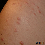
 Lichen Planus 03 | Clinical Images/Slides | Multimedia Library | Practing DPMs | APMA
 Tony NG | University of British Columbia, Vancouver | UBC | Department of Pathology and Laboratory Medicine | Research profile
Sarcoma
- Soft Tissue Sarcoma
Summary Report | CureHunter
 Fibrolipoma of the lip treated by diode laser surgery: A case report | Journal of Medical Case Reports
Absence of germline p16 INK4a alterations in p53 wild type Li-Fraumeni syndrome families | Journal of Medical Genetics
 Safety and Efficacy Evaluation of 4th Generation Safety-engineered CAR T Cells Targeting Sarcomas - Full Text View -...
GIST: Assessment of Tumor Mutations and TKI Plasma Exposure - Full Text View - ClinicalTrials.gov
Low-Grade Malignant Triton Tumor of the Neck: A Case Report and Review of the Literature
 Targeted Therapy Directed by Genetic Testing in Treating Pediatric Patients With Relapsed or Refractory Advanced Solid Tumors,...
 Pediatric Granular Cell Tumor of the Breast: An uncommon neoplasm in an uncommon site and age group
| Autopsy and Case...
Pediatric Granular Cell Tumor of the Breast: An uncommon neoplasm in an uncommon site and age group
| Autopsy and Case...
Carfilzomib (Kyprolis) [Medicare] - Medical Clinical Policy Bulletins | Aetna
 JETO 11.2, p. 147-153 - Old City Publishing
 Naert T et al. (2021),
CRISPR-SID: Identifying EZH2 as a druggable tar... -
Paper
Naert T et al. (2021),
CRISPR-SID: Identifying EZH2 as a druggable tar... -
Paper
 Gastric Schwannoma - A Rare Cause of Dyspepsia - SAGES Abstract Archives
Osteochondromatosis | Profiles RNS
ENDOU | Cancer Genetics Web
Tumors13
- Clinical, cytogenetic, histopathologic, and immunohistochemical data were obtained in a series of 11 small round cell tumors (SRCT) of bone and soft tissue with the translocation t(11;22) (q24;q12). (nih.gov)
- World Health Organization Classification of Tumors: Pathology and Genetics of Tumors of Soft Tissue and Bone, 2002). (curehunter.com)
- Desmoid tumors are fibrous tumors that usually occur in the tissue covering the intestines and may be provoked by surgery to remove the colon. (wikipedia.org)
- Desmoid tumors are soft- tissue neoplasms strictly driven by Wnt signaling network hyperactivation. (xenbase.org)
- Gastric schwannoma represent only 0.2% of all gastric tumors and 4% of all benign gastric neoplasms. (sages.org)
- Desmoid tumors are cytologically bland fibrous neoplasms originating from the musculoaponeurotic structures throughout the body. (medscape.com)
- Desmoid tumors often appear as infiltrative, usually well-differentiated, firm overgrowths of fibrous tissue, and they are locally aggressive. (medscape.com)
- Benign musculoskeletal neoplasms are one hundred times more common than malignant soft tissue tumors. (drrathresearch.org)
- Bone and soft tissue tumors (BSTT) constitute a heterogeneous group of neoplasms of mesenchymal and neuroectodermal origin. (avhandlingar.se)
- The goal of this project is to evaluate the safety and preliminary efficacy of ExAblate magnetic resonance-guided high-intensity focused ultrasound (MRgHIFU) surgery in the treatment of soft tissue tumors of the extremities. (stanford.edu)
- Desmoid tumors are associated with a biallelic APC mutation in the affected tissue. (medscape.com)
- Soft tissue tumors (STT) constitute a heterogeneous group of neoplasms that clinically run the gamut from totally benign to highly malignant neoplasms. (lu.se)
- Soft tissue tumors (STT) constitute a heterogeneous group of approximately 100 distinct neoplasms, including more than 50 malignant subtypes, so-called sarcomas. (lu.se)
Connective and other soft tissue1
- Elevated SMRs were noted for cancers of the stomach, rectum, brain and other central nervous system sites, connective and other soft tissue, as well as for unspecified neoplasms of the nervous system and unspecified anemias. (cdc.gov)
Mesenchymal4
- The second explanation was more plausible, where neoplastic Schwann cells can transform into rhabdomyoblasts, suggesting the possibility of some mesenchymal tissue derivation from neuroectodermal cells. (hindawi.com)
- Gastrointestinal schwannomas are rare benign neoplasms that are distinctively unique when compared to soft-tissue and central nervous system mesenchymal neoplasms. (sages.org)
- Cardiac sarcoma is a rare mesenchymal neoplasm or tumor of the heart that is malignant in nature. (logicalimages.com)
- Rhabdosarcoma , the most common soft tissue sarcoma is mesenchymal in origin, and affects infants and children up to five years (mostly males). (drrathresearch.org)
Tumor9
- Rhabdomyoblastic differentiation in a malignant peripheral nerve sheath tumor (MPNST) is termed malignant triton tumor (MTT), a rare neoplasm that poses a diagnostic dilemma in the differential diagnosis of neck masses and portends poor prognosis. (hindawi.com)
- Malignant peripheral nerve sheath tumor (MPNST) is a rare soft tissue neoplasm with a poor prognosis. (hindawi.com)
- Granular cell tumor (GCT) is a rare soft tissue neoplasm of Schwann cell origin. (usp.br)
- GCT is usually a slow-growing, painless tumor involving the skin and soft tissues that is mostly located in the head and neck region, especially the tongue. (usp.br)
- Epstein-Barr virus-associated smooth muscle tumor is an uncommon neoplasm associated with immunodeficiency. (allenpress.com)
- Epstein-Barr virus (EBV)-associated smooth muscle tumor (SMT) is an uncommon neoplasm typically manifesting in immunodeficient individuals. (allenpress.com)
- Extraskeletal osteosarcoma (EOS) is a malignant tumor of soft tissue origin comprising tumor cells that produce osteoid matrix. (researchsquare.com)
- It ranges from a well-differentiated tumor with EPITHELIAL CELLS indistinguishable from normal HEPATOCYTES to a poorly differentiated neoplasm. (lookformedical.com)
- Papillary intralymphatic angioendothelioma (Dabska tumor) is a rarely metastasizing lymphatic vascular neoplasm that usually affects children and young adults. (anaisdedermatologia.org.br)
Benign neoplasms1
- Malignant neoplasms show a greater degree of anaplasia and have the properties of invasion and metastasis, compared to benign neoplasms. (lookformedical.com)
Pathology2
- 2002) Pathology and genetics of tumours of soft tissue and bone. (scirp.org)
- METHODS: Fifty-three paraffin-embedded tissue blocks of colorectal resections and corresponding patient information were retrieved from the archives of the Anatomic and Molecular Pathology Department of Lagos University Teaching Hospital.A 4-micron slide section was obtained from each specimen and immunohistochemistry for COX-2 and HER-2 expression was performed. (bvsalud.org)
Uncommon neoplasm1
- This paper reviews the clinicopathologic features of this uncommon neoplasm with detailed discussion of the role of Epstein-Barr virus in the pathogenesis. (allenpress.com)
Neuroendocrine neoplasms2
- Pulmonary carcinoids are well differentiated low to intermediate grade lung neuroendocrine tumours (LNETs), that belong to the group of lung neuroendocrine neoplasms which also include highly aggressive lung neuroendocrine carcinomas (LNECs). (who.int)
- These data have been combined with previously published LNET data to perform integrative analysis using multi-omics factor analysis (MOFA), resulting in a molecular map of lung neuroendocrine neoplasms for exploration. (who.int)
Immunohistochemistry1
- PurposePrevious studies indicate that breast cancer molecular subtypes differ with respect to their dependency on autophagy, but our knowledge of the differential expression and prognostic significance of autophagy-related biomarkers in breast cancer is limited.Methods Immunohistochemistry (IHC) was performed on tissue microarrays from a large popu. (researchgate.net)
Metastasis1
- Synovial sarcoma , a soft tissue cancer that most often occurs around leg or arm joints, has a 50% rate of metastasis. (drrathresearch.org)
Diagnosis5
- Tissue examination is necessary to establish the diagnosis. (medscape.com)
- These neoplasms are mainly treated surgically and an accurate histological examination is mandatory for a precise diagnosis. (springer.com)
- With a provisional clinical diagnosis of benign neoplasm, the lesion was surgically excised under local anaesthesia, using a diode laser with a 300 μm fibre and operating at 2,5 W. Direct suture of the surgical margins was unnecessary as no bleeding was observed during and following the excision (Fig. 2 ). (springer.com)
- On histopathological analysis, all the classic features were noted and diagnosis of a spindle cell neoplasm was made without any obscurity. (oldcitypublishing.com)
- This neoplasm should be considered in the differential diagnosis of vascular dermatoses, allowing early diagnosis and treatment. (anaisdedermatologia.org.br)
Spindle cell1
- Further, microscopic examination revealed a spindle cell malignant neoplasm with osteoid matrix. (researchsquare.com)
Sarcomas4
- E ditor -The Li-Fraumeni syndrome (LFS) is a rare familial cancer syndrome that predisposes gene carriers to the development of diverse early onset malignancies, including soft tissue sarcomas, osteosarcomas, adrenocortical carcinomas, brain tumours, breast carcinomas, and leukaemia, 1-3 with other cancer types occurring less frequently. (bmj.com)
- It can occur in association with neurofibromatosis type 1 (NF-1) or sporadically accounting for 5-10% of soft tissue sarcomas [ 1 - 4 ]. (hindawi.com)
- Ewing-like sarcomas of bone and soft tissues: entities, strategies and outcomes. (emsos.org)
- and soft tissue sarcomas and carcinosarcomas (SARCOMICS). (who.int)
Vascular3
- The common causes of unilateral abducens nerve palsy are neoplasm and vascular disease in middle-aged people [ 1 ]. (hindawi.com)
- A 55-year-old white female with a complex medical history including mixed connective tissue disease and peripheral vascular disease developed a group of red-purple papules on her proximal medial thigh that was followed, five months later, by the development of a large violaceous patch. (thedoctorsdoctor.com)
- It is a thin loose covering of keratinizing skin with associated underlying eccrine (sweat) and sebaceous glands and a highly vascular stroma without underlying adipose tissue. (medscape.com)
Bone and soft1
- As the current literature provides few information on reconstruction-technique-dependent outcomes following surgery for bone and soft tissue neoplasms involving the proximal tibia, the current multicentre retrospective study may allow to draw further conclusions on how to best approach tumours at this specific anatomical location. (emsos.org)
Neck2
- Several neoplasms of the adipose tissue can involve the soft tissues of the head and neck region. (springer.com)
- To further elucidate the natural history and prognosis of this rare neoplasm in the head and neck, we present a case of sporadic MTT arising in the neck with an unusual prognosis. (hindawi.com)
Extremities2
- The site of EOS is widespread, and it is prevalent in the deep soft tissues of the extremities, specifically in the thigh muscles 1 , 3 . (researchsquare.com)
- The majority of these cases occur in soft tissues of extremities, and to date less than 40 cases have been described. (anaisdedermatologia.org.br)
Lesions2
- Microsco- pically, both lesions presented a solid proliferation of mature fat cells intermixed with bundles of connec- tive tissue. (scirp.org)
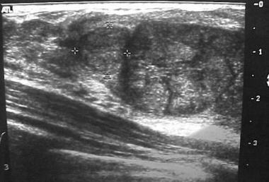
- Magnetic resonance imaging is a diagnostic imaging modality that is capable of demonstrating a wide variety of soft-tissue lesions with contrast resolution equal or superior to CT scanning in various parts of the body. (cms.gov)
Adipose tissue1
- The histological preparations showed an admixture of mature adipose tissue, including variably sized typical adipocytes, embedded within dense collagen fibres (Fig. 3 ), consistent with fibrolipoma. (springer.com)
Retroperitoneal1
- Retroperitoneal neoplasms are more common in familial polyposis coli and Gardner syndrome after abdominal surgery than in other conditions. (medscape.com)
Abnormal growth1
- New abnormal growth of tissue. (lookformedical.com)
Sarcoma patients1
- The primary aim is to assess mid- to long-term survival of Ewing-like sarcoma patients (both soft tissues and bones). (emsos.org)
Humans1
- Substances that increase the risk of NEOPLASMS in humans or animals. (lookformedical.com)
Clinical3
- This Clinical Policy Bulletin addresses treatment approaches for liver and other neoplasms. (aetna.com)
- At clinical examination, the lesion appeared soft and well separated from the surrounding tissues and was covered by intact mucosa (Fig. 1 ). (springer.com)
- Genetic analyses have shown that the clinical and biological variation among these neoplasms is reflected in their genotypes. (lu.se)
Lesion2
- This lesion usually presents as an asymptomatic swelling of soft consistency, mobile on the surrounding tissues. (springer.com)
- Following magnetic resonance (MR) scan revealed a soft mass smaller than first one in the left maxillofacial and single-photon emission computed tomography/computed tomography (SPECT/CT) scan showed increased focal uptake in the lesion. (researchsquare.com)
Immunohistochemical3
- Hence, five of 11 SRCT of bone or soft tissue with the t(11;22) showed morphologic and/or immunohistochemical evidence of neural differentiation. (nih.gov)
- Immunohistochemical staining of neoplasms was performed from biopsies samples. (hindawi.com)
- Materials and methods: We carried out a retrospective hospital-based immunohistochemical study of archival IDC tissue blocks over a four- and half-year period. (bvsalud.org)
Fibrous2
- The lamina propria is a fibrous connective tissue layer that consists of a network of type I and III collagen and elastin fibers in some regions. (wikipedia.org)
- in the bone marrow (precursor cells, also called stem cells) develop and reproduce excessively or are crowded out by an overgrowth of fibrous tissue. (msdmanuals.com)
Molecular1
- WGTA was used to generate reports including molecular alterations and site/tissue of origin prediction. (researchgate.net)
Resection2
- This approach allowed adequate resection of the neoplasm with minimal damage to the adjacent tissues, thus reducing post-surgical scarring. (springer.com)
- En-bloc resection has been the most widely used treatment for grade 2-3 CS, whereas for patients with low-grade CS, curettage is safe and effective. (emsos.org)
Histologically2
- Histologically, an excess amount of keratin is noted on the surface of the tissue, and the tissue has all the layers of an orthokeratinized tissue with its granular and keratin layers. (wikipedia.org)
- The neoplasms may be histologically the same or different, and may be found in the same or different sites. (lookformedical.com)
Uncertain behavior1
- The ICD-10 code range for Neoplasms of uncertain behavior, polycythemia vera and myelodysplastic syndromes D37-D48 is medical classification list by the World Health Organization (WHO). (aapc.com)
Cancer2
- Thus, to check for malignant changes, a baseline biopsy and microscopic study of any whitened tissue may be indicated, especially if in a high-risk cancer category, such with a history of tobacco or alcohol use or are HPV positive. (wikipedia.org)
- Fibrosarcoma , an aggressive and highly metastatic cancer of the connective tissue, primarily develops in the metaphyses of long tubular bones, and affects both children and adults. (drrathresearch.org)
Occur2
- Athelia (ie, absence of nipples) and amastia (ie, absence of breast tissue) may occur bilaterally or unilaterally. (medscape.com)
- Mastitis neonatorum or infections of the breast tissue may also occur during the newborn period. (medscape.com)
Liver4
- Percutaneous ethanol injection (PEI) for liver neoplasms when criteria above are not met. (aetna.com)
- Liver neoplasms. (lookformedical.com)
- A primary malignant neoplasm of epithelial liver cells. (lookformedical.com)
- Closed vesicles of fragmented endoplasmic reticulum created when liver cells or tissue are disrupted by homogenization. (lookformedical.com)
Pediatric1
- Epstein-Barr virus-associated SMT is an uncommon soft tissue neoplasm affecting both adult and pediatric populations. (allenpress.com)
Tumours2
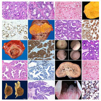
- Soft Tissue and Bone Tumours is the third volume in the 5th edition of the WHO series on the classification of human tumours. (who.int)
- This volume will be of particular interest to pathologists, oncologists, surgeons, and epidemiologists who manage or research soft tissue and bone tumours. (who.int)
Proliferation1
- Soon after birth, the nipples are raised from the shallow mammary pits by proliferation of the surrounding connective tissue. (medscape.com)
Neuroectodermal2
- Our results support the hypothesis that SRCT of bone of soft tissue with the t(11;22) form a single biologic entity displaying varying degrees of neuroectodermal differentiation. (nih.gov)
- Another opinion suggested a differentiation-metaplasia capacity of neuroectodermal tissue [ 20 ]. (hindawi.com)
Surgery1
- The outcome after surgery is excellent as these neoplasms are generally benign in nature. (sages.org)
Rare2
- 1 Subsequently, Chadwick et al 2 reported SMT in human immunodeficiency virus (HIV)-infected children and asserted the association between HIV and these rare neoplasms. (allenpress.com)
- Extraskeletal osteosarcoma (EOS) is a rare malignant soft tissue neoplasm. (researchsquare.com)
Syndromes1
- Sections are included on all recognized neoplasms of the soft tissue and bone, as well as on genetic tumour syndromes affecting these sites. (who.int)
Malignancies1
- Malignancies of the soft tissues (6%) and bone (5%) account for more than 10% of cancers diagnosed in children, adolescents, and young adults. (drrathresearch.org)
Surgical1
- Regressive changes of the tissues located at the surgical margins, such as cellular hyperbasophilia, nuclear chromatin condensations or tissue coarctation were not detected. (springer.com)
Colorectal1
- Gardner syndrome or familial adenomatous polyposis (FAP) is characterized by colorectal adenomatous polyps and soft and hard tissue neoplasms. (medscape.com)
Breast2
- Accessory or ectopic breast tissue responds to hormonal stimulation and may cause discomfort during menstrual cycles. (medscape.com)
- This paper presents a 3-year-old girl who presented with a soft to firm, ill-defined swelling on the right breast with painful ulceration of the overlying skin. (usp.br)
Organs1
- Neoplasms of whatever cell type or origin, occurring in the extraskeletal connective tissue framework of the body including the organs of locomotion and their various component structures, such as nerves, blood vessels, lymphatics, etc. (nih.gov)