Retinal Degeneration
Macular Degeneration
Nerve Degeneration
Wallerian Degeneration
Intervertebral Disc Degeneration
Wet Macular Degeneration
Frontotemporal Lobar Degeneration
Photoreceptor Cells, Vertebrate
Intervertebral Disc
Retina
Retrograde Degeneration
Choroidal Neovascularization
Retinitis Pigmentosa
Disease Models, Animal
Spinocerebellar Degenerations
Retinal Pigment Epithelium
Retinal Drusen
Rhodopsin
Photoreceptor Cells
Eye Proteins
Fluorescein Angiography
Paraneoplastic Cerebellar Degeneration
Subacute Combined Degeneration
Pigment Epithelium of Eye
Choroid
Visual Acuity
Geographic Atrophy
Intervertebral Disc Displacement
Neurodegenerative Diseases
Mice, Inbred C57BL
Retinal Rod Photoreceptor Cells
Neurons
Atrophy
Mutation
Retinal Cone Photoreceptor Cells
Fundus Oculi
Amyotrophic Lateral Sclerosis
Mice, Transgenic
Intravitreal Injections
Spinal Diseases
Lumbar Vertebrae
Nicotinamide-Nucleotide Adenylyltransferase
Bruch Membrane
Immunohistochemistry
Lipofuscin
Aging
Complement Factor H
Nerve Tissue Proteins
Eye Diseases, Hereditary
Cell Death
Brain
Microscopy, Electron
Optic Nerve
Peripherins
Inclusion Bodies
Motor Neuron Disease
Macula Lutea
Cerebellar Diseases
Retinal Ganglion Cells
Mice, Knockout
Tomography, Optical Coherence
Retinal Diseases
Cartilage, Articular
Cyclic Nucleotide Phosphodiesterases, Type 6
Magnetic Resonance Imaging
Substantia Nigra
Dark Adaptation
In Situ Nick-End Labeling
Osteoarthritis, Spine
Cell Count
Axotomy
Animals, Genetically Modified
Disease Progression
Apoptosis
Radiation Injuries, Experimental
Optic Disk Drusen
tau Proteins
Supranuclear Palsy, Progressive
Microscopy, Electron, Transmission
Parkinson Disease
Choroid Diseases
Fluorescent Antibody Technique, Indirect
Gliosis
Phenotype
Rats, Sprague-Dawley
Cerebellar Ataxia
Retinal Detachment
Opsins
Spinal Cord
Myelin Sheath
Sciatic Nerve
Cells, Cultured
Rod Opsins
Alzheimer Disease
Blindness
Spinal Osteophytosis
Spiral Ganglion
Cell Survival
Blotting, Western
Rats, Transgenic
Retinal Dysplasia
Microglia
Dementia
Osteoarthritis
Retinal Photoreceptor Cell Outer Segment
Neuroprotective Agents
Leber Congenital Amaurosis
Basal Ganglia Diseases
Pick Disease of the Brain
Fovea Centralis
Photoreceptor Cells, Invertebrate
Lutein
Oxidative Stress
Neurofilament Proteins
Glial Fibrillary Acidic Protein
Hepatolenticular Degeneration
Neuroglia
Genotype
Gene Expression Regulation
Cerebellum
Axonal Transport
Vision, Ocular
Retinal Neovascularization
Ataxia
Reverse Transcriptase Polymerase Chain Reaction
Eye
Tauopathies
Spinal Cord Diseases
Neurofibrillary Tangles
Parkinsonian Disorders
Dopamine
Pedigree
RNA, Messenger
Peripheral Nerves
Angiogenesis Inhibitors
Zygapophyseal Joint
Gene Expression
TDP-43 Proteinopathies
Cerebral Cortex
Vitreous Body
Histocytochemistry
Molecular Sequence Data
Photography
Arrestin
Optic Nerve Injuries
Pyramidal Tracts
Microscopy, Confocal
Frontotemporal Dementia
Vision Disorders
Rod Cell Outer Segment
Paraneoplastic Syndromes
Schwann Cells
Retinal Neurons
Dog Diseases
Necrosis
Corpus Striatum
Drosophila
alpha-Synuclein
Striatonigral Degeneration
Green Fluorescent Proteins
Mitochondria
Peripheral Nervous System Diseases
Exudates and Transudates
Superoxide Dismutase
Nerve Fibers, Myelinated
Choroideremia
Cervical Vertebrae
Astrocytes
Fibrocartilage
Photochemotherapy
Case-Control Studies
Optic Atrophy
Oxidopamine
Photoreceptor Connecting Cilium
Genetic Predisposition to Disease
Huntington Disease
Spastic Paraplegia, Hereditary
Polymorphism, Single Nucleotide
Dependovirus
RNA-Binding Protein FUS
Two cases of subacute combined degeneration: magnetic resonance findings. (1/8)
We report two cases of subacute combined degeneration. Both patients had undergone total gastrectomy. The chief complaints were numbness in both upper extremities in case 1 and numbness in both the upper and lower extremities and gait disturbance in case 2. The pain, temperature, and vibration senses of both patients were decreased. Laboratory examinations showed macrocytic anemia and a decreased serum vitamin B12 level in both cases. In both cases T2-weighted magnetic resonance images showed an area of hyperintensity in the dorsal columns of the cervical spinal cord. The patients were treated with vitamin B12. The abnormal signals had disappeared on follow-up magnetic resonance examination 1 year later in case 1 and 3 months later in case 2. These patients showed neurological improvement, but the numbness in the upper extremities persisted even after the area of abnormal signal intensity had disappeared in case 1. (+info)Spinal myoclonus in subacute combined degeneration caused by nitrous oxide intoxication. (2/8)
A 26-year-old patient developed ascending weakness and paresthesias. Megaloblastic anemia and mildly reduced serum vitamin B12 (B12) concentration were noted. Myoclonus-like muscular contractions appeared over four extremities and in the trunk. She admitted inhaling nitrous oxide (N2O) as a euphoriant repeatedly at party. Following parenteral B12 administration, her neurological deficit promptly resolved. This case demonstrated the abuse of N2OI is an important cause of subacute combined degeneration (SCD) of the spinal cord. To our knowledge, this is the first report of involuntary movements in a patient with N2O intoxication. Although the mechanism remains unknown, involuntary movements similar to myoclonus should be considered as one of the extraordinary neurological manifestations of N2O intoxication. (+info)Reversible myelopathy with vitamin B12 deficiency. (3/8)
Vitamin B12 deficiency causes haematological, gastrointestinal, psychiatric and neurological diseases. Subacute combined degeneration (SCD) of the spinal cord, characterised by degeneration of the lateral and posterior columns, is often found due to vitamin B12 deficiency. We report SCD occurring in a 57-year-old man who presented with a 2.5-month history of gradually progressing tingling in the fingers and toes and neck ache. Laboratory data revealed vitamin B12 deficiency and magnetic resonance (MR) imaging of the cervical spinal cord demonstrated abnormal hyperintense signal changes on T2-weighted imaging of the posterior columns. In our case, follow-up MR imaging findings correlated well with clinical outcome after treatment with vitamin B12 supplements. Neurological symptoms in vitamin B12 deficiency are frequent. Early spinal MR imaging assists in the early diagnosis and treatment of the disease. (+info)Subacute combined degeneration caused by nitrous oxide intoxication: case reports. (4/8)
PURPOSE: Case reports with a comprehensive review of the current literature concerning subacute combined degeneration induced by nitrous oxide inhalation. A differential diagnosis should be considered when young patients present with progressive myelopathy because that the misuse of nitrous oxide has potentially serious outcomes. CASES REPORT: Three young patients aged from 18 to 24, one male and two females, were diagnosed with progressive ascending numbness in four limbs or both legs and ataxia. They all had been inhaling nitrous oxide from whipped-cream containers for several months. A cervicothoracic magnetic resonance imaging scan revealed long segmental hyperintensity changes at the posterior column of the spinal cord. Serological examination showed a low level of vitamin B12. Subacute combined degeneration of the spinal cord was diagnosed and the etiology was considered related to nitrous oxide misuse. Their neurological status, neuroimage, and neurophysiologic condition improved after vitamin B12 supplementation and cessation of nitrous oxide inhalation. CONCLUSION: Iatrogenic usage of nitrous oxide apparently resulted in subacute combined degeneration in our three patients. Recently, nitrous oxide misuse has increased among young people. Subacute combined degeneration of the spinal cord should be considered as a possible outcome of such abuse. (+info)Hyperintense signal on spinal cord diffusion-weighted imaging in a patient with subacute combined degeneration. (5/8)
(+info)Sequential involvement of the nervous system in subacute combined degeneration. (6/8)
(+info)Subacute combined spinal cord degeneration and pancytopenia secondary to severe vitamin B12 deficiency. (7/8)
(+info)Cobalamin deficiency: clinical picture and radiological findings. (8/8)
(+info)Retinal degeneration is a broad term that refers to the progressive loss of photoreceptor cells (rods and cones) in the retina, which are responsible for converting light into electrical signals that are sent to the brain. This process can lead to vision loss or blindness. There are many different types of retinal degeneration, including age-related macular degeneration, retinitis pigmentosa, and Stargardt's disease, among others. These conditions can have varying causes, such as genetic mutations, environmental factors, or a combination of both. Treatment options vary depending on the specific type and progression of the condition.
Macular degeneration, also known as age-related macular degeneration (AMD), is a medical condition that affects the central part of the retina, called the macula. The macula is responsible for sharp, detailed vision, which is necessary for activities such as reading, driving, and recognizing faces.
In AMD, there is a breakdown or deterioration of the macula, leading to gradual loss of central vision. There are two main types of AMD: dry (atrophic) and wet (exudative). Dry AMD is more common and progresses more slowly, while wet AMD is less common but can cause rapid and severe vision loss if left untreated.
The exact causes of AMD are not fully understood, but risk factors include age, smoking, family history, high blood pressure, obesity, and exposure to sunlight. While there is no cure for AMD, treatments such as vitamin supplements, laser therapy, and medication injections can help slow its progression and reduce the risk of vision loss.
Nerve degeneration, also known as neurodegeneration, is the progressive loss of structure and function of neurons, which can lead to cognitive decline, motor impairment, and various other symptoms. This process occurs due to a variety of factors, including genetics, environmental influences, and aging. It is a key feature in several neurological disorders such as Alzheimer's disease, Parkinson's disease, Huntington's disease, and multiple sclerosis. The degeneration can affect any part of the nervous system, leading to different symptoms depending on the location and extent of the damage.
Wallerian degeneration is a process that occurs following damage to the axons of neurons (nerve cells). After an axon is severed or traumatically injured, it undergoes a series of changes including fragmentation and removal of the distal segment of the axon, which is the part that is separated from the cell body. This process is named after Augustus Waller, who first described it in 1850.
The degenerative changes in the distal axon are characterized by the breakdown of the axonal cytoskeleton, the loss of myelin sheath (the fatty insulating material that surrounds and protects the axon), and the infiltration of macrophages to clear away the debris. These events lead to the degeneration of the distal axon segment, which is necessary for successful regeneration of the injured nerve.
Wallerian degeneration is a crucial process in the nervous system's response to injury, as it enables the regrowth of axons and the reestablishment of connections between neurons. However, if the regenerative capacity of the neuron is insufficient or the environment is not conducive to growth, functional recovery may be impaired, leading to long-term neurological deficits.
Intervertebral disc degeneration is a physiological and biochemical process that occurs in the spinal discs, which are located between each vertebra in the spine. These discs act as shock absorbers and allow for movement and flexibility of the spine.
The degenerative process involves changes in the structure and composition of the disc, including loss of water content, decreased production of proteoglycans (which help to maintain the disc's elasticity), and disorganization of the collagen fibers that make up the disc's outer layer (annulus fibrosus). These changes can lead to a decrease in the disc's height and mobility, as well as the development of tears or cracks in the annulus fibrosus.
In advanced stages of degeneration, the disc may herniate or bulge outward, causing pressure on nearby nerves and potentially leading to pain, numbness, tingling, or weakness in the affected area. It's worth noting that while intervertebral disc degeneration is a normal part of aging, certain factors such as injury, smoking, obesity, and repetitive stress can accelerate the process.
Wet macular degeneration, also known as neovascular or exudative age-related macular degeneration (AMD), is a medical condition that affects the central part of the retina called the macula. It's characterized by the growth of new blood vessels (neovascularization) from the choroid layer behind the retina into the macula, which is not typical in healthy eyes. These abnormal blood vessels are fragile and prone to leakage, leading to the accumulation of fluid or blood in the macula, causing distortion or loss of central vision.
The wet form of AMD can progress rapidly and often leads to more severe visual loss compared to the dry form. It's essential to diagnose and treat wet AMD promptly to preserve as much vision as possible. Common treatments include anti-vascular endothelial growth factor (VEGF) injections, photodynamic therapy, or thermal laser treatment, depending on the specific case and individual patient factors.
Frontotemporal lobar degeneration (FTLD) is a group of disorders characterized by the progressive degeneration of the frontal and temporal lobes of the brain. These areas of the brain are involved in decision-making, behavior, emotion, and language. FTLD can be divided into several subtypes based on the specific clinical features and the underlying protein abnormalities.
The three main subtypes of FTLD are:
1. Behavioral variant frontotemporal dementia (bvFTD): This subtype is characterized by changes in personality, behavior, and judgment. People with bvFTD may lose their social inhibitions, become impulsive, or develop compulsive behaviors. They may also have difficulty with emotional processing and empathy.
2. Primary progressive aphasia (PPA): This subtype is characterized by the gradual deterioration of language skills. People with PPA may have difficulty speaking, understanding spoken or written language, or both. There are three subtypes of PPA: nonfluent/agrammatic variant, semantic variant, and logopenic variant.
3. Motor neuron disease (MND) with FTLD: This subtype is characterized by the degeneration of motor neurons, which are the nerve cells responsible for controlling voluntary muscle movements. People with MND with FTLD may develop symptoms of amyotrophic lateral sclerosis (ALS), such as muscle weakness, stiffness, and twitching, as well as cognitive and behavioral changes associated with FTLD.
The underlying protein abnormalities in FTLD include:
1. Tau protein: In some forms of FTLD, the tau protein accumulates and forms clumps called tangles inside nerve cells. This is also seen in Alzheimer's disease.
2. TDP-43 protein: In other forms of FTLD, the TDP-43 protein accumulates and forms clumps inside nerve cells.
3. Fused in sarcoma (FUS) protein: In a small number of cases, the FUS protein accumulates and forms clumps inside nerve cells.
FTLD is typically a progressive disorder, meaning that symptoms worsen over time. There is currently no cure for FTLD, but there are treatments available to help manage symptoms and improve quality of life.
Photoreceptor cells in vertebrates are specialized types of neurons located in the retina of the eye that are responsible for converting light stimuli into electrical signals. These cells are primarily responsible for the initial process of vision and have two main types: rods and cones.
Rods are more numerous and are responsible for low-light vision or scotopic vision, enabling us to see in dimly lit conditions. They do not contribute to color vision but provide information about the shape and movement of objects.
Cones, on the other hand, are less numerous and are responsible for color vision and high-acuity vision or photopic vision. There are three types of cones, each sensitive to different wavelengths of light: short (S), medium (M), and long (L) wavelengths, which correspond to blue, green, and red, respectively. The combination of signals from these three types of cones allows us to perceive a wide range of colors.
Both rods and cones contain photopigments that consist of a protein called opsin and a light-sensitive chromophore called retinal. When light hits the photopigment, it triggers a series of chemical reactions that ultimately lead to the generation of an electrical signal that is transmitted to the brain via the optic nerve. This process enables us to see and perceive our visual world.
An intervertebral disc is a fibrocartilaginous structure found between the vertebrae of the spinal column in humans and other animals. It functions as a shock absorber, distributes mechanical stress during weight-bearing activities, and allows for varying degrees of mobility between adjacent vertebrae.
The disc is composed of two parts: the annulus fibrosus, which forms the tough, outer layer; and the nucleus pulposus, which is a gel-like substance in the center that contains proteoglycans and water. The combination of these components provides the disc with its unique ability to distribute forces and allow for movement.
The intervertebral discs are essential for the normal functioning of the spine, providing stability, flexibility, and protection to the spinal cord and nerves. However, they can also be subject to degeneration and injury, which may result in conditions such as herniated discs or degenerative disc disease.
The retina is the innermost, light-sensitive layer of tissue in the eye of many vertebrates and some cephalopods. It receives light that has been focused by the cornea and lens, converts it into neural signals, and sends these to the brain via the optic nerve. The retina contains several types of photoreceptor cells including rods (which handle vision in low light) and cones (which are active in bright light and are capable of color vision).
In medical terms, any pathological changes or diseases affecting the retinal structure and function can lead to visual impairment or blindness. Examples include age-related macular degeneration, diabetic retinopathy, retinal detachment, and retinitis pigmentosa among others.
Electroretinography (ERG) is a medical test used to evaluate the functioning of the retina, which is the light-sensitive tissue located at the back of the eye. The test measures the electrical responses of the retina to light stimulation.
During the procedure, a special contact lens or electrode is placed on the surface of the eye to record the electrical activity generated by the retina's light-sensitive cells (rods and cones) and other cells in the retina. The test typically involves presenting different levels of flashes of light to the eye while the electrical responses are recorded.
The resulting ERG waveform provides information about the overall health and function of the retina, including the condition of the photoreceptors, the integrity of the inner retinal layers, and the health of the retinal ganglion cells. This test is often used to diagnose and monitor various retinal disorders, such as retinitis pigmentosa, macular degeneration, and diabetic retinopathy.
Retrograde degeneration is a medical term that refers to the process of degeneration or damage in neurons (nerve cells) that occurs backward from the site of injury or disease along the axon, which is the part of the neuron that transmits electrical signals to other neurons. This can lead to functional loss and may eventually result in the death of the neuron. Retrograde degeneration is often seen in neurodegenerative disorders such as Amyotrophic Lateral Sclerosis (ALS) and Alzheimer's disease, as well as in spinal cord injuries.
Choroidal neovascularization (CNV) is a medical term that refers to the growth of new, abnormal blood vessels in the choroid layer of the eye, which is located between the retina and the sclera. This condition typically occurs as a complication of age-related macular degeneration (AMD), although it can also be caused by other eye diseases or injuries.
In CNV, the new blood vessels that grow into the choroid layer are fragile and can leak fluid or blood, which can cause distortion or damage to the retina, leading to vision loss. Symptoms of CNV may include blurred or distorted vision, a blind spot in the center of the visual field, or changes in color perception.
Treatment for CNV typically involves medications that are designed to stop the growth of new blood vessels, such as anti-VEGF drugs, which target a protein called vascular endothelial growth factor (VEGF) that is involved in the development of new blood vessels. Laser surgery or photodynamic therapy may also be used in some cases to destroy the abnormal blood vessels and prevent further vision loss.
Retinitis pigmentosa (RP) is a group of rare, genetic disorders that involve a breakdown and loss of cells in the retina - a light-sensitive tissue located at the back of the eye. The retina converts light into electrical signals which are then sent to the brain and interpreted as visual images.
In RP, the cells that detect light (rods and cones) degenerate more slowly than other cells in the retina, leading to a progressive loss of vision. Symptoms typically begin in childhood with night blindness (difficulty seeing in low light), followed by a gradual narrowing of the visual field (tunnel vision). Over time, this can lead to significant vision loss and even blindness.
The condition is usually inherited and there are several different genes that have been associated with RP. The diagnosis is typically made based on a combination of genetic testing, family history, and clinical examination. Currently, there is no cure for RP, but researchers are actively working to develop new treatments that may help slow or stop the progression of the disease.
Animal disease models are specialized animals, typically rodents such as mice or rats, that have been genetically engineered or exposed to certain conditions to develop symptoms and physiological changes similar to those seen in human diseases. These models are used in medical research to study the pathophysiology of diseases, identify potential therapeutic targets, test drug efficacy and safety, and understand disease mechanisms.
The genetic modifications can include knockout or knock-in mutations, transgenic expression of specific genes, or RNA interference techniques. The animals may also be exposed to environmental factors such as chemicals, radiation, or infectious agents to induce the disease state.
Examples of animal disease models include:
1. Mouse models of cancer: Genetically engineered mice that develop various types of tumors, allowing researchers to study cancer initiation, progression, and metastasis.
2. Alzheimer's disease models: Transgenic mice expressing mutant human genes associated with Alzheimer's disease, which exhibit amyloid plaque formation and cognitive decline.
3. Diabetes models: Obese and diabetic mouse strains like the NOD (non-obese diabetic) or db/db mice, used to study the development of type 1 and type 2 diabetes, respectively.
4. Cardiovascular disease models: Atherosclerosis-prone mice, such as ApoE-deficient or LDLR-deficient mice, that develop plaque buildup in their arteries when fed a high-fat diet.
5. Inflammatory bowel disease models: Mice with genetic mutations affecting intestinal barrier function and immune response, such as IL-10 knockout or SAMP1/YitFc mice, which develop colitis.
Animal disease models are essential tools in preclinical research, but it is important to recognize their limitations. Differences between species can affect the translatability of results from animal studies to human patients. Therefore, researchers must carefully consider the choice of model and interpret findings cautiously when applying them to human diseases.
Spinocerebellar degenerations (SCDs) are a group of genetic disorders that primarily affect the cerebellum, the part of the brain responsible for coordinating muscle movements, and the spinal cord. These conditions are characterized by progressive degeneration or loss of nerve cells in the cerebellum and/or spinal cord, leading to various neurological symptoms.
SCDs are often inherited in an autosomal dominant manner, meaning that only one copy of the altered gene from either parent is enough to cause the disorder. The most common type of SCD is spinocerebellar ataxia (SCA), which includes several subtypes (SCA1, SCA2, SCA3, etc.) differentiated by their genetic causes and specific clinical features.
Symptoms of spinocerebellar degenerations may include:
1. Progressive ataxia (loss of coordination and balance)
2. Dysarthria (speech difficulty)
3. Nystagmus (involuntary eye movements)
4. Oculomotor abnormalities (problems with eye movement control)
5. Tremors or other involuntary muscle movements
6. Muscle weakness and spasticity
7. Sensory disturbances, such as numbness or tingling sensations
8. Dysphagia (difficulty swallowing)
9. Cognitive impairment in some cases
The age of onset, severity, and progression of symptoms can vary significantly among different SCD subtypes and individuals. Currently, there is no cure for spinocerebellar degenerations, but various supportive treatments and therapies can help manage symptoms and improve quality of life.
The retinal pigment epithelium (RPE) is a single layer of cells located between the photoreceptor cells of the retina and the choroid, which is a part of the eye containing blood vessels. The RPE plays a crucial role in maintaining the health and function of the photoreceptors by providing them with nutrients, removing waste products, and helping to regulate the light-sensitive visual pigments within the photoreceptors.
The RPE cells contain pigment granules that absorb excess light to prevent scattering within the eye and improve visual acuity. They also help to form the blood-retina barrier, which restricts the movement of certain molecules between the retina and the choroid, providing an important protective function for the retina.
Damage to the RPE can lead to a variety of eye conditions, including age-related macular degeneration (AMD), which is a leading cause of vision loss in older adults.
Retinal drusen are yellow-white, deposits of extracellular material that accumulate beneath the retina, most commonly in the macula. They are a common age-related finding and can also be seen in various other conditions such as inherited retinal diseases. Drusen can vary in size and number, and their presence is often associated with an increased risk of developing age-related macular degeneration (AMD), a leading cause of vision loss in older adults. However, not all individuals with drusen will develop AMD, and the significance of drusen depends on factors such as size, number, and location. It's important to monitor drusen and have regular eye examinations to assess any changes or progression that may indicate a higher risk for developing AMD.
An axon is a long, slender extension of a neuron (a type of nerve cell) that conducts electrical impulses (nerve impulses) away from the cell body to target cells, such as other neurons or muscle cells. Axons can vary in length from a few micrometers to over a meter long and are typically surrounded by a myelin sheath, which helps to insulate and protect the axon and allows for faster transmission of nerve impulses.
Axons play a critical role in the functioning of the nervous system, as they provide the means by which neurons communicate with one another and with other cells in the body. Damage to axons can result in serious neurological problems, such as those seen in spinal cord injuries or neurodegenerative diseases like multiple sclerosis.
Rhodopsin, also known as visual purple, is a light-sensitive pigment found in the rods of the vertebrate retina. It is a complex protein molecule made up of two major components: an opsin protein and retinal, a form of vitamin A. When light hits the retinal in rhodopsin, it changes shape, which initiates a series of chemical reactions leading to the activation of the visual pathway and ultimately results in vision. This process is known as phototransduction. Rhodopsin plays a crucial role in low-light vision or scotopic vision.
Photoreceptor cells are specialized neurons in the retina of the eye that convert light into electrical signals. These cells consist of two types: rods and cones. Rods are responsible for vision at low light levels and provide black-and-white, peripheral, and motion sensitivity. Cones are active at higher light levels and are capable of color discrimination and fine detail vision. Both types of photoreceptor cells contain light-sensitive pigments that undergo chemical changes when exposed to light, triggering a series of electrical signals that ultimately reach the brain and contribute to visual perception.
Eye proteins, also known as ocular proteins, are specific proteins that are found within the eye and play crucial roles in maintaining proper eye function and health. These proteins can be found in various parts of the eye, including the cornea, iris, lens, retina, and other structures. They perform a wide range of functions, such as:
1. Structural support: Proteins like collagen and elastin provide strength and flexibility to the eye's tissues, enabling them to maintain their shape and withstand mechanical stress.
2. Light absorption and transmission: Proteins like opsins and crystallins are involved in capturing and transmitting light signals within the eye, which is essential for vision.
3. Protection against damage: Some eye proteins, such as antioxidant enzymes and heat shock proteins, help protect the eye from oxidative stress, UV radiation, and other environmental factors that can cause damage.
4. Regulation of eye growth and development: Various growth factors and signaling molecules, which are protein-based, contribute to the proper growth, differentiation, and maintenance of eye tissues during embryonic development and throughout adulthood.
5. Immune defense: Proteins involved in the immune response, such as complement components and immunoglobulins, help protect the eye from infection and inflammation.
6. Maintenance of transparency: Crystallin proteins in the lens maintain its transparency, allowing light to pass through unobstructed for clear vision.
7. Neuroprotection: Certain eye proteins, like brain-derived neurotrophic factor (BDNF), support the survival and function of neurons within the retina, helping to preserve vision.
Dysfunction or damage to these eye proteins can contribute to various eye disorders and diseases, such as cataracts, age-related macular degeneration, glaucoma, diabetic retinopathy, and others.
Fluorescein angiography is a medical diagnostic procedure used in ophthalmology to examine the blood flow in the retina and choroid, which are the inner layers of the eye. This test involves injecting a fluorescent dye, Fluorescein, into a patient's arm vein. As the dye reaches the blood vessels in the eye, a specialized camera takes rapid sequences of photographs to capture the dye's circulation through the retina and choroid.
The images produced by fluorescein angiography can help doctors identify any damage to the blood vessels, leakage, or abnormal growth of new blood vessels. This information is crucial in diagnosing and managing various eye conditions such as age-related macular degeneration, diabetic retinopathy, retinal vein occlusions, and inflammatory eye diseases.
It's important to note that while fluorescein angiography is a valuable diagnostic tool, it does carry some risks, including temporary side effects like nausea, vomiting, or allergic reactions to the dye. In rare cases, severe adverse reactions can occur, so patients should discuss these potential risks with their healthcare provider before undergoing the procedure.
Paraneoplastic cerebellar degeneration (PCD) is a rare disorder characterized by progressive damage to the cerebellum, the part of the brain responsible for coordinating muscle movements. It is considered a paraneoplastic syndrome, which means it is caused by an abnormal immune system response to a cancerous tumor (neoplasm) located elsewhere in the body.
In PCD, antibodies produced by the immune system to fight the tumor mistakenly attack proteins in the cerebellum that are similar to those found in the tumor. This leads to inflammation and degeneration of the Purkinje cells, a type of neuron critical for maintaining balance and coordinating movements.
PCD can present with symptoms such as unsteady gait, loss of coordination, slurred speech, nystagmus (involuntary eye movement), and tremors. These symptoms often develop rapidly, over the course of days to weeks, and may progress even after the tumor has been removed or treated.
PCD is associated with several types of cancers, including small cell lung cancer, breast cancer, ovarian cancer, Hodgkin's lymphoma, and others. Early diagnosis and treatment of the underlying cancer are essential to slowing down the progression of PCD and improving outcomes.
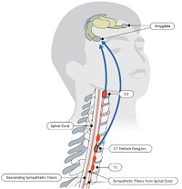
Subacute Combined Degeneration (SCD) is a medical condition that affects the spinal cord and is primarily caused by vitamin B12 deficiency. The term "subacute" refers to the moderate speed of progression of the disease, while "combined degeneration" describes the damage to both the white and gray matter of the spinal cord.
In SCD, the degeneration of the spinal cord occurs in a characteristic pattern, affecting the dorsal columns (posterior column) and lateral corticospinal tracts. This results in various neurological symptoms such as numbness, tingling, weakness, and loss of vibration and position sense in the legs, which can also ascend to affect the arms. Additionally, patients may experience gait disturbances, ataxia (loss of coordination), and urinary or fecal incontinence.
The condition is often associated with pernicious anemia, an autoimmune disorder that impairs vitamin B12 absorption from food. However, other causes of vitamin B12 deficiency, such as dietary insufficiency, gastrointestinal disorders, or certain medications, can also lead to SCD. Early diagnosis and treatment with vitamin B12 supplementation are crucial for preventing further neurological damage and improving symptoms.
The pigment epithelium of the eye, also known as the retinal pigment epithelium (RPE), is a layer of cells located between the photoreceptor cells of the retina and the choroid, which is the vascular layer of the eye. The RPE plays a crucial role in maintaining the health and function of the photoreceptors by providing them with nutrients, removing waste products, and helping to regulate the light that enters the eye.
The RPE cells contain pigment granules that absorb excess light, preventing it from scattering within the eye and improving visual acuity. They also help to create a barrier between the retina and the choroid, which is important for maintaining the proper functioning of the photoreceptors. Additionally, the RPE plays a role in the regeneration of visual pigments in the photoreceptor cells, allowing us to see in different light conditions.
Damage to the RPE can lead to various eye diseases and conditions, including age-related macular degeneration (AMD), which is a leading cause of vision loss in older adults.
The choroid is a layer of the eye that contains blood vessels that supply oxygen and nutrients to the outer layers of the retina. It lies between the sclera (the white, protective coat of the eye) and the retina (the light-sensitive tissue at the back of the eye). The choroid is essential for maintaining the health and function of the retina, particularly the photoreceptor cells that detect light and transmit visual signals to the brain. Damage to the choroid can lead to vision loss or impairment.
Visual acuity is a measure of the sharpness or clarity of vision. It is usually tested by reading an eye chart from a specific distance, such as 20 feet (6 meters). The standard eye chart used for this purpose is called the Snellen chart, which contains rows of letters that decrease in size as you read down the chart.
Visual acuity is typically expressed as a fraction, with the numerator representing the testing distance and the denominator indicating the smallest line of type that can be read clearly. For example, if a person can read the line on the eye chart that corresponds to a visual acuity of 20/20, it means they have normal vision at 20 feet. If their visual acuity is 20/40, it means they must be as close as 20 feet to see what someone with normal vision can see at 40 feet.
It's important to note that visual acuity is just one aspect of overall vision and does not necessarily reflect other important factors such as peripheral vision, depth perception, color vision, or contrast sensitivity.
Geographic atrophy is a medical term used to describe a specific pattern of degeneration of the retinal pigment epithelium (RPE) and the underlying choroidal tissue in the eye. This condition is often associated with age-related macular degeneration (AMD), which is a leading cause of vision loss in older adults.
In geographic atrophy, there are well-defined areas of RPE and choroidal atrophy that appear as pale, irregularly shaped patches in the central part of the retina known as the macula. These patches can grow larger over time and may lead to progressive vision loss. The exact cause of geographic atrophy is not fully understood, but it is thought to be related to oxidative stress, inflammation, and other age-related changes in the eye.
Currently, there are no effective treatments for geographic atrophy, although research is ongoing to find new ways to slow or halt its progression. Regular eye exams and monitoring by an ophthalmologist are important for people with AMD or geographic atrophy to help detect any changes in their vision and manage their condition effectively.
Intervertebral disc displacement, also known as a slipped disc or herniated disc, is a medical condition where the inner, softer material (nucleus pulposus) of the intervertebral disc bulges or ruptures through its outer, tougher ring (annulus fibrosus). This can put pressure on nearby nerves and cause pain, numbness, tingling, or weakness in the affected area, often in the lower back or neck. The displacement may also lead to inflammation and irritation of the surrounding spinal structures, further exacerbating the symptoms. The condition is typically caused by age-related wear and tear (degenerative disc disease) or sudden trauma.
Neurodegenerative diseases are a group of disorders characterized by progressive and persistent loss of neuronal structure and function, often leading to cognitive decline, functional impairment, and ultimately death. These conditions are associated with the accumulation of abnormal protein aggregates, mitochondrial dysfunction, oxidative stress, chronic inflammation, and genetic mutations in the brain. Examples of neurodegenerative diseases include Alzheimer's disease, Parkinson's disease, Huntington's disease, Amyotrophic Lateral Sclerosis (ALS), and Spinal Muscular Atrophy (SMA). The underlying causes and mechanisms of these diseases are not fully understood, and there is currently no cure for most neurodegenerative disorders. Treatment typically focuses on managing symptoms and slowing disease progression.
C57BL/6 (C57 Black 6) is an inbred strain of laboratory mouse that is widely used in biomedical research. The term "inbred" refers to a strain of animals where matings have been carried out between siblings or other closely related individuals for many generations, resulting in a population that is highly homozygous at most genetic loci.
The C57BL/6 strain was established in 1920 by crossing a female mouse from the dilute brown (DBA) strain with a male mouse from the black strain. The resulting offspring were then interbred for many generations to create the inbred C57BL/6 strain.
C57BL/6 mice are known for their robust health, longevity, and ease of handling, making them a popular choice for researchers. They have been used in a wide range of biomedical research areas, including studies of cancer, immunology, neuroscience, cardiovascular disease, and metabolism.
One of the most notable features of the C57BL/6 strain is its sensitivity to certain genetic modifications, such as the introduction of mutations that lead to obesity or impaired glucose tolerance. This has made it a valuable tool for studying the genetic basis of complex diseases and traits.
Overall, the C57BL/6 inbred mouse strain is an important model organism in biomedical research, providing a valuable resource for understanding the genetic and molecular mechanisms underlying human health and disease.
Retinal rod photoreceptor cells are specialized neurons in the retina of the eye that are primarily responsible for vision in low light conditions. They contain a light-sensitive pigment called rhodopsin, which undergoes a chemical change when struck by a single photon of light. This triggers a cascade of biochemical reactions that ultimately leads to the generation of electrical signals, which are then transmitted to the brain via the optic nerve.
Rod cells do not provide color vision or fine detail, but they allow us to detect motion and see in dim light. They are more sensitive to light than cone cells, which are responsible for color vision and detailed sight in bright light conditions. Rod cells are concentrated at the outer edges of the retina, forming a crescent-shaped region called the peripheral retina, with fewer rod cells located in the central region of the retina known as the fovea.
Neurons, also known as nerve cells or neurocytes, are specialized cells that constitute the basic unit of the nervous system. They are responsible for receiving, processing, and transmitting information and signals within the body. Neurons have three main parts: the dendrites, the cell body (soma), and the axon. The dendrites receive signals from other neurons or sensory receptors, while the axon transmits these signals to other neurons, muscles, or glands. The junction between two neurons is called a synapse, where neurotransmitters are released to transmit the signal across the gap (synaptic cleft) to the next neuron. Neurons vary in size, shape, and structure depending on their function and location within the nervous system.
Atrophy is a medical term that refers to the decrease in size and wasting of an organ or tissue due to the disappearance of cells, shrinkage of cells, or decreased number of cells. This process can be caused by various factors such as disuse, aging, degeneration, injury, or disease.
For example, if a muscle is immobilized for an extended period, it may undergo atrophy due to lack of use. Similarly, certain medical conditions like diabetes, cancer, and heart failure can lead to the wasting away of various tissues and organs in the body.
Atrophy can also occur as a result of natural aging processes, leading to decreased muscle mass and strength in older adults. In general, atrophy is characterized by a decrease in the volume or weight of an organ or tissue, which can have significant impacts on its function and overall health.
A mutation is a permanent change in the DNA sequence of an organism's genome. Mutations can occur spontaneously or be caused by environmental factors such as exposure to radiation, chemicals, or viruses. They may have various effects on the organism, ranging from benign to harmful, depending on where they occur and whether they alter the function of essential proteins. In some cases, mutations can increase an individual's susceptibility to certain diseases or disorders, while in others, they may confer a survival advantage. Mutations are the driving force behind evolution, as they introduce new genetic variability into populations, which can then be acted upon by natural selection.
Retinal cone photoreceptor cells are specialized neurons located in the retina of the eye, responsible for visual phototransduction and color vision. They are one of the two types of photoreceptors, with the other being rods, which are more sensitive to low light levels. Cones are primarily responsible for high-acuity, color vision during daylight or bright-light conditions.
There are three types of cone cells, each containing different photopigments that absorb light at distinct wavelengths: short (S), medium (M), and long (L) wavelengths, which correspond to blue, green, and red light, respectively. The combination of signals from these three types of cones allows the human visual system to perceive a wide range of colors and discriminate between them. Cones are densely packed in the central region of the retina, known as the fovea, which provides the highest visual acuity.
"Fundus Oculi" is a medical term that refers to the back part of the interior of the eye, including the optic disc, macula, fovea, retinal vasculature, and peripheral retina. It is the area where light is focused and then transmitted to the brain via the optic nerve, forming visual images. Examinations of the fundus oculi are crucial for detecting various eye conditions such as diabetic retinopathy, macular degeneration, glaucoma, and other retinal diseases. The examination is typically performed using an ophthalmoscope or a specialized camera called a retinal camera.
Amyotrophic Lateral Sclerosis (ALS) is a progressive neurodegenerative disorder that affects nerve cells in the brain and spinal cord responsible for controlling voluntary muscle movements, such as speaking, walking, breathing, and swallowing. The condition is characterized by the degeneration of motor neurons in the brain (upper motor neurons) and spinal cord (lower motor neurons), leading to their death.
The term "amyotrophic" comes from the Greek words "a" meaning no or negative, "myo" referring to muscle, and "trophic" relating to nutrition. When a motor neuron degenerates and can no longer send impulses to the muscle, the muscle becomes weak and eventually atrophies due to lack of use.
The term "lateral sclerosis" refers to the hardening or scarring (sclerosis) of the lateral columns of the spinal cord, which are primarily composed of nerve fibers that carry information from the brain to the muscles.
ALS is often called Lou Gehrig's disease, named after the famous American baseball player who was diagnosed with the condition in 1939. The exact cause of ALS remains unknown, but it is believed to involve a combination of genetic and environmental factors. There is currently no cure for ALS, and treatment primarily focuses on managing symptoms and maintaining quality of life.
The progression of ALS varies from person to person, with some individuals experiencing rapid decline over just a few years, while others may have a more slow-progressing form of the disease that lasts several decades. The majority of people with ALS die from respiratory failure within 3 to 5 years after the onset of symptoms. However, approximately 10% of those affected live for 10 or more years following diagnosis.
Transgenic mice are genetically modified rodents that have incorporated foreign DNA (exogenous DNA) into their own genome. This is typically done through the use of recombinant DNA technology, where a specific gene or genetic sequence of interest is isolated and then introduced into the mouse embryo. The resulting transgenic mice can then express the protein encoded by the foreign gene, allowing researchers to study its function in a living organism.
The process of creating transgenic mice usually involves microinjecting the exogenous DNA into the pronucleus of a fertilized egg, which is then implanted into a surrogate mother. The offspring that result from this procedure are screened for the presence of the foreign DNA, and those that carry the desired genetic modification are used to establish a transgenic mouse line.
Transgenic mice have been widely used in biomedical research to model human diseases, study gene function, and test new therapies. They provide a valuable tool for understanding complex biological processes and developing new treatments for a variety of medical conditions.
An intravitreal injection is a medical procedure in which medication is delivered directly into the vitreous cavity of the eye, which is the clear, gel-like substance that fills the space between the lens and the retina. This type of injection is typically used to treat various eye conditions such as age-related macular degeneration, diabetic retinopathy, retinal vein occlusion, and uveitis. The medication administered in intravitreal injections can help to reduce inflammation, inhibit the growth of new blood vessels, or prevent the formation of abnormal blood vessels in the eye.
Intravitreal injections are usually performed in an outpatient setting, and the procedure typically takes only a few minutes. Before the injection, the eye is numbed with anesthetic drops to minimize discomfort. The medication is then injected into the vitreous cavity using a small needle. After the injection, patients may experience some mild discomfort or a scratchy sensation in the eye, but this usually resolves within a few hours.
While intravitreal injections are generally safe, there are some potential risks and complications associated with the procedure, including infection, bleeding, retinal detachment, and increased intraocular pressure. Patients who undergo intravitreal injections should be closely monitored by their eye care provider to ensure that any complications are promptly identified and treated.
Spinal diseases refer to a range of medical conditions that affect the spinal column, which is made up of vertebrae (bones), intervertebral discs, facet joints, nerves, ligaments, and muscles. These diseases can cause pain, discomfort, stiffness, numbness, weakness, or even paralysis, depending on the severity and location of the condition. Here are some examples of spinal diseases:
1. Degenerative disc disease: This is a condition where the intervertebral discs lose their elasticity and height, leading to stiffness, pain, and decreased mobility.
2. Herniated disc: This occurs when the inner material of the intervertebral disc bulges or herniates out through a tear in the outer layer, causing pressure on the spinal nerves and resulting in pain, numbness, tingling, or weakness in the affected area.
3. Spinal stenosis: This is a narrowing of the spinal canal or the neural foramen (the openings where the spinal nerves exit the spinal column), which can cause pressure on the spinal cord or nerves and result in pain, numbness, tingling, or weakness.
4. Scoliosis: This is a curvature of the spine that can occur in children or adults, leading to an abnormal posture, back pain, and decreased lung function.
5. Osteoarthritis: This is a degenerative joint disease that affects the facet joints in the spine, causing pain, stiffness, and decreased mobility.
6. Ankylosing spondylitis: This is a chronic inflammatory disease that affects the spine and sacroiliac joints, leading to pain, stiffness, and fusion of the vertebrae.
7. Spinal tumors: These are abnormal growths that can occur in the spinal column, which can be benign or malignant, causing pain, neurological symptoms, or even paralysis.
8. Infections: Bacterial or viral infections can affect the spine, leading to pain, fever, and other systemic symptoms.
9. Trauma: Fractures, dislocations, or sprains of the spine can occur due to accidents, falls, or sports injuries, causing pain, neurological deficits, or even paralysis.
The lumbar vertebrae are the five largest and strongest vertebrae in the human spine, located in the lower back region. They are responsible for bearing most of the body's weight and providing stability during movement. The lumbar vertebrae have a characteristic shape, with a large body in the front, which serves as the main weight-bearing structure, and a bony ring in the back, formed by the pedicles, laminae, and processes. This ring encloses and protects the spinal cord and nerves. The lumbar vertebrae are numbered L1 to L5, starting from the uppermost one. They allow for flexion, extension, lateral bending, and rotation movements of the trunk.
Nicotinamide-nucleotide adenylyltransferase (NNAT) is an enzyme that plays a crucial role in the metabolism of nicotinamide adenine dinucleotide (NAD+), which is a coenzyme involved in various redox reactions in the body. NNAT catalyzes the interconversion between nicotinamide mononucleotide (NMN) and NAD+ through the transfer of an adenylyl group.
The reaction catalyzed by NNAT is as follows:
NMN + ATP → NAD+ + PP\_i
NNAT is found in various tissues, including the brain, where it has been implicated in neuronal development and survival. Mutations in the NNAT gene have been associated with neurological disorders such as epilepsy and intellectual disability. Additionally, NNAT has been identified as a potential target for the development of therapies aimed at treating neurodegenerative diseases and cancer.
The Bruch membrane is a thin, layered structure that separates the retina from the choroid in the eye. It is composed of five layers: the basement membrane of the retinal pigment epithelium (RPE), the inner collagenous layer, the elastic layer, the outer collagenous layer, and the basement membrane of the choriocapillaris. The Bruch membrane provides structural support to the RPE and serves as a barrier between the retina and the choroid, allowing for the selective transport of nutrients and waste products. It also plays a role in maintaining the health of the photoreceptors in the retina. Damage to the Bruch membrane is associated with age-related macular degeneration (AMD), a leading cause of vision loss in older adults.
Immunohistochemistry (IHC) is a technique used in pathology and laboratory medicine to identify specific proteins or antigens in tissue sections. It combines the principles of immunology and histology to detect the presence and location of these target molecules within cells and tissues. This technique utilizes antibodies that are specific to the protein or antigen of interest, which are then tagged with a detection system such as a chromogen or fluorophore. The stained tissue sections can be examined under a microscope, allowing for the visualization and analysis of the distribution and expression patterns of the target molecule in the context of the tissue architecture. Immunohistochemistry is widely used in diagnostic pathology to help identify various diseases, including cancer, infectious diseases, and immune-mediated disorders.
Lipofuscin is a type of pigment that accumulates in the lysosomes (membrane-bound organelles found inside cells) of various tissues, particularly in nerve cells and heart muscle cells. It consists of cross-linked proteins and lipids that are resistant to degradation by enzymes. The accumulation of lipofuscin is a normal part of aging but can also be associated with certain diseases such as neurodegenerative disorders.
It's often referred to as "age pigment" because it tends to increase in amount with age, and its presence in tissues has been linked to oxidative stress and cellular damage caused by free radicals. Lipofuscin is autofluorescent, meaning that it emits light when excited by certain wavelengths of light, which can be useful for its detection and quantification in research and diagnostic settings.
Aging is a complex, progressive and inevitable process of bodily changes over time, characterized by the accumulation of cellular damage and degenerative changes that eventually lead to increased vulnerability to disease and death. It involves various biological, genetic, environmental, and lifestyle factors that contribute to the decline in physical and mental functions. The medical field studies aging through the discipline of gerontology, which aims to understand the underlying mechanisms of aging and develop interventions to promote healthy aging and extend the human healthspan.
Complement Factor H is a protein involved in the regulation of the complement system, which is a part of the immune system that helps to clear pathogens and damaged cells from the body. Specifically, Complement Factor H helps to regulate the activation and deactivation of the complement component C3b, preventing excessive or unwanted activation of the complement system and protecting host tissues from damage.
Complement Factor H is a crucial protein in maintaining the balance between the protective effects of the complement system and the potential for harm to the body's own cells and tissues. Deficiencies or mutations in Complement Factor H have been associated with several diseases, including age-related macular degeneration (AMD), atypical hemolytic uremic syndrome (aHUS), and C3 glomerulopathy.
Nerve tissue proteins are specialized proteins found in the nervous system that provide structural and functional support to nerve cells, also known as neurons. These proteins include:
1. Neurofilaments: These are type IV intermediate filaments that provide structural support to neurons and help maintain their shape and size. They are composed of three subunits - NFL (light), NFM (medium), and NFH (heavy).
2. Neuronal Cytoskeletal Proteins: These include tubulins, actins, and spectrins that provide structural support to the neuronal cytoskeleton and help maintain its integrity.
3. Neurotransmitter Receptors: These are specialized proteins located on the postsynaptic membrane of neurons that bind neurotransmitters released by presynaptic neurons, triggering a response in the target cell.
4. Ion Channels: These are transmembrane proteins that regulate the flow of ions across the neuronal membrane and play a crucial role in generating and transmitting electrical signals in neurons.
5. Signaling Proteins: These include enzymes, receptors, and adaptor proteins that mediate intracellular signaling pathways involved in neuronal development, differentiation, survival, and death.
6. Adhesion Proteins: These are cell surface proteins that mediate cell-cell and cell-matrix interactions, playing a crucial role in the formation and maintenance of neural circuits.
7. Extracellular Matrix Proteins: These include proteoglycans, laminins, and collagens that provide structural support to nerve tissue and regulate neuronal migration, differentiation, and survival.
Hereditary eye diseases refer to conditions that affect the eyes and are passed down from parents to their offspring through genetics. These diseases are caused by mutations or changes in an individual's DNA that are inherited from their parents. The mutations can occur in any of the genes associated with eye development, function, or health.
There are many different types of hereditary eye diseases, some of which include:
1. Retinitis Pigmentosa - a group of rare, genetic disorders that involve a breakdown and loss of cells in the retina.
2. Macular Degeneration - a progressive disease that damages the central portion of the retina, impairing vision.
3. Glaucoma - a group of eye conditions that damage the optic nerve, often caused by an increase in pressure inside the eye.
4. Cataracts - clouding of the lens inside the eye, which can lead to blurry vision and blindness.
5. Keratoconus - a progressive eye disease that causes the cornea to thin and bulge outward into a cone shape.
6. Color Blindness - a condition where an individual has difficulty distinguishing between certain colors.
7. Optic Neuropathy - damage to the optic nerve, which can result in vision loss.
The symptoms and severity of hereditary eye diseases can vary widely depending on the specific condition and the individual's genetic makeup. Some conditions may be present at birth or develop in early childhood, while others may not appear until later in life. Treatment options for these conditions may include medication, surgery, or lifestyle changes, and are often most effective when started early.
Cell death is the process by which cells cease to function and eventually die. There are several ways that cells can die, but the two most well-known and well-studied forms of cell death are apoptosis and necrosis.
Apoptosis is a programmed form of cell death that occurs as a normal and necessary process in the development and maintenance of healthy tissues. During apoptosis, the cell's DNA is broken down into small fragments, the cell shrinks, and the membrane around the cell becomes fragmented, allowing the cell to be easily removed by phagocytic cells without causing an inflammatory response.
Necrosis, on the other hand, is a form of cell death that occurs as a result of acute tissue injury or overwhelming stress. During necrosis, the cell's membrane becomes damaged and the contents of the cell are released into the surrounding tissue, causing an inflammatory response.
There are also other forms of cell death, such as autophagy, which is a process by which cells break down their own organelles and proteins to recycle nutrients and maintain energy homeostasis, and pyroptosis, which is a form of programmed cell death that occurs in response to infection and involves the activation of inflammatory caspases.
Cell death is an important process in many physiological and pathological processes, including development, tissue homeostasis, and disease. Dysregulation of cell death can contribute to the development of various diseases, including cancer, neurodegenerative disorders, and autoimmune diseases.
A "mutant strain of mice" in a medical context refers to genetically engineered mice that have specific genetic mutations introduced into their DNA. These mutations can be designed to mimic certain human diseases or conditions, allowing researchers to study the underlying biological mechanisms and test potential therapies in a controlled laboratory setting.
Mutant strains of mice are created through various techniques, including embryonic stem cell manipulation, gene editing technologies such as CRISPR-Cas9, and radiation-induced mutagenesis. These methods allow scientists to introduce specific genetic changes into the mouse genome, resulting in mice that exhibit altered physiological or behavioral traits.
These strains of mice are widely used in biomedical research because their short lifespan, small size, and high reproductive rate make them an ideal model organism for studying human diseases. Additionally, the mouse genome has been well-characterized, and many genetic tools and resources are available to researchers working with these animals.
Examples of mutant strains of mice include those that carry mutations in genes associated with cancer, neurodegenerative disorders, metabolic diseases, and immunological conditions. These mice provide valuable insights into the pathophysiology of human diseases and help advance our understanding of potential therapeutic interventions.
The brain is the central organ of the nervous system, responsible for receiving and processing sensory information, regulating vital functions, and controlling behavior, movement, and cognition. It is divided into several distinct regions, each with specific functions:
1. Cerebrum: The largest part of the brain, responsible for higher cognitive functions such as thinking, learning, memory, language, and perception. It is divided into two hemispheres, each controlling the opposite side of the body.
2. Cerebellum: Located at the back of the brain, it is responsible for coordinating muscle movements, maintaining balance, and fine-tuning motor skills.
3. Brainstem: Connects the cerebrum and cerebellum to the spinal cord, controlling vital functions such as breathing, heart rate, and blood pressure. It also serves as a relay center for sensory information and motor commands between the brain and the rest of the body.
4. Diencephalon: A region that includes the thalamus (a major sensory relay station) and hypothalamus (regulates hormones, temperature, hunger, thirst, and sleep).
5. Limbic system: A group of structures involved in emotional processing, memory formation, and motivation, including the hippocampus, amygdala, and cingulate gyrus.
The brain is composed of billions of interconnected neurons that communicate through electrical and chemical signals. It is protected by the skull and surrounded by three layers of membranes called meninges, as well as cerebrospinal fluid that provides cushioning and nutrients.
Electron microscopy (EM) is a type of microscopy that uses a beam of electrons to create an image of the sample being examined, resulting in much higher magnification and resolution than light microscopy. There are several types of electron microscopy, including transmission electron microscopy (TEM), scanning electron microscopy (SEM), and reflection electron microscopy (REM).
In TEM, a beam of electrons is transmitted through a thin slice of the sample, and the electrons that pass through the sample are focused to form an image. This technique can provide detailed information about the internal structure of cells, viruses, and other biological specimens, as well as the composition and structure of materials at the atomic level.
In SEM, a beam of electrons is scanned across the surface of the sample, and the electrons that are scattered back from the surface are detected to create an image. This technique can provide information about the topography and composition of surfaces, as well as the structure of materials at the microscopic level.
REM is a variation of SEM in which the beam of electrons is reflected off the surface of the sample, rather than scattered back from it. This technique can provide information about the surface chemistry and composition of materials.
Electron microscopy has a wide range of applications in biology, medicine, and materials science, including the study of cellular structure and function, disease diagnosis, and the development of new materials and technologies.
The optic nerve, also known as the second cranial nerve, is the nerve that transmits visual information from the retina to the brain. It is composed of approximately one million nerve fibers that carry signals related to vision, such as light intensity and color, from the eye's photoreceptor cells (rods and cones) to the visual cortex in the brain. The optic nerve is responsible for carrying this visual information so that it can be processed and interpreted by the brain, allowing us to see and perceive our surroundings. Damage to the optic nerve can result in vision loss or impairment.
Peripherins are a family of neuron-specific type III intermediate filament proteins that are expressed in the peripheral nervous system. They play crucial roles in maintaining the structural integrity and stability of nerve cells, particularly during development and regeneration. Peripherins have also been implicated in various neurodegenerative disorders, including amyotrophic lateral sclerosis (ALS) and Charcot-Marie-Tooth disease (CMT). There are several isoforms of peripherins, with peripherin 2 being the most widely studied. Mutations in the gene encoding peripherin 2 have been linked to certain forms of CMT.
Inclusion bodies are abnormal, intracellular accumulations or aggregations of various misfolded proteins, protein complexes, or other materials within the cells of an organism. They can be found in various tissues and cell types and are often associated with several pathological conditions, including infectious diseases, neurodegenerative disorders, and genetic diseases.
Inclusion bodies can vary in size, shape, and location depending on the specific disease or condition. Some inclusion bodies have a characteristic appearance under the microscope, such as eosinophilic (pink) staining with hematoxylin and eosin (H&E) histological stain, while others may require specialized stains or immunohistochemical techniques to identify the specific misfolded proteins involved.
Examples of diseases associated with inclusion bodies include:
1. Infectious diseases: Some viral infections, such as HIV, hepatitis B and C, and herpes simplex virus, can lead to the formation of inclusion bodies within infected cells.
2. Neurodegenerative disorders: Several neurodegenerative diseases are characterized by the presence of inclusion bodies, including Alzheimer's disease (amyloid-beta plaques and tau tangles), Parkinson's disease (Lewy bodies), Huntington's disease (Huntingtin aggregates), and amyotrophic lateral sclerosis (TDP-43 and SOD1 inclusions).
3. Genetic diseases: Certain genetic disorders, such as Danon disease, neuronal intranuclear inclusion disease, and some lysosomal storage disorders, can also present with inclusion bodies due to the accumulation of abnormal proteins or metabolic products within cells.
The exact role of inclusion bodies in disease pathogenesis remains unclear; however, they are often associated with cellular dysfunction, oxidative stress, and increased inflammation, which can contribute to disease progression and neurodegeneration.
Motor Neuron Disease (MND) is a progressive neurodegenerative disorder that affects the motor neurons, which are nerve cells in the brain and spinal cord responsible for controlling voluntary muscles involved in movement, speaking, breathing, and swallowing. As the motor neurons degenerate and die, they stop sending signals to the muscles, causing them to weaken, waste away (atrophy), and eventually lead to paralysis.
There are several types of MND, including:
1. Amyotrophic Lateral Sclerosis (ALS): Also known as Lou Gehrig's disease, this is the most common form of MND. It affects both upper and lower motor neurons, causing muscle weakness, stiffness, twitching, and atrophy throughout the body.
2. Progressive Bulbar Palsy (PBP): This type primarily affects the bulbar muscles in the brainstem, which control speech, swallowing, and chewing. Patients with PBP experience difficulties with speaking, slurred speech, and problems swallowing and may also have weak facial muscles and limb weakness.
3. Primary Lateral Sclerosis (PLS): This form of MND affects only the upper motor neurons, causing muscle stiffness, spasticity, and weakness, primarily in the legs. PLS progresses more slowly than ALS, and patients usually maintain their ability to speak and swallow for a longer period.
4. Progressive Muscular Atrophy (PMA): This type of MND affects only the lower motor neurons, causing muscle wasting, weakness, and fasciculations (muscle twitches). PMA progresses more slowly than ALS but can still be severely disabling over time.
5. Spinal Muscular Atrophy (SMA): This is a genetic form of MND that typically presents in infancy or childhood, although adult-onset forms exist. SMA affects the lower motor neurons in the spinal cord, causing muscle weakness and atrophy, primarily in the legs and trunk.
The exact cause of Motor Neuron Disease is not fully understood, but it is believed to involve a combination of genetic, environmental, and lifestyle factors. There is currently no cure for MND, and treatment focuses on managing symptoms, maintaining quality of life, and slowing disease progression through various therapies and medications.
Motor neurons are specialized nerve cells in the brain and spinal cord that play a crucial role in controlling voluntary muscle movements. They transmit electrical signals from the brain to the muscles, enabling us to perform actions such as walking, talking, and swallowing. There are two types of motor neurons: upper motor neurons, which originate in the brain's motor cortex and travel down to the brainstem and spinal cord; and lower motor neurons, which extend from the brainstem and spinal cord to the muscles. Damage or degeneration of these motor neurons can lead to various neurological disorders, such as amyotrophic lateral sclerosis (ALS) and spinal muscular atrophy (SMA).
The macula lutea, often simply referred to as the macula or fovea centralis, is a part of the eye that is responsible for central vision and color perception. It's located in the center of the retina, the light-sensitive tissue at the back of the eye. The macula contains a high concentration of pigments called xanthophylls, which give it a yellowish color and protect the photoreceptor cells in this area from damage by blue light.
The central part of the macula is called the fovea, which is a small depression that contains only cones, the photoreceptor cells responsible for color vision and high visual acuity. The fovea is surrounded by the parafovea and the perifovea, which contain both cones and rods, the photoreceptor cells responsible for low-light vision and peripheral vision.
Damage to the macula can result in a loss of central vision and color perception, a condition known as age-related macular degeneration (AMD), which is a leading cause of blindness in older adults. Other conditions that can affect the macula include macular edema, macular holes, and macular pucker.
Cerebellar diseases refer to a group of medical conditions that affect the cerebellum, which is the part of the brain located at the back of the head, below the occipital lobe and above the brainstem. The cerebellum plays a crucial role in motor control, coordination, balance, and some cognitive functions.
Cerebellar diseases can be caused by various factors, including genetics, infections, tumors, stroke, trauma, or degenerative processes. These conditions can result in a wide range of symptoms, such as:
1. Ataxia: Loss of coordination and unsteady gait
2. Dysmetria: Inability to judge distance and force while performing movements
3. Intention tremors: Shaking or trembling that worsens during purposeful movements
4. Nystagmus: Rapid, involuntary eye movement
5. Dysarthria: Speech difficulty due to muscle weakness or incoordination
6. Hypotonia: Decreased muscle tone
7. Titubation: Rhythmic, involuntary oscillations of the head and neck
8. Cognitive impairment: Problems with memory, attention, and executive functions
Some examples of cerebellar diseases include:
1. Ataxia-telangiectasia
2. Friedrich's ataxia
3. Multiple system atrophy (MSA)
4. Spinocerebellar ataxias (SCAs)
5. Cerebellar tumors, such as medulloblastomas or astrocytomas
6. Infarctions or hemorrhages in the cerebellum due to stroke or trauma
7. Infections, such as viral encephalitis or bacterial meningitis
8. Autoimmune disorders, like multiple sclerosis (MS) or paraneoplastic syndromes
9. Metabolic disorders, such as Wilson's disease or phenylketonuria (PKU)
10. Chronic alcoholism and withdrawal
Treatment for cerebellar diseases depends on the underlying cause and may involve medications, physical therapy, surgery, or supportive care to manage symptoms and improve quality of life.
Retinal Ganglion Cells (RGCs) are a type of neuron located in the innermost layer of the retina, the light-sensitive tissue at the back of the eye. These cells receive visual information from photoreceptors (rods and cones) via intermediate cells called bipolar cells. RGCs then send this visual information through their long axons to form the optic nerve, which transmits the signals to the brain for processing and interpretation as vision.
There are several types of RGCs, each with distinct morphological and functional characteristics. Some RGCs are specialized in detecting specific features of the visual scene, such as motion, contrast, color, or brightness. The diversity of RGCs allows for a rich and complex representation of the visual world in the brain.
Damage to RGCs can lead to various visual impairments, including loss of vision, reduced visual acuity, and altered visual fields. Conditions associated with RGC damage or degeneration include glaucoma, optic neuritis, ischemic optic neuropathy, and some inherited retinal diseases.
A "knockout" mouse is a genetically engineered mouse in which one or more genes have been deleted or "knocked out" using molecular biology techniques. This allows researchers to study the function of specific genes and their role in various biological processes, as well as potential associations with human diseases. The mice are generated by introducing targeted DNA modifications into embryonic stem cells, which are then used to create a live animal. Knockout mice have been widely used in biomedical research to investigate gene function, disease mechanisms, and potential therapeutic targets.
In the context of medical terminology, "light" doesn't have a specific or standardized definition on its own. However, it can be used in various medical terms and phrases. For example, it could refer to:
1. Visible light: The range of electromagnetic radiation that can be detected by the human eye, typically between wavelengths of 400-700 nanometers. This is relevant in fields such as ophthalmology and optometry.
2. Therapeutic use of light: In some therapies, light is used to treat certain conditions. An example is phototherapy, which uses various wavelengths of ultraviolet (UV) or visible light for conditions like newborn jaundice, skin disorders, or seasonal affective disorder.
3. Light anesthesia: A state of reduced consciousness in which the patient remains responsive to verbal commands and physical stimulation. This is different from general anesthesia where the patient is completely unconscious.
4. Pain relief using light: Certain devices like transcutaneous electrical nerve stimulation (TENS) units have a 'light' setting, indicating lower intensity or frequency of electrical impulses used for pain management.
Without more context, it's hard to provide a precise medical definition of 'light'.
Optical coherence tomography (OCT) is a non-invasive imaging technique that uses low-coherence light to capture high-resolution cross-sectional images of biological tissues, particularly the retina and other ocular structures. OCT works by measuring the echo time delay of light scattered back from different depths within the tissue, creating a detailed map of the tissue's structure. This technique is widely used in ophthalmology to diagnose and monitor various eye conditions such as macular degeneration, diabetic retinopathy, and glaucoma.
Retinal diseases refer to a group of conditions that affect the retina, which is the light-sensitive tissue located at the back of the eye. The retina is responsible for converting light into electrical signals that are sent to the brain and interpreted as visual images. Retinal diseases can cause vision loss or even blindness, depending on their severity and location in the retina.
Some common retinal diseases include:
1. Age-related macular degeneration (AMD): A progressive disease that affects the central part of the retina called the macula, causing blurred or distorted vision.
2. Diabetic retinopathy: A complication of diabetes that can damage the blood vessels in the retina, leading to vision loss.
3. Retinal detachment: A serious condition where the retina becomes separated from its underlying tissue, requiring immediate medical attention.
4. Macular edema: Swelling or thickening of the macula due to fluid accumulation, which can cause blurred vision.
5. Retinitis pigmentosa: A group of inherited eye disorders that affect the retina's ability to respond to light, causing progressive vision loss.
6. Macular hole: A small break in the macula that can cause distorted or blurry vision.
7. Retinal vein occlusion: Blockage of the retinal veins that can lead to bleeding, swelling, and potential vision loss.
Treatment for retinal diseases varies depending on the specific condition and its severity. Some treatments include medication, laser therapy, surgery, or a combination of these options. Regular eye exams are essential for early detection and treatment of retinal diseases.
Articular cartilage is the smooth, white tissue that covers the ends of bones where they come together to form joints. It provides a cushion between bones and allows for smooth movement by reducing friction. Articular cartilage also absorbs shock and distributes loads evenly across the joint, protecting the bones from damage. It is avascular, meaning it does not have its own blood supply, and relies on the surrounding synovial fluid for nutrients. Over time, articular cartilage can wear down or become damaged due to injury or disease, leading to conditions such as osteoarthritis.
Cyclic nucleotide phosphodiesterases (PDEs) are a family of enzymes that play a crucial role in regulating intracellular levels of cyclic nucleotides, which are important second messengers in various cellular signaling pathways. Among the different types of PDEs, type 6 (PDE6) is specifically expressed in the photoreceptor cells of the retina and is involved in the visual signal transduction cascade.
PDE6 is composed of two catalytic subunits, PDE6α and PDE6β, which are arranged in a heterodimeric complex. These subunits have distinct roles in the enzyme's activity: PDE6α contains the catalytic site that hydrolyzes cyclic guanosine monophosphate (cGMP) to GMP, while PDE6β regulates the activity of PDE6α through its inhibitory γ subunit.
In the visual signal transduction pathway, light stimulation leads to the activation of rhodopsin, which triggers a cascade of events that ultimately results in the hydrolysis of cGMP by PDE6. This reduction in cGMP levels causes the closure of cyclic nucleotide-gated channels in the plasma membrane, leading to hyperpolarization of the photoreceptor cells and the transmission of visual signals to the brain.
Defects in PDE6 have been implicated in various retinal disorders, including congenital stationary night blindness, retinitis pigmentosa, and age-related macular degeneration. Therefore, understanding the structure and function of PDE6 is essential for developing novel therapeutic strategies to treat these vision-threatening diseases.
Medical Definition:
Magnetic Resonance Imaging (MRI) is a non-invasive diagnostic imaging technique that uses a strong magnetic field and radio waves to create detailed cross-sectional or three-dimensional images of the internal structures of the body. The patient lies within a large, cylindrical magnet, and the scanner detects changes in the direction of the magnetic field caused by protons in the body. These changes are then converted into detailed images that help medical professionals to diagnose and monitor various medical conditions, such as tumors, injuries, or diseases affecting the brain, spinal cord, heart, blood vessels, joints, and other internal organs. MRI does not use radiation like computed tomography (CT) scans.
Cartilage diseases refer to conditions that affect the cartilaginous tissues in the body. Cartilage is a firm, flexible connective tissue found in many areas of the body, including the joints, ribcage, ears, and nose. It provides structure and support, allows for smooth movement between bones, and protects the ends of bones from friction.
There are several types of cartilage diseases, including:
1. Osteoarthritis (OA): This is a degenerative joint disease that occurs when the protective cartilage that cushions the ends of your bones wears down over time. It can cause pain, stiffness, and loss of mobility in the affected joints.
2. Rheumatoid arthritis (RA): This is an autoimmune disorder that causes inflammation in the lining of the joints, leading to cartilage damage and bone erosion.
3. Traumatic arthritis: This occurs when a joint is injured, causing damage to the cartilage and resulting in pain, stiffness, and loss of mobility.
4. Infectious arthritis: This occurs when a joint becomes infected, leading to inflammation and potential damage to the cartilage.
5. Chondromalacia patellae: This is a condition that affects the cartilage on the back of the kneecap, causing pain and stiffness in the knee.
6. Costochondritis: This is an inflammation of the cartilage in the ribcage, causing chest pain and discomfort.
7. Nasal septal deviation: This is a condition where the cartilage that separates the nostrils is crooked or off-center, causing difficulty breathing through the nose.
8. Osteochondritis dissecans (OCD): This is a joint condition that occurs when a piece of cartilage and bone in a joint becomes detached, causing pain and stiffness.
9. Synovial chondromatosis: This is a rare condition where nodules made up of cartilage form in the lining of a joint, causing pain, swelling, and limited mobility.
Treatment for cartilage diseases varies depending on the specific condition and severity, but may include medication, physical therapy, surgery, or a combination of these.
The Substantia Nigra is a region in the midbrain that plays a crucial role in movement control and reward processing. It is composed of two parts: the pars compacta and the pars reticulata. The pars compacta contains dopamine-producing neurons, whose loss or degeneration is associated with Parkinson's disease, leading to motor symptoms such as tremors, rigidity, and bradykinesia.
In summary, Substantia Nigra is a brain structure that contains dopamine-producing cells and is involved in movement control and reward processing. Its dysfunction or degeneration can lead to neurological disorders like Parkinson's disease.
Dark adaptation is the process by which the eyes adjust to low levels of light. This process allows the eyes to become more sensitive to light and see better in the dark. It involves the dilation of the pupils, as well as chemical changes in the rods and cones (photoreceptor cells) of the retina. These changes allow the eye to detect even small amounts of light and improve visual acuity in low-light conditions. Dark adaptation typically takes several minutes to occur fully, but can be faster or slower depending on various factors such as age, prior exposure to light, and certain medical conditions. It is an important process for maintaining good vision in a variety of lighting conditions.
In situ nick-end labeling (ISEL, also known as TUNEL) is a technique used in pathology and molecular biology to detect DNA fragmentation, which is a characteristic of apoptotic cells (cells undergoing programmed cell death). The method involves labeling the 3'-hydroxyl termini of double or single stranded DNA breaks in situ (within tissue sections or individual cells) using modified nucleotides that are coupled to a detectable marker, such as a fluorophore or an enzyme. This technique allows for the direct visualization and quantification of apoptotic cells within complex tissues or cell populations.
Neurologic mutant mice are genetically engineered or spontaneously mutated rodents that are used as models to study various neurological disorders and conditions. These mice have specific genetic modifications or mutations that affect their nervous system, leading to phenotypes that resemble human neurological diseases.
Some examples of neurologic mutant mice include:
1. Alzheimer's disease models: Mice that overexpress genes associated with Alzheimer's disease, such as the amyloid precursor protein (APP) or presenilin 1 (PS1), to study the pathogenesis and potential treatments of this disorder.
2. Parkinson's disease models: Mice that have genetic mutations in genes associated with Parkinson's disease, such as alpha-synuclein or parkin, to investigate the mechanisms underlying this condition and develop new therapies.
3. Huntington's disease models: Mice that carry an expanded CAG repeat in the huntingtin gene to replicate the genetic defect seen in humans with Huntington's disease and study disease progression and treatment strategies.
4. Epilepsy models: Mice with genetic mutations that cause spontaneous seizures or increased susceptibility to seizures, used to investigate the underlying mechanisms of epilepsy and develop new treatments.
5. Stroke models: Mice that have surgical induction of stroke or genetic modifications that increase the risk of stroke, used to study the pathophysiology of stroke and identify potential therapeutic targets.
Neurologic mutant mice are essential tools in biomedical research, allowing scientists to investigate the complex interactions between genes and the environment that contribute to neurological disorders. These models help researchers better understand disease mechanisms, develop new therapies, and test their safety and efficacy before moving on to clinical trials in humans.
Cis-trans isomeres are molecules that have the same molecular formula and skeletal structure, but differ in the arrangement of their atoms around a double bond. In a cis isomer, the two larger groups or atoms are on the same side of the double bond, while in a trans isomer, they are on opposite sides.
Cis-trans isomerases are enzymes that catalyze the interconversion between cis and trans isomers of various molecules, such as fatty acids, steroids, and retinals. These enzymes play important roles in various biological processes, including membrane fluidity, vision, and the biosynthesis of hormones and other signaling molecules.
Examples of cis-trans isomerases include:
* Fatty acid desaturases, which introduce double bonds into fatty acids and can convert trans isomers to cis isomers
* Retinal isomerases, which interconvert the cis and trans isomers of retinal, a molecule involved in vision
* Steroid isomerases, which catalyze the interconversion of various steroids, including cholesterol and its derivatives.
Osteoarthritis of the spine, also known as spondylosis, is a degenerative joint disease that affects the spine. It is characterized by the breakdown and eventual loss of cartilage in the joints of the spine, which can lead to pain, stiffness, and decreased mobility. The condition most commonly affects the joints in the lower back (lumbar) and neck (cervical) regions of the spine.
The symptoms of osteoarthritis of the spine can vary widely, but may include:
* Pain and stiffness in the neck or back, especially after prolonged periods of inactivity or overuse
* Numbness, tingling, or weakness in the arms or legs, due to nerve compression
* Decreased range of motion and flexibility in the spine
* Popping, cracking, or grinding sounds in the spine with movement
* In severe cases, loss of bladder or bowel control.
The diagnosis of osteoarthritis of the spine is typically made through a combination of physical exam, medical history, and imaging studies such as X-rays, MRI, or CT scan. Treatment options may include pain medication, physical therapy, exercise, and in some cases, surgery.
"Cell count" is a medical term that refers to the process of determining the number of cells present in a given volume or sample of fluid or tissue. This can be done through various laboratory methods, such as counting individual cells under a microscope using a specialized grid called a hemocytometer, or using automated cell counters that use light scattering and electrical impedance techniques to count and classify different types of cells.
Cell counts are used in a variety of medical contexts, including hematology (the study of blood and blood-forming tissues), microbiology (the study of microscopic organisms), and pathology (the study of diseases and their causes). For example, a complete blood count (CBC) is a routine laboratory test that includes a white blood cell (WBC) count, red blood cell (RBC) count, hemoglobin level, hematocrit value, and platelet count. Abnormal cell counts can indicate the presence of various medical conditions, such as infections, anemia, or leukemia.
Axotomy is a medical term that refers to the surgical cutting or severing of an axon, which is the long, slender projection of a neuron (nerve cell) that conducts electrical impulses away from the cell body and toward other cells. Axons are a critical component of the nervous system, allowing for communication between different parts of the body.
Axotomy is often used in research settings to study the effects of axonal injury on neuronal function and regeneration. This procedure can provide valuable insights into the mechanisms underlying neurodegenerative disorders and potential therapies for nerve injuries. However, it is important to note that axotomy can also have significant consequences for the affected neuron, including changes in gene expression, metabolism, and overall survival.
Genetically modified animals (GMAs) are those whose genetic makeup has been altered using biotechnological techniques. This is typically done by introducing one or more genes from another species into the animal's genome, resulting in a new trait or characteristic that does not naturally occur in that species. The introduced gene is often referred to as a transgene.
The process of creating GMAs involves several steps:
1. Isolation: The desired gene is isolated from the DNA of another organism.
2. Transfer: The isolated gene is transferred into the target animal's cells, usually using a vector such as a virus or bacterium.
3. Integration: The transgene integrates into the animal's chromosome, becoming a permanent part of its genetic makeup.
4. Selection: The modified cells are allowed to multiply, and those that contain the transgene are selected for further growth and development.
5. Breeding: The genetically modified individuals are bred to produce offspring that carry the desired trait.
GMAs have various applications in research, agriculture, and medicine. In research, they can serve as models for studying human diseases or testing new therapies. In agriculture, GMAs can be developed to exhibit enhanced growth rates, improved disease resistance, or increased nutritional value. In medicine, GMAs may be used to produce pharmaceuticals or other therapeutic agents within their bodies.
Examples of genetically modified animals include mice with added genes for specific proteins that make them useful models for studying human diseases, goats that produce a human protein in their milk to treat hemophilia, and pigs with enhanced resistance to certain viruses that could potentially be used as organ donors for humans.
It is important to note that the use of genetically modified animals raises ethical concerns related to animal welfare, environmental impact, and potential risks to human health. These issues must be carefully considered and addressed when developing and implementing GMA technologies.
Disease progression is the worsening or advancement of a medical condition over time. It refers to the natural course of a disease, including its development, the severity of symptoms and complications, and the impact on the patient's overall health and quality of life. Understanding disease progression is important for developing appropriate treatment plans, monitoring response to therapy, and predicting outcomes.
The rate of disease progression can vary widely depending on the type of medical condition, individual patient factors, and the effectiveness of treatment. Some diseases may progress rapidly over a short period of time, while others may progress more slowly over many years. In some cases, disease progression may be slowed or even halted with appropriate medical interventions, while in other cases, the progression may be inevitable and irreversible.
In clinical practice, healthcare providers closely monitor disease progression through regular assessments, imaging studies, and laboratory tests. This information is used to guide treatment decisions and adjust care plans as needed to optimize patient outcomes and improve quality of life.
Ophthalmoscopy is a medical examination technique used by healthcare professionals to observe the interior structures of the eye, including the retina, optic disc, and vitreous humor. This procedure typically involves using an ophthalmoscope, a handheld device that consists of a light and magnifying lenses. The healthcare provider looks through the ophthalmoscope and directly observes the internal structures of the eye by illuminating them.
There are several types of ophthalmoscopy, including direct ophthalmoscopy, indirect ophthalmoscopy, and slit-lamp biomicroscopy. Each type has its own advantages and disadvantages, and they may be used in different situations depending on the specific clinical situation and the information needed.
Ophthalmoscopy is an important diagnostic tool for detecting and monitoring a wide range of eye conditions, including diabetic retinopathy, glaucoma, age-related macular degeneration, and other retinal disorders. It can also provide valuable information about the overall health of the individual, as changes in the appearance of the retina or optic nerve may indicate the presence of systemic diseases such as hypertension or diabetes.
Apoptosis is a programmed and controlled cell death process that occurs in multicellular organisms. It is a natural process that helps maintain tissue homeostasis by eliminating damaged, infected, or unwanted cells. During apoptosis, the cell undergoes a series of morphological changes, including cell shrinkage, chromatin condensation, and fragmentation into membrane-bound vesicles called apoptotic bodies. These bodies are then recognized and engulfed by neighboring cells or phagocytic cells, preventing an inflammatory response. Apoptosis is regulated by a complex network of intracellular signaling pathways that involve proteins such as caspases, Bcl-2 family members, and inhibitors of apoptosis (IAPs).
'Radiation injuries, experimental' is not a widely recognized medical term. However, in the field of radiation biology and medicine, it may refer to the study and understanding of radiation-induced damage using various experimental models (e.g., cell cultures, animal models) before applying this knowledge to human health situations. These experiments aim to investigate the effects of ionizing radiation on living organisms' biological processes, tissue responses, and potential therapeutic interventions. The findings from these studies contribute to the development of medical countermeasures, diagnostic tools, and treatment strategies for accidental or intentional radiation exposures in humans.
In the field of medicine, "time factors" refer to the duration of symptoms or time elapsed since the onset of a medical condition, which can have significant implications for diagnosis and treatment. Understanding time factors is crucial in determining the progression of a disease, evaluating the effectiveness of treatments, and making critical decisions regarding patient care.
For example, in stroke management, "time is brain," meaning that rapid intervention within a specific time frame (usually within 4.5 hours) is essential to administering tissue plasminogen activator (tPA), a clot-busting drug that can minimize brain damage and improve patient outcomes. Similarly, in trauma care, the "golden hour" concept emphasizes the importance of providing definitive care within the first 60 minutes after injury to increase survival rates and reduce morbidity.
Time factors also play a role in monitoring the progression of chronic conditions like diabetes or heart disease, where regular follow-ups and assessments help determine appropriate treatment adjustments and prevent complications. In infectious diseases, time factors are crucial for initiating antibiotic therapy and identifying potential outbreaks to control their spread.
Overall, "time factors" encompass the significance of recognizing and acting promptly in various medical scenarios to optimize patient outcomes and provide effective care.
Optic disk drusen are small, calcified deposits that form within the optic nerve head, also known as the optic disc. They are made up of protein and calcium salts and can vary in size and number. These deposits can be seen on ophthalmic examination using an instrument called an ophthalmoscope.
Optic disk drusen are typically asymptomatic and are often discovered during routine eye examinations. However, in some cases, they may cause visual disturbances or even vision loss if they compress the optic nerve fibers. They can also increase the risk of developing other eye conditions such as glaucoma.
Optic disk drusen are more commonly found in individuals with a family history of the condition and tend to occur in younger people, typically before the age of 40. While there is no cure for optic disk drusen, regular eye examinations can help monitor any changes in the condition and manage any associated visual symptoms or complications.
Tau proteins are a type of microtubule-associated protein (MAP) found primarily in neurons of the central nervous system. They play a crucial role in maintaining the stability and structure of microtubules, which are essential components of the cell's cytoskeleton. Tau proteins bind to and stabilize microtubules, helping to regulate their assembly and disassembly.
In Alzheimer's disease and other neurodegenerative disorders known as tauopathies, tau proteins can become abnormally hyperphosphorylated, leading to the formation of insoluble aggregates called neurofibrillary tangles (NFTs) within neurons. These aggregates disrupt the normal function of microtubules and contribute to the degeneration and death of nerve cells, ultimately leading to cognitive decline and other symptoms associated with these disorders.
Progressive Supranuclear Palsy (PSP) is a rare neurological disorder characterized by the progressive degeneration of brain cells that regulate movement, thoughts, behavior, and eye movements. The term "supranuclear" refers to the location of the damage in the brain, specifically above the level of the "nuclei" which are clusters of nerve cells that control voluntary movements.
The most common early symptom of PSP is a loss of balance and difficulty coordinating eye movements, particularly vertical gaze. Other symptoms may include stiffness or rigidity of muscles, slowness of movement, difficulty swallowing, changes in speech and writing, and cognitive decline leading to dementia.
PSP typically affects people over the age of 60, and its progression can vary from person to person. Currently, there is no cure for PSP, and treatment is focused on managing symptoms and maintaining quality of life.
Transmission electron microscopy (TEM) is a type of microscopy in which an electron beam is transmitted through a ultra-thin specimen, interacting with it as it passes through. An image is formed from the interaction of the electrons with the specimen; the image is then magnified and visualized on a fluorescent screen or recorded on an electronic detector (or photographic film in older models).
TEM can provide high-resolution, high-magnification images that can reveal the internal structure of specimens including cells, viruses, and even molecules. It is widely used in biological and materials science research to investigate the ultrastructure of cells, tissues and materials. In medicine, TEM is used for diagnostic purposes in fields such as virology and bacteriology.
It's important to note that preparing a sample for TEM is a complex process, requiring specialized techniques to create thin (50-100 nm) specimens. These include cutting ultrathin sections of embedded samples using an ultramicrotome, staining with heavy metal salts, and positive staining or negative staining methods.
Parkinson's disease is a progressive neurodegenerative disorder that affects movement. It is characterized by the death of dopamine-producing cells in the brain, specifically in an area called the substantia nigra. The loss of these cells leads to a decrease in dopamine levels, which results in the motor symptoms associated with Parkinson's disease. These symptoms can include tremors at rest, stiffness or rigidity of the limbs and trunk, bradykinesia (slowness of movement), and postural instability (impaired balance and coordination). In addition to these motor symptoms, non-motor symptoms such as cognitive impairment, depression, anxiety, and sleep disturbances are also common in people with Parkinson's disease. The exact cause of Parkinson's disease is unknown, but it is thought to be a combination of genetic and environmental factors. There is currently no cure for Parkinson's disease, but medications and therapies can help manage the symptoms and improve quality of life.
The choroid is a part of the eye located between the retina and the sclera, which contains a large number of blood vessels that supply oxygen and nutrients to the outer layers of the retina. Choroid diseases refer to various medical conditions that affect the health and function of the choroid. Here are some examples:
1. Choroidal neovascularization (CNV): This is a condition where new blood vessels grow from the choroid into the retina, leading to fluid accumulation, bleeding, and scarring. CNV can cause vision loss and is often associated with age-related macular degeneration, myopia, and inflammatory eye diseases.
2. Chorioretinitis: This is an infection or inflammation of the choroid and retina, which can be caused by various microorganisms such as bacteria, viruses, fungi, or parasites. Symptoms may include blurred vision, floaters, light sensitivity, and eye pain.
3. Choroidal hemorrhage: This is a rare but serious condition where there is bleeding into the choroid, often caused by trauma, high blood pressure, or blood clotting disorders. It can lead to sudden vision loss and requires urgent medical attention.
4. Choroideremia: This is a genetic disorder that affects the choroid, retina, and optic nerve, leading to progressive vision loss. It is caused by mutations in the CHM gene and primarily affects males.
5. Central serous retinopathy (CSR): This is a condition where fluid accumulates under the retina, often in the macula, causing distortion or blurring of vision. While the exact cause is unknown, CSR is thought to be related to stress, steroid use, and other factors that affect the choroid's ability to regulate fluid.
6. Polypoidal choroidal vasculopathy (PCV): This is a condition where abnormal blood vessels form in the choroid, leading to serous or hemorrhagic detachment of the retina. PCV is often associated with age-related macular degeneration and can cause vision loss if left untreated.
These are just a few examples of choroidal disorders that can affect vision. If you experience any sudden changes in your vision, it's important to seek medical attention promptly.
The Fluorescent Antibody Technique (FAT), Indirect is a type of immunofluorescence assay used to detect the presence of specific antigens in a sample. In this method, the sample is first incubated with a primary antibody that binds to the target antigen. After washing to remove unbound primary antibodies, a secondary fluorescently labeled antibody is added, which recognizes and binds to the primary antibody. This indirect labeling approach allows for amplification of the signal, making it more sensitive than direct methods. The sample is then examined under a fluorescence microscope to visualize the location and amount of antigen based on the emitted light from the fluorescent secondary antibody. It's commonly used in diagnostic laboratories for detection of various bacteria, viruses, and other antigens in clinical specimens.
Gliosis is a term used in histopathology and neuroscience to describe the reaction of support cells in the brain, called glial cells, to injury or disease. This response includes an increase in the number and size of glial cells, as well as changes in their shape and function. The most common types of glial cells involved in gliosis are astrocytes and microglia.
Gliosis can be triggered by a variety of factors, including trauma, infection, inflammation, neurodegenerative diseases, and stroke. In response to injury or disease, astrocytes become hypertrophied (enlarged) and undergo changes in their gene expression profile that can lead to the production of various proteins, such as glial fibrillary acidic protein (GFAP). These changes can result in the formation of a dense network of astrocytic processes, which can contribute to the formation of a glial scar.
Microglia, another type of glial cell, become activated during gliosis and play a role in the immune response in the central nervous system (CNS). They can release pro-inflammatory cytokines, chemokines, and reactive oxygen species that contribute to the inflammatory response.
While gliosis is a protective response aimed at containing damage and promoting tissue repair, it can also have negative consequences. For example, the formation of glial scars can impede axonal regeneration and contribute to neurological deficits. Additionally, chronic activation of microglia has been implicated in various neurodegenerative diseases, such as Alzheimer's disease and Parkinson's disease.
A phenotype is the physical or biochemical expression of an organism's genes, or the observable traits and characteristics resulting from the interaction of its genetic constitution (genotype) with environmental factors. These characteristics can include appearance, development, behavior, and resistance to disease, among others. Phenotypes can vary widely, even among individuals with identical genotypes, due to differences in environmental influences, gene expression, and genetic interactions.
I'm sorry for any confusion, but "Rats, Mutant Strains" is not a recognized medical term or concept. It may be a term used in science fiction, gaming, or other non-medical contexts to refer to genetically modified rats with altered characteristics. However, in the field of medical research, scientists do conduct studies using various strains of lab rats, some of which have been selectively bred or genetically modified to exhibit specific traits, but these are not referred to as "mutant strains." If you have any questions related to medical definitions or concepts, I'd be happy to help with those!
Sprague-Dawley rats are a strain of albino laboratory rats that are widely used in scientific research. They were first developed by researchers H.H. Sprague and R.C. Dawley in the early 20th century, and have since become one of the most commonly used rat strains in biomedical research due to their relatively large size, ease of handling, and consistent genetic background.
Sprague-Dawley rats are outbred, which means that they are genetically diverse and do not suffer from the same limitations as inbred strains, which can have reduced fertility and increased susceptibility to certain diseases. They are also characterized by their docile nature and low levels of aggression, making them easier to handle and study than some other rat strains.
These rats are used in a wide variety of research areas, including toxicology, pharmacology, nutrition, cancer, and behavioral studies. Because they are genetically diverse, Sprague-Dawley rats can be used to model a range of human diseases and conditions, making them an important tool in the development of new drugs and therapies.
Cerebellar ataxia is a type of ataxia, which refers to a group of disorders that cause difficulties with coordination and movement. Cerebellar ataxia specifically involves the cerebellum, which is the part of the brain responsible for maintaining balance, coordinating muscle movements, and regulating speech and eye movements.
The symptoms of cerebellar ataxia may include:
* Unsteady gait or difficulty walking
* Poor coordination of limb movements
* Tremors or shakiness, especially in the hands
* Slurred or irregular speech
* Abnormal eye movements, such as nystagmus (rapid, involuntary movement of the eyes)
* Difficulty with fine motor tasks, such as writing or buttoning a shirt
Cerebellar ataxia can be caused by a variety of underlying conditions, including:
* Genetic disorders, such as spinocerebellar ataxia or Friedreich's ataxia
* Brain injury or trauma
* Stroke or brain hemorrhage
* Infections, such as meningitis or encephalitis
* Exposure to toxins, such as alcohol or certain medications
* Tumors or other growths in the brain
Treatment for cerebellar ataxia depends on the underlying cause. In some cases, there may be no cure, and treatment is focused on managing symptoms and improving quality of life. Physical therapy, occupational therapy, and speech therapy can help improve coordination, balance, and communication skills. Medications may also be used to treat specific symptoms, such as tremors or muscle spasticity. In some cases, surgery may be recommended to remove tumors or repair damage to the brain.
Retinal detachment is a serious eye condition that occurs when the retina, a thin layer of tissue at the back of the eye responsible for processing light and sending visual signals to the brain, pulls away from its normal position. This can lead to significant vision loss or even blindness if not promptly treated. Retinal detachment can be caused by various factors such as aging, trauma, eye disease, or an inflammatory condition. Symptoms of retinal detachment may include sudden flashes of light, floaters, a shadow in the peripheral vision, or a curtain-like covering over part of the visual field. Immediate medical attention is necessary to prevent further damage and preserve vision.
Opsins are a type of protein that are sensitive to light and play a crucial role in vision. They are found in the photoreceptor cells of the retina, which are the specialized cells in the eye that detect light. Opsins are activated by light, which triggers a series of chemical reactions that ultimately result in the transmission of a signal to the brain, allowing us to see.
There are several different types of opsins, including rhodopsin and the cone pigments, which are found in the rods and cones of the retina, respectively. Rhodopsin is responsible for dim-light vision, while the cone pigments are involved in color vision and bright-light vision.
Opsins belong to a larger family of proteins called G protein-coupled receptors (GPCRs), which are involved in many different physiological processes in the body. In addition to their role in vision, opsins have also been found to be involved in other light-dependent processes, such as the regulation of circadian rhythms and the entrainment of the biological clock.
The spinal cord is a major part of the nervous system, extending from the brainstem and continuing down to the lower back. It is a slender, tubular bundle of nerve fibers (axons) and support cells (glial cells) that carries signals between the brain and the rest of the body. The spinal cord primarily serves as a conduit for motor information, which travels from the brain to the muscles, and sensory information, which travels from the body to the brain. It also contains neurons that can independently process and respond to information within the spinal cord without direct input from the brain.
The spinal cord is protected by the bony vertebral column (spine) and is divided into 31 segments: 8 cervical, 12 thoracic, 5 lumbar, 5 sacral, and 1 coccygeal. Each segment corresponds to a specific region of the body and gives rise to pairs of spinal nerves that exit through the intervertebral foramina at each level.
The spinal cord is responsible for several vital functions, including:
1. Reflexes: Simple reflex actions, such as the withdrawal reflex when touching a hot surface, are mediated by the spinal cord without involving the brain.
2. Muscle control: The spinal cord carries motor signals from the brain to the muscles, enabling voluntary movement and muscle tone regulation.
3. Sensory perception: The spinal cord transmits sensory information, such as touch, temperature, pain, and vibration, from the body to the brain for processing and awareness.
4. Autonomic functions: The sympathetic and parasympathetic divisions of the autonomic nervous system originate in the thoracolumbar and sacral regions of the spinal cord, respectively, controlling involuntary physiological responses like heart rate, blood pressure, digestion, and respiration.
Damage to the spinal cord can result in various degrees of paralysis or loss of sensation below the level of injury, depending on the severity and location of the damage.
The myelin sheath is a multilayered, fatty substance that surrounds and insulates many nerve fibers in the nervous system. It is essential for the rapid transmission of electrical signals, or nerve impulses, along these nerve fibers, allowing for efficient communication between different parts of the body. The myelin sheath is produced by specialized cells called oligodendrocytes in the central nervous system (CNS) and Schwann cells in the peripheral nervous system (PNS). Damage to the myelin sheath, as seen in conditions like multiple sclerosis, can significantly impair nerve function and result in various neurological symptoms.
The sciatic nerve is the largest and longest nerve in the human body, running from the lower back through the buttocks and down the legs to the feet. It is formed by the union of the ventral rami (branches) of the L4 to S3 spinal nerves. The sciatic nerve provides motor and sensory innervation to various muscles and skin areas in the lower limbs, including the hamstrings, calf muscles, and the sole of the foot. Sciatic nerve disorders or injuries can result in symptoms such as pain, numbness, tingling, or weakness in the lower back, hips, legs, and feet, known as sciatica.
"Cells, cultured" is a medical term that refers to cells that have been removed from an organism and grown in controlled laboratory conditions outside of the body. This process is called cell culture and it allows scientists to study cells in a more controlled and accessible environment than they would have inside the body. Cultured cells can be derived from a variety of sources, including tissues, organs, or fluids from humans, animals, or cell lines that have been previously established in the laboratory.
Cell culture involves several steps, including isolation of the cells from the tissue, purification and characterization of the cells, and maintenance of the cells in appropriate growth conditions. The cells are typically grown in specialized media that contain nutrients, growth factors, and other components necessary for their survival and proliferation. Cultured cells can be used for a variety of purposes, including basic research, drug development and testing, and production of biological products such as vaccines and gene therapies.
It is important to note that cultured cells may behave differently than they do in the body, and results obtained from cell culture studies may not always translate directly to human physiology or disease. Therefore, it is essential to validate findings from cell culture experiments using additional models and ultimately in clinical trials involving human subjects.
Rhodopsin, also known as visual purple, is a light-sensitive protein found in the rods of the eye's retina. It is a type of opsin, a class of proteins that are activated by light and play a crucial role in vision. Rhodopsin is composed of two parts: an apoprotein called opsin and a chromophore called 11-cis-retinal. When light hits the retina, it changes the shape of the 11-cis-retinal, which in turn activates the rhodopsin protein. This activation triggers a series of chemical reactions that ultimately lead to the transmission of a visual signal to the brain. Rhodopsin is highly sensitive to light and allows for vision in low-light conditions.
Alzheimer's disease is a progressive disorder that causes brain cells to waste away (degenerate) and die. It's the most common cause of dementia — a continuous decline in thinking, behavioral and social skills that disrupts a person's ability to function independently.
The early signs of the disease include forgetting recent events or conversations. As the disease progresses, a person with Alzheimer's disease will develop severe memory impairment and lose the ability to carry out everyday tasks.
Currently, there's no cure for Alzheimer's disease. However, treatments can temporarily slow the worsening of dementia symptoms and improve quality of life.
Blindness is a condition of complete or near-complete vision loss. It can be caused by various factors such as eye diseases, injuries, or birth defects. Total blindness means that a person cannot see anything at all, while near-complete blindness refers to having only light perception or the ability to perceive the direction of light, but not able to discern shapes or forms. Legal blindness is a term used to define a certain level of visual impairment that qualifies an individual for government assistance and benefits; it usually means best corrected visual acuity of 20/200 or worse in the better eye, or a visual field no greater than 20 degrees in diameter.
Spinal osteophytosis, also known as spinal osteophyte formation or bone spurs on the spine, refers to the abnormal growth of bony projections along the vertebral column's margins. These bony outgrowths develop due to degenerative changes, inflammation, or injury in the joints between the vertebrae (facet joints) and can cause stiffness, pain, and reduced mobility. In some cases, spinal osteophytosis may lead to complications such as spinal stenosis or nerve compression.
The spiral ganglion is a structure located in the inner ear, specifically within the cochlea. It consists of nerve cell bodies that form the sensory component of the auditory nervous system. The spiral ganglion's neurons are bipolar and have peripheral processes that form synapses with hair cells in the organ of Corti, which is responsible for converting sound vibrations into electrical signals.
The central processes of these neurons then coalesce to form the cochlear nerve, which transmits these electrical signals to the brainstem and ultimately to the auditory cortex for processing and interpretation as sound. Damage to the spiral ganglion or its associated neural structures can lead to hearing loss or deafness.
Cell survival refers to the ability of a cell to continue living and functioning normally, despite being exposed to potentially harmful conditions or treatments. This can include exposure to toxins, radiation, chemotherapeutic drugs, or other stressors that can damage cells or interfere with their normal processes.
In scientific research, measures of cell survival are often used to evaluate the effectiveness of various therapies or treatments. For example, researchers may expose cells to a particular drug or treatment and then measure the percentage of cells that survive to assess its potential therapeutic value. Similarly, in toxicology studies, measures of cell survival can help to determine the safety of various chemicals or substances.
It's important to note that cell survival is not the same as cell proliferation, which refers to the ability of cells to divide and multiply. While some treatments may promote cell survival, they may also inhibit cell proliferation, making them useful for treating diseases such as cancer. Conversely, other treatments may be designed to specifically target and kill cancer cells, even if it means sacrificing some healthy cells in the process.
Western blotting is a laboratory technique used in molecular biology to detect and quantify specific proteins in a mixture of many different proteins. This technique is commonly used to confirm the expression of a protein of interest, determine its size, and investigate its post-translational modifications. The name "Western" blotting distinguishes this technique from Southern blotting (for DNA) and Northern blotting (for RNA).
The Western blotting procedure involves several steps:
1. Protein extraction: The sample containing the proteins of interest is first extracted, often by breaking open cells or tissues and using a buffer to extract the proteins.
2. Separation of proteins by electrophoresis: The extracted proteins are then separated based on their size by loading them onto a polyacrylamide gel and running an electric current through the gel (a process called sodium dodecyl sulfate-polyacrylamide gel electrophoresis or SDS-PAGE). This separates the proteins according to their molecular weight, with smaller proteins migrating faster than larger ones.
3. Transfer of proteins to a membrane: After separation, the proteins are transferred from the gel onto a nitrocellulose or polyvinylidene fluoride (PVDF) membrane using an electric current in a process called blotting. This creates a replica of the protein pattern on the gel but now immobilized on the membrane for further analysis.
4. Blocking: The membrane is then blocked with a blocking agent, such as non-fat dry milk or bovine serum albumin (BSA), to prevent non-specific binding of antibodies in subsequent steps.
5. Primary antibody incubation: A primary antibody that specifically recognizes the protein of interest is added and allowed to bind to its target protein on the membrane. This step may be performed at room temperature or 4°C overnight, depending on the antibody's properties.
6. Washing: The membrane is washed with a buffer to remove unbound primary antibodies.
7. Secondary antibody incubation: A secondary antibody that recognizes the primary antibody (often coupled to an enzyme or fluorophore) is added and allowed to bind to the primary antibody. This step may involve using a horseradish peroxidase (HRP)-conjugated or alkaline phosphatase (AP)-conjugated secondary antibody, depending on the detection method used later.
8. Washing: The membrane is washed again to remove unbound secondary antibodies.
9. Detection: A detection reagent is added to visualize the protein of interest by detecting the signal generated from the enzyme-conjugated or fluorophore-conjugated secondary antibody. This can be done using chemiluminescent, colorimetric, or fluorescent methods.
10. Analysis: The resulting image is analyzed to determine the presence and quantity of the protein of interest in the sample.
Western blotting is a powerful technique for identifying and quantifying specific proteins within complex mixtures. It can be used to study protein expression, post-translational modifications, protein-protein interactions, and more. However, it requires careful optimization and validation to ensure accurate and reproducible results.
Transgenic rats are genetically modified rats that have incorporated foreign DNA (transgene) into their own genome. This is typically done through the use of recombinant DNA techniques in the laboratory. The transgene can come from any species, including other mammals, plants, or even bacteria. Once the transgene is introduced into the rat's embryonic cells, it becomes a permanent part of the rat's genetic makeup and is passed on to its offspring.
Transgenic rats are used in biomedical research as models for studying human diseases, developing new therapies, and testing the safety and efficacy of drugs. They offer several advantages over traditional laboratory rats, including the ability to manipulate specific genes, study gene function and regulation, and investigate the underlying mechanisms of disease.
Some common applications of transgenic rats in research include:
1. Modeling human diseases: Transgenic rats can be engineered to develop symptoms and characteristics of human diseases, such as cancer, diabetes, Alzheimer's, and Parkinson's. This allows researchers to study the disease progression, test new treatments, and evaluate their effectiveness.
2. Gene function and regulation: By introducing specific genes into rats, scientists can investigate their role in various biological processes, such as development, aging, and metabolism. They can also study how genes are regulated and how they interact with each other.
3. Drug development and testing: Transgenic rats can be used to test the safety and efficacy of new drugs before they are tested in humans. By studying the effects of drugs on transgenic rats, researchers can gain insights into their potential benefits and risks.
4. Toxicology studies: Transgenic rats can be used to study the toxicity of chemicals, pollutants, and other substances. This helps ensure that new products and treatments are safe for human use.
In summary, transgenic rats are genetically modified rats that have incorporated foreign DNA into their own genome. They are widely used in biomedical research to model human diseases, study gene function and regulation, develop new therapies, and test the safety and efficacy of drugs.
Retinal dysplasia is a developmental abnormality of the retina, which is the light-sensitive tissue located at the back of the eye. This condition is characterized by the presence of folds or rosettes (round clusters) in the retinal structure, resulting from improper or disorganized growth of the retinal cells during fetal development.
Retinal dysplasia can be classified into two types:
1. Focal or localized retinal dysplasia: This type is limited to a small area of the retina and usually does not significantly affect vision. It may present as mild folds or rosettes in the retinal structure.
2. Generalized or severe retinal dysplasia: This type involves widespread disorganization of the retinal layers, leading to more significant visual impairment. In extreme cases, it can result in complete detachment of the retina from the underlying tissue, causing blindness.
Retinal dysplasia can be an isolated finding or associated with various genetic disorders, infections, or environmental factors during pregnancy. Depending on the severity and underlying cause, management may include monitoring for visual development, corrective lenses, or treatment of associated conditions.
Microglia are a type of specialized immune cell found in the brain and spinal cord. They are part of the glial family, which provide support and protection to the neurons in the central nervous system (CNS). Microglia account for about 10-15% of all cells found in the CNS.
The primary role of microglia is to constantly survey their environment and eliminate any potentially harmful agents, such as pathogens, dead cells, or protein aggregates. They do this through a process called phagocytosis, where they engulf and digest foreign particles or cellular debris. In addition to their phagocytic function, microglia also release various cytokines, chemokines, and growth factors that help regulate the immune response in the CNS, promote neuronal survival, and contribute to synaptic plasticity.
Microglia can exist in different activation states depending on the nature of the stimuli they encounter. In a resting state, microglia have a small cell body with numerous branches that are constantly monitoring their surroundings. When activated by an injury, infection, or neurodegenerative process, microglia change their morphology and phenotype, retracting their processes and adopting an amoeboid shape to migrate towards the site of damage or inflammation. Based on the type of activation, microglia can release both pro-inflammatory and anti-inflammatory factors that contribute to either neuroprotection or neurotoxicity.
Dysregulation of microglial function has been implicated in several neurological disorders, including Alzheimer's disease, Parkinson's disease, multiple sclerosis, and Amyotrophic Lateral Sclerosis (ALS). Therefore, understanding the role of microglia in health and disease is crucial for developing novel therapeutic strategies to treat these conditions.
Dementia is a broad term that describes a decline in cognitive functioning, including memory, language, problem-solving, and judgment, severe enough to interfere with daily life. It is not a specific disease but rather a group of symptoms that may be caused by various underlying diseases or conditions. Alzheimer's disease is the most common cause of dementia, accounting for 60-80% of cases. Other causes include vascular dementia, Lewy body dementia, frontotemporal dementia, and Huntington's disease.
The symptoms of dementia can vary widely depending on the cause and the specific areas of the brain that are affected. However, common early signs of dementia may include:
* Memory loss that affects daily life
* Difficulty with familiar tasks
* Problems with language or communication
* Difficulty with visual and spatial abilities
* Misplacing things and unable to retrace steps
* Decreased or poor judgment
* Withdrawal from work or social activities
* Changes in mood or behavior
Dementia is a progressive condition, meaning that symptoms will gradually worsen over time. While there is currently no cure for dementia, early diagnosis and treatment can help slow the progression of the disease and improve quality of life for those affected.
Osteoarthritis (OA) is a type of joint disease that is characterized by the breakdown and eventual loss of cartilage - the tissue that cushions the ends of bones where they meet in the joints. This breakdown can cause the bones to rub against each other, causing pain, stiffness, and loss of mobility. OA can occur in any joint, but it most commonly affects the hands, knees, hips, and spine. It is often associated with aging and can be caused or worsened by obesity, injury, or overuse.
The medical definition of osteoarthritis is: "a degenerative, non-inflammatory joint disease characterized by the loss of articular cartilage, bone remodeling, and the formation of osteophytes (bone spurs). It is often associated with pain, stiffness, and decreased range of motion in the affected joint."
The retinal photoreceptor cells, namely rods and cones, are specialized neurons in the retina responsible for converting light into electrical signals that can be processed by the brain. The outer segment of a retinal photoreceptor cell is the portion of the cell where phototransduction primarily occurs. It contains stacks of disc-like structures filled with the visual pigment rhodopsin, which absorbs light and initiates the conversion process.
The outer segment is continuously renewed through a process called shedding and phagocytosis, in which the oldest discs at the base of the outer segment are shed, engulfed by the adjacent retinal pigment epithelium (RPE) cells, and degraded. This turnover helps maintain the sensitivity and functionality of the photoreceptor cells.
In summary, the retinal photoreceptor cell outer segment is a highly specialized compartment where light absorption and initial signal transduction occur in rods and cones, supported by continuous renewal through shedding and phagocytosis.
Neuroprotective agents are substances that protect neurons or nerve cells from damage, degeneration, or death caused by various factors such as trauma, inflammation, oxidative stress, or excitotoxicity. These agents work through different mechanisms, including reducing the production of free radicals, inhibiting the release of glutamate (a neurotransmitter that can cause cell damage in high concentrations), promoting the growth and survival of neurons, and preventing apoptosis (programmed cell death). Neuroprotective agents have been studied for their potential to treat various neurological disorders, including stroke, traumatic brain injury, Parkinson's disease, Alzheimer's disease, and multiple sclerosis. However, more research is needed to fully understand their mechanisms of action and to develop effective therapies.
Leber Congenital Amaurosis (LCA) is a group of inherited retinal degenerative disorders that affect the development and function of the retina, a light-sensitive tissue at the back of the eye. It is characterized by severe visual impairment or blindness from birth or early infancy.
The condition is caused by mutations in various genes involved in the normal functioning of photoreceptor cells (rods and cones) in the retina, which are responsible for capturing light and transmitting visual signals to the brain. As a result, the photoreceptors fail to develop properly or degenerate over time, leading to vision loss.
Symptoms of LCA may include roving eye movements (nystagmus), lack of fixation, decreased or absent response to light, and abnormal pupillary reflexes. Some individuals with LCA may also have other ocular abnormalities such as keratoconus, cataracts, or glaucoma.
LCA is typically inherited in an autosomal recessive manner, meaning that an individual must inherit two copies of the mutated gene (one from each parent) to develop the condition. Currently, there is no cure for LCA, but various treatments such as gene therapy and assistive devices may help improve visual function and quality of life for affected individuals.
Basal ganglia diseases are a group of neurological disorders that affect the function of the basal ganglia, which are clusters of nerve cells located deep within the brain. The basal ganglia play a crucial role in controlling movement and coordination. When they are damaged or degenerate, it can result in various motor symptoms such as tremors, rigidity, bradykinesia (slowness of movement), and difficulty with balance and walking.
Some examples of basal ganglia diseases include:
1. Parkinson's disease - a progressive disorder that affects movement due to the death of dopamine-producing cells in the basal ganglia.
2. Huntington's disease - an inherited neurodegenerative disorder that causes uncontrolled movements, emotional problems, and cognitive decline.
3. Dystonia - a movement disorder characterized by sustained or intermittent muscle contractions that cause twisting and repetitive movements or abnormal postures.
4. Wilson's disease - a rare genetic disorder that causes excessive copper accumulation in the liver and brain, leading to neurological and psychiatric symptoms.
5. Progressive supranuclear palsy (PSP) - a rare brain disorder that affects movement, gait, and balance, as well as speech and swallowing.
6. Corticobasal degeneration (CBD) - a rare neurological disorder characterized by progressive loss of nerve cells in the cerebral cortex and basal ganglia, leading to stiffness, rigidity, and difficulty with movement and coordination.
Treatment for basal ganglia diseases varies depending on the specific diagnosis and symptoms but may include medication, surgery, physical therapy, or a combination of these approaches.
Pick's disease, also known as Frontotemporal dementia (FTD), is a rare form of degenerative brain disorder that affects the frontal and temporal lobes of the brain. It is characterized by progressive shrinkage (atrophy) of these regions, resulting in a decline in cognitive abilities, behavioral changes, and language difficulties.
The medical definition of Pick's disease includes the following key features:
1. Progressive deterioration of cognitive functions, including memory, judgment, and problem-solving skills.
2. Changes in personality, emotional blunting, and loss of social inhibitions.
3. Language difficulties, such as difficulty with word finding, grammar, and comprehension.
4. Presence of abnormal protein deposits called Pick bodies or Pick cells in the affected brain regions.
5. Exclusion of other causes of dementia, such as Alzheimer's disease, vascular dementia, or Lewy body dementia.
Pick's disease typically affects people between the ages of 40 and 60, and it tends to progress more rapidly than other forms of dementia. Currently, there is no cure for Pick's disease, and treatment focuses on managing symptoms and improving quality of life.
The fovea centralis, also known as the macula lutea, is a small pit or depression located in the center of the retina, an light-sensitive tissue at the back of the eye. It is responsible for sharp, detailed vision (central vision) and color perception. The fovea contains only cones, the photoreceptor cells that are responsible for color vision and high visual acuity. It has a higher concentration of cones than any other area in the retina, allowing it to provide the greatest detail and color discrimination. The center of the fovea is called the foveola, which contains the highest density of cones and is avascular, meaning it lacks blood vessels to avoid interfering with the light passing through to the photoreceptor cells.
Photoreceptor cells in invertebrates are specialized sensory neurons that convert light stimuli into electrical signals. These cells are primarily responsible for the ability of many invertebrates to detect and respond to light, enabling behaviors such as phototaxis (movement towards or away from light) and vision.
Invertebrate photoreceptor cells typically contain light-sensitive pigments that absorb light at specific wavelengths. The most common type of photopigment is rhodopsin, which consists of a protein called opsin and a chromophore called retinal. When light hits the photopigment, it changes the conformation of the chromophore, triggering a cascade of molecular events that ultimately leads to the generation of an electrical signal.
Invertebrate photoreceptor cells can be found in various locations throughout the body, depending on their function. For example, simple eyespots containing a few photoreceptor cells may be scattered over the surface of the body in some species, while more complex eyes with hundreds or thousands of photoreceptors may be present in other groups. In addition to their role in vision, photoreceptor cells can also serve as sensory organs for regulating circadian rhythms, detecting changes in light intensity, and mediating social behaviors.
Lutein is a type of carotenoid, specifically a xanthophyll, that is naturally present in many fruits and vegetables. It is considered a dietary antioxidant with potential health benefits for the eyes. Lutein is not a vitamin, but it is often grouped with vitamins and minerals because of its importance to human health.
In the eye, lutein is selectively accumulated in the macula, a small area in the center of the retina responsible for sharp, detailed vision. It helps filter harmful blue light and protects the eye from oxidative damage, which may help maintain eye health and reduce the risk of age-related macular degeneration (AMD), a leading cause of blindness in older adults.
It is important to note that lutein is not produced by the human body and must be obtained through dietary sources or supplements. Foods rich in lutein include dark leafy greens, such as spinach and kale, as well as other fruits and vegetables, such as corn, orange pepper, and egg yolk.
Oxidative stress is defined as an imbalance between the production of reactive oxygen species (free radicals) and the body's ability to detoxify them or repair the damage they cause. This imbalance can lead to cellular damage, oxidation of proteins, lipids, and DNA, disruption of cellular functions, and activation of inflammatory responses. Prolonged or excessive oxidative stress has been linked to various health conditions, including cancer, cardiovascular diseases, neurodegenerative disorders, and aging-related diseases.
Neurofilament proteins (NFs) are type IV intermediate filament proteins that are specific to neurons. They are the major structural components of the neuronal cytoskeleton and play crucial roles in maintaining the structural integrity, stability, and diameter of axons. Neurofilaments are composed of three subunits: light (NFL), medium (NFM), and heavy (NFH) neurofilament proteins, which differ in their molecular weights. These subunits assemble into heteropolymers to form the neurofilament core, while the C-terminal tails of NFH and NFM extend outward from the core, interacting with other cellular components and participating in various neuronal functions. Increased levels of neurofilament proteins, particularly NFL, in cerebrospinal fluid (CSF) and blood are considered biomarkers for axonal damage and neurodegeneration in several neurological disorders, such as Alzheimer's disease, Parkinson's disease, amyotrophic lateral sclerosis (ALS), and multiple sclerosis (MS).
Glial Fibrillary Acidic Protein (GFAP) is a type of intermediate filament protein that is primarily found in astrocytes, which are a type of star-shaped glial cells in the central nervous system (CNS). These proteins play an essential role in maintaining the structural integrity and stability of astrocytes. They also participate in various cellular processes such as responding to injury, providing support to neurons, and regulating the extracellular environment.
GFAP is often used as a marker for astrocytic activation or reactivity, which can occur in response to CNS injuries, neuroinflammation, or neurodegenerative diseases. Elevated GFAP levels in cerebrospinal fluid (CSF) or blood can indicate astrocyte damage or dysfunction and are associated with several neurological conditions, including traumatic brain injury, stroke, multiple sclerosis, Alzheimer's disease, and Alexander's disease.
Retinal vessels refer to the blood vessels that are located in the retina, which is the light-sensitive tissue that lines the inner surface of the eye. The retina contains two types of blood vessels: arteries and veins.
The central retinal artery supplies oxygenated blood to the inner layers of the retina, while the central retinal vein drains deoxygenated blood from the retina. These vessels can be visualized during a routine eye examination using an ophthalmoscope, which allows healthcare professionals to assess their health and any potential abnormalities.
Retinal vessels are essential for maintaining the health and function of the retina, and any damage or changes to these vessels can affect vision and lead to various eye conditions such as diabetic retinopathy, retinal vein occlusion, and hypertensive retinopathy.
Hepatolenticular degeneration, also known as Wilson's disease, is a rare genetic disorder of copper metabolism. It is characterized by the accumulation of copper in various organs, particularly the liver and brain. This leads to progressive damage and impairment of their functions.
The medical definition of Hepatolenticular degeneration (Wilson's disease) is:
A genetic disorder caused by a mutation in the ATP7B gene, resulting in impaired biliary excretion of copper and its accumulation within hepatocytes. This causes liver damage, which can manifest as acute hepatitis, cirrhosis, or fulminant hepatic failure. Additionally, excess copper is released into the bloodstream and deposited in various tissues, including the basal ganglia of the brain, leading to neurological symptoms such as tremors, rigidity, dysarthria, and behavioral changes. Other features include Kayser-Fleischer rings (copper deposition in the cornea), splenomegaly, and hemolytic anemia. Early diagnosis and treatment with copper-chelating agents can significantly improve outcomes and prevent complications.
Nerve regeneration is the process of regrowth and restoration of functional nerve connections following damage or injury to the nervous system. This complex process involves various cellular and molecular events, such as the activation of support cells called glia, the sprouting of surviving nerve fibers (axons), and the reformation of neural circuits. The goal of nerve regeneration is to enable the restoration of normal sensory, motor, and autonomic functions impaired due to nerve damage or injury.
Neuroglia, also known as glial cells or simply glia, are non-neuronal cells that provide support and protection for neurons in the nervous system. They maintain homeostasis, form myelin sheaths around nerve fibers, and provide structural support. They also play a role in the immune response of the central nervous system. Some types of neuroglia include astrocytes, oligodendrocytes, microglia, and ependymal cells.
Genotype, in genetics, refers to the complete heritable genetic makeup of an individual organism, including all of its genes. It is the set of instructions contained in an organism's DNA for the development and function of that organism. The genotype is the basis for an individual's inherited traits, and it can be contrasted with an individual's phenotype, which refers to the observable physical or biochemical characteristics of an organism that result from the expression of its genes in combination with environmental influences.
It is important to note that an individual's genotype is not necessarily identical to their genetic sequence. Some genes have multiple forms called alleles, and an individual may inherit different alleles for a given gene from each parent. The combination of alleles that an individual inherits for a particular gene is known as their genotype for that gene.
Understanding an individual's genotype can provide important information about their susceptibility to certain diseases, their response to drugs and other treatments, and their risk of passing on inherited genetic disorders to their offspring.
'Gene expression regulation' refers to the processes that control whether, when, and where a particular gene is expressed, meaning the production of a specific protein or functional RNA encoded by that gene. This complex mechanism can be influenced by various factors such as transcription factors, chromatin remodeling, DNA methylation, non-coding RNAs, and post-transcriptional modifications, among others. Proper regulation of gene expression is crucial for normal cellular function, development, and maintaining homeostasis in living organisms. Dysregulation of gene expression can lead to various diseases, including cancer and genetic disorders.
The cerebellum is a part of the brain that lies behind the brainstem and is involved in the regulation of motor movements, balance, and coordination. It contains two hemispheres and a central portion called the vermis. The cerebellum receives input from sensory systems and other areas of the brain and spinal cord and sends output to motor areas of the brain. Damage to the cerebellum can result in problems with movement, balance, and coordination.
Purkinje cells are a type of neuron located in the cerebellar cortex, which is the outer layer of the cerebellum, a part of the brain that plays a crucial role in motor control and coordination. These cells have large branching dendrites and receive input from many other neurons, particularly granule cells. The axons of Purkinje cells form the principal output pathway of the cerebellar cortex, synapsing with deep cerebellar nuclei. They are named after Johannes Evangelista Purkinje, a Czech physiologist who first described them in 1837.
Axonal transport is the controlled movement of materials and organelles within axons, which are the nerve fibers of neurons (nerve cells). This intracellular transport system is essential for maintaining the structural and functional integrity of axons, particularly in neurons with long axonal processes. There are two types of axonal transport: anterograde transport, which moves materials from the cell body toward the synaptic terminals, and retrograde transport, which transports materials from the synaptic terminals back to the cell body. Anterograde transport is typically slower than retrograde transport and can be divided into fast and slow components based on velocity. Fast anterograde transport moves vesicles containing neurotransmitters and their receptors, as well as mitochondria and other organelles, at speeds of up to 400 mm/day. Slow anterograde transport moves cytoskeletal elements, proteins, and RNA at speeds of 1-10 mm/day. Retrograde transport is primarily responsible for recycling membrane components, removing damaged organelles, and transmitting signals from the axon terminal to the cell body. Dysfunctions in axonal transport have been implicated in various neurodegenerative disorders, such as Alzheimer's disease, Parkinson's disease, and amyotrophic lateral sclerosis (ALS).
Eye diseases are a range of conditions that affect the eye or visual system, causing damage to vision and, in some cases, leading to blindness. These diseases can be categorized into various types, including:
1. Refractive errors: These include myopia (nearsightedness), hyperopia (farsightedness), astigmatism, and presbyopia, which affect the way light is focused on the retina and can usually be corrected with glasses or contact lenses.
2. Cataracts: A clouding of the lens inside the eye that leads to blurry vision, glare, and decreased contrast sensitivity. Cataract surgery is the most common treatment for this condition.
3. Glaucoma: A group of diseases characterized by increased pressure in the eye, leading to damage to the optic nerve and potential blindness if left untreated. Treatment includes medications, laser therapy, or surgery.
4. Age-related macular degeneration (AMD): A progressive condition that affects the central part of the retina called the macula, causing blurry vision and, in advanced stages, loss of central vision. Treatment may include anti-VEGF injections, laser therapy, or nutritional supplements.
5. Diabetic retinopathy: A complication of diabetes that affects the blood vessels in the retina, leading to bleeding, leakage, and potential blindness if left untreated. Treatment includes laser therapy, anti-VEGF injections, or surgery.
6. Retinal detachment: A separation of the retina from its underlying tissue, which can lead to vision loss if not treated promptly with surgery.
7. Amblyopia (lazy eye): A condition where one eye does not develop normal vision, often due to a misalignment or refractive error in childhood. Treatment includes correcting the underlying problem and encouraging the use of the weaker eye through patching or other methods.
8. Strabismus (crossed eyes): A misalignment of the eyes that can lead to amblyopia if not treated promptly with surgery, glasses, or other methods.
9. Corneal diseases: Conditions that affect the transparent outer layer of the eye, such as keratoconus, Fuchs' dystrophy, and infectious keratitis, which can lead to vision loss if not treated promptly.
10. Uveitis: Inflammation of the middle layer of the eye, which can cause vision loss if not treated promptly with anti-inflammatory medications or surgery.
Ocular vision refers to the ability to process and interpret visual information that is received by the eyes. This includes the ability to see clearly and make sense of the shapes, colors, and movements of objects in the environment. The ocular system, which includes the eye and related structures such as the optic nerve and visual cortex of the brain, works together to enable vision.
There are several components of ocular vision, including:
* Visual acuity: the clarity or sharpness of vision
* Field of vision: the extent of the visual world that is visible at any given moment
* Color vision: the ability to distinguish different colors
* Depth perception: the ability to judge the distance of objects in three-dimensional space
* Contrast sensitivity: the ability to distinguish an object from its background based on differences in contrast
Disorders of ocular vision can include refractive errors such as nearsightedness or farsightedness, as well as more serious conditions such as cataracts, glaucoma, and macular degeneration. These conditions can affect one or more aspects of ocular vision and may require medical treatment to prevent further vision loss.
Retinal neovascularization is a medical condition characterized by the growth of new, abnormal blood vessels on the surface of the retina, which is the light-sensitive tissue located at the back of the eye. This condition typically occurs in response to an insufficient supply of oxygen and nutrients to the retina, often due to damage or disease, such as diabetic retinopathy or retinal vein occlusion.
The new blood vessels that form during neovascularization are fragile and prone to leakage, which can cause fluid and protein to accumulate in the retina, leading to distorted vision, hemorrhages, and potentially blindness if left untreated. Retinal neovascularization is a serious eye condition that requires prompt medical attention and management to prevent further vision loss.
Ataxia is a medical term that refers to a group of disorders affecting coordination, balance, and speech. It is characterized by a lack of muscle control during voluntary movements, causing unsteady or awkward movements, and often accompanied by tremors. Ataxia can affect various parts of the body, such as the limbs, trunk, eyes, and speech muscles. The condition can be congenital or acquired, and it can result from damage to the cerebellum, spinal cord, or sensory nerves. There are several types of ataxia, including hereditary ataxias, degenerative ataxias, cerebellar ataxias, and acquired ataxias, each with its own specific causes, symptoms, and prognosis. Treatment for ataxia typically focuses on managing symptoms and improving quality of life, as there is no cure for most forms of the disorder.
Dopaminergic neurons are a type of specialized brain cells that produce, synthesize, and release the neurotransmitter dopamine. These neurons play crucial roles in various brain functions, including motivation, reward processing, motor control, and cognition. They are primarily located in several regions of the midbrain, such as the substantia nigra pars compacta (SNc) and the ventral tegmental area (VTA).
Dopaminergic neurons have a unique physiology characterized by their ability to generate slow, irregular electrical signals called pacemaker activity. This distinctive firing pattern allows dopamine to be released in a controlled manner, which is essential for proper brain function.
The degeneration and loss of dopaminergic neurons in the SNc are associated with Parkinson's disease, a neurodegenerative disorder characterized by motor impairments such as tremors, rigidity, and bradykinesia (slowness of movement). The reduction in dopamine levels caused by this degeneration leads to an imbalance in the brain's neural circuitry, resulting in the characteristic symptoms of Parkinson's disease.
Reverse Transcriptase Polymerase Chain Reaction (RT-PCR) is a laboratory technique used in molecular biology to amplify and detect specific DNA sequences. This technique is particularly useful for the detection and quantification of RNA viruses, as well as for the analysis of gene expression.
The process involves two main steps: reverse transcription and polymerase chain reaction (PCR). In the first step, reverse transcriptase enzyme is used to convert RNA into complementary DNA (cDNA) by reading the template provided by the RNA molecule. This cDNA then serves as a template for the PCR amplification step.
In the second step, the PCR reaction uses two primers that flank the target DNA sequence and a thermostable polymerase enzyme to repeatedly copy the targeted cDNA sequence. The reaction mixture is heated and cooled in cycles, allowing the primers to anneal to the template, and the polymerase to extend the new strand. This results in exponential amplification of the target DNA sequence, making it possible to detect even small amounts of RNA or cDNA.
RT-PCR is a sensitive and specific technique that has many applications in medical research and diagnostics, including the detection of viruses such as HIV, hepatitis C virus, and SARS-CoV-2 (the virus that causes COVID-19). It can also be used to study gene expression, identify genetic mutations, and diagnose genetic disorders.
The eye is the organ of sight, primarily responsible for detecting and focusing on visual stimuli. It is a complex structure composed of various parts that work together to enable vision. Here are some of the main components of the eye:
1. Cornea: The clear front part of the eye that refracts light entering the eye and protects the eye from harmful particles and microorganisms.
2. Iris: The colored part of the eye that controls the amount of light reaching the retina by adjusting the size of the pupil.
3. Pupil: The opening in the center of the iris that allows light to enter the eye.
4. Lens: A biconvex structure located behind the iris that further refracts light and focuses it onto the retina.
5. Retina: A layer of light-sensitive cells (rods and cones) at the back of the eye that convert light into electrical signals, which are then transmitted to the brain via the optic nerve.
6. Optic Nerve: The nerve that carries visual information from the retina to the brain.
7. Vitreous: A clear, gel-like substance that fills the space between the lens and the retina, providing structural support to the eye.
8. Conjunctiva: A thin, transparent membrane that covers the front of the eye and the inner surface of the eyelids.
9. Extraocular Muscles: Six muscles that control the movement of the eye, allowing for proper alignment and focus.
The eye is a remarkable organ that allows us to perceive and interact with our surroundings. Various medical specialties, such as ophthalmology and optometry, are dedicated to the diagnosis, treatment, and management of various eye conditions and diseases.
Tauopathies are a group of neurodegenerative disorders that are characterized by the abnormal accumulation and aggregation of the microtubule-associated protein Tau in neurons and glial cells. These misfolded Tau proteins form insoluble inclusions, such as neurofibrillary tangles (NFTs) and neuropil threads, which are associated with the degeneration and loss of neurons in specific regions of the brain.
Tauopathies include several well-known diseases, such as Alzheimer's disease (AD), progressive supranuclear palsy (PSP), corticobasal degeneration (CBD), and frontotemporal dementia with Parkinsonism-17 (FTDP-17). The exact cause of Tauopathies remains unclear, but genetic mutations, environmental factors, or a combination of both may contribute to the development and progression of these disorders.
The accumulation of abnormal Tau aggregates is believed to play a central role in the neurodegenerative process, leading to cognitive decline, motor impairment, and other neurological symptoms associated with Tauopathies. The diagnosis of Tauopathies typically involves clinical evaluation, imaging studies, and sometimes postmortem examination of brain tissue. Currently, there are no effective disease-modifying treatments for Tauopathies, but ongoing research is focused on developing therapies that target Tau aggregation and clearance to slow down or halt the progression of these debilitating disorders.
Spinal cord diseases refer to a group of conditions that affect the spinal cord, which is a part of the central nervous system responsible for transmitting messages between the brain and the rest of the body. These diseases can cause damage to the spinal cord, leading to various symptoms such as muscle weakness, numbness, pain, bladder and bowel dysfunction, and difficulty with movement and coordination.
Spinal cord diseases can be congenital or acquired, and they can result from a variety of causes, including infections, injuries, tumors, degenerative conditions, autoimmune disorders, and genetic factors. Some examples of spinal cord diseases include multiple sclerosis, spina bifida, spinal cord injury, herniated discs, spinal stenosis, and motor neuron diseases such as amyotrophic lateral sclerosis (ALS).
The treatment for spinal cord diseases varies depending on the underlying cause and severity of the condition. Treatment options may include medication, physical therapy, surgery, and rehabilitation. In some cases, the damage to the spinal cord may be irreversible, leading to permanent disability or paralysis.
Neurofibrillary tangles are a pathological hallmark of several neurodegenerative disorders, most notably Alzheimer's disease. They are intracellular inclusions composed of abnormally phosphorylated and aggregated tau protein, which forms paired helical filaments. These tangles accumulate within the neurons, leading to their dysfunction and eventual death. The presence and density of neurofibrillary tangles are strongly associated with cognitive decline and disease progression in Alzheimer's disease and other related dementias.
Parkinsonian disorders are a group of neurological conditions characterized by motor symptoms such as bradykinesia (slowness of movement), rigidity, resting tremor, and postural instability. These symptoms are caused by the degeneration of dopamine-producing neurons in the brain, particularly in the substantia nigra pars compacta.
The most common Parkinsonian disorder is Parkinson's disease (PD), which is a progressive neurodegenerative disorder. However, there are also several other secondary Parkinsonian disorders, including:
1. Drug-induced parkinsonism: This is caused by the use of certain medications, such as antipsychotics and metoclopramide.
2. Vascular parkinsonism: This is caused by small vessel disease in the brain, which can lead to similar symptoms as PD.
3. Dementia with Lewy bodies (DLB): This is a type of dementia that shares some features with PD, such as the presence of alpha-synuclein protein clumps called Lewy bodies.
4. Progressive supranuclear palsy (PSP): This is a rare brain disorder that affects movement, gait, and eye movements.
5. Multiple system atrophy (MSA): This is a progressive neurodegenerative disorder that affects multiple systems in the body, including the autonomic nervous system, motor system, and cerebellum.
6. Corticobasal degeneration (CBD): This is a rare neurological disorder that affects both movement and cognition.
It's important to note that while these disorders share some symptoms with PD, they have different underlying causes and may require different treatments.
Dopamine is a type of neurotransmitter, which is a chemical messenger that transmits signals in the brain and nervous system. It plays several important roles in the body, including:
* Regulation of movement and coordination
* Modulation of mood and motivation
* Control of the reward and pleasure centers of the brain
* Regulation of muscle tone
* Involvement in memory and attention
Dopamine is produced in several areas of the brain, including the substantia nigra and the ventral tegmental area. It is released by neurons (nerve cells) and binds to specific receptors on other neurons, where it can either excite or inhibit their activity.
Abnormalities in dopamine signaling have been implicated in several neurological and psychiatric conditions, including Parkinson's disease, schizophrenia, and addiction.
I must clarify that the term "pedigree" is not typically used in medical definitions. Instead, it is often employed in genetics and breeding, where it refers to the recorded ancestry of an individual or a family, tracing the inheritance of specific traits or diseases. In human genetics, a pedigree can help illustrate the pattern of genetic inheritance in families over multiple generations. However, it is not a medical term with a specific clinical definition.
Messenger RNA (mRNA) is a type of RNA (ribonucleic acid) that carries genetic information copied from DNA in the form of a series of three-base code "words," each of which specifies a particular amino acid. This information is used by the cell's machinery to construct proteins, a process known as translation. After being transcribed from DNA, mRNA travels out of the nucleus to the ribosomes in the cytoplasm where protein synthesis occurs. Once the protein has been synthesized, the mRNA may be degraded and recycled. Post-transcriptional modifications can also occur to mRNA, such as alternative splicing and addition of a 5' cap and a poly(A) tail, which can affect its stability, localization, and translation efficiency.
Peripheral nerves are nerve fibers that transmit signals between the central nervous system (CNS, consisting of the brain and spinal cord) and the rest of the body. These nerves convey motor, sensory, and autonomic information, enabling us to move, feel, and respond to changes in our environment. They form a complex network that extends from the CNS to muscles, glands, skin, and internal organs, allowing for coordinated responses and functions throughout the body. Damage or injury to peripheral nerves can result in various neurological symptoms, such as numbness, weakness, or pain, depending on the type and severity of the damage.
Angiogenesis inhibitors are a class of drugs that block the growth of new blood vessels (angiogenesis). They work by targeting specific molecules involved in the process of angiogenesis, such as vascular endothelial growth factor (VEGF) and its receptors. By blocking these molecules, angiogenesis inhibitors can prevent the development of new blood vessels that feed tumors, thereby slowing or stopping their growth.
Angiogenesis inhibitors are used in the treatment of various types of cancer, including colon, lung, breast, kidney, and ovarian cancer. They may be given alone or in combination with other cancer treatments, such as chemotherapy or radiation therapy. Some examples of angiogenesis inhibitors include bevacizumab (Avastin), sorafenib (Nexavar), sunitinib (Sutent), and pazopanib (Votrient).
It's important to note that while angiogenesis inhibitors can be effective in treating cancer, they can also have serious side effects, such as high blood pressure, bleeding, and damage to the heart or kidneys. Therefore, it's essential that patients receive careful monitoring and management of these potential side effects while undergoing treatment with angiogenesis inhibitors.
"Newborn animals" refers to the very young offspring of animals that have recently been born. In medical terminology, newborns are often referred to as "neonates," and they are classified as such from birth until about 28 days of age. During this time period, newborn animals are particularly vulnerable and require close monitoring and care to ensure their survival and healthy development.
The specific needs of newborn animals can vary widely depending on the species, but generally, they require warmth, nutrition, hydration, and protection from harm. In many cases, newborns are unable to regulate their own body temperature or feed themselves, so they rely heavily on their mothers for care and support.
In medical settings, newborn animals may be examined and treated by veterinarians to ensure that they are healthy and receiving the care they need. This can include providing medical interventions such as feeding tubes, antibiotics, or other treatments as needed to address any health issues that arise. Overall, the care and support of newborn animals is an important aspect of animal medicine and conservation efforts.
A zygapophyseal joint, also known as a facet joint, is a type of synovial joint that connects the articulating processes of adjacent vertebrae in the spine. These joints are formed by the superior and inferior articular processes of the vertebral bodies and are covered with hyaline cartilage. They allow for smooth movement between the vertebrae, providing stability and limiting excessive motion while allowing flexibility in the spine. The zygapophyseal joints are supported by a capsule and ligaments that help to maintain their alignment and restrict abnormal movements. These joints can become sources of pain and discomfort when they become inflamed or damaged due to conditions such as arthritis, degenerative disc disease, or injury.
Gene expression is the process by which the information encoded in a gene is used to synthesize a functional gene product, such as a protein or RNA molecule. This process involves several steps: transcription, RNA processing, and translation. During transcription, the genetic information in DNA is copied into a complementary RNA molecule, known as messenger RNA (mRNA). The mRNA then undergoes RNA processing, which includes adding a cap and tail to the mRNA and splicing out non-coding regions called introns. The resulting mature mRNA is then translated into a protein on ribosomes in the cytoplasm through the process of translation.
The regulation of gene expression is a complex and highly controlled process that allows cells to respond to changes in their environment, such as growth factors, hormones, and stress signals. This regulation can occur at various stages of gene expression, including transcriptional activation or repression, RNA processing, mRNA stability, and translation. Dysregulation of gene expression has been implicated in many diseases, including cancer, genetic disorders, and neurological conditions.
TDP-43 proteinopathies refer to a group of neurodegenerative disorders characterized by the abnormal accumulation and aggregation of the TAR DNA-binding protein 43 (TDP-43) in neuronal and glial cells. The accumulated TDP-43 forms inclusions that are rich in ubiquitin and are a hallmark of these disorders.
TDP-43 is a nuclear protein involved in various cellular processes, including transcription, splicing, and transport of RNA. In TDP-43 proteinopathies, the protein undergoes post-translational modifications that lead to its mislocalization from the nucleus to the cytoplasm, where it forms aggregates.
TDP-43 proteinopathies include several neurodegenerative diseases such as amyotrophic lateral sclerosis (ALS), frontotemporal lobar degeneration (FTLD), and Alzheimer's disease (AD). In ALS, TDP-43 pathology is present in almost all cases, while in FTLD, it is found in about half of the cases. In AD, TDP-43 pathology is less common but still significant, particularly in patients with coexisting dementia.
TDP-43 proteinopathies are associated with various clinical manifestations depending on the specific disorder and the extent and location of TDP-43 aggregation. These manifestations include motor neuron degeneration, cognitive decline, behavioral changes, and language impairments. The underlying mechanisms leading to TDP-43 mislocalization and aggregation are not fully understood but are thought to involve genetic, environmental, and aging factors.
The cerebral cortex is the outermost layer of the brain, characterized by its intricate folded structure and wrinkled appearance. It is a region of great importance as it plays a key role in higher cognitive functions such as perception, consciousness, thought, memory, language, and attention. The cerebral cortex is divided into two hemispheres, each containing four lobes: the frontal, parietal, temporal, and occipital lobes. These areas are responsible for different functions, with some regions specializing in sensory processing while others are involved in motor control or associative functions. The cerebral cortex is composed of gray matter, which contains neuronal cell bodies, and is covered by a layer of white matter that consists mainly of myelinated nerve fibers.
The vitreous body, also known simply as the vitreous, is the clear, gel-like substance that fills the space between the lens and the retina in the eye. It is composed mainly of water, but also contains collagen fibers, hyaluronic acid, and other proteins. The vitreous helps to maintain the shape of the eye and provides a transparent medium for light to pass through to reach the retina. With age, the vitreous can become more liquefied and may eventually separate from the retina, leading to symptoms such as floaters or flashes of light.
Histochemistry is the branch of pathology that deals with the microscopic localization of cellular or tissue components using specific chemical reactions. It involves the application of chemical techniques to identify and locate specific biomolecules within tissues, cells, and subcellular structures. This is achieved through the use of various staining methods that react with specific antigens or enzymes in the sample, allowing for their visualization under a microscope. Histochemistry is widely used in diagnostic pathology to identify different types of tissues, cells, and structures, as well as in research to study cellular and molecular processes in health and disease.
Molecular sequence data refers to the specific arrangement of molecules, most commonly nucleotides in DNA or RNA, or amino acids in proteins, that make up a biological macromolecule. This data is generated through laboratory techniques such as sequencing, and provides information about the exact order of the constituent molecules. This data is crucial in various fields of biology, including genetics, evolution, and molecular biology, allowing for comparisons between different organisms, identification of genetic variations, and studies of gene function and regulation.
I'm sorry for any confusion, but "photography" is not a term typically used in medical definitions. Photography refers to the art, application, or process of creating images by recording light or other electromagnetic radiation, either electronically by means of an image sensor, or chemically by means of a light-sensitive material such as photographic film.
If you're looking for a medical term related to imaging, there are several terms that might be relevant, such as:
1. Radiography: This is a technique using X-rays to visualize the internal structures of the body.
2. Ultrasonography: Also known as ultrasound, this is a diagnostic imaging technique using high-frequency sound waves to create images of the inside of the body.
3. Computed Tomography (CT): A type of imaging that uses X-rays to create detailed cross-sectional images of the body.
4. Magnetic Resonance Imaging (MRI): A type of imaging that uses magnetic fields and radio waves to create detailed images of the organs and tissues within the body.
5. Nuclear Medicine: This is a branch of medical imaging that uses small amounts of radioactive material to diagnose and treat diseases.
If you have any questions related to medical definitions or topics, feel free to ask!
Arrestin is a type of protein that plays a crucial role in regulating the signaling of G protein-coupled receptors (GPCRs) in cells. These receptors are involved in various cellular responses to hormones, neurotransmitters, and other signaling molecules.
When a signaling molecule binds to a GPCR, it activates the receptor and triggers a cascade of intracellular events, including the activation of G proteins. Arrestin binds to the activated GPCR and prevents further interaction with G proteins, effectively turning off the signal.
There are two main types of arrestins: visual arrestin (or rod arrestin) and non-visual arrestins (which include β-arrestin1 and β-arrestin2). Visual arrestin is primarily found in the retina and plays a role in regulating the light-sensitive proteins rhodopsin and cone opsin. Non-visual arrestins, on the other hand, are expressed throughout the body and regulate various GPCRs involved in diverse physiological processes such as cell growth, differentiation, and migration.
By modulating GPCR signaling, arrestins help maintain proper cellular function and prevent overactivation of signaling pathways that could lead to disease. Dysregulation of arrestin function has been implicated in various pathologies, including cancer, cardiovascular diseases, and neurological disorders.
Optic nerve injuries refer to damages or trauma inflicted on the optic nerve, which is a crucial component of the visual system. The optic nerve transmits visual information from the retina to the brain, enabling us to see. Injuries to the optic nerve can result in various visual impairments, including partial or complete vision loss, decreased visual acuity, changes in color perception, and reduced field of view.
These injuries may occur due to several reasons, such as:
1. Direct trauma to the eye or head
2. Increased pressure inside the eye (glaucoma)
3. Optic neuritis, an inflammation of the optic nerve
4. Ischemia, or insufficient blood supply to the optic nerve
5. Compression from tumors or other space-occupying lesions
6. Intrinsic degenerative conditions affecting the optic nerve
7. Toxic exposure to certain chemicals or medications
Optic nerve injuries are diagnosed through a comprehensive eye examination, including visual acuity testing, slit-lamp examination, dilated fundus exam, and additional diagnostic tests like optical coherence tomography (OCT) and visual field testing. Treatment options vary depending on the cause and severity of the injury but may include medications, surgery, or vision rehabilitation.
The pyramidal tracts, also known as the corticospinal tracts, are bundles of nerve fibers that run through the brainstem and spinal cord, originating from the cerebral cortex. These tracts are responsible for transmitting motor signals from the brain to the muscles, enabling voluntary movement and control of the body.
The pyramidal tracts originate from the primary motor cortex in the frontal lobe of the brain and decussate (cross over) in the lower medulla oblongata before continuing down the spinal cord. The left pyramidal tract controls muscles on the right side of the body, while the right pyramidal tract controls muscles on the left side of the body.
Damage to the pyramidal tracts can result in various motor impairments, such as weakness or paralysis, spasticity, and loss of fine motor control, depending on the location and extent of the damage.
Confocal microscopy is a powerful imaging technique used in medical and biological research to obtain high-resolution, contrast-rich images of thick samples. This super-resolution technology provides detailed visualization of cellular structures and processes at various depths within a specimen.
In confocal microscopy, a laser beam focused through a pinhole illuminates a small spot within the sample. The emitted fluorescence or reflected light from this spot is then collected by a detector, passing through a second pinhole that ensures only light from the focal plane reaches the detector. This process eliminates out-of-focus light, resulting in sharp images with improved contrast compared to conventional widefield microscopy.
By scanning the laser beam across the sample in a raster pattern and collecting fluorescence at each point, confocal microscopy generates optical sections of the specimen. These sections can be combined to create three-dimensional reconstructions, allowing researchers to study cellular architecture and interactions within complex tissues.
Confocal microscopy has numerous applications in medical research, including studying protein localization, tracking intracellular dynamics, analyzing cell morphology, and investigating disease mechanisms at the cellular level. Additionally, it is widely used in clinical settings for diagnostic purposes, such as analyzing skin lesions or detecting pathogens in patient samples.
Frontotemporal dementia (FTD) is a group of disorders caused by progressive degeneration of the frontal and temporal lobes of the brain. These areas of the brain are associated with personality, behavior, and language.
There are three main types of FTD:
1. Behavioral variant FTD (bvFTD): This type is characterized by changes in personality, behavior, and judgment. Individuals may become socially inappropriate, emotionally indifferent, or impulsive. They may lose interest in things they used to enjoy and have difficulty with tasks that require planning and organization.
2. Primary progressive aphasia (PPA): This type affects language abilities. There are two main subtypes of PPA: semantic dementia and progressive nonfluent aphasia. Semantic dementia is characterized by difficulty understanding words and objects, while progressive nonfluent aphasia is characterized by problems with speech production and articulation.
3. Motor neuron disease (MND) associated FTD: Some individuals with FTD may also develop motor neuron disease, which affects the nerves that control muscle movement. This can lead to weakness, stiffness, and wasting of muscles, as well as difficulty swallowing and speaking.
FTD is a degenerative disorder, meaning that symptoms get worse over time. There is no cure for FTD, but there are treatments available to help manage symptoms and improve quality of life. The exact cause of FTD is not known, but it is believed to be related to abnormalities in certain proteins in the brain. In some cases, FTD may run in families and be caused by genetic mutations.
Vision disorders refer to a wide range of conditions that affect the visual system and result in various symptoms, such as blurry vision, double vision, distorted vision, impaired depth perception, and difficulty with visual tracking or focusing. These disorders can be categorized into several types, including:
1. Refractive errors: These occur when the shape of the eye prevents light from focusing directly on the retina, resulting in blurry vision. Examples include myopia (nearsightedness), hyperopia (farsightedness), astigmatism, and presbyopia (age-related loss of near vision).
2. Strabismus: Also known as crossed eyes or walleye, strabismus is a misalignment of the eyes where they point in different directions, which can lead to double vision or loss of depth perception.
3. Amblyopia: Often called lazy eye, amblyopia is a condition where one eye has reduced vision due to lack of proper visual development during childhood. It may be caused by strabismus, refractive errors, or other factors that interfere with normal visual development.
4. Accommodative disorders: These involve problems with the focusing ability of the eyes, such as convergence insufficiency (difficulty focusing on close objects) and accommodative dysfunction (inability to maintain clear vision at different distances).
5. Binocular vision disorders: These affect how the eyes work together as a team, leading to issues like poor depth perception, eye strain, and headaches. Examples include convergence insufficiency, divergence excess, and suppression.
6. Ocular motility disorders: These involve problems with eye movement, such as nystagmus (involuntary eye movements), strabismus, or restricted extraocular muscle function.
7. Visual processing disorders: These affect the brain's ability to interpret and make sense of visual information, even when the eyes themselves are healthy. Symptoms may include difficulty with reading, recognizing shapes and objects, and understanding spatial relationships.
8. Low vision: This term refers to significant visual impairment that cannot be fully corrected with glasses, contact lenses, medication, or surgery. It includes conditions like macular degeneration, diabetic retinopathy, glaucoma, and cataracts.
9. Blindness: Complete loss of sight in both eyes, which can be caused by various factors such as injury, disease, or genetic conditions.
A rod cell outer segment is a specialized structure in the retina of the eye that is responsible for photoreception, or the conversion of light into electrical signals. Rod cells are one of the two types of photoreceptor cells in the retina, with the other type being cone cells. Rod cells are more sensitive to light than cone cells and are responsible for low-light vision and peripheral vision.
The outer segment of a rod cell is a long, thin structure that contains stacks of discs filled with the visual pigment rhodopsin. When light hits the rhodopsin molecules in the discs, it causes a chemical reaction that leads to the activation of a signaling pathway within the rod cell. This ultimately results in the generation of an electrical signal that is transmitted to the brain via the optic nerve.
The outer segment of a rod cell is constantly being regenerated and broken down through a process called shedding and renewal. The tips of the outer segments are shed and phagocytosed by cells called retinal pigment epithelial (RPE) cells, which help to maintain the health and function of the rod cells.
Paraneoplastic syndromes refer to a group of rare disorders that are caused by an abnormal immune system response to a cancerous (malignant) tumor. These syndromes are characterized by symptoms or signs that do not result directly from the growth of the tumor itself, but rather from substances produced by the tumor or the body's immune system in response to the tumor.
Paraneoplastic syndromes can affect various organs and systems in the body, including the nervous system, endocrine system, skin, and joints. Examples of paraneoplastic syndromes include Lambert-Eaton myasthenic syndrome (LEMS), which affects nerve function and causes muscle weakness; cerebellar degeneration, which can cause difficulty with coordination and balance; and dermatomyositis, which is an inflammatory condition that affects the skin and muscles.
Paraneoplastic syndromes can occur in association with a variety of different types of cancer, including lung cancer, breast cancer, ovarian cancer, and lymphoma. Treatment typically involves addressing the underlying cancer, as well as managing the symptoms of the paraneoplastic syndrome.
Schwann cells, also known as neurolemmocytes, are a type of glial cell that form the myelin sheath around peripheral nervous system (PNS) axons, allowing for the rapid and efficient transmission of nerve impulses. These cells play a crucial role in the maintenance and function of the PNS.
Schwann cells originate from the neural crest during embryonic development and migrate to the developing nerves. They wrap around the axons in a spiral fashion, forming multiple layers of myelin, which insulates the nerve fibers and increases the speed of electrical impulse transmission. Each Schwann cell is responsible for myelinating a single segment of an axon, with the gaps between these segments called nodes of Ranvier.
Schwann cells also provide structural support to the neurons and contribute to the regeneration of injured peripheral nerves by helping to guide the regrowth of axons to their targets. Additionally, Schwann cells can participate in immune responses within the PNS, such as releasing cytokines and chemokines to recruit immune cells during injury or infection.
Retinal neurons are the specialized nerve cells located in the retina, which is the light-sensitive tissue that lines the inner surface of the eye. The retina converts incoming light into electrical signals, which are then transmitted to the brain and interpreted as visual images. There are several types of retinal neurons, including:
1. Photoreceptors (rods and cones): These are the primary sensory cells that convert light into electrical signals. Rods are responsible for low-light vision, while cones are responsible for color vision and fine detail.
2. Bipolar cells: These neurons receive input from photoreceptors and transmit signals to ganglion cells. They can be either ON or OFF bipolar cells, depending on whether they respond to an increase or decrease in light intensity.
3. Ganglion cells: These are the output neurons of the retina that send visual information to the brain via the optic nerve. There are several types of ganglion cells, including parasol, midget, and small bistratified cells, which have different functions in processing visual information.
4. Horizontal cells: These interneurons connect photoreceptors to each other and help regulate the sensitivity of the retina to light.
5. Amacrine cells: These interneurons connect bipolar cells to ganglion cells and play a role in modulating the signals that are transmitted to the brain.
Overall, retinal neurons work together to process visual information and transmit it to the brain for further analysis and interpretation.
The menisci are crescent-shaped fibrocartilaginous structures located in the knee joint. There are two menisci in each knee: the medial meniscus and the lateral meniscus. The tibial menisci, also known as the medial and lateral menisci, are named according to their location in the knee joint. They lie on the top surface of the tibia (shin bone) and provide shock absorption, stability, and lubrication to the knee joint.
The tibial menisci have a complex shape, with a wider outer portion called the peripheral rim and a narrower inner portion called the central portion or root attachment. The menisci are attached to the bones of the knee joint by ligaments and have a rich blood supply in their outer portions, which helps in healing after injury. However, the inner two-thirds of the menisci have a poor blood supply, making them more prone to degeneration and less likely to heal after injury.
Damage to the tibial menisci can occur due to trauma or degenerative changes, leading to symptoms such as pain, swelling, stiffness, and limited mobility of the knee joint. Treatment for meniscal injuries may include physical therapy, bracing, or surgery, depending on the severity and location of the injury.
There is no medical definition for "dog diseases" as it is too broad a term. However, dogs can suffer from various health conditions and illnesses that are specific to their species or similar to those found in humans. Some common categories of dog diseases include:
1. Infectious Diseases: These are caused by viruses, bacteria, fungi, or parasites. Examples include distemper, parvovirus, kennel cough, Lyme disease, and heartworms.
2. Hereditary/Genetic Disorders: Some dogs may inherit certain genetic disorders from their parents. Examples include hip dysplasia, elbow dysplasia, progressive retinal atrophy (PRA), and degenerative myelopathy.
3. Age-Related Diseases: As dogs age, they become more susceptible to various health issues. Common age-related diseases in dogs include arthritis, dental disease, cancer, and cognitive dysfunction syndrome (CDS).
4. Nutritional Disorders: Malnutrition or improper feeding can lead to various health problems in dogs. Examples include obesity, malnutrition, and vitamin deficiencies.
5. Environmental Diseases: These are caused by exposure to environmental factors such as toxins, allergens, or extreme temperatures. Examples include heatstroke, frostbite, and toxicities from ingesting harmful substances.
6. Neurological Disorders: Dogs can suffer from various neurological conditions that affect their nervous system. Examples include epilepsy, intervertebral disc disease (IVDD), and vestibular disease.
7. Behavioral Disorders: Some dogs may develop behavioral issues due to various factors such as anxiety, fear, or aggression. Examples include separation anxiety, noise phobias, and resource guarding.
It's important to note that regular veterinary care, proper nutrition, exercise, and preventative measures can help reduce the risk of many dog diseases.
Necrosis is the premature death of cells or tissues due to damage or injury, such as from infection, trauma, infarction (lack of blood supply), or toxic substances. It's a pathological process that results in the uncontrolled and passive degradation of cellular components, ultimately leading to the release of intracellular contents into the extracellular space. This can cause local inflammation and may lead to further tissue damage if not treated promptly.
There are different types of necrosis, including coagulative, liquefactive, caseous, fat, fibrinoid, and gangrenous necrosis, each with distinct histological features depending on the underlying cause and the affected tissues or organs.
The corpus striatum is a part of the brain that plays a crucial role in movement, learning, and cognition. It consists of two structures called the caudate nucleus and the putamen, which are surrounded by the external and internal segments of the globus pallidus. Together, these structures form the basal ganglia, a group of interconnected neurons that help regulate voluntary movement.
The corpus striatum receives input from various parts of the brain, including the cerebral cortex, thalamus, and other brainstem nuclei. It processes this information and sends output to the globus pallidus and substantia nigra, which then project to the thalamus and back to the cerebral cortex. This feedback loop helps coordinate and fine-tune movements, allowing for smooth and coordinated actions.
Damage to the corpus striatum can result in movement disorders such as Parkinson's disease, Huntington's disease, and dystonia. These conditions are characterized by abnormal involuntary movements, muscle stiffness, and difficulty initiating or controlling voluntary movements.
"Drosophila" is a genus of small flies, also known as fruit flies. The most common species used in scientific research is "Drosophila melanogaster," which has been a valuable model organism for many areas of biological and medical research, including genetics, developmental biology, neurobiology, and aging.
The use of Drosophila as a model organism has led to numerous important discoveries in genetics and molecular biology, such as the identification of genes that are associated with human diseases like cancer, Parkinson's disease, and obesity. The short reproductive cycle, large number of offspring, and ease of genetic manipulation make Drosophila a powerful tool for studying complex biological processes.
Alpha-synuclein is a protein that is primarily found in neurons (nerve cells) in the brain. It is encoded by the SNCA gene and is abundantly expressed in presynaptic terminals, where it is believed to play a role in the regulation of neurotransmitter release.
In certain neurological disorders, including Parkinson's disease, dementia with Lewy bodies, and multiple system atrophy, alpha-synuclein can form aggregates known as Lewy bodies and Lewy neurites. These aggregates are a pathological hallmark of these diseases and are believed to contribute to the death of nerve cells, leading to the symptoms associated with these disorders.
The precise function of alpha-synuclein is not fully understood, but it is thought to be involved in various cellular processes such as maintaining the structure of the presynaptic terminal, regulating synaptic vesicle trafficking and neurotransmitter release, and protecting neurons from stress.
Striatonigral degeneration (SND) is a type of neurodegenerative disorder that affects the basal ganglia, specifically the striatum and the substantia nigra. It is also known as "striatonigral degeneration with olivopontocerebellar atrophy" or "multiple system atrophy-parkinsonian type (MSA-P)".
SND is characterized by the progressive loss of nerve cells in the striatum, which receives input from the cerebral cortex and sends output to the substantia nigra. This results in a decrease in the neurotransmitter dopamine, leading to symptoms similar to those seen in Parkinson's disease (PD), such as stiffness, slowness of movement, rigidity, and tremors.
However, unlike PD, SND is also associated with degeneration of the olivopontocerebellar system, which can lead to additional symptoms such as ataxia, dysarthria, and oculomotor abnormalities. The exact cause of striatonigral degeneration is unknown, but it is believed to involve a combination of genetic and environmental factors. Currently, there is no cure for the condition, and treatment is focused on managing the symptoms.
Green Fluorescent Protein (GFP) is not a medical term per se, but a scientific term used in the field of molecular biology. GFP is a protein that exhibits bright green fluorescence when exposed to light, particularly blue or ultraviolet light. It was originally discovered in the jellyfish Aequorea victoria.
In medical and biological research, scientists often use recombinant DNA technology to introduce the gene for GFP into other organisms, including bacteria, plants, and animals, including humans. This allows them to track the expression and localization of specific genes or proteins of interest in living cells, tissues, or even whole organisms.
The ability to visualize specific cellular structures or processes in real-time has proven invaluable for a wide range of research areas, from studying the development and function of organs and organ systems to understanding the mechanisms of diseases and the effects of therapeutic interventions.
Mitochondria are specialized structures located inside cells that convert the energy from food into ATP (adenosine triphosphate), which is the primary form of energy used by cells. They are often referred to as the "powerhouses" of the cell because they generate most of the cell's supply of chemical energy. Mitochondria are also involved in various other cellular processes, such as signaling, differentiation, and apoptosis (programmed cell death).
Mitochondria have their own DNA, known as mitochondrial DNA (mtDNA), which is inherited maternally. This means that mtDNA is passed down from the mother to her offspring through the egg cells. Mitochondrial dysfunction has been linked to a variety of diseases and conditions, including neurodegenerative disorders, diabetes, and aging.
Peripheral Nervous System (PNS) diseases, also known as Peripheral Neuropathies, refer to conditions that affect the functioning of the peripheral nervous system, which includes all the nerves outside the brain and spinal cord. These nerves transmit signals between the central nervous system (CNS) and the rest of the body, controlling sensations, movements, and automatic functions such as heart rate and digestion.
PNS diseases can be caused by various factors, including genetics, infections, toxins, metabolic disorders, trauma, or autoimmune conditions. The symptoms of PNS diseases depend on the type and extent of nerve damage but often include:
1. Numbness, tingling, or pain in the hands and feet
2. Muscle weakness or cramps
3. Loss of reflexes
4. Decreased sensation to touch, temperature, or vibration
5. Coordination problems and difficulty with balance
6. Sexual dysfunction
7. Digestive issues, such as constipation or diarrhea
8. Dizziness or fainting due to changes in blood pressure
Examples of PNS diseases include Guillain-Barre syndrome, Charcot-Marie-Tooth disease, diabetic neuropathy, and peripheral nerve injuries. Treatment for these conditions varies depending on the underlying cause but may involve medications, physical therapy, lifestyle changes, or surgery.
Exudates and transudates are two types of bodily fluids that can accumulate in various body cavities or tissues as a result of injury, inflammation, or other medical conditions. Here are the medical definitions:
1. Exudates: These are fluids that accumulate due to an active inflammatory process. Exudates contain high levels of protein, white blood cells (such as neutrophils and macrophages), and sometimes other cells like red blood cells or cellular debris. They can be yellow, green, or brown in color and may have a foul odor due to the presence of dead cells and bacteria. Exudates are often seen in conditions such as abscesses, pneumonia, pleurisy, or wound infections.
Examples of exudative fluids include pus, purulent discharge, or inflammatory effusions.
2. Transudates: These are fluids that accumulate due to increased hydrostatic pressure or decreased oncotic pressure within the blood vessels. Transudates contain low levels of protein and cells compared to exudates. They are typically clear and pale yellow in color, with no odor. Transudates can be found in conditions such as congestive heart failure, liver cirrhosis, or nephrotic syndrome.
Examples of transudative fluids include ascites, pleural effusions, or pericardial effusions.
It is essential to differentiate between exudates and transudates because their underlying causes and treatment approaches may differ significantly. Medical professionals often use various tests, such as fluid analysis, to determine whether a fluid sample is an exudate or transudate.
Medical Definition:
Superoxide dismutase (SOD) is an enzyme that catalyzes the dismutation of superoxide radicals (O2-) into oxygen (O2) and hydrogen peroxide (H2O2). This essential antioxidant defense mechanism helps protect the body's cells from damage caused by reactive oxygen species (ROS), which are produced during normal metabolic processes and can lead to oxidative stress when their levels become too high.
There are three main types of superoxide dismutase found in different cellular locations:
1. Copper-zinc superoxide dismutase (CuZnSOD or SOD1) - Present mainly in the cytoplasm of cells.
2. Manganese superoxide dismutase (MnSOD or SOD2) - Located within the mitochondrial matrix.
3. Extracellular superoxide dismutase (EcSOD or SOD3) - Found in the extracellular spaces, such as blood vessels and connective tissues.
Imbalances in SOD levels or activity have been linked to various pathological conditions, including neurodegenerative diseases, cancer, and aging-related disorders.
Myelinated nerve fibers are neuronal processes that are surrounded by a myelin sheath, a fatty insulating substance that is produced by Schwann cells in the peripheral nervous system and oligodendrocytes in the central nervous system. This myelin sheath helps to increase the speed of electrical impulse transmission, also known as action potentials, along the nerve fiber. The myelin sheath has gaps called nodes of Ranvier where the electrical impulses can jump from one node to the next, which also contributes to the rapid conduction of signals. Myelinated nerve fibers are typically found in the peripheral nerves and the optic nerve, but not in the central nervous system (CNS) tracts that are located within the brain and spinal cord.
Choroideremia is a rare inherited eye disorder that causes progressive loss of vision. It primarily affects the choroid, which is the layer of blood vessels that provides oxygen and nutrients to the outer layers of the retina. The disease also damages the retina and the optic nerve over time.
The condition is caused by mutations in the CHM gene, which provides instructions for making a protein called REP-1 that is essential for maintaining the health of the light-sensitive cells in the retina (rods and cones). Without this protein, these cells gradually deteriorate and die, leading to vision loss.
Choroideremia typically affects males more severely than females, and it usually begins in childhood with night blindness (nyctalopia) and decreased visual acuity. Over time, the field of vision becomes narrower (tunnel vision), and eventually, complete blindness can occur. Currently, there is no cure for choroideremia, but research is ongoing to develop potential treatments such as gene therapy.
The cervical vertebrae are the seven vertebrae that make up the upper part of the spine, also known as the neck region. They are labeled C1 to C7, with C1 being closest to the skull and C7 connecting to the thoracic vertebrae in the chest region. The cervical vertebrae have unique structures to allow for a wide range of motion in the neck while also protecting the spinal cord and providing attachment points for muscles and ligaments.
Regeneration in a medical context refers to the process of renewal, restoration, and growth that replaces damaged or missing cells, tissues, organs, or even whole limbs in some organisms. This complex biological process involves various cellular and molecular mechanisms, such as cell proliferation, differentiation, and migration, which work together to restore the structural and functional integrity of the affected area.
In human medicine, regeneration has attracted significant interest due to its potential therapeutic applications in treating various conditions, including degenerative diseases, trauma, and congenital disorders. Researchers are actively studying the underlying mechanisms of regeneration in various model organisms to develop novel strategies for promoting tissue repair and regeneration in humans.
Examples of regeneration in human medicine include liver regeneration after partial hepatectomy, where the remaining liver lobes can grow back to their original size within weeks, and skin wound healing, where keratinocytes migrate and proliferate to close the wound and restore the epidermal layer. However, the regenerative capacity of humans is limited compared to some other organisms, such as planarians and axolotls, which can regenerate entire body parts or even their central nervous system.
An injection is a medical procedure in which a medication, vaccine, or other substance is introduced into the body using a needle and syringe. The substance can be delivered into various parts of the body, including into a vein (intravenous), muscle (intramuscular), under the skin (subcutaneous), or into the spinal canal (intrathecal or spinal).
Injections are commonly used to administer medications that cannot be taken orally, have poor oral bioavailability, need to reach the site of action quickly, or require direct delivery to a specific organ or tissue. They can also be used for diagnostic purposes, such as drawing blood samples (venipuncture) or injecting contrast agents for imaging studies.
Proper technique and sterile conditions are essential when administering injections to prevent infection, pain, and other complications. The choice of injection site depends on the type and volume of the substance being administered, as well as the patient's age, health status, and personal preferences.
A nerve crush injury is a type of peripheral nerve injury that occurs when there is excessive pressure or compression applied to a nerve, causing it to become damaged or dysfunctional. This can happen due to various reasons such as trauma from accidents, surgical errors, or prolonged pressure on the nerve from tight casts, clothing, or positions.
The compression disrupts the normal functioning of the nerve, leading to symptoms such as numbness, tingling, weakness, or pain in the affected area. In severe cases, a nerve crush injury can cause permanent damage to the nerve, leading to long-term disability or loss of function. Treatment for nerve crush injuries typically involves relieving the pressure on the nerve, providing supportive care, and in some cases, surgical intervention may be necessary to repair the damaged nerve.
Astrocytes are a type of star-shaped glial cell found in the central nervous system (CNS), including the brain and spinal cord. They play crucial roles in supporting and maintaining the health and function of neurons, which are the primary cells responsible for transmitting information in the CNS.
Some of the essential functions of astrocytes include:
1. Supporting neuronal structure and function: Astrocytes provide structural support to neurons by ensheathing them and maintaining the integrity of the blood-brain barrier, which helps regulate the entry and exit of substances into the CNS.
2. Regulating neurotransmitter levels: Astrocytes help control the levels of neurotransmitters in the synaptic cleft (the space between two neurons) by taking up excess neurotransmitters and breaking them down, thus preventing excessive or prolonged activation of neuronal receptors.
3. Providing nutrients to neurons: Astrocytes help supply energy metabolites, such as lactate, to neurons, which are essential for their survival and function.
4. Modulating synaptic activity: Through the release of various signaling molecules, astrocytes can modulate synaptic strength and plasticity, contributing to learning and memory processes.
5. Participating in immune responses: Astrocytes can respond to CNS injuries or infections by releasing pro-inflammatory cytokines and chemokines, which help recruit immune cells to the site of injury or infection.
6. Promoting neuronal survival and repair: In response to injury or disease, astrocytes can become reactive and undergo morphological changes that aid in forming a glial scar, which helps contain damage and promote tissue repair. Additionally, they release growth factors and other molecules that support the survival and regeneration of injured neurons.
Dysfunction or damage to astrocytes has been implicated in several neurological disorders, including Alzheimer's disease, Parkinson's disease, amyotrophic lateral sclerosis (ALS), and multiple sclerosis (MS).
Fibrocartilage is a type of tough, dense connective tissue that contains both collagen fibers and cartilaginous matrix. It is composed of fibroblasts embedded in a extracellular matrix rich in collagen types I and II, proteoglycans and elastin. Fibrocartilage is found in areas of the body where strong, flexible support is required, such as intervertebral discs, menisci (knee cartilage), labrum (shoulder and hip cartilage) and pubic symphysis. It has both the elasticity and flexibility of cartilage and the strength and durability of fibrous tissue. Fibrocartilage can withstand high compressive loads and provides cushioning, shock absorption and stability to the joints and spine.
Photochemotherapy is a medical treatment that combines the use of drugs and light to treat various skin conditions. The most common type of photochemotherapy is PUVA (Psoralen + UVA), where the patient takes a photosensitizing medication called psoralen, followed by exposure to ultraviolet A (UVA) light.
The psoralen makes the skin more sensitive to the UVA light, which helps to reduce inflammation and suppress the overactive immune response that contributes to many skin conditions. This therapy is often used to treat severe cases of psoriasis, eczema, and mycosis fungoides (a type of cutaneous T-cell lymphoma). It's important to note that photochemotherapy can increase the risk of skin cancer and cataracts, so it should only be administered under the close supervision of a healthcare professional.
A case-control study is an observational research design used to identify risk factors or causes of a disease or health outcome. In this type of study, individuals with the disease or condition (cases) are compared with similar individuals who do not have the disease or condition (controls). The exposure history or other characteristics of interest are then compared between the two groups to determine if there is an association between the exposure and the disease.
Case-control studies are often used when it is not feasible or ethical to conduct a randomized controlled trial, as they can provide valuable insights into potential causes of diseases or health outcomes in a relatively short period of time and at a lower cost than other study designs. However, because case-control studies rely on retrospective data collection, they are subject to biases such as recall bias and selection bias, which can affect the validity of the results. Therefore, it is important to carefully design and conduct case-control studies to minimize these potential sources of bias.
Optic atrophy is a medical term that refers to the degeneration and shrinkage (atrophy) of the optic nerve, which transmits visual information from the eye to the brain. This condition can result in various vision abnormalities, including loss of visual acuity, color vision deficiencies, and peripheral vision loss.
Optic atrophy can occur due to a variety of causes, such as:
* Traumatic injuries to the eye or optic nerve
* Glaucoma
* Optic neuritis (inflammation of the optic nerve)
* Ischemic optic neuropathy (reduced blood flow to the optic nerve)
* Compression or swelling of the optic nerve
* Hereditary or congenital conditions affecting the optic nerve
* Toxins and certain medications that can damage the optic nerve.
The diagnosis of optic atrophy typically involves a comprehensive eye examination, including visual acuity testing, refraction assessment, slit-lamp examination, and dilated funduscopic examination to evaluate the health of the optic nerve. In some cases, additional diagnostic tests such as visual field testing, optical coherence tomography (OCT), or magnetic resonance imaging (MRI) may be necessary to confirm the diagnosis and determine the underlying cause.
There is no specific treatment for optic atrophy, but addressing the underlying cause can help prevent further damage to the optic nerve. In some cases, vision rehabilitation may be recommended to help patients adapt to their visual impairment.
Oxidopamine is not a recognized medical term or a medication commonly used in clinical practice. However, it is a chemical compound that is often used in scientific research, particularly in the field of neuroscience.
Oxidopamine is a synthetic catecholamine that can be selectively taken up by dopaminergic neurons and subsequently undergo oxidation, leading to the production of reactive oxygen species. This property makes it a useful tool for studying the effects of oxidative stress on dopaminergic neurons in models of Parkinson's disease and other neurological disorders.
In summary, while not a medical definition per se, oxidopamine is a chemical compound used in research to study the effects of oxidative stress on dopaminergic neurons.
A photoreceptor connecting cilium, also known as the connecting cilium or the outer segment initial segment, is a specialized structure found in the eye's photoreceptor cells (rods and cones). It is a thin, non-motile cilium that connects the inner segment of the photoreceptor cell to the outer segment. The outer segment contains the visual pigments that absorb light and initiate the process of vision.
The connecting cilium plays a crucial role in the maintenance and function of the outer segment by providing a passageway for the transport of proteins, lipids, and other molecules from the inner segment to the outer segment. This process is essential for the renewal and turnover of the visual pigments and other components of the outer segment. The connecting cilium also helps maintain the structural integrity of the photoreceptor cells and their sensitivity to light.
Defects in the connecting cilium can lead to various retinal disorders, such as retinitis pigmentosa and Leber congenital amaurosis, which are characterized by progressive vision loss due to the degeneration of the photoreceptor cells.
Genetic predisposition to disease refers to an increased susceptibility or vulnerability to develop a particular illness or condition due to inheriting specific genetic variations or mutations from one's parents. These genetic factors can make it more likely for an individual to develop a certain disease, but it does not guarantee that the person will definitely get the disease. Environmental factors, lifestyle choices, and interactions between genes also play crucial roles in determining if a genetically predisposed person will actually develop the disease. It is essential to understand that having a genetic predisposition only implies a higher risk, not an inevitable outcome.
Huntington Disease (HD) is a genetic neurodegenerative disorder that affects both cognitive and motor functions. It is characterized by the progressive loss of neurons in various areas of the brain, particularly in the striatum and cortex. The disease is caused by an autosomal dominant mutation in the HTT gene, which codes for the huntingtin protein. The most common mutation is a CAG repeat expansion in this gene, leading to the production of an abnormal form of the huntingtin protein that is toxic to nerve cells.
The symptoms of HD typically appear between the ages of 30 and 50, but they can start earlier or later in life. The early signs of HD may include subtle changes in mood, cognition, and coordination. As the disease progresses, individuals with HD experience uncontrolled movements (chorea), emotional disturbances, cognitive decline, and difficulties with communication and swallowing. Eventually, they become dependent on others for their daily needs and lose their ability to walk, talk, and care for themselves.
There is currently no cure for HD, but medications and therapies can help manage the symptoms of the disease and improve quality of life. Genetic testing is available to confirm the diagnosis and provide information about the risk of passing the disease on to future generations.
Hereditary Spastic Paraplegia (HSP) is a group of genetic disorders that affect the long motor neurons in the spinal cord, leading to lower limb spasticity and weakness. It is characterized by progressive stiffness and contraction of the leg muscles, resulting in difficulty with walking and balance.
The symptoms of HSP typically begin in childhood or early adulthood and worsen over time. The severity of the condition can vary widely, even within the same family, depending on the specific genetic mutation involved. In addition to lower limb spasticity, some individuals with HSP may also experience bladder dysfunction, sensory loss, or other neurological symptoms.
HSP is inherited in an autosomal dominant or autosomal recessive pattern, depending on the specific genetic mutation involved. There are over 70 different genes that have been identified as causing HSP, and genetic testing can be used to confirm the diagnosis and identify the specific genetic mutation responsible.
Treatment for HSP is focused on managing symptoms and maintaining mobility. Physical therapy, orthotics, and medications such as baclofen or tizanidine may be used to help reduce muscle spasticity and improve mobility. In some cases, surgery may be necessary to relieve muscle contractures or other complications.
Single Nucleotide Polymorphism (SNP) is a type of genetic variation that occurs when a single nucleotide (A, T, C, or G) in the DNA sequence is altered. This alteration must occur in at least 1% of the population to be considered a SNP. These variations can help explain why some people are more susceptible to certain diseases than others and can also influence how an individual responds to certain medications. SNPs can serve as biological markers, helping scientists locate genes that are associated with disease. They can also provide information about an individual's ancestry and ethnic background.
A dependovirus, also known as a dependent adenovirus or satellite adenovirus, is a type of virus that requires the presence of another virus, specifically an adenovirus, to replicate. Dependoviruses are small, non-enveloped viruses with a double-stranded DNA genome. They cannot complete their replication cycle without the help of an adenovirus, which provides necessary functions for the dependovirus to replicate.
Dependoviruses are clinically significant because they can cause disease in humans, particularly in individuals with weakened immune systems. In some cases, dependoviruses may also affect the severity and outcome of adenovirus infections. However, it is important to note that not all adenovirus infections are associated with dependovirus co-infections.
A homozygote is an individual who has inherited the same allele (version of a gene) from both parents and therefore possesses two identical copies of that allele at a specific genetic locus. This can result in either having two dominant alleles (homozygous dominant) or two recessive alleles (homozygous recessive). In contrast, a heterozygote has inherited different alleles from each parent for a particular gene.
The term "homozygote" is used in genetics to describe the genetic makeup of an individual at a specific locus on their chromosomes. Homozygosity can play a significant role in determining an individual's phenotype (observable traits), as having two identical alleles can strengthen the expression of certain characteristics compared to having just one dominant and one recessive allele.
FUS (Fused in Sarcoma) is a protein that in humans is encoded by the FUS gene. It is primarily located in the nucleus of the cell, but can also be found in the cytoplasm. FUS belongs to the family of RNA-binding proteins, which means it has the ability to bind to RNA molecules and play a role in post-transcriptional regulation of gene expression.
FUS has several functions, including:
1. Transcriptional regulation: FUS can interact with transcription factors and modulate the transcription of genes.
2. mRNA processing: FUS is involved in various aspects of mRNA processing, such as splicing, transport, localization, and stability.
3. DNA repair: FUS plays a role in DNA damage response and repair mechanisms.
4. Translational regulation: FUS can also regulate translation by interacting with ribosomes and other translational factors.
Mutations in the FUS gene have been associated with several neurodegenerative disorders, such as amyotrophic lateral sclerosis (ALS) and frontotemporal lobar degeneration (FTLD). These mutations often lead to an abnormal cytoplasmic accumulation of FUS protein, which can form aggregates and contribute to the pathogenesis of these diseases.
The hippocampus is a complex, curved formation in the brain that resembles a seahorse (hence its name, from the Greek word "hippos" meaning horse and "kampos" meaning sea monster). It's part of the limbic system and plays crucial roles in the formation of memories, particularly long-term ones.
This region is involved in spatial navigation and cognitive maps, allowing us to recognize locations and remember how to get to them. Additionally, it's one of the first areas affected by Alzheimer's disease, which often results in memory loss as an early symptom.
Anatomically, it consists of two main parts: the Ammon's horn (or cornu ammonis) and the dentate gyrus. These structures are made up of distinct types of neurons that contribute to different aspects of learning and memory.
An allele is a variant form of a gene that is located at a specific position on a specific chromosome. Alleles are alternative forms of the same gene that arise by mutation and are found at the same locus or position on homologous chromosomes.
Each person typically inherits two copies of each gene, one from each parent. If the two alleles are identical, a person is said to be homozygous for that trait. If the alleles are different, the person is heterozygous.
For example, the ABO blood group system has three alleles, A, B, and O, which determine a person's blood type. If a person inherits two A alleles, they will have type A blood; if they inherit one A and one B allele, they will have type AB blood; if they inherit two B alleles, they will have type B blood; and if they inherit two O alleles, they will have type O blood.
Alleles can also influence traits such as eye color, hair color, height, and other physical characteristics. Some alleles are dominant, meaning that only one copy of the allele is needed to express the trait, while others are recessive, meaning that two copies of the allele are needed to express the trait.
Xanthophylls are a type of pigment known as carotenoids, which are naturally occurring in various plants and animals. They are characterized by their yellow to orange color and play an important role in photosynthesis. Unlike other carotenoids, xanthophylls contain oxygen in their chemical structure.
In the context of human health, xanthophylls are often studied for their potential antioxidant properties and their possible role in reducing the risk of age-related macular degeneration (AMD), a leading cause of vision loss in older adults. The two main dietary sources of xanthophylls are lutein and zeaxanthin, which are found in green leafy vegetables, such as spinach and kale, as well as in other fruits and vegetables.
It's important to note that while a healthy diet rich in fruits and vegetables has many benefits for overall health, including eye health, more research is needed to fully understand the specific role of xanthophylls in preventing or treating diseases.
'Drosophila proteins' refer to the proteins that are expressed in the fruit fly, Drosophila melanogaster. This organism is a widely used model system in genetics, developmental biology, and molecular biology research. The study of Drosophila proteins has contributed significantly to our understanding of various biological processes, including gene regulation, cell signaling, development, and aging.
Some examples of well-studied Drosophila proteins include:
1. HSP70 (Heat Shock Protein 70): A chaperone protein involved in protein folding and protection from stress conditions.
2. TUBULIN: A structural protein that forms microtubules, important for cell division and intracellular transport.
3. ACTIN: A cytoskeletal protein involved in muscle contraction, cell motility, and maintenance of cell shape.
4. BETA-GALACTOSIDASE (LACZ): A reporter protein often used to monitor gene expression patterns in transgenic flies.
5. ENDOGLIN: A protein involved in the development of blood vessels during embryogenesis.
6. P53: A tumor suppressor protein that plays a crucial role in preventing cancer by regulating cell growth and division.
7. JUN-KINASE (JNK): A signaling protein involved in stress response, apoptosis, and developmental processes.
8. DECAPENTAPLEGIC (DPP): A member of the TGF-β (Transforming Growth Factor Beta) superfamily, playing essential roles in embryonic development and tissue homeostasis.
These proteins are often studied using various techniques such as biochemistry, genetics, molecular biology, and structural biology to understand their functions, interactions, and regulation within the cell.
Neurofibrils are thin, thread-like structures found within the cytoplasm of nerve cells (neurons). They are primarily composed of various proteins and are involved in maintaining the structure and function of neurons. Neurofibrils include two types: neurofilaments and microtubule-associated protein tau (TAU) proteins.
Neurofilaments are intermediate filaments that provide structural support to neurons, while TAU proteins are involved in microtubule assembly, stability, and intracellular transport. Abnormal accumulation and aggregation of these proteins can lead to neurodegenerative disorders, such as Alzheimer's disease, Parkinson's disease, and amyotrophic lateral sclerosis (ALS).
Neurotoxins are substances that are poisonous or destructive to nerve cells (neurons) and the nervous system. They can cause damage by destroying neurons, disrupting communication between neurons, or interfering with the normal functioning of the nervous system. Neurotoxins can be produced naturally by certain organisms, such as bacteria, plants, and animals, or they can be synthetic compounds created in a laboratory. Examples of neurotoxins include botulinum toxin (found in botulism), tetrodotoxin (found in pufferfish), and heavy metals like lead and mercury. Neurotoxic effects can range from mild symptoms such as headaches, muscle weakness, and tremors, to more severe symptoms such as paralysis, seizures, and cognitive impairment. Long-term exposure to neurotoxins can lead to chronic neurological conditions and other health problems.
Retinal pigments refer to the light-sensitive chemicals found in the retina, specifically within the photoreceptor cells called rods and cones. The main types of retinal pigments are rhodopsin (also known as visual purple) in rods and iodopsins in cones. These pigments play a crucial role in the process of vision by absorbing light and initiating a series of chemical reactions that ultimately trigger nerve impulses, which are then transmitted to the brain and interpreted as visual images. Rhodopsin is more sensitive to lower light levels and is responsible for night vision, while iodopsins are sensitive to specific wavelengths of light and contribute to color vision.
1-Methyl-4-phenyl-1,2,3,6-tetrahydropyridine (MPTP) is a chemical compound that can cause permanent parkinsonian symptoms. It is not a medication or a treatment, but rather a toxin that can damage the dopamine-producing neurons in the brain, leading to symptoms similar to those seen in Parkinson's disease.
MPTP itself is not harmful, but it is metabolized in the body into a toxic compound called MPP+, which accumulates in and damages dopaminergic neurons. MPTP was discovered in the 1980s when a group of drug users in California developed parkinsonian symptoms after injecting a heroin-like substance contaminated with MPTP.
Since then, MPTP has been used as a research tool to study Parkinson's disease and develop new treatments. However, it is not used clinically and should be handled with caution due to its toxicity.
Nerve Growth Factors (NGFs) are a family of proteins that play an essential role in the growth, maintenance, and survival of certain neurons (nerve cells). They were first discovered by Rita Levi-Montalcini and Stanley Cohen in 1956. NGF is particularly crucial for the development and function of the peripheral nervous system, which connects the central nervous system to various organs and tissues throughout the body.
NGF supports the differentiation and survival of sympathetic and sensory neurons during embryonic development. In adults, NGF continues to regulate the maintenance and repair of these neurons, contributing to neuroplasticity – the brain's ability to adapt and change over time. Additionally, NGF has been implicated in pain transmission and modulation, as well as inflammatory responses.
Abnormal levels or dysfunctional NGF signaling have been associated with various medical conditions, including neurodegenerative diseases (e.g., Alzheimer's and Parkinson's), chronic pain disorders, and certain cancers (e.g., small cell lung cancer). Therefore, understanding the role of NGF in physiological and pathological processes may provide valuable insights into developing novel therapeutic strategies for these conditions.
Skeletal muscle, also known as striated or voluntary muscle, is a type of muscle that is attached to bones by tendons or aponeuroses and functions to produce movements and support the posture of the body. It is composed of long, multinucleated fibers that are arranged in parallel bundles and are characterized by alternating light and dark bands, giving them a striped appearance under a microscope. Skeletal muscle is under voluntary control, meaning that it is consciously activated through signals from the nervous system. It is responsible for activities such as walking, running, jumping, and lifting objects.
Signal transduction is the process by which a cell converts an extracellular signal, such as a hormone or neurotransmitter, into an intracellular response. This involves a series of molecular events that transmit the signal from the cell surface to the interior of the cell, ultimately resulting in changes in gene expression, protein activity, or metabolism.
The process typically begins with the binding of the extracellular signal to a receptor located on the cell membrane. This binding event activates the receptor, which then triggers a cascade of intracellular signaling molecules, such as second messengers, protein kinases, and ion channels. These molecules amplify and propagate the signal, ultimately leading to the activation or inhibition of specific cellular responses.
Signal transduction pathways are highly regulated and can be modulated by various factors, including other signaling molecules, post-translational modifications, and feedback mechanisms. Dysregulation of these pathways has been implicated in a variety of diseases, including cancer, diabetes, and neurological disorders.
Glaucoma is a group of eye conditions that damage the optic nerve, often caused by an abnormally high pressure in the eye (intraocular pressure). This damage can lead to permanent vision loss or even blindness if left untreated. The most common type is open-angle glaucoma, which has no warning signs and progresses slowly. Angle-closure glaucoma, on the other hand, can cause sudden eye pain, redness, nausea, and vomiting, as well as rapid vision loss. Other less common types of glaucoma also exist. While there is no cure for glaucoma, early detection and treatment can help slow or prevent further vision loss.
An autopsy, also known as a post-mortem examination or obduction, is a medical procedure in which a qualified professional (usually a pathologist) examines a deceased person's body to determine the cause and manner of death. This process may involve various investigative techniques, such as incisions to study internal organs, tissue sampling, microscopic examination, toxicology testing, and other laboratory analyses. The primary purpose of an autopsy is to gather objective evidence about the medical conditions and factors contributing to the individual's demise, which can be essential for legal, insurance, or public health purposes. Additionally, autopsies can provide valuable insights into disease processes and aid in advancing medical knowledge.
A missense mutation is a type of point mutation in which a single nucleotide change results in the substitution of a different amino acid in the protein that is encoded by the affected gene. This occurs when the altered codon (a sequence of three nucleotides that corresponds to a specific amino acid) specifies a different amino acid than the original one. The function and/or stability of the resulting protein may be affected, depending on the type and location of the missense mutation. Missense mutations can have various effects, ranging from benign to severe, depending on the importance of the changed amino acid for the protein's structure or function.
The Central Nervous System (CNS) is the part of the nervous system that consists of the brain and spinal cord. It is called the "central" system because it receives information from, and sends information to, the rest of the body through peripheral nerves, which make up the Peripheral Nervous System (PNS).
The CNS is responsible for processing sensory information, controlling motor functions, and regulating various autonomic processes like heart rate, respiration, and digestion. The brain, as the command center of the CNS, interprets sensory stimuli, formulates thoughts, and initiates actions. The spinal cord serves as a conduit for nerve impulses traveling to and from the brain and the rest of the body.
The CNS is protected by several structures, including the skull (which houses the brain) and the vertebral column (which surrounds and protects the spinal cord). Despite these protective measures, the CNS remains vulnerable to injury and disease, which can have severe consequences due to its crucial role in controlling essential bodily functions.
I believe there might be a misunderstanding in your question. "Dogs" is not a medical term or condition. It is the common name for a domesticated carnivore of the family Canidae, specifically the genus Canis, which includes wolves, foxes, and other extant and extinct species of mammals. Dogs are often kept as pets and companions, and they have been bred in a wide variety of forms and sizes for different purposes, such as hunting, herding, guarding, assisting police and military forces, and providing companionship and emotional support.
If you meant to ask about a specific medical condition or term related to dogs, please provide more context so I can give you an accurate answer.
A retinal hemorrhage is a type of bleeding that occurs in the blood vessels of the retina, which is the light-sensitive tissue located at the back of the eye. This condition can result from various underlying causes, including diabetes, high blood pressure, age-related macular degeneration, or trauma to the eye. Retinal hemorrhages can be categorized into different types based on their location and appearance, such as dot and blot hemorrhages, flame-shaped hemorrhages, or subhyaloid hemorrhages. Depending on the severity and cause of the hemorrhage, treatment options may vary from monitoring to laser therapy, medication, or even surgery. It is essential to consult an ophthalmologist for a proper evaluation and management plan if you suspect a retinal hemorrhage.
"Age factors" refer to the effects, changes, or differences that age can have on various aspects of health, disease, and medical care. These factors can encompass a wide range of issues, including:
1. Physiological changes: As people age, their bodies undergo numerous physical changes that can affect how they respond to medications, illnesses, and medical procedures. For example, older adults may be more sensitive to certain drugs or have weaker immune systems, making them more susceptible to infections.
2. Chronic conditions: Age is a significant risk factor for many chronic diseases, such as heart disease, diabetes, cancer, and arthritis. As a result, age-related medical issues are common and can impact treatment decisions and outcomes.
3. Cognitive decline: Aging can also lead to cognitive changes, including memory loss and decreased decision-making abilities. These changes can affect a person's ability to understand and comply with medical instructions, leading to potential complications in their care.
4. Functional limitations: Older adults may experience physical limitations that impact their mobility, strength, and balance, increasing the risk of falls and other injuries. These limitations can also make it more challenging for them to perform daily activities, such as bathing, dressing, or cooking.
5. Social determinants: Age-related factors, such as social isolation, poverty, and lack of access to transportation, can impact a person's ability to obtain necessary medical care and affect their overall health outcomes.
Understanding age factors is critical for healthcare providers to deliver high-quality, patient-centered care that addresses the unique needs and challenges of older adults. By taking these factors into account, healthcare providers can develop personalized treatment plans that consider a person's age, physical condition, cognitive abilities, and social circumstances.
Muscular diseases, also known as myopathies, refer to a group of conditions that affect the functionality and health of muscle tissue. These diseases can be inherited or acquired and may result from inflammation, infection, injury, or degenerative processes. They can cause symptoms such as weakness, stiffness, cramping, spasms, wasting, and loss of muscle function.
Examples of muscular diseases include:
1. Duchenne Muscular Dystrophy (DMD): A genetic disorder that results in progressive muscle weakness and degeneration due to a lack of dystrophin protein.
2. Myasthenia Gravis: An autoimmune disease that causes muscle weakness and fatigue, typically affecting the eyes and face, throat, and limbs.
3. Inclusion Body Myositis (IBM): A progressive muscle disorder characterized by muscle inflammation and wasting, typically affecting older adults.
4. Polymyositis: An inflammatory myopathy that causes muscle weakness and inflammation throughout the body.
5. Metabolic Myopathies: A group of inherited disorders that affect muscle metabolism, leading to exercise intolerance, muscle weakness, and other symptoms.
6. Muscular Dystonias: Involuntary muscle contractions and spasms that can cause abnormal postures or movements.
It is important to note that muscular diseases can have a significant impact on an individual's quality of life, mobility, and overall health. Proper diagnosis and treatment are crucial for managing symptoms and improving outcomes.
Recessive genes refer to the alleles (versions of a gene) that will only be expressed when an individual has two copies of that particular allele, one inherited from each parent. If an individual inherits one recessive allele and one dominant allele for a particular gene, the dominant allele will be expressed and the recessive allele will have no effect on the individual's phenotype (observable traits).
Recessive genes can still play a role in determining an individual's genetic makeup and can be passed down through generations even if they are not expressed. If two carriers of a recessive gene have children, there is a 25% chance that their offspring will inherit two copies of the recessive allele and exhibit the associated recessive trait.
Examples of genetic disorders caused by recessive genes include cystic fibrosis, sickle cell anemia, and albinism.
Cochlear diseases refer to conditions that affect the structure or function of the cochlea, which is a part of the inner ear responsible for hearing. These diseases can cause various types and degrees of hearing loss, ranging from mild to profound. Some common cochlear diseases include:
1. Cochlear otosclerosis: A condition where there is abnormal bone growth in the cochlea, which can lead to conductive or sensorineural hearing loss.
2. Cochlear Meniere's disease: A disorder that affects the inner ear and causes vertigo, tinnitus, and fluctuating hearing loss.
3. Cochlear damage due to exposure to loud noises: Prolonged or sudden exposure to loud noises can cause permanent cochlear damage and hearing loss.
4. Presbycusis: Age-related hearing loss that affects the cochlea and other structures of the auditory system.
5. Cochlear nerve tumors: Rare benign or malignant growths on the cochlear nerve can cause hearing loss, tinnitus, and balance problems.
6. Infections: Bacterial or viral infections such as meningitis, labyrinthitis, or otitis media can damage the cochlea and lead to hearing loss.
7. Ototoxicity: Certain medications can be toxic to the cochlea and cause hearing loss, tinnitus, or balance problems.
8. Genetic factors: Inherited genetic mutations can cause various types of cochlear diseases, such as connexin 26 deficiency, Waardenburg syndrome, or Usher syndrome.
It is important to note that early diagnosis and treatment of cochlear diseases can help prevent or minimize hearing loss and other complications.
A scotoma is a blind spot or area of reduced vision within the visual field. It's often surrounded by an area of less distinct vision and can be caused by various conditions such as eye diseases, neurological disorders, or brain injuries. A scotoma may be temporary or permanent, depending on its underlying cause.
There are different types of scotomas, including:
1. Central scotoma - a blind spot in the center of the visual field, often associated with conditions like age-related macular degeneration and diabetic retinopathy.
2. Paracentral scotoma - a blind spot located slightly away from the center of the visual field, which can be caused by optic neuritis or other optic nerve disorders.
3. Peripheral scotoma - a blind spot in the peripheral vision, often associated with retinal diseases like retinitis pigmentosa.
4. Absolute scotoma - a complete loss of vision in a specific area of the visual field.
5. Relative scotoma - a partial loss of vision in which some details can still be perceived, but not as clearly or vividly as in normal vision.
It is essential to consult an eye care professional if you experience any changes in your vision or notice a scotoma, as early detection and treatment can help prevent further vision loss.
Subretinal fluid (SRF) refers to the abnormal accumulation of fluid between the neurosensory retina and the pigment epithelium of the eye. This can occur due to various conditions such as age-related macular degeneration, central serous chorioretinopathy, or retinal detachment. The presence of subretinal fluid can distort vision and may require medical intervention depending on the underlying cause and severity of the condition.
Dominant genes refer to the alleles (versions of a gene) that are fully expressed in an individual's phenotype, even if only one copy of the gene is present. In dominant inheritance patterns, an individual needs only to receive one dominant allele from either parent to express the associated trait. This is in contrast to recessive genes, where both copies of the gene must be the recessive allele for the trait to be expressed. Dominant genes are represented by uppercase letters (e.g., 'A') and recessive genes by lowercase letters (e.g., 'a'). If an individual inherits one dominant allele (A) from either parent, they will express the dominant trait (A).
Membrane proteins are a type of protein that are embedded in the lipid bilayer of biological membranes, such as the plasma membrane of cells or the inner membrane of mitochondria. These proteins play crucial roles in various cellular processes, including:
1. Cell-cell recognition and signaling
2. Transport of molecules across the membrane (selective permeability)
3. Enzymatic reactions at the membrane surface
4. Energy transduction and conversion
5. Mechanosensation and signal transduction
Membrane proteins can be classified into two main categories: integral membrane proteins, which are permanently associated with the lipid bilayer, and peripheral membrane proteins, which are temporarily or loosely attached to the membrane surface. Integral membrane proteins can further be divided into three subcategories based on their topology:
1. Transmembrane proteins, which span the entire width of the lipid bilayer with one or more alpha-helices or beta-barrels.
2. Lipid-anchored proteins, which are covalently attached to lipids in the membrane via a glycosylphosphatidylinositol (GPI) anchor or other lipid modifications.
3. Monotopic proteins, which are partially embedded in the membrane and have one or more domains exposed to either side of the bilayer.
Membrane proteins are essential for maintaining cellular homeostasis and are targets for various therapeutic interventions, including drug development and gene therapy. However, their structural complexity and hydrophobicity make them challenging to study using traditional biochemical methods, requiring specialized techniques such as X-ray crystallography, nuclear magnetic resonance (NMR) spectroscopy, and single-particle cryo-electron microscopy (cryo-EM).
"Wistar rats" are a strain of albino rats that are widely used in laboratory research. They were developed at the Wistar Institute in Philadelphia, USA, and were first introduced in 1906. Wistar rats are outbred, which means that they are genetically diverse and do not have a fixed set of genetic characteristics like inbred strains.
Wistar rats are commonly used as animal models in biomedical research because of their size, ease of handling, and relatively low cost. They are used in a wide range of research areas, including toxicology, pharmacology, nutrition, cancer, cardiovascular disease, and behavioral studies. Wistar rats are also used in safety testing of drugs, medical devices, and other products.
Wistar rats are typically larger than many other rat strains, with males weighing between 500-700 grams and females weighing between 250-350 grams. They have a lifespan of approximately 2-3 years. Wistar rats are also known for their docile and friendly nature, making them easy to handle and work with in the laboratory setting.
A cataract is a clouding of the natural lens in the eye that affects vision. This clouding can cause vision to become blurry, faded, or dim, making it difficult to see clearly. Cataracts are a common age-related condition, but they can also be caused by injury, disease, or medication use. In most cases, cataracts develop gradually over time and can be treated with surgery to remove the cloudy lens and replace it with an artificial one.
Secondary Parkinson's disease, also known as acquired or symptomatic Parkinsonism, is a clinical syndrome characterized by the signs and symptoms of classic Parkinson's disease (tremor at rest, rigidity, bradykinesia, and postural instability) but caused by a known secondary cause. These causes can include various conditions such as brain injuries, infections, drugs or toxins, metabolic disorders, and vascular damage. The underlying pathology of secondary Parkinson's disease is different from that of classic Parkinson's disease, which is primarily due to the degeneration of dopamine-producing neurons in a specific area of the brain called the substantia nigra pars compacta.
Spinal muscular atrophy (SMA) is a genetic disorder that affects the motor neurons in the spinal cord, leading to muscle weakness and atrophy. It is caused by a mutation in the survival motor neuron 1 (SMN1) gene, which results in a deficiency of SMN protein necessary for the survival of motor neurons.
There are several types of SMA, classified based on the age of onset and severity of symptoms. The most common type is type 1, also known as Werdnig-Hoffmann disease, which presents in infancy and is characterized by severe muscle weakness, hypotonia, and feeding difficulties. Other types include type 2 (intermediate SMA), type 3 (Kugelberg-Welander disease), and type 4 (adult-onset SMA).
The symptoms of SMA may include muscle wasting, fasciculations, weakness, hypotonia, respiratory difficulties, and mobility impairment. The diagnosis of SMA typically involves genetic testing to confirm the presence of a mutation in the SMN1 gene. Treatment options for SMA may include medications, physical therapy, assistive devices, and respiratory support.
Intermediate filament proteins (IFPs) are a type of cytoskeletal protein that form the intermediate filaments (IFs), which are one of the three major components of the cytoskeleton in eukaryotic cells, along with microtubules and microfilaments. These proteins have a unique structure, characterized by an alpha-helical rod domain flanked by non-helical head and tail domains.
Intermediate filament proteins are classified into six major types based on their amino acid sequence: Type I (acidic) and Type II (basic) keratins, Type III (desmin, vimentin, glial fibrillary acidic protein, and peripherin), Type IV (neurofilaments), Type V (lamins), and Type VI (nestin). Each type of IFP has a distinct pattern of expression in different tissues and cell types.
Intermediate filament proteins play important roles in maintaining the structural integrity and mechanical strength of cells, providing resilience to mechanical stress, and regulating various cellular processes such as cell division, migration, and signal transduction. Mutations in IFP genes have been associated with several human diseases, including cancer, neurodegenerative disorders, and genetic skin fragility disorders.
Extracellular matrix (ECM) proteins are a group of structural and functional molecules that provide support, organization, and regulation to the cells in tissues and organs. The ECM is composed of a complex network of proteins, glycoproteins, and carbohydrates that are secreted by the cells and deposited outside of them.
ECM proteins can be classified into several categories based on their structure and function, including:
1. Collagens: These are the most abundant ECM proteins and provide strength and stability to tissues. They form fibrils that can withstand high tensile forces.
2. Proteoglycans: These are complex molecules made up of a core protein and one or more glycosaminoglycan (GAG) chains. The GAG chains attract water, making proteoglycans important for maintaining tissue hydration and resilience.
3. Elastin: This is an elastic protein that allows tissues to stretch and recoil, such as in the lungs and blood vessels.
4. Fibronectins: These are large glycoproteins that bind to cells and ECM components, providing adhesion, migration, and signaling functions.
5. Laminins: These are large proteins found in basement membranes, which provide structural support for epithelial and endothelial cells.
6. Tenascins: These are large glycoproteins that modulate cell adhesion and migration, and regulate ECM assembly and remodeling.
Together, these ECM proteins create a microenvironment that influences cell behavior, differentiation, and function. Dysregulation of ECM proteins has been implicated in various diseases, including fibrosis, cancer, and degenerative disorders.
Tyrosine 3-Monooxygenase (also known as Tyrosinase or Tyrosine hydroxylase) is an enzyme that plays a crucial role in the synthesis of catecholamines, which are neurotransmitters and hormones in the body. This enzyme catalyzes the conversion of the amino acid L-tyrosine to 3,4-dihydroxyphenylalanine (L-DOPA) by adding a hydroxyl group to the 3rd carbon atom of the tyrosine molecule.
The reaction is as follows:
L-Tyrosine + O2 + pterin (co-factor) -> L-DOPA + pterin (oxidized) + H2O
This enzyme requires molecular oxygen and a co-factor such as tetrahydrobiopterin to carry out the reaction. Tyrosine 3-Monooxygenase is found in various tissues, including the brain and adrenal glands, where it helps regulate the production of catecholamines like dopamine, norepinephrine, and epinephrine. Dysregulation of this enzyme has been implicated in several neurological disorders, such as Parkinson's disease.
The cochlea is a part of the inner ear that is responsible for hearing. It is a spiral-shaped structure that looks like a snail shell and is filled with fluid. The cochlea contains hair cells, which are specialized sensory cells that convert sound vibrations into electrical signals that are sent to the brain.
The cochlea has three main parts: the vestibular canal, the tympanic canal, and the cochlear duct. Sound waves enter the inner ear and cause the fluid in the cochlea to move, which in turn causes the hair cells to bend. This bending motion stimulates the hair cells to generate electrical signals that are sent to the brain via the auditory nerve.
The brain then interprets these signals as sound, allowing us to hear and understand speech, music, and other sounds in our environment. Damage to the hair cells or other structures in the cochlea can lead to hearing loss or deafness.
Fluorescence microscopy is a type of microscopy that uses fluorescent dyes or proteins to highlight and visualize specific components within a sample. In this technique, the sample is illuminated with high-energy light, typically ultraviolet (UV) or blue light, which excites the fluorescent molecules causing them to emit lower-energy, longer-wavelength light, usually visible light in the form of various colors. This emitted light is then collected by the microscope and detected to produce an image.
Fluorescence microscopy has several advantages over traditional brightfield microscopy, including the ability to visualize specific structures or molecules within a complex sample, increased sensitivity, and the potential for quantitative analysis. It is widely used in various fields of biology and medicine, such as cell biology, neuroscience, and pathology, to study the structure, function, and interactions of cells and proteins.
There are several types of fluorescence microscopy techniques, including widefield fluorescence microscopy, confocal microscopy, two-photon microscopy, and total internal reflection fluorescence (TIRF) microscopy, each with its own strengths and limitations. These techniques can provide valuable insights into the behavior of cells and proteins in health and disease.
Vascular Endothelial Growth Factor A (VEGFA) is a specific isoform of the vascular endothelial growth factor (VEGF) family. It is a well-characterized signaling protein that plays a crucial role in angiogenesis, the process of new blood vessel formation from pre-existing vessels. VEGFA stimulates the proliferation and migration of endothelial cells, which line the interior surface of blood vessels, thereby contributing to the growth and development of new vasculature. This protein is essential for physiological processes such as embryonic development and wound healing, but it has also been implicated in various pathological conditions, including cancer, age-related macular degeneration, and diabetic retinopathy. The regulation of VEGFA expression and activity is critical to maintaining proper vascular function and homeostasis.
Follow-up studies are a type of longitudinal research that involve repeated observations or measurements of the same variables over a period of time, in order to understand their long-term effects or outcomes. In medical context, follow-up studies are often used to evaluate the safety and efficacy of medical treatments, interventions, or procedures.
In a typical follow-up study, a group of individuals (called a cohort) who have received a particular treatment or intervention are identified and then followed over time through periodic assessments or data collection. The data collected may include information on clinical outcomes, adverse events, changes in symptoms or functional status, and other relevant measures.
The results of follow-up studies can provide important insights into the long-term benefits and risks of medical interventions, as well as help to identify factors that may influence treatment effectiveness or patient outcomes. However, it is important to note that follow-up studies can be subject to various biases and limitations, such as loss to follow-up, recall bias, and changes in clinical practice over time, which must be carefully considered when interpreting the results.
An animal model in medicine refers to the use of non-human animals in experiments to understand, predict, and test responses and effects of various biological and chemical interactions that may also occur in humans. These models are used when studying complex systems or processes that cannot be easily replicated or studied in human subjects, such as genetic manipulation or exposure to harmful substances. The choice of animal model depends on the specific research question being asked and the similarities between the animal's and human's biological and physiological responses. Examples of commonly used animal models include mice, rats, rabbits, guinea pigs, and non-human primates.
Spinal fusion is a surgical procedure where two or more vertebrae in the spine are fused together to create a solid bone. The purpose of this procedure is to restrict movement between the fused vertebrae, which can help reduce pain and stabilize the spine. This is typically done using bone grafts or bone graft substitutes, along with hardware such as rods, screws, or cages to hold the vertebrae in place while they heal together. The procedure may be recommended for various spinal conditions, including degenerative disc disease, spinal stenosis, spondylolisthesis, scoliosis, or fractures.
Low vision is a term used to describe significant visual impairment that cannot be corrected with standard glasses, contact lenses, medication or surgery. It is typically defined as visual acuity of less than 20/70 in the better-seeing eye after best correction, or a visual field of less than 20 degrees in the better-seeing eye.
People with low vision may have difficulty performing everyday tasks such as reading, recognizing faces, watching television, driving, or simply navigating their environment. They may also experience symptoms such as sensitivity to light, glare, or contrast, and may benefit from the use of visual aids, assistive devices, and rehabilitation services to help them maximize their remaining vision and maintain their independence.
Low vision can result from a variety of causes, including eye diseases such as macular degeneration, diabetic retinopathy, glaucoma, or cataracts, as well as congenital or inherited conditions, brain injuries, or aging. It is important for individuals with low vision to receive regular eye examinations and consult with a low vision specialist to determine the best course of treatment and management.
Neurotoxicity syndromes refer to a group of conditions caused by exposure to neurotoxins, which are substances that can damage the structure or function of the nervous system. Neurotoxicity syndromes can affect both the central and peripheral nervous systems and may cause a wide range of symptoms depending on the type and severity of the exposure.
Symptoms of neurotoxicity syndromes may include:
* Headache
* Dizziness
* Tremors or shaking
* Difficulty with coordination or balance
* Numbness or tingling in the hands and feet
* Vision problems
* Memory loss or difficulty concentrating
* Seizures or convulsions
* Mood changes, such as depression or anxiety
Neurotoxicity syndromes can be caused by exposure to a variety of substances, including heavy metals (such as lead, mercury, and arsenic), pesticides, solvents, and certain medications. In some cases, neurotoxicity syndromes may be reversible with treatment, while in other cases, the damage may be permanent.
Prevention is key in avoiding neurotoxicity syndromes, and it is important to follow safety guidelines when working with or around potential neurotoxins. If exposure does occur, prompt medical attention is necessary to minimize the risk of long-term health effects.
Analysis of Variance (ANOVA) is a statistical technique used to compare the means of two or more groups and determine whether there are any significant differences between them. It is a way to analyze the variance in a dataset to determine whether the variability between groups is greater than the variability within groups, which can indicate that the groups are significantly different from one another.
ANOVA is based on the concept of partitioning the total variance in a dataset into two components: variance due to differences between group means (also known as "between-group variance") and variance due to differences within each group (also known as "within-group variance"). By comparing these two sources of variance, ANOVA can help researchers determine whether any observed differences between groups are statistically significant, or whether they could have occurred by chance.
ANOVA is a widely used technique in many areas of research, including biology, psychology, engineering, and business. It is often used to compare the means of two or more experimental groups, such as a treatment group and a control group, to determine whether the treatment had a significant effect. ANOVA can also be used to compare the means of different populations or subgroups within a population, to identify any differences that may exist between them.
Caspase-3 is a type of protease enzyme that plays a central role in the execution-phase of cell apoptosis, or programmed cell death. It's also known as CPP32 (CPP for ced-3 protease precursor) or apopain. Caspase-3 is produced as an inactive protein that is activated when cleaved by other caspases during the early stages of apoptosis. Once activated, it cleaves a variety of cellular proteins, including structural proteins, enzymes, and signal transduction proteins, leading to the characteristic morphological and biochemical changes associated with apoptotic cell death. Caspase-3 is often referred to as the "death protease" because of its crucial role in executing the cell death program.
Demyelinating diseases are a group of disorders that are characterized by damage to the myelin sheath, which is the protective covering surrounding nerve fibers in the brain, optic nerves, and spinal cord. Myelin is essential for the rapid transmission of nerve impulses, and its damage results in disrupted communication between the brain and other parts of the body.
The most common demyelinating disease is multiple sclerosis (MS), where the immune system mistakenly attacks the myelin sheath. Other demyelinating diseases include:
1. Acute Disseminated Encephalomyelitis (ADEM): An autoimmune disorder that typically follows a viral infection or vaccination, causing widespread inflammation and demyelination in the brain and spinal cord.
2. Neuromyelitis Optica (NMO) or Devic's Disease: A rare autoimmune disorder that primarily affects the optic nerves and spinal cord, leading to severe vision loss and motor disability.
3. Transverse Myelitis: Inflammation of the spinal cord causing damage to both sides of one level (segment) of the spinal cord, resulting in various neurological symptoms such as muscle weakness, numbness, or pain, depending on which part of the spinal cord is affected.
4. Guillain-Barré Syndrome: An autoimmune disorder that causes rapid-onset muscle weakness, often beginning in the legs and spreading to the upper body, including the face and breathing muscles. It occurs when the immune system attacks the peripheral nerves' myelin sheath.
5. Central Pontine Myelinolysis (CPM): A rare neurological disorder caused by rapid shifts in sodium levels in the blood, leading to damage to the myelin sheath in a specific area of the brainstem called the pons.
These diseases can result in various symptoms, such as muscle weakness, numbness, vision loss, difficulty with balance and coordination, and cognitive impairment, depending on the location and extent of the demyelination. Treatment typically focuses on managing symptoms, modifying the immune system's response, and promoting nerve regeneration and remyelination when possible.
"Weight-bearing" is a term used in the medical field to describe the ability of a body part or limb to support the weight or pressure exerted upon it, typically while standing, walking, or performing other physical activities. In a clinical setting, healthcare professionals often use the term "weight-bearing exercise" to refer to physical activities that involve supporting one's own body weight, such as walking, jogging, or climbing stairs. These exercises can help improve bone density, muscle strength, and overall physical function, particularly in individuals with conditions affecting the bones, joints, or muscles.
In addition, "weight-bearing" is also used to describe the positioning of a body part during medical imaging studies, such as X-rays or MRIs. For example, a weight-bearing X-ray of the foot or ankle involves taking an image while the patient stands on the affected limb, allowing healthcare providers to assess any alignment or stability issues that may not be apparent in a non-weight-bearing position.
Laser coagulation, also known as laser photocoagulation, is a medical procedure that uses a laser to seal or destroy abnormal blood vessels or tissue. The laser produces a concentrated beam of light that can be precisely focused on the target area. When the laser energy is absorbed by the tissue, it causes the temperature to rise, which leads to coagulation (the formation of a clot) or destruction of the tissue.
In ophthalmology, laser coagulation is commonly used to treat conditions such as diabetic retinopathy, age-related macular degeneration, and retinal tears or holes. The procedure can help to seal leaking blood vessels, reduce fluid leakage, and prevent further vision loss. It is usually performed as an outpatient procedure and may be repeated if necessary.
In other medical specialties, laser coagulation may be used to control bleeding, destroy tumors, or remove unwanted tissue. The specific technique and parameters of the laser treatment will depend on the individual patient's needs and the condition being treated.
Ciliary Neurotrophic Factor (CNTF) is a protein that belongs to the neurotrophin family and plays a crucial role in the survival, development, and maintenance of certain neurons in the nervous system. It was initially identified as a factor that supports the survival of ciliary ganglion neurons, hence its name.
CNTF has a broad range of effects on various types of neurons, including motor neurons, sensory neurons, and autonomic neurons. It promotes the differentiation and survival of these cells during embryonic development and helps maintain their function in adulthood. CNTF also exhibits neuroprotective properties, protecting neurons from various forms of injury and degeneration.
In addition to its role in the nervous system, CNTF has been implicated in the regulation of immune responses and energy metabolism. It is primarily produced by glial cells, such as astrocytes and microglia, in response to inflammation or injury. The receptors for CNTF are found on various cell types, including neurons, muscle cells, and immune cells.
Overall, CNTF is an essential protein that plays a critical role in the development, maintenance, and protection of the nervous system. Its functions have attracted significant interest in the context of neurodegenerative diseases and potential therapeutic applications.
Retrospective studies, also known as retrospective research or looking back studies, are a type of observational study that examines data from the past to draw conclusions about possible causal relationships between risk factors and outcomes. In these studies, researchers analyze existing records, medical charts, or previously collected data to test a hypothesis or answer a specific research question.
Retrospective studies can be useful for generating hypotheses and identifying trends, but they have limitations compared to prospective studies, which follow participants forward in time from exposure to outcome. Retrospective studies are subject to biases such as recall bias, selection bias, and information bias, which can affect the validity of the results. Therefore, retrospective studies should be interpreted with caution and used primarily to generate hypotheses for further testing in prospective studies.
Biomechanics is the application of mechanical laws to living structures and systems, particularly in the field of medicine and healthcare. A biomechanical phenomenon refers to a observable event or occurrence that involves the interaction of biological tissues or systems with mechanical forces. These phenomena can be studied at various levels, from the molecular and cellular level to the tissue, organ, and whole-body level.
Examples of biomechanical phenomena include:
1. The way that bones and muscles work together to produce movement (known as joint kinematics).
2. The mechanical behavior of biological tissues such as bone, cartilage, tendons, and ligaments under various loads and stresses.
3. The response of cells and tissues to mechanical stimuli, such as the way that bone tissue adapts to changes in loading conditions (known as Wolff's law).
4. The biomechanics of injury and disease processes, such as the mechanisms of joint injury or the development of osteoarthritis.
5. The use of mechanical devices and interventions to treat medical conditions, such as orthopedic implants or assistive devices for mobility impairments.
Understanding biomechanical phenomena is essential for developing effective treatments and prevention strategies for a wide range of medical conditions, from musculoskeletal injuries to neurological disorders.
Nerve fibers are specialized structures that constitute the long, slender processes (axons) of neurons (nerve cells). They are responsible for conducting electrical impulses, known as action potentials, away from the cell body and transmitting them to other neurons or effector organs such as muscles and glands. Nerve fibers are often surrounded by supportive cells called glial cells and are grouped together to form nerve bundles or nerves. These fibers can be myelinated (covered with a fatty insulating sheath called myelin) or unmyelinated, which influences the speed of impulse transmission.
Olivopontocerebellar atrophies (OPCA) are a group of rare, progressive neurodegenerative disorders that primarily affect the cerebellum, olive (inferior olivary nucleus), and pons in the brainstem. The condition is characterized by degeneration and atrophy of these specific areas, leading to various neurological symptoms.
The term "olivopontocerebellar atrophies" encompasses several subtypes, including:
1. Hereditary spastic paraplegia with cerebellar ataxia (SPG/ATA) - Autosomal dominant or recessive inheritance pattern.
2. Hereditary dentatorubral-pallidoluysian atrophy (DRPLA) - Autosomal dominant inheritance pattern.
3. Idiopathic OPCA - No known genetic cause, possibly related to environmental factors or spontaneous mutations.
Symptoms of olivopontocerebellar atrophies may include:
* Progressive cerebellar ataxia (gait and limb incoordination)
* Dysarthria (slurred speech)
* Oculomotor abnormalities (nystagmus, gaze palsy)
* Spasticity (stiffness and rigidity of muscles)
* Dysphagia (difficulty swallowing)
* Tremors or dystonia (involuntary muscle contractions)
Diagnosis typically involves a combination of clinical examination, neuroimaging studies (MRI), genetic testing, and exclusion of other possible causes. Currently, there is no cure for olivopontocerebellar atrophies, but supportive care can help manage symptoms and improve quality of life.
Amyloid beta-peptides (Aβ) are small protein fragments that are crucially involved in the pathogenesis of Alzheimer's disease. They are derived from a larger transmembrane protein called the amyloid precursor protein (APP) through a series of proteolytic cleavage events.
The two primary forms of Aβ peptides are Aβ40 and Aβ42, which differ in length by two amino acids. While both forms can be harmful, Aβ42 is more prone to aggregation and is considered to be the more pathogenic form. These peptides have the tendency to misfold and accumulate into oligomers, fibrils, and eventually insoluble plaques that deposit in various areas of the brain, most notably the cerebral cortex and hippocampus.
The accumulation of Aβ peptides is believed to initiate a cascade of events leading to neuroinflammation, oxidative stress, synaptic dysfunction, and neuronal death, which are all hallmarks of Alzheimer's disease. Although the exact role of Aβ in the onset and progression of Alzheimer's is still under investigation, it is widely accepted that they play a central part in the development of this debilitating neurodegenerative disorder.
Aggrecan is a large, complex proteoglycan molecule found in the extracellular matrix of articular cartilage and other connective tissues. It is a key component of the structural framework of these tissues, helping to provide resiliency, cushioning, and protection to the cells within. Aggrecan contains numerous glycosaminoglycan (GAG) chains, which are negatively charged molecules that attract water and ions, creating a swelling pressure that contributes to the tissue's load-bearing capacity.
The medical definition of 'Aggrecans' can be described as:
1. A large proteoglycan molecule found in articular cartilage and other connective tissues.
2. Composed of a core protein with attached glycosaminoglycan (GAG) chains, primarily chondroitin sulfate and keratan sulfate.
3. Plays a crucial role in the biomechanical properties of articular cartilage by attracting water and ions, creating a swelling pressure that contributes to the tissue's load-bearing capacity.
4. Aggrecan degradation or loss is associated with various joint diseases, such as osteoarthritis, due to reduced structural integrity and shock-absorbing capabilities of articular cartilage.
The knee joint, also known as the tibiofemoral joint, is the largest and one of the most complex joints in the human body. It is a synovial joint that connects the thighbone (femur) to the shinbone (tibia). The patella (kneecap), which is a sesamoid bone, is located in front of the knee joint and helps in the extension of the leg.
The knee joint is made up of three articulations: the femorotibial joint between the femur and tibia, the femoropatellar joint between the femur and patella, and the tibiofibular joint between the tibia and fibula. These articulations are surrounded by a fibrous capsule that encloses the synovial membrane, which secretes synovial fluid to lubricate the joint.
The knee joint is stabilized by several ligaments, including the medial and lateral collateral ligaments, which provide stability to the sides of the joint, and the anterior and posterior cruciate ligaments, which prevent excessive forward and backward movement of the tibia relative to the femur. The menisci, which are C-shaped fibrocartilaginous structures located between the femoral condyles and tibial plateaus, also help to stabilize the joint by absorbing shock and distributing weight evenly across the articular surfaces.
The knee joint allows for flexion, extension, and a small amount of rotation, making it essential for activities such as walking, running, jumping, and sitting.
Transducin is a G protein found in the rod cells of the retina and plays a crucial role in the visual signal transduction pathway. It is responsible for converting the light-induced isomerization of rhodopsin into a biochemical signal, which ultimately leads to the activation of downstream effectors and the generation of a neural response.
Transducin has three subunits: alpha (Tα), beta (Tβ), and gamma (Tγ). When light activates rhodopsin, it interacts with the Tα subunit, causing it to exchange GDP for GTP and dissociate from the Tβγ complex. The activated Tα then interacts with a downstream effector called phosphodiesterase (PDE), which leads to the hydrolysis of cGMP and the closure of cGMP-gated ion channels in the plasma membrane. This results in the hyperpolarization of the rod cell, which is the initial step in the visual signal transduction pathway.
Overall, transducin is a key player in the conversion of light energy into neural signals, allowing us to see and perceive our visual world.
Retinal bipolar cells are a type of neuron located in the inner nuclear layer of the retina, an light-sensitive tissue that lines the interior of the eye. These cells play a crucial role in the visual system by transmitting visual signals from photoreceptors (rods and cones) to ganglion cells, which then relay this information to the brain via the optic nerve.
Bipolar cells have two processes or "arms" that connect to either photoreceptors or ganglion cells: one process receives input from photoreceptors and the other transmits output to ganglion cells. They are called "bipolar" because of this dual connection. These cells can be classified into different types based on their morphology, neurotransmitter usage, and synaptic connections with photoreceptors and ganglion cells.
There are two primary types of retinal bipolar cells: rod bipolar cells and cone bipolar cells. Rod bipolar cells mainly transmit signals from rod photoreceptors, which are responsible for low-light vision, while cone bipolar cells connect to cone photoreceptors that handle color vision and high visual acuity in bright light conditions.
Retinal bipolar cells help process and encode visual information based on contrast, spatial patterns, and temporal changes in light intensity. Their output contributes significantly to the formation of visual perceptions such as brightness, contrast, and motion detection. Dysfunction or damage to retinal bipolar cells can lead to various visual impairments and diseases, including some forms of vision loss.
Proteins are complex, large molecules that play critical roles in the body's functions. They are made up of amino acids, which are organic compounds that are the building blocks of proteins. Proteins are required for the structure, function, and regulation of the body's tissues and organs. They are essential for the growth, repair, and maintenance of body tissues, and they play a crucial role in many biological processes, including metabolism, immune response, and cellular signaling. Proteins can be classified into different types based on their structure and function, such as enzymes, hormones, antibodies, and structural proteins. They are found in various foods, especially animal-derived products like meat, dairy, and eggs, as well as plant-based sources like beans, nuts, and grains.
Indocyanine green (ICG) is a sterile, water-soluble, tricarbocyanine dye that is used as a diagnostic agent in medical imaging. It is primarily used in ophthalmology for fluorescein angiography to examine blood flow in the retina and choroid, and in cardiac surgery to assess cardiac output and perfusion. When injected into the body, ICG binds to plasma proteins and fluoresces when exposed to near-infrared light, allowing for visualization of various tissues and structures. It is excreted primarily by the liver and has a half-life of approximately 3-4 minutes in the bloodstream.
DNA Mutational Analysis is a laboratory test used to identify genetic variations or changes (mutations) in the DNA sequence of a gene. This type of analysis can be used to diagnose genetic disorders, predict the risk of developing certain diseases, determine the most effective treatment for cancer, or assess the likelihood of passing on an inherited condition to offspring.
The test involves extracting DNA from a patient's sample (such as blood, saliva, or tissue), amplifying specific regions of interest using polymerase chain reaction (PCR), and then sequencing those regions to determine the precise order of nucleotide bases in the DNA molecule. The resulting sequence is then compared to reference sequences to identify any variations or mutations that may be present.
DNA Mutational Analysis can detect a wide range of genetic changes, including single-nucleotide polymorphisms (SNPs), insertions, deletions, duplications, and rearrangements. The test is often used in conjunction with other diagnostic tests and clinical evaluations to provide a comprehensive assessment of a patient's genetic profile.
It is important to note that not all mutations are pathogenic or associated with disease, and the interpretation of DNA Mutational Analysis results requires careful consideration of the patient's medical history, family history, and other relevant factors.
Neural conduction is the process by which electrical signals, known as action potentials, are transmitted along the axon of a neuron (nerve cell) to transmit information between different parts of the nervous system. This electrical impulse is generated by the movement of ions across the neuronal membrane, and it propagates down the length of the axon until it reaches the synapse, where it can then stimulate the release of neurotransmitters to communicate with other neurons or target cells. The speed of neural conduction can vary depending on factors such as the diameter of the axon, the presence of myelin sheaths (which act as insulation and allow for faster conduction), and the temperature of the environment.
Genetic therapy, also known as gene therapy, is a medical intervention that involves the use of genetic material, such as DNA or RNA, to treat or prevent diseases. It works by introducing functional genes into cells to replace missing or faulty ones caused by genetic disorders or mutations. The introduced gene is incorporated into the recipient's genome, allowing for the production of a therapeutic protein that can help manage the disease symptoms or even cure the condition.
There are several approaches to genetic therapy, including:
1. Replacing a faulty gene with a healthy one
2. Inactivating or "silencing" a dysfunctional gene causing a disease
3. Introducing a new gene into the body to help fight off a disease, such as cancer
Genetic therapy holds great promise for treating various genetic disorders, including cystic fibrosis, muscular dystrophy, hemophilia, and certain types of cancer. However, it is still an evolving field with many challenges, such as efficient gene delivery, potential immune responses, and ensuring the safety and long-term effectiveness of the therapy.
Polymerase Chain Reaction (PCR) is a laboratory technique used to amplify specific regions of DNA. It enables the production of thousands to millions of copies of a particular DNA sequence in a rapid and efficient manner, making it an essential tool in various fields such as molecular biology, medical diagnostics, forensic science, and research.
The PCR process involves repeated cycles of heating and cooling to separate the DNA strands, allow primers (short sequences of single-stranded DNA) to attach to the target regions, and extend these primers using an enzyme called Taq polymerase, resulting in the exponential amplification of the desired DNA segment.
In a medical context, PCR is often used for detecting and quantifying specific pathogens (viruses, bacteria, fungi, or parasites) in clinical samples, identifying genetic mutations or polymorphisms associated with diseases, monitoring disease progression, and evaluating treatment effectiveness.
Collagen type IX is a type of collagen that is found in the extracellular matrix, particularly in the cartilage and vitreous humor of the eye. It is a heterotrimeric protein made up of three alpha chains (alpha1, alpha2, and alpha3), which are encoded by different genes (COL9A1, COL9A2, and COL9A3). Collagen type IX is thought to play a role in the organization and stability of collagen fibrils, as well as in the interaction between collagen and other extracellular matrix components. It has been implicated in various connective tissue disorders, such as Stickler syndrome and Marshall syndrome.
Spinal ganglia, also known as dorsal root ganglia, are clusters of nerve cell bodies located in the peripheral nervous system. They are situated along the length of the spinal cord and are responsible for transmitting sensory information from the body to the brain. Each spinal ganglion contains numerous neurons, or nerve cells, with long processes called axons that extend into the periphery and innervate various tissues and organs. The cell bodies within the spinal ganglia receive sensory input from these axons and transmit this information to the central nervous system via the dorsal roots of the spinal nerves. This allows the brain to interpret and respond to a wide range of sensory stimuli, including touch, temperature, pain, and proprioception (the sense of the position and movement of one's body).
I'm sorry for any confusion, but "Muscular Dystrophy, Animal" is not a standard medical term. Muscular Dystrophy is a group of genetic disorders that cause progressive weakness and loss of muscle mass. They are primarily human diseases and there are no known animal models of muscular dystrophy that directly correspond to any type of muscular dystrophy in humans.
However, scientists often use animals (like mice, dogs, and cats) as models for human diseases, including various types of muscular dystrophies. These animal models are used to study the disease process and to test potential treatments. For example, the mdx mouse is a well-known model of Duchenne Muscular Dystrophy (DMD), which is caused by a mutation in the dystrophin gene. This mouse lacks the muscle protein dystrophin, similar to humans with DMD, and shows many of the same symptoms, making it a valuable tool for research.
A transgene is a segment of DNA that has been artificially transferred from one organism to another, typically between different species, to introduce a new trait or characteristic. The term "transgene" specifically refers to the genetic material that has been transferred and has become integrated into the host organism's genome. This technology is often used in genetic engineering and biomedical research, including the development of genetically modified organisms (GMOs) for agricultural purposes or the creation of animal models for studying human diseases.
Transgenes can be created using various techniques, such as molecular cloning, where a desired gene is isolated, manipulated, and then inserted into a vector (a small DNA molecule, such as a plasmid) that can efficiently enter the host organism's cells. Once inside the cell, the transgene can integrate into the host genome, allowing for the expression of the new trait in the resulting transgenic organism.
It is important to note that while transgenes can provide valuable insights and benefits in research and agriculture, their use and release into the environment are subjects of ongoing debate due to concerns about potential ecological impacts and human health risks.
Retinoids are a class of chemical compounds that are derivatives of vitamin A. They are widely used in dermatology for the treatment of various skin conditions, including acne, psoriasis, and photoaging. Retinoids can help to reduce inflammation, improve skin texture and tone, and stimulate collagen production.
Retinoids work by binding to specific receptors in the skin cells, which triggers a series of biochemical reactions that regulate gene expression and promote cell differentiation and turnover. This can help to unclog pores, reduce the appearance of fine lines and wrinkles, and improve the overall health and appearance of the skin.
There are several different types of retinoids used in skincare products, including retinoic acid, retinaldehyde, and retinol. Retinoic acid is the most potent form of retinoid and is available by prescription only. Retinaldehyde and retinol are weaker forms of retinoid that can be found in over-the-counter skincare products.
While retinoids can be highly effective for treating various skin conditions, they can also cause side effects such as dryness, irritation, and sensitivity to the sun. It is important to use retinoids as directed by a healthcare professional and to follow proper sun protection measures when using these products.
Caspase-6 is a type of protease enzyme that plays a crucial role in programmed cell death, also known as apoptosis. It is a member of the cysteine-aspartic acid protease (caspase) family, which are characterized by their ability to cleave proteins at specific aspartic acid residues. Caspase-6 is activated during the execution phase of apoptosis and contributes to the dismantling of cellular structures. It is involved in the cleavage of several structural and regulatory proteins, including lamins, nuclear lamina-associated proteins, actin, and sterol regulatory element-binding proteins (SREBPs). Dysregulation of caspase-6 activity has been implicated in various neurological disorders, such as Alzheimer's disease, Huntington's disease, and Parkinson's disease.
A biological marker, often referred to as a biomarker, is a measurable indicator that reflects the presence or severity of a disease state, or a response to a therapeutic intervention. Biomarkers can be found in various materials such as blood, tissues, or bodily fluids, and they can take many forms, including molecular, histologic, radiographic, or physiological measurements.
In the context of medical research and clinical practice, biomarkers are used for a variety of purposes, such as:
1. Diagnosis: Biomarkers can help diagnose a disease by indicating the presence or absence of a particular condition. For example, prostate-specific antigen (PSA) is a biomarker used to detect prostate cancer.
2. Monitoring: Biomarkers can be used to monitor the progression or regression of a disease over time. For instance, hemoglobin A1c (HbA1c) levels are monitored in diabetes patients to assess long-term blood glucose control.
3. Predicting: Biomarkers can help predict the likelihood of developing a particular disease or the risk of a negative outcome. For example, the presence of certain genetic mutations can indicate an increased risk for breast cancer.
4. Response to treatment: Biomarkers can be used to evaluate the effectiveness of a specific treatment by measuring changes in the biomarker levels before and after the intervention. This is particularly useful in personalized medicine, where treatments are tailored to individual patients based on their unique biomarker profiles.
It's important to note that for a biomarker to be considered clinically valid and useful, it must undergo rigorous validation through well-designed studies, including demonstrating sensitivity, specificity, reproducibility, and clinical relevance.
The basal ganglia are a group of interconnected nuclei, or clusters of neurons, located in the base of the brain. They play a crucial role in regulating motor function, cognition, and emotion. The main components of the basal ganglia include the striatum (made up of the caudate nucleus, putamen, and ventral striatum), globus pallidus (divided into external and internal segments), subthalamic nucleus, and substantia nigra (with its pars compacta and pars reticulata).
The basal ganglia receive input from various regions of the cerebral cortex and other brain areas. They process this information and send output back to the thalamus and cortex, helping to modulate and coordinate movement. The basal ganglia also contribute to higher cognitive functions such as learning, decision-making, and habit formation. Dysfunction in the basal ganglia can lead to neurological disorders like Parkinson's disease, Huntington's disease, and dystonia.
Monoclonal antibodies are laboratory-produced proteins that mimic the immune system's ability to fight off harmful antigens such as viruses and cancer cells. They are created by fusing a single B cell (the type of white blood cell responsible for producing antibodies) with a tumor cell, resulting in a hybrid cell called a hybridoma. This hybridoma can then be cloned to produce a large number of identical cells, all producing the same antibody, hence "monoclonal."
Humanized monoclonal antibodies are a type of monoclonal antibody that have been genetically engineered to include human components. This is done to reduce the risk of an adverse immune response in patients receiving the treatment. In this process, the variable region of the mouse monoclonal antibody, which contains the antigen-binding site, is grafted onto a human constant region. The resulting humanized monoclonal antibody retains the ability to bind to the target antigen while minimizing the immunogenicity associated with murine (mouse) antibodies.
In summary, "antibodies, monoclonal, humanized" refers to a type of laboratory-produced protein that mimics the immune system's ability to fight off harmful antigens, but with reduced immunogenicity due to the inclusion of human components in their structure.
Photosensitizing agents are substances that, when exposed to light, particularly ultraviolet or visible light, can cause chemical reactions leading to the production of reactive oxygen species. These reactive oxygen species can interact with biological tissues, leading to damage and a variety of phototoxic or photoallergic adverse effects.
Photosensitizing agents are used in various medical fields, including dermatology and oncology. In dermatology, they are often used in the treatment of conditions such as psoriasis and eczema, where a photosensitizer is applied to the skin and then activated with light to reduce inflammation and slow the growth of skin cells.
In oncology, photosensitizing agents are used in photodynamic therapy (PDT), a type of cancer treatment that involves administering a photosensitizer, allowing it to accumulate in cancer cells, and then exposing the area to light. The light activates the photosensitizer, which produces reactive oxygen species that damage the cancer cells, leading to their death.
Examples of photosensitizing agents include porphyrins, chlorophyll derivatives, and certain antibiotics such as tetracyclines and fluoroquinolones. It is important for healthcare providers to be aware of the potential for photosensitivity when prescribing these medications and to inform patients of the risks associated with exposure to light.
An amino acid sequence is the specific order of amino acids in a protein or peptide molecule, formed by the linking of the amino group (-NH2) of one amino acid to the carboxyl group (-COOH) of another amino acid through a peptide bond. The sequence is determined by the genetic code and is unique to each type of protein or peptide. It plays a crucial role in determining the three-dimensional structure and function of proteins.
The rotator cuff is a group of four muscles and their tendons that attach to the shoulder blade (scapula) and help stabilize and move the shoulder joint. These muscles are the supraspinatus, infraspinatus, teres minor, and subscapularis. The rotator cuff helps to keep the head of the humerus (upper arm bone) centered in the glenoid fossa (shoulder socket), providing stability during shoulder movements. It also allows for rotation and elevation of the arm. Rotator cuff injuries or conditions, such as tears or tendinitis, can cause pain and limit shoulder function.
Cytoprotection refers to the protection of cells, particularly from harmful agents or damaging conditions. This can be achieved through various mechanisms, such as:
1. Activation of cellular defense pathways that help cells resist damage.
2. Inhibition of oxidative stress and inflammation, which can cause cellular damage.
3. Enhancement of cell repair processes, enabling cells to recover from damage more effectively.
4. Prevention of apoptosis (programmed cell death) or promotion of cell survival signals.
In the medical context, cytoprotective agents are often used to protect tissues and organs from injury due to various factors like chemotherapy, radiation therapy, ischemia-reperfusion injury, or inflammation. These agents can include antioxidants, anti-inflammatory drugs, growth factors, and other compounds that help maintain cellular integrity and function.
Neurites are extensions of a neuron (a type of cell in the nervous system) that can be either an axon or a dendrite. An axon is a thin, cable-like extension that carries signals away from the cell body, while a dendrite is a branching extension that receives signals from other neurons. Neurites play a crucial role in the communication between neurons and the formation of neural networks. They are involved in the transmission of electrical and chemical signals, as well as in the growth and development of the nervous system.
Computer-assisted image processing is a medical term that refers to the use of computer systems and specialized software to improve, analyze, and interpret medical images obtained through various imaging techniques such as X-ray, CT (computed tomography), MRI (magnetic resonance imaging), ultrasound, and others.
The process typically involves several steps, including image acquisition, enhancement, segmentation, restoration, and analysis. Image processing algorithms can be used to enhance the quality of medical images by adjusting contrast, brightness, and sharpness, as well as removing noise and artifacts that may interfere with accurate diagnosis. Segmentation techniques can be used to isolate specific regions or structures of interest within an image, allowing for more detailed analysis.
Computer-assisted image processing has numerous applications in medical imaging, including detection and characterization of lesions, tumors, and other abnormalities; assessment of organ function and morphology; and guidance of interventional procedures such as biopsies and surgeries. By automating and standardizing image analysis tasks, computer-assisted image processing can help to improve diagnostic accuracy, efficiency, and consistency, while reducing the potential for human error.
In situ hybridization (ISH) is a molecular biology technique used to detect and localize specific nucleic acid sequences, such as DNA or RNA, within cells or tissues. This technique involves the use of a labeled probe that is complementary to the target nucleic acid sequence. The probe can be labeled with various types of markers, including radioisotopes, fluorescent dyes, or enzymes.
During the ISH procedure, the labeled probe is hybridized to the target nucleic acid sequence in situ, meaning that the hybridization occurs within the intact cells or tissues. After washing away unbound probe, the location of the labeled probe can be visualized using various methods depending on the type of label used.
In situ hybridization has a wide range of applications in both research and diagnostic settings, including the detection of gene expression patterns, identification of viral infections, and diagnosis of genetic disorders.
Chondrocytes are the specialized cells that produce and maintain the extracellular matrix of cartilage tissue. They are responsible for synthesizing and secreting the collagen fibers, proteoglycans, and other components that give cartilage its unique properties, such as elasticity, resiliency, and resistance to compression. Chondrocytes are located within lacunae, or small cavities, in the cartilage matrix, and they receive nutrients and oxygen through diffusion from the surrounding tissue fluid. They are capable of adapting to changes in mechanical stress by modulating the production and organization of the extracellular matrix, which allows cartilage to withstand various loads and maintain its structural integrity. Chondrocytes play a crucial role in the development, maintenance, and repair of cartilaginous tissues throughout the body, including articular cartilage, costal cartilage, and growth plate cartilage.
Spinocerebellar ataxias (SCAs) are a group of genetic disorders that affect the cerebellum, which is the part of the brain responsible for coordinating muscle movements. SCAs are characterized by progressive problems with balance, speech, and coordination. They are caused by mutations in various genes that result in the production of abnormal proteins that accumulate in neurons, leading to their degeneration.
There are over 40 different types of SCAs, each caused by a different genetic mutation. Some of the more common types include SCA1, SCA2, SCA3, SCA6, and SCA7. The symptoms and age of onset can vary widely depending on the type of SCA.
In addition to problems with coordination and balance, people with SCAs may also experience muscle weakness, stiffness, tremors, spasticity, and difficulty swallowing or speaking. Some types of SCAs can also cause visual disturbances, hearing loss, and cognitive impairment. Currently, there is no cure for SCAs, but treatments such as physical therapy, speech therapy, and medications can help manage the symptoms.
A dose-response relationship in the context of drugs refers to the changes in the effects or symptoms that occur as the dose of a drug is increased or decreased. Generally, as the dose of a drug is increased, the severity or intensity of its effects also increases. Conversely, as the dose is decreased, the effects of the drug become less severe or may disappear altogether.
The dose-response relationship is an important concept in pharmacology and toxicology because it helps to establish the safe and effective dosage range for a drug. By understanding how changes in the dose of a drug affect its therapeutic and adverse effects, healthcare providers can optimize treatment plans for their patients while minimizing the risk of harm.
The dose-response relationship is typically depicted as a curve that shows the relationship between the dose of a drug and its effect. The shape of the curve may vary depending on the drug and the specific effect being measured. Some drugs may have a steep dose-response curve, meaning that small changes in the dose can result in large differences in the effect. Other drugs may have a more gradual dose-response curve, where larger changes in the dose are needed to produce significant effects.
In addition to helping establish safe and effective dosages, the dose-response relationship is also used to evaluate the potential therapeutic benefits and risks of new drugs during clinical trials. By systematically testing different doses of a drug in controlled studies, researchers can identify the optimal dosage range for the drug and assess its safety and efficacy.
Methyl-phenyl-tetrahydropyridine (MPTP) poisoning is a rare neurological disorder that occurs due to the accidental exposure or intentional intake of MPTP, a chemical compound that can cause permanent parkinsonian symptoms. MPTP is metabolized into MPP+, which selectively destroys dopaminergic neurons in the substantia nigra pars compacta region of the brain, leading to Parkinson's disease-like features such as rigidity, bradykinesia, resting tremors, and postural instability. MPTP poisoning can be a model for understanding Parkinson's disease pathophysiology and developing potential treatments.
Central nervous system (CNS) diseases refer to medical conditions that primarily affect the brain and spinal cord. The CNS is responsible for controlling various functions in the body, including movement, sensation, cognition, and behavior. Therefore, diseases of the CNS can have significant impacts on a person's quality of life and overall health.
There are many different types of CNS diseases, including:
1. Infectious diseases: These are caused by viruses, bacteria, fungi, or parasites that infect the brain or spinal cord. Examples include meningitis, encephalitis, and polio.
2. Neurodegenerative diseases: These are characterized by progressive loss of nerve cells in the brain or spinal cord. Examples include Alzheimer's disease, Parkinson's disease, and Huntington's disease.
3. Structural diseases: These involve damage to the physical structure of the brain or spinal cord, such as from trauma, tumors, or stroke.
4. Functional diseases: These affect the function of the nervous system without obvious structural damage, such as multiple sclerosis and epilepsy.
5. Genetic disorders: Some CNS diseases are caused by genetic mutations, such as spinal muscular atrophy and Friedreich's ataxia.
Symptoms of CNS diseases can vary widely depending on the specific condition and the area of the brain or spinal cord that is affected. They may include muscle weakness, paralysis, seizures, loss of sensation, difficulty with coordination and balance, confusion, memory loss, changes in behavior or mood, and pain. Treatment for CNS diseases depends on the specific condition and may involve medications, surgery, rehabilitation therapy, or a combination of these approaches.
A zebrafish is a freshwater fish species belonging to the family Cyprinidae and the genus Danio. Its name is derived from its distinctive striped pattern that resembles a zebra's. Zebrafish are often used as model organisms in scientific research, particularly in developmental biology, genetics, and toxicology studies. They have a high fecundity rate, transparent embryos, and a rapid development process, making them an ideal choice for researchers. However, it is important to note that providing a medical definition for zebrafish may not be entirely accurate or relevant since they are primarily used in biological research rather than clinical medicine.
Porphyrins are complex organic compounds that contain four pyrrole rings joined together by methine bridges (=CH-). They play a crucial role in the biochemistry of many organisms, as they form the core structure of various heme proteins and other metalloproteins. Some examples of these proteins include hemoglobin, myoglobin, cytochromes, and catalases, which are involved in essential processes such as oxygen transport, electron transfer, and oxidative metabolism.
In the human body, porphyrins are synthesized through a series of enzymatic reactions known as the heme biosynthesis pathway. Disruptions in this pathway can lead to an accumulation of porphyrins or their precursors, resulting in various medical conditions called porphyrias. These disorders can manifest as neurological symptoms, skin lesions, and gastrointestinal issues, depending on the specific type of porphyria and the site of enzyme deficiency.
It is important to note that while porphyrins are essential for life, their accumulation in excessive amounts or at inappropriate locations can result in pathological conditions. Therefore, understanding the regulation and function of porphyrin metabolism is crucial for diagnosing and managing porphyrias and other related disorders.
DNA-binding proteins are a type of protein that have the ability to bind to DNA (deoxyribonucleic acid), the genetic material of organisms. These proteins play crucial roles in various biological processes, such as regulation of gene expression, DNA replication, repair and recombination.
The binding of DNA-binding proteins to specific DNA sequences is mediated by non-covalent interactions, including electrostatic, hydrogen bonding, and van der Waals forces. The specificity of binding is determined by the recognition of particular nucleotide sequences or structural features of the DNA molecule.
DNA-binding proteins can be classified into several categories based on their structure and function, such as transcription factors, histones, and restriction enzymes. Transcription factors are a major class of DNA-binding proteins that regulate gene expression by binding to specific DNA sequences in the promoter region of genes and recruiting other proteins to modulate transcription. Histones are DNA-binding proteins that package DNA into nucleosomes, the basic unit of chromatin structure. Restriction enzymes are DNA-binding proteins that recognize and cleave specific DNA sequences, and are widely used in molecular biology research and biotechnology applications.
Retinaldehyde, also known as retinal, is a form of vitamin A that is essential for vision. It is the aldehyde form of retinol (vitamin A alcohol) and is involved in the visual cycle, where it plays a crucial role in the process of converting light into electrical signals that are sent to the brain.
When light hits the retina, it activates a protein called rhodopsin, which contains retinaldehyde as one of its components. This activation causes a chemical change in retinaldehyde, leading to the generation of an electrical signal that is transmitted to the brain via the optic nerve.
Retinaldehyde is also involved in other physiological processes, including the regulation of gene expression and cell growth and differentiation. It can be synthesized in the body from beta-carotene, a pigment found in fruits and vegetables, or obtained directly from animal sources such as liver, fish liver oil, and dairy products.
Stria vascularis is a highly vascularized (rich in blood vessels) structure located in the cochlea of the inner ear. It plays a crucial role in the process of hearing by maintaining the endocochlear potential, which is essential for the conversion of sound waves into electrical signals that can be interpreted by the brain. The stria vascularis is composed of three layers: the marginal cells, intermediate cells, and basal cells, which work together to maintain the ionic balance and generate the endocochlear potential. Damage to the stria vascularis can result in hearing loss.
Brain diseases, also known as neurological disorders, refer to a wide range of conditions that affect the brain and nervous system. These diseases can be caused by various factors such as genetics, infections, injuries, degeneration, or structural abnormalities. They can affect different parts of the brain, leading to a variety of symptoms and complications.
Some examples of brain diseases include:
1. Alzheimer's disease - a progressive degenerative disorder that affects memory and cognitive function.
2. Parkinson's disease - a movement disorder characterized by tremors, stiffness, and difficulty with coordination and balance.
3. Multiple sclerosis - a chronic autoimmune disease that affects the nervous system and can cause a range of symptoms such as vision loss, muscle weakness, and cognitive impairment.
4. Epilepsy - a neurological disorder characterized by recurrent seizures.
5. Brain tumors - abnormal growths in the brain that can be benign or malignant.
6. Stroke - a sudden interruption of blood flow to the brain, which can cause paralysis, speech difficulties, and other neurological symptoms.
7. Meningitis - an infection of the membranes surrounding the brain and spinal cord.
8. Encephalitis - an inflammation of the brain that can be caused by viruses, bacteria, or autoimmune disorders.
9. Huntington's disease - a genetic disorder that affects muscle coordination, cognitive function, and mental health.
10. Migraine - a neurological condition characterized by severe headaches, often accompanied by nausea, vomiting, and sensitivity to light and sound.
Brain diseases can range from mild to severe and may be treatable or incurable. They can affect people of all ages and backgrounds, and early diagnosis and treatment are essential for improving outcomes and quality of life.
Genetic linkage is the phenomenon where two or more genetic loci (locations on a chromosome) tend to be inherited together because they are close to each other on the same chromosome. This occurs during the process of sexual reproduction, where homologous chromosomes pair up and exchange genetic material through a process called crossing over.
The closer two loci are to each other on a chromosome, the lower the probability that they will be separated by a crossover event. As a result, they are more likely to be inherited together and are said to be linked. The degree of linkage between two loci can be measured by their recombination frequency, which is the percentage of meiotic events in which a crossover occurs between them.
Linkage analysis is an important tool in genetic research, as it allows researchers to identify and map genes that are associated with specific traits or diseases. By analyzing patterns of linkage between markers (identifiable DNA sequences) and phenotypes (observable traits), researchers can infer the location of genes that contribute to those traits or diseases on chromosomes.
A haplotype is a group of genes or DNA sequences that are inherited together from a single parent. It refers to a combination of alleles (variant forms of a gene) that are located on the same chromosome and are usually transmitted as a unit. Haplotypes can be useful in tracing genetic ancestry, understanding the genetic basis of diseases, and developing personalized medical treatments.
In population genetics, haplotypes are often used to study patterns of genetic variation within and between populations. By comparing haplotype frequencies across populations, researchers can infer historical events such as migrations, population expansions, and bottlenecks. Additionally, haplotypes can provide information about the evolutionary history of genes and genomic regions.
In clinical genetics, haplotypes can be used to identify genetic risk factors for diseases or to predict an individual's response to certain medications. For example, specific haplotypes in the HLA gene region have been associated with increased susceptibility to certain autoimmune diseases, while other haplotypes in the CYP450 gene family can affect how individuals metabolize drugs.
Overall, haplotypes provide a powerful tool for understanding the genetic basis of complex traits and diseases, as well as for developing personalized medical treatments based on an individual's genetic makeup.
Spondylosis is a general term that refers to degenerative changes in the spine, particularly in the joints (facets) between vertebrae and/or intervertebral discs. It's a common age-related condition, which can also be caused by stresses on the spine due to poor posture, repetitive movements, or injury.
The degenerative process often involves loss of hydration and elasticity in the intervertebral discs, leading to decreased disc height and potential disc herniation. This can cause narrowing of the spinal canal (spinal stenosis) or nerve root canal (foraminal stenosis), resulting in pressure on the spinal cord and/or nerves.
Spondylosis can occur throughout the spine, but it is most commonly found in the cervical (neck) and lumbar (lower back) regions. Symptoms may include pain, stiffness, numbness, tingling, or weakness in the neck, arms, legs, or back, depending on the location and severity of the degeneration. However, it's worth noting that many people with spondylosis might not experience any symptoms at all. Treatment options typically include pain management, physical therapy, and, in severe cases, surgery.
Complement Factor B is a protein that plays a crucial role in the complement system, which is a part of the immune system that helps to eliminate pathogens and damaged cells from the body. Specifically, Factor B is a component of the alternative pathway of the complement system, which provides a rapid and amplified response to microbial surfaces.
Factor B is cleaved by another protease called Factor D into two fragments, Ba and Bb. The formation of the C3 convertase (C3bBb) is essential for the activation of the alternative pathway. This complex can cleave and activate more C3 molecules, leading to a cascade of reactions that result in the formation of the membrane attack complex (MAC), which forms pores in the membranes of target cells, causing their lysis and elimination.
Deficiencies or mutations in Complement Factor B can lead to various complement-mediated diseases, such as atypical hemolytic uremic syndrome (aHUS) and age-related macular degeneration (AMD).
'C3H' is the name of an inbred strain of laboratory mice that was developed at the Jackson Laboratory in Bar Harbor, Maine. The mice are characterized by their uniform genetic background and have been widely used in biomedical research for many decades.
The C3H strain is particularly notable for its susceptibility to certain types of cancer, including mammary tumors and lymphomas. It also has a high incidence of age-related macular degeneration and other eye diseases. The strain is often used in studies of immunology, genetics, and carcinogenesis.
Like all inbred strains, the C3H mice are the result of many generations of brother-sister matings, which leads to a high degree of genetic uniformity within the strain. This makes them useful for studying the effects of specific genes or environmental factors on disease susceptibility and other traits. However, it also means that they may not always be representative of the genetic diversity found in outbred populations, including humans.
Visual fields refer to the total area in which objects can be seen while keeping the eyes focused on a central point. It is the entire area that can be observed using peripheral (side) vision while the eye gazes at a fixed point. A visual field test is used to detect blind spots or gaps (scotomas) in a person's vision, which could indicate various medical conditions such as glaucoma, retinal damage, optic nerve disease, brain tumors, or strokes. The test measures both the central and peripheral vision and maps the entire area that can be seen when focusing on a single point.
Carrier proteins, also known as transport proteins, are a type of protein that facilitates the movement of molecules across cell membranes. They are responsible for the selective and active transport of ions, sugars, amino acids, and other molecules from one side of the membrane to the other, against their concentration gradient. This process requires energy, usually in the form of ATP (adenosine triphosphate).
Carrier proteins have a specific binding site for the molecule they transport, and undergo conformational changes upon binding, which allows them to move the molecule across the membrane. Once the molecule has been transported, the carrier protein returns to its original conformation, ready to bind and transport another molecule.
Carrier proteins play a crucial role in maintaining the balance of ions and other molecules inside and outside of cells, and are essential for many physiological processes, including nerve impulse transmission, muscle contraction, and nutrient uptake.
Spondylolisthesis is a medical condition that affects the spine, specifically the vertebrae in the lower back (lumbar region). It occurs when one vertebra slips forward and onto the vertebra below it. This slippage can lead to narrowing of the spinal canal and compression of the nerves exiting the spine, causing pain and discomfort. The condition can be congenital, degenerative, or result from trauma or injury. Symptoms may include lower back pain, stiffness, and radiating pain down the legs. Treatment options range from physical therapy and pain management to surgical intervention in severe cases.
A base sequence in the context of molecular biology refers to the specific order of nucleotides in a DNA or RNA molecule. In DNA, these nucleotides are adenine (A), guanine (G), cytosine (C), and thymine (T). In RNA, uracil (U) takes the place of thymine. The base sequence contains genetic information that is transcribed into RNA and ultimately translated into proteins. It is the exact order of these bases that determines the genetic code and thus the function of the DNA or RNA molecule.
A synapse is a structure in the nervous system that allows for the transmission of signals from one neuron (nerve cell) to another. It is the point where the axon terminal of one neuron meets the dendrite or cell body of another, and it is here that neurotransmitters are released and received. The synapse includes both the presynaptic and postsynaptic elements, as well as the cleft between them.
At the presynaptic side, an action potential travels down the axon and triggers the release of neurotransmitters into the synaptic cleft through exocytosis. These neurotransmitters then bind to receptors on the postsynaptic side, which can either excite or inhibit the receiving neuron. The strength of the signal between two neurons is determined by the number and efficiency of these synapses.
Synapses play a crucial role in the functioning of the nervous system, allowing for the integration and processing of information from various sources. They are also dynamic structures that can undergo changes in response to experience or injury, which has important implications for learning, memory, and recovery from neurological disorders.
A visual field test is a method used to measure an individual's entire scope of vision, which includes what can be seen straight ahead and in peripheral (or side) vision. During the test, the person being tested is asked to focus on a central point while gradually identifying the appearance of objects moving into their peripheral vision. The visual field test helps detect blind spots (scotomas) or gaps in the visual field, which can be caused by various conditions such as glaucoma, brain injury, optic nerve damage, or retinal disorders. It's an essential tool for diagnosing and monitoring eye-related diseases and conditions.
Treatment outcome is a term used to describe the result or effect of medical treatment on a patient's health status. It can be measured in various ways, such as through symptoms improvement, disease remission, reduced disability, improved quality of life, or survival rates. The treatment outcome helps healthcare providers evaluate the effectiveness of a particular treatment plan and make informed decisions about future care. It is also used in clinical research to compare the efficacy of different treatments and improve patient care.
Auditory brainstem evoked potentials (ABEPs or BAEPs) are medical tests that measure the electrical activity in the auditory pathway of the brain in response to sound stimulation. The test involves placing electrodes on the scalp and recording the tiny electrical signals generated by the nerve cells in the brainstem as they respond to clicks or tone bursts presented through earphones.
The resulting waveform is analyzed for latency (the time it takes for the signal to travel from the ear to the brain) and amplitude (the strength of the signal). Abnormalities in the waveform can indicate damage to the auditory nerve or brainstem, and are often used in the diagnosis of various neurological conditions such as multiple sclerosis, acoustic neuroma, and brainstem tumors.
The test is non-invasive, painless, and takes only a few minutes to perform. It provides valuable information about the functioning of the auditory pathway and can help guide treatment decisions for patients with hearing or balance disorders.
A laser is not a medical term per se, but a physical concept that has important applications in medicine. The term "LASER" stands for "Light Amplification by Stimulated Emission of Radiation." It refers to a device that produces and amplifies light with specific characteristics, such as monochromaticity (single wavelength), coherence (all waves moving in the same direction), and high intensity.
In medicine, lasers are used for various therapeutic and diagnostic purposes, including surgery, dermatology, ophthalmology, and dentistry. They can be used to cut, coagulate, or vaporize tissues with great precision, minimizing damage to surrounding structures. Additionally, lasers can be used to detect and measure physiological parameters, such as blood flow and oxygen saturation.
It's important to note that while lasers are powerful tools in medicine, they must be used by trained professionals to ensure safe and effective treatment.
Spinal stenosis is a narrowing of the spinal canal or the neural foramina (the openings through which nerves exit the spinal column), typically in the lower back (lumbar) or neck (cervical) regions. This can put pressure on the spinal cord and/or nerve roots, causing pain, numbness, tingling, or weakness in the affected areas, often in the legs, arms, or hands. It's most commonly caused by age-related wear and tear, but can also be due to degenerative changes, herniated discs, tumors, or spinal injuries.
Pyridinium compounds are organic salts that contain a positively charged pyridinium ion. Pyridinium is a type of cation that forms when pyridine, a basic heterocyclic organic compound, undergoes protonation. The nitrogen atom in the pyridine ring accepts a proton (H+) and becomes positively charged, forming the pyridinium ion.
Pyridinium compounds have the general structure of C5H5NH+X-, where X- is an anion or negatively charged ion. These compounds are often used in research and industry, including as catalysts, intermediates in chemical synthesis, and in pharmaceuticals. Some pyridinium compounds have been studied for their potential therapeutic uses, such as in the treatment of bacterial infections or cancer. However, it is important to note that some pyridinium compounds can also be toxic or reactive, so they must be handled with care.
Diskectomy is a surgical procedure in which all or part of an intervertebral disc (the cushion between two vertebrae) is removed. This procedure is typically performed to alleviate pressure on nerve roots or the spinal cord caused by a herniated or degenerative disc. In a diskectomy, the surgeon accesses the damaged disc through an incision in the back or neck and removes the portion of the disc that is causing the compression. This can help to relieve pain, numbness, tingling, or weakness in the affected limb. Diskectomy may be performed as an open surgery or using minimally invasive techniques, depending on the individual case.
Recoverin is a protein found in the retina of the eye that plays a role in protecting photoreceptor cells from light-induced damage. It is a member of the neuronal calcium sensor family and functions as a calmodulin-binding protein, which means it can bind to calcium ions and regulate various cellular processes.
Recoverin is particularly important for the regulation of visual transduction, the process by which light is converted into electrical signals in the eye. When exposed to light, photoreceptor cells release calcium ions, which then bind to recoverin and cause it to change shape. This shape change allows recoverin to inhibit a key enzyme involved in the visual transduction cascade, helping to prevent excessive signaling and protect the photoreceptor cells from damage.
Mutations in the gene that encodes recoverin have been associated with certain inherited eye diseases, such as congenital stationary night blindness and retinitis pigmentosa. These mutations can disrupt the normal function of recoverin and lead to progressive vision loss.
Paralysis is a loss of muscle function in part or all of your body. It can be localized, affecting only one specific area, or generalized, impacting multiple areas or even the entire body. Paralysis often occurs when something goes wrong with the way messages pass between your brain and muscles. In most cases, paralysis is caused by damage to the nervous system, especially the spinal cord. Other causes include stroke, trauma, infections, and various neurological disorders.
It's important to note that paralysis doesn't always mean a total loss of movement or feeling. Sometimes, it may just cause weakness or numbness in the affected area. The severity and extent of paralysis depend on the underlying cause and the location of the damage in the nervous system.
Up-regulation is a term used in molecular biology and medicine to describe an increase in the expression or activity of a gene, protein, or receptor in response to a stimulus. This can occur through various mechanisms such as increased transcription, translation, or reduced degradation of the molecule. Up-regulation can have important functional consequences, for example, enhancing the sensitivity or response of a cell to a hormone, neurotransmitter, or drug. It is a normal physiological process that can also be induced by disease or pharmacological interventions.
Organ culture techniques refer to the methods used to maintain or grow intact organs or pieces of organs under controlled conditions in vitro, while preserving their structural and functional characteristics. These techniques are widely used in biomedical research to study organ physiology, pathophysiology, drug development, and toxicity testing.
Organ culture can be performed using a variety of methods, including:
1. Static organ culture: In this method, the organs or tissue pieces are placed on a porous support in a culture dish and maintained in a nutrient-rich medium. The medium is replaced periodically to ensure adequate nutrition and removal of waste products.
2. Perfusion organ culture: This method involves perfusing the organ with nutrient-rich media, allowing for better distribution of nutrients and oxygen throughout the tissue. This technique is particularly useful for studying larger organs such as the liver or kidney.
3. Microfluidic organ culture: In this approach, microfluidic devices are used to create a controlled microenvironment for organ cultures. These devices allow for precise control over the flow of nutrients and waste products, as well as the application of mechanical forces.
Organ culture techniques can be used to study various aspects of organ function, including metabolism, secretion, and response to drugs or toxins. Additionally, these methods can be used to generate three-dimensional tissue models that better recapitulate the structure and function of intact organs compared to traditional two-dimensional cell cultures.
A Serine-type D-Ala-D-Ala Carboxypeptidase is a type of enzyme that specifically catalyzes the cleavage of the peptide bond at the carboxyl terminus of a polypeptide, where the penultimate residue is D-alanine and the ultimate residue is D-alanine. This enzyme plays an essential role in bacterial cell wall biosynthesis and is a crucial target for antibiotics such as vancomycin and teicoplanin, which inhibit its activity by binding to the D-Ala-D-Ala motif of the peptidoglycan precursor. The serine residue in the active site of this enzyme is involved in the catalytic mechanism, hence the name "serine-type" carboxypeptidase.
Medical Definition:
"Risk factors" are any attribute, characteristic or exposure of an individual that increases the likelihood of developing a disease or injury. They can be divided into modifiable and non-modifiable risk factors. Modifiable risk factors are those that can be changed through lifestyle choices or medical treatment, while non-modifiable risk factors are inherent traits such as age, gender, or genetic predisposition. Examples of modifiable risk factors include smoking, alcohol consumption, physical inactivity, and unhealthy diet, while non-modifiable risk factors include age, sex, and family history. It is important to note that having a risk factor does not guarantee that a person will develop the disease, but rather indicates an increased susceptibility.
Autophagy is a fundamental cellular process that involves the degradation and recycling of damaged or unnecessary cellular components, such as proteins and organelles. The term "autophagy" comes from the Greek words "auto" meaning self and "phagy" meaning eating. It is a natural process that occurs in all types of cells and helps maintain cellular homeostasis by breaking down and recycling these components.
There are several different types of autophagy, including macroautophagy, microautophagy, and chaperone-mediated autophagy (CMA). Macroautophagy is the most well-known form and involves the formation of a double-membraned vesicle called an autophagosome, which engulfs the cellular component to be degraded. The autophagosome then fuses with a lysosome, an organelle containing enzymes that break down and recycle the contents of the autophagosome.
Autophagy plays important roles in various cellular processes, including adaptation to starvation, removal of damaged organelles, clearance of protein aggregates, and regulation of programmed cell death (apoptosis). Dysregulation of autophagy has been implicated in a number of diseases, including cancer, neurodegenerative disorders, and infectious diseases.
A heterozygote is an individual who has inherited two different alleles (versions) of a particular gene, one from each parent. This means that the individual's genotype for that gene contains both a dominant and a recessive allele. The dominant allele will be expressed phenotypically (outwardly visible), while the recessive allele may or may not have any effect on the individual's observable traits, depending on the specific gene and its function. Heterozygotes are often represented as 'Aa', where 'A' is the dominant allele and 'a' is the recessive allele.
Calpains are a family of calcium-dependent cysteine proteases that play important roles in various cellular processes, including signal transduction, cell death, and remodeling of the cytoskeleton. They are present in most tissues and can be activated by an increase in intracellular calcium levels. There are at least 15 different calpain isoforms identified in humans, which are categorized into two groups based on their calcium requirements for activation: classical calpains (calpain-1 and calpain-2) and non-classical calpains (calpain-3 to calpain-15). Dysregulation of calpain activity has been implicated in several pathological conditions, such as neurodegenerative diseases, muscular dystrophies, and cancer.
Dendrites are the branched projections of a neuron that receive and process signals from other neurons. They are typically short and highly branching, increasing the surface area for receiving incoming signals. Dendrites are covered in small protrusions called dendritic spines, which can form connections with the axon terminals of other neurons through chemical synapses. The structure and function of dendrites play a critical role in the integration and processing of information in the nervous system.
Optic nerve diseases refer to a group of conditions that affect the optic nerve, which transmits visual information from the eye to the brain. These diseases can cause various symptoms such as vision loss, decreased visual acuity, changes in color vision, and visual field defects. Examples of optic nerve diseases include optic neuritis (inflammation of the optic nerve), glaucoma (damage to the optic nerve due to high eye pressure), optic nerve damage from trauma or injury, ischemic optic neuropathy (lack of blood flow to the optic nerve), and optic nerve tumors. Treatment for optic nerve diseases varies depending on the specific condition and may include medications, surgery, or lifestyle changes.
'Animal behavior' refers to the actions or responses of animals to various stimuli, including their interactions with the environment and other individuals. It is the study of the actions of animals, whether they are instinctual, learned, or a combination of both. Animal behavior includes communication, mating, foraging, predator avoidance, and social organization, among other things. The scientific study of animal behavior is called ethology. This field seeks to understand the evolutionary basis for behaviors as well as their physiological and psychological mechanisms.
The neuromuscular junction (NMJ) is the specialized synapse or chemical communication point, where the motor neuron's nerve terminal (presynaptic element) meets the muscle fiber's motor end plate (postsynaptic element). This junction plays a crucial role in controlling muscle contraction and relaxation.
At the NMJ, the neurotransmitter acetylcholine is released from the presynaptic nerve terminal into the synaptic cleft, following an action potential. Acetylcholine then binds to nicotinic acetylcholine receptors on the postsynaptic membrane of the muscle fiber, leading to the generation of an end-plate potential. If sufficient end-plate potentials are generated and summate, they will trigger an action potential in the muscle fiber, ultimately causing muscle contraction.
Dysfunction at the neuromuscular junction can result in various neuromuscular disorders, such as myasthenia gravis, where autoantibodies attack acetylcholine receptors, leading to muscle weakness and fatigue.
Immunoenzyme techniques are a group of laboratory methods used in immunology and clinical chemistry that combine the specificity of antibody-antigen reactions with the sensitivity and amplification capabilities of enzyme reactions. These techniques are primarily used for the detection, quantitation, or identification of various analytes (such as proteins, hormones, drugs, viruses, or bacteria) in biological samples.
In immunoenzyme techniques, an enzyme is linked to an antibody or antigen, creating a conjugate. This conjugate then interacts with the target analyte in the sample, forming an immune complex. The presence and amount of this immune complex can be visualized or measured by detecting the enzymatic activity associated with it.
There are several types of immunoenzyme techniques, including:
1. Enzyme-linked Immunosorbent Assay (ELISA): A widely used method for detecting and quantifying various analytes in a sample. In ELISA, an enzyme is attached to either the capture antibody or the detection antibody. After the immune complex formation, a substrate is added that reacts with the enzyme, producing a colored product that can be measured spectrophotometrically.
2. Immunoblotting (Western blot): A method used for detecting specific proteins in a complex mixture, such as a protein extract from cells or tissues. In this technique, proteins are separated by gel electrophoresis and transferred to a membrane, where they are probed with an enzyme-conjugated antibody directed against the target protein.
3. Immunohistochemistry (IHC): A method used for detecting specific antigens in tissue sections or cells. In IHC, an enzyme-conjugated primary or secondary antibody is applied to the sample, and the presence of the antigen is visualized using a chromogenic substrate that produces a colored product at the site of the antigen-antibody interaction.
4. Immunofluorescence (IF): A method used for detecting specific antigens in cells or tissues by employing fluorophore-conjugated antibodies. The presence of the antigen is visualized using a fluorescence microscope.
5. Enzyme-linked immunosorbent assay (ELISA): A method used for detecting and quantifying specific antigens or antibodies in liquid samples, such as serum or culture supernatants. In ELISA, an enzyme-conjugated detection antibody is added after the immune complex formation, and a substrate is added that reacts with the enzyme to produce a colored product that can be measured spectrophotometrically.
These techniques are widely used in research and diagnostic laboratories for various applications, including protein characterization, disease diagnosis, and monitoring treatment responses.
The testis, also known as the testicle, is a male reproductive organ that is part of the endocrine system. It is located in the scrotum, outside of the abdominal cavity. The main function of the testis is to produce sperm and testosterone, the primary male sex hormone.
The testis is composed of many tiny tubules called seminiferous tubules, where sperm are produced. These tubules are surrounded by a network of blood vessels, nerves, and supportive tissues. The sperm then travel through a series of ducts to the epididymis, where they mature and become capable of fertilization.
Testosterone is produced in the Leydig cells, which are located in the interstitial tissue between the seminiferous tubules. Testosterone plays a crucial role in the development and maintenance of male secondary sexual characteristics, such as facial hair, deep voice, and muscle mass. It also supports sperm production and sexual function.
Abnormalities in testicular function can lead to infertility, hormonal imbalances, and other health problems. Regular self-examinations and medical check-ups are recommended for early detection and treatment of any potential issues.
Biological models, also known as physiological models or organismal models, are simplified representations of biological systems, processes, or mechanisms that are used to understand and explain the underlying principles and relationships. These models can be theoretical (conceptual or mathematical) or physical (such as anatomical models, cell cultures, or animal models). They are widely used in biomedical research to study various phenomena, including disease pathophysiology, drug action, and therapeutic interventions.
Examples of biological models include:
1. Mathematical models: These use mathematical equations and formulas to describe complex biological systems or processes, such as population dynamics, metabolic pathways, or gene regulation networks. They can help predict the behavior of these systems under different conditions and test hypotheses about their underlying mechanisms.
2. Cell cultures: These are collections of cells grown in a controlled environment, typically in a laboratory dish or flask. They can be used to study cellular processes, such as signal transduction, gene expression, or metabolism, and to test the effects of drugs or other treatments on these processes.
3. Animal models: These are living organisms, usually vertebrates like mice, rats, or non-human primates, that are used to study various aspects of human biology and disease. They can provide valuable insights into the pathophysiology of diseases, the mechanisms of drug action, and the safety and efficacy of new therapies.
4. Anatomical models: These are physical representations of biological structures or systems, such as plastic models of organs or tissues, that can be used for educational purposes or to plan surgical procedures. They can also serve as a basis for developing more sophisticated models, such as computer simulations or 3D-printed replicas.
Overall, biological models play a crucial role in advancing our understanding of biology and medicine, helping to identify new targets for therapeutic intervention, develop novel drugs and treatments, and improve human health.
Vision tests are a series of procedures used to assess various aspects of the visual system, including visual acuity, accommodation, convergence, divergence, stereopsis, color vision, and peripheral vision. These tests help healthcare professionals diagnose and manage vision disorders, such as nearsightedness, farsightedness, astigmatism, amblyopia, strabismus, and eye diseases like glaucoma, cataracts, and macular degeneration. Common vision tests include:
1. Visual acuity test (Snellen chart or letter chart): Measures the sharpness of a person's vision at different distances.
2. Refraction test: Determines the correct lens prescription for glasses or contact lenses by assessing how light is bent as it passes through the eye.
3. Color vision test: Evaluates the ability to distinguish between different colors and color combinations, often using pseudoisochromatic plates or Ishihara tests.
4. Stereopsis test: Assesses depth perception and binocular vision by presenting separate images to each eye that, when combined, create a three-dimensional effect.
5. Cover test: Examines eye alignment and the presence of strabismus (crossed eyes or turned eyes) by covering and uncovering each eye while observing eye movements.
6. Ocular motility test: Assesses the ability to move the eyes in various directions and coordinate both eyes during tracking and convergence/divergence movements.
7. Accommodation test: Evaluates the ability to focus on objects at different distances by using lenses, prisms, or dynamic retinoscopy.
8. Pupillary response test: Examines the size and reaction of the pupils to light and near objects.
9. Visual field test: Measures the peripheral (side) vision using automated perimetry or manual confrontation techniques.
10. Slit-lamp examination: Inspects the structures of the front part of the eye, such as the cornea, iris, lens, and anterior chamber, using a specialized microscope.
These tests are typically performed by optometrists, ophthalmologists, or other vision care professionals during routine eye examinations or when visual symptoms are present.
Osteoarthritis (OA) of the knee is a degenerative joint disease that affects the articular cartilage and subchondral bone in the knee joint. It is characterized by the breakdown and eventual loss of the smooth, cushioning cartilage that covers the ends of bones and allows for easy movement within joints. As the cartilage wears away, the bones rub against each other, causing pain, stiffness, and limited mobility. Osteoarthritis of the knee can also lead to the formation of bone spurs (osteophytes) and cysts in the joint. This condition is most commonly found in older adults, but it can also occur in younger people as a result of injury or overuse. Risk factors include obesity, family history, previous joint injuries, and repetitive stress on the knee joint. Treatment options typically include pain management, physical therapy, and in some cases, surgery.
Reproducibility of results in a medical context refers to the ability to obtain consistent and comparable findings when a particular experiment or study is repeated, either by the same researcher or by different researchers, following the same experimental protocol. It is an essential principle in scientific research that helps to ensure the validity and reliability of research findings.
In medical research, reproducibility of results is crucial for establishing the effectiveness and safety of new treatments, interventions, or diagnostic tools. It involves conducting well-designed studies with adequate sample sizes, appropriate statistical analyses, and transparent reporting of methods and findings to allow other researchers to replicate the study and confirm or refute the results.
The lack of reproducibility in medical research has become a significant concern in recent years, as several high-profile studies have failed to produce consistent findings when replicated by other researchers. This has led to increased scrutiny of research practices and a call for greater transparency, rigor, and standardization in the conduct and reporting of medical research.
A cell line is a culture of cells that are grown in a laboratory for use in research. These cells are usually taken from a single cell or group of cells, and they are able to divide and grow continuously in the lab. Cell lines can come from many different sources, including animals, plants, and humans. They are often used in scientific research to study cellular processes, disease mechanisms, and to test new drugs or treatments. Some common types of human cell lines include HeLa cells (which come from a cancer patient named Henrietta Lacks), HEK293 cells (which come from embryonic kidney cells), and HUVEC cells (which come from umbilical vein endothelial cells). It is important to note that cell lines are not the same as primary cells, which are cells that are taken directly from a living organism and have not been grown in the lab.
Prospective studies, also known as longitudinal studies, are a type of cohort study in which data is collected forward in time, following a group of individuals who share a common characteristic or exposure over a period of time. The researchers clearly define the study population and exposure of interest at the beginning of the study and follow up with the participants to determine the outcomes that develop over time. This type of study design allows for the investigation of causal relationships between exposures and outcomes, as well as the identification of risk factors and the estimation of disease incidence rates. Prospective studies are particularly useful in epidemiology and medical research when studying diseases with long latency periods or rare outcomes.
Sclerosis is a medical term that refers to the abnormal hardening or scarring of body tissues, particularly in the context of various degenerative diseases affecting the nervous system. The term "sclerosis" comes from the Greek word "skleros," which means hard. In these conditions, the normally flexible and adaptable nerve cells or their protective coverings (myelin sheath) become rigid and inflexible due to the buildup of scar tissue or abnormal protein deposits.
There are several types of sclerosis, but one of the most well-known is multiple sclerosis (MS). In MS, the immune system mistakenly attacks the myelin sheath surrounding nerve fibers in the brain and spinal cord, leading to scarring and damage that disrupts communication between the brain and the rest of the body. This results in a wide range of symptoms, such as muscle weakness, numbness, vision problems, balance issues, and cognitive impairment.
Other conditions that involve sclerosis include:
1. Amyotrophic lateral sclerosis (ALS): Also known as Lou Gehrig's disease, ALS is a progressive neurodegenerative disorder affecting motor neurons in the brain and spinal cord, leading to muscle weakness, stiffness, and atrophy.
2. Systemic sclerosis: A rare autoimmune connective tissue disorder characterized by thickening and hardening of the skin and internal organs due to excessive collagen deposition.
3. Plaque psoriasis: A chronic inflammatory skin condition marked by red, scaly patches (plaques) resulting from rapid turnover and accumulation of skin cells.
4. Adhesive capsulitis: Also known as frozen shoulder, this condition involves stiffening and thickening of the shoulder joint's capsule due to scarring or inflammation, leading to limited mobility and pain.
A Severity of Illness Index is a measurement tool used in healthcare to assess the severity of a patient's condition and the risk of mortality or other adverse outcomes. These indices typically take into account various physiological and clinical variables, such as vital signs, laboratory values, and co-morbidities, to generate a score that reflects the patient's overall illness severity.
Examples of Severity of Illness Indices include the Acute Physiology and Chronic Health Evaluation (APACHE) system, the Simplified Acute Physiology Score (SAPS), and the Mortality Probability Model (MPM). These indices are often used in critical care settings to guide clinical decision-making, inform prognosis, and compare outcomes across different patient populations.
It is important to note that while these indices can provide valuable information about a patient's condition, they should not be used as the sole basis for clinical decision-making. Rather, they should be considered in conjunction with other factors, such as the patient's overall clinical presentation, treatment preferences, and goals of care.
Denervation is a medical term that refers to the loss or removal of nerve supply to an organ or body part. This can occur as a result of surgical intervention, injury, or disease processes that damage the nerves leading to the affected area. The consequences of denervation depend on the specific organ or tissue involved, but generally, it can lead to changes in function, sensation, and muscle tone. For example, denervation of a skeletal muscle can cause weakness, atrophy, and altered reflexes. Similarly, denervation of an organ such as the heart can lead to abnormalities in heart rate and rhythm. In some cases, denervation may be intentional, such as during surgical procedures aimed at treating chronic pain or spasticity.
Proteoglycans are complex, highly negatively charged macromolecules that are composed of a core protein covalently linked to one or more glycosaminoglycan (GAG) chains. They are a major component of the extracellular matrix (ECM) and play crucial roles in various biological processes, including cell signaling, regulation of growth factor activity, and maintenance of tissue structure and function.
The GAG chains, which can vary in length and composition, are long, unbranched polysaccharides that are composed of repeating disaccharide units containing a hexuronic acid (either glucuronic or iduronic acid) and a hexosamine (either N-acetylglucosamine or N-acetylgalactosamine). These GAG chains can be sulfated to varying degrees, which contributes to the negative charge of proteoglycans.
Proteoglycans are classified into four major groups based on their core protein structure and GAG composition: heparan sulfate/heparin proteoglycans, chondroitin/dermatan sulfate proteoglycans, keratan sulfate proteoglycans, and hyaluronan-binding proteoglycans. Each group has distinct functions and is found in specific tissues and cell types.
In summary, proteoglycans are complex macromolecules composed of a core protein and one or more GAG chains that play important roles in the ECM and various biological processes, including cell signaling, growth factor regulation, and tissue structure maintenance.
'Mice, Inbred mdx' is a genetic strain of laboratory mice that are widely used as a model to study Duchenne muscular dystrophy (DMD), a severe and progressive muscle-wasting disorder in humans. The 'mdx' designation refers to the specific genetic mutation present in these mice, which is a point mutation in the gene encoding for dystrophin, a crucial protein involved in maintaining the structural integrity of muscle fibers.
Inbred mdx mice carry a spontaneous mutation in exon 23 of the dystrophin gene, resulting in the production of a truncated and nonfunctional form of the protein. This leads to a phenotype that closely resembles DMD in humans, including muscle weakness, degeneration, and fibrosis. The inbred nature of these mice ensures consistent genetic backgrounds and disease manifestations, making them valuable tools for studying the pathophysiology of DMD and testing potential therapies.
It is important to note that while the inbred mdx mouse model has been instrumental in advancing our understanding of DMD, it does not fully recapitulate all aspects of the human disease. Therefore, findings from these mice should be carefully interpreted and validated in more complex models or human studies before translating them into clinical applications.
Anterior horn cells, also known as motor neurons, are a type of nerve cell located in the anterior (ventral) horn of the spinal cord's gray matter. These cells play a crucial role in initiating and regulating voluntary muscle movement by transmitting signals from the brain to the muscles via the peripheral nervous system.
Damage or degeneration of the anterior horn cells can result in various neuromuscular disorders, such as spinal muscular atrophy (SMA) and amyotrophic lateral sclerosis (ALS). These conditions can lead to muscle weakness, atrophy, and paralysis.
I believe there may be some confusion in your question. "Rabbits" is a common name used to refer to the Lagomorpha species, particularly members of the family Leporidae. They are small mammals known for their long ears, strong legs, and quick reproduction.
However, if you're referring to "rabbits" in a medical context, there is a term called "rabbit syndrome," which is a rare movement disorder characterized by repetitive, involuntary movements of the fingers, resembling those of a rabbit chewing. It is also known as "finger-chewing chorea." This condition is usually associated with certain medications, particularly antipsychotics, and typically resolves when the medication is stopped or adjusted.
A genetic vector is a vehicle, often a plasmid or a virus, that is used to introduce foreign DNA into a host cell as part of genetic engineering or gene therapy techniques. The vector contains the desired gene or genes, along with regulatory elements such as promoters and enhancers, which are needed for the expression of the gene in the target cells.
The choice of vector depends on several factors, including the size of the DNA to be inserted, the type of cell to be targeted, and the efficiency of uptake and expression required. Commonly used vectors include plasmids, adenoviruses, retroviruses, and lentiviruses.
Plasmids are small circular DNA molecules that can replicate independently in bacteria. They are often used as cloning vectors to amplify and manipulate DNA fragments. Adenoviruses are double-stranded DNA viruses that infect a wide range of host cells, including human cells. They are commonly used as gene therapy vectors because they can efficiently transfer genes into both dividing and non-dividing cells.
Retroviruses and lentiviruses are RNA viruses that integrate their genetic material into the host cell's genome. This allows for stable expression of the transgene over time. Lentiviruses, a subclass of retroviruses, have the advantage of being able to infect non-dividing cells, making them useful for gene therapy applications in post-mitotic tissues such as neurons and muscle cells.
Overall, genetic vectors play a crucial role in modern molecular biology and medicine, enabling researchers to study gene function, develop new therapies, and modify organisms for various purposes.
Lordosis is a term used in the medical field to describe an excessive inward curvature of the spine. It most commonly occurs in the cervical (neck) and lumbar (lower back) regions. When it happens in the lower back, it's often referred to as swayback. While some degree of lordosis is normal and necessary for proper spinal alignment and movement, excessive curvature can lead to pain, discomfort, and difficulty with mobility. It can be caused by a variety of factors, including poor posture, obesity, pregnancy, and certain medical conditions such as kyphosis or spondylolisthesis.
Ubiquitin is a small protein that is present in all eukaryotic cells and plays a crucial role in the regulation of various cellular processes, such as protein degradation, DNA repair, and stress response. It is involved in marking proteins for destruction by attaching to them, a process known as ubiquitination. This modification can target proteins for degradation by the proteasome, a large protein complex that breaks down unneeded or damaged proteins in the cell. Ubiquitin also has other functions, such as regulating the localization and activity of certain proteins. The ability of ubiquitin to modify many different proteins and play a role in multiple cellular processes makes it an essential player in maintaining cellular homeostasis.
Chromosome mapping, also known as physical mapping, is the process of determining the location and order of specific genes or genetic markers on a chromosome. This is typically done by using various laboratory techniques to identify landmarks along the chromosome, such as restriction enzyme cutting sites or patterns of DNA sequence repeats. The resulting map provides important information about the organization and structure of the genome, and can be used for a variety of purposes, including identifying the location of genes associated with genetic diseases, studying evolutionary relationships between organisms, and developing genetic markers for use in breeding or forensic applications.
Duchenne Muscular Dystrophy (DMD) is a genetic disorder characterized by progressive muscle weakness and degeneration. It is caused by the absence of dystrophin, a protein that helps keep muscle cells intact. Without dystrophin, the muscle cells break down and are replaced with scar tissue, leading to loss of muscle function over time.
DMD primarily affects boys, as it is inherited in an X-linked recessive pattern, meaning that females who carry one affected X chromosome typically do not show symptoms but can pass the gene on to their offspring. Symptoms usually begin in early childhood and include difficulty with motor skills such as walking, running, and climbing stairs. Over time, the muscle weakness progresses and can lead to loss of ambulation, respiratory and cardiac complications, and ultimately, premature death.
Currently, there is no cure for DMD, but various treatments such as corticosteroids, physical therapy, and assisted ventilation can help manage symptoms and improve quality of life. Gene therapy approaches are also being investigated as potential treatments for this disorder.
Movement disorders are a group of neurological conditions that affect the control and coordination of voluntary movements. These disorders can result from damage to or dysfunction of the cerebellum, basal ganglia, or other parts of the brain that regulate movement. Symptoms may include tremors, rigidity, bradykinesia (slowness of movement), akathisia (restlessness and inability to remain still), dystonia (sustained muscle contractions leading to abnormal postures), chorea (rapid, unpredictable movements), tics, and gait disturbances. Examples of movement disorders include Parkinson's disease, Huntington's disease, Tourette syndrome, and dystonic disorders.
Scanning electron microscopy (SEM) is a type of electron microscopy that uses a focused beam of electrons to scan the surface of a sample and produce a high-resolution image. In SEM, a beam of electrons is scanned across the surface of a specimen, and secondary electrons are emitted from the sample due to interactions between the electrons and the atoms in the sample. These secondary electrons are then detected by a detector and used to create an image of the sample's surface topography. SEM can provide detailed images of the surface of a wide range of materials, including metals, polymers, ceramics, and biological samples. It is commonly used in materials science, biology, and electronics for the examination and analysis of surfaces at the micro- and nanoscale.
Dystrophin is a protein that provides structural stability to muscle fibers. It is an essential component of the dystrophin-glycoprotein complex, which helps maintain the integrity of the sarcolemma (the membrane surrounding muscle cells) during muscle contraction and relaxation. Dystrophin plays a crucial role in connecting the cytoskeleton of the muscle fiber to the extracellular matrix, allowing for force transmission and protecting the muscle cell from damage.
Mutations in the DMD gene, which encodes dystrophin, can lead to various forms of muscular dystrophy, including Duchenne muscular dystrophy (DMD) and Becker muscular dystrophy (BMD). In DMD, a severe form of the disease, genetic alterations typically result in little or no production of functional dystrophin, causing progressive muscle weakness, wasting, and degeneration. In BMD, a milder form of the disorder, partially functional dystrophin is produced, leading to less severe symptoms and later onset of the disease.
Heredodegenerative disorders of the nervous system are a group of inherited conditions that involve progressive degeneration of the nervous system over time. These disorders are caused by genetic mutations that affect the development and function of nerve cells in the brain and spinal cord. The symptoms and severity of these disorders can vary widely, depending on the specific condition and the location and extent of nerve cell damage.
Examples of heredodegenerative disorders of the nervous system include:
1. Huntington's disease: a genetic disorder that causes the progressive breakdown of nerve cells in the brain, leading to uncontrolled movements, emotional problems, and cognitive decline.
2. Friedreich's ataxia: an inherited disorder that affects the nerves and muscle coordination, causing symptoms such as difficulty walking, poor balance, and speech problems.
3. Spinal muscular atrophy: a genetic disorder that affects the motor neurons in the spinal cord, leading to muscle weakness and wasting.
4. Hereditary sensory and autonomic neuropathies: a group of inherited disorders that affect the nerves that control sensation and automatic functions such as heart rate and digestion.
5. Leukodystrophies: a group of genetic disorders that affect the white matter of the brain, leading to symptoms such as motor and cognitive decline, seizures, and vision loss.
Treatment for heredodegenerative disorders of the nervous system typically focuses on managing symptoms and improving quality of life. There is no cure for most of these conditions, but research is ongoing to develop new treatments and therapies that may help slow or stop the progression of nerve cell damage.
Corneal dystrophies, hereditary are a group of genetic disorders that affect the cornea, which is the clear, outermost layer at the front of the eye. These conditions are characterized by the buildup of abnormal material in the cornea, leading to decreased vision, pain, or cloudiness in the eye.
There are many different types of corneal dystrophies, each affecting a specific layer of the cornea and having its own pattern of inheritance. Some common types include:
1. Fuchs' endothelial dystrophy: This affects the inner lining of the cornea (endothelium) and causes swelling and cloudiness in the cornea. It is typically inherited in an autosomal dominant manner, meaning that a child has a 50% chance of inheriting the condition if one parent has it.
2. Granular dystrophy: This affects the stroma, which is the middle layer of the cornea. It causes the formation of opaque, grayish-white deposits in the cornea that can affect vision. It is typically inherited in an autosomal dominant or recessive manner.
3. Lattice dystrophy: This also affects the stroma and is characterized by the formation of a lattice-like pattern of fine, whitish lines in the cornea. It is typically inherited in an autosomal dominant manner.
4. Macular dystrophy: This affects the central part of the cornea (macula) and can cause cloudiness, leading to decreased vision. It is typically inherited in an autosomal recessive manner.
Treatment for corneal dystrophies may include eyedrops, medications, or surgery, depending on the severity of the condition and its impact on vision. In some cases, a corneal transplant may be necessary to restore vision.
Antioxidants are substances that can prevent or slow damage to cells caused by free radicals, which are unstable molecules that the body produces as a reaction to environmental and other pressures. Antioxidants are able to neutralize free radicals by donating an electron to them, thus stabilizing them and preventing them from causing further damage to the cells.
Antioxidants can be found in a variety of foods, including fruits, vegetables, nuts, and grains. Some common antioxidants include vitamins C and E, beta-carotene, and selenium. Antioxidants are also available as dietary supplements.
In addition to their role in protecting cells from damage, antioxidants have been studied for their potential to prevent or treat a number of health conditions, including cancer, heart disease, and age-related macular degeneration. However, more research is needed to fully understand the potential benefits and risks of using antioxidant supplements.
The sural nerve is a purely sensory peripheral nerve in the lower leg and foot. It provides sensation to the outer ( lateral) aspect of the little toe and the adjacent side of the fourth toe, as well as a small portion of the skin on the back of the leg between the ankle and knee joints.
The sural nerve is formed by the union of branches from the tibial and common fibular nerves (branches of the sciatic nerve) in the lower leg. It runs down the calf, behind the lateral malleolus (the bony prominence on the outside of the ankle), and into the foot.
The sural nerve is often used as a donor nerve during nerve grafting procedures due to its consistent anatomy and relatively low risk for morbidity at the donor site.
Usher Syndromes are a group of genetic disorders that are characterized by hearing loss and visual impairment due to retinitis pigmentosa. They are the most common cause of deafblindness in developed countries. There are three types of Usher Syndromes (Type 1, Type 2, and Type 3) which differ in the age of onset, severity, and progression of hearing loss and vision loss.
Type 1 Usher Syndrome is the most severe form, with profound deafness present at birth or within the first year of life, and retinitis pigmentosa leading to significant vision loss by the teenage years. Type 2 Usher Syndrome is characterized by moderate to severe hearing loss beginning in childhood and vision loss due to retinitis pigmentosa starting in adolescence or early adulthood. Type 3 Usher Syndrome has progressive hearing loss that begins in adolescence and vision loss due to retinitis pigmentosa starting in the third decade of life.
The diagnosis of Usher Syndromes is based on a combination of clinical examination, audiological evaluation, and genetic testing. There is currently no cure for Usher Syndromes, but various assistive devices and therapies can help manage the symptoms and improve quality of life.
Cartilage is a type of connective tissue that is found throughout the body in various forms. It is made up of specialized cells called chondrocytes, which are embedded in a firm, flexible matrix composed of collagen fibers and proteoglycans. This unique structure gives cartilage its characteristic properties of being both strong and flexible.
There are three main types of cartilage in the human body: hyaline cartilage, elastic cartilage, and fibrocartilage.
1. Hyaline cartilage is the most common type and is found in areas such as the articular surfaces of bones (where they meet to form joints), the nose, trachea, and larynx. It has a smooth, glassy appearance and provides a smooth, lubricated surface for joint movement.
2. Elastic cartilage contains more elastin fibers than hyaline cartilage, which gives it greater flexibility and resilience. It is found in structures such as the external ear and parts of the larynx and epiglottis.
3. Fibrocartilage has a higher proportion of collagen fibers and fewer chondrocytes than hyaline or elastic cartilage. It is found in areas that require high tensile strength, such as the intervertebral discs, menisci (found in joints like the knee), and the pubic symphysis.
Cartilage plays a crucial role in supporting and protecting various structures within the body, allowing for smooth movement and providing a cushion between bones to absorb shock and prevent wear and tear. However, cartilage has limited capacity for self-repair and regeneration, making damage or degeneration of cartilage tissue a significant concern in conditions such as osteoarthritis.
Auditory hair cells are specialized sensory receptor cells located in the inner ear, more specifically in the organ of Corti within the cochlea. They play a crucial role in hearing by converting sound vibrations into electrical signals that can be interpreted by the brain.
These hair cells have hair-like projections called stereocilia on their apical surface, which are embedded in a gelatinous matrix. When sound waves reach the inner ear, they cause the fluid within the cochlea to move, which in turn causes the stereocilia to bend. This bending motion opens ion channels at the tips of the stereocilia, allowing positively charged ions (such as potassium) to flow into the hair cells and trigger a receptor potential.
The receptor potential then leads to the release of neurotransmitters at the base of the hair cells, which activate afferent nerve fibers that synapse with these cells. The electrical signals generated by this process are transmitted to the brain via the auditory nerve, where they are interpreted as sound.
There are two types of auditory hair cells: inner hair cells and outer hair cells. Inner hair cells are the primary sensory receptors responsible for transmitting information about sound to the brain. They make direct contact with afferent nerve fibers and are more sensitive to mechanical stimulation than outer hair cells.
Outer hair cells, on the other hand, are involved in amplifying and fine-tuning the mechanical response of the inner ear to sound. They have a unique ability to contract and relax in response to electrical signals, which allows them to adjust the stiffness of their stereocilia and enhance the sensitivity of the cochlea to different frequencies.
Damage or loss of auditory hair cells can lead to hearing impairment or deafness, as these cells cannot regenerate spontaneously in mammals. Therefore, understanding the structure and function of hair cells is essential for developing therapies aimed at treating hearing disorders.
'Drosophila melanogaster' is the scientific name for a species of fruit fly that is commonly used as a model organism in various fields of biological research, including genetics, developmental biology, and evolutionary biology. Its small size, short generation time, large number of offspring, and ease of cultivation make it an ideal subject for laboratory studies. The fruit fly's genome has been fully sequenced, and many of its genes have counterparts in the human genome, which facilitates the understanding of genetic mechanisms and their role in human health and disease.
Here is a brief medical definition:
Drosophila melanogaster (droh-suh-fih-luh meh-lon-guh-ster): A species of fruit fly used extensively as a model organism in genetic, developmental, and evolutionary research. Its genome has been sequenced, revealing many genes with human counterparts, making it valuable for understanding genetic mechanisms and their role in human health and disease.
Diagnostic techniques in ophthalmology refer to the various methods and tests used by eye specialists (ophthalmologists) to examine, evaluate, and diagnose conditions related to the eyes and visual system. Here are some commonly used diagnostic techniques:
1. Visual Acuity Testing: This is a basic test to measure the sharpness of a person's vision. It typically involves reading letters or numbers from an eye chart at a specific distance.
2. Refraction Test: This test helps determine the correct lens prescription for glasses or contact lenses by measuring how light is bent as it passes through the cornea and lens.
3. Slit Lamp Examination: A slit lamp is a microscope that allows an ophthalmologist to examine the structures of the eye, including the cornea, iris, lens, and retina, in great detail.
4. Tonometry: This test measures the pressure inside the eye (intraocular pressure) to detect conditions like glaucoma. Common methods include applanation tonometry and non-contact tonometry.
5. Retinal Imaging: Several techniques are used to capture images of the retina, including fundus photography, fluorescein angiography, and optical coherence tomography (OCT). These tests help diagnose conditions like macular degeneration, diabetic retinopathy, and retinal detachments.
6. Color Vision Testing: This test evaluates a person's ability to distinguish between different colors, which can help detect color vision deficiencies or neurological disorders affecting the visual pathway.
7. Visual Field Testing: This test measures a person's peripheral (or side) vision and can help diagnose conditions like glaucoma, optic nerve damage, or brain injuries.
8. Pupillary Reactions Tests: These tests evaluate how the pupils respond to light and near objects, which can provide information about the condition of the eye's internal structures and the nervous system.
9. Ocular Motility Testing: This test assesses eye movements and alignment, helping diagnose conditions like strabismus (crossed eyes) or nystagmus (involuntary eye movement).
10. Corneal Topography: This non-invasive imaging technique maps the curvature of the cornea, which can help detect irregularities, assess the fit of contact lenses, and plan refractive surgery procedures.
"Silver staining" is a histological term that refers to a technique used to selectively stain various components of biological tissues, making them more visible under a microscope. This technique is often used in the study of histopathology and cytology. The most common type of silver staining is known as "silver impregnation," which is used to demonstrate the presence of argyrophilic structures, such as nerve fibers and neurofibrillary tangles, in tissues.
The process of silver staining involves the use of silver salts, which are reduced by a developer to form metallic silver that deposits on the tissue components. The intensity of the stain depends on the degree of reduction of the silver ions, and it can be modified by adjusting the concentration of the silver salt, the development time, and other factors.
Silver staining is widely used in diagnostic pathology to highlight various structures such as nerve fibers, axons, collagen, basement membranes, and microorganisms like fungi and bacteria. It has also been used in research to study the distribution and organization of these structures in tissues. However, it's important to note that silver staining is not specific for any particular substance, so additional tests are often needed to confirm the identity of the stained structures.
Oligodendroglia are a type of neuroglial cell found in the central nervous system (CNS) of vertebrates, including humans. These cells play a crucial role in providing support and insulation to nerve fibers (axons) in the CNS, which includes the brain and spinal cord.
More specifically, oligodendroglia produce a fatty substance called myelin that wraps around axons, forming myelin sheaths. This myelination process helps to increase the speed of electrical impulse transmission (nerve impulses) along the axons, allowing for efficient communication between different neurons.
In addition to their role in myelination, oligodendroglia also contribute to the overall health and maintenance of the CNS by providing essential nutrients and supporting factors to neurons. Dysfunction or damage to oligodendroglia has been implicated in various neurological disorders, such as multiple sclerosis (MS), where demyelination of axons leads to impaired nerve function and neurodegeneration.
"Motor activity" is a general term used in the field of medicine and neuroscience to refer to any kind of physical movement or action that is generated by the body's motor system. The motor system includes the brain, spinal cord, nerves, and muscles that work together to produce movements such as walking, talking, reaching for an object, or even subtle actions like moving your eyes.
Motor activity can be voluntary, meaning it is initiated intentionally by the individual, or involuntary, meaning it is triggered automatically by the nervous system without conscious control. Examples of voluntary motor activity include deliberately lifting your arm or kicking a ball, while examples of involuntary motor activity include heartbeat, digestion, and reflex actions like jerking your hand away from a hot stove.
Abnormalities in motor activity can be a sign of neurological or muscular disorders, such as Parkinson's disease, cerebral palsy, or multiple sclerosis. Assessment of motor activity is often used in the diagnosis and treatment of these conditions.
Kainic acid is not a medical term per se, but it is a compound that has been widely used in scientific research, particularly in neuroscience. It is a type of excitatory amino acid that acts as an agonist at certain types of receptors in the brain, specifically the AMPA and kainate receptors.
Kainic acid is often used in research to study the effects of excitotoxicity, which is a process that occurs when nerve cells are exposed to excessive amounts of glutamate or other excitatory neurotransmitters, leading to cell damage or death. Kainic acid can induce seizures and other neurological symptoms in animals, making it a valuable tool for studying epilepsy and related disorders.
While kainic acid itself is not a medical treatment or diagnosis, understanding its effects on the brain has contributed to our knowledge of neurological diseases and potential targets for therapy.
Protein transport, in the context of cellular biology, refers to the process by which proteins are actively moved from one location to another within or between cells. This is a crucial mechanism for maintaining proper cell function and regulation.
Intracellular protein transport involves the movement of proteins within a single cell. Proteins can be transported across membranes (such as the nuclear envelope, endoplasmic reticulum, Golgi apparatus, or plasma membrane) via specialized transport systems like vesicles and transport channels.
Intercellular protein transport refers to the movement of proteins from one cell to another, often facilitated by exocytosis (release of proteins in vesicles) and endocytosis (uptake of extracellular substances via membrane-bound vesicles). This is essential for communication between cells, immune response, and other physiological processes.
It's important to note that any disruption in protein transport can lead to various diseases, including neurological disorders, cancer, and metabolic conditions.
Deafness is a hearing loss that is so severe that it results in significant difficulty in understanding or comprehending speech, even when using hearing aids. It can be congenital (present at birth) or acquired later in life due to various causes such as disease, injury, infection, exposure to loud noises, or aging. Deafness can range from mild to profound and may affect one ear (unilateral) or both ears (bilateral). In some cases, deafness may be accompanied by tinnitus, which is the perception of ringing or other sounds in the ears.
Deaf individuals often use American Sign Language (ASL) or other forms of sign language to communicate. Some people with less severe hearing loss may benefit from hearing aids, cochlear implants, or other assistive listening devices. Deafness can have significant social, educational, and vocational implications, and early intervention and appropriate support services are critical for optimal development and outcomes.
DNA fragmentation is the breaking of DNA strands into smaller pieces. This process can occur naturally during apoptosis, or programmed cell death, where the DNA is broken down and packaged into apoptotic bodies to be safely eliminated from the body. However, excessive or abnormal DNA fragmentation can also occur due to various factors such as oxidative stress, exposure to genotoxic agents, or certain medical conditions. This can lead to genetic instability, cellular dysfunction, and increased risk of diseases such as cancer. In the context of reproductive medicine, high levels of DNA fragmentation in sperm cells have been linked to male infertility and poor assisted reproductive technology outcomes.
Complement C2 is a protein that plays a crucial role in the complement system, which is a part of the immune system that helps to eliminate pathogens and damaged cells from the body. Specifically, C2 is a component of the classical complement pathway, which is activated by the binding of antibodies to antigens on the surface of foreign particles or cells.
When the classical pathway is activated, C2 is cleaved into two fragments: C2a and C2b. C2a then binds to C4b to form the C3 convertase (C4b2a), which cleaves C3 into C3a and C3b. C3b can then go on to form the membrane attack complex, which creates a pore in the membrane of the target cell, leading to its lysis.
In summary, Complement C2 is a protein that helps to activate the complement system and destroy foreign particles or cells through the formation of the C3 convertase and the membrane attack complex.
"Light coagulation," also known as "laser coagulation," is a medical term that refers to the use of laser technology to cauterize (seal or close) tissue. This procedure uses heat generated by a laser to cut, coagulate, or destroy tissue. In light coagulation, the laser beam is focused on the blood vessels in question, causing the blood within them to clot and the vessels to seal. This can be used for various medical purposes, such as stopping bleeding during surgery, destroying abnormal tissues (like tumors), or treating eye conditions like diabetic retinopathy and age-related macular degeneration.
It's important to note that this is a general definition, and the specific use of light coagulation may vary depending on the medical specialty and the individual patient's needs. As always, it's best to consult with a healthcare professional for more detailed information about any medical procedure or treatment.
An Eye Bank is an organization that collects, stores, and distributes donated human eyes for corneal transplantation and other ocular medical research purposes. The eye bank's primary function is to ensure the quality of the donated tissue and make it available for those in need of sight-restoring procedures.
The cornea, the clear front part of the eye, can be surgically transplanted from a deceased donor to a recipient with corneal damage or disease, thereby improving or restoring their vision. The eye bank's role includes obtaining consent for donation, retrieving the eyes from the donor, evaluating the tissue for suitability, preserving it properly, and then allocating it to surgeons for transplantation.
Eye banks follow strict medical guidelines and adhere to ethical standards to ensure the safety and quality of the donated tissues. The process involves screening potential donors for infectious diseases and other conditions that may affect the quality or safety of the cornea. Once deemed suitable, the corneas are carefully removed, preserved in specific solutions, and stored until they are needed for transplantation.
In addition to corneal transplants, eye banks also support research and education in ophthalmology by providing human eye tissues for various studies aimed at advancing our understanding of eye diseases and developing new treatments.
Plant poisoning is a form of poisoning that occurs when someone ingests, inhales, or comes into contact with any part of a plant that contains toxic substances. These toxins can cause a range of symptoms, depending on the type and amount of plant consumed or exposed to, as well as the individual's age, health status, and sensitivity to the toxin.
Symptoms of plant poisoning may include nausea, vomiting, diarrhea, abdominal pain, difficulty breathing, skin rashes, seizures, or in severe cases, even death. Some common plants that can cause poisoning include poison ivy, poison oak, foxglove, oleander, and hemlock, among many others.
If you suspect plant poisoning, it is important to seek medical attention immediately and bring a sample of the plant or information about its identity if possible. This will help healthcare providers diagnose and treat the poisoning more effectively.
Primary Progressive Aphasia (PPA) is a neurological disorder characterized by progressive loss of language capabilities, while other cognitive abilities remain preserved. It is a type of dementia that primarily affects speech and language. Unlike other forms of aphasia that result from stroke or head injury, PPA is degenerative and gets worse over time.
There are three main types of PPA:
1. Semantic Variant PPA (svPPA): This type is characterized by difficulty in understanding words and objects, despite having no trouble with the mechanics of speech or writing. Over time, people with svPPA may lose their ability to understand spoken or written language, as well as to recognize objects and faces.
2. Nonfluent/Agrammatic Variant PPA (nfvPPA): This type is characterized by difficulty with speaking and writing, including producing grammatical sentences and articulating words. People with nfvPPA may also have problems with understanding spoken language, particularly when it comes to complex sentences or ambiguous phrases.
3. Logopenic Variant PPA (lvPPA): This type is characterized by difficulty with word-finding and sentence repetition, while speech remains fluent. People with lvPPA may also have problems with understanding spoken language, particularly when it comes to complex sentences or ambiguous phrases.
The exact cause of PPA is not known, but it is believed to be related to degeneration of specific areas of the brain involved in language processing, such as Broca's area and Wernicke's area. There is currently no cure for PPA, but speech and language therapy can help to slow down the progression of the disorder and improve communication skills.
The mesencephalon, also known as the midbrain, is the middle portion of the brainstem that connects the hindbrain (rhombencephalon) and the forebrain (prosencephalon). It plays a crucial role in several important functions including motor control, vision, hearing, and the regulation of consciousness and sleep-wake cycles. The mesencephalon contains several important structures such as the cerebral aqueduct, tectum, tegmentum, cerebral peduncles, and several cranial nerve nuclei (III and IV).
The "age of onset" is a medical term that refers to the age at which an individual first develops or displays symptoms of a particular disease, disorder, or condition. It can be used to describe various medical conditions, including both physical and mental health disorders. The age of onset can have implications for prognosis, treatment approaches, and potential causes of the condition. In some cases, early onset may indicate a more severe or progressive course of the disease, while late-onset symptoms might be associated with different underlying factors or etiologies. It is essential to provide accurate and precise information regarding the age of onset when discussing a patient's medical history and treatment plan.
Multiple System Atrophy (MSA) is a rare, progressive neurodegenerative disorder that affects multiple systems in the body. It is characterized by a combination of symptoms including Parkinsonism (such as stiffness, slowness of movement, and tremors), cerebellar ataxia (lack of muscle coordination), autonomic dysfunction (problems with the autonomic nervous system which controls involuntary actions like heart rate, blood pressure, sweating, and digestion), and pyramidal signs (abnormalities in the corticospinal tracts that control voluntary movements).
The disorder is caused by the degeneration of nerve cells in various parts of the brain and spinal cord, leading to a loss of function in these areas. The exact cause of MSA is unknown, but it is thought to involve a combination of genetic and environmental factors. There is currently no cure for MSA, and treatment is focused on managing symptoms and improving quality of life.
Nervous system diseases, also known as neurological disorders, refer to a group of conditions that affect the nervous system, which includes the brain, spinal cord, nerves, and muscles. These diseases can affect various functions of the body, such as movement, sensation, cognition, and behavior. They can be caused by genetics, infections, injuries, degeneration, or tumors. Examples of nervous system diseases include Alzheimer's disease, Parkinson's disease, multiple sclerosis, epilepsy, migraine, stroke, and neuroinfections like meningitis and encephalitis. The symptoms and severity of these disorders can vary widely, ranging from mild to severe and debilitating.
A bioprosthesis is a type of medical implant that is made from biological materials, such as heart valves or tendons taken from animals (xenografts) or humans (allografts). These materials are processed and sterilized to be used in surgical procedures to replace damaged or diseased tissues in the body.
Bioprosthetic implants are often used in cardiac surgery, such as heart valve replacement, because they are less likely to cause an immune response than synthetic materials. However, they may have a limited lifespan due to calcification and degeneration of the biological tissue over time. Therefore, bioprosthetic implants may need to be replaced after several years.
Bioprostheses can also be used in other types of surgical procedures, such as ligament or tendon repair, where natural tissue is needed to restore function and mobility. These prostheses are designed to mimic the properties of native tissues and provide a more physiological solution than synthetic materials.
The Rotarod performance test is not a medical diagnosis or condition, but rather a laboratory test used in both preclinical research and clinical settings to evaluate various aspects of motor function and balance in animals, including mice and rats. The test is often used to assess the neurological status, sensorimotor function, and coordination abilities of animals following drug treatments, surgical interventions, or in models of neurodegenerative diseases.
In this test, a rodent is placed on a rotating rod with a diameter that allows the animal to comfortably grip it. The rotation speed gradually increases over time, and the researcher records how long the animal can maintain its balance and stay on the rod without falling off. This duration is referred to as the "latency to fall" or "rotarod performance."
The Rotarod performance test offers several advantages, such as its sensitivity to various neurological impairments, ease of use, and ability to provide quantitative data for statistical analysis. It can help researchers evaluate potential therapeutic interventions, monitor disease progression, and investigate the underlying mechanisms of motor function and balance in health and disease.
F344 is a strain code used to designate an outbred stock of rats that has been inbreeded for over 100 generations. The F344 rats, also known as Fischer 344 rats, were originally developed at the National Institutes of Health (NIH) and are now widely used in biomedical research due to their consistent and reliable genetic background.
Inbred strains, like the F344, are created by mating genetically identical individuals (siblings or parents and offspring) for many generations until a state of complete homozygosity is reached, meaning that all members of the strain have identical genomes. This genetic uniformity makes inbred strains ideal for use in studies where consistent and reproducible results are important.
F344 rats are known for their longevity, with a median lifespan of around 27-31 months, making them useful for aging research. They also have a relatively low incidence of spontaneous tumors compared to other rat strains. However, they may be more susceptible to certain types of cancer and other diseases due to their inbred status.
It's important to note that while F344 rats are often used as a standard laboratory rat strain, there can still be some genetic variation between individual animals within the same strain, particularly if they come from different suppliers or breeding colonies. Therefore, it's always important to consider the source and history of any animal model when designing experiments and interpreting results.
Spinal nerve roots are the initial parts of spinal nerves that emerge from the spinal cord through the intervertebral foramen, which are small openings between each vertebra in the spine. These nerve roots carry motor, sensory, and autonomic fibers to and from specific regions of the body. There are 31 pairs of spinal nerve roots in total, with 8 cervical, 12 thoracic, 5 lumbar, 5 sacral, and 1 coccygeal pair. Each root has a dorsal (posterior) and ventral (anterior) ramus that branch off to form the peripheral nervous system. Irritation or compression of these nerve roots can result in pain, numbness, weakness, or loss of reflexes in the affected area.
A syndrome, in medical terms, is a set of symptoms that collectively indicate or characterize a disease, disorder, or underlying pathological process. It's essentially a collection of signs and/or symptoms that frequently occur together and can suggest a particular cause or condition, even though the exact physiological mechanisms might not be fully understood.
For example, Down syndrome is characterized by specific physical features, cognitive delays, and other developmental issues resulting from an extra copy of chromosome 21. Similarly, metabolic syndromes like diabetes mellitus type 2 involve a group of risk factors such as obesity, high blood pressure, high blood sugar, and abnormal cholesterol or triglyceride levels that collectively increase the risk of heart disease, stroke, and diabetes.
It's important to note that a syndrome is not a specific diagnosis; rather, it's a pattern of symptoms that can help guide further diagnostic evaluation and management.
"Macaca fascicularis" is the scientific name for the crab-eating macaque, also known as the long-tailed macaque. It's a species of monkey that is native to Southeast Asia. They are called "crab-eating" macaques because they are known to eat crabs and other crustaceans. These monkeys are omnivorous and their diet also includes fruits, seeds, insects, and occasionally smaller vertebrates.
Crab-eating macaques are highly adaptable and can be found in a wide range of habitats, including forests, grasslands, and wetlands. They are also known to live in close proximity to human settlements and are often considered pests due to their tendency to raid crops and steal food from humans.
These monkeys are social animals and live in large groups called troops. They have a complex social structure with a clear hierarchy and dominant males. Crab-eating macaques are also known for their intelligence and problem-solving abilities.
In medical research, crab-eating macaques are often used as animal models due to their close genetic relationship to humans. They are used in studies related to infectious diseases, neuroscience, and reproductive biology, among others.
Coloring agents, also known as food dyes or color additives, are substances that are added to foods, medications, and cosmetics to improve their appearance by giving them a specific color. These agents can be made from both synthetic and natural sources. They must be approved by regulatory agencies such as the U.S. Food and Drug Administration (FDA) before they can be used in products intended for human consumption.
Coloring agents are used for various reasons, including:
* To replace color lost during food processing or preparation
* To make foods more visually appealing
* To help consumers easily identify certain types of food
* To indicate the flavor of a product (e.g., fruit-flavored candies)
It's important to note that while coloring agents can enhance the appearance of products, they do not affect their taste or nutritional value. Some people may have allergic reactions to certain coloring agents, so it's essential to check product labels if you have any known allergies. Additionally, excessive consumption of some synthetic coloring agents has been linked to health concerns, so moderation is key.
A cadaver is a deceased body that is used for medical research or education. In the field of medicine, cadavers are often used in anatomy lessons, surgical training, and other forms of medical research. The use of cadavers allows medical professionals to gain a deeper understanding of the human body and its various systems without causing harm to living subjects. Cadavers may be donated to medical schools or obtained through other means, such as through consent of the deceased or their next of kin. It is important to handle and treat cadavers with respect and dignity, as they were once living individuals who deserve to be treated with care even in death.
Lewy bodies are abnormal aggregates of alpha-synuclein protein that develop in nerve cells (neurons) in the brain. They are named after Frederick Lewy, a German-American neurologist who discovered them while working with Dr. Alois Alzheimer. The presence of Lewy bodies is a hallmark feature of Lewy body dementia, which includes both Parkinson's disease dementia and dementia with Lewy bodies.
Lewy bodies can lead to the dysfunction and death of neurons in areas of the brain that control movement, cognition, and behavior. This can result in a range of symptoms, including motor impairments, cognitive decline, visual hallucinations, and mood changes. The exact role of Lewy bodies in the development and progression of these disorders is not fully understood, but they are believed to contribute to the neurodegenerative process that underlies these conditions.
1-Methyl-4-phenylpyridinium (MPP+) is a neurotoxic compound that is widely used in scientific research to study Parkinson's disease and other neurological disorders. MPP+ is an ionic form of 1-methyl-4-phenyl-1,2,3,6-tetrahydropyridine (MPTP), which is a lipophilic compound that can cross the blood-brain barrier and be converted to MPP+ by monoamine oxidase B (MAO-B) in glial cells.
MPP+ is taken up by dopaminergic neurons through the dopamine transporter (DAT), where it inhibits complex I of the electron transport chain, leading to mitochondrial dysfunction and energy depletion. This results in the death of dopaminergic neurons, which are the primary cells affected in Parkinson's disease.
MPP+ has been used as a model compound to study the mechanisms of neurodegeneration in Parkinson's disease and other neurological disorders, and it has also been used in the development of potential therapeutic strategies for these conditions.
Organ size refers to the volume or physical measurement of an organ in the body of an individual. It can be described in terms of length, width, and height or by using specialized techniques such as imaging studies (like CT scans or MRIs) to determine the volume. The size of an organ can vary depending on factors such as age, sex, body size, and overall health status. Changes in organ size may indicate various medical conditions, including growths, inflammation, or atrophy.
Collagen Type II is a specific type of collagen that is a major component of the extracellular matrix in articular cartilage, which is the connective tissue that covers and protects the ends of bones in joints. It is also found in other tissues such as the vitreous humor of the eye and the inner ear.
Collagen Type II is a triple helix molecule composed of three polypeptide chains that contain a high proportion of the amino acids proline and hydroxyproline. This type of collagen provides structural support and elasticity to tissues, and it also plays a role in the regulation of cell behavior and signaling.
Collagen Type II is a target for autoimmune responses in conditions such as rheumatoid arthritis, where the immune system mistakenly attacks the body's own collagen, leading to joint inflammation and damage. It is also a common component of various dietary supplements and therapies used to support joint health and treat osteoarthritis.
Low back pain is a common musculoskeletal disorder characterized by discomfort or pain in the lower part of the back, typically between the costal margin (bottom of the ribcage) and the gluteal folds (buttocks). It can be caused by several factors including strain or sprain of the muscles or ligaments, disc herniation, spinal stenosis, osteoarthritis, or other degenerative conditions affecting the spine. The pain can range from a dull ache to a sharp stabbing sensation and may be accompanied by stiffness, limited mobility, and radiating pain down the legs in some cases. Low back pain is often described as acute (lasting less than 6 weeks), subacute (lasting between 6-12 weeks), or chronic (lasting more than 12 weeks).
Mechanical stress, in the context of physiology and medicine, refers to any type of force that is applied to body tissues or organs, which can cause deformation or displacement of those structures. Mechanical stress can be either external, such as forces exerted on the body during physical activity or trauma, or internal, such as the pressure changes that occur within blood vessels or other hollow organs.
Mechanical stress can have a variety of effects on the body, depending on the type, duration, and magnitude of the force applied. For example, prolonged exposure to mechanical stress can lead to tissue damage, inflammation, and chronic pain. Additionally, abnormal or excessive mechanical stress can contribute to the development of various musculoskeletal disorders, such as tendinitis, osteoarthritis, and herniated discs.
In order to mitigate the negative effects of mechanical stress, the body has a number of adaptive responses that help to distribute forces more evenly across tissues and maintain structural integrity. These responses include changes in muscle tone, joint positioning, and connective tissue stiffness, as well as the remodeling of bone and other tissues over time. However, when these adaptive mechanisms are overwhelmed or impaired, mechanical stress can become a significant factor in the development of various pathological conditions.
Intraocular injections are a type of medical procedure where medication is administered directly into the eye. This technique is often used to deliver drugs that treat various eye conditions, such as age-related macular degeneration, diabetic retinopathy, and endophthalmitis. The most common type of intraocular injection is an intravitreal injection, which involves injecting medication into the vitreous cavity, the space inside the eye filled with a clear gel-like substance called the vitreous humor. This procedure is typically performed by an ophthalmologist in a clinical setting and may be repeated at regular intervals depending on the condition being treated.
The olivary nucleus is a structure located in the medulla oblongata, which is a part of the brainstem. It consists of two main parts: the inferior olive and the accessory olive. The inferior olive is further divided into several subnuclei.
The olivary nucleus plays an important role in the coordination of movements, particularly in the regulation of fine motor control and rhythmic movements. It receives input from various sources, including the cerebellum, spinal cord, and other brainstem nuclei, and sends output to the cerebellum via the climbing fibers.
Damage to the olivary nucleus can result in a variety of neurological symptoms, including ataxia (loss of coordination), tremors, and dysarthria (speech difficulties). Certain neurodegenerative disorders, such as multiple system atrophy, may also affect the olivary nucleus and contribute to its degeneration.
Muscular atrophy is a condition characterized by a decrease in the size and mass of muscles due to lack of use, disease, or injury. This occurs when there is a disruption in the balance between muscle protein synthesis and degradation, leading to a net loss of muscle proteins. There are two main types of muscular atrophy:
1. Disuse atrophy: This type of atrophy occurs when muscles are not used or are immobilized for an extended period, such as after an injury, surgery, or prolonged bed rest. In this case, the nerves that control the muscles may still be functioning properly, but the muscles themselves waste away due to lack of use.
2. Neurogenic atrophy: This type of atrophy is caused by damage to the nerves that supply the muscles, leading to muscle weakness and wasting. Conditions such as amyotrophic lateral sclerosis (ALS), spinal cord injuries, and peripheral neuropathies can cause neurogenic atrophy.
In both cases, the affected muscles may become weak, shrink in size, and lose their tone and mass. Treatment for muscular atrophy depends on the underlying cause and may include physical therapy, exercise, and medication to manage symptoms and improve muscle strength and function.
A point mutation is a type of genetic mutation where a single nucleotide base (A, T, C, or G) in DNA is altered, deleted, or substituted with another nucleotide. Point mutations can have various effects on the organism, depending on the location of the mutation and whether it affects the function of any genes. Some point mutations may not have any noticeable effect, while others might lead to changes in the amino acids that make up proteins, potentially causing diseases or altering traits. Point mutations can occur spontaneously due to errors during DNA replication or be inherited from parents.
DNA repeat expansion is a genetic alteration in which a particular sequence of DNA base pairs is repeated multiple times. In normal genes, these repeats are relatively short and stable, but in certain diseases, the number of repeats can expand beyond a threshold, leading to changes in the structure or function of the gene. This type of mutation is often associated with neurological and neuromuscular disorders, such as Huntington's disease, myotonic dystrophy, and fragile X syndrome. The expanded repeats can also be unstable and may increase in size over generations, leading to more severe symptoms or earlier age of onset.
Diabetic retinopathy is a diabetes complication that affects the eyes. It's caused by damage to the blood vessels of the light-sensitive tissue at the back of the eye (retina).
At first, diabetic retinopathy may cause no symptoms or only mild vision problems. Eventually, it can cause blindness. The condition usually affects both eyes.
There are two main stages of diabetic retinopathy:
1. Early diabetic retinopathy. This is when the blood vessels in the eye start to leak fluid or bleed. You might not notice any changes in your vision at this stage, but it's still important to get treatment because it can prevent the condition from getting worse.
2. Advanced diabetic retinopathy. This is when new, abnormal blood vessels grow on the surface of the retina. These vessels can leak fluid and cause severe vision problems, including blindness.
Diabetic retinopathy can be treated with laser surgery, injections of medication into the eye, or a vitrectomy (a surgical procedure to remove the gel-like substance that fills the center of the eye). It's important to get regular eye exams to detect diabetic retinopathy early and get treatment before it causes serious vision problems.
Retinal dystrophies are a group of genetic eye disorders that primarily affect the retina, a light-sensitive layer at the back of the eye. These conditions are characterized by progressive degeneration and death of photoreceptor cells (rods and cones) in the retina, leading to vision loss.
The term "dystrophy" refers to a condition that results from the abnormal or defective development and function of tissues or organs. In the case of retinal dystrophies, the photoreceptor cells do not develop or function properly, resulting in visual impairment.
Retinal dystrophies can present at any age, from infancy to adulthood, and can have varying degrees of severity. Some common symptoms include night blindness, decreased visual acuity, loss of peripheral vision, light sensitivity, and color vision abnormalities.
Examples of retinal dystrophies include retinitis pigmentosa, Stargardt disease, Usher syndrome, and Leber congenital amaurosis, among others. These conditions are typically inherited and can be caused by mutations in various genes that play a role in the development and function of the retina.
There is currently no cure for retinal dystrophies, but research is ongoing to develop treatments that may slow or halt the progression of these conditions, such as gene therapy and stem cell transplantation.
Bardet-Biedl Syndrome (BBD) is a rare genetic disorder that affects multiple organs and systems in the body. It is characterized by a combination of symptoms including:
1. Obesity: Excessive weight gain, especially around the trunk and face, is a common feature of BBS.
2. Polydactyly: Extra fingers or toes are present at birth in about 70% of individuals with BBS.
3. Retinal degeneration: Progressive loss of vision due to retinal dystrophy is a hallmark of the syndrome.
4. Renal abnormalities: Structural and functional kidney problems, such as cysts, nephronophthisis, and chronic kidney disease, are common in BBS patients.
5. Learning difficulties: Intellectual disability or developmental delay is often present in individuals with BBS.
6. Hypogonadism: Abnormalities of the reproductive system, such as small genitals, delayed puberty, and infertility, are common in both males and females with BBS.
7. Other features: Additional symptoms may include speech and language delay, behavioral problems, diabetes mellitus, heart defects, and hearing loss.
Bardet-Biedl Syndrome is inherited as an autosomal recessive trait, meaning that an individual must inherit two copies of the mutated gene (one from each parent) to develop the syndrome. The disorder affects both males and females equally and has a prevalence of about 1 in 100,000-160,000 individuals worldwide.
Retinoschisis is a medical term that refers to a specific eye condition where there is a separation (schisis) of the retinal layers, particularly the neurosensory retina. This condition often affects the peripheral retina and can be classified as congenital or acquired. Congenital retinoschisis is usually present at birth or develops during early childhood, while acquired retinoschisis occurs later in life due to various reasons such as trauma, inflammation, or degenerative changes.
In retinoschisis, the inner layers of the retina split apart, creating a cavity filled with fluid. This separation can lead to visual symptoms like blurred vision, shadows, or blind spots in the affected area of the visual field. However, it is important to note that many cases of retinoschisis do not cause significant visual impairment and may only require monitoring by an eye care professional.
Retinoschisis can be diagnosed through a comprehensive eye examination, including a dilated fundus exam, which allows the eye care professional to examine the retina thoroughly. In some cases, additional diagnostic tests like optical coherence tomography (OCT) or fluorescein angiography may be used to confirm the diagnosis and assess the extent of the condition.
Treatment for retinoschisis depends on the severity and location of the separation. Mild cases may not require any treatment, while more severe cases may need surgical intervention to prevent complications such as retinal detachment or bleeding in the eye. Regular follow-up appointments with an eye care professional are essential to monitor the condition and ensure appropriate management.
Choline O-Acetyltransferase (COAT, ChAT) is an enzyme that plays a crucial role in the synthesis of the neurotransmitter acetylcholine. It catalyzes the transfer of an acetyl group from acetyl CoA to choline, resulting in the formation of acetylcholine. Acetylcholine is a vital neurotransmitter involved in various physiological processes such as memory, cognition, and muscle contraction. COAT is primarily located in cholinergic neurons, which are nerve cells that use acetylcholine to transmit signals to other neurons or muscles. Inhibition of ChAT can lead to a decrease in acetylcholine levels and may contribute to neurological disorders such as Alzheimer's disease and myasthenia gravis.
The sacrum is a triangular-shaped bone in the lower portion of the human vertebral column, located between the lumbar spine and the coccyx (tailbone). It forms through the fusion of several vertebrae during fetal development. The sacrum's base articulates with the fifth lumbar vertebra, while its apex connects with the coccyx.
The sacrum plays an essential role in supporting the spine and transmitting weight from the upper body to the pelvis and lower limbs. It also serves as an attachment site for various muscles and ligaments. The sacral region is often a focus in medical and chiropractic treatments due to its importance in spinal stability, posture, and overall health.
The medial forebrain bundle (MFB) is a group of fiber tracts in the brain that carries various neurotransmitters, including dopamine, serotonin, and norepinephrine. It plays a crucial role in reward processing, motivation, and reinforcement, as well as regulation of motor function, cognition, and emotion.
The MFB is located in the ventral part of the forebrain and extends from the ventral tegmental area (VTA) in the midbrain to the prefrontal cortex, nucleus accumbens, amygdala, and other limbic structures in the basal forebrain.
Damage to the MFB can result in various neurological and psychiatric symptoms, such as motor impairment, mood disorders, and addiction. Stimulation of the MFB has been shown to produce rewarding effects and is implicated in the reinforcing properties of drugs of abuse.
The lumbosacral region is the lower part of the back where the lumbar spine (five vertebrae in the lower back) connects with the sacrum (a triangular bone at the base of the spine). This region is subject to various conditions such as sprains, strains, herniated discs, and degenerative disorders that can cause pain and discomfort. It's also a common site for surgical intervention when non-surgical treatments fail to provide relief.
The Organ of Corti is the sensory organ of hearing within the cochlea of the inner ear. It is a structure in the inner spiral sulcus of the cochlear duct and is responsible for converting sound vibrations into electrical signals that are sent to the brain via the auditory nerve.
The Organ of Corti consists of hair cells, which are sensory receptors with hair-like projections called stereocilia on their apical surfaces. These stereocilia are embedded in a gelatinous matrix and are arranged in rows of different heights. When sound vibrations cause the fluid in the cochlea to move, the stereocilia bend, which opens ion channels and triggers nerve impulses that are sent to the brain.
Damage or loss of hair cells in the Organ of Corti can result in hearing loss, making it a critical structure for maintaining normal auditory function.
Sciatic neuropathy is a condition that results from damage or injury to the sciatic nerve, which is the largest nerve in the human body. The sciatic nerve originates from the lower spine (lumbar and sacral regions) and travels down through the buttocks, hips, and legs to the feet.
Sciatic neuropathy can cause various symptoms, including pain, numbness, tingling, weakness, or difficulty moving the affected leg or foot. The pain associated with sciatic neuropathy is often described as sharp, shooting, or burning and may worsen with movement, coughing, or sneezing.
The causes of sciatic neuropathy include compression or irritation of the nerve due to conditions such as herniated discs, spinal stenosis, bone spurs, tumors, or piriformis syndrome. Trauma or injury to the lower back, hip, or buttocks can also cause sciatic neuropathy.
Diagnosing sciatic neuropathy typically involves a physical examination and medical history, as well as imaging tests such as X-rays, MRI, or CT scans to visualize the spine and surrounding structures. Treatment options may include pain management, physical therapy, steroid injections, or surgery, depending on the severity and underlying cause of the condition.
Retinal horizontal cells are a type of neuron located in the outer retina of the eye, specifically in the inner nuclear layer. These cells receive input from photoreceptors (rods and cones) and provide feedback to them through their extensive lateral connections, forming a neural network that helps in processing visual information.
Horizontal cells have dendrites that branch out and connect with multiple photoreceptor cells. They respond to light by hyperpolarizing, which means they become less excitable when exposed to light. This response is the opposite of photoreceptors, which depolarize (become more excitable) in response to light.
The primary function of retinal horizontal cells is to mediate lateral inhibition, a process that helps sharpen the contrast between adjacent areas of the visual scene. By comparing the signals from neighboring photoreceptors, horizontal cells can enhance the differences in light intensity and help create a more detailed and precise image. This information is then sent to bipolar cells, which relay it further to ganglion cells and ultimately to the brain for visual perception.
Exons are the coding regions of DNA that remain in the mature, processed mRNA after the removal of non-coding intronic sequences during RNA splicing. These exons contain the information necessary to encode proteins, as they specify the sequence of amino acids within a polypeptide chain. The arrangement and order of exons can vary between different genes and even between different versions of the same gene (alternative splicing), allowing for the generation of multiple protein isoforms from a single gene. This complexity in exon structure and usage significantly contributes to the diversity and functionality of the proteome.
Cell differentiation is the process by which a less specialized cell, or stem cell, becomes a more specialized cell type with specific functions and structures. This process involves changes in gene expression, which are regulated by various intracellular signaling pathways and transcription factors. Differentiation results in the development of distinct cell types that make up tissues and organs in multicellular organisms. It is a crucial aspect of embryonic development, tissue repair, and maintenance of homeostasis in the body.
DNA primers are short single-stranded DNA molecules that serve as a starting point for DNA synthesis. They are typically used in laboratory techniques such as the polymerase chain reaction (PCR) and DNA sequencing. The primer binds to a complementary sequence on the DNA template through base pairing, providing a free 3'-hydroxyl group for the DNA polymerase enzyme to add nucleotides and synthesize a new strand of DNA. This allows for specific and targeted amplification or analysis of a particular region of interest within a larger DNA molecule.
Capillaries are the smallest blood vessels in the body, with diameters that range from 5 to 10 micrometers. They form a network of tiny tubes that connect the arterioles (small branches of arteries) and venules (small branches of veins), allowing for the exchange of oxygen, carbon dioxide, nutrients, and waste products between the blood and the surrounding tissues.
Capillaries are composed of a single layer of endothelial cells that surround a hollow lumen through which blood flows. The walls of capillaries are extremely thin, allowing for easy diffusion of molecules between the blood and the surrounding tissue. This is essential for maintaining the health and function of all body tissues.
Capillaries can be classified into three types based on their structure and function: continuous, fenestrated, and sinusoidal. Continuous capillaries have a continuous layer of endothelial cells with tight junctions that restrict the passage of large molecules. Fenestrated capillaries have small pores or "fenestrae" in the endothelial cell walls that allow for the passage of larger molecules, such as proteins and lipids. Sinusoidal capillaries are found in organs with high metabolic activity, such as the liver and spleen, and have large, irregular spaces between the endothelial cells that allow for the exchange of even larger molecules.
Overall, capillaries play a critical role in maintaining the health and function of all body tissues by allowing for the exchange of nutrients, oxygen, and waste products between the blood and surrounding tissues.
Intercellular signaling peptides and proteins are molecules that mediate communication and interaction between different cells in living organisms. They play crucial roles in various biological processes, including cell growth, differentiation, migration, and apoptosis (programmed cell death). These signals can be released into the extracellular space, where they bind to specific receptors on the target cell's surface, triggering intracellular signaling cascades that ultimately lead to a response.
Peptides are short chains of amino acids, while proteins are larger molecules made up of one or more polypeptide chains. Both can function as intercellular signaling molecules by acting as ligands for cell surface receptors or by being cleaved from larger precursor proteins and released into the extracellular space. Examples of intercellular signaling peptides and proteins include growth factors, cytokines, chemokines, hormones, neurotransmitters, and their respective receptors.
These molecules contribute to maintaining homeostasis within an organism by coordinating cellular activities across tissues and organs. Dysregulation of intercellular signaling pathways has been implicated in various diseases, such as cancer, autoimmune disorders, and neurodegenerative conditions. Therefore, understanding the mechanisms underlying intercellular signaling is essential for developing targeted therapies to treat these disorders.
G-Protein-Coupled Receptor Kinase 1 (GRK1) is a serine/threonine kinase that specifically phosphorylates and desensitizes G-protein-coupled receptors (GPCRs) upon agonist activation. GRK1 plays a crucial role in the regulation of GPCR signaling, which is involved in various physiological processes, including sensory perception, neurotransmission, and hormonal regulation.
GRK1 is primarily expressed in the retina and testis, where it regulates the activity of rhodopsin and β-adrenergic receptors, respectively. The kinase activity of GRK1 leads to the recruitment of arrestin proteins, which uncouple the receptor from its G protein, thereby terminating the signaling response. Additionally, GRK1-mediated phosphorylation creates binding sites for β-arrestins, leading to receptor internalization and subsequent degradation or recycling.
Mutations in GRK1 have been associated with various diseases, including retinitis pigmentosa, a genetic disorder that causes progressive vision loss. Therefore, understanding the function and regulation of GRK1 is essential for developing therapeutic strategies targeting GPCR-mediated diseases.
The Anterior Cruciate Ligament (ACL) is a major stabilizing ligament in the knee. It is one of the four strong bands of tissue that connect the bones of the knee joint together. The ACL runs diagonally through the middle of the knee and helps to control the back and forth motion of the knee, as well as provide stability to the knee joint. Injuries to the ACL often occur during sports or physical activities that involve sudden stops, changes in direction, or awkward landings.
N-Acylsphingosine Galactosyltransferase is a type of enzyme that plays a role in the synthesis of galactosylceramide, which is a critical component of the myelin sheath in the nervous system. The enzyme's systematic name is UDP-galactose:N-acylsphingosine galactosyltransferase, and it catalyzes the following chemical reaction:
UDP-galactose + N-acylsphingosine = UDP + D-galactosyl-N-acylsphingosine.
This enzyme is also known as galactosylceramide synthase, and it is involved in the biosynthesis of galactolipids, which are essential for the formation and maintenance of the myelin sheath around neurons. Deficiencies in this enzyme have been linked to certain genetic disorders, such as Krabbe disease and hereditary sensory and autonomic neuropathy type I.
Sensorineural hearing loss (SNHL) is a type of hearing impairment that occurs due to damage to the inner ear (cochlea) or to the nerve pathways from the inner ear to the brain. It can be caused by various factors such as aging, exposure to loud noises, genetics, certain medical conditions (like diabetes and heart disease), and ototoxic medications.
SNHL affects the ability of the hair cells in the cochlea to convert sound waves into electrical signals that are sent to the brain via the auditory nerve. As a result, sounds may be perceived as muffled, faint, or distorted, making it difficult to understand speech, especially in noisy environments.
SNHL is typically permanent and cannot be corrected with medication or surgery, but hearing aids or cochlear implants can help improve communication and quality of life for those affected.
Nervous system trauma, also known as neurotrauma, refers to damage or injury to the nervous system, including the brain and spinal cord. This type of trauma can result from various causes, such as vehicular accidents, sports injuries, falls, violence, or penetrating traumas. Nervous system trauma can lead to temporary or permanent impairments in sensory, motor, or cognitive functions, depending on the severity and location of the injury.
Traumatic brain injury (TBI) is a common form of nervous system trauma that occurs when an external force causes brain dysfunction. TBIs can be classified as mild, moderate, or severe, based on factors such as loss of consciousness, memory loss, and neurological deficits. Mild TBIs, also known as concussions, may not cause long-term damage but still require medical attention to ensure proper healing and prevent further complications.
Spinal cord injuries (SCI) are another form of nervous system trauma that can have severe consequences. SCI occurs when the spinal cord is damaged due to a sudden, traumatic blow or cut, causing loss of motor function, sensation, or autonomic function below the level of injury. The severity and location of the injury determine the extent of impairment, which can range from partial to complete paralysis.
Immediate medical intervention is crucial in cases of nervous system trauma to minimize secondary damage, prevent complications, and optimize recovery outcomes. Treatment options may include surgery, medication, rehabilitation, or a combination of these approaches.
Neuropsychological tests are a type of psychological assessment that measures cognitive functions, such as attention, memory, language, problem-solving, and perception. These tests are used to help diagnose and understand the cognitive impact of neurological conditions, including dementia, traumatic brain injury, stroke, Parkinson's disease, and other disorders that affect the brain.
The tests are typically administered by a trained neuropsychologist and can take several hours to complete. They may involve paper-and-pencil tasks, computerized tasks, or interactive activities. The results of the tests are compared to normative data to help identify any areas of cognitive weakness or strength.
Neuropsychological testing can provide valuable information for treatment planning, rehabilitation, and assessing response to treatment. It can also be used in research to better understand the neural basis of cognition and the impact of neurological conditions on cognitive function.
BALB/c is an inbred strain of laboratory mouse that is widely used in biomedical research. The strain was developed at the Institute of Cancer Research in London by Henry Baldwin and his colleagues in the 1920s, and it has since become one of the most commonly used inbred strains in the world.
BALB/c mice are characterized by their black coat color, which is determined by a recessive allele at the tyrosinase locus. They are also known for their docile and friendly temperament, making them easy to handle and work with in the laboratory.
One of the key features of BALB/c mice that makes them useful for research is their susceptibility to certain types of tumors and immune responses. For example, they are highly susceptible to developing mammary tumors, which can be induced by chemical carcinogens or viral infection. They also have a strong Th2-biased immune response, which makes them useful models for studying allergic diseases and asthma.
BALB/c mice are also commonly used in studies of genetics, neuroscience, behavior, and infectious diseases. Because they are an inbred strain, they have a uniform genetic background, which makes it easier to control for genetic factors in experiments. Additionally, because they have been bred in the laboratory for many generations, they are highly standardized and reproducible, making them ideal subjects for scientific research.
The Amyloid Beta-Protein Precursor (AβPP) is a type of transmembrane protein that is widely expressed in various tissues and organs, including the brain. It plays a crucial role in normal physiological processes, such as neuronal development, synaptic plasticity, and repair.
AβPP undergoes proteolytic processing by enzymes called secretases, resulting in the production of several protein fragments, including the amyloid-beta (Aβ) peptide. Aβ is a small peptide that can aggregate and form insoluble fibrils, which are the main component of amyloid plaques found in the brains of patients with Alzheimer's disease (AD).
The accumulation of Aβ plaques is believed to contribute to the neurodegeneration and cognitive decline observed in AD. Therefore, AβPP and its proteolytic processing have been the focus of extensive research aimed at understanding the pathogenesis of AD and developing potential therapies.
Hereditary Optic Atrophy, Leber type (LOA) is a mitochondrial DNA-associated inherited condition that primarily affects the optic nerve and leads to vision loss. It is characterized by the degeneration of retinal ganglion cells and their axons, which make up the optic nerve. This results in bilateral, painless, and progressive visual deterioration, typically beginning in young adulthood (14-35 years).
Leber's hereditary optic atrophy is caused by mutations in the mitochondrial DNA (mtDNA) gene MT-ND4 or MT-ND6. The condition follows a maternal pattern of inheritance, meaning that it is passed down through the mother's lineage.
The onset of LOA usually occurs in one eye first, followed by the second eye within weeks to months. Central vision is initially affected, leading to blurriness and loss of visual acuity. Color vision may also be impaired. The progression of the condition generally stabilizes after a few months, but complete recovery of vision is unlikely.
Currently, there is no cure for Leber's hereditary optic atrophy. Treatment focuses on managing symptoms and providing visual rehabilitation to help affected individuals adapt to their visual impairment.
Eye color is a characteristic determined by variations in a person's genes. The color of the eyes depends on the amount and type of pigment called melanin found in the eye's iris.
There are three main types of eye colors: brown, blue, and green. Brown eyes have the most melanin, while blue eyes have the least. Green eyes have a moderate amount of melanin combined with a golden tint that reflects light to give them their unique color.
Eye color is a polygenic trait, which means it is influenced by multiple genes. The two main genes responsible for eye color are OCA2 and HERC2, both located on chromosome 15. These genes control the production, transport, and storage of melanin in the iris.
It's important to note that eye color can change during infancy and early childhood due to the development of melanin in the iris. Additionally, some medications or medical conditions may also cause changes in eye color over time.
An Enzyme-Linked Immunosorbent Assay (ELISA) is a type of analytical biochemistry assay used to detect and quantify the presence of a substance, typically a protein or peptide, in a liquid sample. It takes its name from the enzyme-linked antibodies used in the assay.
In an ELISA, the sample is added to a well containing a surface that has been treated to capture the target substance. If the target substance is present in the sample, it will bind to the surface. Next, an enzyme-linked antibody specific to the target substance is added. This antibody will bind to the captured target substance if it is present. After washing away any unbound material, a substrate for the enzyme is added. If the enzyme is present due to its linkage to the antibody, it will catalyze a reaction that produces a detectable signal, such as a color change or fluorescence. The intensity of this signal is proportional to the amount of target substance present in the sample, allowing for quantification.
ELISAs are widely used in research and clinical settings to detect and measure various substances, including hormones, viruses, and bacteria. They offer high sensitivity, specificity, and reproducibility, making them a reliable choice for many applications.
Presbycusis is an age-related hearing loss, typically characterized by the progressive loss of sensitivity to high-frequency sounds. It's a result of natural aging of the auditory system and is often seen as a type of sensorineural hearing loss. The term comes from the Greek words "presbus" meaning old man and "akousis" meaning hearing.
This condition usually develops slowly over many years and can affect both ears equally. Presbycusis can make understanding speech, especially in noisy environments, quite challenging. It's a common condition, and its prevalence increases with age. While it's not reversible, various assistive devices like hearing aids can help manage the symptoms.
A muscle is a soft tissue in our body that contracts to produce force and motion. It is composed mainly of specialized cells called muscle fibers, which are bound together by connective tissue. There are three types of muscles: skeletal (voluntary), smooth (involuntary), and cardiac. Skeletal muscles attach to bones and help in movement, while smooth muscles are found within the walls of organs and blood vessels, helping with functions like digestion and circulation. Cardiac muscle is the specific type that makes up the heart, allowing it to pump blood throughout the body.
Trimethyltin compounds are organometallic chemical substances that contain the trimethyltin (TMT) group, where tin is bound to three methyl groups. These compounds have been used in various industrial applications, including as biocides and polyvinyl chloride stabilizers. However, they are also known for their high toxicity, particularly affecting the nervous system, leading to symptoms such as seizures, memory loss, and behavioral changes. Therefore, their use is regulated and limited to specific applications where lower-toxicity alternatives are not available.
Medical Definition:
Microtubule-associated proteins (MAPs) are a diverse group of proteins that bind to microtubules, which are key components of the cytoskeleton in eukaryotic cells. MAPs play crucial roles in regulating microtubule dynamics and stability, as well as in mediating interactions between microtubules and other cellular structures. They can be classified into several categories based on their functions, including:
1. Microtubule stabilizers: These MAPs promote the assembly of microtubules and protect them from disassembly by enhancing their stability. Examples include tau proteins and MAP2.
2. Microtubule dynamics regulators: These MAPs modulate the rate of microtubule polymerization and depolymerization, allowing for dynamic reorganization of the cytoskeleton during cell division and other processes. Examples include stathmin and XMAP215.
3. Microtubule motor proteins: These MAPs use energy from ATP hydrolysis to move along microtubules, transporting various cargoes within the cell. Examples include kinesin and dynein.
4. Adapter proteins: These MAPs facilitate interactions between microtubules and other cellular structures, such as membranes, organelles, or signaling molecules. Examples include MAP4 and CLASPs.
Dysregulation of MAPs has been implicated in several diseases, including neurodegenerative disorders like Alzheimer's disease (where tau proteins form abnormal aggregates called neurofibrillary tangles) and cancer (where altered microtubule dynamics can contribute to uncontrolled cell division).
Compressive strength is a measure of the maximum compressive load that a material or structure can withstand before failure or deformation. It is typically expressed in units of pressure, such as pounds per square inch (psi) or megapascals (MPa). Compressive strength is an important property in the design and analysis of structures and materials, as it helps to ensure their safety and durability under compressive loads.
In medical terminology, compressive strength may refer to the ability of biological tissues, such as bone or cartilage, to withstand compressive forces without deforming or failing. For example, osteoporosis is a condition characterized by reduced bone density and compressive strength, which can increase the risk of fractures in affected individuals. Similarly, degenerative changes in articular cartilage can lead to decreased compressive strength and joint pain or stiffness.
Hypertrophy, in the context of physiology and pathology, refers to an increase in the size of an organ or tissue due to an enlargement of its constituent cells. It is often used to describe the growth of muscle cells (myocytes) in response to increased workload or hormonal stimulation, resulting in an increase in muscle mass. However, hypertrophy can also occur in other organs such as the heart (cardiac hypertrophy) in response to high blood pressure or valvular heart disease.
It is important to note that while hypertrophy involves an increase in cell size, hyperplasia refers to an increase in cell number. In some cases, both hypertrophy and hyperplasia can occur together, leading to a significant increase in the overall size and function of the organ or tissue.
Light signal transduction is a biological process that refers to the way in which cells convert light signals into chemical or electrical responses. This process typically involves several components, including a light-sensitive receptor (such as a photopigment), a signaling molecule (like a G-protein or calcium ion), and an effector protein that triggers a downstream response.
In the visual system, for example, light enters the eye and activates photoreceptor cells in the retina. These cells contain a light-sensitive pigment called rhodopsin, which undergoes a chemical change when struck by a photon of light. This change triggers a cascade of signaling events that ultimately lead to the transmission of visual information to the brain.
Light signal transduction is also involved in other biological processes, such as the regulation of circadian rhythms and the synthesis of vitamin D. In these cases, specialized cells contain light-sensitive receptors that allow them to detect changes in ambient light levels and adjust their physiology accordingly.
Overall, light signal transduction is a critical mechanism by which organisms are able to sense and respond to their environment.
Cone opsins are a type of photopigment protein found in the cone cells of the retina, which are responsible for color vision. There are three types of cone opsins in humans, each sensitive to different wavelengths of light: short-wavelength (S) sensitive cone opsin (also known as blue cone opsin), medium-wavelength (M) sensitive cone opsin (also known as green cone opsin), and long-wavelength (L) sensitive cone opsin (also known as red cone opsin).
These cone opsins are activated by light, which triggers a chemical reaction that sends signals to the brain and enables us to perceive color. Differences in the genes that code for these cone opsins can result in variations in color perception and can contribute to individual differences in color vision. Certain genetic mutations can also lead to various forms of color blindness, including red-green color blindness and blue-yellow color blindness.
Immunoblotting, also known as western blotting, is a laboratory technique used in molecular biology and immunogenetics to detect and quantify specific proteins in a complex mixture. This technique combines the electrophoretic separation of proteins by gel electrophoresis with their detection using antibodies that recognize specific epitopes (protein fragments) on the target protein.
The process involves several steps: first, the protein sample is separated based on size through sodium dodecyl sulfate-polyacrylamide gel electrophoresis (SDS-PAGE). Next, the separated proteins are transferred onto a nitrocellulose or polyvinylidene fluoride (PVDF) membrane using an electric field. The membrane is then blocked with a blocking agent to prevent non-specific binding of antibodies.
After blocking, the membrane is incubated with a primary antibody that specifically recognizes the target protein. Following this, the membrane is washed to remove unbound primary antibodies and then incubated with a secondary antibody conjugated to an enzyme such as horseradish peroxidase (HRP) or alkaline phosphatase (AP). The enzyme catalyzes a colorimetric or chemiluminescent reaction that allows for the detection of the target protein.
Immunoblotting is widely used in research and clinical settings to study protein expression, post-translational modifications, protein-protein interactions, and disease biomarkers. It provides high specificity and sensitivity, making it a valuable tool for identifying and quantifying proteins in various biological samples.
Calbindins are a family of calcium-binding proteins that are widely distributed in various tissues, including the gastrointestinal tract, brain, and kidney. They play important roles in regulating intracellular calcium levels and modulating calcium-dependent signaling pathways. Calbindin D28k, one of the major isoforms, is particularly abundant in the central nervous system and has been implicated in neuroprotection, neuronal plasticity, and regulation of neurotransmitter release. Deficiencies or alterations in calbindins have been associated with various pathological conditions, including neurological disorders and cancer.
Muscular dystrophies are a group of genetic disorders that primarily affect skeletal muscles, causing progressive weakness and degeneration. They are characterized by the lack or deficiency of a protein called dystrophin, which is essential for maintaining the integrity of muscle fibers. The most common form is Duchenne muscular dystrophy (DMD), but there are many other types with varying symptoms and severity. Over time, muscle wasting and weakness can lead to disability and shortened lifespan, depending on the type and progression of the disease. Treatment typically focuses on managing symptoms, maintaining mobility, and supporting quality of life.
Tissue Inhibitor of Metalloproteinase-3 (TIMP-3) is a member of the tissue inhibitors of metalloproteinases (TIMPs) family, which are natural inhibitors of matrix metalloproteinases (MMPs), a group of enzymes involved in the degradation and remodeling of extracellular matrix components.
TIMP-3 is unique among TIMPs because it can inhibit all known MMPs and also has the ability to inhibit some members of the ADAM (a disintegrin and metalloproteinase) family, which are involved in protein ectodomain shedding and cell adhesion.
TIMP-3 is a secreted glycoprotein that binds to the extracellular matrix and regulates MMP activity locally. It has been shown to play important roles in various biological processes, including tissue remodeling, angiogenesis, inflammation, and apoptosis. Dysregulation of TIMP-3 expression or function has been implicated in several diseases, such as cancer, fibrosis, and neurodegenerative disorders.
Inbred strains of mice are defined as lines of mice that have been brother-sister mated for at least 20 consecutive generations. This results in a high degree of homozygosity, where the mice of an inbred strain are genetically identical to one another, with the exception of spontaneous mutations.
Inbred strains of mice are widely used in biomedical research due to their genetic uniformity and stability, which makes them useful for studying the genetic basis of various traits, diseases, and biological processes. They also provide a consistent and reproducible experimental system, as compared to outbred or genetically heterogeneous populations.
Some commonly used inbred strains of mice include C57BL/6J, BALB/cByJ, DBA/2J, and 129SvEv. Each strain has its own unique genetic background and phenotypic characteristics, which can influence the results of experiments. Therefore, it is important to choose the appropriate inbred strain for a given research question.
The Fluorescent Antibody Technique (FAT) is a type of immunofluorescence assay used in laboratory medicine and pathology for the detection and localization of specific antigens or antibodies in tissues, cells, or microorganisms. In this technique, a fluorescein-labeled antibody is used to selectively bind to the target antigen or antibody, forming an immune complex. When excited by light of a specific wavelength, the fluorescein label emits light at a longer wavelength, typically visualized as green fluorescence under a fluorescence microscope.
The FAT is widely used in diagnostic microbiology for the identification and characterization of various bacteria, viruses, fungi, and parasites. It has also been applied in the diagnosis of autoimmune diseases and certain cancers by detecting specific antibodies or antigens in patient samples. The main advantage of FAT is its high sensitivity and specificity, allowing for accurate detection and differentiation of various pathogens and disease markers. However, it requires specialized equipment and trained personnel to perform and interpret the results.
Gene transfer techniques, also known as gene therapy, refer to medical procedures where genetic material is introduced into an individual's cells or tissues to treat or prevent diseases. This can be achieved through various methods:
1. **Viral Vectors**: The most common method uses modified viruses, such as adenoviruses, retroviruses, or lentiviruses, to carry the therapeutic gene into the target cells. The virus infects the cell and inserts the new gene into the cell's DNA.
2. **Non-Viral Vectors**: These include methods like electroporation (using electric fields to create pores in the cell membrane), gene guns (shooting gold particles coated with DNA into cells), or liposomes (tiny fatty bubbles that can enclose DNA).
3. **Direct Injection**: In some cases, the therapeutic gene can be directly injected into a specific tissue or organ.
The goal of gene transfer techniques is to supplement or replace a faulty gene with a healthy one, thereby correcting the genetic disorder. However, these techniques are still largely experimental and have their own set of challenges, including potential immune responses, issues with accurate targeting, and risks of mutations or cancer development.
Calcium-binding proteins (CaBPs) are a diverse group of proteins that have the ability to bind calcium ions (Ca^2+^) with high affinity and specificity. They play crucial roles in various cellular processes, including signal transduction, muscle contraction, neurotransmitter release, and protection against oxidative stress.
The binding of calcium ions to these proteins induces conformational changes that can either activate or inhibit their functions. Some well-known CaBPs include calmodulin, troponin C, S100 proteins, and parvalbumins. These proteins are essential for maintaining calcium homeostasis within cells and for mediating the effects of calcium as a second messenger in various cellular signaling pathways.
Charcot-Marie-Tooth disease (CMT) is a group of inherited disorders that cause nerve damage, primarily affecting the peripheral nerves. These are the nerves that transmit signals between the brain and spinal cord to the rest of the body. CMT affects both motor and sensory nerves, leading to muscle weakness and atrophy, as well as numbness or tingling in the hands and feet.
The disease is named after the three physicians who first described it: Jean-Martin Charcot, Pierre Marie, and Howard Henry Tooth. CMT is characterized by its progressive nature, meaning symptoms typically worsen over time, although the rate of progression can vary significantly among individuals.
There are several types of CMT, classified based on their genetic causes and patterns of inheritance. The two most common forms are CMT1 and CMT2:
1. CMT1: This form is caused by mutations in the genes responsible for the myelin sheath, which insulates peripheral nerves and allows for efficient signal transmission. As a result, demyelination occurs, slowing down nerve impulses and causing muscle weakness, particularly in the lower limbs. Symptoms usually begin in childhood or adolescence and include foot drop, high arches, and hammertoes.
2. CMT2: This form is caused by mutations in the genes responsible for the axons, the nerve fibers that transmit signals within peripheral nerves. As a result, axonal degeneration occurs, leading to muscle weakness and atrophy. Symptoms usually begin in early adulthood and progress more slowly than CMT1. They primarily affect the lower limbs but can also involve the hands and arms.
Diagnosis of CMT typically involves a combination of clinical evaluation, family history, nerve conduction studies, and genetic testing. While there is no cure for CMT, treatment focuses on managing symptoms and maintaining mobility and function through physical therapy, bracing, orthopedic surgery, and pain management.
Tendinopathy is a general term referring to the degeneration or dysrepair of a tendon, which can result in pain and impaired function. It was previously referred to as tendinitis or tendinosis, but tendinopathy is now preferred because it describes various pathological conditions within the tendon, rather than a specific diagnosis.
Tendinopathy often develops due to overuse, repetitive strain, or age-related wear and tear. The condition typically involves collagen breakdown in the tendon, along with an increase in disorganized tenocytes (tendon cells) and vascular changes. This process can lead to thickening of the tendon, loss of elasticity, and the formation of calcium deposits or nodules.
Commonly affected tendons include the Achilles tendon, patellar tendon, rotator cuff tendons in the shoulder, and the extensor carpi radialis brevis tendon in the elbow (also known as tennis elbow). Treatment for tendinopathy often includes rest, physical therapy, exercise, pain management, and occasionally, surgical intervention.
Iodates are salts or esters of iodic acid (HIO3). They contain the iodate ion (IO3-) which consists of an iodine atom bonded to three oxygen atoms. Iodates are commonly used as a source of iodine in dietary supplements and in some disinfectants. In medicine, potassium iodate is used for the prevention of thyroid gland enlargement (goiter) caused by iodine deficiency. It works by providing the necessary iodine to the body.
Diffusion Tensor Imaging (DTI) is a type of magnetic resonance imaging (MRI) technique that allows for the measurement and visualization of water diffusion in biological tissues, particularly in the brain. DTI provides information about the microstructural organization and integrity of nerve fibers within the brain by measuring the directionality of water diffusion in the brain's white matter tracts.
In DTI, a tensor is used to describe the three-dimensional diffusion properties of water molecules in each voxel (three-dimensional pixel) of an MRI image. The tensor provides information about the magnitude and direction of water diffusion, which can be used to calculate various diffusion metrics such as fractional anisotropy (FA), mean diffusivity (MD), axial diffusivity (AD), and radial diffusivity (RD). These metrics provide insights into the structural properties of nerve fibers, including their orientation, density, and integrity.
DTI has numerous clinical applications, such as in the diagnosis and monitoring of neurological disorders like multiple sclerosis, traumatic brain injury, and neurodegenerative diseases. It can also be used for presurgical planning to identify critical white matter tracts that need to be preserved during surgery.
Glial Cell Line-Derived Neurotrophic Factor (GDNF) is a protein that plays a crucial role in the survival, development, and function of certain neurons in the nervous system. It is a member of the transforming growth factor-β (TGF-β) superfamily and was initially identified for its ability to support the survival and differentiation of midbrain dopaminergic neurons, which are critical for movement control and motivation. GDNF also supports other types of neurons, including motor neurons and sensory neurons. It exerts its effects by binding to a receptor complex consisting of GFRα1 and RET tyrosine kinase receptors, activating intracellular signaling pathways that promote neuronal survival, growth, and synaptic plasticity. GDNF has been investigated as a potential therapeutic agent for various neurodegenerative disorders, including Parkinson's disease and amyotrophic lateral sclerosis (ALS).
Arthroscopy is a minimally invasive surgical procedure where an orthopedic surgeon uses an arthroscope (a thin tube with a light and camera on the end) to diagnose and treat problems inside a joint. The surgeon makes a small incision, inserts the arthroscope into the joint, and then uses the attached camera to view the inside of the joint on a monitor. They can then insert other small instruments through additional incisions to repair or remove damaged tissue.
Arthroscopy is most commonly used for joints such as the knee, shoulder, hip, ankle, and wrist. It offers several advantages over traditional open surgery, including smaller incisions, less pain and bleeding, faster recovery time, and reduced risk of infection. The procedure can be used to diagnose and treat a wide range of conditions, including torn ligaments or cartilage, inflamed synovial tissue, loose bone or cartilage fragments, and joint damage caused by arthritis.
'Hyalin' is not a medical condition or disease, but rather a histological term used to describe a particular type of tissue structure. Hyalin refers to the homogeneous, translucent, and eosinophilic (pink) appearance of a tissue under a microscope due to the accumulation of an amorphous, acellular, and protein-rich matrix.
Hyalinization can occur in various tissues, including blood vessels, cardiac valves, cartilage, and other connective tissues. It is often associated with aging, injury, inflammation, or degenerative changes, such as those seen in hyaline membrane disease (a respiratory disorder in premature infants) or hypertrophic cardiomyopathy (thickening of the heart muscle).
In summary, Hyalin is a histological term used to describe the appearance of tissue under a microscope due to the accumulation of an amorphous, acellular, and protein-rich matrix.
Gene deletion is a type of mutation where a segment of DNA, containing one or more genes, is permanently lost or removed from a chromosome. This can occur due to various genetic mechanisms such as homologous recombination, non-homologous end joining, or other types of genomic rearrangements.
The deletion of a gene can have varying effects on the organism, depending on the function of the deleted gene and its importance for normal physiological processes. If the deleted gene is essential for survival, the deletion may result in embryonic lethality or developmental abnormalities. However, if the gene is non-essential or has redundant functions, the deletion may not have any noticeable effects on the organism's phenotype.
Gene deletions can also be used as a tool in genetic research to study the function of specific genes and their role in various biological processes. For example, researchers may use gene deletion techniques to create genetically modified animal models to investigate the impact of gene deletion on disease progression or development.
Caspases are a family of protease enzymes that play essential roles in programmed cell death, also known as apoptosis. These enzymes are produced as inactive precursors and are activated when cells receive signals to undergo apoptosis. Once activated, caspases cleave specific protein substrates, leading to the characteristic morphological changes and DNA fragmentation associated with apoptotic cell death. Caspases also play roles in other cellular processes, including inflammation and differentiation. There are two types of caspases: initiator caspases (caspase-2, -8, -9, and -10) and effector caspases (caspase-3, -6, and -7). Initiator caspases are activated in response to various apoptotic signals and then activate the effector caspases, which carry out the proteolytic cleavage of cellular proteins. Dysregulation of caspase activity has been implicated in a variety of diseases, including neurodegenerative disorders, ischemic injury, and cancer.
Collagen is the most abundant protein in the human body, and it is a major component of connective tissues such as tendons, ligaments, skin, and bones. Collagen provides structure and strength to these tissues and helps them to withstand stretching and tension. It is made up of long chains of amino acids, primarily glycine, proline, and hydroxyproline, which are arranged in a triple helix structure. There are at least 16 different types of collagen found in the body, each with slightly different structures and functions. Collagen is important for maintaining the integrity and health of tissues throughout the body, and it has been studied for its potential therapeutic uses in various medical conditions.
I'm happy to help! "Rats, Inbred Lew" is a specific strain of laboratory rats that have been inbred for research purposes. The "Lew" part of the name refers to the location where they were first developed, the Lewis Institute in Lake Bluff, Illinois, USA.
Inbreeding is a process of mating closely related individuals over many generations to create a genetically homogeneous population. This results in a high degree of genetic similarity among members of the strain, making them ideal for use as experimental models because any differences observed between individuals are more likely to be due to the experimental manipulation rather than genetic variation.
Inbred Lew rats have been widely used in biomedical research, particularly in studies related to hypertension and cardiovascular disease. They exhibit a number of unique characteristics that make them useful for these types of studies, including their susceptibility to developing high blood pressure when fed a high-salt diet or given certain drugs.
It's important to note that while inbred strains like Lew rats can be very useful tools for researchers, they are not perfect models for human disease. Because they have been bred in a controlled environment and selected for specific traits, they may not respond to experimental manipulations in the same way that humans or other animals would. Therefore, it's important to interpret findings from these studies with caution and consider multiple lines of evidence before drawing any firm conclusions.
3',5'-Cyclic guanosine monophosphate (cGMP) phosphodiesterases are a group of enzymes that play a role in regulating the levels of cGMP, an important intracellular signaling molecule involved in various biological processes. These enzymes catalyze the hydrolysis of cGMP to 5'-GMP, thereby terminating cGMP-mediated signals within cells.
There are several isoforms of cGMP phosphodiesterases, which differ in their regulatory properties, substrate specificity, and cellular distribution. These enzymes can be activated or inhibited by various factors, including drugs, hormones, and neurotransmitters, and play a crucial role in modulating the activity of cGMP-dependent signaling pathways in different tissues and organs.
Dysregulation of cGMP phosphodiesterase activity has been implicated in various diseases, including cardiovascular disorders, pulmonary hypertension, neurodegenerative diseases, and cancer. Therefore, these enzymes are considered important targets for the development of novel therapeutic strategies for the treatment of these conditions.
Neuroaxonal dystrophies (NADs) are a group of inherited neurological disorders characterized by degeneration of the neuronal axons, which are the long extensions of nerve cells that transmit impulses to other cells. This degeneration leads to progressive loss of motor and cognitive functions.
The term "neuroaxonal dystrophy" refers to a specific pattern of abnormalities seen on electron microscopy in nerve cells, including accumulation of membranous structures called "spheroids" or "tubulovesicular structures" within the axons.
NADs can be caused by mutations in various genes that play a role in maintaining the structure and function of neuronal axons. The most common forms of NADs include Infantile Neuroaxonal Dystrophy (INAD) or Seitelberger's Disease, and Late-Onset Neuroaxonal Dystrophy (LONAD).
Symptoms of INAD typically begin between ages 6 months and 2 years, and may include muscle weakness, hypotonia, decreased reflexes, vision loss, hearing impairment, and developmental delay. LONAD usually presents in childhood or adolescence with symptoms such as ataxia, dysarthria, cognitive decline, and behavioral changes.
Currently, there is no cure for NADs, and treatment is focused on managing symptoms and improving quality of life.
The Peripheral Nervous System (PNS) is that part of the nervous system which lies outside of the brain and spinal cord. It includes all the nerves and ganglia ( clusters of neurons) outside of the central nervous system (CNS). The PNS is divided into two components: the somatic nervous system and the autonomic nervous system.
The somatic nervous system is responsible for transmitting sensory information from the skin, muscles, and joints to the CNS, and for controlling voluntary movements of the skeletal muscles.
The autonomic nervous system, on the other hand, controls involuntary actions, such as heart rate, digestion, respiratory rate, salivation, perspiration, pupillary dilation, and sexual arousal. It is further divided into the sympathetic and parasympathetic systems, which generally have opposing effects and maintain homeostasis in the body.
Damage to the peripheral nervous system can result in various medical conditions such as neuropathies, neuritis, plexopathies, and radiculopathies, leading to symptoms like numbness, tingling, pain, weakness, or loss of reflexes in the affected area.
S100 calcium binding protein G, also known as calgranulin A or S100A8, is a member of the S100 family of proteins. These proteins are characterized by their ability to bind calcium ions and play a role in intracellular signaling and regulation of various cellular processes.
S100 calcium binding protein G forms a heterodimer with S100 calcium binding protein B (S100A9) and is involved in the inflammatory response, immune function, and tumor growth and progression. The S100A8/A9 heterocomplex has been shown to play a role in neutrophil activation and recruitment, as well as the regulation of cytokine production and cell proliferation.
Elevated levels of S100 calcium binding protein G have been found in various inflammatory conditions, such as rheumatoid arthritis, Crohn's disease, and psoriasis, as well as in several types of cancer, including breast, lung, and colon cancer. Therefore, it has been suggested that S100 calcium binding protein G may be a useful biomarker for the diagnosis and prognosis of these conditions.
Mitochondrial diseases are a group of disorders caused by dysfunctions in the mitochondria, which are the energy-producing structures in cells. These diseases can affect people of any age and can manifest in various ways, depending on which organs or systems are affected. Common symptoms include muscle weakness, neurological problems, cardiac disease, diabetes, and vision/hearing loss. Mitochondrial diseases can be inherited from either the mother's or father's side, or they can occur spontaneously due to genetic mutations. They can range from mild to severe and can even be life-threatening in some cases.
Real-Time Polymerase Chain Reaction (RT-PCR) is a laboratory technique used in molecular biology to amplify and detect specific DNA sequences in real-time. It is a sensitive and specific method that allows for the quantification of target nucleic acids, such as DNA or RNA, through the use of fluorescent reporter molecules.
The RT-PCR process involves several steps: first, the template DNA is denatured to separate the double-stranded DNA into single strands. Then, primers (short sequences of DNA) specific to the target sequence are added and allowed to anneal to the template DNA. Next, a heat-stable enzyme called Taq polymerase adds nucleotides to the annealed primers, extending them along the template DNA until a new double-stranded DNA molecule is formed.
During each amplification cycle, fluorescent reporter molecules are added that bind specifically to the newly synthesized DNA. As more and more copies of the target sequence are generated, the amount of fluorescence increases in proportion to the number of copies present. This allows for real-time monitoring of the PCR reaction and quantification of the target nucleic acid.
RT-PCR is commonly used in medical diagnostics, research, and forensics to detect and quantify specific DNA or RNA sequences. It has been widely used in the diagnosis of infectious diseases, genetic disorders, and cancer, as well as in the identification of microbial pathogens and the detection of gene expression.
"Cattle" is a term used in the agricultural and veterinary fields to refer to domesticated animals of the genus *Bos*, primarily *Bos taurus* (European cattle) and *Bos indicus* (Zebu). These animals are often raised for meat, milk, leather, and labor. They are also known as bovines or cows (for females), bulls (intact males), and steers/bullocks (castrated males). However, in a strict medical definition, "cattle" does not apply to humans or other animals.
Rhizotomy is a surgical procedure where the root(s) of a nerve are cut. It is often used to treat chronic pain, spasticity, or other neurological symptoms that have not responded to other treatments. In some cases, only a portion of the nerve root may be severed (selective rhizotomy), while in others the entire root may be cut (root transaction). The specific nerves targeted during a rhizotomy depend on the individual patient's condition and symptoms.
This procedure is typically performed by a neurosurgeon, and it can be done through an open surgical approach or using minimally invasive techniques such as endoscopic or percutaneous approaches. After the surgery, patients may require physical therapy to help regain strength and mobility in the affected area. Potential risks of rhizotomy include numbness, weakness, and loss of reflexes in the areas served by the severed nerves.
Skeletal muscle fibers, also known as striated muscle fibers, are the type of muscle cells that make up skeletal muscles, which are responsible for voluntary movements of the body. These muscle fibers are long, cylindrical, and multinucleated, meaning they contain multiple nuclei. They are surrounded by a connective tissue layer called the endomysium, and many fibers are bundled together into fascicles, which are then surrounded by another layer of connective tissue called the perimysium.
Skeletal muscle fibers are composed of myofibrils, which are long, thread-like structures that run the length of the fiber. Myofibrils contain repeating units called sarcomeres, which are responsible for the striated appearance of skeletal muscle fibers. Sarcomeres are composed of thick and thin filaments, which slide past each other during muscle contraction to shorten the sarcomere and generate force.
Skeletal muscle fibers can be further classified into two main types based on their contractile properties: slow-twitch (type I) and fast-twitch (type II). Slow-twitch fibers have a high endurance capacity and are used for sustained, low-intensity activities such as maintaining posture. Fast-twitch fibers, on the other hand, have a higher contractile speed and force generation capacity but fatigue more quickly and are used for powerful, explosive movements.
A mammalian embryo is the developing offspring of a mammal, from the time of implantation of the fertilized egg (blastocyst) in the uterus until the end of the eighth week of gestation. During this period, the embryo undergoes rapid cell division and organ differentiation to form a complex structure with all the major organs and systems in place. This stage is followed by fetal development, which continues until birth. The study of mammalian embryos is important for understanding human development, evolution, and reproductive biology.
Guanylate cyclase-activating proteins (GCAPs) are a family of small, calcium-binding proteins that play a crucial role in the regulation of photoreceptor cells' light sensitivity in the eye. They are primarily located in the outer segments of rod and cone photoreceptors.
GCAPs function as sensors of intracellular calcium levels within photoreceptor cells. When calcium levels are high, GCAPs bind to and inhibit a specific type of enzyme called guanylate cyclase (GC). However, when calcium levels decrease due to light exposure, GCAPs undergo a conformational change and release their inhibition on guanylate cyclase. This activation leads to an increase in the production of cGMP (cyclic guanosine monophosphate), which in turn regulates the opening and closing of ion channels in the photoreceptor cell membrane, ultimately affecting the electrical signal transduction process that underlies vision.
Mutations in GCAPs have been linked to various retinal degenerative diseases, such as autosomal dominant cone-rod dystrophy and age-related macular degeneration, highlighting their importance in maintaining healthy visual function.
Rotenone is not strictly a medical term, but it is a pesticide that is used in some medical situations. According to the National Pesticide Information Center, rotenone is a pesticide derived from the roots and stems of several plants, including Derris Eliptica, Lonchocarpus utilis, and Tephrosia vogelii. It is used as a pesticide to control insects, mites, and fish in both agricultural and residential settings.
In medical contexts, rotenone has been studied for its potential effects on human health, particularly in relation to Parkinson's disease. Some research suggests that exposure to rotenone may increase the risk of developing Parkinson's disease, although more studies are needed to confirm this link. Rotenone works by inhibiting the mitochondria in cells, which can lead to cell death and neurodegeneration.
It is important to note that rotenone is highly toxic and should be handled with care. It can cause skin and eye irritation, respiratory problems, and gastrointestinal symptoms if ingested or inhaled. Therefore, it is recommended to use personal protective equipment when handling rotenone and to follow all label instructions carefully.
Phosphorylation is the process of adding a phosphate group (a molecule consisting of one phosphorus atom and four oxygen atoms) to a protein or other organic molecule, which is usually done by enzymes called kinases. This post-translational modification can change the function, localization, or activity of the target molecule, playing a crucial role in various cellular processes such as signal transduction, metabolism, and regulation of gene expression. Phosphorylation is reversible, and the removal of the phosphate group is facilitated by enzymes called phosphatases.
The extracellular matrix (ECM) is a complex network of biomolecules that provides structural and biochemical support to cells in tissues and organs. It is composed of various proteins, glycoproteins, and polysaccharides, such as collagens, elastin, fibronectin, laminin, and proteoglycans. The ECM plays crucial roles in maintaining tissue architecture, regulating cell behavior, and facilitating communication between cells. It provides a scaffold for cell attachment, migration, and differentiation, and helps to maintain the structural integrity of tissues by resisting mechanical stresses. Additionally, the ECM contains various growth factors, cytokines, and chemokines that can influence cellular processes such as proliferation, survival, and differentiation. Overall, the extracellular matrix is essential for the normal functioning of tissues and organs, and its dysregulation can contribute to various pathological conditions, including fibrosis, cancer, and degenerative diseases.
A fatal outcome is a term used in medical context to describe a situation where a disease, injury, or illness results in the death of an individual. It is the most severe and unfortunate possible outcome of any medical condition, and is often used as a measure of the severity and prognosis of various diseases and injuries. In clinical trials and research, fatal outcome may be used as an endpoint to evaluate the effectiveness and safety of different treatments or interventions.
Elastic tissue is a type of connective tissue found in the body that is capable of returning to its original shape after being stretched or deformed. It is composed mainly of elastin fibers, which are protein molecules with a unique structure that allows them to stretch and recoil. Elastic tissue is found in many areas of the body, including the lungs, blood vessels, and skin, where it provides flexibility and resilience.
The elastin fibers in elastic tissue are intertwined with other types of connective tissue fibers, such as collagen, which provide strength and support. The combination of these fibers allows elastic tissue to stretch and recoil efficiently, enabling organs and tissues to function properly. For example, the elasticity of lung tissue allows the lungs to expand and contract during breathing, while the elasticity of blood vessels helps maintain blood flow and pressure.
Elastic tissue can become less flexible and resilient with age or due to certain medical conditions, such as emphysema or Marfan syndrome. This can lead to a variety of health problems, including respiratory difficulties, cardiovascular disease, and skin sagging.
The brainstem is the lower part of the brain that connects to the spinal cord. It consists of the midbrain, pons, and medulla oblongata. The brainstem controls many vital functions such as heart rate, breathing, and blood pressure. It also serves as a relay center for sensory and motor information between the cerebral cortex and the rest of the body. Additionally, several cranial nerves originate from the brainstem, including those that control eye movements, facial movements, and hearing.
Spermatogenesis is the process by which sperm cells, or spermatozoa, are produced in male organisms. It occurs in the seminiferous tubules of the testes and involves several stages:
1. Spermatocytogenesis: This is the initial stage where diploid spermatogonial stem cells divide mitotically to produce more spermatogonia, some of which will differentiate into primary spermatocytes.
2. Meiosis: The primary spermatocytes undergo meiotic division to form haploid secondary spermatocytes, which then divide again to form haploid spermatids. This process results in the reduction of chromosome number from 46 (diploid) to 23 (haploid).
3. Spermiogenesis: The spermatids differentiate into spermatozoa, undergoing morphological changes such as the formation of a head and tail. During this stage, most of the cytoplasm is discarded, resulting in highly compacted and streamlined sperm cells.
4. Spermation: The final stage where mature sperm are released from the seminiferous tubules into the epididymis for further maturation and storage.
The entire process takes approximately 72-74 days in humans, with continuous production throughout adulthood.
An osteophyte, also known as a bone spur, is a bony projection that forms along the margins of joints, often as a result of degenerative changes in the cartilage and underlying bone. These changes are most commonly seen in conditions such as osteoarthritis, where the protective cartilage that cushions the ends of bones breaks down, leading to inflammation, pain, and reduced mobility.
Osteophytes can develop in any joint in the body, but they are most commonly found in the spine, hips, knees, and hands. They may vary in size from small bumps to large, irregular growths that can restrict joint movement and cause discomfort or pain. In some cases, osteophytes may also compress nearby nerves, leading to symptoms such as numbness, tingling, or weakness in the affected limb.
While osteophytes are often considered a sign of aging or joint degeneration, they can also be caused by other conditions that put excessive stress on the joints, such as injury, infection, or inflammatory arthritis. Treatment for osteophytes typically involves addressing the underlying cause of joint damage, along with pain management strategies such as physical therapy, medication, or in some cases, surgery.
"Rats, Inbred BN" are a strain of laboratory rats (Rattus norvegicus) that have been inbred for many generations to maintain a high level of genetic consistency and uniformity within the strain. The "BN" designation refers to the place where they were first developed, Bratislava, Czechoslovakia (now Slovakia).
These rats are often used in biomedical research because their genetic homogeneity makes them useful for studying the effects of specific genes or environmental factors on health and disease. They have been widely used as a model organism to study various physiological and pathophysiological processes, including hypertension, kidney function, immunology, and neuroscience.
Inbred BN rats are known for their low renin-angiotensin system activity, which makes them a useful model for studying hypertension and related disorders. They also have a unique sensitivity to dietary protein, making them a valuable tool for studying the relationship between diet and kidney function.
Overall, Inbred BN rats are an important tool in biomedical research, providing researchers with a consistent and well-characterized model organism for studying various aspects of human health and disease.
The cell nucleus is a membrane-bound organelle found in the eukaryotic cells (cells with a true nucleus). It contains most of the cell's genetic material, organized as DNA molecules in complex with proteins, RNA molecules, and histones to form chromosomes.
The primary function of the cell nucleus is to regulate and control the activities of the cell, including growth, metabolism, protein synthesis, and reproduction. It also plays a crucial role in the process of mitosis (cell division) by separating and protecting the genetic material during this process. The nuclear membrane, or nuclear envelope, surrounding the nucleus is composed of two lipid bilayers with numerous pores that allow for the selective transport of molecules between the nucleoplasm (nucleus interior) and the cytoplasm (cell exterior).
The cell nucleus is a vital structure in eukaryotic cells, and its dysfunction can lead to various diseases, including cancer and genetic disorders.
Inflammation is a complex biological response of tissues to harmful stimuli, such as pathogens, damaged cells, or irritants. It is characterized by the following signs: rubor (redness), tumor (swelling), calor (heat), dolor (pain), and functio laesa (loss of function). The process involves the activation of the immune system, recruitment of white blood cells, and release of inflammatory mediators, which contribute to the elimination of the injurious stimuli and initiation of the healing process. However, uncontrolled or chronic inflammation can also lead to tissue damage and diseases.
Gene targeting is a research technique in molecular biology used to precisely modify specific genes within the genome of an organism. This technique allows scientists to study gene function by creating targeted genetic changes, such as insertions, deletions, or mutations, in a specific gene of interest. The process typically involves the use of engineered nucleases, such as CRISPR-Cas9 or TALENs, to introduce double-stranded breaks at desired locations within the genome. These breaks are then repaired by the cell's own DNA repair machinery, often leading to the incorporation of designed changes in the targeted gene. Gene targeting is a powerful tool for understanding gene function and has wide-ranging applications in basic research, agriculture, and therapeutic development.
Complement C3 is a protein that plays a central role in the complement system, which is a part of the immune system that helps to clear pathogens and damaged cells from the body. Complement C3 can be activated through three different pathways: the classical pathway, the lectin pathway, and the alternative pathway. Once activated, it breaks down into two fragments, C3a and C3b.
C3a is an anaphylatoxin that helps to recruit immune cells to the site of infection or injury, while C3b plays a role in opsonization, which is the process of coating pathogens or damaged cells with proteins to make them more recognizable to the immune system. Additionally, C3b can also activate the membrane attack complex (MAC), which forms a pore in the membrane of target cells leading to their lysis or destruction.
In summary, Complement C3 is an important protein in the complement system that helps to identify and eliminate pathogens and damaged cells from the body through various mechanisms.
The term "stifle" is commonly used in veterinary medicine to refer to the joint in the leg of animals, specifically the knee joint in quadrupeds such as dogs and horses. In human anatomy, this joint is called the patellofemoral joint or knee joint. The stifle is a complex joint made up of several bones, including the femur, tibia, and patella (kneecap), as well as various ligaments, tendons, and cartilage that provide stability and support. Injuries or diseases affecting the stifle can cause lameness, pain, and decreased mobility in animals.
A nonsense codon is a sequence of three nucleotides in DNA or RNA that does not code for an amino acid. Instead, it signals the end of the protein-coding region of a gene and triggers the termination of translation, the process by which the genetic code is translated into a protein.
In DNA, the nonsense codons are UAA, UAG, and UGA, which are also known as "stop codons." When these codons are encountered during translation, they cause the release of the newly synthesized polypeptide chain from the ribosome, bringing the process of protein synthesis to a halt.
Nonsense mutations are changes in the DNA sequence that result in the appearance of a nonsense codon where an amino acid-coding codon used to be. These types of mutations can lead to premature termination of translation and the production of truncated, nonfunctional proteins, which can cause genetic diseases or contribute to cancer development.
The putamen is a round, egg-shaped structure that is a part of the basal ganglia, located in the forebrain. It is situated laterally to the globus pallidus and medially to the internal capsule. The putamen plays a crucial role in regulating movement and is involved in various functions such as learning, motivation, and habit formation.
It receives input from the cerebral cortex via the corticostriatal pathway and sends output to the globus pallidus and substantia nigra pars reticulata, which are also part of the basal ganglia circuitry. The putamen is heavily innervated by dopaminergic neurons from the substantia nigra pars compacta, and degeneration of these neurons in Parkinson's disease leads to a significant reduction in dopamine levels in the putamen, resulting in motor dysfunction.
Osteochondritis is a joint condition where a piece of cartilage or bone in the joint separates from its attachment due to a lack of blood supply. This can cause pain, stiffness, and potentially restricted movement in the affected joint. It often occurs in weight-bearing joints like the knee or ankle, and is more common in children and adolescents. The separated piece may sometimes float around in the joint space, causing further damage to the cartilage and bone. If left untreated, it can lead to long-term joint problems. Also known as osteochondrosis or osteochondritis dissecans.
Glycosaminoglycans (GAGs) are long, unbranched polysaccharides composed of repeating disaccharide units. They are a major component of the extracellular matrix and connective tissues in the body. GAGs are negatively charged due to the presence of sulfate and carboxyl groups, which allows them to attract positively charged ions and water molecules, contributing to their ability to retain moisture and maintain tissue hydration and elasticity.
GAGs can be categorized into four main groups: heparin/heparan sulfate, chondroitin sulfate/dermatan sulfate, keratan sulfate, and hyaluronic acid. These different types of GAGs have varying structures and functions in the body, including roles in cell signaling, inflammation, and protection against enzymatic degradation.
Heparin is a highly sulfated form of heparan sulfate that is found in mast cells and has anticoagulant properties. Chondroitin sulfate and dermatan sulfate are commonly found in cartilage and contribute to its resiliency and ability to withstand compressive forces. Keratan sulfate is found in corneas, cartilage, and bone, where it plays a role in maintaining the structure and function of these tissues. Hyaluronic acid is a large, nonsulfated GAG that is widely distributed throughout the body, including in synovial fluid, where it provides lubrication and shock absorption for joints.
The frontal lobe is the largest lobes of the human brain, located at the front part of each cerebral hemisphere and situated in front of the parietal and temporal lobes. It plays a crucial role in higher cognitive functions such as decision making, problem solving, planning, parts of social behavior, emotional expressions, physical reactions, and motor function. The frontal lobe is also responsible for what's known as "executive functions," which include the ability to focus attention, understand rules, switch focus, plan actions, and inhibit inappropriate behaviors. It is divided into five areas, each with its own specific functions: the primary motor cortex, premotor cortex, Broca's area, prefrontal cortex, and orbitofrontal cortex. Damage to the frontal lobe can result in a wide range of impairments, depending on the location and extent of the injury.
Articular Range of Motion (AROM) is a term used in physiotherapy and orthopedics to describe the amount of movement available in a joint, measured in degrees of a circle. It refers to the range through which synovial joints can actively move without causing pain or injury. AROM is assessed by measuring the degree of motion achieved by active muscle contraction, as opposed to passive range of motion (PROM), where the movement is generated by an external force.
Assessment of AROM is important in evaluating a patient's functional ability and progress, planning treatment interventions, and determining return to normal activities or sports participation. It is also used to identify any restrictions in joint mobility that may be due to injury, disease, or surgery, and to monitor the effectiveness of rehabilitation programs.
Apraxia is a motor disorder characterized by the inability to perform learned, purposeful movements despite having the physical ability and mental understanding to do so. It is not caused by weakness, paralysis, or sensory loss, and it is not due to poor comprehension or motivation.
There are several types of apraxias, including:
1. Limb-Kinematic Apraxia: This type affects the ability to make precise movements with the limbs, such as using tools or performing complex gestures.
2. Ideomotor Apraxia: In this form, individuals have difficulty executing learned motor actions in response to verbal commands or visual cues, but they can still perform the same action when given the actual object to use.
3. Ideational Apraxia: This type affects the ability to sequence and coordinate multiple steps of a complex action, such as dressing oneself or making coffee.
4. Oral Apraxia: Also known as verbal apraxia, this form affects the ability to plan and execute speech movements, leading to difficulties with articulation and speech production.
5. Constructional Apraxia: This type impairs the ability to draw, copy, or construct geometric forms and shapes, often due to visuospatial processing issues.
Apraxias can result from various neurological conditions, such as stroke, brain injury, dementia, or neurodegenerative diseases like Parkinson's disease and Alzheimer's disease. Treatment typically involves rehabilitation and therapy focused on retraining the affected movements and compensating for any residual deficits.
Cilia are tiny, hair-like structures that protrude from the surface of many types of cells in the body. They are composed of a core bundle of microtubules surrounded by a protein matrix and are covered with a membrane. Cilia are involved in various cellular functions, including movement of fluid or mucus across the cell surface, detection of external stimuli, and regulation of signaling pathways.
There are two types of cilia: motile and non-motile. Motile cilia are able to move in a coordinated manner to propel fluids or particles across a surface, such as those found in the respiratory tract and reproductive organs. Non-motile cilia, also known as primary cilia, are present on most cells in the body and serve as sensory organelles that detect chemical and mechanical signals from the environment.
Defects in cilia structure or function can lead to a variety of diseases, collectively known as ciliopathies. These conditions can affect multiple organs and systems in the body, including the brain, kidneys, liver, and eyes. Examples of ciliopathies include polycystic kidney disease, Bardet-Biedl syndrome, and Meckel-Gruber syndrome.
"Cat" is a common name that refers to various species of small carnivorous mammals that belong to the family Felidae. The domestic cat, also known as Felis catus or Felis silvestris catus, is a popular pet and companion animal. It is a subspecies of the wildcat, which is found in Europe, Africa, and Asia.
Domestic cats are often kept as pets because of their companionship, playful behavior, and ability to hunt vermin. They are also valued for their ability to provide emotional support and therapy to people. Cats are obligate carnivores, which means that they require a diet that consists mainly of meat to meet their nutritional needs.
Cats are known for their agility, sharp senses, and predatory instincts. They have retractable claws, which they use for hunting and self-defense. Cats also have a keen sense of smell, hearing, and vision, which allow them to detect prey and navigate their environment.
In medical terms, cats can be hosts to various parasites and diseases that can affect humans and other animals. Some common feline diseases include rabies, feline leukemia virus (FeLV), feline immunodeficiency virus (FIV), and toxoplasmosis. It is important for cat owners to keep their pets healthy and up-to-date on vaccinations and preventative treatments to protect both the cats and their human companions.
The cochlear nerve, also known as the auditory nerve, is the sensory nerve that transmits sound signals from the inner ear to the brain. It consists of two parts: the outer spiral ganglion and the inner vestibular portion. The spiral ganglion contains the cell bodies of the bipolar neurons that receive input from hair cells in the cochlea, which is the snail-shaped organ in the inner ear responsible for hearing. These neurons then send their axons to form the cochlear nerve, which travels through the internal auditory meatus and synapses with neurons in the cochlear nuclei located in the brainstem.
Damage to the cochlear nerve can result in hearing loss or deafness, depending on the severity of the injury. Common causes of cochlear nerve damage include acoustic trauma, such as exposure to loud noises, viral infections, meningitis, and tumors affecting the nerve or surrounding structures. In some cases, cochlear nerve damage may be treated with hearing aids, cochlear implants, or other assistive devices to help restore or improve hearing function.
A larva is a distinct stage in the life cycle of various insects, mites, and other arthropods during which they undergo significant metamorphosis before becoming adults. In a medical context, larvae are known for their role in certain parasitic infections. Specifically, some helminth (parasitic worm) species use larval forms to infect human hosts. These invasions may lead to conditions such as cutaneous larva migrans, visceral larva migrans, or gnathostomiasis, depending on the specific parasite involved and the location of the infection within the body.
The larval stage is characterized by its markedly different morphology and behavior compared to the adult form. Larvae often have a distinct appearance, featuring unsegmented bodies, simple sense organs, and undeveloped digestive systems. They are typically adapted for a specific mode of life, such as free-living or parasitic existence, and rely on external sources of nutrition for their development.
In the context of helminth infections, larvae may be transmitted to humans through various routes, including ingestion of contaminated food or water, direct skin contact with infective stages, or transmission via an intermediate host (such as a vector). Once inside the human body, these parasitic larvae can cause tissue damage and provoke immune responses, leading to the clinical manifestations of disease.
It is essential to distinguish between the medical definition of 'larva' and its broader usage in biology and zoology. In those fields, 'larva' refers to any juvenile form that undergoes metamorphosis before reaching adulthood, regardless of whether it is parasitic or not.
Tissue culture techniques refer to the methods used to maintain and grow cells, tissues or organs from multicellular organisms in an artificial environment outside of the living body, called an in vitro culture. These techniques are widely used in various fields such as biology, medicine, and agriculture for research, diagnostics, and therapeutic purposes.
The basic components of tissue culture include a sterile growth medium that contains nutrients, growth factors, and other essential components to support the growth of cells or tissues. The growth medium is often supplemented with antibiotics to prevent contamination by microorganisms. The cells or tissues are cultured in specialized containers called culture vessels, which can be plates, flasks, or dishes, depending on the type and scale of the culture.
There are several types of tissue culture techniques, including:
1. Monolayer Culture: In this technique, cells are grown as a single layer on a flat surface, allowing for easy observation and manipulation of individual cells.
2. Organoid Culture: This method involves growing three-dimensional structures that resemble the organization and function of an organ in vivo.
3. Co-culture: In co-culture, two or more cell types are grown together to study their interactions and communication.
4. Explant Culture: In this technique, small pieces of tissue are cultured to maintain the original structure and organization of the cells within the tissue.
5. Primary Culture: This refers to the initial culture of cells directly isolated from a living organism. These cells can be further subcultured to generate immortalized cell lines.
Tissue culture techniques have numerous applications, such as studying cell behavior, drug development and testing, gene therapy, tissue engineering, and regenerative medicine.
Intraocular pressure (IOP) is the fluid pressure within the eye, specifically within the anterior chamber, which is the space between the cornea and the iris. It is measured in millimeters of mercury (mmHg). The aqueous humor, a clear fluid that fills the anterior chamber, is constantly produced and drained, maintaining a balance that determines the IOP. Normal IOP ranges from 10-21 mmHg, with average values around 15-16 mmHg. Elevated IOP is a key risk factor for glaucoma, a group of eye conditions that can lead to optic nerve damage and vision loss if not treated promptly and effectively. Regular monitoring of IOP is essential in diagnosing and managing glaucoma and other ocular health issues.
Infrared rays are not typically considered in the context of medical definitions. They are a type of electromagnetic radiation with longer wavelengths than those of visible light, ranging from 700 nanometers to 1 millimeter. In the field of medicine, infrared radiation is sometimes used in therapeutic settings for its heat properties, such as in infrared saunas or infrared therapy devices. However, infrared rays themselves are not a medical condition or diagnosis.
Lysosomes are membrane-bound organelles found in the cytoplasm of eukaryotic cells. They are responsible for breaking down and recycling various materials, such as waste products, foreign substances, and damaged cellular components, through a process called autophagy or phagocytosis. Lysosomes contain hydrolytic enzymes that can break down biomolecules like proteins, nucleic acids, lipids, and carbohydrates into their basic building blocks, which can then be reused by the cell. They play a crucial role in maintaining cellular homeostasis and are often referred to as the "garbage disposal system" of the cell.
Synucleins are a family of small, heat-stable, water-soluble proteins that are primarily expressed in neurons. They are involved in various cellular processes such as modulating synaptic plasticity, vesicle trafficking, and neurotransmitter release. The most well-known members of this family are alpha-synuclein, beta-synuclein, and gamma-synuclein.
Abnormal accumulation and aggregation of alpha-synuclein into insoluble fibrils called Lewy bodies and Lewy neurites are hallmark features of several neurodegenerative disorders, including Parkinson's disease, dementia with Lewy bodies, and multiple system atrophy. These conditions are collectively referred to as synucleinopathies. The dysfunction and aggregation of alpha-synuclein are thought to contribute to the progressive loss of dopaminergic neurons in the substantia nigra pars compacta, a region of the brain involved in motor control, leading to the characteristic symptoms observed in these disorders.
Joint diseases is a broad term that refers to various conditions affecting the joints, including but not limited to:
1. Osteoarthritis (OA): A degenerative joint disease characterized by the breakdown of cartilage and underlying bone, leading to pain, stiffness, and potential loss of function.
2. Rheumatoid Arthritis (RA): An autoimmune disorder causing inflammation in the synovial membrane lining the joints, resulting in swelling, pain, and joint damage if left untreated.
3. Infectious Arthritis: Joint inflammation caused by bacterial, viral, or fungal infections that spread through the bloodstream or directly enter the joint space.
4. Gout: A type of arthritis resulting from the buildup of uric acid crystals in the joints, typically affecting the big toe and characterized by sudden attacks of severe pain, redness, and swelling.
5. Psoriatic Arthritis (PsA): An inflammatory joint disease associated with psoriasis, causing symptoms such as pain, stiffness, and swelling in the joints and surrounding tissues.
6. Juvenile Idiopathic Arthritis (JIA): A group of chronic arthritis conditions affecting children, characterized by joint inflammation, pain, and stiffness.
7. Ankylosing Spondylitis: A form of arthritis primarily affecting the spine, causing inflammation, pain, and potential fusion of spinal vertebrae.
8. Bursitis: Inflammation of the fluid-filled sacs (bursae) that cushion joints, leading to pain and swelling.
9. Tendinitis: Inflammation or degeneration of tendons, which connect muscles to bones, often resulting in pain and stiffness near joints.
These conditions can impact the function and mobility of affected joints, causing discomfort and limiting daily activities. Proper diagnosis and treatment are essential for managing joint diseases and preserving joint health.
Nerve Growth Factor (NGF) is a small secreted protein that is involved in the growth, maintenance, and survival of certain neurons (nerve cells). It was the first neurotrophin to be discovered and is essential for the development and function of the nervous system. NGF binds to specific receptors on the surface of nerve cells and helps to promote their differentiation, axonal growth, and synaptic plasticity. Additionally, NGF has been implicated in various physiological processes such as inflammation, immune response, and wound healing. Deficiencies or excesses of NGF have been linked to several neurological disorders, including Alzheimer's disease, Parkinson's disease, and pain conditions.
Glutamic acid is an alpha-amino acid, which is one of the 20 standard amino acids in the genetic code. The systematic name for this amino acid is (2S)-2-Aminopentanedioic acid. Its chemical formula is HO2CCH(NH2)CH2CH2CO2H.
Glutamic acid is a crucial excitatory neurotransmitter in the human brain, and it plays an essential role in learning and memory. It's also involved in the metabolism of sugars and amino acids, the synthesis of proteins, and the removal of waste nitrogen from the body.
Glutamic acid can be found in various foods such as meat, fish, beans, eggs, dairy products, and vegetables. In the human body, glutamic acid can be converted into gamma-aminobutyric acid (GABA), another important neurotransmitter that has a calming effect on the nervous system.
Nitro compounds, also known as nitro derivatives or nitro aromatics, are organic compounds that contain the nitro group (-NO2) bonded to an aromatic hydrocarbon ring. They are named as such because they contain a nitrogen atom in a -3 oxidation state and are typically prepared by the nitration of aromatic compounds using nitric acid or a mixture of nitric and sulfuric acids.
Nitro compounds have significant importance in organic chemistry due to their versatile reactivity, which allows for various chemical transformations. They can serve as useful intermediates in the synthesis of other chemical products, including dyes, pharmaceuticals, and explosives. However, some nitro compounds can also be hazardous, with potential health effects such as skin and respiratory irritation, and they may pose environmental concerns due to their persistence and potential toxicity.
It is important to handle nitro compounds with care, following appropriate safety guidelines and regulations, to minimize risks associated with their use.
Vacuoles are membrane-bound organelles found in the cells of most eukaryotic organisms. They are essentially fluid-filled sacs that store various substances, such as enzymes, waste products, and nutrients. In plants, vacuoles often contain water, ions, and various organic compounds, while in fungi, they may store lipids or pigments. Vacuoles can also play a role in maintaining the turgor pressure of cells, which is critical for cell shape and function.
In animal cells, vacuoles are typically smaller and less numerous than in plant cells. Animal cells have lysosomes, which are membrane-bound organelles that contain digestive enzymes and break down waste materials, cellular debris, and foreign substances. Lysosomes can be considered a type of vacuole, but they are more specialized in their function.
Overall, vacuoles are essential for maintaining the health and functioning of cells by providing a means to store and dispose of various substances.
Diffusion Magnetic Resonance Imaging (MRI) is a non-invasive medical imaging technique that uses magnetic fields and radio waves to produce detailed images of the body's internal structures, particularly the brain and nervous system. In diffusion MRI, the movement of water molecules in biological tissues is measured and analyzed to generate contrast in the images based on the microstructural properties of the tissue.
Diffusion MRI is unique because it allows for the measurement of water diffusion in various directions, which can reveal important information about the organization and integrity of nerve fibers in the brain. This technique has been widely used in research and clinical settings to study a variety of neurological conditions, including stroke, traumatic brain injury, multiple sclerosis, and neurodegenerative diseases such as Alzheimer's disease.
In summary, diffusion MRI is a specialized type of MRI that measures the movement of water molecules in biological tissues to generate detailed images of the body's internal structures, particularly the brain and nervous system. It provides valuable information about the microstructural properties of tissues and has important applications in both research and clinical settings.
Developmental gene expression regulation refers to the processes that control the activation or repression of specific genes during embryonic and fetal development. These regulatory mechanisms ensure that genes are expressed at the right time, in the right cells, and at appropriate levels to guide proper growth, differentiation, and morphogenesis of an organism.
Developmental gene expression regulation is a complex and dynamic process involving various molecular players, such as transcription factors, chromatin modifiers, non-coding RNAs, and signaling molecules. These regulators can interact with cis-regulatory elements, like enhancers and promoters, to fine-tune the spatiotemporal patterns of gene expression during development.
Dysregulation of developmental gene expression can lead to various congenital disorders and developmental abnormalities. Therefore, understanding the principles and mechanisms governing developmental gene expression regulation is crucial for uncovering the etiology of developmental diseases and devising potential therapeutic strategies.
Neuromuscular diseases are a group of disorders that involve the peripheral nervous system, which includes the nerves and muscles outside of the brain and spinal cord. These conditions can affect both children and adults, and they can be inherited or acquired. Neuromuscular diseases can cause a wide range of symptoms, including muscle weakness, numbness, tingling, pain, cramping, and twitching. Some common examples of neuromuscular diseases include muscular dystrophy, amyotrophic lateral sclerosis (ALS), peripheral neuropathy, and myasthenia gravis. The specific symptoms and severity of these conditions can vary widely depending on the underlying cause and the specific muscles and nerves that are affected. Treatment for neuromuscular diseases may include medications, physical therapy, assistive devices, or surgery, depending on the individual case.
The facial nerve, also known as the seventh cranial nerve (CN VII), is a mixed nerve that carries both sensory and motor fibers. Its functions include controlling the muscles involved in facial expressions, taste sensation from the anterior two-thirds of the tongue, and secretomotor function to the lacrimal and salivary glands.
The facial nerve originates from the brainstem and exits the skull through the internal acoustic meatus. It then passes through the facial canal in the temporal bone before branching out to innervate various structures of the face. The main branches of the facial nerve include:
1. Temporal branch: Innervates the frontalis, corrugator supercilii, and orbicularis oculi muscles responsible for eyebrow movements and eyelid closure.
2. Zygomatic branch: Supplies the muscles that elevate the upper lip and wrinkle the nose.
3. Buccal branch: Innervates the muscles of the cheek and lips, allowing for facial expressions such as smiling and puckering.
4. Mandibular branch: Controls the muscles responsible for lower lip movement and depressing the angle of the mouth.
5. Cervical branch: Innervates the platysma muscle in the neck, which helps to depress the lower jaw and wrinkle the skin of the neck.
Damage to the facial nerve can result in various symptoms, such as facial weakness or paralysis, loss of taste sensation, and dry eyes or mouth due to impaired secretion.
"Genetic crosses" refer to the breeding of individuals with different genetic characteristics to produce offspring with specific combinations of traits. This process is commonly used in genetics research to study the inheritance patterns and function of specific genes.
There are several types of genetic crosses, including:
1. Monohybrid cross: A cross between two individuals that differ in the expression of a single gene or trait.
2. Dihybrid cross: A cross between two individuals that differ in the expression of two genes or traits.
3. Backcross: A cross between an individual from a hybrid population and one of its parental lines.
4. Testcross: A cross between an individual with unknown genotype and a homozygous recessive individual.
5. Reciprocal cross: A cross in which the male and female parents are reversed to determine if there is any effect of sex on the expression of the trait.
These genetic crosses help researchers to understand the mode of inheritance, linkage, recombination, and other genetic phenomena.
Neuropil refers to the complex network of interwoven nerve cell processes (dendrites, axons, and their synaptic connections) in the central nervous system that forms the basis for information processing and transmission. It is the part of the brain or spinal cord where the neuronal cell bodies are not present, and it mainly consists of unmyelinated axons, dendrites, and synapses. Neuropil plays a crucial role in neural communication and is often the site of various neurochemical interactions.
Calcinosis is a medical condition characterized by the abnormal deposit of calcium salts in various tissues of the body, commonly under the skin or in the muscles and tendons. These calcium deposits can form hard lumps or nodules that can cause pain, inflammation, and restricted mobility. Calcinosis can occur as a complication of other medical conditions, such as autoimmune disorders, kidney disease, and hypercalcemia (high levels of calcium in the blood). In some cases, the cause of calcinosis may be unknown. Treatment for calcinosis depends on the underlying cause and may include medications to manage calcium levels, physical therapy, and surgical removal of large deposits.
The patella, also known as the kneecap, is a sesamoid bone located at the front of the knee joint. It is embedded in the tendon of the quadriceps muscle and serves to protect the knee joint and increase the leverage of the extensor mechanism, allowing for greater extension force of the lower leg. The patella moves within a groove on the femur called the trochlea during flexion and extension of the knee.
I'm sorry for any confusion, but "Mice, 129 Strain" is not a medical definition. Instead, it refers to a specific strain of laboratory mice used in biomedical research. The 129 strain is one of the most commonly used inbred mouse strains and has been extensively characterized genetically and phenotypically. These mice are often used as models for various human diseases due to their well-defined genetic background, which facilitates reproducible experimental results.
The 129 strain is maintained through brother-sister mating for many generations, resulting in a high degree of genetic homogeneity within the strain. There are several substrains of the 129 strain, including 129S1/SvImJ, 129X1/SvJ, 129S6/SvEvTac, and 129P3/J, among others. Each substrain may have distinct genetic differences that can influence experimental outcomes. Therefore, it is essential to specify the exact substrain when reporting research findings involving 129 mice.
An ophthalmoscope is a medical device used by healthcare professionals to examine the interior structures of the eye, including the retina, optic disc, and vitreous humor. It consists of a handle with a battery-powered light source and a head that contains lenses for focusing. When placed in contact with the patient's dilated pupil, the ophthalmoscope allows the examiner to visualize the internal structures of the eye and assess their health. Ophthalmoscopes are commonly used in routine eye examinations, as well as in the diagnosis and management of various eye conditions and diseases.
Down-regulation is a process that occurs in response to various stimuli, where the number or sensitivity of cell surface receptors or the expression of specific genes is decreased. This process helps maintain homeostasis within cells and tissues by reducing the ability of cells to respond to certain signals or molecules.
In the context of cell surface receptors, down-regulation can occur through several mechanisms:
1. Receptor internalization: After binding to their ligands, receptors can be internalized into the cell through endocytosis. Once inside the cell, these receptors may be degraded or recycled back to the cell surface in smaller numbers.
2. Reduced receptor synthesis: Down-regulation can also occur at the transcriptional level, where the expression of genes encoding for specific receptors is decreased, leading to fewer receptors being produced.
3. Receptor desensitization: Prolonged exposure to a ligand can lead to a decrease in receptor sensitivity or affinity, making it more difficult for the cell to respond to the signal.
In the context of gene expression, down-regulation refers to the decreased transcription and/or stability of specific mRNAs, leading to reduced protein levels. This process can be induced by various factors, including microRNA (miRNA)-mediated regulation, histone modification, or DNA methylation.
Down-regulation is an essential mechanism in many physiological processes and can also contribute to the development of several diseases, such as cancer and neurodegenerative disorders.
Anisotropy is a medical term that refers to the property of being directionally dependent, meaning that its properties or characteristics vary depending on the direction in which they are measured. In the context of medicine and biology, anisotropy can refer to various biological structures, tissues, or materials that exhibit different physical or chemical properties along different axes.
For example, certain types of collagen fibers in tendons and ligaments exhibit anisotropic behavior because they are stronger and stiffer when loaded along their long axis compared to being loaded perpendicular to it. Similarly, some brain tissues may show anisotropy due to the presence of nerve fibers that are organized in specific directions, leading to differences in electrical conductivity or diffusion properties depending on the orientation of the measurement.
Anisotropy is an important concept in various medical fields, including radiology, neurology, and materials science, as it can provide valuable information about the structure and function of biological tissues and help guide diagnostic and therapeutic interventions.
Cholinergic neurons are specialized types of nerve cells (neurons) that release the neurotransmitter acetylcholine to transmit signals to other neurons or effector cells, such as muscle cells. These neurons play important roles in various physiological functions, including modulation of motor control, cognition, memory, arousal, and sensory perception. Cholinergic neurons are widely distributed throughout the nervous system, with significant concentrations found in the basal forebrain, brainstem, and spinal cord. Dysfunction or degeneration of cholinergic neurons has been implicated in several neurological disorders, such as Alzheimer's disease, Parkinson's disease, and various forms of dementia.
Joint instability is a condition characterized by the loss of normal joint function and increased risk of joint injury due to impaired integrity of the supporting structures, such as ligaments, muscles, or cartilage. This can result in excessive movement or laxity within the joint, leading to decreased stability and increased susceptibility to dislocations or subluxations. Joint instability may cause pain, swelling, and limited range of motion, and it can significantly impact a person's mobility and quality of life. It is often caused by trauma, degenerative conditions, or congenital abnormalities and may require medical intervention, such as physical therapy, bracing, or surgery, to restore joint stability.
The Tibial nerve is a major branch of the sciatic nerve that originates in the lower back and runs through the buttock and leg. It provides motor (nerve impulses that control muscle movement) and sensory (nerve impulses that convey information about touch, temperature, and pain) innervation to several muscles and skin regions in the lower limb.
More specifically, the Tibial nerve supplies the following structures:
1. Motor Innervation: The Tibial nerve provides motor innervation to the muscles in the back of the leg (posterior compartment), including the calf muscles (gastrocnemius and soleus) and the small muscles in the foot (intrinsic muscles). These muscles are responsible for plantarflexion (pointing the foot downward) and inversion (turning the foot inward) of the foot.
2. Sensory Innervation: The Tibial nerve provides sensory innervation to the skin on the sole of the foot, as well as the heel and some parts of the lower leg.
The Tibial nerve travels down the leg, passing behind the knee and through the calf, where it eventually joins with the common fibular (peroneal) nerve to form the tibial-fibular trunk. This trunk then divides into several smaller nerves that innervate the foot's intrinsic muscles and skin.
Damage or injury to the Tibial nerve can result in various symptoms, such as weakness or paralysis of the calf and foot muscles, numbness or tingling sensations in the sole of the foot, and difficulty walking or standing on tiptoes.
Matrix metalloproteinase 3 (MMP-3), also known as stromelysin-1, is a member of the matrix metalloproteinase family. These are a group of enzymes involved in the degradation of the extracellular matrix, the network of proteins and other molecules that provides structural and biochemical support to surrounding cells. MMP-3 is secreted by various cell types, including fibroblasts, synovial cells, and chondrocytes, in response to inflammatory cytokines.
MMP-3 has the ability to degrade several extracellular matrix components, such as proteoglycans, laminin, fibronectin, and various types of collagen. It also plays a role in processing and activating other MMPs, thereby contributing to the overall breakdown of the extracellular matrix. This activity is crucial during processes like tissue remodeling, wound healing, and embryonic development; however, uncontrolled or excessive MMP-3 activation can lead to pathological conditions, including arthritis, cancer, and cardiovascular diseases.
In summary, Matrix metalloproteinase 3 (MMP-3) is a proteolytic enzyme involved in the degradation of the extracellular matrix and the activation of other MMPs. Its dysregulation has been implicated in several diseases.
Immunoelectron microscopy (IEM) is a specialized type of electron microscopy that combines the principles of immunochemistry and electron microscopy to detect and localize specific antigens within cells or tissues at the ultrastructural level. This technique allows for the visualization and identification of specific proteins, viruses, or other antigenic structures with a high degree of resolution and specificity.
In IEM, samples are first fixed, embedded, and sectioned to prepare them for electron microscopy. The sections are then treated with specific antibodies that have been labeled with electron-dense markers, such as gold particles or ferritin. These labeled antibodies bind to the target antigens in the sample, allowing for their visualization under an electron microscope.
There are several different methods of IEM, including pre-embedding and post-embedding techniques. Pre-embedding involves labeling the antigens before embedding the sample in resin, while post-embedding involves labeling the antigens after embedding. Post-embedding techniques are generally more commonly used because they allow for better preservation of ultrastructure and higher resolution.
IEM is a valuable tool in many areas of research, including virology, bacteriology, immunology, and cell biology. It can be used to study the structure and function of viruses, bacteria, and other microorganisms, as well as the distribution and localization of specific proteins and antigens within cells and tissues.
The Survival Motor Neuron (SMN) complex is a protein complex that plays a crucial role in the biogenesis of small nuclear ribonucleoproteins (snRNPs), which are essential components of the spliceosome involved in pre-messenger RNA (pre-mRNA) splicing. The SMN complex consists of several proteins, including the SMN protein itself, Gemins2-8, and unrip.
The SMN protein is the central component of the complex and is encoded by the SMN1 gene located on chromosome 5q13.2. Mutations in this gene can lead to spinal muscular atrophy (SMA), a genetic disorder characterized by degeneration of motor neurons in the spinal cord, leading to muscle weakness and atrophy.
The SMN complex assembles in the cytoplasm and facilitates the assembly of spliceosomal snRNPs by helping to load Sm proteins onto small nuclear RNA (snRNA) molecules. Proper functioning of the SMN complex is essential for the correct splicing of pre-mRNA, and its dysfunction can lead to various developmental abnormalities and diseases, including SMA.
"Inbred strains of rats" are genetically identical rodents that have been produced through many generations of brother-sister mating. This results in a high degree of homozygosity, where the genes at any particular locus in the genome are identical in all members of the strain.
Inbred strains of rats are widely used in biomedical research because they provide a consistent and reproducible genetic background for studying various biological phenomena, including the effects of drugs, environmental factors, and genetic mutations on health and disease. Additionally, inbred strains can be used to create genetically modified models of human diseases by introducing specific mutations into their genomes.
Some commonly used inbred strains of rats include the Wistar Kyoto (WKY), Sprague-Dawley (SD), and Fischer 344 (F344) rat strains. Each strain has its own unique genetic characteristics, making them suitable for different types of research.
The thalamus is a large, paired structure in the brain that serves as a relay station for sensory and motor signals to the cerebral cortex. It is located in the dorsal part of the diencephalon and is made up of two symmetrical halves, each connected to the corresponding cerebral hemisphere.
The thalamus receives inputs from almost all senses, except for the olfactory system, and processes them before sending them to specific areas in the cortex. It also plays a role in regulating consciousness, sleep, and alertness. Additionally, the thalamus is involved in motor control by relaying information between the cerebellum and the motor cortex.
The thalamus is divided into several nuclei, each with distinct connections and functions. Some of these nuclei are involved in sensory processing, while others are involved in motor function or regulation of emotions and cognition. Overall, the thalamus plays a critical role in integrating information from various brain regions and modulating cognitive and emotional processes.
The pons is a part of the brainstem that lies between the medulla oblongata and the midbrain. Its name comes from the Latin word "ponte" which means "bridge," as it serves to connect these two regions of the brainstem. The pons contains several important structures, including nerve fibers that carry signals between the cerebellum (the part of the brain responsible for coordinating muscle movements) and the rest of the nervous system. It also contains nuclei (clusters of neurons) that help regulate various functions such as respiration, sleep, and facial movements.
Transfection is a term used in molecular biology that refers to the process of deliberately introducing foreign genetic material (DNA, RNA or artificial gene constructs) into cells. This is typically done using chemical or physical methods, such as lipofection or electroporation. Transfection is widely used in research and medical settings for various purposes, including studying gene function, producing proteins, developing gene therapies, and creating genetically modified organisms. It's important to note that transfection is different from transduction, which is the process of introducing genetic material into cells using viruses as vectors.
Gene expression profiling is a laboratory technique used to measure the activity (expression) of thousands of genes at once. This technique allows researchers and clinicians to identify which genes are turned on or off in a particular cell, tissue, or organism under specific conditions, such as during health, disease, development, or in response to various treatments.
The process typically involves isolating RNA from the cells or tissues of interest, converting it into complementary DNA (cDNA), and then using microarray or high-throughput sequencing technologies to determine which genes are expressed and at what levels. The resulting data can be used to identify patterns of gene expression that are associated with specific biological states or processes, providing valuable insights into the underlying molecular mechanisms of diseases and potential targets for therapeutic intervention.
In recent years, gene expression profiling has become an essential tool in various fields, including cancer research, drug discovery, and personalized medicine, where it is used to identify biomarkers of disease, predict patient outcomes, and guide treatment decisions.
Arthroplasty is a surgical procedure to restore the integrity and function of a joint. The term is derived from two Greek words: "arthro" meaning joint, and "plasty" meaning to mold or form. There are several types of arthroplasty, but most involve resurfacing the damaged joint cartilage with artificial materials such as metal, plastic, or ceramic.
The goal of arthroplasty is to relieve pain, improve mobility, and restore function in a joint that has been damaged by arthritis, injury, or other conditions. The most common types of arthroplasty are total joint replacement (TJR) and partial joint replacement (PJR).
In TJR, the surgeon removes the damaged ends of the bones in the joint and replaces them with artificial components called prostheses. These prostheses can be made of metal, plastic, or ceramic materials, and are designed to mimic the natural movement and function of the joint.
In PJR, only one side of the joint is resurfaced, typically because the damage is less extensive. This procedure is less invasive than TJR and may be recommended for younger patients who are still active or have a higher risk of complications from a full joint replacement.
Other types of arthroplasty include osteotomy, in which the surgeon cuts and reshapes the bone to realign the joint; arthrodesis, in which the surgeon fuses two bones together to create a stable joint; and resurfacing, in which the damaged cartilage is removed and replaced with a smooth, artificial surface.
Arthroplasty is typically recommended for patients who have tried other treatments, such as physical therapy, medication, or injections, but have not found relief from their symptoms. While arthroplasty can be highly effective in relieving pain and improving mobility, it is not without risks, including infection, blood clots, and implant failure. Patients should discuss the benefits and risks of arthroplasty with their healthcare provider to determine if it is the right treatment option for them.
Cell transplantation is the process of transferring living cells from one part of the body to another or from one individual to another. In medicine, cell transplantation is often used as a treatment for various diseases and conditions, including neurodegenerative disorders, diabetes, and certain types of cancer. The goal of cell transplantation is to replace damaged or dysfunctional cells with healthy ones, thereby restoring normal function to the affected area.
In the context of medical research, cell transplantation may involve the use of stem cells, which are immature cells that have the ability to develop into many different types of specialized cells. Stem cell transplantation has shown promise in the treatment of a variety of conditions, including spinal cord injuries, stroke, and heart disease.
It is important to note that cell transplantation carries certain risks, such as immune rejection and infection. As such, it is typically reserved for cases where other treatments have failed or are unlikely to be effective.
Calcium is an essential mineral that is vital for various physiological processes in the human body. The medical definition of calcium is as follows:
Calcium (Ca2+) is a crucial cation and the most abundant mineral in the human body, with approximately 99% of it found in bones and teeth. It plays a vital role in maintaining structural integrity, nerve impulse transmission, muscle contraction, hormonal secretion, blood coagulation, and enzyme activation.
Calcium homeostasis is tightly regulated through the interplay of several hormones, including parathyroid hormone (PTH), calcitonin, and vitamin D. Dietary calcium intake, absorption, and excretion are also critical factors in maintaining optimal calcium levels in the body.
Hypocalcemia refers to low serum calcium levels, while hypercalcemia indicates high serum calcium levels. Both conditions can have detrimental effects on various organ systems and require medical intervention to correct.
Afferent neurons, also known as sensory neurons, are a type of nerve cell that conducts impulses or signals from peripheral receptors towards the central nervous system (CNS), which includes the brain and spinal cord. These neurons are responsible for transmitting sensory information such as touch, temperature, pain, sound, and light to the CNS for processing and interpretation. Afferent neurons have specialized receptor endings that detect changes in the environment and convert them into electrical signals, which are then transmitted to the CNS via synapses with other neurons. Once the signals reach the CNS, they are processed and integrated with other information to produce a response or reaction to the stimulus.
Body weight is the measure of the force exerted on a scale or balance by an object's mass, most commonly expressed in units such as pounds (lb) or kilograms (kg). In the context of medical definitions, body weight typically refers to an individual's total weight, which includes their skeletal muscle, fat, organs, and bodily fluids.
Healthcare professionals often use body weight as a basic indicator of overall health status, as it can provide insights into various aspects of a person's health, such as nutritional status, metabolic function, and risk factors for certain diseases. For example, being significantly underweight or overweight can increase the risk of developing conditions like malnutrition, diabetes, heart disease, and certain types of cancer.
It is important to note that body weight alone may not provide a complete picture of an individual's health, as it does not account for factors such as muscle mass, bone density, or body composition. Therefore, healthcare professionals often use additional measures, such as body mass index (BMI), waist circumference, and blood tests, to assess overall health status more comprehensively.
Intra-articular injections refer to the administration of medication directly into a joint space. This route of administration is used for treating various joint conditions such as inflammation, pain, and arthritis. Commonly injected medications include corticosteroids, local anesthetics, and viscosupplementation agents. The procedure is usually performed using imaging guidance, like ultrasound or fluoroscopy, to ensure accurate placement of the medication within the joint.
Reactive Oxygen Species (ROS) are highly reactive molecules containing oxygen, including peroxides, superoxide, hydroxyl radical, and singlet oxygen. They are naturally produced as byproducts of normal cellular metabolism in the mitochondria, and can also be generated by external sources such as ionizing radiation, tobacco smoke, and air pollutants. At low or moderate concentrations, ROS play important roles in cell signaling and homeostasis, but at high concentrations, they can cause significant damage to cell structures, including lipids, proteins, and DNA, leading to oxidative stress and potential cell death.
Membrane glycoproteins are proteins that contain oligosaccharide chains (glycans) covalently attached to their polypeptide backbone. They are integral components of biological membranes, spanning the lipid bilayer and playing crucial roles in various cellular processes.
The glycosylation of these proteins occurs in the endoplasmic reticulum (ER) and Golgi apparatus during protein folding and trafficking. The attached glycans can vary in structure, length, and composition, which contributes to the diversity of membrane glycoproteins.
Membrane glycoproteins can be classified into two main types based on their orientation within the lipid bilayer:
1. Type I (N-linked): These glycoproteins have a single transmembrane domain and an extracellular N-terminus, where the oligosaccharides are predominantly attached via asparagine residues (Asn-X-Ser/Thr sequon).
2. Type II (C-linked): These glycoproteins possess two transmembrane domains and an intracellular C-terminus, with the oligosaccharides linked to tryptophan residues via a mannose moiety.
Membrane glycoproteins are involved in various cellular functions, such as:
* Cell adhesion and recognition
* Receptor-mediated signal transduction
* Enzymatic catalysis
* Transport of molecules across membranes
* Cell-cell communication
* Immunological responses
Some examples of membrane glycoproteins include cell surface receptors (e.g., growth factor receptors, cytokine receptors), adhesion molecules (e.g., integrins, cadherins), and transporters (e.g., ion channels, ABC transporters).
Stilbamidines are a class of chemical compounds that are primarily used as veterinary medicines, specifically as parasiticides for the treatment and prevention of ectoparasites such as ticks and lice in livestock animals. Stilbamidines belong to the family of chemicals known as formamidines, which are known to have insecticidal and acaricidal properties.
The most common stilbamidine compound is chlorphentermine, which has been used as an appetite suppressant in human medicine. However, its use as a weight loss drug was discontinued due to its addictive properties and potential for serious side effects.
It's important to note that Stilbamidines are not approved for use in humans and should only be used under the supervision of a veterinarian for the intended purpose of treating and preventing ectoparasites in animals.
"Intraperitoneal injection" is a medical term that refers to the administration of a substance or medication directly into the peritoneal cavity, which is the space between the lining of the abdominal wall and the organs contained within it. This type of injection is typically used in clinical settings for various purposes, such as delivering chemotherapy drugs, anesthetics, or other medications directly to the abdominal organs.
The procedure involves inserting a needle through the abdominal wall and into the peritoneal cavity, taking care to avoid any vital structures such as blood vessels or nerves. Once the needle is properly positioned, the medication can be injected slowly and carefully to ensure even distribution throughout the cavity.
It's important to note that intraperitoneal injections are typically reserved for situations where other routes of administration are not feasible or effective, as they carry a higher risk of complications such as infection, bleeding, or injury to surrounding organs. As with any medical procedure, it should only be performed by trained healthcare professionals under appropriate clinical circumstances.
Brain-Derived Neurotrophic Factor (BDNF) is a type of protein called a neurotrophin, which is involved in the growth and maintenance of neurons (nerve cells) in the brain. BDNFA is encoded by the BDNF gene and is widely expressed throughout the central nervous system. It plays an essential role in supporting the survival of existing neurons, encouraging the growth and differentiation of new neurons and synapses, and contributing to neuroplasticity - the ability of the brain to change and adapt as a result of experience. Low levels of BDNF have been associated with several neurological disorders, including depression, Alzheimer's disease, and Huntington's disease.
The spine, also known as the vertebral column, is a complex structure in the human body that is part of the axial skeleton. It is composed of 33 individual vertebrae (except in some people where there are fewer due to fusion of certain vertebrae), intervertebral discs, facet joints, ligaments, muscles, and nerves.
The spine has several important functions:
1. Protection: The spine protects the spinal cord, which is a major component of the nervous system, by enclosing it within a bony canal.
2. Support: The spine supports the head and upper body, allowing us to maintain an upright posture and facilitating movement of the trunk and head.
3. Movement: The spine enables various movements such as flexion (bending forward), extension (bending backward), lateral flexion (bending sideways), and rotation (twisting).
4. Weight-bearing: The spine helps distribute weight and pressure evenly across the body, reducing stress on individual vertebrae and other structures.
5. Blood vessel and nerve protection: The spine protects vital blood vessels and nerves that pass through it, including the aorta, vena cava, and spinal nerves.
The spine is divided into five regions: cervical (7 vertebrae), thoracic (12 vertebrae), lumbar (5 vertebrae), sacrum (5 fused vertebrae), and coccyx (4 fused vertebrae, also known as the tailbone). Each region has unique characteristics that allow for specific functions and adaptations to the body's needs.
The caudate nucleus is a part of the brain located within the basal ganglia, a group of structures that are important for movement control and cognition. It has a distinctive C-shaped appearance and plays a role in various functions such as learning, memory, emotion, and motivation. The caudate nucleus receives inputs from several areas of the cerebral cortex and sends outputs to other basal ganglia structures, contributing to the regulation of motor behavior and higher cognitive processes.
Interleukin-1 beta (IL-1β) is a member of the interleukin-1 cytokine family and is primarily produced by activated macrophages in response to inflammatory stimuli. It is a crucial mediator of the innate immune response and plays a key role in the regulation of various biological processes, including cell proliferation, differentiation, and apoptosis. IL-1β is involved in the pathogenesis of several inflammatory diseases, such as rheumatoid arthritis, inflammatory bowel disease, and atherosclerosis. It exerts its effects by binding to the interleukin-1 receptor, which triggers a signaling cascade that leads to the activation of various transcription factors and the expression of target genes.
Night blindness, also known as nyctalopia, is a visual impairment characterized by the inability to see well in low light or darkness. It's not an eye condition itself but rather a symptom of various underlying eye disorders, most commonly vitamin A deficiency and retinal diseases like retinitis pigmentosa.
In a healthy eye, a molecule called rhodopsin is present in the rods (special light-sensitive cells in our eyes responsible for vision in low light conditions). This rhodopsin requires sufficient amounts of vitamin A to function properly. When there's a deficiency of vitamin A or damage to the rods, the ability to see in dim light gets affected, leading to night blindness.
People with night blindness often have difficulty adjusting to changes in light levels, such as when entering a dark room from bright sunlight. They may also experience trouble seeing stars at night, driving at dusk or dawn, and navigating in poorly lit areas. If you suspect night blindness, it's essential to consult an eye care professional for proper diagnosis and treatment of the underlying cause.
Radiography is a diagnostic technique that uses X-rays, gamma rays, or similar types of radiation to produce images of the internal structures of the body. It is a non-invasive procedure that can help healthcare professionals diagnose and monitor a wide range of medical conditions, including bone fractures, tumors, infections, and foreign objects lodged in the body.
During a radiography exam, a patient is positioned between an X-ray machine and a special film or digital detector. The machine emits a beam of radiation that passes through the body and strikes the film or detector, creating a shadow image of the internal structures. Denser tissues, such as bones, block more of the radiation and appear white on the image, while less dense tissues, such as muscles and organs, allow more of the radiation to pass through and appear darker.
Radiography is a valuable tool in modern medicine, but it does involve exposure to ionizing radiation, which can carry some risks. Healthcare professionals take steps to minimize these risks by using the lowest possible dose of radiation necessary to produce a diagnostic image, and by shielding sensitive areas of the body with lead aprons or other protective devices.
The prosencephalon is a term used in the field of neuroembryology, which refers to the developmental stage of the forebrain in the embryonic nervous system. It is one of the three primary vesicles that form during the initial stages of neurulation, along with the mesencephalon (midbrain) and rhombencephalon (hindbrain).
The prosencephalon further differentiates into two secondary vesicles: the telencephalon and diencephalon. The telencephalon gives rise to structures such as the cerebral cortex, basal ganglia, and olfactory bulbs, while the diencephalon develops into structures like the thalamus, hypothalamus, and epithalamus.
It is important to note that 'prosencephalon' itself is not used as a medical term in adult neuroanatomy, but it is crucial for understanding the development of the human brain during embryogenesis.
Proteostasis is the process by which cells regulate the proper functioning and folding of proteins within the body to maintain cellular homeostasis. A deficiency in proteostasis refers to an impairment in this regulatory process, leading to the accumulation of misfolded or aggregated proteins. This can result in various diseases, such as neurodegenerative disorders, cancer, and metabolic conditions.
Proteostasis deficiencies can occur due to genetic mutations, environmental factors, or aging, which can affect the function of protein quality control systems, including chaperones, the ubiquitin-proteasome system, and autophagy. These systems are responsible for recognizing and disposing of misfolded proteins, preventing their accumulation and subsequent toxicity.
In summary, proteostasis deficiencies refer to impairments in the regulation of protein homeostasis within cells, leading to the accumulation of misfolded or aggregated proteins and contributing to various diseases.
An amino acid substitution is a type of mutation in which one amino acid in a protein is replaced by another. This occurs when there is a change in the DNA sequence that codes for a particular amino acid in a protein. The genetic code is redundant, meaning that most amino acids are encoded by more than one codon (a sequence of three nucleotides). As a result, a single base pair change in the DNA sequence may not necessarily lead to an amino acid substitution. However, if a change does occur, it can have a variety of effects on the protein's structure and function, depending on the nature of the substituted amino acids. Some substitutions may be harmless, while others may alter the protein's activity or stability, leading to disease.
ATP-binding cassette (ABC) transporters are a family of membrane proteins that utilize the energy from ATP hydrolysis to transport various substrates across extra- and intracellular membranes. These transporters play crucial roles in several biological processes, including detoxification, drug resistance, nutrient uptake, and regulation of cellular cholesterol homeostasis.
The structure of ABC transporters consists of two nucleotide-binding domains (NBDs) that bind and hydrolyze ATP, and two transmembrane domains (TMDs) that form the substrate-translocation pathway. The NBDs are typically located adjacent to each other in the cytoplasm, while the TMDs can be either integral membrane domains or separate structures associated with the membrane.
The human genome encodes 48 distinct ABC transporters, which are classified into seven subfamilies (ABCA-ABCG) based on their sequence similarity and domain organization. Some well-known examples of ABC transporters include P-glycoprotein (ABCB1), multidrug resistance protein 1 (ABCC1), and breast cancer resistance protein (ABCG2).
Dysregulation or mutations in ABC transporters have been implicated in various diseases, such as cystic fibrosis, neurological disorders, and cancer. In cancer, overexpression of certain ABC transporters can contribute to drug resistance by actively effluxing chemotherapeutic agents from cancer cells, making them less susceptible to treatment.
Gene frequency, also known as allele frequency, is a measure in population genetics that reflects the proportion of a particular gene or allele (variant of a gene) in a given population. It is calculated as the number of copies of a specific allele divided by the total number of all alleles at that genetic locus in the population.
For example, if we consider a gene with two possible alleles, A and a, the gene frequency of allele A (denoted as p) can be calculated as follows:
p = (number of copies of allele A) / (total number of all alleles at that locus)
Similarly, the gene frequency of allele a (denoted as q) would be:
q = (number of copies of allele a) / (total number of all alleles at that locus)
Since there are only two possible alleles for this gene in this example, p + q = 1. These frequencies can help researchers understand genetic diversity and evolutionary processes within populations.
X-ray computed tomography (CT or CAT scan) is a medical imaging method that uses computer-processed combinations of many X-ray images taken from different angles to produce cross-sectional (tomographic) images (virtual "slices") of the body. These cross-sectional images can then be used to display detailed internal views of organs, bones, and soft tissues in the body.
The term "computed tomography" is used instead of "CT scan" or "CAT scan" because the machines take a series of X-ray measurements from different angles around the body and then use a computer to process these data to create detailed images of internal structures within the body.
CT scanning is a noninvasive, painless medical test that helps physicians diagnose and treat medical conditions. CT imaging provides detailed information about many types of tissue including lung, bone, soft tissue and blood vessels. CT examinations can be performed on every part of the body for a variety of reasons including diagnosis, surgical planning, and monitoring of therapeutic responses.
In computed tomography (CT), an X-ray source and detector rotate around the patient, measuring the X-ray attenuation at many different angles. A computer uses this data to construct a cross-sectional image by the process of reconstruction. This technique is called "tomography". The term "computed" refers to the use of a computer to reconstruct the images.
CT has become an important tool in medical imaging and diagnosis, allowing radiologists and other physicians to view detailed internal images of the body. It can help identify many different medical conditions including cancer, heart disease, lung nodules, liver tumors, and internal injuries from trauma. CT is also commonly used for guiding biopsies and other minimally invasive procedures.
In summary, X-ray computed tomography (CT or CAT scan) is a medical imaging technique that uses computer-processed combinations of many X-ray images taken from different angles to produce cross-sectional images of the body. It provides detailed internal views of organs, bones, and soft tissues in the body, allowing physicians to diagnose and treat medical conditions.
Myelin Basic Protein (MBP) is a key structural protein found in the myelin sheath, which is a multilayered membrane that surrounds and insulates nerve fibers (axons) in the nervous system. The myelin sheath enables efficient and rapid transmission of electrical signals (nerve impulses) along the axons, allowing for proper communication between different neurons.
MBP is one of several proteins responsible for maintaining the structural integrity and organization of the myelin sheath. It is a basic protein, meaning it has a high isoelectric point due to its abundance of positively charged amino acids. MBP is primarily located in the intraperiod line of the compact myelin, which is a region where the extracellular leaflets of the apposing membranes come into close contact without fusing.
MBP plays crucial roles in the formation, maintenance, and repair of the myelin sheath:
1. During development, MBP helps mediate the compaction of the myelin sheath by interacting with other proteins and lipids in the membrane.
2. MBP contributes to the stability and resilience of the myelin sheath by forming strong ionic bonds with negatively charged phospholipids in the membrane.
3. In response to injury or disease, MBP can be cleaved into smaller peptides that act as chemoattractants for immune cells, initiating the process of remyelination and repair.
Dysregulation or damage to MBP has been implicated in several demyelinating diseases, such as multiple sclerosis (MS), where the immune system mistakenly attacks the myelin sheath, leading to its degradation and loss. The presence of autoantibodies against MBP is a common feature in MS patients, suggesting that an abnormal immune response to this protein may contribute to the pathogenesis of the disease.
Peripheral nerve injuries refer to damage or trauma to the peripheral nerves, which are the nerves outside the brain and spinal cord. These nerves transmit information between the central nervous system (CNS) and the rest of the body, including sensory, motor, and autonomic functions. Peripheral nerve injuries can result in various symptoms, depending on the type and severity of the injury, such as numbness, tingling, weakness, or paralysis in the affected area.
Peripheral nerve injuries are classified into three main categories based on the degree of damage:
1. Neuropraxia: This is the mildest form of nerve injury, where the nerve remains intact but its function is disrupted due to a local conduction block. The nerve fiber is damaged, but the supporting structures remain intact. Recovery usually occurs within 6-12 weeks without any residual deficits.
2. Axonotmesis: In this type of injury, there is damage to both the axons and the supporting structures (endoneurium, perineurium). The nerve fibers are disrupted, but the connective tissue sheaths remain intact. Recovery can take several months or even up to a year, and it may be incomplete, with some residual deficits possible.
3. Neurotmesis: This is the most severe form of nerve injury, where there is complete disruption of the nerve fibers and supporting structures (endoneurium, perineurium, epineurium). Recovery is unlikely without surgical intervention, which may involve nerve grafting or repair.
Peripheral nerve injuries can be caused by various factors, including trauma, compression, stretching, lacerations, or chemical exposure. Treatment options depend on the type and severity of the injury and may include conservative management, such as physical therapy and pain management, or surgical intervention for more severe cases.
The neostriatum is a component of the basal ganglia, a group of subcortical nuclei in the brain that are involved in motor control, procedural learning, and other cognitive functions. It is composed primarily of two types of neurons: medium spiny neurons and aspiny interneurons. The neostriatum receives input from various regions of the cerebral cortex and projects to other parts of the basal ganglia, forming an important part of the cortico-basal ganglia-thalamo-cortical loop.
In medical terminology, the neostriatum is often used interchangeably with the term "striatum," although some sources reserve the term "neostriatum" for the caudate nucleus and putamen specifically, while using "striatum" to refer to the entire structure including the ventral striatum (also known as the nucleus accumbens).
Damage to the neostriatum has been implicated in various neurological conditions, such as Huntington's disease and Parkinson's disease.
The inner segment of a retinal photoreceptor cell, also known as the inner segment of a rod or cone cell, is the portion of the cell that contains the majority of its metabolic and energy-generating components. It is responsible for providing the energy needed for the outer segment, which is the part of the cell that contains the visual pigments and is responsible for phototransduction, or the conversion of light into electrical signals.
The inner segment is divided into two main parts: the ellipsoid and the myoid. The ellipsoid contains a high concentration of mitochondria, which provide energy to the cell through the process of oxidative phosphorylation. The myoid contains the endoplasmic reticulum and the Golgi apparatus, which are involved in protein synthesis and transport.
Damage to the inner segment of the retinal photoreceptor cells can lead to vision loss or impairment, as it can affect the ability of the outer segment to function properly and transmit visual signals to the brain.
The liver is a large, solid organ located in the upper right portion of the abdomen, beneath the diaphragm and above the stomach. It plays a vital role in several bodily functions, including:
1. Metabolism: The liver helps to metabolize carbohydrates, fats, and proteins from the food we eat into energy and nutrients that our bodies can use.
2. Detoxification: The liver detoxifies harmful substances in the body by breaking them down into less toxic forms or excreting them through bile.
3. Synthesis: The liver synthesizes important proteins, such as albumin and clotting factors, that are necessary for proper bodily function.
4. Storage: The liver stores glucose, vitamins, and minerals that can be released when the body needs them.
5. Bile production: The liver produces bile, a digestive juice that helps to break down fats in the small intestine.
6. Immune function: The liver plays a role in the immune system by filtering out bacteria and other harmful substances from the blood.
Overall, the liver is an essential organ that plays a critical role in maintaining overall health and well-being.
Amyloid plaque is a pathological hallmark of several degenerative diseases, including Alzheimer's disease. It refers to extracellular deposits of misfolded proteins that accumulate in various tissues and organs, but are most commonly found in the brain. The main component of these plaques is an abnormally folded form of a protein called amyloid-beta (Aβ). This protein is produced through the normal processing of the amyloid precursor protein (APP), but in amyloid plaques, it aggregates into insoluble fibrils that form the core of the plaque.
The accumulation of amyloid plaques is thought to contribute to neurodegeneration and cognitive decline in Alzheimer's disease and other related disorders. The exact mechanisms by which this occurs are not fully understood, but it is believed that the aggregation of Aβ into plaques leads to the disruption of neuronal function and viability, as well as the activation of inflammatory responses that can further damage brain tissue.
It's important to note that while amyloid plaques are a key feature of Alzheimer's disease, they are not exclusive to this condition. Amyloid plaques have also been found in other neurodegenerative disorders, as well as in some normal aging brains, although their significance in these contexts is less clear.
"Lycium" is a genus name that refers to a group of plants commonly known as "goji berries" or "wolfberries." These plants belong to the nightshade family (Solanaceae) and are native to Asia, particularly China. The fruit of these plants, the goji berry, has been used in traditional medicine for centuries due to its high antioxidant content and potential health benefits. However, it is important to note that while some studies suggest that goji berries may have positive effects on certain aspects of health, more research is needed to confirm these findings and establish recommended dosages and safety guidelines.
Seminiferous tubules are the long, convoluted tubes within the testicles that are responsible for producing sperm in males. They are lined with specialized epithelial cells called Sertoli cells, which provide structural support and nourishment to developing sperm cells. The seminiferous tubules also contain germ cells, which divide and differentiate into spermatozoa (sperm) through the process of spermatogenesis.
The seminiferous tubules are surrounded by a thin layer of smooth muscle called the tunica albuginea, which helps to maintain the structure and integrity of the testicle. The tubules are connected to the rete testis, a network of channels that transport sperm to the epididymis for further maturation and storage before ejaculation.
Damage or dysfunction of the seminiferous tubules can lead to male infertility, as well as other reproductive health issues.
Deoxyribonucleic acid (DNA) is the genetic material present in the cells of organisms where it is responsible for the storage and transmission of hereditary information. DNA is a long molecule that consists of two strands coiled together to form a double helix. Each strand is made up of a series of four nucleotide bases - adenine (A), guanine (G), cytosine (C), and thymine (T) - that are linked together by phosphate and sugar groups. The sequence of these bases along the length of the molecule encodes genetic information, with A always pairing with T and C always pairing with G. This base-pairing allows for the replication and transcription of DNA, which are essential processes in the functioning and reproduction of all living organisms.
Prions are misfolded proteins that can induce other normal proteins to also adopt the misfolded shape, leading to the formation of aggregates. These abnormal prion protein aggregates are associated with a group of progressive neurodegenerative diseases known as transmissible spongiform encephalopathies (TSEs). Examples of TSEs include bovine spongiform encephalopathy (BSE or "mad cow disease") in cattle, variant Creutzfeldt-Jakob disease (vCJD) in humans, and scrapie in sheep. The misfolded prion proteins are resistant to degradation by proteases, which contributes to their accumulation and subsequent neuronal damage, ultimately resulting in spongiform degeneration of the brain and other neurological symptoms associated with TSEs.
Serine endopeptidases are a type of enzymes that cleave peptide bonds within proteins (endopeptidases) and utilize serine as the nucleophilic amino acid in their active site for catalysis. These enzymes play crucial roles in various biological processes, including digestion, blood coagulation, and programmed cell death (apoptosis). Examples of serine endopeptidases include trypsin, chymotrypsin, thrombin, and elastase.
A peptide fragment is a short chain of amino acids that is derived from a larger peptide or protein through various biological or chemical processes. These fragments can result from the natural breakdown of proteins in the body during regular physiological processes, such as digestion, or they can be produced experimentally in a laboratory setting for research or therapeutic purposes.
Peptide fragments are often used in research to map the structure and function of larger peptides and proteins, as well as to study their interactions with other molecules. In some cases, peptide fragments may also have biological activity of their own and can be developed into drugs or diagnostic tools. For example, certain peptide fragments derived from hormones or neurotransmitters may bind to receptors in the body and mimic or block the effects of the full-length molecule.
The temporal lobe is one of the four main lobes of the cerebral cortex in the brain, located on each side of the head roughly level with the ears. It plays a major role in auditory processing, memory, and emotion. The temporal lobe contains several key structures including the primary auditory cortex, which is responsible for analyzing sounds, and the hippocampus, which is crucial for forming new memories. Damage to the temporal lobe can result in various neurological symptoms such as hearing loss, memory impairment, and changes in emotional behavior.
The Y chromosome is one of the two sex-determining chromosomes in humans and many other animals, along with the X chromosome. The Y chromosome contains the genetic information that helps to determine an individual's sex as male. It is significantly smaller than the X chromosome and contains fewer genes.
The Y chromosome is present in males, who inherit it from their father. Females, on the other hand, have two X chromosomes, one inherited from each parent. The Y chromosome includes a gene called SRY (sex-determining region Y), which initiates the development of male sexual characteristics during embryonic development.
It is worth noting that the Y chromosome has a relatively high rate of genetic mutation and degeneration compared to other chromosomes, leading to concerns about its long-term viability in human evolution. However, current evidence suggests that the Y chromosome has been stable for at least the past 25 million years.
Quinolinic acid is a metabolite found in the human body, produced during the metabolism of tryptophan, an essential amino acid. It is a component of the kynurenine pathway and acts as a neuroexcitatory chemical in the brain. In excessive amounts, quinolinic acid can lead to neurotoxicity, causing damage to neurons and contributing to several neurological disorders such as Huntington's disease, Alzheimer's disease, Parkinson's disease, AIDS-dementia complex, and multiple sclerosis. It also plays a role in the pathogenesis of psychiatric conditions like schizophrenia and major depressive disorder.
Prion diseases, also known as transmissible spongiform encephalopathies (TSEs), are a group of progressive neurodegenerative disorders that affect both humans and animals. They are unique in that they are caused by prions, which are misfolded proteins rather than infectious agents like bacteria or viruses. These abnormal prions can cause other normal proteins to misfold and accumulate in the brain, leading to brain damage and neurodegeneration.
Prion diseases can be sporadic, inherited, or acquired. Sporadic forms occur without a known cause and are the most common type. Inherited prion diseases are caused by mutations in the PRNP gene and are often associated with a family history of the disease. Acquired prion diseases can result from exposure to contaminated food (as in variant Creutzfeldt-Jakob disease), medical procedures (iatrogenic Creutzfeldt-Jakob disease), or inherited forms of the disease that cause abnormal prions to be secreted in body fluids (like kuru).
Common prion diseases in humans include:
1. Creutzfeldt-Jakob disease (CJD) - sporadic, inherited, and acquired forms
2. Variant Creutzfeldt-Jakob disease (vCJD) - acquired form linked to consumption of contaminated beef products
3. Gerstmann-Sträussler-Scheinker syndrome (GSS) - inherited form
4. Fatal familial insomnia (FFI) - inherited form
5. Kuru - an acquired form that occurred in a isolated tribe due to cannibalistic practices, now eradicated
Prion diseases are characterized by rapidly progressing dementia, neurological symptoms, and motor dysfunction. There is no known cure for these diseases, and they are universally fatal.
Ubiquitin-protein ligases, also known as E3 ubiquitin ligases, are a group of enzymes that play a crucial role in the ubiquitination process. Ubiquitination is a post-translational modification where ubiquitin molecules are attached to specific target proteins, marking them for degradation by the proteasome or for other regulatory functions.
Ubiquitin-protein ligases catalyze the final step in this process by binding to both the ubiquitin protein and the target protein, facilitating the transfer of ubiquitin from an E2 ubiquitin-conjugating enzyme to the target protein. There are several different types of ubiquitin-protein ligases, each with their own specificity for particular target proteins and regulatory functions.
Ubiquitin-protein ligases have been implicated in various cellular processes such as protein degradation, DNA repair, signal transduction, and regulation of the cell cycle. Dysregulation of ubiquitination has been associated with several diseases, including cancer, neurodegenerative disorders, and inflammatory responses. Therefore, understanding the function and regulation of ubiquitin-protein ligases is an important area of research in biology and medicine.
Observer variation, also known as inter-observer variability or measurement agreement, refers to the difference in observations or measurements made by different observers or raters when evaluating the same subject or phenomenon. It is a common issue in various fields such as medicine, research, and quality control, where subjective assessments are involved.
In medical terms, observer variation can occur in various contexts, including:
1. Diagnostic tests: Different radiologists may interpret the same X-ray or MRI scan differently, leading to variations in diagnosis.
2. Clinical trials: Different researchers may have different interpretations of clinical outcomes or adverse events, affecting the consistency and reliability of trial results.
3. Medical records: Different healthcare providers may document medical histories, physical examinations, or treatment plans differently, leading to inconsistencies in patient care.
4. Pathology: Different pathologists may have varying interpretations of tissue samples or laboratory tests, affecting diagnostic accuracy.
Observer variation can be minimized through various methods, such as standardized assessment tools, training and calibration of observers, and statistical analysis of inter-rater reliability.
Fetal tissue transplantation is a medical procedure that involves the surgical implantation of tissue from developing fetuses into patients for therapeutic purposes. The tissue used in these procedures typically comes from elective abortions, and can include tissues such as neural cells, liver cells, pancreatic islets, and heart valves.
The rationale behind fetal tissue transplantation is that the developing fetus has a high capacity for cell growth and regeneration, making its tissues an attractive source of cells for transplantation. Additionally, because fetal tissue is often less mature than adult tissue, it may be less likely to trigger an immune response in the recipient, reducing the risk of rejection.
Fetal tissue transplantation has been explored as a potential treatment for a variety of conditions, including Parkinson's disease, diabetes, and heart disease. However, the use of fetal tissue in medical research and therapy remains controversial due to ethical concerns surrounding the sourcing of the tissue.
Fluorescence is not a medical term per se, but it is widely used in the medical field, particularly in diagnostic tests, medical devices, and research. Fluorescence is a physical phenomenon where a substance absorbs light at a specific wavelength and then emits light at a longer wavelength. This process, often referred to as fluorescing, results in the emission of visible light that can be detected and measured.
In medical terms, fluorescence is used in various applications such as:
1. In-vivo imaging: Fluorescent dyes or probes are introduced into the body to highlight specific structures, cells, or molecules during imaging procedures. This technique can help doctors detect and diagnose diseases such as cancer, inflammation, or infection.
2. Microscopy: Fluorescence microscopy is a powerful tool for visualizing biological samples at the cellular and molecular level. By labeling specific proteins, nucleic acids, or other molecules with fluorescent dyes, researchers can observe their distribution, interactions, and dynamics within cells and tissues.
3. Surgical guidance: Fluorescence-guided surgery is a technique where surgeons use fluorescent markers to identify critical structures such as blood vessels, nerves, or tumors during surgical procedures. This helps ensure precise and safe surgical interventions.
4. Diagnostic tests: Fluorescence-based assays are used in various diagnostic tests to detect and quantify specific biomarkers or analytes. These assays can be performed using techniques such as enzyme-linked immunosorbent assay (ELISA), polymerase chain reaction (PCR), or flow cytometry.
In summary, fluorescence is a physical process where a substance absorbs and emits light at different wavelengths. In the medical field, this phenomenon is harnessed for various applications such as in-vivo imaging, microscopy, surgical guidance, and diagnostic tests.
Sensory receptor cells are specialized structures that convert physical stimuli from our environment into electrical signals, which are then transmitted to the brain for interpretation. These receptors can be found in various tissues throughout the body and are responsible for detecting sensations such as touch, pressure, temperature, taste, and smell. They can be classified into two main types: exteroceptors, which respond to stimuli from the external environment, and interoceptors, which react to internal conditions within the body. Examples of sensory receptor cells include hair cells in the inner ear, photoreceptors in the eye, and taste buds on the tongue.
Muscle denervation is a medical term that refers to the loss of nerve supply to a muscle or group of muscles. This can occur due to various reasons, such as injury to the nerves, nerve compression, or certain medical conditions like neuromuscular disorders. When the nerve supply to the muscle is interrupted, it can lead to muscle weakness, atrophy (wasting), and ultimately, paralysis.
In denervation, the communication between the nervous system and the muscle is disrupted, which means that the muscle no longer receives signals from the brain to contract and move. Over time, this can result in significant muscle wasting and disability, depending on the severity and extent of the denervation.
Denervation may be treated with various therapies, including physical therapy, medication, or surgical intervention, such as nerve grafting or muscle transfers, to restore function and prevent further muscle wasting. The specific treatment approach will depend on the underlying cause and severity of the denervation.
A heart valve prosthesis is a medical device that is implanted in the heart to replace a damaged or malfunctioning heart valve. The prosthetic valve can be made of biological tissue (such as from a pig or cow) or artificial materials (such as carbon or polyester). Its function is to allow for the proper directional flow of blood through the heart, opening and closing with each heartbeat to prevent backflow of blood.
There are several types of heart valve prostheses, including:
1. Mechanical valves: These are made entirely of artificial materials and have a longer lifespan than biological valves. However, they require the patient to take blood-thinning medication for the rest of their life to prevent blood clots from forming on the valve.
2. Bioprosthetic valves: These are made of biological tissue and typically last 10-15 years before needing replacement. They do not require the patient to take blood-thinning medication, but there is a higher risk of reoperation due to degeneration of the tissue over time.
3. Homografts or allografts: These are human heart valves that have been donated and preserved for transplantation. They have similar longevity to bioprosthetic valves and do not require blood-thinning medication.
4. Autografts: In this case, the patient's own pulmonary valve is removed and used to replace the damaged aortic valve. This procedure is called the Ross procedure and has excellent long-term results, but it requires advanced surgical skills and is not widely available.
The choice of heart valve prosthesis depends on various factors, including the patient's age, overall health, lifestyle, and personal preferences.
Sensory thresholds are the minimum levels of stimulation that are required to produce a sensation in an individual, as determined through psychophysical testing. These tests measure the point at which a person can just barely detect the presence of a stimulus, such as a sound, light, touch, or smell.
There are two types of sensory thresholds: absolute and difference. Absolute threshold is the minimum level of intensity required to detect a stimulus 50% of the time. Difference threshold, also known as just noticeable difference (JND), is the smallest change in intensity that can be detected between two stimuli.
Sensory thresholds can vary between individuals and are influenced by factors such as age, attention, motivation, and expectations. They are often used in clinical settings to assess sensory function and diagnose conditions such as hearing or vision loss.
Genetic association studies are a type of epidemiological research that aims to identify statistical associations between genetic variations and particular traits or diseases. These studies typically compare the frequency of specific genetic markers, such as single nucleotide polymorphisms (SNPs), in individuals with a given trait or disease to those without it.
The goal of genetic association studies is to identify genetic factors that contribute to the risk of developing common complex diseases, such as diabetes, heart disease, or cancer. By identifying these genetic associations, researchers hope to gain insights into the underlying biological mechanisms of these diseases and develop new strategies for prevention, diagnosis, and treatment.
It's important to note that while genetic association studies can identify statistical associations between genetic markers and traits or diseases, they cannot prove causality. Further research is needed to confirm and validate these findings and to understand the functional consequences of the identified genetic variants.
"Gene knock-in techniques" refer to a group of genetic engineering methods used in molecular biology to precisely insert or "knock-in" a specific gene or DNA sequence into a specific location within the genome of an organism. This is typically done using recombinant DNA technology and embryonic stem (ES) cells, although other techniques such as CRISPR-Cas9 can also be used.
The goal of gene knock-in techniques is to create a stable and heritable genetic modification in which the introduced gene is expressed at a normal level and in the correct spatial and temporal pattern. This allows researchers to study the function of individual genes, investigate gene regulation, model human diseases, and develop potential therapies for genetic disorders.
In general, gene knock-in techniques involve several steps: first, a targeting vector is constructed that contains the desired DNA sequence flanked by homologous regions that match the genomic locus where the insertion will occur. This vector is then introduced into ES cells, which are cultured and allowed to undergo homologous recombination with the endogenous genome. The resulting modified ES cells are selected for and characterized to confirm the correct integration of the DNA sequence. Finally, the modified ES cells are used to generate chimeric animals, which are then bred to produce offspring that carry the genetic modification in their germline.
Overall, gene knock-in techniques provide a powerful tool for studying gene function and developing new therapies for genetic diseases.
Tritolyl phosphates are not a medical term, but rather a class of industrial chemicals. They are organophosphate esters made from the reaction of toluene with phosphoric acid. These chemicals have various uses, including as plasticizers, flame retardants, and hydraulic fluids.
Exposure to high levels of tritolyl phosphates can cause irritation to the skin, eyes, and respiratory tract. However, they are not typically considered a significant health concern at the low levels encountered in most occupational or environmental settings. There is no known medical condition specifically associated with "tritolyl phosphates."
A cohort study is a type of observational study in which a group of individuals who share a common characteristic or exposure are followed up over time to determine the incidence of a specific outcome or outcomes. The cohort, or group, is defined based on the exposure status (e.g., exposed vs. unexposed) and then monitored prospectively to assess for the development of new health events or conditions.
Cohort studies can be either prospective or retrospective in design. In a prospective cohort study, participants are enrolled and followed forward in time from the beginning of the study. In contrast, in a retrospective cohort study, researchers identify a cohort that has already been assembled through medical records, insurance claims, or other sources and then look back in time to assess exposure status and health outcomes.
Cohort studies are useful for establishing causality between an exposure and an outcome because they allow researchers to observe the temporal relationship between the two. They can also provide information on the incidence of a disease or condition in different populations, which can be used to inform public health policy and interventions. However, cohort studies can be expensive and time-consuming to conduct, and they may be subject to bias if participants are not representative of the population or if there is loss to follow-up.
Survival of Motor Neuron 1 (SMN1) protein is a critical component for the survival of motor neurons, which are nerve cells that control muscle movements. The SMN1 protein is produced by the Survival of Motor Neuron 1 gene, located on human chromosome 5q13.
The primary function of the SMN1 protein is to assist in the biogenesis of small nuclear ribonucleoproteins (snRNPs), which are essential for spliceosomes - complex molecular machines responsible for RNA processing in the cell. The absence or significant reduction of SMN1 protein leads to defective snRNP assembly, impaired RNA splicing, and ultimately results in motor neuron degeneration.
Mutations in the SMN1 gene can cause Spinal Muscular Atrophy (SMA), a genetic disorder characterized by progressive muscle weakness, atrophy, and paralysis due to the loss of lower motor neurons in the spinal cord. The severity of SMA depends on the amount of functional SMN1 protein produced, with less protein leading to more severe symptoms.
The corpus callosum is the largest collection of white matter in the brain, consisting of approximately 200 million nerve fibers. It is a broad, flat band of tissue that connects the two hemispheres of the brain, allowing them to communicate and coordinate information processing. The corpus callosum plays a crucial role in integrating sensory, motor, and cognitive functions between the two sides of the brain. Damage to the corpus callosum can result in various neurological symptoms, including difficulties with movement, speech, memory, and social behavior.
Canavan disease is a rare, inherited genetic disorder that affects the white matter (the nerve cells that transmit nerve impulses) in the brain. It is caused by mutations in the gene for an enzyme called aspartoacylase, which is responsible for breaking down a compound called N-acetylaspartic acid (NAA) in the brain. As a result of this genetic defect, NAA accumulates to toxic levels in the brain, leading to progressive damage to the white matter and resulting in a number of neurological symptoms.
Canavan disease is typically diagnosed in infancy or early childhood, and it affects both boys and girls. The symptoms of Canavan disease can vary widely, but they often include developmental delays, hypotonia (low muscle tone), difficulty with movements and balance, seizures, and optic atrophy (degeneration of the optic nerve). As the disease progresses, individuals with Canavan disease may lose previously acquired skills, such as the ability to sit, stand, or walk.
There is currently no cure for Canavan disease, and treatment is focused on managing the symptoms and supporting the individual's overall health and well-being. Physical therapy, occupational therapy, and speech therapy can help improve motor function and communication skills, while medications may be used to control seizures and other symptoms. In some cases, individuals with Canavan disease may require assistive devices, such as wheelchairs or communication aids, to help them navigate their environment and communicate with others.
N-Methyl-D-Aspartate (NMDA) is not a medication but a type of receptor, specifically a glutamate receptor, found in the post-synaptic membrane in the central nervous system. Glutamate is a major excitatory neurotransmitter in the brain. NMDA receptors are involved in various functions such as synaptic plasticity, learning, and memory. They also play a role in certain neurological disorders like epilepsy, neurodegenerative diseases, and chronic pain.
NMDA receptors are named after N-Methyl-D-Aspartate, a synthetic analog of the amino acid aspartic acid, which is a selective agonist for this type of receptor. An agonist is a substance that binds to a receptor and causes a response similar to that of the natural ligand (in this case, glutamate).
"Long-Evans" is a strain of laboratory rats commonly used in scientific research. They are named after their developers, the scientists Long and Evans. This strain is albino, with a brownish-black hood over their eyes and ears, and they have an agouti (salt-and-pepper) color on their backs. They are often used as a model organism due to their size, ease of handling, and genetic similarity to humans. However, I couldn't find any specific medical definition related to "Long-Evans rats" as they are not a medical condition or disease.
'Staining and labeling' are techniques commonly used in pathology, histology, cytology, and molecular biology to highlight or identify specific components or structures within tissues, cells, or molecules. These methods enable researchers and medical professionals to visualize and analyze the distribution, localization, and interaction of biological entities, contributing to a better understanding of diseases, cellular processes, and potential therapeutic targets.
Medical definitions for 'staining' and 'labeling' are as follows:
1. Staining: A process that involves applying dyes or stains to tissues, cells, or molecules to enhance their contrast and reveal specific structures or components. Stains can be categorized into basic stains (which highlight acidic structures) and acidic stains (which highlight basic structures). Common staining techniques include Hematoxylin and Eosin (H&E), which differentiates cell nuclei from the surrounding cytoplasm and extracellular matrix; special stains, such as PAS (Periodic Acid-Schiff) for carbohydrates or Masson's trichrome for collagen fibers; and immunostains, which use antibodies to target specific proteins.
2. Labeling: A process that involves attaching a detectable marker or tag to a molecule of interest, allowing its identification, quantification, or tracking within a biological system. Labels can be direct, where the marker is directly conjugated to the targeting molecule, or indirect, where an intermediate linker molecule is used to attach the label to the target. Common labeling techniques include fluorescent labels (such as FITC, TRITC, or Alexa Fluor), enzymatic labels (such as horseradish peroxidase or alkaline phosphatase), and radioactive labels (such as ³²P or ¹⁴C). Labeling is often used in conjunction with staining techniques to enhance the specificity and sensitivity of detection.
Together, staining and labeling provide valuable tools for medical research, diagnostics, and therapeutic development, offering insights into cellular and molecular processes that underlie health and disease.
"Recovery of function" is a term used in medical rehabilitation to describe the process in which an individual regains the ability to perform activities or tasks that were previously difficult or impossible due to injury, illness, or disability. This can involve both physical and cognitive functions. The goal of recovery of function is to help the person return to their prior level of independence and participation in daily activities, work, and social roles as much as possible.
Recovery of function may be achieved through various interventions such as physical therapy, occupational therapy, speech-language therapy, and other rehabilitation strategies. The specific approach used will depend on the individual's needs and the nature of their impairment. Recovery of function can occur spontaneously as the body heals, or it may require targeted interventions to help facilitate the process.
It is important to note that recovery of function does not always mean a full return to pre-injury or pre-illness levels of ability. Instead, it often refers to the person's ability to adapt and compensate for any remaining impairments, allowing them to achieve their maximum level of functional independence and quality of life.
Apoptosis Inducing Factor (AIF) is a protein that triggers programmed cell death, also known as apoptosis. It is primarily located in the mitochondria, but upon activation, it translocates to the nucleus where it contributes to DNA fragmentation and chromatin condensation, which are key features of apoptosis. AIF can be released from the mitochondria in response to various cellular stressors or signals, such as during development, tissue homeostasis, or in response to certain types of cellular damage or injury.
Complement activation is the process by which the complement system, a part of the immune system, is activated to help eliminate pathogens and damaged cells from the body. The complement system consists of a group of proteins that work together to recognize and destroy foreign substances.
Activation of the complement system can occur through three different pathways: the classical pathway, the lectin pathway, and the alternative pathway. Each pathway involves a series of proteolytic reactions that ultimately result in the formation of the membrane attack complex (MAC), which creates a pore in the membrane of the target cell, leading to its lysis and removal.
The classical pathway is typically activated by the binding of antibodies to antigens on the surface of a pathogen or damaged cell. The lectin pathway is activated by the recognition of specific carbohydrate structures on the surface of microorganisms. The alternative pathway can be spontaneously activated and serves as an amplification loop for both the classical and lectin pathways.
Complement activation plays a crucial role in the immune response, but uncontrolled or excessive activation can also lead to tissue damage and inflammation. Dysregulation of complement activation has been implicated in various diseases, including autoimmune disorders, inflammatory conditions, and neurodegenerative diseases.
The cerebellar cortex is the outer layer of the cerebellum, which is a part of the brain that plays a crucial role in motor control, balance, and coordination of muscle movements. The cerebellar cortex contains numerous small neurons called granule cells, as well as other types of neurons such as Purkinje cells, basket cells, and stellate cells. These neurons are organized into distinct layers and microcircuits that process information related to motor function and possibly other functions such as cognition and emotion. The cerebellar cortex receives input from various sources, including the spinal cord, vestibular system, and cerebral cortex, and sends output to brainstem nuclei and thalamus, which in turn project to the cerebral cortex. Damage to the cerebellar cortex can result in ataxia, dysmetria, dysdiadochokinesia, and other motor symptoms.
Hyperpigmentation is a medical term that refers to the darkening of skin areas due to an increase in melanin, the pigment that provides color to our skin. This condition can affect people of all races and ethnicities, but it's more noticeable in those with lighter skin tones.
Hyperpigmentation can be caused by various factors, including excessive sun exposure, hormonal changes (such as during pregnancy), inflammation, certain medications, and underlying medical conditions like Addison's disease or hemochromatosis. It can also result from skin injuries, such as cuts, burns, or acne, which leave dark spots known as post-inflammatory hyperpigmentation.
There are several types of hyperpigmentation, including:
1. Melasma: This is a common form of hyperpigmentation that typically appears as symmetrical, blotchy patches on the face, particularly the forehead, cheeks, and upper lip. It's often triggered by hormonal changes, such as those experienced during pregnancy or while taking birth control pills.
2. Solar lentigos (age spots or liver spots): These are small, darkened areas of skin that appear due to prolonged sun exposure over time. They typically occur on the face, hands, arms, and decolletage.
3. Post-inflammatory hyperpigmentation: This type of hyperpigmentation occurs when an injury or inflammation heals, leaving behind a darkened area of skin. It's more common in people with darker skin tones.
Treatment for hyperpigmentation depends on the underlying cause and may include topical creams, chemical peels, laser therapy, or microdermabrasion. Preventing further sun damage is crucial to managing hyperpigmentation, so wearing sunscreen with a high SPF and protective clothing is recommended.
Chemokine (C-C motif) ligand 2, also known as monocyte chemoattractant protein-1 (MCP-1), is a small signaling protein that belongs to the chemokine family. Chemokines are a group of cytokines, or regulatory proteins, that play important roles in immune responses and inflammation by recruiting various immune cells to sites of infection or injury.
CCL2 specifically acts as a chemoattractant for monocytes, memory T cells, and dendritic cells, guiding them to migrate towards the source of infection or tissue damage. It does this by binding to its receptor, CCR2, which is expressed on the surface of these immune cells.
CCL2 has been implicated in several pathological conditions, including atherosclerosis, rheumatoid arthritis, and various cancers, where it contributes to the recruitment of immune cells that can exacerbate tissue damage or promote tumor growth and metastasis. Therefore, targeting CCL2 or its signaling pathways has emerged as a potential therapeutic strategy for these diseases.
Collagen type X is a specific type of collagen that is primarily found in the hypertrophic zone of mature cartilage, which is located near the site of bone formation during endochondral ossification. It plays a crucial role in the mineralization process of the cartilage matrix and is essential for the formation of healthy bones. Collagen type X is composed of three identical alpha chains that form a triple helix structure, and it is synthesized by chondrocytes, which are the specialized cells found in cartilage tissue. Mutations in the gene that encodes collagen type X have been associated with certain skeletal disorders, such as Schmid metaphyseal chondrodysplasia.
"Silene" is a genus of flowering plants in the family Caryophyllaceae. It includes over 700 species that are found worldwide, particularly in temperate regions. These plants are commonly known as catchflies or campions. They are usually herbaceous and can vary in size from small annuals to large perennials. The flowers of Silene species are typically radial symmetrical with five distinct petals, often with notched or lobed ends. Some species have inflated calyxes that enclose the flower buds, giving them a bladder-like appearance.
However, it's important to note that "Silene" is not a medical term and does not have a direct application in human health or medicine.
Neuronal Ceroid-Lipofuscinoses (NCLs) are a group of inherited neurodegenerative disorders characterized by the intracellular accumulation of autofluorescent lipopigment granules, known as ceroid-lipofuscin, in various tissues including the brain and retina. This accumulation is caused by mutations in different genes involved in lysosomal function or protein degradation pathways. The condition primarily affects neurons, leading to progressive neurological deterioration, including motor and cognitive decline, seizures, visual loss, and premature death. NCLs are also known as Batten disease, and they have several subtypes classified based on the age of onset, clinical presentation, and genetic defects.
Ceruloplasmin is a protein found in blood plasma that binds and transports copper ions. It plays a crucial role in copper metabolism, including the oxidation of ferrous iron to ferric iron, which is necessary for the incorporation of iron into transferrin, another protein responsible for transporting iron throughout the body. Ceruloplasmin also acts as an antioxidant by scavenging free radicals and has been implicated in neurodegenerative disorders like Alzheimer's disease and Wilson's disease, a genetic disorder characterized by abnormal copper accumulation in various organs.
Gerbillinae is a subfamily of rodents that includes gerbils, jirds, and sand rats. These small mammals are primarily found in arid regions of Africa and Asia. They are characterized by their long hind legs, which they use for hopping, and their long, thin tails. Some species have adapted to desert environments by developing specialized kidneys that allow them to survive on minimal water intake.
Histological techniques are a set of laboratory methods and procedures used to study the microscopic structure of tissues, also known as histology. These techniques include:
1. Tissue fixation: The process of preserving tissue specimens to maintain their structural integrity and prevent decomposition. This is typically done using formaldehyde or other chemical fixatives.
2. Tissue processing: The preparation of fixed tissues for embedding by removing water, fat, and other substances that can interfere with sectioning and staining. This is usually accomplished through a series of dehydration, clearing, and infiltration steps.
3. Embedding: The placement of processed tissue specimens into a solid support medium, such as paraffin or plastic, to facilitate sectioning.
4. Sectioning: The cutting of thin slices (usually 4-6 microns thick) from embedded tissue blocks using a microtome.
5. Staining: The application of dyes or stains to tissue sections to highlight specific structures or components. This can be done through a variety of methods, including hematoxylin and eosin (H&E) staining, immunohistochemistry, and special stains for specific cell types or molecules.
6. Mounting: The placement of stained tissue sections onto glass slides and covering them with a mounting medium to protect the tissue from damage and improve microscopic visualization.
7. Microscopy: The examination of stained tissue sections using a light or electron microscope to observe and analyze their structure and composition.
These techniques are essential for the diagnosis and study of various diseases, including cancer, neurological disorders, and infections. They allow pathologists and researchers to visualize and understand the cellular and molecular changes that occur in tissues during disease processes.
Arthrography is a medical imaging technique used to diagnose problems within joints. It involves the injection of a contrast agent, such as a radiopaque dye or air, into the joint space, followed by the use of fluoroscopy or X-ray imaging to visualize the internal structures of the joint. This can help to identify injuries, tears, or other abnormalities in the cartilage, ligaments, tendons, or bones within the joint.
The procedure is typically performed on an outpatient basis and may be used to diagnose conditions such as shoulder dislocations, rotator cuff tears, meniscal tears in the knee, or hip labral injuries. It is a relatively safe and minimally invasive procedure, although there may be some temporary discomfort or swelling at the injection site. Patients are usually advised to avoid strenuous activity for a day or two following the procedure to allow the contrast agent to fully dissipate from the joint.
Synaptophysin is a protein found in the presynaptic vesicles of neurons, which are involved in the release of neurotransmitters during synaptic transmission. It is often used as a marker for neuronal differentiation and is widely expressed in neuroendocrine cells and tumors. Synaptophysin plays a role in the regulation of neurotransmitter release and has been implicated in various neurological disorders, including Alzheimer's disease and synaptic dysfunction-related conditions.
Enzyme inhibitors are substances that bind to an enzyme and decrease its activity, preventing it from catalyzing a chemical reaction in the body. They can work by several mechanisms, including blocking the active site where the substrate binds, or binding to another site on the enzyme to change its shape and prevent substrate binding. Enzyme inhibitors are often used as drugs to treat various medical conditions, such as high blood pressure, abnormal heart rhythms, and bacterial infections. They can also be found naturally in some foods and plants, and can be used in research to understand enzyme function and regulation.
Human chromosome pair 10 refers to a group of genetic materials that are present in every cell of the human body. Chromosomes are thread-like structures that carry our genes and are located in the nucleus of most cells. They come in pairs, with one set inherited from each parent.
Chromosome pair 10 is one of the 22 autosomal chromosome pairs, meaning they contain genes that are not related to sex determination. Each member of chromosome pair 10 is a single, long DNA molecule that contains thousands of genes and other genetic material.
Chromosome pair 10 is responsible for carrying genetic information that influences various traits and functions in the human body. Some of the genes located on chromosome pair 10 are associated with certain medical conditions, such as hereditary breast and ovarian cancer syndrome, neurofibromatosis type 1, and Waardenburg syndrome type 2A.
It's important to note that while chromosomes carry genetic information, not all variations in the DNA sequence will result in a change in phenotype or function. Some variations may have no effect at all, while others may lead to changes in how proteins are made and function, potentially leading to disease or other health issues.
A brain injury is defined as damage to the brain that occurs following an external force or trauma, such as a blow to the head, a fall, or a motor vehicle accident. Brain injuries can also result from internal conditions, such as lack of oxygen or a stroke. There are two main types of brain injuries: traumatic and acquired.
Traumatic brain injury (TBI) is caused by an external force that results in the brain moving within the skull or the skull being fractured. Mild TBIs may result in temporary symptoms such as headaches, confusion, and memory loss, while severe TBIs can cause long-term complications, including physical, cognitive, and emotional impairments.
Acquired brain injury (ABI) is any injury to the brain that occurs after birth and is not hereditary, congenital, or degenerative. ABIs are often caused by medical conditions such as strokes, tumors, anoxia (lack of oxygen), or infections.
Both TBIs and ABIs can range from mild to severe and may result in a variety of physical, cognitive, and emotional symptoms that can impact a person's ability to perform daily activities and function independently. Treatment for brain injuries typically involves a multidisciplinary approach, including medical management, rehabilitation, and supportive care.
Tissue distribution, in the context of pharmacology and toxicology, refers to the way that a drug or xenobiotic (a chemical substance found within an organism that is not naturally produced by or expected to be present within that organism) is distributed throughout the body's tissues after administration. It describes how much of the drug or xenobiotic can be found in various tissues and organs, and is influenced by factors such as blood flow, lipid solubility, protein binding, and the permeability of cell membranes. Understanding tissue distribution is important for predicting the potential effects of a drug or toxin on different parts of the body, and for designing drugs with improved safety and efficacy profiles.
Matrix metalloproteinases (MMPs) are a group of enzymes responsible for the degradation and remodeling of the extracellular matrix, the structural framework of most tissues in the body. These enzymes play crucial roles in various physiological processes such as tissue repair, wound healing, and embryonic development. They also participate in pathological conditions like tumor invasion, metastasis, and inflammatory diseases by breaking down the components of the extracellular matrix, including collagens, elastins, proteoglycans, and gelatins. MMPs are zinc-dependent endopeptidases that require activation from their proenzyme form to become fully functional. Their activity is tightly regulated at various levels, including gene expression, protein synthesis, and enzyme inhibition by tissue inhibitors of metalloproteinases (TIMPs). Dysregulation of MMPs has been implicated in several diseases, making them potential therapeutic targets for various clinical interventions.
Nuclear proteins are a category of proteins that are primarily found in the nucleus of a eukaryotic cell. They play crucial roles in various nuclear functions, such as DNA replication, transcription, repair, and RNA processing. This group includes structural proteins like lamins, which form the nuclear lamina, and regulatory proteins, such as histones and transcription factors, that are involved in gene expression. Nuclear localization signals (NLS) often help target these proteins to the nucleus by interacting with importin proteins during active transport across the nuclear membrane.
Myoclonus is a medical term that describes a quick, involuntary jerking muscle spasm. These spasms can happen once or repeat in a series, and they can range from mild to severe in nature. Myoclonus can affect any muscle in the body and can be caused by several different conditions, including certain neurological disorders, injuries, or diseases. In some cases, myoclonus may occur without an identifiable cause.
There are various types of myoclonus, classified based on their underlying causes, patterns of occurrence, and associated symptoms. Some common forms include:
1. Action myoclonus: Occurs during voluntary muscle movements
2. Stimulus-sensitive myoclonus: Triggered by external or internal stimuli, such as touch, sound, or light
3. Physiological myoclonus: Normal muscle jerks that occur during sleep onset (hypnic jerks) or during sleep (nocturnal myoclonus)
4. Reflex myoclonus: Result of a reflex arc activation due to a peripheral nerve stimulation
5. Epileptic myoclonus: Part of an epilepsy syndrome, often involving the brainstem or cortex
6. Symptomatic myoclonus: Occurs as a result of an underlying medical condition, such as metabolic disorders, infections, or neurodegenerative diseases
Treatment for myoclonus depends on the specific type and underlying cause. Medications, physical therapy, or lifestyle modifications may be recommended to help manage symptoms and improve quality of life.
Spinal cord injuries (SCI) refer to damage to the spinal cord that results in a loss of function, such as mobility or feeling. This injury can be caused by direct trauma to the spine or by indirect damage resulting from disease or degeneration of surrounding bones, tissues, or blood vessels. The location and severity of the injury on the spinal cord will determine which parts of the body are affected and to what extent.
The effects of SCI can range from mild sensory changes to severe paralysis, including loss of motor function, autonomic dysfunction, and possible changes in sensation, strength, and reflexes below the level of injury. These injuries are typically classified as complete or incomplete, depending on whether there is any remaining function below the level of injury.
Immediate medical attention is crucial for spinal cord injuries to prevent further damage and improve the chances of recovery. Treatment usually involves immobilization of the spine, medications to reduce swelling and pressure, surgery to stabilize the spine, and rehabilitation to help regain lost function. Despite advances in treatment, SCI can have a significant impact on a person's quality of life and ability to perform daily activities.
A hindlimb, also known as a posterior limb, is one of the pair of extremities that are located distally to the trunk in tetrapods (four-legged vertebrates) and include mammals, birds, reptiles, and amphibians. In humans and other primates, hindlimbs are equivalent to the lower limbs, which consist of the thigh, leg, foot, and toes.
The primary function of hindlimbs is locomotion, allowing animals to move from one place to another. However, they also play a role in other activities such as balance, support, and communication. In humans, the hindlimbs are responsible for weight-bearing, standing, walking, running, and jumping.
In medical terminology, the term "hindlimb" is not commonly used to describe human anatomy. Instead, healthcare professionals use terms like lower limbs or lower extremities to refer to the same region of the body. However, in comparative anatomy and veterinary medicine, the term hindlimb is still widely used to describe the corresponding structures in non-human animals.
Protein binding, in the context of medical and biological sciences, refers to the interaction between a protein and another molecule (known as the ligand) that results in a stable complex. This process is often reversible and can be influenced by various factors such as pH, temperature, and concentration of the involved molecules.
In clinical chemistry, protein binding is particularly important when it comes to drugs, as many of them bind to proteins (especially albumin) in the bloodstream. The degree of protein binding can affect a drug's distribution, metabolism, and excretion, which in turn influence its therapeutic effectiveness and potential side effects.
Protein-bound drugs may be less available for interaction with their target tissues, as only the unbound or "free" fraction of the drug is active. Therefore, understanding protein binding can help optimize dosing regimens and minimize adverse reactions.
Vesicular Monoamine Transporter Proteins (VMATs) are a type of transmembrane protein that play a crucial role in the packaging and transport of monoamines, such as serotonin, dopamine, and norepinephrine, into synaptic vesicles within neurons. There are two main isoforms of VMATs, VMAT1 and VMAT2, which differ in their distribution and function.
VMAT1 (also known as SLC18A1) is primarily found in neuroendocrine cells and is responsible for transporting monoamines into large dense-core vesicles. VMAT2 (also known as SLC18A2), on the other hand, is mainly expressed in presynaptic neurons and is involved in the transport of monoamines into small synaptic vesicles.
Both VMAT1 and VMAT2 are integral membrane proteins that utilize a proton gradient to drive the uptake of monoamines against their concentration gradient, allowing for their storage and subsequent release during neurotransmission. Dysregulation of VMAT function has been implicated in several neurological and psychiatric disorders, including Parkinson's disease and depression.
Peptides are short chains of amino acid residues linked by covalent bonds, known as peptide bonds. They are formed when two or more amino acids are joined together through a condensation reaction, which results in the elimination of a water molecule and the formation of an amide bond between the carboxyl group of one amino acid and the amino group of another.
Peptides can vary in length from two to about fifty amino acids, and they are often classified based on their size. For example, dipeptides contain two amino acids, tripeptides contain three, and so on. Oligopeptides typically contain up to ten amino acids, while polypeptides can contain dozens or even hundreds of amino acids.
Peptides play many important roles in the body, including serving as hormones, neurotransmitters, enzymes, and antibiotics. They are also used in medical research and therapeutic applications, such as drug delivery and tissue engineering.
Premature aging, also known as "accelerated aging" or "early aging," refers to the physiological process in which the body shows signs of aging at an earlier age than typically expected. This can include various symptoms such as wrinkles, graying hair, decreased energy and mobility, cognitive decline, and increased risk of chronic diseases.
The medical definition of premature aging is not well-established, as aging is a complex process influenced by a variety of genetic and environmental factors. However, certain conditions and syndromes are associated with premature aging, such as Hutchinson-Gilford progeria syndrome, Werner syndrome, and Down syndrome.
In general, the signs of premature aging may be caused by a combination of genetic predisposition, lifestyle factors (such as smoking, alcohol consumption, and poor diet), exposure to environmental toxins, and chronic stress. While some aspects of aging are inevitable, maintaining a healthy lifestyle and reducing exposure to harmful factors can help slow down the aging process and improve overall quality of life.
Photometry is the measurement and study of light, specifically its brightness or luminous intensity. In a medical context, photometry is often used in ophthalmology to describe diagnostic tests that measure the amount and type of light that is perceived by the eye. This can help doctors diagnose and monitor various eye conditions and diseases, such as cataracts, glaucoma, and retinal disorders. Photometry may also be used in other medical fields, such as dermatology, to evaluate the effects of different types of light on skin conditions.
Linkage disequilibrium (LD) is a term used in genetics that refers to the non-random association of alleles at different loci (genetic locations) on a chromosome. This means that certain combinations of genetic variants, or alleles, at different loci occur more frequently together in a population than would be expected by chance.
Linkage disequilibrium can arise due to various factors such as genetic drift, selection, mutation, and population structure. It is often used in the context of genetic mapping studies to identify regions of the genome that are associated with particular traits or diseases. High levels of LD in a region of the genome suggest that the loci within that region are in linkage, meaning they tend to be inherited together.
The degree of LD between two loci can be measured using various statistical methods, such as D' and r-squared. These measures provide information about the strength and direction of the association between alleles at different loci, which can help researchers identify causal genetic variants underlying complex traits or diseases.
Maneb is not a term that has a widely accepted medical definition. However, in the field of agriculture and toxicology, Maneb is a commonly used fungicide to control various plant diseases. It is a complex organometallic compound containing manganese.
In some contexts, Maneb may be mentioned in relation to human health because it has been found to have potential reproductive and developmental effects, as well as being potentially carcinogenic. However, it is not considered a medical term or diagnosis. If you have any concerns about exposure to Maneb or its potential health effects, it would be best to consult with a healthcare professional.
Lewy body disease, also known as dementia with Lewy bodies, is a type of progressive degenerative dementia that affects thinking, behavior, and movement. It's named after Dr. Friedrich Lewy, the scientist who discovered the abnormal protein deposits, called Lewy bodies, that are characteristic of this disease.
Lewy bodies are made up of a protein called alpha-synuclein and are found in the brain cells of individuals with Lewy body disease. These abnormal protein deposits are also found in people with Parkinson's disease, but they are more widespread in Lewy body disease, affecting multiple areas of the brain.
The symptoms of Lewy body disease can vary from person to person, but they often include:
* Cognitive decline, such as memory loss, confusion, and difficulty with problem-solving
* Visual hallucinations and delusions
* Parkinsonian symptoms, such as stiffness, tremors, and difficulty walking or moving
* Fluctuations in alertness and attention
* REM sleep behavior disorder, where a person acts out their dreams during sleep
Lewy body disease is a progressive condition, which means that the symptoms get worse over time. Currently, there is no cure for Lewy body disease, but medications can help manage some of the symptoms.
Contrast sensitivity is a measure of the ability to distinguish between an object and its background based on differences in contrast, rather than differences in luminance. Contrast refers to the difference in light intensity between an object and its immediate surroundings. Contrast sensitivity is typically measured using specially designed charts that have patterns of parallel lines with varying widths and contrast levels.
In clinical settings, contrast sensitivity is often assessed as part of a comprehensive visual examination. Poor contrast sensitivity can affect a person's ability to perform tasks such as reading, driving, or distinguishing objects from their background, especially in low-light conditions. Reduced contrast sensitivity is a common symptom of various eye conditions, including cataracts, glaucoma, and age-related macular degeneration.
Genetic polymorphism refers to the occurrence of multiple forms (called alleles) of a particular gene within a population. These variations in the DNA sequence do not generally affect the function or survival of the organism, but they can contribute to differences in traits among individuals. Genetic polymorphisms can be caused by single nucleotide changes (SNPs), insertions or deletions of DNA segments, or other types of genetic rearrangements. They are important for understanding genetic diversity and evolution, as well as for identifying genetic factors that may contribute to disease susceptibility in humans.
Rodent-borne diseases are infectious diseases transmitted to humans (and other animals) by rodents, their parasites or by contact with rodent urine, feces, or saliva. These diseases can be caused by viruses, bacteria, fungi, or parasites. Some examples of rodent-borne diseases include Hantavirus Pulmonary Syndrome, Leptospirosis, Salmonellosis, Rat-bite fever, and Plague. It's important to note that rodents can also cause allergic reactions in some people through their dander, urine, or saliva. Proper sanitation, rodent control measures, and protective equipment when handling rodents can help prevent the spread of these diseases.
A tendon is the strong, flexible band of tissue that connects muscle to bone. It helps transfer the force produced by the muscle to allow various movements of our body parts. Tendons are made up of collagen fibers arranged in parallel bundles and have a poor blood supply, making them prone to injuries and slow to heal. Examples include the Achilles tendon, which connects the calf muscle to the heel bone, and the patellar tendon, which connects the kneecap to the shinbone.
Encephalitis is defined as inflammation of the brain parenchyma, which is often caused by viral infections but can also be due to bacterial, fungal, or parasitic infections, autoimmune disorders, or exposure to toxins. The infection or inflammation can cause various symptoms such as headache, fever, confusion, seizures, and altered consciousness, ranging from mild symptoms to severe cases that can lead to brain damage, long-term disabilities, or even death.
The diagnosis of encephalitis typically involves a combination of clinical evaluation, imaging studies (such as MRI or CT scans), and laboratory tests (such as cerebrospinal fluid analysis). Treatment may include antiviral medications, corticosteroids, immunoglobulins, and supportive care to manage symptoms and prevent complications.
Longevity, in a medical context, refers to the condition of living for a long period of time. It is often used to describe individuals who have reached a advanced age, such as 85 years or older, and is sometimes associated with the study of aging and factors that contribute to a longer lifespan.
It's important to note that longevity can be influenced by various genetic and environmental factors, including family history, lifestyle choices, and access to quality healthcare. Some researchers are also studying the potential impact of certain medical interventions, such as stem cell therapies and caloric restriction, on lifespan and healthy aging.
Ependymoglial cells are a type of neuroglial cell that lines the ventricular system of the brain and the central canal of the spinal cord. They are called ependymal cells and have hair-like projections called cilia that help to circulate cerebrospinal fluid (CSF) through the ventricles.
Ependymoglial cells also include a subpopulation known as tanycytes, which are specialized ependymal cells found in specific areas of the brain such as the third ventricle and the hypothalamus. Tanycytes have long processes that extend into the CSF and the adjacent brain tissue, allowing them to act as sensors for various chemical signals present in the CSF.
In addition to their role in maintaining CSF flow, ependymoglial cells also provide structural support to the central nervous system (CNS) and contribute to the formation of the blood-brain barrier. They have been shown to play important roles in CNS development, injury response, and disease processes such as tumor formation and neurodegeneration.
According to the National Institutes of Health (NIH), stem cells are "initial cells" or "precursor cells" that have the ability to differentiate into many different cell types in the body. They can also divide without limit to replenish other cells for as long as the person or animal is still alive.
There are two main types of stem cells: embryonic stem cells, which come from human embryos, and adult stem cells, which are found in various tissues throughout the body. Embryonic stem cells have the ability to differentiate into all cell types in the body, while adult stem cells have more limited differentiation potential.
Stem cells play an essential role in the development and repair of various tissues and organs in the body. They are currently being studied for their potential use in the treatment of a wide range of diseases and conditions, including cancer, diabetes, heart disease, and neurological disorders. However, more research is needed to fully understand the properties and capabilities of these cells before they can be used safely and effectively in clinical settings.
Transcription factors are proteins that play a crucial role in regulating gene expression by controlling the transcription of DNA to messenger RNA (mRNA). They function by binding to specific DNA sequences, known as response elements, located in the promoter region or enhancer regions of target genes. This binding can either activate or repress the initiation of transcription, depending on the properties and interactions of the particular transcription factor. Transcription factors often act as part of a complex network of regulatory proteins that determine the precise spatiotemporal patterns of gene expression during development, differentiation, and homeostasis in an organism.
A mutant protein is a protein that has undergone a genetic mutation, resulting in an altered amino acid sequence and potentially changed structure and function. These changes can occur due to various reasons such as errors during DNA replication, exposure to mutagenic substances, or inherited genetic disorders. The alterations in the protein's structure and function may have no significant effects, lead to benign phenotypic variations, or cause diseases, depending on the type and location of the mutation. Some well-known examples of diseases caused by mutant proteins include cystic fibrosis, sickle cell anemia, and certain types of cancer.
Polyneuropathy is a medical condition that refers to the damage or dysfunction of peripheral nerves (nerves outside the brain and spinal cord) in multiple areas of the body. These nerves are responsible for transmitting sensory, motor, and autonomic signals between the central nervous system and the rest of the body.
In polyneuropathies, this communication is disrupted, leading to various symptoms depending on the type and extent of nerve damage. Commonly reported symptoms include:
1. Numbness or tingling in the hands and feet
2. Muscle weakness and cramps
3. Loss of reflexes
4. Burning or stabbing pain
5. Balance and coordination issues
6. Increased sensitivity to touch
7. Autonomic dysfunction, such as bowel, bladder, or digestive problems, and changes in blood pressure
Polyneuropathies can be caused by various factors, including diabetes, alcohol abuse, nutritional deficiencies, autoimmune disorders, infections, toxins, inherited genetic conditions, or idiopathic (unknown) causes. The treatment for polyneuropathy depends on the underlying cause and may involve managing underlying medical conditions, physical therapy, pain management, and lifestyle modifications.
Cytoplasm is the material within a eukaryotic cell (a cell with a true nucleus) that lies between the nuclear membrane and the cell membrane. It is composed of an aqueous solution called cytosol, in which various organelles such as mitochondria, ribosomes, endoplasmic reticulum, Golgi apparatus, lysosomes, and vacuoles are suspended. Cytoplasm also contains a variety of dissolved nutrients, metabolites, ions, and enzymes that are involved in various cellular processes such as metabolism, signaling, and transport. It is where most of the cell's metabolic activities take place, and it plays a crucial role in maintaining the structure and function of the cell.
Amacrine cells are a type of neuron found in the inner nuclear layer of the retina, a light-sensitive tissue located at the back of the eye. These interneurons derive their name from the Greek word "amakrin," meaning "short-tailed," due to their short or absent axons.
Amacrine cells play a crucial role in processing and transmitting visual information within the retina. They receive input from bipolar cells, another type of retinal neuron, and synapse onto ganglion cells, which transmit visual signals to the brain via the optic nerve.
There are more than 30 different types of amacrine cells identified based on their morphology, neurotransmitter expression, and synaptic connections. These diverse cells contribute to various retinal functions, such as motion detection, contrast enhancement, direction selectivity, and spatial and temporal processing of visual signals.
Some amacrine cells release the neurotransmitter gamma-aminobutyric acid (GABA), which inhibits the activity of target neurons, while others use excitatory neurotransmitters like acetylcholine or glutamate. The intricate interplay between these various types of amacrine cells and other retinal neurons enables the retina to perform complex computations on visual information before it is relayed to the brain.
Biological metamorphosis is a complex process of transformation that certain organisms undergo during their development from embryo to adult. This process involves profound changes in form, function, and structure of the organism, often including modifications of various body parts, reorganization of internal organs, and changes in physiology.
In metamorphosis, a larval or juvenile form of an animal is significantly different from its adult form, both morphologically and behaviorally. This phenomenon is particularly common in insects, amphibians, and some fish and crustaceans. The most well-known examples include the transformation of a caterpillar into a butterfly or a tadpole into a frog.
The mechanisms that drive metamorphosis are regulated by hormonal signals and genetic programs. In many cases, metamorphosis is triggered by environmental factors such as temperature, moisture, or food availability, which interact with the organism's internal developmental cues to initiate the transformation. The process of metamorphosis allows these organisms to exploit different ecological niches at different stages of their lives and contributes to their evolutionary success.
Iron metabolism disorders are a group of medical conditions that affect the body's ability to absorb, transport, store, or utilize iron properly. Iron is an essential nutrient that plays a crucial role in various bodily functions, including oxygen transportation and energy production. However, imbalances in iron levels can lead to several health issues.
There are two main types of iron metabolism disorders:
1. Iron deficiency anemia (IDA): This condition occurs when the body lacks adequate iron to produce sufficient amounts of hemoglobin, a protein in red blood cells responsible for carrying oxygen throughout the body. Causes of IDA may include inadequate dietary iron intake, blood loss, or impaired iron absorption due to conditions like celiac disease or inflammatory bowel disease.
2. Hemochromatosis: This is a genetic disorder characterized by excessive absorption and accumulation of iron in various organs, including the liver, heart, and pancreas. Over time, this excess iron can lead to organ damage and diseases such as cirrhosis, heart failure, diabetes, and arthritis. Hemochromatosis is typically caused by mutations in the HFE gene, which regulates iron absorption in the intestines.
Other iron metabolism disorders include:
* Anemia of chronic disease (ACD): A type of anemia that occurs in individuals with chronic inflammation or infection, where iron is not efficiently used for hemoglobin production due to altered regulation.
* Sideroblastic anemias: These are rare disorders characterized by the abnormal formation of ringed sideroblasts (immature red blood cells containing iron-laden mitochondria) in the bone marrow, leading to anemia and other symptoms.
* Iron-refractory iron deficiency anemia (IRIDA): A rare inherited disorder caused by mutations in the TMPRSS6 gene, resulting in impaired regulation of hepcidin, a hormone that controls iron absorption and distribution in the body. This leads to both iron deficiency and iron overload.
Proper diagnosis and management of iron metabolism disorders are essential to prevent complications and maintain overall health. Treatment options may include dietary modifications, iron supplementation, phlebotomy (bloodletting), or chelation therapy, depending on the specific disorder and its severity.
A pupillary reflex is a type of reflex that involves the constriction or dilation of the pupils in response to changes in light or near vision. It is mediated by the optic and oculomotor nerves. The pupillary reflex helps regulate the amount of light that enters the eye, improving visual acuity and protecting the retina from excessive light exposure.
In a clinical setting, the pupillary reflex is often assessed as part of a neurological examination. A normal pupillary reflex consists of both direct and consensual responses. The direct response occurs when light is shone into one eye and the pupil of that same eye constricts. The consensual response occurs when light is shone into one eye, causing the pupil of the other eye to also constrict.
Abnormalities in the pupillary reflex can indicate various neurological conditions, such as brainstem injuries or diseases affecting the optic or oculomotor nerves.
Multiple Sclerosis (MS) is a chronic autoimmune disease that affects the central nervous system (CNS), which includes the brain, spinal cord, and optic nerves. In MS, the immune system mistakenly attacks the protective covering of nerve fibers, called myelin, leading to damage and scarring (sclerosis). This results in disrupted communication between the brain and the rest of the body, causing a variety of neurological symptoms that can vary widely from person to person.
The term "multiple" refers to the numerous areas of scarring that occur throughout the CNS in this condition. The progression, severity, and specific symptoms of MS are unpredictable and may include vision problems, muscle weakness, numbness or tingling, difficulty with balance and coordination, cognitive impairment, and mood changes. There is currently no cure for MS, but various treatments can help manage symptoms, modify the course of the disease, and improve quality of life for those affected.
Stem cell transplantation is a medical procedure where stem cells, which are immature and unspecialized cells with the ability to differentiate into various specialized cell types, are introduced into a patient. The main purpose of this procedure is to restore the function of damaged or destroyed tissues or organs, particularly in conditions that affect the blood and immune systems, such as leukemia, lymphoma, aplastic anemia, and inherited metabolic disorders.
There are two primary types of stem cell transplantation: autologous and allogeneic. In autologous transplantation, the patient's own stem cells are collected, stored, and then reinfused back into their body after high-dose chemotherapy or radiation therapy to destroy the diseased cells. In allogeneic transplantation, stem cells are obtained from a donor (related or unrelated) whose human leukocyte antigen (HLA) type closely matches that of the recipient.
The process involves several steps: first, the patient undergoes conditioning therapy to suppress their immune system and make space for the new stem cells. Then, the harvested stem cells are infused into the patient's bloodstream, where they migrate to the bone marrow and begin to differentiate and produce new blood cells. This procedure requires close monitoring and supportive care to manage potential complications such as infections, graft-versus-host disease, and organ damage.
Sensory aids are devices or equipment that are used to improve or compensate for impaired sensory functions such as hearing, vision, or touch. They are designed to help individuals with disabilities or impairments to better interact with their environment and perform daily activities. Here are some examples:
1. Hearing aids - electronic devices worn in or behind the ear that amplify sounds for people with hearing loss.
2. Cochlear implants - surgically implanted devices that provide sound sensations to individuals with severe to profound hearing loss.
3. Visual aids - devices used to improve vision, such as eyeglasses, contact lenses, magnifiers, or telescopic lenses.
4. Low vision devices - specialized equipment for people with significant visual impairment, including large print books, talking watches, and screen readers.
5. Tactile aids - devices that provide tactile feedback to individuals with visual or hearing impairments, such as Braille displays or vibrating pagers.
Overall, sensory aids play an essential role in enhancing the quality of life for people with sensory impairments by improving their ability to communicate, access information, and navigate their environment.
Single-Stranded Conformational Polymorphism (SSCP) is not a medical condition but rather a laboratory technique used in molecular biology and genetics. It refers to the phenomenon where a single-stranded DNA or RNA molecule can adopt different conformations or shapes based on its nucleotide sequence, even if the difference in the sequence is as small as a single base pair change. This property is used in SSCP analysis to detect mutations or variations in DNA or RNA sequences.
In SSCP analysis, the denatured single-stranded DNA or RNA sample is subjected to electrophoresis on a non-denaturing polyacrylamide gel. The different conformations of the single-stranded molecules migrate at different rates in the gel, creating multiple bands that can be visualized by staining or other detection methods. The presence of additional bands or shifts in band patterns can indicate the presence of a sequence variant or mutation.
SSCP analysis is often used as a screening tool for genetic diseases, cancer, and infectious diseases to identify genetic variations associated with these conditions. However, it has largely been replaced by more sensitive and accurate methods such as next-generation sequencing.
The crystalline lens is a biconvex transparent structure in the eye that helps to refract (bend) light rays and focus them onto the retina. It is located behind the iris and pupil and is suspended by small fibers called zonules that connect it to the ciliary body. The lens can change its shape to accommodate and focus on objects at different distances, a process known as accommodation. With age, the lens may become cloudy or opaque, leading to cataracts.
Corneal opacity refers to a condition in which the cornea, the clear front part of the eye, becomes cloudy or opaque. This can occur due to various reasons such as injury, infection, degenerative changes, or inherited disorders. As a result, light is not properly refracted and vision becomes blurred or distorted. In some cases, corneal opacity can lead to complete loss of vision in the affected eye. Treatment options depend on the underlying cause and may include medication, corneal transplantation, or other surgical procedures.
Monoclonal antibodies are a type of antibody that are identical because they are produced by a single clone of cells. They are laboratory-produced molecules that act like human antibodies in the immune system. They can be designed to attach to specific proteins found on the surface of cancer cells, making them useful for targeting and treating cancer. Monoclonal antibodies can also be used as a therapy for other diseases, such as autoimmune disorders and inflammatory conditions.
Monoclonal antibodies are produced by fusing a single type of immune cell, called a B cell, with a tumor cell to create a hybrid cell, or hybridoma. This hybrid cell is then able to replicate indefinitely, producing a large number of identical copies of the original antibody. These antibodies can be further modified and engineered to enhance their ability to bind to specific targets, increase their stability, and improve their effectiveness as therapeutic agents.
Monoclonal antibodies have several mechanisms of action in cancer therapy. They can directly kill cancer cells by binding to them and triggering an immune response. They can also block the signals that promote cancer growth and survival. Additionally, monoclonal antibodies can be used to deliver drugs or radiation directly to cancer cells, increasing the effectiveness of these treatments while minimizing their side effects on healthy tissues.
Monoclonal antibodies have become an important tool in modern medicine, with several approved for use in cancer therapy and other diseases. They are continuing to be studied and developed as a promising approach to treating a wide range of medical conditions.
Connective tissue is a type of biological tissue that provides support, strength, and protection to various structures in the body. It is composed of cells called fibroblasts, which produce extracellular matrix components such as collagen, elastin, and proteoglycans. These components give connective tissue its unique properties, including tensile strength, elasticity, and resistance to compression.
There are several types of connective tissue in the body, each with its own specific functions and characteristics. Some examples include:
1. Loose or Areolar Connective Tissue: This type of connective tissue is found throughout the body and provides cushioning and support to organs and other structures. It contains a large amount of ground substance, which allows for the movement and gliding of adjacent tissues.
2. Dense Connective Tissue: This type of connective tissue has a higher concentration of collagen fibers than loose connective tissue, making it stronger and less flexible. Dense connective tissue can be further divided into two categories: regular (or parallel) and irregular. Regular dense connective tissue, such as tendons and ligaments, has collagen fibers that run parallel to each other, providing great tensile strength. Irregular dense connective tissue, such as the dermis of the skin, has collagen fibers arranged in a more haphazard pattern, providing support and flexibility.
3. Adipose Tissue: This type of connective tissue is primarily composed of fat cells called adipocytes. Adipose tissue serves as an energy storage reservoir and provides insulation and cushioning to the body.
4. Cartilage: A firm, flexible type of connective tissue that contains chondrocytes within a matrix of collagen and proteoglycans. Cartilage is found in various parts of the body, including the joints, nose, ears, and trachea.
5. Bone: A specialized form of connective tissue that consists of an organic matrix (mainly collagen) and an inorganic mineral component (hydroxyapatite). Bone provides structural support to the body and serves as a reservoir for calcium and phosphate ions.
6. Blood: Although not traditionally considered connective tissue, blood does contain elements of connective tissue, such as plasma proteins and leukocytes (white blood cells). Blood transports nutrients, oxygen, hormones, and waste products throughout the body.
Prevalence, in medical terms, refers to the total number of people in a given population who have a particular disease or condition at a specific point in time, or over a specified period. It is typically expressed as a percentage or a ratio of the number of cases to the size of the population. Prevalence differs from incidence, which measures the number of new cases that develop during a certain period.
Corneal diseases are a group of disorders that affect the cornea, which is the clear, dome-shaped surface at the front of the eye. The cornea plays an important role in focusing vision, and any damage or disease can cause significant visual impairment or loss. Some common types of corneal diseases include:
1. Keratoconus: A progressive disorder in which the cornea thins and bulges outward into a cone shape, causing distorted vision.
2. Fuchs' dystrophy: A genetic disorder that affects the inner layer of the cornea called the endothelium, leading to swelling, cloudiness, and decreased vision.
3. Dry eye syndrome: A condition in which the eyes do not produce enough tears or the tears evaporate too quickly, causing discomfort, redness, and blurred vision.
4. Corneal ulcers: Open sores on the cornea that can be caused by infection, trauma, or other factors.
5. Herpes simplex keratitis: A viral infection of the cornea that can cause recurrent episodes of inflammation, scarring, and vision loss.
6. Corneal dystrophies: Inherited disorders that affect the structure and clarity of the cornea, leading to visual impairment or blindness.
7. Bullous keratopathy: A condition in which the endothelium fails to pump fluid out of the cornea, causing it to swell and form blisters.
8. Corneal trauma: Injury to the cornea caused by foreign objects, chemicals, or other factors that can lead to scarring, infection, and vision loss.
Treatment for corneal diseases varies depending on the specific condition and severity of the disease. Options may include eyedrops, medications, laser surgery, corneal transplantation, or other treatments.
Endoplasmic reticulum (ER) stress refers to a cellular condition characterized by the accumulation of misfolded or unfolded proteins within the ER lumen, leading to disruption of its normal functions. The ER is a membrane-bound organelle responsible for protein folding, modification, and transport, as well as lipid synthesis and calcium homeostasis. Various physiological and pathological conditions can cause an imbalance between the rate of protein entry into the ER and its folding capacity, resulting in ER stress.
To cope with this stress, cells have evolved a set of signaling pathways called the unfolded protein response (UPR). The UPR aims to restore ER homeostasis by reducing global protein synthesis, enhancing ER-associated degradation (ERAD) of misfolded proteins, and upregulating the expression of genes involved in protein folding, modification, and quality control.
The UPR is mediated by three major signaling branches:
1. Inositol-requiring enzyme 1α (IRE1α): IRE1α is an ER transmembrane protein with endoribonuclease activity that catalyzes the splicing of X-box binding protein 1 (XBP1) mRNA, leading to the expression of a potent transcription factor, spliced XBP1 (sXBP1). sXBP1 upregulates genes involved in ERAD and protein folding.
2. Activating transcription factor 6 (ATF6): ATF6 is an ER transmembrane protein that, upon ER stress, undergoes proteolytic cleavage to release its cytoplasmic domain, which acts as a potent transcription factor. ATF6 upregulates genes involved in protein folding and degradation.
3. Protein kinase R-like endoplasmic reticulum kinase (PERK): PERK is an ER transmembrane protein that phosphorylates the α subunit of eukaryotic initiation factor 2 (eIF2α) upon ER stress, leading to a global reduction in protein synthesis and preferential translation of activating transcription factor 4 (ATF4). ATF4 upregulates genes involved in amino acid metabolism, redox homeostasis, and apoptosis.
These three branches of the UPR work together to restore ER homeostasis by increasing protein folding capacity, reducing global protein synthesis, and promoting degradation of misfolded proteins. However, if the stress persists or becomes too severe, the UPR can trigger cell death through apoptosis.
In summary, the unfolded protein response (UPR) is a complex signaling network that helps maintain ER homeostasis by detecting and responding to the accumulation of misfolded proteins in the ER lumen. The UPR involves three main branches: IRE1α, ATF6, and PERK, which work together to restore ER homeostasis through increased protein folding capacity, reduced global protein synthesis, and enhanced degradation of misfolded proteins. Persistent or severe ER stress can lead to the activation of cell death pathways by the UPR.
Electrooculography (EOG) is a technique for measuring the resting potential of the eye and the changes in this potential that occur with eye movements. It involves placing electrodes near the eyes to detect the small electric fields generated by the movement of the eyeball within the surrounding socket. This technique is used in research and clinical settings to study eye movements and their control, as well as in certain diagnostic applications such as assessing the function of the oculomotor system in patients with neurological disorders.
Aptamers are short, single-stranded oligonucleotides (DNA or RNA) that bind to specific target molecules with high affinity and specificity. They are generated through an iterative process called Systematic Evolution of Ligands by EXponential enrichment (SELEX), where large libraries of randomized oligonucleotides are subjected to repeated rounds of selection and amplification until sequences with the desired binding properties are identified. Nucleotide aptamers have potential applications in various fields, including diagnostics, therapeutics, and research tools.
The term "nucleotide" refers to the basic building blocks of nucleic acids (DNA and RNA). A nucleotide consists of a pentose sugar (ribose for RNA and deoxyribose for DNA), a phosphate group, and a nitrogenous base. The nitrogenous bases in nucleotides are adenine, guanine, cytosine, thymine (in DNA) or uracil (in RNA). In aptamers, the nucleotide sequences form specific three-dimensional structures that enable them to recognize and bind to their target molecules.
Total disc replacement (TDR), also known as total disc arthroplasty, is a surgical procedure in which the damaged or degenerated intervertebral disc in the spine is removed and replaced with an artificial device. The primary goal of this procedure is to maintain motion within the spinal segment while alleviating pain and other symptoms caused by the damaged disc.
The artificial disc, typically made from materials such as metal or polymer, is designed to replicate the natural movement and function of a healthy intervertebral disc. The surgery can be performed at various levels of the spine, including cervical (neck) and lumbar (lower back), depending on the location of the damaged disc.
TDR is generally considered for patients with degenerative disc disease who have not responded to non-surgical treatments such as physical therapy or pain management. The potential benefits of TDR over traditional spinal fusion surgery include preserving motion, reducing the risk of adjacent segment degeneration, and potentially faster recovery times. However, as with any surgical procedure, there are risks involved, including infection, implant wear, dislocation, or subsidence (sinking of the implant into the bone). It is essential to discuss these potential risks and benefits with a qualified medical professional before making a decision about undergoing TDR surgery.
Triamcinolone Acetonide is a synthetic glucocorticoid, which is a class of corticosteroids. It is used in the form of topical creams, ointments, and sprays to reduce skin inflammation, itching, and allergies. It can also be administered through injection for the treatment of various conditions such as arthritis, bursitis, and tendonitis. Triamcinolone Acetonide works by suppressing the immune system's response, reducing inflammation, and blocking the production of substances that cause allergies.
It is important to note that prolonged use or overuse of triamcinolone acetonide can lead to side effects such as thinning of the skin, easy bruising, and increased susceptibility to infections. Therefore, it should be used under the guidance of a healthcare professional.
Macrophages are a type of white blood cell that are an essential part of the immune system. They are large, specialized cells that engulf and destroy foreign substances, such as bacteria, viruses, parasites, and fungi, as well as damaged or dead cells. Macrophages are found throughout the body, including in the bloodstream, lymph nodes, spleen, liver, lungs, and connective tissues. They play a critical role in inflammation, immune response, and tissue repair and remodeling.
Macrophages originate from monocytes, which are a type of white blood cell produced in the bone marrow. When monocytes enter the tissues, they differentiate into macrophages, which have a larger size and more specialized functions than monocytes. Macrophages can change their shape and move through tissues to reach sites of infection or injury. They also produce cytokines, chemokines, and other signaling molecules that help coordinate the immune response and recruit other immune cells to the site of infection or injury.
Macrophages have a variety of surface receptors that allow them to recognize and respond to different types of foreign substances and signals from other cells. They can engulf and digest foreign particles, bacteria, and viruses through a process called phagocytosis. Macrophages also play a role in presenting antigens to T cells, which are another type of immune cell that helps coordinate the immune response.
Overall, macrophages are crucial for maintaining tissue homeostasis, defending against infection, and promoting wound healing and tissue repair. Dysregulation of macrophage function has been implicated in a variety of diseases, including cancer, autoimmune disorders, and chronic inflammatory conditions.
DNA Sequence Analysis is the systematic determination of the order of nucleotides in a DNA molecule. It is a critical component of modern molecular biology, genetics, and genetic engineering. The process involves determining the exact order of the four nucleotide bases - adenine (A), guanine (G), cytosine (C), and thymine (T) - in a DNA molecule or fragment. This information is used in various applications such as identifying gene mutations, studying evolutionary relationships, developing molecular markers for breeding, and diagnosing genetic diseases.
The process of DNA Sequence Analysis typically involves several steps, including DNA extraction, PCR amplification (if necessary), purification, sequencing reaction, and electrophoresis. The resulting data is then analyzed using specialized software to determine the exact sequence of nucleotides.
In recent years, high-throughput DNA sequencing technologies have revolutionized the field of genomics, enabling the rapid and cost-effective sequencing of entire genomes. This has led to an explosion of genomic data and new insights into the genetic basis of many diseases and traits.
Oligonucleotide Array Sequence Analysis is a type of microarray analysis that allows for the simultaneous measurement of the expression levels of thousands of genes in a single sample. In this technique, oligonucleotides (short DNA sequences) are attached to a solid support, such as a glass slide, in a specific pattern. These oligonucleotides are designed to be complementary to specific target mRNA sequences from the sample being analyzed.
During the analysis, labeled RNA or cDNA from the sample is hybridized to the oligonucleotide array. The level of hybridization is then measured and used to determine the relative abundance of each target sequence in the sample. This information can be used to identify differences in gene expression between samples, which can help researchers understand the underlying biological processes involved in various diseases or developmental stages.
It's important to note that this technique requires specialized equipment and bioinformatics tools for data analysis, as well as careful experimental design and validation to ensure accurate and reproducible results.
The endoplasmic reticulum (ER) is a network of interconnected tubules and sacs that are present in the cytoplasm of eukaryotic cells. It is a continuous membranous organelle that plays a crucial role in the synthesis, folding, modification, and transport of proteins and lipids.
The ER has two main types: rough endoplasmic reticulum (RER) and smooth endoplasmic reticulum (SER). RER is covered with ribosomes, which give it a rough appearance, and is responsible for protein synthesis. On the other hand, SER lacks ribosomes and is involved in lipid synthesis, drug detoxification, calcium homeostasis, and steroid hormone production.
In summary, the endoplasmic reticulum is a vital organelle that functions in various cellular processes, including protein and lipid metabolism, calcium regulation, and detoxification.
The myocardium is the middle layer of the heart wall, composed of specialized cardiac muscle cells that are responsible for pumping blood throughout the body. It forms the thickest part of the heart wall and is divided into two sections: the left ventricle, which pumps oxygenated blood to the rest of the body, and the right ventricle, which pumps deoxygenated blood to the lungs.
The myocardium contains several types of cells, including cardiac muscle fibers, connective tissue, nerves, and blood vessels. The muscle fibers are arranged in a highly organized pattern that allows them to contract in a coordinated manner, generating the force necessary to pump blood through the heart and circulatory system.
Damage to the myocardium can occur due to various factors such as ischemia (reduced blood flow), infection, inflammation, or genetic disorders. This damage can lead to several cardiac conditions, including heart failure, arrhythmias, and cardiomyopathy.
Photic stimulation is a medical term that refers to the exposure of the eyes to light, specifically repetitive pulses of light, which is used as a method in various research and clinical settings. In neuroscience, it's often used in studies related to vision, circadian rhythms, and brain function.
In a clinical context, photic stimulation is sometimes used in the diagnosis of certain medical conditions such as seizure disorders (like epilepsy). By observing the response of the brain to this light stimulus, doctors can gain valuable insights into the functioning of the brain and the presence of any neurological disorders.
However, it's important to note that photic stimulation should be conducted under the supervision of a trained healthcare professional, as improper use can potentially trigger seizures in individuals who are susceptible to them.
Genetic variation refers to the differences in DNA sequences among individuals and populations. These variations can result from mutations, genetic recombination, or gene flow between populations. Genetic variation is essential for evolution by providing the raw material upon which natural selection acts. It can occur within a single gene, between different genes, or at larger scales, such as differences in the number of chromosomes or entire sets of chromosomes. The study of genetic variation is crucial in understanding the genetic basis of diseases and traits, as well as the evolutionary history and relationships among species.
Excitatory amino acid agonists are substances that bind to and activate excitatory amino acid receptors, leading to an increase in the excitation or activation of neurons. The most common excitatory amino acids in the central nervous system are glutamate and aspartate.
Agonists of excitatory amino acid receptors can be divided into two main categories: ionotropic and metabotropic. Ionotropic receptors, such as N-methyl-D-aspartate (NMDA), α-amino-3-hydroxy-5-methyl-4-isoxazolepropionic acid (AMPA), and kainite receptors, are ligand-gated ion channels that directly mediate fast excitatory synaptic transmission. Metabotropic receptors, on the other hand, are G protein-coupled receptors that modulate synaptic activity through second messenger systems.
Excitatory amino acid agonists have been implicated in various physiological and pathophysiological processes, including learning and memory, neurodevelopment, and neurodegenerative disorders such as stroke, epilepsy, and Alzheimer's disease. They are also used in research to study the functions of excitatory amino acid receptors and their roles in neuronal signaling. However, due to their potential neurotoxic effects, the therapeutic use of excitatory amino acid agonists is limited.
Reference values, also known as reference ranges or reference intervals, are the set of values that are considered normal or typical for a particular population or group of people. These values are often used in laboratory tests to help interpret test results and determine whether a patient's value falls within the expected range.
The process of establishing reference values typically involves measuring a particular biomarker or parameter in a large, healthy population and then calculating the mean and standard deviation of the measurements. Based on these statistics, a range is established that includes a certain percentage of the population (often 95%) and excludes extreme outliers.
It's important to note that reference values can vary depending on factors such as age, sex, race, and other demographic characteristics. Therefore, it's essential to use reference values that are specific to the relevant population when interpreting laboratory test results. Additionally, reference values may change over time due to advances in measurement technology or changes in the population being studied.
Tendon injuries, also known as tendinopathies, refer to the damage or injury of tendons, which are strong bands of tissue that connect muscles to bones. Tendon injuries typically occur due to overuse or repetitive motion, causing micro-tears in the tendon fibers. The most common types of tendon injuries include tendinitis, which is inflammation of the tendon, and tendinosis, which is degeneration of the tendon's collagen.
Tendon injuries can cause pain, swelling, stiffness, and limited mobility in the affected area. The severity of the injury can vary from mild discomfort to severe pain that makes it difficult to move the affected joint. Treatment for tendon injuries may include rest, ice, compression, elevation (RICE) therapy, physical therapy, medication, or in some cases, surgery. Preventing tendon injuries involves warming up properly before exercise, using proper form and technique during physical activity, gradually increasing the intensity and duration of workouts, and taking regular breaks to rest and recover.
Cysteine proteinase inhibitors are a type of molecule that bind to and inhibit the activity of cysteine proteases, which are enzymes that cleave proteins at specific sites containing the amino acid cysteine. These inhibitors play important roles in regulating various biological processes, including inflammation, immune response, and programmed cell death (apoptosis). They can also have potential therapeutic applications in diseases where excessive protease activity contributes to pathology, such as cancer, arthritis, and neurodegenerative disorders. Examples of cysteine proteinase inhibitors include cystatins, kininogens, and serpins.
Metabolic brain diseases refer to a group of conditions that are caused by disruptions in the body's metabolic processes, which affect the brain. These disorders can be inherited or acquired and can result from problems with the way the body produces, breaks down, or uses energy and nutrients.
Examples of metabolic brain diseases include:
1. Mitochondrial encephalomyopathies: These are a group of genetic disorders that affect the mitochondria, which are the energy-producing structures in cells. When the mitochondria don't function properly, it can lead to muscle weakness, neurological problems, and developmental delays.
2. Leukodystrophies: These are a group of genetic disorders that affect the white matter of the brain, which is made up of nerve fibers covered in myelin, a fatty substance that insulates the fibers and helps them transmit signals. When the myelin breaks down or is not produced properly, it can lead to cognitive decline, motor problems, and other neurological symptoms.
3. Lysosomal storage disorders: These are genetic disorders that affect the lysosomes, which are structures in cells that break down waste products and recycle cellular materials. When the lysosomes don't function properly, it can lead to the accumulation of waste products in cells, including brain cells, causing damage and neurological symptoms.
4. Maple syrup urine disease: This is a genetic disorder that affects the way the body breaks down certain amino acids, leading to a buildup of toxic levels of these substances in the blood and urine. If left untreated, it can cause brain damage, developmental delays, and other neurological problems.
5. Homocystinuria: This is a genetic disorder that affects the way the body processes an amino acid called methionine, leading to a buildup of homocysteine in the blood. High levels of homocysteine can cause damage to the blood vessels and lead to neurological problems, including seizures, developmental delays, and cognitive decline.
Treatment for metabolic brain diseases may involve dietary changes, supplements, medications, or other therapies aimed at managing symptoms and preventing further damage to the brain. In some cases, a stem cell transplant may be recommended as a treatment option.
A retinal perforation is a full-thickness break or hole in the retina, which is the light-sensitive tissue that lines the inner surface of the eye. This condition can lead to a serious complication called retinal detachment, where the retina separates from the underlying tissue, potentially resulting in vision loss if not promptly treated. Retinal perforations may be caused by trauma, certain eye conditions, or invasive eye procedures. Immediate medical attention is required for retinal perforations to prevent further damage and preserve vision.
Knee injuries refer to damages or harm caused to the structures surrounding or within the knee joint, which may include the bones (femur, tibia, and patella), cartilage (meniscus and articular cartilage), ligaments (ACL, PCL, MCL, and LCL), tendons (patellar and quadriceps), muscles, bursae, and other soft tissues. These injuries can result from various causes, such as trauma, overuse, degeneration, or sports-related activities. Symptoms may include pain, swelling, stiffness, instability, reduced range of motion, and difficulty walking or bearing weight on the affected knee. Common knee injuries include fractures, dislocations, meniscal tears, ligament sprains or ruptures, and tendonitis. Proper diagnosis and treatment are crucial to ensure optimal recovery and prevent long-term complications.
Genetic markers are specific segments of DNA that are used in genetic mapping and genotyping to identify specific genetic locations, diseases, or traits. They can be composed of short tandem repeats (STRs), single nucleotide polymorphisms (SNPs), restriction fragment length polymorphisms (RFLPs), or variable number tandem repeats (VNTRs). These markers are useful in various fields such as genetic research, medical diagnostics, forensic science, and breeding programs. They can help to track inheritance patterns, identify genetic predispositions to diseases, and solve crimes by linking biological evidence to suspects or victims.
I'm sorry for any confusion, but "reading" is not a term that has a specific medical definition. Reading is the activity or process of deciphering and understanding written words or text. It is a fundamental skill in language acquisition and communication, and is not typically used in a medical context unless there is a concern related to reading difficulties or disorders, such as dyslexia. If you have any questions related to medical terminology or health concerns, I'd be happy to try to help answer those for you!
The red nucleus is a round-shaped collection of neurons located in the midbrain, specifically in the rostral part of the mesencephalon. It is called "red" due to its deep red color, which comes from the rich vascularization and numerous iron-containing red blood cells present in the region.
The red nucleus plays a crucial role in the motor system, primarily involved in controlling and coordinating movements, particularly on the contralateral side of the body. It is part of the rubrospinal tract, which descends from the red nucleus to the spinal cord and helps regulate fine motor movements and muscle tone.
There are two main types of neurons present in the red nucleus: magnocellular (large cells) and parvocellular (small cells). Magnocellular neurons form the rubrospinal tract, while parvocellular neurons project to the inferior olivary nucleus, which is part of the cerebellum. The connections between the red nucleus, cerebellum, and spinal cord allow for the integration and coordination of motor information and the execution of smooth movements.
Damage to the red nucleus can result in various motor impairments, such as ataxia (lack of muscle coordination), tremors, and weakness on the contralateral side of the body.
'DBA' is an abbreviation for 'Database of Genotypes and Phenotypes,' but in the context of "Inbred DBA mice," it refers to a specific strain of laboratory mice that have been inbred for many generations. The DBA strain is one of the oldest inbred strains, and it was established in 1909 by C.C. Little at the Bussey Institute of Harvard University.
The "Inbred DBA" mice are genetically identical mice that have been produced by brother-sister matings for more than 20 generations. This extensive inbreeding results in a homozygous population, where all members of the strain have the same genetic makeup. The DBA strain is further divided into several sub-strains, including DBA/1, DBA/2, and DBA/J, among others.
DBA mice are known for their black coat color, which can fade to gray with age, and they exhibit a range of phenotypic traits that make them useful for research purposes. For example, DBA mice have a high incidence of retinal degeneration, making them a valuable model for studying eye diseases. They also show differences in behavior, immune response, and susceptibility to various diseases compared to other inbred strains.
In summary, "Inbred DBA" mice are a specific strain of laboratory mice that have been inbred for many generations, resulting in a genetically identical population with distinct phenotypic traits. They are widely used in biomedical research to study various diseases and biological processes.
Cell proliferation is the process by which cells increase in number, typically through the process of cell division. In the context of biology and medicine, it refers to the reproduction of cells that makes up living tissue, allowing growth, maintenance, and repair. It involves several stages including the transition from a phase of quiescence (G0 phase) to an active phase (G1 phase), DNA replication in the S phase, and mitosis or M phase, where the cell divides into two daughter cells.
Abnormal or uncontrolled cell proliferation is a characteristic feature of many diseases, including cancer, where deregulated cell cycle control leads to excessive and unregulated growth of cells, forming tumors that can invade surrounding tissues and metastasize to distant sites in the body.
X-linked genetic diseases refer to a group of disorders caused by mutations in genes located on the X chromosome. These conditions primarily affect males since they have only one X chromosome and therefore don't have a second normal copy of the gene to compensate for the mutated one. Females, who have two X chromosomes, are typically less affected because they usually have one normal copy of the gene on their other X chromosome.
Examples of X-linked genetic diseases include Duchenne and Becker muscular dystrophy, hemophilia A and B, color blindness, and fragile X syndrome. Symptoms and severity can vary widely depending on the specific condition and the nature of the genetic mutation involved. Treatment options depend on the particular disease but may include physical therapy, medication, or in some cases, gene therapy.
"Swine" is a common term used to refer to even-toed ungulates of the family Suidae, including domestic pigs and wild boars. However, in a medical context, "swine" often appears in the phrase "swine flu," which is a strain of influenza virus that typically infects pigs but can also cause illness in humans. The 2009 H1N1 pandemic was caused by a new strain of swine-origin influenza A virus, which was commonly referred to as "swine flu." It's important to note that this virus is not transmitted through eating cooked pork products; it spreads from person to person, mainly through respiratory droplets produced when an infected person coughs or sneezes.
Myoclonic cerebellar dyssynergia is not a widely recognized or formally defined medical term. However, based on its individual components, it can be inferred to refer to a neurological condition characterized by:
1. Myoclonus: These are sudden, involuntary jerking movements of a muscle or group of muscles. They typically occur as a result of hyperexcitability of the neurons in the brain that control movement (motor neurons).
2. Cerebellar: The cerebellum is a part of the brain responsible for coordinating muscle movements, maintaining posture and balance, and fine-tuning motor skills. When a condition is described as "cerebellar," it implies that there is some dysfunction or abnormality in this region of the brain.
3. Dyssynergia: This term refers to a lack of coordination between muscles and muscle groups during voluntary movements. It can result from damage to the cerebellum or other parts of the nervous system involved in motor control.
Therefore, myoclonic cerebellar dyssynergia could be interpreted as a condition characterized by involuntary muscle jerks (myoclonus) and impaired coordination of voluntary movements (dyssynergia), likely due to cerebellar dysfunction. However, it is essential to consult with a medical professional for an accurate diagnosis and treatment plan if you or someone else experiences symptoms that may align with this description.
3,4-Dihydroxyphenylacetic Acid (3,4-DOPAC) is a major metabolite of dopamine, which is a neurotransmitter in the brain. Dopamine is metabolized by the enzyme monoamine oxidase to form dihydroxyphenylacetaldehyde, which is then further metabolized to 3,4-DOPAC by the enzyme aldehyde dehydrogenase.
3,4-DOPAC is found in the urine and can be used as a marker for dopamine turnover in the brain. Changes in the levels of 3,4-DOPAC have been associated with various neurological disorders such as Parkinson's disease and schizophrenia. Additionally, 3,4-DOPAC has been shown to have antioxidant properties and may play a role in protecting against oxidative stress in the brain.
Recombinant fusion proteins are artificially created biomolecules that combine the functional domains or properties of two or more different proteins into a single protein entity. They are generated through recombinant DNA technology, where the genes encoding the desired protein domains are linked together and expressed as a single, chimeric gene in a host organism, such as bacteria, yeast, or mammalian cells.
The resulting fusion protein retains the functional properties of its individual constituent proteins, allowing for novel applications in research, diagnostics, and therapeutics. For instance, recombinant fusion proteins can be designed to enhance protein stability, solubility, or immunogenicity, making them valuable tools for studying protein-protein interactions, developing targeted therapies, or generating vaccines against infectious diseases or cancer.
Examples of recombinant fusion proteins include:
1. Etaglunatide (ABT-523): A soluble Fc fusion protein that combines the heavy chain fragment crystallizable region (Fc) of an immunoglobulin with the extracellular domain of the human interleukin-6 receptor (IL-6R). This fusion protein functions as a decoy receptor, neutralizing IL-6 and its downstream signaling pathways in rheumatoid arthritis.
2. Etanercept (Enbrel): A soluble TNF receptor p75 Fc fusion protein that binds to tumor necrosis factor-alpha (TNF-α) and inhibits its proinflammatory activity, making it a valuable therapeutic option for treating autoimmune diseases like rheumatoid arthritis, ankylosing spondylitis, and psoriasis.
3. Abatacept (Orencia): A fusion protein consisting of the extracellular domain of cytotoxic T-lymphocyte antigen 4 (CTLA-4) linked to the Fc region of an immunoglobulin, which downregulates T-cell activation and proliferation in autoimmune diseases like rheumatoid arthritis.
4. Belimumab (Benlysta): A monoclonal antibody that targets B-lymphocyte stimulator (BLyS) protein, preventing its interaction with the B-cell surface receptor and inhibiting B-cell activation in systemic lupus erythematosus (SLE).
5. Romiplostim (Nplate): A fusion protein consisting of a thrombopoietin receptor agonist peptide linked to an immunoglobulin Fc region, which stimulates platelet production in patients with chronic immune thrombocytopenia (ITP).
6. Darbepoetin alfa (Aranesp): A hyperglycosylated erythropoiesis-stimulating protein that functions as a longer-acting form of recombinant human erythropoietin, used to treat anemia in patients with chronic kidney disease or cancer.
7. Palivizumab (Synagis): A monoclonal antibody directed against the F protein of respiratory syncytial virus (RSV), which prevents RSV infection and is administered prophylactically to high-risk infants during the RSV season.
8. Ranibizumab (Lucentis): A recombinant humanized monoclonal antibody fragment that binds and inhibits vascular endothelial growth factor A (VEGF-A), used in the treatment of age-related macular degeneration, diabetic retinopathy, and other ocular disorders.
9. Cetuximab (Erbitux): A chimeric monoclonal antibody that binds to epidermal growth factor receptor (EGFR), used in the treatment of colorectal cancer and head and neck squamous cell carcinoma.
10. Adalimumab (Humira): A fully humanized monoclonal antibody that targets tumor necrosis factor-alpha (TNF-α), used in the treatment of various inflammatory diseases, including rheumatoid arthritis, psoriasis, and Crohn's disease.
11. Bevacizumab (Avastin): A recombinant humanized monoclonal antibody that binds to VEGF-A, used in the treatment of various cancers, including colorectal, lung, breast, and kidney cancer.
12. Trastuzumab (Herceptin): A humanized monoclonal antibody that targets HER2/neu receptor, used in the treatment of breast cancer.
13. Rituximab (Rituxan): A chimeric monoclonal antibody that binds to CD20 antigen on B cells, used in the treatment of non-Hodgkin's lymphoma and rheumatoid arthritis.
14. Palivizumab (Synagis): A humanized monoclonal antibody that binds to the F protein of respiratory syncytial virus, used in the prevention of respiratory syncytial virus infection in high-risk infants.
15. Infliximab (Remicade): A chimeric monoclonal antibody that targets TNF-α, used in the treatment of various inflammatory diseases, including Crohn's disease, ulcerative colitis, rheumatoid arthritis, and ankylosing spondylitis.
16. Natalizumab (Tysabri): A humanized monoclonal antibody that binds to α4β1 integrin, used in the treatment of multiple sclerosis and Crohn's disease.
17. Adalimumab (Humira): A fully human monoclonal antibody that targets TNF-α, used in the treatment of various inflammatory diseases, including rheumatoid arthritis, psoriatic arthritis, ankylosing spondylitis, Crohn's disease, and ulcerative colitis.
18. Golimumab (Simponi): A fully human monoclonal antibody that targets TNF-α, used in the treatment of rheumatoid arthritis, psoriatic arthritis, ankylosing spondylitis, and ulcerative colitis.
19. Certolizumab pegol (Cimzia): A PEGylated Fab' fragment of a humanized monoclonal antibody that targets TNF-α, used in the treatment of rheumatoid arthritis, psoriatic arthritis, ankylosing spondylitis, and Crohn's disease.
20. Ustekinumab (Stelara): A fully human monoclonal antibody that targets IL-12 and IL-23, used in the treatment of psoriasis, psoriatic arthritis, and Crohn's disease.
21. Secukinumab (Cosentyx): A fully human monoclonal antibody that targets IL-17A, used in the treatment of psoriasis, psoriatic arthritis, and ankylosing spondylitis.
22. Ixekizumab (Taltz): A fully human monoclonal antibody that targets IL-17A, used in the treatment of psoriasis and psoriatic arthritis.
23. Brodalumab (Siliq): A fully human monoclonal antibody that targets IL-17 receptor A, used in the treatment of psoriasis.
24. Sarilumab (Kevzara): A fully human monoclonal antibody that targets the IL-6 receptor, used in the treatment of rheumatoid arthritis.
25. Tocilizumab (Actemra): A humanized monoclonal antibody that targets the IL-6 receptor, used in the treatment of rheumatoid arthritis, systemic juvenile idiopathic arthritis, polyarticular juvenile idiopathic arthritis, giant cell arteritis, and chimeric antigen receptor T-cell-induced cytokine release syndrome.
26. Siltuximab (Sylvant): A chimeric monoclonal antibody that targets IL-6, used in the treatment of multicentric Castleman disease.
27. Satralizumab (Enspryng): A humanized monoclonal antibody that targets IL-6 receptor alpha, used in the treatment of neuromyelitis optica spectrum disorder.
28. Sirukumab (Plivensia): A human monoclonal antibody that targets IL-6, used in the treatment
Acoustic microscopy is a non-invasive imaging technique that uses sound waves to visualize and analyze the structure and properties of various materials, including biological samples. In the context of medical diagnostics and research, acoustic microscopy can be used to examine tissues, cells, and cellular components with high resolution, providing valuable information about their mechanical and physical properties.
In acoustic microscopy, high-frequency sound waves are focused onto a sample using a transducer. The interaction between the sound waves and the sample generates echoes, which contain information about the sample's internal structure and properties. These echoes are then recorded and processed to create an image of the sample.
Acoustic microscopy offers several advantages over other imaging techniques, such as optical microscopy or electron microscopy. For example, it does not require staining or labeling of samples, which can be time-consuming and potentially damaging. Additionally, acoustic microscopy can provide high-resolution images of samples in their native state, allowing researchers to study the effects of various treatments or interventions on living cells and tissues.
In summary, acoustic microscopy is a non-invasive imaging technique that uses sound waves to visualize and analyze the structure and properties of biological samples with high resolution, providing valuable information for medical diagnostics and research.
Complement Factor I is a protein involved in the regulation of the complement system, which is a part of the immune system that helps to clear pathogens and damaged cells from the body. Specifically, Complement Factor I is a serine protease that regulates the complement component C3b by cleaving it into inactive fragments, thereby preventing the excessive activation of the complement system and protecting host tissues from damage.
Complement Factor I functions in conjunction with other regulatory proteins, such as complement receptor 1 (CR1) and membrane cofactor protein (MCP), to control the activity of the complement system at various stages. Deficiencies or mutations in Complement Factor I have been associated with several diseases, including atypical hemolytic uremic syndrome (aHUS), age-related macular degeneration (AMD), and systemic lupus erythematosus (SLE).
Ethylnitrosourea (ENU) is an alkylating agent, which is a type of chemical compound that has the ability to interact with and modify the structure of DNA. It is commonly used in laboratory research as a mutagen, which is a substance that increases the frequency of mutations or changes in the genetic material of organisms.
ENU is known to cause point mutations, which are small changes in the DNA sequence that can lead to alterations in the function of genes. This property makes ENU a valuable tool for studying gene function and for creating animal models of human diseases caused by genetic mutations.
It is important to note that ENU is a potent carcinogen, meaning it can cause cancer, and should be handled with care in laboratory settings. It is not used as a medical treatment in humans or animals.
Functional laterality, in a medical context, refers to the preferential use or performance of one side of the body over the other for specific functions. This is often demonstrated in hand dominance, where an individual may be right-handed or left-handed, meaning they primarily use their right or left hand for tasks such as writing, eating, or throwing.
However, functional laterality can also apply to other bodily functions and structures, including the eyes (ocular dominance), ears (auditory dominance), or legs. It's important to note that functional laterality is not a strict binary concept; some individuals may exhibit mixed dominance or no strong preference for one side over the other.
In clinical settings, assessing functional laterality can be useful in diagnosing and treating various neurological conditions, such as stroke or traumatic brain injury, where understanding any resulting lateralized impairments can inform rehabilitation strategies.
A lentivirus is a type of slow-acting retrovirus that can cause chronic diseases and cancers. The term "lentivirus" comes from the Latin word "lentus," which means slow. Lentiviruses are characterized by their ability to establish a persistent infection, during which they continuously produce new viral particles.
Lentiviruses have a complex genome that includes several accessory genes, in addition to the typical gag, pol, and env genes found in all retroviruses. These accessory genes play important roles in regulating the virus's replication cycle and evading the host's immune response.
One of the most well-known lentiviruses is the human immunodeficiency virus (HIV), which causes AIDS. Other examples include the feline immunodeficiency virus (FIV) and the simian immunodeficiency virus (SIV). Lentiviruses have also been used as vectors for gene therapy, as they can efficiently introduce new genes into both dividing and non-dividing cells.
The Bowman membrane, also known as the Bowman's capsule, is a part of the nephron in the kidney. It is the outermost layer of the renal corpuscle and surrounds the glomerulus. The primary function of the Bowman membrane is to filter blood and produce urine.
The Bowman membrane is composed of two layers: an inner visceral layer, which is closely applied to the glomerular capillaries, and an outer parietal layer, which forms the inner lining of the Bowman's capsule. The space between these two layers is called the urinary space or Bowman's space.
The filtration process in the Bowman membrane allows for the passage of small molecules such as water, glucose, and amino acids from the blood into the urinary space, while larger molecules like proteins and blood cells are retained in the bloodstream. The fluid that passes through the Bowman membrane then flows into the tubular part of the nephron, where it is further modified before being excreted as urine.
Triamcinolone is a glucocorticoid medication, which is a class of corticosteroids. It is used to treat various inflammatory and autoimmune conditions due to its anti-inflammatory and immunosuppressive effects. Triamcinolone is available in several forms, including topical creams, ointments, and lotions for skin application; oral tablets and injectable solutions for systemic use; and inhaled preparations for the treatment of asthma and other respiratory conditions.
Triamcinolone works by binding to specific receptors in cells, which leads to a decrease in the production of inflammatory chemicals such as prostaglandins and leukotrienes. This results in reduced swelling, redness, itching, and pain associated with inflammation.
Some common uses of triamcinolone include treating skin conditions like eczema, psoriasis, and dermatitis; managing allergic reactions; reducing inflammation in respiratory diseases like asthma and COPD; and alleviating symptoms of rheumatoid arthritis and other autoimmune disorders.
As with any medication, triamcinolone can have side effects, especially when used in high doses or for extended periods. Common side effects include increased appetite, weight gain, mood changes, insomnia, acne, thinning of the skin, and easy bruising. Long-term use may also lead to more serious complications such as osteoporosis, adrenal suppression, and increased susceptibility to infections. It is essential to follow your healthcare provider's instructions carefully when using triamcinolone or any other prescription medication.
Spinal nerves are the bundles of nerve fibers that transmit signals between the spinal cord and the rest of the body. There are 31 pairs of spinal nerves in the human body, which can be divided into five regions: 8 cervical, 12 thoracic, 5 lumbar, 5 sacral, and 1 coccygeal. Each spinal nerve carries both sensory information (such as touch, temperature, and pain) from the periphery to the spinal cord, and motor information (such as muscle control) from the spinal cord to the muscles and other structures in the body. Spinal nerves also contain autonomic fibers that regulate involuntary functions such as heart rate, digestion, and blood pressure.
Myopia, also known as nearsightedness, is a common refractive error of the eye. It occurs when the eye is either too long or the cornea (the clear front part of the eye) is too curved. As a result, light rays focus in front of the retina instead of directly on it, causing distant objects to appear blurry while close objects remain clear.
Myopia typically develops during childhood and can progress gradually or rapidly until early adulthood. It can be corrected with glasses, contact lenses, or refractive surgery such as LASIK. Regular eye examinations are essential for people with myopia to monitor any changes in their prescription and ensure proper correction.
While myopia is generally not a serious condition, high levels of nearsightedness can increase the risk of certain eye diseases, including cataracts, glaucoma, retinal detachment, and myopic degeneration. Therefore, it's crucial to manage myopia effectively and maintain regular follow-ups with an eye care professional.
The thoracic vertebrae are the 12 vertebrae in the thoracic region of the spine, which is the portion between the cervical and lumbar regions. These vertebrae are numbered T1 to T12, with T1 being closest to the skull and T12 connecting to the lumbar region.
The main function of the thoracic vertebrae is to provide stability and support for the chest region, including protection for the vital organs within, such as the heart and lungs. Each thoracic vertebra has costal facets on its sides, which articulate with the heads of the ribs, forming the costovertebral joints. This connection between the spine and the ribcage allows for a range of movements while maintaining stability.
The thoracic vertebrae have a unique structure compared to other regions of the spine. They are characterized by having long, narrow bodies, small bony processes, and prominent spinous processes that point downwards. This particular shape and orientation of the thoracic vertebrae contribute to their role in limiting excessive spinal movement and providing overall trunk stability.
Antiparkinson agents are a class of medications used to treat the symptoms of Parkinson's disease and related disorders. These agents work by increasing the levels or activity of dopamine, a neurotransmitter in the brain that is responsible for regulating movement and coordination.
There are several types of antiparkinson agents, including:
1. Levodopa: This is the most effective treatment for Parkinson's disease. It is converted to dopamine in the brain and helps to replace the missing dopamine in people with Parkinson's.
2. Dopamine agonists: These medications mimic the effects of dopamine in the brain and can be used alone or in combination with levodopa. Examples include pramipexole, ropinirole, and rotigotine.
3. Monoamine oxidase B (MAO-B) inhibitors: These medications block the breakdown of dopamine in the brain and can help to increase its levels. Examples include selegiline and rasagiline.
4. Catechol-O-methyltransferase (COMT) inhibitors: These medications block the breakdown of levodopa in the body, allowing it to reach the brain in higher concentrations. Examples include entacapone and tolcapone.
5. Anticholinergic agents: These medications block the action of acetylcholine, another neurotransmitter that can contribute to tremors and muscle stiffness in Parkinson's disease. Examples include trihexyphenidyl and benztropine.
It is important to note that antiparkinson agents can have side effects, and their use should be carefully monitored by a healthcare professional. The choice of medication will depend on the individual patient's symptoms, age, overall health, and other factors.
Tartrazine is a synthetic food coloring agent, also known as E number E102. It is a yellow dye derived from coal tar and is primarily used in the food industry to add or restore color. Tartrazine can be found in various products such as candies, desserts, beverages, sauces, and baked goods.
In addition to its use in food, tartrazine is also employed in cosmetics, personal care items, and pharmaceuticals. It serves as a coloring agent and can help improve the appearance of these products. However, some people may experience allergic reactions or sensitivities to tartrazine, which can result in symptoms like hives, itching, or swelling.
It is essential to note that tartrazine has been approved for use by regulatory bodies worldwide, including the U.S. Food and Drug Administration (FDA) and the European Food Safety Authority (EFSA). However, these organizations recommend limiting its usage in food products due to potential health concerns.
The tegmentum mesencephali, also known as the mesencephalic tegmentum, is a region in the midbrain (mesencephalon) of the brainstem. It contains several important structures including the periaqueductal gray matter, the nucleus raphe, the reticular formation, and various cranial nerve nuclei. The tegmentum mesencephali plays a crucial role in various functions such as pain modulation, sleep-wake regulation, eye movement control, and cardiovascular regulation.
Alcian Blue is a type of dye that is commonly used in histology, which is the study of the microscopic structure of tissues. It is particularly useful for staining acidic mucopolysaccharides and proteoglycans, which are important components of the extracellular matrix in many tissues.
Alcian Blue binds to these negatively charged molecules through ionic interactions, forming a complex that can be visualized under a microscope. The dye is often used in combination with other stains to provide contrast and highlight specific structures within tissues.
The intensity of the Alcian Blue stain can also provide information about the degree of sulfation or carboxylation of the mucopolysaccharides, which can be useful in diagnosing certain diseases or abnormalities. For example, changes in the staining pattern of proteoglycans have been associated with various types of arthritis and other joint disorders.
Overall, Alcian Blue is an important tool in the field of histology and has contributed significantly to our understanding of tissue structure and function.
Gyrate atrophy is a rare inherited eye disorder that is characterized by progressive degeneration of the retina, which is the light-sensitive tissue at the back of the eye. It is caused by a deficiency in an enzyme called ornithine aminotransferase (OAT), which is necessary for the normal metabolism of an amino acid called ornithine.
The accumulation of ornithine in the retinal cells leads to their degeneration and the formation of well-demarcated, circular areas of atrophy (gyrates) in the retina. This can result in decreased vision, night blindness, and a progressive loss of visual field, which can ultimately lead to legal or complete blindness.
Gyrate atrophy is typically inherited as an autosomal recessive trait, meaning that an individual must inherit two copies of the mutated gene (one from each parent) in order to develop the condition. The disorder usually becomes apparent in childhood or adolescence and can progress slowly over several decades. There is currently no cure for gyrate atrophy, but dietary restrictions and supplements may help slow its progression.
'Caenorhabditis elegans' is a species of free-living, transparent nematode (roundworm) that is widely used as a model organism in scientific research, particularly in the fields of biology and genetics. It has a simple anatomy, short lifespan, and fully sequenced genome, making it an ideal subject for studying various biological processes and diseases.
Some notable features of C. elegans include:
* Small size: Adult hermaphrodites are about 1 mm in length.
* Short lifespan: The average lifespan of C. elegans is around 2-3 weeks, although some strains can live up to 4 weeks under laboratory conditions.
* Development: C. elegans has a well-characterized developmental process, with adults developing from eggs in just 3 days at 20°C.
* Transparency: The transparent body of C. elegans allows researchers to observe its internal structures and processes easily.
* Genetics: C. elegans has a fully sequenced genome, which contains approximately 20,000 genes. Many of these genes have human homologs, making it an excellent model for studying human diseases.
* Neurobiology: C. elegans has a simple nervous system, with only 302 neurons in the hermaphrodite and 383 in the male. This simplicity makes it an ideal organism for studying neural development, function, and behavior.
Research using C. elegans has contributed significantly to our understanding of various biological processes, including cell division, apoptosis, aging, learning, and memory. Additionally, studies on C. elegans have led to the discovery of many genes associated with human diseases such as cancer, neurodegenerative disorders, and metabolic conditions.
The alternative complement pathway is one of the three initiating pathways of the complement system, which is a part of the innate immune system that helps to clear pathogens and damaged cells from the body. The other two pathways are the classical and lectin pathways.
The alternative pathway is continuously activated at a low level, even in the absence of infection or injury, through the spontaneous cleavage of complement component C3 into C3a and C3b by the protease factor D in the presence of magnesium ions. The generated C3b can then bind covalently to nearby surfaces, including pathogens and host cells.
On self-surfaces, regulatory proteins like decay-accelerating factor (DAF) or complement receptor 1 (CR1) help to prevent the formation of the alternative pathway convertase and thus further activation of the complement system. However, on foreign surfaces, the C3b can recruit more complement components, forming a complex called the alternative pathway convertase (C3bBb), which cleaves additional C3 molecules into C3a and C3b.
The generated C3b can then bind to the surface and participate in the formation of the membrane attack complex (MAC), leading to the lysis of the target cell. The alternative pathway plays a crucial role in the defense against gram-negative bacteria, fungi, and parasites, as well as in the clearance of immune complexes and apoptotic cells. Dysregulation of the alternative complement pathway has been implicated in several diseases, including autoimmune disorders and atypical hemolytic uremic syndrome (aHUS).
Nonparametric statistics is a branch of statistics that does not rely on assumptions about the distribution of variables in the population from which the sample is drawn. In contrast to parametric methods, nonparametric techniques make fewer assumptions about the data and are therefore more flexible in their application. Nonparametric tests are often used when the data do not meet the assumptions required for parametric tests, such as normality or equal variances.
Nonparametric statistical methods include tests such as the Wilcoxon rank-sum test (also known as the Mann-Whitney U test) for comparing two independent groups, the Wilcoxon signed-rank test for comparing two related groups, and the Kruskal-Wallis test for comparing more than two independent groups. These tests use the ranks of the data rather than the actual values to make comparisons, which allows them to be used with ordinal or continuous data that do not meet the assumptions of parametric tests.
Overall, nonparametric statistics provide a useful set of tools for analyzing data in situations where the assumptions of parametric methods are not met, and can help researchers draw valid conclusions from their data even when the data are not normally distributed or have other characteristics that violate the assumptions of parametric tests.
Luminescent proteins are a type of protein that emit light through a chemical reaction, rather than by absorbing and re-emitting light like fluorescent proteins. This process is called bioluminescence. The light emitted by luminescent proteins is often used in scientific research as a way to visualize and track biological processes within cells and organisms.
One of the most well-known luminescent proteins is Green Fluorescent Protein (GFP), which was originally isolated from jellyfish. However, GFP is actually a fluorescent protein, not a luminescent one. A true example of a luminescent protein is the enzyme luciferase, which is found in fireflies and other bioluminescent organisms. When luciferase reacts with its substrate, luciferin, it produces light through a process called oxidation.
Luminescent proteins have many applications in research, including as reporters for gene expression, as markers for protein-protein interactions, and as tools for studying the dynamics of cellular processes. They are also used in medical imaging and diagnostics, as well as in the development of new therapies.
HEK293 cells, also known as human embryonic kidney 293 cells, are a line of cells used in scientific research. They were originally derived from human embryonic kidney cells and have been adapted to grow in a lab setting. HEK293 cells are widely used in molecular biology and biochemistry because they can be easily transfected (a process by which DNA is introduced into cells) and highly express foreign genes. As a result, they are often used to produce proteins for structural and functional studies. It's important to note that while HEK293 cells are derived from human tissue, they have been grown in the lab for many generations and do not retain the characteristics of the original embryonic kidney cells.
A primary cell culture is the very first cell culture generation that is established by directly isolating cells from an original tissue or organ source. These cells are removed from the body and then cultured in controlled conditions in a laboratory setting, allowing them to grow and multiply. Primary cell cultures maintain many of the characteristics of the cells in their original tissue environment, making them valuable for research purposes. However, they can only be passaged (subcultured) a limited number of times before they undergo senescence or change into a different type of cell.
Leukoencephalopathies are a group of medical conditions that primarily affect the white matter of the brain, which consists mainly of nerve fibers covered by myelin sheaths. These conditions are characterized by abnormalities in the structure and function of the white matter, leading to various neurological symptoms such as cognitive decline, motor impairment, seizures, and behavioral changes.
The term "leukoencephalopathy" is derived from two Greek words: "leukos," meaning white, and "enkephalos," meaning brain. The suffix "-pathy" refers to a disease or suffering. Therefore, leukoencephalopathies refer specifically to diseases that affect the white matter of the brain.
There are various types of leukoencephalopathies, including genetic, metabolic, infectious, toxic, and immune-mediated forms. Some examples include multiple sclerosis, adrenoleukodystrophy, Alexander disease, Canavan disease, and Marchiafava-Bignami disease. The diagnosis of leukoencephalopathies typically involves a combination of clinical evaluation, imaging studies such as MRI, and sometimes genetic or laboratory testing to identify the underlying cause. Treatment depends on the specific type and severity of the condition and may include medications, dietary modifications, physical therapy, or supportive care.
The blood-retinal barrier (BRB) is a specialized physiological barrier in the eye that helps regulate the movement of molecules between the retina and the bloodstream. It is made up of tight junctions between the endothelial cells of retinal blood vessels and between the pigment epithelium cells of the retina, which restrict the paracellular diffusion of solutes.
The BRB plays a crucial role in maintaining the health and function of the retina by preventing harmful substances from entering the retina while allowing essential nutrients and oxygen to reach the retinal tissues. Disruption of the BRB has been implicated in various retinal diseases, including diabetic retinopathy, age-related macular degeneration, and retinal vein occlusion.
Radiculopathy is a medical term that refers to the condition where there is damage or disturbance in the nerve roots as they exit the spinal column. These nerve roots, also known as radicles, can become damaged due to various reasons such as compression, inflammation, or injury, leading to a range of symptoms.
Radiculopathy may occur in any part of the spine, but it is most commonly found in the cervical (neck) and lumbar (lower back) regions. When the nerve roots in the cervical region are affected, it can result in symptoms such as neck pain, shoulder pain, arm pain, numbness, tingling, or weakness in the arms or fingers. On the other hand, when the nerve roots in the lumbar region are affected, it can cause lower back pain, leg pain, numbness, tingling, or weakness in the legs or feet.
The symptoms of radiculopathy can vary depending on the severity and location of the damage to the nerve roots. In some cases, the condition may resolve on its own with rest and conservative treatment. However, in more severe cases, medical intervention such as physical therapy, medication, or surgery may be necessary to alleviate the symptoms and prevent further damage.
Microtubules are hollow, cylindrical structures composed of tubulin proteins in the cytoskeleton of eukaryotic cells. They play crucial roles in various cellular processes such as maintaining cell shape, intracellular transport, and cell division (mitosis and meiosis). Microtubules are dynamic, undergoing continuous assembly and disassembly, which allows them to rapidly reorganize in response to cellular needs. They also form part of important cellular structures like centrioles, basal bodies, and cilia/flagella.
Sarcoglycans are a group of proteins that are part of the dystrophin-glycoprotein complex in muscle cells. This complex helps to maintain the structural integrity of the muscle fiber by forming a link between the cytoskeleton and the extracellular matrix. Sarcoglycans are located on the surface of the muscle fiber and play a critical role in protecting the muscle from damage during contraction.
There are four main sarcoglycans, known as alpha, beta, gamma, and delta-sarcoglycan. Mutations in any one of these proteins can lead to a group of genetic disorders known as the sarcoglycanopathies, which are characterized by progressive muscle weakness and wasting. The most severe form of this disorder is called limb-girdle muscular dystrophy type 2C (LGMD2C), which is caused by mutations in the gamma-sarcoglycan gene.
In addition to their role in muscle cells, sarcoglycans have also been found to be expressed in other tissues, including the brain and the lungs, suggesting that they may have additional functions beyond their structural role in muscle.
Shy-Drager syndrome (SDS) is a rare and progressive neurodegenerative disorder that affects the autonomic nervous system (ANS). The ANS controls involuntary bodily functions such as heart rate, blood pressure, sweating, digestion, and pupil dilation. SDS is also known as multiple system atrophy with orthostatic hypotension or Bradbury-Eggleston syndrome.
SDS is characterized by a combination of symptoms related to the dysfunction of the autonomic nervous system, including:
1. Orthostatic hypotension (a sudden drop in blood pressure upon standing)
2. Autonomic failure (manifesting as erectile dysfunction, urinary retention or incontinence, and gastrointestinal disturbances)
3. Parkinsonian features (tremors, rigidity, bradykinesia, and postural instability)
4. Respiratory abnormalities (breathing difficulties, especially during sleep)
5. Ocular symptoms (abnormal pupil dilation and convergence insufficiency)
6. Smooth muscle atrophy (leading to reduced bladder capacity and gastrointestinal motility issues)
The underlying cause of Shy-Drager syndrome is the degeneration of nerve cells in specific areas of the brain, particularly within the autonomic nervous system centers. The exact etiology remains unclear; however, it is believed to involve a combination of genetic and environmental factors. There is no known cure for SDS, and treatment primarily focuses on managing symptoms and improving quality of life.
Sensitivity and specificity are statistical measures used to describe the performance of a diagnostic test or screening tool in identifying true positive and true negative results.
* Sensitivity refers to the proportion of people who have a particular condition (true positives) who are correctly identified by the test. It is also known as the "true positive rate" or "recall." A highly sensitive test will identify most or all of the people with the condition, but may also produce more false positives.
* Specificity refers to the proportion of people who do not have a particular condition (true negatives) who are correctly identified by the test. It is also known as the "true negative rate." A highly specific test will identify most or all of the people without the condition, but may also produce more false negatives.
In medical testing, both sensitivity and specificity are important considerations when evaluating a diagnostic test. High sensitivity is desirable for screening tests that aim to identify as many cases of a condition as possible, while high specificity is desirable for confirmatory tests that aim to rule out the condition in people who do not have it.
It's worth noting that sensitivity and specificity are often influenced by factors such as the prevalence of the condition in the population being tested, the threshold used to define a positive result, and the reliability and validity of the test itself. Therefore, it's important to consider these factors when interpreting the results of a diagnostic test.
Genetic testing is a type of medical test that identifies changes in chromosomes, genes, or proteins. The results of a genetic test can confirm or rule out a suspected genetic condition or help determine a person's chance of developing or passing on a genetic disorder. Genetic tests are performed on a sample of blood, hair, skin, amniotic fluid (the fluid that surrounds a fetus during pregnancy), or other tissue. For example, a physician may recommend genetic testing to help diagnose a genetic condition, confirm the presence of a gene mutation known to increase the risk of developing certain cancers, or determine the chance for a couple to have a child with a genetic disorder.
There are several types of genetic tests, including:
* Diagnostic testing: This type of test is used to identify or confirm a suspected genetic condition in an individual. It may be performed before birth (prenatal testing) or at any time during a person's life.
* Predictive testing: This type of test is used to determine the likelihood that a person will develop a genetic disorder. It is typically offered to individuals who have a family history of a genetic condition but do not show any symptoms themselves.
* Carrier testing: This type of test is used to determine whether a person carries a gene mutation for a genetic disorder. It is often offered to couples who are planning to have children and have a family history of a genetic condition or belong to a population that has an increased risk of certain genetic disorders.
* Preimplantation genetic testing: This type of test is used in conjunction with in vitro fertilization (IVF) to identify genetic changes in embryos before they are implanted in the uterus. It can help couples who have a family history of a genetic disorder or who are at risk of having a child with a genetic condition to conceive a child who is free of the genetic change in question.
* Pharmacogenetic testing: This type of test is used to determine how an individual's genes may affect their response to certain medications. It can help healthcare providers choose the most effective medication and dosage for a patient, reducing the risk of adverse drug reactions.
It is important to note that genetic testing should be performed under the guidance of a qualified healthcare professional who can interpret the results and provide appropriate counseling and support.
Lipid peroxidation is a process in which free radicals, such as reactive oxygen species (ROS), steal electrons from lipids containing carbon-carbon double bonds, particularly polyunsaturated fatty acids (PUFAs). This results in the formation of lipid hydroperoxides, which can decompose to form a variety of compounds including reactive carbonyl compounds, aldehydes, and ketones.
Malondialdehyde (MDA) is one such compound that is commonly used as a marker for lipid peroxidation. Lipid peroxidation can cause damage to cell membranes, leading to changes in their fluidity and permeability, and can also result in the modification of proteins and DNA, contributing to cellular dysfunction and ultimately cell death. It is associated with various pathological conditions such as atherosclerosis, neurodegenerative diseases, and cancer.
Inclusion body myositis (IBM) is a rare inflammatory muscle disease characterized by progressive weakness and wasting (atrophy) of skeletal muscles. The term "inclusion body" refers to the presence of abnormal protein accumulations within muscle fibers, which are observed under a microscope during muscle biopsy. These inclusions are primarily composed of aggregated forms of amyloid-β and tau proteins, similar to those found in neurodegenerative disorders like Alzheimer's disease.
IBM typically affects individuals over 50 years old, and it is more common in men than women. The disease usually starts with weakness in the wrist and finger flexors, making it difficult to perform tasks such as gripping, buttoning shirts, or lifting objects. Over time, the weakness spreads to other muscle groups, including the thigh muscles (quadriceps), resulting in difficulty climbing stairs or rising from a seated position.
The exact cause of inclusion body myositis remains unclear; however, both immune-mediated and degenerative mechanisms are believed to contribute to its pathogenesis. Currently, there is no cure for IBM, and treatment options are primarily aimed at managing symptoms and improving quality of life. Immunosuppressive medications may be used to target the inflammatory component of the disease; however, their efficacy varies among patients. Physical therapy and exercise programs can help maintain muscle strength and function as much as possible.
Prostheses: Artificial substitutes or replacements for missing body parts, such as limbs, eyes, or teeth. They are designed to restore the function, appearance, or mobility of the lost part. Prosthetic devices can be categorized into several types, including:
1. External prostheses: Devices that are attached to the outside of the body, like artificial arms, legs, hands, and feet. These may be further classified into:
a. Cosmetic or aesthetic prostheses: Primarily designed to improve the appearance of the affected area.
b. Functional prostheses: Designed to help restore the functionality and mobility of the lost limb.
2. Internal prostheses: Implanted artificial parts that replace missing internal organs, bones, or tissues, such as heart valves, hip joints, or intraocular lenses.
Implants: Medical devices or substances that are intentionally placed inside the body to replace or support a missing or damaged biological structure, deliver medication, monitor physiological functions, or enhance bodily functions. Examples of implants include:
1. Orthopedic implants: Devices used to replace or reinforce damaged bones, joints, or cartilage, such as knee or hip replacements.
2. Cardiovascular implants: Devices that help support or regulate heart function, like pacemakers, defibrillators, and artificial heart valves.
3. Dental implants: Artificial tooth roots that are placed into the jawbone to support dental prostheses, such as crowns, bridges, or dentures.
4. Neurological implants: Devices used to stimulate nerves, brain structures, or spinal cord tissues to treat various neurological conditions, like deep brain stimulators for Parkinson's disease or cochlear implants for hearing loss.
5. Ophthalmic implants: Artificial lenses that are placed inside the eye to replace a damaged or removed natural lens, such as intraocular lenses used in cataract surgery.
In epidemiology, the incidence of a disease is defined as the number of new cases of that disease within a specific population over a certain period of time. It is typically expressed as a rate, with the number of new cases in the numerator and the size of the population at risk in the denominator. Incidence provides information about the risk of developing a disease during a given time period and can be used to compare disease rates between different populations or to monitor trends in disease occurrence over time.
Neural pathways, also known as nerve tracts or fasciculi, refer to the highly organized and specialized routes through which nerve impulses travel within the nervous system. These pathways are formed by groups of neurons (nerve cells) that are connected in a series, creating a continuous communication network for electrical signals to transmit information between different regions of the brain, spinal cord, and peripheral nerves.
Neural pathways can be classified into two main types: sensory (afferent) and motor (efferent). Sensory neural pathways carry sensory information from various receptors in the body (such as those for touch, temperature, pain, and vision) to the brain for processing. Motor neural pathways, on the other hand, transmit signals from the brain to the muscles and glands, controlling movements and other effector functions.
The formation of these neural pathways is crucial for normal nervous system function, as it enables efficient communication between different parts of the body and allows for complex behaviors, cognitive processes, and adaptive responses to internal and external stimuli.
The term "Asian Continental Ancestry Group" is a medical/ethnic classification used to describe a person's genetic background and ancestry. According to this categorization, individuals with origins in the Asian continent are grouped together. This includes populations from regions such as East Asia (e.g., China, Japan, Korea), South Asia (e.g., India, Pakistan, Bangladesh), Southeast Asia (e.g., Philippines, Indonesia, Thailand), and Central Asia (e.g., Kazakhstan, Uzbekistan, Tajikistan). It is important to note that this broad categorization may not fully capture the genetic diversity within these regions or accurately reflect an individual's specific ancestral origins.
Cognitive disorders are a category of mental health disorders that primarily affect cognitive abilities including learning, memory, perception, and problem-solving. These disorders can be caused by various factors such as brain injury, degenerative diseases, infection, substance abuse, or developmental disabilities. Examples of cognitive disorders include dementia, amnesia, delirium, and intellectual disability. It's important to note that the specific definition and diagnostic criteria for cognitive disorders may vary depending on the medical source or classification system being used.
Aldehydes are a class of organic compounds characterized by the presence of a functional group consisting of a carbon atom bonded to a hydrogen atom and a double bonded oxygen atom, also known as a formyl or aldehyde group. The general chemical structure of an aldehyde is R-CHO, where R represents a hydrocarbon chain.
Aldehydes are important in biochemistry and medicine as they are involved in various metabolic processes and are found in many biological molecules. For example, glucose is converted to pyruvate through a series of reactions that involve aldehyde intermediates. Additionally, some aldehydes have been identified as toxicants or environmental pollutants, such as formaldehyde, which is a known carcinogen and respiratory irritant.
Formaldehyde is also commonly used in medical and laboratory settings for its disinfectant properties and as a fixative for tissue samples. However, exposure to high levels of formaldehyde can be harmful to human health, causing symptoms such as coughing, wheezing, and irritation of the eyes, nose, and throat. Therefore, appropriate safety measures must be taken when handling aldehydes in medical and laboratory settings.
Ophthalmology is a branch of medicine that deals with the diagnosis, treatment, and prevention of diseases and disorders of the eye and visual system. It is a surgical specialty, and ophthalmologists are medical doctors who complete additional years of training to become experts in eye care. They are qualified to perform eye exams, diagnose and treat eye diseases, prescribe glasses and contact lenses, and perform eye surgery. Some subspecialties within ophthalmology include cornea and external disease, glaucoma, neuro-ophthalmology, pediatric ophthalmology, retina and vitreous, and oculoplastics.
Protein isoforms are different forms or variants of a protein that are produced from a single gene through the process of alternative splicing, where different exons (or parts of exons) are included in the mature mRNA molecule. This results in the production of multiple, slightly different proteins that share a common core structure but have distinct sequences and functions. Protein isoforms can also arise from genetic variations such as single nucleotide polymorphisms or mutations that alter the protein-coding sequence of a gene. These differences in protein sequence can affect the stability, localization, activity, or interaction partners of the protein isoform, leading to functional diversity and specialization within cells and organisms.
I apologize for any confusion, but "Zineb" is not a medical term or concept that I am familiar with in the field of medicine. Zineb is actually a pesticide that contains the active ingredient ferbam, which is used to control fungi on crops such as grapes, apples, and potatoes. It is not a term used within the context of medical definitions or human health. I hope this clarifies any confusion. If you have any questions related to medicine or healthcare, I would be happy to try and help answer them for you.
Mosaicism, in the context of genetics and medicine, refers to the presence of two or more cell lines with different genetic compositions in an individual who has developed from a single fertilized egg. This means that some cells have one genetic makeup, while others have a different genetic makeup. This condition can occur due to various reasons such as errors during cell division after fertilization.
Mosaicism can involve chromosomes (where whole or parts of chromosomes are present in some cells but not in others) or it can involve single genes (where a particular gene is present in one form in some cells and a different form in others). The symptoms and severity of mosaicism can vary widely, depending on the type and location of the genetic difference and the proportion of cells that are affected. Some individuals with mosaicism may not experience any noticeable effects, while others may have significant health problems.
Tumor Necrosis Factor-alpha (TNF-α) is a cytokine, a type of small signaling protein involved in immune response and inflammation. It is primarily produced by activated macrophages, although other cell types such as T-cells, natural killer cells, and mast cells can also produce it.
TNF-α plays a crucial role in the body's defense against infection and tissue injury by mediating inflammatory responses, activating immune cells, and inducing apoptosis (programmed cell death) in certain types of cells. It does this by binding to its receptors, TNFR1 and TNFR2, which are found on the surface of many cell types.
In addition to its role in the immune response, TNF-α has been implicated in the pathogenesis of several diseases, including autoimmune disorders such as rheumatoid arthritis, inflammatory bowel disease, and psoriasis, as well as cancer, where it can promote tumor growth and metastasis.
Therapeutic agents that target TNF-α, such as infliximab, adalimumab, and etanercept, have been developed to treat these conditions. However, these drugs can also increase the risk of infections and other side effects, so their use must be carefully monitored.
Back pain is a common symptom characterized by discomfort or soreness in the back, often occurring in the lower region of the back (lumbago). It can range from a mild ache to a sharp stabbing or shooting pain, and it may be accompanied by stiffness, restricted mobility, and difficulty performing daily activities. Back pain is typically caused by strain or sprain to the muscles, ligaments, or spinal joints, but it can also result from degenerative conditions, disc herniation, spinal stenosis, osteoarthritis, or other medical issues affecting the spine. The severity and duration of back pain can vary widely, with some cases resolving on their own within a few days or weeks, while others may require medical treatment and rehabilitation.
Homeostasis is a fundamental concept in the field of medicine and physiology, referring to the body's ability to maintain a stable internal environment, despite changes in external conditions. It is the process by which biological systems regulate their internal environment to remain in a state of dynamic equilibrium. This is achieved through various feedback mechanisms that involve sensors, control centers, and effectors, working together to detect, interpret, and respond to disturbances in the system.
For example, the body maintains homeostasis through mechanisms such as temperature regulation (through sweating or shivering), fluid balance (through kidney function and thirst), and blood glucose levels (through insulin and glucagon secretion). When homeostasis is disrupted, it can lead to disease or dysfunction in the body.
In summary, homeostasis is the maintenance of a stable internal environment within biological systems, through various regulatory mechanisms that respond to changes in external conditions.
The cornea is the clear, dome-shaped surface at the front of the eye. It plays a crucial role in focusing vision. The cornea protects the eye from harmful particles and microorganisms, and it also serves as a barrier against UV light. Its transparency allows light to pass through and get focused onto the retina. The cornea does not contain blood vessels, so it relies on tears and the fluid inside the eye (aqueous humor) for nutrition and oxygen. Any damage or disease that affects its clarity and shape can significantly impact vision and potentially lead to blindness if left untreated.
Ophthalmologic surgical procedures refer to various types of surgeries performed on the eye and its surrounding structures by trained medical professionals called ophthalmologists. These procedures aim to correct or improve vision, diagnose and treat eye diseases or injuries, and enhance the overall health and functionality of the eye. Some common examples of ophthalmologic surgical procedures include:
1. Cataract Surgery: This procedure involves removing a cloudy lens (cataract) from the eye and replacing it with an artificial intraocular lens (IOL).
2. LASIK (Laser-Assisted In Situ Keratomileusis): A type of refractive surgery that uses a laser to reshape the cornea, correcting nearsightedness, farsightedness, and astigmatism.
3. Glaucoma Surgery: Several surgical options are available for treating glaucoma, including laser trabeculoplasty, traditional trabeculectomy, and various drainage device implantations. These procedures aim to reduce intraocular pressure (IOP) and prevent further optic nerve damage.
4. Corneal Transplant: This procedure involves replacing a damaged or diseased cornea with a healthy donor cornea to restore vision and improve the eye's appearance.
5. Vitreoretinal Surgery: These procedures focus on treating issues within the vitreous humor (gel-like substance filling the eye) and the retina, such as retinal detachment, macular holes, or diabetic retinopathy.
6. Strabismus Surgery: This procedure aims to correct misalignment of the eyes (strabismus) by adjusting the muscles responsible for eye movement.
7. Oculoplastic Surgery: These procedures involve reconstructive, cosmetic, and functional surgeries around the eye, such as eyelid repair, removal of tumors, or orbital fracture repairs.
8. Pediatric Ophthalmologic Procedures: Various surgical interventions are performed on children to treat conditions like congenital cataracts, amblyopia (lazy eye), or blocked tear ducts.
These are just a few examples of ophthalmic surgical procedures. The specific treatment plan will depend on the individual's condition and overall health.
Medical definitions for visual impairment often vary, but according to the World Health Organization (WHO), visually impaired persons are those who have a best-corrected visual acuity of less than 0.3 (6/12) in their better eye or a visual field of less than 20 degrees in their better eye. This includes people who are blind, as well as those with partial sight.
Visual impairment can range from mild to severe and may result from a variety of causes, including genetic disorders, diseases, trauma, or aging. It is important to note that visual impairment does not necessarily mean total blindness; many visually impaired individuals have some remaining vision and can benefit from low vision services and assistive devices.
A pupil, in medical terms, refers to the circular opening in the center of the iris (the colored part of the eye) that allows light to enter and reach the retina. The size of the pupil can change involuntarily in response to light intensity and emotional state, as well as voluntarily through certain eye exercises or with the use of eye drops. Pupillary reactions are important in clinical examinations as they can provide valuable information about the nervous system's functioning, particularly the brainstem and cranial nerves II and III.
Brain tissue transplantation is a medical procedure that involves the surgical implantation of healthy brain tissue into a damaged or diseased brain. The goal of this procedure is to replace the non-functioning brain cells with healthy ones, in order to restore lost function or improve neurological symptoms.
The brain tissue used for transplantation can come from various sources, including fetal brain tissue, embryonic stem cells, or autologous cells (the patient's own cells). The most common type of brain tissue transplantation is fetal brain tissue transplantation, where tissue from aborted fetuses is used.
Brain tissue transplantation has been explored as a potential treatment for various neurological conditions, including Parkinson's disease, Huntington's disease, and stroke. However, the procedure remains highly experimental and is not widely available outside of clinical trials. There are also ethical concerns surrounding the use of fetal brain tissue, which has limited its widespread adoption.
It is important to note that while brain tissue transplantation holds promise as a potential treatment for neurological disorders, it is still an area of active research and much more needs to be learned about its safety and efficacy before it becomes a standard treatment option.
Acrylamide is a chemical that is primarily used in the production of polyacrylamide, which is a widely used flocculent in the treatment of wastewater and drinking water. Acrylamide itself is not intentionally added to food or consumer products. However, it can form in certain foods during high-temperature cooking processes, such as frying, roasting, and baking, particularly in starchy foods like potatoes and bread. This occurs due to a reaction between amino acids (such as asparagine) and reducing sugars (like glucose or fructose) under high heat.
Acrylamide has been classified as a probable human carcinogen based on animal studies, but the risks associated with dietary exposure are still being researched. Public health organizations recommend minimizing acrylamide intake by varying cooking methods and avoiding overly browned or burnt foods.
Cobra cardiotoxin proteins are a type of toxin found in the venom of some cobra snakes. These toxins belong to a larger group of proteins known as three-finger toxins, due to their distinctive three-dimensional shape. Cardiotoxins are so named because they specifically target and disrupt the function of heart muscle cells, leading to serious cardiovascular symptoms such as abnormal heart rhythms, low blood pressure, and even heart failure in severe cases.
Cardiotoxins work by binding to and inserting themselves into the membrane of heart muscle cells, where they form pores that disrupt the electrical activity of the cells. This can lead to arrhythmias, or abnormal heart rhythms, which can be life-threatening in severe cases. Cardiotoxins can also cause direct damage to heart muscle cells, leading to decreased contractility and reduced pumping efficiency of the heart.
Cobra cardiotoxin proteins are being studied for their potential therapeutic uses, particularly in the development of new drugs for the treatment of heart disease. However, they are also a significant medical concern in areas where cobra snakes are common, as their venom can cause serious and potentially fatal symptoms in humans and animals.
Complement C3b inactivator proteins, also known as complement regulators or decay-accelerating factor (DAF), are a group of proteins that play a crucial role in regulating the complement system. The complement system is a part of the immune system that helps to eliminate pathogens and damaged cells from the body.
The complement C3b inactivator proteins include two main types: complement receptor 1 (CR1) and decay-accelerating factor (DAF). These proteins work by regulating the formation of the membrane attack complex (MAC), a protein structure that forms pores in the cell membrane, leading to cell lysis.
Complement C3b inactivator proteins bind to C3b and C4b components of the complement system, preventing them from forming the MAC. By doing so, they help to prevent excessive activation of the complement system, which can damage healthy cells and tissues.
Deficiencies or dysfunction of complement C3b inactivator proteins have been associated with several diseases, including autoimmune disorders, inflammatory diseases, and infectious diseases. Therefore, understanding the role of these proteins in regulating the complement system is essential for developing new therapies to treat these conditions.
Genetic models are theoretical frameworks used in genetics to describe and explain the inheritance patterns and genetic architecture of traits, diseases, or phenomena. These models are based on mathematical equations and statistical methods that incorporate information about gene frequencies, modes of inheritance, and the effects of environmental factors. They can be used to predict the probability of certain genetic outcomes, to understand the genetic basis of complex traits, and to inform medical management and treatment decisions.
There are several types of genetic models, including:
1. Mendelian models: These models describe the inheritance patterns of simple genetic traits that follow Mendel's laws of segregation and independent assortment. Examples include autosomal dominant, autosomal recessive, and X-linked inheritance.
2. Complex trait models: These models describe the inheritance patterns of complex traits that are influenced by multiple genes and environmental factors. Examples include heart disease, diabetes, and cancer.
3. Population genetics models: These models describe the distribution and frequency of genetic variants within populations over time. They can be used to study evolutionary processes, such as natural selection and genetic drift.
4. Quantitative genetics models: These models describe the relationship between genetic variation and phenotypic variation in continuous traits, such as height or IQ. They can be used to estimate heritability and to identify quantitative trait loci (QTLs) that contribute to trait variation.
5. Statistical genetics models: These models use statistical methods to analyze genetic data and infer the presence of genetic associations or linkage. They can be used to identify genetic risk factors for diseases or traits.
Overall, genetic models are essential tools in genetics research and medical genetics, as they allow researchers to make predictions about genetic outcomes, test hypotheses about the genetic basis of traits and diseases, and develop strategies for prevention, diagnosis, and treatment.
Spinal muscular atrophies (SMAs) of childhood are a group of inherited neuromuscular disorders characterized by degeneration and loss of lower motor neurons in the spinal cord, leading to progressive muscle weakness and atrophy. The severity and age of onset can vary significantly, with some forms presenting in infancy and others in later childhood or even adulthood.
The most common form of SMA is 5q autosomal recessive SMA, also known as survival motor neuron (SMN) disease, which results from mutations in the SMN1 gene. The severity of this form can range from severe (type I or Werdnig-Hoffmann disease), intermediate (type II or chronic infantile neurodegenerative disorder), to mild (type III or Kugelberg-Welander disease).
Type I SMA is the most severe form, with onset before 6 months of age and rapid progression leading to death within the first two years of life if left untreated. Type II SMA has an onset between 6 and 18 months of age, with affected children never achieving the ability to walk independently. Type III SMA has a later onset, typically after 18 months of age, and is characterized by a slower progression, allowing for the ability to walk unaided, although mobility may be lost over time.
Other forms of childhood-onset SMA include autosomal dominant distal SMA, X-linked SMA, and spinal bulbar muscular atrophy (SBMA or Kennedy's disease). These forms have distinct genetic causes and clinical presentations.
In general, SMAs are characterized by muscle weakness, hypotonia, fasciculations, tongue atrophy, and depressed or absent deep tendon reflexes. Respiratory and nutritional support is often required in more severe cases. Recent advances in gene therapy have led to the development of disease-modifying treatments for some forms of SMA.
"CBA" is an abbreviation for a specific strain of inbred mice that were developed at the Cancer Research Institute in London. The "Inbred CBA" mice are genetically identical individuals within the same strain, due to many generations of brother-sister matings. This results in a homozygous population, making them valuable tools for research because they reduce variability and increase reproducibility in experimental outcomes.
The CBA strain is known for its susceptibility to certain diseases, such as autoimmune disorders and cancer, which makes it a popular choice for researchers studying those conditions. Additionally, the CBA strain has been widely used in studies related to transplantation immunology, infectious diseases, and genetic research.
It's important to note that while "Inbred CBA" mice are a well-established and useful tool in biomedical research, they represent only one of many inbred strains available for scientific investigation. Each strain has its own unique characteristics and advantages, depending on the specific research question being asked.
Iodoacetic acid is not typically defined in the context of medical terminology, but rather it is a chemical compound with the formula CH2ICO2H. It is a colorless, oily liquid that is used in organic synthesis as an alkylating agent and also has been studied for its potential antibacterial and antifungal properties.
In medical contexts, iodoacetic acid may be mentioned in relation to its use in research or in the discussion of certain chemical reactions that may occur in the body. For example, it can inhibit the enzyme glyceraldehyde 3-phosphate dehydrogenase (GAPDH), which plays a crucial role in energy metabolism. However, iodoacetic acid itself is not a medical treatment or therapy.
Night vision refers to the ability to see in low light conditions, typically during night time. In a medical context, it often relates to the functionality of the eye and visual system. There are two types of night vision:
1. Scotopic vision: This is the primary type of night vision, enabled by the rod cells in our retina which are highly sensitive to light but lack color vision. During twilight or night conditions, when light levels are low, the rods take over from the cone cells (which are responsible for color and daytime vision) and provide us with limited vision, typically in shades of gray.
2. Mesopic vision: This is a state between photopic (daytime) and scotopic (night-time) vision, where both rod and cone cells contribute to vision. It allows for better color discrimination and visual acuity compared to scotopic vision alone.
In some cases, night vision can be impaired due to eye conditions such as cataracts, glaucoma, or retinal disorders. There are also medical devices called night vision goggles that amplify available light to enhance a person's ability to see in low-light environments.
"Random allocation," also known as "random assignment" or "randomization," is a process used in clinical trials and other research studies to distribute participants into different intervention groups (such as experimental group vs. control group) in a way that minimizes selection bias and ensures the groups are comparable at the start of the study.
In random allocation, each participant has an equal chance of being assigned to any group, and the assignment is typically made using a computer-generated randomization schedule or other objective methods. This process helps to ensure that any differences between the groups are due to the intervention being tested rather than pre-existing differences in the participants' characteristics.
Trinucleotide Repeat Expansion is a genetic mutation where a sequence of three DNA nucleotides is repeated more frequently than what is typically found in the general population. In this type of mutation, the number of repeats can expand or increase from one generation to the next, leading to an increased risk of developing certain genetic disorders.
These disorders are often neurological and include conditions such as Huntington's disease, myotonic dystrophy, fragile X syndrome, and Friedreich's ataxia. The severity of these diseases can be related to the number of repeats present in the affected gene, with a higher number of repeats leading to more severe symptoms or an earlier age of onset.
It is important to note that not all trinucleotide repeat expansions will result in disease, and some people may carry these mutations without ever developing any symptoms. However, if the number of repeats crosses a certain threshold, it can lead to genetic instability and an increased risk of disease development.
The vestibulocochlear nerve, also known as the 8th cranial nerve, is responsible for transmitting sound and balance information from the inner ear to the brain. Vestibulocochlear nerve diseases refer to conditions that affect this nerve and can result in hearing loss, vertigo, and balance problems.
These diseases can be caused by various factors, including genetics, infection, trauma, tumors, or degeneration. Some examples of vestibulocochlear nerve diseases include:
1. Vestibular neuritis: an inner ear infection that causes severe vertigo, nausea, and balance problems.
2. Labyrinthitis: an inner ear infection that affects both the vestibular and cochlear nerves, causing vertigo, hearing loss, and tinnitus.
3. Acoustic neuroma: a benign tumor that grows on the vestibulocochlear nerve, causing hearing loss, tinnitus, and balance problems.
4. Meniere's disease: a inner ear disorder that causes vertigo, hearing loss, tinnitus, and a feeling of fullness in the ear.
5. Ototoxicity: damage to the inner ear caused by certain medications or chemicals that can result in hearing loss and balance problems.
6. Vestibular migraine: a type of migraine that is associated with vertigo, dizziness, and balance problems.
Treatment for vestibulocochlear nerve diseases varies depending on the specific condition and its severity. It may include medication, physical therapy, surgery, or a combination of these approaches.
Lens diseases refer to conditions that affect the lens of the eye, which is a transparent structure located behind the iris and pupil. The main function of the lens is to focus light onto the retina, enabling clear vision. Here are some examples of lens diseases:
1. Cataract: A cataract is a clouding of the lens that affects vision. It is a common age-related condition, but can also be caused by injury, disease, or medication.
2. Presbyopia: This is not strictly a "disease," but rather an age-related change in the lens that causes difficulty focusing on close objects. It typically becomes noticeable in people over the age of 40.
3. Lens dislocation: This occurs when the lens slips out of its normal position, usually due to trauma or a genetic disorder. It can cause vision problems and may require surgical intervention.
4. Lens opacity: This refers to any clouding or opacification of the lens that is not severe enough to be considered a cataract. It can cause visual symptoms such as glare or blurred vision.
5. Anterior subcapsular cataract: This is a type of cataract that forms in the front part of the lens, often as a result of injury or inflammation. It can cause significant visual impairment.
6. Posterior subcapsular cataract: This is another type of cataract that forms at the back of the lens, often as a result of diabetes or certain medications. It can also cause significant visual impairment.
Overall, lens diseases can have a significant impact on vision and quality of life, and may require medical intervention to manage or treat.
A nerve growth factor (NGF) receptor is a type of protein found on the surface of certain cells that selectively binds to NGF, a neurotrophin or a small signaling protein that promotes the growth and survival of nerve cells. There are two main types of NGF receptors: tyrosine kinase receptor A (TrkA) and p75 neurotrophin receptor (p75NTR). TrkA is a high-affinity receptor that activates various signaling pathways leading to the survival, differentiation, and growth of nerve cells. In contrast, p75NTR has lower affinity for NGF and can either promote or inhibit NGF signaling depending on its interactions with other proteins. Together, these two types of receptors help regulate the development, maintenance, and function of the nervous system.
Toxicity tests, also known as toxicity assays, are a set of procedures used to determine the harmful effects of various substances on living organisms, typically on cells, tissues, or whole animals. These tests measure the degree to which a substance can cause damage, inhibit normal functioning, or lead to death in exposed organisms.
Toxicity tests can be conducted in vitro (in a test tube or petri dish) using cell cultures or in vivo (in living organisms) using animals such as rats, mice, or rabbits. The results of these tests help researchers and regulators assess the potential risks associated with exposure to various chemicals, drugs, or environmental pollutants.
There are several types of toxicity tests, including:
1. Acute toxicity tests: These tests measure the immediate effects of a single exposure to a substance over a short period (usually 24 hours or less).
2. Chronic toxicity tests: These tests evaluate the long-term effects of repeated exposures to a substance over an extended period (weeks, months, or even years).
3. Genotoxicity tests: These tests determine whether a substance can damage DNA or cause mutations in genetic material.
4. Developmental and reproductive toxicity tests: These tests assess the impact of a substance on fertility, embryonic development, and offspring health.
5. Carcinogenicity tests: These tests evaluate the potential of a substance to cause cancer.
6. Ecotoxicity tests: These tests determine the effects of a substance on entire ecosystems, including plants, animals, and microorganisms.
Toxicity tests play a crucial role in protecting public health by helping to identify potentially harmful substances and establish safe exposure levels. They also contribute to the development of new drugs, chemicals, and consumer products by providing critical data for risk assessment and safety evaluation.
Ornithine-oxo-acid transaminase (OAT), also known as ornithine aminotransferase, is a urea cycle enzyme that catalyzes the reversible transfer of an amino group from ornithine to α-ketoglutarate, producing glutamate semialdehyde and glutamate. This reaction is an essential part of the urea cycle, which is responsible for the detoxification of ammonia in the body. Deficiencies in OAT can lead to a genetic disorder called ornithine transcarbamylase deficiency (OTCD), which can cause hyperammonemia and neurological symptoms.
A kidney, in medical terms, is one of two bean-shaped organs located in the lower back region of the body. They are essential for maintaining homeostasis within the body by performing several crucial functions such as:
1. Regulation of water and electrolyte balance: Kidneys help regulate the amount of water and various electrolytes like sodium, potassium, and calcium in the bloodstream to maintain a stable internal environment.
2. Excretion of waste products: They filter waste products from the blood, including urea (a byproduct of protein metabolism), creatinine (a breakdown product of muscle tissue), and other harmful substances that result from normal cellular functions or external sources like medications and toxins.
3. Endocrine function: Kidneys produce several hormones with important roles in the body, such as erythropoietin (stimulates red blood cell production), renin (regulates blood pressure), and calcitriol (activated form of vitamin D that helps regulate calcium homeostasis).
4. pH balance regulation: Kidneys maintain the proper acid-base balance in the body by excreting either hydrogen ions or bicarbonate ions, depending on whether the blood is too acidic or too alkaline.
5. Blood pressure control: The kidneys play a significant role in regulating blood pressure through the renin-angiotensin-aldosterone system (RAAS), which constricts blood vessels and promotes sodium and water retention to increase blood volume and, consequently, blood pressure.
Anatomically, each kidney is approximately 10-12 cm long, 5-7 cm wide, and 3 cm thick, with a weight of about 120-170 grams. They are surrounded by a protective layer of fat and connected to the urinary system through the renal pelvis, ureters, bladder, and urethra.
Visual pathways, also known as the visual system or the optic pathway, refer to the series of specialized neurons in the nervous system that transmit visual information from the eyes to the brain. This complex network includes the retina, optic nerve, optic chiasma, optic tract, lateral geniculate nucleus, pulvinar, and the primary and secondary visual cortices located in the occipital lobe of the brain.
The process begins when light enters the eye and strikes the photoreceptor cells (rods and cones) in the retina, converting the light energy into electrical signals. These signals are then transmitted to bipolar cells and subsequently to ganglion cells, whose axons form the optic nerve. The fibers from each eye's nasal hemiretina cross at the optic chiasma, while those from the temporal hemiretina continue without crossing. This results in the formation of the optic tract, which carries visual information from both eyes to the opposite side of the brain.
The majority of fibers in the optic tract synapse with neurons in the lateral geniculate nucleus (LGN), a part of the thalamus. The LGN sends this information to the primary visual cortex, also known as V1 or Brodmann area 17, located in the occipital lobe. Here, simple features like lines and edges are initially processed. Further processing occurs in secondary (V2) and tertiary (V3-V5) visual cortices, where more complex features such as shape, motion, and depth are analyzed. Ultimately, this information is integrated to form our perception of the visual world.
Broca's aphasia, also known as expressive aphasia or nonfluent aphasia, is a type of language disorder that results from damage to the brain's Broca's area, which is located in the frontal lobe of the dominant hemisphere (usually the left).
Individuals with Broca's aphasia have difficulty producing spoken or written language. They often know what they want to say but have trouble getting the words out, resulting in short and grammatically simplified sentences. Speech may be slow, laborious, and agrammatic, with limited vocabulary and poor sentence structure. Comprehension of language is typically less affected than expression, although individuals with Broca's aphasia may have difficulty understanding complex grammatical structures or following rapid speech.
It's important to note that the severity and specific symptoms of Broca's aphasia can vary depending on the extent and location of the brain damage. Rehabilitation and therapy can help improve language skills in individuals with Broca's aphasia, although recovery may be slow and limited.
The notochord is a flexible, rod-shaped structure that is present in the embryos of chordates, including humans. It is composed of cells called chordocytes and is surrounded by a sheath. The notochord runs along the length of the body, providing support and flexibility. In human embryos, the notochord eventually becomes part of the discs between the vertebrae in the spine. An abnormal or absent notochord can lead to developmental problems with the spine and nervous system.
The seminiferous epithelium is a specialized type of epithelial tissue that lines the seminiferous tubules within the testes. It is composed of various cell types, including germ cells in different stages of development (spermatogonia, primary and secondary spermatocytes, spermatids) and supportive cells called Sertoli cells.
The primary function of the seminiferous epithelium is to support sperm production (spermatogenesis). The Sertoli cells provide structural support and nourishment to the developing germ cells, helping them to differentiate into mature spermatozoa (sperm). This process involves a series of complex cellular events, including mitosis, meiosis, and spermiogenesis.
In addition to its role in sperm production, the seminiferous epithelium also plays a crucial part in maintaining the blood-testis barrier, which separates the testicular environment from the systemic circulation. This barrier helps protect developing germ cells from potential immune attacks and maintains an optimal microenvironment for spermatogenesis.
Spondylolysis is a defect or stress fracture in the pars interarticularis, which is a part of the vertebra in the lower back (lumbar spine). This condition most commonly affects young athletes who participate in sports that involve repetitive hyperextension of the lower back, such as gymnastics, football, and dance. Spondylolysis can cause lower back pain and stiffness, and if left untreated, it may lead to spondylolisthesis, a condition where one vertebra slips forward over the one below it. In some cases, spondylolysis may not cause any symptoms and may be discovered during an imaging test performed for another reason.
Elastin is a protein that provides elasticity to tissues and organs, allowing them to resume their shape after stretching or contracting. It is a major component of the extracellular matrix in many tissues, including the skin, lungs, blood vessels, and ligaments. Elastin fibers can stretch up to 1.5 times their original length and then return to their original shape due to the unique properties of this protein. The elastin molecule is made up of cross-linked chains of the protein tropoelastin, which are produced by cells called fibroblasts and then assembled into larger elastin fibers by enzymes called lysyl oxidases. Elastin has a very long half-life, with some estimates suggesting that it can remain in the body for up to 70 years or more.
Dopamine plasma membrane transport proteins, also known as dopamine transporters (DAT), are a type of protein found in the cell membrane that play a crucial role in the regulation of dopamine neurotransmission. They are responsible for the reuptake of dopamine from the synaptic cleft back into the presynaptic neuron, thereby terminating the signal transduction of dopamine and regulating the amount of dopamine available for further release.
Dopamine transporters belong to the family of sodium-dependent neurotransmitter transporters and are encoded by the SLC6A3 gene in humans. Abnormalities in dopamine transporter function have been implicated in several neurological and psychiatric disorders, including Parkinson's disease, attention deficit hyperactivity disorder (ADHD), and substance use disorders.
In summary, dopamine plasma membrane transport proteins are essential for the regulation of dopamine neurotransmission by mediating the reuptake of dopamine from the synaptic cleft back into the presynaptic neuron.
Presynaptic terminals, also known as presynaptic boutons or nerve terminals, refer to the specialized structures located at the end of axons in neurons. These terminals contain numerous small vesicles filled with neurotransmitters, which are chemical messengers that transmit signals between neurons.
When an action potential reaches the presynaptic terminal, it triggers the influx of calcium ions into the terminal, leading to the fusion of the vesicles with the presynaptic membrane and the release of neurotransmitters into the synaptic cleft, a small gap between the presynaptic and postsynaptic terminals.
The released neurotransmitters then bind to receptors on the postsynaptic terminal, leading to the generation of an electrical or chemical signal that can either excite or inhibit the postsynaptic neuron. Presynaptic terminals play a crucial role in regulating synaptic transmission and are targets for various drugs and toxins that modulate neuronal communication.
Immunoprecipitation (IP) is a research technique used in molecular biology and immunology to isolate specific antigens or antibodies from a mixture. It involves the use of an antibody that recognizes and binds to a specific antigen, which is then precipitated out of solution using various methods, such as centrifugation or chemical cross-linking.
In this technique, an antibody is first incubated with a sample containing the antigen of interest. The antibody specifically binds to the antigen, forming an immune complex. This complex can then be captured by adding protein A or G agarose beads, which bind to the constant region of the antibody. The beads are then washed to remove any unbound proteins, leaving behind the precipitated antigen-antibody complex.
Immunoprecipitation is a powerful tool for studying protein-protein interactions, post-translational modifications, and signal transduction pathways. It can also be used to detect and quantify specific proteins in biological samples, such as cells or tissues, and to identify potential biomarkers of disease.
Protein-Serine-Threonine Kinases (PSTKs) are a type of protein kinase that catalyzes the transfer of a phosphate group from ATP to the hydroxyl side chains of serine or threonine residues on target proteins. This phosphorylation process plays a crucial role in various cellular signaling pathways, including regulation of metabolism, gene expression, cell cycle progression, and apoptosis. PSTKs are involved in many physiological and pathological processes, and their dysregulation has been implicated in several diseases, such as cancer, diabetes, and neurodegenerative disorders.
Cyclic nucleotide-gated (CNG) channels are a type of ion channel found in the membranes of certain cells, particularly in the sensory neurons of the visual and olfactory systems. They are called cyclic nucleotide-gated because they can be activated or regulated by the binding of cyclic nucleotides, such as cyclic adenosine monophosphate (cAMP) or cyclic guanosine monophosphate (cGMP), to the intracellular domain of the channel.
CNG channels are permeable to cations, including sodium (Na+) and calcium (Ca2+) ions, and their activation allows these ions to flow into the cell. This influx of cations can trigger a variety of cellular responses, such as the initiation of visual or olfactory signaling pathways.
CNG channels are composed of four subunits that form a functional channel. Each subunit has a cyclic nucleotide-binding domain (CNBD) in its intracellular region, which can bind to cyclic nucleotides and regulate the opening and closing of the channel. The CNBD is connected to the pore-forming region of the channel by a flexible linker, allowing for conformational changes in the CNBD to be transmitted to the pore and modulate ion conductance.
CNG channels play important roles in various physiological processes, including sensory perception, neurotransmission, and cellular signaling. Dysfunction of CNG channels has been implicated in several human diseases, such as retinitis pigmentosa, congenital stationary night blindness, and cystic fibrosis.
Neuritis is a general term that refers to inflammation of a nerve or nerves, often causing pain, loss of function, and/or sensory changes. It can affect any part of the nervous system, including the peripheral nerves (those outside the brain and spinal cord) or the cranial nerves (those that serve the head and neck). Neuritis may result from various causes, such as infections, autoimmune disorders, trauma, toxins, or metabolic conditions. The specific symptoms and treatment depend on the underlying cause and the affected nerve(s).
A cyst is a closed sac, having a distinct membrane and division between the sac and its surrounding tissue, that contains fluid, air, or semisolid material. Cysts can occur in various parts of the body, including the skin, internal organs, and bones. They can be caused by various factors, such as infection, genetic predisposition, or blockage of a duct or gland. Some cysts may cause symptoms, such as pain or discomfort, while others may not cause any symptoms at all. Treatment for cysts depends on the type and location of the cyst, as well as whether it is causing any problems. Some cysts may go away on their own, while others may need to be drained or removed through a surgical procedure.
Three-dimensional (3D) imaging in medicine refers to the use of technologies and techniques that generate a 3D representation of internal body structures, organs, or tissues. This is achieved by acquiring and processing data from various imaging modalities such as X-ray computed tomography (CT), magnetic resonance imaging (MRI), ultrasound, or confocal microscopy. The resulting 3D images offer a more detailed visualization of the anatomy and pathology compared to traditional 2D imaging techniques, allowing for improved diagnostic accuracy, surgical planning, and minimally invasive interventions.
In 3D imaging, specialized software is used to reconstruct the acquired data into a volumetric model, which can be manipulated and viewed from different angles and perspectives. This enables healthcare professionals to better understand complex anatomical relationships, detect abnormalities, assess disease progression, and monitor treatment response. Common applications of 3D imaging include neuroimaging, orthopedic surgery planning, cancer staging, dental and maxillofacial reconstruction, and interventional radiology procedures.
Manganese poisoning, also known as manganism, is a condition that results from excessive exposure to manganese. Manganese is an essential nutrient, but at high levels, it can become toxic to the body. The symptoms of manganese poisoning can be similar to those of Parkinson's disease and may include tremors, difficulty walking, and mood changes. In severe cases, manganese poisoning can lead to irreversible neurological damage. Exposure to manganese can occur through inhalation, ingestion, or skin contact, and is often associated with occupational exposure in industries such as welding, mining, and manufacturing of batteries and fertilizers.
Pathologic neovascularization is the abnormal growth of new blood vessels in previously avascular tissue or excessive growth within existing vasculature, which occurs as a result of hypoxia, inflammation, or angiogenic stimuli. These newly formed vessels are often disorganized, fragile, and lack proper vessel hierarchy, leading to impaired blood flow and increased vascular permeability. Pathologic neovascularization can be observed in various diseases such as cancer, diabetic retinopathy, age-related macular degeneration, and chronic inflammation. This process contributes to disease progression by promoting tumor growth, metastasis, and edema formation, ultimately leading to tissue damage and organ dysfunction.
Hydrogen peroxide (H2O2) is a colorless, odorless, clear liquid with a slightly sweet taste, although drinking it is harmful and can cause poisoning. It is a weak oxidizing agent and is used as an antiseptic and a bleaching agent. In diluted form, it is used to disinfect wounds and kill bacteria and viruses on the skin; in higher concentrations, it can be used to bleach hair or remove stains from clothing. It is also used as a propellant in rocketry and in certain industrial processes. Chemically, hydrogen peroxide is composed of two hydrogen atoms and two oxygen atoms, and it is structurally similar to water (H2O), with an extra oxygen atom. This gives it its oxidizing properties, as the additional oxygen can be released and used to react with other substances.
Growth Differentiation Factor 5 (GDF5) is a member of the transforming growth factor-beta (TGF-β) superfamily of proteins, which are involved in various developmental processes such as cell growth, differentiation, and migration. GDF5 plays crucial roles in skeletal development, joint formation, and cartilage maintenance. It is a secreted signaling molecule that binds to specific receptors on the cell surface, activating intracellular signaling pathways that regulate gene expression and ultimately influence cell behavior.
GDF5 has been associated with several genetic disorders affecting the musculoskeletal system, such as brachydactyly type C (shortened fingers or toes), Grebe's recessive chondrodysplasia (disproportionate short stature and joint deformities), and Hunter-Thompson syndrome (a rare skeletal disorder characterized by abnormal bone growth, joint laxity, and other features). Additionally, GDF5 has been implicated in osteoarthritis, a degenerative joint disease, due to its role in maintaining cartilage homeostasis.
Proteolysis is the biological process of breaking down proteins into smaller polypeptides or individual amino acids by the action of enzymes called proteases. This process is essential for various physiological functions, including digestion, protein catabolism, cell signaling, and regulation of numerous biological activities. Dysregulation of proteolysis can contribute to several pathological conditions, such as cancer, neurodegenerative diseases, and inflammatory disorders.
A biopsy is a medical procedure in which a small sample of tissue is taken from the body to be examined under a microscope for the presence of disease. This can help doctors diagnose and monitor various medical conditions, such as cancer, infections, or autoimmune disorders. The type of biopsy performed will depend on the location and nature of the suspected condition. Some common types of biopsies include:
1. Incisional biopsy: In this procedure, a surgeon removes a piece of tissue from an abnormal area using a scalpel or other surgical instrument. This type of biopsy is often used when the lesion is too large to be removed entirely during the initial biopsy.
2. Excisional biopsy: An excisional biopsy involves removing the entire abnormal area, along with a margin of healthy tissue surrounding it. This technique is typically employed for smaller lesions or when cancer is suspected.
3. Needle biopsy: A needle biopsy uses a thin, hollow needle to extract cells or fluid from the body. There are two main types of needle biopsies: fine-needle aspiration (FNA) and core needle biopsy. FNA extracts loose cells, while a core needle biopsy removes a small piece of tissue.
4. Punch biopsy: In a punch biopsy, a round, sharp tool is used to remove a small cylindrical sample of skin tissue. This type of biopsy is often used for evaluating rashes or other skin abnormalities.
5. Shave biopsy: During a shave biopsy, a thin slice of tissue is removed from the surface of the skin using a sharp razor-like instrument. This technique is typically used for superficial lesions or growths on the skin.
After the biopsy sample has been collected, it is sent to a laboratory where a pathologist will examine the tissue under a microscope and provide a diagnosis based on their findings. The results of the biopsy can help guide further treatment decisions and determine the best course of action for managing the patient's condition.
X-ray microtomography, often referred to as micro-CT, is a non-destructive imaging technique used to visualize and analyze the internal structure of objects with high spatial resolution. It is based on the principles of computed tomography (CT), where multiple X-ray images are acquired at different angles and then reconstructed into cross-sectional slices using specialized software. These slices can be further processed to create 3D visualizations, allowing researchers and clinicians to examine the internal structure and composition of samples in great detail. Micro-CT is widely used in materials science, biology, medicine, and engineering for various applications such as material characterization, bone analysis, and defect inspection.
Mitochondrial DNA (mtDNA) is the genetic material present in the mitochondria, which are specialized structures within cells that generate energy. Unlike nuclear DNA, which is present in the cell nucleus and inherited from both parents, mtDNA is inherited solely from the mother.
MtDNA is a circular molecule that contains 37 genes, including 13 genes that encode for proteins involved in oxidative phosphorylation, a process that generates energy in the form of ATP. The remaining genes encode for rRNAs and tRNAs, which are necessary for protein synthesis within the mitochondria.
Mutations in mtDNA can lead to a variety of genetic disorders, including mitochondrial diseases, which can affect any organ system in the body. These mutations can also be used in forensic science to identify individuals and establish biological relationships.
Adenoviridae is a family of viruses that includes many species that can cause various types of illnesses in humans and animals. These viruses are non-enveloped, meaning they do not have a lipid membrane, and have an icosahedral symmetry with a diameter of approximately 70-90 nanometers.
The genome of Adenoviridae is composed of double-stranded DNA, which contains linear chromosomes ranging from 26 to 45 kilobases in length. The family is divided into five genera: Mastadenovirus, Aviadenovirus, Atadenovirus, Siadenovirus, and Ichtadenovirus.
Human adenoviruses are classified under the genus Mastadenovirus and can cause a wide range of illnesses, including respiratory infections, conjunctivitis, gastroenteritis, and upper respiratory tract infections. Some serotypes have also been associated with more severe diseases such as hemorrhagic cystitis, hepatitis, and meningoencephalitis.
Adenoviruses are highly contagious and can be transmitted through respiratory droplets, fecal-oral route, or by contact with contaminated surfaces. They can also be spread through contaminated water sources. Infections caused by adenoviruses are usually self-limiting, but severe cases may require hospitalization and supportive care.
Phagocytosis is the process by which certain cells in the body, known as phagocytes, engulf and destroy foreign particles, bacteria, or dead cells. This mechanism plays a crucial role in the immune system's response to infection and inflammation. Phagocytes, such as neutrophils, monocytes, and macrophages, have receptors on their surface that recognize and bind to specific molecules (known as antigens) on the target particles or microorganisms.
Once attached, the phagocyte extends pseudopodia (cell extensions) around the particle, forming a vesicle called a phagosome that completely encloses it. The phagosome then fuses with a lysosome, an intracellular organelle containing digestive enzymes and other chemicals. This fusion results in the formation of a phagolysosome, where the engulfed particle is broken down by the action of these enzymes, neutralizing its harmful effects and allowing for the removal of cellular debris or pathogens.
Phagocytosis not only serves as a crucial defense mechanism against infections but also contributes to tissue homeostasis by removing dead cells and debris.
Haplorhini is a term used in the field of primatology and physical anthropology to refer to a parvorder of simian primates, which includes humans, apes (both great and small), and Old World monkeys. The name "Haplorhini" comes from the Greek words "haploos," meaning single or simple, and "rhinos," meaning nose.
The defining characteristic of Haplorhini is the presence of a simple, dry nose, as opposed to the wet, fleshy noses found in other primates, such as New World monkeys and strepsirrhines (which include lemurs and lorises). The nostrils of haplorhines are located close together at the tip of the snout, and they lack the rhinarium or "wet nose" that is present in other primates.
Haplorhini is further divided into two infraorders: Simiiformes (which includes apes and Old World monkeys) and Tarsioidea (which includes tarsiers). These groups are distinguished by various anatomical and behavioral differences, such as the presence or absence of a tail, the structure of the hand and foot, and the degree of sociality.
Overall, Haplorhini is a group of primates that share a number of distinctive features related to their sensory systems, locomotion, and social behavior. Understanding the evolutionary history and diversity of this group is an important area of research in anthropology, biology, and psychology.
Familial dysautonomia (FD) is a genetic disorder that affects the autonomic nervous system (ANS), which controls automatic functions such as heart rate, blood pressure, body temperature, and digestion. It is also known as Riley-Day syndrome or Hereditary Sensory and Autonomic Neuropathy Type III (HSAN III).
FD is caused by a mutation in the IKBKAP gene, which provides instructions for making a protein that is essential for the development and function of certain nerves. The condition is inherited in an autosomal recessive manner, meaning that an individual must inherit two copies of the mutated gene (one from each parent) to have the disease.
The symptoms of familial dysautonomia can vary widely, but often include:
* Difficulty regulating blood pressure and heart rate, leading to fluctuations in blood pressure, dizziness, and fainting spells
* Poor temperature regulation, causing episodes of sweating or flushing
* Difficulty swallowing and poor muscle tone in the face and tongue
* Absent or reduced deep tendon reflexes
* Delayed growth and development
* Reduced sensitivity to pain and temperature changes
* Emotional lability and behavioral problems
There is no cure for familial dysautonomia, but treatment can help manage symptoms and improve quality of life. Treatment may include medications to regulate blood pressure and heart rate, physical therapy to improve muscle tone and coordination, and feeding tubes or special diets to ensure adequate nutrition.
The odds ratio (OR) is a statistical measure used in epidemiology and research to estimate the association between an exposure and an outcome. It represents the odds that an event will occur in one group versus the odds that it will occur in another group, assuming that all other factors are held constant.
In medical research, the odds ratio is often used to quantify the strength of the relationship between a risk factor (exposure) and a disease outcome. An OR of 1 indicates no association between the exposure and the outcome, while an OR greater than 1 suggests that there is a positive association between the two. Conversely, an OR less than 1 implies a negative association.
It's important to note that the odds ratio is not the same as the relative risk (RR), which compares the incidence rates of an outcome in two groups. While the OR can approximate the RR when the outcome is rare, they are not interchangeable and can lead to different conclusions about the association between an exposure and an outcome.
Species specificity is a term used in the field of biology, including medicine, to refer to the characteristic of a biological entity (such as a virus, bacterium, or other microorganism) that allows it to interact exclusively or preferentially with a particular species. This means that the biological entity has a strong affinity for, or is only able to infect, a specific host species.
For example, HIV is specifically adapted to infect human cells and does not typically infect other animal species. Similarly, some bacterial toxins are species-specific and can only affect certain types of animals or humans. This concept is important in understanding the transmission dynamics and host range of various pathogens, as well as in developing targeted therapies and vaccines.
Anterior cerebral artery infarction refers to the death of brain tissue (also known as an infarct) in the territory supplied by the anterior cerebral artery (ACA) due to insufficient blood flow. The ACA supplies oxygenated blood to the frontal lobes of the brain, which are responsible for higher cognitive functions such as reasoning, problem-solving, and decision-making, as well as motor control of the lower extremities.
An infarction in this territory can result from various causes, including atherosclerosis, embolism, thrombosis, or vasospasm. Symptoms of an ACA infarction may include weakness or paralysis on one side of the body (usually the lower extremities), difficulty with coordination and balance, urinary incontinence, changes in personality or behavior, and impaired cognitive function. The severity of symptoms depends on the extent and location of the infarct. Immediate medical attention is necessary to prevent further damage and improve the chances of recovery.
Recombinant proteins are artificially created proteins produced through the use of recombinant DNA technology. This process involves combining DNA molecules from different sources to create a new set of genes that encode for a specific protein. The resulting recombinant protein can then be expressed, purified, and used for various applications in research, medicine, and industry.
Recombinant proteins are widely used in biomedical research to study protein function, structure, and interactions. They are also used in the development of diagnostic tests, vaccines, and therapeutic drugs. For example, recombinant insulin is a common treatment for diabetes, while recombinant human growth hormone is used to treat growth disorders.
The production of recombinant proteins typically involves the use of host cells, such as bacteria, yeast, or mammalian cells, which are engineered to express the desired protein. The host cells are transformed with a plasmid vector containing the gene of interest, along with regulatory elements that control its expression. Once the host cells are cultured and the protein is expressed, it can be purified using various chromatography techniques.
Overall, recombinant proteins have revolutionized many areas of biology and medicine, enabling researchers to study and manipulate proteins in ways that were previously impossible.
Articular ligaments, also known as fibrous ligaments, are bands of dense, fibrous connective tissue that connect and stabilize bones to each other at joints. They help to limit the range of motion of a joint and provide support, preventing excessive movement that could cause injury. Articular ligaments are composed mainly of collagen fibers arranged in a parallel pattern, making them strong and flexible. They have limited blood supply and few nerve endings, which makes them less prone to injury but also slower to heal if damaged. Examples of articular ligaments include the anterior cruciate ligament (ACL) and posterior cruciate ligament (PCL) in the knee joint, and the medial collateral ligament (MCL) and lateral collateral ligament (LCL) in the elbow joint.
Color vision defects, also known as color blindness, are conditions in which a person has difficulty distinguishing between certain colors. The most common types of color vision defects involve the inability to distinguish between red and green or blue and yellow. These deficiencies result from an alteration or absence of one or more of the three types of cone cells in the retina that are responsible for normal color vision.
In red-green color vision defects, there is a problem with either the red or green cones, or both. This results in difficulty distinguishing between these two colors and their shades. Protanopia is a type of red-green color vision defect where there is an absence of red cone cells, making it difficult to distinguish between red and green as well as between red and black or green and black. Deuteranopia is another type of red-green color vision defect where there is an absence of green cone cells, resulting in similar difficulties distinguishing between red and green, as well as between blue and yellow.
Blue-yellow color vision defects are less common than red-green color vision defects. Tritanopia is a type of blue-yellow color vision defect where there is an absence of blue cone cells, making it difficult to distinguish between blue and yellow, as well as between blue and purple or yellow and pink.
Color vision defects are usually inherited and present from birth, but they can also result from eye diseases, chemical exposure, aging, or medication side effects. They affect both men and women, although red-green color vision defects are more common in men than in women. People with color vision defects may have difficulty with tasks that require color discrimination, such as matching clothes, selecting ripe fruit, reading colored maps, or identifying warning signals. However, most people with mild to moderate color vision defects can adapt and function well in daily life.
Medical Definition of Vitamin A:
Vitamin A is a fat-soluble vitamin that is essential for normal vision, immune function, and cell growth. It is also an antioxidant that helps protect the body's cells from damage caused by free radicals. Vitamin A can be found in two main forms: preformed vitamin A, which is found in animal products such as dairy, fish, and meat, particularly liver; and provitamin A carotenoids, which are found in plant-based foods such as fruits, vegetables, and vegetable oils.
The most active form of vitamin A is retinoic acid, which plays a critical role in the development and maintenance of the heart, lungs, kidneys, and other organs. Vitamin A deficiency can lead to night blindness, dry skin, and increased susceptibility to infections. Chronic vitamin A toxicity can cause nausea, dizziness, headaches, coma, and even death.
Cholinergic fibers are nerve cell extensions (neurons) that release the neurotransmitter acetylcholine at their synapses, which are the junctions where they transmit signals to other neurons or effector cells such as muscles and glands. These fibers are a part of the cholinergic system, which plays crucial roles in various physiological processes including learning and memory, attention, arousal, sleep, and muscle contraction.
Cholinergic fibers can be found in both the central nervous system (CNS) and the peripheral nervous system (PNS). In the CNS, cholinergic neurons are primarily located in the basal forebrain and brainstem, and their projections innervate various regions of the cerebral cortex, hippocampus, thalamus, and other brain areas. In the PNS, cholinergic fibers are responsible for activating skeletal muscles through neuromuscular junctions, as well as regulating functions in smooth muscles, cardiac muscles, and glands via the autonomic nervous system.
Dysfunction of the cholinergic system has been implicated in several neurological disorders, such as Alzheimer's disease, Parkinson's disease, and myasthenia gravis.
Docosahexaenoic acid (DHA) is a type of long-chain omega-3 fatty acid that is essential for human health. It is an important structural component of the phospholipid membranes in the brain and retina, and plays a crucial role in the development and function of the nervous system. DHA is also involved in various physiological processes, including inflammation, blood pressure regulation, and immune response.
DHA is not produced in sufficient quantities by the human body and must be obtained through dietary sources or supplements. The richest dietary sources of DHA are fatty fish such as salmon, mackerel, and sardines, as well as algae and other marine organisms. DHA can also be found in fortified foods such as eggs, milk, and juice.
Deficiency in DHA has been linked to various health issues, including cognitive decline, vision problems, and cardiovascular disease. Therefore, it is recommended that individuals consume adequate amounts of DHA through diet or supplementation to maintain optimal health.
Myelin Proteolipid Protein (PLP) is a major component of the myelin sheath, which is a fatty insulating substance that covers and protects nerve fibers in the central nervous system (CNS). PLP makes up about 50% of the proteins found in the myelin sheath. It plays a crucial role in the structure and function of the myelin sheath, including maintaining its compactness and stability. Defects or mutations in the gene that encodes for PLP can lead to various demyelinating diseases, such as X-linked adrenoleukodystrophy (X-ALD) and Pelizaeus-Merzbacher disease (PMD), which are characterized by the degeneration of the myelin sheath and subsequent neurological impairments.
Organ specificity, in the context of immunology and toxicology, refers to the phenomenon where a substance (such as a drug or toxin) or an immune response primarily affects certain organs or tissues in the body. This can occur due to various reasons such as:
1. The presence of specific targets (like antigens in the case of an immune response or receptors in the case of drugs) that are more abundant in these organs.
2. The unique properties of certain cells or tissues that make them more susceptible to damage.
3. The way a substance is metabolized or cleared from the body, which can concentrate it in specific organs.
For example, in autoimmune diseases, organ specificity describes immune responses that are directed against antigens found only in certain organs, such as the thyroid gland in Hashimoto's disease. Similarly, some toxins or drugs may have a particular affinity for liver cells, leading to liver damage or specific drug interactions.
The X chromosome is one of the two types of sex-determining chromosomes in humans (the other being the Y chromosome). It's one of the 23 pairs of chromosomes that make up a person's genetic material. Females typically have two copies of the X chromosome (XX), while males usually have one X and one Y chromosome (XY).
The X chromosome contains hundreds of genes that are responsible for the production of various proteins, many of which are essential for normal bodily functions. Some of the critical roles of the X chromosome include:
1. Sex Determination: The presence or absence of the Y chromosome determines whether an individual is male or female. If there is no Y chromosome, the individual will typically develop as a female.
2. Genetic Disorders: Since females have two copies of the X chromosome, they are less likely to be affected by X-linked genetic disorders than males. Males, having only one X chromosome, will express any recessive X-linked traits they inherit.
3. Dosage Compensation: To compensate for the difference in gene dosage between males and females, a process called X-inactivation occurs during female embryonic development. One of the two X chromosomes is randomly inactivated in each cell, resulting in a single functional copy per cell.
The X chromosome plays a crucial role in human genetics and development, contributing to various traits and characteristics, including sex determination and dosage compensation.
Excitatory Amino Acid Transporter 1 (EAAT1) is a type of glutamate transporter protein found in the membranes of glial cells in the central nervous system. Glutamate is the primary excitatory neurotransmitter in the brain, and its levels must be carefully regulated to maintain normal neuronal function and survival. EAAT1 plays a crucial role in this regulation by transporting glutamate from the synaptic cleft back into the glial cells, where it can be converted to glutamine or stored for later use. In this way, EAAT1 helps to terminate the excitatory signal and prevent excessive accumulation of glutamate in the extracellular space, which can lead to excitotoxicity and neurodegeneration. Mutations in the gene that encodes EAAT1 have been associated with certain neurological disorders, including episodic ataxia type 6 and amyotrophic lateral sclerosis (ALS).
Methylnitrosourea (MNU) is not a medical term per se, but it is a chemical compound that has been widely used in biomedical research, particularly in cancer studies. Therefore, I will provide you with a scientific definition of this compound.
Methylnitrosourea (MNU) is an alkylating agent and a nitrosourea compound. It is known to be highly mutagenic and carcinogenic. MNU acts by transferring its methyl group (-CH3) to DNA, RNA, and proteins, causing damage to these macromolecules. This methylation can lead to point mutations, chromosomal aberrations, and DNA strand breaks, which contribute to genomic instability and cancer initiation and progression.
In research settings, MNU has been used as a model carcinogen to induce tumors in various animal models, primarily rodents, to study the mechanisms of carcinogenesis and evaluate potential chemopreventive or therapeutic agents. However, due to its high toxicity and mutagenicity, handling and use of MNU require strict safety measures and precautions.
Sequence homology, amino acid, refers to the similarity in the order of amino acids in a protein or a portion of a protein between two or more species. This similarity can be used to infer evolutionary relationships and functional similarities between proteins. The higher the degree of sequence homology, the more likely it is that the proteins are related and have similar functions. Sequence homology can be determined through various methods such as pairwise alignment or multiple sequence alignment, which compare the sequences and calculate a score based on the number and type of matching amino acids.
Cellular aging, also known as cellular senescence, is a natural process that occurs as cells divide and grow older. Over time, cells accumulate damage to their DNA, proteins, and lipids due to various factors such as genetic mutations, oxidative stress, and epigenetic changes. This damage can impair the cell's ability to function properly and can lead to changes associated with aging, such as decreased tissue repair and regeneration, increased inflammation, and increased risk of age-related diseases.
Cellular aging is characterized by several features, including:
1. Shortened telomeres: Telomeres are the protective caps on the ends of chromosomes that shorten each time a cell divides. When telomeres become too short, the cell can no longer divide and becomes senescent or dies.
2. Epigenetic changes: Epigenetic modifications refer to chemical changes to DNA and histone proteins that affect gene expression without changing the underlying genetic code. As cells age, they accumulate epigenetic changes that can alter gene expression and contribute to cellular aging.
3. Oxidative stress: Reactive oxygen species (ROS) are byproducts of cellular metabolism that can damage DNA, proteins, and lipids. Accumulated ROS over time can lead to oxidative stress, which is associated with cellular aging.
4. Inflammation: Senescent cells produce pro-inflammatory cytokines, chemokines, and matrix metalloproteinases that contribute to a low-grade inflammation known as inflammaging. This chronic inflammation can lead to tissue damage and increase the risk of age-related diseases.
5. Genomic instability: DNA damage accumulates with age, leading to genomic instability and an increased risk of mutations and cancer.
Understanding cellular aging is crucial for developing interventions that can delay or prevent age-related diseases and improve healthy lifespan.
Fibroblast Growth Factor 2 (FGF-2), also known as basic fibroblast growth factor, is a protein involved in various biological processes such as cell growth, proliferation, and differentiation. It plays a crucial role in wound healing, embryonic development, and angiogenesis (the formation of new blood vessels). FGF-2 is produced and secreted by various cells, including fibroblasts, and exerts its effects by binding to specific receptors on the cell surface, leading to activation of intracellular signaling pathways. It has been implicated in several diseases, including cancer, where it can contribute to tumor growth and progression.
In medical terms, the skin is the largest organ of the human body. It consists of two main layers: the epidermis (outer layer) and dermis (inner layer), as well as accessory structures like hair follicles, sweat glands, and oil glands. The skin plays a crucial role in protecting us from external factors such as bacteria, viruses, and environmental hazards, while also regulating body temperature and enabling the sense of touch.
Mercury poisoning, specifically affecting the nervous system, is also known as erethism or cerebral mercurialism. It is a condition that results from prolonged exposure to mercury or its compounds, which can lead to serious neurological and psychiatric symptoms. The central nervous system is particularly sensitive to mercury's toxic effects.
The symptoms of mercury poisoning affecting the nervous system may include:
1. Personality changes: This might include increased irritability, excitability, or emotional lability.
2. Cognitive impairment: There can be issues with memory, attention, and concentration, leading to difficulties in learning and performing complex tasks.
3. Neuromuscular symptoms: These may include tremors, fine motor coordination problems, and muscle weakness. In severe cases, it might lead to ataxia (loss of balance and coordination) or even paralysis.
4. Sensory impairment: Mercury poisoning can cause sensory disturbances such as numbness, tingling, or pain in the extremities (peripheral neuropathy). Additionally, visual and auditory disturbances might occur.
5. Speech and hearing problems: Changes in speech patterns, including slurred speech, or difficulties with hearing may also be present.
6. Mood disorders: Depression, anxiety, and other psychiatric symptoms can develop as a result of mercury poisoning.
7. Insomnia: Sleep disturbances are common in individuals exposed to mercury.
It is important to note that these symptoms might not appear immediately after exposure to mercury but could take months or even years to develop, depending on the severity and duration of exposure. If you suspect mercury poisoning, seek medical attention promptly for proper diagnosis and treatment.
Taurine is an organic compound that is widely distributed in animal tissues. It is a conditionally essential amino acid, meaning it can be synthesized by the human body under normal circumstances, but there may be increased requirements during certain periods such as infancy, infection, or illness. Taurine plays important roles in various physiological functions, including bile salt formation, membrane stabilization, neuromodulation, and antioxidation. It is particularly abundant in the brain, heart, retina, and skeletal muscles. In the human body, taurine is synthesized from the amino acids cysteine and methionine with the aid of vitamin B6.
Taurine can also be found in certain foods like meat, fish, and dairy products, as well as in energy drinks, where it is often added as a supplement for its potential performance-enhancing effects. However, there is ongoing debate about the safety and efficacy of taurine supplementation in healthy individuals.
Brain chemistry refers to the chemical processes that occur within the brain, particularly those involving neurotransmitters, neuromodulators, and neuropeptides. These chemicals are responsible for transmitting signals between neurons (nerve cells) in the brain, allowing for various cognitive, emotional, and physical functions.
Neurotransmitters are chemical messengers that transmit signals across the synapse (the tiny gap between two neurons). Examples of neurotransmitters include dopamine, serotonin, norepinephrine, GABA (gamma-aminobutyric acid), and glutamate. Each neurotransmitter has a specific role in brain function, such as regulating mood, motivation, attention, memory, and movement.
Neuromodulators are chemicals that modify the effects of neurotransmitters on neurons. They can enhance or inhibit the transmission of signals between neurons, thereby modulating brain activity. Examples of neuromodulators include acetylcholine, histamine, and substance P.
Neuropeptides are small protein-like molecules that act as neurotransmitters or neuromodulators. They play a role in various physiological functions, such as pain perception, stress response, and reward processing. Examples of neuropeptides include endorphins, enkephalins, and oxytocin.
Abnormalities in brain chemistry can lead to various neurological and psychiatric conditions, such as depression, anxiety disorders, schizophrenia, Parkinson's disease, and Alzheimer's disease. Understanding brain chemistry is crucial for developing effective treatments for these conditions.
Myofibrils are the basic contractile units of muscle fibers, composed of highly organized arrays of thick and thin filaments. They are responsible for generating the force necessary for muscle contraction. The thick filaments are primarily made up of the protein myosin, while the thin filaments are mainly composed of actin. Myofibrils are surrounded by a membrane called the sarcolemma and are organized into repeating sections called sarcomeres, which are the functional units of muscle contraction.
I am not aware of a medical definition for the term "darkness." In general, darkness refers to the absence of light. It is not a term that is commonly used in the medical field, and it does not have a specific clinical meaning. If you have a question about a specific medical term or concept, I would be happy to try to help you understand it.
Genes in insects refer to the hereditary units of DNA that are passed down from parents to offspring and contain the instructions for the development, function, and reproduction of an organism. These genetic materials are located within the chromosomes in the nucleus of insect cells. They play a crucial role in determining various traits such as physical characteristics, behavior, and susceptibility to diseases.
Insect genes, like those of other organisms, consist of exons (coding regions) that contain information for protein synthesis and introns (non-coding regions) that are removed during the process of gene expression. The expression of insect genes is regulated by various factors such as transcription factors, enhancers, and silencers, which bind to specific DNA sequences to activate or repress gene transcription.
Understanding the genetic makeup of insects has important implications for various fields, including agriculture, public health, and evolutionary biology. For example, genes associated with insect pests' resistance to pesticides can be identified and targeted to develop more effective control strategies. Similarly, genes involved in disease transmission by insect vectors such as mosquitoes can be studied to develop novel interventions for preventing the spread of infectious diseases.
Physiological stress is a response of the body to a demand or threat that disrupts homeostasis and activates the autonomic nervous system and hypothalamic-pituitary-adrenal (HPA) axis. This results in the release of stress hormones such as adrenaline, cortisol, and noradrenaline, which prepare the body for a "fight or flight" response. Increased heart rate, rapid breathing, heightened sensory perception, and increased alertness are some of the physiological changes that occur during this response. Chronic stress can have negative effects on various bodily functions, including the immune, cardiovascular, and nervous systems.
Gene knockdown techniques are methods used to reduce the expression or function of specific genes in order to study their role in biological processes. These techniques typically involve the use of small RNA molecules, such as siRNAs (small interfering RNAs) or shRNAs (short hairpin RNAs), which bind to and promote the degradation of complementary mRNA transcripts. This results in a decrease in the production of the protein encoded by the targeted gene.
Gene knockdown techniques are often used as an alternative to traditional gene knockout methods, which involve completely removing or disrupting the function of a gene. Knockdown techniques allow for more subtle and reversible manipulation of gene expression, making them useful for studying genes that are essential for cell survival or have redundant functions.
These techniques are widely used in molecular biology research to investigate gene function, genetic interactions, and disease mechanisms. However, it is important to note that gene knockdown can have off-target effects and may not completely eliminate the expression of the targeted gene, so results should be interpreted with caution.
Computer-assisted image interpretation is the use of computer algorithms and software to assist healthcare professionals in analyzing and interpreting medical images. These systems use various techniques such as pattern recognition, machine learning, and artificial intelligence to help identify and highlight abnormalities or patterns within imaging data, such as X-rays, CT scans, MRI, and ultrasound images. The goal is to increase the accuracy, consistency, and efficiency of image interpretation, while also reducing the potential for human error. It's important to note that these systems are intended to assist healthcare professionals in their decision making process and not to replace them.
Zinc is an essential mineral that is vital for the functioning of over 300 enzymes and involved in various biological processes in the human body, including protein synthesis, DNA synthesis, immune function, wound healing, and cell division. It is a component of many proteins and participates in the maintenance of structural integrity and functionality of proteins. Zinc also plays a crucial role in maintaining the sense of taste and smell.
The recommended daily intake of zinc varies depending on age, sex, and life stage. Good dietary sources of zinc include red meat, poultry, seafood, beans, nuts, dairy products, and fortified cereals. Zinc deficiency can lead to various health problems, including impaired immune function, growth retardation, and developmental delays in children. On the other hand, excessive intake of zinc can also have adverse effects on health, such as nausea, vomiting, and impaired immune function.
Ocular adaptation is the ability of the eye to adjust and accommodate to changes in visual input and lighting conditions. This process allows the eye to maintain a clear and focused image over a range of different environments and light levels. There are several types of ocular adaptation, including:
1. Light Adaptation: This refers to the eye's ability to adjust to different levels of illumination. When moving from a dark environment to a bright one, the pupils constrict to let in less light, and the sensitivity of the retina decreases. Conversely, when moving from a bright environment to a dark one, the pupils dilate to let in more light, and the sensitivity of the retina increases.
2. Dark Adaptation: This is the process by which the eye adjusts to low light conditions. It involves the dilation of the pupils and an increase in the sensitivity of the rods (specialised cells in the retina that are responsible for vision in low light conditions). Dark adaptation can take several minutes to occur fully.
3. Color Adaptation: This refers to the eye's ability to adjust to changes in the color temperature of light sources. For example, when moving from a room lit by incandescent light to one lit by fluorescent light, the eye may need to adjust its perception of colors to maintain accurate color vision.
4. Accommodation: This is the process by which the eye changes focus from distant to near objects. The lens of the eye changes shape to bend the light rays entering the eye and bring them into sharp focus on the retina.
Overall, ocular adaptation is an essential function that allows us to see clearly and accurately in a wide range of environments and lighting conditions.
Electrophysiological processes refer to the electrical activities that occur within biological cells or organ systems, particularly in nerve and muscle tissues. These processes involve the generation, transmission, and reception of electrical signals that are essential for various physiological functions, such as nerve impulse transmission, muscle contraction, and hormonal regulation.
At the cellular level, electrophysiological processes are mediated by the flow of ions across the cell membrane through specialized protein channels. This ion movement generates a voltage difference across the membrane, leading to the development of action potentials, which are rapid changes in electrical potential that travel along the cell membrane and transmit signals between cells.
In clinical medicine, electrophysiological studies (EPS) are often used to diagnose and manage various cardiac arrhythmias and neurological disorders. These studies involve the recording of electrical activity from the heart or brain using specialized equipment, such as an electrocardiogram (ECG) or an electroencephalogram (EEG). By analyzing these recordings, physicians can identify abnormalities in the electrical activity of these organs and develop appropriate treatment plans.
Cell culture is a technique used in scientific research to grow and maintain cells from plants, animals, or humans in a controlled environment outside of their original organism. This environment typically consists of a sterile container called a cell culture flask or plate, and a nutrient-rich liquid medium that provides the necessary components for the cells' growth and survival, such as amino acids, vitamins, minerals, and hormones.
There are several different types of cell culture techniques used in research, including:
1. Adherent cell culture: In this technique, cells are grown on a flat surface, such as the bottom of a tissue culture dish or flask. The cells attach to the surface and spread out, forming a monolayer that can be observed and manipulated under a microscope.
2. Suspension cell culture: In suspension culture, cells are grown in liquid medium without any attachment to a solid surface. These cells remain suspended in the medium and can be agitated or mixed to ensure even distribution of nutrients.
3. Organoid culture: Organoids are three-dimensional structures that resemble miniature organs and are grown from stem cells or other progenitor cells. They can be used to study organ development, disease processes, and drug responses.
4. Co-culture: In co-culture, two or more different types of cells are grown together in the same culture dish or flask. This technique is used to study cell-cell interactions and communication.
5. Conditioned medium culture: In this technique, cells are grown in a medium that has been conditioned by previous cultures of other cells. The conditioned medium contains factors secreted by the previous cells that can influence the growth and behavior of the new cells.
Cell culture techniques are widely used in biomedical research to study cellular processes, develop drugs, test toxicity, and investigate disease mechanisms. However, it is important to note that cell cultures may not always accurately represent the behavior of cells in a living organism, and results from cell culture experiments should be validated using other methods.
GTP-binding protein (G protein) alpha subunits are a family of proteins that play a crucial role in cell signaling pathways, particularly those involved in the transmission of signals across the plasma membrane in response to hormones, neurotransmitters, and other extracellular signals. These proteins bind to guanosine triphosphate (GTP) and undergo conformational changes upon activation, which enables them to interact with downstream effectors and modulate various cellular responses.
There are several classes of G protein alpha subunits, including Gs, Gi/o, Gq/11, and G12/13, each of which activates distinct signaling cascades upon activation. For instance, Gs alpha subunits activate adenylyl cyclase, leading to increased levels of cAMP and the activation of protein kinase A (PKA), while Gi/o alpha subunits inhibit adenylyl cyclase and reduce cAMP levels. Gq/11 alpha subunits activate phospholipase C-beta (PLC-β), which leads to the production of inositol trisphosphate (IP3) and diacylglycerol (DAG), while G12/13 alpha subunits modulate cytoskeletal rearrangements through activation of Rho GTPases.
Mutations in G protein alpha subunits have been implicated in various human diseases, including cancer, neurological disorders, and cardiovascular disease. Therefore, understanding the structure, function, and regulation of these proteins is essential for developing novel therapeutic strategies to target these conditions.
Degenerative Myopia is a progressive form of nearsightedness, characterized by excessive elongation of the eyeball, which results in a steep curvature of the cornea and an overly long axial length. This condition causes light to focus in front of the retina instead of directly on it, resulting in blurred distance vision.
In degenerative myopia, this elongation continues throughout adulthood and is often associated with various complications such as thinning of the retinal tissue, stretching of the layers beneath the retina, and abnormal blood vessel growth. These changes can lead to a higher risk of developing retinal detachment, macular holes, glaucoma, and cataracts.
Degenerative myopia is considered a more severe form of myopia than the common or simple myopia, which usually stabilizes in the teenage years. It is also sometimes referred to as pathological myopia or malignant myopia. Regular eye examinations are essential for individuals with degenerative myopia to monitor and manage any potential complications.
The Substantia Innominata is not a widely used medical term and it doesn't have a standardized anatomical or clinical definition. However, in historical neuroanatomy, it refers to a region of the brain located in the forebrain, specifically within the basal forebrain. It's a somewhat vague term that has been used to describe a collection of cell groups and nerve fibers that are not easily classified under other specific names.
These cell groups include the diagonal band of Broca, the medial septal nucleus, and the nucleus basalis of Meynert. The Substantia Innominata is known to be involved in various functions such as memory, learning, and regulation of the sleep-wake cycle. However, due to its complex and not well-defined nature, it's not commonly used in modern medical or scientific contexts.
RNA interference (RNAi) is a biological process in which RNA molecules inhibit the expression of specific genes. This process is mediated by small RNA molecules, including microRNAs (miRNAs) and small interfering RNAs (siRNAs), that bind to complementary sequences on messenger RNA (mRNA) molecules, leading to their degradation or translation inhibition.
RNAi plays a crucial role in regulating gene expression and defending against foreign genetic elements, such as viruses and transposons. It has also emerged as an important tool for studying gene function and developing therapeutic strategies for various diseases, including cancer and viral infections.
The basement membrane is a thin, specialized layer of extracellular matrix that provides structural support and separates epithelial cells (which line the outer surfaces of organs and blood vessels) from connective tissue. It is composed of two main layers: the basal lamina, which is produced by the epithelial cells, and the reticular lamina, which is produced by the connective tissue. The basement membrane plays important roles in cell adhesion, migration, differentiation, and survival.
The basal lamina is composed mainly of type IV collagen, laminins, nidogens, and proteoglycans, while the reticular lamina contains type III collagen, fibronectin, and other matrix proteins. The basement membrane also contains a variety of growth factors and cytokines that can influence cell behavior.
Defects in the composition or organization of the basement membrane can lead to various diseases, including kidney disease, eye disease, and skin blistering disorders.
Nerve endings, also known as terminal branches or sensory receptors, are the specialized structures present at the termination point of nerve fibers (axons) that transmit electrical signals to and from the central nervous system (CNS). They primarily function in detecting changes in the external environment or internal body conditions and converting them into electrical impulses.
There are several types of nerve endings, including:
1. Free Nerve Endings: These are unencapsulated nerve endings that respond to various stimuli like temperature, pain, and touch. They are widely distributed throughout the body, especially in the skin, mucous membranes, and visceral organs.
2. Encapsulated Nerve Endings: These are wrapped by specialized connective tissue sheaths, which can modify their sensitivity to specific stimuli. Examples include Pacinian corpuscles (responsible for detecting deep pressure and vibration), Meissner's corpuscles (for light touch), Ruffini endings (for stretch and pressure), and Merkel cells (for sustained touch).
3. Specialised Nerve Endings: These are nerve endings that respond to specific stimuli, such as auditory, visual, olfactory, gustatory, and vestibular information. They include hair cells in the inner ear, photoreceptors in the retina, taste buds in the tongue, and olfactory receptors in the nasal cavity.
Nerve endings play a crucial role in relaying sensory information to the CNS for processing and initiating appropriate responses, such as reflex actions or conscious perception of the environment.
Orthopedic nursing is a specialized area of nursing practice that focuses on the care of patients with musculoskeletal disorders or conditions, such as arthritis, osteoporosis, fractures, dislocations, and spinal cord injuries. Orthopedic nurses work closely with orthopedic surgeons, physical therapists, and other healthcare professionals to provide comprehensive care that addresses the patient's physical, psychological, and social needs.
The responsibilities of an orthopedic nurse may include:
* Assessing the patient's condition and developing a plan of care
* Administering medications and monitoring their effects
* Providing wound care and managing pain
* Assisting with activities of daily living, such as bathing and dressing
* Educating patients and families about the condition and treatment options
* Collaborating with other healthcare professionals to coordinate care
* Using assistive devices and equipment, such as braces, splints, and wheelchairs
* Providing emotional support and counseling to patients and their families.
Orthopedic nurses may work in a variety of settings, including hospitals, clinics, private practices, rehabilitation centers, and long-term care facilities. They must have a strong understanding of anatomy, physiology, and pathophysiology related to the musculoskeletal system, as well as excellent assessment, communication, and critical thinking skills.
A frameshift mutation is a type of genetic mutation that occurs when the addition or deletion of nucleotides in a DNA sequence is not divisible by three. Since DNA is read in groups of three nucleotides (codons), which each specify an amino acid, this can shift the "reading frame," leading to the insertion or deletion of one or more amino acids in the resulting protein. This can cause a protein to be significantly different from the normal protein, often resulting in a nonfunctional protein and potentially causing disease. Frameshift mutations are typically caused by insertions or deletions of nucleotides, but they can also result from more complex genetic rearrangements.
There are many diseases that can affect cats, and the specific medical definitions for these conditions can be quite detailed and complex. However, here are some common categories of feline diseases and examples of each:
1. Infectious diseases: These are caused by viruses, bacteria, fungi, or parasites. Examples include:
* Feline panleukopenia virus (FPV), also known as feline parvovirus, which can cause severe gastrointestinal symptoms and death in kittens.
* Feline calicivirus (FCV), which can cause upper respiratory symptoms such as sneezing and nasal discharge.
* Feline leukemia virus (FeLV), which can suppress the immune system and lead to a variety of secondary infections and diseases.
* Bacterial infections, such as those caused by Pasteurella multocida or Bartonella henselae, which can cause abscesses or other symptoms.
2. Neoplastic diseases: These are cancerous conditions that can affect various organs and tissues in cats. Examples include:
* Lymphoma, which is a common type of cancer in cats that can affect the lymph nodes, spleen, liver, and other organs.
* Fibrosarcoma, which is a type of soft tissue cancer that can arise from fibrous connective tissue.
* Squamous cell carcinoma, which is a type of skin cancer that can be caused by exposure to sunlight or tobacco smoke.
3. Degenerative diseases: These are conditions that result from the normal wear and tear of aging or other factors. Examples include:
* Osteoarthritis, which is a degenerative joint disease that can cause pain and stiffness in older cats.
* Dental disease, which is a common condition in cats that can lead to tooth loss, gum inflammation, and other problems.
* Heart disease, such as hypertrophic cardiomyopathy (HCM), which is a thickening of the heart muscle that can lead to congestive heart failure.
4. Hereditary diseases: These are conditions that are inherited from a cat's parents and are present at birth or develop early in life. Examples include:
* Polycystic kidney disease (PKD), which is a genetic disorder that causes cysts to form in the kidneys and can lead to kidney failure.
* Hypertrophic cardiomyopathy (HCM), which can be inherited as an autosomal dominant trait in some cats.
* Progressive retinal atrophy (PRA), which is a group of genetic disorders that cause degeneration of the retina and can lead to blindness.
The femur is the medical term for the thigh bone, which is the longest and strongest bone in the human body. It connects the hip bone to the knee joint and plays a crucial role in supporting the weight of the body and allowing movement during activities such as walking, running, and jumping. The femur is composed of a rounded head, a long shaft, and two condyles at the lower end that articulate with the tibia and patella to form the knee joint.
Alternative splicing is a process in molecular biology that occurs during the post-transcriptional modification of pre-messenger RNA (pre-mRNA) molecules. It involves the removal of non-coding sequences, known as introns, and the joining together of coding sequences, or exons, to form a mature messenger RNA (mRNA) molecule that can be translated into a protein.
In alternative splicing, different combinations of exons are selected and joined together to create multiple distinct mRNA transcripts from a single pre-mRNA template. This process increases the diversity of proteins that can be produced from a limited number of genes, allowing for greater functional complexity in organisms.
Alternative splicing is regulated by various cis-acting elements and trans-acting factors that bind to specific sequences in the pre-mRNA molecule and influence which exons are included or excluded during splicing. Abnormal alternative splicing has been implicated in several human diseases, including cancer, neurological disorders, and cardiovascular disease.
Smith-Lemli-Opitz syndrome (SLOS) is a genetic disorder that affects the development of multiple body systems. It is caused by a deficiency in the enzyme 7-dehydrocholesterol reductase, which is needed for the production of cholesterol in the body.
The symptoms of SLOS can vary widely in severity, but often include developmental delays, intellectual disability, low muscle tone (hypotonia), feeding difficulties, and behavioral problems. Physical abnormalities may also be present, such as cleft palate, heart defects, extra fingers or toes (polydactyly), and genital abnormalities in males.
SLOS is an autosomal recessive disorder, which means that an individual must inherit two copies of the mutated gene (one from each parent) in order to develop the condition. It is typically diagnosed through genetic testing and biochemical analysis of blood or body fluids. Treatment for SLOS may include cholesterol supplementation, special education services, and management of associated medical conditions.
Consanguinity is a medical and genetic term that refers to the degree of genetic relationship between two individuals who share common ancestors. Consanguineous relationships exist when people are related by blood, through a common ancestor or siblings who have children together. The closer the relationship between the two individuals, the higher the degree of consanguinity.
The degree of consanguinity is typically expressed as a percentage or fraction, with higher values indicating a closer genetic relationship. For example, first-degree relatives, such as parents and children or full siblings, share approximately 50% of their genes and have a consanguinity coefficient of 0.25 (or 25%).
Consanguinity can increase the risk of certain genetic disorders and birth defects in offspring due to the increased likelihood of sharing harmful recessive genes. The risks depend on the degree of consanguinity, with closer relationships carrying higher risks. It is important for individuals who are planning to have children and have a history of consanguinity to consider genetic counseling and testing to assess their risk of passing on genetic disorders.
The nervous system is a complex, highly organized network of specialized cells called neurons and glial cells that communicate with each other via electrical and chemical signals to coordinate various functions and activities in the body. It consists of two main parts: the central nervous system (CNS), including the brain and spinal cord, and the peripheral nervous system (PNS), which includes all the nerves and ganglia outside the CNS.
The primary function of the nervous system is to receive, process, and integrate information from both internal and external environments and then respond by generating appropriate motor outputs or behaviors. This involves sensing various stimuli through specialized receptors, transmitting this information through afferent neurons to the CNS for processing, integrating this information with other inputs and memories, making decisions based on this processed information, and finally executing responses through efferent neurons that control effector organs such as muscles and glands.
The nervous system can be further divided into subsystems based on their functions, including the somatic nervous system, which controls voluntary movements and reflexes; the autonomic nervous system, which regulates involuntary physiological processes like heart rate, digestion, and respiration; and the enteric nervous system, which is a specialized subset of the autonomic nervous system that controls gut functions. Overall, the nervous system plays a critical role in maintaining homeostasis, regulating behavior, and enabling cognition and consciousness.
High-performance liquid chromatography (HPLC) is a type of chromatography that separates and analyzes compounds based on their interactions with a stationary phase and a mobile phase under high pressure. The mobile phase, which can be a gas or liquid, carries the sample mixture through a column containing the stationary phase.
In HPLC, the mobile phase is a liquid, and it is pumped through the column at high pressures (up to several hundred atmospheres) to achieve faster separation times and better resolution than other types of liquid chromatography. The stationary phase can be a solid or a liquid supported on a solid, and it interacts differently with each component in the sample mixture, causing them to separate as they travel through the column.
HPLC is widely used in analytical chemistry, pharmaceuticals, biotechnology, and other fields to separate, identify, and quantify compounds present in complex mixtures. It can be used to analyze a wide range of substances, including drugs, hormones, vitamins, pigments, flavors, and pollutants. HPLC is also used in the preparation of pure samples for further study or use.
Pregnancy is a physiological state or condition where a fertilized egg (zygote) successfully implants and grows in the uterus of a woman, leading to the development of an embryo and finally a fetus. This process typically spans approximately 40 weeks, divided into three trimesters, and culminates in childbirth. Throughout this period, numerous hormonal and physical changes occur to support the growing offspring, including uterine enlargement, breast development, and various maternal adaptations to ensure the fetus's optimal growth and well-being.
Phosphoric diester hydrolases are a class of enzymes that catalyze the hydrolysis of phosphoric diester bonds. These enzymes are also known as phosphatases or nucleotidases. They play important roles in various biological processes, such as signal transduction, metabolism, and regulation of cellular activities.
Phosphoric diester hydrolases can be further classified into several subclasses based on their substrate specificity and catalytic mechanism. For example, alkaline phosphatases (ALPs) are a group of phosphoric diester hydrolases that preferentially hydrolyze phosphomonoester bonds in a variety of organic molecules, releasing phosphate ions and alcohols. On the other hand, nucleotidases are a subclass of phosphoric diester hydrolases that specifically hydrolyze the phosphodiester bonds in nucleotides, releasing nucleosides and phosphate ions.
Overall, phosphoric diester hydrolases are essential for maintaining the balance of various cellular processes by regulating the levels of phosphorylated molecules and nucleotides.
Zebrafish proteins refer to the diverse range of protein molecules that are produced by the organism Danio rerio, commonly known as the zebrafish. These proteins play crucial roles in various biological processes such as growth, development, reproduction, and response to environmental stimuli. They are involved in cellular functions like enzymatic reactions, signal transduction, structural support, and regulation of gene expression.
Zebrafish is a popular model organism in biomedical research due to its genetic similarity with humans, rapid development, and transparent embryos that allow for easy observation of biological processes. As a result, the study of zebrafish proteins has contributed significantly to our understanding of protein function, structure, and interaction in both zebrafish and human systems.
Some examples of zebrafish proteins include:
* Transcription factors that regulate gene expression during development
* Enzymes involved in metabolic pathways
* Structural proteins that provide support to cells and tissues
* Receptors and signaling molecules that mediate communication between cells
* Heat shock proteins that assist in protein folding and protect against stress
The analysis of zebrafish proteins can be performed using various techniques, including biochemical assays, mass spectrometry, protein crystallography, and computational modeling. These methods help researchers to identify, characterize, and understand the functions of individual proteins and their interactions within complex networks.
The cytoskeleton is a complex network of various protein filaments that provides structural support, shape, and stability to the cell. It plays a crucial role in maintaining cellular integrity, intracellular organization, and enabling cell movement. The cytoskeleton is composed of three major types of protein fibers: microfilaments (actin filaments), intermediate filaments, and microtubules. These filaments work together to provide mechanical support, participate in cell division, intracellular transport, and help maintain the cell's architecture. The dynamic nature of the cytoskeleton allows cells to adapt to changing environmental conditions and respond to various stimuli.
The tibia, also known as the shin bone, is the larger of the two bones in the lower leg and part of the knee joint. It supports most of the body's weight and is a major insertion point for muscles that flex the foot and bend the leg. The tibia articulates with the femur at the knee joint and with the fibula and talus bone at the ankle joint. Injuries to the tibia, such as fractures, are common in sports and other activities that put stress on the lower leg.
Fluorescent dyes are substances that emit light upon excitation by absorbing light of a shorter wavelength. In a medical context, these dyes are often used in various diagnostic tests and procedures to highlight or mark certain structures or substances within the body. For example, fluorescent dyes may be used in imaging techniques such as fluorescence microscopy or fluorescence angiography to help visualize cells, tissues, or blood vessels. These dyes can also be used in flow cytometry to identify and sort specific types of cells. The choice of fluorescent dye depends on the specific application and the desired properties, such as excitation and emission spectra, quantum yield, and photostability.
I understand you are asking for a medical definition of "Horse Diseases." However, it's important to note that there isn't a specific medical field dedicated to horse diseases as we typically categorize medical fields by human diseases and conditions. Veterinary medicine is the field responsible for studying, diagnosing, and treating diseases in animals, including horses.
Here's a general definition of 'Horse Diseases':
Horse diseases are health issues or medical conditions that affect equine species, particularly horses. These diseases can be caused by various factors such as bacterial, viral, fungal, or parasitic infections; genetic predispositions; environmental factors; and metabolic disorders. Examples of horse diseases include Strangles (Streptococcus equi), Equine Influenza, Equine Herpesvirus, West Nile Virus, Rabies, Potomac Horse Fever, Lyme Disease, and internal or external parasites like worms and ticks. Additionally, horses can suffer from musculoskeletal disorders such as arthritis, laminitis, and various injuries. Regular veterinary care, preventative measures, and proper management are crucial for maintaining horse health and preventing diseases.
Ataxia telangiectasia is a rare, inherited genetic disorder that affects the nervous system, immune system, and overall development. The condition is characterized by progressive difficulty with coordination and balance (ataxia), as well as the development of small, dilated blood vessels (telangiectasias) on the skin and eyes.
The underlying cause of ataxia telangiectasia is a mutation in the ATM gene, which provides instructions for making a protein that plays a critical role in DNA repair and maintaining genetic stability. When this gene is mutated, cells are unable to properly repair damaged DNA, leading to an increased risk of cancer and other health problems.
Individuals with ataxia telangiectasia typically begin to show symptoms during early childhood, with progressive difficulties in coordination and balance, slurred speech, and recurrent respiratory infections due to weakened immune function. Over time, these symptoms can worsen, leading to significant disability and reduced life expectancy.
There is currently no cure for ataxia telangiectasia, and treatment is focused on managing the symptoms and complications of the condition. This may include physical therapy, speech therapy, and medications to help control infections and other health problems.
I have searched through various medical and scientific databases, and I cannot find a specific medical definition for "Crocus." Crocus is actually the name of a genus of flowering plants in the iris family. The most commonly known species is the saffron crocus (Crocus sativus), which produces the spice saffron.
While there are no direct medical definitions for "Crocus," some compounds derived from certain Crocus species have been studied for their potential medicinal properties. For example, safranal and crocin, both found in saffron, have been investigated for their possible benefits in treating conditions like depression, PMS symptoms, and age-related macular degeneration. However, more research is needed to confirm these effects and establish recommended dosages.
In summary, "Crocus" generally refers to a genus of flowering plants, with some species' compounds having potential medicinal properties. It does not have a specific medical definition on its own.
The Complement Membrane Attack Complex (MAC), also known as the Terminal Complement Complex (TCC), is a protein structure that forms in the final stages of the complement system's immune response. The complement system is a part of the innate immune system that helps to eliminate pathogens and damaged cells from the body.
The MAC is composed of several proteins, including C5b, C6, C7, C8, and multiple subunits of C9, which assemble on the surface of target cells. The formation of the MAC creates a pore-like structure in the cell membrane, leading to disruption of the membrane's integrity and ultimately causing cell lysis or damage.
The MAC plays an important role in the immune response by helping to eliminate pathogens that have evaded other immune defenses. However, uncontrolled activation of the complement system and formation of the MAC can also contribute to tissue damage and inflammation in various diseases, such as autoimmune disorders, age-related macular degeneration, and ischemia-reperfusion injury.
Hyaluronic acid is a glycosaminoglycan, a type of complex carbohydrate, that is naturally found in the human body. It is most abundant in the extracellular matrix of soft connective tissues, including the skin, eyes, and joints. Hyaluronic acid is known for its remarkable capacity to retain water, which helps maintain tissue hydration, lubrication, and elasticity. Its functions include providing structural support, promoting wound healing, and regulating cell growth and differentiation. In the medical field, hyaluronic acid is often used in various forms as a therapeutic agent for conditions like osteoarthritis, dry eye syndrome, and skin rejuvenation.
RNA-binding proteins (RBPs) are a class of proteins that selectively interact with RNA molecules to form ribonucleoprotein complexes. These proteins play crucial roles in the post-transcriptional regulation of gene expression, including pre-mRNA processing, mRNA stability, transport, localization, and translation. RBPs recognize specific RNA sequences or structures through their modular RNA-binding domains, which can be highly degenerate and allow for the recognition of a wide range of RNA targets. The interaction between RBPs and RNA is often dynamic and can be regulated by various post-translational modifications of the proteins or by environmental stimuli, allowing for fine-tuning of gene expression in response to changing cellular needs. Dysregulation of RBP function has been implicated in various human diseases, including neurological disorders and cancer.
Mercury poisoning, also known as hydrargyria or mercurialism, is a type of metal poisoning caused by exposure to mercury or its compounds. It can occur through inhalation, ingestion, or skin absorption. Symptoms may vary but can include sensory impairment (vision, hearing, speech), disturbed sensation and a lack of coordination. The type of symptoms can vary greatly, depending on the type and amount of mercury and the form in which it was taken. Long-term exposure to mercury can lead to serious neurological and kidney problems. It is usually diagnosed through tests that measure the amount of mercury in the body, such as blood or urine tests. Treatment generally involves eliminating the source of mercury exposure, supportive care, and, in some cases, chelation therapy which helps to remove mercury from the body.
Fibrosis is a pathological process characterized by the excessive accumulation and/or altered deposition of extracellular matrix components, particularly collagen, in various tissues and organs. This results in the formation of fibrous scar tissue that can impair organ function and structure. Fibrosis can occur as a result of chronic inflammation, tissue injury, or abnormal repair mechanisms, and it is a common feature of many diseases, including liver cirrhosis, lung fibrosis, heart failure, and kidney disease.
In medical terms, fibrosis is defined as:
"The process of producing scar tissue (consisting of collagen) in response to injury or chronic inflammation in normal connective tissue. This can lead to the thickening and stiffening of affected tissues and organs, impairing their function."
Aspartic acid is an α-amino acid with the chemical formula HO2CCH(NH2)CO2H. It is one of the twenty standard amino acids, and it is a polar, negatively charged, and hydrophilic amino acid. In proteins, aspartic acid usually occurs in its ionized form, aspartate, which has a single negative charge.
Aspartic acid plays important roles in various biological processes, including metabolism, neurotransmitter synthesis, and energy production. It is also a key component of many enzymes and proteins, where it often contributes to the formation of ionic bonds and helps stabilize protein structure.
In addition to its role as a building block of proteins, aspartic acid is also used in the synthesis of other important biological molecules, such as nucleotides, which are the building blocks of DNA and RNA. It is also a component of the dipeptide aspartame, an artificial sweetener that is widely used in food and beverages.
Like other amino acids, aspartic acid is essential for human health, but it cannot be synthesized by the body and must be obtained through the diet. Foods that are rich in aspartic acid include meat, poultry, fish, dairy products, eggs, legumes, and some fruits and vegetables.
Epithelial cells are types of cells that cover the outer surfaces of the body, line the inner surfaces of organs and glands, and form the lining of blood vessels and body cavities. They provide a protective barrier against the external environment, regulate the movement of materials between the internal and external environments, and are involved in the sense of touch, temperature, and pain. Epithelial cells can be squamous (flat and thin), cuboidal (square-shaped and of equal height), or columnar (tall and narrow) in shape and are classified based on their location and function.
The olfactory mucosa is a specialized mucous membrane that is located in the upper part of the nasal cavity, near the septum and the superior turbinate. It contains the olfactory receptor neurons, which are responsible for the sense of smell. These neurons have hair-like projections called cilia that are covered in a mucus layer, which helps to trap and identify odor molecules present in the air we breathe. The olfactory mucosa also contains supporting cells, blood vessels, and nerve fibers that help to maintain the health and function of the olfactory receptor neurons. Damage to the olfactory mucosa can result in a loss of smell or anosmia.
A cross-sectional study is a type of observational research design that examines the relationship between variables at one point in time. It provides a snapshot or a "cross-section" of the population at a particular moment, allowing researchers to estimate the prevalence of a disease or condition and identify potential risk factors or associations.
In a cross-sectional study, data is collected from a sample of participants at a single time point, and the variables of interest are measured simultaneously. This design can be used to investigate the association between exposure and outcome, but it cannot establish causality because it does not follow changes over time.
Cross-sectional studies can be conducted using various data collection methods, such as surveys, interviews, or medical examinations. They are often used in epidemiology to estimate the prevalence of a disease or condition in a population and to identify potential risk factors that may contribute to its development. However, because cross-sectional studies only provide a snapshot of the population at one point in time, they cannot account for changes over time or determine whether exposure preceded the outcome.
Therefore, while cross-sectional studies can be useful for generating hypotheses and identifying potential associations between variables, further research using other study designs, such as cohort or case-control studies, is necessary to establish causality and confirm any findings.
Enzyme activation refers to the process by which an enzyme becomes biologically active and capable of carrying out its specific chemical or biological reaction. This is often achieved through various post-translational modifications, such as proteolytic cleavage, phosphorylation, or addition of cofactors or prosthetic groups to the enzyme molecule. These modifications can change the conformation or structure of the enzyme, exposing or creating a binding site for the substrate and allowing the enzymatic reaction to occur.
For example, in the case of proteolytic cleavage, an inactive precursor enzyme, known as a zymogen, is cleaved into its active form by a specific protease. This is seen in enzymes such as trypsin and chymotrypsin, which are initially produced in the pancreas as inactive precursors called trypsinogen and chymotrypsinogen, respectively. Once they reach the small intestine, they are activated by enteropeptidase, a protease that cleaves a specific peptide bond, releasing the active enzyme.
Phosphorylation is another common mechanism of enzyme activation, where a phosphate group is added to a specific serine, threonine, or tyrosine residue on the enzyme by a protein kinase. This modification can alter the conformation of the enzyme and create a binding site for the substrate, allowing the enzymatic reaction to occur.
Enzyme activation is a crucial process in many biological pathways, as it allows for precise control over when and where specific reactions take place. It also provides a mechanism for regulating enzyme activity in response to various signals and stimuli, such as hormones, neurotransmitters, or changes in the intracellular environment.
Caspase inhibitors are substances or molecules that block the activity of caspases, which are a family of enzymes involved in programmed cell death, also known as apoptosis. Caspases play a crucial role in the execution phase of apoptosis by cleaving various proteins and thereby bringing about characteristic changes in the cell, such as cell shrinkage, membrane blebbing, and DNA fragmentation.
Caspase inhibitors can be synthetic or natural compounds that bind to caspases and prevent them from carrying out their function. These inhibitors have been used in research to study the role of caspases in various biological processes and have also been explored as potential therapeutic agents for conditions associated with excessive apoptosis, such as neurodegenerative diseases and ischemia-reperfusion injury.
It's important to note that while caspase inhibitors can prevent apoptotic cell death, they may also have unintended consequences, such as promoting the survival of damaged or cancerous cells. Therefore, their use as therapeutic agents must be carefully evaluated and balanced against potential risks.
The shoulder joint, also known as the glenohumeral joint, is the most mobile joint in the human body. It is a ball and socket synovial joint that connects the head of the humerus (upper arm bone) to the glenoid cavity of the scapula (shoulder blade). The shoulder joint allows for a wide range of movements including flexion, extension, abduction, adduction, internal rotation, and external rotation. It is surrounded by a group of muscles and tendons known as the rotator cuff that provide stability and enable smooth movement of the joint.
Glycoproteins are complex proteins that contain oligosaccharide chains (glycans) covalently attached to their polypeptide backbone. These glycans are linked to the protein through asparagine residues (N-linked) or serine/threonine residues (O-linked). Glycoproteins play crucial roles in various biological processes, including cell recognition, cell-cell interactions, cell adhesion, and signal transduction. They are widely distributed in nature and can be found on the outer surface of cell membranes, in extracellular fluids, and as components of the extracellular matrix. The structure and composition of glycoproteins can vary significantly depending on their function and location within an organism.
Myelin proteins are proteins that are found in the myelin sheath, which is a fatty (lipid-rich) substance that surrounds and insulates nerve fibers (axons) in the nervous system. The myelin sheath enables the rapid transmission of electrical signals (nerve impulses) along the axons, allowing for efficient communication between different parts of the nervous system.
There are several types of myelin proteins, including:
1. Proteolipid protein (PLP): This is the most abundant protein in the myelin sheath and plays a crucial role in maintaining the structure and function of the myelin sheath.
2. Myelin basic protein (MBP): This protein is also found in the myelin sheath and helps to stabilize the compact structure of the myelin sheath.
3. Myelin-associated glycoprotein (MAG): This protein is involved in the adhesion of the myelin sheath to the axon and helps to maintain the integrity of the myelin sheath.
4. 2'3'-cyclic nucleotide 3' phosphodiesterase (CNP): This protein is found in oligodendrocytes, which are the cells that produce the myelin sheath in the central nervous system. CNP plays a role in maintaining the structure and function of the oligodendrocytes.
Damage to myelin proteins can lead to demyelination, which is a characteristic feature of several neurological disorders, including multiple sclerosis (MS), Guillain-Barré syndrome, and Charcot-Marie-Tooth disease.
Vitamin B12 deficiency is a condition characterized by insufficient levels of vitamin B12 in the body, leading to impaired production of red blood cells, nerve function damage, and potential neurological complications. Vitamin B12 is an essential nutrient that plays a crucial role in DNA synthesis, fatty acid metabolism, and maintaining the health of the nervous system.
The medical definition of vitamin B12 deficiency includes:
1. Reduced serum or whole blood vitamin B12 concentrations (typically below 200 pg/mL or 145 pmol/L)
2. Presence of clinical symptoms and signs, such as:
* Fatigue, weakness, and lethargy
* Pale skin, shortness of breath, and heart palpitations due to anemia (megaloblastic or macrocytic anemia)
* Neurological symptoms like numbness, tingling, or burning sensations in the hands and feet (peripheral neuropathy), balance problems, confusion, memory loss, and depression
3. Laboratory findings consistent with deficiency, such as:
* Increased mean corpuscular volume (MCV) of red blood cells
* Reduced numbers of red and white blood cells and platelets in severe cases
* Elevated homocysteine and methylmalonic acid levels in the blood due to impaired metabolism
The most common causes of vitamin B12 deficiency include dietary insufficiency (common in vegetarians and vegans), pernicious anemia (an autoimmune condition affecting intrinsic factor production), gastrointestinal disorders (such as celiac disease, Crohn's disease, or gastric bypass surgery), and certain medications that interfere with vitamin B12 absorption.
Untreated vitamin B12 deficiency can lead to severe complications, including irreversible nerve damage, cognitive impairment, and increased risk of cardiovascular diseases. Therefore, prompt diagnosis and treatment are essential for preventing long-term health consequences.
Small interfering RNA (siRNA) is a type of short, double-stranded RNA molecule that plays a role in the RNA interference (RNAi) pathway. The RNAi pathway is a natural cellular process that regulates gene expression by targeting and destroying specific messenger RNA (mRNA) molecules, thereby preventing the translation of those mRNAs into proteins.
SiRNAs are typically 20-25 base pairs in length and are generated from longer double-stranded RNA precursors called hairpin RNAs or dsRNAs by an enzyme called Dicer. Once generated, siRNAs associate with a protein complex called the RNA-induced silencing complex (RISC), which uses one strand of the siRNA (the guide strand) to recognize and bind to complementary sequences in the target mRNA. The RISC then cleaves the target mRNA, leading to its degradation and the inhibition of protein synthesis.
SiRNAs have emerged as a powerful tool for studying gene function and have shown promise as therapeutic agents for a variety of diseases, including viral infections, cancer, and genetic disorders. However, their use as therapeutics is still in the early stages of development, and there are challenges associated with delivering siRNAs to specific cells and tissues in the body.
The Posterior Cruciate Ligament (PCL) is one of the major ligaments in the knee, providing stability to the joint. It is a strong band of tissue located in the back of the knee, connecting the thighbone (femur) to the shinbone (tibia). The PCL limits the backward motion of the tibia relative to the femur and provides resistance to forces that tend to push the tibia backwards. It also assists in maintaining the overall alignment and function of the knee joint during various movements and activities. Injuries to the PCL are less common compared to injuries to the Anterior Cruciate Ligament (ACL) but can still occur due to high-energy trauma, such as motor vehicle accidents or sports incidents involving direct impact to the front of the knee.
Eye protective devices are specialized equipment designed to protect the eyes from various hazards and injuries. They include items such as safety glasses, goggles, face shields, welding helmets, and full-face respirators. These devices are engineered to provide a barrier between the eyes and potential dangers like chemical splashes, impact particles, radiation, and other environmental hazards.
Safety glasses are designed to protect against flying debris, dust, and other airborne particles. They typically have side shields to prevent objects from entering the eye from the sides. Goggles offer a higher level of protection than safety glasses as they form a protective seal around the eyes, preventing liquids and fine particles from reaching the eyes.
Face shields and welding helmets are used in industrial settings to protect against radiation, sparks, and molten metal during welding or cutting operations. Full-face respirators are used in environments with harmful airborne particles or gases, providing protection for both the eyes and the respiratory system.
It is essential to choose the appropriate eye protective device based on the specific hazard present to ensure adequate protection.
Taurochenodeoxycholic acid (TCDCA) is a bile acid that is conjugated with the amino acid taurine. Bile acids are synthesized from cholesterol in the liver and released into the small intestine to aid in the digestion and absorption of fats and fat-soluble vitamins. TCDCA, along with other bile acids, is reabsorbed in the terminal ileum and transported back to the liver through the enterohepatic circulation. It plays a role in maintaining cholesterol homeostasis and has been studied for its potential therapeutic effects in various medical conditions, including gallstones, cholestatic liver diseases, and neurological disorders.
Tolonium Chloride, also known as Toluidine Blue O, is a basic thiazine metachromatic dye that is used in medical and research settings. It is often used as a diagnostic agent in procedures such as the Toluidine Blue Test for identifying cancerous or precancerous cells in the cervix, oral mucosa, and other tissues. The dye selectively binds to acidic components in the extracellular matrix of neoplastic cells, making them more visible under a microscope. It is also used in research to study cell membrane permeability and lysosomal function. Please note that the use of Tolonium Chloride should be under medical supervision and professional guidance.
Cataract extraction is a surgical procedure that involves removing the cloudy lens (cataract) from the eye. This procedure is typically performed to restore vision impairment caused by cataracts and improve overall quality of life. There are two primary methods for cataract extraction:
1. Phacoemulsification: This is the most common method used today. It involves making a small incision in the front part of the eye (cornea), inserting an ultrasonic probe to break up the cloudy lens into tiny pieces, and then removing those pieces with suction. After removing the cataract, an artificial intraocular lens (IOL) is inserted to replace the natural lens and help focus light onto the retina.
2. Extracapsular Cataract Extraction: In this method, a larger incision is made on the side of the cornea, allowing the surgeon to remove the cloudy lens in one piece without breaking it up. The back part of the lens capsule is left intact to support the IOL. This technique is less common and typically reserved for more advanced cataracts or when phacoemulsification cannot be performed.
Recovery from cataract extraction usually involves using eye drops to prevent infection and inflammation, as well as protecting the eye with a shield or glasses during sleep for a few weeks after surgery. Most people experience improved vision within a few days to a week following the procedure.
A dietary supplement is a product that contains nutrients, such as vitamins, minerals, amino acids, herbs or other botanicals, and is intended to be taken by mouth, to supplement the diet. Dietary supplements can include a wide range of products, such as vitamin and mineral supplements, herbal supplements, and sports nutrition products.
Dietary supplements are not intended to treat, diagnose, cure, or alleviate the effects of diseases. They are intended to be used as a way to add extra nutrients to the diet or to support specific health functions. It is important to note that dietary supplements are not subject to the same rigorous testing and regulations as drugs, so it is important to choose products carefully and consult with a healthcare provider if you have any questions or concerns about using them.
The proteasome endopeptidase complex is a large protein complex found in the cells of eukaryotic organisms, as well as in archaea and some bacteria. It plays a crucial role in the degradation of damaged or unneeded proteins through a process called proteolysis. The proteasome complex contains multiple subunits, including both regulatory and catalytic particles.
The catalytic core of the proteasome is composed of four stacked rings, each containing seven subunits, forming a structure known as the 20S core particle. Three of these rings are made up of beta-subunits that contain the proteolytic active sites, while the fourth ring consists of alpha-subunits that control access to the interior of the complex.
The regulatory particles, called 19S or 11S regulators, cap the ends of the 20S core particle and are responsible for recognizing, unfolding, and translocating targeted proteins into the catalytic chamber. The proteasome endopeptidase complex can cleave peptide bonds in various ways, including hydrolysis of ubiquitinated proteins, which is an essential mechanism for maintaining protein quality control and regulating numerous cellular processes, such as cell cycle progression, signal transduction, and stress response.
In summary, the proteasome endopeptidase complex is a crucial intracellular machinery responsible for targeted protein degradation through proteolysis, contributing to various essential regulatory functions in cells.
Autoradiography is a medical imaging technique used to visualize and localize the distribution of radioactively labeled compounds within tissues or organisms. In this process, the subject is first exposed to a radioactive tracer that binds to specific molecules or structures of interest. The tissue is then placed in close contact with a radiation-sensitive film or detector, such as X-ray film or an imaging plate.
As the radioactive atoms decay, they emit particles (such as beta particles) that interact with the film or detector, causing chemical changes and leaving behind a visible image of the distribution of the labeled compound. The resulting autoradiogram provides information about the location, quantity, and sometimes even the identity of the molecules or structures that have taken up the radioactive tracer.
Autoradiography has been widely used in various fields of biology and medical research, including pharmacology, neuroscience, genetics, and cell biology, to study processes such as protein-DNA interactions, gene expression, drug metabolism, and neuronal connectivity. However, due to the use of radioactive materials and potential hazards associated with them, this technique has been gradually replaced by non-radioactive alternatives like fluorescence in situ hybridization (FISH) or immunofluorescence techniques.
Glutaryl-CoA Dehydrogenase (GCDH) is an enzyme that plays a crucial role in the catabolism of the amino acids lysine and hydroxylysine. It is located in the inner mitochondrial membrane and functions as a homotetramer, with each subunit containing one molecule of FAD as a cofactor.
GCDH catalyzes the oxidative decarboxylation of glutaryl-CoA to form succinyl-CoA, which is then further metabolized in the citric acid cycle. This reaction also involves the reduction of FAD to FADH2, which can subsequently be used in the electron transport chain to generate ATP.
Deficiency in GCDH function can lead to a rare inherited disorder called glutaric acidemia type I (GA-I), which is characterized by an accumulation of glutaryl-CoA and its metabolites, including glutaric acid and 3-hydroxyglutaric acid. These metabolites can cause neurological damage and intellectual disability if left untreated.
Mitochondrial proteins are any proteins that are encoded by the nuclear genome or mitochondrial genome and are located within the mitochondria, an organelle found in eukaryotic cells. These proteins play crucial roles in various cellular processes including energy production, metabolism of lipids, amino acids, and steroids, regulation of calcium homeostasis, and programmed cell death or apoptosis.
Mitochondrial proteins can be classified into two main categories based on their origin:
1. Nuclear-encoded mitochondrial proteins (NEMPs): These are proteins that are encoded by genes located in the nucleus, synthesized in the cytoplasm, and then imported into the mitochondria through specific import pathways. NEMPs make up about 99% of all mitochondrial proteins and are involved in various functions such as oxidative phosphorylation, tricarboxylic acid (TCA) cycle, fatty acid oxidation, and mitochondrial dynamics.
2. Mitochondrial DNA-encoded proteins (MEPs): These are proteins that are encoded by the mitochondrial genome, synthesized within the mitochondria, and play essential roles in the electron transport chain (ETC), a key component of oxidative phosphorylation. The human mitochondrial genome encodes only 13 proteins, all of which are subunits of complexes I, III, IV, and V of the ETC.
Defects in mitochondrial proteins can lead to various mitochondrial disorders, which often manifest as neurological, muscular, or metabolic symptoms due to impaired energy production. These disorders are usually caused by mutations in either nuclear or mitochondrial genes that encode mitochondrial proteins.
Phospholipase C beta (PLCβ) is an enzyme that plays a crucial role in intracellular signaling transduction pathways. It is a subtype of Phospholipase C, which is responsible for cleaving phospholipids into secondary messengers, thereby mediating various cellular responses.
PLCβ is activated by G protein-coupled receptors (GPCRs) and can be found in various tissues throughout the body. Once activated, PLCβ hydrolyzes a specific phospholipid, PIP2 (Phosphatidylinositol 4,5-bisphosphate), into two secondary messengers: IP3 (Inositol 1,4,5-trisphosphate) and DAG (Diacylglycerol). These second messengers then trigger a series of downstream events, such as calcium mobilization and protein kinase C activation, which ultimately lead to changes in cell functions, including gene expression, cell growth, differentiation, and secretion.
There are four isoforms of PLCβ (PLCβ1, PLCβ2, PLCβ3, and PLCβ4) that differ in their tissue distribution, regulation, and substrate specificity. Mutations or dysregulation of PLCβ have been implicated in several diseases, including cancer, cardiovascular disease, and neurological disorders.
A Synovial Cyst is a type of benign cyst that typically develops in the synovium, which is the membrane that lines and lubricates joint capsules. These cysts are filled with synovial fluid, which is the same lubricating fluid found inside joints. They usually form as a result of degenerative changes, trauma, or underlying joint diseases such as osteoarthritis.
Synovial cysts commonly occur in the spine (particularly in the facet joints), but they can also develop in other areas of the body, including the knees, hips, and hands. While synovial cysts are generally not harmful, they may cause discomfort or pain if they press on nearby nerves or restrict movement in the affected joint. Treatment options for synovial cysts range from conservative measures like physical therapy and pain management to surgical intervention in severe cases.
Electrophysiology is a branch of medicine that deals with the electrical activities of the body, particularly the heart. In a medical context, electrophysiology studies (EPS) are performed to assess abnormal heart rhythms (arrhythmias) and to evaluate the effectiveness of certain treatments, such as medication or pacemakers.
During an EPS, electrode catheters are inserted into the heart through blood vessels in the groin or neck. These catheters can record the electrical activity of the heart and stimulate it to help identify the source of the arrhythmia. The information gathered during the study can help doctors determine the best course of treatment for each patient.
In addition to cardiac electrophysiology, there are also other subspecialties within electrophysiology, such as neuromuscular electrophysiology, which deals with the electrical activity of the nervous system and muscles.
Ischemia is the medical term used to describe a lack of blood flow to a part of the body, often due to blocked or narrowed blood vessels. This can lead to a shortage of oxygen and nutrients in the tissues, which can cause them to become damaged or die. Ischemia can affect many different parts of the body, including the heart, brain, legs, and intestines. Symptoms of ischemia depend on the location and severity of the blockage, but they may include pain, cramping, numbness, weakness, or coldness in the affected area. In severe cases, ischemia can lead to tissue death (gangrene) or organ failure. Treatment for ischemia typically involves addressing the underlying cause of the blocked blood flow, such as through medication, surgery, or lifestyle changes.
Adaptor proteins are a type of protein that play a crucial role in intracellular signaling pathways by serving as a link between different components of the signaling complex. Specifically, "signal transducing adaptor proteins" refer to those adaptor proteins that are involved in signal transduction processes, where they help to transmit signals from the cell surface receptors to various intracellular effectors. These proteins typically contain modular domains that allow them to interact with multiple partners, thereby facilitating the formation of large signaling complexes and enabling the integration of signals from different pathways.
Signal transducing adaptor proteins can be classified into several families based on their structural features, including the Src homology 2 (SH2) domain, the Src homology 3 (SH3) domain, and the phosphotyrosine-binding (PTB) domain. These domains enable the adaptor proteins to recognize and bind to specific motifs on other signaling molecules, such as receptor tyrosine kinases, G protein-coupled receptors, and cytokine receptors.
One well-known example of a signal transducing adaptor protein is the growth factor receptor-bound protein 2 (Grb2), which contains an SH2 domain that binds to phosphotyrosine residues on activated receptor tyrosine kinases. Grb2 also contains an SH3 domain that interacts with proline-rich motifs on other signaling proteins, such as the guanine nucleotide exchange factor SOS. This interaction facilitates the activation of the Ras small GTPase and downstream signaling pathways involved in cell growth, differentiation, and survival.
Overall, signal transducing adaptor proteins play a critical role in regulating various cellular processes by modulating intracellular signaling pathways in response to extracellular stimuli. Dysregulation of these proteins has been implicated in various diseases, including cancer and inflammatory disorders.
Trinucleotide repeats refer to a specific type of DNA sequence expansion where a particular trinucleotide (a sequence made up of three nucleotides) is repeated multiple times. In normal genomic DNA, these repeats are usually present in a relatively stable and consistent range. However, when the number of repeats exceeds a certain threshold, it can result in an unstable genetic variant known as a trinucleotide repeat expansion.
These expansions can occur in various genes and are associated with several neurogenetic disorders, such as Huntington's disease, myotonic dystrophy, fragile X syndrome, and Friedreich's ataxia. The length of the trinucleotide repeat tends to expand further in subsequent generations, which can lead to anticipation – an earlier age of onset and increased severity of symptoms in successive generations.
The most common trinucleotide repeats involve CAG (cytosine-adenine-guanine) or CTG (cytosine-thymine-guanine) repeats, although other combinations like CGG, GAA, and GCT can also be involved. These repeat expansions can result in altered gene function, protein misfolding, aggregation, and toxicity, ultimately leading to the development of neurodegenerative diseases and other clinical manifestations.
Color vision is the ability to perceive and differentiate colors, which is a result of the way that our eyes and brain process different wavelengths of light. In the eye, there are two types of photoreceptor cells called rods and cones. While rods are more sensitive to low levels of light and help us see in dim conditions, cones are responsible for color vision.
There are three types of cone cells in the human eye, each containing a different type of pigment that is sensitive to specific wavelengths of light. One type of cone cell is most sensitive to short wavelengths (blue light), another is most sensitive to medium wavelengths (green light), and the third is most sensitive to long wavelengths (red light). When light enters the eye, it is absorbed by these pigments in the cones, which then send signals to the brain. The brain interprets these signals and translates them into the perception of color.
People with normal color vision can distinguish between millions of different colors based on the specific combinations of wavelengths that are present in a given scene. However, some people have deficiencies or abnormalities in their color vision, which can make it difficult or impossible to distinguish between certain colors. These conditions are known as color vision deficiencies or color blindness.
Cell shape refers to the physical form or configuration of a cell, which is determined by the cytoskeleton (the internal framework of the cell) and the extracellular matrix (the external environment surrounding the cell). The shape of a cell can vary widely depending on its type and function. For example, some cells are spherical, such as red blood cells, while others are elongated or irregularly shaped. Changes in cell shape can be indicative of various physiological or pathological processes, including development, differentiation, migration, and disease.
I'm sorry for any confusion, but "Ubiquitin Thiolesterase" is not a widely recognized medical term or a well-defined concept in the field of medicine. Ubiquitination, however, is a post-translational modification that plays a crucial role in various cellular processes, including protein degradation and regulation of signaling pathways.
Ubiquitin Thiolesterase could potentially refer to an enzyme that catalyzes the hydrolysis of a thioester bond between ubiquitin and a target protein. This process would be part of the ubiquitination cascade, where ubiquitin is transferred from one protein to another through various intermediates, including thioester bonds. However, I would recommend consulting primary literature or speaking with an expert in the field for more precise information on this topic.
I'm sorry for any confusion, but "Wisconsin" is a U.S. state located in the Midwest and is not a medical term or condition. If you have any medical questions or terms you would like defined, I'd be happy to help with those!
Proto-oncogene proteins c-bcl-2 are a group of proteins that play a role in regulating cell death (apoptosis). The c-bcl-2 gene produces one of these proteins, which helps to prevent cells from undergoing apoptosis. This protein is located on the membrane of mitochondria and endoplasmic reticulum and it can inhibit the release of cytochrome c, a key player in the activation of caspases, which are enzymes that trigger apoptosis.
In normal cells, the regulation of c-bcl-2 protein helps to maintain a balance between cell proliferation and cell death, ensuring proper tissue homeostasis. However, when the c-bcl-2 gene is mutated or its expression is dysregulated, it can contribute to cancer development by allowing cancer cells to survive and proliferate. High levels of c-bcl-2 protein have been found in many types of cancer, including leukemia, lymphoma, and carcinomas, and are often associated with a poor prognosis.
In medical terms, the iris refers to the colored portion of the eye that surrounds the pupil. It is a circular structure composed of thin, contractile muscle fibers (radial and circumferential) arranged in a regular pattern. These muscles are controlled by the autonomic nervous system and can adjust the size of the pupil in response to changes in light intensity or emotional arousal. By constricting or dilating the iris, the amount of light entering the eye can be regulated, which helps maintain optimal visual acuity under various lighting conditions.
The color of the iris is determined by the concentration and distribution of melanin pigments within the iris stroma. The iris also contains blood vessels, nerves, and connective tissue that support its structure and function. Anatomically, the iris is continuous with the ciliary body and the choroid, forming part of the uveal tract in the eye.
Autosomal dominant optic atrophy (ADOA) is a genetic disorder that affects the optic nerve, which transmits visual information from the eye to the brain. The term "optic atrophy" refers to degeneration or damage to the optic nerve. In ADOA, this condition is inherited in an autosomal dominant manner, meaning that only one copy of the mutated gene, located on one of the autosomal chromosomes (not a sex chromosome), needs to be present for the individual to develop the disorder.
The most common form of ADOA is caused by mutations in the OPA1 gene, which provides instructions for making a protein involved in the maintenance of mitochondria, the energy-producing structures in cells. The exact role of this protein in optic nerve function is not fully understood, but it is thought to play a critical role in maintaining the health and function of retinal ganglion cells, which are the neurons that make up the optic nerve.
In ADOA, mutations in the OPA1 gene lead to progressive degeneration of retinal ganglion cells and their axons (nerve fibers) within the optic nerve. This results in decreased visual acuity, color vision deficits, and a characteristic visual field defect called centrocecal scotoma, which is an area of blindness near the center of the visual field. The onset and severity of these symptoms can vary widely among individuals with ADOA.
It's important to note that medical definitions may contain complex terminology. In simpler terms, autosomal dominant optic atrophy (ADOA) is a genetic condition affecting the optic nerve, leading to decreased visual acuity and other vision problems due to degeneration of retinal ganglion cells. The disorder is inherited in an autosomal dominant manner, meaning only one copy of the mutated gene is needed for the individual to develop ADOA.
A joint is the location at which two or more bones make contact. They are constructed to allow movement and provide support and stability to the body during motion. Joints can be classified in several ways, including structure, function, and the type of tissue that forms them. The three main types of joints based on structure are fibrous (or fixed), cartilaginous, and synovial (or diarthrosis). Fibrous joints do not have a cavity and have limited movement, while cartilaginous joints allow for some movement and are connected by cartilage. Synovial joints, the most common and most movable type, have a space between the articular surfaces containing synovial fluid, which reduces friction and wear. Examples of synovial joints include hinge, pivot, ball-and-socket, saddle, and condyloid joints.
Coculture techniques refer to a type of experimental setup in which two or more different types of cells or organisms are grown and studied together in a shared culture medium. This method allows researchers to examine the interactions between different cell types or species under controlled conditions, and to study how these interactions may influence various biological processes such as growth, gene expression, metabolism, and signal transduction.
Coculture techniques can be used to investigate a wide range of biological phenomena, including the effects of host-microbe interactions on human health and disease, the impact of different cell types on tissue development and homeostasis, and the role of microbial communities in shaping ecosystems. These techniques can also be used to test the efficacy and safety of new drugs or therapies by examining their effects on cells grown in coculture with other relevant cell types.
There are several different ways to establish cocultures, depending on the specific research question and experimental goals. Some common methods include:
1. Mixed cultures: In this approach, two or more cell types are simply mixed together in a culture dish or flask and allowed to grow and interact freely.
2. Cell-layer cultures: Here, one cell type is grown on a porous membrane or other support structure, while the second cell type is grown on top of it, forming a layered coculture.
3. Conditioned media cultures: In this case, one cell type is grown to confluence and its culture medium is collected and then used to grow a second cell type. This allows the second cell type to be exposed to any factors secreted by the first cell type into the medium.
4. Microfluidic cocultures: These involve growing cells in microfabricated channels or chambers, which allow for precise control over the spatial arrangement and flow of nutrients, waste products, and signaling molecules between different cell types.
Overall, coculture techniques provide a powerful tool for studying complex biological systems and gaining insights into the mechanisms that underlie various physiological and pathological processes.
A neurological examination is a series of tests used to evaluate the functioning of the nervous system, including both the central nervous system (the brain and spinal cord) and peripheral nervous system (the nerves that extend from the brain and spinal cord to the rest of the body). It is typically performed by a healthcare professional such as a neurologist or a primary care physician with specialized training in neurology.
During a neurological examination, the healthcare provider will assess various aspects of neurological function, including:
1. Mental status: This involves evaluating a person's level of consciousness, orientation, memory, and cognitive abilities.
2. Cranial nerves: There are 12 cranial nerves that control functions such as vision, hearing, smell, taste, and movement of the face and neck. The healthcare provider will test each of these nerves to ensure they are functioning properly.
3. Motor function: This involves assessing muscle strength, tone, coordination, and reflexes. The healthcare provider may ask the person to perform certain movements or tasks to evaluate these functions.
4. Sensory function: The healthcare provider will test a person's ability to feel different types of sensations, such as touch, pain, temperature, vibration, and proprioception (the sense of where your body is in space).
5. Coordination and balance: The healthcare provider may assess a person's ability to perform coordinated movements, such as touching their finger to their nose or walking heel-to-toe.
6. Reflexes: The healthcare provider will test various reflexes throughout the body using a reflex hammer.
The results of a neurological examination can help healthcare providers diagnose and monitor conditions that affect the nervous system, such as stroke, multiple sclerosis, Parkinson's disease, or peripheral neuropathy.
Arthralgia is a medical term that refers to pain in the joints. It does not involve inflammation, which would be referred to as arthritis. The pain can range from mild to severe and may occur in one or multiple joints. Arthralgia can have various causes, including injuries, infections, degenerative conditions, or systemic diseases. In some cases, the underlying cause of arthralgia remains unknown. Treatment typically focuses on managing the pain and addressing the underlying condition if it can be identified.
"Sex factors" is a term used in medicine and epidemiology to refer to the differences in disease incidence, prevalence, or response to treatment that are observed between males and females. These differences can be attributed to biological differences such as genetics, hormones, and anatomy, as well as social and cultural factors related to gender.
For example, some conditions such as autoimmune diseases, depression, and osteoporosis are more common in women, while others such as cardiovascular disease and certain types of cancer are more prevalent in men. Additionally, sex differences have been observed in the effectiveness and side effects of various medications and treatments.
It is important to consider sex factors in medical research and clinical practice to ensure that patients receive appropriate and effective care.
Complement C1 Inactivator proteins are a part of the complement system, which is a group of proteins in the blood that play a crucial role in the body's immune defense system. Specifically, Complement C1 Inactivator proteins are responsible for regulating the activation of the first component of the complement system, C1.
The complement system is activated in response to the presence of foreign substances such as bacteria or viruses in the body. The activation of C1 leads to a cascade of reactions that result in the destruction of the foreign substance. However, if this process is not properly regulated, it can lead to damage to the body's own cells and tissues.
Complement C1 Inactivator proteins help to prevent this by regulating the activation of C1. They do this by binding to and inhibiting the activity of C1, preventing it from initiating the complement cascade. A deficiency in Complement C1 Inactivator proteins can lead to a condition called hereditary angioedema, which is characterized by recurrent episodes of swelling in various parts of the body.
Tensile strength is a material property that measures the maximum amount of tensile (pulling) stress that a material can withstand before failure, such as breaking or fracturing. It is usually measured in units of force per unit area, such as pounds per square inch (psi) or pascals (Pa). In the context of medical devices or biomaterials, tensile strength may be used to describe the mechanical properties of materials used in implants, surgical tools, or other medical equipment. High tensile strength is often desirable in these applications to ensure that the material can withstand the stresses and forces it will encounter during use.
Vitreous detachment, also known as posterior vitreous detachment (PVD), is a common age-related eye condition characterized by the separation of the vitreous gel from the retina. The vitreous is a clear, gel-like substance that fills the space between the lens and the retina in the eye. As we age, the vitreous may change in consistency, becoming more liquefied, leading to the formation of pockets of liquid within the gel.
In vitreous detachment, the posterior part of the vitreous closest to the retina begins to pull away from the retinal surface due to the shrinkage and liquefaction of the vitreous gel. This separation can cause symptoms such as floaters (spots or strands in the field of vision), flashes of light, or a decrease in vision sharpness. While vitreous detachment is typically not a serious condition on its own, it can sometimes lead to complications like retinal tears or retinal detachment, which require immediate medical attention.
Anti-inflammatory agents are a class of drugs or substances that reduce inflammation in the body. They work by inhibiting the production of inflammatory mediators, such as prostaglandins and leukotrienes, which are released during an immune response and contribute to symptoms like pain, swelling, redness, and warmth.
There are two main types of anti-inflammatory agents: steroidal and nonsteroidal. Steroidal anti-inflammatory drugs (SAIDs) include corticosteroids, which mimic the effects of hormones produced by the adrenal gland. Nonsteroidal anti-inflammatory drugs (NSAIDs) are a larger group that includes both prescription and over-the-counter medications, such as aspirin, ibuprofen, naproxen, and celecoxib.
While both types of anti-inflammatory agents can be effective in reducing inflammation and relieving symptoms, they differ in their mechanisms of action, side effects, and potential risks. Long-term use of NSAIDs, for example, can increase the risk of gastrointestinal bleeding, kidney damage, and cardiovascular events. Corticosteroids can have significant side effects as well, particularly with long-term use, including weight gain, mood changes, and increased susceptibility to infections.
It's important to use anti-inflammatory agents only as directed by a healthcare provider, and to be aware of potential risks and interactions with other medications or health conditions.
Homeodomain proteins are a group of transcription factors that play crucial roles in the development and differentiation of cells in animals and plants. They are characterized by the presence of a highly conserved DNA-binding domain called the homeodomain, which is typically about 60 amino acids long. The homeodomain consists of three helices, with the third helix responsible for recognizing and binding to specific DNA sequences.
Homeodomain proteins are involved in regulating gene expression during embryonic development, tissue maintenance, and organismal growth. They can act as activators or repressors of transcription, depending on the context and the presence of cofactors. Mutations in homeodomain proteins have been associated with various human diseases, including cancer, congenital abnormalities, and neurological disorders.
Some examples of homeodomain proteins include PAX6, which is essential for eye development, HOX genes, which are involved in body patterning, and NANOG, which plays a role in maintaining pluripotency in stem cells.
Anatomic models are three-dimensional representations of body structures used for educational, training, or demonstration purposes. They can be made from various materials such as plastic, wax, or rubber and may depict the entire body or specific regions, organs, or systems. These models can be used to provide a visual aid for understanding anatomy, physiology, and pathology, and can be particularly useful in situations where actual human specimens are not available or practical to use. They may also be used for surgical planning and rehearsal, as well as in medical research and product development.
Paresthesia is a medical term that describes an abnormal sensation such as tingling, numbness, prickling, or burning, usually in the hands, feet, arms, or legs. These sensations can occur without any obvious cause, often described as "pins and needles" or falling asleep in a limb. However, persistent paresthesia can be a sign of an underlying medical condition, such as nerve damage, diabetes, multiple sclerosis, or a vitamin deficiency. It is important to consult with a healthcare professional if experiencing persistent paresthesia to determine the cause and appropriate treatment.
"Macaca mulatta" is the scientific name for the Rhesus macaque, a species of monkey that is native to South, Central, and Southeast Asia. They are often used in biomedical research due to their genetic similarity to humans.
Human chromosome pair 1 refers to the first pair of chromosomes in a set of 23 pairs found in the cells of the human body, excluding sex cells (sperm and eggs). Each cell in the human body, except for the gametes, contains 46 chromosomes arranged in 23 pairs. These chromosomes are rod-shaped structures that contain genetic information in the form of DNA.
Chromosome pair 1 is the largest pair, making up about 8% of the total DNA in a cell. Each chromosome in the pair consists of two arms - a shorter p arm and a longer q arm - connected at a centromere. Chromosome 1 carries an estimated 2,000-2,500 genes, which are segments of DNA that contain instructions for making proteins or regulating gene expression.
Defects or mutations in the genes located on chromosome 1 can lead to various genetic disorders and diseases, such as Charcot-Marie-Tooth disease type 1A, Huntington's disease, and certain types of cancer.
Apolipoprotein E (ApoE) is a protein involved in the metabolism of lipids, particularly cholesterol. It is produced primarily by the liver and is a component of several types of lipoproteins, including very low-density lipoproteins (VLDL) and high-density lipoproteins (HDL).
ApoE plays a crucial role in the transport and uptake of lipids in the body. It binds to specific receptors on cell surfaces, facilitating the delivery of lipids to cells for energy metabolism or storage. ApoE also helps to clear cholesterol from the bloodstream and is involved in the repair and maintenance of tissues.
There are three major isoforms of ApoE, designated ApoE2, ApoE3, and ApoE4, which differ from each other by only a few amino acids. These genetic variations can have significant effects on an individual's risk for developing certain diseases, particularly cardiovascular disease and Alzheimer's disease. For example, individuals who inherit the ApoE4 allele have an increased risk of developing Alzheimer's disease, while those with the ApoE2 allele may have a reduced risk.
In summary, Apolipoprotein E is a protein involved in lipid metabolism and transport, and genetic variations in this protein can influence an individual's risk for certain diseases.
Tertiary protein structure refers to the three-dimensional arrangement of all the elements (polypeptide chains) of a single protein molecule. It is the highest level of structural organization and results from interactions between various side chains (R groups) of the amino acids that make up the protein. These interactions, which include hydrogen bonds, ionic bonds, van der Waals forces, and disulfide bridges, give the protein its unique shape and stability, which in turn determines its function. The tertiary structure of a protein can be stabilized by various factors such as temperature, pH, and the presence of certain ions. Any changes in these factors can lead to denaturation, where the protein loses its tertiary structure and thus its function.
Promoter regions in genetics refer to specific DNA sequences located near the transcription start site of a gene. They serve as binding sites for RNA polymerase and various transcription factors that regulate the initiation of gene transcription. These regulatory elements help control the rate of transcription and, therefore, the level of gene expression. Promoter regions can be composed of different types of sequences, such as the TATA box and CAAT box, and their organization and composition can vary between different genes and species.
Metabolic brain diseases are a group of disorders caused by genetic defects that affect the body's metabolism and result in abnormal accumulation of harmful substances in the brain. These conditions are present at birth (inborn) or develop during infancy or early childhood. Examples of metabolic brain diseases that are present at birth include:
1. Phenylketonuria (PKU): A disorder caused by a deficiency of the enzyme phenylalanine hydroxylase, which leads to an accumulation of phenylalanine in the brain and can cause intellectual disability, seizures, and behavioral problems if left untreated.
2. Maple syrup urine disease (MSUD): A disorder caused by a deficiency of the enzyme branched-chain ketoacid dehydrogenase, which leads to an accumulation of branched-chain amino acids in the body and can cause intellectual disability, seizures, and metabolic crisis if left untreated.
3. Urea cycle disorders: A group of disorders caused by defects in enzymes that help remove ammonia from the body. Accumulation of ammonia in the blood can lead to brain damage, coma, or death if not treated promptly.
4. Organic acidemias: A group of disorders caused by defects in enzymes that help break down certain amino acids and other organic compounds. These conditions can cause metabolic acidosis, seizures, and developmental delays if left untreated.
Early diagnosis and treatment of these conditions are crucial to prevent irreversible brain damage and other complications. Treatment typically involves dietary restrictions, supplements, and medications to manage the underlying metabolic imbalance. In some cases, enzyme replacement therapy or liver transplantation may be necessary.
Cardiomyopathies are a group of diseases that affect the heart muscle, leading to mechanical and/or electrical dysfunction. The American Heart Association (AHA) defines cardiomyopathies as "a heterogeneous group of diseases of the myocardium associated with mechanical and/or electrical dysfunction that usually (but not always) exhibit inappropriate ventricular hypertrophy or dilatation and frequently lead to heart failure."
There are several types of cardiomyopathies, including:
1. Dilated cardiomyopathy (DCM): This is the most common type of cardiomyopathy, characterized by an enlarged left ventricle and impaired systolic function, leading to heart failure.
2. Hypertrophic cardiomyopathy (HCM): In this type, there is abnormal thickening of the heart muscle, particularly in the septum between the two ventricles, which can obstruct blood flow and increase the risk of arrhythmias.
3. Restrictive cardiomyopathy (RCM): This is a rare form of cardiomyopathy characterized by stiffness of the heart muscle, impaired relaxation, and diastolic dysfunction, leading to reduced filling of the ventricles and heart failure.
4. Arrhythmogenic right ventricular cardiomyopathy (ARVC): In this type, there is replacement of the normal heart muscle with fatty or fibrous tissue, primarily affecting the right ventricle, which can lead to arrhythmias and sudden cardiac death.
5. Unclassified cardiomyopathies: These are conditions that do not fit into any of the above categories but still significantly affect the heart muscle and function.
Cardiomyopathies can be caused by genetic factors, acquired conditions (e.g., infections, toxins, or autoimmune disorders), or a combination of both. The diagnosis typically involves a comprehensive evaluation, including medical history, physical examination, electrocardiogram (ECG), echocardiography, cardiac magnetic resonance imaging (MRI), and sometimes genetic testing. Treatment depends on the type and severity of the condition but may include medications, lifestyle modifications, implantable devices, or even heart transplantation in severe cases.
Ideomotor apraxia is a neurological disorder that affects the ability to perform learned, purposeful movements in the absence of muscle weakness or paralysis. It results from damage to specific areas of the brain that are responsible for motor planning and execution.
In ideomotor apraxia, the person has difficulty translating an intention or idea into the appropriate movement. For example, if asked to pantomime using a toothbrush, they may not be able to recall and execute the correct sequence of movements required for this task, even though they understand what is being asked of them and have no problem moving their arm or hand.
This disorder can manifest as awkward, poorly coordinated, or incomplete movements, often with inconsistent errors. Ideomotor apraxia is typically seen following lesions to the left hemisphere of the brain, particularly in regions associated with language and motor function, such as Broca's area and the parietal lobe. Treatment usually involves occupational therapy and strategies to help compensate for the impaired motor skills.
Diabetic neuropathies refer to a group of nerve disorders that are caused by diabetes. High blood sugar levels can injure nerves throughout the body, but diabetic neuropathies most commonly affect the nerves in the legs and feet.
There are four main types of diabetic neuropathies:
1. Peripheral neuropathy: This is the most common type of diabetic neuropathy. It affects the nerves in the legs and feet, causing symptoms such as numbness, tingling, burning, or shooting pain.
2. Autonomic neuropathy: This type of neuropathy affects the autonomic nerves, which control involuntary functions such as heart rate, blood pressure, digestion, and bladder function. Symptoms may include dizziness, fainting, digestive problems, sexual dysfunction, and difficulty regulating body temperature.
3. Proximal neuropathy: Also known as diabetic amyotrophy, this type of neuropathy affects the nerves in the hips, thighs, or buttocks, causing weakness, pain, and difficulty walking.
4. Focal neuropathy: This type of neuropathy affects a single nerve or group of nerves, causing symptoms such as weakness, numbness, or pain in the affected area. Focal neuropathies can occur anywhere in the body, but they are most common in the head, torso, and legs.
The risk of developing diabetic neuropathies increases with the duration of diabetes and poor blood sugar control. Other factors that may contribute to the development of diabetic neuropathies include genetics, age, smoking, and alcohol consumption.
The ligamentum flavum is a pair of elastic bands of tissue located in the spine. They connect the laminae, which are parts of the vertebral arch, from one vertebra to the next in the spine. These ligaments help maintain the stability and alignment of the vertebral column, allowing for a limited range of movement while preventing excessive motion that could cause injury. The elasticity of the ligamentum flavum also facilitates the return of the spinal column to its normal position after flexion.
These ligaments are named "flavum" because they have a yellowish color due to their high elastin content. They play an essential role in protecting the spinal cord and nerve roots from damage during movements of the spine. Any degeneration, thickening, or calcification of the ligamentum flavum may lead to conditions such as spinal stenosis, which can cause pain, numbness, or weakness in the back, legs, or arms.
The Globus Pallidus is a structure in the brain that is part of the basal ganglia, a group of nuclei associated with movement control and other functions. It has two main subdivisions: the external (GPe) and internal (GPi) segments. The GPe receives input from the striatum and sends inhibitory projections to the subthalamic nucleus, while the GPi sends inhibitory projections to the thalamus, which in turn projects to the cerebral cortex. These connections allow for the regulation of motor activity, with abnormal functioning of the Globus Pallidus being implicated in various movement disorders such as Parkinson's disease and Huntington's disease.
TrkB (Tropomyosin receptor kinase B) is a type of receptor tyrosine kinase that binds to and is activated by the neurotrophin called brain-derived neurotrophic factor (BDNF). TrkB receptors are widely expressed in the nervous system, including the brain and spinal cord.
The binding of BDNF to TrkB receptors leads to the activation of several intracellular signaling pathways that play important roles in neuronal survival, differentiation, synaptic plasticity, and neurotransmission. Dysregulation of TrkB signaling has been implicated in various neurological disorders, including depression, anxiety, and neurodegenerative diseases such as Alzheimer's disease and Parkinson's disease.
Therefore, targeting TrkB receptors and their signaling pathways has emerged as a potential therapeutic strategy for the treatment of these conditions.
I'm sorry for any confusion, but "pilot projects" is not a medical term per se. Instead, it is a general term used in various fields, including healthcare and medicine, to describe a small-scale initiative that is implemented on a temporary basis to evaluate its feasibility, effectiveness, or impact before deciding whether to expand or continue it.
In the context of healthcare, pilot projects might involve testing new treatment protocols, implementing innovative care models, or introducing technology solutions in a limited setting to assess their potential benefits and drawbacks. The results of these projects can help inform decisions about broader implementation and provide valuable insights for improving the quality and efficiency of healthcare services.
The Unfolded Protein Response (UPR) is a cellular stress response pathway that is activated when the endoplasmic reticulum (ER), an organelle responsible for protein folding and processing, becomes overwhelmed with misfolded or unfolded proteins. The UPR is initiated by three ER transmembrane sensors: IRE1, PERK, and ATF6. These sensors detect the accumulation of unfolded proteins in the ER lumen and transmit signals to the nucleus to induce a variety of adaptive responses aimed at restoring ER homeostasis.
These responses include:
* Transcriptional upregulation of genes encoding chaperones, folding enzymes, and components of the ER-associated degradation (ERAD) machinery to enhance protein folding capacity and promote the clearance of misfolded proteins.
* Attenuation of global protein synthesis to reduce the influx of new proteins into the ER.
* Activation of autophagy, a process that helps eliminate damaged organelles and aggregated proteins.
If these adaptive responses are insufficient to restore ER homeostasis, the UPR can also trigger apoptosis, or programmed cell death, as a last resort to eliminate damaged cells and prevent the spread of protein misfolding diseases such as neurodegenerative disorders.
Antibodies are proteins produced by the immune system in response to the presence of a foreign substance, such as a bacterium or virus. They are capable of identifying and binding to specific antigens (foreign substances) on the surface of these invaders, marking them for destruction by other immune cells. Antibodies are also known as immunoglobulins and come in several different types, including IgA, IgD, IgE, IgG, and IgM, each with a unique function in the immune response. They are composed of four polypeptide chains, two heavy chains and two light chains, that are held together by disulfide bonds. The variable regions of the heavy and light chains form the antigen-binding site, which is specific to a particular antigen.
"Chickens" is a common term used to refer to the domesticated bird, Gallus gallus domesticus, which is widely raised for its eggs and meat. However, in medical terms, "chickens" is not a standard term with a specific definition. If you have any specific medical concern or question related to chickens, such as food safety or allergies, please provide more details so I can give a more accurate answer.
The inner ear is the innermost part of the ear that contains the sensory organs for hearing and balance. It consists of a complex system of fluid-filled tubes and sacs called the vestibular system, which is responsible for maintaining balance and spatial orientation, and the cochlea, a spiral-shaped organ that converts sound vibrations into electrical signals that are sent to the brain.
The inner ear is located deep within the temporal bone of the skull and is protected by a bony labyrinth. The vestibular system includes the semicircular canals, which detect rotational movements of the head, and the otolith organs (the saccule and utricle), which detect linear acceleration and gravity.
Damage to the inner ear can result in hearing loss, tinnitus (ringing in the ears), vertigo (a spinning sensation), and balance problems.
Oral administration is a route of giving medications or other substances by mouth. This can be in the form of tablets, capsules, liquids, pastes, or other forms that can be swallowed. Once ingested, the substance is absorbed through the gastrointestinal tract and enters the bloodstream to reach its intended target site in the body. Oral administration is a common and convenient route of medication delivery, but it may not be appropriate for all substances or in certain situations, such as when rapid onset of action is required or when the patient has difficulty swallowing.
The fornix, in the context of brain anatomy, is a bundle of nerve fibers that arises from the hippocampus, a major component of the limbic system associated with memory and spatial navigation. The fornix plays a crucial role in conveying information between different parts of the brain.
The fornix has two primary divisions: the precommissural fornix and the postcommissural fornix. The precommissural fornix contains fibers that originate from the hippocampus and the subiculum, while the postcommissural fornix consists of fibers that originate from the septal nuclei and other structures in the limbic system.
The two divisions of the fornix join together to form a structure called the body of the fornix, which then curves around the thalamus and continues as the crura (plural of crus) of the fornix. The crura split into two columns that pass through the interventricular foramen and terminate in the hypothalamus, specifically at the mammillary bodies.
The fornix is an essential structure for memory function, particularly episodic memory (memory of specific events or episodes). Damage to the fornix can result in various cognitive impairments, including memory loss and difficulties with spatial navigation.
Hemianopsia is a medical term that refers to a loss of vision in half of the visual field in one or both eyes. It can be either homonymous (the same side in both eyes) or heteronymous (different sides in each eye). Hemianopsia usually results from damage to the optic radiations or occipital cortex in the brain, often due to stroke, trauma, tumor, or other neurological conditions. It can significantly impact a person's daily functioning and may require visual rehabilitation to help compensate for the vision loss.
Sex chromosomes, often denoted as X and Y, are one of the 23 pairs of human chromosomes found in each cell of the body. Normally, females have two X chromosomes (46,XX), and males have one X and one Y chromosome (46,XY). The sex chromosomes play a significant role in determining the sex of an individual. They contain genes that contribute to physical differences between men and women. Any variations or abnormalities in the number or structure of these chromosomes can lead to various genetic disorders and conditions related to sexual development and reproduction.
Auditory outer hair cells are specialized sensory receptor cells located in the cochlea of the inner ear. They are part of the organ of Corti and play a crucial role in hearing by converting sound energy into electrical signals that can be interpreted by the brain.
Unlike the more numerous and simpler auditory inner hair cells, outer hair cells are equipped with unique actin-based molecular motors called "motile" or "piezoelectric" properties. These motors enable the outer hair cells to change their shape and length in response to electrical signals, which in turn amplifies the mechanical vibrations of the basilar membrane where they are located. This amplification increases the sensitivity and frequency selectivity of hearing, allowing us to detect and discriminate sounds over a wide range of intensities and frequencies.
Damage or loss of outer hair cells is a common cause of sensorineural hearing loss, which can result from exposure to loud noises, aging, genetics, ototoxic drugs, and other factors. Currently, there are no effective treatments to regenerate or replace damaged outer hair cells, making hearing loss an irreversible condition in most cases.
Electromyography (EMG) is a medical diagnostic procedure that measures the electrical activity of skeletal muscles during contraction and at rest. It involves inserting a thin needle electrode into the muscle to record the electrical signals generated by the muscle fibers. These signals are then displayed on an oscilloscope and may be heard through a speaker.
EMG can help diagnose various neuromuscular disorders, such as muscle weakness, numbness, or pain, and can distinguish between muscle and nerve disorders. It is often used in conjunction with other diagnostic tests, such as nerve conduction studies, to provide a comprehensive evaluation of the nervous system.
EMG is typically performed by a neurologist or a physiatrist, and the procedure may cause some discomfort or pain, although this is usually minimal. The results of an EMG can help guide treatment decisions and monitor the progression of neuromuscular conditions over time.
BCL-2-associated X protein, often abbreviated as BAX, is a type of protein belonging to the BCL-2 family. The BCL-2 family of proteins plays a crucial role in regulating programmed cell death, also known as apoptosis. Specifically, BAX is a pro-apoptotic protein, which means that it promotes cell death.
BAX is encoded by the BAX gene, and it functions by forming pores in the outer membrane of the mitochondria, leading to the release of cytochrome c and other pro-apoptotic factors into the cytosol. This triggers a cascade of events that ultimately leads to cell death.
Dysregulation of BAX and other BCL-2 family proteins has been implicated in various diseases, including cancer and neurodegenerative disorders. For example, reduced levels of BAX have been observed in some types of cancer, which may contribute to tumor growth and resistance to chemotherapy. On the other hand, excessive activation of BAX has been linked to neuronal death in conditions such as Alzheimer's disease and Parkinson's disease.
Beta-carotene is a type of carotenoid, which is a pigment found in plants that gives them their vibrant colors. It is commonly found in fruits and vegetables, such as carrots, sweet potatoes, and spinach.
Beta-carotene is converted into vitamin A in the body, which is an essential nutrient for maintaining healthy vision, immune function, and cell growth. It acts as an antioxidant, helping to protect cells from damage caused by free radicals.
According to the medical definition, beta-carotene is a provitamin A carotenoid that is converted into vitamin A in the body. It has a variety of health benefits, including supporting eye health, boosting the immune system, and reducing the risk of certain types of cancer. However, it is important to note that excessive consumption of beta-carotene supplements can lead to a condition called carotenemia, which causes the skin to turn yellow or orange.
Primary Progressive Nonfluent Aphasia (PPNA) is a rare type of dementia that primarily affects language abilities. According to the National Aphasia Association, it is characterized by progressive difficulty with speaking and writing, while comprehension of single words and object knowledge remains relatively intact. The "nonfluent" descriptor refers to the hesitant, effortful, and halting speech pattern observed in individuals with this condition.
The Medical Subject Headings (MeSH) term provided by the National Library of Medicine defines PPNA as:
"A progressive aphasia characterized by agrammatism and/or anomia with relatively preserved single word comprehension and object knowledge. This condition often, but not always, begins between the sixth and seventh decades of life. As the disorder progresses, it may be accompanied by ideomotor apraxia, alien hand syndrome, and elements of corticobasal degeneration."
It is important to note that PPNA is a clinical diagnosis, and there are currently no established biomarkers or imaging techniques to definitively diagnose this condition. The underlying neuropathology may vary between individuals with PPNA, but the most common causes include frontotemporal lobar degeneration (FTLD) and corticobasal degeneration (CBD).
Alström Syndrome is a rare inherited genetic disorder characterized by the combination of several features, including:
1. Progressive visual impairment due to retinal degeneration (retinitis pigmentosa), which typically begins in childhood and can lead to blindness.
2. Hearing loss, which can also begin in childhood and progress over time.
3. Obesity, which often develops in early childhood and can lead to type 2 diabetes, high blood pressure, and other cardiovascular complications.
4. Dilated cardiomyopathy, a condition in which the heart muscle becomes weakened and enlarged, leading to heart failure.
5. Kidney disease, which can range from mild to severe and may require dialysis or transplantation.
6. Neurological symptoms, such as developmental delays, cognitive impairment, and movement disorders.
7. Hormonal imbalances, including problems with growth hormone, thyroid function, and sexual development.
Alström Syndrome is caused by mutations in the ALMS1 gene, which provides instructions for making a protein that is believed to play a role in maintaining the structure and function of various organelles within cells. The disorder is inherited in an autosomal recessive manner, meaning that an individual must inherit two copies of the mutated gene (one from each parent) in order to develop the condition.
There is no cure for Alström Syndrome, but early diagnosis and management of its various symptoms can help improve quality of life and prolong survival. Treatment typically involves a multidisciplinary approach, with input from specialists such as ophthalmologists, audiologists, cardiologists, nephrologists, endocrinologists, and neurologists.
Evoked potentials, visual, also known as visually evoked potentials (VEPs), are electrical responses recorded from the brain following the presentation of a visual stimulus. These responses are typically measured using electroencephalography (EEG) and can provide information about the functioning of the visual pathways in the brain.
There are several types of VEPs, including pattern-reversal VEPs and flash VEPs. Pattern-reversal VEPs are elicited by presenting alternating checkerboard patterns, while flash VEPs are elicited by flashing a light. The responses are typically analyzed in terms of their latency (the time it takes for the response to occur) and amplitude (the size of the response).
VEPs are often used in clinical settings to help diagnose and monitor conditions that affect the visual system, such as multiple sclerosis, optic neuritis, and brainstem tumors. They can also be used in research to study the neural mechanisms underlying visual perception.
Hearing loss is a partial or total inability to hear sounds in one or both ears. It can occur due to damage to the structures of the ear, including the outer ear, middle ear, inner ear, or nerve pathways that transmit sound to the brain. The degree of hearing loss can vary from mild (difficulty hearing soft sounds) to severe (inability to hear even loud sounds). Hearing loss can be temporary or permanent and may be caused by factors such as exposure to loud noises, genetics, aging, infections, trauma, or certain medical conditions. It is important to note that hearing loss can have significant impacts on a person's communication abilities, social interactions, and overall quality of life.
A LOD (Logarithm of Odds) score is not a medical term per se, but rather a statistical concept that is used in genetic research and linkage analysis to determine the likelihood of a gene or genetic marker being linked to a particular disease or trait. The LOD score compares the odds of observing the pattern of inheritance of a genetic marker in a family if the marker is linked to the disease, versus the odds if the marker is not linked. A LOD score of 3 or higher is generally considered evidence for linkage, while a score of -2 or lower is considered evidence against linkage.
Molecular cloning is a laboratory technique used to create multiple copies of a specific DNA sequence. This process involves several steps:
1. Isolation: The first step in molecular cloning is to isolate the DNA sequence of interest from the rest of the genomic DNA. This can be done using various methods such as PCR (polymerase chain reaction), restriction enzymes, or hybridization.
2. Vector construction: Once the DNA sequence of interest has been isolated, it must be inserted into a vector, which is a small circular DNA molecule that can replicate independently in a host cell. Common vectors used in molecular cloning include plasmids and phages.
3. Transformation: The constructed vector is then introduced into a host cell, usually a bacterial or yeast cell, through a process called transformation. This can be done using various methods such as electroporation or chemical transformation.
4. Selection: After transformation, the host cells are grown in selective media that allow only those cells containing the vector to grow. This ensures that the DNA sequence of interest has been successfully cloned into the vector.
5. Amplification: Once the host cells have been selected, they can be grown in large quantities to amplify the number of copies of the cloned DNA sequence.
Molecular cloning is a powerful tool in molecular biology and has numerous applications, including the production of recombinant proteins, gene therapy, functional analysis of genes, and genetic engineering.
Postoperative complications refer to any unfavorable condition or event that occurs during the recovery period after a surgical procedure. These complications can vary in severity and may include, but are not limited to:
1. Infection: This can occur at the site of the incision or inside the body, such as pneumonia or urinary tract infection.
2. Bleeding: Excessive bleeding (hemorrhage) can lead to a drop in blood pressure and may require further surgical intervention.
3. Blood clots: These can form in the deep veins of the legs (deep vein thrombosis) and can potentially travel to the lungs (pulmonary embolism).
4. Wound dehiscence: This is when the surgical wound opens up, which can lead to infection and further complications.
5. Pulmonary issues: These include atelectasis (collapsed lung), pneumonia, or respiratory failure.
6. Cardiovascular problems: These include abnormal heart rhythms (arrhythmias), heart attack, or stroke.
7. Renal failure: This can occur due to various reasons such as dehydration, blood loss, or the use of certain medications.
8. Pain management issues: Inadequate pain control can lead to increased stress, anxiety, and decreased mobility.
9. Nausea and vomiting: These can be caused by anesthesia, opioid pain medication, or other factors.
10. Delirium: This is a state of confusion and disorientation that can occur in the elderly or those with certain medical conditions.
Prompt identification and management of these complications are crucial to ensure the best possible outcome for the patient.
Eosinophilic granuloma is a term used in pathology to describe a specific type of inflammatory lesion that is characterized by the accumulation of eosinophils, a type of white blood cell, and the formation of granulomas. A granuloma is a small nodular structure formed by the accumulation of immune cells, typically including macrophages, lymphocytes, and other inflammatory cells.
Eosinophilic granulomas can occur in various organs of the body, but they are most commonly found in the lungs, skin, and bones. In the lungs, eosinophilic granulomas are often associated with hypersensitivity reactions to inhaled antigens, such as dust mites or fungal spores. They can also be seen in association with certain diseases, such as Langerhans cell histiocytosis, an uncommon disorder characterized by the abnormal proliferation of a type of immune cell called Langerhans cells.
The symptoms of eosinophilic granuloma depend on the location and extent of the lesion. In the lungs, eosinophilic granulomas may cause cough, chest pain, or shortness of breath. In the skin, they may present as nodules, plaques, or ulcers. In the bones, they can cause pain, swelling, and fractures.
The diagnosis of eosinophilic granuloma is typically made based on a combination of clinical, radiological, and pathological findings. Treatment may include avoidance of known antigens, corticosteroids, or other immunosuppressive medications, depending on the severity and location of the lesion.
Basic-leucine zipper (bZIP) transcription factors are a family of transcriptional regulatory proteins characterized by the presence of a basic region and a leucine zipper motif. The basic region, which is rich in basic amino acids such as lysine and arginine, is responsible for DNA binding, while the leucine zipper motif mediates protein-protein interactions and dimerization.
BZIP transcription factors play important roles in various cellular processes, including gene expression regulation, cell growth, differentiation, and stress response. They bind to specific DNA sequences called AP-1 sites, which are often found in the promoter regions of target genes. BZIP transcription factors can form homodimers or heterodimers with other bZIP proteins, allowing for combinatorial control of gene expression.
Examples of bZIP transcription factors include c-Jun, c-Fos, ATF (activating transcription factor), and CREB (cAMP response element-binding protein). Dysregulation of bZIP transcription factors has been implicated in various diseases, including cancer, inflammation, and neurodegenerative disorders.
'Caenorhabditis elegans' (C. elegans) is a type of free-living, transparent nematode (roundworm) that is often used as a model organism in scientific research. C. elegans proteins refer to the various types of protein molecules that are produced by the organism's genes and play crucial roles in maintaining its biological functions.
Proteins are complex molecules made up of long chains of amino acids, and they are involved in virtually every cellular process, including metabolism, DNA replication, signal transduction, and transportation of molecules within the cell. In C. elegans, proteins are encoded by genes, which are transcribed into messenger RNA (mRNA) molecules that are then translated into protein sequences by ribosomes.
Studying C. elegans proteins is important for understanding the basic biology of this organism and can provide insights into more complex biological systems, including humans. Because C. elegans has a relatively simple nervous system and a short lifespan, it is often used to study neurobiology, aging, and development. Additionally, because many of the genes and proteins in C. elegans have counterparts in other organisms, including humans, studying them can provide insights into human disease processes and potential therapeutic targets.
Fibroblasts are specialized cells that play a critical role in the body's immune response and wound healing process. They are responsible for producing and maintaining the extracellular matrix (ECM), which is the non-cellular component present within all tissues and organs, providing structural support and biochemical signals for surrounding cells.
Fibroblasts produce various ECM proteins such as collagens, elastin, fibronectin, and laminins, forming a complex network of fibers that give tissues their strength and flexibility. They also help in the regulation of tissue homeostasis by controlling the turnover of ECM components through the process of remodeling.
In response to injury or infection, fibroblasts become activated and start to proliferate rapidly, migrating towards the site of damage. Here, they participate in the inflammatory response, releasing cytokines and chemokines that attract immune cells to the area. Additionally, they deposit new ECM components to help repair the damaged tissue and restore its functionality.
Dysregulation of fibroblast activity has been implicated in several pathological conditions, including fibrosis (excessive scarring), cancer (where they can contribute to tumor growth and progression), and autoimmune diseases (such as rheumatoid arthritis).
Complementary DNA (cDNA) is a type of DNA that is synthesized from a single-stranded RNA molecule through the process of reverse transcription. In this process, the enzyme reverse transcriptase uses an RNA molecule as a template to synthesize a complementary DNA strand. The resulting cDNA is therefore complementary to the original RNA molecule and is a copy of its coding sequence, but it does not contain non-coding regions such as introns that are present in genomic DNA.
Complementary DNA is often used in molecular biology research to study gene expression, protein function, and other genetic phenomena. For example, cDNA can be used to create cDNA libraries, which are collections of cloned cDNA fragments that represent the expressed genes in a particular cell type or tissue. These libraries can then be screened for specific genes or gene products of interest. Additionally, cDNA can be used to produce recombinant proteins in heterologous expression systems, allowing researchers to study the structure and function of proteins that may be difficult to express or purify from their native sources.
Neuropil threads are abnormal, twisted protein filaments found in the neuropil region of the brain. The neuropil is the part of the brain composed of nerve cell processes (dendrites and axons) and their synapses. Neuropil threads are a pathological feature seen in several neurodegenerative disorders, including Alzheimer's disease and other forms of dementia.
These protein filaments are primarily composed of tau protein, which becomes abnormally modified and aggregates into twisted strands. The accumulation of neuropil threads is thought to contribute to the degeneration of nerve cells in these disorders, leading to cognitive decline and other neurological symptoms. However, it's important to note that the exact role of neuropil threads in neurodegenerative diseases is still an area of ongoing research.
Free radical scavengers, also known as antioxidants, are substances that neutralize or stabilize free radicals. Free radicals are highly reactive atoms or molecules with unpaired electrons, capable of causing damage to cells and tissues in the body through a process called oxidative stress. Antioxidants donate an electron to the free radical, thereby neutralizing it and preventing it from causing further damage. They can be found naturally in foods such as fruits, vegetables, and nuts, or they can be synthesized and used as dietary supplements. Examples of antioxidants include vitamins C and E, beta-carotene, and selenium.
Atrophic muscular disorders are medical conditions that involve the progressive loss of muscle mass and weakness due to the degeneration of muscle tissue. This process occurs because of a decrease in the size or number of muscle fibers, which can be caused by various factors such as nerve damage, lack of use, or underlying diseases.
There are two main types of atrophic muscular disorders: neurogenic and myopathic. Neurogenic atrophy is caused by damage to the nerves that supply the muscles, leading to muscle weakness and wasting. Examples of conditions that can cause neurogenic atrophy include motor neuron disease, spinal cord injury, and peripheral neuropathy.
Myopathic atrophy, on the other hand, is caused by primary muscle diseases that affect the muscle fibers themselves. Conditions such as muscular dystrophy, metabolic myopathies, and inflammatory myopathies can all lead to myopathic atrophy.
Symptoms of atrophic muscular disorders may include muscle weakness, wasting, cramping, spasms, and difficulty with movement and coordination. Treatment for these conditions depends on the underlying cause and may involve physical therapy, medication, or surgery. In some cases, the damage to the muscles may be irreversible, and the goal of treatment is to manage symptoms and maintain function as much as possible.
Glutathione is a tripeptide composed of three amino acids: cysteine, glutamic acid, and glycine. It is a vital antioxidant that plays an essential role in maintaining cellular health and function. Glutathione helps protect cells from oxidative stress by neutralizing free radicals, which are unstable molecules that can damage cells and contribute to aging and diseases such as cancer, heart disease, and dementia. It also supports the immune system, detoxifies harmful substances, and regulates various cellular processes, including DNA synthesis and repair.
Glutathione is found in every cell of the body, with particularly high concentrations in the liver, lungs, and eyes. The body can produce its own glutathione, but levels may decline with age, illness, or exposure to toxins. As such, maintaining optimal glutathione levels through diet, supplementation, or other means is essential for overall health and well-being.
Cytokines are a broad and diverse category of small signaling proteins that are secreted by various cells, including immune cells, in response to different stimuli. They play crucial roles in regulating the immune response, inflammation, hematopoiesis, and cellular communication.
Cytokines mediate their effects by binding to specific receptors on the surface of target cells, which triggers intracellular signaling pathways that ultimately result in changes in gene expression, cell behavior, and function. Some key functions of cytokines include:
1. Regulating the activation, differentiation, and proliferation of immune cells such as T cells, B cells, natural killer (NK) cells, and macrophages.
2. Coordinating the inflammatory response by recruiting immune cells to sites of infection or tissue damage and modulating their effector functions.
3. Regulating hematopoiesis, the process of blood cell formation in the bone marrow, by controlling the proliferation, differentiation, and survival of hematopoietic stem and progenitor cells.
4. Modulating the development and function of the nervous system, including neuroinflammation, neuroprotection, and neuroregeneration.
Cytokines can be classified into several categories based on their structure, function, or cellular origin. Some common types of cytokines include interleukins (ILs), interferons (IFNs), tumor necrosis factors (TNFs), chemokines, colony-stimulating factors (CSFs), and transforming growth factors (TGFs). Dysregulation of cytokine production and signaling has been implicated in various pathological conditions, such as autoimmune diseases, chronic inflammation, cancer, and neurodegenerative disorders.
Uveitis is the inflammation of the uvea, the middle layer of the eye between the retina and the white of the eye (sclera). The uvea consists of the iris, ciliary body, and choroid. Uveitis can cause redness, pain, and vision loss. It can be caused by various systemic diseases, infections, or trauma. Depending on the part of the uvea that's affected, uveitis can be classified as anterior (iritis), intermediate (cyclitis), posterior (choroiditis), or pan-uveitis (affecting all layers). Treatment typically includes corticosteroids and other immunosuppressive drugs to control inflammation.
Introns are non-coding sequences of DNA that are present within the genes of eukaryotic organisms, including plants, animals, and humans. Introns are removed during the process of RNA splicing, in which the initial RNA transcript is cut and reconnected to form a mature, functional RNA molecule.
After the intron sequences are removed, the remaining coding sequences, known as exons, are joined together to create a continuous stretch of genetic information that can be translated into a protein or used to produce non-coding RNAs with specific functions. The removal of introns allows for greater flexibility in gene expression and regulation, enabling the generation of multiple proteins from a single gene through alternative splicing.
In summary, introns are non-coding DNA sequences within genes that are removed during RNA processing to create functional RNA molecules or proteins.
A chronic brain injury, also known as a traumatic brain injury (TBI), is an injury to the brain that results in long-term or permanent impairment. It is caused by a significant blow to the head or body, or by a penetrating head injury that disrupts the normal functioning of the brain.
Chronic brain injuries can result in a wide range of physical, cognitive, and emotional symptoms, including:
* Persistent headaches or migraines
* Difficulty with memory, concentration, and decision-making
* Changes in mood, such as depression, anxiety, or irritability
* Difficulty with communication, including speaking and understanding language
* Sensory problems, such as vision or hearing loss
* Seizures
* Balance and coordination problems
* Weakness or paralysis on one side of the body
These symptoms can vary in severity and may not be immediately apparent following the initial injury. In some cases, they may not become apparent until days, weeks, or even months after the injury.
Chronic brain injuries are often classified as mild, moderate, or severe based on the level of consciousness loss and the presence of other neurological deficits. Mild TBIs, also known as concussions, may not cause long-term impairment, while moderate to severe TBIs can result in significant disability and require ongoing rehabilitation and support.
Treatment for chronic brain injuries typically involves a multidisciplinary approach that includes medical management of symptoms, physical therapy, occupational therapy, speech and language therapy, and counseling or psychotherapy. In some cases, surgery may be necessary to address structural damage to the brain.
Industrial fungicides are antimicrobial agents used to prevent, destroy, or inhibit the growth of fungi and their spores in industrial settings. These can include uses in manufacturing processes, packaging materials, textiles, paints, and other industrial products. They work by interfering with the cellular structure or metabolic processes of fungi, thereby preventing their growth or reproduction. Examples of industrial fungicides include:
* Sodium hypochlorite (bleach)
* Formaldehyde
* Glutaraldehyde
* Quaternary ammonium compounds
* Peracetic acid
* Chlorhexidine
* Iodophors
It's important to note that some of these fungicides can be harmful or toxic to humans and other organisms, so they must be used with caution and in accordance with safety guidelines.
I'm sorry for any confusion, but "Finite Element Analysis" (FEA) is not a medical term. It is a computational technique used in engineering and physical sciences. FEA is a computerized method for predicting how a product reacts to real-world forces, vibration, heat, fluid flow, and other physical effects. It's a way that engineers can simulate the performance of a product or system before it is built, which can help reduce costs, improve quality, and shorten the development time.
However, in a medical context, FEA might be used in the field of biomechanical engineering to analyze the mechanical behavior of biological systems, such as bones, joints, or soft tissues, under various loads and conditions. This can help researchers and clinicians better understand the mechanisms of injury, disease, or the effects of treatment, and develop more effective prevention, diagnostic, or therapeutic strategies.
Molecular evolution is the process of change in the DNA sequence or protein structure over time, driven by mechanisms such as mutation, genetic drift, gene flow, and natural selection. It refers to the evolutionary study of changes in DNA, RNA, and proteins, and how these changes accumulate and lead to new species and diversity of life. Molecular evolution can be used to understand the history and relationships among different organisms, as well as the functional consequences of genetic changes.
A cell membrane, also known as the plasma membrane, is a thin semi-permeable phospholipid bilayer that surrounds all cells in animals, plants, and microorganisms. It functions as a barrier to control the movement of substances in and out of the cell, allowing necessary molecules such as nutrients, oxygen, and signaling molecules to enter while keeping out harmful substances and waste products. The cell membrane is composed mainly of phospholipids, which have hydrophilic (water-loving) heads and hydrophobic (water-fearing) tails. This unique structure allows the membrane to be flexible and fluid, yet selectively permeable. Additionally, various proteins are embedded in the membrane that serve as channels, pumps, receptors, and enzymes, contributing to the cell's overall functionality and communication with its environment.
Vitamin A deficiency (VAD) is a condition that occurs when there is a lack of vitamin A in the diet. This essential fat-soluble vitamin plays crucial roles in vision, growth, cell division, reproduction, and immune system regulation.
In its severe form, VAD leads to xerophthalmia, which includes night blindness (nyctalopia) and keratomalacia - a sight-threatening condition characterized by dryness of the conjunctiva and cornea, with eventual ulceration and perforation. Other symptoms of VAD may include Bitot's spots (foamy, triangular, white spots on the conjunctiva), follicular hyperkeratosis (goose bump-like bumps on the skin), and increased susceptibility to infections due to impaired immune function.
Vitamin A deficiency is most prevalent in developing countries where diets are often low in animal source foods and high in plant-based foods with low bioavailability of vitamin A. It primarily affects children aged 6 months to 5 years, pregnant women, and lactating mothers. Prevention strategies include dietary diversification, food fortification, and supplementation programs.
The complement system is a group of proteins found in the blood and on the surface of cells that when activated, work together to help eliminate pathogens such as bacteria, viruses, and fungi from the body. The proteins are normally inactive in the bloodstream. When they encounter an invading microorganism or foreign substance, a series of reactions take place leading to the activation of the complement system. Activation results in the production of effector molecules that can punch holes in the cell membranes of pathogens, recruit and activate immune cells, and help remove debris and dead cells from the body.
There are three main pathways that can lead to complement activation: the classical pathway, the lectin pathway, and the alternative pathway. Each pathway involves a series of proteins that work together in a cascade-like manner to amplify the response and generate effector molecules. The three main effector molecules produced by the complement system are C3b, C4b, and C5b. These molecules can bind to the surface of pathogens, marking them for destruction by other immune cells.
Complement proteins also play a role in the regulation of the immune response. They help to prevent excessive activation of the complement system, which could damage host tissues. Dysregulation of the complement system has been implicated in a number of diseases, including autoimmune disorders and inflammatory conditions.
In summary, Complement System Proteins are a group of proteins that play a crucial role in the immune response by helping to eliminate pathogens and regulate the immune response. They can be activated through three different pathways, leading to the production of effector molecules that mark pathogens for destruction. Dysregulation of the complement system has been linked to various diseases.
Ecdysterone is a type of steroid hormone that occurs naturally in various plants and animals. In animals, ecdysterones are known to play important roles in the growth, development, and reproduction of arthropods, such as insects and crustaceans. They are called "ecdysteroids" and are crucial for the process of molting, in which the arthropod sheds its exoskeleton to grow a new one.
In plants, ecdysterones are believed to function as growth regulators and defense compounds. Some studies suggest that they may help protect plants against pests and pathogens.
Ecdysterone has also gained attention in the context of human health and performance enhancement. While it is not a hormone naturally produced by the human body, some research suggests that ecdysterone may have anabolic effects, meaning it could potentially promote muscle growth and improve physical performance. However, more studies are needed to confirm these findings and establish the safety and efficacy of ecdysterone supplementation in humans.
It is important to note that the use of performance-enhancing substances, including ecdysterone, may be subject to regulations and anti-doping rules in various sports organizations. Always consult with a healthcare professional before starting any new supplement regimen.
Male infertility is a condition characterized by the inability to cause pregnancy in a fertile female. It is typically defined as the failure to achieve a pregnancy after 12 months or more of regular unprotected sexual intercourse.
The causes of male infertility can be varied and include issues with sperm production, such as low sperm count or poor sperm quality, problems with sperm delivery, such as obstructions in the reproductive tract, or hormonal imbalances that affect sperm production. Other factors that may contribute to male infertility include genetic disorders, environmental exposures, lifestyle choices, and certain medical conditions or treatments.
It is important to note that male infertility can often be treated or managed with medical interventions, such as medication, surgery, or assisted reproductive technologies (ART). A healthcare provider can help diagnose the underlying cause of male infertility and recommend appropriate treatment options.
Paraquat is a highly toxic herbicide that is used for controlling weeds and grasses in agricultural settings. It is a non-selective contact weed killer, meaning it kills any green plant it comes into contact with. Paraquat is a fast-acting chemical that causes rapid desiccation of plant tissues upon contact.
In a medical context, paraquat is classified as a toxicological emergency and can cause severe poisoning in humans if ingested, inhaled, or comes into contact with the skin or eyes. Paraquat poisoning can lead to multiple organ failure, including the lungs, kidneys, and liver, and can be fatal in severe cases. There is no specific antidote for paraquat poisoning, and treatment typically focuses on supportive care and managing symptoms.
It's important to note that paraquat is highly regulated and its use is restricted to licensed professionals due to its high toxicity. Proper protective equipment, including gloves, goggles, and respiratory protection, should be used when handling paraquat to minimize the risk of exposure.
I believe there may be some confusion in your question. "Organic chemicals" is a broad term that refers to chemical compounds containing carbon, often bonded to hydrogen. These can include natural substances like sugars and proteins, as well as synthetic materials like plastics and pharmaceuticals.
However, if you're asking about "organic" in the context of farming or food production, it refers to things that are produced without the use of synthetic pesticides, fertilizers, genetically modified organisms, irradiation, and sewage sludge.
In the field of medicine, there isn't a specific definition for 'organic chemicals'. If certain organic chemicals are used in medical contexts, they would be defined by their specific use or function (like a specific drug name).
Auditory inner hair cells are specialized sensory receptor cells located in the inner ear, more specifically in the organ of Corti within the cochlea. They play a crucial role in hearing by converting mechanical sound energy into electrical signals that can be processed and interpreted by the brain.
Human ears have about 3,500 inner hair cells arranged in one row along the length of the basilar membrane in each cochlea. These hair cells are characterized by their stereocilia, which are hair-like projections on the apical surface that are embedded in a gelatinous matrix called the tectorial membrane.
When sound waves cause the basilar membrane to vibrate, the stereocilia of inner hair cells bend and deflect. This deflection triggers a cascade of biochemical events leading to the release of neurotransmitters at the base of the hair cell. These neurotransmitters then stimulate the afferent auditory nerve fibers (type I fibers) that synapse with the inner hair cells, transmitting the electrical signals to the brain for further processing and interpretation as sound.
Damage or loss of these inner hair cells can lead to significant hearing impairment or deafness, as they are essential for normal auditory function. Currently, there is no effective way to regenerate damaged inner hair cells in humans, making hearing loss due to their damage permanent.
A Genome-Wide Association Study (GWAS) is an analytical approach used in genetic research to identify associations between genetic variants, typically Single Nucleotide Polymorphisms (SNPs), and specific traits or diseases across the entire genome. This method involves scanning the genomes of many individuals, usually thousands, to find genetic markers that occur more frequently in people with a particular disease or trait than in those without it.
The goal of a GWAS is to identify genetic loci (positions on chromosomes) associated with a trait or disease, which can help researchers understand the underlying genetic architecture and biological mechanisms contributing to the condition. It's important to note that while GWAS can identify associations between genetic variants and traits/diseases, these studies do not necessarily prove causation. Further functional validation studies are often required to confirm the role of identified genetic variants in the development or progression of a trait or disease.
Utrophin is a protein that is found in muscle cells. It is similar in structure and function to dystrophin, which is a protein that is deficient or abnormal in people with Duchenne and Becker muscular dystrophy. Utrophin is present in both fetal and adult muscle, but its expression is usually limited to the nerve endings of the muscle fibers. However, in certain conditions such as muscle injury or disease, utrophin can be upregulated and expressed more widely throughout the muscle fiber. Research has shown that increasing the levels of utrophin in muscle cells could potentially compensate for the lack of dystrophin and provide a therapeutic approach to treating muscular dystrophy.
Vesicular Acetylcholine Transport Proteins (VAChT) are specialized integral membrane proteins that play a crucial role in the storage and release of the neurotransmitter acetylcholine (ACh) within synaptic vesicles. These transport proteins are located in the membranes of synaptic vesicles, which are small, membrane-bound organelles found in nerve terminals.
VAChT is responsible for actively transporting ACh from the cytosol (the fluid inside the cell) into these synaptic vesicles. The protein uses the energy derived from the hydrolysis of ATP to move ACh against its concentration gradient, accumulating it within the vesicles to high concentrations. This allows for the efficient and rapid release of ACh into the synapse upon stimulation of the nerve terminal, facilitating neurotransmission between neurons.
Defects in VAChT function or expression have been implicated in several neurological disorders, including certain forms of epilepsy and mental retardation, highlighting its importance in maintaining normal neural communication.
Mesopic vision is a term used to describe the intermediate level of vision that occurs in conditions of decreased illumination, specifically between 0.02 and 3 candelas per square meter (cd/m²). This range falls between photopic vision, which is vision in bright light (>3 cd/m²), and scotopic vision, which is vision in very low light (
Corrosion casting is a specialized technique used in anatomy and pathology to create detailed casts or molds of biological specimens, particularly vascular systems. This method is also known as "acid etching" or "corrosive casting." Here's the medical definition:
Corrosion casting is a process that involves injecting a special resin or plastic material into the vasculature or other hollow structures of a biological specimen, such as an organ or tissue. The injected material thoroughly fills the cavity and then hardens once it has set. After hardening, the surrounding tissues are corroded or dissolved using strong acids or bases, leaving behind only the cast or mold of the internal structures.
This technique results in a detailed three-dimensional representation of the complex internal networks, like blood vessels, which can be used for further study, research, and education. Corrosion casting is particularly useful in visualizing the intricate branching patterns and structural relationships within these systems.
"Postmortem changes," also known as "autolysis" or "decomposition," refer to the natural biological processes that occur in a deceased body after death. These changes include various chemical, physical, and biological alterations such as livor mortis (pooling of blood), algor mortis (drop in body temperature), rigor mortis (stiffening of muscles), putrefaction (breakdown by microorganisms), and decomposition by insects and other animals. These changes help forensic experts estimate the time since death, known as the postmortem interval.
Cell movement, also known as cell motility, refers to the ability of cells to move independently and change their location within tissue or inside the body. This process is essential for various biological functions, including embryonic development, wound healing, immune responses, and cancer metastasis.
There are several types of cell movement, including:
1. **Crawling or mesenchymal migration:** Cells move by extending and retracting protrusions called pseudopodia or filopodia, which contain actin filaments. This type of movement is common in fibroblasts, immune cells, and cancer cells during tissue invasion and metastasis.
2. **Amoeboid migration:** Cells move by changing their shape and squeezing through tight spaces without forming protrusions. This type of movement is often observed in white blood cells (leukocytes) as they migrate through the body to fight infections.
3. **Pseudopodial extension:** Cells extend pseudopodia, which are temporary cytoplasmic projections containing actin filaments. These protrusions help the cell explore its environment and move forward.
4. **Bacterial flagellar motion:** Bacteria use a whip-like structure called a flagellum to propel themselves through their environment. The rotation of the flagellum is driven by a molecular motor in the bacterial cell membrane.
5. **Ciliary and ependymal movement:** Ciliated cells, such as those lining the respiratory tract and fallopian tubes, have hair-like structures called cilia that beat in coordinated waves to move fluids or mucus across the cell surface.
Cell movement is regulated by a complex interplay of signaling pathways, cytoskeletal rearrangements, and adhesion molecules, which enable cells to respond to environmental cues and navigate through tissues.
The talus is a bone in the foot that articulates with the tibia and fibula to form the ankle joint, also known as the talocrural joint. It is unique because it doesn't have any muscle attachments and gets its blood supply from surrounding vessels. Its main function is to transfer weight and force during movement from the lower leg to the foot.
Labyrinth supporting cells are specialized cells that are located in the inner ear and provide structural and functional support to the sensory hair cells within the labyrinth, which is the complex system of tubes and sacs responsible for maintaining balance and hearing. These supporting cells form a crucial part of the architecture of the inner ear and help to maintain the proper functioning of the sensory hair cells by providing mechanical support, contributing to the development and maintenance of the extracellular matrix, and playing a role in the recycling of neurotransmitters. Additionally, labyrinth supporting cells can also transform into new hair cells in certain circumstances, which has implications for potential regenerative therapies aimed at treating hearing loss and balance disorders.
Dexfenfluramine is a medication that was previously used as an appetite suppressant for weight loss. It is a stereoisomer (enantiomer) of fenfluramine, which is another appetite suppressant. Dexfenfluramine works by increasing the levels of serotonin in the brain, which helps to reduce appetite and promote weight loss.
However, dexfenfluramine was withdrawn from the market in 1997 due to concerns about its safety. Studies found that long-term use of dexfenfluramine was associated with an increased risk of primary pulmonary hypertension, a rare but serious condition that can lead to heart failure. Additionally, when dexfenfluramine was used in combination with phentermine (a different appetite suppressant), there was an increased risk of valvular heart disease.
Therefore, dexfenfluramine is no longer available for medical use and its prescription is not recommended due to these safety concerns.
An action potential is a brief electrical signal that travels along the membrane of a nerve cell (neuron) or muscle cell. It is initiated by a rapid, localized change in the permeability of the cell membrane to specific ions, such as sodium and potassium, resulting in a rapid influx of sodium ions and a subsequent efflux of potassium ions. This ion movement causes a brief reversal of the electrical potential across the membrane, which is known as depolarization. The action potential then propagates along the cell membrane as a wave, allowing the electrical signal to be transmitted over long distances within the body. Action potentials play a crucial role in the communication and functioning of the nervous system and muscle tissue.
Cytoskeletal proteins are a type of structural proteins that form the cytoskeleton, which is the internal framework of cells. The cytoskeleton provides shape, support, and structure to the cell, and plays important roles in cell division, intracellular transport, and maintenance of cell shape and integrity.
There are three main types of cytoskeletal proteins: actin filaments, intermediate filaments, and microtubules. Actin filaments are thin, rod-like structures that are involved in muscle contraction, cell motility, and cell division. Intermediate filaments are thicker than actin filaments and provide structural support to the cell. Microtubules are hollow tubes that are involved in intracellular transport, cell division, and maintenance of cell shape.
Cytoskeletal proteins are composed of different subunits that polymerize to form filamentous structures. These proteins can be dynamically assembled and disassembled, allowing cells to change their shape and move. Mutations in cytoskeletal proteins have been linked to various human diseases, including cancer, neurological disorders, and muscular dystrophies.
Genetic transduction is a process in molecular biology that describes the transfer of genetic material from one bacterium to another by a viral vector called a bacteriophage (or phage). In this process, the phage infects one bacterium and incorporates a portion of the bacterial DNA into its own genetic material. When the phage then infects a second bacterium, it can transfer the incorporated bacterial DNA to the new host. This can result in the horizontal gene transfer (HGT) of traits such as antibiotic resistance or virulence factors between bacteria.
There are two main types of transduction: generalized and specialized. In generalized transduction, any portion of the bacterial genome can be packaged into the phage particle, leading to a random assortment of genetic material being transferred. In specialized transduction, only specific genes near the site where the phage integrates into the bacterial chromosome are consistently transferred.
It's important to note that genetic transduction is not to be confused with transformation or conjugation, which are other mechanisms of HGT in bacteria.
Propionates, in a medical context, most commonly refer to a group of medications that are used as topical creams or gels to treat fungal infections of the skin. Propionic acid and its salts, such as propionate, are the active ingredients in these medications. They work by inhibiting the growth of fungi, which causes the infection. Common examples of propionate-containing medications include creams used to treat athlete's foot, ringworm, and jock itch.
It is important to note that there are many different types of medications and compounds that contain the word "propionate" in their name, as it refers to a specific chemical structure. However, in a medical context, it most commonly refers to antifungal creams or gels.
The aortic valve is the valve located between the left ventricle (the lower left chamber of the heart) and the aorta (the largest artery in the body, which carries oxygenated blood from the heart to the rest of the body). It is made up of three thin flaps or leaflets that open and close to regulate blood flow. During a heartbeat, the aortic valve opens to allow blood to be pumped out of the left ventricle into the aorta, and then closes to prevent blood from flowing back into the ventricle when it relaxes. Any abnormality or damage to this valve can lead to various cardiovascular conditions such as aortic stenosis, aortic regurgitation, or infective endocarditis.
Microfilament proteins are a type of structural protein that form part of the cytoskeleton in eukaryotic cells. They are made up of actin monomers, which polymerize to form long, thin filaments. These filaments are involved in various cellular processes such as muscle contraction, cell division, and cell motility. Microfilament proteins also interact with other cytoskeletal components like intermediate filaments and microtubules to maintain the overall shape and integrity of the cell. Additionally, they play a crucial role in the formation of cell-cell junctions and cell-matrix adhesions, which are essential for tissue structure and function.
The epididymis is a tightly coiled tube located on the upper and posterior portion of the testicle that serves as the site for sperm maturation and storage. It is an essential component of the male reproductive system. The epididymis can be divided into three parts: the head (where newly produced sperm enter from the testicle), the body, and the tail (where mature sperm exit and are stored). Any abnormalities or inflammation in the epididymis may lead to discomfort, pain, or infertility.
I'm sorry for any confusion, but "Famous Persons" is not a medical term or concept. It refers to individuals who are widely known and recognized in the public sphere due to their achievements, contributions, or notoriety in various fields such as entertainment, politics, science, sports, and arts. If you have any questions related to medicine or health, I would be happy to try to help answer them!
Gene silencing is a process by which the expression of a gene is blocked or inhibited, preventing the production of its corresponding protein. This can occur naturally through various mechanisms such as RNA interference (RNAi), where small RNAs bind to and degrade specific mRNAs, or DNA methylation, where methyl groups are added to the DNA molecule, preventing transcription. Gene silencing can also be induced artificially using techniques such as RNAi-based therapies, antisense oligonucleotides, or CRISPR-Cas9 systems, which allow for targeted suppression of gene expression in research and therapeutic applications.
In medical terms, lubrication refers to the application of a slippery substance or fluid to reduce friction and facilitate smooth movement between two surfaces. This is particularly relevant in the context of human anatomy, where lubrication plays a crucial role in various bodily functions. For instance, the mucous membranes that line body cavities such as the mouth, vagina, and rectum secrete fluids to provide lubrication for easy movement of tissues and foreign substances (like food or during sexual intercourse). Similarly, synovial fluid, a viscous substance found in joints, provides lubrication that enables smooth articulation between bones. Artificial lubricants may also be used in medical procedures to facilitate the insertion and movement of medical devices such as catheters or endoscopes.
 Subacute combined degeneration of spinal cord
Ataxia
Hydroxocobalamin
Vitamin B12 deficiency
James Samuel Risien Russell
James Stansfield Collier
Pernicious anemia
Nutritional neuroscience
Charles Cady Ungley
Copper deficiency
Ray Farquharson
Diphyllobothriasis
Neuritis
Madelaine Ray Brown
Methylcobalamin
Sulfuric acid
Lateral grey column
Vitamin B12
Spinal cord
SACD
Imerslund-Gräsbeck syndrome
List of ICD-9 codes 320-389: diseases of the nervous system and sense organs
Ludwig Lichtheim
Anemia
Myelin
Lhermitte's sign
Dissociated sensory loss
Wernicke encephalopathy
Formiminoglutamic acid
Homocysteine
Subacute combined degeneration of spinal cord - Wikipedia
 Subacute combined degeneration: MedlinePlus Medical Encyclopedia
 Subacute Combined Degeneration - Neurologic Disorders - MSD Manual Professional Edition
Subacute combined degeneration of the spinal cord in a vegan
Sequential involvement of the nervous system in subacute combined degeneration<...
 Subacute combined degeneration of the cord
 Clinical Applications of Somatosensory Evoked Potentials: Overview, Clinical Interpretation, Clinical Uses
 Recreational Nitrous Oxide Use as a Cause of Subacute Combined Degeneration: A Case Report - PM&R Meeting Abstracts
Neurosyphilis: Overview of Syphilis of the CNS, Pathophysiology of Syphilis, Epidemiology of Syphilis
 Chronic myelogenous leukemia - wikidoc
 Bioline International Official Site (site up-dated regularly)
ICD-9-CM Diagnosis Code 336.8 : Other myelopathy
 DailyMed - CYANOCOBALAMIN injection, solution
 TwitLonger - When you talk too much for Twitter
 Nutrition and Growth: Domestic Guidance | CDC
 Horizons in Neuroscience Research. Volume 24 - Nova Science Publishers
Cyanocobalamin - wikidoc
 Neurol India: Table of Contents
 Alok Sahay, MD | UC Health Provider Profile
Methylmalonic Acid: Reference Range, Interpretation, Collection and Panels
 Vitamin B12 & Folate
 FAB: News
 Clinical features
Somatosensory System Anatomy: Overview, Gross Anatomy, Microscopic Anatomy
 Pathology Outlines - Helicobacter pylori
 Fosamprenavir Adverse Events and Side Effects Reported to the FDA (AERS)
 E Wayne Massey (Scuba Diving): Influence Statistics - Durham North Carolina Ireland
 Browsing Case Studies by Author "Manodhya, A.P.D."
 Neurosurgery
Spinal8
- Subacute combined degeneration of spinal cord, also known as myelosis funiculus, or funicular myelosis, also Lichtheim's disease, and Putnam-Dana syndrome, refers to degeneration of the posterior and lateral columns of the spinal cord as a result of vitamin B12 deficiency (most common), vitamin E deficiency, and copper deficiency. (wikipedia.org)
- Purpose: Subacute combined degeneration (SCD) involves progressive degeneration of the spinal cord, optic nerve, and peripheral nerves. (elsevierpure.com)
- Conclusion: In SCD, degeneration appears to progress in the following order: lower spinal cord, cervical spinal cord, peripheral nerve/optic nerve, and finally, the brain. (elsevierpure.com)
- This vitamin B12-deficient patient illustrates the characteristic appearance of subacute combined degeneration in his cervical spinal cord. (svuhradiology.ie)
- Intrinsic factor deficiency causes pernicious anemia, which may be associated with subacute combined degeneration of the spinal cord. (nih.gov)
- Megaloblastic anemia and sub-acute combined degeneration of spinal cord are only the extreme manifestations observed at the far end of the spectrum with severe deficiency. (bmj.com)
- Recreational use of nitrous oxide (also known as laughing gas) can lead to subacute combined degeneration of the spinal cord, a condition that can cause serious and permanent disability in young people. (fabresearch.org)
- 6. Riascos R, Simonetta AB, Valenzuela R, Ramirez M, Ocasio L. Rare Case of Subacute Combined Degeneration of the Spinal Cord Secondary to Nitrous Oxide Abuse. (uth.edu)
Myelopathy1
- Intrathecal methotrexate-induced myelopathy mimicking subacute combined degeneration, with normal B12 levels, has been documented. (houstonmethodist.org)
Cord2
- In severe vitamin B12 deficiency, a more serious nerve damage may occur known as subacute combined degeneration of the cord or SACD, where severe weakness and incoordination may occur. (labtestsonline.org.uk)
- Farhana was diagnosed with sensory ataxia / myeloneuropathy and sub-acute combined degeneration of the cord. (nrtimes.co.uk)
Peripheral neuropathy1
- Neuropathological examination in such cases reveals loss of dorsal ganglionic neurons with degeneration of their peripheral and central axons (peripheral neuropathy and posterior column degeneration respectively). (wikipedia.org)
Clinical3
- In partnership with the University of Cincinnati, one of the nation's top 25 public research universities, UC Health combines clinical expertise and compassion with teaching and leading-edge medical research-a combination that provides specialty and subspecialty care unavailable elsewhere in our community. (uchealth.com)
- There is growing clinical evidence supporting a connection between copper deficiency and subacute combined degeneration. (keyopinionleaders.com)
- The clinical process and evidence are similar to subacute combined degeneration. (neuralpress.org)
Vitamin3
- Administration of nitrous oxide anesthesia can precipitate subacute combined degeneration in people with subclinical vitamin B12 deficiency, while chronic nitrous oxide exposure can cause it even in persons with normal B12 levels. (wikipedia.org)
- Vitamin E deficiency, which is associated with malabsorption disorders such as cystic fibrosis and Bassen-Kornzweig syndrome, can cause a similar presentation due to the degeneration of the dorsal columns. (wikipedia.org)
- Because most patients with folate deficiency have normal MMA levels, and the remainder have only mild elevations, vitamin B-12 deficiency can be differentiated from folate deficiency with the combined use of homocysteine and MMA levels. (medscape.com)
Spine2
- Subacute combined degeneration (SCD) is a disorder of the spine, brain, and nerves. (medlineplus.gov)
- Magnetic resonance imaging of the cervical spine demonstrated abnormal signal within the dorsal columns extending from C2-3 through C5, consistent with subacute combined degeneration (SCD). (pmrjabstracts.org)
Nervous system1
- Because each cell that constitutes the myelin component in the central nervous system and peripheral nervous system is different, it is improbable that these cells undergo simultaneous degeneration. (elsevierpure.com)
Brain1
- But its effects on the brain and the peripheral (body) nerves are the reason for the term "combined. (medlineplus.gov)
Disease2
- We report the diagnostic process of a rare patient with a combined central and peripheral demyelinating disease with antibodies against paranodal protein neurofascin 186 (anti-NF186)-the patient presented with weakness in both lower limbs. (neuralpress.org)
- The final diagnosis was a combined central and peripheral demyelinating disease. (neuralpress.org)
Findings2
- The pathological findings of subacute combined degeneration consist of patchy losses of myelin in the dorsal and lateral columns. (wikipedia.org)
- In later stages, spasticity, hyperreflexia with absent Achilles reflexes (combined central and peripheral findings), extensor plantar responses, greater loss of position and vibratory sensation in the lower extremities, and ataxia develop, sometimes in a stocking-glove distribution. (msdmanuals.com)
Practice1
- Genomic data provides useful information for public health practice, particularly when combined with epidemiologic data. (cdc.gov)
Patient1
- The patient was later diagnosed with subacute cerebellar degeneration by detecting Paratumor-related antibodies in cerebrospinal fluid and blood. (neuralpress.org)
Weakness1
- In the early stages of subacute combined degeneration, decreased position and vibratory sensation in the extremities is accompanied by mild to moderate weakness and hyporeflexia. (msdmanuals.com)
Central1
- Enfermedades que se caracterizan por pérdida o disfunción de la mielina en el sistema nervioso central o periférico. (bvsalud.org)
Pernicious1
- The patient was diagnosed with subacute combined degeneration and pernicious anemia. (shmabstracts.org)
Dorsal columns1
- Vitamin E deficiency, which is associated with malabsorption disorders such as cystic fibrosis and Bassen-Kornzweig syndrome, can cause a similar presentation due to the degeneration of the dorsal columns. (wikipedia.org)
Nitrous oxide2
- Administration of nitrous oxide anesthesia can precipitate subacute combined degeneration in people with subclinical vitamin B12 deficiency, while chronic nitrous oxide exposure can cause it even in persons with normal B12 levels. (wikipedia.org)
- Subacute Combined Degeneration due to nitrous oxide-induced Vitamin B12 inactivation. (myebr.org)
Disorders2
- Batten's disease is a group of rare inherited autosomal recessive disorders that present in childhood with retinal and cerebral degeneration and terminate fatally. (acnr.co.uk)
- The original estimates of dietary folate and vitamin B12 requirements and recommended dietary allowances (RDAs) were based on the amount needed to avoid manifest deficiency disorders (megaloblastic anemia, with sub-acute combined degeneration of the cord in the case of vitamin B12 deficiency) and on levels observed in populations. (healthjade.net)
Diagnosis1
- The final diagnosis was a combined central and peripheral demyelinating disease. (neuralpress.org)
Neuropathy1
- Neuropathological examination in such cases reveals loss of dorsal ganglionic neurons with degeneration of their peripheral and central axons (peripheral neuropathy and posterior column degeneration respectively). (wikipedia.org)
Peripheral1
- But its effects on the brain and the peripheral (body) nerves are the reason for the term "combined. (medlineplus.gov)
Sclerosis1
- Two patients with subacute combined degeneration died and posterio-lateral sclerosis of the cord was confirmed at autopsy. (unboundmedicine.com)
Cerebral1
- Interestingly Batten cited an earlier 1897 study [4] on familial macula degeneration by his brother Rayner D Batten, but with hindsight, there was no mental defect and no cerebral involvement. (acnr.co.uk)
Disorder1
- Subacute combined degeneration (SCD) is a disorder of the spine, brain, and nerves. (medlineplus.gov)
Sensation1
- In the early stages of subacute combined degeneration, decreased position and vibratory sensation in the extremities is accompanied by mild to moderate weakness and hyporeflexia. (msdmanuals.com)
Unclear1
- His case history raised suspicion of polyneuropathy, but it was unclear whether an acute or more subacute form was involved. (tidsskriftet.no)