Synovitis, Pigmented Villonodular
Synovitis
Giant Cell Tumors
Chondroblastoma
Hemosiderin
Mononeuropathies
Synovial Membrane
Hip Joint
Magnetic Resonance Imaging
Different mechanisms of synovial hyperplasia in rheumatoid arthritis and pigmented villonodular synovitis: the role of telomerase activity in synovial proliferation. (1/56)
OBJECTIVE: To elucidate the involvement of telomerase activity in the pathogenesis of rheumatoid arthritis (RA) and pigmented villonodular synovitis (PVS). METHODS: Peripheral blood lymphocytes (PBL), synovial infiltrating lymphocytes, and synoviocytes were isolated from peripheral blood samples and synovial tissue obtained from 18 patients with RA, 9 with PVS, 12 with osteoarthritis (OA), and 10 with knee joint trauma. Cellular telomerase activity was measured by the telomeric repeat amplification protocol assay. In RA patients, the telomerase activity level in synovial infiltrating lymphocytes was assessed for correlations with histologic features in rheumatoid synovium. RESULTS: A high level of telomerase activity was detected in the PBL and synovial infiltrating lymphocytes from RA patients and in the synoviocytes from PVS patients, whereas the enzyme activity was expressed at a low-to-borderline level in the PBL and synovial lymphocytes from OA, PVS, and trauma patients and was absent in the synoviocytes from RA as well as OA and trauma patients. In RA patients, the telomerase activity level in synovial infiltrating lymphocytes was significantly correlated with the intensity of synovial lining hyperplasia, microvessel proliferation, lymphocyte infiltration, and percentage of synovial cells positive for proliferating cell nuclear antigen in rheumatoid synovium. CONCLUSION: Telomerase activation in lymphocytes may provide insights into the progression of synovitis and synovial proliferation in RA. Moreover, the enzyme may be implicated in the proliferation of synoviocytes in PVS. (+info)Pigmented villonodular synovitis of the hip and knee. (2/56)
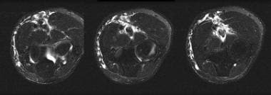
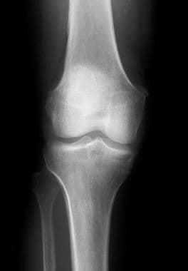
Pigmented villonodular synovitis is an uncommon disease that remains a diagnostic challenge. Presenting complaints commonly involve one joint, most often the knee or hip. Symptoms of pain and swelling characteristically have an insidious onset and are slowly progressive. The physical examination may be completely normal. Radiographs of the knee may appear normal or may show a periarticular soft tissue density, expansion of the suprapatellar pouch and local osseous changes confined to the patellofemoral articulation. Radiographs of the hip may show erosions in the head and neck of the femur and acetabulum. Magnetic resonance imaging usually demonstrates key diagnostic features, which include joint effusion, elevation of the joint capsule, hyperplastic synovium and low signal intensity resulting from hemosiderin deposition. The diagnosis of pigmented villonodular synovitis is confirmed by biopsy, and the treatment of choice is synovectomy. (+info)Localized pigmented villonodular synovitis of the knee joint: neoplasm or reactive granuloma? A review of 18 cases. (3/56)
OBJECTIVE: The localized form of pigmented villonodular synovitis of the knee joint is a rare disease with limited alteration of the synovial membrane, the pathogenesis of which is the subject of controversial discussion. METHODS: Eighteen cases have been documented in our hospital since 1976. All of the patients had additional cartilage or meniscus damage. Treatment consisted of excision of the lesion and the adjacent synovial membrane, as well as therapy of the additional damage. RESULTS: The patients who had received such therapy were followed for 3-9 yr, without any clinical, sonographic or magnetic resonance tomographic signs of recurrence. In addition to the lack of a tendency towards recurrence, none of the cases displayed any further characteristics of the diffuse form of villonodular synovitis, such as invasiveness or malignant transformation. CONCLUSIONS: We therefore suggest that pigmented villonodular synovitis of the knee joint should be classified more strictly than before into a potentially neoplastic (diffuse) form and a reactive granulomatous (local) form. From the cases observed, we conclude that degenerative joint lesions may be the cause of the reactive granulomatous form. (+info)Pathology of the synovium. (4/56)
Synovium is specialized mesenchymal tissue that is essential for the appropriate function of the locomotor apparatus. It is the site for a series of pathologic processes that are characteristic, and in some cases specific, to this distinctive tissue. In this article, the normal microscopic anatomy of synovium is briefly reviewed. Synovial proliferative disorders, including pigmented villonodular synovitis, giant cell tumor of tendon sheath, hemosiderotic synovitis, and fatty infiltration of the synovial membrane are discussed. Additionally, the subjects of intrasynovial cartilaginous lesions (primary and secondary synovial chondromatosis) and crystal deposition diseases are reviewed. Finally, the response of synovial tissues to implanted foreign materials that are used in large and small joint arthroplasty are described. (+info)Proliferating cell nuclear antigen labelling index in localised pigmented villo-nodular synovitis and its relationship to the size of nodules. (5/56)
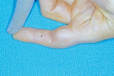
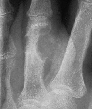
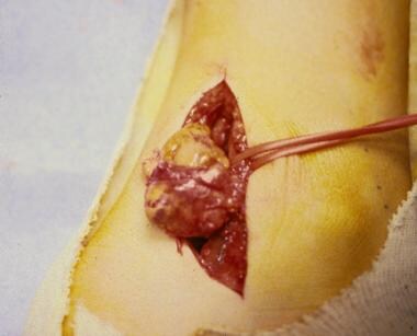
Proliferating cell nuclear antigen (PCNA) is one of the cell cycle-related proteins directly involved in DNA synthesis. It is a marker of cellular proliferation and has been shown to correlate with ploidy and proliferative activity of cells. Its expression has been used to estimate the growth fraction of human cancer and its prognostic value. Pigmented villo-nodular synovitis (PVNS) is characterised by a nodular lesion in the paratendinous synovial tissue or, less frequently, in a joint. Whether PVNS is a neoplastic or inflammatory lesion remains controversial. We have studied immunohistochemical PCNA expression with pc10 monoclonal antibody in 16 paraffin sections, in 16 cases of localised PVNS, or giant cell tumour of tendon sheath. We have found significant correlation between the size of the lesions and PCNA-LI (labelling index). (+info)Pigmented villonodular synovitis of the ankle in an adolescent. (6/56)
We report a case of pigmented villonodular synovitis (PVNS) in an adolescent with monarticular involvement of the ankle and without congenital anomalies or sibling involvement. (+info)Growing wrist mass. (7/56)
A 46 year old man presented with a growing mass over his wrist. Erosions of the triquetrum and hamate were present radiographically. Magnetic resonance imaging (MRI) showed a solid mass arising from the extensor carpi ulnaris tendon, which was T(1) hypointense and isointense, T(2) hypointense, and bloomed on gradient echo images. The preoperative diagnosis of giant cell tumour of the tendon sheath was confirmed on histopathological examination of the excised specimen. The clinical, pathological, and imaging features, with emphasis on MRI findings, of this condition are reviewed. (+info)Synovial hemangioma--a case report. (8/56)
True synovial based hemangiomas are rare lesions and should be differentiated from mass lesions especially pigmented villonodular synovitis. A case of 4 years old male child, presenting with recurrent painful swelling, is described in the present article. The differential diagnosis is also discussed. (+info)Pigmented villonodular synovitis (PVNS) is a rare, benign condition that affects the synovial membrane, which lines the joints. It is characterized by the proliferation of synovial cells and the deposition of hemosiderin, a pigment resulting from the breakdown of blood products. This can lead to joint swelling, pain, stiffness, and limited mobility. PVNS typically affects the large joints such as the knee or hip, but it can also occur in smaller joints, bursae, or tendon sheaths.
There are two forms of PVNS: localized and diffuse. Localized PVNS, also known as giant cell tumor of the tendon sheath, affects a specific area within the joint and is more likely to be treated successfully with surgery. Diffuse PVNS, on the other hand, involves the entire synovial lining of the joint and has a higher recurrence rate even after surgical removal.
The exact cause of PVNS remains unclear, but it is not considered a malignant condition. Treatment usually involves surgical removal of the affected synovium, with or without radiation therapy or chemotherapy to reduce the risk of recurrence. In some cases, arthroscopic surgery may be an option for localized PVNS.
Synovitis is a medical condition characterized by inflammation of the synovial membrane, which is the soft tissue that lines the inner surface of joint capsules and tendon sheaths. The synovial membrane produces synovial fluid, which lubricates the joint and allows for smooth movement.
Inflammation of the synovial membrane can cause it to thicken, redden, and become painful and swollen. This can lead to stiffness, limited mobility, and discomfort in the affected joint or tendon sheath. Synovitis may occur as a result of injury, overuse, infection, or autoimmune diseases such as rheumatoid arthritis.
If left untreated, synovitis can cause irreversible damage to the joint and surrounding tissues, including cartilage loss and bone erosion. Treatment typically involves a combination of medications, physical therapy, and lifestyle modifications to reduce inflammation and manage pain.
Giant cell tumors (GCTs) are a type of benign or rarely malignant bone tumor that is characterized by the presence of multinucleated giant cells. These tumors typically affect adults between the ages of 20 and 40, and they can occur in any bone, but they most commonly involve the long bones near the knee joint.
GCTs are composed of three types of cells: mononuclear stromal cells, which produce the matrix of the tumor; multinucleated osteoclast-like giant cells, which resemble the bone-resorbing cells found in normal bone; and macrophages, which are part of the body's immune system.
The mononuclear stromal cells produce a variety of growth factors that stimulate the formation and activity of the osteoclast-like giant cells, leading to localized bone destruction. The tumor may cause pain, swelling, and limited mobility in the affected area.
While GCTs are typically benign, they can be aggressive and locally destructive, with a tendency to recur after surgical removal. In some cases, GCTs may undergo malignant transformation, leading to the development of sarcomas. Treatment options for GCTs include curettage (scraping out) of the tumor, followed by bone grafting or the use of a cement spacer to fill the defect, and/or adjuvant therapy with radiation or chemotherapy.
Chondroblastoma is a rare, benign (non-cancerous) bone tumor that typically develops in the epiphysis, which is the rounded end of a long bone near a joint. It primarily affects children and adolescents, with around 90% of cases occurring before the age of 20.
The tumor arises from chondroblasts, cells responsible for producing cartilage during bone growth. Chondroblastoma is usually slow-growing and typically causes localized pain, swelling, or tenderness in the affected area. In some cases, it may weaken the bone and lead to fractures.
Treatment generally involves surgical removal of the tumor, followed by curettage (scraping) of the surrounding bone tissue and replacement with bone grafts or substitutes. Recurrence is possible but rare, and long-term prognosis is usually favorable.
Hemosiderin is a golden-brown pigment that consists of iron-containing protein complexes called ferritin and ferrikinase. It is insoluble in water and forms as a result of the breakdown of hemoglobin in the reticuloendothelial system, primarily in macrophages. Hemosiderin deposits can be found in various tissues and organs, such as the spleen, liver, and brain, under conditions of increased red blood cell destruction or impaired iron metabolism. These deposits are often associated with diseases such as hemochromatosis, thalassemia, and chronic inflammation.
Mononeuropathy is a medical condition that refers to damage or dysfunction affecting a single peripheral nerve, outside of the brain and spinal cord. This can result in weakness, numbness, or pain in the area served by that specific nerve. Mononeuropathies can occur due to various reasons such as trauma, compression, infection, or systemic diseases like diabetes. The symptoms and severity may vary depending on the type and location of the affected nerve.
The knee joint, also known as the tibiofemoral joint, is the largest and one of the most complex joints in the human body. It is a synovial joint that connects the thighbone (femur) to the shinbone (tibia). The patella (kneecap), which is a sesamoid bone, is located in front of the knee joint and helps in the extension of the leg.
The knee joint is made up of three articulations: the femorotibial joint between the femur and tibia, the femoropatellar joint between the femur and patella, and the tibiofibular joint between the tibia and fibula. These articulations are surrounded by a fibrous capsule that encloses the synovial membrane, which secretes synovial fluid to lubricate the joint.
The knee joint is stabilized by several ligaments, including the medial and lateral collateral ligaments, which provide stability to the sides of the joint, and the anterior and posterior cruciate ligaments, which prevent excessive forward and backward movement of the tibia relative to the femur. The menisci, which are C-shaped fibrocartilaginous structures located between the femoral condyles and tibial plateaus, also help to stabilize the joint by absorbing shock and distributing weight evenly across the articular surfaces.
The knee joint allows for flexion, extension, and a small amount of rotation, making it essential for activities such as walking, running, jumping, and sitting.
Arthroscopy is a minimally invasive surgical procedure where an orthopedic surgeon uses an arthroscope (a thin tube with a light and camera on the end) to diagnose and treat problems inside a joint. The surgeon makes a small incision, inserts the arthroscope into the joint, and then uses the attached camera to view the inside of the joint on a monitor. They can then insert other small instruments through additional incisions to repair or remove damaged tissue.
Arthroscopy is most commonly used for joints such as the knee, shoulder, hip, ankle, and wrist. It offers several advantages over traditional open surgery, including smaller incisions, less pain and bleeding, faster recovery time, and reduced risk of infection. The procedure can be used to diagnose and treat a wide range of conditions, including torn ligaments or cartilage, inflamed synovial tissue, loose bone or cartilage fragments, and joint damage caused by arthritis.
The synovial membrane, also known as the synovium, is the soft tissue that lines the inner surface of the capsule of a synovial joint, which is a type of joint that allows for smooth movement between bones. This membrane secretes synovial fluid, a viscous substance that lubricates and nourishes the cartilage and helps to reduce friction within the joint during movement.
The synovial membrane has a highly specialized structure, consisting of two layers: the intima and the subintima. The intima is a thin layer of cells that are in direct contact with the synovial fluid, while the subintima is a more fibrous layer that contains blood vessels and nerves.
The main function of the synovial membrane is to produce and regulate the production of synovial fluid, as well as to provide nutrients to the articular cartilage. It also plays a role in the immune response within the joint, helping to protect against infection and inflammation. However, abnormalities in the synovial membrane can lead to conditions such as rheumatoid arthritis, where the membrane becomes inflamed and produces excess synovial fluid, leading to pain, swelling, and joint damage.
The hip joint, also known as the coxal joint, is a ball-and-socket type synovial joint that connects the femur (thigh bone) to the pelvis. The "ball" is the head of the femur, while the "socket" is the acetabulum, a concave surface on the pelvic bone.
The hip joint is surrounded by a strong fibrous capsule and is reinforced by several ligaments, including the iliofemoral, ischiofemoral, and pubofemoral ligaments. The joint allows for flexion, extension, abduction, adduction, medial and lateral rotation, and circumduction movements, making it one of the most mobile joints in the body.
The hip joint is also supported by various muscles, including the gluteus maximus, gluteus medius, gluteus minimus, iliopsoas, and other hip flexors and extensors. These muscles provide stability and strength to the joint, allowing for weight-bearing activities such as walking, running, and jumping.
Medical Definition:
Magnetic Resonance Imaging (MRI) is a non-invasive diagnostic imaging technique that uses a strong magnetic field and radio waves to create detailed cross-sectional or three-dimensional images of the internal structures of the body. The patient lies within a large, cylindrical magnet, and the scanner detects changes in the direction of the magnetic field caused by protons in the body. These changes are then converted into detailed images that help medical professionals to diagnose and monitor various medical conditions, such as tumors, injuries, or diseases affecting the brain, spinal cord, heart, blood vessels, joints, and other internal organs. MRI does not use radiation like computed tomography (CT) scans.
 Villonodular synovitis
Lars Joachim Grimstad
Tenosynovial giant cell tumor
Synovectomy
Hip pain
Tumor-associated macrophage
Ulnar deviation
T2*-weighted imaging
Intermittent hydrarthrosis
Nikos Athanasou
Radiation therapy
List of skin conditions
List of MeSH codes (C05)
Synovial fluid
Synovial osteochondromatosis
List of diseases (P)
Index of trauma and orthopaedics articles
 Pigmented Villonodular Synovitis Imaging: Practice Essentials, Radiography, Computed Tomography
 Treatment of tenosynovial giant cell tumor and pigmented villonodular synovitis
 Pigmented Villonodular Synovitis Treatment & Guidance | Buoy
Pigmented Villonodular synovitis (PVNS) - Sumer's Radiology Blog
Arthroscopy in Pigmented Villonodular Synovitis of the Ankle
Langerhans Cell Granulomatosis
 Open anterior synovectomy for diffuse pigmented villonodular synovitis of the knee Surgical Technique - OrthOracle
 Pigmented Villonodular Synovitis: A Rare Case of Anterior Ankle Impingement | Journal of Orthopaedic Case Reports
 Diffuse pigmented villonodular synovitis (diffuse-type giant cell tumour) of the foot and ankle | Bone & Joint
Villonodular synovitis - Wikipedia
Pigmented villonodular synovitis of the knee joint in a 5-year-old girl treated with combined open and arthroscopic surgery : a...
 Tarsal Tunnel Syndrome | 5-Minute Clinical Consult
 Jagdish MENON | Professor | MS(Ortho) MRCS(Edin) DNB MNAMS FACS PGDHQM | Jawaharlal Institute of Postgraduate Medical Education...
 Radiology Quiz 74271 | Radiopaedia.org
 Vol 63, Issue 4: Aug (2014) | West Indian Medical Journal
 Bone cancer - Doctors and departments - Mayo Clinic
 SICOT e-Newsletter - December 2015: Case of the Month | SICOT
Pes Anserine Bursitis Workup: Approach Considerations, Laboratory Studies, Radiography
Gellissen J[au] - Search Results - PubMed
 Andrew John Cosgarea, M.D., Professor of Orthopaedic Surgery | Johns Hopkins Medicine
 Rare Disease
 Magiran | فهرست مطالب «Meisam Jafari Kafiabadi»
 Turalio (Pexidartinib Capsules): Uses, Dosage, Side Effects, Interactions, Warning
 Frontiers | Unbiased transcriptome mapping and modeling identify candidate genes and compounds of osteoarthritis
 T1-weighted image, knee | Radsource
 Recurrent cyclops lesion after primary anterior cruciate ligament reconstruction using bone tendon bone allograft: A case report
 Bilateral synovial chondromatosis of the elbow in an adolescent: a case report and literature review | BMC Musculoskeletal...
 Knee Pain
PVNS15
- Pigmented villonodular synovitis (PVNS), also known as diffuse tenosynovial giant cell tumor, is a benign proliferative disorder of uncertain etiology that affects synovial lined joints, bursae, and tendon sheaths (see the images below). (medscape.com)
- To review recent developments in the molecular pathogenesis of tenosynovial giant cell tumor (TGCT) or pigmented villonodular synovitis (PVNS) and its therapeutic implications. (nih.gov)
- In the following report, LCG was manifested as a villous synovial proliferation mimicking pigmented villonodular synovitis (PVNS). (medscape.com)
- Pigmented villonodular synovitis (PVNS) is a rare, benign locally aggressive disorder of the synovium of joints, bursae and tendon sheaths and has three main subtypes. (orthoracle.com)
- In this paper, we report a rare case of pigmented villonodular synovitis (PVNS) in a 37-year-old Caucasian male soccer player, with a 4-year story of ankle swelling and ROM painful limitation. (jocr.co.in)
- Pigmented villonodular synovitis (PVNS) is a rare benign disease of the synovium of joints and tendon sheaths, which may be locally aggressive. (boneandjoint.org.uk)
- Pigmented villonodular synovitis (PVNS) is a rare benign proliferative condition affecting synovial membranes of joints, bursae or tendons, resulting from possibly neoplastic synovial proliferation with villous and nodular projections and haemosiderin deposition. (sicot.org)
- Pigmented villonodular synovitis (PVNS) of the knee joint: magnetic resonance imaging (MRI) using standard and dynamic paramagnetic contrast media. (sicot.org)
- In addition to melanoma and other solid tumors to be studied in this collaborative trial, PLX3397 is being evaluated in several other clinical indications, including tenosynovial giant cell tumor (TGCT), historically called pigmented villonodular synovitis (PVNS) or giant cell tumor of the tendon sheath (GCT-TS), breast cancer and glioblastoma. (merck.com)
- TGCT is also known as giant cell tumor of the tendon sheath (GCT-TS) or pigmented villonodular synovitis (PVNS). (rxwiki.com)
- Pigmented Villonodular Synovitis (PVNS) is a medical condition that refers to the thickening of the synovial membrane. (orthotexas.com)
- The main purpose of this study is to gather information about the investigational drug pexidartinib, which may help to treat tumors of pigmented villonodular synovitis (PVNS) or giant cell tumor of the tendon sheath (GCT-TS). (stanford.edu)
- The financing will enable SynOx to continue the development of emactuzumab, for the treatment of diffuse tenosynovial giant cell tumours (TGCT), also known as pigmented villonodular synovitis (PVNS), and other indications. (privateequitywire.co.uk)
- Tenosynovial giant cell tumor (also known as pigmented villonodular synovitis [PVNS]) causes the lining of the joint to become swollen and grow. (msdmanuals.com)
- Pigmented Villonodular Synovitis (PVNS) our Co-Founder's patient experience with ultra rare disease. (fibroflutters.com)
Arthroscopy2
- Given the limited soft tissues surrounding the ankle in conjunction with the ability of arthroscopy to easily access the entire joint, arthroscopic management of ankle pigmented villonodular synovitis allows for successful treatment while minimizing the surgical complications of an open approach. (medscape.com)
- The diagnosis of synovitis with rice body formation was confirmed at arthroscopy. (scielo.org.za)
Tumor2
- Types include: Pigmented villonodular synovitis Giant cell tumor of the tendon sheath Though they have very different names, they have the same histology, and stain positive for CD68, HAM56, and vimentin. (wikipedia.org)
- Letter to the Editor: Does Osteoarticular Allograft Reconstruction Achieve Long-term Survivorship After En Bloc Resection of Grade 3 Giant Cell Tumor Of Bone? (ucdavis.edu)
Pathology1
- Knee swelling is unusual in anterior knee pain and generally implies intra-articular pathology, synovitis or loose body. (biomedcentral.com)
Inflammatory2
- Pigmented villonodular synovitis is a proliferative synovial-based inflammatory process that can lead to joint destruction and debilitating pain. (medscape.com)
- Radiosynovectomy is a safe and repeatable treatment method of chronic synovitis with synovial overgrowth and refractory chronic or acute inflammatory joint effusion. (jultrason.pl)
Synovial Membrane1
- An arthroscopic exam demonstrated a deep, brown-pigmented fronds of synovial membrane. (jhu.edu)
Arthroscopic1
- The favorable outcome in this case suggests that combined open and arthroscopic surgery may be an effective method for treating pigmented villonodular synovitis in skeletally immature patients. (tokushima-u.ac.jp)
Ankle4
- Arthroscopically assisted synovectomy in patients with pigmented villonodular synovitis of the ankle and hindfoot is an effective treatment option, providing adequate visualization for complete excision while minimizing soft-tissue complications related to larger surgical exposures. (medscape.com)
- Pigmented villonodular synovitis should be considered in diagnostic flow chart of anterior ankle impingement. (jocr.co.in)
- Ankle, impingement, pigmented villonodular synovitis. (jocr.co.in)
- Stevenson JD, Jaiswal A, Gregory JJ, Mangham DC, Cribb G, Cool P. Diffuse pigmented villonodular synovitis (diffuse-type giant cell tumour) of the foot and ankle. (boneandjoint.org.uk)
Synovium2
- Ours is the first reported case presenting clinically in the synovium of the hip joint as pigmented villonodular synovitis. (medscape.com)
- Pigmented villonodular synovitis is an uncommon disease characterized by hyperplastic synovium, large effusions and bone erosions. (radiopaedia.org)
Diagnosis2
- Postoperative histopathological examination confirmed the diagnosis of diffuse pigmented villonodular synovitis. (tokushima-u.ac.jp)
- Pigmented villonodular synovitis of the knee: diagnosis and treatment. (sicot.org)
Anterior2
- Learn the Open anterior synovectomy for diffuse pigmented villonodular synovitis of the knee surgical technique with step by step instructions on OrthOracle. (orthoracle.com)
- Our e-learning platform contains high resolution images and a certified CME of the Open anterior synovectomy for diffuse pigmented villonodular synovitis of the knee surgical procedure. (orthoracle.com)
Treatment3
- Surgical excision is the "gold standard" for treatment of pigmented villonodular synovitis. (medscape.com)
- Treatment of Diffuse Pigmented Villonodular Synovitis of the knee with combined surgical and radio synovectomy. (sicot.org)
- He maintains a database that tracks surgical treatment in patients with pigmented villonodular synovitis. (hopkinsmedicine.org)
Joints1
- Additionally, three patients with pigmented villonodular synovitis in the tibiotalar and subtalar joints who were successfully treated with arthroscopically assisted synovectomy are reported.Three patients with pigmented villonodular synovitis in the tibiotalar and subtalar joints underwent arthroscopically assisted synovectomy without adjuvant radiotherapy. (medscape.com)
Findings1
- We report the case of a 5-year-old girl with left knee pain and swelling who was diagnosed with diffuse pigmented villonodular synovitis of the left knee based on MRI findings. (tokushima-u.ac.jp)
Patients2
- Pigmented villonodular synovitis is an extremely rare disease in skeletally immature patients. (tokushima-u.ac.jp)
- The important differential diagnoses that need to be excluded in patients with rice bodies are synovial chondromatosis and pigmented villonodular synovitis. (scielo.org.za)
Bodies1
- stage III is the regression of synovitis, with only a single or multiple loose bodies. (biomedcentral.com)
Case1
- We report a case of villonodular synovitis that, also less common, has to be considered in diagnostic flowchart. (jocr.co.in)
Generally1
- The term pigmented villonodular synovitis is generally used when diffuse intraarticular involvement is present (2,3). (facmedicine.com)
Type1
- Villonodular synovitis is a type of synovial swelling. (wikipedia.org)
Tenosynovial giant cell tumor5
- 2. Distinct extra-articular invasion patterns of diffuse pigmented villonodular synovitis/tenosynovial giant cell tumor in the knee joints. (nih.gov)
- 7. Giant cell tumor of tendon sheath, tenosynovial giant cell tumor, and pigmented villonodular synovitis: defining the presentation, surgical therapy and recurrence. (nih.gov)
- 17. Long-term outcome of the treatment of high-risk tenosynovial giant cell tumor/pigmented villonodular synovitis with radiotherapy and surgery. (nih.gov)
- However, 2 conditions-synovial chondromatosis and tenosynovial giant cell tumor (pigmented villonodular synovitis)-occur in the lining (synovium) of joints. (msdmanuals.com)
- Tenosynovial giant cell tumor (previously called pigmented villonodular synovitis) is considered a benign neoplastic tumor of the synovium that can occur around as well as in a joint. (msdmanuals.com)
Tenosynovitis5
- Histopathological examination revealed pigmented villonodular tenosynovitis which stained positive for CD 68. (scirp.org)
- Conclusion: Pigmented villonodular tenosynovitis of temporomandibular joint is a rare entity. (scirp.org)
- in 1941 [2] who first introduced the term pigmented villonodular tenosynovitis. (scirp.org)
- 4. Jaffe HL, Litchtenstein L, Sutro C. Pigmented Villonodular Synovitis: bursitis and tenosynovitis. (jbstjournal.com)
- 20. Myers B.W, Masi A.T. Pigmented villonodular synovitis and tenosynovitis: a clinical epidemiological study of 166 cases and literature review. (jbstjournal.com)
Tendon sheath2
- Types include: Pigmented villonodular synovitis Giant cell tumor of the tendon sheath Though they have very different names, they have the same histology, and stain positive for CD68, HAM56, and vimentin. (wikipedia.org)
- 5. Llauger J, Palmer J, Roson N. Pigmented villonodular synovitis and giant cell tumors of the tendon sheath: radiologic and pathologic features. (jbstjournal.com)
Malignant1
- 14. Malignant giant cell tumor of the tendon sheaths and joints (malignant pigmented villonodular synovitis). (nih.gov)
Recurrence1
- High recurrence and good functional results after arthroscopic resection of pigmented villonodular synovitis. (upf.edu)
Synovial cell1
- 11. Molecular pathways involved in synovial cell inflammation and tumoral proliferation in diffuse pigmented villonodular synovitis. (nih.gov)
Knee6
- A 47-year-old white woman presents with right knee villonodular synovitis. (medscape.com)
- [ 1 ] Davidson and Bentley [ 1 ] reported a case of a nodular variant of pigmented villonodular synovitis of the knee that developed following laser treatment. (medscape.com)
- Pigmented villonodular synovitis of the knee. (medscape.com)
- Eighty per cent of the time pigmented villonodular synovitis affects just one joint of the body, primarily the knee joint. (eorthopod.com)
- C). During his follow-up, the patient developed multifocal pigmented villonodular synovitis which first affected the left knee and shortly after both elbows. (nih.gov)
- Pigmented villonodular synovitis can affect people of all ages, but it occurs most often in young adults from 20 to 50 years of age and most commonly occurs in the knee. (bicmd.com)
Inflammation2
- Pigmented villonodular synovitis is most often painless inflammation or swelling, and overgrowth of the lining of a joint. (eorthopod.com)
- In our body, the dis-order, Synovitis is usually painful, particularly on motion, indicative to a specific set of signs, symptoms or other health indicators, associated with an inflammation of a synovial membrane characterized by a fluctuating swelling due to effusion within a synovial sac. (wellnessadvantage.com)
Proliferation1
- INTRODUCTION: Pigmented villonodular synovitis (PVS) is a synovial proliferation disorder of uncertain aetiology, with some controversy as regards its proper treatment. (upf.edu)
Lesions1
- Pigmented villonodular synovitis and related lesions: the spectrum of imaging findings. (jbstjournal.com)
MACROPHAGES1
- pigmented HEMOSIDERIN -laden MACROPHAGES and inflammatory infiltrate. (bvsalud.org)
Magnetic1
- 19. Ugai K., Morimoto K. Magnetic resonance imaging of pigmented villonodular synovitis in subtalar joint. (jbstjournal.com)
Giant1
- 6. Tenosynovial giant cell tumour/pigmented villonodular synovitis: outcome of 294 patients before the era of kinase inhibitors. (nih.gov)
Occurs1
- Pigmented villonodular synovitis occurs in less than two persons per million per year. (eorthopod.com)
Joint2
- Dynamic stereometry to assess condylar movements relative to the fossa was performed at the 5 year follow-up of a patient who underwent condylar resection of the right TMJ followed by total alloplastic joint reconstruction to treat pigmented villonodular synovitis. (uzh.ch)
- Pigmented villonodular synovitis of the talonavicular joint: A case report and review of the literature. (jbstjournal.com)