Temporomandibular Joint Disorders
Temporomandibular Joint
Temporomandibular Joint Disc
Facial Pain
Temporomandibular Joint Dysfunction Syndrome
Shamanism
Dislocations
Central Nervous System Sensitization
Mandibular Condyle
Chondromatosis, Synovial
Joint Diseases
Osteoarthritis
Masticatory Muscles
Pterygoid Muscles
Temporal Bone
Hip Dysplasia, Canine
Occlusal Splints
Joints
Jaw Relation Record
Mandible
Arthritis
Fibrocartilage
Serotonin 5-HT2 Receptor Antagonists
Pain Measurement
Motivation for and satisfaction with orthodontic-surgical treatment: a retrospective study of 28 patients. (1/493)
Motivation for starting treatment and satisfaction with treatment results were evaluated on the basis of replies to a 14-item questionnaire and clinical examination of 28 orthognathic patients from 6 months to 2 years after treatment. The most common reasons for seeking professional help were problems in biting and chewing (68 per cent). Another major reason was dissatisfaction with facial appearance (36 per cent). Many patients also complained of temporomandibular joint symptoms (32 per cent) and headache (32 per cent). Women (8/19) were more often dissatisfied with their facial appearance than men (2/9), but the difference was not statistically significant. In agreement with earlier studies, the results of orthognathic treatment fulfilled the expectations of almost every patient. Nearly 100 per cent of the patients (27/28) were satisfied with treatment results, although 40 per cent experienced some degree of numbness in the lips and/or jaw 1 year post-operatively. The most satisfied patients were those who stated temporomandibular disorders as the main reason for seeking treatment and whose PAR-index had improved greatly. The majority of the patients experienced the orthodontic treatment as painful and as the most unpleasant part of the whole treatment, but all the patients were satisfied with the pre-treatment information they were given on orthodontics. Orthodontic-surgical therapy should be of a high professional standard technically, but the psychological aspects are equally important in the treatment protocol. The professionals should make efforts to understand the patient's motivations for and expectations of treatment. Patients should be well prepared for surgery and supported for a long time after to help them to adjust to post-surgical changes. (+info)SAPHO syndrome of the temporomandibular joint associated with sudden deafness. (2/493)
We report a case of arthritis of the temporomandibular joint (TMJ) associated with sclerosing osteomyelitis of the mandible and temporal bone, causing deafness. The presence of a palmoplantar pustulosis established the diagnosis of SAPHO syndrome. SAPHO (an acronym referring to synovitis, acne, palmoplantar pustulosis, hyperostosis, and osteitis) syndrome is defined by the association of characteristic osteoarticular and dermatologic manifestations, with diffuse sclerosing osteomyelitis of the mandible being a part of this entity. We review the literature of SAPHO syndrome with mandibular manifestations and discuss the mechanisms of inflammatory spread from the TMJ to the cochlea. To our knowledge, this is the first description of skull base involvement in a patient with SAPHO syndrome leading to sudden deafness. (+info)Craniofacial pain and motor function: pathogenesis, clinical correlates, and implications. (3/493)
Many structural, behavioral, and pharmacological interventions imply that favorable treatment effects in musculoskeletal pain states are mediated through the correction of muscle function. The common theme of these interventions is captured in the popular idea that structural or psychological factors cause muscle hyperactivity, muscle overwork, muscle fatigue, and ultimately pain. Although symptoms and signs of motor dysfunction can sometimes be explained by changes in structure, there is strong evidence that they can also be caused by pain. This new understanding has resulted in a better appreciation of the pathogenesis of symptoms and signs of the musculoskeletal pain conditions, including the sequence of events that leads to the development of motor dysfunction. With the improved understanding of the relationship between pain and motor function, including the inappropriateness of many clinical assumptions, a new literature emerges that opens the door to exciting therapeutic opportunities. Novel treatments are expected to have a profound impact on the care of musculoskeletal pain and its effect on motor function in the not-too-distant future. (+info)Diagnostic accuracy of film-based, TIFF, and wavelet compressed digital temporomandibular joint images. (4/493)
The purpose of this research was to determine if digitization and the application of various compression routines to digital images of temporomandibular joint (TMJ) radiographs would diminish observer accuracy in the detection of specific osseous characteristics associated with TMJ degenerative joint disease (DJD). Nine observers viewed 6 cropped hard-copy radiographic films each of 34 TMJs (17 radiographic series). Regions of interest measuring 2 in x 2 in were digitized using an 8-bit scanner with transparency adapter at 300 dpi. The images were placed into a montage of 6 images and stored as tagged image file format (TIFF), compressed at 4 levels (25:1, 50:1, 75:1, and 100:1) using a wavelet algorithm, and displayed to the observers on a computer monitor. Their observations regarding condylar faceting, sclerosis, osteophyte formation, erosion, and abnormal shape were analyzed using ROC. Kappa values were determined for relative condylar size and condylar position within the glenoid fossa. Indices were compared using ANOVA at a significance level of P < .05. Although significant and substantial observer variability was demonstrated, there were no statistically significant differences between image modalities, except for condylar position, in which TIFF and wavelet (at all compression ratios) performed better than the original image. For faceting, wavelet 100:1 performed better than radiographic film images. Little actual image file reduction was achieved at compression ratios above 25:1. (+info)Long-term follow-up of clinical symptoms in TMD patients who underwent occlusal reconstruction by orthodontic treatment. (5/493)
Fifty-eight patients (mean age 18.4 years) who had received splint therapy for internal derangement of the temporomandibular joint (TMJ) were examined retrospectively to investigate the efficacy of occlusal reconstruction by orthodontic treatment. The subjects were divided into three groups: 18 patients (mean age 18.6 years) who underwent orthodontic treatment combined with the use of splints (ST group); 27 patients (mean age 18.2 years) who underwent orthodontic treatment without the use of splints (NST group); and 13 patients (mean age 17.9 years) who received only splint therapy for temporomandibular joint disorders (TMD; control group). TMJ sound, pain on movement and restriction of mandibular movement were examined at the initial examination (T1), at the end of the splint therapy for TMD or beginning of orthodontic treatment (T2), at the end of orthodontic treatment (T3), and at recall or 1 year after orthodontic treatment (T4). The following results were found. (1) The percentage of patients with no joint sound at T2 was 20-30 per cent. The percentage of such patients in both the ST and NST groups increased to over 50 per cent at T3, but slightly decreased to 39-50 per cent at T4. There were no significant inter-group differences at any time point. (2) The number of patients who had no pain on movement at T2 was 60-80 per cent. The percentage of such patients in both the ST and NST groups increased to over 90 per cent at T3, but then slightly decreased to 80 per cent at T4. There were no significant inter-group differences at any time point. (3) None of the patients showed restriction of movement of the TMJ at T2 or T4. One patient in the ST group was found to have restriction at T3. There were no significant inter-group differences at any time point. (4) The most frequent type of malocclusion in both ST and NST groups was anterior open bite. These results suggest that TMD symptoms that have been eliminated by splint therapy are not likely to recur due to subsequent orthodontic treatment, but it cannot be concluded that orthodontic treatment itself had a positive effect on TMD symptoms. The results also indicate that there is a relationship between anterior open bite and TMD. (+info)Internal derangements of the temporomandibular joint: the role of arthroscopic surgery and arthrocentesis. (6/493)
Arthroscopic surgery appears to be a safe, minimally invasive and effective method for treating internal derangements of the temporomandibular joint (TMJ), reducing pain and increasing mandibular range of motion for approximately 80% of patients. Although these results are encouraging, they are largely based on retrospective, uncontrolled and short-term studies. The landmark observation that lysis and lavage in only the upper compartment of the TMJ produce successful clinical results without repositioning the disc has prompted clinicians to question the importance of disc position as a significant factor in the etiology of TMJ pain dysfunction. Although there are prospective, controlled, randomized short-term studies indicating that arthrocentesis and arthroscopic surgery have comparable success rates in the management of acute TMJ closed lock, similar long-term studies are lacking. Until they have been done, the roles of arthroscopic surgery and arthrocentesis in the management of TMJ internal derangements remain unclear. (+info)Oral health and juvenile idiopathic arthritis: a review. (7/493)
Juvenile idiopathic arthritis (JIA) results in significant morbidity that includes an adverse impact on oral health that is generally not well recognized. This review describes current literature which demonstrates poor oral health in children with JIA. The impact of JIA on oral health is probably multifactorial and these factors are discussed. This review emphasizes the role of paediatric dentistry in the multidisciplinary management of JIA and highlights the need for further research. (+info)Signs of temporomandibular disorders in girls receiving orthodontic treatment. A prospective and longitudinal comparison with untreated Class II malocclusions and normal occlusion subjects. (8/493)
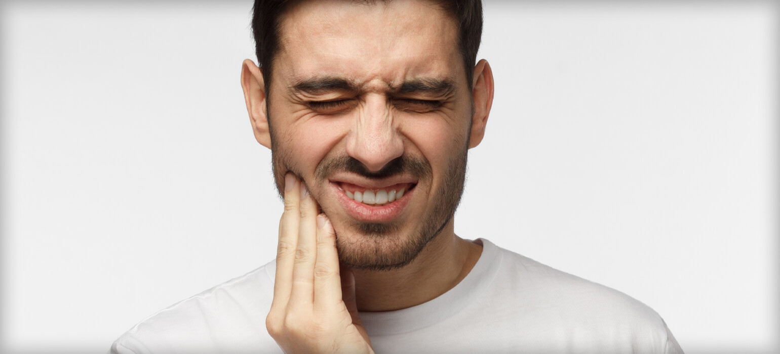
The aim of this investigation was to prospectively and longitudinally study signs of temporomandibular disorders (TMD) and occlusal changes in girls with Class II malocclusion receiving orthodontic treatment and to compare them with subjects with untreated Class II malocclusions and with normal occlusion subjects. Three groups of age-matched adolescent girls were examined for clinical signs of TMD and re-examined 2 years later. Sixty-five Class II subjects received orthodontic fixed straight-wire appliance treatment (Orthodontic group), 58 subjects were orthodontically untreated (Class II group), and 60 subjects had a normal occlusion (Normal group). In the Orthodontic group, the prevalence of muscular signs of TMD was significantly less common post-treatment. The Class II and the Normal groups showed minor changes during the 2-year period. Temporomandibular joint clicking increased in all three groups over the 2 years, but was less common in the Normal group. The Normal group also had a lower overall prevalence of signs of TMD than the Orthodontic and the Class II groups at both registrations. Functional occlusal interferences decreased in the Orthodontic group, but remained the same in the other groups over the 2 years. In conclusion, orthodontic treatment did not increase the risk for or worsen pretreatment signs of TMD. On the contrary, subjects with Class II malocclusions and signs of TMD of muscular origin seemed to benefit functionally from orthodontic treatment in a 2-year perspective. The Normal group had a lower prevalence of signs of TMD than the Orthodontic and the untreated Class II groups. (+info)Temporomandibular Joint Disorders (TMD) refer to a group of conditions that cause pain and dysfunction in the temporomandibular joint (TMJ) and the muscles that control jaw movement. The TMJ is the hinge joint that connects the lower jaw (mandible) to the skull (temporal bone) in front of the ear. It allows for movements required for activities such as eating, speaking, and yawning.
TMD can result from various causes, including:
1. Muscle tension or spasm due to clenching or grinding teeth (bruxism), stress, or jaw misalignment
2. Dislocation or injury of the TMJ disc, which is a small piece of cartilage that acts as a cushion between the bones in the joint
3. Arthritis or other degenerative conditions affecting the TMJ
4. Bite problems (malocclusion) leading to abnormal stress on the TMJ and its surrounding muscles
5. Stress, which can exacerbate existing TMD symptoms by causing muscle tension
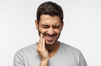
Symptoms of Temporomandibular Joint Disorders may include:
- Pain or tenderness in the jaw, face, neck, or shoulders
- Limited jaw movement or locking of the jaw
- Clicking, popping, or grating sounds when moving the jaw
- Headaches, earaches, or dizziness
- Difficulty chewing or biting
- Swelling on the side of the face
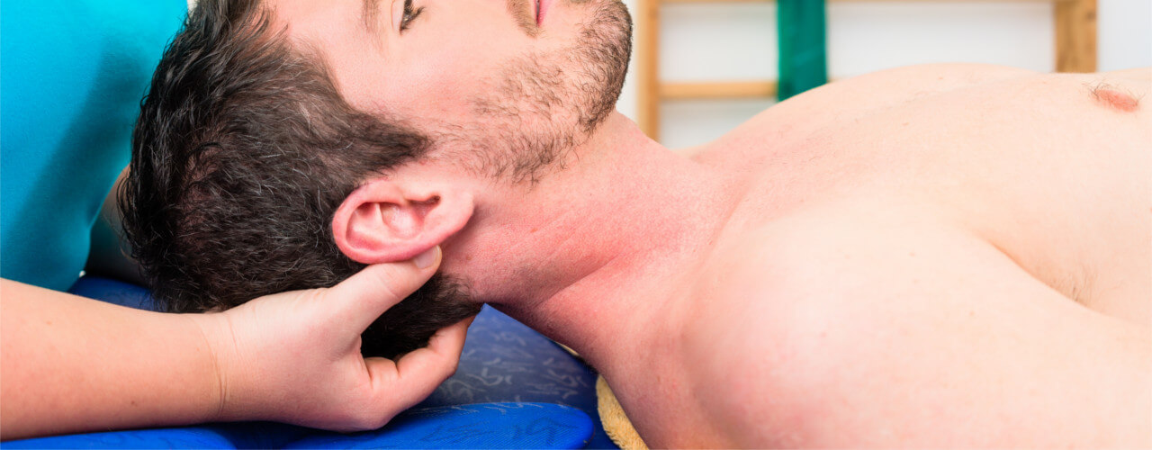
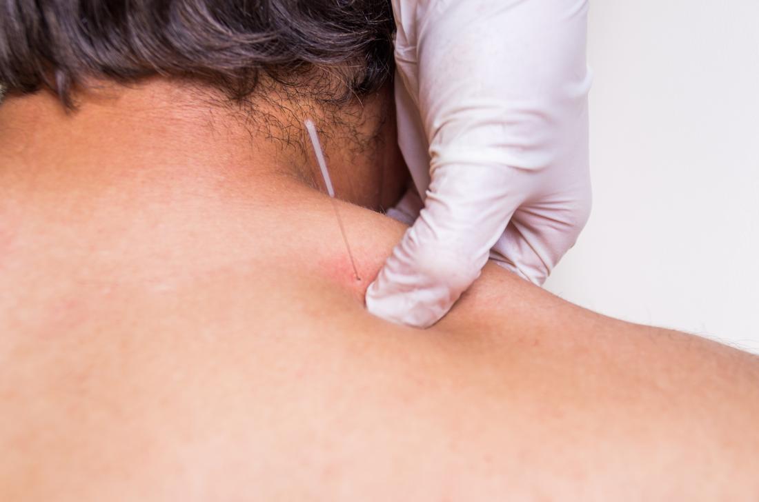
Treatment for TMD varies depending on the severity and cause of the condition. It may include self-care measures (like eating soft foods, avoiding extreme jaw movements, and applying heat or cold packs), physical therapy, medications (such as muscle relaxants, pain relievers, or anti-inflammatory drugs), dental work (including bite adjustments or orthodontic treatment), or even surgery in severe cases.
The temporomandibular joint (TMJ) is the articulation between the mandible (lower jaw) and the temporal bone of the skull. It's a complex joint that involves the movement of two bones, several muscles, and various ligaments. The TMJ allows for movements like rotation and translation, enabling us to open and close our mouth, chew, speak, and yawn. Dysfunction in this joint can lead to temporomandibular joint disorders (TMD), which can cause pain, discomfort, and limited jaw movement.
The temporomandibular joint (TMJ) disc is a small, thin piece of fibrocartilaginous tissue located within the TMJ, which is the joint that connects the mandible (jawbone) to the temporal bone of the skull. The disc acts as a cushion and allows for smooth movement of the jaw during activities such as eating, speaking, and yawning. It divides the joint into two compartments: the upper and lower compartments.
The TMJ disc is composed of several types of tissue, including collagen fibers, elastin fibers, and a small number of cells called fibroblasts. The disc's unique structure allows it to withstand the forces generated during jaw movement and helps to distribute these forces evenly across the joint.
The TMJ disc can become damaged or displaced due to various factors such as trauma, teeth grinding (bruxism), or degenerative joint diseases like osteoarthritis. This can lead to temporomandibular disorders (TMDs) characterized by pain, stiffness, and limited jaw movement.
Facial pain is a condition characterized by discomfort or pain felt in any part of the face. It can result from various causes, including nerve damage or irritation, injuries, infections, dental problems, migraines, or sinus congestion. The pain can range from mild to severe and may be sharp, dull, constant, or intermittent. In some cases, facial pain can also be associated with other symptoms such as headaches, redness, swelling, or changes in sensation. Accurate diagnosis and treatment of the underlying cause are essential for effective management of facial pain.
Temporomandibular Joint Dysfunction Syndrome, often abbreviated as TMJD or TMD, is a group of conditions that cause pain and dysfunction in the temporomandibular joint (TMJ) - the joint that connects the jawbone to the skull. Here's a more detailed medical definition:
Temporomandibular Joint Dysfunction Syndrome is a complex disorder characterized by pain, clicking, popping, or grating sounds in the TMJ; limited movement or locking of the jaw; and/or painful chewing movements. The condition may be caused by a variety of factors, including muscle tension, joint inflammation, structural problems with the joint itself, or injury to the head, neck, or jaw.
Symptoms of TMJD can include:
- Pain or tenderness in the face, jaw joint area, neck, and/or shoulders
- Limited ability to open the mouth wide
- Jaw locking, making it difficult to close or open the mouth
- Clicking, popping, or grating sounds in the TMJ when opening or closing the mouth
- A significant change in the way the upper and lower teeth fit together
- Headaches, earaches, dizziness, and hearing problems
Treatment for TMJD can vary depending on the severity of the condition and its underlying cause. It may include self-care practices such as eating soft foods, avoiding extreme jaw movements, and practicing relaxation techniques; physical therapy; medication to reduce pain and inflammation; dental treatments such as mouthguards or bite adjustments; and, in rare cases, surgery.
Shamanism is not a medical term, but rather a cultural and anthropological concept. It refers to the religious or spiritual practices of certain indigenous cultures, particularly in Asia, Africa, and the Americas. A shaman is a community leader or healer who uses altered states of consciousness, often induced by rhythmic drumming or trance-inducing plants, to communicate with spirits or supernatural entities. They believe that these interactions can help diagnose and treat illnesses, provide guidance, and ensure the wellbeing of their community.
While shamanic practices are not considered a medical treatment in Western medicine, some elements of shamanism, such as the use of plants for healing purposes, have been incorporated into complementary and alternative medicine approaches. However, it is important to note that these practices should not replace evidence-based medical treatments.
A dislocation is a condition in which a bone slips out of its normal position in a joint. This can happen as a result of trauma or injury, such as a fall or direct blow to the body. Dislocations can cause pain, swelling, and limited mobility in the affected area. In some cases, a dislocation may also damage surrounding tissues, such as ligaments, tendons, and nerves.
Dislocations are typically treated by reducing the dislocation, which means putting the bone back into its normal position. This is usually done with the help of medication to relieve pain and relaxation techniques to help the person stay still during the reduction. In some cases, surgery may be necessary to repair damaged tissues or if the dislocation cannot be reduced through other methods. After the dislocation has been reduced, the joint may be immobilized with a splint or sling to allow it to heal properly.
It is important to seek medical attention promptly if you suspect that you have a dislocation. If left untreated, a dislocation can lead to further complications, such as joint instability and chronic pain.
Central nervous system (CNS) sensitization refers to a state in which the CNS, specifically the brain and spinal cord, becomes increasingly hypersensitive to stimuli. This heightened sensitivity results in an amplified response to painful or non-painful stimuli.
In CNS sensitization, there is an increased responsiveness of neurons in the CNS, leading to a lower threshold for activation and an enhanced transmission of nociceptive (pain) signals. This can occur due to various factors such as tissue injury, inflammation, or nerve damage, which trigger changes in the nervous system that contribute to the development and maintenance of chronic pain conditions.
CNS sensitization is associated with functional and structural reorganization within the CNS, including alterations in neurotransmitter release, ion channel function, and synaptic plasticity. These changes can result in long-term modifications in the processing and perception of pain, making it more difficult to manage and treat chronic pain conditions.
The mandibular condyle is a part of the temporomandibular joint (TMJ) in the human body. It is a rounded eminence at the end of the mandible (lower jawbone) that articulates with the glenoid fossa of the temporal bone in the skull, allowing for movements such as opening and closing the mouth, chewing, speaking, and swallowing. The mandibular condyle has both a fibrocartilaginous articular surface and a synovial joint capsule surrounding it, which provides protection and lubrication during these movements.
Ankylosis is a medical term that refers to the abnormal joining or fusion of bones, typically in a joint. This can occur as a result of various conditions such as injury, infection, or inflammatory diseases like rheumatoid arthritis. The fusion of bones can restrict movement and cause stiffness in the affected joint. In some cases, ankylosis can lead to deformity and disability if not treated promptly and effectively.
There are different types of ankylosis depending on the location and extent of bone fusion. For instance, when it affects the spine, it is called "ankylosing spondylitis," which is a chronic inflammatory disease that can cause stiffness and pain in the joints between the vertebrae.
Treatment for ankylosis depends on the underlying cause and severity of the condition. In some cases, physical therapy or surgery may be necessary to restore mobility and function to the affected joint.
Bruxism is the medical term for grinding or clenching your teeth. It's often an unconscious habit that can occur during the day or at night (nocturnal bruxism). Mild bruxism may not require treatment, but chronic, severe grinding can lead to jaw disorders, headaches, and damaged teeth.
There are several potential causes of bruxism, including stress, anxiety, certain medications, alcohol and drug use, and sleep disorders. Dentists often diagnose bruxism based on the visible signs of wear on your teeth, or they may ask you about symptoms you're experiencing. Treatment for bruxism can include stress management techniques, dental guards to protect your teeth during sleep, and in some cases, medication.
Synovial chondromatosis is a rare condition that affects the synovial membrane, which is the lining of joints, bursae (fluid-filled sacs that cushion bones), and tendon sheaths. In this condition, nodules made up of cartilage form in the synovial membrane. These nodules can detach from the synovial membrane and float freely in the synovial fluid, which lubricates the joint. If they become numerous, they can cause joint pain, stiffness, and decreased range of motion. In some cases, the loose bodies may also cause locking or catching sensations in the joint. Surgery is typically required to remove the cartilaginous nodules and relieve symptoms. If left untreated, synovial chondromatosis can lead to osteoarthritis and other joint problems.
Facial asymmetry refers to a condition in which the facial features are not identical or proportionate on both sides of a vertical line drawn down the middle of the face. This can include differences in the size, shape, or positioning of facial features such as the eyes, ears, nose, cheeks, and jaw. Facial asymmetry can be mild and barely noticeable, or it can be more severe and affect a person's appearance and/or functionality of the mouth and jaw.
Facial asymmetry can be present at birth (congenital) or can develop later in life due to various factors such as injury, surgery, growth disorders, nerve damage, or tumors. In some cases, facial asymmetry may not cause any medical problems and may only be of cosmetic concern. However, in other cases, it may indicate an underlying medical condition that requires treatment.
Depending on the severity and cause of the facial asymmetry, treatment options may include cosmetic procedures such as fillers or surgery, orthodontic treatment, physical therapy, or medication to address any underlying conditions.
Joint diseases is a broad term that refers to various conditions affecting the joints, including but not limited to:
1. Osteoarthritis (OA): A degenerative joint disease characterized by the breakdown of cartilage and underlying bone, leading to pain, stiffness, and potential loss of function.
2. Rheumatoid Arthritis (RA): An autoimmune disorder causing inflammation in the synovial membrane lining the joints, resulting in swelling, pain, and joint damage if left untreated.
3. Infectious Arthritis: Joint inflammation caused by bacterial, viral, or fungal infections that spread through the bloodstream or directly enter the joint space.
4. Gout: A type of arthritis resulting from the buildup of uric acid crystals in the joints, typically affecting the big toe and characterized by sudden attacks of severe pain, redness, and swelling.
5. Psoriatic Arthritis (PsA): An inflammatory joint disease associated with psoriasis, causing symptoms such as pain, stiffness, and swelling in the joints and surrounding tissues.
6. Juvenile Idiopathic Arthritis (JIA): A group of chronic arthritis conditions affecting children, characterized by joint inflammation, pain, and stiffness.
7. Ankylosing Spondylitis: A form of arthritis primarily affecting the spine, causing inflammation, pain, and potential fusion of spinal vertebrae.
8. Bursitis: Inflammation of the fluid-filled sacs (bursae) that cushion joints, leading to pain and swelling.
9. Tendinitis: Inflammation or degeneration of tendons, which connect muscles to bones, often resulting in pain and stiffness near joints.
These conditions can impact the function and mobility of affected joints, causing discomfort and limiting daily activities. Proper diagnosis and treatment are essential for managing joint diseases and preserving joint health.
Osteoarthritis (OA) is a type of joint disease that is characterized by the breakdown and eventual loss of cartilage - the tissue that cushions the ends of bones where they meet in the joints. This breakdown can cause the bones to rub against each other, causing pain, stiffness, and loss of mobility. OA can occur in any joint, but it most commonly affects the hands, knees, hips, and spine. It is often associated with aging and can be caused or worsened by obesity, injury, or overuse.
The medical definition of osteoarthritis is: "a degenerative, non-inflammatory joint disease characterized by the loss of articular cartilage, bone remodeling, and the formation of osteophytes (bone spurs). It is often associated with pain, stiffness, and decreased range of motion in the affected joint."
Masticatory muscles are a group of skeletal muscles responsible for the mastication (chewing) process in humans and other animals. They include:
1. Masseter muscle: This is the primary muscle for chewing and is located on the sides of the face, running from the lower jawbone (mandible) to the cheekbone (zygomatic arch). It helps close the mouth and elevate the mandible during chewing.
2. Temporalis muscle: This muscle is situated in the temporal region of the skull, covering the temple area. It assists in closing the jaw, retracting the mandible, and moving it sideways during chewing.
3. Medial pterygoid muscle: Located deep within the cheek, near the angle of the lower jaw, this muscle helps move the mandible forward and grind food during chewing. It also contributes to closing the mouth.
4. Lateral pterygoid muscle: Found inside the ramus (the vertical part) of the mandible, this muscle has two heads - superior and inferior. The superior head helps open the mouth by pulling the temporomandibular joint (TMJ) downwards, while the inferior head assists in moving the mandible sideways during chewing.
These muscles work together to enable efficient chewing and food breakdown, preparing it for swallowing and digestion.
The pterygoid muscles are a pair of muscles located in the deep part of the lateral aspect of the nasopharynx, in the human head. They are part of the group of muscles known as the muscles of mastication, which are involved in the chewing process.
There are two sets of pterygoid muscles: the medial and lateral pterygoids. The medial pterygoids are located deep within the jaw, near the temporomandibular joint (TMJ). They originate from the medial surface of the lateral pterygoid plate of the sphenoid bone and insert onto the inner aspect of the angle of the mandible (lower jawbone). The main function of the medial pterygoids is to assist in closing the jaw and moving it forward during chewing.
The lateral pterygoids, on the other hand, are located more superficially than the medial pterygoids and are situated near the TMJ. They have two heads: the upper head originates from the greater wing of the sphenoid bone, while the lower head arises from the lateral surface of the lateral pterygoid plate. The lateral pterygoids insert onto the front part of the neck of the mandible and the disc of the TMJ. Their main function is to assist in opening the jaw and moving it sideways during chewing.
Together, the pterygoid muscles play a crucial role in the movement and function of the jaw, allowing us to chew food effectively and speak clearly.
Arthralgia is a medical term that refers to pain in the joints. It does not involve inflammation, which would be referred to as arthritis. The pain can range from mild to severe and may occur in one or multiple joints. Arthralgia can have various causes, including injuries, infections, degenerative conditions, or systemic diseases. In some cases, the underlying cause of arthralgia remains unknown. Treatment typically focuses on managing the pain and addressing the underlying condition if it can be identified.
The temporal bone is a paired bone that is located on each side of the skull, forming part of the lateral and inferior walls of the cranial cavity. It is one of the most complex bones in the human body and has several important structures associated with it. The main functions of the temporal bone include protecting the middle and inner ear, providing attachment for various muscles of the head and neck, and forming part of the base of the skull.
The temporal bone is divided into several parts, including the squamous part, the petrous part, the tympanic part, and the styloid process. The squamous part forms the lateral portion of the temporal bone and articulates with the parietal bone. The petrous part is the most medial and superior portion of the temporal bone and contains the inner ear and the semicircular canals. The tympanic part forms the lower and anterior portions of the temporal bone and includes the external auditory meatus or ear canal. The styloid process is a long, slender projection that extends downward from the inferior aspect of the temporal bone and serves as an attachment site for various muscles and ligaments.
The temporal bone plays a crucial role in hearing and balance, as it contains the structures of the middle and inner ear, including the oval window, round window, cochlea, vestibule, and semicircular canals. The stapes bone, one of the three bones in the middle ear, is entirely encased within the petrous portion of the temporal bone. Additionally, the temporal bone contains important structures for facial expression and sensation, including the facial nerve, which exits the skull through the stylomastoid foramen, a small opening in the temporal bone.
Canine hip dysplasia (CHD) is a common skeletal disorder in dogs, particularly in large and giant breeds, characterized by the abnormal development and degeneration of the coxofemoral joint - the joint where the head of the femur (thigh bone) meets the acetabulum (hip socket) of the pelvis. This condition is often caused by a combination of genetic and environmental factors that lead to laxity (looseness) of the joint, which can result in osteoarthritis (OA), pain, and decreased mobility over time.
In a healthy hip joint, the femoral head fits snugly into the acetabulum, allowing smooth and stable movement. However, in dogs with CHD, the following abnormalities may occur:
1. Shallow acetabulum: The hip socket may not be deep enough to provide adequate coverage of the femoral head, leading to joint instability.
2. Flared acetabulum: The rim of the acetabulum may become stretched and flared due to excessive forces exerted on it by the lax joint.
3. Misshapen or malformed femoral head: The femoral head may not have a normal round shape, further contributing to joint instability.
4. Laxity of the joint: The ligament that holds the femoral head in place within the acetabulum (ligamentum teres) can become stretched, allowing for excessive movement and abnormal wear of the joint surfaces.
These changes can lead to the development of osteoarthritis, which is characterized by the breakdown and loss of cartilage within the joint, as well as the formation of bone spurs (osteophytes) and thickening of the joint capsule. This results in pain, stiffness, and decreased range of motion, making it difficult for affected dogs to perform everyday activities such as walking, running, or climbing stairs.
Canine hip dysplasia is typically diagnosed through a combination of physical examination, medical history, and imaging techniques such as radiographs (X-rays). Treatment options may include conservative management, such as weight management, exercise modification, joint supplements, and pain medication, or surgical intervention, such as total hip replacement. The choice of treatment depends on the severity of the disease, the age and overall health of the dog, and the owner's financial resources.
Preventing canine hip dysplasia is best achieved through selective breeding practices that aim to eliminate affected animals from breeding populations. Additionally, maintaining a healthy weight, providing appropriate exercise, and ensuring proper nutrition throughout a dog's life can help reduce the risk of developing this debilitating condition.
Occlusal splints, also known as bite guards or night guards, are removable dental appliances that are used to provide protection and stabilization for the teeth and jaw joint (temporomandibular joint or TMJ). They are typically made of hard acrylic or soft materials and are custom-fit to a patient's mouth.
Occlusal splints work by covering and separating the upper and lower teeth, preventing them from coming into contact with each other. This can help to reduce tooth grinding and clenching (bruxism), which can cause tooth wear, sensitivity, and TMJ disorders. They may also be used to help stabilize the jaw joint and muscles in patients with TMJ disorders or to provide protection for teeth that have undergone restorative dental work.
It is important to note that occlusal splints should only be worn under the guidance of a dentist, as improper use can lead to further dental problems.
A joint is the location at which two or more bones make contact. They are constructed to allow movement and provide support and stability to the body during motion. Joints can be classified in several ways, including structure, function, and the type of tissue that forms them. The three main types of joints based on structure are fibrous (or fixed), cartilaginous, and synovial (or diarthrosis). Fibrous joints do not have a cavity and have limited movement, while cartilaginous joints allow for some movement and are connected by cartilage. Synovial joints, the most common and most movable type, have a space between the articular surfaces containing synovial fluid, which reduces friction and wear. Examples of synovial joints include hinge, pivot, ball-and-socket, saddle, and condyloid joints.
A Jaw Relation Record (also known as a "mounted cast" or "articulated record") is a dental term used to describe the process of recording and replicating the precise spatial relationship between the upper and lower jaws. This information is crucial in various dental treatments, such as designing and creating dental restorations, dentures, or orthodontic appliances.
The Jaw Relation Record typically involves these steps:
1. Determining the optimal jaw position (occlusion) during a clinical procedure called "bite registration." This is done by using various materials like waxes, silicones, or impression compounds to record the relationship between the upper and lower teeth in a static position or at specific movements.
2. Transferring this bite registration to an articulator, which is a mechanical device that simulates jaw movement. The articulator holds dental casts (replicas of the patient's teeth) and allows for adjustments based on the recorded jaw relationship.
3. Mounting the dental casts onto the articulator according to the bite registration. This creates an accurate representation of the patient's oral structures, allowing dentists or technicians to evaluate, plan, and fabricate dental restorations that will fit harmoniously in the mouth and provide optimal function and aesthetics.
In summary, a Jaw Relation Record is a critical component in dental treatment planning and restoration design, as it captures and replicates the precise spatial relationship between the upper and lower jaws.
The mandible, also known as the lower jaw, is the largest and strongest bone in the human face. It forms the lower portion of the oral cavity and plays a crucial role in various functions such as mastication (chewing), speaking, and swallowing. The mandible is a U-shaped bone that consists of a horizontal part called the body and two vertical parts called rami.
The mandible articulates with the skull at the temporomandibular joints (TMJs) located in front of each ear, allowing for movements like opening and closing the mouth, protrusion, retraction, and side-to-side movement. The mandible contains the lower teeth sockets called alveolar processes, which hold the lower teeth in place.
In medical terminology, the term "mandible" refers specifically to this bone and its associated structures.
Arthritis is a medical condition characterized by inflammation in one or more joints, leading to symptoms such as pain, stiffness, swelling, and reduced range of motion. There are many different types of arthritis, including osteoarthritis, rheumatoid arthritis, psoriatic arthritis, gout, and lupus, among others.
Osteoarthritis is the most common form of arthritis and is caused by wear and tear on the joints over time. Rheumatoid arthritis, on the other hand, is an autoimmune disorder in which the body's immune system mistakenly attacks the joint lining, causing inflammation and damage.
Arthritis can affect people of all ages, including children, although it is more common in older adults. Treatment for arthritis may include medications to manage pain and reduce inflammation, physical therapy, exercise, and in some cases, surgery.
Bite force refers to the amount of force or pressure that can be exerted by the teeth and jaw when biting down or clenching together. It is a measure of an individual's maximum biting strength, typically expressed in units such as pounds (lb) or newtons (N). Bite force is an important factor in various biological and medical contexts, including oral health, nutrition, and the study of animal behavior and evolution.
In humans, bite force can vary widely depending on factors such as age, sex, muscle strength, and dental health. On average, a healthy adult human male may have a maximum bite force of around 150-200 pounds (670-890 newtons), while an adult female may have a bite force of around 100-130 pounds (445-578 newtons). However, these values can vary significantly from person to person.
Abnormalities in bite force can be indicative of various medical conditions or injuries, such as temporomandibular joint disorders (TMD), muscle weakness, or neurological disorders affecting the facial muscles. Assessing and measuring bite force may also be useful in evaluating the effectiveness of dental treatments or appliances, such as dentures or orthodontic devices.
Fibrocartilage is a type of tough, dense connective tissue that contains both collagen fibers and cartilaginous matrix. It is composed of fibroblasts embedded in a extracellular matrix rich in collagen types I and II, proteoglycans and elastin. Fibrocartilage is found in areas of the body where strong, flexible support is required, such as intervertebral discs, menisci (knee cartilage), labrum (shoulder and hip cartilage) and pubic symphysis. It has both the elasticity and flexibility of cartilage and the strength and durability of fibrous tissue. Fibrocartilage can withstand high compressive loads and provides cushioning, shock absorption and stability to the joints and spine.
The masseter muscle is a strong chewing muscle in the jaw. It is a broad, thick, quadrilateral muscle that extends from the zygomatic arch (cheekbone) to the lower jaw (mandible). The masseter muscle has two distinct parts: the superficial part and the deep part.
The superficial part of the masseter muscle originates from the lower border of the zygomatic process of the maxilla and the anterior two-thirds of the inferior border of the zygomatic arch. The fibers of this part run almost vertically downward to insert on the lateral surface of the ramus of the mandible and the coronoid process.
The deep part of the masseter muscle originates from the deep surface of the zygomatic arch and inserts on the medial surface of the ramus of the mandible, blending with the temporalis tendon.
The primary function of the masseter muscle is to elevate the mandible, helping to close the mouth and clench the teeth together during mastication (chewing). It also plays a role in stabilizing the jaw during biting and speaking. The masseter muscle is one of the most powerful muscles in the human body relative to its size.
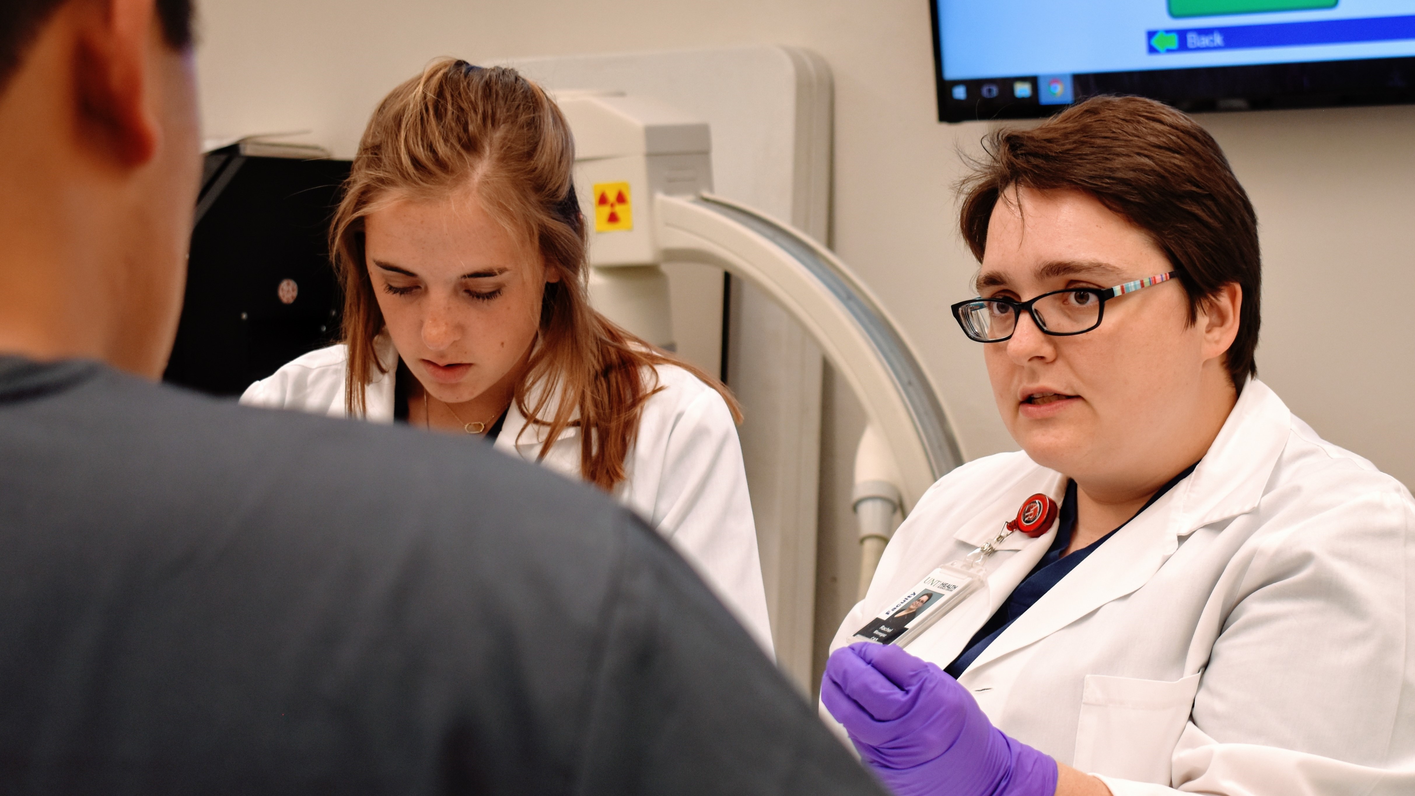
X-ray tomography, also known as computed tomography (CT) or computerized axial tomography (CAT), is a medical imaging technique that uses X-rays to create detailed cross-sectional images of the body. In this technique, an X-ray source and detectors rotate around the patient, acquiring multiple X-ray projections at different angles. A computer then processes these projections to reconstruct tomographic images (slices) of the internal structures of the body, such as bones, organs, and soft tissues.
The term "tomography" comes from the Greek words "tome," meaning slice or section, and "graphein," meaning to write or record. X-ray tomography allows radiologists and other medical professionals to visualize and diagnose various conditions, such as fractures, tumors, infections, and internal injuries, more accurately and efficiently than with traditional X-ray imaging techniques.
It is important to note that while X-ray tomography provides valuable diagnostic information, it does involve exposure to ionizing radiation. Therefore, the benefits of the examination should outweigh the potential risks, and the use of this technique should be justified based on clinical necessity and patient safety considerations.
Serotonin 5-HT2 receptor antagonists are a class of drugs that block the action of serotonin, a neurotransmitter, at 5-HT2 receptors. These receptors are found in the central and peripheral nervous systems and are involved in various physiological functions such as mood regulation, cognition, appetite control, and vasoconstriction.
By blocking the action of serotonin at these receptors, serotonin 5-HT2 receptor antagonists can produce a range of effects depending on the specific receptor subtype that they target. For example, some serotonin 5-HT2 receptor antagonists are used to treat psychiatric disorders such as schizophrenia and depression, while others are used to treat migraines or prevent nausea and vomiting associated with chemotherapy.
Some common examples of serotonin 5-HT2 receptor antagonists include risperidone, olanzapine, and paliperidone (used for the treatment of schizophrenia), mirtazapine (used for the treatment of depression), sumatriptan (used for the treatment of migraines), and ondansetron (used to prevent nausea and vomiting).
Pain measurement, in a medical context, refers to the quantification or evaluation of the intensity and/or unpleasantness of a patient's subjective pain experience. This is typically accomplished through the use of standardized self-report measures such as numerical rating scales (NRS), visual analog scales (VAS), or categorical scales (mild, moderate, severe). In some cases, physiological measures like heart rate, blood pressure, and facial expressions may also be used to supplement self-reported pain ratings. The goal of pain measurement is to help healthcare providers better understand the nature and severity of a patient's pain in order to develop an effective treatment plan.
A joint capsule is the fibrous sac that encloses a synovial joint, which is a type of joint characterized by the presence of a cavity filled with synovial fluid. The joint capsule provides stability and strength to the joint, while also allowing for a range of motion. It consists of two layers: an outer fibrous layer and an inner synovial membrane. The fibrous layer is made up of dense connective tissue that helps to stabilize the joint, while the synovial membrane produces synovial fluid, which lubricates the joint and reduces friction during movement.
 Surgery for temporomandibular joint dysfunction
 MRI of Temporomandibular Joint Disorders
 RFA-DE-03-005: PATHOBIOLOGY OF TEMPOROMANDIBULAR JOINT DISORDERS
 Temporomandibular joint and muscle disorder-type pain and comorbid pains in a national US sample
 JCM | Free Full-Text | Efficacy of Manual Therapy in Temporomandibular Joint Disorders and Its Medium-and Long-Term Effects on...
 TMJ Model- Temporomandibular Joints Disorder - Buyamag INC
 Temporomandibular joint and muscle disorder-type pain in U.S. adults: the National Health Interview Survey - PubMed
 Internal Temporomandibular Joint (TMJ) Derangement - Dental Disorders - MSD Manual Professional Edition
 Temporomandibular Joint Dysfunction: MedlinePlus
 Temporomandibular Joint Disorders
 Temporomandibular Joint Disorder | TMJ Treatment Brunswick
 Temporomandibular joint disorder - Zahnärzte am Augustaplatz
 Temporomandibular Joint Disorders: Causes, Symptoms, & Treatment
 TMJ Disorders | Temporomandibular and Joint Muscle Disorder
 TMJ & TMD: temporomandibular disorders | Delta Dental
 Temporomandibular disorder and generalized joint hypermobility: electromyographic analysis of the masticatory muscles