Thrombophilia
Factor V
Activated Protein C Resistance
Protein S Deficiency
Protein C Deficiency
Pregnancy Complications, Hematologic
Prothrombin
Antithrombin III Deficiency
Factor V Deficiency
Protein S
Protein C
Antiphospholipid Syndrome
Methylenetetrahydrofolate Reductase (NADPH2)
Abruptio Placentae
Blood Coagulation Factor Inhibitors
Venous Thromboembolism
Thromboembolism
Fibrinogens, Abnormal
Heparin, Low-Molecular-Weight
Antithrombins
Blood Coagulation Disorders, Inherited
Risk Factors
Pregnancy
Lupus Coagulation Inhibitor
Antithrombin III
Antibodies, Antiphospholipid
Coagulation Protein Disorders
Oxidoreductases Acting on CH-NH Group Donors
Pre-Eclampsia
Fibrin Fibrinogen Degradation Products
Hyperhomocysteinemia
Blood Coagulation
Heterozygote
Contraceptives, Oral
Varicose Ulcer
Factor VIII
Case-Control Studies
Legg-Calve-Perthes Disease
Fetal Growth Retardation
Pregnancy Complications, Cardiovascular
Mutation
Lebanon
Femur Head Necrosis
Pregnancy Outcome
Blood Coagulation Factors
Single and combined prothrombotic factors in patients with idiopathic venous thromboembolism: prevalence and risk assessment. (1/567)
The inherited thrombophilias--deficiencies of protein C, protein S, and antithrombin III--and the prothrombotic polymorphisms factor V G1691A and factor II G20210A predispose patients toward venous thromboembolism (VTE). The aim of this study was to determine the prevalence of single and combined prothrombotic factors in patients with idiopathic VTE and to estimate the associated risks. The study group consisted of 162 patients referred for work-up of thrombophilia after documented VTE. The controls were 336 consecutively admitted patients. In all subjects factor V G1691A, factor II G20210A, and methylenetetrahydrofolate reductase (MTHFR) C677T were analyzed by specific polymerase chain reactions and restriction enzymes. Activities of antithrombin III and protein C, free protein S antigen, and lupus anticoagulant were determined in a subset of 109 patients who were not receiving oral anticoagulants. The prevalences of heterozygotes and homozygotes for factor V G1691A and factor II G20210A among patients and controls were 40.1% versus 3.9% and 18.5% versus 5.4%, respectively (P=0.0001). The prevalence of homozygotes for MTHFR C677T in patients was 22.8% and in controls, 14.3% (P=0.025). Heterozygous and homozygous factor V G1691A, factor II G20210A, and homozygous MTHFR C677T were found to be independent risk factors for VTE, with odds ratios of 16.3, 3.6, and 2.1, respectively. Two or more polymorphisms were detected in 27 of 162 patients (16.7%) and in 3 of 336 controls (0.9%). Logistic regression analysis disclosed odds ratios of 58.6 (confidence interval [CI], 22.1 to 155.2) for joint occurrence of factor V and factor II polymorphisms, of 35.0 (CI, 14.5 to 84.7) for factor V and MTHFR polymorphisms, and of 7.7 (CI, 3.0 to 19.6) for factor II and MTHFR polymorphisms. Among 109 patients in whom a complete thrombophilic work-up was performed, 74% had at least 1 underlying defect. These data indicate that in most patients referred for evaluation of thrombophilia due to idiopathic VTE, 1 or more underlying genetic predispositions were discernible. The presence of >1 of the prothrombotic polymorphisms was associated with a substantial risk of VTE. (+info)Thrombophilia as a multigenic disease. (2/567)
BACKGROUND AND OBJECTIVE: Venous thrombosis is a common disease annually affecting 1 in 1000 individuals. The multifactorial nature of the disease is illustrated by the frequent identification of one or more predisposing genetic and/or environmental risk factors in thrombosis patients. Most of the genetic defects known today affect the function of the natural anticoagulant pathways and in particular the protein C system. This presentation focuses on the importance of the genetic factors in the pathogenesis of inherited thrombophilia with particular emphasis on those defects which affect the protein C system. INFORMATION SOURCES: Published results in articles covered by the Medline database have been integrated with our original studies in the field of thrombophilia. STATE OF THE ART AND PERSPECTIVES: The risk of venous thrombosis is increased when the hemostatic balance between pro- and anti-coagulant forces is shifted in favor of coagulation. When this is caused by an inherited defect, the resulting hypercoagulable state is a lifelong risk factor for thrombosis. Resistance to activated protein C (APC resistance) is the most common inherited hypercoagulable state found to be associated with venous thrombosis. It is caused by a single point mutation in the factor V (FV) gene, which predicts the substitution of Arg506 with a Gln. Arg506 is one of three APC-cleavage sites and the mutation results in the loss of this APC-cleavage site. The mutation is only found in Caucasians but the prevalence of the mutant FV allele (FV:Q506) varies between countries. It is found to be highly prevalent (up to 15%) in Scandinavian populations, in areas with high incidence of thrombosis. FV:Q506 is associated with a 5-10-fold increased risk of thrombosis and is found in 20-60% of Caucasian patients with thrombosis. The second most common inherited risk factor for thrombosis is a point mutation (G20210A) in the 3' untranslated region of the prothrombin gene. This mutation is present in approximately 2% of healthy individuals and in 6-7% of thrombosis patients, suggesting it to be a mild risk factor of thrombosis. Other less common genetic risk factors for thrombosis are the deficiencies of natural anticoagulant proteins such as antithrombin, protein C or protein S. Such defects are present in less than 1% of healthy individuals and together they account for 5-10% of genetic defects found in patients with venous thrombosis. Owing to the high prevalence of inherited APC resistance (FV:Q506) and of the G20210A mutation in the prothrombin gene, combinations of genetic defects are relatively common in the general population. As each genetic defect is an independent risk factor for thrombosis, individuals with multiple defects have a highly increased risk of thrombosis. As a consequence, multiple defects are often found in patients with thrombosis. (+info)Factor V Leiden and antibodies against phospholipids and protein S in a young woman with recurrent thromboses and abortion. (3/567)
We describe the case of a 39-year-old woman who suffered two iliofemoral venous thromboses, a cerebral ischemic infarct and recurrent fetal loss. Initial studies showed high levels of antiphospholipid antibodies (APAs) and a moderate thrombocytopenia. After her second miscarriage, laboratory diagnosis revealed that the woman was heterozygous for the factor V Leiden mutation and had a functional protein S deficiency as well as anti-protein S and anti-beta 2-glycoprotein I antibodies. The impairment of the protein C pathway at various points could well explain the recurrent thromboses in the patient and supports the role of a disturbed protein C system in the pathophysiology of thrombosis in patients with APAs. (+info)Antiphospholipid antibodies from antiphospholipid syndrome patients activate endothelial cells in vitro and in vivo. (4/567)
BACKGROUND: Antiphospholipid (aPL) antibodies are associated with thrombosis in patients diagnosed with antiphospholipid syndrome (APS) and enhance thrombus formation in vivo in mice, but the mechanism of thrombosis by aPL is not completely understood. Although aPL antibodies have been shown to inhibit protein C activation and activate endothelial cells (ECs) in vitro, no study has examined whether these antibodies activate ECs in vivo. Therefore, human affinity-purified aPL (ap aPL) antibodies from APS patients were tested in a mouse model of microcirculation using the cremaster muscle that allows direct microscopic examination of thrombus formation and adhesion of white blood cells (WBCs) to ECs as an indication of EC activation in vivo. Adhesion molecule expression on human umbilical vein endothelial cells (HUVECs) after aPL exposure was performed to confirm EC activation in vitro. METHODS AND RESULTS: All 6 ap aPL antibodies significantly increased the expression of VCAM-1 (2.3- to 4.4-fold), with one of the antibodies also increasing the expression of E-selectin (1.6-fold) on HUVECs in vitro. In the in vivo experiments, each ap aPL antibody except for 1 preparation increased WBC sticking (mean number of WBCs ranged from 22.7 to 50.6) compared with control (14.4), which correlated with enhanced thrombus formation (mean thrombus size ranged from 1098 to 6476 versus 594 microm2 for control). CONCLUSIONS: Activation of ECs by aPL antibodies in vivo may create a prothrombotic state on ECs, which may be the first pathophysiological event of thrombosis in APS. (+info)Association of the alpha-fibrinogen Thr312Ala polymorphism with poststroke mortality in subjects with atrial fibrillation. (5/567)
BACKGROUND: The alpha-fibrinogen Thr312Ala polymorphism occurs in close proximity to several sites important for factor XIIIa-dependent cross-linking, which raises the possibility that it affects fibrin clot stability. METHODS AND RESULTS: We determined the association of this polymorphism with ischemic stroke, stroke subtype, and poststroke mortality. There was no significant difference in the genotype distributions of patients with acute ischemic stroke (n=519) and healthy control subjects (n=423), nor was there any association of this polymorphism with stroke subtype. In a Cox regression model, a significant interaction between Thr312Ala and atrial fibrillation was identified in relation to poststroke mortality (P=0.002). In subjects in sinus rhythm (n=418), there was no difference according to genotype in the proportion of subjects who survived (approximately 60% in each group), whereas in subjects with atrial fibrillation (n=101), there was decreased survival in those possessing the A allele (TT=42.1%, TA=18%, AA=0%). CONCLUSIONS: The Thr312Ala polymorphism may give rise to an increased susceptibility for embolization of intra-atrial clot, and these findings could have important implications for identifying subjects most at risk of developing thromboembolic complications. (+info)C-reactive protein as a cardiovascular risk factor: more than an epiphenomenon? (6/567)
BACKGROUND: Circulating levels of C-reactive protein (CRP) may constitute an independent risk factor for cardiovascular disease. How CRP as a risk factor is involved in cardiovascular disease is still unclear. METHODS AND RESULTS: By reviewing available studies, we discuss explanations for the associations between CRP and cardiovascular disease. CRP levels within the upper quartile/quintile of the normal range constitute an increased risk for cardiovascular events, both in apparently healthy persons and in persons with preexisting angina pectoris. High CRP responses after acute myocardial infarction indicate an unfavorable outcome, even after correction for other risk factors. This link between CRP and cardiovascular disease has been considered to reflect the response of the body to the inflammatory reactions in the atherosclerotic (coronary) vessels and adjacent myocardium. However, because CRP localizes in infarcted myocardium (with colocalization of activated complement), we hypothesize that CRP may directly interact with atherosclerotic vessels or ischemic myocardium by activation of the complement system, thereby promoting inflammation and thrombosis. CONCLUSIONS: CRP constitutes an independent cardiovascular risk factor. Unraveling the molecular background of this association may provide new directions for prevention of cardiovascular events. (+info)In vitro generation of endothelial microparticles and possible prothrombotic activity in patients with lupus anticoagulant. (7/567)
Microparticles (MPs) resulting from vesiculation of platelets and other blood cells have been extensively documented in vitro and have been found in increased numbers in several vascular diseases, but little is known about MPs of endothelial origin. The aim of this study was to analyze morphological, immunological, and functional characteristics of MPs derived from human umbilical vein endothelial cells (HUVECs) stimulated by TNF, and to investigate whether these MPs are detectable in healthy individuals and in patients with a prothrombotic coagulation abnormality. Electron microscopy evidenced bleb formation on the membrane of TNF-stimulated HUVECs, leading to increased numbers of MPs released in the supernatant. These endothelial microparticles (EMPs) expressed the same antigenic determinants as the corresponding cell surface, both in resting and activated conditions. MPs derived from TNF-stimulated cells induced coagulation in vitro, via a tissue factor/factor VII-dependent pathway. The expression of E-selectin, ICAM-1, alphavbeta3, and PECAM-1 suggests that MPs have an adhesion potential in addition to their procoagulant activity. In patients, labeling with alphavbeta3 was selected to discriminate EMPs from those of other origins. We provide evidence that endothelial-derived MPs are detectable in normal human blood and are increased in patients with a coagulation abnormality characterized by the presence of lupus anticoagulant. Thus, MPs can be induced by TNF in vitro, and may participate in vivo in the dissemination of proadhesive and procoagulant activities in thrombotic disorders. (+info)Homozygotes for prothrombin gene 20210 A allele in a thrombophilic family without clinical manifestations of venous thromboembolism. (8/567)
BACKGROUND AND OBJECTIVE: A new genetic risk factor for venous thromboembolism has recently been described which involves a G to A transition at position 20210 in the 3' untranslated region of the prothrombin gene. To date, only a few homozygotes for this mutation have been reported and in most of cases, they suffered from thrombotic disease. Here, we describe a pedigree including both heterozygous and homozygous subjects for prothrombin (PT) 20210 A. DESIGN AND METHODS: This family was recruited in 1996 as part of our GAIT (Genetic Analysis of Idiopathic Thrombophilia) project. To qualify for the GAIT study, a pedigree was required to have at least 10 living individuals in three or more generations (i.e. extended pedigree). The pedigrees were selected through probands with idiopathic thrombophilia. A complete set of plasma and DNA determinations related to hemostasis was performed on this family. RESULTS: The plasma studies yielded normal results in all of the individuals. The family members who had a history of thromboembolism were heterozygous carriers of the PT 20210 A variant. In addition, 4 relatives who were heterozygous, and two who were homozygous for this A allele, failed to show clinical manifestations. These two homozygotes were 51 and 19 years old. INTERPRETATION AND CONCLUSIONS: This case exemplifies the complexity of thrombotic disease since individuals homozygous for a mutant gene do not exhibit symptoms while heterozygous individuals often do exhibit the disease. This case suggests that the new genetic risk factor for thrombosis (i.e. PT 20210 A) may not be as strong as most of the previously described genetic risk factors. (+info)Thrombophilia is a medical condition characterized by an increased tendency to form blood clots (thrombi) due to various genetic or acquired abnormalities in the coagulation system. These abnormalities can lead to a hypercoagulable state, which can cause thrombosis in both veins and arteries. Commonly identified thrombophilias include factor V Leiden mutation, prothrombin G20210A mutation, antithrombin deficiency, protein C deficiency, and protein S deficiency.
Acquired thrombophilias can be caused by various factors such as antiphospholipid antibody syndrome (APS), malignancies, pregnancy, oral contraceptive use, hormone replacement therapy, and certain medical conditions like inflammatory bowel disease or nephrotic syndrome.
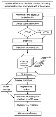
It is essential to diagnose thrombophilia accurately, as it may influence the management of venous thromboembolism (VTE) events and guide decisions regarding prophylactic anticoagulation in high-risk situations.
Factor V, also known as proaccelerin or labile factor, is a protein involved in the coagulation cascade, which is a series of chemical reactions that leads to the formation of a blood clot. Factor V acts as a cofactor for the activation of Factor X to Factor Xa, which is a critical step in the coagulation cascade.
When blood vessels are damaged, the coagulation cascade is initiated to prevent excessive bleeding. During this process, Factor V is activated by thrombin, another protein involved in coagulation, and then forms a complex with activated Factor X and calcium ions on the surface of platelets or other cells. This complex converts prothrombin to thrombin, which then converts fibrinogen to fibrin to form a stable clot.
Deficiency or dysfunction of Factor V can lead to bleeding disorders such as hemophilia B or factor V deficiency, while mutations in the gene encoding Factor V can increase the risk of thrombosis, as seen in the Factor V Leiden mutation.
Activated Protein C (APC) resistance is a condition in which the body's natural anticoagulant system is impaired, leading to an increased risk of thrombosis or blood clot formation. APC is an enzyme that plays a crucial role in regulating blood coagulation by inactivating clotting factors Va and VIIIa.
APC resistance is most commonly caused by a genetic mutation in the Factor V gene, known as Factor V Leiden. This mutation results in the production of a variant form of Factor V called Factor V Leiden, which is resistant to APC-mediated inactivation. As a result, the body's ability to regulate blood clotting is impaired, leading to an increased risk of thrombosis.
APC resistance can be measured by performing a functional assay that compares the activity of APC in normal plasma versus plasma from a patient with suspected APC resistance. The assay measures the rate of inactivation of Factor Va by APC, and a reduced rate of inactivation indicates APC resistance.
It is important to note that not all individuals with APC resistance will develop thrombosis, and other factors such as age, obesity, pregnancy, oral contraceptive use, and smoking can increase the risk of thrombosis in individuals with APC resistance.
Protein S deficiency is a genetic disorder that affects the body's ability to coagulate blood properly. Protein S is a naturally occurring protein in the blood that helps regulate the clotting process by deactivating clotting factors when they are no longer needed. When Protein S levels are too low, it can lead to an increased risk of abnormal blood clots forming within blood vessels, a condition known as thrombophilia.
There are three types of Protein S deficiency: Type I (quantitative deficiency), Type II (qualitative deficiency), and Type III (dysfunctional protein). These types refer to the amount or function of Protein S in the blood. In Type I, there is a decrease in both free and total Protein S levels. In Type II, there is a decrease in functional Protein S despite normal total Protein S levels. In Type III, there is a decrease in free Protein S with normal total Protein S levels.
Protein S deficiency can be inherited or acquired. Inherited forms of the disorder are caused by genetic mutations and are usually present from birth. Acquired forms of Protein S deficiency can develop later in life due to certain medical conditions, such as liver disease, vitamin K deficiency, or the use of certain medications that affect blood clotting.
Symptoms of Protein S deficiency may include recurrent blood clots, usually in the legs (deep vein thrombosis) or lungs (pulmonary embolism), skin discoloration, pain, and swelling in the affected area. In severe cases, it can lead to complications such as chronic leg ulcers, pulmonary hypertension, or damage to the heart or lungs.
Diagnosis of Protein S deficiency typically involves blood tests to measure Protein S levels and function. Treatment may include anticoagulant medications to prevent blood clots from forming or growing larger. Lifestyle modifications such as regular exercise, maintaining a healthy weight, and avoiding smoking can also help reduce the risk of blood clots in people with Protein S deficiency.
Protein C deficiency is a genetic disorder that affects the body's ability to control blood clotting. Protein C is a protein in the blood that helps regulate the formation of blood clots. When blood clots form too easily or do not dissolve properly, they can block blood vessels and lead to serious medical conditions such as deep vein thrombosis (DVT) or pulmonary embolism (PE).
People with protein C deficiency have lower than normal levels of this protein in their blood, which can increase their risk of developing abnormal blood clots. The condition is usually inherited and present from birth, but it may not cause any symptoms until later in life, such as during pregnancy, after surgery, or due to other factors that increase the risk of blood clots.
Protein C deficiency can be classified into two types: type I and type II. Type I deficiency is characterized by lower than normal levels of both functional and immunoreactive protein C in the blood. Type II deficiency is characterized by normal or near-normal levels of immunoreactive protein C, but reduced functional activity.
Protein C deficiency can be diagnosed through blood tests that measure the level and function of protein C in the blood. Treatment may include anticoagulant medications to prevent blood clots from forming or dissolve existing ones. Regular monitoring of protein C levels and careful management of risk factors for blood clots are also important parts of managing this condition.
Hematologic pregnancy complications refer to disorders related to the blood and blood-forming tissues that occur during pregnancy. These complications can have serious consequences for both the mother and the fetus if not properly managed. Some common hematologic pregnancy complications include:
1. Anemia: A condition characterized by a decrease in the number of red blood cells or hemoglobin in the blood, which can lead to fatigue, weakness, and shortness of breath. Iron-deficiency anemia is the most common type of anemia during pregnancy.
2. Thrombocytopenia: A condition characterized by a decrease in the number of platelets (cells that help blood clot) in the blood. Mild thrombocytopenia is relatively common during pregnancy, but severe thrombocytopenia can increase the risk of bleeding during delivery.
3. Gestational thrombotic thrombocytopenic purpura (GTTP): A rare but serious disorder that can cause blood clots to form in small blood vessels throughout the body, leading to a decrease in the number of platelets and red blood cells. GTTP can cause serious complications such as stroke, kidney failure, and even death if not promptly diagnosed and treated.
4. Disseminated intravascular coagulation (DIC): A condition characterized by abnormal clotting and bleeding throughout the body. DIC can be triggered by various conditions such as severe infections, pregnancy complications, or cancer.
5. Hemolysis, elevated liver enzymes, and low platelets (HELLP) syndrome: A serious complication of pregnancy that can cause damage to the liver and lead to bleeding. HELLP syndrome is often associated with preeclampsia, a condition characterized by high blood pressure and damage to organs such as the liver and kidneys.
It's important for pregnant women to receive regular prenatal care to monitor for these and other potential complications, and to seek prompt medical attention if any concerning symptoms arise.
Prothrombin is a protein present in blood plasma, and it's also known as coagulation factor II. It plays a crucial role in the coagulation cascade, which is a complex series of reactions that leads to the formation of a blood clot.
When an injury occurs, the coagulation cascade is initiated to prevent excessive blood loss. Prothrombin is converted into its active form, thrombin, by another factor called factor Xa in the presence of calcium ions, phospholipids, and factor Va. Thrombin then catalyzes the conversion of fibrinogen into fibrin, forming a stable clot.
Prothrombin levels can be measured through a blood test, which is often used to diagnose or monitor conditions related to bleeding or coagulation disorders, such as liver disease or vitamin K deficiency.
Venous thrombosis is a medical condition characterized by the formation of a blood clot (thrombus) in the deep veins, often in the legs (deep vein thrombosis or DVT), but it can also occur in other parts of the body such as the arms, pelvis, or lungs (pulmonary embolism).
The formation of a venous thrombus can be caused by various factors, including injury to the blood vessel wall, changes in blood flow, and alterations in the composition of the blood. These factors can lead to the activation of clotting factors and platelets, which can result in the formation of a clot that blocks the vein.
Symptoms of venous thrombosis may include swelling, pain, warmth, and redness in the affected area. In some cases, the clot can dislodge and travel to other parts of the body, causing potentially life-threatening complications such as pulmonary embolism.
Risk factors for venous thrombosis include advanced age, obesity, smoking, pregnancy, use of hormonal contraceptives or hormone replacement therapy, cancer, recent surgery or trauma, prolonged immobility, and a history of previous venous thromboembolism. Treatment typically involves the use of anticoagulant medications to prevent further clotting and dissolve existing clots.
Antithrombin III (ATIII) deficiency is a genetic disorder that affects the body's ability to regulate blood clotting. ATIII is a protein produced in the liver that inhibits the activity of thrombin and other coagulation factors, preventing excessive clot formation.
People with ATIII deficiency have lower than normal levels of this protein, which can lead to an increased risk of developing abnormal blood clots (thrombosis) in veins, particularly deep vein thrombosis (DVT) and pulmonary embolism (PE). These clots can cause serious complications, including damage to the affected veins, organ damage, and even death.
ATIII deficiency can be classified into two types: type I and type II. Type I is characterized by a quantitative decrease in ATIII levels, while type II is characterized by a qualitative defect that results in reduced functional activity of the protein.
The condition is usually inherited in an autosomal dominant manner, meaning that a person has a 50% chance of inheriting the gene mutation from an affected parent. However, some cases may occur spontaneously due to new mutations in the ATIII gene. Treatment for ATIII deficiency typically involves anticoagulation therapy with medications such as heparin or warfarin to prevent blood clots from forming.
Factor V deficiency is a rare bleeding disorder that is caused by a mutation in the gene that produces coagulation factor V, a protein involved in the clotting process. This condition can lead to excessive bleeding following injury or surgery, and may also cause menorrhagia (heavy menstrual periods) in women.
Factor V deficiency is inherited in an autosomal recessive manner, meaning that an individual must inherit two copies of the mutated gene (one from each parent) in order to develop the condition. People who inherit only one copy of the mutated gene are carriers and may have a milder form of the disorder or no symptoms at all.
Treatment for factor V deficiency typically involves replacement therapy with fresh frozen plasma or clotting factor concentrates, which can help to reduce bleeding episodes and prevent complications. In some cases, medications such as desmopressin or antifibrinolytics may also be used to manage the condition.
Protein S is a vitamin K-dependent protein found in the blood that functions as a natural anticoagulant. It plays a crucial role in regulating the body's clotting system by inhibiting the activation of coagulation factors, thereby preventing excessive blood clotting. Protein S also acts as a cofactor for activated protein C, which is another important anticoagulant protein.
Protein S exists in two forms: free and bound to a protein called C4b-binding protein (C4BP). Only the free form of Protein S has biological activity in inhibiting coagulation. Inherited or acquired deficiencies in Protein S can lead to an increased risk of thrombosis, or abnormal blood clot formation, which can cause various medical conditions such as deep vein thrombosis (DVT) and pulmonary embolism (PE). Regular monitoring of Protein S levels is essential for patients with a history of thrombotic events or those who have a family history of thrombophilia.
Thrombosis is the formation of a blood clot (thrombus) inside a blood vessel, obstructing the flow of blood through the circulatory system. When a clot forms in an artery, it can cut off the supply of oxygen and nutrients to the tissues served by that artery, leading to damage or tissue death. If a thrombus forms in the heart, it can cause a heart attack. If a thrombus breaks off and travels through the bloodstream, it can lodge in a smaller vessel, causing blockage and potentially leading to damage in the organ that the vessel supplies. This is known as an embolism.
Thrombosis can occur due to various factors such as injury to the blood vessel wall, abnormalities in blood flow, or changes in the composition of the blood. Certain medical conditions, medications, and lifestyle factors can increase the risk of thrombosis. Treatment typically involves anticoagulant or thrombolytic therapy to dissolve or prevent further growth of the clot, as well as addressing any underlying causes.
Protein C is a vitamin K-dependent protease that functions as an important regulator of coagulation and inflammation. It is a plasma protein produced in the liver that, when activated, degrades clotting factors Va and VIIIa to limit thrombus formation and prevent excessive blood clotting. Protein C also has anti-inflammatory properties by inhibiting the release of pro-inflammatory cytokines and reducing endothelial cell activation. Inherited or acquired deficiencies in Protein C can lead to an increased risk of thrombosis, a condition characterized by abnormal blood clot formation within blood vessels.
Antiphospholipid syndrome (APS) is an autoimmune disorder characterized by the presence of antiphospholipid antibodies in the blood. These antibodies are directed against phospholipids, a type of fat molecule found in cell membranes and plasma lipoproteins. The presence of these antibodies can lead to abnormal blood clotting, which can cause serious complications such as stroke, heart attack, deep vein thrombosis, and pulmonary embolism.
APS can occur either on its own (primary APS) or in conjunction with other autoimmune disorders, such as systemic lupus erythematosus (secondary APS). The exact cause of APS is not fully understood, but it is believed to involve a combination of genetic and environmental factors.
Symptoms of APS can vary widely depending on the location and severity of the blood clots. They may include:
* Recurrent miscarriages or stillbirths
* Blood clots in the legs, lungs, or other parts of the body
* Skin ulcers or lesions
* Headaches, seizures, or stroke-like symptoms
* Kidney problems
* Heart valve abnormalities
Diagnosis of APS typically involves blood tests to detect the presence of antiphospholipid antibodies. Treatment may include medications to prevent blood clots, such as anticoagulants and antiplatelet agents, as well as management of any underlying autoimmune disorders.
Abruptio placentae, also known as placental abruption, is a medical condition that occurs when the placenta separates from the uterus before the baby is born. The placenta is an organ that develops in the uterus during pregnancy to provide oxygen and nutrients to the growing fetus.
In abruptio placentae, the separation of the placenta from the uterus can cause bleeding, which can be serious or life-threatening for both the mother and the baby. The severity of the condition depends on how much of the placenta has separated from the uterus and how much bleeding has occurred.
Abruptio placentae can cause a range of symptoms, including vaginal bleeding, abdominal pain, contractions, and fetal distress. In severe cases, it can lead to preterm labor, low birth weight, and even stillbirth. The exact cause of abruptio placentae is not always known, but risk factors include high blood pressure, smoking, cocaine use, trauma to the abdomen, and advanced maternal age. Treatment may involve hospitalization, bed rest, medication to prevent contractions, or delivery of the baby if the pregnancy is at term.
Blood coagulation factor inhibitors are substances that interfere with the normal blood clotting process by inhibiting the function of coagulation factors. These inhibitors can be either naturally occurring or artificially produced.
Naturally occurring coagulation factor inhibitors include antithrombin, protein C, and tissue factor pathway inhibitor (TFPI). These inhibitors play a crucial role in regulating the coagulation cascade and preventing excessive clot formation.
Artificially produced coagulation factor inhibitors are used as therapeutic agents to treat thrombotic disorders. Examples include direct oral anticoagulants (DOACs) such as apixaban, rivaroxaban, and dabigatran, which selectively inhibit specific coagulation factors (factor Xa or thrombin).
Additionally, there are also antibodies that can act as coagulation factor inhibitors. These include autoantibodies that develop in some individuals and cause bleeding disorders such as acquired hemophilia A or antiphospholipid syndrome.
Anticoagulants are a class of medications that work to prevent the formation of blood clots in the body. They do this by inhibiting the coagulation cascade, which is a series of chemical reactions that lead to the formation of a clot. Anticoagulants can be given orally, intravenously, or subcutaneously, depending on the specific drug and the individual patient's needs.
There are several different types of anticoagulants, including:
1. Heparin: This is a naturally occurring anticoagulant that is often used in hospitalized patients who require immediate anticoagulation. It works by activating an enzyme called antithrombin III, which inhibits the formation of clots.
2. Low molecular weight heparin (LMWH): LMWH is a form of heparin that has been broken down into smaller molecules. It has a longer half-life than standard heparin and can be given once or twice daily by subcutaneous injection.
3. Direct oral anticoagulants (DOACs): These are newer oral anticoagulants that work by directly inhibiting specific clotting factors in the coagulation cascade. Examples include apixaban, rivaroxaban, and dabigatran.
4. Vitamin K antagonists: These are older oral anticoagulants that work by inhibiting the action of vitamin K, which is necessary for the formation of clotting factors. Warfarin is an example of a vitamin K antagonist.
Anticoagulants are used to prevent and treat a variety of conditions, including deep vein thrombosis (DVT), pulmonary embolism (PE), atrial fibrillation, and prosthetic heart valve thrombosis. It is important to note that anticoagulants can increase the risk of bleeding, so they must be used with caution and regular monitoring of blood clotting times may be required.
Blood coagulation tests, also known as coagulation studies or clotting tests, are a series of medical tests used to evaluate the blood's ability to clot. These tests measure the functioning of various clotting factors and regulatory proteins involved in the coagulation cascade, which is a complex process that leads to the formation of a blood clot to prevent excessive bleeding.
The most commonly performed coagulation tests include:
1. Prothrombin Time (PT): Measures the time it takes for a sample of plasma to clot after the addition of calcium and tissue factor, which activates the extrinsic pathway of coagulation. The PT is reported in seconds and can be converted to an International Normalized Ratio (INR) to monitor anticoagulant therapy.
2. Activated Partial Thromboplastin Time (aPTT): Measures the time it takes for a sample of plasma to clot after the addition of calcium, phospholipid, and a contact activator, which activates the intrinsic pathway of coagulation. The aPTT is reported in seconds and is used to monitor heparin therapy.
3. Thrombin Time (TT): Measures the time it takes for a sample of plasma to clot after the addition of thrombin, which directly converts fibrinogen to fibrin. The TT is reported in seconds and can be used to detect the presence of fibrin degradation products or abnormalities in fibrinogen function.
4. Fibrinogen Level: Measures the amount of fibrinogen, a protein involved in clot formation, present in the blood. The level is reported in grams per liter (g/L) and can be used to assess bleeding risk or the effectiveness of fibrinogen replacement therapy.
5. D-dimer Level: Measures the amount of D-dimer, a protein fragment produced during the breakdown of a blood clot, present in the blood. The level is reported in micrograms per milliliter (µg/mL) and can be used to diagnose or exclude venous thromboembolism (VTE), such as deep vein thrombosis (DVT) or pulmonary embolism (PE).
These tests are important for the diagnosis, management, and monitoring of various bleeding and clotting disorders. They can help identify the underlying cause of abnormal bleeding or clotting, guide appropriate treatment decisions, and monitor the effectiveness of therapy. It is essential to interpret these test results in conjunction with a patient's clinical presentation and medical history.
Venous Thromboembolism (VTE) is a medical condition that includes both deep vein thrombosis (DVT) and pulmonary embolism (PE). DVT is a blood clot that forms in the deep veins, usually in the legs, while PE occurs when a clot breaks off and travels to the lungs, blocking a pulmonary artery or one of its branches. This condition can be life-threatening if not diagnosed and treated promptly.
The medical definition of Venous Thromboembolism is:
"The formation of a blood clot (thrombus) in a deep vein, most commonly in the legs, which can then dislodge and travel to the lungs, causing a potentially life-threatening blockage of the pulmonary artery or one of its branches (pulmonary embolism). VTE is a complex disorder resulting from an interplay of genetic and environmental factors that affect the balance between thrombosis and fibrinolysis."
Some common risk factors for VTE include immobility, surgery, trauma, cancer, hormonal therapy, pregnancy, advanced age, and inherited or acquired thrombophilia. Symptoms of DVT may include swelling, pain, warmth, and redness in the affected limb, while symptoms of PE can range from shortness of breath and chest pain to coughing up blood or even sudden death. Diagnosis typically involves a combination of clinical assessment, imaging studies (such as ultrasound, CT scan, or MRI), and laboratory tests (such as D-dimer). Treatment usually includes anticoagulation therapy to prevent further clot formation and reduce the risk of recurrence.
Thromboembolism is a medical condition that refers to the obstruction of a blood vessel by a thrombus (blood clot) that has formed elsewhere in the body and then been transported by the bloodstream to a narrower vessel, where it becomes lodged. This process can occur in various parts of the body, leading to different types of thromboembolisms:
1. Deep Vein Thrombosis (DVT): A thrombus forms in the deep veins, usually in the legs or pelvis, and then breaks off and travels to the lungs, causing a pulmonary embolism.
2. Pulmonary Embolism (PE): A thrombus formed elsewhere, often in the deep veins of the legs, dislodges and travels to the lungs, blocking one or more pulmonary arteries. This can lead to shortness of breath, chest pain, and potentially life-threatening complications if not treated promptly.
3. Cerebral Embolism: A thrombus formed in another part of the body, such as the heart or carotid artery, dislodges and travels to the brain, causing a stroke or transient ischemic attack (TIA).
4. Arterial Thromboembolism: A thrombus forms in an artery and breaks off, traveling to another part of the body and blocking blood flow to an organ or tissue, leading to potential damage or loss of function. Examples include mesenteric ischemia (intestinal damage due to blocked blood flow) and retinal artery occlusion (vision loss due to blocked blood flow in the eye).
Prevention, early detection, and appropriate treatment are crucial for managing thromboembolism and reducing the risk of severe complications.
Abnormal fibrinogen refers to any variation in the structure, function, or concentration of fibrinogen proteins outside of their normal physiological range. Fibrinogen is a soluble glycoprotein complex produced by the liver that plays a crucial role in blood coagulation. It is composed of three pairs of nonidentical polypeptide chains (Aα, Bβ, and γ) and is converted into fibrin by thrombin during the coagulation cascade.
Abnormalities in fibrinogen can be quantitative or qualitative and may result from genetic mutations, acquired conditions, or medications. Examples of abnormal fibrinogens include:
1. Hypofibrinogenemia: A decrease in the concentration of fibrinogen below the normal range (200-400 mg/dL). This can be caused by genetic defects, liver disease, or consumption during disseminated intravascular coagulation (DIC).
2. Afibrinogenemia: A rare autosomal recessive disorder characterized by the complete absence of fibrinogen due to mutations in the genes encoding its subunits. This condition results in a severe bleeding diathesis.
3. Dysfibrinogenemia: A qualitative defect in fibrinogen structure or function caused by genetic mutations affecting the assembly, configuration, or stability of the fibrinogen complex. These abnormalities can lead to impaired clot formation, increased fibrinolysis, or both, resulting in a bleeding diathesis or thrombotic tendency.
4. Dysproteinemias: Abnormal fibrinogens may also be observed in various dysproteinemias, such as dysglobulinemias and paraproteinemias, where monoclonal immunoglobulins produced by plasma cell dyscrasias can interfere with fibrinogen function.
5. Medication-induced abnormalities: Certain medications, like fibrinolytic agents (e.g., tissue plasminogen activator), can lower fibrinogen levels or impair its function by promoting premature fibrin degradation.
In summary, various genetic and acquired conditions can lead to the production of abnormal fibrinogens with altered structure, stability, or function. These defects may result in bleeding diatheses, thrombotic tendencies, or both, depending on the specific nature of the abnormality.
Low-molecular-weight heparin (LMWH) is a type of heparin used as an anticoagulant, which refers to a group of medications that prevent the formation of blood clots. Heparin is a naturally occurring substance in the body, and low-molecular-weight heparins are obtained through the depolymerization of standard heparin.
LMWH has a lower molecular weight than standard heparin, which results in several pharmacological differences. LMWHs have a more predictable dose response, longer half-life, and higher bioavailability when administered subcutaneously compared to standard heparin. They also exhibit greater anti-factor Xa activity relative to their antithrombin (anti-IIa) activity, which contributes to their anticoagulant effects.
LMWHs are used for the prevention and treatment of deep vein thrombosis (DVT), pulmonary embolism (PE), and other thromboembolic disorders. Common LMWHs include enoxaparin, dalteparin, tinzaparin, and nadroparin.
It is essential to monitor the patient's kidney function when using LMWH since they are primarily cleared by the kidneys. In patients with renal impairment, dose adjustments or alternative anticoagulants may be necessary to reduce the risk of bleeding complications.
Thrombophlebitis is a medical condition characterized by the inflammation and clotting of blood in a vein, usually in the legs. The term thrombophlebitis comes from two words: "thrombo" which means blood clot, and "phlebitis" which refers to inflammation of the vein.
The condition can occur in superficial or deep veins. Superficial thrombophlebitis affects the veins just below the skin's surface, while deep vein thrombophlebitis (DVT) occurs in the deeper veins. DVT is a more serious condition as it can lead to complications such as pulmonary embolism if the blood clot breaks off and travels to the lungs.
Symptoms of thrombophlebitis may include redness, warmth, pain, swelling, or discomfort in the affected area. In some cases, there may be visible surface veins that are hard, tender, or ropy to touch. If left untreated, thrombophlebitis can lead to chronic venous insufficiency and other long-term complications. Treatment typically involves medications such as anticoagulants, antiplatelet agents, or thrombolytics, along with compression stockings and other supportive measures.
Antithrombins are substances that prevent the formation or promote the dissolution of blood clots (thrombi). They include:
1. Anticoagulants: These are medications that reduce the ability of the blood to clot. Examples include heparin, warfarin, and direct oral anticoagulants (DOACs) such as apixaban, rivaroxaban, and dabigatran.
2. Thrombolytic agents: These are medications that break down existing blood clots. Examples include alteplase, reteplase, and tenecteplase.
3. Fibrinolytics: These are a type of thrombolytic agent that specifically target fibrin, a protein involved in the formation of blood clots.
4. Natural anticoagulants: These are substances produced by the body to regulate blood clotting. Examples include antithrombin III, protein C, and protein S.
Antithrombins are used in the prevention and treatment of various thromboembolic disorders, such as deep vein thrombosis (DVT), pulmonary embolism (PE), stroke, and myocardial infarction (heart attack). It is important to note that while antithrombins can help prevent or dissolve blood clots, they also increase the risk of bleeding, so their use must be carefully monitored.
Blood coagulation disorders, inherited, also known as coagulopathies, are genetic conditions that affect the body's ability to form blood clots in response to injury or damage to blood vessels. These disorders can lead to excessive bleeding or hemorrhage, and in some cases, abnormal clotting.
There are several types of inherited blood coagulation disorders, including:
1. Hemophilia A and B: These are X-linked recessive disorders that affect the production of factors VIII and IX, respectively, which are essential for normal blood clotting. People with hemophilia may experience prolonged bleeding after injury or surgery, and spontaneous bleeding into joints and muscles.
2. Von Willebrand disease: This is the most common inherited coagulation disorder, affecting both men and women. It results from a deficiency or abnormality of von Willebrand factor, a protein that helps platelets stick to damaged blood vessels and assists in the activation of factor VIII. People with von Willebrand disease may experience excessive bleeding after injury, surgery, or dental work.
3. Factor XI deficiency: This is an autosomal recessive disorder that affects the production of factor XI, a protein involved in the intrinsic pathway of blood coagulation. People with factor XI deficiency may have a mild to moderate bleeding tendency, particularly after surgery or trauma.
4. Rare coagulation factor deficiencies: There are several other rare inherited coagulation disorders that affect the production of other clotting factors, such as factors II, V, VII, X, and XIII. These conditions can lead to a range of bleeding symptoms, from mild to severe.
Inherited blood coagulation disorders are usually diagnosed through a combination of medical history, physical examination, and laboratory tests that measure the levels and function of clotting factors in the blood. Treatment may include replacement therapy with purified clotting factor concentrates, medications to control bleeding, and management of bleeding symptoms as they arise.
Medical Definition:
"Risk factors" are any attribute, characteristic or exposure of an individual that increases the likelihood of developing a disease or injury. They can be divided into modifiable and non-modifiable risk factors. Modifiable risk factors are those that can be changed through lifestyle choices or medical treatment, while non-modifiable risk factors are inherent traits such as age, gender, or genetic predisposition. Examples of modifiable risk factors include smoking, alcohol consumption, physical inactivity, and unhealthy diet, while non-modifiable risk factors include age, sex, and family history. It is important to note that having a risk factor does not guarantee that a person will develop the disease, but rather indicates an increased susceptibility.
A pulmonary embolism (PE) is a medical condition that occurs when a blood clot, often formed in the deep veins of the legs (deep vein thrombosis), breaks off and travels to the lungs, blocking one or more pulmonary arteries. This blockage can lead to various symptoms such as shortness of breath, chest pain, rapid heart rate, and coughing up blood. In severe cases, it can cause life-threatening complications like low oxygen levels, hypotension, and even death if not promptly diagnosed and treated with anticoagulant medications or thrombolytic therapy to dissolve the clot.
Pregnancy is a physiological state or condition where a fertilized egg (zygote) successfully implants and grows in the uterus of a woman, leading to the development of an embryo and finally a fetus. This process typically spans approximately 40 weeks, divided into three trimesters, and culminates in childbirth. Throughout this period, numerous hormonal and physical changes occur to support the growing offspring, including uterine enlargement, breast development, and various maternal adaptations to ensure the fetus's optimal growth and well-being.
I'm sorry for any confusion, but "Lupus Coagulation Inhibitor" is not a recognized medical term or concept in hematology or immunology.
Systemic Lupus Erythematosus (SLE), often referred to simply as lupus, is an autoimmune disease where the body's immune system mistakenly attacks healthy tissue. However, this does not result in a specific coagulation inhibitor.
If you're asking about lupus anticoagulants, these are antibodies that can interfere with clotting tests but paradoxically increase the risk of blood clots in vivo. They are sometimes seen in patients with SLE and other autoimmune diseases.
Please provide more context if you meant something else, so I can give a more accurate response.
Antithrombin III is a protein that inhibits the formation of blood clots (thrombi) in the body. It does this by inactivating several enzymes involved in coagulation, including thrombin and factor Xa. Antithrombin III is produced naturally by the liver and is also available as a medication for the prevention and treatment of thromboembolic disorders, such as deep vein thrombosis and pulmonary embolism. It works by binding to and neutralizing excess clotting factors in the bloodstream, thereby reducing the risk of clot formation.
The medical definition of "Habitual Abortion" refers to a woman who has three or more consecutive pregnancies that end in spontaneous miscarriages before 20 weeks of gestation. The cause of habitual abortions can be difficult to determine and may involve genetic, anatomical, hormonal, or immune system factors. Treatment is often aimed at addressing any underlying issues that may be contributing to the recurrent miscarriages. It's important to note that the terminology has changed over time and the term "recurrent pregnancy loss" is now more commonly used in place of "habitual abortion".
Antiphospholipid antibodies are a type of autoantibody that targets and binds to certain proteins found in the blood that attach to phospholipids (a type of fat molecule). These antibodies are associated with an increased risk of developing antiphospholipid syndrome, a disorder characterized by abnormal blood clotting.
There are several types of antiphospholipid antibodies, including:
1. Lupus anticoagulant: This type of antiphospholipid antibody can interfere with blood clotting tests and may increase the risk of thrombosis (blood clots) in both arteries and veins.
2. Anticardiolipin antibodies: These antibodies target a specific phospholipid called cardiolipin, which is found in the inner membrane of mitochondria. High levels of anticardiolipin antibodies are associated with an increased risk of thrombosis and pregnancy complications such as recurrent miscarriage.
3. Anti-β2 glycoprotein I antibodies: These antibodies target a protein called β2 glycoprotein I, which binds to negatively charged phospholipids on the surface of cells. High levels of anti-β2 glycoprotein I antibodies are associated with an increased risk of thrombosis and pregnancy complications.
The exact mechanism by which antiphospholipid antibodies cause blood clotting is not fully understood, but it is thought to involve the activation of platelets, the inhibition of natural anticoagulants, and the promotion of inflammation. Antiphospholipid syndrome can be treated with medications that thin the blood or prevent clots from forming, such as aspirin, warfarin, or heparin.
Coagulation protein disorders are a group of medical conditions that affect the body's ability to form blood clots properly. These disorders can be caused by genetic defects or acquired factors, such as liver disease or vitamin K deficiency.
The coagulation system is a complex process that involves various proteins called clotting factors. When there is an injury to a blood vessel, these clotting factors work together in a specific order to form a clot and prevent excessive bleeding. In coagulation protein disorders, one or more of these clotting factors are missing or not functioning properly, leading to abnormal bleeding or clotting.
There are several types of coagulation protein disorders, including:
1. Hemophilia: This is a genetic disorder that affects the clotting factor VIII or IX. People with hemophilia may experience prolonged bleeding after injuries, surgery, or dental work.
2. Von Willebrand disease: This is another genetic disorder that affects the von Willebrand factor, a protein that helps platelets stick together and form a clot. People with this condition may have nosebleeds, easy bruising, and excessive bleeding during menstruation or after surgery.
3. Factor XI deficiency: This is a rare genetic disorder that affects the clotting factor XI. People with this condition may experience prolonged bleeding after surgery or trauma.
4. Factor VII deficiency: This is a rare genetic disorder that affects the clotting factor VII. People with this condition may have nosebleeds, easy bruising, and excessive bleeding during menstruation or after surgery.
5. Acquired coagulation protein disorders: These are conditions that develop due to other medical factors, such as liver disease, vitamin K deficiency, or the use of certain medications. These disorders can affect one or more clotting factors and may cause abnormal bleeding or clotting.
Treatment for coagulation protein disorders depends on the specific condition and severity of symptoms. In some cases, replacement therapy with the missing clotting factor may be necessary to prevent excessive bleeding. Other treatments may include medications to control bleeding, such as desmopressin or antifibrinolytic agents, and lifestyle changes to reduce the risk of injury and bleeding.
Oxidoreductases acting on CH-NH group donors are a class of enzymes within the larger group of oxidoreductases, which are responsible for catalyzing oxidation-reduction reactions. Specifically, this subclass of enzymes acts on CH-NH group donors, where the CH-NH group is a chemical functional group consisting of a carbon atom (C) bonded to a nitrogen atom (N) via a single covalent bond.
These enzymes play a crucial role in various biological processes by transferring electrons from the CH-NH group donor to an acceptor molecule, which results in the oxidation of the donor and reduction of the acceptor. This process can lead to the formation or breakdown of chemical bonds, and plays a key role in metabolic pathways such as amino acid degradation and nitrogen fixation.
Examples of enzymes that fall within this class include:
* Amino oxidases, which catalyze the oxidative deamination of amino acids to produce alpha-keto acids, ammonia, and hydrogen peroxide.
* Transaminases, which transfer an amino group from one molecule to another, often in the process of amino acid biosynthesis or degradation.
* Amine oxidoreductases, which catalyze the oxidation of primary amines to aldehydes and secondary amines to ketones, with the concomitant reduction of molecular oxygen to hydrogen peroxide.
Pre-eclampsia is a pregnancy-related disorder, typically characterized by the onset of high blood pressure (hypertension) and damage to organs, such as the kidneys, after the 20th week of pregnancy. It is often accompanied by proteinuria, which is the presence of excess protein in the urine. Pre-eclampsia can lead to serious complications for both the mother and the baby if left untreated or unmanaged.
The exact causes of pre-eclampsia are not fully understood, but it is believed that placental issues, genetic factors, and immune system problems may contribute to its development. Risk factors include first-time pregnancies, history of pre-eclampsia in previous pregnancies, chronic hypertension, obesity, older age (35 or older), and assisted reproductive technology (ART) pregnancies.
Pre-eclampsia can progress to a more severe form called eclampsia, which is characterized by the onset of seizures. HELLP syndrome, another severe complication, involves hemolysis (breaking down of red blood cells), elevated liver enzymes, and low platelet count.
Early detection and management of pre-eclampsia are crucial to prevent severe complications. Regular prenatal care, including frequent blood pressure checks and urine tests, can help identify early signs of the condition. Treatment typically involves close monitoring, medication to lower blood pressure, corticosteroids to promote fetal lung maturity, and, in some cases, delivery of the baby if the mother's or baby's health is at risk.
Fetal death, also known as stillbirth or intrauterine fetal demise, is defined as the death of a fetus at 20 weeks of gestation or later. The criteria for defining fetal death may vary slightly by country and jurisdiction, but in general, it refers to the loss of a pregnancy after the point at which the fetus is considered viable outside the womb.
Fetal death can occur for a variety of reasons, including chromosomal abnormalities, placental problems, maternal health conditions, infections, and umbilical cord accidents. In some cases, the cause of fetal death may remain unknown.
The diagnosis of fetal death is typically made through ultrasound or other imaging tests, which can confirm the absence of a heartbeat or movement in the fetus. Once fetal death has been diagnosed, medical professionals will work with the parents to determine the best course of action for managing the pregnancy and delivering the fetus. This may involve waiting for labor to begin naturally, inducing labor, or performing a cesarean delivery.
Experiencing a fetal death can be a very difficult and emotional experience for parents, and it is important for them to receive supportive care from their healthcare providers, family members, and friends. Grief counseling and support groups may also be helpful in coping with the loss.
Fibrin(ogen) degradation products (FDPs) are a group of proteins that result from the breakdown of fibrinogen and fibrin, which are key components of blood clots. This process occurs during the normal physiological process of fibrinolysis, where clots are dissolved to maintain blood flow.
FDPs can be measured in the blood as a marker for the activation of the coagulation and fibrinolytic systems. Elevated levels of FDPs may indicate the presence of a disorder that causes abnormal clotting or bleeding, such as disseminated intravascular coagulation (DIC), deep vein thrombosis (DVT), pulmonary embolism (PE), or certain types of cancer.
It is important to note that FDPs are not specific to any particular disorder and their measurement should be interpreted in conjunction with other clinical and laboratory findings.
Hyperhomocysteinemia is a medical condition characterized by an excessively high level of homocysteine, an amino acid, in the blood. Generally, a level of 15 micromoles per liter (μmol/L) or higher is considered elevated.
Homocysteine is a byproduct of methionine metabolism, an essential amino acid obtained from dietary proteins. Normally, homocysteine gets converted back to methionine with the help of vitamin B12 and folate (vitamin B9), or it can be converted to another amino acid, cysteine, with the aid of vitamin B6.
Hyperhomocysteinemia can occur due to genetic defects in these enzymes, nutritional deficiencies of vitamins B12, B6, or folate, renal insufficiency, or aging. High homocysteine levels are associated with increased risks of cardiovascular diseases, including atherosclerosis, thrombosis, and stroke. It may also contribute to neurodegenerative disorders like Alzheimer's disease and cognitive decline.
It is essential to diagnose and manage hyperhomocysteinemia early to prevent potential complications. Treatment typically involves dietary modifications, supplementation of the deficient vitamins, and, in some cases, medication.
Blood coagulation, also known as blood clotting, is a complex process that occurs in the body to prevent excessive bleeding when a blood vessel is damaged. This process involves several different proteins and chemical reactions that ultimately lead to the formation of a clot.
The coagulation cascade is initiated when blood comes into contact with tissue factor, which is exposed after damage to the blood vessel wall. This triggers a series of enzymatic reactions that activate clotting factors, leading to the formation of a fibrin clot. Fibrin is a protein that forms a mesh-like structure that traps platelets and red blood cells to form a stable clot.
Once the bleeding has stopped, the coagulation process is regulated and inhibited to prevent excessive clotting. The fibrinolytic system degrades the clot over time, allowing for the restoration of normal blood flow.
Abnormalities in the blood coagulation process can lead to bleeding disorders or thrombotic disorders such as deep vein thrombosis and pulmonary embolism.
Recurrence, in a medical context, refers to the return of symptoms or signs of a disease after a period of improvement or remission. It indicates that the condition has not been fully eradicated and may require further treatment. Recurrence is often used to describe situations where a disease such as cancer comes back after initial treatment, but it can also apply to other medical conditions. The likelihood of recurrence varies depending on the type of disease and individual patient factors.
A heterozygote is an individual who has inherited two different alleles (versions) of a particular gene, one from each parent. This means that the individual's genotype for that gene contains both a dominant and a recessive allele. The dominant allele will be expressed phenotypically (outwardly visible), while the recessive allele may or may not have any effect on the individual's observable traits, depending on the specific gene and its function. Heterozygotes are often represented as 'Aa', where 'A' is the dominant allele and 'a' is the recessive allele.
Oral contraceptives, also known as "birth control pills," are medications taken by mouth to prevent pregnancy. They contain synthetic hormones that mimic the effects of natural hormones estrogen and progesterone in a woman's body, thereby preventing ovulation, fertilization, or implantation of a fertilized egg in the uterus.
There are two main types of oral contraceptives: combined pills, which contain both estrogen and progestin, and mini-pills, which contain only progestin. Combined pills work by preventing ovulation, thickening cervical mucus to make it harder for sperm to reach the egg, and thinning the lining of the uterus to make it less likely for a fertilized egg to implant. Mini-pills work mainly by thickening cervical mucus and changing the lining of the uterus.
Oral contraceptives are highly effective when used correctly, but they do not protect against sexually transmitted infections (STIs). It is important to use them consistently and as directed by a healthcare provider. Side effects may include nausea, breast tenderness, headaches, mood changes, and irregular menstrual bleeding. In rare cases, oral contraceptives may increase the risk of serious health problems such as blood clots, stroke, or liver tumors. However, for most women, the benefits of using oral contraceptives outweigh the risks.
A varicose ulcer is a type of chronic wound that typically occurs on the lower leg, often as a result of poor circulation and venous insufficiency. These ulcers form when there is increased pressure in the veins, leading to damage and leakage of fluids into the surrounding tissues. Over time, this can cause the skin to break down and form an open sore or ulcer.
Varicose ulcers are often associated with varicose veins, which are swollen and twisted veins that are visible just beneath the surface of the skin. These veins have weakened walls and valves, which can lead to the pooling of blood and fluid in the lower legs. This increased pressure can cause damage to the surrounding tissues, leading to the formation of an ulcer.
Varicose ulcers are typically slow to heal and may require extensive treatment, including compression therapy, wound care, and sometimes surgery. Risk factors for developing varicose ulcers include obesity, smoking, sedentary lifestyle, and a history of deep vein thrombosis or other circulatory problems.
Factor VIII is a protein in the blood that is essential for normal blood clotting. It is also known as antihemophilic factor (AHF). Deficiency or dysfunction of this protein results in hemophilia A, a genetic disorder characterized by prolonged bleeding and easy bruising. Factor VIII works together with other proteins to help form a clot and stop bleeding at the site of an injury. It acts as a cofactor for another clotting factor, IX, in the so-called intrinsic pathway of blood coagulation. Intravenous infusions of Factor VIII concentrate are used to treat and prevent bleeding episodes in people with hemophilia A.
A case-control study is an observational research design used to identify risk factors or causes of a disease or health outcome. In this type of study, individuals with the disease or condition (cases) are compared with similar individuals who do not have the disease or condition (controls). The exposure history or other characteristics of interest are then compared between the two groups to determine if there is an association between the exposure and the disease.
Case-control studies are often used when it is not feasible or ethical to conduct a randomized controlled trial, as they can provide valuable insights into potential causes of diseases or health outcomes in a relatively short period of time and at a lower cost than other study designs. However, because case-control studies rely on retrospective data collection, they are subject to biases such as recall bias and selection bias, which can affect the validity of the results. Therefore, it is important to carefully design and conduct case-control studies to minimize these potential sources of bias.
Legg-Calve-Perthes disease is a childhood hip disorder that occurs when the blood supply to the ball part of the thigh bone (femoral head) is disrupted. This causes the bone tissue to die, leading to its collapse and deformity. The femoral head then regenerates itself, but often not as round and smooth as it should be, which can lead to hip problems in later life.
The disease is named after three doctors who independently described it: Arthur Legg, Jacques Calve, and Georg Perthes. It typically affects children between the ages of 4 and 10, more commonly boys than girls. Symptoms may include limping, pain in the hip or knee, reduced range of motion in the hip, and muscle wasting. Treatment often involves rest, physical therapy, and sometimes surgery to realign or reshape the femoral head.
Fetal growth retardation, also known as intrauterine growth restriction (IUGR), is a condition in which a fetus fails to grow at the expected rate during pregnancy. This can be caused by various factors such as maternal health problems, placental insufficiency, chromosomal abnormalities, and genetic disorders. The fetus may be smaller than expected for its gestational age, have reduced movement, and may be at risk for complications during labor and delivery. It is important to monitor fetal growth and development closely throughout pregnancy to detect any potential issues early on and provide appropriate medical interventions.
Cardiovascular complications in pregnancy refer to conditions that affect the heart and blood vessels, which can arise during pregnancy, childbirth, or after delivery. These complications can be pre-existing or new-onset and can range from mild to severe, potentially threatening the life of both the mother and the fetus. Some examples of cardiovascular complications in pregnancy include:
1. Hypertension disorders: This includes chronic hypertension (high blood pressure before pregnancy), gestational hypertension (high blood pressure that develops after 20 weeks of pregnancy), and preeclampsia/eclampsia (a pregnancy-specific disorder characterized by high blood pressure, proteinuria, and potential organ damage).
2. Cardiomyopathy: A condition in which the heart muscle becomes weakened, leading to an enlarged heart and reduced pumping efficiency. Peripartum cardiomyopathy is a specific type that occurs during pregnancy or in the months following delivery.
3. Arrhythmias: Irregularities in the heart's rhythm, such as tachycardia (rapid heartbeat) or bradycardia (slow heartbeat), can occur during pregnancy and may require medical intervention.
4. Valvular heart disease: Pre-existing valve disorders, like mitral stenosis or aortic insufficiency, can worsen during pregnancy due to increased blood volume and cardiac output. Additionally, new valve issues might develop during pregnancy.
5. Venous thromboembolism (VTE): Pregnancy increases the risk of developing blood clots in the veins, particularly deep vein thrombosis (DVT) or pulmonary embolism (PE).
6. Ischemic heart disease: Although rare, coronary artery disease and acute coronary syndrome can occur during pregnancy, especially in women with risk factors such as obesity, diabetes, or smoking history.
7. Heart failure: Severe cardiac dysfunction leading to fluid accumulation, shortness of breath, and reduced exercise tolerance may develop due to any of the above conditions or other underlying heart diseases.
Early recognition, monitoring, and appropriate management of these cardiovascular complications in pregnancy are crucial for maternal and fetal well-being.
A mutation is a permanent change in the DNA sequence of an organism's genome. Mutations can occur spontaneously or be caused by environmental factors such as exposure to radiation, chemicals, or viruses. They may have various effects on the organism, ranging from benign to harmful, depending on where they occur and whether they alter the function of essential proteins. In some cases, mutations can increase an individual's susceptibility to certain diseases or disorders, while in others, they may confer a survival advantage. Mutations are the driving force behind evolution, as they introduce new genetic variability into populations, which can then be acted upon by natural selection.
Hematologic tests, also known as hematology tests, are a group of diagnostic exams that evaluate the health and function of different components of blood, such as red and white blood cells, platelets, and clotting factors. These tests can detect various disorders, including anemia, infection, bleeding problems, and several types of cancer. Common hematologic tests include complete blood count (CBC), coagulation studies, peripheral smear examination, and erythrocyte sedimentation rate (ESR). The specific test or combination of tests ordered will depend on the patient's symptoms, medical history, and physical examination findings.
I'm sorry for any confusion, but "Lebanon" is not a medical term. It is the name of a country located in the Middle East, known officially as the Lebanese Republic. If you have any questions about medical conditions or terms, please provide them, and I would be happy to help.
Femoral head necrosis, also known as avascular necrosis of the femoral head, is a medical condition that results from the interruption of blood flow to the femoral head, which is the rounded end of the thigh bone that fits into the hip joint. This lack of blood supply can cause the bone tissue to die, leading to the collapse of the femoral head and eventually resulting in hip joint damage or arthritis.
The condition can be caused by a variety of factors, including trauma, alcohol abuse, corticosteroid use, radiation therapy, and certain medical conditions such as sickle cell disease and lupus. Symptoms may include pain in the hip or groin, limited range of motion, and difficulty walking. Treatment options depend on the severity and progression of the necrosis and may include medication, physical therapy, or surgical intervention.
Pregnancy outcome refers to the final result or status of a pregnancy, including both the health of the mother and the newborn baby. It can be categorized into various types such as:
1. Live birth: The delivery of one or more babies who show signs of life after separation from their mother.
2. Stillbirth: The delivery of a baby who has died in the womb after 20 weeks of pregnancy.
3. Miscarriage: The spontaneous loss of a pregnancy before the 20th week.
4. Abortion: The intentional termination of a pregnancy before the fetus can survive outside the uterus.
5. Ectopic pregnancy: A pregnancy that develops outside the uterus, usually in the fallopian tube, which is not viable and requires medical attention.
6. Preterm birth: The delivery of a baby before 37 weeks of gestation, which can lead to various health issues for the newborn.
7. Full-term birth: The delivery of a baby between 37 and 42 weeks of gestation.
8. Post-term pregnancy: The delivery of a baby after 42 weeks of gestation, which may increase the risk of complications for both mother and baby.
The pregnancy outcome is influenced by various factors such as maternal age, health status, lifestyle habits, genetic factors, and access to quality prenatal care.
Blood coagulation factors, also known as clotting factors, are a group of proteins that play a crucial role in the blood coagulation process. They are essential for maintaining hemostasis, which is the body's ability to stop bleeding after injury.
There are 13 known blood coagulation factors, and they are designated by Roman numerals I through XIII. These factors are produced in the liver and are normally present in an inactive form in the blood. When there is an injury to a blood vessel, the coagulation process is initiated, leading to the activation of these factors in a specific order.
The coagulation cascade involves two pathways: the intrinsic and extrinsic pathways. The intrinsic pathway is activated when there is damage to the blood vessel itself, while the extrinsic pathway is activated by tissue factor released from damaged tissues. Both pathways converge at the common pathway, leading to the formation of a fibrin clot.
Blood coagulation factors work together in a complex series of reactions that involve activation, binding, and proteolysis. When one factor is activated, it activates the next factor in the cascade, and so on. This process continues until a stable fibrin clot is formed.
Deficiencies or abnormalities in blood coagulation factors can lead to bleeding disorders such as hemophilia or thrombosis. Hemophilia is a genetic disorder that affects one or more of the coagulation factors, leading to excessive bleeding and difficulty forming clots. Thrombosis, on the other hand, occurs when there is an abnormal formation of blood clots in the blood vessels, which can lead to serious complications such as stroke or pulmonary embolism.
Genetic predisposition to disease refers to an increased susceptibility or vulnerability to develop a particular illness or condition due to inheriting specific genetic variations or mutations from one's parents. These genetic factors can make it more likely for an individual to develop a certain disease, but it does not guarantee that the person will definitely get the disease. Environmental factors, lifestyle choices, and interactions between genes also play crucial roles in determining if a genetically predisposed person will actually develop the disease. It is essential to understand that having a genetic predisposition only implies a higher risk, not an inevitable outcome.
 Thrombophilia - Wikipedia
 Prothrombin thrombophilia: MedlinePlus Genetics
 About Factor V Leiden Thrombophilia
 Hemophilia and Thrombophilia Care | RUSH
 Thrombophilia And Pregnancy - ABC Homeopathy
 Thrombophilias in Pregnancy: Practice Essentials, Pathophysiology, Epidemiology
 Thrombophilia | Blogs | CDC
 What does thrombophilia due to V Leiden mutation mean?
![Thrombophilia due to activated protein C resistance AND humans[mesh] AND review[publication type] - Search Results - PubMed](data:image/png;base64,iVBORw0KGgoAAAANSUhEUgAAABAAAAAQCAMAAAAoLQ9TAAAARVBMVEVHcEwoU45gYmYAUpQAUpRPYGVgYmZLXnJgYmYAUZUAUpRJXnIAUpQAUpRgYmYAUpRgYmZgYmZhYmYAUpQAUpQAUpRgYmaDiPJuAAAAFXRSTlMADOJ+6QewGO8/uTRqtH7GdFJ11p1bCL3TAAAAZUlEQVQYlV2PVw7AIAxDTeney7n/UcsoldX3E+VJOAboEi7MBpHWMs1ADlG8u7UYWauwyZFeRQVPOhG2o+aiwhByJxUx91Jxhje3iJSqGfHuLKI0+0TpXvY1twCOPlFh5pa/++MB0vIOBm+1zaoAAAAASUVORK5CYII=) Thrombophilia due to activated protein C resistance AND humans[mesh] AND review[publication type] - Search Results - PubMed
Thrombophilia due to activated protein C resistance AND humans[mesh] AND review[publication type] - Search Results - PubMed
 Study recommends ending heparin use for women with recurrent miscarriage and inherited thrombophilia
 Hereditary thrombophilia in an unselected cohort of venous thrombosis patients in Singapore | Journal of Clinical Pathology
An audit of thrombophilia screens: results from the National Pathology Alliance benchmarking review | Journal of Clinical...
 What Is a Clotting Disorder? | Thrombophilia
 Classic Thrombophilias and Thrombotic Risk Among Middle-Aged and Older Adults : A Population-Based Cohort Study | Lund...
 Thrombophilia screening in asymptomatic children.
 Clotting Disorders or Thrombophilias | babyMed.com
 Thrombophilia | Clinical Genomics: Practical Applications in Adult Patient Care | AccessMedicine | McGraw Hill Medical
 Thrombophilia
 Paediatrics: Thrombophilia
Thrombophilia - wikidoc
 Devyser Thrombophilia
 Thrombophilia and infertility
 Thrombophilia Case File
 Thrombophilia genes | treegene.kz
 THROMBOPHILIA - Personal Genomics EN
What is Thrombophilia? - Thrombosis DNA
 What are the clinical signs of Thrombophilia?
 What is a Clotting Disorder (Thrombophilia)? - Health Hearty
 Risk Factors: thrombophilia - Factor V Leiden - Coagulation Conversation
Thrombosis15
- Thrombophilia (sometimes called hypercoagulability or a prothrombotic state) is an abnormality of blood coagulation that increases the risk of thrombosis (blood clots in blood vessels). (wikipedia.org)
- There is no specific treatment for most thrombophilias, but recurrent episodes of thrombosis may be an indication for long-term preventive anticoagulation. (wikipedia.org)
- The most common conditions associated with thrombophilia are deep vein thrombosis (DVT) and pulmonary embolism (PE), which are referred to collectively as venous thromboembolism (VTE). (wikipedia.org)
- Whether thrombophilia also increases the risk of arterial thrombosis (which is the underlying cause of heart attacks and strokes) is less well established. (wikipedia.org)
- Congenital thrombophilia refers to inborn conditions (and usually hereditary, in which case "hereditary thrombophilia" may be used) that increase the tendency to develop thrombosis, while, on the other hand, acquired thrombophilia refers to conditions that arise later in life. (wikipedia.org)
- Children with a family history of thrombophilia and/or thrombosis are often referred to pediatric thrombosis centers for evaluation. (duke.edu)
- There is no specific treatment for most thrombophilias, but recurrent episodes of thrombosis may be an indication for long-term preventative anticoagulation . (ipfs.io)
- Inherited thrombophilia should be considered when there is an unexplained arterial or venous thrombosis, neonatal venous thrombosis, or positive family history. (brainkart.com)
- As The World Thrombosis explains, thrombophilia is a condition where the blood clots very easily and these do not dissolve. (medmesafe.com)
- Taking estrogen-containing hormonal contraceptives combined with disorders in the F2 and F5 thrombophilia genes can lead to very dangerous health risks for you: thrombosis, bleeding, cardiovascular disease. (treegene.kz)
- Thrombophilia refers to a group of conditions where there is a higher tendency for blood to form abnormal clots, leading to an increased risk of developing thrombosis. (thrombosisdna.com)
- There are normally no symptoms for people with thrombophilia, unless they develop thrombosis from blood clots. (thrombosisdna.com)
- A rare coding mutation in the MAST2 gene causes venous thrombosis in a French family with unexplained thrombophilia: The Breizh MAST2 Arg89Gln variant. (bvsalud.org)
- In portant in thrombophilia since thrombosis uncertain cases fluorescein angiography can be familial and may be associated with was also done. (who.int)
- thrombosis (thrombophilia) may be a con- sequence of both acquired and inherited or genetic causes. (who.int)
Hereditary thrombophilia6
- Factor V Leiden (FVLeiden ) is a common hereditary thrombophilia that causes activated protein C (APC) resistance . (nih.gov)
- Determining the patterns of hereditary thrombophilia may improve screening strategies. (bmj.com)
- Conclusion In Singapore and countries with similar demographics, hereditary thrombophilia screening should be confined to testing for protein C, protein S and antithrombin III. (bmj.com)
- Genetic test allows to identify if an individual is a carrier or not for one or more of the mutations associated with hereditary thrombophilia and this can allow to start a preventive antithrombotic treatment, it also permits early identification of other family members at risk. (personalgenomics.it)
- In contrast, discussions about the treatment of hereditary thrombophilia continue. (poliklinika-mirabiliss.rs)
- Pregnancy outcomes in women with hereditary thrombophilia are generally good, even without therapeutic interventions. (poliklinika-mirabiliss.rs)
Prothrombin18
- Prothrombin thrombophilia is an inherited disorder of blood clotting. (medlineplus.gov)
- Most people with prothrombin thrombophilia never develop abnormal blood clots, however. (medlineplus.gov)
- Some research suggests that prothrombin thrombophilia is associated with a somewhat increased risk of pregnancy loss (miscarriage) and may also increase the risk of other complications during pregnancy. (medlineplus.gov)
- It is important to note, however, that most women with prothrombin thrombophilia have normal pregnancies. (medlineplus.gov)
- Prothrombin thrombophilia is the second most common inherited form of thrombophilia after factor V Leiden thrombophilia . (medlineplus.gov)
- Approximately 1 in 50 people in the white population in the United States and Europe has prothrombin thrombophilia. (medlineplus.gov)
- Prothrombin thrombophilia is caused by a particular mutation in the F2 gene. (medlineplus.gov)
- The particular mutation that causes prothrombin thrombophilia results in an overactive F2 gene that causes too much prothrombin to be produced. (medlineplus.gov)
- Other factors also increase the risk of blood clots in people with prothrombin thrombophilia. (medlineplus.gov)
- The combination of prothrombin thrombophilia and mutations in other genes involved in blood clotting can also influence risk. (medlineplus.gov)
- The risk of developing an abnormal clot in a blood vessel depends on whether a person inherits one or two copies of the F2 gene mutation that causes prothrombin thrombophilia. (medlineplus.gov)
- Other genes, outside the MHC, give their contribution to the development of this autoimmune syndrome, such as IRF5, STAT4 and those related to inherited thrombophilia - factor V Leiden and G20210A prothrombin polymorphisms. (nih.gov)
- BACKGROUND: Five classic thrombophilias have been recognized: factor V Leiden (rs6025), the prothrombin G20210A variant (rs1799963), and protein C, protein S, and antithrombin deficiencies. (lu.se)
- Inherited thrombophilias include deficiencies of the three natural anticoagulant proteins antithrombin (AT), protein C (PC), and protein S (PS), and specific mutations in the genes for factor V (factor V Leiden) and prothrombin (prothrombin 20210G>A). (mhmedical.com)
- Mutations in the factor V Leiden and prothrombin 20210G>A predispose to thrombophilia both in the heterozygous and homozygous states but the latter tends to confer higher risk (see later). (mhmedical.com)
- There are different inherited conditions for thrombophilia, but the more common types are Factor V Leiden, Prothrombin 20210, Protein C deficiency, Protein S deficiency, Antithrombin deficiency, Dysfibrinogenaemia and Combined inherited thrombophilias. (thrombosisdna.com)
- Heterozygous prothrombin G20210A mutation, the second most common inherited thrombophilia, is present in up to ~8% of patients with VTE. (unboundmedicine.com)
- The most common genetic thrombophilias (FVL, prothrombin G20210A mutation, proteins C and S defects, and antithrombin III deficiency) are inherited in an autosomal dominant pattern. (unboundmedicine.com)
Factor V Leiden thro6
- Factor V Leiden thrombophilia is an inherited disorder of blood clotting. (genome.gov)
- What is factor V Leiden thrombophilia? (genome.gov)
- A mutation in the factor V gene (F5) increases the risk of developing factor V Leiden thrombophilia. (genome.gov)
- What are the symptoms of factor V Leiden thrombophilia? (genome.gov)
- Factor V Leiden thrombophilia dental considerations. (nih.gov)
- Factor V Leiden thrombophilia , a relatively common inherited type of hypercoagulability resulting from a mutation in the gene for factor V, has received minimal attention in the dental literature. (nih.gov)
Form of thrombophilia1
- The first major form of thrombophilia to be identified by medical science, antithrombin deficiency, was identified in 1965, while the most common abnormalities (including factor V Leiden) were described in the 1990s. (wikipedia.org)
Genetic6
- Factor V Leiden is the name of a specific mutation (genetic alteration) that results in thrombophilia, or an increased tendency to form abnormal blood clots in blood vessels. (genome.gov)
- The clinical expression of an inherited thrombophilia reflects a complex interplay between genetic and acquired risk factors. (mhmedical.com)
- The genetic test consists in a blood sample, in the extraction of genomic DNA and in the analysis of the aforementioned variants using the CE-IVD Devyser Thrombophilia kit based on the multiplex PCR technique and separation of the fragments by capillary electrophoresis on an automatic sequencer. (personalgenomics.it)
- Inherited thrombophilia is caused by genetic mutations, which can be passed from generation to generation. (thrombosisdna.com)
- Acquired thrombophilia is caused by non-genetic causes, such as compromised immune system or other existing medical complications. (thrombosisdna.com)
- Genetic risk factors for thrombophilia in Japanese. (elsevierpure.com)
Devyser Thrombophilia1
- Want to know more about Devyser Thrombophilia or do you have any questions? (devyser.com)
Congenital thrombophilia3
- The most common types of congenital thrombophilia are those that arise as a result of overactivity of coagulation factors. (wikipedia.org)
- The rare forms of congenital thrombophilia are typically caused by a deficiency of natural anticoagulants. (wikipedia.org)
- The main clinical sign associated with congenital thrombophilia is DVT. (stago.com)
Vascular1
- Conclusions: Maternal inherited thrombophilia, possibly also inherited from monochorial fetuses, may result in impaired development of the placental vascular architecture. (univpm.it)
Complications4
- Thrombophilia has been linked to recurrent miscarriage, and possibly various complications of pregnancy such as intrauterine growth restriction, stillbirth, severe pre-eclampsia and abruptio placentae. (wikipedia.org)
- Multiple studies have evaluated the relationship between thrombophilias and pregnancy complications, with conflicting results. (medscape.com)
- If there are already complications of pregnancy, and in our practice we met such severe patients who had a history of more than 10 miscarriages and after learning the cause - disorders in the thrombophilia genes, with the prescription of appropriate treatment, carried and gave birth to healthy babies! (treegene.kz)
- Inherited thrombophilias have been linked with various pregnancy complications such as recurrent pregnancy loss, preeclampsia, placental abruption and fetal growth restriction. (jpgo.org)
Genes4
- Information about your thrombophilia genes will allow you to find the necessary prevention and timely treatment ! (treegene.kz)
- You need several factors: lifestyle, diseases, disorders in the thrombophilia genes! (treegene.kz)
- Analysis of thrombophilia genes will help to work out in which part of the hemostasis system there is a disorder. (treegene.kz)
- This thrombophilia panel without folate exchange genes. (treegene.kz)
Association between inherited3
- The association between inherited thrombophilias and adverse obstetric outcomes such as RPL, preeclampsia, abruption, intrauterine fetal growth restriction (IUGR), and intrauterine fetal demise (IUFD) is controversial. (medscape.com)
- Available evidence that suggests an association between inherited thrombophilias and adverse pregnancy outcomes rest on small case-control studies. (medscape.com)
- Patients and doctors will always value knowing about any factor which could be associated with recurrent miscarriage, but the association between inherited thrombophilia and recurrent miscarriage isn't proven: a recent review of research showed that thrombophilia is as common in the general population as it is in women with recurrent miscarriage. (medicalxpress.com)
Leiden5
- What does thrombophilia due to V Leiden mutation mean? (ndtv.com)
- Home » Frequently asked Questions on Health » What does thrombophilia due to V Leiden mutation mean? (ndtv.com)
- The doctor says that he may be having thrombophilia , which may be due to factor V Leiden mutation. (ndtv.com)
- Factor V Leiden (FVL) is the most common inherited thrombophilia (1/2 of all currently characterizable inherited thrombophilia cases involve the FVL mutation), and it is present in its heterozygous form in up to ~20% of patients with a first VTE. (unboundmedicine.com)
- Personer, der er homozygote for faktor V Leiden - eller faktor II-varianten, eller som har en dobbelt heterozygoti, har en forhøjet risiko for VTE [5]. (ugeskriftet.dk)
Mutation1
- Milder rare congenital thrombophilias are factor XIII mutation and familial dysfibrinogenemia (an abnormal fibrinogen). (wikipedia.org)
Haematology1
- This study audits national practice of routine thrombophilia screening against the current standards, as set out in the British committee for standards in haematology (BCSH) guidelines on investigation of heritable thrombophilia. (bmj.com)
Tendency to form3
- Thrombophilia is an increased tendency to form abnormal blood clots in blood vessels. (medlineplus.gov)
- Researchers are now advising doctors to stop offering the anticoagulant Low Molecular Weight Heparin (heparin) to women and birthing people with inherited thrombophilia-a condition where the blood has an increased tendency to form clots in veins and arteries. (medicalxpress.com)
- While those with thrombophilia have an increased tendency to form blood clots, those with hemophilia have an increased tendency to bleed. (healthhearty.com)
Clotting Disorder1
- What is a Clotting Disorder (Thrombophilia)? (healthhearty.com)
Heritable1
- These results were then compared against the BCSH guidelines on investigation of heritable thrombophilia. (bmj.com)
Confirmed inherited3
- [ 20 ] For example, a randomized controlled trial by Quenby et al (ALIFE2) showed that the use of low-dose low molecular weight heparin (LMWH) had no effect on live birth rates among women who had confirmed inherited thrombophilia and two or more pregnancy losses. (medscape.com)
- A new study published in The Lancet shows that a daily injection of heparin does not improve the chance of a live birth for women who have previously had 2 or more miscarriages and confirmed inherited thrombophilia, when compared to standard care. (medicalxpress.com)
- Professor Siobhan Quenby says, "Based on these findings, we don't recommend the use of Low Molecular Weight Heparin for women with recurrent pregnancy loss and confirmed inherited thrombophilia. (medicalxpress.com)
Antiphospholipid antibodies1
- Acquired thrombophilias include antiphospholipid antibodies, high levels of several clotting factors (factors VIII, IX, XI), myeloproliferative disorders and paroxysmal nocturnal hemoglobinuria (PNH). (mhmedical.com)
Recurrent miscarriage4
- Despite the lack of evidence and guidance, doctors often prescribe heparin to women with recurrent miscarriage and inherited thrombophilia. (medicalxpress.com)
- 326 women with inherited thrombophilia and recurrent miscarriage were split into 2 groups-164 received heparin across the course of their pregnancy, starting from as soon as possible after a positive pregnancy test and ending at the start of labor. (medicalxpress.com)
- Many women with recurrent miscarriage around the world are tested for inherited thrombophilia and are treated with heparin daily. (medicalxpress.com)
- Heparin for women with recurrent miscarriage and inherited thrombophilia: an international multicentre randomised control trial (ALIFE2), The Lancet (2023). (medicalxpress.com)
Risk22
- However, more recent data suggest some forms of inherited thrombophilia are associated with increased risk for arterial ischemic stroke. (wikipedia.org)
- Since thrombophilias are a common and important risk factor for VTE in pregnancy, the obstetrician must be familiar with appropriate screening for thrombophilia disorders and the management of thrombophilias. (medscape.com)
- Evidence is conflicting as to whether thrombophilias convey additional risk of other adverse pregnancy outcomes such as recurrent pregnancy loss (RPL), stillbirths, abruption, or preeclampsia . (medscape.com)
- [ 19 ] It is unlikely that thrombophilia represents a major risk factor for these adverse pregnancy outcomes, and there are no confirmatory data to define effective treatment in preventing these adverse outcomes. (medscape.com)
- This study aimed to determine the thrombotic risk of classic thrombophilias in a cohort of middle-aged and older adults. (lu.se)
- CONCLUSIONS: The 5 classic thrombophilias are associated with a dose-graded risk of VTE in middle-aged and older adults. (lu.se)
- Inherited thrombophilias increase the risk for a first venous thromboembolism (VTE) 2- to 20-fold but are not major risk factors for arterial thromboembolism. (mhmedical.com)
- A very simple procedure for testing six relevant mutations implicated in the risk of developing thrombophilia and venous thromboembolism. (devyser.com)
- Risk factors increase the likelihood of developing thrombophilia. (treegene.kz)
- This abnormality is called thrombophilia, which encompasses a group of conditions, wherein there is an increased risk of forming unwanted blood clots (thromboses). (healthhearty.com)
- Here is a list of congenital blood clotting disorders, along with some acquired conditions that increase the risk of thrombophilia. (healthhearty.com)
- Congenital and acquired thrombophilia are associated with the risk of impaired pregnancy. (poliklinika-mirabiliss.rs)
- For low-risk thrombophilia without prior VTE (venous thromboembolism), antepartum monitoring without anticoagulation or NMH prophylaxis is recommended. (poliklinika-mirabiliss.rs)
- For low-risk thrombophilia and one previous episode of VTE in women who do not have long-term anticoagulant therapy, prenatal treatment means giving prophylactic doses or a medium dose of NMH or monitoring without anticoagulants. (poliklinika-mirabiliss.rs)
- For high-risk thrombophilia without prior VTE, prophylactic dose of NMH in pregnancy and postpartum anticoagulation therapy are given. (poliklinika-mirabiliss.rs)
- High-risk thrombophilias with one previous episode of VTE in women who are not on continuous anticoagulant therapy receive prophylactic intermediate doses or adjusted doses of NMH during pregnancy. (poliklinika-mirabiliss.rs)
- Women without thrombophilia but with a previous episode of VTE associated with one transient risk factor that is no longer present (other than pregnancy or other estrogen-related risk factors) should be monitored without anticoagulant antepartum and anticoagulation therapy postpartum. (poliklinika-mirabiliss.rs)
- Women without thrombophilia with one previous episode of pregnancy-related VTE or estrogen-related risk factors should receive prophylactic doses of NMH and anticoagulant therapy postpartum. (poliklinika-mirabiliss.rs)
- Most disorders that cause thrombophilia increase the risk of blood clot formation in veins. (msdmanuals.com)
- Other factors may increase the risk of clotting along with thrombophilia. (msdmanuals.com)
- [ 5 ] The next most common risk factor is thrombophilia, which is present in 20-50% of women with VTE in pregnancy. (medscape.com)
- Thrombophilia is a common risk factor for VTE in pregnancy and can be found in 20-50% of pregnant women presenting with VTE. (medscape.com)
Centers1
- Because Rush has one of the busiest hemophilia and thrombophilia centers in Illinois, our hematologists have experience with all types of bleeding and clotting disorders. (rush.edu)
Pregnancies1
- Background: To study the frequency of inherited thrombophilia in monochorionic twin pregnancies with twin-twin transfusion syndrome (TTTS). (univpm.it)
Anticoagulant1
- Regardless of whether they have thrombophilia, women who have had 2 or more episodes of VTE and are not receiving long-term anticoagulant therapy should receive prophylactic or therapeutic dose of NMH antepartum and anticoagulation therapy postpartum. (poliklinika-mirabiliss.rs)
Asymptomatic3
- Scholars@Duke publication: Thrombophilia screening in asymptomatic children. (duke.edu)
- Thrombophilia screening in asymptomatic children. (duke.edu)
- Most inherited thrombophilias are asymptomatic or present in adult life. (brainkart.com)
Diagnosis2
- In a minority of laboratories, a clinically significant diagnosis would be missed by the failure to include one or more tests in a thrombophilia screen. (bmj.com)
- Let's look at the history of molecular diagnosis of thrombophilia. (intechopen.com)
Coagulation2
- Thrombophilia Profile - Maxi test helps diagnose abnormalities of Blood coagulation. (metropolisindia.com)
- Thrombophilias refer to inherited or acquired deficiencies of one or more inhibitory proteins of the coagulation cascade which can lead to hypercoagulabilty and recurrent venous thromboembolism. (jpgo.org)
Contraceptives1
- Pregnancy, oral contraceptives and post-menopausal hormone replacement therapy are all triggering events for DVT in women with thrombophilia. (babymed.com)
Diagnostic1
- Algorithm for diagnostic testing for thrombophilia. (mhmedical.com)
Predisposition1
- The term thrombophilia refers to an inherited or acquired predisposition to thromboembolism. (mhmedical.com)
Outcomes3
- A brief overview of evidence relating thrombophilias to adverse pregnancy outcomes is provided. (medscape.com)
- [ 19 ] However, most prospective studies have failed to find any correlation between inherited thrombophilias and adverse pregnancy outcomes. (medscape.com)
- No randomized placebo-controlled clinical trials have confirmed any benefit in the treatment of thrombophilias (other than antiphospholipid antibody syndrome) in terms of decreasing adverse pregnancy outcomes. (medscape.com)
Inherited and acquired2
- Thrombophilias are inherited and acquired clotting disorders. (babymed.com)
- There is a possibility of mixed thrombophilia for inherited and acquired thrombophilia. (thrombosisdna.com)
Common2
- Thrombophilia is an increasingly common problem. (medmesafe.com)
- The common types of acquired thrombophilia are Antiphospholipid syndrome or acquired conditions from other medical disorders. (thrombosisdna.com)
Blood5
- Hi everyone, Recently I have been diagnosed with a tendency towards thrombophilia( blood is clotting mire easily than normal). (abchomeopathy.com)
- Thrombophilia is a term used to describe a group of conditions in which there is an increased tendency to develop a blood clot. (ihtc.org)
- Here is a brief overview of thrombophilia, a collective term used to denote blood clotting disorders. (healthhearty.com)
- Thrombophilia doesn't cause any visible symptoms, so you might not even know you have it unless you have a blood clot. (metropolisindia.com)
- Excessive clotting (thrombophilia) occurs when the blood clots too easily or excessively. (msdmanuals.com)
Variants1
- HR was 3.9 (95% CI, 3.1-5.0) for carriers of ≥2 thrombophilia variants. (lu.se)
Screening3
- Stopping screening for inherited thrombophilia and ending the use of heparin as a treatment for these women could save the NHS around £20m per year, researchers say, with funding diverted to other services or treatments. (medicalxpress.com)
- We also suggest that screening for inherited thrombophilia in women with recurrent pregnancy loss is not needed. (medicalxpress.com)
- The case against routine screening for thrombophilia. (wustl.edu)