Fracture Healing
Hip Fractures
A combined frontal and maxillary sinus approach for repulsion of the third maxillary molar in a horse. (1/158)
The 3rd maxillary molar is a difficult tooth to remove by extraction or repulsion. A combined frontal and maxillary approach provides good exposure for repulsion of this tooth, debridement of the sinuses, and placement of an alveolar seal. The improved exposure should minimize operative difficulties and postoperative complications. (+info)Dental abnormalities in rats after a single large dose of cyclophosphamide. (2/158)
Delayed drug-related mortality in rats treated with a single high dose (75 mg/kg) of cyclophosphamide complicated experiments using this drug treatment. We observed that this delayed mortality was due to dental abnormalities including broken teeth, absent teeth, extra long teeth, and/or supernumerary teeth. These dental abnormalities developed about 140 days after treatment and, if left untreated, interfered with eating. Eventually, the untreated rats starved. Clipping their long teeth and feeding the rats powdered chow eliminated the deaths. Researchers should be aware that high doses of cyclophosphamide may result in dental abnormalities several months after the treatment. (+info)Good occlusal practice in children's dentistry. (3/158)
The difference between paediatric dentistry and most other branches of dentistry is that in the child the occlusion is changing. Consequently 'Good Occlusal Practice' in children is a matter of making the right clinical decisions for the future occlusion. The clinician needs to be able to predict the influence that different treatment options will have on the occlusion when the child's development is complete. (+info)Effect of tubule orientation and dentin location on the microtensile strength of bovine root dentin. (4/158)
To investigate the mechanical properties of root dentin and to further clarify the cause of vertical root fracture (VRF), this study evaluated the effect of tubule orientation (parallel, perpendicular and oblique to the cross-section of dumbbell specimens in microtensile tests) and dentin location (cervical, middle, and apical location of the root) on the microtensile strength of bovine root dentin. Each specimen was stressed in tension at a crosshead speed of 1.0 mm/min. The results of the microtensile strength measurements were statistically analyzed with one-way ANOVA and the Fisher PLSD. The oblique group (95.18+/-23.80 MPa) was significantly (p<0.01) higher than the parallel group (38.93+/-5.28 MPa) or the perpendicular group (32.64+/-4.69 MPa). There were no significant differences among the different dentin locations within the parallel group (p>0.05). It was clarified that the VRF occurs frequently in practical situations due to the tubule orientation of root dentin. (+info)A modified impression technique for accurate registration of peri-implant soft tissues. (5/158)
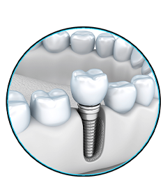
Replacement of single missing teeth with an implant-supported restoration is recognized as a highly successful treatment. An impression technique for peri-implant soft-tissue replication in an anterior zone is described. The technique involves use of an interim restoration as an abutment for the final impression. This allows accurate duplication of the soft tissues and fabrication of a final restoration with the correct emergence profile. (+info)Unusual migratory foreign body in the neck. (6/158)
Reports of ingested foreign bodies penetrating the pharynx and migrating through the neck are rare, and mostly involved fish bones. We describe a 44-year-old man who was involved in a motor vehicle accident and accidentally swallowed his tooth. The swallowed tooth penetrated the pharynx and became lodged adjacent to his right thyroid gland. It was successfully removed via neck exploration and the patient recovered well. (+info)Effect of the ferrule on fracture resistance of teeth restored with prefabricated posts and composite cores. (7/158)
BACKGROUND: The ferrule effect in root treated teeth requiring cast posts and cores has been shown to greatly improve fracture resistance. Studies have also shown that in the case of a cast post and core, the longer the ferrule, the greater the fracture resistance. However few studies have considered the effect of different ferrule designs on prefabricated post and composite core systems. AIM: This study investigated the effect of different ferrule designs on the fracture resistance of teeth incorporating prefabricated posts and composite cores. It also assessed the necessity of a post in the restoration of endodontically treated teeth. METHODOLOGY: Sixty-two extracted maxillary incisors (centrals and laterals) and canines were randomly assigned into three groups and restored. Two groups had a prefabricated post and composite core with varying ferrule designs. A third group had a core with composite packed into the root canal but no post. An Instron universal testing machine was used to apply compressive loads until failure occurred. RESULTS: There was no significant difference amongst the three groups as regards fracture resistance. The two groups with prefabricated posts and composite core required a mean force of 931N, std +/-283 and 931N, Std +/-242 to fracture. The third with no post group required a mean force of 1036N, std +/-269 to fracture. CONCLUSION: In the restoration of an anterior endodontically treated tooth with a prefabricated post and composite core and where there is at least 2 mm or more of remaining coronal dentine, a ferrule may not be necessary. (+info)Orthodontic extrusion: periodontal considerations and applications. (8/158)
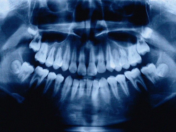
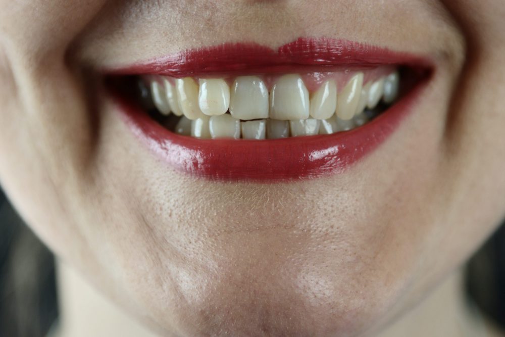
Human teeth erupt naturally to compensate for tooth wear and tear. When a subgingival lesion such as crown fracture occurs, the general practitioner must consider orthodontic extrusion of the tooth to allow for prosthetic rehabilitation. However, because this therapeutic approach is not appropriate in all cases, each tooth must be carefully analyzed before treatment. The amount of force applied depends on the desired effect. Orthodontic extrusion can also be used to augment bone and tissue in the course of preparing an implant site. In most cases, endodontic treatment must be completed first, with close attention being paid to the contour of the final restoration. The benefits of extrusion are clear, but patients must nonetheless be informed of the disadvantages. (+info)A tooth fracture is a dental health condition characterized by a break or crack in the tooth structure. It can occur in different parts of the tooth, including the crown (the visible part), root, or filling. Tooth fractures can result from various factors such as trauma, biting or chewing on hard objects, grinding or clenching teeth, and having large, old amalgam fillings that weaken the tooth structure over time. Depending on the severity and location of the fracture, it may cause pain, sensitivity, or affect the tooth's functionality and appearance. Treatment options for tooth fractures vary from simple bonding to root canal treatment or even extraction in severe cases. Regular dental check-ups are essential for early detection and management of tooth fractures.
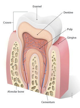
A tooth is a hard, calcified structure found in the jaws (upper and lower) of many vertebrates and used for biting and chewing food. In humans, a typical tooth has a crown, one or more roots, and three layers: the enamel (the outermost layer, hardest substance in the body), the dentin (the layer beneath the enamel), and the pulp (the innermost layer, containing nerves and blood vessels). Teeth are essential for proper nutrition, speech, and aesthetics. There are different types of teeth, including incisors, canines, premolars, and molars, each designed for specific functions in the mouth.
A bone fracture is a medical condition in which there is a partial or complete break in the continuity of a bone due to external or internal forces. Fractures can occur in any bone in the body and can vary in severity from a small crack to a shattered bone. The symptoms of a bone fracture typically include pain, swelling, bruising, deformity, and difficulty moving the affected limb. Treatment for a bone fracture may involve immobilization with a cast or splint, surgery to realign and stabilize the bone, or medication to manage pain and prevent infection. The specific treatment approach will depend on the location, type, and severity of the fracture.
Fracture healing is the natural process by which a broken bone repairs itself. When a fracture occurs, the body responds by initiating a series of biological and cellular events aimed at restoring the structural integrity of the bone. This process involves the formation of a hematoma (a collection of blood) around the fracture site, followed by the activation of inflammatory cells that help to clean up debris and prepare the area for repair.
Over time, specialized cells called osteoblasts begin to lay down new bone matrix, or osteoid, along the edges of the broken bone ends. This osteoid eventually hardens into new bone tissue, forming a bridge between the fracture fragments. As this process continues, the callus (a mass of newly formed bone and connective tissue) gradually becomes stronger and more compact, eventually remodeling itself into a solid, unbroken bone.
The entire process of fracture healing can take several weeks to several months, depending on factors such as the severity of the injury, the patient's age and overall health, and the location of the fracture. In some cases, medical intervention may be necessary to help promote healing or ensure proper alignment of the bone fragments. This may include the use of casts, braces, or surgical implants such as plates, screws, or rods.
A hip fracture is a medical condition referring to a break in the upper part of the femur (thigh) bone, which forms the hip joint. The majority of hip fractures occur due to falls or direct trauma to the area. They are more common in older adults, particularly those with osteoporosis, a condition that weakens bones and makes them more prone to breaking. Hip fractures can significantly impact mobility and quality of life, often requiring surgical intervention and rehabilitation.
A femoral fracture is a medical term that refers to a break in the thigh bone, which is the longest and strongest bone in the human body. The femur extends from the hip joint to the knee joint and is responsible for supporting the weight of the upper body and allowing movement of the lower extremity. Femoral fractures can occur due to various reasons such as high-energy trauma, low-energy trauma in individuals with weak bones (osteoporosis), or as a result of a direct blow to the thigh.
Femoral fractures can be classified into different types based on their location, pattern, and severity. Some common types of femoral fractures include:
1. Transverse fracture: A break that occurs straight across the bone.
2. Oblique fracture: A break that occurs at an angle across the bone.
3. Spiral fracture: A break that occurs in a helical pattern around the bone.
4. Comminuted fracture: A break that results in multiple fragments of the bone.
5. Open or compound fracture: A break in which the bone pierces through the skin.
6. Closed or simple fracture: A break in which the bone does not pierce through the skin.
Femoral fractures can cause severe pain, swelling, bruising, and difficulty walking or bearing weight on the affected leg. Diagnosis typically involves a physical examination, medical history, and imaging tests such as X-rays or CT scans. Treatment may involve surgical intervention, including the use of metal rods, plates, or screws to stabilize the bone, followed by rehabilitation and physical therapy to restore mobility and strength.
A spinal fracture, also known as a vertebral compression fracture, is a break in one or more bones (vertebrae) of the spine. This type of fracture often occurs due to weakened bones caused by osteoporosis, but it can also result from trauma such as a car accident or a fall.
In a spinal fracture, the front part of the vertebra collapses, causing the height of the vertebra to decrease, while the back part of the vertebra remains intact. This results in a wedge-shaped deformity of the vertebra. Multiple fractures can lead to a hunched forward posture known as kyphosis or dowager's hump.
Spinal fractures can cause pain, numbness, tingling, or weakness in the back, legs, or arms, depending on the location and severity of the fracture. In some cases, spinal cord compression may occur, leading to more severe symptoms such as paralysis or loss of bladder and bowel control.


![Do Alligators Attack Kayaks And Kayakers? All to Know [2023]](https://ecpaddlesports.com/wp-content/uploads/do-alligators-attack-kayaks.jpg)