Toxoplasmosis, Congenital
Toxoplasmosis, Ocular
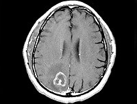
Toxoplasmosis, Cerebral
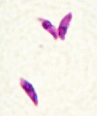
Toxoplasma
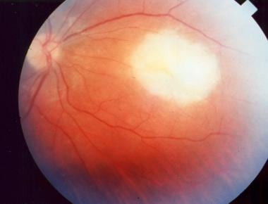
Chorioretinitis
Sulfadiazine
Pregnancy Complications, Parasitic
Cerebral Infarction
Cerebral Palsy
Middle Cerebral Artery
Immunoglobulin M
Lung Diseases, Parasitic
Antigens, Protozoan
Brain
Immunoglobulin G
Cerebral Angiography
Pregnancy
Seroepidemiologic Studies
Infarction, Middle Cerebral Artery
AIDS-Related Opportunistic Infections
Malaria, Cerebral
Enzyme-Linked Immunosorbent Assay
Brain Ischemia
Atovaquone
Cerebral Hemorrhage
Protozoan Vaccines
Antibody Affinity
Pyrimethamine
Neonatal Screening
Brazil
Sensitivity and Specificity
Uveitis, Posterior
Brain Diseases
Pregnancy Complications, Infectious
Magnetic Resonance Imaging
Infectious Disease Transmission, Vertical
Encephalitis
Fatal Outcome
Immunocompromised Host
Cats
Ischemic Attack, Transient
Immunoglobulin A
Polymerase Chain Reaction
Anterior Cerebral Artery
Cerebral Amyloid Angiopathy
Posterior Cerebral Artery
Naphthoquinones
Prenatal Diagnosis
Disease Models, Animal
Cerebral Ventricles
Cat Diseases
Food Parasitology
Brain Edema
Agglutination Tests
Immunocompetence
Suriname
Subarachnoid Hemorrhage
Uveitis
Tomography, X-Ray Computed
Oocysts
HLA-B15 Antigen
Kyrgyzstan
Cerebrovascular Disorders
Amniotic Fluid
Blood Flow Velocity
Intracranial Embolism and Thrombosis
Intracranial Pressure
Opportunistic Infections
Intracranial Aneurysm
Trichechus manatus
Risk Factors
Hydrocephalus
Cerebral Revascularization
Cerebrum
Aqueous Humor
Vasospasm, Intracranial
Hemagglutination Tests
Fetal Diseases
Immunoassay
Host-Parasite Interactions
Prospective Studies
Apicomplexa
Central Nervous System Protozoal Infections
Retrospective Studies
Hypoxia, Brain
Cerebrospinal Fluid
Neuroprotective Agents
Antibodies
Mice, Inbred C57BL
Retinitis
Animals, Domestic
Meninges
Xenon Radioisotopes
Clinical Laboratory Techniques
Carbon Dioxide
Inhibition of inducible nitric oxide synthase exacerbates chronic cerebral toxoplasmosis in Toxoplasma gondii-susceptible C57BL/6 mice but does not reactivate the latent disease in T. gondii-resistant BALB/c mice. (1/197)
Infection of C57BL/6 mice with Toxoplasma gondii leads to progressive and ultimately fatal chronic Toxoplasma encephalitis (TE). Genetic deletion or inhibition of inducible nitric oxide synthase (iNOS) from the beginning of infection increased the number of T. gondii cysts in the brain and markedly reduced the time-to-death in this mouse strain. In the present study, we addressed whether iNOS also contributes to the control of intracerebral parasites in a clinically stable latent infection that develops in T. gondii-resistant BALB/c mice after resolution of the acute phase of TE. iNOS was expressed in the inflammatory cerebral infiltrates of latently infected BALB/c mice, but the number of iNOS+ cells was significantly lower than in the brains of chronically infected T. gondii-susceptible C57BL/6 mice. In BALB/c mice with latent TE (> 30 days of infection), treatment with the iNOS inhibitors L-N6-iminoethyl-lysine or L-nitroarginine-methylester for < or = 40 days did not result in an increase of the intracerebral parasitic load and a reactivation of the disease, despite the presence of iNOS-suppressive inhibitor levels in the brain. However, L-nitroarginine-methylester treatment had remarkably toxic effects and induced a severe wasting syndrome with high mortality. In contrast to BALB/c mice, L-N6-iminoethyl-lysine treatment rapidly exacerbated the already established chronic TE of C57BL/6 mice. Thus, the containment of latent toxoplasms in T. gondii-resistant BALB/c mice is independent of iNOS, whereas the temporary control of intracerebral parasites in T. gondii-susceptible C57BL/6 mice with chronic TE requires iNOS activity. (+info)Successful treatment of cerebral toxoplasmosis in a marrow transplant recipient: contribution of a PCR test in diagnosis and early detection. (2/197)
We report successful treatment of cerebral toxoplasmosis in an unrelated donor marrow transplant recipient. The clinical diagnosis was confirmed by polymerase chain reaction (PCR) amplification for T. gondii-DNA performed both on cerebrospinal fluid and blood leukocytes. Retrospective testing of stored blood samples demonstrated positive leukocyte PCR signal detected up to 52 days prior to onset of clinical symptoms. This case highlights the value of PCR in the diagnosis and early detection of cerebral toxoplasmosis. (+info)Incidence and risk factors of toxoplasmosis in a cohort of human immunodeficiency virus-infected patients: 1988-1995. HEMOCO and SEROCO Study Groups. (3/197)
The incidence of cerebral and extracerebral toxoplasmosis among 1,699 HIV-infected patients followed in the SEROCO and HEMOCO cohorts (1988-1995) was studied. It increased from 0.7 per 100 person-years in 1988 to 2.1 per 100 person-years in 1992, as a result of the increasing prevalence of patients with CD4 cell counts below 200/microL. It decreased thereafter to 0.2 per 100 person-years in 1995, while the proportion of patients receiving specific prophylaxis was increasing. A Toxoplasma antibody titer of >150 IU/mL was an important predictor of toxoplasmosis (adjusted relative risk [aRR], 3.6 [95% confidence interval, 2.1-6.0]), independent of a CD4+ cell count of <200/microL (aRR, 20.8) and specific prophylaxis (aRR, 0.2 [0.1-0.3]). The median CD4+ cell count was 389/microL at the time the antibody titer was first noted to be >150 IU/mL, while the median CD4 cell count at onset of toxoplasmosis was 58/microL. Thus, disease was diagnosed 10 days to 74 months after the rise in Toxoplasma antibody titers. While the risk factors for development of toxoplasmosis remain incompletely defined, the importance of specific prophylaxis for patients with low CD4 cell counts and high Toxoplasma antibody titers is supported by these findings. (+info)Discontinuation of primary prophylaxis against Pneumocystis carinii pneumonia in HIV-1-infected adults treated with combination antiretroviral therapy. Swiss HIV Cohort Study. (4/197)
BACKGROUND: It is unclear whether primary prophylaxis against Pneumocystis carinii pneumonia can be discontinued in patients infected with the human immunodeficiency virus (HIV) who are successfully treated with combination antiretroviral therapy. We prospectively studied the safety of stopping prophylaxis among patients in the Swiss HIV Cohort Study. METHODS: Patients were eligible for our study if their CD4 counts had increased to at least 200 cells per cubic millimeter and 14 percent of total lymphocytes while they were receiving combination antiretroviral therapy, with these levels sustained for at least 12 weeks. Prophylaxis was stopped at study entry, and patients were examined every three months thereafter. The development of P. carinii pneumonia was the primary end point, and the development of toxoplasmic encephalitis the secondary end point. RESULTS: Of the 262 patients included in our analysis, 121 (46.2 percent) were positive for IgG antibodies to Toxoplasma gondii at base line. The median CD4 count at study entry was 325 per cubic millimeter (range, 210 to 806); the median nadir CD4 count was 110 per cubic millimeter (range, 0 to 240). During a median follow-up of 11.3 months (range, 3.0 to 18.8), prophylaxis was resumed in nine patients, and two patients died. There were no cases of P. carinii pneumonia or toxoplasmic encephalitis. The one-sided upper 99 percent confidence limit for the incidence of P. carinii pneumonia was 1.9 cases per 100 patient-years (based on 238 patient-years of follow-up). The corresponding figure for toxoplasmic encephalitis was 4.2 per 100 patient-years (based on 110 patient-years of follow-up). CONCLUSIONS: Stopping primary prophylaxis against P. carinii pneumonia appears to be safe in HIV-infected patients who are receiving combination antiretroviral treatment and who have had a sustained increase in their CD4 counts to at least 200 cells per cubic millimeter and to at least 14 percent of total lymphocytes. (+info)Attachment ligands of viable Toxoplasma gondii induce soluble immunosuppressive factors in human monocytes. (5/197)
Previous studies have demonstrated that surface antigen proteins, in particular SAG-1, of Toxoplasma gondii are important to this parasite as attachment ligands for the host cell. An in vitro assay was developed to test whether these ligands and other secretory proteins are involved in the immune response of human cells to toxoplasma. Human monocytes were infected with tachyzoites in the presence of antiparasite antibodies, and their effect on mitogen-induced lymphoproliferation was examined. The presence of antibody to either parasite-excreted proteins (MIC-1 and MIC-2) or surface proteins (SAG-1 and SAG-2) during infection neutralized the marked decrease seen in mitogen-induced lymphoproliferation in the presence of infected monocytes. Conversely, antibodies to other secreted proteins (ROP-1) and cytoplasmic molecules had no effect on parasite-induced, monocyte-mediated downregulation. Fluorescence microscope analysis detected microneme and surface antigen proteins on the monocyte cell surface during infection. These results suggest that microneme and surface antigen proteins trigger monocytes to downregulate mitogen-induced lymphoproliferation. (+info)Interferon-gamma receptor-mediated but not tumor necrosis factor receptor type 1- or type 2-mediated signaling is crucial for the activation of cerebral blood vessel endothelial cells and microglia in murine Toxoplasma encephalitis. (6/197)
The regulatory role of interferon-gamma receptor (IFN-gammaR)- and tumor necrosis factor receptor (TNFR)-mediated immune reactions for the activation of cerebral endothelial cells, microglia, and astrocytes was evaluated in a model of murine Toxoplasma encephalitis (TE). Brain endothelial cells of wild-type mice reacted in response to Toxoplasma infection with a strong up-regulation of the vascular cell adhesion molecule, the intercellular adhesion molecule (ICAM)-1, and major histocompatibility complex (MHC) class I and II antigens. A similar response was seen in mice genetically deficient for either TNFR1, TNFR2, or both TNFRs, whereas IFN-gammaR-deficient (IFN-gammaR0/0) mice were found to be defective in the up-regulation of these molecules. However, recruitment of leukocytes to the brain and their intracerebral movement were not impaired in IFN-gammaR0/0 mice. In addition, microglia of Toxoplasma gondii-infected IFN-gammaR0/0 mice failed to induce expression of ICAM-1, leukocyte function-associated antigen (LFA)-1, and MHC class I and II antigens, whereas wild-type and TNFR-deficient mice up-regulated these molecules. Moreover, TNF-alpha mRNA production of F4/80(+) microglia/macrophages was impaired in IFN-gammaR0/0 mice, but not in TNFR-deficient mutants. However, induction of interleukin (IL)-1beta, IL-10, IL-12p40, and IL-15 mRNA was independent of IFN-gammaR and TNFR signaling. In conclusion, IFN-gammaR, but not TNFR signaling, is the major pathway for the activation of endothelial cells and microglia in murine TE. These findings differ from observations in other inflammatory central nervous system disorders, indicating specific regulatory mechanisms in this parasitic cerebral infection. (+info)Opportunistic CNS infection after bone marrow transplantation. (7/197)
We retrospectively identified opportunistic CNS infections in 655 patients who had undergone allogeneic, syngeneic or autologous BMT or PBSCT between 1990 and 1997. Twenty-seven patients (4%) developed CNS infections. All CNS infections occurred in allogeneic BMT or PBSCT patients. The most common CNS infections were toxoplasma encephalitis (74%) and cerebral aspergillosis (18%). Furthermore, we identified one patient with candida encephalitis and one patient with viral encephalitis. Overall mortality of patients with opportunistic CNS infection was 67%. There were two different groups of toxoplasma encephalitis with a different appearance on MR imaging. The first group showed edema, but no gadolinium enhancement, whereas the second group exhibited typical MRI appearances with the exception of frequent hemorrhagic transformation. The first group had a significant shorter latency between BMT and onset of CNS infection (mean 45 days vs 180 days, P = 0.02), a significant higher daily dose of corticosteroids as treatment for graft-versus-host disease (GVHD) (P = 0.01), more severe GVHD and a higher mortality (71% vs 36%). This study shows that the most common CNS infections in our patient population are toxoplasma encephalitis and cerebral aspergillosis, that there are two distinct subgroups of toxoplasma encephalitis and that CNS infections occur after allogeneic BMT only. (+info)Detection of Toxoplasma gondii, Epstein-Barr virus, and JC virus DNAs in the cerebrospinal fluid in acquired immunodeficiency syndrome patients with focal central nervous system complications. (8/197)
OBJECT: Toxoplasmic encephalitis (TE), primary central nervous system lymphoma (PCNSL) and progressive multifocal leukoencephalopathy (PML) are major central nervous system (CNS) diseases in patients with acquired immunodeficiency syndrome (AIDS). We assessed the diagnostic value of polymerase chain reaction (PCR) in the detection of DNAs of Toxoplasma gondii (T. gondii), Epstein-Barr virus (EBV) and JC virus (JCV) in the cerebrospinal fluid (CSF). METHODS: We compared the PCR results with those of pathological findings at autopsy. PATIENTS OR MATERIALS: The present study included 23 autopsies representing those in whom CSF samples were obtained before death while the patient was hospitalized or at autopsy. RESULTS: The threshold levels for PCR detection were 4 tachyzoites of T. gondii, 5-15 genomes of EBV and 10 genomes of JCV. We identified T. gondii DNA in 4 out of 5 autopsy-defined cases of TE, EBV DNA in 5 out of 5 cases with PCNSL, and JCV DNA in 2 out of 2 cases with PML. The specificity of PCR was 100% in TE, 78% in PCNSL, and 100% in PML. CONCLUSION: Although the number of cases was relatively small in this study, PCR correctly identified T. gondii DNA in those cases in which PML or PCNSL was the sole clinical diagnosis. Our results indicate that PCR examination of CSF is a clinically useful tool for the diagnosis of focal brain lesions in patients with AIDS. (+info)Toxoplasmosis is a disease caused by the parasitic protozoan Toxoplasma gondii. It can infect humans, birds, and most warm-blooded animals, including marine mammals. In humans, it is usually contracted through eating undercooked, contaminated meat or ingesting oocysts (a form of the parasite) from cat feces, often through contact with litter boxes or gardening in soil that has been contaminated with cat feces.
The infection can also be passed to the fetus if a woman becomes infected during or just before pregnancy. Most healthy individuals who become infected with Toxoplasma gondii experience few symptoms and are not aware they have the disease. However, for those with weakened immune systems, such as people with HIV/AIDS, organ transplant recipients, and pregnant women, toxoplasmosis can cause severe complications, including damage to the brain, eyes, and other organs.
Symptoms of toxoplasmosis in individuals with weakened immune systems may include swollen lymph nodes, fever, fatigue, muscle aches, and headache. In pregnant women, infection can lead to miscarriage, stillbirth, or severe developmental problems in the baby. Treatment typically involves antiparasitic medications such as pyrimethamine and sulfadiazine.
Congenital toxoplasmosis is a medical condition that results from the transmission of the Toxoplasma gondii parasite from an infected pregnant woman to her developing fetus through the placenta. The severity of the infection can vary widely, depending on the stage of pregnancy at which the mother becomes infected.
Infection during early pregnancy is associated with a higher risk of severe symptoms in the newborn, including:
* Intracranial calcifications
* Hydrocephalus (fluid buildup in the brain)
* Microcephaly (abnormally small head)
* Chorioretinitis (inflammation of the eye's retina and choroid layer)
* Seizures
* Developmental delays
* Hearing loss
Infection later in pregnancy may result in less severe symptoms or be asymptomatic at birth, but can still lead to developmental delays, learning disabilities, and vision problems as the child grows.
Diagnosis of congenital toxoplasmosis typically involves a combination of tests, such as blood tests to detect antibodies against Toxoplasma gondii, imaging studies (e.g., ultrasound, CT, or MRI) to assess any structural abnormalities in the brain and other organs, and ophthalmologic examinations to evaluate potential eye damage.
Treatment for congenital toxoplasmosis usually involves a combination of antiparasitic medications (such as spiramycin, pyrimethamine, and sulfadiazine) and corticosteroids to reduce inflammation. Early treatment can help minimize the severity of symptoms and improve outcomes for affected children.
Ocular toxoplasmosis is an inflammatory eye disease caused by the parasitic infection of Toxoplasma gondii in the eye's retina. It can lead to lesions and scarring in the retina, resulting in vision loss or impairment. The severity of ocular toxoplasmosis depends on the location and extent of the infection in the eye. In some cases, it may cause only mild symptoms, while in others, it can result in severe damage to the eye. Ocular toxoplasmosis is usually treated with medications that target the Toxoplasma gondii parasite, such as pyrimethamine and sulfadiazine, often combined with corticosteroids to reduce inflammation.
Cerebral toxoplasmosis is a type of toxoplasmosis, which is an infection caused by the Toxoplasma gondii parasite. In cerebral toxoplasmosis, the infection primarily affects the brain, leading to inflammation and the formation of lesions or abscesses in the brain tissue.
This condition is most commonly observed in individuals with weakened immune systems, such as those living with HIV/AIDS, receiving immunosuppressive therapy after organ transplantation, or having other conditions that compromise their immune function. The infection can cause a range of neurological symptoms, including headaches, seizures, confusion, memory loss, poor coordination, and in severe cases, coma or even death. Early diagnosis and treatment with appropriate antiparasitic medications are crucial to manage the infection and prevent complications.
"Toxoplasma" is a genus of protozoan parasites, and the most well-known species is "Toxoplasma gondii." This particular species is capable of infecting virtually all warm-blooded animals, including humans. It's known for its complex life cycle that involves felines (cats) as the definitive host.
Infection in humans, called toxoplasmosis, often occurs through ingestion of contaminated food or water, or through contact with cat feces that contain T. gondii oocysts. While many people infected with Toxoplasma show no symptoms, it can cause serious health problems in immunocompromised individuals and developing fetuses if a woman becomes infected during pregnancy.
It's important to note that while I strive to provide accurate information, this definition should not be used for self-diagnosis or treatment. Always consult with a healthcare professional for medical advice.
Toxoplasmosis is a zoonotic disease, meaning it can be transmitted from animals to humans. It is caused by the intracellular protozoan parasite Toxoplasma gondii. This parasite can infect a wide range of warm-blooded animals, including birds and mammals, as intermediate hosts. However, cats are the primary definitive host for this parasite because the sexual stage of the parasite's life cycle occurs in their intestines, leading to the shedding of oocysts (environmentally resistant stages) in their feces.
Animals can become infected with Toxoplasma gondii through several routes:
1. Ingestion of sporulated oocysts from contaminated soil, water, or food.
2. Consumption of tissue cysts present in the tissues of infected animals during predation.
3. Vertical transmission (transplacental) from an infected mother to her offspring.
Clinical signs and symptoms of toxoplasmosis in animals can vary depending on their age, immune status, and the parasite's virulence. In many cases, animals may not show any apparent signs of infection, but some may develop:
1. Generalized illness with fever, lethargy, and loss of appetite.
2. Lymphadenopathy (swollen lymph nodes).
3. Neurological symptoms such as tremors, ataxia (lack of coordination), or seizures if the central nervous system is affected.
4. Eye lesions, including inflammation and scarring of the retina, which can lead to vision loss in severe cases.
5. Reproductive issues, such as abortion, stillbirths, or birth defects in offspring when pregnant females are infected.
It is important to note that while toxoplasmosis can cause significant health problems in animals, particularly in immunocompromised individuals and developing fetuses, it is often asymptomatic or mild in healthy adult animals. Nonetheless, the zoonotic potential of Toxoplasma gondii highlights the importance of practicing good hygiene and taking necessary precautions when handling infected animals or their waste to minimize the risk of transmission to humans.
Chorioretinitis is a medical term that refers to the inflammation of the choroid and the retina, which are both important structures in the eye. The choroid is a layer of blood vessels that supplies oxygen and nutrients to the retina, while the retina is a light-sensitive tissue that converts light into electrical signals that are sent to the brain and interpreted as visual images.
Chorioretinitis can be caused by various infectious and non-infectious conditions, such as bacterial, viral, fungal, or parasitic infections, autoimmune diseases, or cancer. The symptoms of chorioretinitis may include decreased vision, floaters, blurry vision, sensitivity to light, and eye pain. Treatment for chorioretinitis depends on the underlying cause and may include antibiotics, antiviral medications, corticosteroids, or other immunosuppressive therapies. It is important to seek medical attention promptly if you experience any symptoms of chorioretinitis, as timely diagnosis and treatment can help prevent permanent vision loss.
Antibodies, protozoan, refer to the immune system's response to an infection caused by a protozoan organism. Protozoa are single-celled microorganisms that can cause various diseases in humans, such as malaria, giardiasis, and toxoplasmosis.
When the body is infected with a protozoan, the immune system responds by producing specific proteins called antibodies. Antibodies are produced by a type of white blood cell called a B-cell, and they recognize and bind to specific antigens on the surface of the protozoan organism.
There are five main types of antibodies: IgA, IgD, IgE, IgG, and IgM. Each type of antibody has a different role in the immune response. For example, IgG is the most common type of antibody and provides long-term immunity to previously encountered pathogens. IgM is the first antibody produced in response to an infection and is important for activating the complement system, which helps to destroy the protozoan organism.
Overall, the production of antibodies against protozoan organisms is a critical part of the immune response and helps to protect the body from further infection.
Sulfadiazine is an antibacterial drug, specifically a sulfonamide. It is chemically described as 4-amino-N-(2-pyrimidinyl)benzenesulfonamide. Sulfadiazine works by inhibiting the bacterial synthesis of dihydrofolic acid, which is essential for bacterial growth and reproduction.
It is used to treat a wide range of infections caused by susceptible bacteria, including urinary tract infections, respiratory infections, and certain types of meningitis. Sulfadiazine is often combined with other antibiotics, such as trimethoprim, to increase its effectiveness against certain bacteria.
Like all sulfonamides, sulfadiazine can cause side effects, including skin rashes, allergic reactions, and stomach upset. It should be used with caution in people who are allergic to sulfa drugs or have kidney or liver disease. Additionally, it is important to note that the use of sulfonamides during pregnancy, especially during the third trimester, should be avoided due to the risk of kernicterus in the newborn.
Parasitic pregnancy complications refer to a rare condition where a parasitic twin takes over the development of the dominant twin's reproductive system and becomes pregnant. This condition is also known as fetus in fetu or vanishing twin syndrome with a parasitic twin. The parasitic twin may have some organs developed, but it is not fully formed and relies on the dominant twin for survival. The pregnancy can pose risks to the dominant twin, such as abnormal growth patterns, organ damage, and complications during childbirth. This condition is usually detected during prenatal ultrasound examinations.
Cerebral arteries refer to the blood vessels that supply oxygenated blood to the brain. These arteries branch off from the internal carotid arteries and the vertebral arteries, which combine to form the basilar artery. The major cerebral arteries include:
1. Anterior cerebral artery (ACA): This artery supplies blood to the frontal lobes of the brain, including the motor and sensory cortices responsible for movement and sensation in the lower limbs.
2. Middle cerebral artery (MCA): The MCA is the largest of the cerebral arteries and supplies blood to the lateral surface of the brain, including the temporal, parietal, and frontal lobes. It is responsible for providing blood to areas involved in motor function, sensory perception, speech, memory, and vision.
3. Posterior cerebral artery (PCA): The PCA supplies blood to the occipital lobe, which is responsible for visual processing, as well as parts of the temporal and parietal lobes.
4. Anterior communicating artery (ACoA) and posterior communicating arteries (PComAs): These are small arteries that connect the major cerebral arteries, forming an important circulatory network called the Circle of Willis. The ACoA connects the two ACAs, while the PComAs connect the ICA with the PCA and the basilar artery.
These cerebral arteries play a crucial role in maintaining proper brain function by delivering oxygenated blood to various regions of the brain. Any damage or obstruction to these arteries can lead to serious neurological conditions, such as strokes or transient ischemic attacks (TIAs).
Cerebral infarction, also known as a "stroke" or "brain attack," is the sudden death of brain cells caused by the interruption of their blood supply. It is most commonly caused by a blockage in one of the blood vessels supplying the brain (an ischemic stroke), but can also result from a hemorrhage in or around the brain (a hemorrhagic stroke).
Ischemic strokes occur when a blood clot or other particle blocks a cerebral artery, cutting off blood flow to a part of the brain. The lack of oxygen and nutrients causes nearby brain cells to die. Hemorrhagic strokes occur when a weakened blood vessel ruptures, causing bleeding within or around the brain. This bleeding can put pressure on surrounding brain tissues, leading to cell death.
Symptoms of cerebral infarction depend on the location and extent of the affected brain tissue but may include sudden weakness or numbness in the face, arm, or leg; difficulty speaking or understanding speech; vision problems; loss of balance or coordination; and severe headache with no known cause. Immediate medical attention is crucial for proper diagnosis and treatment to minimize potential long-term damage or disability.
Cerebral palsy (CP) is a group of disorders that affect a person's ability to move and maintain balance and posture. According to the Mayo Clinic, CP is caused by abnormal brain development or damage to the developing brain that affects a child's ability to control movement.
The symptoms of cerebral palsy can vary in severity and may include:
* Spasticity (stiff or tight muscles)
* Rigidity (resistance to passive movement)
* Poor coordination and balance
* Weakness or paralysis
* Tremors or involuntary movements
* Abnormal gait or difficulty walking
* Difficulty with fine motor skills, such as writing or using utensils
* Speech and language difficulties
* Vision, hearing, or swallowing problems
It's important to note that cerebral palsy is not a progressive condition, meaning that it does not worsen over time. However, the symptoms may change over time, and some individuals with CP may experience additional medical conditions as they age.
Cerebral palsy is usually caused by brain damage that occurs before or during birth, but it can also be caused by brain injuries that occur in the first few years of life. Some possible causes of cerebral palsy include:
* Infections during pregnancy
* Lack of oxygen to the brain during delivery
* Traumatic head injury during birth
* Brain bleeding or stroke in the newborn period
* Genetic disorders
* Maternal illness or infection during pregnancy
There is no cure for cerebral palsy, but early intervention and treatment can help improve outcomes and quality of life. Treatment may include physical therapy, occupational therapy, speech therapy, medications to manage symptoms, surgery, and assistive devices such as braces or wheelchairs.
Antiprotozoal agents are a type of medication used to treat protozoal infections, which are infections caused by microscopic single-celled organisms called protozoa. These agents work by either killing the protozoa or inhibiting their growth and reproduction. They can be administered through various routes, including oral, topical, and intravenous, depending on the type of infection and the severity of the illness.
Examples of antiprotozoal agents include:
* Metronidazole, tinidazole, and nitazoxanide for treating infections caused by Giardia lamblia and Entamoeba histolytica.
* Atovaquone, clindamycin, and pyrimethamine-sulfadoxine for treating malaria caused by Plasmodium falciparum or other Plasmodium species.
* Pentamidine and suramin for treating African trypanosomiasis (sleeping sickness) caused by Trypanosoma brucei gambiense or T. b. rhodesiense.
* Nitroimidazoles, such as benznidazole and nifurtimox, for treating Chagas disease caused by Trypanosoma cruzi.
* Sodium stibogluconate and paromomycin for treating leishmaniasis caused by Leishmania species.
Antiprotozoal agents can have side effects, ranging from mild to severe, depending on the drug and the individual patient's response. It is essential to follow the prescribing physician's instructions carefully when taking these medications and report any adverse reactions promptly.
Cerebrovascular circulation refers to the network of blood vessels that supply oxygenated blood and nutrients to the brain tissue, and remove waste products. It includes the internal carotid arteries, vertebral arteries, circle of Willis, and the intracranial arteries that branch off from them.
The internal carotid arteries and vertebral arteries merge to form the circle of Willis, a polygonal network of vessels located at the base of the brain. The anterior cerebral artery, middle cerebral artery, posterior cerebral artery, and communicating arteries are the major vessels that branch off from the circle of Willis and supply blood to different regions of the brain.
Interruptions or abnormalities in the cerebrovascular circulation can lead to various neurological conditions such as stroke, transient ischemic attack (TIA), and vascular dementia.
The Middle Cerebral Artery (MCA) is one of the main blood vessels that supplies oxygenated blood to the brain. It arises from the internal carotid artery and divides into several branches, which supply the lateral surface of the cerebral hemisphere, including the frontal, parietal, and temporal lobes.
The MCA is responsible for providing blood flow to critical areas of the brain, such as the primary motor and sensory cortices, Broca's area (associated with speech production), Wernicke's area (associated with language comprehension), and the visual association cortex.
Damage to the MCA or its branches can result in a variety of neurological deficits, depending on the specific location and extent of the injury. These may include weakness or paralysis on one side of the body, sensory loss, language impairment, and visual field cuts.
Immunoglobulin M (IgM) is a type of antibody that is primarily found in the blood and lymph fluid. It is the first antibody to be produced in response to an initial exposure to an antigen, making it an important part of the body's primary immune response. IgM antibodies are large molecules that are composed of five basic units, giving them a pentameric structure. They are primarily found on the surface of B cells as membrane-bound immunoglobulins (mlgM), where they function as receptors for antigens. Once an mlgM receptor binds to an antigen, it triggers the activation and differentiation of the B cell into a plasma cell that produces and secretes large amounts of soluble IgM antibodies.
IgM antibodies are particularly effective at agglutination (clumping) and complement activation, which makes them important in the early stages of an immune response to help clear pathogens from the bloodstream. However, they are not as stable or long-lived as other types of antibodies, such as IgG, and their levels tend to decline after the initial immune response has occurred.
In summary, Immunoglobulin M (IgM) is a type of antibody that plays a crucial role in the primary immune response to antigens by agglutination and complement activation. It is primarily found in the blood and lymph fluid, and it is produced by B cells after they are activated by an antigen.
Parasitic lung diseases refer to conditions caused by infection of the lungs by parasites. These are small organisms that live on or in a host organism and derive their sustenance at the expense of the host. Parasitic lung diseases can be caused by various types of parasites, including helminths (worms) and protozoa.
Examples of parasitic lung diseases include:
1. Pulmonary echinococcosis (hydatid disease): This is a rare infection caused by the larval stage of the tapeworm Echinococcus granulosus. The larvae form cysts in various organs, including the lungs.
2. Paragonimiasis: This is a food-borne lung fluke infection caused by Paragonimus westermani and other species. Humans become infected by eating raw or undercooked crustaceans (such as crabs or crayfish) that contain the larval stage of the parasite.
3. Toxocariasis: This is a soil-transmitted helminth infection caused by the roundworm Toxocara canis or T. cati, which are found in the intestines of dogs and cats. Humans become infected through accidental ingestion of contaminated soil, undercooked meat, or through contact with an infected animal's feces. Although the primary site of infection is the small intestine, larval migration can lead to lung involvement in some cases.
4. Amebic lung disease: This is a rare complication of amebiasis, which is caused by the protozoan Entamoeba histolytica. The parasite usually infects the large intestine, but it can spread to other organs, including the lungs, through the bloodstream.
5. Cryptosporidiosis: This is a waterborne protozoan infection caused by Cryptosporidium parvum or C. hominis. Although the primary site of infection is the small intestine, immunocompromised individuals can develop disseminated disease, including pulmonary involvement.
Symptoms of parasitic lung diseases vary depending on the specific organism and the severity of infection but may include cough, chest pain, shortness of breath, fever, and sputum production. Diagnosis typically involves a combination of clinical evaluation, imaging studies, and laboratory tests, such as stool or blood examinations for parasites or their antigens. Treatment depends on the specific organism but may include antiparasitic medications, supportive care, and management of complications.
Antigens are substances (usually proteins) found on the surface of cells, or viruses, that can be recognized by the immune system and stimulate an immune response. In the context of protozoa, antigens refer to the specific proteins or other molecules found on the surface of these single-celled organisms that can trigger an immune response in a host organism.
Protozoa are a group of microscopic eukaryotic organisms that include a diverse range of species, some of which can cause diseases in humans and animals. When a protozoan infects a host, the host's immune system recognizes the protozoan antigens as foreign and mounts an immune response to eliminate the infection. This response involves the activation of various types of immune cells, such as T-cells and B-cells, which recognize and target the protozoan antigens.
Understanding the nature of protozoan antigens is important for developing vaccines and other immunotherapies to prevent or treat protozoan infections. For example, researchers have identified specific antigens on the surface of the malaria parasite that are recognized by the human immune system and have used this information to develop vaccine candidates. However, many protozoan infections remain difficult to prevent or treat, and further research is needed to identify new targets for vaccines and therapies.
The brain is the central organ of the nervous system, responsible for receiving and processing sensory information, regulating vital functions, and controlling behavior, movement, and cognition. It is divided into several distinct regions, each with specific functions:
1. Cerebrum: The largest part of the brain, responsible for higher cognitive functions such as thinking, learning, memory, language, and perception. It is divided into two hemispheres, each controlling the opposite side of the body.
2. Cerebellum: Located at the back of the brain, it is responsible for coordinating muscle movements, maintaining balance, and fine-tuning motor skills.
3. Brainstem: Connects the cerebrum and cerebellum to the spinal cord, controlling vital functions such as breathing, heart rate, and blood pressure. It also serves as a relay center for sensory information and motor commands between the brain and the rest of the body.
4. Diencephalon: A region that includes the thalamus (a major sensory relay station) and hypothalamus (regulates hormones, temperature, hunger, thirst, and sleep).
5. Limbic system: A group of structures involved in emotional processing, memory formation, and motivation, including the hippocampus, amygdala, and cingulate gyrus.
The brain is composed of billions of interconnected neurons that communicate through electrical and chemical signals. It is protected by the skull and surrounded by three layers of membranes called meninges, as well as cerebrospinal fluid that provides cushioning and nutrients.
Immunoglobulin G (IgG) is a type of antibody, which is a protective protein produced by the immune system in response to foreign substances like bacteria or viruses. IgG is the most abundant type of antibody in human blood, making up about 75-80% of all antibodies. It is found in all body fluids and plays a crucial role in fighting infections caused by bacteria, viruses, and toxins.
IgG has several important functions:
1. Neutralization: IgG can bind to the surface of bacteria or viruses, preventing them from attaching to and infecting human cells.
2. Opsonization: IgG coats the surface of pathogens, making them more recognizable and easier for immune cells like neutrophils and macrophages to phagocytose (engulf and destroy) them.
3. Complement activation: IgG can activate the complement system, a group of proteins that work together to help eliminate pathogens from the body. Activation of the complement system leads to the formation of the membrane attack complex, which creates holes in the cell membranes of bacteria, leading to their lysis (destruction).
4. Antibody-dependent cellular cytotoxicity (ADCC): IgG can bind to immune cells like natural killer (NK) cells and trigger them to release substances that cause target cells (such as virus-infected or cancerous cells) to undergo apoptosis (programmed cell death).
5. Immune complex formation: IgG can form immune complexes with antigens, which can then be removed from the body through various mechanisms, such as phagocytosis by immune cells or excretion in urine.
IgG is a critical component of adaptive immunity and provides long-lasting protection against reinfection with many pathogens. It has four subclasses (IgG1, IgG2, IgG3, and IgG4) that differ in their structure, function, and distribution in the body.
Cerebral angiography is a medical procedure that involves taking X-ray images of the blood vessels in the brain after injecting a contrast dye into them. This procedure helps doctors to diagnose and treat various conditions affecting the blood vessels in the brain, such as aneurysms, arteriovenous malformations, and stenosis (narrowing of the blood vessels).
During the procedure, a catheter is inserted into an artery in the leg and threaded through the body to the blood vessels in the neck or brain. The contrast dye is then injected through the catheter, and X-ray images are taken to visualize the blood flow through the brain's blood vessels.
Cerebral angiography provides detailed images of the blood vessels in the brain, allowing doctors to identify any abnormalities or blockages that may be causing symptoms or increasing the risk of stroke. Based on the results of the cerebral angiography, doctors can develop a treatment plan to address these issues and prevent further complications.
Pregnancy is a physiological state or condition where a fertilized egg (zygote) successfully implants and grows in the uterus of a woman, leading to the development of an embryo and finally a fetus. This process typically spans approximately 40 weeks, divided into three trimesters, and culminates in childbirth. Throughout this period, numerous hormonal and physical changes occur to support the growing offspring, including uterine enlargement, breast development, and various maternal adaptations to ensure the fetus's optimal growth and well-being.
Seroepidemiologic studies are a type of epidemiological study that measures the presence and levels of antibodies in a population's blood serum to investigate the prevalence, distribution, and transmission of infectious diseases. These studies help to identify patterns of infection and immunity within a population, which can inform public health policies and interventions.
Seroepidemiologic studies typically involve collecting blood samples from a representative sample of individuals in a population and testing them for the presence of antibodies against specific pathogens. The results are then analyzed to estimate the prevalence of infection and immunity within the population, as well as any factors associated with increased or decreased risk of infection.
These studies can provide valuable insights into the spread of infectious diseases, including emerging and re-emerging infections, and help to monitor the effectiveness of vaccination programs. Additionally, seroepidemiologic studies can also be used to investigate the transmission dynamics of infectious agents, such as identifying sources of infection or tracking the spread of antibiotic resistance.
There doesn't seem to be a specific medical definition for "DNA, protozoan" as it is simply a reference to the DNA found in protozoa. Protozoa are single-celled eukaryotic organisms that can be found in various environments such as soil, water, and the digestive tracts of animals.
Protozoan DNA refers to the genetic material present in these organisms. It is composed of nucleic acids, including deoxyribonucleic acid (DNA) and ribonucleic acid (RNA), which contain the instructions for the development, growth, and reproduction of the protozoan.
The DNA in protozoa, like in other organisms, is made up of two strands of nucleotides that coil together to form a double helix. The four nucleotide bases that make up protozoan DNA are adenine (A), thymine (T), guanine (G), and cytosine (C). These bases pair with each other to form the rungs of the DNA ladder, with A always pairing with T and G always pairing with C.
The genetic information stored in protozoan DNA is encoded in the sequence of these nucleotide bases. This information is used to synthesize proteins, which are essential for the structure and function of the organism's cells. Protozoan DNA also contains other types of genetic material, such as regulatory sequences that control gene expression and repetitive elements with no known function.
Understanding the DNA of protozoa is important for studying their biology, evolution, and pathogenicity. It can help researchers develop new treatments for protozoan diseases and gain insights into the fundamental principles of genetics and cellular function.
Middle Cerebral Artery (MCA) infarction is a type of ischemic stroke that occurs when there is an obstruction in the blood supply to the middle cerebral artery, which is one of the major blood vessels that supplies oxygenated blood to the brain. The MCA supplies blood to a large portion of the brain, including the motor and sensory cortex, parts of the temporal and parietal lobes, and the basal ganglia.
An infarction is the death of tissue due to the lack of blood supply, which can lead to damage or loss of function in the affected areas of the brain. Symptoms of MCA infarction may include weakness or numbness on one side of the body, difficulty speaking or understanding speech, vision problems, and altered levels of consciousness.
MCA infarctions can be caused by various factors, including embolism (a blood clot that travels to the brain from another part of the body), thrombosis (a blood clot that forms in the MCA itself), or stenosis (narrowing of the artery due to atherosclerosis or other conditions). Treatment for MCA infarction may include medications to dissolve blood clots, surgery to remove the obstruction, or rehabilitation to help regain lost function.
AIDS-related opportunistic infections (AROIs) are infections that occur more frequently or are more severe in people with weakened immune systems, such as those with advanced HIV infection or AIDS. These infections take advantage of a weakened immune system and can affect various organs and systems in the body.
Common examples of AROIs include:
1. Pneumocystis pneumonia (PCP), caused by the fungus Pneumocystis jirovecii
2. Mycobacterium avium complex (MAC) infection, caused by a type of bacteria called mycobacteria
3. Candidiasis, a fungal infection that can affect various parts of the body, including the mouth, esophagus, and genitals
4. Toxoplasmosis, caused by the parasite Toxoplasma gondii
5. Cryptococcosis, a fungal infection that affects the lungs and central nervous system
6. Cytomegalovirus (CMV) infection, caused by a type of herpes virus
7. Tuberculosis (TB), caused by the bacterium Mycobacterium tuberculosis
8. Cryptosporidiosis, a parasitic infection that affects the intestines
9. Progressive multifocal leukoencephalopathy (PML), a viral infection that affects the brain
Preventing and treating AROIs is an important part of managing HIV/AIDS, as they can cause significant illness and even death in people with weakened immune systems. Antiretroviral therapy (ART) is used to treat HIV infection and prevent the progression of HIV to AIDS, which can help reduce the risk of opportunistic infections. In addition, medications to prevent specific opportunistic infections may be prescribed for people with advanced HIV or AIDS.
Cerebral malaria is a severe form of malaria that affects the brain. It is caused by Plasmodium falciparum parasites, which are transmitted to humans through the bites of infected Anopheles mosquitoes. In cerebral malaria, the parasites infect and destroy red blood cells, leading to their accumulation in small blood vessels in the brain. This can cause swelling of the brain, impaired consciousness, seizures, coma, and even death if left untreated.
The medical definition of cerebral malaria is:
A severe form of malaria caused by Plasmodium falciparum parasites that affects the brain and results in altered mental status, seizures, coma, or other neurological symptoms. It is characterized by the sequestration of infected red blood cells in the cerebral microvasculature, leading to inflammation, endothelial activation, and disruption of the blood-brain barrier. Cerebral malaria can cause long-term neurological deficits or death if not promptly diagnosed and treated with appropriate antimalarial therapy.
Choroiditis is an inflammatory condition that affects the choroid, a layer of blood vessels in the eye located between the retina (the light-sensitive tissue at the back of the eye) and the sclera (the white outer coat of the eye). The choroid provides oxygen and nutrients to the outer layers of the retina.
Choroiditis is characterized by spots or patches of inflammation in the choroid, which can lead to damage and scarring of the tissue. This can result in vision loss if it affects the macula (the central part of the retina responsible for sharp, detailed vision). Symptoms of choroiditis may include blurred vision, floaters, sensitivity to light, and decreased color perception.
There are several types of choroiditis, including:
1. Multifocal choroiditis: This type is characterized by multiple, small areas of inflammation in the choroid, often accompanied by scarring. It can affect both eyes and may cause vision loss if it involves the macula.
2. Serpiginous choroiditis: This is a chronic, relapsing form of choroiditis that affects the outer layers of the retina and the choroid. It typically causes well-defined, wavy or serpentine-shaped lesions in the posterior pole (the back part) of the eye.
3. Birdshot chorioretinopathy: This is a rare form of choroiditis that primarily affects the peripheral retina and choroid. It is characterized by multiple, cream-colored or yellowish spots throughout the fundus (the interior surface of the eye).
4. Sympathetic ophthalmia: This is a rare condition that occurs when one eye is injured, leading to inflammation in both eyes. The choroid and other structures in the uninjured eye become inflamed due to an autoimmune response.
5. Vogt-Koyanagi-Harada (VKH) disease: This is a multisystemic autoimmune disorder that affects the eyes, skin, hair, and inner ear. In the eye, it causes choroiditis, retinal inflammation, and sometimes optic nerve swelling.
Treatment for choroiditis depends on the underlying cause and may include corticosteroids, immunosuppressive medications, or biologic agents to control inflammation. In some cases, laser therapy or surgery might be necessary to address complications such as retinal detachment or cataracts.
Cerebral veins are the blood vessels that carry deoxygenated blood from the brain to the dural venous sinuses, which are located between the layers of tissue covering the brain. The largest cerebral vein is the superior sagittal sinus, which runs along the top of the brain. Other major cerebral veins include the straight sinus, transverse sinus, sigmoid sinus, and cavernous sinus. These veins receive blood from smaller veins called venules that drain the surface and deep structures of the brain. The cerebral veins play an important role in maintaining normal circulation and pressure within the brain.
A newborn infant is a baby who is within the first 28 days of life. This period is also referred to as the neonatal period. Newborns require specialized care and attention due to their immature bodily systems and increased vulnerability to various health issues. They are closely monitored for signs of well-being, growth, and development during this critical time.
An Enzyme-Linked Immunosorbent Assay (ELISA) is a type of analytical biochemistry assay used to detect and quantify the presence of a substance, typically a protein or peptide, in a liquid sample. It takes its name from the enzyme-linked antibodies used in the assay.
In an ELISA, the sample is added to a well containing a surface that has been treated to capture the target substance. If the target substance is present in the sample, it will bind to the surface. Next, an enzyme-linked antibody specific to the target substance is added. This antibody will bind to the captured target substance if it is present. After washing away any unbound material, a substrate for the enzyme is added. If the enzyme is present due to its linkage to the antibody, it will catalyze a reaction that produces a detectable signal, such as a color change or fluorescence. The intensity of this signal is proportional to the amount of target substance present in the sample, allowing for quantification.
ELISAs are widely used in research and clinical settings to detect and measure various substances, including hormones, viruses, and bacteria. They offer high sensitivity, specificity, and reproducibility, making them a reliable choice for many applications.
Brain ischemia is the medical term used to describe a reduction or interruption of blood flow to the brain, leading to a lack of oxygen and glucose delivery to brain tissue. This can result in brain damage or death of brain cells, known as infarction. Brain ischemia can be caused by various conditions such as thrombosis (blood clot formation), embolism (obstruction of a blood vessel by a foreign material), or hypoperfusion (reduced blood flow). The severity and duration of the ischemia determine the extent of brain damage. Symptoms can range from mild, such as transient ischemic attacks (TIAs or "mini-strokes"), to severe, including paralysis, speech difficulties, loss of consciousness, and even death. Immediate medical attention is required for proper diagnosis and treatment to prevent further damage and potential long-term complications.
Serologic tests are laboratory tests that detect the presence or absence of antibodies or antigens in a patient's serum (the clear liquid that separates from clotted blood). These tests are commonly used to diagnose infectious diseases, as well as autoimmune disorders and other medical conditions.
In serologic testing for infectious diseases, a sample of the patient's blood is collected and allowed to clot. The serum is then separated from the clot and tested for the presence of antibodies that the body has produced in response to an infection. The test may be used to identify the specific type of infection or to determine whether the infection is active or has resolved.
Serologic tests can also be used to diagnose autoimmune disorders, such as rheumatoid arthritis and lupus, by detecting the presence of antibodies that are directed against the body's own tissues. These tests can help doctors confirm a diagnosis and monitor the progression of the disease.
It is important to note that serologic tests are not always 100% accurate and may produce false positive or false negative results. Therefore, they should be interpreted in conjunction with other clinical findings and laboratory test results.
Atovaquone is an antiprotozoal medication used for the treatment and prevention of certain parasitic infections. It works by inhibiting the mitochondria of the parasites, disrupting their energy production and ultimately leading to their death. Atovaquone is available as a oral suspension or coated tablets and is often prescribed for conditions such as Pneumocystis pneumonia (PCP), Toxoplasma gondii encephalitis, and babesiosis. It is also used for the prevention of PCP in people with weakened immune systems due to HIV/AIDS or other causes.
The medical definition of Atovaquone can be stated as:
"Atovaquone is an antiprotozoal medication (synthetic hydroxynaphthoquinone) that exhibits activity against a variety of protozoa, including Plasmodium falciparum (the parasite responsible for malaria), Pneumocystis jirovecii (the causative agent of PCP), Toxoplasma gondii, and Babesia microti. It is used primarily for the treatment and prevention of PCP in individuals with compromised immune systems, as well as for the treatment of babesiosis and toxoplasmosis."
An acute disease is a medical condition that has a rapid onset, develops quickly, and tends to be short in duration. Acute diseases can range from minor illnesses such as a common cold or flu, to more severe conditions such as pneumonia, meningitis, or a heart attack. These types of diseases often have clear symptoms that are easy to identify, and they may require immediate medical attention or treatment.
Acute diseases are typically caused by an external agent or factor, such as a bacterial or viral infection, a toxin, or an injury. They can also be the result of a sudden worsening of an existing chronic condition. In general, acute diseases are distinct from chronic diseases, which are long-term medical conditions that develop slowly over time and may require ongoing management and treatment.
Examples of acute diseases include:
* Acute bronchitis: a sudden inflammation of the airways in the lungs, often caused by a viral infection.
* Appendicitis: an inflammation of the appendix that can cause severe pain and requires surgical removal.
* Gastroenteritis: an inflammation of the stomach and intestines, often caused by a viral or bacterial infection.
* Migraine headaches: intense headaches that can last for hours or days, and are often accompanied by nausea, vomiting, and sensitivity to light and sound.
* Myocardial infarction (heart attack): a sudden blockage of blood flow to the heart muscle, often caused by a buildup of plaque in the coronary arteries.
* Pneumonia: an infection of the lungs that can cause coughing, chest pain, and difficulty breathing.
* Sinusitis: an inflammation of the sinuses, often caused by a viral or bacterial infection.
It's important to note that while some acute diseases may resolve on their own with rest and supportive care, others may require medical intervention or treatment to prevent complications and promote recovery. If you are experiencing symptoms of an acute disease, it is always best to seek medical attention to ensure proper diagnosis and treatment.
A cerebral hemorrhage, also known as an intracranial hemorrhage or intracerebral hemorrhage, is a type of stroke that results from bleeding within the brain tissue. It occurs when a weakened blood vessel bursts and causes localized bleeding in the brain. This bleeding can increase pressure in the skull, damage nearby brain cells, and release toxic substances that further harm brain tissues.
Cerebral hemorrhages are often caused by chronic conditions like hypertension (high blood pressure) or cerebral amyloid angiopathy, which weakens the walls of blood vessels over time. Other potential causes include trauma, aneurysms, arteriovenous malformations, illicit drug use, and brain tumors. Symptoms may include sudden headache, weakness, numbness, difficulty speaking or understanding speech, vision problems, loss of balance, and altered level of consciousness. Immediate medical attention is required to diagnose and manage cerebral hemorrhage through imaging techniques, supportive care, and possible surgical interventions.
There is no medical definition for "Protozoan Vaccines" as such because there are currently no licensed vaccines available for human protozoan diseases. Protozoa are single-celled microorganisms that can cause various diseases in humans, such as malaria, toxoplasmosis, and leishmaniasis.
Researchers have been working on developing vaccines against some of these diseases, but none have yet been approved for use in humans. Therefore, it is not possible to provide a medical definition for "Protozoan Vaccines" as a recognized category of vaccines.
Antibody affinity refers to the strength and specificity of the interaction between an antibody and its corresponding antigen at a molecular level. It is a measure of how strongly and selectively an antibody binds to its target antigen. A higher affinity indicates a more stable and specific binding, while a lower affinity suggests weaker and less specific interactions. Affinity is typically measured in terms of the dissociation constant (Kd), which describes the concentration of antigen needed to achieve half-maximal binding to an antibody. Generally, a smaller Kd value corresponds to a higher affinity, indicating a tighter and more selective bond. This parameter is crucial in the development of diagnostic and therapeutic applications, such as immunoassays and targeted therapies, where high-affinity antibodies are preferred for improved sensitivity and specificity.
Pyrimethamine is an antiparasitic medication that is primarily used to treat and prevent protozoan infections, such as toxoplasmosis and malaria. It works by inhibiting the dihydrofolate reductase enzyme, which is essential for the parasite's survival. By doing so, it interferes with the synthesis of folate, a vital component for the growth and reproduction of the parasite.
Pyrimethamine is often used in combination with other medications, such as sulfonamides or sulfones, to increase its effectiveness and prevent the development of drug-resistant strains. Common side effects of pyrimethamine include nausea, vomiting, loss of appetite, and headache. It is important to note that pyrimethamine should only be used under the supervision of a healthcare professional due to its potential for serious side effects and interactions with other medications.
I'm sorry for any confusion, but "Protozoan Proteins" is not a specific medical or scientific term. Protozoa are single-celled eukaryotic organisms, and proteins are large biological molecules consisting of one or more chains of amino acid residues. Therefore, "Protozoan Proteins" generally refers to the various types of proteins found in protozoa.
However, if you're looking for information about proteins specific to certain protozoan parasites with medical relevance (such as Plasmodium falciparum, which causes malaria), I would be happy to help! Please provide more context or specify the particular protozoan of interest.
Neonatal screening is a medical procedure in which specific tests are performed on newborn babies within the first few days of life to detect certain congenital or inherited disorders that are not otherwise clinically apparent at birth. These conditions, if left untreated, can lead to serious health problems, developmental delays, or even death.
The primary goal of neonatal screening is to identify affected infants early so that appropriate treatment and management can be initiated as soon as possible, thereby improving their overall prognosis and quality of life. Commonly screened conditions include phenylketonuria (PKU), congenital hypothyroidism, galactosemia, maple syrup urine disease, sickle cell disease, cystic fibrosis, and hearing loss, among others.
Neonatal screening typically involves collecting a small blood sample from the infant's heel (heel stick) or through a dried blood spot card, which is then analyzed using various biochemical, enzymatic, or genetic tests. In some cases, additional tests such as hearing screenings and pulse oximetry for critical congenital heart disease may also be performed.
It's important to note that neonatal screening is not a diagnostic tool but rather an initial step in identifying infants who may be at risk of certain conditions. Positive screening results should always be confirmed with additional diagnostic tests before any treatment decisions are made.
Parasitology is a branch of biology that deals with the study of parasites, their life cycles, the relationship between parasites and their hosts, the transmission of parasitic diseases, and the development of methods for their control and elimination. It involves understanding various types of parasites including protozoa, helminths, and arthropods that can infect humans, animals, and plants. Parasitologists also study the evolution, genetics, biochemistry, and ecology of parasites to develop effective strategies for their diagnosis, treatment, and prevention.
I apologize, but "Brazil" is not a medical term or condition. It is the largest country in both South America and Latin America by land area and population. If you have any questions related to medical terminology or health concerns, please provide more information and I will do my best to help.
Sensitivity and specificity are statistical measures used to describe the performance of a diagnostic test or screening tool in identifying true positive and true negative results.
* Sensitivity refers to the proportion of people who have a particular condition (true positives) who are correctly identified by the test. It is also known as the "true positive rate" or "recall." A highly sensitive test will identify most or all of the people with the condition, but may also produce more false positives.
* Specificity refers to the proportion of people who do not have a particular condition (true negatives) who are correctly identified by the test. It is also known as the "true negative rate." A highly specific test will identify most or all of the people without the condition, but may also produce more false negatives.
In medical testing, both sensitivity and specificity are important considerations when evaluating a diagnostic test. High sensitivity is desirable for screening tests that aim to identify as many cases of a condition as possible, while high specificity is desirable for confirmatory tests that aim to rule out the condition in people who do not have it.
It's worth noting that sensitivity and specificity are often influenced by factors such as the prevalence of the condition in the population being tested, the threshold used to define a positive result, and the reliability and validity of the test itself. Therefore, it's important to consider these factors when interpreting the results of a diagnostic test.
Posterior uveitis is a type of uveitis that specifically affects the back portion of the uvea, which includes the choroid (a layer of blood vessels that provides nutrients to the outer layers of the retina), the retina (the light-sensitive tissue at the back of the eye), and the optic nerve (which carries visual information from the eye to the brain).
Posterior uveitis can cause symptoms such as blurred vision, floaters, sensitivity to light, and decreased vision. It may also lead to complications such as retinal scarring, cataracts, glaucoma, and retinal detachment if left untreated. The condition can be caused by a variety of factors, including infections, autoimmune diseases, and trauma. Treatment typically involves the use of corticosteroids or other immunosuppressive medications to reduce inflammation and prevent complications.
Lymphadenitis is a medical term that refers to the inflammation of one or more lymph nodes, which are small, bean-shaped glands that are part of the body's immune system. Lymph nodes contain white blood cells called lymphocytes, which help fight infection and disease.
Lymphadenitis can occur as a result of an infection in the area near the affected lymph node or as a result of a systemic infection that has spread through the bloodstream. The inflammation causes the lymph node to become swollen, tender, and sometimes painful to the touch.
The symptoms of lymphadenitis may include fever, fatigue, and redness or warmth in the area around the affected lymph node. In some cases, the overlying skin may also appear red and inflamed. Lymphadenitis can occur in any part of the body where there are lymph nodes, including the neck, armpits, groin, and abdomen.
The underlying cause of lymphadenitis must be diagnosed and treated promptly to prevent complications such as the spread of infection or the formation of an abscess. Treatment may include antibiotics, pain relievers, and warm compresses to help reduce swelling and discomfort.
Brain diseases, also known as neurological disorders, refer to a wide range of conditions that affect the brain and nervous system. These diseases can be caused by various factors such as genetics, infections, injuries, degeneration, or structural abnormalities. They can affect different parts of the brain, leading to a variety of symptoms and complications.
Some examples of brain diseases include:
1. Alzheimer's disease - a progressive degenerative disorder that affects memory and cognitive function.
2. Parkinson's disease - a movement disorder characterized by tremors, stiffness, and difficulty with coordination and balance.
3. Multiple sclerosis - a chronic autoimmune disease that affects the nervous system and can cause a range of symptoms such as vision loss, muscle weakness, and cognitive impairment.
4. Epilepsy - a neurological disorder characterized by recurrent seizures.
5. Brain tumors - abnormal growths in the brain that can be benign or malignant.
6. Stroke - a sudden interruption of blood flow to the brain, which can cause paralysis, speech difficulties, and other neurological symptoms.
7. Meningitis - an infection of the membranes surrounding the brain and spinal cord.
8. Encephalitis - an inflammation of the brain that can be caused by viruses, bacteria, or autoimmune disorders.
9. Huntington's disease - a genetic disorder that affects muscle coordination, cognitive function, and mental health.
10. Migraine - a neurological condition characterized by severe headaches, often accompanied by nausea, vomiting, and sensitivity to light and sound.
Brain diseases can range from mild to severe and may be treatable or incurable. They can affect people of all ages and backgrounds, and early diagnosis and treatment are essential for improving outcomes and quality of life.
Infectious pregnancy complications refer to infections that occur during pregnancy and can affect the mother, fetus, or both. These infections can lead to serious consequences such as preterm labor, low birth weight, birth defects, stillbirth, or even death. Some common infectious agents that can cause pregnancy complications include:
1. Bacteria: Examples include group B streptococcus, Escherichia coli, and Listeria monocytogenes, which can cause sepsis, meningitis, or pneumonia in the mother and lead to preterm labor or stillbirth.
2. Viruses: Examples include cytomegalovirus, rubella, varicella-zoster, and HIV, which can cause congenital anomalies, developmental delays, or transmission of the virus to the fetus.
3. Parasites: Examples include Toxoplasma gondii, which can cause severe neurological damage in the fetus if transmitted during pregnancy.
4. Fungi: Examples include Candida albicans, which can cause fungal infections in the mother and lead to preterm labor or stillbirth.
Preventive measures such as vaccination, good hygiene practices, and avoiding high-risk behaviors can help reduce the risk of infectious pregnancy complications. Prompt diagnosis and treatment of infections during pregnancy are also crucial to prevent adverse outcomes.
Coccidiostats are a type of medication used to prevent and treat coccidiosis, which is an infection caused by protozoan parasites of the genus Coccidia. These medications work by inhibiting the growth and reproduction of the parasites in the gastrointestinal tract of animals, particularly poultry and livestock.
Coccidiostats are commonly added to animal feed to prevent infection and reduce the spread of coccidiosis within a flock or herd. They can also be used to treat active infections, often in combination with other medications. Common examples of coccidiostats include sulfaquinoxaline, monensin, and lasalocid.
It's important to note that the use of coccidiostats in food-producing animals is regulated by government agencies such as the US Food and Drug Administration (FDA) and the European Medicines Agency (EMA) to ensure their safe use and to minimize the risk of residues in animal products.
Medical Definition:
Magnetic Resonance Imaging (MRI) is a non-invasive diagnostic imaging technique that uses a strong magnetic field and radio waves to create detailed cross-sectional or three-dimensional images of the internal structures of the body. The patient lies within a large, cylindrical magnet, and the scanner detects changes in the direction of the magnetic field caused by protons in the body. These changes are then converted into detailed images that help medical professionals to diagnose and monitor various medical conditions, such as tumors, injuries, or diseases affecting the brain, spinal cord, heart, blood vessels, joints, and other internal organs. MRI does not use radiation like computed tomography (CT) scans.
Vertical transmission of infectious diseases refers to the spread of an infection from an infected mother to her offspring during pregnancy, childbirth, or breastfeeding. This mode of transmission can occur through several pathways:
1. Transplacental transmission: The infection crosses the placenta and reaches the fetus while it is still in the womb. Examples include HIV, syphilis, and toxoplasmosis.
2. Intrauterine infection: The mother's infection causes direct damage to the developing fetus or its surrounding tissues, leading to complications such as congenital defects. Examples include rubella and cytomegalovirus (CMV).
3. Perinatal transmission: This occurs during childbirth when the infant comes into contact with the mother's infected genital tract or bodily fluids. Examples include group B streptococcus, herpes simplex virus (HSV), and hepatitis B.
4. Postnatal transmission: This occurs after birth, often through breastfeeding, when the infant ingests infected milk or comes into contact with the mother's contaminated bodily fluids. Examples include HIV and HTLV-I (human T-lymphotropic virus type I).
Vertical transmission is a significant concern in public health, as it can lead to severe complications, congenital disabilities, or even death in newborns. Preventive measures, such as prenatal screening, vaccination, and antimicrobial treatment, are crucial for reducing the risk of vertical transmission and ensuring better outcomes for both mothers and their offspring.
Encephalitis is defined as inflammation of the brain parenchyma, which is often caused by viral infections but can also be due to bacterial, fungal, or parasitic infections, autoimmune disorders, or exposure to toxins. The infection or inflammation can cause various symptoms such as headache, fever, confusion, seizures, and altered consciousness, ranging from mild symptoms to severe cases that can lead to brain damage, long-term disabilities, or even death.
The diagnosis of encephalitis typically involves a combination of clinical evaluation, imaging studies (such as MRI or CT scans), and laboratory tests (such as cerebrospinal fluid analysis). Treatment may include antiviral medications, corticosteroids, immunoglobulins, and supportive care to manage symptoms and prevent complications.
Parasitic skin diseases are conditions caused by parasites living on or in the skin. These parasites can be insects, mites, or fungi that feed off of the host for their own survival. They can cause a variety of symptoms including itching, rashes, blisters, and lesions on the skin. Examples of parasitic skin diseases include scabies, lice infestations, and ringworm. Treatment typically involves the use of topical or oral medications to kill the parasites and alleviate symptoms.
A fatal outcome is a term used in medical context to describe a situation where a disease, injury, or illness results in the death of an individual. It is the most severe and unfortunate possible outcome of any medical condition, and is often used as a measure of the severity and prognosis of various diseases and injuries. In clinical trials and research, fatal outcome may be used as an endpoint to evaluate the effectiveness and safety of different treatments or interventions.
An immunocompromised host refers to an individual who has a weakened or impaired immune system, making them more susceptible to infections and decreased ability to fight off pathogens. This condition can be congenital (present at birth) or acquired (developed during one's lifetime).
Acquired immunocompromised states may result from various factors such as medical treatments (e.g., chemotherapy, radiation therapy, immunosuppressive drugs), infections (e.g., HIV/AIDS), chronic diseases (e.g., diabetes, malnutrition, liver disease), or aging.
Immunocompromised hosts are at a higher risk for developing severe and life-threatening infections due to their reduced immune response. Therefore, they require special consideration when it comes to prevention, diagnosis, and treatment of infectious diseases.
"Cat" is a common name that refers to various species of small carnivorous mammals that belong to the family Felidae. The domestic cat, also known as Felis catus or Felis silvestris catus, is a popular pet and companion animal. It is a subspecies of the wildcat, which is found in Europe, Africa, and Asia.
Domestic cats are often kept as pets because of their companionship, playful behavior, and ability to hunt vermin. They are also valued for their ability to provide emotional support and therapy to people. Cats are obligate carnivores, which means that they require a diet that consists mainly of meat to meet their nutritional needs.
Cats are known for their agility, sharp senses, and predatory instincts. They have retractable claws, which they use for hunting and self-defense. Cats also have a keen sense of smell, hearing, and vision, which allow them to detect prey and navigate their environment.
In medical terms, cats can be hosts to various parasites and diseases that can affect humans and other animals. Some common feline diseases include rabies, feline leukemia virus (FeLV), feline immunodeficiency virus (FIV), and toxoplasmosis. It is important for cat owners to keep their pets healthy and up-to-date on vaccinations and preventative treatments to protect both the cats and their human companions.
A Transient Ischemic Attack (TIA), also known as a "mini-stroke," is a temporary period of symptoms similar to those you'd get if you were having a stroke. A TIA doesn't cause permanent damage and is often caused by a temporary decrease in blood supply to part of your brain, which may last as little as five minutes.
Like an ischemic stroke, a TIA occurs when a clot or debris blocks blood flow to part of your nervous system. However, unlike a stroke, a TIA doesn't leave lasting damage because the blockage is temporary.
Symptoms of a TIA can include sudden onset of weakness, numbness or paralysis in your face, arm or leg, typically on one side of your body. You could also experience slurred or garbled speech, or difficulty understanding others. Other symptoms can include blindness in one or both eyes, dizziness, or a severe headache with no known cause.
Even though TIAs usually last only a few minutes, they are a serious condition and should not be ignored. If you suspect you or someone else is experiencing a TIA, seek immediate medical attention. TIAs can be a warning sign that a full-blown stroke is imminent.
Immunoglobulin A (IgA) is a type of antibody that plays a crucial role in the immune function of the human body. It is primarily found in external secretions, such as saliva, tears, breast milk, and sweat, as well as in mucous membranes lining the respiratory and gastrointestinal tracts. IgA exists in two forms: a monomeric form found in serum and a polymeric form found in secretions.
The primary function of IgA is to provide immune protection at mucosal surfaces, which are exposed to various environmental antigens, such as bacteria, viruses, parasites, and allergens. By doing so, it helps prevent the entry and colonization of pathogens into the body, reducing the risk of infections and inflammation.
IgA functions by binding to antigens present on the surface of pathogens or allergens, forming immune complexes that can neutralize their activity. These complexes are then transported across the epithelial cells lining mucosal surfaces and released into the lumen, where they prevent the adherence and invasion of pathogens.
In summary, Immunoglobulin A (IgA) is a vital antibody that provides immune defense at mucosal surfaces by neutralizing and preventing the entry of harmful antigens into the body.
Polymerase Chain Reaction (PCR) is a laboratory technique used to amplify specific regions of DNA. It enables the production of thousands to millions of copies of a particular DNA sequence in a rapid and efficient manner, making it an essential tool in various fields such as molecular biology, medical diagnostics, forensic science, and research.
The PCR process involves repeated cycles of heating and cooling to separate the DNA strands, allow primers (short sequences of single-stranded DNA) to attach to the target regions, and extend these primers using an enzyme called Taq polymerase, resulting in the exponential amplification of the desired DNA segment.
In a medical context, PCR is often used for detecting and quantifying specific pathogens (viruses, bacteria, fungi, or parasites) in clinical samples, identifying genetic mutations or polymorphisms associated with diseases, monitoring disease progression, and evaluating treatment effectiveness.
The Anterior Cerebral Artery (ACA) is a paired set of arteries that originate from the internal carotid artery or its branch, the posterior communicating artery. They supply oxygenated blood to the frontal lobes and parts of the parietal lobes of the brain.
The ACA runs along the medial side of each hemisphere, anterior to the corpus callosum, which is the largest bundle of nerve fibers connecting the two hemispheres of the brain. It gives off branches that supply the motor and sensory areas of the lower extremities, as well as the areas responsible for higher cognitive functions such as language, memory, and emotion.
The ACA is divided into several segments: A1, A2, A3, and A4. The A1 segment runs from its origin at the internal carotid artery to the anterior communicating artery, which connects the two ACAs. The A2 segment extends from the anterior communicating artery to the bifurcation of the ACA into its terminal branches. The A3 and A4 segments are the distal branches that supply the frontal and parietal lobes.
Interruptions or blockages in the flow of blood through the ACA can lead to various neurological deficits, including weakness or paralysis of the lower extremities, language impairment, and changes in cognitive function.
Cerebral amyloid angiopathy (CAA) is a medical condition characterized by the accumulation of beta-amyloid protein in the walls of small to medium-sized blood vessels in the brain. This protein buildup can cause damage to the vessel walls, leading to bleeding (cerebral hemorrhage), cognitive decline, and other neurological symptoms.
CAA is often associated with aging and is a common finding in older adults. It can also be seen in people with Alzheimer's disease and other forms of dementia. The exact cause of CAA is not fully understood, but it is believed to result from the abnormal processing and clearance of beta-amyloid protein in the brain.
The diagnosis of CAA typically involves a combination of clinical evaluation, imaging studies such as MRI or CT scans, and sometimes cerebrospinal fluid analysis. Treatment for CAA is generally supportive and focused on managing symptoms and preventing complications. There are currently no approved disease-modifying treatments for CAA.
The Posterior Cerebral Artery (PCA) is one of the major arteries that supplies blood to the brain. It is a branch of the basilar artery, which is formed by the union of the two vertebral arteries. The PCA supplies oxygenated blood to the occipital lobe (responsible for visual processing), the temporal lobe (involved in auditory and memory functions), and the thalamus and midbrain (relay station for sensory and motor signals).
The PCA has two segments: the precommunicating segment (P1) and the postcommunicating segment (P2). The P1 segment runs posteriorly along the cerebral peduncle, while the P2 segment courses around the midbrain to reach the occipital lobe.
Atherosclerosis, embolism, or other vascular conditions can affect the PCA and lead to a variety of neurological symptoms, including visual loss, memory impairment, and difficulty with language processing.
Naphthoquinones are a type of organic compound that consists of a naphthalene ring (two benzene rings fused together) with two ketone functional groups (=O) at the 1 and 2 positions. They exist in several forms, including natural and synthetic compounds. Some well-known naphthoquinones include vitamin K1 (phylloquinone) and K2 (menaquinone), which are important for blood clotting and bone metabolism. Other naphthoquinones have been studied for their potential medicinal properties, including anticancer, antibacterial, and anti-inflammatory activities. However, some naphthoquinones can also be toxic or harmful to living organisms, so they must be used with caution.
Prenatal diagnosis is the medical testing of fetuses, embryos, or pregnant women to detect the presence or absence of certain genetic disorders or birth defects. These tests can be performed through various methods such as chorionic villus sampling (CVS), amniocentesis, or ultrasound. The goal of prenatal diagnosis is to provide early information about the health of the fetus so that parents and healthcare providers can make informed decisions about pregnancy management and newborn care. It allows for early intervention, treatment, or planning for the child's needs after birth.
Animal disease models are specialized animals, typically rodents such as mice or rats, that have been genetically engineered or exposed to certain conditions to develop symptoms and physiological changes similar to those seen in human diseases. These models are used in medical research to study the pathophysiology of diseases, identify potential therapeutic targets, test drug efficacy and safety, and understand disease mechanisms.
The genetic modifications can include knockout or knock-in mutations, transgenic expression of specific genes, or RNA interference techniques. The animals may also be exposed to environmental factors such as chemicals, radiation, or infectious agents to induce the disease state.
Examples of animal disease models include:
1. Mouse models of cancer: Genetically engineered mice that develop various types of tumors, allowing researchers to study cancer initiation, progression, and metastasis.
2. Alzheimer's disease models: Transgenic mice expressing mutant human genes associated with Alzheimer's disease, which exhibit amyloid plaque formation and cognitive decline.
3. Diabetes models: Obese and diabetic mouse strains like the NOD (non-obese diabetic) or db/db mice, used to study the development of type 1 and type 2 diabetes, respectively.
4. Cardiovascular disease models: Atherosclerosis-prone mice, such as ApoE-deficient or LDLR-deficient mice, that develop plaque buildup in their arteries when fed a high-fat diet.
5. Inflammatory bowel disease models: Mice with genetic mutations affecting intestinal barrier function and immune response, such as IL-10 knockout or SAMP1/YitFc mice, which develop colitis.
Animal disease models are essential tools in preclinical research, but it is important to recognize their limitations. Differences between species can affect the translatability of results from animal studies to human patients. Therefore, researchers must carefully consider the choice of model and interpret findings cautiously when applying them to human diseases.
The cerebral ventricles are a system of interconnected fluid-filled cavities within the brain. They are located in the center of the brain and are filled with cerebrospinal fluid (CSF), which provides protection to the brain by cushioning it from impacts and helping to maintain its stability within the skull.
There are four ventricles in total: two lateral ventricles, one third ventricle, and one fourth ventricle. The lateral ventricles are located in each cerebral hemisphere, while the third ventricle is located between the thalami of the two hemispheres. The fourth ventricle is located at the base of the brain, above the spinal cord.
CSF flows from the lateral ventricles into the third ventricle through narrow passageways called the interventricular foramen. From there, it flows into the fourth ventricle through another narrow passageway called the cerebral aqueduct. CSF then leaves the fourth ventricle and enters the subarachnoid space surrounding the brain and spinal cord, where it can be absorbed into the bloodstream.
Abnormalities in the size or shape of the cerebral ventricles can indicate underlying neurological conditions, such as hydrocephalus (excessive accumulation of CSF) or atrophy (shrinkage) of brain tissue. Imaging techniques, such as computed tomography (CT) or magnetic resonance imaging (MRI), are often used to assess the size and shape of the cerebral ventricles in clinical settings.
There are many diseases that can affect cats, and the specific medical definitions for these conditions can be quite detailed and complex. However, here are some common categories of feline diseases and examples of each:
1. Infectious diseases: These are caused by viruses, bacteria, fungi, or parasites. Examples include:
* Feline panleukopenia virus (FPV), also known as feline parvovirus, which can cause severe gastrointestinal symptoms and death in kittens.
* Feline calicivirus (FCV), which can cause upper respiratory symptoms such as sneezing and nasal discharge.
* Feline leukemia virus (FeLV), which can suppress the immune system and lead to a variety of secondary infections and diseases.
* Bacterial infections, such as those caused by Pasteurella multocida or Bartonella henselae, which can cause abscesses or other symptoms.
2. Neoplastic diseases: These are cancerous conditions that can affect various organs and tissues in cats. Examples include:
* Lymphoma, which is a common type of cancer in cats that can affect the lymph nodes, spleen, liver, and other organs.
* Fibrosarcoma, which is a type of soft tissue cancer that can arise from fibrous connective tissue.
* Squamous cell carcinoma, which is a type of skin cancer that can be caused by exposure to sunlight or tobacco smoke.
3. Degenerative diseases: These are conditions that result from the normal wear and tear of aging or other factors. Examples include:
* Osteoarthritis, which is a degenerative joint disease that can cause pain and stiffness in older cats.
* Dental disease, which is a common condition in cats that can lead to tooth loss, gum inflammation, and other problems.
* Heart disease, such as hypertrophic cardiomyopathy (HCM), which is a thickening of the heart muscle that can lead to congestive heart failure.
4. Hereditary diseases: These are conditions that are inherited from a cat's parents and are present at birth or develop early in life. Examples include:
* Polycystic kidney disease (PKD), which is a genetic disorder that causes cysts to form in the kidneys and can lead to kidney failure.
* Hypertrophic cardiomyopathy (HCM), which can be inherited as an autosomal dominant trait in some cats.
* Progressive retinal atrophy (PRA), which is a group of genetic disorders that cause degeneration of the retina and can lead to blindness.
Food parasitology is not a commonly used term in medical or scientific communities. However, it generally refers to the study of parasites that are transmitted through food, including parasitic protozoa, helminths (worms), and arthropods (e.g., tapeworms, roundworms, Giardia, Cryptosporidium, etc.). Food parasitology involves understanding the life cycles, epidemiology, diagnosis, treatment, and prevention of these foodborne parasites. It is an important field within medical and veterinary parasitology, as well as food safety and public health.
Brain edema is a medical condition characterized by the abnormal accumulation of fluid in the brain, leading to an increase in intracranial pressure. This can result from various causes, such as traumatic brain injury, stroke, infection, brain tumors, or inflammation. The swelling of the brain can compress vital structures, impair blood flow, and cause neurological symptoms, which may range from mild headaches to severe cognitive impairment, seizures, coma, or even death if not treated promptly and effectively.
Agglutination tests are laboratory diagnostic procedures used to detect the presence of antibodies or antigens in a sample, such as blood or serum. These tests work by observing the clumping (agglutination) of particles, like red blood cells or bacteriophages, coated with specific antigens or antibodies when mixed with a patient's sample.
In an agglutination test, the sample is typically combined with a reagent containing known antigens or antibodies on the surface of particles, such as latex beads, red blood cells, or bacteriophages. If the sample contains the corresponding antibodies or antigens, they will bind to the particles, forming visible clumps or agglutinates. The presence and strength of agglutination are then assessed visually or with automated equipment to determine the presence and quantity of the target antigen or antibody in the sample.
Agglutination tests are widely used in medical diagnostics for various applications, including:
1. Bacterial and viral infections: To identify specific bacterial or viral antigens in a patient's sample, such as group A Streptococcus, Legionella pneumophila, or HIV.
2. Blood typing: To determine the ABO blood group and Rh type of a donor or recipient before a blood transfusion or organ transplantation.
3. Autoimmune diseases: To detect autoantibodies in patients with suspected autoimmune disorders, such as rheumatoid arthritis, systemic lupus erythematosus, or Hashimoto's thyroiditis.
4. Allergies: To identify specific IgE antibodies in a patient's sample to determine allergic reactions to various substances, such as pollen, food, or venom.
5. Drug monitoring: To detect and quantify the presence of drug-induced antibodies, such as those developed in response to penicillin or hydralazine therapy.
Agglutination tests are simple, rapid, and cost-effective diagnostic tools that provide valuable information for clinical decision-making and patient management. However, they may have limitations, including potential cross-reactivity with other antigens, false-positive results due to rheumatoid factors or heterophile antibodies, and false-negative results due to the prozone effect or insufficient sensitivity. Therefore, it is essential to interpret agglutination test results in conjunction with clinical findings and other laboratory data.
Clindamycin is a antibiotic medication used to treat a variety of bacterial infections. It is a type of antibiotic known as a lincosamide, which works by binding to the bacterial ribosome and inhibiting protein synthesis. This leads to the death of the bacteria and helps to clear the infection.
Clindamycin is effective against a wide range of gram-positive and some anaerobic bacteria, making it a useful antibiotic for treating many different types of infections, including skin and soft tissue infections, bone and joint infections, respiratory infections, and dental infections. It is also sometimes used to treat certain types of bacterial vaginal infections.
Like all antibiotics, clindamycin should be used only under the direction of a healthcare provider, as misuse can lead to antibiotic resistance. Additionally, clindamycin can cause side effects such as diarrhea, nausea, and vomiting, and it may increase the risk of developing a serious intestinal infection called Clostridioides difficile-associated diarrhea (CDAD). It is important to follow your healthcare provider's instructions carefully when taking this medication.
Immunocompetence is the condition of having a properly functioning immune system that can effectively respond to the presence of foreign substances, such as pathogens (like bacteria, viruses, and parasites) and other potentially harmful agents. It involves the ability of the immune system to recognize, attack, and eliminate these foreign substances while also maintaining tolerance to self-tissues and promoting tissue repair.
Immunocompetence is essential for overall health and wellbeing, as it helps protect the body from infections and diseases. Factors that can affect immunocompetence include age, genetics, stress, nutrition, sleep, and certain medical conditions or treatments (like chemotherapy or immunosuppressive drugs) that can weaken the immune system.
I'm sorry for any confusion, but "Suriname" is not a medical term. It is a country located in South America, known for its diverse culture and rich biodiversity. If you have any questions about medical terminology or Surinamese health issues, I would be happy to try to help answer those!
A subarachnoid hemorrhage is a type of stroke that results from bleeding into the space surrounding the brain, specifically within the subarachnoid space which contains cerebrospinal fluid (CSF). This space is located between the arachnoid membrane and the pia mater, two of the three layers that make up the meninges, the protective covering of the brain and spinal cord.
The bleeding typically originates from a ruptured aneurysm, a weakened area in the wall of a cerebral artery, or less commonly from arteriovenous malformations (AVMs) or head trauma. The sudden influx of blood into the CSF-filled space can cause increased intracranial pressure, irritation to the brain, and vasospasms, leading to further ischemia and potential additional neurological damage.
Symptoms of a subarachnoid hemorrhage may include sudden onset of severe headache (often described as "the worst headache of my life"), neck stiffness, altered mental status, nausea, vomiting, photophobia, and focal neurological deficits. Rapid diagnosis and treatment are crucial to prevent further complications and improve the chances of recovery.
In the field of medicine, "time factors" refer to the duration of symptoms or time elapsed since the onset of a medical condition, which can have significant implications for diagnosis and treatment. Understanding time factors is crucial in determining the progression of a disease, evaluating the effectiveness of treatments, and making critical decisions regarding patient care.
For example, in stroke management, "time is brain," meaning that rapid intervention within a specific time frame (usually within 4.5 hours) is essential to administering tissue plasminogen activator (tPA), a clot-busting drug that can minimize brain damage and improve patient outcomes. Similarly, in trauma care, the "golden hour" concept emphasizes the importance of providing definitive care within the first 60 minutes after injury to increase survival rates and reduce morbidity.
Time factors also play a role in monitoring the progression of chronic conditions like diabetes or heart disease, where regular follow-ups and assessments help determine appropriate treatment adjustments and prevent complications. In infectious diseases, time factors are crucial for initiating antibiotic therapy and identifying potential outbreaks to control their spread.
Overall, "time factors" encompass the significance of recognizing and acting promptly in various medical scenarios to optimize patient outcomes and provide effective care.
Uveitis is the inflammation of the uvea, the middle layer of the eye between the retina and the white of the eye (sclera). The uvea consists of the iris, ciliary body, and choroid. Uveitis can cause redness, pain, and vision loss. It can be caused by various systemic diseases, infections, or trauma. Depending on the part of the uvea that's affected, uveitis can be classified as anterior (iritis), intermediate (cyclitis), posterior (choroiditis), or pan-uveitis (affecting all layers). Treatment typically includes corticosteroids and other immunosuppressive drugs to control inflammation.
X-ray computed tomography (CT or CAT scan) is a medical imaging method that uses computer-processed combinations of many X-ray images taken from different angles to produce cross-sectional (tomographic) images (virtual "slices") of the body. These cross-sectional images can then be used to display detailed internal views of organs, bones, and soft tissues in the body.
The term "computed tomography" is used instead of "CT scan" or "CAT scan" because the machines take a series of X-ray measurements from different angles around the body and then use a computer to process these data to create detailed images of internal structures within the body.
CT scanning is a noninvasive, painless medical test that helps physicians diagnose and treat medical conditions. CT imaging provides detailed information about many types of tissue including lung, bone, soft tissue and blood vessels. CT examinations can be performed on every part of the body for a variety of reasons including diagnosis, surgical planning, and monitoring of therapeutic responses.
In computed tomography (CT), an X-ray source and detector rotate around the patient, measuring the X-ray attenuation at many different angles. A computer uses this data to construct a cross-sectional image by the process of reconstruction. This technique is called "tomography". The term "computed" refers to the use of a computer to reconstruct the images.
CT has become an important tool in medical imaging and diagnosis, allowing radiologists and other physicians to view detailed internal images of the body. It can help identify many different medical conditions including cancer, heart disease, lung nodules, liver tumors, and internal injuries from trauma. CT is also commonly used for guiding biopsies and other minimally invasive procedures.
In summary, X-ray computed tomography (CT or CAT scan) is a medical imaging technique that uses computer-processed combinations of many X-ray images taken from different angles to produce cross-sectional images of the body. It provides detailed internal views of organs, bones, and soft tissues in the body, allowing physicians to diagnose and treat medical conditions.
An aborted fetus refers to a developing human organism that is expelled or removed from the uterus before it is viable, typically as a result of an induced abortion. An abortion is a medical procedure that intentionally ends a pregnancy and can be performed through various methods, depending on the stage of the pregnancy.
It's important to note that the term "abortion" is often used in different contexts and may carry different connotations depending on one's perspective. In medical terminology, an abortion refers specifically to the intentional ending of a pregnancy before viability. However, in other contexts, the term may be used more broadly to refer to any spontaneous or induced loss of a pregnancy, including miscarriages and stillbirths.
The definition of "viable" can vary, but it generally refers to the point at which a fetus can survive outside the uterus with medical assistance, typically around 24 weeks of gestation. Fetal viability is a complex issue that depends on many factors, including the availability and accessibility of medical technology and resources.
In summary, an aborted fetus is a developing human organism that is intentionally expelled or removed from the uterus before it is viable, typically as a result of a medical procedure called an abortion.
An oocyst is a thick-walled, environmentally resistant spore-like structure produced by some protozoan parasites, such as Cryptosporidium and Cyclospora, during their life cycle. These oocysts can survive for long periods in the environment and can infect a host when ingested, leading to infection and disease. The term "oocyst" is specific to certain groups of protozoan parasites and should not be confused with other types of spores produced by fungi or bacteria.
The HLA-B15 antigen is a human leukocyte antigen (HLA) type B serotype that is encoded by the HLA-B gene located on chromosome 6 in humans. The HLA system plays an essential role in the immune system, presenting foreign peptides to T-cells and triggering an immune response.
The HLA-B15 antigen is a type of class I major histocompatibility complex (MHC) protein, which presents endogenous peptides to CD8+ cytotoxic T lymphocytes (CTLs). The HLA-B15 serotype includes several subtypes, such as HLA-B*1501, HLA-B*1502, and others.
The HLA-B15 antigen is associated with certain diseases, including an increased risk of developing Behçet's disease, a rare autoimmune disorder that causes inflammation in various parts of the body. Additionally, the HLA-B*1502 subtype has been found to be strongly associated with carbamazepine-induced Stevens-Johnson syndrome and toxic epidermal necrolysis (SJS/TEN) in individuals of Asian descent.
I'm sorry for any confusion, but "Kyrgyzstan" is not a medical term. It is the name of a country located in Central Asia. If you have any questions about medical terms or concepts, I would be happy to help clarify those for you.
Cerebrovascular disorders are a group of medical conditions that affect the blood vessels of the brain. These disorders can be caused by narrowing, blockage, or rupture of the blood vessels, leading to decreased blood flow and oxygen supply to the brain. The most common types of cerebrovascular disorders include:
1. Stroke: A stroke occurs when a blood vessel in the brain becomes blocked or bursts, causing a lack of oxygen and nutrients to reach brain cells. This can lead to permanent damage or death of brain tissue.
2. Transient ischemic attack (TIA): Also known as a "mini-stroke," a TIA occurs when blood flow to the brain is temporarily blocked, often by a blood clot. Symptoms may last only a few minutes to a few hours and typically resolve on their own. However, a TIA is a serious warning sign that a full-blown stroke may occur in the future.
3. Aneurysm: An aneurysm is a weakened or bulging area in the wall of a blood vessel. If left untreated, an aneurysm can rupture and cause bleeding in the brain.
4. Arteriovenous malformation (AVM): An AVM is a tangled mass of abnormal blood vessels that connect arteries and veins. This can lead to bleeding in the brain or stroke.
5. Carotid stenosis: Carotid stenosis occurs when the carotid arteries, which supply blood to the brain, become narrowed or blocked due to plaque buildup. This can increase the risk of stroke.
6. Vertebrobasilar insufficiency: This condition occurs when the vertebral and basilar arteries, which supply blood to the back of the brain, become narrowed or blocked. This can lead to symptoms such as dizziness, vertigo, and difficulty swallowing.
Cerebrovascular disorders are a leading cause of disability and death worldwide. Risk factors for these conditions include age, high blood pressure, smoking, diabetes, high cholesterol, and family history. Treatment may involve medications, surgery, or lifestyle changes to reduce the risk of further complications.
Amniotic fluid is a clear, slightly yellowish liquid that surrounds and protects the developing baby in the uterus. It is enclosed within the amniotic sac, which is a thin-walled sac that forms around the embryo during early pregnancy. The fluid is composed of fetal urine, lung secretions, and fluids that cross over from the mother's bloodstream through the placenta.
Amniotic fluid plays several important roles in pregnancy:
1. It provides a shock-absorbing cushion for the developing baby, protecting it from injury caused by movement or external forces.
2. It helps to maintain a constant temperature around the fetus, keeping it warm and comfortable.
3. It allows the developing baby to move freely within the uterus, promoting normal growth and development of the muscles and bones.
4. It provides a source of nutrients and hydration for the fetus, helping to support its growth and development.
5. It helps to prevent infection by providing a barrier between the fetus and the outside world.
Throughout pregnancy, the volume of amniotic fluid increases as the fetus grows. The amount of fluid typically peaks around 34-36 weeks of gestation, after which it begins to gradually decrease. Abnormalities in the volume of amniotic fluid can indicate problems with the developing baby or the pregnancy itself, and may require medical intervention.
Blood flow velocity is the speed at which blood travels through a specific part of the vascular system. It is typically measured in units of distance per time, such as centimeters per second (cm/s) or meters per second (m/s). Blood flow velocity can be affected by various factors, including cardiac output, vessel diameter, and viscosity of the blood. Measuring blood flow velocity is important in diagnosing and monitoring various medical conditions, such as heart disease, stroke, and peripheral vascular disease.
1. Intracranial Embolism: This is a medical condition that occurs when a blood clot or other particle (embolus) formed elsewhere in the body, travels through the bloodstream and lodges itself in the intracranial blood vessels, blocking the flow of blood to a part of the brain. This can lead to various neurological symptoms such as weakness, numbness, speech difficulties, or even loss of consciousness, depending on the severity and location of the blockage.
2. Intracranial Thrombosis: This is a medical condition that occurs when a blood clot (thrombus) forms within the intracranial blood vessels. The clot can partially or completely obstruct the flow of blood, leading to various symptoms such as headache, confusion, seizures, or neurological deficits, depending on the severity and location of the thrombosis. Intracranial thrombosis can occur due to various factors including atherosclerosis, hypertension, diabetes, and other medical conditions that increase the risk of blood clot formation.
Cerebral dominance is a concept in neuropsychology that refers to the specialization of one hemisphere of the brain over the other for certain cognitive functions. In most people, the left hemisphere is dominant for language functions such as speaking and understanding spoken or written language, while the right hemisphere is dominant for non-verbal functions such as spatial ability, face recognition, and artistic ability.
Cerebral dominance does not mean that the non-dominant hemisphere is incapable of performing the functions of the dominant hemisphere, but rather that it is less efficient or specialized in those areas. The concept of cerebral dominance has been used to explain individual differences in cognitive abilities and learning styles, as well as the laterality of brain damage and its effects on cognition and behavior.
It's important to note that cerebral dominance is a complex phenomenon that can vary between individuals and can be influenced by various factors such as genetics, environment, and experience. Additionally, recent research has challenged the strict lateralization of functions and suggested that there is more functional overlap and interaction between the two hemispheres than previously thought.
Intracranial pressure (ICP) is the pressure inside the skull and is typically measured in millimeters of mercury (mmHg). It's the measurement of the pressure exerted by the cerebrospinal fluid (CSF), blood, and brain tissue within the confined space of the skull.
Normal ICP ranges from 5 to 15 mmHg in adults when lying down. Intracranial pressure may increase due to various reasons such as bleeding in the brain, swelling of the brain, increased production or decreased absorption of CSF, and brain tumors. Elevated ICP is a serious medical emergency that can lead to brain damage or even death if not promptly treated. Symptoms of high ICP may include severe headache, vomiting, altered consciousness, and visual changes.
Opportunistic infections (OIs) are infections that occur more frequently or are more severe in individuals with weakened immune systems, often due to a underlying condition such as HIV/AIDS, cancer, or organ transplantation. These infections are caused by microorganisms that do not normally cause disease in people with healthy immune function, but can take advantage of an opportunity to infect and cause damage when the body's defense mechanisms are compromised. Examples of opportunistic infections include Pneumocystis pneumonia, tuberculosis, candidiasis (thrush), and cytomegalovirus infection. Preventive measures, such as antimicrobial medications and vaccinations, play a crucial role in reducing the risk of opportunistic infections in individuals with weakened immune systems.
An intracranial aneurysm is a localized, blood-filled dilation or bulging in the wall of a cerebral artery within the skull (intracranial). These aneurysms typically occur at weak points in the arterial walls, often at branching points where the vessel divides into smaller branches. Over time, the repeated pressure from blood flow can cause the vessel wall to weaken and balloon out, forming a sac-like structure. Intracranial aneurysms can vary in size, ranging from a few millimeters to several centimeters in diameter.
There are three main types of intracranial aneurysms:
1. Saccular (berry) aneurysm: This is the most common type, characterized by a round or oval shape with a narrow neck and a bulging sac. They usually develop at branching points in the arteries due to congenital weaknesses in the vessel wall.
2. Fusiform aneurysm: These aneurysms have a dilated segment along the length of the artery, forming a cigar-shaped or spindle-like structure. They are often caused by atherosclerosis and can affect any part of the cerebral arteries.
3. Dissecting aneurysm: This type occurs when there is a tear in the inner lining (intima) of the artery, allowing blood to flow between the layers of the vessel wall. It can lead to narrowing or complete blockage of the affected artery and may cause subarachnoid hemorrhage if it ruptures.
Intracranial aneurysms can be asymptomatic and discovered incidentally during imaging studies for other conditions. However, when they grow larger or rupture, they can lead to severe complications such as subarachnoid hemorrhage, stroke, or even death. Treatment options include surgical clipping, endovascular coiling, or flow diversion techniques to prevent further growth and potential rupture of the aneurysm.
'Trichechus manatus' is the scientific name for the West Indian Manatee, also known as the American Manatee. It is a large, aquatic, herbivorous mammal that lives in warm, shallow waters of the Caribbean Sea, Gulf of Mexico, and Atlantic Ocean along the North American coast. They are known for their slow movement, wrinkled skin, and paddle-like flippers. West Indian Manatees are an endangered species due to habitat loss, boat strikes, and other human activities.
Medical Definition:
"Risk factors" are any attribute, characteristic or exposure of an individual that increases the likelihood of developing a disease or injury. They can be divided into modifiable and non-modifiable risk factors. Modifiable risk factors are those that can be changed through lifestyle choices or medical treatment, while non-modifiable risk factors are inherent traits such as age, gender, or genetic predisposition. Examples of modifiable risk factors include smoking, alcohol consumption, physical inactivity, and unhealthy diet, while non-modifiable risk factors include age, sex, and family history. It is important to note that having a risk factor does not guarantee that a person will develop the disease, but rather indicates an increased susceptibility.
Hydrocephalus is a medical condition characterized by an abnormal accumulation of cerebrospinal fluid (CSF) within the brain, leading to an increase in intracranial pressure and potentially causing damage to the brain tissues. This excessive buildup of CSF can result from either overproduction or impaired absorption of the fluid, which typically causes the ventricles (fluid-filled spaces) inside the brain to expand and put pressure on surrounding brain structures.
The condition can be congenital, present at birth due to genetic factors or abnormalities during fetal development, or acquired later in life as a result of injuries, infections, tumors, or other disorders affecting the brain's ability to regulate CSF flow and absorption. Symptoms may vary depending on age, severity, and duration but often include headaches, vomiting, balance problems, vision issues, cognitive impairment, and changes in behavior or personality.
Treatment for hydrocephalus typically involves surgically implanting a shunt system that diverts the excess CSF from the brain to another part of the body where it can be absorbed, such as the abdominal cavity. In some cases, endoscopic third ventriculostomy (ETV) might be an alternative treatment option, creating a new pathway for CSF flow within the brain. Regular follow-ups with neurosurgeons and other healthcare professionals are essential to monitor the condition and make any necessary adjustments to the treatment plan.
Cerebral revascularization is a surgical procedure aimed at restoring blood flow to the brain. This is often performed in cases where there is narrowing or blockage of the cerebral arteries, a condition known as cerebrovascular disease. The most common type of cerebral revascularization is called carotid endarterectomy, which involves removing plaque buildup from the carotid artery in the neck to improve blood flow to the brain. Another type is extracranial-intracranial bypass, where a new connection is created between an external carotid artery and an intracranial artery to bypass a blockage.
The cerebrum is the largest part of the brain, located in the frontal part of the skull. It is divided into two hemispheres, right and left, which are connected by a band of nerve fibers called the corpus callosum. The cerebrum is responsible for higher cognitive functions such as thinking, learning, memory, language, perception, and consciousness.
The outer layer of the cerebrum is called the cerebral cortex, which is made up of gray matter containing billions of neurons. This region is responsible for processing sensory information, generating motor commands, and performing higher-level cognitive functions. The cerebrum also contains several subcortical structures such as the thalamus, hypothalamus, hippocampus, and amygdala, which play important roles in various brain functions.
Damage to different parts of the cerebrum can result in a range of neurological symptoms, depending on the location and severity of the injury. For example, damage to the left hemisphere may affect language function, while damage to the right hemisphere may affect spatial perception and visual-spatial skills.
Parasite load, in medical terms, refers to the total number or quantity of parasites (such as worms, protozoa, or other infectious agents) present in a host organism's body. It is often used to describe the severity of a parasitic infection and can be an important factor in determining the prognosis and treatment plan for the infected individual.
Parasite load can vary widely depending on the type of parasite, the route of infection, the immune status of the host, and other factors. In some cases, even a small number of parasites may cause significant harm if they are highly virulent or located in critical areas of the body. In other cases, large numbers of parasites may be necessary to produce noticeable symptoms.
Measuring parasite load can be challenging, as it often requires specialized laboratory techniques and equipment. However, accurate assessment of parasite load is important for both research and clinical purposes, as it can help researchers develop more effective treatments and allow healthcare providers to monitor the progression of an infection and evaluate the effectiveness of treatment.
Aqueous humor is a clear, watery fluid that fills the anterior and posterior chambers of the eye. It is produced by the ciliary processes in the posterior chamber and circulates through the pupil into the anterior chamber, where it provides nutrients to the cornea and lens, maintains intraocular pressure, and helps to shape the eye. The aqueous humor then drains out of the eye through the trabecular meshwork and into the canal of Schlemm, eventually reaching the venous system.
Intracranial vasospasm is a medical condition characterized by the narrowing or constriction of the intracranial arteries, which are the blood vessels that supply blood to the brain. This narrowing is usually caused by the contraction or spasming of the smooth muscle in the walls of the arteries, leading to reduced blood flow and oxygen delivery to the brain tissue.
Intracranial vasospasm is often associated with subarachnoid hemorrhage (SAH), a type of stroke caused by bleeding in the space surrounding the brain. SAH can cause the release of blood components, such as hemoglobin and iron, which can irritate and damage the walls of the arteries. This irritation can trigger an inflammatory response that leads to the contraction of the smooth muscle in the artery walls, causing vasospasm.
Vasospasm can cause further ischemia (reduced blood flow) or infarction (tissue death) in the brain, leading to serious neurological deficits or even death. Therefore, prompt diagnosis and treatment of intracranial vasospasm are crucial for improving patient outcomes. Treatment options may include medications to dilate the blood vessels, angioplasty (balloon dilation) or stenting procedures to mechanically open up the arteries, or surgical intervention to relieve pressure on the brain.
Hemagglutination tests are laboratory procedures used to detect the presence of antibodies or antigens in a sample, typically in blood serum. These tests rely on the ability of certain substances, such as viruses or bacteria, to agglutinate (clump together) red blood cells.
In a hemagglutination test, a small amount of the patient's serum is mixed with a known quantity of red blood cells that have been treated with a specific antigen. If the patient has antibodies against that antigen in their serum, they will bind to the antigens on the red blood cells and cause them to agglutinate. This clumping can be observed visually, indicating a positive test result.
Hemagglutination tests are commonly used to diagnose infectious diseases caused by viruses or bacteria that have hemagglutinating properties, such as influenza, parainfluenza, and HIV. They can also be used in blood typing and cross-matching before transfusions.
Fetal diseases are medical conditions or abnormalities that affect a fetus during pregnancy. These diseases can be caused by genetic factors, environmental influences, or a combination of both. They can range from mild to severe and may impact various organ systems in the developing fetus. Examples of fetal diseases include congenital heart defects, neural tube defects, chromosomal abnormalities such as Down syndrome, and infectious diseases such as toxoplasmosis or rubella. Fetal diseases can be diagnosed through prenatal testing, including ultrasound, amniocentesis, and chorionic villus sampling. Treatment options may include medication, surgery, or delivery of the fetus, depending on the nature and severity of the disease.
An immunoassay is a biochemical test that measures the presence or concentration of a specific protein, antibody, or antigen in a sample using the principles of antibody-antigen reactions. It is commonly used in clinical laboratories to diagnose and monitor various medical conditions such as infections, hormonal disorders, allergies, and cancer.
Immunoassays typically involve the use of labeled reagents, such as enzymes, radioisotopes, or fluorescent dyes, that bind specifically to the target molecule. The amount of label detected is proportional to the concentration of the target molecule in the sample, allowing for quantitative analysis.
There are several types of immunoassays, including enzyme-linked immunosorbent assay (ELISA), radioimmunoassay (RIA), fluorescence immunoassay (FIA), and chemiluminescent immunoassay (CLIA). Each type has its own advantages and limitations, depending on the sensitivity, specificity, and throughput required for a particular application.
Host-parasite interactions refer to the relationship between a parasitic organism (the parasite) and its host, which can be an animal, plant, or human body. The parasite lives on or inside the host and derives nutrients from it, often causing harm in the process. This interaction can range from relatively benign to severe, depending on various factors such as the species of the parasite, the immune response of the host, and the duration of infection.
The host-parasite relationship is often categorized based on the degree of harm caused to the host. Parasites that cause little to no harm are called commensals, while those that cause significant damage or disease are called parasitic pathogens. Some parasites can even manipulate their hosts' behavior and physiology to enhance their own survival and reproduction, leading to complex interactions between the two organisms.
Understanding host-parasite interactions is crucial for developing effective strategies to prevent and treat parasitic infections, as well as for understanding the ecological relationships between different species in natural ecosystems.
Prospective studies, also known as longitudinal studies, are a type of cohort study in which data is collected forward in time, following a group of individuals who share a common characteristic or exposure over a period of time. The researchers clearly define the study population and exposure of interest at the beginning of the study and follow up with the participants to determine the outcomes that develop over time. This type of study design allows for the investigation of causal relationships between exposures and outcomes, as well as the identification of risk factors and the estimation of disease incidence rates. Prospective studies are particularly useful in epidemiology and medical research when studying diseases with long latency periods or rare outcomes.
Apicomplexa is a phylum of single-celled, parasitic organisms that includes several medically important genera, such as Plasmodium (which causes malaria), Toxoplasma (which causes toxoplasmosis), and Cryptosporidium (which causes cryptosporidiosis). These organisms are characterized by the presence of a unique apical complex, which is a group of specialized structures at one end of the cell that are used during invasion and infection of host cells. They have a complex life cycle involving multiple stages, including sexual and asexual reproduction, often in different hosts. Many Apicomplexa are intracellular parasites, meaning they live and multiply inside the cells of their hosts.
Central nervous system (CNS) protozoal infections refer to diseases caused by protozoa that invade and infect the brain and spinal cord. These infections can lead to serious neurological symptoms and complications.
There are several types of protozoa that can cause CNS infections, including:
1. Toxoplasma gondii: This parasite is commonly found in cats and can be transmitted to humans through contact with infected cat feces or consumption of undercooked meat. In people with weakened immune systems, T. gondii can cause severe CNS symptoms such as seizures, confusion, and coma.
2. Naegleria fowleri: Also known as the "brain-eating amoeba," N. fowleri is a free-living protozoan found in warm freshwater environments. When people swim or dive in infected water, the amoeba can enter the body through the nose and travel to the brain, causing primary amoebic meningoencephalitis (PAM), a rare but often fatal CNS infection.
3. Acanthamoeba: Like N. fowleri, Acanthamoeba is a free-living protozoan found in freshwater and soil. It can cause a range of CNS infections, including granulomatous amoebic encephalitis (GAE), which typically affects people with weakened immune systems.
4. Trypanosoma brucei: This parasite is transmitted through the bite of infected tsetse flies and causes African sleeping sickness, a CNS infection that can lead to coma and death if left untreated.
5. Plasmodium falciparum: While not strictly a protozoan, P. falciparum is a parasite that causes malaria, a mosquito-borne disease that can cause severe CNS symptoms such as seizures, coma, and cerebral malaria.
Treatment for CNS protozoal infections depends on the specific type of infection and may include antiprotozoal medications, antibiotics, or supportive care to manage symptoms. Prevention measures include avoiding contact with infected animals or insects, practicing good hygiene, and using appropriate protective measures such as insect repellent or bed nets in areas where these infections are common.
Retrospective studies, also known as retrospective research or looking back studies, are a type of observational study that examines data from the past to draw conclusions about possible causal relationships between risk factors and outcomes. In these studies, researchers analyze existing records, medical charts, or previously collected data to test a hypothesis or answer a specific research question.
Retrospective studies can be useful for generating hypotheses and identifying trends, but they have limitations compared to prospective studies, which follow participants forward in time from exposure to outcome. Retrospective studies are subject to biases such as recall bias, selection bias, and information bias, which can affect the validity of the results. Therefore, retrospective studies should be interpreted with caution and used primarily to generate hypotheses for further testing in prospective studies.
Brain hypoxia is a medical condition characterized by a reduced supply of oxygen to the brain. The brain requires a continuous supply of oxygen to function properly, and even a brief period of hypoxia can cause significant damage to brain cells.
Hypoxia can result from various conditions, such as cardiac arrest, respiratory failure, carbon monoxide poisoning, or high altitude exposure. When the brain is deprived of oxygen, it can lead to a range of symptoms, including confusion, disorientation, seizures, loss of consciousness, and ultimately, brain death.
Brain hypoxia is a medical emergency that requires immediate treatment to prevent long-term neurological damage or death. Treatment typically involves addressing the underlying cause of hypoxia, such as administering oxygen therapy, resuscitating the heart, or treating respiratory failure. In some cases, more invasive treatments, such as therapeutic hypothermia or mechanical ventilation, may be necessary to prevent further brain damage.
Cerebrospinal fluid (CSF) is a clear, colorless fluid that surrounds and protects the brain and spinal cord. It acts as a shock absorber for the central nervous system and provides nutrients to the brain while removing waste products. CSF is produced by specialized cells called ependymal cells in the choroid plexus of the ventricles (fluid-filled spaces) inside the brain. From there, it circulates through the ventricular system and around the outside of the brain and spinal cord before being absorbed back into the bloodstream. CSF analysis is an important diagnostic tool for various neurological conditions, including infections, inflammation, and cancer.
Eye diseases are a range of conditions that affect the eye or visual system, causing damage to vision and, in some cases, leading to blindness. These diseases can be categorized into various types, including:
1. Refractive errors: These include myopia (nearsightedness), hyperopia (farsightedness), astigmatism, and presbyopia, which affect the way light is focused on the retina and can usually be corrected with glasses or contact lenses.
2. Cataracts: A clouding of the lens inside the eye that leads to blurry vision, glare, and decreased contrast sensitivity. Cataract surgery is the most common treatment for this condition.
3. Glaucoma: A group of diseases characterized by increased pressure in the eye, leading to damage to the optic nerve and potential blindness if left untreated. Treatment includes medications, laser therapy, or surgery.
4. Age-related macular degeneration (AMD): A progressive condition that affects the central part of the retina called the macula, causing blurry vision and, in advanced stages, loss of central vision. Treatment may include anti-VEGF injections, laser therapy, or nutritional supplements.
5. Diabetic retinopathy: A complication of diabetes that affects the blood vessels in the retina, leading to bleeding, leakage, and potential blindness if left untreated. Treatment includes laser therapy, anti-VEGF injections, or surgery.
6. Retinal detachment: A separation of the retina from its underlying tissue, which can lead to vision loss if not treated promptly with surgery.
7. Amblyopia (lazy eye): A condition where one eye does not develop normal vision, often due to a misalignment or refractive error in childhood. Treatment includes correcting the underlying problem and encouraging the use of the weaker eye through patching or other methods.
8. Strabismus (crossed eyes): A misalignment of the eyes that can lead to amblyopia if not treated promptly with surgery, glasses, or other methods.
9. Corneal diseases: Conditions that affect the transparent outer layer of the eye, such as keratoconus, Fuchs' dystrophy, and infectious keratitis, which can lead to vision loss if not treated promptly.
10. Uveitis: Inflammation of the middle layer of the eye, which can cause vision loss if not treated promptly with anti-inflammatory medications or surgery.
I. Definition:
An abortion in a veterinary context refers to the intentional or unintentional termination of pregnancy in a non-human animal before the fetus is capable of surviving outside of the uterus. This can occur spontaneously (known as a miscarriage) or be induced through medical intervention (induced abortion).
II. Common Causes:
Spontaneous abortions may result from genetic defects, hormonal imbalances, infections, exposure to toxins, trauma, or other maternal health issues. Induced abortions are typically performed for population control, humane reasons (such as preventing the birth of a severely deformed or non-viable fetus), or when the pregnancy poses a risk to the mother's health.
III. Methods:
Veterinarians may use various methods to induce abortion depending on the species, stage of gestation, and reason for the procedure. These can include administering drugs that stimulate uterine contractions (such as prostaglandins), physically removing the fetus through surgery (dilation and curettage or hysterectomy), or using techniques specific to certain animal species (e.g., intrauterine infusion of hypertonic saline in equids).
IV. Ethical Considerations:
The ethics surrounding veterinary abortions are complex and multifaceted, often involving considerations related to animal welfare, conservation, population management, and human-animal relationships. Veterinarians must weigh these factors carefully when deciding whether to perform an abortion and which method to use. In some cases, legal regulations may also influence the decision-making process.
V. Conclusion:
Abortion in veterinary medicine is a medical intervention that can be used to address various clinical scenarios, ranging from unintentional pregnancy loss to deliberate termination of pregnancy for humane or population control reasons. Ethical considerations play a significant role in the decision-making process surrounding veterinary abortions, and veterinarians must carefully evaluate each situation on a case-by-case basis.
Neuroprotective agents are substances that protect neurons or nerve cells from damage, degeneration, or death caused by various factors such as trauma, inflammation, oxidative stress, or excitotoxicity. These agents work through different mechanisms, including reducing the production of free radicals, inhibiting the release of glutamate (a neurotransmitter that can cause cell damage in high concentrations), promoting the growth and survival of neurons, and preventing apoptosis (programmed cell death). Neuroprotective agents have been studied for their potential to treat various neurological disorders, including stroke, traumatic brain injury, Parkinson's disease, Alzheimer's disease, and multiple sclerosis. However, more research is needed to fully understand their mechanisms of action and to develop effective therapies.
Antibodies are proteins produced by the immune system in response to the presence of a foreign substance, such as a bacterium or virus. They are capable of identifying and binding to specific antigens (foreign substances) on the surface of these invaders, marking them for destruction by other immune cells. Antibodies are also known as immunoglobulins and come in several different types, including IgA, IgD, IgE, IgG, and IgM, each with a unique function in the immune response. They are composed of four polypeptide chains, two heavy chains and two light chains, that are held together by disulfide bonds. The variable regions of the heavy and light chains form the antigen-binding site, which is specific to a particular antigen.
C57BL/6 (C57 Black 6) is an inbred strain of laboratory mouse that is widely used in biomedical research. The term "inbred" refers to a strain of animals where matings have been carried out between siblings or other closely related individuals for many generations, resulting in a population that is highly homozygous at most genetic loci.
The C57BL/6 strain was established in 1920 by crossing a female mouse from the dilute brown (DBA) strain with a male mouse from the black strain. The resulting offspring were then interbred for many generations to create the inbred C57BL/6 strain.
C57BL/6 mice are known for their robust health, longevity, and ease of handling, making them a popular choice for researchers. They have been used in a wide range of biomedical research areas, including studies of cancer, immunology, neuroscience, cardiovascular disease, and metabolism.
One of the most notable features of the C57BL/6 strain is its sensitivity to certain genetic modifications, such as the introduction of mutations that lead to obesity or impaired glucose tolerance. This has made it a valuable tool for studying the genetic basis of complex diseases and traits.
Overall, the C57BL/6 inbred mouse strain is an important model organism in biomedical research, providing a valuable resource for understanding the genetic and molecular mechanisms underlying human health and disease.
Retinitis is a medical term that refers to the inflammation of the retina, which is the light-sensitive tissue located at the back of the eye. The retina is responsible for converting light into electrical signals that are then sent to the brain and interpreted as visual images. Retinitis can be caused by various factors, including infections, autoimmune diseases, or genetic conditions.
The inflammation associated with retinitis can affect any part of the retina, but it typically involves the retinal pigment epithelium (RPE) and the photoreceptor cells (rods and cones). Depending on the severity and location of the inflammation, retinitis can cause a range of visual symptoms, such as blurry vision, floaters, loss of peripheral vision, or night blindness.
Retinitis is often distinguished from another condition called retinopathy, which refers to damage to the retina caused by diabetes or other systemic diseases. While both conditions can affect the retina and cause visual symptoms, retinitis is characterized by inflammation, while retinopathy is characterized by damage due to circulatory problems.
It's important to note that retinitis is a serious condition that requires prompt medical attention. If left untreated, it can lead to permanent vision loss or blindness. Treatment options for retinitis depend on the underlying cause and may include antibiotics, corticosteroids, or other immunosuppressive medications.
Domestic animals, also known as domestic animals or pets, are species that have been tamed and kept by humans for various purposes. These purposes can include companionship, work, protection, or food production. Some common examples of domestic animals include dogs, cats, cows, sheep, goats, pigs, horses, and chickens.
Domestic animals are distinguished from wild animals in that they are dependent on humans for their survival and are able to live in close proximity to people. They have often been selectively bred over generations to possess certain traits or characteristics that make them more suitable for their intended uses. For example, dogs may be bred for their size, strength, agility, or temperament, while cats may be bred for their coat patterns or behaviors.
It is important to note that the term "domestic animal" does not necessarily mean that an animal is tame or safe to handle. Some domestic animals, such as certain breeds of dogs, can be aggressive or dangerous if not properly trained and managed. It is always important to approach and handle any animal, domestic or wild, with caution and respect.
The meninges are the protective membranes that cover the brain and spinal cord. They consist of three layers: the dura mater (the outermost, toughest layer), the arachnoid mater (middle layer), and the pia mater (the innermost, delicate layer). These membranes provide protection and support to the central nervous system, and contain blood vessels that supply nutrients and remove waste products. Inflammation or infection of the meninges is called meningitis, which can be a serious medical condition requiring prompt treatment.
Xenon radioisotopes are unstable isotopes of the element xenon that emit radiation as they decay into more stable forms. These isotopes can be produced through various nuclear reactions and have a wide range of applications, including medical imaging and cancer treatment. Examples of commonly used xenon radioisotopes include xenon-127, xenon-131m, xenon-133, and xenon-135.
It's important to note that the use of radioisotopes in medical settings must be carefully regulated and monitored to ensure safety and minimize potential risks to patients and healthcare workers.
Clinical laboratory techniques are methods and procedures used in medical laboratories to perform various tests and examinations on patient samples. These techniques help in the diagnosis, treatment, and prevention of diseases by analyzing body fluids, tissues, and other specimens. Some common clinical laboratory techniques include:
1. Clinical chemistry: It involves the analysis of bodily fluids such as blood, urine, and cerebrospinal fluid to measure the levels of chemicals, hormones, enzymes, and other substances in the body. These measurements can help diagnose various medical conditions, monitor treatment progress, and assess overall health.
2. Hematology: This technique focuses on the study of blood and its components, including red and white blood cells, platelets, and clotting factors. Hematological tests are used to diagnose anemia, infections, bleeding disorders, and other hematologic conditions.
3. Microbiology: It deals with the identification and culture of microorganisms such as bacteria, viruses, fungi, and parasites. Microbiological techniques are essential for detecting infectious diseases, determining appropriate antibiotic therapy, and monitoring the effectiveness of treatment.
4. Immunology: This technique involves studying the immune system and its response to various antigens, such as bacteria, viruses, and allergens. Immunological tests are used to diagnose autoimmune disorders, immunodeficiencies, and allergies.
5. Histopathology: It is the microscopic examination of tissue samples to identify any abnormalities or diseases. Histopathological techniques are crucial for diagnosing cancer, inflammatory conditions, and other tissue-related disorders.
6. Molecular biology: This technique deals with the study of DNA, RNA, and proteins at the molecular level. Molecular biology tests can be used to detect genetic mutations, identify infectious agents, and monitor disease progression.
7. Cytogenetics: It involves analyzing chromosomes and genes in cells to diagnose genetic disorders, cancer, and other diseases. Cytogenetic techniques include karyotyping, fluorescence in situ hybridization (FISH), and comparative genomic hybridization (CGH).
8. Flow cytometry: This technique measures physical and chemical characteristics of cells or particles as they flow through a laser beam. Flow cytometry is used to analyze cell populations, identify specific cell types, and detect abnormalities in cells.
9. Diagnostic radiology: It uses imaging technologies such as X-rays, computed tomography (CT), magnetic resonance imaging (MRI), and ultrasound to diagnose various medical conditions.
10. Clinical chemistry: This technique involves analyzing body fluids, such as blood and urine, to measure the concentration of various chemicals and substances. Clinical chemistry tests are used to diagnose metabolic disorders, electrolyte imbalances, and other health conditions.
Carbon dioxide (CO2) is a colorless, odorless gas that is naturally present in the Earth's atmosphere. It is a normal byproduct of cellular respiration in humans, animals, and plants, and is also produced through the combustion of fossil fuels such as coal, oil, and natural gas.
In medical terms, carbon dioxide is often used as a respiratory stimulant and to maintain the pH balance of blood. It is also used during certain medical procedures, such as laparoscopic surgery, to insufflate (inflate) the abdominal cavity and create a working space for the surgeon.
Elevated levels of carbon dioxide in the body can lead to respiratory acidosis, a condition characterized by an increased concentration of carbon dioxide in the blood and a decrease in pH. This can occur in conditions such as chronic obstructive pulmonary disease (COPD), asthma, or other lung diseases that impair breathing and gas exchange. Symptoms of respiratory acidosis may include shortness of breath, confusion, headache, and in severe cases, coma or death.
Protozoan infections are diseases caused by microscopic, single-celled organisms known as protozoa. These parasites can enter the human body through contaminated food, water, or contact with an infected person or animal. Once inside the body, they can multiply and cause a range of symptoms depending on the type of protozoan and where it infects in the body. Some common protozoan infections include malaria, giardiasis, amoebiasis, and toxoplasmosis. Symptoms can vary widely but may include diarrhea, abdominal pain, fever, fatigue, and skin rashes. Treatment typically involves the use of antiprotozoal medications to kill the parasites and alleviate symptoms.
 Astrocyte
Vincent Hanley
Epilepsy
Primary familial brain calcification
Birth defect
Chorioretinitis
Toxoplasmosis
Human betaherpesvirus 5
Hemiballismus
Folinic acid
Deaf plus
Parinaud's syndrome
Cerebral palsy
List of diseases (O)
List of MeSH codes (C01)
Carlos Andrés García (politician)
List of MeSH codes (C10)
Primary central nervous system lymphoma
List of MeSH codes (C03)
Hydranencephaly
Colpocephaly
Causes of seizures
Hydrocephalus
Protozoan infection
Babesiosis
Differential diagnoses of depression
Amniocentesis
Aicardi-Goutières syndrome
Encephalitis
HIV associated cardiomyopathy
 Cerebral toxoplasmosis | Radiology Case | Radiopaedia.org
Persistent enhancement after treatment for cerebral toxoplasmosis in patients with AIDS: predictive value for subsequent...
 Cerebral toxoplasmosis in a patient with myasthenia gravis and thymoma with immunodeficiency/Good's syndrome: a case report |...
Cerebral Toxoplasmosis
Table 1 - Toxoplasmosis in Transplant Recipients, Europe, 2010-2014 - Volume 24, Number 8-August 2018 - Emerging Infectious...
 Trends in overall opportunistic illnesses, Pneumocystis carinii pneumonia, cerebral toxoplasmosis and Mycobacterium avium...
 Toxoplasmosis: Background, Etiology and Pathophysiology, Epidemiology
Astrocyte - Wikipedia
 View of Case report: 29-year-old male with Hemichorea-hemiballismus as the initial manifestation caused by cerebral...
 SciELO - Revista da Sociedade Brasileira de Medicina Tropical, Volume: 55, Published: 2022
 Infectious Disease News - Case Studies | MedPage Today
Primary CNS Lymphoma: Overview, Etiology, Epidemiology
 What Is Cerebral Palsy? Symptoms, Causes, Diagnosis, Treatment, and Prevention
 Chapter 3: What is AIDS? - HIV symptoms - AIDS symtoms - free AIDS book
 Management of IRIS - Clinical Guidelines Program
 Seminars in Pediatric Neurology
 How to Identify and Treat Toxoplasmosis Infection
 Advanced Search Results - Public Health Image Library(PHIL)
 A Case of Impaired Tongue Movement in an Elderly Person | European Neurology | Karger Publishers
CRISPR/Cas9 and genetic screens in malaria parasites: small genomes, big impact | Biochemical Society Transactions | Portland...
 prof. dr. Hans Van Vlierberghe
 Publications - Department of Microbiology & Cell Biology | Montana State University
Cerebral aneurysmal arteriopathy in childhood AIDS | Neurology
 Djeneba Sylla Sow - Articles - Scientific Research Publishing
Geometry.Net - Health Conditions: Cerebral Abscess
 An Evaluation of AIDS Reported in Health-Care Personnel
 Complicações neurológicas em pacientes vivendo com HIV/AIDS: coorte observacional...
THE BURDEN OF NEUROLOGICAL DISEASE IN THE ACUTE INPATIENT HIV POPULATION; AN AUDIT OF 73 CONSECUTIVE ADMISSIONS | Journal of...
Toxoplasma9
- CNS toxoplasmosis Toxoplasmosis is infection with Toxoplasma gondii . (merckmanuals.com)
- Toxoplasmosis is a parasitic infection caused by Toxoplasma gondii . (glowm.com)
- Toxoplasmosis, a common parasitic disease, is caused by Toxoplasma gondii , which infects approximately 30% of the world's population. (nature.com)
- These data demonstrated that ME49Δ cdpk3 inoculation induced effective cellular and humoral immune responses against acute and chronic Toxoplasma infections with various strains and was a potential candidate to develop a vaccine against toxoplasmosis. (nature.com)
- The above characteristics of Toxoplasma strains pose great challenges to the control of toxoplasmosis. (nature.com)
- Thus, the development of an effective toxoplasmosis vaccine is critical for limiting the infection of various Toxoplasma strains. (nature.com)
- Toxoplasmosis is an important zoonosis caused by a protozoan, Toxoplasma gondii . (aaem.pl)
- As the causative agent of toxoplasmosis, Toxoplasma gondii can infect all warm-blooded animals, affecting approximately 30% of the world's population [ 12 , 25 ]. (parasite-journal.org)
- Toxoplasmosis is a disease caused by an obligate intracellular protozoal parasite, Toxoplasma gondii , whose name was derived from the crescent shape of the parasite ( toxon is Greek for "arc"), as well as the name of the North African rodent in which it was first observed, Ctenodactylus gundi . (medscape.com)
Gondii5
- Among immunodeficient individuals, toxoplasmosis most often occurs in those with defects of T-cell-mediated immunity, such as those with hematologic malignancies, bone marrow and solid organ transplants, or acquired immunodeficiency syndrome ( AIDS ).In most immunocompetent individuals, primary or chronic (latent) T gondii infection is asymptomatic. (medscape.com)
- The name of the parasite that causes toxoplasmosis is T. gondii . (healthline.com)
- Aside from congenital toxoplasmosis, which is passed from a pregnant parent to their baby, most people become infected when they ingest the T. gondii parasite. (healthline.com)
- It is important to differentiate patients with clinical infection from those who are simply seropositive for T gondii via exposure to toxoplasmosis . (medscape.com)
- The present findings on human toxoplasmosis in Gabon suggest that at an older age, reactivation of old infections seems more frequent than new infections, as indicated by the presence of T. gondii using PCR among elevated IgG subjects without IgM. (bvsalud.org)
Diagnosis3
- Click on Images for Enlarged View Diagnosis cerebral abscess. (geometry.net)
- Diagnosis: Cerebral abscess Discussion: Between (30% and 60%) of pyogenic abscesses are mixed infections, with aerobic isolates outnumbering anaerobic isolates approximately 2 to 1. (geometry.net)
- When a diagnosis of CP Cerebral Palsy is made, the mother and father often feel guilty and wonder what they did to cause their child to have this disorder. (thetole.com)
Congenital16
- In Europe and the United States, type II genotype is responsible for most cases of congenital toxoplasmosis. (medscape.com)
- Congenital toxoplasmosis usually is a subclinical infection. (medscape.com)
- Primary prevention based on prenatal education could be an effective strategy to reduce congenital toxoplasmosis. (medscape.com)
- This is called congenital toxoplasmosis and can have serious consequences like miscarriage or stillbirth . (healthline.com)
- Children born with congenital toxoplasmosis can also develop other health problems later in life. (healthline.com)
- In some cases, congenital toxoplasmosis can be detected alongside irregularities in a baby's brain and eyes. (healthline.com)
- Sometimes babies with congenital toxoplasmosis aren't noticeably ill at birth but develop signs and symptoms as they get older. (healthline.com)
- Miscarriage, abortion, and congenital toxoplasmosis are also possible. (frontiersin.org)
- When acquired during pregnancy, toxoplasmosis often goes unrecognized in the mother, but it can produce a severe congenital infection with ocular and neurologic damage to the infant. (glowm.com)
- The increased occurrence of microcephaly associated with cerebral damage characteristically seen in congenital infections in Zika virus-affected areas is suggestive of a possible relationship. (cdc.gov)
- Congenital toxoplasmosis may be associated with anomalies such as microcephaly, microphthalmia, hydranencephaly, hydrocephalus secondary to aqueduct stenosis, porencephalic cyst, and periventricular calcification. (medscape.com)
- CP Cerebral Palsy -Still we are unable to identify exactly, we are unable to determine what caused CP Cerebral Palsy in most children who have congenital CP Cerebral Palsy . (thetole.com)
- Babies who have congenital malformations in systems such as the heart, kidneys, or spine are also more likely to develop CP Cerebral Palsy , probably because they also have malformations in the brain. (thetole.com)
- Children with CP Cerebral Palsy have a congenital malformation of the brain, meaning that the malformation existed at birth and was not caused by factors occurring during the birthing process. (thetole.com)
- Not all of these malformations can be seen by the physician, even with today's most sophisticated scans, but when CP Cerebral Palsy is recognized in a newborn, a congenital malformation is suspected. (thetole.com)
- If you talk to any reasonably educated woman of childbearing age, she knows the basic measures to prevent listeriosis and toxoplasmosis, but they're a blip on our radar compared to congenital CMV,' he said. (cerebralpalsy.org.au)
Palsy26
- What Is Cerebral Palsy? (everydayhealth.com)
- Cerebral palsy (CP) is a group of neurological disorders caused by a brain injury or abnormality that happens before, during, or immediately after birth, while the brain is still developing. (everydayhealth.com)
- A person cannot catch cerebral palsy or transmit it to someone else. (everydayhealth.com)
- People with cerebral palsy have difficulty controlling certain body movements or cannot control them at all. (everydayhealth.com)
- Cerebral palsy is a permanent condition, but many therapies and treatments can help people manage their condition and improve their quality of life. (everydayhealth.com)
- The symptoms and early signs of cerebral palsy are a long list, but they nearly all involve some evidence that a person lacks control over their muscle movements. (everydayhealth.com)
- Cerebral palsy may also cause certain neural pathways - the paths in the body where nerve signals travel - to work improperly or slowly. (everydayhealth.com)
- However, more than half of people with cerebral palsy can walk on their own. (everydayhealth.com)
- A wide range of exposures or incidents can cause the brain injury that leads to cerebral palsy. (everydayhealth.com)
- Traumatic brain injury due to a fall, car accident, or other serious accident can cause cerebral palsy. (everydayhealth.com)
- Severe or untreated jaundice , when the liver does not break down old red blood cells as it should, can also increase the likelihood of cerebral palsy. (everydayhealth.com)
- Brain injury or inadequate oxygen during birth can cause cerebral palsy. (everydayhealth.com)
- A mother's infection during pregnancy may cause problems that contribute to developing cerebral palsy in a growing fetus. (everydayhealth.com)
- Some exposures or circumstances can increase the risk of cerebral palsy but may not necessarily be the sole cause of it. (everydayhealth.com)
- Placenta problems during pregnancy, such as placenta previa, placental abruption, or placental insufficiency, raise the risk of cerebral palsy. (everydayhealth.com)
- We do know that the child who is at highest risk for developing CP Cerebral Palsy is the premature, very small baby who does not cry in the first five minutes after delivery, who needs to be on a ventilator for over four weeks, and who has bleeding in his brain. (thetole.com)
- Seizures in a newborn also increase the risk of CP Cerebral Palsy . (thetole.com)
- Even the small premature infant has a better than 90 percent chance of not having CP Cerebral Palsy . (thetole.com)
- Cigarette smoking by the mother has been shown to decrease birth weight, and low birth weight is associated with several disorders, including CP Cerebral Palsy . (thetole.com)
- Most infants whose mothers used cocaine during pregnancy develop mental retardation rather than CP Cerebral Palsy , however. (thetole.com)
- While she has profound bilateral deafness and uses cochlear implants, she is free of the neurodevelopmental disabilities such as epilepsy and cerebral palsy that can occur. (cerebralpalsy.org.au)
- The couple now also feature in an RACGP-accredited CPD course about the condition, a project that involved input from the Cerebral Palsy Alliance. (cerebralpalsy.org.au)
- Cerebral palsy (CP) is characterized by nonprogressive brain injury, the most common cause of child disability. (bvsalud.org)
- Cerebral palsy (CP) is a general term that includes very different clinical manifestations that have in common motor difficulty due to a brain injury. (bvsalud.org)
- Cerebral palsy (CP) describes a group of permanent disorders of movement and posture, causing activity limitation, attributed to a non-progressive disorder that occurs in the developing brain. (bvsalud.org)
- Cerebral palsy (CP) is a risk condition for child development and triggers a number of situations that may aggravate or protect children and their development. (bvsalud.org)
Cryptococcal meningitis1
- Six of these patients had new CNS infections, including cerebral toxoplasmosis (1 patient), cryptococcal meningitis (1 patient), progressive multifocal leukoencephalopathy (1 patient), CMV retinitis (2 patients) and neurosyphilis (1 patient). (bmj.com)
Cytomegalovirus1
- Infections such as rubella (German measles), toxoplasmosis, and cytomegalovirus (CMV), ( if a woman has them during pregnancy), also may injure the brain of the fetus. (thetole.com)
Rubella1
- Rubella can be prevented by immunization, prior to becoming pregnant, and the chances of becoming infected with toxoplasmosis can be minimized by not handling the feces of cats and by avoiding raw or uncooked meat. (thetole.com)
Calcifications2
- Over 80% of children with AIDS have CNS involvement including acquired microcephaly, diffuse cerebral atrophy, calcifications of the basal ganglia, and HIV-associated encephalitis. (neurology.org)
- Cranial computed tomography done at the outside hospital shows diffuse cerebral calcifications. (contemporarypediatrics.com)
Infections1
- Studies reporting rates based on person-time for all opportunistic illnesses and/or the three opportunistic infections of interest, namely, Pneumocystis carinii pneumonia, cerebral toxoplasmosis, and Mycobacterium avium complex were included. (fiocruz.br)
Opportunistic2
- Toxoplasmosis is an opportunistic infection in patients who are immunocompromised, either due to induced immunosuppression after organ transplantation, like in this case or due to HIV/AIDS . (radiopaedia.org)
- Cerebral toxoplasmosis is the most common neurological opportunistic disease manifested in HIV infected patients. (nel.edu)
Pathology2
- Subsequently, the patient's hypothalamic disorder emerged as a component of a steroid-responsive relapsing encephalomyelitis with cerebral pathology restricted to the basal ganglia and brainstem. (researchgate.net)
- cerebral abscess Looks at the common causes,bacterial aetiology, pathology, clinical presentation and treatment. (geometry.net)
Parasitic1
- Keep reading to learn more about toxoplasmosis, how it's diagnosed and treated, and the steps you can take to avoid getting this parasitic infection. (healthline.com)
Abscess4
- Ten (91%) of these 11 patients with recurrence showed focal persistent enhancement after the initial treatment of toxoplasmosis abscess. (ajnr.org)
- cerebral abscess is a medical emergency. (geometry.net)
- cerebral abscess Web Site Links. (geometry.net)
- Virtual Hospital InfectiousDiseases of the Central Nervous System Image of cerebral abscess. (geometry.net)
Basal ganglia1
- Typically cerebral toxoplasmosis manifest as multiple lesions, with a predilection for the basal ganglia and corticomedullary junction. (radiopaedia.org)
Chronic toxoplasmosis1
- Thus, fetal infection has not been reported in women with chronic toxoplasmosis that occurred before pregnancy. (glowm.com)
Predilection1
- The predilection of PCNSL for certain cerebral sites gives rise to its characteristic appearance on neuroimaging studies. (medscape.com)
Humans3
- Overall, it's very rare for humans to get toxoplasmosis from cats. (healthline.com)
- The COUG genotype was first isolated from mountain lions in British Columbia, Canada during investigation of a waterborne outbreak of toxoplasmosis in humans. (frontiersin.org)
- However, the development of an effective toxoplasmosis vaccine in humans remains a challenge to date. (nature.com)
Diseases1
- Members of the phylum Apicomplexa are unicellular parasites that cause many important livestock and human diseases including malaria and toxoplasmosis [ 1 , 2 ]. (portlandpress.com)
Patients5
- Persistent enhancement after treatment for cerebral toxoplasmosis in patients with AIDS: predictive value for subsequent recurrence. (ajnr.org)
- METHODS The imaging studies of patients with brain toxoplasmosis were retrospectively reviewed. (ajnr.org)
- We hypothesize that demonstration of persistent areas of contrast enhancement after treatment for initial toxoplasmosis may be a valuable sign for identifying patients at risk for recurrence. (ajnr.org)
- Individuals at risk for toxoplasmosis include fetuses, newborns, and immunologically impaired patients. (medscape.com)
- Pneumocystis pneumonia was the most common respiratory disease (8.8%), while cerebral toxoplasmosis was diagnosed in 8.4% of patients. (who.int)
Acquired immunode1
- PURPOSE To determine the predictive imaging (CT and/or MR) features of brain toxoplasmosis recurrences in acquired immunodeficiency syndrome. (ajnr.org)
Respiratory1
- One patient was diagnosed with cerebral toxoplasmosis later during a prolonged admission for a respiratory infection. (bmj.com)
Newborns1
- About 75 percent of newborns born with toxoplasmosis don't have any noticeable symptoms at birth. (healthline.com)
Intracranial1
- Radswiki T, Intracranial toxoplasmosis. (radiopaedia.org)
Symptoms1
- Cats don't usually show symptoms of toxoplasmosis. (healthline.com)
Systemic1
- From February 2020 to March 2022, four southern sea otters ( Enhydra lutris nereis ) stranded in California with severe protozoal steatitis and systemic toxoplasmosis. (frontiersin.org)
Protozoal1
- High parasite burdens were observed in all tissues except the central nervous system, and toxoplasmosis with severe protozoal steatitis was the primary cause of death for all cases. (frontiersin.org)
Computed tomography2
- Transaxial contrast-enhanced computed tomography scan in a 24-year-old man with human immunodeficiency virus infection and central nervous system toxoplasmosis (same patient as in the previous 2 images). (medscape.com)
- Magnetic resonance imaging (MRI) is considered superior to computed tomography (CT) scanning in the detection of brain toxoplasmosis. (medscape.com)
Severe1
- [ 3 ] However, certain individuals are at high risk for severe or life-threatening toxoplasmosis . (medscape.com)
Brain5
- CONCLUSIONS Recurrences of brain toxoplasmosis in our series correlated with persistent contrast enhancement. (ajnr.org)
- They perform many functions, including biochemical control of endothelial cells that form the blood-brain barrier, provision of nutrients to the nervous tissue, maintenance of extracellular ion balance, regulation of cerebral blood flow, and a role in the repair and scarring process of the brain and spinal cord following infection and traumatic injuries. (wikipedia.org)
- In particular, drug delivery is impaired by the blood-brain barrier, and cerebral toxicity limits the use of treatment modalities. (medscape.com)
- Toxoplasmosis is especially serious for people who have severely weakened immune systems, potentially leading to complications involving the eyes, brain, lungs, or other organs. (healthline.com)
- Details are presented in regard to the clinical picture as well as brain imaging with cerebral angiography and magnetic resonance angiography in some cases. (neurology.org)
AIDS2
- To identify cerebral aneurysmal arteriopathy in children with longstanding AIDS. (neurology.org)
- The development of cerebral aneurysmal arteriopathy in childhood AIDS tends to occur after a prolonged delay and is usually followed by death in a short period of time. (neurology.org)
Lesion3
- u003c/p\u003e","gb":"\u003cp\u003eRing enhancing lesion in the right cerebral peduncle with a thick but smooth rim. (radiopaedia.org)
- This lesion pattern differs substantially from all prior reports of toxoplasmosis in sea otters. (frontiersin.org)
- Our findings reveal a novel and concerning lesion pattern for sea otters with toxoplasmosis. (frontiersin.org)
Fetus2
- Mothers with toxoplasmosis must be completely informed of the disease's potential consequences to the fetus. (medscape.com)
- Pregnant people with toxoplasmosis can also pass it on to the fetus. (healthline.com)
Pregnancy1
- This leaves about 62% of women at risk to acquire toxoplasmosis during pregnancy. (glowm.com)
Treatment1
- Placebo-controlled prospective trials have not been conducted to evaluate the effectiveness of the treatment of toxoplasmosis in utero . (glowm.com)
Feces1
- A cat with toxoplasmosis can shed millions of eggs in its feces for 1 to 3 weeks after infection. (healthline.com)
Babies1
- Of those with the parasite, the people most at risk of serious illness are people with weakened immune systems and babies born to parents with toxoplasmosis. (healthline.com)
Exposure1
- Human exposure to toxoplasmosis is common wherever cats are found. (msdmanuals.com)
Risk1
- This protozoan parasite can cause serious toxoplasmosis, posing with a risk to public health. (parasite-journal.org)
INTRODUCTION1
- Introduction : Les maladies opportunistes surviennent chez les personnes vivant avec le VIH (PvVIH) dans les situations de prise en charge tardive. (bvsalud.org)
Contrast2
- A) Contrast enhanced CT shows mild enlargement of the supraclinoid segment of the left internal carotid artery and proximal portions of the left anterior and middle cerebral arteries. (neurology.org)
- Findings: CT scan was performed before and after the administration of IV contrast and reveals a left cerebral mass abutting the falx. (geometry.net)
Magnetic1
- T2-weighted coronal magnetic resonance image at the level of the insulae in a patient with human immunodeficiency virus infection and central nervous system toxoplasmosis (same patient as in the previous 3 images). (medscape.com)